geodesic registration for cervical cancer...
TRANSCRIPT
Geodesic Registration for Cervical CancerRadiotherapy
Sharmili Roy1, John J. Totman1, Joseph Ng2, Jeffrey Low2, and Bok A. Choo2
1 A*Star-NUS Clinical Imaging Research Centre, [email protected]
2 National University Cancer Institute, Singapore
Abstract. Uterus, bladder and rectum are the maximally exposed or-gans during cervical cancer radiotherapy and are at high risk of radiationexposure. Estimation of dose accumulation in these organs across mul-tiple fractions of external beam radiotherapy (EBRT) and brachyther-apy (BT) is extremely challenging due to structural mis-correspondencesand complex anatomical deformations between the EBRT and BT im-ages. This paper proposes a unified registration framework that alignsmultiple EBRT and BT images of a patient to a single coordinate framefor a cumulative dose assessment. The proposed method transforms theradiotherapy anatomical images to their distance maps from the criticalorgans (uterus, bladder, and rectum) and registers the distance maps.A Markov random field model is used to fuse the resulting dense defor-mations and transform the anatomical image. Registration accuracy isevaluated on 42 clinical image pairs and it is shown that the proposedsystem outperforms existing methods in the literature.
Keywords: Brachytherapy, dose accumulation, cervical cancer, geodesicregistration
1 Introduction
Radiotherapy has proven to be very effective for cervical cancer treatment. Con-current weekly chemotherapy, external beam radiotherapy (EBRT) and high-dose-rate brachytherapy (BT) is the standard treatment strategy for locallyadvanced cervical cancer. Brachytherapy, many a times in multiple fractions,is used to augment EBRT. Treatment outcome highly depends on organ dosesover multiple treatment fractions [1]. Treatment evaluation should, therefore,consider the combined EBRT and BT dose in each tissue over all sessions. Incurrent clinical practices, however, each treatment fraction is optimized indepen-dently resulting in multiple dose distributions correlated with their correspond-ing anatomical images. The anatomical images are collected at different pointsin time and exhibit large anatomical variations. Accurate registration of the un-derlying anatomical images is essential for dose accumulation [2]. Many factorsmake this registration very challenging. The brachytherapy applicator and thebladder balloon used for brachytherapy delivery introduce missing structural

2 Sharmili Roy, John J. Totman, Joseph Ng, Jeffrey Low, and Bok A. Choo
correspondences between EBRT and BT images and cause complicated anatom-ical deformations. Differences in rectum and bladder filling and tumor shrinkagewith treatment are also factors that magnify registration difficulties.
A recent study on EBRT to BT dose mapping using intensity-based non-rigidregistration available on a commercial software report that registration failed in40% patients due to ‘unreasonable’ anatomical deformations [2]. Four papershave presented algorithms for registration between EBRT and BT images [1],[3,4] or between two BT images [5] using customized registration routines. The firstattempted approach was to use a viscous fluid model and transform the EBRTimage to the BT image without using any bio-mechanical models [1]. Multipleruns of the algorithm were often required to achieve satisfactory results. Recently,Berendsen et al. [3] proposed a geometric penalty-based registration that foldsthe applicator region and brings its volume to zero. This algorithm assumes thatan applicator model is always available before each registration. Applicators aretypically chosen and installed according to a patient’s uterus/cervix size priorto every fraction. Creating an applicator model before each session is clinicallyimpractical. Osorio et al. [4] segment various tube-like and sheet-like featuressurrounding the critical organs and independently register each pair of featuresand each pair of critical organs. Composition of the individual registrations givesa final deformation vector. No regularization constraints the underlying regis-trations to be coherent. Another paper by Zhen et al. [5] registers BT imagepairs by segmenting the applicator region in both fixed and moving images andmatching surface points on the cavity left by the segmented applicators. Thismethod is very accurate in matching the applicator volume, however, organ-based registration accuracies are not reported.
These existing studies focus either only on (EBRT,BT) image pairs or only on(BT,BT) image pairs. No published algorithm, to the best of our knowledge, canregister any combination of EBRT and BT images within the same framework.This paper proposes such a unified framework that registers any combination ofradiotherapy images with respect to the main organs at risk (uterus, bladder,rectum). This enables assessment of the total dose received over all EBRT andBT sessions while a patient is still undergoing treatment. The ultimate goal ofthis research is to empower personalized dose painting so that radiotherapy canbe fine-tuned based on an individual’s response to therapy.
2 Methods
This paper presents a registration approach that is designed on a data set wherethe EBRT and the BT images are acquired at different clinical sites and in scan-ners that have different field strengths and scanning protocols. No assumptionsare made regarding the deformation of the individual organs. As a first step inthe registration pipeline, the fixed and the moving gray-level images are regis-tered using a mutual information based affine transformation. The fixed imagefor (EBRT,BT) pair registration is the BT image and the moving image is theEBRT image. To overcome different acquisition protocols and structural dissim-
Geodesic Registration for Cervical Cancer 3
Moving image, IM Affine registration, IA
Distance maps from critical organs, DA,U, DA,B, DA,R
Fixed image, IF Distance maps from critical organs, DF,U, DF,B, DF,R
Pairwise b-spline based registration between
DA,U and DF,U
DA,B and DF,B
DA,R and DF,R
Moving images based on distance map registrations,
IU, IB, IR
MRF Fusion, fv Final Image, IT
Section 2.2
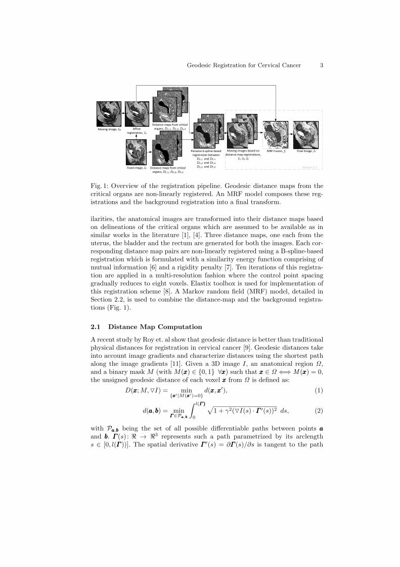
Fig. 1: Overview of the registration pipeline. Geodesic distance maps from thecritical organs are non-linearly registered. An MRF model composes these reg-istrations and the background registration into a final transform.
ilarities, the anatomical images are transformed into their distance maps basedon delineations of the critical organs which are assumed to be available as insimilar works in the literature [1], [4]. Three distance maps, one each from theuterus, the bladder and the rectum are generated for both the images. Each cor-responding distance map pairs are non-linearly registered using a B-spline-basedregistration which is formulated with a similarity energy function comprising ofmutual information [6] and a rigidity penalty [7]. Ten iterations of this registra-tion are applied in a multi-resolution fashion where the control point spacinggradually reduces to eight voxels. Elastix toolbox is used for implementation ofthis registration scheme [8]. A Markov random field (MRF) model, detailed inSection 2.2, is used to combine the distance-map and the background registra-tions (Fig. 1).
2.1 Distance Map Computation
A recent study by Roy et. al show that geodesic distance is better than traditionalphysical distances for registration in cervical cancer [9]. Geodesic distances takeinto account image gradients and characterize distances using the shortest pathalong the image gradients [11]. Given a 3D image I, an anatomical region Ω ,and a binary mask M (with M(xxx) ∈ 0, 1 ∀xxx) such that xxx ∈ Ω ⇐⇒ M (xxx) = 0,the unsigned geodesic distance of each voxel xxx from Ω is defined as:
D(xxx;M,OI) = minxxx′|M(xxx′)=0
d(xxx,xxx′), (1)
d(aaa,bbb) = minΓΓΓ∈Paaa,bbb
∫ l(Γ )Γ )Γ )
0
√1 + γ2(OI(s) ·ΓΓΓ ′(s))2 ds, (2)
with Paaa,bbb being the set of all possible differentiable paths between points aaaand bbb. ΓΓΓ (s) : < → <3 represents such a path parametrized by its arclengths ∈ [0, l(ΓΓΓ ))]. The spatial derivative ΓΓΓ ′(s) = ∂ΓΓΓ (s)/∂s is tangent to the path

4 Sharmili Roy, John J. Totman, Joseph Ng, Jeffrey Low, and Bok A. Choo
direction. γ controls the contribution of the image gradient with respect to thephysical distances and for γ = 0, D reduces to Euclidean distance.
2.2 Markov Random Field Fusion
Fusing multiple registrations of an image pair using MRF is known to yieldhigh accuracy [10]. We formulate registration fusion as a labeling problem wherethe goal is to find the best registration for each voxel. The four registrationsto choose from are the background affine registration and the three non-lineardistance map registrations. Let IF (xxx) : ΩF and IM (xxx) : ΩM be the fixed andthe moving anatomical images with domains ΩF ,ΩM ⊂ R3 respectively. Theaffine registered image, IA = IM (TA(xxx)) : ΩF , is obtained by interpolatingIM at the affine transformed voxel coordinates of IF . Let DF,U and DA,U bethe uterus-based distance maps of IF and IA respectively. The distance mapsDF,U and DA,U are registered using a B-spline transformation model Tµ(xxx) withcoefficients µ. The resulting dense deformation is used to transform IA andcompute IU = Tµ(IA). Similarly, DF,B , DA,B and DF,R, DA,R are defined asthe bladder-based and rectum-based distance maps of IF and IA respectively.The images IB and IR are computed by registering the bladder-based and therectum-based distance maps and transforming IA respectively (refer Fig. 1).
Let IT be the final transformed image. The labeling problem assigns eachvoxel v ∈ V , a registration that minimizes the disagreement between IF (v) andIT (v). The label set consists of the four registrations that are assigned labelsU for uterus-based, B for bladder-based, R for rectum-based and A for thebackground affine registration. The goal is to find a labeling function f thatassigns a fv ∈ U,B,R,A, ∀v ∈ V such that the following objective functionE(f) is minimized:
E(f) =∑v∈VVv(fv) +
∑u∈Nv
Vuv(fu, fv), (3)
where Vu measures the difference between Ifv (v) and IF (v) and Vuv penalizesfu 6= fv for all neighbors Nv of v. Each voxel v is connected to 26 neighborsin the 3D space. We use the Potts model for the pairwise potential defined asV(x, y) = K · T (x 6= y), where T (·) is 1 if its argument is true, and 0 otherwise.The data term, Vv, is defined as:
Vv(fv) =1
|Nv|+ 1
∑u∈(Nv∪v)
(IF (u)− Ifv (u))2 + αDA,fv (v) · T (fv 6= A), (4)
where α weighs the voxel’s geodesic distance from the corresponding criticalorgan. Geodesic distances are less accurate as the voxel moves further away fromthe organ surface. The second term in Eq. 4 increases the energy if the voxelis too far away from the organ on which the distance map and the registrationis based. Since the background affine transformation is not dependent on anyorgan or distance transformation, DA,A is not defined and hence excluded fromthe second term. The solution of Eq. 3 gives a voxel-wise registration map thatis used to compose the final image as IT (v) = Ifv (v),∀v ∈ V .

Geodesic Registration for Cervical Cancer 5
(a) (b)
(c)
Fig. 2: (EBRT,BT) registration. (a) Fixed BT image (IF ) and the critical or-gan delineations on IF . Blue, red and gray colors are for uterus, rectum andbladder respectively. (b) Moving EBRT image (IM ) and the organ delineations.(c) The registered image (IT ), propagated organ delineations and their absolutedifference with ground truth on IF . Higher intensities show higher differences.
Table 1: This table compares the mean registration accuracy of the proposedmethod with Osorio et. al. [4] and Berendsen et al. [3] for (EBRT,BT) registra-tion. Mean Hausdorff distances (HD) in mm and Dice coefficients are compared.
Anatomy Osorio et al. Berendsen et al. Ours Masked Reg.(Mean shortest dist.) (Dice coeff.) (HD/ Dice coeff.) (HD/ Dice coeff.)
Uterus 1.60 0.77 0.67/0.88 6.67/0.40Bladder 1.25 0.75 0.30/0.91 3.58/0.50Rectum 1.15 0.78 1.00/0.82 4.21/0.44
3 Results
42 image pairs from clinical stage 2B−4A cervical cancer patients are used toassess the registration accuracy. The BT and EBRT images are acquired on 1.5TMagnetic Resonance (MR) and 3T simultaneous positron emission technology/magnetic resonance (PET/MR) machines respectively. In some cases EBRT andBT images are acquired three months apart. Fig. 2 illustrates a (EBRT,BT)image pair registration and it is observed that the proposed method successfullyhandles large deformations in the critical organs. Table 1 compares the meanregistration accuracy of (EBRT,BT) pairs with those reported in related worksand shows that the presented framework outperforms existing methods. TheDice coefficients and the Hausdorff distances are computed between the organdelineations on the fixed image and the propagated delineations on the registered
6 Sharmili Roy, John J. Totman, Joseph Ng, Jeffrey Low, and Bok A. Choo
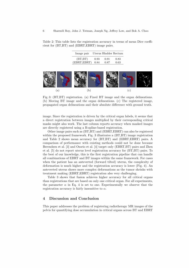
Table 2: This table lists the registration accuracy in terms of mean Dice coeffi-cient for (BT,BT) and (EBRT,EBRT) image pairs.
Image pair Uterus Bladder Rectum
(BT,BT) 0.93 0.91 0.83(EBRT,EBRT) 0.84 0.87 0.63
(a) (b) (c)
Fig. 3: (BT,BT) registration. (a) Fixed BT image and the organ delineations.(b) Moving BT image and the organ delineations. (c) The registered image,propagated organ delineations and their absolute difference with ground truth.
image. Since the registration is driven by the critical organ labels, it seems thata direct registration between images multiplied by their corresponding criticalmasks might also work. The last column reports accuracy when masked imagesare directly registered using a B-spline-based registration.
Other image pairs such as (BT,BT) and (EBRT,EBRT) can also be registeredwithin the proposed framework. Fig. 3 illustrates a (BT,BT) image registrationand Table 2 shows mean accuracy for (BT,BT) and (EBRT,EBRT) pairs. Acomparison of performance with existing methods could not be done becauseBerendsen et al. [3] and Osorio et al. [4] target only (EBRT,BT) pairs and Zhenet al. [5] do not report uterus level registration accuracy for (BT,BT) pairs. Tothe best of our knowledge, this is the first registration pipeline that can handleall combinations of EBRT and BT images within the same framework. For caseswhen the patient has an anteverted (forward tilted) uterus, the complexity ofdeformation is much higher and the registration accuracy is lower (Fig. 4). Ananteverted uterus shows more complex deformations as the tumor shrinks withtreatment making (EBRT,EBRT) registration also very challenging.
Table 3 shows that fusion achieves higher accuracy for all critical organsthan registrations that are based on only one critical organ. For all experiments,the parameter α in Eq. 4 is set to one. Experimentally we observe that theregistration accuracy is fairly insensitive to α.
4 Discussion and Conclusion
This paper addresses the problem of registering radiotherapy MR images of thepelvis for quantifying dose accumulation in critical organs across BT and EBRT

Geodesic Registration for Cervical Cancer 7
Table 3: This table compares registration accuracy of MRF fusion with registra-tions based on individual critical organs for (EBRT,BT) image pairs in terms ofDice coefficients.
Anatomy Uterus-based reg. Bladder-based reg. Rectum-based reg. MRF fusion
Uterus 0.88 0.49 0.45 0.88Bladder 0.63 0.90 0.54 0.91Rectum 0.60 0.54 0.85 0.82
(a) (b) (c)
Fig. 4: (EBRT,BT) registration for an anteverted uterus. Registration accuracyis lower due to highly complex deformations in the uterus. (a) IF and the or-gan delineations. (b) IM and the organ delineations. (c) The registered image,propagated organ delineations and their absolute difference with ground truth.
sessions. Traditional deformable registration methods fail to handle the complexdeformations present in these images. The paper advocates the use of geodesicdistance maps that can be more robustly registered than the original anatomicalimages. Converting anatomical images to distance maps effectively simplifies theregistration challenges and allows to register a pair of EBRT images or a pairof BT images or a combination of EBRT and BT images within the same regis-tration scheme. The idea is to be able to transfer organ locations/ delineationsfrom one treatment day to the other for a personalized and adaptive planning ofradiotherapy. We believe that the idea of registering distance maps from criticalorgans can also be used for registration and dose accumulation during radiother-apy of other cancers in the pelvis such as prostate and rectum. Evaluation of themethod for rectal cancer dose accumulation is under investigation.
The proposed method assumes apriori critical organ delineations. Typicallyin MRI-guided radiation therapy planning for cervical cancer, the tumor, cervix,vagina, uterus, bladder and rectum are circumscribed in MR images by a radi-ation therapist [12]. In theory, this existing data can be used for registration ifaccessible. Further, in future we plan to study the sensitivity of registration withrespect to segmentation accuracy and investigate the use of semi-automated/ au-tomated segmentation.
A limitation of this work is that MRF fusion cannot guarantee smoothnessat the label ‘seams’, potentially leading to folding in the transformation. Com-peting methods also suffer from this problem. We are currently investigatingGaussian smoothing as suggested in [10] for folding removal as a post-processing
8 Sharmili Roy, John J. Totman, Joseph Ng, Jeffrey Low, and Bok A. Choo
step. In addition, we are working on evaluating the method using a landmarkbased approach where anatomically relevant landmarks will be placed on andin-between the critical organs. By doing this, we can quantify the accuracy ofthe dense deformation field and also compute the dose volume histograms whichis a more powerful tool for dose accumulation.
Acknowledgment The work is partially funded by NMRC NUHS Centre GrantMedical Image Analysis Core (NMRC/CG/013/2013).
References
1. Christensen, G.E., Carlson, B., Chao, K.S.C., Yin, P., Grigsby, P.W., Nguyen, K.,Dempsey, J.F., Lerma, F.A., Bae, K.T., Vannier, M.W., others: Image-based doseplanning of intracavitary brachytherapy: registration of serial-imaging studies usingdeformable anatomic templates, International Journal of Radiation Oncology* Biol-ogy* Physics, 51(1), 227-243, (2001)
2. Kim, H., Huq, M.S., Houser, C., Beriwal, S., Michalski, D.: Mapping of dose distribu-tion from IMRT onto MRI-guided high dose rate brachytherapy using deformable im-age registration for cervical cancer treatments: preliminary study with commerciallyavailable software, Journal of Contemporary Brachytherapy, 6(2), 178-184, (2014)
3. Berendsen, F.F., Kotte, A.N.T.J., de Leeuw, A.A.C., Jurgenliemk-Schulz, I.M.,Viergever, M.A., Pluim, J.P.W.: Registration of structurally dissimilar images inMRI-based brachytherapy, Physics in Medicine and Biology, 59(15), 4033, (2014)
4. Osorio, E.M.V., Kolkman-Deurloo, I.-K.K., Schuring-Pereira, M., Zolnay, A., Hei-jmen, B.J.M., Hoogeman, M.S.: Improving anatomical mapping of complexly de-formed anatomy for external beam radiotherapy and brachytherapy dose accumula-tion in cervical cancer, Medical Physics, 42(1), 206-220, (2015)
5. Zhen, X., Chen, H., Yan, H., Zhou, L., Mell, L.K., Yashar, C.M., Jiang, S., Jia,X., Gu, X., Cervino, L.: A segmentation and point-matching enhanced efficient de-formable image registration method for dose accumulation between HDR CT images,Physics in Medicine and Biology, 60(7), 2981, (2015)
6. Mattes, D., Haynor, D.R., Vesselle, H., Lewellyn, T.K., Eubank, W.: Nonrigid mul-timodality image registration, Medical Imaging, 1609-1620, (2001)
7. Staring, M., Klein, S., Pluim, J.P.W.: A rigidity penalty term for nonrigid registra-tion, Medical Physics, 34(11), 4098-4108, (2007)
8. Klein, S., Staring, M., Murphy, K., Viergever, M., Pluim, J.P.W., others: Elastix: atoolbox for intensity-based medical image registration, IEEE Transactions on MedicalImaging, 29(1), 196-205, (2010)
9. Roy, S., Totman, J. J., Choo, B. A.: Unified registration framework for cumulativedose assessment in cervical cancer across external beam radiotherapy and brachyther-apy, Proc. SPIE Medical Imaging 2016: Image Processing, 9784 (2016)
10. Gass, T., Gabor S., Orcun, G.: Registration fusion using markov random fields,Biomedical Image Registration, Springer International Publishing, 213-222, (2014)
11. Wang, Z., Bhatia, K. K., Glocker, B., Marvao, A., Dawes, T., Misawa, K., Mori,K., Rueckert, D.: Geodesic patch-based segmentation, Medical Image Computing andComputer Assisted Intervention MICCAI, 666-673, (2014)
12. Ghose, S., Holloway, L., Lim, K., Chan, P., Veera, J., Vinod, S.K., Liney, G., Greer,P.B., Dowling, J.: A review of segmentation and deformable registration methodsapplied to adaptive cervical cancer radiation therapy treatment planning, ArtificialIntelligence in Medicine, (2015)