genotoxicity of 1,2,4-benzenetriol : roles of oxygen
TRANSCRIPT

Genotoxicity of 1,2,4-Benzenetriol:
Roles of Oxygen-Derived Active Species and Quinones
Luoping Zhang
B. Sc., Wuhan University, 1982 M. Sc., Huazhong University of Science and Technology, 1985
A DISSERTATION SUBMITTED IN PARTIAL FULFILLMENT OF THE REQUIREMENTS FOR THE DEGREE OF
DOCTOR OF PHILOSOPHY
Biochemical Toxicology School of Kinesiology
Luoping Zhang 1993
SIMON FRASER UNIVERSITY
September, 1993
All rights reserved. This dissertation may not be reproduced in whole or in part, by photocopy or other means, without permission of the author.

APPROVAL
Name:
Degree:
Title of Thesis:
I'_saniining Committee:
Chair:
Luoping Zhang
Doctor of Philosophy
Gcnotosicity of 1,2,4-Benzcnetriol: Roles of Osygen-Dcrived Active Specics and Quinones
Dr. Danicl Weeks
- - Dr. Allan J. Davison Professor of Biochemistry Senior Supervisor
-
Dr. Marty'q ,T. Smith Professor of Toxicology, University of California at Berkeley
Dr. MirinniXosin Professor of Environmental Cancinogenesis
Dr. G y b i t s Prof? qf Cardiac Biology
BFrfmS;id & ~ e a n Professor of Denatology, University of British Columbia Internal Examiner
Dr. E -zabeth Snow Prof 3 ssor of Environmental Medicine, New York University External Examiner
Date Approved: 16 G& k 1793

PARTIAL COPYRIGHT LICENSE
I hereby g r a n t t o Simon Fraser U n i v e r s i t y t he r i g h t t o lend
my t h e s i s , p r o j e c t o r extended essay ( t h e t i t l e o f which i s shown below)
t o users o f t he Simon Fraser U n i v e r s i t y L i b r a r y , and t o make p a r t i a l o r
s i n g l e cop ies o n l y f o r such users o r i n response t o a request from the
l i b r a r y o f any o t h e r u n i v e r s i t y , o r o t h e r educa t i ona l i n s t i t u t i o n , on
i t s own b e h a l f o r f o r one o f i t s users . I f u r t h e r agree t h a t permiss ion
f o r m u l t i p l e copy ing o f t h i s work f o r s c h o l a r l y purposes may be g ran ted
by me o r t he Dean o f Graduate S tud ies . It i s unders tood t h a t copy ing
o r p u b l i c a t i o n o f t h i s work f o r f i n a n c i a l ga i n s h a l l n o t be a l lowed
w i t h o u t my w r i t t e n permiss ion .
T i t l e o f Thes is /Pro jec t /Ex tended Essay
G e n o t o x i c i t y of 1 , 2 , 4 - B e n z e n e t r i o l :
R o l e s of Oxygen-Derived A c t i v e S p e c i e s and Qu inones
Author :
(s i cpa tu re )
Luoping Zhang 7- ----
(name)
Sep tember 20 , 1993
(da te )

ABSTRACT
Benzene causes leukemia in humans. It also induces structural and numerical
chromosomal aberrations in lymphocytes and bone marrow of exposed workers. Recent
evidence indicates that such chromosomal changes are important in the progression of
many cancers, including leukemia. Studying the mechanisms whereby benzene induces
chromosomal alterations will provide information regarding the mechanism of its
carcinogenicity. Benzene, however, is unlikely as the immediate toxic species since its
metabolism is required for toxicity. 1,2,4-Benzenetriol is one of the most reactive
metabolites of benzene. This research characterizes the genotoxicity exhibited by
benzenetrio1 and investigates the mechanisms involved.
Benzenetriol produced both structural and numerical chromosomal changes, as
measured by a modified micronucleus assay. Micronuclei containing either whole
chromosomes or acentric chromosome fragments can be distinguished by using an
antikinetochore antibody. Benzenetriol increased the frequency of micronucleus formation
eight-fold in HL60 cells. Addition of copper ions ( C U ~ + ) enhanced the frequency of
benzenetriol-induced micronuclei a further three-fold and altered the pattern of micronuclei
from predominantly kinetochore-positive to kinetochore-negative.
The cell-free system confirms the influence of Cu2+ in changing the mechanism of
benzenetriol-induced micronucleus formation from aneuploidy to clastogenicity. In this
system, Cu2+ changed the free radical propagated chain reactions from one-electron
transfer to two-electron transfer. Reactive oxygen species are implicated in the Cu2+-
mediated chromosome breakage through their direct or metal-mediated irlteractions with
DNA. 8-Hydroxy-2'-deoxyguanosine (8-OH-dG) is a marker of oxidative DNA damage

and ultimately leads to point mutations. Benzenetriol increased the level of 8-OH-dG, and
Cu2+ again enhanced this effect.
Benzenetriol also induced numerical aneuploidy in the form of hyperdiploidy of
chromosomes 7 and 9, which was determined by fluorescence in situ hybridization
(FISH). The hyperdiploidy frequencies were increased approximately three-fold over the
controls. Microtubule integrity is required for proper chromosomal segregation.
Benzenetriol disrupted normal microtubular organization, as shown by immuno-
cytochemical staining with anti-tubulin antibodies. The quinonoid oxidation products of
benzenemol and reactive oxygen species are the most llkely cause of this disruption and the
mitotic abnormalities which result in aneuploidy. These data provide further insight into
the mechanisms involved in benzene-induced genotoxicity and leukemia.

DEDICATION
To my mother whose spirit has been encouraging me to succeed in my studies and
scientific career, and to my country where I grew up and received my early education.

ACKNOWLEDGEMENTS
I would like to extend acknowledgement to my thesis supervisor, Professor Allan
Davison, for providing research guidance, critical insight, and the opportunity to follow my
scientific interests and to further my education both in SFU and at Berkeley. I am also
thankful for his critical review of this dissertation. I am especially indebted to Professor
Martyn Smith who, as my research advisor, guided my study direction, appraised critically
my work and taught me the scientific writing skills that have been so important in
completing this dissertation. I also appreciate the encouragement and advice offered by the
other members of my thesis committee, Professors Miriam Rosin and Glen Tibbits.
My thanks greatly express to Dr. Moire L. Robertson-Creek and Brian Bandy for
freely sharing their laboratory expertise, scientific insight and friendship. My appreciation
also goes to Dr. Prema Kolachana and Pravina Venkatesh who have collaborated in this
research project and have made contributions to the data presented in this dissertation. I am
thankful for the encouragement and suggestions provided by my friends and colleagues Dr.
Kathleen Meyer, Dr. Stan Tamaki, Dr. Vangala Subrahmanyam, Margy Lambert, Lee
Moore, Dr. Nina Titenko-Holland, Sharan Campleman, Joseph Wiemels, Dr. Jenny
Compton and Elinor Fanning in University of California at Berkeley; and Anna Li, Dr. Jim
Moon and Eunice Rousseau in Simon Fraser University.
Finally, I am truly grateful to my family and my best friends: Philip Heimann, Anna
Li and Christopher Kelly for their love, support and close hendship over the years.

TABLE OF CONTENTS
APPROVAL ........................................................................................ ..ii
ABSTRACT ......................................................................................... i i i
DEDICATION ...................................................................................... .v
ACKNOWLEDGEMENTS ....................................................................... .vi . .
TABLE OF CONTENTS ........................................................................ . . v i ~ . . ................................................................................ LIST OF FIGURES xll
LIST OF TABLES ................................................................................. xiv
PREFACE ........................................................................................... x v
CHAPTERS
I . General Introduction
.................................................................. 1.1 BACKGROUND 1
1.1.1 Human Exposure to Benzene
1.1.2 Toxic Effects of Benzene
1.1.3 Metabolism of Benzene
1.2 TOXIC QUINONES AND REACTIVE OXYGEN SPECIES ............... 9
1.2.1 Toxicity of Benzene-Derived Quinones
1.2.2 Mechanisms of Quinone-Mediated Toxicities
1.2.3 Toxicity of Reactive Oxygen Species
1.2.4 DNA Damage by Oxidative Stress
1.2.5 DNA Damage by Activation of Endonucleases
1.2.6 Defenses against Reactive Oxygen Species
1.2.7 Roles of Reactive Oxygen Species in Benzene Toxicity
1.3 TOXICITY OF 1,2,4-BENZENETRIOL ..................................... 21
1.3.1 BT is one of the most reactive metabolites of benzene
1.3.2 BT activates molecular oxygen to reactive oxygen species
1.3.3 BT causes oxidative DNA damage both in vitro and in vivo
1.3.4 BT binds to DNA and inhibits DNA synthesis
1.3.5 BT causes sister chromatid exchanges and chromosomal alterations
v i i

1.4 CHROMOSOMAL ABERRATIONS ........................................ ..23
1.4.1 Common Chromosomal Changes in Leukemia
1.4.2 Benzene Induced Chromosomal Aberrations
1.4.3 Micronucleus Formation and Detection
1.4.4 Aneuploidy and Fluorescence in situ Hybridization
1.4.5 Roles of Microtubules in Aneuploidy
1.5 OBJECTIVES ............................................. 1.5.1 Specific Aims
1.5.2 Hypotheses
1.6 REFERENCES .................................................................. .34
1.7 LEGENDS FOR FIGURES .................................................... 54
1.8 FIGURES ......................................................................... 56
1.9 TABLES ........................................................................... 69
11. Benzene Metabolite, 1,2,4-Benzenetriol Induces Micronuclei and
Oxidative DNA Damage in Human Lymphocytes and HL60 Cells
2.1 ABSTRACT ....................................................................... 7 1
2.2 INTRODUCTION ................................................................ 72
2.3 MATERIALS AND METHODS ............................................... -75
2.3.1 Cell Culture
2.3.2 Treatment Conditions
2.3.3 Staining Procedures
2.3.4 Scoring
2.3.5 Cell Viability and Cell Division Kinetics
2.3.6 Extraction, Purification and Enzymatic Hydrolysis of Cell DNA
2.3.7 Synthesis of 8-OH-dG Standard
2.3.8 Determination of 8-OH-dG in DNA
2.3.9 Statistical Analysis
......................................................................... 2.4 RESULTS 79
2.4.1 Effect of BT on Micronucleus Induction in Human Lymphocytes
2.4.2 Effect of BT on Micronucleus Induction in HL60 Cells
v i i i

2.4.3 Effect of CU*+ on the Induction of Micronuclei in HL60 Cells by BT
2.4.4 Effect of BT on 8-OH-dG Levels in DNA of HL60 Cells
2.4.5 Effect of Cu2+ on BT-Induced 8-OH-dG Formation
DISCUSSION ................................................................... .83
ACKNOWLEDGEMENTS .................................................... .88
REFERENCES .................................................................. .89
LEGENDS FOR FIGURES .................................................... 94
FIGURES ......................................................................... 95
TABLES ......................................................................... 102
111. Detection of 1,2,4-Benzenetriol Induced Aneuploidy and Microtubule
Disruption by Fluorescence in situ Hybridization and Immunocyto- chemistry
3.1 SUMMARY ..................................................................... 104
............................................................. 3.2 INTRODUCTION . l o 5
3.3 MATERIALS AND METHODS ............................................. . lo7
3.3.1 Cell Culture
3.3.2 Chemical Treatment
3.3.3 In situ Hybridization
3.3.4 Detection and Amplification
3.3.5 Scoring Procedures and Criteria
3.3.6 Microtubule Staining
3.3.7 Photography
3.3.8 Statistical Analysis
3.4 RESULTS ....................................................................... 1 12
3.4.1 Fluorescence in situ Hybridization in HL60 Cells
3.4.2 Colchicine as a Positive Control for Aneuploidy Induction
3.4.3 BT-Induced Hyperdiploidy of Chromosome 9
3.4.4 BT-Induced Hyperdiploidy of Chromosome 7
3.4.5 BT-Induced Trisomy in Hyperdiploidy
3.4.6 BT-Induced Microtubule Disruption

3.5 DISCUSSION ................................................................... 117
3.6 ACKNOWLEDGEMENTS ................................ .................... 123
3.7 REFERENCES ... ..... ......... .... ... ....... ... .. .. .. ... .... ..... ... .. .... ... .. 124
3.8 LEGENDS FOR FIGURES ................................................... 129
3.9 FIGURES ........................................................................ 130
3.10 TABLES ......................................................................... 133
I V . Effect of Metals, Ligands and Antioxidants on the Reactions of
Oxygen with 1,2,4-Benzenetriol
4.1 ABSTRACT ..................................................................... 139
4.2 INTRODUCTION ... .. .. . .. ... . . .. . ... .. . . . .. ... . . . . . . .. .. ..... ... ..... .. ... ... 140
4.3 MATERIALS AND METHODS .................... ...... ..... ............... 144
4.3.1 Reagents
4.3.2 Anaerobic Solution of 1,2,4-Benzenetriol
4.3.3 Assay Procedures
4.3.4 Data Analyses
4.3.5 Statistical Analysis
4.4 RESULTS ....................................................................,.. 146
4.4.1 Characterization of Benzenetriol Autoxidation
4.4.2 Catalytic Effect of Cu2+ and Fe3+
4.4.3 Inhibitory Effects of Superoxide Dismutase and Catalase
4.4.4 Stimulation or Inhibition by Desfenioxamine
4.4.5 Effects of Formate and Mannitol
4.5 DISCUSSION ................................................................... 149
4.5.1 Cu2+ Is a More Effective Catalyst than Fe3+
4.5.2 Superoxide Propagates the Redox Reactions
4.5.3 Effect of C U ~ + on the Free Radical-propagated Chain Reactions
4.5.4 Effect of Fe3+ on the Reaction Mechanism
4.5.5 Cu2+ Changes Type of Benzenetriol-induced Micronuclei
4.5.6 Desfemoxarnine Stimulates Benzenetriol Autoxidation
4.5.7 Complete Inhibition by Superoxide Dismutase and Desferrioxamine

4.6 ACKNOWLEDGEMENTS .................................................... 156 .................................................................. 4.7 REFERENCES 157
................................................... 4.8 LEGENDS FOR FIGURES 160 ....................................................................... 4.9 FIGURES $163
4.10 TABLES ......................................................................... 171
V . Conclusion and Perspective ...................................................... 173
APPENDICES
Appendix 1
Appendix 2
Appendix 3A
Appendix 3B
Appendix 4
Appendix 5
Appendix 6
Appendix 7
Description of HL60 Cells from ATCC ...................................... 177
................................... Mitosis Index and Growth of HL60 Cells 178
Binucleate Cell with a Micronucleus .......................................... 179
................ Binucleate Cell with a Kinetochore-positive Micronucleus 180
Sample of Score Sheet for Micronucleus Assay ............................. 181
Sample of Score Sheet for FISH Assay ...................................... 182
FISH Staining of Chromosome 9 in HL60 Cells ........................... 183
List of Contributions from Author and Co-authors ......................... 184

LIST OF FIGURES
I . General Introduction
FIGURE 1 .
FIGURE 2 .
FIGURE 3 .
FIGURE 4 .
FIGURE 5 .
FIGURE 6 .
FIGURE 7 . FIGURE 8 .
FIGURE 9 .
FIGURE 10 .
FIGURE 1 1 .
FIGURE 12 .
FIGURE 13 .
.......................................................... Blood cell development 56
Pathways of benzene metabolism and excretion .............................. 57
Proposed mechanism of benzene-induced myelotoxicity .................... 58
Redox cycling and biological fates of quinones ............................... 59
Redox cycling pathways for 1.2.4.benzenetriol. its quinones and
.......................................................... reactive oxygen species 60
............................................ DNA adducts of hydroxyl radicals 61
................................. Formation of 8-hydroxy-2'-deoxyguanosine 62
............. Hypothetical mechanisms of DNA damage by oxidative stress 63
......................... Antioxidant enzymes in the cellular defense systems 64
Hypothetical scheme for the possible pathways of free radical formation
...................................................... during benzene metabolism 65
................ Micronucleus assay with anti-kinetochore antibody staining 66
.......... Scheme for fluorescence in situ hybridization (FISH) technique 67
Involvement of microtubules in mitotic chromosomal segregation ......... 68
11 . Benzene Metabolite. 1.2. 4.Benzenetriol Induces Micronuclei and Oxidative DNA Damage in Human Lymphocytes and HL60 Cells
. ............................. FIGURE 1 Induction of micronuclei in human lymphocytes 95
. FIGURE 2 1.2.4.Benzenetrio 1.induced micronuclei in HL60 cells ...................... 96
. FIGURE 3A Effect of C U ~ + on 1.2.4.benzenetrio 1.induced micronuclei ................. 97
. FIGURE 3B Effect of Cu2+ doses on benzenetriol-induced micronuclei ................. 98
xii
FIGURE 4A . Effect of 1.2. 4.benzenemol on the 8-OH-dG level in cell DNA ............ 99
FIGURE 4B . Effect of Cu2+ on benzenetriol-induced 8-OH-dG level ................... 100
............... FIGURE 5 . Proposed mechanisms of benzenemol-induced genotoxicity 101
111 . Detection of 1.2. 4.Benzenetriol Induced Aneuploidy and Microtubule
Disruption by Fluorescence in situ Hybridization and Immunocyto- chemistry
FIGURE 1 . Dose-response for benzenetriol-induced hyperdiploidy .................... 130
FIGURE 2 . Immunocytochemical anti-tubulin antibody staining ........................ 131
FIGURE 3 . Effects of benzenetriol and colchicine on microtubule integrity and
cytoskeletal organization in HL60 cells ....................................... 132
I V . Effect of Metals. Ligands and Antioxidants on the Reaction of
Oxygen with 1.2. 4.Benzenetriol
FIGURE 1A . Rate constant of benzenetriol autoxidation ................................... 163
..................... . FIGURE 1B Effect of benzenetrio1 doses on the initial reaction rate 164
. ............................ FIGURE 2A Catalyses of C U ~ + and Fe3+ in the autoxidation 165
FIGURE 2B . Cu2+ is more efficient than Fe3+ ........................................ 1 6 6
FIGURE 3 . Inhibition of superoxide dismutase and catalase ............................. 167
FIGURE 4 . Effect of superoxide dismutase in the presence of metal ions .............. 168
FIGURE 5 . Stimulation and inhibition by desferrioxamine .............................. 169
FIGURE 6 . Possible chemical structure of [benzenemol-Cu-oxygen] and the
............ schematic electron flow from benzenetriol via Cu2+ to oxygen 170

LIST OF TABLES
Chapter I
TABLE 1. Effects of benzene exposure in humans ........................................ 69
Chapter I1
TABLE 1. Viability and replicative index of lymphocytes and HL60 cells treated with
1,2,4-benzenetriol and copper ions ........................................... 102
Chapter I11
TABLE 1.
TABLE 2.
TABLE 3.
TABLE 4.
TABLE 5.
Chapter IV
Baseline and effect of colchicine on nuclear spot frequencies in HL60 cells
using a centromeric probe specific for chromosome 9 ...................... 133
Number of hybridization domains of chromosome 9 in interphase nuclei of
HL60 cells treated with benzenetriol for 24 h in media ..................... 134
Number of hybridization domains of chromosome 9 in interphase nuclei of
HL60 cells treated with benzenetriol for 1 h in PBS ........................ 135
Nuclear spot frequencies in HL60 cells treated with benzenetriol for 24 h
in media using a centromeric probe specific for chromosome 7 ........... 136
Proportion of trisomy and tetrasomy in total hyperdiploidy induced by
1,2,4-benzenetriol in HL60 cells .............................................. 137
TABLE 1. Effects of Cu2+/Fe3+ on the oxidation of benzenetriol ...................... 171
TABLE 2. Summary of results ..... ...... ... . .. . ... ... .. ... . ... ....... ... .. ... ... ..... .. . .. 172
x iv

PREFACE
This dissertation comprises five chapters including a general introduction, three
papers written for publication, and a final conclusion.
The general introduction (Chapter I) outlines the context and goals of the research.
It summarizes the mechanisms of toxic quinones and reactive oxygen species, the effects of
1,2,4-benzenetriol, and chromosomal aberrations. This study investigates the role of
oxygen-derived active species, metal ions and quinones in the genotoxicity of benzenemol
as well as the possible mechanisms involved.
Chapter I1 has been recently published in the journal of Environmental Molecular Mutagenesis (21:339-348, 1993). In this section, we demonstrate that benzenemol induces
micronuclei and oxidative DNA damage in human cells. We also present the data as
evidence that copper ions alter benzenetriol's genotoxicity from aneuploidy to
~lastogenicit~.
Chapter I11 has been accepted for publication in the journal of Mutation Research. Here, we provide further evidence that benzenetriol can induce aneuploidy and
microtubular disruption in human HL60 cells.
Chapter IV has been submitted to the journal of Free Radical Biology & Medicine for publication. This final study consists of observations of the stirnulatory or inhibitory
effect of metals, ligands and antioxidants on the autoxidation of benzenetriol. It describes
the free radical propagated chain reactions during the oxidation.
The last section (Chapter V) is an overview of the findings in this research. It
concludes that benzenemol induces numerical and structural chromosomal alterations and
point mutations by two distinct mechanisms. The disruption of cytoskeletal microtubules
and the damage of DNA by oxygen-derived active species and toxic quinones are involved
in the benzenemol induced genotoxicity in human cells.
The respective contributions of the author and each co-author in this research are
listed in Appendix 7.

CHAPTER ONE
GENERAL INTRODUCTION
1 .I BACKGROUND
Annual production of benzene in the United States was reportedly 11.8
billion pounds in 1988 (CEN 1989). The high volume production and the
volatility has contributed to benzene being a ubiquitous environmental pollutant
and a public health concern (USEPA, 1980; Mehlman, 1991). High levels of
human exposure occur during the manufacturing of benzene Or its use as an
organic solvent or as a starting material for the synthesis of other chemicals in
the pharmaceutical and chemical industries (Brief et al., 1980). In addition,
benzene from other sources such as gasoline emission, automobile exhaust
and cigarette smoke further contribute to exposure. Epidemiological studies
have established that benzene is a human carcinogen and occupational
exposure to benzene can lead to the development of various blood disorders
including aplastic anemia, pancytopenia, and acute myelogenous leukemia
(IARC, 1982). The mechanism by which benzene causes genetic damage
remains unclear. However, it has been postulated that the genotoxicity of
Primary metabolites of benzene such as hydroquinone, catecho1 and 1,2,4-
benzenetrio1 are associated with benzene-induced carcinogenicity.
Benzenetrio1 is a highly reactive metabolite and its oxidation leads to the
formation of genotoxic oxygen-derived active species. The goal of this research
is to characterize genetic alterations induced by benzenetriol and to assess
whether it may potentially play a role in benzene-induced carcinogenicity.

1.1.1 Human Exposure to Benzene
Human contact with benzene occurs primarily from occupational and
environmental exposure. Occupational exposure to benzene numbers
approximately 0.2 to 2 million workers in North America (NIOSH, 1977; OSHA,
1985). A air concentration of benzene in the workplace has been reported at
1600 mgIm3 (500 ppm) and at times in excess of 3200 mg/m3 (1000 pprn)
(IARC, 1982). The Occupational Safety and Health Administration (OSHA) has
set the standard limit for occupational exposure to benzene at 10 pprn time-
weight average (TWA) per &hour (OSHA, 1985). In compliance with the OSHA
standard, benzene exposure levels have decreased dramatically (Runion and
Scott, 1985). In 38,000 air samples from 7 factories using or producing
benzene in the U.S., the concentrations of benzene in air were below 1 pprn in
87% of samples, between 1 and 10 pprn in 11.4% , and greater than 10 pprn in
only 1.6% (Runion and Scott, 1985).
The recommended benzene exposure level of 10 pprn TWA per day was
designed to prevent developing aplastic anaemia and/or other perturbances
which cause depression of cellular elements in blood (Yardley-Jones et al.,
1991 ). Subsequently, epidemiologic studies indicated an excess risk for the
development of benzene-induced leukemia at 10 pprn (Rinsky et al., 1987).
OSHA (1987), therefore, has recently promulgated a new standard limiting
worker exposed to 1 pprn &hour TWA in the U. S. Occupational exposure in
many Third World countries frequently exceeds 10 pprn in industrial settings
(Holmberg and Lundberg, 1985). And in extreme cases such as Chinese
workers, the exposure levels are still up to 100 pprn benzene.

Although occupational exposure levels in the U. S. have been declining,
benzene is still present above recommended levels in ambient air (Holmberg
and Lundberg, 1985). Thus, environmental exposure spreads to the general
population. An estimated 75% of the U.S. population has been environmentally
exposed to benzene, usually 2-3 orders of magnitude lower than occupational
exposure (Van Raalte, 1982). Environmental sources of benzene exposure
include industrial processes, gasoline engine emissions, gasoline re-fueling,
solvents in consumer products, and cigarette smoke.
Cigarette smoke is a major component of environmental exposure with
levels in smokers reported at 15 pg/m3 compared to 1.5 - 2 pg/m3 in non-
smokers (Wallace, 1989). Cigarette smokers are exposed to an estimated 2 mg
of benzene per day (approximately 60 pg / cigarette). Non-smokers are
exposed to benzene from 'sidestream' smoke since a single cigarette emits
between 12 to 480 pg of benzene into the air (IARC, 1986; Wallace et al., 1987).
Based on linear extrapolation of leukemia risk at higher levels of
exposure, Wallace (1 989) estimated that 1000 cases of leukemia per year in the
U. S. can be attributed to benzene exposure, nearly half of them are associated
with cigarette smoking. Indeed, cigarette smokers have a 50% higher risk of
developing leukemia than non-smokers (Sandler, 1985; McLaughlin el al.,
1989; Severson et al., 1990). In summary, smokers and those occupationally
exposed face the greatest risk of developing benzene-induced leukemia.

1.1.2 Toxic Effects of Benzene
Toxic responses to benzene in humans include central nervous system
(CNS) anesthesia from short-term, intensive exposures, hematopoietic toxicity
from relatively long-term, chronic exposure, and leukemia from much longer-
term exposure to much lower concentrations of benzene.
Acute exposure to 20,000 ppm benzene in human was fatal within 5-10
min (Flury, 1928). Workers exposed to acute, non-lethal levels (<20,000 ppm)
of benzene developed CNS depression, loss of consciousness, irregular heart-
beat, dizziness, headache, and nausea (Deutsche F., 1974). In a cohort of
more than 500,000 occupationally exposed persons, benzene poisoning,
defined as peripheral leukocyte counts less than 4000 cells/mm3 with an
Occupational history of benzene exposure and CNS symptoms, was detected.
with average exposures as low as 12.5 ppm (Yin et al., 1987). Since benzene
is regulated at concentrations far below those required to induce CNS toxicity,
its neurotoxic effect is infrequently studied (Snyder, 1988).
Humans are susceptible to benzene myelotoxicity (hematopoietic
toxicity) as shown by the high incidence of blood dyscrasias among workers
with significant exposure (Goldstein, 1989). Ever since Santesson (1897) and
Selling (1916, both referenced in IARC, 1982) reported that chronic exposure to
benzene could cause death from aplastic anemia, the toxic effects of benzene
to the blood of man were recognized. In addition to aplastic anemia, benzene
exposure induces thrombocytopenia, granulocytopenia, lymphocytopenia,
Pancytopenia, leukopenia, and bone marrow hypoplasia or aplasia (Laskin and
Goldstein, 1977; IARC, 1982; OSHA, 1987).

Pluriopotent bone marrow stem cells divide and differentiate into all the
major blood cell types (Figure 1). Damage of the stem cells could potentially
lead to disorders of various mature blood cells (Golde, 1991). The decrease or
absence of erythrocytes, leukocytes, platelets and their precursors within the
bone marrow can result from alterations in processes governing stem cell self-
renewal (aplastic anemia). Another major disease that can ensur from stem cell
toxicity is leukemia, in which the stem cell acquires a heritable alteration in
processes controlling replication and differentiation, and populates the blood
system with immature, non-functional leukocytes (Dexter et al., 1985). It
remains to be determined why benzene is toxic to bone marrow stem cells, and
how it acts at the molecular level to alter self-renewal and differentiation.
However, the correlation of benzene exposure levels to specific hematopoietic
effects in humans has been well documented and is summarized in Table 1.
An association between occupational exposure to benzene and the
development of leukemia was noted by Delore and Borgomano as long ago as
1928. Since then, numerous case reports and more than 100 occurrences of
leukemia associated with benzene exposure have been published (Vigliani,
1976). In the 19701s, Aksoy showed that the incidence of leukemia among
28,500 Turkish shoe workers exposed chronically to benzene was 13.6 per
100,000, significantly above the incidence of 6 per 100,000 in general
population (Aksoy, 1972, 1976 and 1977). Rinsky et al. (1987) examined a
cohort of 1165 men working in a rubber industry in Ohio, and demonstrated an
overall standardized mortality ratio (SMR; the ratio of observed over expected
deaths x 100) of 337 for leukemia and 409 for multiple myeloma. And a clear
dose-response relationship was discerned between the cumulative benzene
exPosure and development of leukemia in this thorough retrospective study.

The largest investigation to date involved 233 benzene factories and 83 control
factories in 12 Chinese cities (Yin et al., 1987; 1989). The benzene-exposed
cohort includes 28,460 workers and the control cohort contains 28,257. The
overall SMR for exposed cohort was 574 (containing 76.6% of the acute
leukemia cases). Most benzene-induced malignancies are particularly acute
myelogenous leukemias (AML). The International Agency for Research on
Cancer (IARC, 1982) states:
"The relationship between benzene exposure and the development of
acute myelogenous leukemia has been established in epidemiologic studies".
Based on these epidemiologic data, benzene has been classified as a
human carcinogen. In addition to AML, other leukemias, such as multiple
myeloma, chronic myelogenous leukemia, chronic lymphocytic leukemia,
Hodgkin's lymphoma, non-Hodgkin's lymphoma, and acute lymphocytic
leukemia, are also associated with benzene exposure (reviewed by Goldstein,
1977; Kipen et al., 1988; Austin, 1988). Benzene also induces an excess
incidence of cancer at sites other than the hematopoietic system, including the
urinary bladder (Steineck et al., 1990) and lung (Aksoy, 1976; Yin et al., 1989).
1 .I .3 Metabolism of Benzene
Benzene, like some other carcinogens, apparently lacks direct-acting
carcinogenicity and mutagenicity. It must first be metabolized to the "proximate"
carcinogens (Andrews et al., 1977). Thus, benzene by itself is weakly
mutagenic or non-mutagenic in standard bacterial and mammalian cell
mutation assays (Dean, 1978; Huff et al., 1989). The inability to detect

benzene-induced mutation with these assays may be explained by the lack of
enzymes needed to convert benzene to its metabolites (IARC, 1982; Glatt et al.,
1989). The liver is the primary site for bioactivation of benzene because it
contains the requisite cytochrome P450 enzymes. The cytochrome P450 2E1 in
the P450 family has been established to specifically metabolize benzene (Koop
et al., 1989).
The metabolism of benzene is complex and not completely understood.
The major pathways of benzene metabolism and excretion are shown in Figure
2. Benzene is metabolized primarily in the liver by cytochrome P450 2E1 to
benzene epoxide which rearranges to yield phenol (PH) or conjugates with
glutathione to yield phenyl mercapturic acid (Tunek, 1978; Koop et al., 1989).
Benzene epoxide can also be catalyzed by epoxide hydrolase to benzene-1,2-
trans-dihydrodiol which is further converted by cytosolic dehydrogenation
(Jerina, 1968) to catechol (CAT). This is the major pathway from benzene to
catechol (Sata et al., 1963; Tomaszewski et al., 1975). Phenol can be
hydroxylated mainly to p-hydroquinone (HQ) and perhaps to o-catechol
(Sawahata and Neal, 1983). CAT can be further hydroxylated to form the
triphenolic metabolite of benzene, 1,2,4-benzenetriol (BT) (Tunek et al., 1980).
BT can also arise from the hydroxylation of HQ (Inoue et al., 1989a). The ring-
opened metabolite, trans,trans-muconaldehyde can also be formed from
benzene dihydrodiol (Latriano et al., 1986) or benzene oxide oxepin (Gad-El-
h i m et at., 1986) by various pathways (Figure 2).
Most above benzene metabolites have been detected in rodent blood,
bone marrow and urine (Rickert et al., 1979), and in human urine (Inoue et al.,
' 989a, b). Parke and Williams (1953al b) administered I4C-benzene (0.34 - 0.5

ml/kg) orally to rabbits, and 84-89% of the dose was recovered as radioactivity
in expired air (43%), urine (34.5%), feces and body tissues (5-10%). The 34.5%
radioactivity recovered in urine consisted of PH (23.5%), HQ (4.8%), CAT
(2.2%), BT (0.3%), trans,trans-muconic acid (1.3%) and L-phenyl mercapturic
acid (0.5%). The more stable metabolites are found at higher concentrations in
blood, bone marrow, lymphoid tissue, and fat (Bergman, 1979; Greenlee et al.,
1981). Surprisingly, no one has measured the BT level in blood and bone
marrow yet because the BT is too unstable.
The relationship among benzene, its metabolites, and bone marrow
toxicity is complex. Although metabolism of benzene is necessary for bone
marrow toxicity, the reactive metabolites are formed in the liver and transported
to bone marrow. The currently accepted mechanism of the transport from liver
to bone marrow is shown in Figure 3. Once the metabolites are formed in the
liver, they enter the blood and preferentially accumulate in the bone marrow
where they exert their toxicity (Andrews et al., 1979; Rickert et al., 1979). The
most potent toxicants may be, not the primary benzene metabolites, but the
secondary metabolites arising from the further action of myeloperoxidase (MPO)
in bone marrow (Smith et al., 1989). Thus, bone marrow provides another site
for further metabolism, in which yet more toxic quinone metabolites of benzene
are derived and more reactive oxygen species are generated (Greenlee et al.,
1981 ; Irons, 1985). Both quinones and reactive oxygen species, therefore, are
candidates for the acute toxicants which contribute to benzene-induced
hemotoxicity and carcinogenicity (Smith et al., 1989; Subrahmanyam et at.,
1 991 a).

1.2 TOXIC QUINONES AND REACTIVE OXYGEN SPECIES
Quinones are aromatic diketones, widely distributed in nature, and found
in plants, fungi, bacteria, and animals. They function as components of the
electron transport chains involved in cellular respiration and photosynthesis.
Quinones have been used as anticancer chemotherapeutic drugs and as
pigments for dyes and cosmetics. Quinones are also prevalent as
environmental pollutants many of which are toxic in human. The chemistry and
toxicity of quinones have been recently reviewed in detail by Monks et al.
(1 992a). Quinones have reduction potentials appropriate to redox-cycling, and
therefore they create oxidative stress by generating reactive oxygen species.
1.2.1 Toxicity of Benzene-Derived Quinones
Polyhydroxylated metabolites of benzene , CAT, HQ, and BT each
oxidize (either enzymatically or spontaneously) to their respective semi-
quinones and quinones (Greenlee et al., 1981; Subrahmanyam et at., 1991b).
Benzene-derived quinones include 1,2-benzoquinone (0-BQ), 1,4-
benzoquinone (p-BQ), and 2-hydroxy-benzoquinone (2-OH-BQ) as well as their
corresponding semiquinones (Figure 2). The semiquinones (at physiological
pH) exist as anion radicals, each containing one unpaired electron (Mukherjee
et al., 1989). Benzene-derived semiquinone radicals and quinones exert
various toxicities both in vitro and in vivo.
Quinone-mediated toxicities include neurotoxicity, cytotcxicity,
genotoxicity and perhaps carcinogenicity (Smith et al., 1989; Monks et al.,
1992a). As mentioned above, acute exposure to benzene results in a central

nervous system (CNS) depression (Haley, 1977) probably caused by quinone
metabolites of benzene (Snyder, 1988). A well known neurotoxicant,
6-hydroxydopamine, however, provides a good example for quinone-induced
neurotoxicity. It undergoes a rapid autoxidation to its quinone form which is
cytotoxic through genesis of the products of partial reduction of oxygen (Gee
and Davison, 1989).
BQ, HQ, CAT and BT are cytotoxic in both human lymphocytes and HL60
cells (Yager et al., 1990; Levay and Bodell, 1992; Zhang et at., 1993). To be
toxic in bone marrow, benzene's phenolic metabolites require further
bioactivation. Bone marrow stroma consists predominantly of two cell types,
macrophages and fibroblastoid stromal cells. HQ is selectively toxic at the level
of the macrophage in bone marrow stroma cells, whereas the fibroblastoid
stromal cell is relatively resistant (Thomas et al., 1989). Thus selective toxicity
toward macrophage stroma cells in the bone marrow is because they contain
considerably greater peroxidase activity and less DT-diaphorase (NAD(P)H :
quinone oxidoreductase) than the fibroblastoid cells. Peroxidases catalyze the
further oxidation of the phenolic metabolites to their quinones, and DT-
diaphorase is an obligate two-electron reductase, which converts quinones to
their parent hydroquinones (Ross et at., 1990). Consequently, HQ is activated
to p B Q in the bone marrow macrophage stroma cells rather than in the
fibroblastoid cells (Thomas et al., 1990).
The resultant quinones or semiquinone radicals can react directly with
DNA, RNA, and proteins. Benzene and its metabolites bind to DNA i~ rabbit
bone marrow in vitro (Rushmore et at., 1984), and to RNA in rat and mouse
organs in vivo (Arfellini et at., 1985). The DNA adducts in HL60 cells treated

with CAT, HQ and BT have been identified by 32P postlabeling (Levay and
Bodell, 1992). In addition, these benzene-derived quinones form adducts with
glutathione (GSH) (Tunek et al., 1980) and proteins such as hemoglobin
(Adams and Biemann, 1 WO), cytoskeletal protein (Irons, l985), and enzymes
including prostaglandin H synthase (Schlosser et al., 1990). Interestingly, all of
these adducts have been identified as the same structural product of the
"Michael addition", namely, the formation of quinone-thioethers.
Benzene-derived quinones, then, are cytotoxic and genotoxic, and in
addition mutagenic and carcinogenic. For instance, BQ produced a 100-fold
increase in the frequency of expression of 6-thioguanine resistance in Chinese
hamster V79 cells (Glatt et al., 1989). HQ, BT and CAT also induced this gene
mutation but PH did not, suggesting that formation of quinones is responsible.
However, none of them induced substantial mutations in S. typhimurium (Glatt
et al., 1989). The mechanisms underlying benzene-induced bone marrow
toxicity and leukemogenesis are therefore presumed to result from the toxic
effects of benzene-derived quinones.
1.2.2 Mechanisms of Quinone-Mediated Toxicities
The toxicological activity of benzene-derived quinones resides in their
ability to react directly with cellular nucleophiles and also their ability to undergo
redox cycling inducing oxidative stress (Figure 4). In the context of toxicology,
or more generally in their interactions with macromolecules in living cells,
quinones possess two paramount chemical properties. They are electrophiles
and oxidants.

Quinones, as electrophiles, react directly with cellular nucleophiles such
as protein and nonprotein sulfhydryls. GSH is the major intracellular nonprotein
sulfhydryl reagents (Reed, 1985). The conjugation of potentially toxic
electrophiles with GSH usually results in detoxification and excretion (Figure 4).
Quinones conjugated with GSH are excreted in urine as their corresponding
sulfate conjugates and/or as mercapturic acids. The multiple conjugates have
been recently shown to have their own toxic properties (Monks and Lau,
1992b). Interaction of quinones with nucleophiles may lead to toxicity by
inhibition of critical thiol groups in enzymes, or by alteration of the thiol balance
in the cell, either of which can interfere with cellular regulatory processes (Sies,
1985). Reactions of quinones with thioles by Michael addition (Finley, 1974)
result in formation of hydroquinone thioethers.
+ HS-R - 2-Alkylt hiohydroquinone
Quinones, as oxidants, are enzymatically reduced by both one-electron
(NADPH-cytochrome P450 reductase, etc.) and two-electron (DT-diaphorase)
reductions, resulting in the formation of semiquinones or hydroquinones. The
reduction of 2-OH-BQ, as an example of a benzene-derived quinone, is
depicted in Figure 5. Two-electron reduction of quinones catalyzed by
1 2

DT-diaphorase has generally been considered to be a detoxification pathway
since the resulting hydroquinones can be further conjugated and excreted
(Figure 4). In some cases, such as with certain bioreductive alkylating agents,
the reduction by DT-diaphorase leads to activation of the compound. For
example, the reduction of 2-OH-BQ, by DT-diaphorase, may lead to redox
cycling dependent on the rate of reduction to BT followed by its autoxidation
back to the parent quinone (Brunmark and Cadenas, 1988). Formation of
semiquinones therefore occurs during either the reduction of quinones or the
oxidation of hydroquinones (Figure 5).
Semiquinones disproportionate to their quinone and hydroquinone forms
with great facility (Mukherjee et al., 1989), enabling semiquinones and
quinones to be "redox facilitators". Thus, if their redox potential is sufficiently
low, semiquinones can transfer their single unpaired electron to other oxidants
such as molecular oxygen (02). If there is an efficient mechanism for re-
reduction of the quinone product to the semiquinone, a redox cycle facilitated by
the quinone/semiquinone couple ensues (Figure 4). Semiquinones can react
with O2 forming superoxide anion radical ((I2*-) and regenerating the parent
quinone, which is then available for re-reduction in a redox cycling. The net
result of this redox cycling is oxidative stress resulting from disproportionate
Consumption of cellular reducing equivalents and the generation of active
oxygen species. Ultimately, the flux of 02.- and other oxygen-derived active
species destroys unsaturated lipids, DNA, proteins, and other essential cellular
molecules, with lethal results. Much recent research has emphasized the role
of oxidative stress and redox cycling in quinone and polyphenol toxicity (Smith
et a[., 1 985).
1.2.3 Toxicity of Reactive Oxygen Species
During the oxidation of benzene's polyhydroxy metabolites and their
semiquinones, molecular oxygen (02) is primarily reduced to 02*-, and a
number of oxygen radicals and active species are generated (Figure 5).
Superoxide dismutase can further convert 02.- to hydrogen peroxide (H202),
which may generate hydroxyl radicals (HOD) in the presence of metal ions and
reducing equivalents. Singlet oxygen ('02) is perhaps produced during the
formation of benzene-derived quinones based on the chemiluminescence seen
in the oxidation of at least one triphenol (Fatur and Davison, 1987). Since
quinones are colored and absorb ultraviolet and visible iight energy, photo-
excited quinones can be quite reactive to hydrogen and electron abstractions,
andlor energy transfer to another acceptor molecule, such as 0 2 (i.e., chemical
photosensitization) (Bruce, 1974).
Ail of these reactive oxygen species cause oxidative stress and cellular
injury. Subjecting cells to oxidative stress can result in severe dysfunctions,
including peroxidation of membrane lipids, depletion of nicotinamide
nucleotides, rises in intracellular free Ca2+ ions, cytoskeletal disruption and
DNA damage. The latter is often measured as formation of single-strand
breaks, double strand breaks or DNA adducts etc.. Indeed, DNA damage has
been almost invariably observed in a wide range of mammalian cell types
exposed to oxidative stress (Halliwell and Aruoma, 1991). However, neither
02.- nor H202 induce scission of DNA, as measured by strand breakage
(Rowley and Halliwell, 1983) or by chemical changes in the deoxyribose,
Purines or pyrimidines (Aruoma et al., 1989a and b). The ultimate damaging
is putatively the HOD radical.

The HO* radical induces oxidative changes in DNA bases (Figure 6),
resulting, for example, in 8-hydroxy-2'-deoxyguanosine (8-OH-dG), as a end-
product of the attack of Hog on guanine at the C-8 site (Figure 7). 8-OH-dG thus
represents the covalent addition of HO* (or lo2) to guanine in DNA (Floyd et al.,
1986; Devasagayam et al., 1991) and has become a useful and sensitive
marker of oxidative DNA damage both in vitro and in vivo (Roy et al., 1991).
This is a potentially mutagenic change since the hydroxylated DNA base can be
misread at the modified and adjacent base residues in the DNA-polymerase
reaction in vitro (Kuchino et al., 1987). Thus, 8-OH-dG causes the DNA point
mutations from G -> T substitution (Cheng et al., 1992).
Abundant evidence indicates the involvement of reactive oxygen species
in mutagenesis and carcinogenesis. Thus the rate of spontaneous mutation is
enhanced in E coli that lacks the activity of superoxide dismutase (Touati,
1989). Similar mutagenic effects have been shown in a range of mammalian
cell types subjected to oxidative stress. Single base changes and deletions
were the main patterns of mutation in cells exposed to H202 (Moraes et al.,
1990). The majority of base changes were at GC base pairs, the GC to AT base
transition being predominant. The base deletions cause frameshift leading to
loss of the information caved in the affected gene. Moreover, mutations caused
by active oxygen species in certain coding regions of tumor-related genes might
be responsible for subsequent activation of oncogenes andlor inactivation of
tumor suppressor genes (Wei, 1992). Such changes can cause neoplastic
transformation of normal cells through an increase in expression of certain
Proto-oncogenes such as c-fos, c-jun, and c-myc (Sherman et al., 1990; Yin et
al.. 1 992). Furthermore, reactive oxygen species are involved in initiation
(irreversible change in DNA), promotion (further changes in gene expression),

and perhaps progression (production of malignant tumor) in the multistage
model of carcinogenesis (Sun, 1990; Halliwell and Aruoma, 1991).
Taken together, reactive oxygen species cause cellular injury, DNA
damage, gene mutation, and probably cancer. Most studies have focused on
oxidative DNA damage, but this can not involve direct attack of 02.- or of H202
on the DNA, unless transition metal ions are present to allow H o g formation.
The formation of HO* catalyzed by transition metal ions is crucial in oxidative
DNA damage. DNA damage induced by oxidative stress may involve the direct
reaction of HO- with DNA or the oxidative activation of nucleases which cleave
the DNA backbone (Halliwell and Aruoma, 1991). Metal ions play a essential
role in both mechanisms, as summarized in Figure 8.
1.2.4 DNA Damage by Oxidative Stress
Hydroxyl radicals damage DNA. A major source of HOW radicals is the
one-electron reduction of H202 catalyzed by transition metal ions. The most
efficient intracellular metal ions in physiological systems are iron and copper
(Miller et al., iggo), which play essential roles in many enzymes and proteins. If
they become "decompartrnentalized", traces of their soluble ions can convert
H2O2 to HO- via Fenton and Haber-Weiss type reactions:
FeICu H202 > Hoe + HO- (Fenton)
Fe/Cu 02*- + H202 > HO- + HO- + 0 2 (Haber-Weiss)

Redox cycling of the metal ions amplifies their ability to transfer electrons to
H202 (Chevion, 1988). Thus, the above reaction (2) involves two electron-
transfer steps as below:
(3) 02.- + Fe3+ / Cu2+ > Fe2+ / Cu+ + 0 2
(4) H202 + Fe2+ / CU+ > Fe3+ / Cu2+ + HO- + HO-
Such mechanisms can mediate DNA damage only if the HO* is
generated upon or very close to the DNA. Otherwise, the high reactivity of HOm
precludes its diffusing significant distances within the cell. Since H202 crosses
biological membranes easily, it can penetrate to the nucleus and react with
DNA-bound iron or copper to form 'site-specific' hydroxyl radicals. The metal
ions might be those originally bound to the DNA in vivo, or those released
within the cell as a result of oxidative stress, and then bound to the DNA
(Dizdaroglu et al., 1991 ; Halliwell, 1987).
1.2.5 DNA Damage by Activation of Endonucleases
Oxidative stress initiates a series of events within the cell, which leads to
activation of C$+-dependent endonucleases that can fragment DNA (Birn boim
and Kanabus-Kaminska, 1 985; Bi rnboim, 1 988). Thus, oxidative stress
increases intracellular free Ca2+ by: (1) concomitant inactivation of Ca2+-binding
by the endoplasmic reticulum, (2) inhibition of plasma membrane Ca*+-
extrusion systems, and (3) release of Ca2+ from mitochondria (Halliwell and
Aruoma, 1991 ).

The two mechanisms are not mutually exclusive. Changes in the
availability of Ca2+ may depend upon, or give rise to, changes in the availability
of iron or copper ions. DNA damage could be inhibited by preventing the rise
either of C U ~ + / Fe3+ or of Ca2+, using their appropriate chelators (Goldstein and
Czapski, 1990; Dypbukt et at., 1990) or by scavenging the reactive oxygen
species using endogenous defense systems (Sun, 1990).
1.2.6 Defenses against Reactive Oxygen Species
Aerobic organisms have evolved multiple defense systems to protect
themselves against the reactive oxygen species generated in vivo, The
enzymatic defenses remove 02*- by superoxide dismutase, and H202 by
catalase and glutathione peroxidase which works in coordination with
glutathione reductase and glucose-6-phosphate dehydrogenase (Sun, 1990).
These enzymes function in cooperation to diminish oxidative damage (Figure
9). Moreover, DNA repair enzymes can correct certain oxidative DNA defects
such as single-strand breaks (Friedberg, 1985), but may not double-strand
breakage (Kohen et al., 1986). Since the HO' radical oxidizes non-specifically
almost all biomolecules, including superoxide dismutase and catalase, no
specific enzymes can selectively destroy it. Non-enzymatic defenses protect
against uncontrolled oxidative reactions initiated by Hoe.
The non-enzymatic defenses comprise metal-binding and chain breaking
antioxidants. Low molecular mass antioxidants for chain breaking include lipid-
soluble vitamins, such as a-tocopherol, carotenoids and retinoids which prevent
lipid peroxidation and peroxidative damage by scavenging or quenching
Oxygen-derived active species. Ascorbic acid and GSH, as water-soluble

antioxidants, may consume HO* formed in the cytoplasm. Other scavengers,
including urate, formate and polyalcohols (e. g. sugars) also protect the system
against radical mediated damage both in vitro and in vivo (Goldstein and
Czapski, 1990). In addition, the administration of synthetic scavengers may
diminish the damage in mammals subjected to experimental oxidative stress.
Metal chelators which remove a metal ion from access to redox cycling
can block the oxidant action of the metal. For example, desferrioxamine, a
synthesized chelator, binds specifically iron and copper as well as other metal
ions into redox resistant chelates (Halliwell, 1989). Since desferrioxamine does
not readily cross cell membranes, its effects in vivo are variable (Halliwell and
Aruoma, 1991). As a copper chelator, bathocuproine inhibits copper-initiated
human DNA damage in cultured human cells (Kawanishi et al., 1989), and D-
penicillamine extends the lifespan of patients with copper overload diseases
(Goldstein and Czapski, 1990). Proteins can also bind metal ions in forms
unable to accelerate free radical reactions (Halliwell and Aruoma, 1991).
Ferritin and metallothionein can store Fe3+ and CU*+, respectively (Miller et al.,
1990). Even relatively weak ligands which do not defer redox cycling or HO*
production can prevent 'site specific' oxidative damage. They do so by
displacing the metal from more sensitive target so that the damage occurs at
less vital cellular components.
In summary, oxidative DNA damage results when reactive oxygen
species and/or metal ions are not adequately removed, for example, when
antioxidants are depleted and/or if the formation of reactive oxygen species is
increased beyond the capacity of the defense systems (Sies, 1991).

1.2.7 Roles of Reactive Oxygen Species in Benzene Toxicity
Although the benzene-derived quinones (discussed in 1.2.1) most likely
mediate benzene-induced toxic effect, little attention has been paid to oxygen
radicals and active species as further mediators of toxicity. The myelotoxic
effects of benzene have been attributed to free radical formation, as metabolites
of benzene, oxygen radicals, or lipid peroxidation products. The potential role
of oxygen free radical in benzene-induced myelotoxicity and leukemia has
been carefully reviewed by Subrahmanyam et al. (1 991 a).
The variety of reactions generating reactive oxygen species during
benzene metabolisms is summarized in Figure 10. Those specific to bone
marrow may occur (1) during peroxidative metabolism catalyzed by
myeloperoxidase (MPO) (or epoxidase and perhaps prostaglandin H synthase),
(2) during autoxidation of benzene's phenolic metabolites, or (3) by the
membrane-bound NADPH-oxidase activity of phagocytic bone marrow cells.
However, the role of active oxygen species playing in benzene-induced
myelotoxicity and carcinogenesis remains unknown. To further understand the
roles and mechanisms involved in the toxicities of benzene-derived quinones
and reactive oxygen species, this research has focused on studies of the
genotoxicity of 1,2,4-benzenetriol (BT). BT, a triphenolic metabolite of benzene,
has a strong ability to activate oxygen and to be oxidized to its corresponding
quinones (Figure 5). Relatively few studies have been conducted on toxicity of
BT and little is known about the mechanisms involved. BT's toxic effects are
reviewed in next section.

1.3 TOXICITY OF 1,2,4-BENZENETRIOL
1.3.1 BT is one of the most reactive metabolites of benzene.
BT has been identified as a human urinary metabolite of benzene. In the
urine of benzene-exposed workers, the concentration of BT is related linearly to
the intensity of benzene exposure (Inoue et al., 1989a, b). BT is presumably
also formed in bone marrow via the activation of primary benzene metabolites
by myeloperoxidase (MPO) (Subrahmanyam et al., 1991b) or hydrolysis of
o-BQ (Mason, 1949). The accumulation of BT in blood and bone marrow
remains unknown because it is unstable. Although BT is a relatively minor
product in the liver during the metabolism of benzene in quantitative terms
(Rusch et al., 1977), the low steady state levels may be biologically significant
because of its high reactivity.
1.3.2 BT activates molecular oxygen to reactive oxygen species.
BT is more unstable in molecular oxygen than the other metabolites due
to its high potency to donate electrons (Greenlee et al., 1981). BT's two
0-hydroxy substitutes are capable of binding transition metal ions that catalyze
its autoxidation (Bandy et al., 1990). Thus, it readily autoxidizes to its
Corresponding quinones and activates molecular oxygen to a mixture of
reactive species, including superoxide anion (02.-), hydrogen peroxide (H202)
and hydroxyl radical (HO) as well as perhaps singlet oxygen ( lo2) (Figure 5).
These species are known to damage DNA and other cellular macromolecules
(Halliwell and Aruoma, 1991 ; Stadtman and Oliver, 1991).

1.3.3 BT causes oxidative DNA damage both in vitro and in vivo.
BT induces: (1) single- and double-stranded DNA breaks in the mouse
lymphoma cell line, L5178YS (Pellack-Walker and Blumer, 1986), (2) DNA
cleavage in isolated human DNA fragments (Kawanishi et al., 1989), and (3)
Supercoiled DNA breakage in isolated Escherichia coli plasmids (Li, 1992).
The BT-induced DNA damage is elevated by transition metal ions, such as
C U ~ + and Fe3+, but inhibited by superoxide dismutase, catalase and hydroxyl
radical scavengers (Lewis et al., 1988; Kawanishi et al., 1989; Li, 1992).
However, all of these findings were in vitro. In the only in vivo study (Kolachana
et al., 1993), the administration of BT to mice induces a significant increases in
the level of 8-hydroxy-2'-deoxyguanosine (8-OH-dG) in bone marrow DNA.
Here BT has much higher potency than the other metabolites of benzene tested
(pH, CAT and HQ). In another study, the repeated administration of BT to rats
for six weeks induced myelotoxicity (Rao et al., 1988).
1.3.4 BT binds to DNA and inhibits DNA synthesis.
In addition, BT or its oxidative products can bind to DNA directly with
uncertain consequences. The DNA adducts have been identified by 32P
Postlabelling in HL60 cells treated with BT (Levay and Bodell, 1992). Moreover,
BT inhibits nuclear DNA and/or RNA synthesis in L5178YS cells (Pellack-
Walker et al., 1985) and in mouse bone marrow cells (Lee et al., 1989). BT also
decreases the mitotic index and slows the cell cycle in human lymphocytes
(Erexson et at., 1985).

1.3.5 BT causes sister chromatid exchanges and chromosomal
alterations.
BT induces dose-related increases in the sister chromatid exchanges
(SCE) in human peripheral blood lymphocytes (Erexson et at., 1985) and
Chinese hamster V79 cells (Glatt et al., 1989). Tice and co-workers (1982)
further demonstrated that a 4 h exposure to 28 ppm benzene in DBA/2 and
C57~116 mice significantly increased SCE. However, increased frequencies of
SCE were not seen in workers exposed to benzene, on the contrary, decreased
frequencies have been reported (Watanabe et al., 1980). BT induces both
HPRT (Hypoxanthine-guanine Phosphoribosyl Transferase) gene mutation
(6-thioguanine resistance) and micronuclei in Chinese hamster V79 cells (Glatt
et al., 1989). Little is known about the mechanisms involved in BT-induced
toxicity. It therefore seemed to us timely to investigate the formation of
micronuclei and other chromosomal alterations caused by BT, which has not
been determined in human cells yet.
1.4 CHROMOSOMAL ABERRATIONS
There are two principal categories of chromosome abnormalities: (I)
structural chromosome alterations (deletion, inversion, and translocations etc.),
and (2) numerical chromosome alterations (the gain or loss of entire
chromosomes). Inductions of such non-random chromosomal changes are
thought to be primary events crucial to the onset of neoplasia. They are
associated with a variety of cancers, including leukemia (Yunis, 1983; Rowley,
1 984).

1 A.1 Common Chromosomal Changes in Leukemia
Most human leukemias and lymphomas have at least one type of
nonrandom clonal abnormality. Chromosome gains and losses are often seen
in both myeloid and lymphoid neoplasms. An extra chromosome 8 or 21 is the
most common change, and a loss of chromosome 5 or 7 also occurs in acute
myelogenous leukemias (AML) (LeBeau and Rowley, 1984). Trisomy of
chromosome 12 is often found in chronic lymphocytic leukemia (CLL) (Han et
al., 1983; Losada et al., 1991). In addition to these numerical chromosomal
aberrations, the remarkably specific structural changes are seen in certain
hematological leukemias. Among the most dramatic are the structural
rearrangements uniquely associated with a particular leukemia. Thus,
translocations of chromosomes 8 and 21 [t(8; 21) (q22, q22)], and
chromosomes 15 and 17 [t(15; 17) (q22, q21)J are observed in AML, whereas a
translocation of chromosome 9 and 22 [t(9; 22) (q34, q l l ) ] is seen in most
(>go%) cases of chronic myelogenous leukemia (CML) and in some patients
with AML and ALL. The chromosomal alterations in human cancers and
leukemias have been extensively reviewed by Sandberg (1990).
These specific chromosomal abnormalities are reportedly involved in
tumor development. The products of genes adjacent to the breakpoints indicate
how chromosome translocations facilitate tumor initiation and progression
(Rabbitts, 1991 ). The Philadelphia chromosome (Phl) represents a reciprocal
translocation, t(9; 22) (q34, q l 1). The Abelson proto-oncogene, c-abl, normally
located on chromosome 9, resides on the Phl (De Klein et al., 1982). The
translocation fuses the c-abl gene with bcr, a transcription unit on chromosome
22. The bcr sequence stimulates the transcription of c-abl, activates the tyrosine

kinase activity of c-Abl (Konepka et al., 1984) and converts Abl to a transforming
protein for hematopoietic cells (McLaughlin et al., 1987). The chimeric protein
of the bcr-a61 fusion gene (p210) has growth promoting tyrosine kinase activity
(Sawyers et al., 1991) and can induce CML in transgenic mice (Heisterkamp et
al., 1990). Thus, the c-a61 proto-oncogene is converted to an oncogene by this
chromosomal rearrangement.
In addition, the loss of the long arm portions of chromosome 5 and 7
predicts a high risk of leukemic transformation and particularly occurs in AML.
Deletions of 5/5q and/or 7/7q were found in patients who developed
myelodysplastic syndrome (MDS) or AML arising secondary to cytotoxic therapy
for a previous malignant disease (Rowley et al., 1981). A specific gene involved
in the genesis of MDS has not been identified, but the 5q chromosomal region
is the gene location of several hematopoietic growth factors (IL-3, IL-4 IL-5,
M-CSF, GM-CSF, IL-9) and growth factor receptors (M-CSF receptor, platelet-
derived growth factor) (Groopman et at., 1989).
1 A.2 Benzene Induced Chromosomal Aberrations
Benzene consistently induces chromosome alterations in mammalian in
vivo and in vitro test systems (reviewed by Dean, 1985). Frequencies of altered
structural chromosome changes and of abnormal chromosome numbers are
increased in lymphocytes and bone marrow cells from workers exposed to
benzene. Sasiadek (1992) recently reported that benzene-exposed workers
had breakpoints primarily in chromosomes 2, 4 and 7. The chromosome 5
anomaly was observed in lymphocytes of workers exposed to benzene or other
Organic solvents (Van der Berghe et al., 1985). Chromosomal abnormalities in
the chromosome C-group (through chromosomes 6 to 12 and X), such as
trisomy of chromosomes 6, 8, and 9, monosomy of chromosome 7, or deletion of
the long arm of chromosome 7 (7q-) were detected in patients with benzene-
induced pancytopenia and leukemia (Erdogan and Aksoy, 1973). Interestingly,
the C-group chromosomes are also frequently abnormal in hematological
disorders and leukemias (Chen et al., 1992; Poddighe et al., 1991 ; Anastasi et
al., 1990). Study of these non-random chromosome alterations provides an
opportunity to investigate potential genetic regulation of hematopoiesis, and
may provide clues to the molecular mechanisms of benzene-induced leukemia.
1.4.3 Micronucleus Formation and Detection
Micronuclei are novel markers of genetic damage, which are visible in
the cytoplasm as small nuclear bodies. They contain either chromosome
fragments or entire chromosomes that fail to incorporate into the daughter
nuclei during cell division (Schmid, 1975). Benzene unequivocally induces
micronuclei in bone marrow cells of mice and rats (Tice et al., 1980; 1982).
Lymphocytes isolated from benzene-exposed workers also contain excess
micronuclei (Dean, 1978; IARC, 1982). Micronuclei arise following exposure to
chemicals that cause structural (chromosome breakage) or numerical
(disruption of chromosome segregation in mitosis) chromosome aberrations. In
assessing their action, the micronucleus assay compares well with
chromosome aberration assays (Fench, 1 993).
The micronucleus assay was originally developed by Evans and co-
workers (1 957) to detect genotoxicity in irradiated plant root tips. The assay
was subsequently modified to detect cytogenetic damage in human peripheral

lymphocytes (Fenech and Morley, 1985), red blood cells (Schlegel et al., 1986),
bone marrow cells (Heddle, 1973), and exfoliated cells (Stich and Rosin, 1984).
In the modified micronucleus assay, cytochalasin b is used to disrupt actin
filaments, thus preventing the cell from undergoing cytokinesis. Therefore, cells
that have initiated cell division are distinguished by their multinucleated form,
and only cells having undergone one cell division (containing binuclei) are
scored for the presence of micronuclei. However, the cytokinesis-blocked
micronucleus assay fails to determine by which mechanisms micronuclei are
formed.
Discovery of antibodies that recognize the kinetochore proteins (Moroi et
at., 1980; Brenner et al., 1981) extended the cytokinesis-blocked micronucleus
assay (Vig and Swearngin, 1986; Degrassi and Tanzarella, 1988). This new
micronucleus assay distinguishes micronuclei containing chromosome
fragments (kinetochore-negative) from those containing entire chromosomes
(kinetochore-positive). It differentiates chemicals that cause chromosome
breakage (clastogens, e. g. radiation and sodium arsenite) from those which
interfere with chromosome separation during mitosis (aneuploidogens, e. g.
diethylstilbestrol or colchicine) (Eastmond and Tucker, 1989). The mechanisms
whereby these agents induce micronucleus formation are fairly well
understood. Chromosome breakage occurs most often by scission of the DNA
and sometimes by interference with DNA-associated proteins such as DNA
polymerases (Gualden, 1987). In contrast, chromosome lagging reflects
chromosomes improper segregation into the daughter nuclei during cell
division. The principle of the modified cytokinesis-blocked micronucleus assay
system and these two mechanisms of micronucleus formation are summarized
in Figure 11.
Using the anti-kinetochore antibody adapted micronucleus assay, Yager
et al. (1990) investigated induction of micronuclei in human lymphocytes by
individual metabolite of benzene, such as PH, CAT, HQ and BQ but not BT.
Their ability tc elevate the frequency of micronucleated cells is: HQ >> BQ >
CAT > PH. In combination, HQ and CAT (at equimolar concentrations) acted
synergistically in increasing both micronucleated cells and kinetochore-positive
micronucleated cells (Robertson et al., 1991). We, therefore, selected the
modified micronucleus assay to investigate the ability of benzene's metabolite
BT to cause genotoxicity in this study.
1.4.4 Aneuploidy and Fluorescence in situ Hybridization
Aneuploidy is the loss or gain of whole chromosome(s) in the cell
karyotype. It is common in leukemia and plays an important role in cancer
progression and metastasis (Barrett et al., 1987; Fearon and Vogelstein, 1990).
Aneuploidy is detected by traditional karyotyping and augmented by the
modified micronucleus assay (kinetochore-positive micronucleus). However,
karyotyping can be applied only to cells that can be stimulated into mitosis and
reliably banded. This limits cytogenetic studies in solid tumors. In addition,
karyotypic analyses are time consuming and labor intensive. Moreover, some
chemicals such as mitomycin C cleave the kinetochore proteins from
chromosomal centromeres (Brinkley and Shaw, 1969). Thus, use of this
modified micronucleus assay to detect aneuploidy may lead to false positive
results. To avoid the limitations and possible errors by these two assays, a
better cytogenetic technique is needed for detecting aneuploidy.

In recent years, a new method, fluorescence in situ hybridization (FISH),
has become available for detecting numerical and structural chromosomal
changes in interphase nuclei and metaphase spreads (Gray and Pinkel, 1992;
Zahed and Vekemans, 1991). This method (Figure 12) utilizes in situ
hybridization with DNA probes specific to blocks of repetitive DNA sequences
on defined regions of specific chromosomes (Willard and Waye, 1987; Pinkel et
al., 1986; 1988). The visualization of chemically-modified probes involves the
use of non-radioactive fluorescent antibodies. The determination of aneuploidy
is performed by simply counting the number of label regions representing a
particular chromosome of interest within the isolated nucleus. This method has
been widely used for the molecular cytogenetic analysis of cancer cells (Cremer
et al., 1988a, b; Hcrpman et al., 1991), but has also been applied to cultured
human lymphocytes (Raimondi et al., 1989; Eastmond and Pinkel, 1990), bone
marrow (Poddighe et al., 1991 ; Jenkins et al., 1992) and exfoliated human cells
(Moore et al., 1993).
Many carcinogens are also aneuploidogens (Oshimura and Barrett,
1986). Benzene is a well known human leukemogen, and its primary
metabolite HQ is an inducer of aneuploidy (Miller and Adler, 1992). It seemed
timely to determine whether BT induces aneuploidy using the FISH technique
and which mechanism is involved.
1.4.5 Roles of Microtubules in Aneuploidy
There are several potential targets for aneuploidy induction in mitotic
cells (Liang and Brinkley, 1985). For example, colchicine and diethylstilbestrol
Interfere with microtubule assembly by inhibiting polymerization of the tubulin

subunits. Ethdium bromide affects the centrioles / centrosomes, mitomycin C
removes kinetochores from chromosomes, and chloral hydrate blocks pole-to-
pole microtubule elongation.
Since the mitotic apparatus consists predominantly of microtubules, any
compound that affects microtubules might cause lagging chromosomes so as to
be a potential aneuploidy-inducer. One function of microtubules is to attach to
the chromosomes at the kinetochore and to guide them to their respective poles
during cell division (Figure 13). The disruption of microtubules can lead to the
formation of micronucleus and/or the induction of aneuploidy.
Microtubules are composed primarily of polymers of alpha and beta
tubulin, which are rich in nucleophilic sulfhydryl groups important for
microtubule assembly. The polyhydroxy benzene metabolites, BT and HQ,
inhibit tubulin polymerization in vitro , with BT being approximately twice as
potent as HQ (Irons et al., 1981 ; Irons, 1985; Pfieffer and Irons, 1983). Another
triphenolic compound similar to BT, the neurotoxin 6-hydroxydopamine
(6-OHDA) inhibits microtubule assembly in vitro (Davison et al., 1986). All these
hydroquinones are readily oxidized to their electrophilic quinone and
semiquinone intermediates, which can interact with nucleophilic sulfhydryls of
tubulin and cause mitotic abnormalities resulting in aneuploidy. In the latter
study, however, the quinonoid oxidation products were relatively ineffective.
In conclusion, both structural and numerical chromosomal aberrations
have been observed in benzene-exposed workers and in laboratory animals
treated with benzene. These aberrations may therefore be important in
benzene-induced leukemia. Thus, studying the mechanisms whereby benzene
or its metabolites produce chromosomal damage may reveal details of the
mechanisms of benzene-induced leukemia.
1.5 OBJECTIVES
1.5.1 Specific Aims
The purpose of this research was to study the role of oxygen-derived
active species and toxic quinones in benzenetriol-induced genotoxicity and to
understand the possible mechanisms involved. We therefore decided to
investigate benzenetriol-induced genetic damage, including structural and
numerical chromosomal alterations, oxidative DNA damage, and microtubule
disruption by the modified micronucleus assay, the FISH technique, HPLC-EC
analysis, and immunocytochemical methods. The human myeloblastic
leukemia (HL60) cell line was chosen as a surrogate of bone marrow cells.
HL60 cells are from human origin, at an early stage in hematopoietic
development, with high myeloperoxidase content, and with a rapid growth rate
(Gallagher et al., 1979). In addition, a cell-free chemical system was used to
study role of radicals and transition metal ions in the mechanisms of free radical
propagated chain reactions of benzenetriol with oxygen. The specific aims
were as follows.
(1) To characterize the genotoxicity of 1,2,4-benzenetriol and determine the
extent to which produces micronuclei and aneuploidy in HL60 cells.

(2) To characterize benzenetriol-induced numerical (kinetochore-positive
micronuclei) and structural (kinetochore-negative micronuclei) chromosomal
alterations, and to determine whether the pattern of micronucleus formation is
contingent upon the presence or absence of metal ions (Cu2+, Fe3+).
(3) To study the mechanisms involved and to determine the mechanisms of
induction of kinetochore positive and negative micronuclei, aneuploidy, and
oxidative DNA damage.
(4) To examine the effects of metals, chelators and antioxidants on the
oxidation of benzenetriol, and to determine how metal ions change the
pathways of free radical chain reactions.
1 S.2 Hypotheses
This dissertation addresses three main sets of hypotheses as follow:
Hypothesis I:
1,2,4-Benzenetriol increases the frequency of micronuclei and levels of
8-OH-dG in human cells. Cu*+ may enhance the benzenetriol-induced
genotoxicity and alter the mechanisms involved.
Hypothesis 11:
1,2,4-Benzenetriol induces aneuploidy and microtubule disruption in
HL60 cells. The benzenetriol-induced microtubule disruption may be a
predominant mechanism of the induction of aneuploidy.

Hypo thesis 111:
Metal ions stimulate and antioxidant enzymes inhibit the autoxidation of
1,2,4-benzenetriol. Cu2+ may change the free radical propagated chain
reactions from the sequential one-electron transfer to a concerted two-electron
transfer.
This research is intended to provide a better understanding of the
mechanisms involved in benzene-induced genotoxicity and will ultimately aid
understanding of how and why benzene causes human leukemia.

REFERENCES
Adams, W.J. and K. Biemann (1989) Tandem mass spectrometry of human hemoglobin adducts formed by in vitro reaction of 1,4-benzoquinone with erythrocytes. Proceedings from the 38th ASMS conference on Mass
Spectrometry and Allied Topics, pp. 337-338.
Aksoy, M. (1972) Acute leukemia due to chronic benzene exposure. American Journal of Medicine. 52:l6O - 166.
Aksoy, M., S. Erden and G. Dincol (1976) Types of leukemia in chronic benzene poisoning. A study of forty-four patients. Acta Haematologica, 55: 65-72.
Aksoy, M. (1977) Leukemia in workers due to occupational exposure to
benzene, New Istanbul Contrib. Clin. Sci., 12: 3-1 4.
Anastasi, J., M. M. Le Beau, J. W. Vardiman and C. A. Westbrook (1990) Detection of numerical chromosomal abnormalities in neoplastic hematopoietic cells by in situ hybridization with chromosome-specific probe. American Journal of Pathology, 136: 131 - 139.
Andrews, L.S., E.W. Lee, C. M. Witmer, J. J. Kocsis and R. Snyder (1 977)
Effects of toluene on the disposition and hematopoietic toxicity of 13H] benzene.
Biochemical Pharmacology, 26: 293 - 300.
Andrews, L., H. Sasame and J. Gillette (1979) 3H-benzene metabolism in rabbit
bone marrow. Life Sciences, 25: 567 - 572.
Arfellini, G., S, Grilli, A. Colacci, M. Mazzullo and G. Prodi (1 985) In vivo and in vitro binding of benzene to nucleic acids and proteins of various rat and mouse
organs. Cancer Letters, 28: 159 - 168.
Aruoma, O.I., B. Halliwell and M. Dizdaroglu (1 989a) Iron ion-dependent
modification of bases in DNA by the superoxide radical-generating system
hypoxanthinelxanthine oxidase. J. Biol. Chem., 264: 13024 - 13028.

Aruoma, O.I., B. Halliwell, E. Gajewski and M. Dizdaroglu (1989b) Damage to the bases in DNA induced by hydrogen peroxide and ferric ion chelates. J. Biol. Chem., 264: 20509 - 2051 2.
Austin, H., Delzell, E. and P. Cole (1988) Benzene and Leukemia. A review of the literature and a risk assessment. Am. J. Epidemiol., 127:419-439.
Bandy B., Moon J., Davison A.J. (1990) Multiple actions of superoxide dismutase: why can it both inhibit and stimulate reduction of oxygen by hydroquinones? Free Radical Biol. Med., 9:143-148.
Barrett, J.C., M. Oshimura, N. Tanaka, and T. Tsutsui (1987) Genetic and epigenetic mechanisms of presumed nongenotoxic carcinogens, in: B.E. Butterworth and T.J. Slaga (Eds.), Banbury Report 25: Nongenotoxic Mechanisms in Carcinogenesis, Cold Spring Harbor laboratory, pp. 31 1-324.
Bergman, K. (1979) Whole body autoradiography and allied tracer techniques in distribution and elimination studies of some organic solvents. Scandinavian Journal of Worker Environmental Health, 5 (suppl 1): 29 - 92.
Birnboim, H.C. and M. Kanabus-Kaminska (1985) The production of DNA strand breaks in' human leukocytes by superoxide anion may involve a metabolic process. Proc. Natl. Acad. Sci. USA, 82: 6820 - 6824.
Birnboim, H.C (1 988) A superoxide anion induced DNA strand-break metabolic
pathway in human leukocytes: effects of vanadate. Biochem. Cell Biology, 66:
374 - 381.
Brenner, S., D. Pepper, M.W. Berns, E. Tan and B.R. Brinkley (1981)
Kinetochore structure, duplication, and distribution in mammalian cells: Analysis by human autoantibodies from scleroderma patients. J. Cell Biol., 91 :
95 -1 02.
Brief, R. S., J. Lynch, T. Bernath and R. A. Scala (1980) Benzene in the
workplace. Industrial Hygiene Association Journal, 41 : 61 6-623.

Brinkley, B.R. and M.W. Shaw (1 969) Ultrastructural aspects of chromosome damage in genetic concepts and neoplasia. 23rd Symposium on Fundamental Cancer Research, U. of Texas M.D., Houston, Texas.
Bruce, J.M. (1 974) Photochemistry of quinones. In The Chemistry of Quinonoid Compounds (S. Patai, Ed.), Part 1, pp. 465 -538. Wiley, New York.
Brunmark, A. and E. Cadenas (1988) Redox and inhibition chemistry of quinonoid compounds and its biological implications. Free Rad. Biol. Med., 7:
435 - 477.
CEN (1989) Top 50 chemicals production reaches record high. Chemical and Engineering News.
Chen, Z., R. Morgan, C.S. Berger and A.A. Sandberg (1992) Application of fluorescence in situ hybridization in hematological disorders. Cancer Genet Cytogenet. , 63: 62 - 69.
Cheng K. C., D. S. Cahill, H. Kasai, S. Nishimura, L. A. Loeb (1992) 8-Hydroxyguanine, an abundant from of oxidative DNA damage, cause G -> T and A -> C substitutions. J Biol Chem, 267:166-172.
Chevion, M., (1988) A site-specific mechanism for free radical induced biological damage: the essential role of redox-active transition metals. Free Radical Biol. Med., 5: 27-37.
Cremer, T., D. Tesin, A.H. Hopman, and L. Manuelidis (1988a) Rapid
interphase and metaphase assessment of specific chromosomal changes in neuroectodermal tumor cells by in situ hybridization with chemically modified
DNA probes. Exp. Cell Res., 176: 199-220.
Cremer, T., P. Lichter, J. Borden, D.C. Ward, and L. Manuelidis (1988b) Detection of chromosome aberrations in metaphase and interphase tumor cells
by in situ hybridization using chromosome-specific library probes, Hum. Genet, 80: 235-246.

Davison, A.J., N.A. Legault and D.W. Steele (1986) Effect of 6- hydroxydopamine on polymerization of tubulin. Biochemical Pharmacology, 35: 1411 - 1417.
De Klein, A., A. H. M. Geurts van Kessel, G. Grosveld, C. R. Bartram, A. Hagemeiier, D. Bootsma, N. K. Spurr, N. Heisterkamp, J. Groffen and J. R.
Stephenson (1982) A cellular oncogene is translocated to the Philadelphia chromosome in chronic myelocytic leukemia. Nature, 300: 765 -767.
Dean, B. (1978) Genetic toxicology of benzene, toluene, xylenes and phenols. Mutation Research , 47: 75 - 97.
Dean, B.J. (1985) Recent findings on the genetic toxicology of benzene, toluene, xylenes and phenols. Mutation Research, 154: 153 - 181.
Degrassi F., C. Tanzarella (1 988) lmmunofluorescent staining of kinetochores in micronuclei: a new assay for the detection of aneuploidy. Mutat Res., 203: 339-345.
Delore, P. and C. Borgmano (1928) Acute leukemia following benzene poisoning. On the toxic origin of certain acute leukemias and their relation to
serious anaemias. Journal Medical Lyon, 9: 227 - 233.
Deutsche, F. (1974) Benzol am arbeitsplatz [Benzene in the workplace]. Verlag
Chemie Gmbh.
Devasagayam T.P.A., Steenken S., Obendorf M.S.W., Schulz W.A., Sies H. (1 991 ) Formation of 8-hydroxy(deoxy)guanosine and generation of strand breaks at guanine residues in DNA by singlet oxygen. Biochemistry, 30: 6283- 6289.
Dexter, T., C. Heyworth and A. Whetton (1 985) The role of haemopoietic cell growth factor (interleukin 3) in the development of haemopoietic cells. Growth
Factor Biology and Medicine, London, Pitman, 129 - 142.

Dizdaroglu, M., G. Rao, B. Halliwell and E. Gajewski (1991) Damage to the DNA bases in mammalian chromatin by hydrogen peroxide in the presence of ferric and cupric ions. Arch. Biochem. Biophys., 285: 31 7 - 324.
Dypbukt, J.M., H. Thor and P. Nicotera (1990) lntracellular Ca*+ chelators prevent hematoma 1 C1 C7 cells from qu~none-induced cell killing. Free Radical
Res. Commun.. 8: 347 - 354.
Eastmond D.A., Tucker J.D. (1 989) Identification of aneuploidy-inducing agents using cytokinesis-blocked human lymphocytes and an antikinetochore
antibody. Environ Mol Mutagen., 13: 34-43.
Eastmond, D.A., and D. Pinkel (1990) Detection of aneuploidy and aneuploidy- inducing agents in human lymphocytes using fluorescence in situ hybridization
with chromosome-specific DNA probes, Mutation Res., 234: 303-31 8.
Erdogan, G., and M. Aksoy (1973) Cytogenetic studies in thirteen patients with pancytopenia and leukaemia associated with long-term exposure to benzene,
New Istanbul. Contrib. Clin. Sci., 10: 230-247.
Erexson GL, Wilmer JL, Kligerman AD (1985) Sister chromatid exchange
induction in human lymphocytes exposed to benzene and its metabolites in vitro. Cancer Res., 45: 2471 -2477.
Evans, H.J., G.J. Neary and F.S. Williamson (1957) The relative biological
efficiency of single doses of fast neutrons and gamma rays on vicia faba roots and the effect of oxygen. Part II. Chromosome damage: The production of micronuclei. Int. J. Rad. Biol., 1 : 21 6 - 229.
Fatur, D.J., R. H. San, A. J. Davison and H. F. Stich (1987) Chemiluminescence in the oxidation of 6-hydroxydopamine: effects of lucigenin, scavengers of active oxygen, metal chelators and the presence of oxygen. Photochem. Photobiol., 45: 413 - 420.
Fearon, E. R., and B. Vogelstein (1990) A genetic model for colorectal
carcinogenesis, Cell, 61 : 759-767.

Fenech, M. and A.A. Morley (1985) Measurement of micronuclei in
lymphocytes. Mutation Research, 147: 29 - 36.
Fenech, M. (1 993) The cytokinesis-block micronucleus technique: A detailed
description of the method and its application to genotoxicity studies in human
populations. Mutation Research, 285: 35 - 44.
Finley, K.T. (1974) The addition and substitution chemistry of quinones. In The Chemistry of Quinoid Compounds (S. Patai, Ed.), Part II, pp. 878-1 144. Wiley, London.
Floyd R.A., J.J. Watson, P.K. Wong, D.H. Altmiller, R.C. Rickard (1986) Hydroxyl free radical adduct of deoxyguanosine: sensitive detection and mechanisms of
formation. Free Rad Res Comms., 1 : 163-1 72.
Flury, F. (1 928) Modern industrial poisoning in respect to pharmacology and toxicology (Ger). Naunyn-Schmiedeberg's Archives of Experimental Pathology
and Pharmacology, 138: 65 -82.
Friedberg, E.C. (1985) DNA Repair, W.H. Freeman and Co., New York.
Gad-El-Karim, M. V. Sadagopa Ramanujam and M. Legator (1 986) Correlation between the induction of micronuclei in bone marrow by benzene exposure and the excretion of metabolites in urine of CD-1 mice. Toxicology and Applied
Pharmacology, 85: 464 - 477.
Gallagher R., S. Collins, J. Trujillo, K. McCredie, M. Ahearn, S. Tsai, R. Metzgar, G. Aulakh, R. Ting, F. Ruscetti, R. Gallo (1979) Characterization of the continuous differentiating myeloid cell line (HL-60) from a patient with acute
Promyelocytic leukemia. Blood, 54: 71 3-733.
Gee, P. and A.J. Davison (1 989) Intermediates in the aerobic autoxidation of 6-
hydroxydopamine: relative importance under different reaction conditions.
Free Radical Biology and Medicine, 6: 271 - 284.

Glatt H., R. Padykula, G.A. Berchtold, G. Ludewig, K.L. Platt, J. Klein, F. Oesch
(1989) Multiple activation pathways of benzene leading to products with varying genotoxic characteristics. Environ Health Perspect, 82: 81 -89.
Golde, D. (1 991 ) The Stem Cell. Scientific American, December: 86 - 93.
Goldstein B. D. (1 977) Hematotoxicity in humans. J Toxicol Environ Health,
(SUPPI.) 2:69-105.
Goldstein, B.D. (1 989) Introduction: Occam's razor is dull. Environmental
Health Perspectives, 82: 3 - 6.
Goldstein, S. and G. Czapski (1990) Transition metal ions and oxygen radicals. International Review of Experimental Pathology, 31 : 133 - 164.
Gray, J.W., and D. Pinkel, (1992) Molecular cytogenetics in human cancer diagnosis, Cancer, 69: 1 536-1 542.
Greenlee W.F., J.D. Sun, J.S. Bus (1981) A proposed mechanism of benzene toxicity: Formation of reactive intermediates from polyphenol metabolites. Toxicol Appl Pharmacol., 59: 187-1 95.
Groopman, J.E., J.M. Molina and D.T. Scadden (1989) Hematopoietic growth factors: biology and clinical applications. N. Engl. J. Med., 3 44: 1449 - 1459.
Gualden, M.E. (1987) Hypothesis: some mutagens directly alter specific
chromosomal proteins (DNA topoisomerase II and peripheral proteins) to produce chromosome stickiness, which causes chromosome aberrations.
Mutagenesis, 2: 357 - 365.
Haley, T. (1977) Evaluation of the health effects of benzene inhalation. Clin.
Toxicol. , 11 : 531.
Halliwell, B. (1 987) Oxidants and human disease: some new concepts. FASEB
J., 1 : 358 - 364.

Halliwell, B. (1989) Protection against tissue damage in vivo by desferrioxamine: what is its mechanism of action? Free Rad. Biol. Med,, 7: 645 - 651.
Halliwell, B. and 0. I. Aruoma (1 991 ) DNA damage by oxygen-derived species: Its mechanism and measurement in mammalian systems. FEBS Lett., 281 :9-19.
Han, T., H. Ozer, N. Sadamori, R.Gajera, G. Gomez, E.S. Henderson, M.L. Bloom, and A.A. Sandberg (1 983) Significance of pure trisomy 12 copared to
other karyotypes in chronic lymphocytic leukemia, in: Crispen, R.G. (Ed), Cancer: Etiology and Prevention, Amsterdam, ElsevierINorth-Holland, pp 21 -27.
Heddle, J.A. (1973) A rapid in vivo test for chromosomal damage. Mutation Res., 18: 187- 190.
Heisterkamp, N., G. Jenster, J.T. Hoeve, D. Zovich, P.K. Pattengale and J. Groffen (1 990) Acute leukemia in bcr/abl transgenic mice. Nature, 344:251-253.
Holm berg, B. and P. Lundberg (1 985) Benzene standards, occurrence, and
exposure. American Journal of Industrial Medicine, 7: 375 - 383.
Hopman, A., 0. Moesker, A. Smeets, P. Pauwels, P. Vooijs, and F. Ramaekers
(1991) Numerical changes of chromosomes 1, 7, 9 and 11 in bladder cancer
detected by in situ hybridization, Cancer Res., 15: 644-651.
Huff, J.E., J.K. Haseman, D.M. DeMarini, S. Eustis, R.R. Maronpot, A.C. Peters,
R.L. Persing, C.E. Chrisp et al, (1 989) Multiple-site carcinogenicity of benzene in Fischer 344 rats and B6C3F1 mice. Environ. Health Perspect., 82: 125 - 163.
International Agency for Research on Cancer (1982) Benzene, IARC Monographs on the Evaluation of the Carcinogenic Risk of Chemicals to Humans, Vol. 29, Lyon, France.
International Agency for Research on Cancer (1986) Tobacco smoking, IARC
Monographs on the Evaluation of the Carcinogenic Risk of Chemicals to
Humans, Vol. 38, Lyon, France.

lnoue O., K. Seiji, H. Nakatsuka, T. Watanabe, S.N. Yin, G.L. Li, S.X. Cai, C. Jin, M. lkeda (1989a) Excretion of 1,2,4-benzenetriol in the urine of workers exposed to benzene. Br. J. Ind. Med., 46: 559-565.
lnoue O., K. Seiji, H. Nakatsuka, T. Watanabe, S.N. Yin, G.L. Li, S.X. Cai, C. Jin,
M. lkeda (1989b) Urinary t,t-muconic acid as an indicator of exposure to benzene. Br. J. lnd. Med., 46: 122-127.
Irons, R.D., D.A. Neptun, and R.W. Pfeifer (1981) Inhibition of lymphocyte
transformation and microtubule assembly by quinone metabolites of benzene: evidence for a common mechanism, J. Reticuloendothel. Soc., 30: 359-372.
Irons R.D. (1985) Quinones as toxic metabolites of benzene. J. Toxicol.
Environ. HealthJ6: 673-678.
Jenkins, R.B., M.M. Le Beau, W.J. Kraker, T.J. Borell, P.G. Stalboerger, E.M.Davis, L. Penland, R. Espinosa Ill, D.J. Schaid, P. Noel, and G.W. Dewald (1992) Fluorescence in situ hybridization: a sensitive method for trisomy 8
detection in bone marrow specimens, Blood, 79: 3307-331 5.
Jerina, D., J. Daly, B. Witkop, P. Zaltzman-Nirenberg and S. Undenfriend (1 968) Role of the arene oxide-oxepin system in the metabolism of aromatic
substances. I. In vitro conversion of benzene oxide to premercapturic acid and
a dihydrodiol. Archives Biochemistry and Biophysics , 128: 1767 - 183.
Kawanishi S., S. Inoue, M. Kawanishi (1989) Human DNA damage induced by
1,2,4-benzenetriol, a benzene metabolite. Cancer Res., 49: 164-1 68.
Kolachana, P., V.V. Subrahmanyam, K.B. Meyer, L. Zhang and M.T. Smith (1 993). Benzene and its phenolic metabolites produce oxidative DNA damage in HL60 cells in vitro and in bone marrow in vivo. Cancer Res., 53: 1023 - 1026.

Kipen, H. M., Cody, R.P., Crump, K.S. Allen, B.C. and B.D. Goldstein (1 988) Hematologic effects of benzene: A thirty-five year longitudinal study of rubber workers. Toxicol. Ind. Health, 4: 41 1-430.
Kohen, R., M. Szyf and M. Chevion (1986) Quantitation of single and double strand DNA breaks in vitro and in vivo. Anal. Biochem., 2 43: 485 - 491.
Konopka, J.B., S.M. Watanabe and O.N. Witte (1984) An alteration of the human c-abl protein in K562 leukemia cells unmasks associated tyrosine
kinase activity. Cell, 37: 1035 - 1042.
Koop, D. C. Laetham and G. Schneir (1 989) Identification of ethanol-inducible P450 isozyme 3a (P45011E1) as a benzene and phenol hydroxylase. Toxicology and Applied Pharmacology, 98: 278 - 288.
Kuchino Y., F. Mori, H. Kasai, H. Inoue, S. Iwai, K. Miura, E. Ohtsuka, S. Nishimura (1987) Misreading of DNA templates containing 8- hydroxydeoxyguanosine at the modified base and at adjacent residues.
Nature, 327: 77-79.
Laskin, S. and B.D. Goldstein (1977) Benzene Toxicity: A critical evaluation. J.
Toxicol. and Environ. Health, (Supplement) 2: 1 - 148.
Latriano, L., B. Goldstein and G. Witz (1986) Formation of muconaldehyde, an open-ring metabolite of benzene, in mouse liver microsomes: A novel pathway for toxic metabolites. Proc. Natl. Acad. Sci. USA, 83: 8356 - 8360.
Le Beau, M.M. and J.D. Rowley (1984) Recurring chromosomal abnormalities
in leukaemia and lymphoma. Cancer Surveys, 3: 371 - 394
Lee, E.W., J.T. Johnson and C.D. Garner (1989) Inhibitory effect of benzene metabolites on nuclear DNA synthesis in bone marrow cells. Journal of Toxicology and Environmental Health, 26: 277 - 291.

Levay, G. and W. Bodell (1992) Potentiation of DNA adduct formation in HL-60 cells by combinations of benzene metabolites. Proc. Natl. Acad. Sci. USA, 89:
71 05 - 71 09.
Lewis J.G., W. Stewart, D.O. Adams (1988) Role of oxygen radicals in induction of DNA damage by metabolites of benzene. Cancer Res., 48:4762-4765.
Liang, J.C. and B.R. Brinkley (1985) Chemical probes and possible targets for
the induction of aneuploidy, in V. Dellarco, P.E. Voytek and A. Hollaender
(Eds.), Aneuploidy: Etiology and Mechanisms, Plenum, New York, pp..491-505.
Li, A.S. (1 992) DNA breakage induced by 1,2,4-benzenetriol: Roles of oxygen- derived active species and transition metal ions. Simon Fraser University,
Burnaby, B.C., Canada, (M. Sc. Thesis).
Losada, A.P., M. Wessman, M. Tiainen, A.H.N. Hopman, H.F. Willard, F. Sole, M.R. Caballin, S. Woessner, and S. Knuutila (1991) Trisomy 12 in chronic
lymphocytic leukemia: an interphase cytogenetic study, Blood, 78: 755-779.
Mason, H. S.(1949) The chemistry of melanin: VI. Mechanisms of the oxidation
of catechol by tyrosinase. J. Biol. Chem., 181 :803-812.
McLaughlin, J., E. Chianese and O.N. Witte (1987) In vitro transformation of
immature hematopoietic cells by the p210 bcrlabl oncogene product of the
Philadelphia chromosome. Proc. Natl. Acad. Sci. USA, 84: 6558 - 6562.
McLaughlin, J., Z. Hrubec, M. Linet, E. Heinman, W. Blot and J. Fraumeni (1989) Cigarette smoking and leukemia. Journal of the National Cancer Institute, 81 :
1262 - 1263.
Mehlman, M.S. (1991) Benzene health effects: unanswered questions still not
addressed. Am. J. lnd. Med., 20:707-711.
Miller, D.M., G.R. Buettner and S.D. Aust (1990) Transition metals as catalysts Of 'autoxidation' reactions. Free Radical Biology and Medicine, 8: 95 - 108.

Miller B.N. and I.-D. Adler (1992) Aneuploidy induction in mouse
spermatocytes. Mutagenesis, 7: 69 - 76.
Monks, T.J., R.P. Hanzlik, G.M. Cohen, E. Ross and D.G. Graham (1 992) Contemporary Issues in Toxicology: Quinone Chemistry and Toxicity Toxicology and Applied Pharmacology, 1 12: 2 - 16.
Monks, T.J. and S.S. Lau (1992b) Toxicology of Quinone-Thioethers. Critica
Reviews in Toxicology, 22: 243 - 270.
Moore, L.E., N. Titenko-Holland, and M.T. Smith (1993) Use of fluorescence in situ hybridization to detect aneuploidy in exfoliated human bladder and oral mucosa cells. Envir. Mol. Mutagenesis, (in press)
Moraes, E.C., S.M. Keyse and R.M. Tyrrell (1990) Mutagenesis by hydrogen
peroxide treatment of mammalian cells: a molecular analysis. Carcinogenesis,
11 : 283-293.
Moroi, Y., C. Peebles, M.J. Fritzler, J. Steigerwald and E.M. Tan (1980) Autoantibody to centromere (kinetochore) in scleroderma sera. Proc. Natl.
Acad. Sci. USA, 77: 1627 - 1631.
Mukherjee, T., E:J. Land, A.J. Swallow and M. Bruce (1989) One electron reduction of adriamycin and daunomycin: Short-term stability of the Semiquinones. Arch. Biochem. Biophys., 272: 450 - 458.
NlOSH (1977) Occupational Diseases. A guide to their recognition. U.S.
Department of Health, Education, and Welfare, Washington, D.C.
OSHA (1985) Occupational exposure to benzene: proposed rule and notice of Public hearing. Federal Register, 50 (237): 5051 2 - 50586.
OSHA (1 987) Occupational exposure to benzene; final rule. Federal Register,
52 (176): 34460 - 34578.

Oshimura, M., and J.C. Barrett (1986) Chemically induced aneuploidy in mammalian cells: mechanisms and biological significance in cancer. Envir.
Mutagenesis, 8: 129-1 59.
Parke, D.V. and R.T. Williams (1953a) Studies of detoxification. The metabolism of benzene. (a) The formation of phenylglucuronide and pheylsulphuric acid from [A%] benzene. (b) The metabolism of [14C] phenol.
Biochemistry Journal. 55: 337 - 340.
Parke, D.V. and R.T. Williams (1953b) Studies of detoxification. The metabolism of benzene containing [14C] benzene. Biochemistry Journal, 54:
231 - 238.
Pellack-Walker, P., J.K. Walker, H.H. Evans and J.L. Blumer (1985) Relationship between the oxidation potential of benzene metabolites and their inhibitory effect on DNA synthesis in L5178YS cells. Mol. Pharmacol., 30:42-
47.
Pellack-Walker P., J.L. Blumer (1 986) DNA damage in L5178YS cells following exposure to benzene metabolites. Mol. Pharmacol., 30: 42-47.
Pfeifer R. W., and R. D. Irons (1983) Alteration of lymphocyte function by
quinones through a sulfhydryl-dependent disruption of microtubule assembly, Int. J. Immunopharmacol., 5: 463-470.
Pinkel, D., T. Straume, and J.W. Gray (1986) Cytogenetic analysis using
quantitative, high sensitivity, fluorescence hybridization, Proc. Natl. Acad. Sci. USA, 83: 2934-2938.
Pinkel, D., J. Landegent, C. Collins, J. Fuscoe, R. Segraves, J. Lucas, and J. Gray (1 988) Fluorescence in situ hybridization with human chromosome-
Specific libraries: detection of trisomy 21 and translocation of chromosome 4, P~oc. Natl. Acad. Sci. USA, 85: 91 38-9142.
Poddighe, P.J., 0. Moesker, D. Smeets, B.H. Awwad, and F.C.S. Ramaekers
(1 991 ) lnterphase cytogenetics of hematological cancer: Comparison of

classical karyotyping and in situ hybridization using a panel of eleven chromosome specific DNA probes, Cancer Res., 51 : 1959-1 967.
Rabbitts, T.H. (1 991 ) Translocations, master genes, and differences between the origins of acute and chronic leukemias. Cell, 67: 641 - 644.
Raimondi, E., S. Scariolo, A. De Sario and L. De Carli (1989) Aneuploidy
assays on interphase nuclei by means of in situ hybridization with DNA probes.
Mutagenesis, 4: 165 - 169.
Rao, G.S., S.M. Siddiqui, K.P. Pandya and R. Shanker (1988) Relative toxicity
of metabolites of benzene in mice. Vet. Human Toxicol., 3 0: 517 - 520.
Reed, D.J. (1985) Cellular defense mechanisms against reactive metabolites.
In Bioactivation of Foreign Compounds, (M.W. Anders, Ed.), pp. 71-108.
Academic Press, New York.
Rickert, D.E., T.S. Baker, J.S. Bus, C.S. Barrow and R.D. Irons (1979) Benzene disposition in the rat after inhalation. Toxicology and Applied Pharmacology,
49: 41 7 - 423.
Rinsky, R.A., A.B. Smith, R. Hornung, T.G. Filloon, R.J. Young, A.H. Okun and P.J. Landrigan (1987) Benzene and leukemia: An epidemiological risk
assessment. -N. Engl. J. Med., 31 6: 1044 - 1050.
Robertson M.L., D.A. Eastmond, M.T. Smith (1991) Two benzene metabolites,
catechol and hydroquinone, produce a synergistic induction of micronuclei and toxicity in cultured human lymphocytes. Mutat. Res., 249: 201 -209.
Ross, D., D. Siegel, N.W. Gibson, D. Pacheco, D.J. Thomas, M. Reasor and D. Wierda (1990) Activation and deactivation of quinones catalyzed by DT- diaphorase. Evidence for bioreductive activation of diaziquinone (AZQ) in
human tumor cells and detoxification of benzene metabolites in bone marrow
stroma. Free Rad. Res. Commun., 8: 373 - 381.

Rowley, D.A. and 6. Halliwell (1983) DNA damage by superoxide-generating
systems in relation to the mechanism of action of the anti-tumor antibiotic adriamycin. Biochem. Biophys. Acta, 761 : 86 - 93.
Rowley, J., J. Golomb and J. Vardiman (1981) Nonrandom chromosome abnormalities in acute leukemia and dysmyelopoietic syndromes in patients with previously treated malignant disease. Blood, 58: 759 - 767.
Rowley, J.D. (1 984) Biological implications of consistent chromosome rearrangements in leukemia and lymphoma. Cancer Res., 44: 3159 - 3161.
Roy D., R.A. Floyd, J.G. Liehr (1 991 ) Elevated 8-hydroxydeoxyguanosine levels in DNA of diethylstilbestrol-treated syrian hamsters: covalent DNA damage by free radicals generated by redox cycling of diethylstilbestrol. Cancer Res., 51 :
3882-3885.
Runion, H.E. and L.M. Scott (1985) Benzene exposure in the United States 1978 - 1983: An overview. Am. J. Ind. Med., 7: 385 - 393.
Rusch G.M., B.K.J. Leong, S. Laskin (1977) Benzene metabolism. J. Toxicol.
Environ. Health, (Suppl.) 2: 23-36.
Rushmore, T., R. Snyder and G. Kalf (1984) Covalent binding of benzene and its metabolites to DNA in rabbit bone marrow mitochondria in vitro. Chem. Biol.
Interactions , 49: 133 - 154.
Sandberg, A.A. (1990) The chromosomes in human cancer and leukemia,
Second Ed. Elsevier Science Publishing Co., New York.
Sandler, D., R. Everson, A. Wilcox and J. Broder (1985) Cancer risk in
adulthood from early life exposure to parents' smoking. American Journal of
Public Health, 75: 487 - 492.
Sasiadek, M. (1 992) Nonrandom distribution of breakpoints in the karyotypes of Norkers occupationally exposed to benzene. Environ. Health Perspect., 97:
255 - 257.

Sata, T. T. Fukuyama, T. Suzuki and H. Yoshikawa (1963) 1,2-Dihydro-1,2-
dihydroxybenzene and several other substances in the metabolism of benzene. Journal of Biochemistry (Tokyo), 53: 23 - 27.
Sawahata, T. and R. Neal (1 983) Hydrogen peroxide-dependent metabolism of phenone in bone marrow. Molecular Pharmacology, 23: 453 - 460.
Sawyers, C.L., C.T. Denny and O.N. Witte (1991) Leukemia and the disruption of normal hematopoiesis. Cell, 64: 337 - 350.
Schlegel, R., J.T. MacGregor and R.B. Everson (1986) Assessment of cytogenetic damage by quantitation of micronuclei in human peripheral blood erythrocytes. Cancer Research, 36: 371 7 - 3721.
Schlosser, M.J., Shurina, R.D. and G. F. Kalf (1990) Postaglandin H synthase
catalyzed oxidation of hydroquinone to a sulfhydryl-binding and DNA damaging
metabolite. Chem. Res. Toxicol., 3: 333-339.
Schmid, W. (1 975) The micronucleus test. Mutation Research, 31 : 9 - 15.
Severson, R., S. Davis, L. Heuser, J. Daling and D. Thomas (1990) Cigarette smoking and acute nonlymphocytic leukemia. American Journal of
Epidemiology, 132: 41 8 - 422.
Sherman, M.L., R. Datta, D.E. Hallahan, R.R. Weichselbaum and D.W. Kufe
(1 990) Proc. Natl. Acad. Sci. USA, 87: 5663 - 5666.
Sies, H. (Ed.) (1 985) Oxidative Stress, Academic Press, London.
Sies, H. (Ed.) (1 991) Oxidative Stress "Oxidants and Antioxidants" Academic
Press, New York.
Smith M.T., C.G. Evans, H. Thor and S. Orrenius (1985) Quinone induced
Oxidative injury to cells and tissues. In Oxidative Stress, (H. Sies, Ed.) pp. 91 - 1 13. Academic Press, London.

Smith M.T., J.W. Yager, K.L. Steinmetz, and D.A. Eastmond (1989) Peroxidase- dependent metabolism of benzene's phenolic metabolites and its potential role in benzene toxicity and carcinogenicity. Environ. Health Perspect., 82: 23 - 29.
Snyder, C. (1 988) Experimental Benzene Toxicity. In: Benzene Carcinogenicity, (M. Aksoy, Ed.) pp. 23 - 32. CRC Press, Boca Raton, USA.
Stadtman E.R., C.N. Oliver
Chem., 266: 2005-2008.
(1 991 ) Metal-catalyzed oxidation of proteins. J. Biol.
Steineck, G., Plato, N., Gerhardsson, M., Norell, S. E., and C. Hogstedt (1990) Increased risk of urothelial cancer in stockholm during 1985-1987 after exposure to benzene and exhausts. Int. J. Cancer, 45: 101 2-1 01 7.
Stich, H.F. and M.P. Rosin (1984) Micronuclei in exfoliated human cells as a tool for studies in cancer risk and cancer intervention. Cancer Lett., 22:241-
253.
Subrahmanyam, V.V., D. Ross, D.A. Eastmond and M. Smith (1 991 a) Potential role of free radicals in benzene-induced myelotoxicity and leukemia. Free
Radical Biology and Medicine, 11 : 495 - 51 5.
Subrahmanyam, V., P. Kolachana and M. Smith (1991b) Metabolism of hydroquinone by human myeloperoxidase: Mechanisms of stimulation by other
phenolic compounds. Archives of Biochemistry and Biophysics, 286: 76 - 84.
Sun, Y. (1990) Free radicals, antioxidant enzymes, and carcinogenesis. Free
Radical Biology and Medicine, 8: 583 - 599.
Thomas, D.J., M.J. Reason and D. Wierda (1989) Macrophage regulation of
myelopoiesis is altered by exposure to the benzene metabolite hydroquinone. Toxicol. Appl. Pharmacol., 97: 440 - 443.
Thomas, D.J., A. Sadler, V.V. Subrahmanyam, D. Siegel, M. Reasor, D. Wierda
and D. Ross (1 990) Bone marrow stromal cell bioactivation and detoxification

of the benzene metabolite hydroquinone. Comparison of macrophages and fibroblastoid cells. Mol. Pharmacol., 37: 255 - 262.
Tice, R.R., D.L. Costa and R. Drew (1980) Cytogdnetic effects of inhaled benzene in murine bone marrow: induction of sister chromatid exchanges,
chromosomal aberrations, and cellular proliferation inhibition in DBAl2 mice. Proceedings of the National Academy of Sciences (USA, 77: 21 48 - 21 52.
Tice, R.R., T.F. Vogt and D.L. Costa (1982) Cytogenetic effects of inhaled benzene in murine bone marrow. In R.R. Tice, T.F. Vogt and K.M. Schaich
(Eds.), Genotoxic Effects of Airborne Agents, Plenum, New York, pp. 257 - 275.
Tomaszewski, J. J. Jerina and J. Daly (1975) Deuterium isotope effects during formation of phenols by hepatic metabolism. Biochemistry, 14: 2024 - 2031.
Touati, D. (1989) The molecular genetics of superoxide dismutase in E. coli.
An approach to understanding the biological role and regulation of SODS in relation to other elements of the defence system against oxygen toxicity. Free
Radical Res. Comm., 8: 1 - 9.
Tunek, A., K. Platt, P. Bentley and F. Oesch (1 978) Microsomal metabolism of
benzene to species irreversibly binding to microsomal protein and effects of modification of this metabolism. Molecular Pharmacology, 14: 920 - 929.
Tunek, A., K. Platt, P. Bentley and F. Oesch (1980) Multi-step metabolic activation of benzene. Effect of superoxide dismutase on covalent binding to
microsomal macromolecules, and identification of glutathione conjugates using high pressure liquid chromatography and field desorption mass spectrometry. Chemical and Biological Interactions, 33: 1 - 17.
U.S. EPA (1980) Ambient water quality criteria for benzene. EPA 44015-80-
01 8. U.S. Environmental Protection Agency, Washington, D.C.
Van Raalte, H. (1 982) A critical look at hazards from benzene in workplace and community air. Regulatory Toxicology and Pharmacology, 2: 67 -76.

Van den Berghe, H., K. Vermaelen, C. Mecucci, and D. Barbieri (1985) The 5q- Anomaly. Cancer Genetics and Cytogenetics, 17: 189-255.
Vigliani, E.C. (1 976) Leukemia associated with benzene exposure. Annals of the New York Academy of Sciences, 271 : 143 - 151.
Vig, B.K. and S.E. Swearngin (1986) Sequence of centromere separation: kinetochore formation in induced laggards and micronuclei. Mutagenesis, 1:
461 -465
Wallace, L., E. Pellizzari, T.D. Hartwell, R. Perritt, and R. Ziegenfus (1987) Exposure to benzene and other volatile compounds from active and passive smoking. Arch. Environ. Health., 42: 272-279.
Wallace, L. (1989) The exposure to the general population to benzene.
Benzene: Occupational and Environmental Hazards-Scientific Update. Princeton, NJ, Princeton Scientific Publishing Co., Inc. pp. 1 13 - 130.
Watanabe, T., A. Endo, Y. Kato, S. Shima, T. Watanabe and M. lkeda (1980) Cytogenetics and cytokinetics of cultured lymphocytes from benzene-exposed workers. Int. Arch. Occup. Environ. Health, 46: 31 - 41.
Wei, H. (1992) Activation of oncogenes and/or inactivation of anti-oncogenes by reactive oxygen species. Medical Hypotheses, 39: 267 - 270.
Willard, H., and J. Waye (1987) Hierarchial order in chromosome specific human alpha satellite DNA. Trend Genet., 3: 192-1 98.
Yager, J.W., D.A. Eastmond, M.L. Robertson, W.M. Paradisin, and M.T. Smith (1990) Characterization of micronuclei induced in human lymphocytes by benzene metabolites, Cancer Res., 50: 393-399.
Yardley-Jones, A., D. Anderson, and D. V. Parke (1991) The toxicity of benzene
and its metabolism and molecular pathology in human risk assessment. Brit. J. hd . Med., 48: 437-444.

Yin, S.N.,G.L. Li, F.D.TianJ.1. Fu,C.Yin,Y.J.Chen,S.J. Luo, P.Z.Ye, J.Z.
Zhang, G.C. Wang, X.C. Zhang, H.N. Wu and Q.C. Zhong (1987) Leukemia in benzene workers: A retrospective cohort study. Br. J. Ind. Med., 44: 124 - 128.
Yin, S.N., G.L. Li, F.D. Tian, Z.I. Fu, C. Yin, Y.J. Chen, S.J. Luo, P.Z. Ye, J.Z.
Zhang, G.C. Wang, X.C. Zhang, H.N. Wu and Q.C. Zhong (1989) A
retrospective cohort study of leukemia and other cancers in benzene workers.
Env. Health Perspect., 82: 207 - 21 4.
Yin, X., A. J. Davison, S. S. Tsang (1992) Vanadate-induced gene expression
in mouse C127 cells: roles of oxygen derived active species. Molecular and Cellular Biochemistry, 1 15: 85-96.
Yunis, J.J. (1983) The chromosomal basis of human neoplasia. Science,221: 227 - 236.
Zahed, L. and Vekemans, M. (1991) Use of in situ hybridization in clinical cytogenetics. Pediatric Pathology, 1 1 : 497-505.
Zhang, L., M. Robertson, P. Kolachana, A. J. Davison, and M. T. Smith (1993) The benzene metabolite, 1, 2, 4-benzenetriol induces micronuclei and oxidative DNA damage in human lymphocytes and HL60 cells. Environ. Mol. Mutagenesis, 21 : 339-348.

LEGENDS FOR FIGURES
Figure 1. Blood Cell Development. The schematic process from stem cells
in bone marrow to matured cells in blood.
Figure 2. Pathways of Benzene Metabolism and Excretion. Values in
parentheses are percentages of metabolic products detected in urine of animals
(rabbits, rats, mice, dogs) or humans. Asterisks denote putative or
demonstrated alkylating activity toward intracellular nucleophiles, AHH, aryl
hydrocarbon hydroxylase; UDPG, uridine diphosphate glucuronyltransferase;
PAPS, 3'-phosphoadenosine-5'-phosphosulfate. Dashed lines indicate
putative pathways.
Figure 3. Proposed Mechanism of Benzene-Induced Myelotoxicity. The
abbreviations are: P450, cytochrome P450 2E1; PH, phenol; CT, catechol; HQ,
p-hydroquinone; BT, 1,2,4-benzenetriol; and MPO, myeloperoxidase.
Figure 4. Redox Cycling and Biological Fates of Quinones. Q, quinones;
QH2, hydroquinones; Qe', semiquinones; and GSH, glutathione. Multiple
arrows mean multiple steps. Dashed line indicates putative pathway.
Figure 5. Redox Cycling Pathways for 1,2,4-Benzenetriol and Its Quinones.
The reaction scheme demonstrates the concomitant production of reactive
oxygen species and prevention of antioxidant enzymes. SOD, superoxide
dismutase; and CAT, catalase.

Figure 6. DNA Base Adducts of Hydroxyl Radicals. Some of the end-
products that result from attack of hydroxyl radicals upon the bases of DNA.
Figure 7. Formation of 8-Hydroxy-2'-Deoxyguanosine.
Figure 8. Hypothetical Mechanisms of DNA Damage Resulting from
Exposing Cells to Oxidative Stress.
Figure 9. Antioxidant Enzymes in the Cellular Defense Systems. SOD,
superoxide dismutase; and CAT, catalase; GPx, glutathione peroxidase; GR,
glutathione reductase; GGPD, glucose-6-phosphate dehydrogenase; GSH,
glutathione; and GSSG, oxidative glutathione.
Figure 10. Hypothetical Scheme for the Possible Pathways of Free Radical
Formation during Benzene Metabolism. The induction of oxygen activation,
lipid peroxidation, glutathione oxidation and DNAIprotein damage. The
abbreviations are: BZ, benzene; PHI phenol; CT, catechol; HQ, phydroquinone;
BT, 1.2.4-benzenetriol; t,t-MA, trans, trans-muconaldehyde; SQ, semiquinones;
HSQ, 2-hydroxy-p-semiquinone; BQ, benzoquinones; HBQ, 2-hydroxy-p-
benzoquinone; P450, cytochrome P450 2E1; and MPO, myeloperoxidase.
Figure 11. Anti-Kinetochore Antibody Modification of the Micronucleus Assay.
Figure 12. Schematic Representation of a Basic Fluorescence in situ
Hybridization (FISH) Technique.
Figure 13. lnvolvement of Microtubules in Mitotic Chromosomal Segregation.


Ben
zene
0
It
FIG
UR
E 3
. Pr
opos
ed M
echa
nism
of
Ben
zene
-Ind
uced
Mye
loto
xici
ty.
(Mod
ifie
d fr
om S
mit
h et
al.,
198
9; C
ourt
esy
of D
r. K
athl
een
Mey
er)


4C
K
ME
MB
RA
NE
S
I P
OLY
ME
RS
DN
A D
AM
AG
E
MD
A +
OTH
ER
PR
OD
UC
TS
P
RO
TEIN
DA
MA
GE
MY
ELO
TOX
ICTY
I
LEU
KE
MIA
I
FIG
UR
E 1
0.
Hyp
othe
tica
l S
chem
e fo
r th
e Po
ssib
le P
athw
ays
of F
ree
Rad
ical
For
mat
ion
duri
ng B
enze
ne M
etab
olis
m.
(fro
m S
ubra
hman
yam
et a
l., 1
99 1
)


FIGURE 12. Schematic Representation of a Basic Fluorescence in siru Hybridization Technique
(from Zahed and Vekemans, 1991)

Table 1 Effects of Benzene Exposure in Humans.
(Adapted from Holmberg and Lundberg, 1985)
Concentration Effeds ( P P ~ )
Death after a few hours Narcosis Risk from very severe pancytopenia Abnormal blood values in 50-80% of cases Considerable risk for leukemia Severe pancytopenia Milder forms of pancytopenias and other
cytopenias With acute exposure headache and fatigue
after a few hours Hemocytopenia Red blood cells affected Reduced blood hemoglobin Probably the critical level for leukemia risk Chromosomal damage Odor threshold

CHAPTER TWO
Benzene Metabolite, 1,2,4-Benzenetriol Induces Micronuclei and Oxidative DNA Damage in Human Lymphocytes and HL60 Cells
Luoping Zhang, Moire L. Robertson, Prema Kolachana, Allan J. Davison, and Martyn T. Smith
Department of Biomedical and Environmental Health Sciences, School of
Public Health, University of California, Berkeley, (M. L. R., P. K., M. T. S.) and Bioenergetics Research Laboratory Faculty of Applied Science, Simon Fraser University, Burnaby, B. C., Canada (L.Z., A. J. D.)
Running Title: Genotoxicity of Benzenetriol in Lymphocytes and HL60 Cells
Published in: Fnvironmental Mf?lec&r M u w e n e s i ~ 21 : 339-348 (1 993).
--
Address reprint requests to M.T. Smith, Department of Biomedical and Environmental Health
Sciences, School of Public Health, University of California, Berkeley, CA 94720.
Abbreviation: BN, binucleated cells; BT, 1,2,4-benzenetriol; dG, deoxyguanosine; K- ,
kinetochore-negative; K+, kinetochore-positive; MN, micronuclei; MPO, myeloperoxidase;
8-OH-dG, 8-hydroxy-2'-deoxyguanosine.
ABSTRACT
The triphenolic metabolite of benzene, 1,2,4-benzenetriol (BT) ,is readily
oxidized to its corresponding quinone via a semiquinone radical. During this
process, active oxygen species are formed that may damage DNA and other
cellular macromolecules. The ability of BT to induce micronuclei (MN) and
oxidative DNA damage has been investigated in both human lymphocytes and
HL60 cells. An antikinetochore antibody based micronucleus assay was used
to distinguish MN containing kinetochores and potentially entire chromosomes
(kinetochore-positive, K+) from those containing acentric chromosome
fragments (kinetochore-negative, K-). BT increased the frequency of MN
formation twofold in lymphocytes and eightfold in HL60 cells with the MN being
62% and 82% K+, respectively. A linear dose-related increase in total MN,
mainly in K+-MN, was observed in both HL60 cells and lymphocytes. Addition
of copper ions (Cu2+) potentiated the effect of BT on MN induction threefold in
HL60 cells and altered the pattern of MN formation from predominantly K+ to K-.
BT also increased the level of 8-hydroxy-2'-deoxyguanosine (8-OH-dG), a
marker of active oxygen-induced DNA damage. C U ~ + again enhanced this
effect. Thus, BT has the potential to cause both numerical and structural
chromosomal changes in human cells. Further, it may cause point mutations
indirectly by generating oxygen radicals. BT may therefore play an important
role in benzene-induced leukemia.
Key Words: 1,2,4-benzenetriol, micronuclei, oxygen radicals, 8-hydroxy-2'-deoxyguanosine, human leukemia
INTRODUCTION
Benzene is an important industrial chemical and an ubiquitous
environmental pollutant. It causes myelotoxicity and myelocytic leukemia in
humans [Goldstein, 1977; IARC, 19881 and multiple forms of cancer in rodents
[Huff et al., 19891. Benzene itself is unlikely to be the ultimate carcinogen. It is
metabolized in the liver to its primary metabolite phenol (PH), which is further
hydroxylated to catechol (CAT), hydroquinone (HQ), and 1,2,4-benzenetriol
(BT) [Inoue et al., 1989a, b]. trans,trans-Muconic acid, a ring-opened benzene
metabolite, is also formed via a different pathway [Inoue et al., 1989~1. Each of
these metabolites and intermediary species may play a role in benzene-
induced carcinogenesis.
Even though BT is a relatively minor benzene metabolite, its chemical
properties suggest a high biological potency. BT is more unstable in molecular
oxygen than the other metabolites, has a strong ability to bind transition metal
ions and rapidly autoxidizes to its corresponding quinone via semiquinone
radical intermediates [Greenlee et al., 1981; Bandy et al., 19901. During the
autoxidation of BT, oxygen is converted to active oxygen-derived species,
including superoxide anion radical, hydrogen peroxide, hydroxyl radical, and
Perhaps singlet oxygen [Greenlee et al., 1981 ; Pellack and Blumer, 1986;
Bandy et al., 1990; Zhang and Davison, 19901. These reactive oxygen species
can damage DNA and other cellular macromo~ecu~es [Halliwell and Aruoma,
1991 ; Stadtman and Oliver, 19911. BT has been reported to induce DNA strand
breaks, which are elevated by Cu2+ and Fe3+ but inhibited by superoxide
7 2

dismutase, catalase and hydroxyl radical scavengers [Lewis et al., 1988;
Kawanishi et al., 19891. BT also induces sister chrorratid exchanges (SCEs) in
human lymphocytes [Erexson et al., 19851 and gene mutations (6-thioguanine
resistance) in V79 cells [Glatt et al., 19891. Further, BT suppresses mitogenic
responses and interferes with microtubule assembly in lymphocytes
presumably via formation of quinones and active oxygen species [Irons and
Neptun, 1980; Irons et al., 1981 ; Irons, 19851. These data suggest that BT may
produce both chromosomal or DNA breakage (clastogenicity) via oxidative
mechanisms and chromosomal lag (aneuploidy) by disrupting microtubules.
Here, we have investigated the ability of BT to produce numerical and
structural types of chromosomal changes using a modified micronucleus assay
[Fenech and Morley, 1985; Eastmond and Tucker, 19891. Using cells that have
been blocked from cytokinesis by cytochalasin 6, the presence or absence of
micronuclei (MN) can easily be scored in cells having completed exactly one
nuclear division. Use of an antikinetochore antibody allows differentiation of
MN containing entire chromosomes from those containing chromosome
fragments. Kinetochore-positive micronuclei (K+-MN) have a high probability of
containing entire chromosomes, whereas kinetochore-negative micronuclei (K--
MN) contain only chromosomal fragments [Vig and Swearngin, 1986; Degrassi
and Tanzarella, 1988; Gudi et al., 19901. We have successfully used this
technique previously to show that benzene's primary phenolic metabolites PH,
CAT and HQ are each capable of producing both K+-MN and K--MN in human
lymphocytes [Yager et al., 19901. We have also discovered that HQ and CAT
act in concert to produce a synergistic induction of primarily K+-MN in human
lymphocytes [Robertson et al., 19911.
Whereas lymphocytes have been used in this and previous studies, the
human HL60 myeloid leukemia cell line is a particularly suitable model to
characterize the effects of benzene's metabolites and to study the mechanisms
involved [Meyer et al., 19911. HL60 cells are representative of early lineage
myelocytic bone marrow cells and contain high levels of myeloperoxidase
(MPO), which facilitates the bioactivation of many benzene metabolites,
including BT [Gallagher et al., 1979; Smith et al., 1989; Subrahmanyam et at.,
19921. In this report, we have extended our studies to BT and shown that BT
induces both K+-MN and K--MN in human lymphocytes and HL60 cells. In
combination with copper (Cu2+), the pattern of MN formation was altered from
primarily K+ to K- accompanied by an increase in total MN. This suggests that
active oxygen species, whose production is catalyzed by C U ~ + , are involved in
K--MN formation. To further investigate their role, we used an assay for the
hydroxylated DNA base, 8-hydroxy-2'-deoxyguanosine (8-OH-dG) and show
that BT increases levels of this modified base in HL60 cells, an effect also
potentiated by C$+. These data indicate the involvement of active oxygen
species in BT-induced DNA damage and implicate the potential importance of
BT-mediated genetic damage in benzene-induced human leukemia.

MATERIALS AND METHODS
Cell Culture Human lymphocytes were isolated from peripheral blood
of a healthy adult female using Ficoll-Paque (Pharmacia, Piscataway, NJ)
density gradients and cultured as described previously [Yager et al., 1990;
Robertson et al., 19911. Briefly, 1 x 10⁶ lymphocytes were seeded into 2 ml of
culture medium consisting of RPMl 1640 supplemented with 10% fetal bovine
serum (Hyclone, Logan, UT), 2 mM L-glutamine, 100 unitslml penicillin, 100
pg/ml streptomycin (Gibco, Grand Island, NY), and 1.5% phytohemagglutinin
(PHA) (HA15, Burroughs-Wellcome, Greenville, NC). The lymphocytes were
grown in a humidified incubator with 5% C02 at 37 OC for 72 hr. HL60 cells, a
human myeloid cell line originally derived from the peripheral blood leukocytes
of a patient with acute myeloid leukemia [Gallagher et al., 19791, were obtained
from American Type Culture Collection (Rockville, MD). HL60 cells were also
cultured in RPMl 1640 supplemented with 10% fetal bovine serum and 50 pglml
gentamicin sulfate (UCSF, San Francisco, CA) at 37 OC in a 5% C02 moist
atmosphere and passaged twice weekly to maintain a density between 1 x 10⁵
to 1 x 10⁶ cells/ml. The cells in the plateau phase of growth were then seeded
in 15 ml sterile centrifuge tubes and diluted with fresh complete medium to a
density of 0.5 x 10⁶ cells/ml (2 ml per each culture).
Treatment Conditions 1,2,4-benzenetriol (99%) (Aldrich, Milwaukee,
WI) was dissolved in phosphate-buffered saline (PBS, Ca2+ and Mg2+ free, pH
7.4) immediately prior to treatment, and a solution of copper (11) chloride
(Aldrich, Milwaukee, WI) was made fresh in distilled-deionized water. At 24 hr
after culture initiation, lymphocytes were treated with BT in complete medium
Continually until 72 hr, and HL60 cells were treated in PBS with BT only or in
7 5
combination with CU*+ for 1 hr then washed and resuspended in complete
medium. All treatments were performed in duplicate for each dose and
repeated at least three times in HL60 cells.
Cytochalasin B (Sigma, St. Louis, MO) was added at 44 hr for
lymphocytes and at 28 hr for HL60 cells to block the cells in cytokinesis (3
pglml, final concentration). Both treated lymphocytes and HL60 cells were
harvested onto glass slides at 72 hr and 48 hr, respectively, using a
cytocentrifuge (Cytospin-ll, Shandon, Sewickley, PA) at 600 rpm for 10 min.
Staining Procedures Detailed procedures for performing the
micronucleus assay with the antikinetochore antibody have been described
previously [Eastmond and Tucker, 1989; Yager et al., 19901. Briefly, following
fixation of cells in methanol for 15 min, slides were dried completely in air,
rinsed in PBS containing 0.1% Tween 20 (Fisher, Fair Lawn, NJ) for 5 min, and
stained with an antikinetochore antibody (Chemicon, Los Angeles, CA) diluted
1 :1 with 0.2% Tween 20 in PBS. The slides were then coverslipped and
incubated for 1 hr, washed twice in 0.1% Tween 20 in PBS and incubated with
fluoresceinated rabbit or goat antihuman IgG (Chemicon, Los Angeles, CA)
diluted 1 :I 20 with PBS containing 0.5% Tween 20 for another hour. Again the
slides were washed twice and then stained with the DNA-specific stain 4,6-
diamino-2-phenylindole (DAPI) (Sigma, St. Louis, MO) prepared in an antifade
solution [Johnson and Nogueira Araujo, 19811.
Scoring The stained slides were randomized and coded prior to
scoring; 1,000 binucleated cells per dose (500 per duplicate) were then scored
for the presence of MN using a Nikon microscope equipped with epifluorescent
7 6
illumination, a 100x oil immersion lens and the appropriate filters for fluorescein
and DAPl [Robertson et al., 19911. The presence or absence of a kinetochore
spot in each micronucleus was determined by switching from the DAPl filter to
the fluorescein filter. Both the total number of MN, including K+-MN and K--MN,
and the number of cells containing at least one micronucleus were recorded.
Cell Viability and Cell Division Kinetics Cell viabilities were
determined at 72 hr (lymphocytes) or 48 hr (HL60 cells) using trypan blue
(0.16%). Replicative index (R. I.), a measure of cell division kinetics, was
calculated by scoring 400 cells per dose (200 per duplicate) (see Table I
legend).
Extraction, Purification, and Enzymatic Hydrolysis of DNA
from HL60 Cells DNA was isolated from HL60 cells according to a phenol
extraction procedure [Gupta, 19841. High purity double-distilled phenol
(International Biotechnologies, New Haven, CT) was used for extraction to
avoid additional oxidative DNA damage by peroxide or quinone contaminants
in phenol. Following the DNA isolation, 200 - 400 l g of DNA from HL60 cells
was resuspended in 200 pI of 20 mM sodium acetate (pH 4.8) and digested to
nucleotides with 20 pg of nuclease P I (Sigma, St. Louis, MO) at 70 O C for 15
min. Then, 20 pI of 1 M Tris-HCI (pH 7.4) was added to the nucleotide mixture
to adjust the pH, and the mixture was subsequently treated with 1.3 units of
E.Coli alkaline phosphatase (Sigma, St. Louis, MO) at 37 OC for 60 min.
Synthesis of 8-OH-dG Standard Standards of 8-OH-dG were
synthesized by Udenfriend's hydroxylating system [Kasai and Nishimura. 19841.
Deoxyguanosine (dG) was hydroxylated at the C-8 position by sequentially
7 7

adding 25 pI of 0.1 M dG, 14 pI of 1 M ascorbic acid, 6.5 pI of 1 M ethylene
diamine tetra acetic acid (EDTA), and 13 pI of 0.13 M FeS04 to 0.942 ml of 0.1
M sodium phosphate buffer (pH 6.8). The mixture was incubated for 15 min at
37 OC in the dark with vigorous shaking. Following incubation, aliquots of the
reaction mixture were analyzed by high pressure liquid chromatography
(HPLC). Fractions containing 8-OH-dG were collected from HPLC eluates,
lyophilized and stored at 4 OC.
Determination of 8-OH-dG in DNA The hydrolyzed DNA solution
was filtered using an "ultrafree" millipore filtration system (10,000 dalton cutoff).
The amount of 8-OH-dG in DNA was measured by HPLC equipped with
electrochemical detection using an ESA Coulochem detector (model 6010).
The applied potential was 0.05 V at the first detector and 0.35 V at the second
detector. Levels of 8-OH-dG were expressed relative to the content of DNA
detected by absorbance at 260 nm. Standard solutions of 8-OH-dG (1 - 5 pmol)
and dG (0.5 mM) were simultaneously analyzed to calibrate levels of 8-OH-dG
and DNA, respectively. Data were digitized by a Nelson 760 (Cupertino, CA)
analytical interface and processed by Nelson analytical series 4400 data
acquisition software on a Hewlett-Packard 9816 computer. Results are
expressed in pmol 8-OH-dG/pg DNA.
Statistical Analysis The statistical comparisons of multiple means
were assessed by analysis of variance (ANOVA). Individual means were
compared using a one-tailed Fisher exact test. The minimum level of
significance chosen was p < 0.05. Slopes of dose response and Pearson's
correlation coefficient (r-value) were determined, where appropriate, by
regression analysis.
7 8

RESULTS
Effect of BT on Micronucleus Induction in Human Lymphocytes
Figure 1 shows the induction of MN in human lymphocytes treated with a
range of BT concentrations in a complete medium. BT increased the total
number of MN per 1000 binucleated cells in a dose-dependent manner (Fig. 1).
The mechanism of MN formation was examined using an antikinetochore
antibody to distinguish between K+-MN and K--MN. More K+-MN were formed
in BT treated lymphocytes than K'-MN at each concentration (Fig. 1). Although
BT produced a minimal response between 10 and 50 pM, at 100 p M (the
maximum concentration tested) it caused a 2.3-fold increase (p < 0.01) of total
MN above background, with 62% being K+. However, notable cytotoxicity as
indicated by trypan blue dye exclusion (65%) and R. 1. (1.47) was also observed
at this concentration (Table I). BT is therefore only a moderate inducer of MN in
human lymphocytes.
Effect of BT on Micronucleus Induction in HL60 Cells
To avoid potential confounding effects of antioxidants and other
substances present in complete medium, HL60 cells were treated with BT for 1
h in PBS rather than in medium. A dose-dependent increase (p < 0.01) in the
total number of MN was observed in HL60 cells treated with BT between 5 and
50 pM (Fig. 2). The proportion of K+-MN was consistently increased (p < 0.001)
over the BT concentration range tested. The minimum concentration of BT
tested (5 pM) induced a twofold increase in MN with 73% being K+. The
maximum effective concentration of BT used (50 pM) induced an eightfold
increase in MN with 82% being K+. In contrast, 68% of the total MN were K'-MN
in untreated HL60 cells. At 100 p M BT, MN frequency and the R. I. were
7 9

unscorable and cell viability was decreased to 15O/0 (Table I). Regression
analysis showed that the dose-related increase in both total MN and K+-MN
was linear (correlation coefficient r > 0.99). The slopes of the dose response
curves were MN: 0.947 > K+-MN: 0.825 >> K--MN: 0.122. The increase in K--
MN was not statistically significant. Consequently, the increase in total number
of MN is almost entirely due to an increase in K+-MN. BT therefore induces
predominantly K+-MN in HL60 cells.
Effect of C U ~ + on the Induction of Micronuclei in HL60 Cells by BT
Since transition metal ions (Cu2+ and Fe3+ ) stimulate BT-induced DNA
strand breaks and accelerate the autoxidation of BT [Lewis et al., 1988;
Kawanishi et at., 1989; Zhang and Davison, 19901, the effect of Cu2+ on the
level and type of MN induced by BT was investigated. C U ~ + was chosen
because it is more efficient than Fe3+ [Kawanishi et al., 1989; Zhang and
Davison, 19901. When HL60 cells were incubated with varying concentrations
of BT (0 - 50 pM) while maintaining Cu2+ Constant at 5 pM, the total number of
MN induced reached a maximum at 10 pM BT (Fig. 3A). When compared with
10 pM BT alone (Fig. 2), the presence of 5 pM Cu2+ increased the incidence of
total MN twofold (p = 0.05) and the formation of K'-MN fivefold (p c 0.001) but
decreased the proportion of K+-MN from 75% to 30%. Treatment with 5 pM
C U ~ + by itself did not significantly effect MN or K--MN formation. Thus the
addition of Cu2+ not only potentiated the MN formation but also altered the
nature of BT-induced MN from K+ to K-. In contrast to the linear dose response
seen in Figure 2, the total number of MN decreased at concentrations above 20
pM BT in the presence of C U ~ + (Fig. 3A). This may be attributed to the
Combined toxicity of BT and C$+. However, cell viability and the R. I. were not
measurably reduced at these doses (Table I). A possible explanation for this is
8 0

that the measurements of cytotoxicity may not estimate the true toxicity because
they were assessed 24 h after the treatment and damaged cells might have
been lost to analysis.
Similarly, BT (5 pM) with varying concentrations of C U ~ + (1 - 10 pM)
induced primarily K--MN in HL60 cells, with at least twice as many K--MN as K+-
MN (Fig. 3B). At the maximum response, the simultaneous administration of 5
pM BT and 2 pM C U ~ + produced a threefold increase (p < 0.001) in the total
number of MN with 85% being K'-MN in comparison with 5 pM BT alone, which
produced only 27% K--MN. Although the combination of BT (5 pM) with 5 or 10
pM Cu2+ again decreased the total number of MN, these concentrations were
essentially nontoxic as measured by cell viability and R. I. (data not shown).
C U ~ + (1 - 10 pM), by itself, had no statistically significant effect on the induction
of MN (data not shown). In summary, co-incubation with Cu*+ increases the
total number of MN induced by BT and alters the pattern of MN formation from
K+-MN to K'-MN.
Effect of BT on 8-OH-dG Levels in DNA of HL60 Cells
8-OH-dG is a DNA-hydroxyl radical or DNA-singlet oxygen adduct [Kasai
and Nishimura, 1984; Floyd et al., 1986, 1989; Devasagayam et al., 19911. It
has become a useful and sensitive marker of oxidative DNA damage [Floyd et
al., 19861. To test BT's ability to cause oxidative DNA damage, 8-OH-dG
formation in the DNA of HL60 cells treated with different concentrations of BT (5,
10 and 50 pM) in PBS was measured (Fig. 4A). BT, at all concentrations tested,
increased the 8-OH-dG content in DNA to 0.10 - 0.30 pmollpg DNA above the
background level of 0.06 pmollpg DNA. Treatments with 5 and 10 pM BT for 30
min caused a twofold and fivefold increase over the control, respectively. These
8 1

values returned to steady-state levels by 60 min suggesting an efficient DNA
repair mechanism. Cells treated with 50 pM BT took twice as long (60 min) to
show a fourfold increase in the 8-OH-dG level, which returned back to the
original level after further incubation for 24 hr at 37 O C (data not shown).
Though the reason for this is unclear, it may be due to excess BT and/or its
semiquinone acting as an antioxidant and reacting with active oxygen species,
thereby delaying the rise in 8-OH-dG. In PBS controls, the 8-OH-dG level
remained almost constant around 0.06 pmol/pg DNA (Fig. 4A).
Effect of CU*+ on BT-Induced 8-OH-dG Formation
Because simultaneous treatment with 5 pM BT and 2 pM Cu2+ induced
the maximum response in K--MN formation (Fig. 3B), the same combination was
chosen to examine the formation of 8-OH-dG in HL60 cells (Fig. 48). As
mentioned earlier, 5 pM BT alone increases the 8-OH-dG level twofold after 30
min. Levels of 8-OH-dG were almost unchanged when cells were treated with
2 p M Cu2+ alone for 15, 30 and 60 min. However, at 15 and 30 min, the
combination of 5 p M BT and 2 p M Cu2+ increased 8-OH-dG levels by 0.04
pmollpg DNA above the 5 pM BT treatment (Fig 48). Thus the presence of Cu2+
potentiated BT-induced oxidative DNA damage. Moreover, the level of 8-OH-
dG failed to return to steady-state by 60 min. This may be explained by Cu2+
catalyzing the continuous production of hydroxyl radicals, singlet oxygen, and
other active oxygen species that damage DNA. The presence of Cu2+ would
therefore be expected to enhance BT-induced chromosome breakage, which
appears to be the case since Cu2+ also potentiates BT-induced K-- MN
formation.

DISCUSSION
Our laboratory continues to investigate the role of different benzene
metabolites in benzene-induced human leukemia. Each metabolite tested so
far, including PH, CAT, HQ, and 1,4-benzoquinone, induces genetic damage in
human lymphocytes as measured by their ability to increase the incidence of
micronuclei (MN) [Yager et al., 1990; Robertson et al., 19911. In this report, we
have extended our investigations to BT another benzene metabolite, which is
relatively unknown. BT has been detected in the urine of workers exposed to
benzene [Inoue et al., 1989bl. Even though it is a minor metabolite in
quantitative terms [Rusch et al., 19771, BT may be important due to its instability
in air and its strong ability to bind metals [Greenlee et al., 1981 ; Pellack and
Blumer, 1986; Bandy et al., 1990; Zhang and Davison, 19901. Thus it readily
autoxidizes and activates molecular oxygen to a number of reactive species
including superoxide anion (02=-), hydrogen peroxide (H202), and hydroxyl
radical (*OH) as well as perhaps singlet oxygen ( lo2) . These reactive species
are known to damage DNA and other cellular macromolecules [Halliwell and
Aruoma, 1991 ; Stadtman and Oliver, 19911. The products of BT oxidation,
mainly, 2-hydroxy-1,4-benzoquinone (2-OH-BQ) and its corresponding
semiquinone radical (s-(2.-), could also produce genetic damage by, e.g.,
adduction to DNA or by disrupting the mitotic spindle leading to chromosome
loss. Different pathways for the induction of genetic damage by BT and its
oxidative products in cellular systems are proposed in Figure 5.
Here, we have shown that BT increases the incidence of MN in human
lymphocytes (Fig.1). MN arise when replicating cell populations are subjected
to chromosomal breakage by clastogens or to chromosome lag by mitotic
8 3

spindle dysfunction. In the antikinetochore antibody modification of the MN
assay [Fenech and Morley, 1985; Eastmond ana Tucker, 19891, K+-MN
represent the misincorporation of whole chromosomes into the daughter nuclei
or aneuploidy and K--MN indicate the formation of acentric chromosome
fragments or clastogenicity [Vig and Swearngin, 1986; Degrassi and
Tanzarella, 1988; Eastmond and Tucker, 1989; Gudi et al., 19901. Using this
assay, we have shown that BT can increase both chromosomal lagging and
chromosomal breakage. These events are significant genetically since they
represent a high probability of a net loss of genetic material.
Relatively few studies have been conducted on the genotoxicity of BT
and data regarding BT's potency relative to other benzene metabolites are
mixed. Our results show that in lymphocytes BT behaves similarly to HQ, with
the dose required to produce the maximal response being in the 100 pM range
[Yager et al., 19901. HQ, however, produces a much higher number of MN than
BT. Glatt et al. [I9891 found HQ and 14-benzoquinone (p-BQ) to be more
efficient than BT at producing MN in Chinese hamster V79 cells with BT being
more comparable to CAT. In the same study, SCEs were analyzed and BT and
CAT were found to be more efficient than the other metabolites tested. This is in
contrast to results obtained in human lymphocytes, where BT was the least
efficient at producing SCEs except for PH [Erexson et al., 19851. Overall, BT
appears to consistently test positive as a genotoxic agent, leaving little doubt
that it may contribute to the genotoxicity observed following benzene exposure.
Our investigations were continued using HL60 cells, a human rnyeloid
leukemia cell line, representative of early lineage myelocytic bone marrow-
derived cells [Gallagher et al., 19791. Similar to bone marrow, HL60 cells
8 4

contain an appreciable amount of MPO, which facilitates the bioactivation of BT
enzymatically [Subrahmanyam et al., 19921. In adaition, they can be readily
cultured in vitro. In HL60 cells, BT not only increased the frequency of MN but
also induced a high proportion of K+-MN, which indicates that chromosomes
are not being properly segregated at mitosis. This may result from the
disruption of the mitotic spindle microtubules by the oxidative products of BT, 2-
OH-BQ and/or s-Q--. BT inhibits rat brain tubulin polymerization in vitro,
accelerates the decay of tubulin-colchicine binding activity, and suppresses
mitogenic responses in lymphocytes [Irons and Neptun, 1980; Irons et al., 1981 ;
Pfeifer and Irons, 19811. Recently, using an immunocytochemical staining
assay, we have shown that BT also disrupts microtubule assembly in HL60 cells
(Zhang et al., 1993).
The oxidation of BT generates reactive oxygen species, which is
stimulated by transition metal ions such as copper (Cu+, C U ~ + ) and iron (Fe2+,
Fe3+). Thus, BT in combination with metal ions provides a model to investigate
the role of these active oxygen species in BT-induced DNA damage. Previous
studies have shown that BT induces DNA strand breaks, which are increased in
the presence of Cu2+ and inhibited by the metal chelating agent, bathocuproine
[Kawanishi et al., 19891. In addition, Cu2+ was more efficient than Fe3+ at
stimulating BT-induced DNA strand breaks [Kawanishi et al., 19891, catalyzing
the autoxidation of BT [Zhang and Davison, 19901, and causing DNA base
damage in the presence of H202 [Aruoma et al., 19911. In this study, we
therefore tested the effect of Cu2+ on BT-induced DNA damage in two ways:
first, by measuring its effect on the clastogenic potential of BT using K--MN as
the marker, and second, by measuring the formation of the hydroxylated DNA
base, 8-OH-dG.
8 5

We found that BT alone produced only a small proportion of K--MN but
when Cu2+ was present, not only were more MN formed but also the proportion
of K--MN increased from 27% to 85%. The fact that Cu2+ changes the pattern of
MN formation from K+-MN to K--MN in HL60 cells is consistent with our previous
finding in a chemical system where Cu2+ changes BT-initiated free radical
chain reactions from superoxide-propagated to CU~+-mediated [Zhang and
Davison, 19901. In this cell-free system, superoxide dismutase normally
inhibited the autoxidation of BT, but when Cu2+ was present, it did not do so.
Thus, Cu2+ will stimulate the oxidation of BT in cells containing superoxide
dismutase and produce more active oxygen species, which is probably the
primary cause of K--MN formation. In particular, Cu2+ catalyzes continual
formation of -OH via the Fenton reaction (H202 -> -OH + OH-), which would
cause DNA base modification, strand breaks, and DNA or deoxyribose
fragmentation [Halliwell and Aruoma, 19911.
The ability of BT to produce oxidative DNA damage was evaluated using
a sensitive HPLC technique to determine 8-OH-dG in DNA. 8-OH-dG
represents the covalent addition of *OH or 1 0 2 to guanine in DNA [Kasai and
Nishimura, 1984; Floyd et al., 1986, 1989; Devasagayam et al., 19911 and has
become a useful marker of oxidative DNA damage both in vitro and in vivo
[Floyd et al., 1986; Roy et al., 19911. 8-OH-dG can be misread at the modified
and adjacent base residues in a DNA-polymerase reaction in vitro, which is
potentially mutagenic [Kuchino et al., 19871. Recently, Loeb and his coworkers
[Cheng et al., 19921 have demonstrated that 8-OH-dG causes DNA base
substitutions leading to point mutations. We found that exposure of HL60 cells
to BT increased the level of 8-OH-dG. This increase was transient, however,
8 6

and a decrease was observed during prolonged cell incubation. A possible
explanation is that HL60 cells have an efficient repsir mechanism, which may
function to maintain a low 8-OH-dG level in the cells. In this regard, Kasai et al.
[I 9861 found that 8-OH-dG formation in DNA isolated from liver of y-irradiated
mice rapidly decreased with time and speculated the presence of repair
enzymes for 8-OH-dG removal. The presence of Cu2+ potentiated the BT-
induced increase in 8-OH-dG, which did not return to background levels at 60
min. This most likely reflects the fact that Cu2+-binding sites on
macromolecules serve as centers for repeated production of -OH radicals that
are generated via the Fenton reaction.
In conclusion, our data demonstrate that BT is genotoxic in both human
lymphocytes and HL60 cells by increasing the frequency of MN and levels of 8-
OH-dG. BT-induced MN most likely result either from the disruption of
microtubules by its corresponding quinones or from oxidative DNA damage
produced by reactive oxygen species generated during its oxidation. BT, by
itself, causes mainly aneuploidy as indicated by the predominant induction of
K+-MN. However, in combination with Cu2+ or other transition metal ions, BT
causes mainly clastogenicity and point mutations as shown by the formation of
K--MN and 8-OH-dG. Thus, BT may cause both numerical and structural
chromosome aberrations as well as point mutations by two different
mechanisms. BT's high potency suggests that it may play a role in benzene-
induced genetic damage and perhaps human leukemogenesis.

ACKNOWLEDGEMENTS
The authors are grateful to Dr. Vangala Subrahmanyam for his critical
reading of this manuscript. L. Zhang is an awardee of The William and Ada
lsabell Steel Memorial Graduate Scholarship from Simon Fraser University,
B.C., Canada. M. L. R. and P. K. are trainees of the Health Effects Component
of the University of California Toxic Substances Program. This work was
financially supported by NIH grants P42-ES04705 and P30-ES01896.

REFERENCES
Aruoma 01, Halliwell B, Gajewski E, Dizdaroglu M (1991): Copper-ion- dependent damage to the bases in DNA in the presence of hydrogen peroxide.
Biochem J 273:601 - 604.
Bandy 6, Moon J, Davison AJ (1990): Multiple actions of superoxide dismutase:
Why can it both inhibit and stimulate reduction of oxygen by hydroquinones? Free Radical Biol Med 9:143 - 148.
Cheng KC, Cahill DS, Kasai H, Nishimura S, Loeb LA (1992): 8- Hydroxyguanine, an abundant from of oxidative DNA damage, cause G -> T and A -> C substitutions. J Biol Chem 267:166 - 172.
Degrassi F, Tanzarella C (1 988): ~~munof lu~rescent staining of kinetochores in micronuclei: A new assay for the detection of aneuploidy. Mutat Res 203339 - 345.
Devasagayam TPA, Steenken S, Obendorf MSW, Schulz WA, Sies H (1 991 ): Formation of 8-hydroxy(deoxy)guan~~ine and generation of strand breaks at
guanine residues in DNA by singlet oxygen. Biochemistry 30:6283 - 6289.
Eastmond DA, Tucker JD (1 989): Identification of aneuploidy-inducing agents
using cytokinesis-blocked human lymphocytes and an antikinetochore antibody. Environ Mol Mutagen 1334 - 43.
Erexson GL, Wilmer JL, Kligerman AD (1 985): Sister chromatid exchange
induction in human lymphocytes exposed to benzene and its metabolites in
vitro. Cancer Res 45:2471 - 2477.
Fenech M, Morley AA (1985): Measurement of micronuclei in lymphocytes.
Mutat Res 147: 29 - 36.

Floyd RA, West MS, Eneff KL, Schneider JE (1989): Methylene blue plus light mediates 8-hydroxyguanine formation in DNA. Arch Biochem Biophys 273:lO6
- 111.
Floyd RA, Watson JJ, Wong PK, Altmiller DH, Rickard RC (1986): Hydroxyl free
radical adduct of deoxyguanosine: sensitive detection and mechanisms of formation. Free Rad Res Comms 1 :I63 - 172.
Gallagher R, Collins S, Trujillo J, McCredie K, Ahearn M, Tsai S, Metzgar R,
Aulakh G, Ting R, Ruscetti F, Gallo R (1979): Characterization of the continuous differentiating myeloid cell line (HL-60) from a patient with acute promyalocytic
leukemia. Blood 54:713 - 733.
Glatt HI Padykula R, Berchtold GA, Ludewig GI Platt KL, Klein J, Oesch F (1989):
Multiple activation pathways of benzene leading to products with varying genotoxic characteristics. Environ Health Perspect 82: 81 - 89.
Goldstein BD (1977): Hematotoxicity in humans. J Toxicol Environ Health
(Suppl.) 2:69 - 105.
Greenlee WF, Sun JD, Bus JS (1981): A proposed mechanism of benzene
toxicity: Formation of reactive intermediates from polyphenol metabolites.
Toxicol Appl ~harmacol 59:187-195.
Gudi R, Sandhu SS, Athwal RS (1990): Kinetochore identification in micronuclei in mouse bone-marrow erythrocytes: An assay for the detection of
aneuploidy-inducing agents. Mutat Res 234:263 - 268.
Gupta RC (1984): Nonrandom binding of the carcinogen N-hydroxy-2-
acetylaminofluorene to repetitive sequences of rat liver DNA in vivo. Proc Natl
Acad Sci 81 :6943 - 6947.
Halliwell B, Aruoma 01 (1991): DNA damage by oxygen-derived species: Its mechanism and measurement in mammalian systems. FEBS Lett 281 :9 - 19.

Huff JE, Haseman JK, DeMarini DM, Eustis S, Maronpot RR, Peters AC, Persing RL, C hrisp CE, Jacobs AC (1 989): Multiple-site carcinogenicity of benzene in Fisher 344 rats and B6C3F 1 mice. Environ Health Perspect 82:125 - 163.
IARC (1988): IARC Monographs on the Carcinogenic Risk of Human. In
Fishbein L, O'neill IK (eds): "Methods of Analysis and Exposure Measurement - Benzene and Alkylated Benzenes," Vol 10. Lyon: International Agency for
Research on Cancer, pp 10.3 - 10.1 8.
lnoue 0 , Seiji K, lkeda M (1989a): Pathways for formation of catechol and
1,2,4-benzenetriol in rabbits. Bull Environ Contam Toxicol 43:220 - 224.
lnoue 0, Seiji K, Nakatsuka H, Watanabe T, Yin SN, Li GL, Cai SX, Jin C, lkeda
M (1989b): Excretion of 1,2,4-benzenetrio1 in the urine of workers exposed to benzene. Br J Ind Med 46:559 - 565.
lnoue 0, Seiji K, Nakatsuka H, Watanabe T, Yin SN, Li GL, Cai SX, Jin C, lkeda M (1989~): Urinary t,t-muconic acid as an indicator of exposure to benzene. Br
J Ind Med 46:122 - 127.
lrons RD (1985): Quinones as toxic metabolites of benzene. J Toxicol Environ
Health 16:673 - 678.
lrons RD, Neptun DA (1980): Effects of the principal hydroxy-metabolites of
benzene on microtubule polymerization. Arch Toxicol 45:297 - 305.
lrons RD, Neptun DA, Pfeifer RW (1981): Inhibition of lymphocyte
transformation and microtubule assembly by quinone metabolites of benzene: Evidence for a common mechanism. J Reticuloendothel Soc 30:359 - 372.
Johnson GD, Nogueira Araujo GMdeC (1981): A simple method of reducing the
fading of immunofluorescence during microscopy. J lmmunol Methods 43:349 -
350.

Kasai H, Nishimura S (1984): Hydroxylation of deoxyguanosine at the C-8
position by ascorbic acid and other reducing agents. Nucleic Acids Res 12:2137 - 21 45.
Kasai H, Crain PF, Kuchino Y, Nishimura S, Ootsuyama A, Tanooka H (1986):
Formation of 8-hydroxyguanine moiety in cellular DNA by agents producing
oxygen radicals and evidence for its repair. Carcinogenesis 7:1849 - 1851.
Kawanishi S, lnoue S, Kawanishi M (1989): Human DNA damage induced by 1,2,4-benzenetriol, a benzene metabolite. Cancer Res 49:164 - 168.
Kuchino Y, Mori F, Kasai H, lnoue H, lwai S, Miura K, Ohtsuka E, Nishimura S
(1987): Misreading of DNA templates containing 8-hydroxydeoxyguanosine at the modified base and at adjacent residues. Nature 327:77 - 79.
Lewis JG, Stewart W, Adams DO (1 988): Role of oxygen radicals in induction of
DNA damage by metabolites of benzene. Cancer Res 48:4762 - 4765.
Meyer KB, Subrahmanyam VV, Kolachana P, Smith MT (1 991): Bioactivation and cytotoxicity of hydroquinone in human promyelocytic leukemia (HL60) cells.
Toxicologist 1 1 :180.
Pellack-Walker P, Blumer JL (1 986): I3NA damage in L5178YS cells following exposure to benzene metabolites. Mol Pharmacol 30:42 - 47.
Pfeifer RW, Irons RD (1 981 ): Inhibition of lectin-stimulated lymphocyte
agglutination and mitogenesis by hydroquinone: Reactivity with intracellular
sulfhydryl groups. Exp Mol Path01 3 5 : W - 198.
Robertson ML, Eastmond DA, Smith MT (1991): Two benzene metabolites, catechol and hydroquinone, produce a synergistic induction of micronuclei and toxicity in cultured human lymphocytes. Mutat Res 249:201 - 209.

Roy D, Floyd RA, Liehr JG (1 991): Elevated 8-hydroxydeoxyguanosine levels in
DNA of diethylstilbestrol-treated syrian hamsters: covalent DNA damage by I
free radicals generated by redox cycling of diethylstilbestrol. Cancer Res 1 51 :3882 - 3885.
Rusch GM, Leong BKJ, Laskin S (1977): Benzene metabolism. J Toxic01 Environ Health (Suppl.) 2:23 - 36.
Smith MT, Yager JW, Steinmetz KL, Eastmond DA (1989): Peroxidase- dependent metabolism of benzene's phenolic metabolites and its potential role
1 in benzene toxicity and carcinogenicity. Environ Health Perspect 82:23 - 29.
Stadtman ER, Oliver CN (1991): Metal-catalyzed oxidation of proteins. J Biol
Chem 266:2005-2008.
Subrahmanyam VV, Kolachana P, Smith MT (1 992): Metabolism of 1,2,4-
benzenetriol by human myeloperoxidase: Implications in benzene-induced
myelotoxicity. Toxicologist 1259.
Vig BK, Swearngin SE (1986): Sequence of centromere separation: Kinetochore formation in induced laggards and micronuclei. Mutagenesis
1 1461 - 465.
Yager JW, Eastmond DA, Robertson ML, Paradisin WM, Smith MT (1990): Characterization of micronuclei induced in human lymphocytes by benzene
metabolites. Cancer Res 50:393 - 399.
Zhang L, Davison AJ (1990): The effect of metals on the autoxidation of
benzenetriol: Cu changes the free radical propagated chain reaction but Fe
does not. Free Radical Bio Med (Suppl. 1) 9:185.
Zhang L., Venkatesh P, Smith MT (1993): Use of fluorescence in situ
hybridization to detect aneuploidy induced by 1,2,4-benzenetriol in HL60 cells.
Proceedings of the American Association for Cancer Research, 34: 128.

Legends for Figures:
Figure 1. Induction of micronuclei in cytokinesis-blocked human lymphocytes
following treatment with 10 - 100 pM 1,2,4-benzenetriol in complete medium.
Details of the procedures are described in Materials and Methods.
Figure 2. Effect of 1,2,4-benzenetriol on micronucleus induction in HL60 cells.
The data represent the mean f SE of at least three different experiments.
Figure 3. Effect of C U ~ + on 1,2,4-benzenetriol-induced micronuclei In HL60
cells. A: HL60 cells treated with different concentrations of 1,2,4-benzenetrio1
in combination with 5 pM Cu2+. B: HL60 cells treated with different
concentrations of C U ~ + in combination with 5 pM 1,2,4-benzenetriol. Control
indicates cells treated with neither BT nor Cu2+. Each bar represents the mean
f SE of at least three different experiments.
Figure 4. Effect of 1,2,4-Benzenetriol on the 8-OH-dG level in the DNA of
H L ~ O cells. A: Dose-response for 124-benzenetriol-induced 8 - O H - d ~
formation. Cells were treated with 5 pM BT, 10 pM BT, and 50 pM BT in PBS for
0, 15, 30 and 60 min. Control cells were incubated in PBS and sampled at the
same time points. B: Effect of 1,2,4-benzenetrio1 and Cu2+ in combination on
8-OH-dG formation in DNA of HL60 cells. Cells were treated with 5 pM BT, 2
pM C$+, and 5 pM BT + 2 pM CU*+ in PBS for 0, 15,30, and 60 min.
Figure 5. Different mechanisms for the induction of genetic damage by 1,2,4-
benzenetrio1 and its oxidative products.


Fig
ure
3A
#MN
llOO
O
H K
inet
ocho
re +
Kin
etoc
hore
- I
Con
trol
0
5 1
0
20
5
0
Co
nce
ntr
atio
n o
f 1,
2,4-
Ben
zen
etri
ol
(pM
)
[ C
u2+
] =
5 FM


Tab
le I
. V
iab
ility
an
d R
eplic
ativ
e In
dex
(R
. I.)
of
Lym
ph
ocy
tes
and
HL
60 C
ells
T
reat
ed w
ith
1,2
,4=B
enze
net
rio
l (B
T)
and
Co
pp
er I
on
s (C
U~
+)*
Cel
l L
vm
~h
ocv
te
sa
HL
60
Cel
lsb
Tre
atm
ent
BT
/ M
ediu
m
BT
/ PB
S [
BT
] +
5 pM c
u2
+
[ B
T ]
( pM
)
Via
bili
ty
R.
I.
Via
bili
ty
R.
I.
Via
bili
ty
R.
I.
(%
)
(%
(96)
0 95
2.
05
94
1.56
94
1.
52
1 9
1 1.
85
-
-
-
-
5 -
-
9 1
1.62
9
1 1.
57
10
8 6
1.88
9
1 1.
61
95
1.48
20
-
-
90
1.60
92
1 .
50
25
8 9
1.86
-
-
-
-
50
8 7
1.93
84
1 S
O
8 6c
1.50
c 10
0 65
1.
47
15C
N/A
d -
-
* C
ell
Via
bili
ty (
%)
= (
num
ber
of c
ount
ed l
ivin
g ce
lls /
400
) x
100%
;
R. I
. = [
( %
mon
onuc
lear
cel
ls)
+ (
2 x
% b
inuc
lear
cel
ls )
+ (
3 x
% tr
i- or
> tr
inuc
lear
cel
ls )
] 11 0
0.
a L
ymph
ocyt
es
wer
e tr
eate
d w
ith
BT
in
co
mpl
ete
med
ium
. b
HL
60 c
ells
wer
e tr
eate
d w
ith
BT
in
PB
S.
c R
esul
ts
wer
e th
e av
erag
e fr
om
only
tw
o ex
peri
men
ts.
d R
. I.
was
uns
cora
ble
beca
use
of
the
toxi
city
of
100
pM
BT
to
HL
60 c
ells
.

CHAPTER THREE
Detection of 1,2,4-Benzenetriol Induced Aneuploidy and Microtubule Disruption by Fluorescence in situ Hybridization and lmmunocytochemistry
Luoping Zhangl, Pravina Venkatesh, Moire L. Robertson' and Martyn T. Smith
Department of Biomedical and Environmental Health Sciences, School of
Public Health, University of California, Berkeley, California 94720, USA.
Running Title: Benzenetriol-Induced Aneuploidy and Microtubule Disruption
Key Words: 1,2,4-benzenetriol, FISH, aneuploidy, microtubules, leukemia.
Submitted to : -on R e s e m (1993)
Correspondence: Dr. Martyn T. Smith, Department of Biomedical and Environmental Health Sciences, School of Public Health, University of California, Berkeley, California 94720, Tel: (510)642-8781, Fax: (510)642-5815.
' Bioenergetics Research Laboratory, Faculty of Applied Science, Simon Fraser University, Burnaby, B.C., V5A 1S6, Canada.
Current address: Biology and Biotechnology Research Program, Lawrence Livermore National Laboratory, Livermore, CA 94550, USA.

Summary
Fluorescence in situ hybridization (FISH) is becoming increasingly used
to detect chromosomal changes in cancer cytogenetics. Here, we report its use
in human HL60 cells to detect aneuploidy induced by the benzene metabolite,
1,2,4-benzenetriol (BT). Human centromeric probes specific for chromosomes
9 and 7 were used. Untreated HL60 cells were 0.72 f 0.29% hyperdiploid for
chromosome 9. Treatment with 5 pM BT increased this level three-fold to
2.20 f 0.87% and 50 pM increased it four-fold to 2.96 f 0.74%. Similar results
were obtained with the chromosome 7 probe. The induction of aneuploidy by
BT is therefore not chromosome-specific nor is it artifactual. Immunocyto-
chemical staining with anti-tubulin antibodies also showed that BT disrupted
microtubule organization at these concentrations. Thus, mitotic spindle
disruption probably plays an important role in BT-induced aneuploidy. Trisomy
and not tetrasomy accounted for the majority of the hyperdiploidy induced by BT
in the two C-group chromosomes 7 and 9. Since trisomy of C-group
chromosomes is commonly observed in leukemia, BT-induced aneuploidy may
be involved in benzene-induced leukemia.

INTRODUCTION
Benzene is an important industrial chemical and environmental pollutant
that produces leukemia and other bone marrow disorders in humans (Aksoy,
1977; Yardley-Jones, et al., 1991). It is metabolized primarily to phenol, which
is subsequently metabolized to polyhydroxylated metabolites, namely
hydroquinone (HQ), catechol (CAT) and 1,2,4benzenetriol (BT) (Inoue et al.,
1989a, b). All of these metabolites are capable of inducing various forms of
genetic damage (Erexson et al., 1985; Glatt et al., 1989) including micronucleus
formation (Yager et al., 1990; Robertson et al., 1991). Recently, we showed that
BT, a minor metabolite of benzene with highly active chemical properties,
significantly increased micronucleus formation in both human lymphocytes and
HL60 cells (Zhang et al., 1993). Further, by using an anti-kinetochore antibody
to delineate the mechanism of micronucleus formation, we observed that the
majority of micronuclei formed were kinetochore positive and therefore mainly
resulted from chromosome lagging p a n g et al., 1993). This suggested that BT
could be a potent inducer of aneuploidy. In order to test this hypothesis, we
have used a novel rapid method of detecting aneuploidy, namely fluorescence
in situ hybridization (FISH) with chromosome-specific DNA probes (Pinkel et al,
1986; Gray and Pinkel, 1992). This method has been widely used in cancer
cytogenetics (Cremer et at., 1988a, b; Hopman et al., 1991) and in cultured
lymphocytes (Eastmond and Pinkel, 1990) to detect changes in both
chromosome structure and chromosome number.
Aneuploidy is the loss or gain of whole chromosome(s) in the cell
karyotype. Recent evidence suggests that it plays an important role in cancer
Progression and metastasis (Barrett et al., 1987; Fearon and Vogelstein, 1990)

and many carcinogens are also aneuploidogens (Oshimura and Barrett, 1986).
Aneuploidy is commonly detected in leukemia and involves changes in specific
chromosomes. For example, trisomy of chromosome 8 is often found in acute
myeloid leukemia (Le Beau, 1990) and trisomy of chromosome 12 is common in
chronic lymphocytic leukemia (Han et al., 1983; Losada et al., 1991).
Occupational exposure to benzene has been also shown to cause changes in
specific chromosomes especially of the C-group (Erdogan and Aksoy, 1973;
Sasiadek, 1992). Here, we have used FISH with centromeric probes specific to
chromosomes 7 and 9 to determine whether BT can induce aneuploidy of these
chromosomes in the human myeloid HL60 cell line. Further, in an effort to
investigate the mechanism of aneuploidy induction, we have also used
immunocytochemistry with anti-tubulin antibodies to determine if BT is capable
of interfering with mitotic spindle assembly. In this study, we demonstrate that
BT induces hyperdiploidy of chromosomes 7 and 9 and disrupts microtubules in
HL60 cells. This suggests that BT-induced aneuploidy could play a role in
benzene-induced leukemia.

MATERIALS AND METHODS
Cell Culture
HL60 cells, a human myeloid cell line originally derived from the
peripheral blood leukocytes of a patient with acute myelocytic leukemia
(Gallagher et al., 1979; Dalton et al.. 1988). were obtained from American Type
Culture Collection (Rockville, MD). HL60 cells were cultured in RPMl 1640
supplemented with 10% fetal bovine serum and 50 pglml gentamycin sulfate
(ucSF, Cell Culture Facility, San Francisco, CA) at 37 OC in a 5% C02 moist
atmosphere and passaged twice weekly to maintain a density between 105 and
106 cells/ml. The cells in the plateau phase of growth were then seeded in 15
rnl sterile centrifuge tubes and diluted with fresh complete medium to a density
of 0 .5~106 cells/ml (2 ml per each culture). At this starting concentration of
cells, the doubling time of HL60 cells was approximately 36-40 h.
Chemical Treatment
1,2,4-benzenetrio1 (99%) (Aldrich, Milwaukee, WI) and colchicine
(Sigma, St. Louis, MO) were dissolved in phosphate-buffered saline (PBS,
Ca2+ and Mg2+ free, pH 7.4) immediately prior to treatment. At 24 h after culture
initiation, HL60 cells were treated with BT in complete medium for 24 h or 1 h
and in PBS for 1 h (FISH assay). Colchicine, as a positive control, was added
to HL60 cells for 24 h in the media. All treatments were performed in duplicate
for each dose. The treated cells were then washed in PBS and harvested
following 48 h of culture initiation. The washed cells were incubated with
hypotonic solution (0.075 M KCI) for 15 min at 37 OC and fixed three times with
Carnoyts solution (methanol : glacial acetic acid = 3: l ) . The fixed cells were

I dropped onto glass slides, allowed to air dry, and stored at -20 OC under a
nitrogen atmosphere.
I In Situ Hybridization 1 I Biotinylated human centromeric probe specific for chromosome g
(classical-satellite) and centromeric cocktail probe for chromosome 7
I (a-satellite) were purchased from Oncor Inc. (Gaithersburg, MD). Detailed
procedures for performing fluorescent in situ hybridization with repetitive DNA
probes have been described previously (Pinkel et al., 1986; Eastmond and
Pinkel, 1990). Briefly, the best spots on each slide were located and marked.
Slides were immersed in 70% formamide (Fluka, Buchs, Switzerland) in
2XSSC (0.3 M sodium chloride and 0.03 M sodium citrate, pH 7.0) at 72 OC for 2
min to denature the cell DNA, and then quickly removed to ice-cold 70%, 8501~
and 100% ethanol series to dehydrate and air dried. The chromosome probes
(0.1 nglpl) was mixed with sonicated salmon sperm carrier DNA (50 ngipl) in
Master Mix 2.1 solution (55% formamidell xSSC/1O0/~ dextran sulfate). The
probe mixture was heated at 70 OC for 5 min to denature the probe DNA. After
rapid removal to ice, the denatured probe was applied to prewarmed slides
(10-15 p1 / spot ) at 37 OC on a slide warmer. The slides were coverslipped, air
bubbles carefully removed, and incubated overnight at 37 OC in a humidified
chamber.
Detection and Amplification
The first post-wash was performed 3 times in 50% formamicfel2XSSc
and once in 2XSSC at 45 OC for 5 min each followed by a second wash in
2XSSC at room temperature for 5 min. The biotinylated probe was detected
using FITC conjugated avidin (Vector, Burlingame, CA) 5 pg/ml in pre-block

solution (4XSSC, 0.1% Triton X-100, 0.02% sodium azide, and 5% Carnation
non-fat dry milk). After 3 washes in 0.1M phosphate buffer (pH 8.0) of 3 min
each with intermittent agitation at room temperature, the hybridization signal
was amplified using a biotinylated goat anti-avidin antibody (Vector,
Burlingame, CA) 5 pglml in pre-block solution followed by a second layer of
FITC-avidin. The red fluorescent dye propidium iodide (0.5 pglml) in a
mounting medium (Vector, Burlingame, CA) was used as counterstain DNA.
Scoring Procedures and Criteria
The stained slides were randomized and coded prior to scoring. 1000
cells per dose from each treatment were then scored by two observers for the
presence of fluorescent probe spots in each nucleus. A Nikon microscope
equipped with epifluorescent illumination, a 100x oil immersion lens and a filter
for fluorescein (excitation at 450-490 nm, dichroic at 510 nm, emission at 520
nm) was used. The nuclei appeared red with bright green-yellow spots
indicating the hybridization regions. Both the number of interphase nuclei with
0, 1, 2, 3, 4 and 25 spots, and the total number of scored cells were recorded.
Each observer scored exactly half of the cells (500 nuclei per dose) from each
replicate. Both were trained in the same manner according to the standard
scoring criteria described briefly as follows: 1) Scorable interphase nuclei were
intact and separated from each other; 2) hybridization regions appeared bright
and had the same homogeneous fluorescence intensity; 3) two regions were
scored as two spots only if they were clearly separated, otherwise two
overlapping spots or split spots were scored as one if they could not be
separated by changing focus; 4) if more than 2 spots presented in a single
nucleus, they appeared the same in spot size and staining intensity even with a
change of focus; and 5) if a nucleus contained no hybridization regions, it was
scored as a zero only when its neighboring cells were clearly stained with
fluorescent spots. When cells did not follow these five criteria, they were
recorded as unscorable. However, very few, less than 1 per 1000 nuclei in our
HL60 cell preparations were unscorable. In general, over 99.8% of the
interphase nuclei of HL60 cells showed one, two, three or more in situ
hybridization regions in all preparations.
Micro tubule Staining
The organization of microtubules was assessed in HL60 cells by a new
immunocytochemical method (Leung et al., 1992). HL60 cells were treated with
BT or colchicine in media for 1 h, and washed twice with PBS ( c ~ ~ + / M ~ ~ + free,
pH 7.4). They were then resuspended in a microtubule stabilizing buffer
(MTSB) consisting of 0.1 M piperazine-N,N'-bis-2-ethane-sulfonic acid (PIPES),
1 mM MgS04, 2 mM ethylene glycol-bis(beta-amino-ethyl ether) (EGTA) and 2 M
glycerol, pH 6.9 (all from Sigma, St. Louis, MO). Washed cells were collected
directly onto glass slides using a Cytospin-ll cytocentrifuge (Shandon,
Sewickley, PA) for 10 min at 600 rpm. Slides were fixed in freshly prepared
3.7% para-formaldehyde (Sigma, St. Louis, MO) in MTSB (pH 6.9) for 30 min at
room temperature. Fixed cells were incubated with PBS and 0.1 M glycine
(Sigma, St. Louis, MO) in PBS for 5 min each, then extracted with 0.3% Nonidet
P-40 (Sigma, St. Louis, MO) in PBS for 10 min, and washed in PBS twice. Cells
were then stained with a mouse monoclonal anti-beta tubulin antibody (Sigma,
St. Louis, MO) diluted 1 :200 with PBS followed by a 30 min incubation at 37 OC
in a humidified incubator. Following two rinses in PBS, slides were then
incubated with a 1 :64 dilution with PBS of fluoresceinated goat anti-mouse IgG
(Sigma, St. Louis, MO) under the same conditions. The stained slides were
washed twice in PBS, mounted using a fresh antifade solution and then stored

at -5 OC in the dark. Microtubule integrity was qualitatively assessed by
immunofluorescence microscopy using the following criteria: 1) presence or
absence of microtubule staining; 2) fluorescence intensity; and 3) abnormal
mitotic figures e.g. lagging chromosomes, micronuclei, monopolar or multipolar
mitoses etc..
Photography
Photography was conducted using a Nikon 35-mm camera attachment
with either a 5x or 2 . 5 ~ projection lens coupled with a 100x oil immersion lens.
Since the fluorescein label rapidly fades upon long exposure of the slide to
light, high ASA rating films were, therefore, used such as Kodak Ektachrome
color slide film (ASA 400) or Fujicolor (ASA 1600) color print film. The black
and white prints displayed in Figures 2 and 3 were made by laser-imaging from
the color slides.
Statistical Analysis
To test for consistency of BT treatment replicates, one-way ANOVA
(analysis of variance) was performed on transformed data. In order to stabilize
the variance, the arcsine of the square root of the proportion of hyperdiploid
cells was used as the transformation. To examine for trend of elevated
frequency of hyperdiploidy with increasing concentrations of BT, the Chi-square
Test for Trend (Binomial Trend Test) was performed on the raw data. The
potential difference in frequency of hyperdiploidy at various doses versus the
control was analyzed by using Fisher's Exact Test with the allowance for Type I
error adjusted appropriately. Critical values were determined using a 0.05
probability of Type I error unless otherwise stated. The "Stata" program for
statistical analyses was used on a Maclntosh Ilsi computer.

RESULTS
Fluorescence in situ hybridization in HL 60 cells
Scoring cells stained with chromosome-specific fluorescent probes
enumerates the number of cell nuclei with 0, 1, 2, 3, 4 and 25 hybridization
regions (fluorescent spots) corresponding to numbers of a specific chromosome
present (Eastmond and Pinkel, 1990). To determine the background
frequencies of hybridization regions observed in untreated HL60 cells, a total of
10,000 interphase nuclei were examined after hybridization with a centromeric
probe specific for chromosome 9. The frequencies of nuclei containing 0, 1, 2,
3, 4 and 25 hybridization regions for this chromosome were 0.02, 9.92, 89.34,
0.63, 0.09 and 0%, respectively (Table 1). A normal diploid cell should contain
two copies of chromosome 9, indicated as two bright yellow-green hybridization
spots in the cell nucleus. The frequency of apparent hypodiploidy (9.94%) was
considerably higher than the hyperdiploid frequency (0.72%). The same
phenomenon has been found in untreated human lymphocytes, in which the
frequencies of hypodiploidy and hyperdiploidy have been reported to be 9.04%
and 0.39%, respectively (Eastmond and Pinkel, 1990). This is most likely due to
overlap and visual fusion of the hybridization regions from both copies of the
chromosome, which leads to an artifactually-high rate of monosomy. The
baseline level of hyperdiploidy in HL60 cells was significantly higher than that
of lymphocytes (p < 0.01). The frequency of hypodiploidy observed in both
untreated HL60 cells and lymphocytes was also statistically different. These
differences are probably due to gene instability in the long term transformed cell
line.

Colchicine as a positive control for aneuploidy induction
Colchicine is a well-known inducer of aneuploidy. It interacts with
microtubules by binding to tubulin proteins and inhibits microtubule assembly
leading to mitotic arrest (Wallin et al., 1988). Colchicine was chosen as a
positive control to determine whether FlSH could be used as a sensitive assay
in the HL60 cell line. HL60 cells were treated with 0.1 pM colchicine for 24 h in
media. A total of 5000 interphase nuclei of treated HL60 cells were examined
with 1000 cells from each treatment being scored. The number of hybridization
regions per 1000 cells is presented as mean and standard deviation (S.D. in
parentheses) in Table 1. The hyperdiploid frequency (3.04 k 1.26 %) was
significantly elevated above control values (p < 0.0001), whereas the frequency
of hypodiploid cells (1 0.26 f 1.46 %) was approximately the same as in controls
(Table 1). Similar results for colchicine-induced aneuploidy, hypodiploidy
(1 2.4%) and hyperdiploidy (5.0%), were obtained in lymphocytes by Eastmond
and Pinkel (1990). Thus, colchicine induces similar levels of aneuploidy in both
HL60 cells and lymphocytes. The human myeloid HL60 cell line, therefore, is a
suitable model system for using FlSH to detect aneuploidy-inducing agents.
BT-induced hyperdiploidy of chromosome 9
HL60 cells were treated with BT for 24 h in media and hybridized with
DNA probe specific for chromosome 9. Identical experiments with BT treatment
were repeated five times for statistical precision, and no significant difference in
these replicates were detected by ANOVA. 5000 interphase cells were scored
(1000 for each experiment) at each non-toxic dose of BT (0, 5, 10, 20 and
50 p M ) Notable cytotoxicity as indicated by trypan blue dye exclusion was

observed only after treatment with 80 and 100 pM BT in HL60 cells, when the
cell viability was decreased to 32% and 15%, respectively. Only one to two
thousand cells were scorable at these toxic doses. The number of hybridization
domains per 1000 nuclei are shown in Table 2. BT treatment at all
concentrations ranging from 5 to 100 pM resulted in a significant increase in the
frequency (2.20 - 3.40 %) of hyperdiploid cells over the control (p < 0.001). To
determine if there was a trend in frequency of hyperdiploidy as the dose of BT
was increased, we categorized scored cells as either hyperdiploid or non-
hyperdiploid and assessed the trend using the Binomial Trend Test. A
significant trend (p c 0.001) was observed indicating that elevated hyperdiploid
frequency is associated with increasing doses of BT (Fig. 1A). However, the
frequency of hypodiploidy in BT treated cells was not significantly different from
that found in untreated HL60 cells.
To avoid potential confounding effects of thiol-containing compounds
and antioxidants, such as glutathione, ascorbate and serum, present in
complete medium, HL60 cells were also treated with BT at 5 to 50 pM for only 1
h in PBS and harvested at 48 h after culture initiation. Comparable increases in
hyperdiploid frequency were observed (Table 3). The increase (at 50 pM of BT)
is statistically significant in comparison with the PBS control (p c 0.001).
Surprisingly, exposure of HL60 cells to PBS, instead of media, significantly
increased the background frequency of hyperdiploidy to 1 .7O0/0, which is twice
that of cells in media. Thus, PBS may act as an inducer of hyperdiplo~dy in
HL60 cells. However, treatment with BT for 1 h in medium produced only a
minimal increase in hyperdiploidy, and a plateau phase was observed at
increasing doses (Fig. 1 B). This shows the protective effect of complete
medium. In addition, the hyperdiploid frequency observed at 50 pM x 1 h in

medium (Fig. 1 B) is in agreement with the observation at this same dose (CxT)
in Figure 1A. This indicates that 24 h exposure in medium delivered a much
higher dose than 1 h exposure even though the exposure level was the same.
Overall, BT induced a dose-related increase in hyperdiploidy of chromosome 9
in HL60 cells independent of whether the treatment was performed in media or
in PBS.
BT-induced hyperdiploidy of chromosome 7
In addition to the chromosome 9 classical satellite probe, the smaller
sized a-satellite probe (Cocktail) specific for chromosome 7 was used. Table 4
shows the background frequencies of nuclei containing 0, 1, 2, 3, and 4
hybridization regions in untreated HL60 cells stained with the chromosome 7
probe. These were 0, 6.40, 92.90, 0.50 and 0.20 %, respectively. Similar
background levels (0.30, 6.60, 92.70, 0.40 and 0 %) have been found in human
lymphocytes using the same probe (Eastmond and Pinkel, 1990). However, the
frequency of monosomy (6.40 '10) of chromosome 7 in control cells was
significantly lower (p < 0.01) than the frequency (9.32 %) of chromosome 9
(Tables 2 and 4). Similar results were also found in treated cells. This suggests
that the use of the smaller sized hybridization probe decreases overlapping
events which artificially increase the frequency of monosomy. Nuclear spot
frequencies in HL60 cells treated with BT at non-toxic doses (5 - 50 pM) for 24 h
in media were obtained using the a-satellite probe of chromosome 7 (Table 4).
BT, again, significantly increased hyperdiploid frequencies of chromosome 7 at
levels similar to that found with chromosome 9. Moreover, the increase in
hyperdiploidy was mostly dependent on the increase in the frequency of 3
hybridization domains per nucleus (Table 4).

BT-induced trisomy in hyperdiploidy
BT increased the frequency of hyperdiploidy of chromosomes 7 and 9 in
HL60 cells treated with BT both in media (Tables 2 and 4) and in PBS (Table 3).
The proportion of nuclei with 3 spots (trisomy) in total hyperdiploidy was
consistently two- to four-fold higher than the proportion of cells with 4
hybridization regions (tetrasomy) in both control and treated cells (Table 5).
Treatment with BT in PBS tended to increase the proportion of hyperdiploidy in
chromosome 9 caused by trisomy. A similar pattern was also seen for treatment
with BT in media, which was observed for chromosome 7 but not for
chromosome 9 (Table 5). Thus, trisomy accounts for majority of the background
and BT-induced hyperdiploidy observed in HL60 cells.
BT-induced microtubule disruption
Disruption of the mitotic spindle leads to improper chromosome
segregation during mitosis, resulting in chromosome lag and eventually leading
to aneuploidy. To investigate mechanisms of BT-induced aneuploidy, the effect
of BT on microtubule assembly was therefore tested in HL60 cells using an
immunofluorescence staining assay with anti-tubulin antibody. The normal
patterns of microtubule distribution in interphase and metaphase of untreated
cells are shown in Figure 2. The full complement of cytoplasmic microtubules in
the interphase cell (Fig. 2A) and spindle microtubules in the mitotic cell (Fig. 26)
was observed. Experiments on microtubule disruption by BT were performsd
for 1 h in complete medium. Treatment of HL60 cells with BT and colchicine
resulted in decreased microtubule integrity as indicated by decreased
fluorescence intensity as compared with controls (Fig. 3). At the lowest dose of

BT tested (20 pM), the microtubules were intact and the fluorescence intensity
slightly increased, but abnormal tripolar configurations were observed (Fig. 3A).
An intermediate concentration of BT (50 pM) dramatically decreased the
fluorescence staining intensity and disrupted most microtu bules (Fig. 3B). At
the maximal dose tested (100 pM BT), microtubules were essentially absent
due to complete disruption, and only microtubule organizing centers were
visible (Fig. 3C). Colchicine (1 kM), used as a positive control (Fig. 3D),
produced complete microtubule disruption in both mitotic and interphase cells.
Thus, both BT and colchicine disrupt microtubule assembly in HL60 cells.
DISCUSSION
In spite of the evidence implicating the involvement of aneuploidy and
aneuploidy-inducing agents in the carcinogenic process, rapid methods for
detecting aneuploidy in systems relevant to humans have not been well
established (Oshimura and Barett, 1986). Standard cytogenetic techniques are
time consuming and labor intensive, require highly trained personnel, and are
prone to technical artifacts. In addition, karyotypic analyses are restricted to
cells and tissues from which good metaphase spreads can be obtained. In
recent years, a new method, named fluorescence in situ hybridization (FISH) for
detecting numerical and structural chromosomal changes in both interphase
nuclei and metaphase spreads has become available (Gray and Pinkel, 1992).
This methodology utilizes in situ hybridization with DNA probes specific to
blocks of repetitive DNA sequences on defined regions of specific
chromosomes (Willard and Waye, 1987; Pinkel et al., 1986; 1988). The

visualization of chemically-modified probes involves the use of non-radioactive
fluorescent antibodies. The determination of aneuploidy is performed by simply
counting the number of labeled regions representing a particular chromosome
of interest within the isolated nucleus. This method has been widely used for
the molecular cytogenetic analysis of cancer cells (Cremer et al., 1988a, b;
Hopman et al., 1991), but has also been used in cultured human lymphocytes
(Eastmond and Pinkel, 1990) and bone marrow (Poddighe et al., 1991; Jenkins
et al., 1992). Recently, our laboratory has also adapted this procedure so that it
can be used in exfoliated human cells (Moore et al., 1993).
In this study, we have used FlSH to determine if the benzene metabolite,
1,2,4-benzenetriol (BT), can induce aneuploidy in a model cell system, HL60
cells. These cells contain high levels of the enzyme myeloperoxidase, which
activates BT (Subrahmanyam et al., 1992) and are representative of immature
myeloid cells. Although the background level of aneusomy was slightly higher
in HL60 cells than that in human lymphocytes, use of a positive control
(colchicine) showed that it increased the frequency of hyperdiploidy in HL60
cells in a manner similar to that found in lymphocytes. Thus, HL60 cells are a
suitable model for studying the aneuploidogenic effects of BT. Further, our data
show that BT induces a dose-related increase in aneuploidy of chromosomes 7
and 9 in HL60 cells.
Virtually all of the BT-induced aneuploidy in HL60 cells detected by FlSH
was in the form of hyperdiploidy. No BT-induced change in hypodiploidy was
detected. However, no significant change in the level of hypodiploidy was
observed after treatment with the known aneuploidy-inducing agent, colchicine.
This is because the background level of hypodiploidy (monosomy) detected by

FlSH is artificially high due to probe overlap or close juxtaposition of signals.
Indeed, with the larger sized classical-satellite probe of chromosome 9, the
amount of overlap would be expected to be greater than that seen with the
smaller a-satellite centromeric probe for chromosome 7. This most likely
explains the higher frequency of monosomy detected by FlSH for chromosome
9 as compared with chromosome 7 in this study. Eastmond and Pinkel (1990)
have calculated that as much as 85% of the apparent monosomy detected by
FlSH is due to probe overlap. FlSH is, therefore, unsuitable for the detection of
monosomy in interphase nuclei. It is, however, a sensitive method for the
detection of hyperdiploidy, and both colchicine and BT increased the level of
hyperdiploidy significantly in HL60 cells.
Although an increase in hyperdiploidy of chromosomes 9 was observed
after BT treatment, this could have been due to chromosomal breakage
occurring within the region stained by the DNA probe. For example, the target
region 9q12 for the classical-satellite chromosome 9 probe used in this study,
has been reported to be susceptible to non-random chromosomal breakage by
y-rays (Dubos et al., 1978). To determine whether the effects of BT on
hyperdiploidy of chromosome 9 were caused by true aneuploid events or non-
random chromosome breakage, we used a smaller a-satellite probe specific to
another chromosome, chromosome 7. A similar increase in hyperdiploidy of
chromosome 7 was observed in HL60 cells (Table 4) as that detected using the
chromosome 9 probe (Table 2). We, therefore, conclude that the BT-induced
hyperdiploidy is real and not artificial. Moreover, our results also show the
effects of BT on inducing hyperdiploidy are not chromosome specific, since they
are observed in both chromosome 7 and 9 probes. Chromosome 7 and 9 are,
however, both C-group chromosomes which benzene has been shown to

specifically affect (Erdogan and Aksoy, 1973; Sasiadek, 1992). Thus, further
work is needed to determine if benzene's metabolites produce specific effects
on the C-group chromosomes as compared to chromosomes from other groups.
Trisomy of group C chromosomes has also been observed in myeloid
metaplasia (Sandberg et al., 1964), myeloproliferative disorders (Winkelstein et
al., 1966), and in benzene-induced leukemia (Erdogan and Aksoy, 1973).
Although possible mechanisms of trisomy induction are obscure, it is clear that
trisomy of a chromosome with a dominant-acting gene will result in an
increased expression of the gene product. Here, we have shown the majority of
BT-induced hyperdiploidy of chromosome 7 and 9 in HL60 cells is in the form of
trisomy. The ratio of trisomy to tetrasomy induced by BT was around 3:1
(Table 5).
The BT-induced increases in hyperdiploidy of chromosomes 7 and 9 in
HL60 cells were dose-dependent and independent of whether the treatment
was performed in'media or in PBS (Fig. 1). The presence of complete medium
was protective, however, and incubations with BT had to be much longer in
media than in PBS for an effect to be observed (Fig. 1A). Interestingly,
incubation of HL60 cells in PBS for one hour elevated the baseline of
hyperdiploid frequency by two-fold, but BT still markedly increased this level.
The enhanced background may be explained by our unexpected finding that
microtubular structures were unstable when HL60 cells were suspended in
PBS for 1 h (data not shown). Thus, PBS may act as a possible aneuploidy-
inducer under these circumstances.

Since the mitotic apparatus consists predominantly of microtubules, any
compound that affects microtubules might be a potential aneuploidy-inducer.
We, therefore, hypothesized that the mechanism by which BT induces
hyperdiploidy in HL60 cells most likely involves microtubule disruption and
dysfunction. Microtu bules are composed primarily of tu bulin polymers which
are rich in nucleophilic sulfhydryl groups important for microtubule assembly.
Their integrity is required for spindle formation and proper segregation of
chromosomes into the daughter nuclei during cell division. It has been shown
that the polyhydroxy benzene metabolites, BT and HQ, inhibit tubulin
polymerization in vitro , with BT being approximately twice as potent as HQ
(Irons et al., 1981). These effects are oxygen dependent and significantly
inhibited under anaerobic conditions, showing that the oxidation of BT and HQ
to their semiquinone intermediates and/or quinone metabolites must be
responsible for their effects on microtubules (Irons, 1985; Pfieffer and Irons,
1983). BT can be spontaneously (Zhang and Davison, 1990) or enzymatically
(Subrahmanyam et at., 1992) oxidized to electrophilic quinone and
semiquinone intermediates which may interact with nucleophilic sulfhydryls of
tubulin and cause mitotic abnormalities resulting in aneuploidy. This may also
explain our finding of a dose-related increase in hyperdiploidy of chromosome
9 was found in HL60 cells treated with BT 1 h in PBS, whereas a plateau phase
was observed at increasing doses of BT treated 1 h in medium (Fig. 16). The
difference may be due to the presence of serum or other SH-rich proteins and
glutathione (GSH) in the medium which act as additional targets for BT's
quinone metabolites.
A new immunocytochernical assay (Leung, et al., 1992) was used to
allow us to visualize the microtubular structure within HL60 cells and showed

that BT causes disruption of the intercellular microtubular apparatus. Although
BT almost completely disrupted the microtubular structure at high
aneuploidogenic concentrations, it unexpectedly produced a small increase in
antitubulin fluorescent intensity and abnormal mitotic figures, such as tripolar
spindle formation, at relatively low concentrations (Fig. 3A). Similar results
were obtained in another study, in which human lymphocytes were treated with
other benzene metabolites, HQ and catechol alone and in combination. These
treatments decreased microtubule integrity at higher concentrations but
increased fluorescent intensity at lower doses (Robertson, 1992). A previous
study on BT-induced genotoxic damage also showed that BT increased
micronucleus formation predominantly in the form of kinetochore-positive
micronuclei, indicating whole chromosomes, in both lymphocytes and HL60
cells (Zhang, et al., 1993). Since microtubule disruption is observed in HL60
cells treated with BT at the same concentrations as those which produce
aneuploidy and micronuclei, we conclude that the effect of BT on microtubule
disruption is most likely involved in its production of hyperdiploidy and
kinetochore-positive micronuclei.
In summary, we have shown that the benzene metabolite BT is able to
induce aneuploidy and microtubular disruption in human myeloid HL60 cells.
The disruption of microtubules may be involved in the production of BT-induced
aneuploidy. The observed aneuploidy was mainly in the form of hyperdiploidy.
Specifically, BT induced hyperdiploidy of the C-group chromosomes 7 and 9.
Moreover, the majority of the hyperdiploidy induced was due to trisomy. Since
trisomy of C-group chromosomes is commonly found in the leukemias, we
suggest that BT-induced aneuploidy could play a role in benzene-induced
leukemia.

ACKNOWLEDGEMENTS
The authors are indebted to Dr. David Eastmond for his helpful
comments and discussions, and to Malia D. Beaulieu for her assistance in the
statistical analysis. L. Zhang is an awardee of The William and Ada lsabell
Steel Memorial Graduate Scholarship from Simon Fraser University, B.C.
Canada and is also a trainee of the Health Effects Component of the University
of California Toxic Substances Program. This work was financially supported
by NIH grants P42-ES04705 and P30-ES01896.

REFERENCES
Aksoy, M. (1977) Leukemia in workers due to occupational exposure to benzene, New Istanbul Contrib. Clin. Sci., 12, 3-14.
Barrett, J.C., M. Oshimura, N. Tanaka, and T. Tsutsui (1987) Genetic and epigenetic mechanisms of presumed nongenotoxic carcinogens, in: B.E. Butterworth and T.J. Slaga (Eds.), Banbury Report 25: Nongenotoxic Mechanisms in Carcinogenesis, Cold Spring Harbor laboratory, pp. 31 1-324.
Cremer, T., D. Tesin, A.H. Hopman, and L. Manuelidis (1988a) Rapid interphase and metaphase assessment of specific chromosomal changes in neuroectodermal tumor cells by in situ hybridization with chemically modified DNA probes, Exp. Cell Res., 176, 199-220.
Cremer, T., P. Lichter, J. Borden, D.C. Ward, and L. Manuelidis (1988b) Detection of chromosome aberrations in metaphase and interphase tumor cells by in situ hybridization using chromosome-specific library probes, Hum. Genet,
80, 235-246.
Dalton, W.T., M.J. Ahearn, K.B. McCredie, E.J. Freireich, S.A. Stass, and J.M.
Trujillo (1988) HL-60 cell line was derived from a patient with FAB-M2 and not
FAB-M3, Blood, 71 , 242-247.
Dubos, C., E.V. Pequignot, and B. Dutrillaux (1978) Localization of y-rays
induced chromatid breaks using a three consecutive staining technique,
Mutation Res., 49, 127-1 31.
Eastmond, D.A., and D. Pinkel (1 990) Detection of aneuploidy and aneuploidy-
inducing agents in human lymphocytes using fluorescence in situ hybridization
with chromosome-specific DNA probes, Mutation Res., 234, 303-31 8.
Erdogan, G., and M. Aksoy (1973) Cytogenetic studies in thirteen patients with
pancytopenia and leukaemia associated with long-term exposure to benzene,
New Istanbul. Contrib. Clin. Sci., 10, 230-247.

Erexson G.L., J.L. Wilmer, and A.D. Kligerman (1985) Sister chromatid exchange induction in human lymphocytes exposed to benzene and its metabolites in vitro, Cancer Res., 45, 2471-2477.
Fearon, E.R., and B. Vogelstein (1990) A genetic model for colorectal carcinogenesis, Cell, 61, 759-767.
Gallagher R, S. Collins, J. Trujillo, K. McCredie, M. Ahearn, S. Tsai, R. Metzgar,
G. Aulakh, R. Ting, F. Ruscetti, and R. Gallo (1979) Characterization of the continuous differentiating myeloid cell line (HL-60) from a patient with acute
promyelocytic leukemia, Blood, 54, 71 3-733.
Glatt, H., R. Padykula, G.A. Berchtold, G. Ludewig, K.L. Platt, J. Klein, and F.
Oesch (1 989) Multiple activation pathways of benzene leading to products with varying genotoxic characteristics, Environ. Health Perspect., 82, 81 -89.
Gray, J.W., and D. Pinkel, (1992) Molecular cytogenetics in human cancer diagnosis, Cancer, 69, 1536-1 542.
Han, T., H. Ozer, N. Sadamori, R.Gajera, G. Gomez, E.S. Henderson, M.L.
Bloom, and A.A. Sandberg (1983) Significance of pure trisomy 12 copared to other karyotypes in chronic lymphocytic leukemia, in: Crispen, R.G. (Ed),
Cancer: Etiology and Prevention, Amsterdam, ElsevierINorth-Holland, pp. 21-
Hopman, A., 0. Moesker, A. Smeets, P. Pauwels, P. Vooijs, and F. Ramaekers
(1 991) Numerical changes of chromosomes 1, 7, 9 and 11 in bladder cancer detected by in situ hybridization, Cancer Res., 15, 644-651.
Inoue, 0. K. Seiji, M. lkeda (1989a) Pathways for formation of catechol and 1,2,4-benzenetriol in rabbits, Bull. Environ. Contam. Toxicol., 43, 220-224.

Inoue, O.K., H. Seiji, T. Watanabe, S.N. Yin, G.L. Li, S.X. Cai, C. Jin, and M. lkeda (1989b) Excretion of 1,2,4-benzenetriol in the urine of workers exposed
to benzene, Br. J. Ind. Med., 46, 559-565.
Irons, R.D., D.A. Neptun, and R.W. Pfeifer (1981) Inhibition of lymphocyte
transformation and microtubule assembly by quinone metabolites of benzene: evidence for a common mechanism, J. Reticuloendothel. Soc., 30, 359-372.
Irons, R.D. (1985) Quinones as toxic metabolites of benzene, J .Toxicol.
Environ. Health, 1 6, 673-678.
Jenkins, R.B., M.M. Le Beau, W.J. Kraker, T.J. Borell, P.G. Stalboerger, E.M. Davis, L. Penland, R. Espinosa Ill, D.J. Schaid, P. Noel, and G.W. Dewald
(1992) Fluorescence in situ hybridization: a sensitive method for trisomy 8 detection in bone marrow specimens, Blood, 79, 3307-331 5.
Le Beau, M M. (1990) Chromosomal abnormalities in hematologic malignant diseases, in: M.L. Mendelsohn and R.J. Albertini (Eds.), Progress in clinical and Biological Research, vol. 3408, Wiley-Liss, Mutation and the Environment, part B, pp. 325-335.
Leung, M.F., J.A. Sokoloski, and A.C. Sartorelli (1992) Changes in
microtubules, microtubule-associated proteins and intermediate filaments during the differentiation of HL60 leukemia cells, Cancer Res., 52, 949-954.
Losada, A.P., M. Wessman, M. Tiainen, A.H.N. Hopman, H.F. Willard, F. Sole, M.R. Caballin, S. Woessner, and S. Knuutila (1991) Trisomy 12 in chronic
lymphocytic leukemia: an interphase cytogenetic study, Blood, 78, 755-779.
Moore, L.E., N. Titenko-Holland, and M.T. Smith (1993) Use of fluorescence in situ hybridization to detect aneuploidy in exfoliated human bladder and oral mucosa cells, Envir. Mol. Mutagenesis, (in press)
Oshimura, M., and J.C. Barrett (1986) Chemically induced aneuploidy in
mammalian cells: mechanisms and biological significance in cancer, Envir.
Mutagenesis, 8, 129-1 59.

Pfeifer R.W., and R.D. Irons (1983) Alteration of lymphocyte function by quinones through a sulfhydryl-dependent disruption of microtubule assembly, Int J Immunopharmacol, 5, 463-470.
Pinkel, D., T. Straume, and J.W. Gray (1986) Cytogenetic analysis using quantitative, high sensitivity, fluorescence hybridization, Proc. Natl. Acad. Sci.
(USA), 83, 2934-2938.
Pinkel, D., J. Landegent, C. Collins, J. Fuscoe, R. Segraves, J. Lucas, and J.
Gray (1 988) Fluorescence in situ hybridization with human chromosome-
specific libraries: detection of trisomy 21 and translocation of chromosome 4, Proc. Natl. Acad. Sci. (USA), 85, 91 38-91 42.
Poddighe, P.J., 0. Moesker, D. Smeets, B.H. Awwad, and F.C.S. Ramaekers (1991) lnterphase cytogenetics of hematological cancer: Comparison of classical karyotyping and in situ hybridization using a panel of eleven chromosome specific DNA probes, Cancer Res., 51, 1959-1 967.
Robertson, M.L., D.A. Eastmond, and M.T. Smith (1991) Two benzene metabolites, catechol and hydroquinone, produce a synergistic induction of micronuclei and toxicity in cultured human lymphocytes, Mutation Res., 249,
201 -209..
Robertson, M.L. (1992) Induction of micronuclei by benzene metabolites: studies with isolated human lymphocytes, Ph. D. Dissertation in Environmental Health Sciences of University of California at Berkeley (USA).
Sandberg, A.A., T. Ishihara, and L.H. Crosswhite (1964) Group C-trisomy in
rnyeloid metaplasia with possible leukaemia, Blood, 24, 71 6-725.
Sasiadek, M. (1 992) Nonrandom distribution of breakpoints in the karyotypes of workers occupationally exposed to benzene, Environ. Health Perspect., 97,
255-257.

Subrahmanyam, V.V., P. Kolachana, and M.T. Smith (1992) Metabolism of
1,2,4-benzenetriol by human myeloperoxidase: Implications in benzene- induced myelotoxicity, Toxicologist, 12, 59.
Willard, H., and J. Waye (1987) Hierarchial order in chromosome specific
human alpha satellite DNA, Trend Genet., 3, 192-1 98.
Wallin, M., B. Friden, and M. Billger (1988) Studies of the interaction of
chemicals with microtubule assembly in vitro can be used as an assay for detection of cytotoxic chemicals and possible inducers of aneuploidy, Mutation Res., 201 , 303-31 1.
Winkelstein, A., R.S. Sparkes, and G.G. Craddock (1966) Trisomy of group C in a myeloproliferative disorder, Blood, 27, 722-733.
Yager, J.W., D.A. Eastmond, M.L. Robertson, W.M. Paradisin, and M.T. Smith (1 990) Characterization of micronuclei induced in human lymphocytes by benzene metabolites, Cancer Res., 50, 393-399.
Yardley-Jones, A., D. Anderson, and D. V. Parke (1991) The toxicity of benzene and its metabolism and molecular pathology in human risk assessment, Brit. J. Ind. Med., 48, 437-444.
Zhang, L., and A.J. Davison (1990) The effect of metals on the autoxidation of
benzenetriol: Cu changes the free radical propagated chain reaction but Fe does not, Free Radical Bio. Med. (Suppl. I ) , 9, 185.
Zhang, L., M. Robertson, P. Kolachana, A. J. Davison, and M. T. Smith (1993)
The benzene metabolite, 1, 2, 4-benzenetriol induces micronuclei and oxidative DNA damage in human lymphocytes and HL60 cells, Env. Mol. Mutagenesis,
21 , 339-348.

Legends for Figures:
Figure 1. Dose-response for 1,2,4-benzenetriol-induced hyperdiploidy in
HL60 cells following treatment with 5 to 50 pM 1,2,4-benzenetriol in complete
medium for 24 hours (A) and in the medium or PBS for 1 hour (B). Doses are
expressed as concentration x time (C x T) so that 24 and 1 hour exposures can
be directly compared. Figure 1A also shows the level of hyperdiploidy from five
separate experiments. Means (solid dots) f standard deviation (error bars) are
shown. Details of the procedures are described in Material and Methods.
Figure 2. lmmunocytochemical anti-tubulin antibody staining of control HL60
cells. Microtubules are clearly visible in the interphase cell (A) and the cell
undergoing mitosis (B).
Figure 3. Effects of 1,2,4-benzenetriol and colchicine on microtubule integrity
and cytoskeletal organization in HL60 cells. The fluorescence intensity of
treated HL60 cells stained with anti-tubulin antibodies was decreased as the
concentration of 1,2,4-benzenetriol was increased. HL60 cells treated with
1,2,4-benzenetriol at 20 pM (A), 50 pM (B) and 100 pM (C), and treated with 1
pM colchicine as a positive control (D) are shown.

Figure 1
Dose-response for Benzenetriol-induced Hyperdiploidy in HL60 Cells
0 240 480 720 960 1200 1440 l
I I I I I
0 10 20 30 40 50 60 BT Dose (C x T) (pM x h)
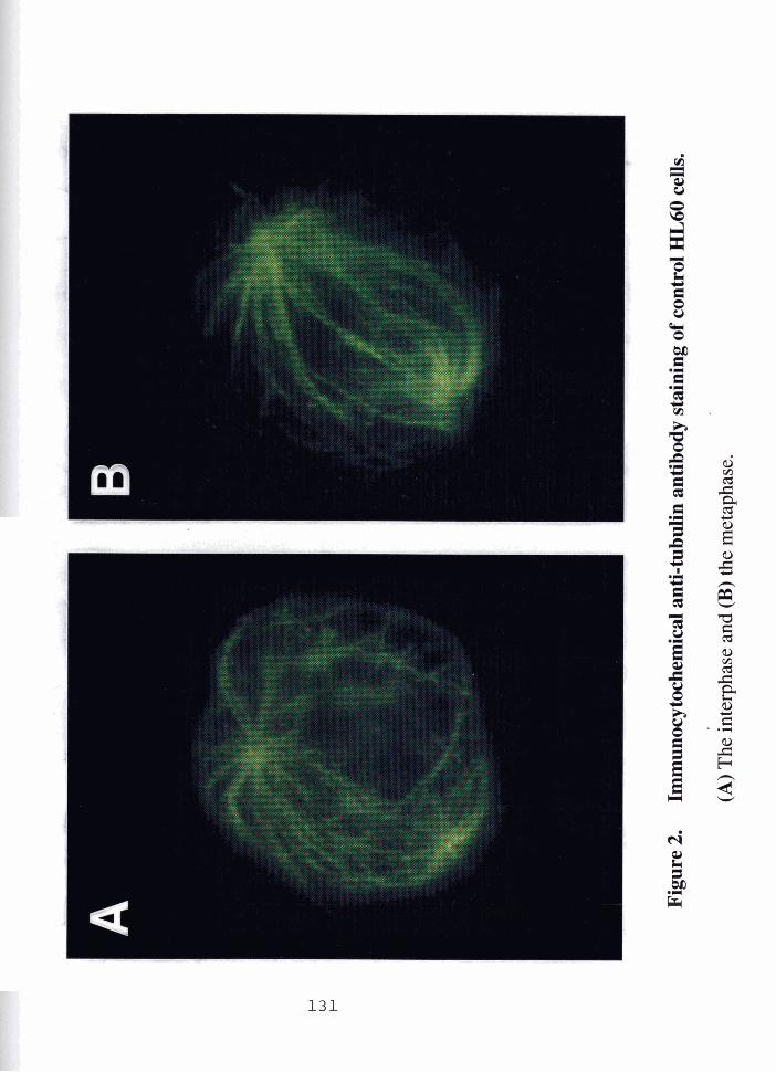

Effects of 1,2,4-benzenetriol and colchicine on microtubule integrity
and cytoskeletal organization in HL60 cells.
(A) 20 pM 1,2,4-benzenetriol, (B) 50 pM 1,2,4-benzenetriol,
(C) 100 pM 1,2,4-benzenetriol, and (D) 1 pM colchicine.

Tab
le 1
B
asel
ine
and
effe
ct o
f col
chic
ine
on n
ucle
ar s
pot f
requ
enci
es in
H
L60
cel
ls u
sing
a c
entr
omer
ic p
robe
spe
cifi
c fo
r ch
rom
osom
e 9.
Con
trol
s 10
,000
(S
.D.)
*
Tre
atm
ent
Tot
al C
ells
Sco
red
Col
chic
ine
5,00
0 (0
.1 PM)
(S.D
.)*
* S
tand
ard
Dev
iatio
n (S
.D.)
of
5 se
para
te e
xper
imen
ts is
sho
wn
in p
aren
thes
es.
2000
cel
ls e
ach
for
cont
rols
and
10
00 c
ells
eac
h fo
r co
lchi
cine
trea
tmen
ts w
ere
scor
ed.
Num
ber
of s
~o
ts
1 10
00 c
ells
0 1
2 3
4 25
Tot
al
Hyp
erdi
ploi
dy
per
1000
Nuc
lei

Tab
le 2
N
umbe
r of
hyb
ridi
zati
on d
omai
ns o
f ch
rom
osom
e 9
in in
terp
hase
nuc
lei
of H
L60
cel
ls fo
llow
ing
1,2,
4=be
nzen
etri
ol tr
eatm
ent f
or 2
4 h
in m
edia
.
Tot
al n
umbe
r
[ B
TI PM
of
scor
ed c
ells
Num
ber
of d
omai
ns /
1000
nuc
lei *
0 1
2 3
4 15
Tot
al
Hyp
erdi
ploi
dy
per
1000
Nuc
lei
* T
he d
ata
pres
ente
d he
re a
re th
e m
ean f S
.D. (
Sta
ndar
d D
evia
tion)
.

Tab
le 3
N
umbe
r of
hyb
ridi
zati
on d
omai
ns o
f chr
omos
ome
9 in
inte
rpha
se n
ucle
i of
HL
60 c
ells
follo
win
g 1,
2,4=
benz
enet
riol
trea
tmen
t fo
r 1
h in
PB
S.
Tot
al n
umbe
r
[ B
TI
PM
of
sc
ored
cel
ls
Num
ber
of h
vbri
diza
tion
dom
ains
0 1
2 3
4 25
Tot
al
Hyp
erdi
ploi
dy
Der
100
0 C
ells

Tab
le 4
N
ucle
ar s
pot f
requ
enci
es in
HL
60 c
ells
trea
ted
wit
h 1,
2,4-
benz
enet
riol
fo
r 24
h in
med
ia u
sing
a c
entr
omer
ic p
robe
spe
cifi
c fo
r ch
rom
osom
e 7.
Tot
al n
umbe
r
[B
TI
W
of
Num
ber
of d
omai
ns 1
100
0 nu
clei
T
otal
num
ber
of
hype
rdip
loid
y sc
ored
cel
ls
0 1
2 3
4 ! p
er 1
000
cell
s
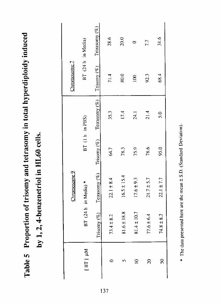
'Kt' . - 2

CHAPTER FOUR
Effect of Metals, Ligands and Antioxidants on the Reactions of Oxygen with 1,2,4-Benzenetriol
Luoping Zhang and Allan J. Davisonl
Bioenergetics Research Laboratory, School of Kinesiology, Faculty of Applied Science, Simon Fraser University, Burnaby, B.C., Canada, V5A IS6 and BC Cancer Research Center, 601 West 10th Ave., Vancouver, B.C., Canada V5Z
I L3
Running Title: Effect of metals on autoxidation of 1,2,4-benzenetriol
Key Words: 1,2,4-benzenetriol, autoxidation, quinones, transition metals, oxygen-derived active species, superoxide dismutase and desferrioxamine.
Correspondence: Dr. Allan Davison, BC Cancer Research Center, 601 West 10th Ave.,
Vancouver, B.C., V5Z 1 L3, Canada.

ABSTRACT
1,2,4-Benzenetriol is a reactive metabolite of the human leukemogen
benzene. The genotoxicity of benzenetriol reportedly results from its ability to
reduce molecular oxygen (02) to active species in the course of its oxidation to
its corresponding quinones. The mechanism of reaction of 0 2 with benzenetriol
is poorly understood and little is known of the effects of metals, metal chelators,
radical scavenljers and antioxidants on the rate of reduction of 0 2 . We
therefore compared candidate free radical propagators of benzenetriol's
reaction with 0 2 at pH 7.4 in the presence or absence of selected metal ions
(Cu2+ and Fe3+). Catalytic amounts of C U ~ + and Fe3+ accelerated the
oxidation of benzenetriol (250 pM) in a dose-dependent manner. Fe3+ (50 pM)
increased the rate of autoxidation by approximately 91% and Cu2+ (1 0 pM)
increased it by 1076%. In the absence of metals, superoxide dismutase
inhibited and desferrioxamine stimulated the autoxidation. In the Cu2+-
catalyzed reaction, superoxide dismutase neither inhibited nor stimulated, but
desferrioxamine abolished the catalysis by Cu2+. In the presence of Fe3+,
superoxide dismutase slowed the reaction, but desferrioxamine, surprisingly,
did not. When both superoxide dismutase and desferrioxamine were present,
the autoxidation was blocked regardless independent of the presence or
absence of metals. We therefore conclude: (1) Superoxide is a propagator of
sequential one-electron transfer reactions in the absence of added metals. (2)
Addition of Cu2+ removes the dependence of the reaction on propagation by
superoxide. Apparently Cu2+ changes the free radical propagated chain
reaction to a concerted two-electron transfer, but Fe3+ does not. (3) The further
addition of desferrioxamine restores superoxide-dependent propagation.

INTRODUCTION
Benzene is metabolized in the liver via benzene oxide to phenol, which
is further hydroxylated to catechol, hydroquinone and 1,2,4-benzenetriol [Inoue
et al., 1989a, b]. Each of these metabolites and a ring-opened metabolite, t,t-
muconaldehyde, may mediate the myelotoxicity and carcinogenicity of benzene
[Yardley-Jones, et al., 19911. The chemistry of benzene metabolites are
therefore of interest. Among these, the triphenolic metabolite of benzene,
benzenetriol, is the most reactive toward molecular oxygen (02) [Greenlee et at.,
19811.
Benzenetriol is a strong reductant and rapidly autoxidizes to its
corresponding quinone via semiquinone radical intermediates [Greenlee et at.,
1981 ; Bandy et al., 19901. Its strong chemical reactivity toward O2 is consistent
with a high biological potency. Benzenetriol induces both single and double
strand breaks in DNA [Lewis et al., 1988; Kawanishi et al., 1989; Li, 19921,
causes sister chromatid exchanges in human lymphocytes [Erexson et at.,
19851 and gene mutations (6-thioguanine resistance) in V79 cells [Glatt et al.,
1 9891.
The current research is the third in a series of studies on the roles of
quinones, oxygen and transition metal ions in the genotoxicity of bsnzenetriol.
It complements previous studies in which benzenetriol induces micronucleus
formation, aneuploidy and microtubule disruption in cultured human cells
[Zhang et al., 1993a, b]. In these studies, we demonstrated that benzenetriol
induces oxidative DNA damage (formation of 8-hydroxy-2'-deoxyguanosine)

both in cultured human cells in vitro [Zhang et al., 1993al and in mouse bone
marrow in vivo [Kolachana et al., 19931. These manifestations of genotoxicity
are presumed to result, at least in part, from benzenetriol's ability to reduce O2
to reactive species during its autoxidation. Despite the evidence that
benzenetriol-induced active species are cytotoxic and genotoxic, surprisingly
little is known regarding the mechanisms of its reaction with 0 2 .
Autoxidation is the uncatalyzed oxidation of a substance exposed to O2
in air (Uri, 1961). However, because the direct transfer of an electron pair to 0 2
is spin forbidden, the sample reaction QH2 + 0 2 -> Q + H202 does not occur
directly. Instead, reduction of oxygen is sometimes depicted as sequence of
two single-electron transfers:
The first of these reactions is, however, plausible only in the presence of metal
ions. Transition metals are efficient catalysts of redox reactions in part because
they contain unpaired electrons, and therefore their reactions with 0 2 are not
spin restricted (Miller et al., 1990). The "autoxidation" observed for many
compounds usually turns out on closer examination to be metal catalyzed.
Redox active metals can serve as free radical initiators, bypassing the
unfavorable reaction (1) as shown below:

Iron and copper are the predominant among the transition metals which, if
decompartmentalized, can become prooxidant in viva Their addition to the
culture medium increases the genotoxicity of benzenetriol as measured by DNA
strand breaks [Lewis et al., 1988; Kawanishi et al., 1989; Li, 19921, oxidative
DNA damage and micronucleus formation [Zhang et al., 1993al.
The autoxidation of benzenetriol is mediated by reactive oxygen species
including superoxide (02.-), hydrogen peroxide (H202) and hydroxyl radicals
(Hoe) [Greenlee et al., 19811. In the process, benzenetriol is oxidized to its
major oxidative product, 2-hydroxy-1,4-benzoquinone (2-OH-BQ) [Mason,
19491. The reduction of O2 by benzenetriol can occur either by sequential
transfer of individual electrons (one-electron transfer), or by concerted transfer
of two electrons within the same solvent cage (two-electron transfer) [Bandy et
al., 19901. The one-electron propagators, in theory, are 02.- from the reduction
of 02, the corresponding semiquinone radical anion (s-Qo-) from the oxidation of
benzenetriol, and perhaps HOD in the further reduction of H202. The concerted
two-electron reaction, in theory, requires metal ions to release the spin
restriction.
The observation of a stimulation or inhibition by a given scavenger or
ligand indicates which propagators are accessible to scavenging. We therefore
undertook a systematic investigation of the effects of metals, metal chelators,
scavengers and antioxidants on the autoxidation of benzenetriol. Specifically
we sought to learn the roles of metals and chelators in determining: (1) whether
one-electron transfer or two-electron transfer is dominant; (2) which propagators
are predominantly involved in the reaction; and, (3) under what circumstances a

given scavenger or ligand will stimulate or inhibit the autoxidation of
benzenetriol.
We therefore examined the acceleration of the autoxidation of
benzenetriol by Cu2+ and Fe3+, and the influence of superoxide dismutase or
desferrioxamine on the pathway for the reduction of oxygen. These results are
interpreted in the light of our earlier finding that Cu2+ changes the mechanism
of benzenetriol-induced micronucleus formation from aneuploidy to
clastogenicity. The data have implications regarding the roles of transition
metals and benzenetriol in benzene-induced human leukemia.

MATERIALS AND METHODS
Reagents. 1,2,4-Benzenetriol (99%) was purchased from Aldrich Chemical
Co. (Milwaukee, WI). All the chemicals for phosphate buffers (analytic purity)
were obtainable from American Scientific and Chemical (Seattle, WA). Cupric
sulfate and ferric chloride were purchased from Fisher Scientific Co. (Fair Lawn,
NJ). Superoxide dismutase (4050 U/mg, E. C. 1.15.1.1) was from
Pharmaceutical Inc. (Mountain View, CA). Catalase (65,000 Ulmg, E. C.
1.1 1.1.6) was from Boehringer Mannheim Co. (Indianapolis, IN).
Desferrioxamine was a gift from ClBA Pharmaceutical Co. (Summit, NJ).
Sodium formate was purchased from J. T. Baker Chemical Co. (Phillipsburg,
NJ) and mannitol was from Sigma Chemical Co. (St. Louis, MO).
Anaerobic solution of 1,2,4-Benzenetriol. 1,2,4-Benzenetriol stock
solution (25 mM) was prepared daily by dissolving benzenetriol in argon-
saturated deionized-distilled water, and then flushing with argon for 5 min.
Rubber stoppers used to seal the Virtis vials were boiled and evacuated to
remove any air in the rubber. A gas-tight syringe was used to inject aliquots of
the benzenetriol solution into a measured amount of aerated buffer solution.
Assay Procedures. Experiments were conducted in air-saturated 50 mM
phosphate buffer, pH 7.4, at 25 "C. 2.5 ml of the buffer was transferred to a
cuvette and appropriate combinations of metals, chelators and scavengers
(indicated in legends of the figures) were added. Reactions were initiated by
addition of a 25 pI aliquot of the anaerobic preparation of benzenetriol with
simultaneous agitation by a cuvette mixer. Benzenetriol (Ama, = 290 nm) at

physiological pH (7.4) spontaneously autoxidizes to its corresponding quinone
product, 2-OH-BQ. The oxidized product absorbs in the ultraviolet (&,, = 267
nm) and in the visible (hmax = 490 nm). The formation of 2-OH-BQ was
monitored spectrophotometrically at 490 nm in this study. Further details are
given in the legends of the figures.
Data Analyses. Data were collected on both a linear strip chart recorder and
simultaneously digitized using a twelve bit analogue-digital converter, and were
transferred to an IBM mainframe computer using a microprocessor data
bufferlcoupler locally designed and constructed. Digitized voltages were
converted to absorbance values at 490 nm. Subsequent data analyses were
performed using Excel programme. To determine the molar extinction
coefficient (E), it was assumed that benzenetriol was completely converted into
2-OH-BQ when the reaction approached substantial completion. The E at pH
7.4 was then derived from the slope of the plot of maximal absorption at 490 nm
as a function of the initial concentration of benzenetriol. Rate constants (k) were
obtained from the'slope of In (3 /a-4) as a function of time, where "g is the initial
concentration of benzenetrio1 and "L1" is the concentration of 2-OH-BQ. The
initial rates of benzenetriol autoxidation were the slopes determined by linear
regression over the p-quinone product (pM) formation as function of time (s)
during the first 60 s reactions.
Statistical Analysis The statistical comparisons of individual means were
compared using a one-tailed Student t-test. The level for significance was
chosen in advance to be p < 0.05.

RESULTS
Characterization of Benzenetriol Autoxidation
Since the autoxidation of benzenetriol has not been well characterized,
we determined selected characteristics of the reaction including: the molar
extinction coefficient of the quinonoid product, rate constants and order of the
reaction. The molar extinction coefficient (E) at h ,,, = 490 nm, pH 7.4 was
2079 M-1 cm-1 from regression analysis of the plot of absorbance versus
benzenetriol concentration. This E value is in agreement with the earlier reports
for E of 2051 M-I cm-I (h = 480 - 485 nm, pH 5.4) (Mason, 1949) and 2042
M-1 cm-1 (h ,,, = 483 nm, pH > 12) (Corbett, 1970).
Next, the linearity of a plot of In (a 1 a-x) against time (t) (Fig. l a ) indicates
that benzenetriol autoxidation followed the first order kinetics with respect to
time. From the slope, the apparent first order rate constant (k) for 2-OH-BQ
formation was 3.08 x 10-%-I. Initial rates of benzenetriol autoxidation (1 0 - 500
PM) were estimated from the slopes of the reaction during the first 60 s following
initiation. The initial rate increased as the concentration of benzenetriol
increased (Fig. 1 b). Figure 1 b shows that the dependence of rate on
concentration corresponds to a fractional order reaction with the apparent order
being 0.6 with respect to benzenetriol.
Catalytic Effect of Cu2+ and Fe3+
Since cuprous copper or ferrous iron can generate active oxygen
directly, only cupric copper and the ferric form of iron were tested in this study.
The dependence of the rate on the concentrations of C U ~ + and Fe3+ is shown in
Figure 2a. The oxidation rate increased as the concentration of Cu2+
increased, following saturation kinetics. When the concentration of Fe3+
increased, the rate linearly increased in a dose-dependent manner (Fig. 2a).
Fe3+ (50 pM) and Cu2+ (10 pM) catalyzed the oxidation of benzenetriol in a
time-dependent manner (Fig. 2b). As shown in Table 1, 50 pM Fe3+ (tin =I04 s)
doubled the rate of oxidation compared with the control (203 s), while 10 pM
Cu2+ enhanced the oxidation by a factor of 12 (17 s). Further, Fe3+ (50 pM)
accelerated the initial rate of 2-OH-BQ formation by approximately 91% in
comparison with control, and at only 10 pM, Cu*+ accelerated it by 1076%
(Table 1). Thus, Cu2+ Was a more efficient catalyst of benzenetriol oxidation
than Fe3+, by about 12-fold.
Inhibitory Effects of Superoxide Dismutase and Catalase
In the absence of metal ions, both superoxide dismutase (25 Ulml) and
catalase (25 Ulml) inhibited the autoxidation of benzenetriol by 74% and 27%,
respectively (Table 2). At 2 Ulml, superoxide dismutase still slowed the reaction
by 60•‹/o, but catalase did not (data not shown). Thus inhibition by superoxide
dismutase was considerably more effective than that by catalase. A
combination of both enzymes (each 2 U h l , or 5 or 12.5 Ulml) inhibited
synergistically, inducing a prolonged latent period (Fig. 3). Inactivated
superoxide dismutase and catalase failed to inhibit (data not shown).
However, in the presence of Cu2+ (10 pM), catalase (25 Ulml) inhibited
the reaction by 35% (Table 2), but superoxide dismutase (2 and 25 Ulml)
neither inhibited nor accelerated it (Fig. 4). Moreover, a combination of both
enzymes (each 12.5 Ulml) did not significantly inhibit the reaction (Table 2). In
contrast, superoxide dismutase (2 or 25 Ulml) significantly inhibited the Fe3+
(50 pM) catalyzed reaction by 28% or 64% compared with the metal control,

respectively (Fig. 4). Therefore, Cu2+ removes the sensitivity of benzenetriol
oxidation to superoxide dismutase, but Fe3+ does not.
Stimulation or inhibit ion by Desferrioxamine
Unexpectedly, desferrioxamine (0.5 mM) stimulated the autoxidation by
83% when no metals were added (Fig. 5). It counteracted catalysis by Cu2+ (10
pM) but did not diminish the effect of Fe3+ (50 pM). Desferrioxamine inhibited
the Cu2+ catalyzed oxidation by 94% but stimulated the Fe3+ catalyzed reaction
by only 20% in comparison with their own metal controls (Table 2). Thus,
stimulation rate by desferrioxamine was blocked by the addition of Cu2+,
whereas the addition of Fe3+ rendered desferrioxarnine inhibiting.
Whether or not Cu2+ or Fe3+ were added, the oxidation of benzenetriol
was inhibited virtually completely (no detectable progress in 24 h) when both
superoxide dismutase and desferrioxamine were present simultaneously
(Fig. 5). Catalase combined with desferrioxamine did not block the reaction but
merely diminished approximately 30•‹h of the stimulatory effect of
desferrioxamine with metals absent (Table 2).
Effects of Formate and Mannitol
Formate and mannitol neither inhibited nor stimulated benzenetriol
oxidation significantly whether or not metals were added (Table 2). In the
absence of metals, however, formate (50 mM) combined with superoxide
dismutase (25 Ulml) or both superoxide dismutase and catalase (each 12.5
Ulml) inhibited the reaction by 63% or 79%, respectively (Table 2). In contrast,
a combination of catalase (25 Ulml) and formate (50 mM) did not significantly
inhibit it. Thus, HO* plays no kinetically detectable roles in the reaction.

DISCUSSION
Cu2+ is a more effective catalyst than Fe3+.
Oxidation of benzenetriol at physiological pH occurs both spontaneously
(autoxidation) or enzymatically (through the action of myeloperoxidase)
[Subrahmanyam, et al., 19921. In addition, the oxidation is catalyzed by iron
and copper salts. Cu2+ was a more effective catalyst than Fe3+ (Fig. 2b), which
is consistent with findings from the previous biological studies. For example,
C U ~ + was more efficient than Fe3+ at stimulating benzenetriol-induced DNA
strand breaks [Kawanishi et al., 1989; Li, 19921 and at causing DNA base
damage in the presence of Hz02 [Aruoma et al., 19911. The differences
between iron and copper in vivo may reflect differences in the effectiveness of
biological compartmentation, or different rates of reduction of the metal
complexes or rate constants for the Fenton reaction [Koppenol and Butler,
19851. The rate constant for the reaction of Cu+ with H202 is much higher than
the corresponding one for iron [Walling, 19821.
Superoxide propagates the redox reactions in the absence of
added metals.
When metal ions were omitted from the system (Fig. 3), the ability of
superoxide dismutase to inhibit benzenetriol autoxidation conforms the findings
by Greenlee et al. (1981) and Bandy et al. (1990). The extent of inhibition
reflects the importance of superoxide as a propagator of free radical chain
reactions:

Termination:
(QH2: benzenetriol; s-Q*-: semiquinone; Q: 2-O~-benzoquinone)
The initiation step (1) involves redox cycling of metal ions (see reaction
(3) and (4) in Introduction). Reactions (5) and (6) show how superoxide can
propagate a sequential one-electron transfer pathway. In addition,
semiquinone and hydroxyl radicals are candidate propagators of one-electron
transfer chain reactions. Since neither formate nor mannitol influence the rate
of benzenetriol autoxidation, HO. is excluded as a kinetically significant
propagator. Since the initial rate was not significantly enhanced when
benzenetriol was exposed to 0 2 in the presence of reaction products (data not
shown), we conclude that s-Q'- is not a significant propagator of the one-
electron transfer in autoxidation of benzenetriol. Note that semiquinone
propagation is reflected in the same reactions (5 and 6) as superoxide
propagation, but in the reverse order.

Effect of C U ~ + on the free radical-propagated chain reactions
We interpret the failure of superoxide dismutase (25 U/ml) to alter the rate
of benzenetriol oxidation in the presence of C U ~ + , on the basis that Cu2+
changes the sequential one-electron transfer pathway to a concerted two-
electron transfer pathway*. The o-quinol of benzenetriol provides for strong
binding of metal ions, encouraging its oxidation by such an inner sphere
mechanism. Electron transfer within a [BT-CU-021 inner sphere complex may
well preclude superoxide release by allowing synchronous transfer of two
electrons, from the bound benzenetriol via Cu2+ to 0 2 (Fig. 6). The possible
mechanisms as follows:
(a) Metal-binding: BT + Cu2+ > BT-CU + 2H+
(b) Ternary complex formation: BT-CU + 0 2 - > [BT-CU-021 "
(c) Two-electron transfer: [BT-CU-021 + 2H+ -> 2-OH-BQ + H202 + Cu2+
Taken together, the sum of reactions (a) + (b) + (c) is equivalent to reactions
(1)+(8), the overall reaction being:
By a two-electron pathway we mean that any one electron intermediates are too transient, or too
tightly sequestered in the solvent cage to be accessible to scavengers.
** This may be a true reaction intermediate or merely an inner sphere "collision complex"

Figure 6 shows the putative chemical structure of a benzenetriol-Cu-02
complex and the schematic flow of two electrons from benzenetriol via Cu2+ to
02. In the long term, H202 released from the ternary complex by the concerted
inner sphere transfer of two electrons is also a candidate propagator. However,
catalase inhibited the Cu2+-catalyzed reaction by 35% (Table 2), suggesting
that although Cu2+ may well facilitate peroxidative oxidation of benzenetriol, the
processes are largely subsequent to the rate determining step.
Desferrioxamine restores superoxide sensitivity to the Cu2+-stimulated
reaction. The concerted two-electron reaction is disrupted by the presence of
metal chelators, presumably because they remove the metal from association
with partly reduced 0 2 , forcing the propagators into the solvent where they are
accessible to scavengers.
Cu2+ also accelerates the autoxidation of hydroquinone, another
metabolite of benzene, and increases the cytotoxicity of hydroquinone in bone
marrow stromal cells [Li and Trush, 19931. Superoxide dismutase stimulates
the oxidation of hydroquinone to benzoquinone [Greenlee et al., 1981 ; Bandy et
al., 19911. To the extent that hydroquinone resembles benzenetriol, however,
Cu2+-mediated oxidation of hydroquinone should be superoxide dismutase
inhibitable. Catalase, on the other hand, fails to protect against cytotoxicity
induced by hydroquinone with Cu2+ present [Li and Trush, 19931.
Effect of Fe3+ on the reaction mechanism
In contrast to the actions of added Cu2+, addition of Fe3+ fails to diminish
dependence on the propagation by superoxide in one-electron transfer steps.
Thus, the hypothetical ternary complex, [BT-Fe-021 apparently does not permit

inner sphere electron transfer to 0 2 . A partial explanation might be that
superoxide dismutase binds to Fe3+ more effectively than C U ~ + . Further studies
are needed to explore this and other possibilities.
C u 2 + also changes mechanisms of benzenetriol-induced
micronucleus formation.
Cu2+ enhances benzenetriol-induced oxidative DNA damage and
micronucleus formation. Moreover, C U ~ + changes the mechanism of
benzenetriol-induced micronucleus formation from kinetochore-positive (mitotic
aneuploidy) to kinetochore-negative (clastogenicity) [Zhang et al., 1993al. It
presumably reflects Cu2+-induced changes in the mechanism of autoxidation of
benzenetriol. We have seen that the presence of Cu2+ decreases superoxide-
propagated one-electron transfer, while the Cu2+-induced increase in overall
reaction rate stimulates production of H202 by Cu2+-mediated two-electron
transfer.
Benzenetriol, therefore, causes chromosomal aberrations by two distinct
mechanisms. It can disrupt microtubules by oxygen-derived autoxidation
products (its corresponding quinones and active oxygen species) [Zhang, et al.,
1993b1, and can oxidatively damage DNA by reactive oxygen species
generated during its autoxidation. Added Cu2+ increases chromosomal DNA
(as opposed to microtubular) damage by increasing the yield of H202 at the
expense of 0 2 7 On this basis, the presence of C U ~ + directs the H202-
mediated damage toward the chromosomes, whereas the absence of Cu2+
directs the 02---mediated damage toward the microtubules. One can assume
that Cu2+-catalyzed site specific formation of HO*, which in turn mediates: DNA
base modification, strand breaks and the fragmentation of DNA [Halliwell and
Aruoma, 19911. The participation of H202 in microtubular breakage in the
absence of added Cu2+ is consistent with protection by catalase against
destruction of tubulin by the benzenetriol analog, 6-hydroxydopamine (Davison
et al., 1986). Taken together, these observations suggest that the additional
genotoxicity of benzenetriol in the presence of Cu2+ results from the ability of
CU*+ to reduce 0 2 to H202 and further to produce HO* radicals. The roles of
copper are likely: 1) to accelerate H202 production, 2) to catalyze formation of
active species in a Fenton-type reaction, and 3) to direct the site-specific radical
production to a site where the copper is bound (i.e. to the chromosomal DNA in
the case of copper mediated micronucleus formation) [Chevion, 19881.
Desferrioxamine stimulates benzenetriol autoxidation by lowering
the reduction potential of iron.
The effect of desferrioxamine is conditioned by the presence or absence
of metal ions. Thus desferrioxamine stimulated autoxidation (Fig. 5) when the
metals were not added but inhibited by counteracting the effects of added C U ~ +
(but not Fe3+). Desferrioxamine is an avid chelator of both Fe3+ and CU~+ . The
chelation of iron diminishes reducibility and accessibility, but simultaneously
increases the ability of iron to reduce O2 by lowering its reduction potential. The
optimal reduction potential for catalysis by a redox cycling metal is
approximately midway between those of the reduction potentials of electron
donor and electron acceptor [Davison et al., manuscript in preparation]. Since
benzenetriol is a very strong reductant, even though the reduction potential of
unchelated iron is high, desferrioxamine may move it closer to this optimal
value between 0 2 and benzenetriol. In contrast, the [Cu2+-desferrioxamine]
complex has a reduction potential incompatible with redox cycling.
Combination of superoxide dismutase and desferrioxamine blocks
both superoxide-propagated and Cu2+-mediated pathways.
Although desferrioxamine stimulates autoxidation of benzenetriol when
present alone, it strongly inhibits in the presence of small quantities (2 U 1 ml) of
superoxide dismutase (Fig. 5). Thus desferrioxamine acted synergistically with
superoxide dismutase to prevent autoxidation of benzenetriol, irregardless of
the presence or absence of the metal ions. The presence of desferrioxamine
blocks the metal propagated inner sphere mechanism, while the presence of
superoxide dismutase blocks the propagation by superoxide. Arrest of the
reaction when both one-electron and two-electron pathways were blocked
implies that there is no additional pathway independent route for the aerobic
oxidation of benzenetriol. In the simultaneous presence of desferrioxamine and
superoxide dismutase, benzenetriol is inert toward molecular oxygen.
Conclusion
In summary, oxidant-mediated genotoxicity of benzenetriol is a
complicated and multifactorial process. It involves the reactions of oxygen with
benzenetriol, and production of oxygen-derived active species and quinones.
C U ~ + stimulates the oxidation of benzenetriol, which is consistent with its effect
on enhancing the benzenetriol-induced genotoxicity. C U ~ + further changes
mechanisms of the reaction from 02*--propagated one-electron transfer to C U ~ + -
mediated two-electron transfer, which correlates with the changes in the pattern
of benzenetriol-induced micronucleus formation from kinetochore-positive to
kinetochore-negative. We, therefore, conclude that the oxidation of
benzenetriol and the catalysis of C U ~ + may play an important role in the
benzenetriol-induced genotoxicity.

ACKNOWLEDGEMENTS
This work was supported by a grant from the Natural Sciences and
Engineering Research Council of Canada. The authors are grateful to Dr. Brian
Bandy for his valuable technical assistance and critical reading of this
manuscript. L. Zhang was a recipient of the William and Ada lsabell Steel
Memorial Graduate Scholarship from Simon Fraser University, B.C. Canada.

REFERENCES
Aruoma, 0.1.; Halliwell, B.; Gajewski, E.; Dizdaroglu, M. Copper-ion-dependent damage to the bases in DNA in the presence of hydrogen peroxide. Biochem J.
273:601-604; 1991.
Bandy, B.; Moon, J.; Davison, A.J. Multiple actions of superoxide dismutase: why can it both inhibit and stimulate reduction of oxygen by hydroquinones? Free Radical Biol. Med. 9:143-148; 1 990.
Bandy, 6.; Davison, A.J. Dual effects of superoxide dismutase: when does it inhibit or accelerate autoxidations? Canadian Federation of Biological Societies 34th Annual Meeting (Program Proceedings), pp144 abstr., June 9-1 1
Chevion, M. A site-specific mechanism for free radical
damage: the essential role of redox-active transition metals. Med. 5:27-37; 1988.
Corbett, J.F. The chemistry of hydroxy-quinones. Part V
nduced biological Free Radical Biol.
Formation of 2-
hydroxy-semiquinones during the autoxidation of benzene-1,2,4-triols in alkaline solution. J. Chem. Soc. C:2101-2106; 1970.
Davison, A.J.; Legault, N.A.; Steele, D.W. Effect of 6-hydroxydopamine on polymerization of tubulin. Biochemical Pharmacology, 35: 141 1-1 41 7; 1986.
Davison, A.J.; Bandy, 8.; Gee, P.; Yin, X. Mechanisms of free radical generation
in the reduction of oxygen and hydrogen peroxide: Hydroquinones can reduce oxygen and hydrogen peroxide by two-electron metal dependent, or one-
electron radical dependent propagation. (in preparation, 1993).
Erexson, G.L.; Wilmer, J.L.; Kligerman, A.D. Sister chromatid exchange induction in human lymphocytes exposed to benzene and its metabolites in
vitro. Cancer Res. 45:2471-2477; 1985.

Glatt, H.; Padykula, R.; Berchtold, G.A.; Ludewig, G.; Platt, K.L.; Klein, J.; Oesch, F. Multiple activation pathways of benzene leading to products with varying genotoxic characteristics. Environ. Health Perspect. 82:81-89; 1989.
Greenlee, W.F.; Sun, J.D.; Bus, J.S. A proposed mechanism of benzene
toxicity: Formation of reactive intermediates from polyphenol metabolites. Toxicol Appl Pharmacol. 59: 1 87- 1 95; 1 981 .
Halliwell, B.; Aruoma, 0.1. DNA damage by oxygen-derived species: Its mechanism and measurement in mammalian systems. FEBS Lett. 281 :9-19;
1991.
Inoue, 0 . K.; Seiji, H.; Ikeda, M. Pathways for formation of catechol and 1,2,4- benzenetriol in rabbits. Bull. Environ. Contam. Toxicol. 43:220-224; 1989a.
Inoue, O.K.; Seiji, H.; Watanabe, T.; Yin, S.N.; Li, G.L.; Cai, S.X.; Jin, C.; Ikeda, M. Excretion of 1,2,4-benzenetriol in the urine of workers exposed to benzene. Br. J. Ind. Med. 46559-565; 1989b.
Kawanishi, S.; Inoue, S.; Kawanishi, M. Human DNA damage induced by 1,2,4- benzenetriol, a benzene metabolite. Cancer Res. 49:164-168; 1989.
Kolachana, P.; Subrahmanyam, V.V.; Meyer, K.B.; Zhang, L.; Smith, M.T.
Benzene and its phenolic metabolites produce oxidative DNA damage in HL60
cells in vitro and in bone marrow in vivo. Cancer Res. 53:1023-1026: 1993.
Koppenol, W.H.; Butler, J. Energetics of interconversion reactions of oxygen
radicals. Adv. Free Radical Biol. Med. 1 :91-132: 1985.
Lewis, J.G.; Stewart, W.; Adams, D.O. Role of oxygen radicals in induction of DNA damage by metabolites of benzene. Cancer Res. 48:4762-4765; 1988.
Li, A.S. DNA breakage induced by 1,2,4-benzenetriol: Roles of oxygen-derived
active species and transition metal ions. Simon Fraser University, Burnaby, B.C., Canada, 1992 (M. Sc. Thesis).

Li, Y.; Trush, M. A. Oxidation of hydroquinone by copper: Chemical mechanism and biological effects. Arch. Biochem. Biophys. 300:346-355; 1993.
Mason, H. S. The chemistry of melanin: VI. Mechanisms of the oxidation of catechol by tyrosinase. J. Biol. Chem. 181 :803-812; 1949.
Miller, D.M.; Buettner, G.R.; Aust, S.D. Transition metals as catalysts of
'autoxidation' reactions. Free Radical Biol. Med. 8:95-108; 1990.
Subrahmanyam, V.V.; Kolachana, P.; Smith, M.T. Metabolism of 1,2,4- benzenetriol by human myeloperoxidase: Implications in benzene-induced
myelotoxicity. Toxicologist 1259 abstr. (1 992).
Uri, N. Physico-chemical aspects of autoxidation. In: Lundberg, W.O., ed. Autoxidation and antioxidants. Vol. 1, New York: Interscience; 1961 :55-106.
Walling, C. The nature of the primary oxidants in oxidation mediated by metal
ions. In: King, T. E.; Mason, H. S.; Morrison, M., eds. Oxidases and related redox systems. Oxford: Pergamon Press; 1982:85-97.
Yardley-Jones, A.; Anderson, D.; Parke, D. V. The toxicity of benzene and its
metabolism and molecular pathology in human risk assessment. Brit. J. Indust. Med. 48:437-444; 1991.
Zhang, L.; Robertson, M.; Kolachana, P.; Davison, A. J.; Smith, M, T. Benzene metabolite, 1, 2, 4-benzenetriol induces micronuclei and oxidative DNA damage in human lymphocytes and HL60 cells. Env. Mol. Mutagenesis
21 :339-348; 1993a .
Zhang, L.; Venkatesh, P.; Robertson, M.; Smith, M. T. Detection of 1,2,4- benzenetriol induced aneuploidy and microtubule disruption by fluorescence in situ hybridization and imrnunocytochemistry. Mutation Res. 1993b (in press).
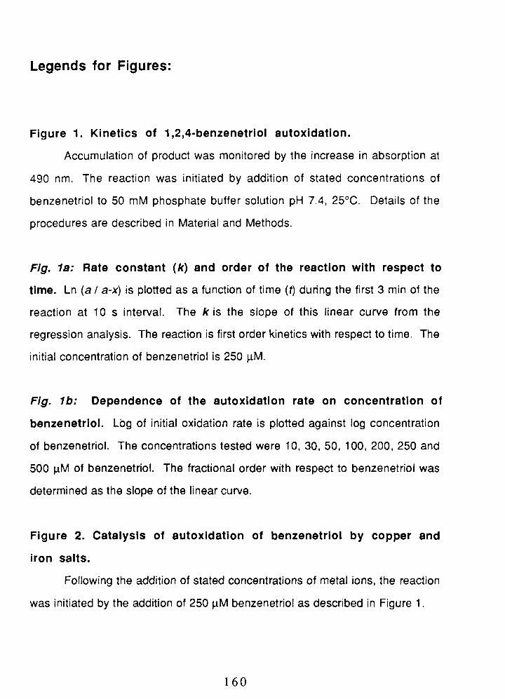
Legends for Figures:
Figure 1. Kinetics of 1,2,4-benzenetriol autoxidation.
Accumulation of product was monitored by the increase in absorption at
490 nm. The reaction was initiated by addition of stated concentrations of
benzenetriol to 50 mM phosphate buffer solution pH 7.4, 25OC. Details of the
procedures are described in Material and Methods.
Flg. la: Rate constant ( k ) and order of the reaction with respect to
time. Ln (a / a-x) is plotted as a function of time (t) during the first 3 min of the
reaction at 10 s interval. The k is the slope of this linear curve from the
regression analysis. The reaction is first order kinetics with respect to time. The
initial concentration of benzenetriol is 250 pM.
Fig. Ib : Dependence of the autoxidation rate on concentration of
benzenetriol. Log of initial oxidation rate is plotted against log concentration
of benzenetriol. The concentrations tested were 10, 30, 50, 100, 200, 250 and
500 pM of benzenetriol. The fractional order with respect to benzenetriol was
determined as the slope of the linear curve.
Figure 2. Catalysis of autoxidation of benzenetriol by copper and
iron salts.
Following the addition of stated concentrations of metal ions, the reaction
was initiated by the addition of 250 pM benzenetriol as described in Figure 1.

Fig. 2a: Cu2+ and Fe3+ accelerate the autoxidation of benzenetriol
in a dose-dependent manner. Reactions were conducted in the presence
of 2, 4, 10, 20 and 40 pM Cu2+ or 10, 20, 50, 100, and 200 pM Fe3+.
Absorbance of the p-quinone product at 30 s after initiation of the reaction is
plotted on the log scale of the vertical axis against log concentration of metal
ions.
Fig. 2b: Cu2+ and Fe3+ accelerate the autoxidation of benzenetriol
in a time-dependent manner. Where indicated, the concentrations of Cu2+
and Fe3+ were 10 and 50 pM, respectively. Benzenetriol (BT) at 250 pM was
used in both the absence and the presence of metal ions. The absorbance at
490 nm is plotted as a function of time. The half-life time (tin) was obtained
from this figure.
Figure 3. Inhibition of autoxldatlon of benzenetrlol by superoxide
dismutase or catalase individually or in combination.
In the absence or presence of antioxidant enzymes, the reaction was
initiated by the addition of 250 pM benzenetriol (BT) as described in Figure 1.
Where indicated, 25 U I ml of superoxide dismutase (SOD) or catalase (CAT)
were present in the reaction medium prior to initiating the reaction. In
combination, both enzymes were present in the medium at equal
concentrations (2, 5, and 12.5 U I ml).
Figure 4. Effects of superoxide dismutase in the presence of Cu2+
and Fe3+ on the autoxidation of benzenetriol.
Percentage of control ({A490[test] 1 A490[control~) x 100) was calculated by
dividing A490 nm after 30 s for the test reaction by A490 nm (0.0412 f 0.0016)
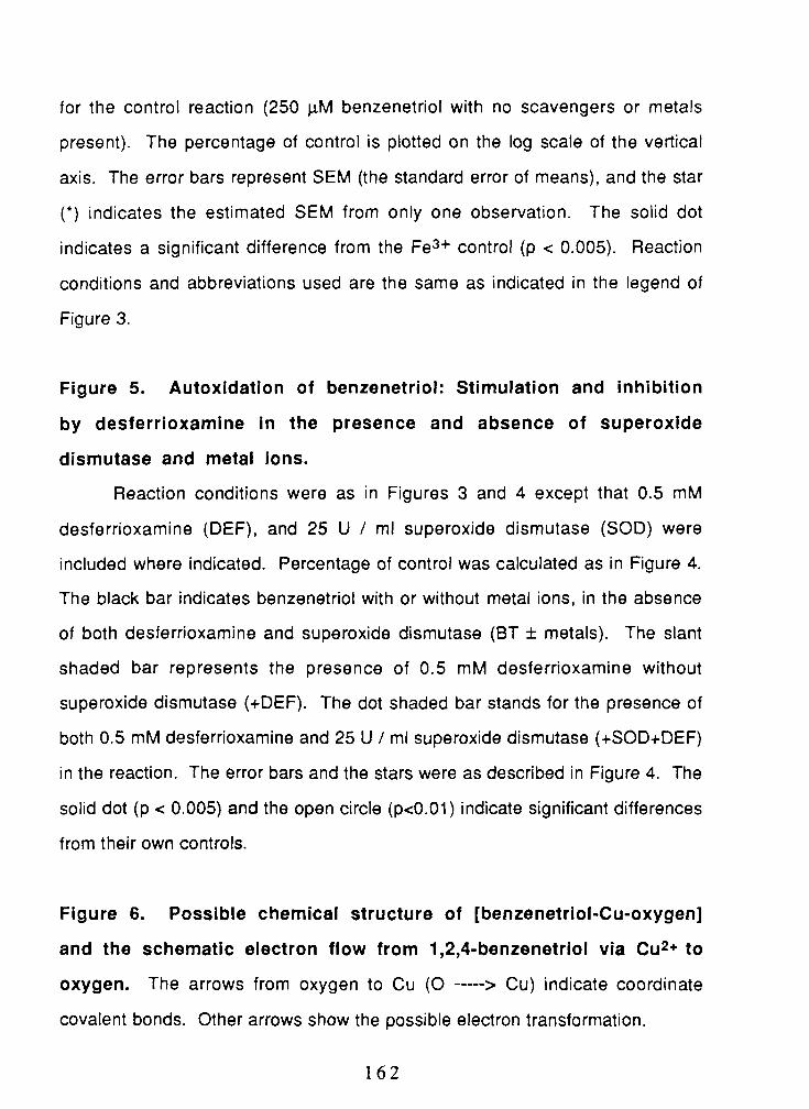
for the control reaction (250 pM benzenetriol with no scavengers or metals
present). The percentage of control is plotted on the log scale of the vertical
axis. The error bars represent SEM (the standard error of means), and the star
(') indicates the estimated SEM from only one observation. The solid dot
indicates a significant difference from the Fe3+ control (p < 0.005). Reaction
conditions and abbreviations used are the same as indicated in the legend of
Figure 3.
Figure 5. Autoxidation of benzenetriol: Stimulation and inhibition
by desferrioxamine in the presence and absence of superoxide
disrnutase and metal ions.
Reaction conditions were as in Figures 3 and 4 except that 0.5 mM
desferrioxamine (DEF), and 25 U I ml superoxide dismutase (SOD) were
included where indicated. Percentage of control was calculated as in Figure 4.
The black bar indicates benzenetriol with or without metal ions, in the absence
of both desferrioxamine and superoxide dismutase (BT f metals). The slant
shaded bar represents the presence of 0.5 mM desferrioxamine without
superoxide dismutase (+DEF). The dot shaded bar stands for the presence of
both 0.5 mM desferrioxamine and 25 U I ml superoxide dismutase (+SOD+DEF)
in the reaction. The error bars and the stars were as described in Figure 4. The
solid dot (p < 0.005) and the open circle (pc0.01) indicate significant differences
from their own controls.
Figure 6. Possible chemical structure of [benzenetriol-Cu-oxygen]
and the schematic electron flow from 1,2,4-benzenetriol via C U ~ + to
oxygen. The arrows from oxygen to Cu (0 ----> Cu) indicate coordinate
covalent bonds. Other arrows show the possible electron transformation.


Fig
ure
3
Su
per
oxi
de
dis
mu
tase
and
cat
alas
e in
hib
it a
uto
xid
atio
n o
f b
enze
net
rio
l.
250
pM B
T
Cat
alas
e (2
5U)
A
SO
D (2
5U)
0
SO
D (2
U)+
CA
T (2
U)
IJ
SO
D (5
U)+
CA
T (5
U)
I
1
A S
OD
(12
.5U
)+C
AT
(12.
5U)
0
30
60
9
0
12
0
15
0
18
0
Tim
e (s
)


FIGURE 6. Possible chemical structure of [benzenetriol-Cu-oxygen] and
the schematic electron flow from 1,2,4-benzenetriol via Cu(2+) to oxygen

Table 1 Effects of C U ~ + and Fe3+ on the oxidation of
1,2,4-benzenetriol.
* Absorbance (490 nm) at 30s after initiation of the reaction. ** The data presented as mean f SEM (Standard Error of Means). *** The variance shown as a relative error of the ratio: xly (dx + bly);
(a = x's SEM, b = y's SEM).
Half-life Tln (s)
Absorbance (490 nm)*
Percentage of Control
250 pM BT
203
0.0412 f 0.0016**
100f 8***
BT + 10 pM Cu2+
17
0.4847 f 0.0043
1176+ 56
BT + 50 pM Fe3+
1 04
0.0787 f 0.0008
191 f 9

Table 2 Summary of Results:
Percentage of Inhibition (-) or Activation (+)
Benzenetriol (BT) Controls 2
Superoxide Dismutase(25U/ml)
Catalase (25 U/ml)
SOD+Catalase(l2.5+ 12.5U/ml)
Desfemoxarnine (0.5mM)
SOD + Desfemoxamine3
Catalase + Desferrioxamine
Formate (50 mM)
SOD + Formate
Catalase + Formate
SOD + Catalase + Formate
Mannitol (50 mM)
Catalase + Mannitol
1) The data are presented as percentage of inhibition (-) or activation (+) in this table.
2) Absolute values for the controls are presented in Table 1 as absorbance at 490 nm.
3) The concentration of antioxidants or chelators is the same as stated at first time unless restated it again.
4) N/A: Data are not available.

CHAPTER FIVE
CONCLUSION AND PERSPECTIVE
In summary, the results presented in this dissertation demonstrate that
1,2,4-benzenetriol is genotoxic in cultured human lymphocytes and HL60 cells
on the basis of increased micronucleus formation, increased frequency of
aneuploidy, and increased levels of 8-hydroxy-2'-deoxyguanosine (8-OH-dG).
It is concluded that benzenetriol induces numerical and structural chromosomal
changes and point mutations by two distinct mechanisms.
Benzenetriol, by itself, induces predominantly kinetochore-positive
micronuclei, which have a high probability of containing a entire chromosome.
It was confirmed by FISH that benzenetriol induces numerical aneuploidy of
chromosomes 7 and 9, mainly in the form of hyperdiploidy. On this basis,
benzenetriol is a potent aneuploidogen. The induction of both kinetochore-
positive micronuclei and hyperdiploidy is likely to result from lagging
chromosomes which fail to be incorporated into the daughter nuclei during cell
division.
Proper chromosomal segregation requires microtubule integrity, and
microtubule disruption can lead to aneuploidy. Thus, the effect of benzenetriol
on microtubule integrity was investigated. It was shown that benzenetriol
interferes with microtubule assembly during mitosis. The oxidative products of
benzenetriol, its quinone and semiquinone, are the most likely toxicants
involved. Since they are electrophilic, quinones can interact with the

nucleophilic sulfhydryls of tubulin, the component protein of microtubules. The
sulfhydryl groups are important for microtubule assembly and the covalent
addition of quinones may block their function thereby causing mitotic
abnormalities. The disruption of microtubules is therefore most likely
responsible for the production of benzenetriol-induced aneuploidy.
In addition, benzenetriol-induced micronuclei may also result from
oxidative DNA damage caused by reactive oxygen species, which are
generated during oxidation of benzenetriol. Catalytic amounts of copper (Cu2+)
or other transition metal ions accelerate the reaction of benzenetriol with
oxygen and stimulate the formation of these reactive oxygen species in a cell-
free system. We further tested the effect of benzenetriol in combination with
Cu2+ on the induction of micronuclei in cultured human cells. Addition of C U ~ +
increases the total micronuclei induced by benzenetriol and also alters the
pattern of micronucleus formation from kinetochore-positive to kinetochore-
negative. Kinetochore-negative micronuclei indicate chromosome breakage.
Thus, benzenetriol gains another mechanism for clastogenicity in the presence
of metal ions.
The change in the mechanism of micronucleus formation from numerical
aneuploidy (kinetochore-positive) to clastogenicity (kinetochore-negative) in the
presence of Cu2+ correlates with our findings in the cell-free system. Cu2+
changes the pathway of benzenetriol oxidation from superoxide-propagated
one-electron transfer to Cu2+-mediated two-electron transfer. By accelerating
the oxidation of benzenetriol, Cu2+ stimulates the generation of hydrogen
peroxide which can be further reduced to hydroxyl radicals via Fenton-type
reactions. Moreover, Cu2+ catalyzes the site-specific formation of hydroxyl

radicals, which are the immediate DNA-damaging clastogens in the presence of
benzenetriol.
Hydroxyl radicals cause DNA base modification, strand breaks and
fragmentation of DNA or deoxyribose. 8-OH-dG represents the covalent
addition of HO* or ' 0 2 to guanine in DNA and is a sensitive marker of oxidative
DNA damage. Benzenetriol increases the level of 8-OH-dG in cell DNA,
particularly in the presence of C U ~ + . Because 8-OH-dG causes DNA base
G ->T substitution, the misreading at the modified and adjacent base
residues can lead to point mutations. These observations illustrate that the
genotoxicity of benzenetriol in the presence of Cu2+ results from the ability of
Cu2+ to activate oxygen to H202 and further to produce HOm radicals. Taken
together, the genotoxicity of benzenetriol has been characterized by its ability to
cause aneuploidy, clastogenicity and point mutations. The high potency of
benzenetriol suggests that it may play a role in benzene-induced genetic
damage and perhaps human leukemogenesis.
Our findings open new avenues for investigation of the impact of oxygen-
derived active species and toxic quinones on cytotoxicity, genotoxicity and
tumorigenesis. Further studies should consider detailed mechanisms by which
benzenetriol induces distinct cellular damage. The future goal is to distinguish
the role of reactive oxygen species from that of quinones in benzenetriol-
induced genotoxicity. For example, we can apply H202 with or without metal
ions to the cell system to test whether microtubule disruption results from
quinones or from oxygen radicals. It will be also important to measure the
effects of antioxidants on the formation of micronuclei and 8-OH-dG in order to
understand the mechanisms involved.

APPENDICES
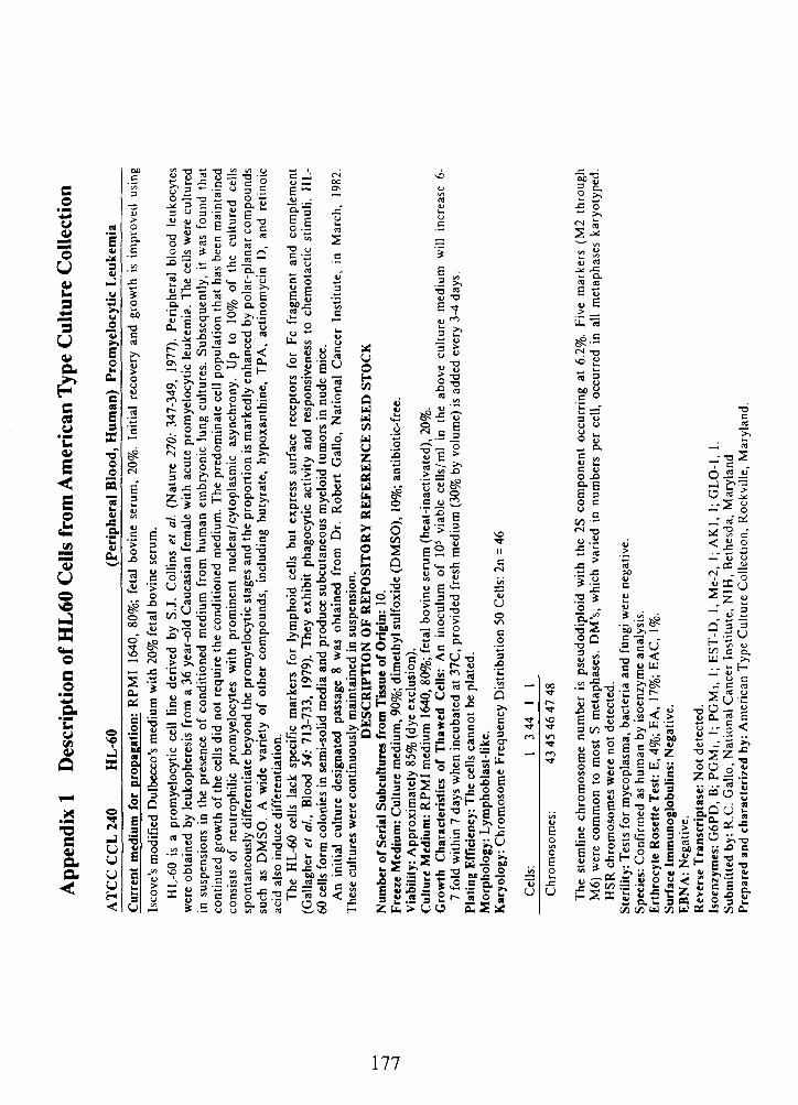
App
endi
x 1
Des
crip
tion
of H
L60
Cel
ls fr
om A
mer
ican
Typ
e C
ultu
re C
olle
ctio
n
AT
CC
CC
L 2
40
HL
-60
(Per
iphe
ral B
lood
, Hum
an)
Pro
mye
locy
tic
Leu
kem
ia
Cur
rent
med
ium
for
pro
paga
tion
: R
PM
I 16
40,
80%
; fe
tal
bovi
ne s
erum
, 20
%.
Init
ial
reco
very
and
gro
wth
is
impr
oved
usi
ng
Isco
ve's
mod
ifie
d D
ulbe
cco'
s m
ediu
m w
ith
20%
fet
al b
ovin
e se
rum
. H
L-6
0 is
a p
rom
yelo
cyti
c ce
ll li
ne d
eriv
ed b
y S
.J.
Col
lins
et
al.
(Nat
ure
270:
34
7-34
9,
1977
). P
erip
hera
l bl
ood
leuk
ocyt
es
wer
e ob
tain
ed b
y le
ukop
here
sis
from
a 3
6 ye
ar-o
ld C
auca
sian
fem
ale
wit
h ac
ute
prom
yelo
cyti
c le
ukem
ia.
The
cel
ls w
ere
cult
ured
in
sus
pens
ions
in
the
pres
ence
of
cond
itio
ned
med
ium
fro
m h
uman
em
bryo
nic
lung
cul
ture
s. S
ubse
quen
tly,
it
was
fou
nd t
hat
cont
inue
d gr
owth
of
the
cells
did
not
req
uire
the
con
diti
oned
med
ium
. T
he p
redo
min
ate
cell
popu
lati
on t
hat
has
been
mai
ntai
ned
cons
ists
of
neu
trop
hili
c pr
omye
locy
tes
wit
h pr
omin
ent
nucl
ear/
cyto
pias
mic
as
ynch
rony
. U
p to
10
% o
f th
e cu
ltur
ed
cells
sp
onta
neou
sly
diff
eren
tiat
e be
yond
the
pro
mye
locy
tic
stag
es a
nd t
he p
ropo
rtio
n is
mar
kedl
y en
hanc
ed b
y po
lar-
plan
ar c
ompo
unds
su
ch a
s D
MS
O.
A w
ide
vari
ety
of o
ther
com
poun
ds,
incl
udin
g bu
tyra
te,
hypo
xant
hine
, T
PA
, ac
tino
myc
in D
, an
d re
tino
ic
acid
als
o in
duce
dif
fere
ntia
tion
. T
he H
L-6
0 ce
lls l
ack
spec
ific
mar
kers
for
lym
phoi
d ce
lls b
ut e
xpre
ss s
urfa
ce r
ecep
tors
for
Fc
frag
men
t an
d co
mpl
emen
t (G
alla
gher
el
al.,
Blo
od 5
4:
713-
733,
19
79).
The
y ex
hibi
t ph
agoc
ytic
act
ivit
y an
d re
spon
sive
ness
to
che
mot
acti
c st
imul
i. H
L-
60 c
ells
for
m c
olon
ies
in s
emi-
solid
med
ia a
nd p
rodu
ce s
ubcu
tane
ous
mye
loid
tum
ors
in n
ude
mic
e.
An
init
ial
cult
ure
desi
gnat
ed p
assa
ge 8
was
obt
aine
d fr
om D
r. R
ober
t G
allo
, N
atio
nal
Can
cer
Inst
itut
e, i
n M
arch
, 19
82.
The
se c
ultu
res
wer
e co
ntin
uous
ly m
aint
aine
d in
sus
pens
ion.
D
ES
CR
IPT
ION
OF
RE
PO
SIT
OR
Y R
EF
ER
EN
CE
SE
ED
ST
OC
K
Num
ber
of S
eria
l Sub
cult
ures
from
Ti
ue
of O
rigi
n: 1
0.
Fre
eze
Med
ium
: C
ultu
re m
ediu
m, 9
0%;
dim
cthy
l sul
foxi
de (
DM
SO
), 1
0%;
anti
biot
ic-f
ree.
V
iabi
lity:
App
roxi
mat
ely
85%
(dye
exc
lusi
on).
C
ultu
re M
ediu
m:
RP
MI
med
ium
164
0,80
%;
feta
l bo
vine
ser
um (
heat
-ina
ctiv
ated
), 2
0%.
Gro
wth
Cha
ract
eris
tics
of
Tha
wed
Cel
ls:
An
inoc
ulum
of
lo5
viab
le c
ells
/ml
in t
he a
bove
cul
ture
med
ium
will
in
crea
se 6
- 7
fold
wit
hin
7 da
ys w
hen
incu
bate
d at
37C
, pro
vide
d fr
esh
med
ium
(30
% b
y vo
lum
e) is
add
ed e
very
34
days
. P
lati
ng E
ffic
ienc
y: T
he c
ells
can
not b
e pl
ated
. M
orph
olog
y: L
ymph
obla
st-l
ike.
K
aryo
logy
: C
hrom
osom
e F
requ
ency
Dis
trib
utio
n 50
Cel
ls:
2n =
46
Cel
ls:
13
44
11
Chr
omos
omes
: 43
45
46 4
7 48
The
ste
mli
ne c
hrom
osom
e nu
mbe
r is
pse
udod
iplo
id w
ith
the
2s c
ompo
nent
occ
urri
ng a
t 6.
2%.
Five
mar
kers
(M
2 th
roug
h M
6) w
ere
com
mon
to
mos
t S
met
apha
ses.
DM
'S,
whi
ch v
arie
d in
num
bers
per
cel
l, oc
curr
ed i
n al
l m
etap
hase
s ka
ryot
yped
. H
SR
chr
omos
omes
wer
e no
t de
tect
ed.
Ster
ilit
y: T
ests
for
myc
opla
sma,
bac
teri
a an
d fu
ngi
wer
e ne
gati
ve.
Spec
ies:
Con
firm
ed a
s hu
man
by
isoe
nzym
e an
alys
is.
Ert
hroc
yte
Ros
ette
Tes
t: E
, 4%
; E
A,
17%
; EA
C,
1 %.
Sur
face
Im
mun
oglo
buli
ns:
Neg
ativ
e.
EB
NA
: N
egat
ive.
R
ever
se T
rans
crip
tase
: Not
det
ecte
d.
Isoe
nzym
es:
G6P
D,
B;
PG
MI,
I; P
GM
I, I
; E
ST
-D,
1; M
e-2,
I;
AK
I, 1
; GL
O-I
, 1.
S
ubm
itte
d by
: R
.C. G
allo
, Nat
iona
l C
ance
r In
stit
ute,
NIH
, B
ethe
sda,
Mar
ylan
d P
repa
red
and
char
acte
rize
d by
: A
mer
ican
Typ
e C
ultu
re C
olle
ctio
n, R
ockv
ille
, M
aryl
and.


Appendix 4: Sample of Score Sheet for Micronucleus Assay
Modified MN Score Sheet
Sample Code: Date: Decode: Scorer:
Slide
# MN Cells wl
Re~licative Index MONO BI >BI TOTAL
# BN Cdl
R.I.= Dup 1
D u p 2
Total
Percent
200 -
200
400
100
# MN (U) if Unscorahle I , ~ ~ a t i o n Comments


Appendix 7
Respective Contributions of the Author and Co-authors
This study was proposed and designed by the author. The author of this
dissertation, collected and analyzed most of data from the micronucleus assay, FISH
technique, microtubule staining and chemical system. The major conmbutions from the
author in this research are: (1) designing, initiating and organizing all these major studies;
(2) designing the experimental procedures and applying these techniques; (3) treating HL60
cells with BT and Cu2+ in PBS simultaneously; (4) interpreting the results of the chemical
and cellular studies; and (5) writing up this dissertation and the three publications. Other
co-authors participated in part of the study as follows:
In Chapter 11, the experiment of benzenemol-induced micronucleus formation in
human lymphocytes was set up in collaboration with Moire Robertson. Dr. Prema
Kolachana determined the benzenetriol-induced 8-OH-dG formation (by HPLC) in Dr.
Bruce Ames' laboratory. Dr. Allan Davison did most of the statistical analysis. Dr.
Martyn Smith, Dr. Allan Davison, and Moire Robertson critically read a few versions of
the manuscript and made suggestions for improving the writing.
In Chapter 111, the irnrnunocytochemical assay with anti-tubulin antibody staining in
human lymphocytes and HL60 cells was set up in collaboration with Moire Robertson.
Pravina Venkatesh scored some of the stained slides for FISH. Dr. Martyn Smith critically
read the final manuscript and provided suggestions for the discussion.
In Chapter IV, Dr. Allan Davison proposed the topic of this research, provided
suggestions and critically read the previous version and final manuscript.