genome-wide kinetics of dna excision repair in relation to
TRANSCRIPT
Genome-wide kinetics of DNA excision repair inrelation to chromatin state and mutagenesisSheera Adara,1, Jinchuan Hua,1, Jason D. Liebb,2, and Aziz Sancara,2
aDepartment of Biochemistry and Biophysics, School of Medicine, University of North Carolina, Chapel Hill, NC 27599; and bDepartment of Human Genetics,University of Chicago, Chicago, IL 60637
Contributed by Aziz Sancar, March 1, 2016 (sent for review January 4, 2016; reviewed by Paul Nghiem and Dong Wang)
We recently developed a high-resolution genome-wide assay formapping DNA excision repair named eXcision Repair-sequencing(XR-seq) and have now used XR-seq to determine which regions ofthe genome are subject to repair very soon after UV exposure andwhich regions are repaired later. Over a time course, we measuredrepair of the UV-induced damage of cyclobutane pyrimidinedimers (CPDs) (at 1, 4, 8, 16, 24, and 48 h) and (6-4)pyrimidine-pyrimidone photoproducts [(6-4)PPs] (at 5 and 20 min and 1, 2, and4 h) in normal human skin fibroblasts. Each type of damage hasdistinct repair kinetics. The (6-4)PPs are detected as early as 5 minafter UV treatment, with the bulk of repair completed by 4 h. Re-pair of CPDs, which we previously showed is intimately coupled totranscription, is slower and in certain regions persists even 2 d afterUV irradiation. We compared our results to the Encyclopedia ofDNA Elements data regarding histone modifications, chromatinstate, and transcription. For both damage types, and for bothtranscription-coupled and general excision repair, the earliest re-pair occurred preferentially in active and open chromatin states.Conversely, repair in regions classified as “heterochromatic” and“repressed” was relatively low at early time points, with repairpersisting into the late time points. Damage that remains duringDNA replication increases the risk for mutagenesis. Indeed, late-repaired regions are associated with a higher level of cancer-linkedmutations. In summary, we show that XR-seq is a powerful ap-proach for studying relationships among chromatin state, DNArepair, genome stability, mutagenesis, and carcinogenesis.
DNA repair | DNA damage | chromatin | transcription | mutation
DNA damage blocks transcription and replication and com-promises the integrity of the genome. Bulky adducts in DNA,
the focus of this work, are caused by a variety of genotoxic agentsincluding UV radiation in sunlight. In human cells, this damageis removed exclusively by the nucleotide excision repair mech-anism (1–3). In nucleotide excision repair, a single-strandedoligomer that contains the bulky adduct is excised by dual inci-sions bracketing the lesion. The resulting gap is filled in by DNApolymerases, and the newly synthesized repair patch is then sealedby DNA ligase. In general excision repair, damage recognition isachieved by the repair factors xeroderma pigmentosum (XP)complementation group C (XPC) together with replicationprotein A (RPA) and XPA (4–6). In DNA that is being activelytranscribed, damage recognition can also be achieved by a stalledRNA polymerase II, which, with the aid of the CockayneSyndrome B (CSB) protein, accelerates the recruitment of theexcision repair factors. This recognition process leads to tran-scription-coupled repair (7, 8). The subsequent dual-incisionreaction is carried out by the core excision repair factors RPA,XPA, transcription factor II H (TFIIH), XPG, and XPF-ERCC1, releasing an excised oligomer that is 24–32 nucleotides(nt) in length (6, 9).The mechanism of DNA excision repair has been reconstituted
in vitro and is well characterized (10). However, a full under-standing of repair must take into account how it occurs in livecells, where DNA is packed into chromatin and serves as atemplate for transcription. A genome-wide comparison of open
chromatin status, histone modifications, RNA expression, andDNA damage and repair would allow inference of how theseprocesses are coordinated. We recently developed an assay formapping DNA excision repair at nucleotide resolution across thehuman genome called eXcision Repair sequencing (XR-seq)(11). In XR-seq, the excised oligonucleotides, released during re-pair in vivo, are isolated and sequenced, resulting in a stranded,single-nucleotide resolution map of repair (Fig. S1). In this study,we used XR-seq to measure repair kinetics after UV irradiationof two types of UV-induced damage: cyclobutane pyrimidinedimers (CPDs) and (6-4)pyrimidine-pyrimidone photoprod-ucts [(6-4)PPs], and then related the kinetics of repair to chro-matin state and mutagenesis.
ResultsIdentification of Sites of CPD Damage Repaired Early and Late AfterUV Exposure in Normal Human Fibroblasts. To determine whichgenomic regions were repaired rapidly after damage and whichwere repaired more slowly, we conducted a time course of CPDand (6-4)PP repair in normal human skin fibroblasts (NHF1).The bulk repair kinetics of these two types of damage is known tobe quite different (4, 12, 13). CPDs, which do not distort the DNAhelix as strongly and escape recognition by the general repairmachinery, are recognized primarily in a transcription-coupledmanner. Their complete repair requires 12–48 h, depending onthe UV dose. In contrast, the general repair pathway efficientlyremoves (6-4)PPs, with most of the damage excised within 4 hof UV treatment.
Significance
Nucleotide excision repair is the sole mechanism for removingbulky adducts from the human genome, including those formedby UV radiation and chemotherapeutic drugs. We used eXcisionRepair-sequencing, a genomic assay for measuring DNA repair, tomap the kinetics of repair after UV treatment. These genome-wide repair maps, in turn, allowed us to infer how excision repairis influenced by DNA packaging. Active and open chromatin re-gions were repaired more rapidly than other genomic regions.Repair in repressed and heterochromatic regions is slower andpersists for up to 2 d. Furthermore, late-repaired regions are as-sociated with a higher level of cancer-linked somatic mutations,highlighting the importance of efficient DNA repair and linkingchromatin organization to cancer mutagenesis.
Author contributions: S.A., J.H., J.D.L., and A.S. designed research; J.H. performed re-search; S.A. analyzed data; and S.A., J.H., J.D.L., and A.S. wrote the paper.
Reviewers: P.N., University of Washington; and D.W., University of California, San Diego.
The authors declare no conflict of interest.
Data deposition: The sequence data reported in this paper have been deposited in theGene Expression Omnibus (GEO) database, www.ncbi.nlm.nih.gov/geo (accession no.GSE76391).1S.A. and J.H. contributed equally to this work.2To whom correspondence may be addressed. Email: [email protected] or [email protected].
This article contains supporting information online at www.pnas.org/lookup/suppl/doi:10.1073/pnas.1603388113/-/DCSupplemental.
E2124–E2133 | PNAS | Published online March 28, 2016 www.pnas.org/cgi/doi/10.1073/pnas.1603388113
Dow
nloa
ded
by g
uest
on
Oct
ober
11,
202
1
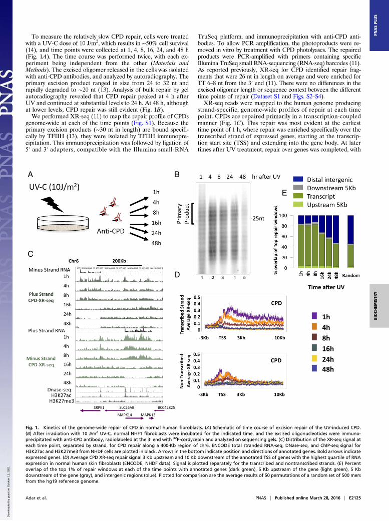
To measure the relatively slow CPD repair, cells were treatedwith a UV-C dose of 10 J/m2, which results in ∼50% cell survival(14), and time points were collected at 1, 4, 8, 16, 24, and 48 h(Fig. 1A). The time course was performed twice, with each ex-periment being independent from the other (Materials andMethods). The excised oligomer released in the cells was isolatedwith anti-CPD antibodies, and analyzed by autoradiography. Theprimary excision product ranged in size from 24 to 32 nt andrapidly degraded to ∼20 nt (13). Analysis of bulk repair by gelautoradiography revealed that CPD repair peaked at 4 h afterUV and continued at substantial levels to 24 h. At 48 h, althoughat lower levels, CPD repair was still evident (Fig. 1B).We performed XR-seq (11) to map the repair profile of CPDs
genome-wide at each of the time points (Fig. S1). Because theprimary excision products (∼30 nt in length) are bound specifi-cally by TFIIH (13), they were isolated by TFIIH immunopre-cipitation. This immunoprecipitation was followed by ligation of5′ and 3′ adapters, compatible with the Illumina small-RNA
TruSeq platform, and immunoprecipitation with anti-CPD anti-bodies. To allow PCR amplification, the photoproducts were re-moved in vitro by treatment with CPD photolyases. The repairedproducts were PCR-amplified with primers containing specificIllumina TruSeq small RNA-sequencing (RNA-seq) barcodes (11).As reported previously, XR-seq for CPD identified repair frag-ments that were 26 nt in length on average and were enriched forTT 6–8 nt from the 3′ end (11). There were no differences in theexcised oligomer length or sequence context between the differenttime points of repair (Dataset S1 and Figs. S2–S4).XR-seq reads were mapped to the human genome producing
strand-specific, genome-wide profiles of repair at each timepoint. CPDs are repaired primarily in a transcription-coupledmanner (Fig. 1C). This repair was most evident at the earliesttime point of 1 h, where repair was enriched specifically over thetranscribed strand of expressed genes, starting at the transcrip-tion start site (TSS) and extending into the gene body. At latertimes after UV treatment, repair over genes was completed, with
A B
C
D
E
Fig. 1. Kinetics of the genome-wide repair of CPD in normal human fibroblasts. (A) Schematic of time course of excision repair of the UV-induced CPD.(B) After irradiation with 10 J/m2 UV-C, normal NHF1 fibroblasts were incubated for the indicated time, and the excised oligonucleotides were immuno-precipitated with anti-CPD antibody, radiolabeled at the 3′ end with 32P-cordycepin and analyzed on sequencing gels. (C) Distribution of the XR-seq signal ateach time point, separated by strand, for CPD repair along a 400-Kb region of chr6. ENCODE total stranded RNA-seq, DNase-seq, and ChIP-seq signal forH3K27ac and H3K27me3 from NHDF cells are plotted in black. Arrows in the bottom indicate position and directions of annotated genes. Bold arrows indicateexpressed genes. (D) Average CPD XR-seq repair signal 3 Kb upstream and 10 Kb downstream of the annotated TSS of genes with the highest quartile of RNAexpression in normal human skin fibroblasts (ENCODE, NHDF data). Signal is plotted separately for the transcribed and nontranscribed strands. (E) Percentoverlap of the top 1% of repair windows at each of the time points with annotated genes (dark green), 5 Kb upstream of the gene (light green), 5 Kbdownstream of the gene (gray), and intergenic regions (blue). Plotted for comparison are the average results of 50 permutations of a random set of 500 mersfrom the hg19 reference genome.
Adar et al. PNAS | Published online March 28, 2016 | E2125
BIOCH
EMISTR
YPN
ASPL
US
Dow
nloa
ded
by g
uest
on
Oct
ober
11,
202
1
virtually no detectable repair over the template strand at 24 h or48 h after UV treatment (Fig. 1 C and D). At promoters, repairon the nontemplate strand was slightly higher and extended awayfrom the gene, consistent with repair linked to divergent tran-scription at promoters. Repair of the nontranscribed strandwithin gene bodies, and over intergenic regions, persisted atrelatively lower levels for up to 48 h after treatment (Fig. 1C). Aspreviously reported (11), there was a positive correlation be-tween the RNA and repair levels, with genes of lower expressionlevels exhibiting similar but lower-amplitude patterns (Fig. S5).To define the top repair sites at each time point, XR-seq
counts were obtained for 500-nt windows across the human ge-nome. The top 1% of windows (62,743 in total) was identified foreach strand. Compared with the whole genome, depending onthe sample, repair in these regions was enriched 5- to 12-fold(Fig. S6D). For CPD, >80% of these top repair windows over-lapped annotated transcripts up to 8 h after UV. At the later timepoints, the degree of overlap with transcript regions declined to48% at 48 h after UV. The overall proportion of top repair
windows that overlap transcripts in each of the time points werehigher than that calculated for the total of XR-seq reads at eachtime point or the overlap calculated for a random set of genomicintervals (P < 0.02; Fig. 1E and Fig. S6E).
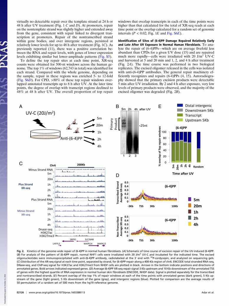
Identification of Sites of (6-4)PP Damage Repaired Relatively Earlyand Late After UV Exposure in Normal Human Fibroblasts. To ana-lyze the repair of (6-4)PPs—which are on average fivefold lessabundant than CPDs for a given UV dose (15) and are repairedmuch more rapidly—cells were irradiated with 20 J/m2 UV-Cand harvested at 5 and 20 min and 1, 2, and 4 h after treatment(Fig. 2A). The time course was performed in two biologicalreplicates. The excised oligomer released in the cells was isolatedwith anti-(6-4)PP antibodies. The general repair machinery ef-ficiently recognizes and repairs (6-4)PPs (4, 15). Autoradiogra-phy showed that the primary excision products were detectable5 min after UV irradiation. By 2 and 4 h after exposure, very lowlevels of primary products were observed, and the majority of theexcised oligomer was degraded (Fig. 2B).
A B E
C
D
Fig. 2. Kinetics of the genome-wide repair of (6-4)PP in normal human fibroblasts. (A) Schematic of time course of excision repair of the UV-induced (6-4)PP.(B) For analysis of the pattern of (6-4)PP repair, normal NHF1 cells were irradiated with 20 J/m2 UV-C and incubated for the indicated time. The excisedoligonucleotides were immunoprecipitated with anti-(6-4)PP antibody, radiolabeled at the 3′ end with 32P-cordycepin, and analyzed on sequencing gels.(C) Distribution of the XR-seq signal at each time point, separated by strand, for (6-4)PP repair along a 400-Kb region of chr6. ENCODE total stranded RNA-seq,DNase-seq, and ChIP-seq signal for H3K27ac and H3K27me3 from NHDF cells are plotted in black. Arrows in the bottom indicate positions and directions ofannotated genes. Bold arrows indicated expressed genes. (D) Average (6-4)PP XR-seq repair signal 3 Kb upstream and 10 Kb downstream of the annotated TSSof genes with the highest quartile of RNA expression in normal human skin fibroblasts (ENCODE, NHDF data). Signal is plotted separately for the transcribedand nontranscribed strands. (E) Percent overlap of the top 1% of repair windows at each of the time points with annotated genes (dark green), 5 Kb up-stream of the gene (light green), 5 Kb downstream of the gene (gray), and intergenic regions (blue). Plotted for comparison are the average results of50 permutation of a random set of 500 mers from the hg19 reference genome.
E2126 | www.pnas.org/cgi/doi/10.1073/pnas.1603388113 Adar et al.
Dow
nloa
ded
by g
uest
on
Oct
ober
11,
202
1
A B
C
D F
E
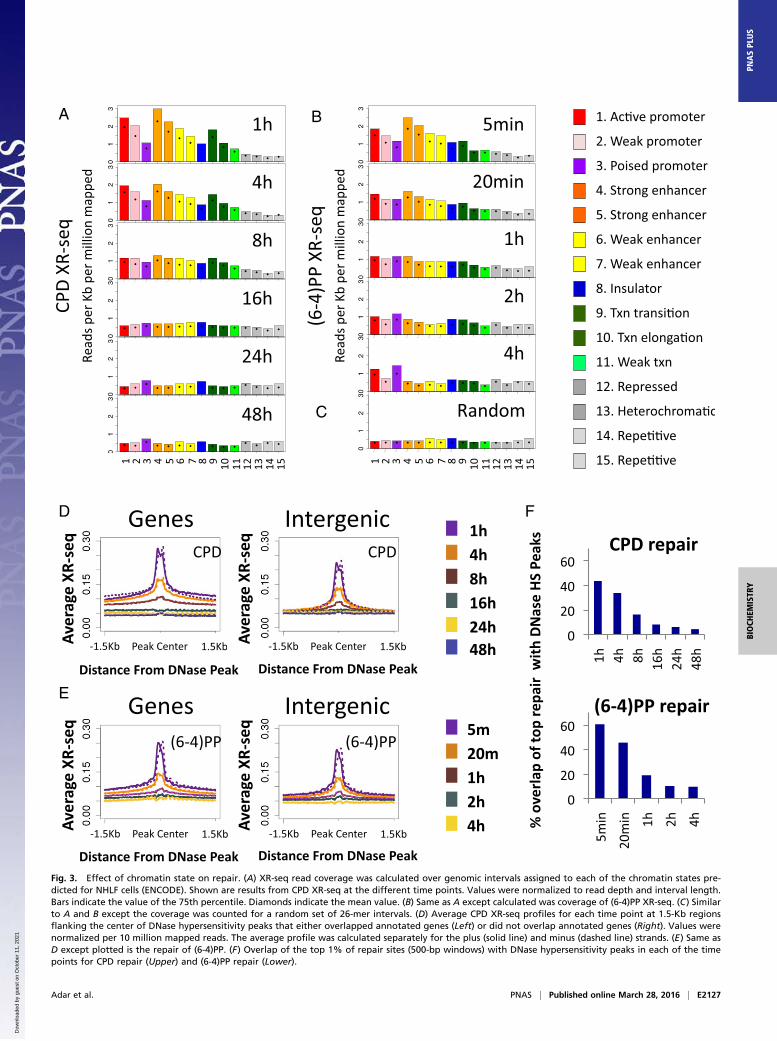
Fig. 3. Effect of chromatin state on repair. (A) XR-seq read coverage was calculated over genomic intervals assigned to each of the chromatin states pre-dicted for NHLF cells (ENCODE). Shown are results from CPD XR-seq at the different time points. Values were normalized to read depth and interval length.Bars indicate the value of the 75th percentile. Diamonds indicate the mean value. (B) Same as A except calculated was coverage of (6-4)PP XR-seq. (C) Similarto A and B except the coverage was counted for a random set of 26-mer intervals. (D) Average CPD XR-seq profiles for each time point at 1.5-Kb regionsflanking the center of DNase hypersensitivity peaks that either overlapped annotated genes (Left) or did not overlap annotated genes (Right). Values werenormalized per 10 million mapped reads. The average profile was calculated separately for the plus (solid line) and minus (dashed line) strands. (E) Same asD except plotted is the repair of (6-4)PP. (F) Overlap of the top 1% of repair sites (500-bp windows) with DNase hypersensitivity peaks in each of the timepoints for CPD repair (Upper) and (6-4)PP repair (Lower).
Adar et al. PNAS | Published online March 28, 2016 | E2127
BIOCH
EMISTR
YPN
ASPL
US
Dow
nloa
ded
by g
uest
on
Oct
ober
11,
202
1
XR-seq for (6-4)PP was performed similarly to CPD, exceptthat immunoprecipitation was performed with anti-(6-4)PPantibodies, and before PCR amplification, the photoproductswere removed in vitro by treatment with (6-4)PP photolyases. At5 min after UV exposure, repair was evident, particularly at DNasehypersensitivity sites (Fig. 2C). Repair was especially prevalent inthe areas surrounding the promoters of highly expressed genes,but unlike CPD, (6-4)PP repair was enriched on both strands,was limited to the regions flanking the TSS, and did not extendinto the gene body (Fig. 2 C and D). The profile of the elevatedrepair of (6-4)PP contains two regions of higher repair flankingthe TSS. At 4 h after UV, repair of (6-4)PP is enriched over theTSS, specifically between these initial highly repaired regions(Fig. 2D). This repair appears to be higher on the templatestrand compared with the nontemplate strand, which could bedue to a contribution of transcription-coupled repair of (6-4)PP atthese later time points. However, a slight enrichment of transcribedstrand repair at late time points is also observed in a CS-B cell linethat lacks transcription-coupled recognition (Fig. S6 B and C),suggesting that chromatin states other than transcription affectrepair efficiency. It is known that nucleosomes at promoters ofactive genes are enriched in H3K4me3 and H3K27ac histonemodifications (16). We plotted ChIP-sequencing (ChIP-seq) datafor these two modifications along with the DNase-seq hypersensi-tivity signal (17) over these active genes. The early repaired regionsfollowed the pattern of H3K27ac signal. The small “dip” betweenthe two repair peaks contained the DNase hypersensitivity peakand, adjacent to it, the H3K4me3 signal. The late-repaired (6-4)PPregion overlapped the sites of H3K4me3 signal (Fig. S7).To identify regions repaired relatively early and relatively late,
we defined the top (6-4)PP repair sites as the 1% of genomic500-nt windows with the highest repair signal. Similarly to CPDrepair, ∼70% of the top (6-4)PP repair sites overlap annotatedtranscripts at all of the measured time points, despite beingrepaired by the general, and not transcription-coupled, mecha-nism. At each of the time points, the overlap of the top (6-4)PPrepair sites with transcripts was higher than that calculated for all(6-4)PP XR-seq reads or for a random set of genomic intervals(P < 0.02; Fig. 2E and Fig. S6E).
Priority of Repair at Open and Active Chromatin States. The pack-aging of DNA into chromatin can hinder the access of repairproteins and affect the efficiency of repair (18). Specific histonemodifications are associated with different functional and cyto-logical chromatin states. The ChromHMM algorithm predictschromatin states based on underlying chromatin modificationprofiles. We used publicly available chromatin segmentationsfor an adult human fibroblast cell line [normal human lungfibroblast (NHLF) cells from the Encyclopedia of DNA Ele-ments (ENCODE) (19)]. For each of the 15 predicted states, wecalculated the number of XR-seq reads over each state, nor-malized to the proportion of the genome covered by that state. Atthe initial time point of 1 h after UV exposure, CPD repair countswere highest over the active promoters or strong enhancers. Thisenrichment over active states is consistent with CPD repair beingprimarily transcription-coupled. Less active states (for example,weak or poised promoters and weak enhancers) exhibited lowerrepair levels. Repressed, heterochromatic, and repetitive statesexhibited the lowest XR-seq counts. At later time points, the dif-ferences between states diminished, and at the latest time points,they were no longer apparent because repair was largely completed(Fig. 3A).Initial (6-4)PP repair at 5 min after UV was also high in
active regions (Fig. 3B). As in CPD repair, the differencebetween active and inactive states was diminished at later timepoints; however, repair over active and poised promotersremained higher than the other states, even at the latest mea-surement 4 h after UV treatment. A similar pattern was observed
for both types of damage in a CS-B cell line that lacked tran-scription-coupled repair, indicating that this enrichment overactive regions is a characteristic of general repair and does notrequire transcription-coupled recognition of (6-4)PP (Fig. S8 Aand B).Active promoters and enhancers are characterized by nucle-
osome loss, which is sometimes referred to as “open” chromatin.We mapped average XR-seq profiles around the center ofDNase hypersensitivity peaks in normal human fibroblasts[ENCODE data from NHDF cells (17)]. Knowing that tran-scription itself enhances repair, we split these peaks into those thatoverlapped annotated transcripts and peaks that were intergenic.Repair of both CPD and (6-4)PP was significantly enriched at allDNase hypersensitivity peaks at the initial time points, and thisenrichment diminished through time (Fig. 3 D and E). A smalldip in the repair signal occurred precisely at the center of thepeak. At earlier time points, repair of CPD within genes washigher overall in the 3-Kb region surrounding the DNase hy-persensitivity peaks. Repair of (6-4)PP surrounding DNase peakswas also higher within genes, although to a lesser extent. Ele-vated repair of (6-4)PP over genes is not solely due to the effectof transcription-coupled repair of (6-4)PP, as evidenced by anelevated level of repair over transcribed regions even in a CS-Bmutant cell line (Fig. S8B).For both types of damage, there was significant enrichment of
repair over intergenic DNase hypersensitivity sites at the initialtime points. For (6-4)PP, higher repair over DNAse peaks oc-curred within 5 min of UV irradiation. For CPD, this enhancedrepair required 1 h, because of the slower recognition of CPD bythe general repair factors (Fig. 3 D and E).Analysis of the top repair windows showed that at the initial
time points, 60% of top (6-4)PP and 43% of top CPD repair sitesoverlapped DNase hypersensitivity peaks, which are 14- and 10-fold enriched, respectively, over what is expected by a stochasticrepair mechanism (4.3%, P < 0.02; Dataset S2). In both cases,only 10% or fewer of the top repair windows at later time pointsoverlapped the DNase hypersensitivity sites (Fig. 3F).
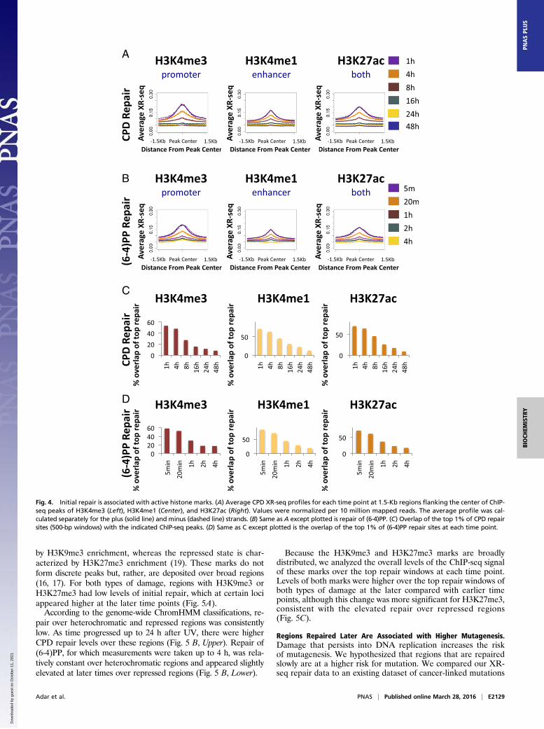
Early Repair Is Associated with Active Histone Marks. We analyzedrepair at sites of histone modifications associated with activechromatin. The modifications we examined were H3K4me3, whichis associated with actively transcribed promoters; H3K4me1 whichis associated with active enhancers that are also often transcribed;and H3K27ac, which is associated with both (16, 20). It is impor-tant to note that, although these marks may be more enriched inpromoters or enhancers, they are often found in both.The average repair of both CPD and (6-4)PP was enriched
around peak centers of H3K4me3, H3K4me1, and H3K27ac.With time, this enrichment was reduced (Fig. 4 A and B). Oncemore, a similar enrichment for (6-4)PP repair at sites of activehistone marks was observed in CS-B cells, indicating that it is notdriven by transcription-coupled recognition of damage (Fig. S9).We analyzed the degree of overlap of the top repair windows
at each time point with the peak regions of the active histonemarks (Fig. 4 C and D and Dataset S2). More than 50% of thetop CPD and (6-4)PP repair windows at the initial time pointsoverlapped H3K4me3, which is associated with actively tran-scribed promoters (53% and 58% respectively, compared with7% for a stochastic control; P < 0.02). At later times, the degreeof overlap was significantly reduced. An even higher degree ofoverlap (>70%) was observed for H3K4me1 and H3K27ac peaks(compared with 14% and 12%, respectively, for a uniform sto-chastic repair control).
Repressed and Heterochromatic States Exhibit Relatively Low Repairthat Persists to Late Time Points. Two chromatin states associatedwith a lack of gene expression have been called “heterochromatic”and “repressed.” The heterochromatic state is characterized
E2128 | www.pnas.org/cgi/doi/10.1073/pnas.1603388113 Adar et al.
Dow
nloa
ded
by g
uest
on
Oct
ober
11,
202
1
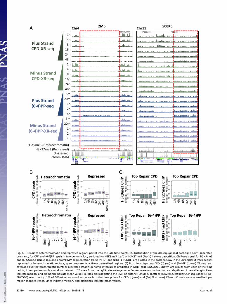
by H3K9me3 enrichment, whereas the repressed state is char-acterized by H3K27me3 enrichment (19). These marks do notform discrete peaks but, rather, are deposited over broad regions(16, 17). For both types of damage, regions with H3K9me3 orH3K27me3 had low levels of initial repair, which at certain lociappeared higher at the later time points (Fig. 5A).According to the genome-wide ChromHMM classifications, re-
pair over heterochromatic and repressed regions was consistentlylow. As time progressed up to 24 h after UV, there were higherCPD repair levels over these regions (Fig. 5 B, Upper). Repair of(6-4)PP, for which measurements were taken up to 4 h, was rela-tively constant over heterochromatic regions and appeared slightlyelevated at later times over repressed regions (Fig. 5 B, Lower).
Because the H3K9me3 and H3K27me3 marks are broadlydistributed, we analyzed the overall levels of the ChIP-seq signalof these marks over the top repair windows at each time point.Levels of both marks were higher over the top repair windows ofboth types of damage at the later compared with earlier timepoints, although this change was more significant for H3K27me3,consistent with the elevated repair over repressed regions(Fig. 5C).
Regions Repaired Later Are Associated with Higher Mutagenesis.Damage that persists into DNA replication increases the riskof mutagenesis. We hypothesized that regions that are repairedslowly are at a higher risk for mutation. We compared our XR-seq repair data to an existing dataset of cancer-linked mutations
A
B
C
D
Fig. 4. Initial repair is associated with active histone marks. (A) Average CPD XR-seq profiles for each time point at 1.5-Kb regions flanking the center of ChIP-seq peaks of H3K4me3 (Left), H3K4me1 (Center), and H3K27ac (Right). Values were normalized per 10 million mapped reads. The average profile was cal-culated separately for the plus (solid line) and minus (dashed line) strands. (B) Same as A except plotted is repair of (6-4)PP. (C) Overlap of the top 1% of CPD repairsites (500-bp windows) with the indicated ChIP-seq peaks. (D) Same as C except plotted is the overlap of the top 1% of (6-4)PP repair sites at each time point.
Adar et al. PNAS | Published online March 28, 2016 | E2129
BIOCH
EMISTR
YPN
ASPL
US
Dow
nloa
ded
by g
uest
on
Oct
ober
11,
202
1
A
B C
Fig. 5. Repair of heterochromatin and repressed regions persist into the late time points. (A) Distribution of the XR-seq signal at each time point, separatedby strand, for CPD and (6-4)PP repair in two genomic loci, enriched for H3K9me3 (Left) or H3K27me3 (Right) histone deposition. ChIP-seq signal for H3K9me3and H3K27me3, DNase-seq, and ChromHMM segmentation tracks (NHDF and NHLF, ENCODE) are plotted in the bottom. Gray in the ChromHMM track depictsrepressed or heterochromatic regions; green represents actively transcribed regions. (B) Box plots depicting CPD (Upper) and (6-4)PP (Lower) XR-seq readcoverage over heterochromatic (Left) or repressed (Right) genomic intervals as predicted in NHLF cells (ENCODE). Shown are results from each of the timepoints, in comparison with a random dataset of 26 mers from the hg19 reference genome. Values were normalized to read depth and interval length. Linesindicate median, and diamonds indicate mean values. (C) Box plots depicting the level of histone H3K9me3 (Left) or H3K27me3 (Right) ChIP-seq signal (NHDF,ENCODE) over the top 1% of 500-nt repair windows in each of the time points for CPD (Upper) and (6-4)PP (Lower) XR-seq. Counts were normalized permillion mapped reads. Lines indicate median, and diamonds indicate mean values.
E2130 | www.pnas.org/cgi/doi/10.1073/pnas.1603388113 Adar et al.
Dow
nloa
ded
by g
uest
on
Oct
ober
11,
202
1
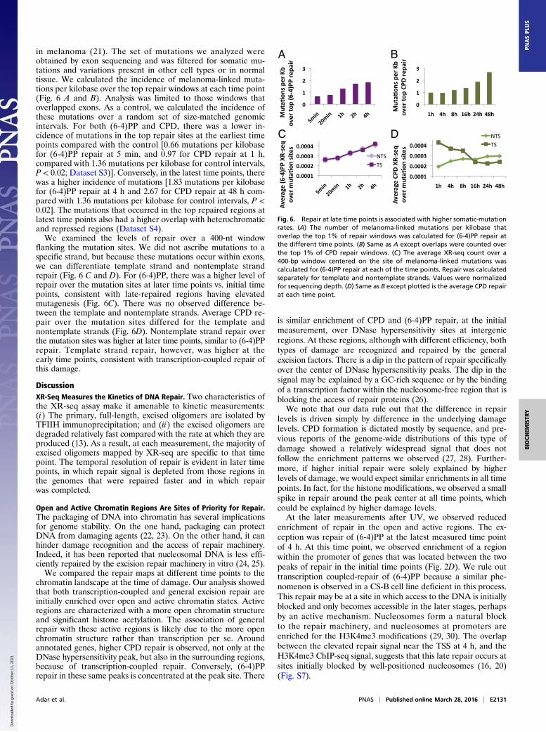
in melanoma (21). The set of mutations we analyzed wereobtained by exon sequencing and was filtered for somatic mu-tations and variations present in other cell types or in normaltissue. We calculated the incidence of melanoma-linked muta-tions per kilobase over the top repair windows at each time point(Fig. 6 A and B). Analysis was limited to those windows thatoverlapped exons. As a control, we calculated the incidence ofthese mutations over a random set of size-matched genomicintervals. For both (6-4)PP and CPD, there was a lower in-cidence of mutations in the top repair sites at the earliest timepoints compared with the control [0.66 mutations per kilobasefor (6-4)PP repair at 5 min, and 0.97 for CPD repair at 1 h,compared with 1.36 mutations per kilobase for control intervals,P < 0.02; Dataset S3)]. Conversely, in the latest time points, therewas a higher incidence of mutations [1.83 mutations per kilobasefor (6-4)PP repair at 4 h and 2.67 for CPD repair at 48 h com-pared with 1.36 mutations per kilobase for control intervals, P <0.02]. The mutations that occurred in the top repaired regions atlatest time points also had a higher overlap with heterochromaticand repressed regions (Dataset S4).We examined the levels of repair over a 400-nt window
flanking the mutation sites. We did not ascribe mutations to aspecific strand, but because these mutations occur within exons,we can differentiate template strand and nontemplate strandrepair (Fig. 6 C and D). For (6-4)PP, there was a higher level ofrepair over the mutation sites at later time points vs. initial timepoints, consistent with late-repaired regions having elevatedmutagenesis (Fig. 6C). There was no observed difference be-tween the template and nontemplate strands. Average CPD re-pair over the mutation sites differed for the template andnontemplate strands (Fig. 6D). Nontemplate strand repair overthe mutation sites was higher at later time points, similar to (6-4)PPrepair. Template strand repair, however, was higher at theearly time points, consistent with transcription-coupled repair ofthis damage.
DiscussionXR-Seq Measures the Kinetics of DNA Repair. Two characteristics ofthe XR-seq assay make it amenable to kinetic measurements:(i) The primary, full-length, excised oligomers are isolated byTFIIH immunoprecipitation; and (ii) the excised oligomers aredegraded relatively fast compared with the rate at which they areproduced (13). As a result, at each measurement, the majority ofexcised oligomers mapped by XR-seq are specific to that timepoint. The temporal resolution of repair is evident in later timepoints, in which repair signal is depleted from those regions inthe genomes that were repaired faster and in which repairwas completed.
Open and Active Chromatin Regions Are Sites of Priority for Repair.The packaging of DNA into chromatin has several implicationsfor genome stability. On the one hand, packaging can protectDNA from damaging agents (22, 23). On the other hand, it canhinder damage recognition and the access of repair machinery.Indeed, it has been reported that nucleosomal DNA is less effi-ciently repaired by the excision repair machinery in vitro (24, 25).We compared the repair maps at different time points to the
chromatin landscape at the time of damage. Our analysis showedthat both transcription-coupled and general excision repair areinitially enriched over open and active chromatin states. Activeregions are characterized with a more open chromatin structureand significant histone acetylation. The association of generalrepair with these active regions is likely due to the more openchromatin structure rather than transcription per se. Aroundannotated genes, higher CPD repair is observed, not only at theDNase hypersensitivity peak, but also in the surrounding regions,because of transcription-coupled repair. Conversely, (6-4)PPrepair in these same peaks is concentrated at the peak site. There
is similar enrichment of CPD and (6-4)PP repair, at the initialmeasurement, over DNase hypersensitivity sites at intergenicregions. At these regions, although with different efficiency, bothtypes of damage are recognized and repaired by the generalexcision factors. There is a dip in the pattern of repair specificallyover the center of DNase hypersensitivity peaks. The dip in thesignal may be explained by a GC-rich sequence or by the bindingof a transcription factor within the nucleosome-free region that isblocking the access of repair proteins (26).We note that our data rule out that the difference in repair
levels is driven simply by difference in the underlying damagelevels. CPD formation is dictated mostly by sequence, and pre-vious reports of the genome-wide distributions of this type ofdamage showed a relatively widespread signal that does notfollow the enrichment patterns we observed (27, 28). Further-more, if higher initial repair were solely explained by higherlevels of damage, we would expect similar enrichments in all timepoints. In fact, for the histone modifications, we observed a smallspike in repair around the peak center at all time points, whichcould be explained by higher damage levels.At the later measurements after UV, we observed reduced
enrichment of repair in the open and active regions. The ex-ception was repair of (6-4)PP at the latest measured time pointof 4 h. At this time point, we observed enrichment of a regionwithin the promoter of genes that was located between the twopeaks of repair in the initial time points (Fig. 2D). We rule outtranscription coupled-repair of (6-4)PP because a similar phe-nomenon is observed in a CS-B cell line deficient in this process.This repair may be at a site in which access to the DNA is initiallyblocked and only becomes accessible in the later stages, perhapsby an active mechanism. Nucleosomes form a natural blockto the repair machinery, and nucleosomes at promoters areenriched for the H3K4me3 modifications (29, 30). The overlapbetween the elevated repair signal near the TSS at 4 h, and theH3K4me3 ChIP-seq signal, suggests that this late repair occurs atsites initially blocked by well-positioned nucleosomes (16, 20)(Fig. S7).
A B
C D
Fig. 6. Repair at late time points is associated with higher somatic-mutationrates. (A) The number of melanoma-linked mutations per kilobase thatoverlap the top 1% of repair windows was calculated for (6-4)PP repair atthe different time points. (B) Same as A except overlaps were counted overthe top 1% of CPD repair windows. (C) The average XR-seq count over a400-bp window centered on the site of melanoma-linked mutations wascalculated for (6-4)PP repair at each of the time points. Repair was calculatedseparately for template and nontemplate strands. Values were normalizedfor sequencing depth. (D) Same as B except plotted is the average CPD repairat each time point.
Adar et al. PNAS | Published online March 28, 2016 | E2131
BIOCH
EMISTR
YPN
ASPL
US
Dow
nloa
ded
by g
uest
on
Oct
ober
11,
202
1
Repair of Heterochromatic and Repressed Chromatin Persists intoLate Time Points. We examined the repair of the heterochro-matic, repressed, and repetitive states in the genome. Becauselow read counts over repetitive states could be attributed to thelow mappability of these regions, we did not address them. Ini-tially, repair of heterochromatic and repressed regions was sub-stantially lower than repair of active regions. These chromatinregions are characterized by the histone modifications H3K9me3and H3K27me3, respectively, and we report higher levels ofthese marks at later-repaired regions. The current “access–repair–restore” model for excision repair in chromatin suggestsactive removal of nucleosomes to allow repair (31). Given thatthe sequencing data is normalized to read depth, we cannotdistinguish whether elevated repair levels at later time points area result of completion of repair in other regions or the result ofan active process, including histone modification, chromatinremodeling, or gene activation in response to UV, that makesthese regions more accessible (32, 33). However, our assay willbe a powerful tool to test for candidates involved in facilitatingrepair of tightly packed chromatin.
Slow Repair Is Associated with Higher Somatic Mutagenesis in CancerCells. UV in sunlight is a known mutagen and causative agent ofskin cancer (34, 35). Mutations in excision repair proteins causesevere genetic disorders such as xeroderma pigmentosum andelevate the risk of cancer (36, 37). A recent study reported anassociation between lower somatic mutation frequency and openchromatin regions that was significantly lower in cells that wereexcision repair deficient (38). Our data adds mechanistic insight,showing a negative correlation between repair timing and mu-tagenesis. This association is clear for general excision repair of(6-4)PP. In the skin fibroblasts used in our study, the top repairregions at the latest time points are associated with the highestincidence of melanoma-linked mutations. Conversely, sites ofmelanoma-linked mutations exhibited higher average repair atthe later time points. For CPDs, there is also a higher incidenceof mutations in the top repair regions at the latest time points.However, the pattern of average repair over mutations sitesdiffers for the template and nontemplate strand. Average non-template strand repair over mutation sites is higher at the latertime points. Conversely, template-strand repair is higher at theearlier time points, consistent with transcription-coupled repair,preferentially removing damage from transcribed exons. We didnot assign mutations to a specific strand. The pattern of repair,however, suggests that mutations originated from the non-template strand. Higher mutation levels on the nontranscribedstrand would be consistent with reports of transcription-basedstrand-asymmetries in mutagenesis (21, 39).In general, mutation rates associated with CPD repair are
lower if compared with the mutation rate associated with (6-4)PPrepair at the parallel time points (1–4 h). These lower mutationrates could be because mutations associated with CPD repairsites are derived only from the nontranscribed strand or perhapsbecause of the ability of mammalian cells to bypass this damagein an error-free mechanism (40).DNA replication may also affect repair efficiencies and, in
addition, has been implicated in mutagenesis strand asymmetries(39). Future work in synchronized cells will be able to addressthe effect of replication timing on DNA repair efficiencies.Our work here investigates how the initial chromatin state at the
time of damage affects the efficiency and priority of DNA repair.However, the chromatin landscape is not static, and as a result ofgenotoxic stress, it is altered. DNA damage induces a transcrip-tional response and is associated with changes in histone modifi-cations. Histone modifications play an active role in the responseto DNA damage and in the recruitment of repair proteins (18).Future studies will incorporate measurements of DNA damage,DNA repair, RNA levels, and histone modifications in response to
damage and will further improve our understanding for how DNArepair is orchestrated within chromatin.Nucleotide excision repair is the sole mechanism for removing
bulky adducts from the human genome, including those formedby chemotherapeutic drugs such as cisplatin and oxaliplatin. Im-proving our understanding of DNA repair is beneficial, not onlyfor understanding carcinogenesis, but also for understandingthe processes cancer cells use to cope with chemotherapy. Suchinformation is expected to aid in improving currently usedchemotherapy regimens.
Materials and MethodsCell Culture and UV Irradiation. Telomerase-immortalized normal human fi-broblast NHF1 was obtained from W. K. Kaufmann, University of NorthCarolina at Chapel Hill, Chapel Hill, NC (41). CS-B (CS1ANps3g2, GM16095)mutant human skin fibroblasts were purchased from the National Instituteof General Medical Sciences Human Genetic Cell Repository (Coriell Institute).CS-B cells were cultured in DMEM supplemented with 10% (vol/vol) FBS at37 °C in a 5% atmosphere CO2 humidified chamber. NHF1 cells were main-tained under the same conditions with the addition of 2 mM glutamine.
UV irradiation was performed as described with the indicated dose (12).After incubation at 37 °C for the indicated time, cells were washed andcollected in cold PBS.
Detection of Excision Products. Excision products from UV-irradiated cellswere purified, radiolabeled, and separated by electrophoresis as described(13). Short DNA fragments were extracted by modified Hirt’s method andsubjected to immunoprecipitation against anti-(6-4)PP or -CPD antibodies.Purified excised oligonucleotides were 3′ radiolabeled by terminal deoxy-nucleotidyl transferase (NEB) and [α-32P]-3′-deoxyadenosine 5′-triphosphate(cordycepin 5′-triphosphate) (Perkin-Elmer) and resolved in 10% denaturingsequencing gels.
XR-Seq Library Preparation. XR-seq libraries were prepared as described (11).Briefly, primary excision products pulled down by TFIIH coimmunoprecipitationwere ligated to both 5′ and 3′ adaptors. Ligation products containing (6-4)PP orCPD were purified by immunoprecipitation with corresponding antibodies andrepaired by specific photolyases. Repaired DNA were PCR-amplified with Indexprimers and purified by 10% native polyacrylamide gels.
Sequencing and Genome Alignment. Libraries were sequenced on a Hiseq 2000platform by the University of North Carolina High-Throughput SequencingFacility. Flanking adapter sequences were removed from the reads by usingtrimmomatic (42). Reads were aligned to the hg19 human genome by usingbowtie (43) with the command options -q–nomaqround–phred33-quals -m4 -n 2 -e 70 -l 20–best –S. Uniquely aligned reads were obtained by usingsamtools. We obtained a total of at least 19 million uniquely mapped readsin each of the time points (Dataset S1). For comparison of the DNA-repairsignal, we normalized all of the count data by the sequencing depth; dataare available for viewing as a track hub on the UCSC genome browser(https://genome.ucsc.edu/cgi-bin/hgGateway) by pasting the link: http://trackhubs.its.unc.edu/sancarlb/XRseqTimeCourse/hub.txt. Sequencing datafor (6-4)PP repair at 1 h in NHF1 and CS-B cells and CPD repair at 1 h in CS-Bcells were taken from the published dataset (11). The raw data and bigwigtracks are available under the Gene Expression Omnibus database (www.ncbi.nlm.nih.gov/geo/, accession no. GSE76391).
ENCODE Data. NHDF long total stranded RNA-seq [ENCODE Data CoordinationCenter (DCC) accession no. ENCSR00CUH], H3K4me1 (accession no. ENCS-R000ARV), H3K4me3 (accession no. ENCSR000DPR), H3K27ac (accession no.ENCSR000APN), H3K27me3 (accession no. ENCSR000APO), H3K9me3 (accessionno. ENCSR000ARX), and DNase-seq (accession no. ENCSR000EMP) fastq,aligned reads .bam files, and peak files, as well as the NHLF chromHMMchromatin state segmentation (UCSC accession no. wgEncodeEH000792),were downloaded from the ENCODE portal (genome.ucsc.edu/ENCODE/)or viewed on the UCSC browser.
Analysis of Top Repair Windows. The hg19 genome was divided into 500-ntwindows, and the repair coverage for each window was calculated for eachsample by using bedtools. The 62,743windowswith the highest score on eachstrand and at each time pointwere taken as the top 1%.Genomic distributionof reads and overlap with annotated genes was calculated by using the UCSCrefGene.txt gene annotation, as described in Hu et al. (11). Bedtools intersect
E2132 | www.pnas.org/cgi/doi/10.1073/pnas.1603388113 Adar et al.
Dow
nloa
ded
by g
uest
on
Oct
ober
11,
202
1
was used to calculate all overlaps, including those to peak intervals andmutations. For comparison and to calculate P values, we conducted the sameanalysis on 50 random datasets of 62,743 intervals of 500 nt from the hg19reference genome. P values for comparison of distributions were calculatedas [number of times distribution of experimental and control data over-lapped]/[total number of control data tests].
Chromatin State Analysis. Repair over the genomic intervals for each of the 15predicted chromatin states defined by the ChromHMM algorithm was cal-culated by using bedtools coverage. Values were normalized per millionmapped reads and per kilobase of interval length.
Cancer Mutation Analysis. Cancer-linked somatic mutation datasets, which werefiltered for variations found in normal cells and tissues, were obtained fromAlexandrov et al. (21). For melanoma, these include exome-only datasets. Thenumber of mutations overlapping the top 1% of repair windows at each timepoint was calculated with bedtools intersect. The results were normalized per
kilobase of top windows that overlapped exons. As a control, the same analysiswas performed with a random set of 500-nt windows from the hg19 referencegenome. To calculate average repair over mutation sites, a 400-bp intervalcentered on the mutation site was created by using bedtools slop, and coveragewas calculated with bedtools coverage, normalizing to 1 million mapped reads.
Plotting Average XR-Seq Profiles. Gene lists, grouped into expression quartilesbased on RNA-seq in NHDF cells, were taken from our previous analysis (11).For average XR-seq profiles relative to the annotated TSS, we limited thegene list to genes that do not have overlapping or neighboring genes for atleast 6,000 bp upstream or downstream on either strand and were at least10,000 bp in length. Read counts were calculated from the aligned .bam filesby using bedtools coverage and normalized to 10 million mapped reads.
ACKNOWLEDGMENTS. We thank Dr. Sebastian Pott for fruitful discussionand suggestions. This work was supported by National Institutes of HealthGrants GM32833 and GM31082 (to A.S.) and HG006787 (to J.D.L.).
1. Reardon JT, Sancar A (2005) Nucleotide excision repair. Prog Nucleic Acid Res Mol Biol
79(79):183–235.2. Sancar A (1996) DNA excision repair. Annu Rev Biochem 65:43–81.3. Wood RD (1997) Nucleotide excision repair in mammalian cells. J Biol Chem 272(38):
23465–23468.4. Reardon JT, Sancar A (2003) Recognition and repair of the cyclobutane thymine di-
mer, a major cause of skin cancers, by the human excision nuclease. Genes Dev 17(20):
2539–2551.5. Sugasawa K, et al. (1998) Xeroderma pigmentosum group C protein complex is the
initiator of global genome nucleotide excision repair. Mol Cell 2(2):223–232.6. Wakasugi M, Sancar A (1998) Assembly, subunit composition, and footprint of human
DNA repair excision nuclease. Proc Natl Acad Sci USA 95(12):6669–6674.7. Hanawalt PC, Spivak G (2008) Transcription-coupled DNA repair: Two decades of
progress and surprises. Nat Rev Mol Cell Biol 9(12):958–970.8. Mellon I, Spivak G, Hanawalt PC (1987) Selective removal of transcription-blocking
DNA damage from the transcribed strand of the mammalian DHFR gene. Cell 51(2):
241–249.9. Huang JC, Svoboda DL, Reardon JT, Sancar A (1992) Human nucleotide excision nu-
clease removes thymine dimers from DNA by incising the 22nd phosphodiester bond
5′ and the 6th phosphodiester bond 3′ to the photodimer. Proc Natl Acad Sci USA
89(8):3664–3668.10. Mu D, et al. (1995) Reconstitution of human DNA repair excision nuclease in a highly
defined system. J Biol Chem 270(6):2415–2418.11. Hu J, Adar S, Selby CP, Lieb JD, Sancar A (2015) Genome-wide analysis of human
global and transcription-coupled excision repair of UV damage at single-nucleotide
resolution. Genes Dev 29(9):948–960.12. Gaddameedhi S, et al. (2010) Similar nucleotide excision repair capacity in melano-
cytes and melanoma cells. Cancer Res 70(12):4922–4930.13. Hu J, et al. (2013) Nucleotide excision repair in human cells: Fate of the excised oli-
gonucleotide carrying DNA damage in vivo. J Biol Chem 288(29):20918–20926.14. Bassett E, et al. (2004) The role of DNA polymerase eta in translesion synthesis past
platinum-DNA adducts in human fibroblasts. Cancer Res 64(18):6469–6475.15. Mitchell DL (1988) The relative cytotoxicity of (6-4) photoproducts and cyclobutane
dimers in mammalian cells. Photochem Photobiol 48(1):51–57.16. Zhou VW, Goren A, Bernstein BE (2011) Charting histone modifications and the
functional organization of mammalian genomes. Nat Rev Genet 12(1):7–18.17. Consortium EP; ENCODE Project Consortium (2012) An integrated encyclopedia of
DNA elements in the human genome. Nature 489(7414):57–74.18. Gospodinov A, Herceg Z (2013) Shaping chromatin for repair.Mutat Res 752(1):45–60.19. Ernst J, et al. (2011) Mapping and analysis of chromatin state dynamics in nine human
cell types. Nature 473(7345):43–49.20. Kim TK, Shiekhattar R (2015) Architectural and functional commonalities between
enhancers and promoters. Cell 162(5):948–959.21. Alexandrov LB, et al.; Australian Pancreatic Cancer Genome Initiative; ICGC Breast
Cancer Consortium; ICGC MMML-Seq Consortium; ICGC PedBrain (2013) Signatures of
mutational processes in human cancer. Nature 500(7463):415–421.
22. Mitchell DL, Nguyen TD, Cleaver JE (1990) Nonrandom induction of pyrimidine-pyrimidone (6-4) photoproducts in ultraviolet-irradiated human chromatin. J Biol Chem265(10):5353–5356.
23. Gale JM, Smerdon MJ (1990) UV induced (6-4) photoproducts are distributed differ-ently than cyclobutane dimers in nucleosomes. Photochem Photobiol 51(4):411–417.
24. Hara R, Sancar A (2002) The SWI/SNF chromatin-remodeling factor stimulates repairby human excision nuclease in the mononucleosome core particle. Mol Cell Biol22(19):6779–6787.
25. Wang D, Hara R, Singh G, Sancar A, Lippard SJ (2003) Nucleosomes inhibit nucleotideexcision repair of site-specific platinum-DNA adducts. Biochemistry 42(22):6747.
26. Wang J, et al. (2012) Sequence features and chromatin structure around the genomicregions bound by 119 human transcription factors. Genome Res 22(9):1798–1812.
27. Powell JR, et al. (2015) 3D-DIP-Chip: A microarray-based method to measure genomicDNA damage. Sci Rep 5:7975.
28. Zavala AG, Morris RT, Wyrick JJ, Smerdon MJ (2014) High-resolution characterizationof CPD hotspot formation in human fibroblasts. Nucleic Acids Res 42(2):893–905.
29. Hughes AL, Rando OJ (2014) Mechanisms underlying nucleosome positioning in vivo.Annu Rev Biophys 43:41–63.
30. Hara R, Mo J, Sancar A (2000) DNA damage in the nucleosome core is refractory torepair by human excision nuclease. Mol Cell Biol 20(24):9173–9181.
31. Polo SE, Almouzni G (2015) Chromatin dynamics after DNA damage: The legacy of theaccess-repair-restore model. DNA Repair (Amst) 36:114–121.
32. Schick S, et al. (2015) Dynamics of chromatin accessibility and epigenetic state in re-sponse to UV damage. J Cell Sci 128(23):4380–4394.
33. Yu Y, Teng Y, Liu H, Reed SH, Waters R (2005) UV irradiation stimulates histoneacetylation and chromatin remodeling at a repressed yeast locus. Proc Natl Acad SciUSA 102(24):8650–8655.
34. Pleasance ED, et al. (2010) A comprehensive catalogue of somatic mutations from ahuman cancer genome. Nature 463(7278):191–196.
35. Hodis E, et al. (2012) A landscape of driver mutations in melanoma. Cell 150(2):251–263.
36. Cleaver JE (1968) Defective repair replication of DNA in xeroderma pigmentosum.Nature 218(5142):652–656.
37. DiGiovanna JJ, Kraemer KH (2012) Shining a light on xeroderma pigmentosum.J Invest Dermatol 132(3 Pt 2):785–796.
38. Polak P, et al. (2014) Reduced local mutation density in regulatory DNA of cancergenomes is linked to DNA repair. Nat Biotechnol 32(1):71–75.
39. Haradhvala NJ, et al. (2016) Mutational strand asymmetries in cancer genomes revealmechanisms of DNA damage and repair. Cell 164(3):538–549.
40. Pfeifer GP, Besaratinia A (2012) UV wavelength-dependent DNA damage and humannon-melanoma and melanoma skin cancer. Photochem Photobiol Sci 11(1):90–97.
41. Heffernan TP, et al. (2002) An ATR- and Chk1-dependent S checkpoint inhibitsreplicon initiation following UVC-induced DNA damage. Mol Cell Biol 22(24):8552–8561.
42. Bolger AM, Lohse M, Usadel B (2014) Trimmomatic: A flexible trimmer for Illuminasequence data. Bioinformatics 30(15):2114–2120.
43. Langmead B, Trapnell C, Pop M, Salzberg SL (2009) Ultrafast and memory-efficientalignment of short DNA sequences to the human genome. Genome Biol 10(3):R25.
Adar et al. PNAS | Published online March 28, 2016 | E2133
BIOCH
EMISTR
YPN
ASPL
US
Dow
nloa
ded
by g
uest
on
Oct
ober
11,
202
1