genome-wide analysis of heterochromatin associates clonally
TRANSCRIPT
Cell Host & Microbe
Article
Genome-wide Analysis of Heterochromatin AssociatesClonally Variant Gene Regulation with PerinuclearRepressive Centers in Malaria ParasitesJose-Juan Lopez-Rubio,1,2 Liliana Mancio-Silva,1,2 and Artur Scherf1,*1Unite de Biologie des Interactions Hote-Parasite, CNRS URA2581, Institut Pasteur, 25 Rue du Dr. Roux, 75724 Paris, France2These authors contributed equally to this work
*Correspondence: [email protected] 10.1016/j.chom.2008.12.012
SUMMARY
Clonally variant gene families underlie phenotypicplasticity in Plasmodium falciparum, a process indis-pensable for survival of the pathogen in its humanhost. Differential transcription of one of these genefamilies in clonal parasite lineages has been associ-ated with chromatin modifications. Here, we deter-mine the genome-wide distribution in P. falciparumof a histone mark of heterochromatin, trimethylationof histone H3 lysine 9 (H3K9me3), using high-resolu-tion ChIP-chip analysis. We show that H3K9me3 isspecifically associated with clonally variant genefamilies, which are clustered on subtelomeric andsome chromosome internal regions. High levels ofH3K9me3 correlate with genes localized to thenuclear periphery, implying chromosome loopformation. Disruption of the histone deacetylasePfSir2 causes changes in H3K9me3 that are discon-tinuous along chromosomes and associated withdisrupted monoallelic transcription. Our data pointto the existence of perinuclear repressive centersassociated with control of expression of malariaparasite genes involved in phenotypic variation andpathogenesis.
INTRODUCTION
The human protozoan malaria parasite P. falciparum kills more
than 2 million people (mainly children) per year (Snow et al.,
2005). This parasite is well adapted to survive in changing host
environments such as the Anopheles mosquito midgut and sali-
vary glands, human liver hepatocytes, and erythrocytes. Malaria
pathogenesis is linked to the parasite’s capacity to develop
different phenotypic states in genetically clonal blood stage
parasites (Fried and Duffy, 1996; Jensen et al., 2004). However,
most processes causing pathogenesis are not yet understood at
the molecular level and may involve expression of particular
combinations of clonally variant molecules.
In P. falciparum, phenotypic diversity is commonly achieved
by the expression of clonally variant surface molecules either
at the erythrocyte membrane or merozoite surface (Cortes
Cell Hos
et al., 2007; Scherf et al., 1998; Stubbs et al., 2005). Switching
of expression to another variant molecule prolongs the period
of infection and generates alternative evasion pathways. Little
is known about factors that govern phenotypic plasticity. The
best-studied example of variant surface proteins is the P. falcipa-
rum erythrocyte membrane protein 1 (PfEMP1) (Leech et al.,
1984). PfEMP1 is a major virulence factor involved in antigenic
variation and adherence of infected erythrocytes to host recep-
tors, resulting in their sequestration to capillaries in the brain
and other critical organs (Kyes et al., 2007). PfEMP1 is encoded
by genes of the 60 member var family, which are located on sub-
telomeric and internal chromosome loci. In a single parasite, only
one var gene is expressed in a mutually exclusive manner under
the control of epigenetic factors (Scherf et al., 1998).
A number of recent publications point to reversible chromatin
changes, such as histone methylation and acetylation, as impor-
tant control elements of P. falciparum gene silencing and mono-
allelic activation (reviewed in Scherf et al., 2008a). Enzymes that
add or remove acetyl and methyl marks in P. falciparum have
also been identified (Cui et al., 2008; Merrick and Duraisingh,
2007; Miao et al., 2006). It has been demonstrated that trimethy-
lation of histone H3 lysine 9 (H3K9me3) is associated to tran-
scriptionally silent var genes (Chookajorn et al., 2007; Lopez-
Rubio et al., 2007). Upon var gene activation, in the 50 flanking
region (50UTR), methylation is replaced by acetylation at lysine
9 of histone H3 (H3K9ac), and histone H3 lysine 4 is di- and trime-
thylated (H3K4me2/3) (Lopez-Rubio et al., 2007). Furthermore,
PfSir2 histone deacetylase has been shown to spread from telo-
meres into the subtelomeric coding regions, leading to stable but
reversible repression of subtelomeric var genes (Duraisingh
et al., 2005; Freitas-Junior et al., 2005; Mancio-Silva et al.,
2008). Genetic elements such as the 50UTR (Voss et al., 2006)
contain essential information able to interact with the epigenetic
control machinery. Experimental data suggest that the var
intron-linked promoter activity also contributes to monoallelic
expression (Dzikowski et al., 2006).
Sequencing of the P. falciparum genome has led to the
discovery of multiple gene families mainly clustered at subtelo-
meres adjacent to var genes (Gardner et al., 2002). A few of these
have been studied, such as rif (Fernandez et al., 1999; Kyes et al.,
1999), clag (Cortes et al., 2007), stevor, and PfMC-2TM (Blythe
et al., 2008; Lavazec et al., 2007) and, similarly to var genes, are
clonally variant and possibly involved in immune evasion (Scherf
et al., 2008a). Telomeres are spatially restricted to nuclear
periphery, where they form clusters of three to seven
t & Microbe 5, 179–190, February 19, 2009 ª2009 Elsevier Inc. 179
Cell Host & Microbe
H3K9me Is Restricted to Perinuclear Virulence Genes
180 Cell Host & Microbe 5, 179–190, February 19, 2009 ª2009 Elsev
custom tiling array included probes only to the extremities of
one chromosome (chromosome 12) (see Supplemental Experi-
mental Procedures). We observed a strong enrichment in
H3K9me3 in the entire TAREs 1–6 (Figures 1B, right, 2, both
flanks, and S1). On the contrary, telomeric repeat DNA pre-
sented only a modest H3K9me3 level (Figure S1, right).
H3K4me3 and H3K9ac were completely absent from TAREs
and telomere repeats, and H4K20me3 was detectable only at
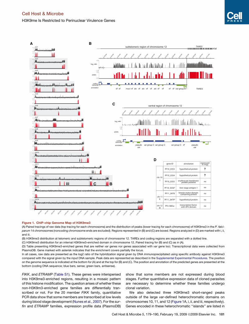
low levels (Figure 2, both flanks).
Eukaryotic centromeres and pericentromeric regions are
embedded in heterochromatin and exhibit unique chromatin
architecture necessary for their chromosome segregation
function (Grewal and Jia, 2007). P. falciparum centromeric
DNA differs from centromeres of other organisms. It consists
of AT-rich (97%) domains with a sharply defined size limit of
2.3–2.5 kb, which lack interchromosomal conserved motifs
(Kelly et al., 2006). Our tiled array also covered pericentromeric
DNA. In contrast to other organisms in which this region is en-
riched in H3K9me3, our ChIP-chip analysis showed that pericen-
tromeric chromatin in P. falciparum was not enriched for
H3K9me3 (example for pericentromere in Figure 2), which is
consistent with the unusual nature of centromeric DNA in this
parasite.
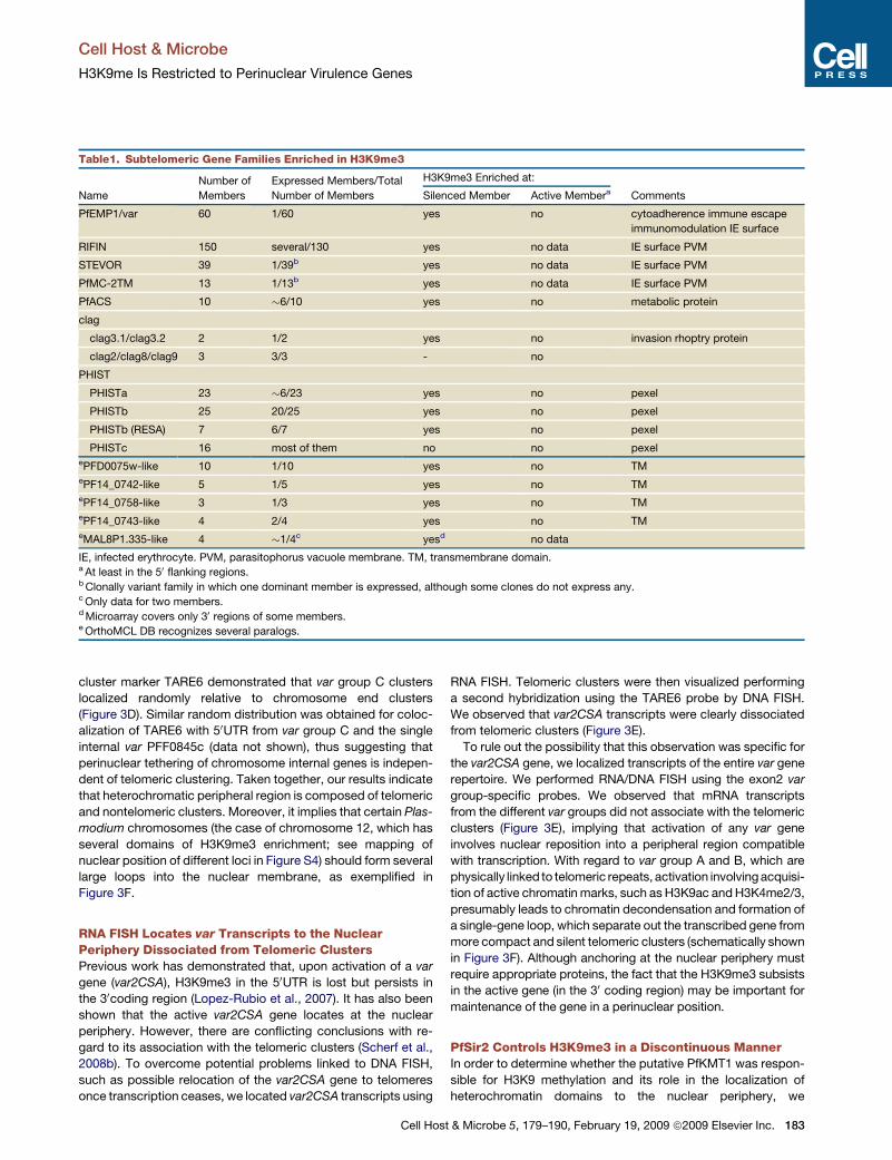
H3K9me3 Is Associated with Differentially TranscribedVirulence Gene FamiliesDetailed examination of the major heterochromatin regions
revealed that the entire var gene family was enriched in
H3K9me3, which agrees with previous predictions (Chookajorn
et al., 2007; Lopez-Rubio et al., 2007). In addition, we observed
that many distinct gene families adjacent to subtelomeric var
genes were also enriched in H3K9me3 in the 50UTR and coding
region (Figure 1B). Chromosome internal H3K9me3-enriched
domains contained generally only var, rif, and stevor genes
(Figure 1C). We found H3K9me3 enrichments in seven previously
identified subtelomeric gene families: var, rif, stevor, Pfmc-2TM,
clag, PfAcs, and PHIST (Table 1). For almost all of these gene
families, there are experimental evidences demonstrating
a differential transcription mode of individual members. Another
common feature is that many of these gene families code for
variant surface proteins participating in the vast phenotypic plas-
ticity of blood stage parasites and immune evasion (Scherf et al.,
2008a).
Additionally, based on our H3K9me3 findings together with
OrthoMCL database (Chen et al., 2006), we identified five putative
subtelomeric gene families. In the absence of any biological data,
we called these families PFD0075w-like, PF14_0742-like,
PF14_0758-like, PF14_0743-like, and MAL8P1.335-like (Table 1).
Published expression profile data of these genes (Le Roch et al.,
2003) analyzed using PlasmoDB (http://plasmodb.org/plasmo/)
strongly suggest a differential transcription profile of these
gene families, which is a hallmark for phenotypic variation.
Silent members presented high levels of H3K9me3, whereas
transcribed members had low levels of this mark in the 50UTR
(Table 1). Therefore, our results indicate that H3K9me3 in the
50UTR may predict genes that are controlled by a similar epige-
netic mechanism.
However, our analysis also showed that some subtelomeric
gene families were not enriched for H3K9me3, such as surfin,
heterologous chromosome ends (Freitas-Junior et al., 2000). This
compartment is believed to enhance continual genetic exchange
between members of gene families and may explain the vast var
repertoire diversity observed between field isolates (Barry et al.,
2007). Investigating the role of clusters in gene expression has re-
sulted in inconsistent data. Experimental evidence, using para-
sites transfected with various constructs, reported relocation of
an active var gene into a cluster (Duraisingh et al., 2005; Marty
et al., 2006; Voss et al., 2006), whereas other studies, using para-
site populations expressing a single var gene, observed the relo-
cation outside of cluster (Ralph et al., 2005).
Whether phenotypic variation shares common epigenetic
control mechanisms in any pathogenic parasite remains unclear.
Here, we report that repressive centers at the perinuclear space
participate in virulence gene family silencing in P. falciparum.
Genome-wide analysis of histone modifications revealed that
heterochromatic mark H3K9me3 is restricted to clonally variant
gene families. Furthermore, we demonstrate that H3K9me3 is
associated to the perinuclear location of internal and subtelo-
meric regions. This histone modification is controlled by the
histone deacetylase PfSir2—however, only for a subset of
variant gene family members—revealing an unusual mechanism
of PfSir2 repression distinct from the postulated telomere posi-
tion effect. In addition, this study allowed us to identify subtelo-
meric gene families not yet characterized, which are apparently
differentially transcribed and represent potential virulence
factors participating in phenotypic plasticity of malaria parasites.
RESULTS
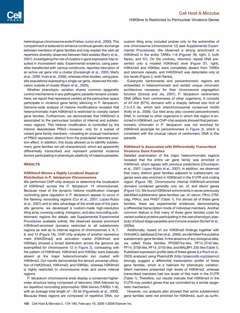
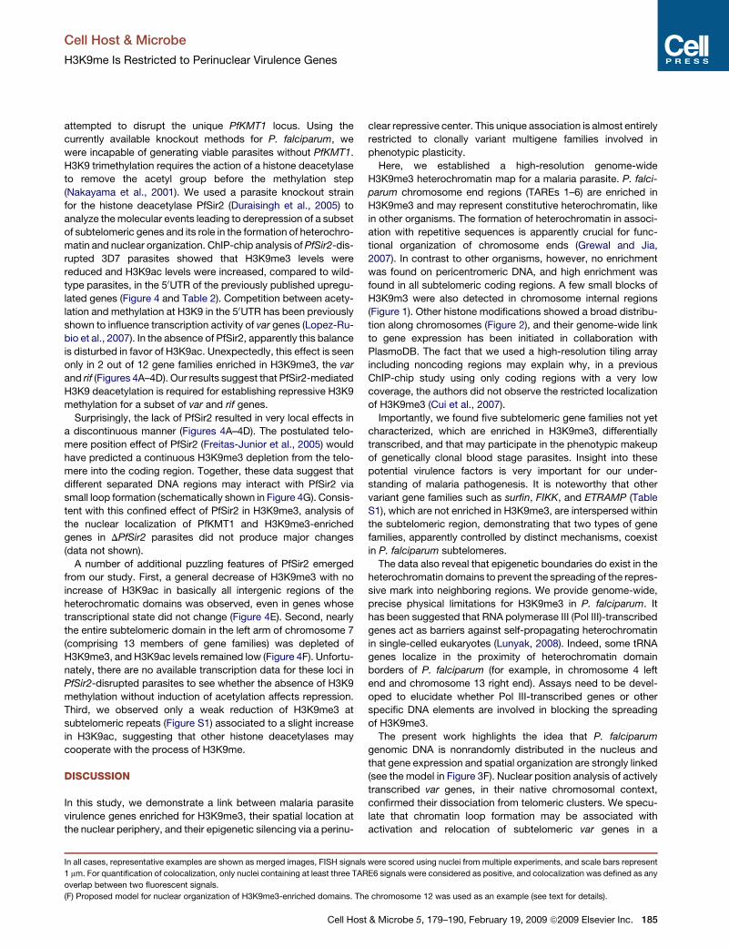
H3K9me3 Shows a Highly Localized AtypicalDistribution in P. falciparum ChromosomesWe performed ChIP-chip analysis to determine the localization
of H3K9me3 across the P. falciparum 14 chromosomes.
Because most of the dynamic histone modification changes
controlling gene regulation in P. falciparum appear to occur in
the flanking noncoding regions (Cui et al., 2007; Lopez-Rubio
et al., 2007) and to take advantage of the small size of the para-
site genome, we developed a custom-made high-resolution
tiling array covering coding, intergenic, and also noncoding sub-
telomeric regions (for details, see Supplemental Experimental
Procedures available online). We observed several prominent
H3K9me3-enriched domains restricted to all subtelomeric
regions as well as to internal regions of chromosomes 4, 6, 7,
8, and 12 (Figure 1A). ChIP-chip analysis of another repressive
mark (H4K20me3) and activation marks (H3K4me3 and
H3K9ac) showed a broad distribution across the genome (as
exemplified for chromosome 12 in Figure 2), contrasting with
the pattern of H3K9me3. H3K4me3 and H3K9ac were basically
absent at the major heterochromatin loci coated with
H3K9me3. Our results demonstrate the almost universal utiliza-
tion of H4K20me3, H3K4me3, and H3K9ac, whereas H3K9me3
is highly restricted to chromosome ends and some internal
regions.
P. falciparum chromosome ends display a conserved higher-
order structure being composed of telomeric DNA followed by
six repetitive noncoding polymorphic DNA blocks (TAREs 1–6),
with an average total length of �30 kb (Figueiredo et al., 2000).
Because these regions are composed of repetitive DNA, our
ier Inc.
Cell Host & Microbe
H3K9me Is Restricted to Perinuclear Virulence Genes
A B
C
D
Figure 1. ChIP-chip Genome Map of H3K9me3(A) Paired tracings of raw data (top tracing for each chromosome) and the distribution of peaks (lower tracing for each chromosome) of H3K9me3 in the P. falci-
parum 14 chromosomes (noncoding chromosome ends are excluded). Regions represented in (B) and (C) are boxed. Regions analyzed in (D) are marked with i, ii,
and iii.
(B) H3K9me3 distribution at telomeric and subtelomeric regions of chromosome 12. TAREs and coding regions are separated with a dotted line.
(C) H3K9me3 distribution for an internal H3K9me3-enriched domain in chromosome 12. Paired tracing for (B) and (C) as in (A).
(D) Table presenting H3K9me3-enriched genes that are neither var genes nor genes associated with var gene loci. Transcriptional data were collected from
PlasmoDB. Gene marked with asterisk indicates that the enrichment covers partially the locus.
In all cases, raw data are presented as the log2 ratio of the hybridization signal given by DNA immunoprecipitated using specific antibody against H3K9me3
compared with the signal given by the input DNA sample. Peak data are represented as described in the Supplemental Experimental Procedures. The position
on the genome sequence is indicated at the bottom for (A) and at the top for (B) and (C). The position and annotation of the predicted genes are presented at the
bottom (coding DNA sequence; blue bars, sense; green bars, antisense).
Cell Ho
show that some members are not expressed during blood
stages. Further quantitative expression data of cloned parasites
are necessary to determine whether these families undergo
clonal variation.
We also detected three H3K9me3 short-ranged peaks
outside of the large var-defined heterochromatic domains on
chromosomes 10, 11, and 12 (Figure 1A, i, ii, and iii, respectively).
Genes encoded in these heterochromatic ‘‘islands’’ are listed in
FIKK, and ETRAMP (Table S1). These genes were interspersed
into H3K9me3-enriched regions, resulting in a mosaic pattern
of this histone modification. The question arises of whether these
non-H3K9m3-enriched gene families are differentially tran-
scribed or not. For the 20 member FIKK family, quantitative
PCR data show that some members are transcribed at low levels
during blood stage development (Nunes et al., 2007). For the sur-
fin and ETRAMP families, expression profile data (PlasmoDB)
st & Microbe 5, 179–190, February 19, 2009 ª2009 Elsevier Inc. 181
Cell Host & Microbe
H3K9me Is Restricted to Perinuclear Virulence Genes
Figure 2. Chromosome 12 Distribution Profiles of Different Histone Modifications
The raw data (top tracing) and the distribution of peaks (lower tracing) identified for H3K9me3, H4K20me3, H3K4me3, and H3K9ac are presented. Noncoding
chromosome ends (TAREs and telomeres) are separated with a dotted line. A schematic representation of chromosome 12 showing main features and the posi-
tion on the genome is indicated at the top. Raw and peak data are presented as in Figure 1.
182 Cell Host & Microbe 5, 179–190, February 19, 2009 ª2009 Else
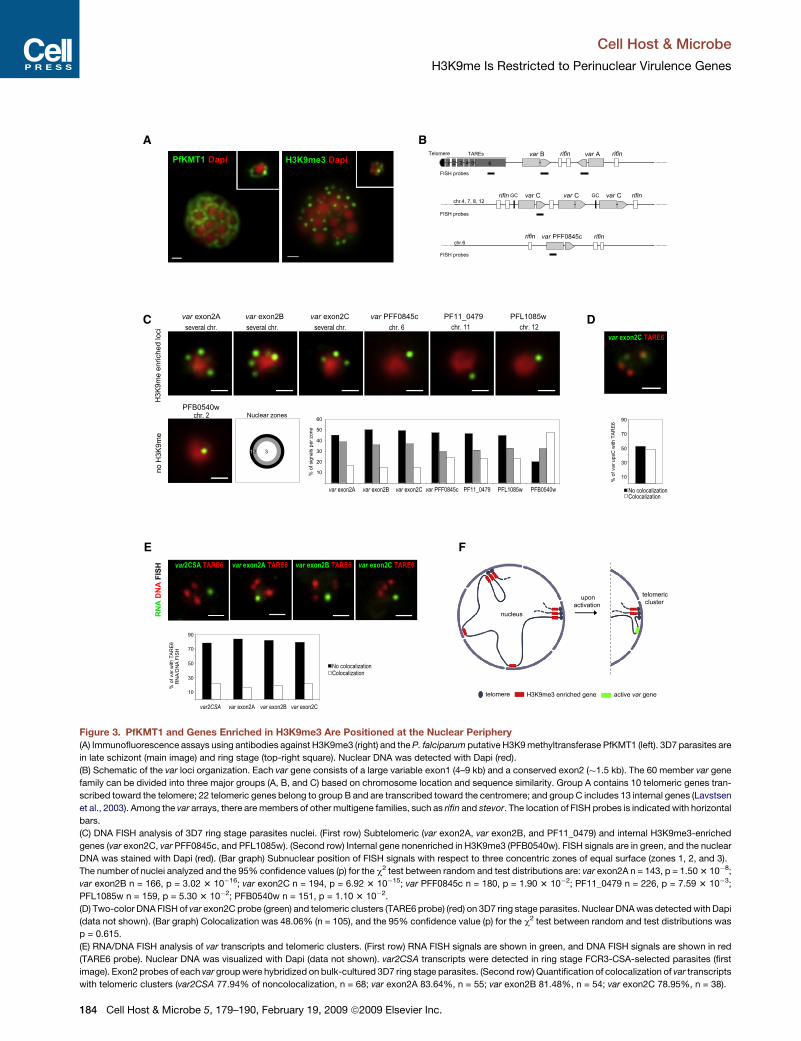
telomeric genes), and group C (13 genes grouped in 7 internal
regions) (Lavstsen et al., 2003) (Figures 1A and 3B). We observed
two to five foci per nucleus for each var exon2 probe, indicating
clustering of var genes from groups A, B, and C (Figures 3C and
S2). The other FISH probes tested were gene-specific probes
(single internal var PFF0845c, PF11_0479, and PFL1085w) and
produced single foci per nucleus (Figure 3C).
In order to precisely define the nuclear boundaries, we devel-
oped an antibody that recognizes a P. falciparum protein
encoded by a gene orthologous to a yeast nuclear pore protein
(PF14_0706) (Figure S3). The area of the nuclear section
obtained in immunofluorescence staining was calculated and
then divided into equal thirds. FISH signals were scored as local-
izing to zones 1, 2, and 3 (Hediger et al., 2002), as illustrated in
Figures 3C and S3. Quantitative analysis of nuclear position
revealed that all var and non-var genes enriched for H3K9me3
and analyzed were not randomly distributed but, rather, located
predominantly at the nuclear periphery. By contrast, internal
genes nonenriched in H3K9me3 were found mostly in internal
nuclear regions (Figures 3C and S4). Thus, virulence gene fami-
lies and single-copy genes enriched in H3K9me3 are localized at
the perinuclear space, irrespective of their chromosomal posi-
tion. This led us to hypothesize that PfKMT1 and H3K9me3 might
determine the nuclear organization of genes.
P. falciparum chromosome ends are tethered to the nuclear
membrane forming clusters (Freitas-Junior et al., 2000). To
exclude the possibility of internal H3K9me3-enriched genes
(var group C, var PFF0845c, and PFL1085w) being at the nuclear
periphery as a consequence of being associated to telomeric
clusters, we analyzed their relative position to these clusters.
Simultaneous FISH analysis of var exon2C with a telomeric
Figure 1D. Apart from PFL1085w, which encodes a member of
the ApiAP2 family of transcription factors (De Silva et al.,
2008), very little information is available for the other genes.
Further analysis is needed to determine whether these genes
participate in phenotypic diversity of parasites or are epigeneti-
cally coregulated.
PfKMT1 and H3K9me3-Enriched Genes Are Localizedto the Nuclear PeripheryGiven the unusual highly localized H3K9me3 pattern in P. falcipa-
rum genome, we were interested in investigating the nuclear
organization of H3K9me3 domains. H3K9 methylation is per-
formed by a histone lysine 9 methyltransferase KMT1 (Allis
et al., 2007), of which an homolog (PF08_0012) has been found
in P. falciparum genome (Cui et al., 2008). Antibodies raised
against the P. falciparum KMT1 were used to study the cellular
location of this molecule. Surprisingly, we observed that PfKMT1
fluorescent signals located largely at the periphery of P. falcipa-
rum nuclei (Figure 3A). Likewise, antibodies specific for
H3K9me3 showed a predominant enrichment in the perinuclear
space (Figure 3A), strongly suggesting that H3K9me3-enriched
genes might locate in this nuclear compartment.
To test this hypothesis, we performed a quantitative large-
scale localization study of genes enriched in H3K9me3 using
fluorescent in situ hybridization (FISH). We chose to examine
the entire var gene family and two non-var genes, which form
distinct H3K9me3 peaks at internal (PFL1085w) and subtelo-
meric chromosomal positions (PF11_0479) (Figures 1A and 1D,
iii and ii, respectively). For analysis of the var 60 members, we
used exon2 probes that crosshybridize to var genes of each
major group: group A (10 subtelomeric genes), group B (22 sub-
vier Inc.
Cell Host & Microbe
H3K9me Is Restricted to Perinuclear Virulence Genes
Table1. Subtelomeric Gene Families Enriched in H3K9me3
Number of
Members
Expressed Members/Total
Number of Members
H3K9me3 Enriched at:
Name Silenced Member Active Membera Comments
PfEMP1/var 60 1/60 yes no cytoadherence immune escape
immunomodulation IE surface
RIFIN 150 several/130 yes no data IE surface PVM
STEVOR 39 1/39b yes no data IE surface PVM
PfMC-2TM 13 1/13b yes no data IE surface PVM
PfACS 10 �6/10 yes no metabolic protein
clag
clag3.1/clag3.2 2 1/2 yes no invasion rhoptry protein
clag2/clag8/clag9 3 3/3 - no
PHIST
PHISTa 23 �6/23 yes no pexel
PHISTb 25 20/25 yes no pexel
PHISTb (RESA) 7 6/7 yes no pexel
PHISTc 16 most of them no no pexelePFD0075w-like 10 1/10 yes no TMePF14_0742-like 5 1/5 yes no TMePF14_0758-like 3 1/3 yes no TMePF14_0743-like 4 2/4 yes no TMeMAL8P1.335-like 4 �1/4c yesd no data
IE, infected erythrocyte. PVM, parasitophorus vacuole membrane. TM, transmembrane domain.a At least in the 50 flanking regions.b Clonally variant family in which one dominant member is expressed, although some clones do not express any.c Only data for two members.d Microarray covers only 30 regions of some members.e OrthoMCL DB recognizes several paralogs.
Cell Ho
RNA FISH. Telomeric clusters were then visualized performing
a second hybridization using the TARE6 probe by DNA FISH.
We observed that var2CSA transcripts were clearly dissociated
from telomeric clusters (Figure 3E).
To rule out the possibility that this observation was specific for
the var2CSA gene, we localized transcripts of the entire var gene
repertoire. We performed RNA/DNA FISH using the exon2 var
group-specific probes. We observed that mRNA transcripts
from the different var groups did not associate with the telomeric
clusters (Figure 3E), implying that activation of any var gene
involves nuclear reposition into a peripheral region compatible
with transcription. With regard to var group A and B, which are
physically linked to telomeric repeats, activation involving acquisi-
tion of active chromatin marks, such as H3K9ac and H3K4me2/3,
presumably leads to chromatin decondensation and formation of
a single-gene loop, which separate out the transcribed gene from
more compact and silent telomeric clusters (schematically shown
in Figure 3F). Although anchoring at the nuclear periphery must
require appropriate proteins, the fact that the H3K9me3 subsists
in the active gene (in the 30 coding region) may be important for
maintenance of the gene in a perinuclear position.
PfSir2 Controls H3K9me3 in a Discontinuous MannerIn order to determine whether the putative PfKMT1 was respon-
sible for H3K9 methylation and its role in the localization of
heterochromatin domains to the nuclear periphery, we
cluster marker TARE6 demonstrated that var group C clusters
localized randomly relative to chromosome end clusters
(Figure 3D). Similar random distribution was obtained for coloc-
alization of TARE6 with 50UTR from var group C and the single
internal var PFF0845c (data not shown), thus suggesting that
perinuclear tethering of chromosome internal genes is indepen-
dent of telomeric clustering. Taken together, our results indicate
that heterochromatic peripheral region is composed of telomeric
and nontelomeric clusters. Moreover, it implies that certain Plas-
modium chromosomes (the case of chromosome 12, which has
several domains of H3K9me3 enrichment; see mapping of
nuclear position of different loci in Figure S4) should form several
large loops into the nuclear membrane, as exemplified in
Figure 3F.
RNA FISH Locates var Transcripts to the NuclearPeriphery Dissociated from Telomeric ClustersPrevious work has demonstrated that, upon activation of a var
gene (var2CSA), H3K9me3 in the 50UTR is lost but persists in
the 30coding region (Lopez-Rubio et al., 2007). It has also been
shown that the active var2CSA gene locates at the nuclear
periphery. However, there are conflicting conclusions with re-
gard to its association with the telomeric clusters (Scherf et al.,
2008b). To overcome potential problems linked to DNA FISH,
such as possible relocation of the var2CSA gene to telomeres
once transcription ceases, we located var2CSA transcripts using
st & Microbe 5, 179–190, February 19, 2009 ª2009 Elsevier Inc. 183
Cell Host & Microbe
H3K9me Is Restricted to Perinuclear Virulence Genes
C
E
D
F
BA
Figure 3. PfKMT1 and Genes Enriched in H3K9me3 Are Positioned at the Nuclear Periphery
(A) Immunofluorescence assays using antibodies against H3K9me3 (right) and the P. falciparum putative H3K9 methyltransferase PfKMT1 (left). 3D7 parasites are
in late schizont (main image) and ring stage (top-right square). Nuclear DNA was detected with Dapi (red).
(B) Schematic of the var loci organization. Each var gene consists of a large variable exon1 (4–9 kb) and a conserved exon2 (�1.5 kb). The 60 member var gene
family can be divided into three major groups (A, B, and C) based on chromosome location and sequence similarity. Group A contains 10 telomeric genes tran-
scribed toward the telomere; 22 telomeric genes belong to group B and are transcribed toward the centromere; and group C includes 13 internal genes (Lavstsen
et al., 2003). Among the var arrays, there are members of other multigene families, such as rifin and stevor. The location of FISH probes is indicated with horizontal
bars.
(C) DNA FISH analysis of 3D7 ring stage parasites nuclei. (First row) Subtelomeric (var exon2A, var exon2B, and PF11_0479) and internal H3K9me3-enriched
genes (var exon2C, var PFF0845c, and PFL1085w). (Second row) Internal gene nonenriched in H3K9me3 (PFB0540w). FISH signals are in green, and the nuclear
DNA was stained with Dapi (red). (Bar graph) Subnuclear position of FISH signals with respect to three concentric zones of equal surface (zones 1, 2, and 3).
The number of nuclei analyzed and the 95% confidence values (p) for the c2 test between random and test distributions are: var exon2A n = 143, p = 1.50 3 10�8;
var exon2B n = 166, p = 3.02 3 10�16; var exon2C n = 194, p = 6.92 3 10�15; var PFF0845c n = 180, p = 1.90 3 10�2; PF11_0479 n = 226, p = 7.59 3 10�3;
PFL1085w n = 159, p = 5.30 3 10�2; PFB0540w n = 151, p = 1.10 3 10�2.
(D) Two-color DNA FISH of var exon2C probe (green) and telomeric clusters (TARE6 probe) (red) on 3D7 ring stage parasites. Nuclear DNA was detected with Dapi
(data not shown). (Bar graph) Colocalization was 48.06% (n = 105), and the 95% confidence value (p) for the c2 test between random and test distributions was
p = 0.615.
(E) RNA/DNA FISH analysis of var transcripts and telomeric clusters. (First row) RNA FISH signals are shown in green, and DNA FISH signals are shown in red
(TARE6 probe). Nuclear DNA was visualized with Dapi (data not shown). var2CSA transcripts were detected in ring stage FCR3-CSA-selected parasites (first
image). Exon2 probes of each var group were hybridized on bulk-cultured 3D7 ring stage parasites. (Second row) Quantification of colocalization of var transcripts
with telomeric clusters (var2CSA 77.94% of noncolocalization, n = 68; var exon2A 83.64%, n = 55; var exon2B 81.48%, n = 54; var exon2C 78.95%, n = 38).
184 Cell Host & Microbe 5, 179–190, February 19, 2009 ª2009 Elsevier Inc.
Cell Host & Microbe
H3K9me Is Restricted to Perinuclear Virulence Genes
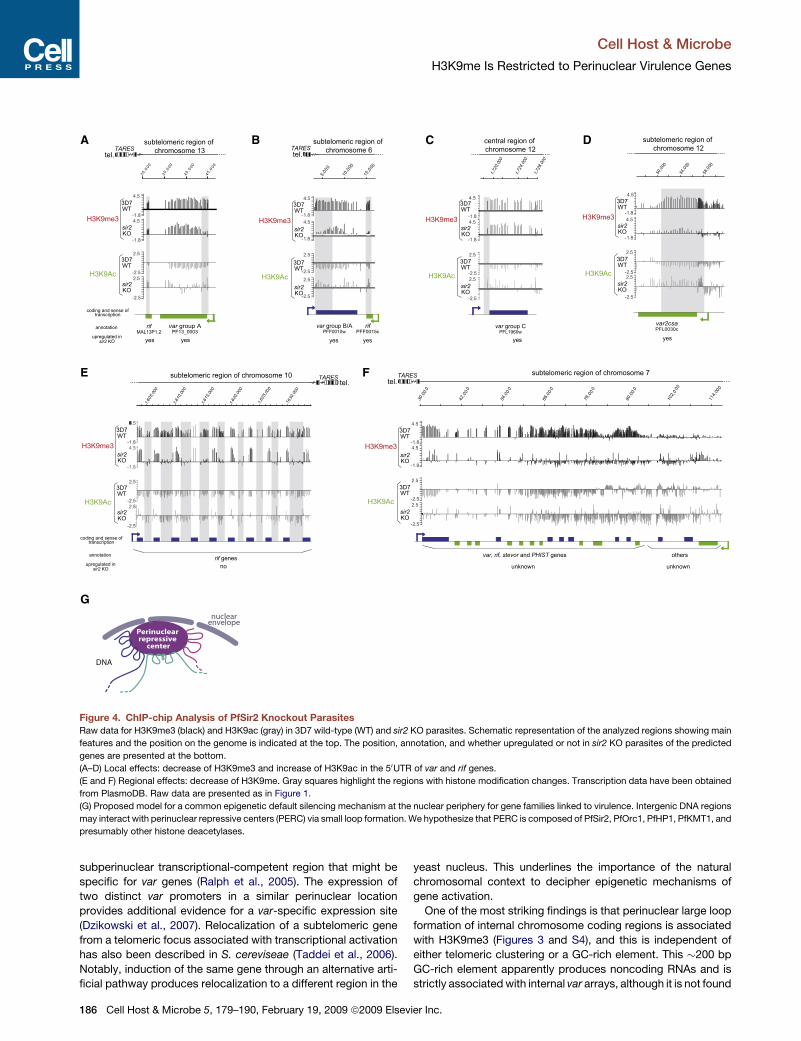
attempted to disrupt the unique PfKMT1 locus. Using the
currently available knockout methods for P. falciparum, we
were incapable of generating viable parasites without PfKMT1.
H3K9 trimethylation requires the action of a histone deacetylase
to remove the acetyl group before the methylation step
(Nakayama et al., 2001). We used a parasite knockout strain
for the histone deacetylase PfSir2 (Duraisingh et al., 2005) to
analyze the molecular events leading to derepression of a subset
of subtelomeric genes and its role in the formation of heterochro-
matin and nuclear organization. ChIP-chip analysis of PfSir2-dis-
rupted 3D7 parasites showed that H3K9me3 levels were
reduced and H3K9ac levels were increased, compared to wild-
type parasites, in the 50UTR of the previously published upregu-
lated genes (Figure 4 and Table 2). Competition between acety-
lation and methylation at H3K9 in the 50UTR has been previously
shown to influence transcription activity of var genes (Lopez-Ru-
bio et al., 2007). In the absence of PfSir2, apparently this balance
is disturbed in favor of H3K9ac. Unexpectedly, this effect is seen
only in 2 out of 12 gene families enriched in H3K9me3, the var
and rif (Figures 4A–4D). Our results suggest that PfSir2-mediated
H3K9 deacetylation is required for establishing repressive H3K9
methylation for a subset of var and rif genes.
Surprisingly, the lack of PfSir2 resulted in very local effects in
a discontinuous manner (Figures 4A–4D). The postulated telo-
mere position effect of PfSir2 (Freitas-Junior et al., 2005) would
have predicted a continuous H3K9me3 depletion from the telo-
mere into the coding region. Together, these data suggest that
different separated DNA regions may interact with PfSir2 via
small loop formation (schematically shown in Figure 4G). Consis-
tent with this confined effect of PfSir2 in H3K9me3, analysis of
the nuclear localization of PfKMT1 and H3K9me3-enriched
genes in DPfSir2 parasites did not produce major changes
(data not shown).
A number of additional puzzling features of PfSir2 emerged
from our study. First, a general decrease of H3K9me3 with no
increase of H3K9ac in basically all intergenic regions of the
heterochromatic domains was observed, even in genes whose
transcriptional state did not change (Figure 4E). Second, nearly
the entire subtelomeric domain in the left arm of chromosome 7
(comprising 13 members of gene families) was depleted of
H3K9me3, and H3K9ac levels remained low (Figure 4F). Unfortu-
nately, there are no available transcription data for these loci in
PfSir2-disrupted parasites to see whether the absence of H3K9
methylation without induction of acetylation affects repression.
Third, we observed only a weak reduction of H3K9me3 at
subtelomeric repeats (Figure S1) associated to a slight increase
in H3K9ac, suggesting that other histone deacetylases may
cooperate with the process of H3K9me.
DISCUSSION
In this study, we demonstrate a link between malaria parasite
virulence genes enriched for H3K9me3, their spatial location at
the nuclear periphery, and their epigenetic silencing via a perinu-
Cell Hos
clear repressive center. This unique association is almost entirely
restricted to clonally variant multigene families involved in
phenotypic plasticity.
Here, we established a high-resolution genome-wide
H3K9me3 heterochromatin map for a malaria parasite. P. falci-
parum chromosome end regions (TAREs 1–6) are enriched in
H3K9me3 and may represent constitutive heterochromatin, like
in other organisms. The formation of heterochromatin in associ-
ation with repetitive sequences is apparently crucial for func-
tional organization of chromosome ends (Grewal and Jia,
2007). In contrast to other organisms, however, no enrichment
was found on pericentromeric DNA, and high enrichment was
found in all subtelomeric coding regions. A few small blocks of
H3K9m3 were also detected in chromosome internal regions
(Figure 1). Other histone modifications showed a broad distribu-
tion along chromosomes (Figure 2), and their genome-wide link
to gene expression has been initiated in collaboration with
PlasmoDB. The fact that we used a high-resolution tiling array
including noncoding regions may explain why, in a previous
ChIP-chip study using only coding regions with a very low
coverage, the authors did not observe the restricted localization
of H3K9me3 (Cui et al., 2007).
Importantly, we found five subtelomeric gene families not yet
characterized, which are enriched in H3K9me3, differentially
transcribed, and that may participate in the phenotypic makeup
of genetically clonal blood stage parasites. Insight into these
potential virulence factors is very important for our under-
standing of malaria pathogenesis. It is noteworthy that other
variant gene families such as surfin, FIKK, and ETRAMP (Table
S1), which are not enriched in H3K9me3, are interspersed within
the subtelomeric region, demonstrating that two types of gene
families, apparently controlled by distinct mechanisms, coexist
in P. falciparum subtelomeres.
The data also reveal that epigenetic boundaries do exist in the
heterochromatin domains to prevent the spreading of the repres-
sive mark into neighboring regions. We provide genome-wide,
precise physical limitations for H3K9me3 in P. falciparum. It
has been suggested that RNA polymerase III (Pol III)-transcribed
genes act as barriers against self-propagating heterochromatin
in single-celled eukaryotes (Lunyak, 2008). Indeed, some tRNA
genes localize in the proximity of heterochromatin domain
borders of P. falciparum (for example, in chromosome 4 left
end and chromosome 13 right end). Assays need to be devel-
oped to elucidate whether Pol III-transcribed genes or other
specific DNA elements are involved in blocking the spreading
of H3K9me3.
The present work highlights the idea that P. falciparum
genomic DNA is nonrandomly distributed in the nucleus and
that gene expression and spatial organization are strongly linked
(see the model in Figure 3F). Nuclear position analysis of actively
transcribed var genes, in their native chromosomal context,
confirmed their dissociation from telomeric clusters. We specu-
late that chromatin loop formation may be associated with
activation and relocation of subtelomeric var genes in a
In all cases, representative examples are shown as merged images, FISH signals were scored using nuclei from multiple experiments, and scale bars represent
1 mm. For quantification of colocalization, only nuclei containing at least three TARE6 signals were considered as positive, and colocalization was defined as any
overlap between two fluorescent signals.
(F) Proposed model for nuclear organization of H3K9me3-enriched domains. The chromosome 12 was used as an example (see text for details).
t & Microbe 5, 179–190, February 19, 2009 ª2009 Elsevier Inc. 185
Cell Host & Microbe
H3K9me Is Restricted to Perinuclear Virulence Genes
A
E
G
F
B C D
nuclearenvelope
Figure 4. ChIP-chip Analysis of PfSir2 Knockout Parasites
Raw data for H3K9me3 (black) and H3K9ac (gray) in 3D7 wild-type (WT) and sir2 KO parasites. Schematic representation of the analyzed regions showing main
features and the position on the genome is indicated at the top. The position, annotation, and whether upregulated or not in sir2 KO parasites of the predicted
genes are presented at the bottom.
(A–D) Local effects: decrease of H3K9me3 and increase of H3K9ac in the 50UTR of var and rif genes.
(E and F) Regional effects: decrease of H3K9me. Gray squares highlight the regions with histone modification changes. Transcription data have been obtained
from PlasmoDB. Raw data are presented as in Figure 1.
(G) Proposed model for a common epigenetic default silencing mechanism at the nuclear periphery for gene families linked to virulence. Intergenic DNA regions
may interact with perinuclear repressive centers (PERC) via small loop formation. We hypothesize that PERC is composed of PfSir2, PfOrc1, PfHP1, PfKMT1, and
presumably other histone deacetylases.
subperinuclear transcriptional-competent region that might be
specific for var genes (Ralph et al., 2005). The expression of
two distinct var promoters in a similar perinuclear location
provides additional evidence for a var-specific expression site
(Dzikowski et al., 2007). Relocalization of a subtelomeric gene
from a telomeric focus associated with transcriptional activation
has also been described in S. cereviseae (Taddei et al., 2006).
Notably, induction of the same gene through an alternative arti-
ficial pathway produces relocalization to a different region in the
186 Cell Host & Microbe 5, 179–190, February 19, 2009 ª2009 Elsev
yeast nucleus. This underlines the importance of the natural
chromosomal context to decipher epigenetic mechanisms of
gene activation.
One of the most striking findings is that perinuclear large loop
formation of internal chromosome coding regions is associated
with H3K9me3 (Figures 3 and S4), and this is independent of
either telomeric clustering or a GC-rich element. This �200 bp
GC-rich element apparently produces noncoding RNAs and is
strictly associated with internal var arrays, although it is not found
ier Inc.
Cell Host & Microbe
H3K9me Is Restricted to Perinuclear Virulence Genes
Table 2. Histone Changes Linked to Derepressed var Genes in DPfSir2 Parasites
var Group
PlasmoDB Accession
Number
H3K9me3 Levels in the 50 Flanking
Region in DPfSir2 Parasites
H3K9ac Levels in the 50 Flanking
Region in DPfSir2 Parasites
Fold Expression DPfsir2/WTa
Experiment 1 Experiment 2
var2CSA PFL0030c low high 2 10
group A PF13_0003 low high 100 90
group A PFA0015c low high 50 30
group A PFF0020c low low 19 3
group A PFD1235w no data no data 11 7
group B/A PFF1580c low high 24 22
group B/A PFF0010w low high 36 29
group B/C PF07_0050 low high 67 34
group C PFL1960w low high 26 12a (Duraisingh et al., 2005). Only genes with fold expression DPfSir2/WT R 10 are shown.
Cell Hos
in Figure 4G). In the case of PfSir2, this interaction appears to
focus in the 50UTR of some var and rif genes, underlining the
important role of this DNA region in var gene control.
Several elements suggest that subtelomeric chromatin is
made up of other histone deacetylases. First, PfSir2 only
controls repression of a small subset of the H3K9me3-enriched
genes (5%), and second, PfSir2 inactivation has no major
change in the H3K9me3 levels in the noncoding subtelomeric
region. This is in contrast to S. pombe, wherein loss of Sir2
resulted in a general decrease in H3K9me3 and an increase in
H3K9ac at telomeres and telomeric-associated sequences
(Shankaranarayana et al., 2003). We had searched in the P. falci-
parum genome for other Sir-like genes and found an open
reading frame on chromosome 14 that is homologous to human
Sirt6, which acts at subtelomeric repeats (Michishita et al., 2008).
Antibodies raised against this protein show a similar location at
the nuclear periphery as PfSir2 (L.M.-S. and A.S., unpublished
data), strongly suggesting that a second telomere-associated
histone deacetylase contributes to epigenetic silencing of viru-
lence gene families.
In conclusion, our study demonstrates that P. falciparum has
a specific mechanism to restrict repressive H3K9me3 almost
exclusively to clonally variant gene families. Moreover, our data
show a complex spatial distribution of plasmodial chromosomes
in the nucleus, which apparently creates functional distinct
compartments that are important for epigenetic gene expression
of a subgroup of genes. We postulate that distinct DNA loops
may exist: large chromosomal loops, which bring specific chro-
mosome regions to the nuclear periphery; small DNA loops that
allow interaction with different components of PERC; and chro-
matin loops associated with relocation from telomeric clusters of
active genes into a perinuclear expression site. In addition, we
provide a detailed insight into the underlying chromatin modifi-
cations that are necessary to overcome the precise monoallelic
counting mechanism of var genes. Our PfSir2 mutant analysis
demonstrates that the removal of the H3K9me3 and replace-
ment by acetylation in the 50UTR is a key step toward unlocking
the default silent state of var genes. It will be interesting to inves-
tigate whether other protozoan pathogens that rely on subtelo-
meric gene families for immune escape (Borst, 2003) may use
this type of epigenetic silencing mechanism. Insight into the
complexity of control of gene families involved in malaria
near the single internal var genes on chromosomes 6 and 12 (Hall
et al., 2002; Mourier et al., 2008). Together, our observations
point to PfKMT1 and H3K9me3 as determinants of spatial orga-
nization of chromosomes in P. falciparum nuclei. This is sup-
ported by data obtained in S. pombe showing SpKMT1 to be
crucial for correct subnuclear localization of the mating type
region (Alfredsson-Timmins et al., 2007). In P. falciparum,
however, deletion of PfKMT1 produces a lethal effect on parasite
proliferation. Improved inducible gene knockdown systems will
be necessary to gain insight into the PfKMT1 function in spatial
organization of P. falciparum nuclei.
Our results are compatible with the existence of ‘‘perinuclear-
repressive centers’’ (PERC) (see model in Figure 4G) composed
of PfSir2 and other potential silencing factors such as PfOrc1
(Mancio-Silva et al., 2008), PfHP1 (A.S. and R. Hernandez-Rivas,
unpublished data), and probably PfKMT1. Although experiments
are needed to clarify whether these telomeric-associated
proteins form complexes, evidences from studies in S. pombe
(Bannister et al., 2001; Nakayama et al., 2001; Yamada et al.,
2005) indicate that H3K9me provides the binding site for HP1 to
recruit other proteins, such as histone deacetylases and methyl-
ases, involved in heterochromatin stabilization and spreading.
DNA-binding proteins and/or transcribed repetitive subtelomeric
noncoding RNAs (Mourier et al., 2008) are possibly involved in the
initial targeting of histone-modifying enzymes to the PERC. It will
be also interesting to investigate whether telomeric and internal
heterochromatic regions share the same PERC components.
Having established high-resolution ChIP-chip assays for
P. falciparum, we were in a position to analyze chromatin
changes in PfSir2 mutant parasites, which display transcriptional
derepression of a subset of var and rif genes (Duraisingh et al.,
2005). We show that PfSir2 acts precisely on H3K9 methylation
in the 50UTR of genes, which have been shown to be upregulated
in DPfsir2 parasites. The discontinuous pattern of H3K9me3
removal along subtelomeric regions and replacement by acety-
lation was unexpected given the current model of telomere posi-
tion effect, which predicts a gradient along the telomere into
the subtelomeric coding regions (Freitas-Junior et al., 2005;
Mancio-Silva et al., 2008). The results strongly suggest a distinct
mechanism of PfSir2-mediated silencing. We hypothesize that
multiple small loop structures of chromosome regions harboring
variant multigene families interact directly with PERC (see model
t & Microbe 5, 179–190, February 19, 2009 ª2009 Elsevier Inc. 187
Cell Host & Microbe
H3K9me Is Restricted to Perinuclear Virulence Genes
pathogenesis is vital for understanding the disease and will
provide a major step toward controlling it.
EXPERIMENTAL PROCEDURES
Parasites
P. falciparum blood stage parasites were cultivated in the conditions previ-
ously described (Nunes et al., 2007). The 3D7DSir2 parasites were cultivated
in the presence of 2 mM Pyrimethamine. Selection of FCR3 parasites for
CSA binding was performed according to Scherf et al. (1998).
Antibodies
Antibodies against histone modifications were purchased from Upstate for
H3K9me3, H3K9ac, and H4K20me3 (07-442, 07-352, and 07-463, respec-
tively) and from Abcam for H3K4me3 (ab8580). To obtain polyclonal antibody
serum against PfKMT1, we used GenScript Corporation standard protocols for
immunizing a rabbit with a synthetic peptide (N-CASRDIQPNEPLKYH-C).
Custom P. falciparum High-Resolution Tiling Array Design
The design of the array was done with the assistance of NimbleGen Systems
Inc. We downloaded the P. falciparum genome sequence from The Sanger
Center website and subjected it to several iterative rounds of probe selection
(see Supplemental Experimental Procedures).
ChIP-chip Analysis
ChIP assays were carried out as previously described (Lopez-Rubio et al.,
2007) using ring stage unselected 3D7 parasites.
We subjected precipitated DNA and DNA from parasite extracts (input)
recovered after reverse crosslinking to amplification using Sigma GenomePlex
WGA kit. After amplification, the immunoprecipitated DNA was tested for
enrichment of control loci by qPCR (Realplex4 EpgradientS thermalcycler,
Eppendorf). DNA was labeled, hybridized, and the data extracted according
to standard operating procedures by NimbleGen Systems Inc. At least two
biological replicates were performed for each condition and two technical
replicates for each biological replicate. Detailed procedures and quantitative
real-time PCR validation of ChIP-chip data are described in the Supplemental
Experimental Procedures.
Immunofluorescence and FISH
Immunofluorescence assays were performed as previously described (Tonkin
et al., 2004) for PfKMT1 and (Mancio-Silva et al., 2008) H3K9me3. Antibody
dilutions were: anti-PfKMT1, 1:500; anti-H3K9me3, 1:500; and Alexa-Fluor-
488-conjugated anti-rabbit highly crossabsorbed, 1:500.
For FISH, infected red blood cells were lysed with saponine and the released
parasites fixed in suspension with 4% paraformaldehyde. Parasites were then
deposited on microscope slides and subjected to DNA FISH in the conditions
previously described (Mancio-Silva et al., 2008). For combined RNA/DNA
FISH, parasites were deposited on slides, treated with 0.1% Triton X-100 for
5 min, and hybridized first with var probes at 50�C for 16 hr. The slides were
washed three times in 2 3 SSC at 50�C, denaturated at 80�C, and hybridized
with the TARE6 probe at 37�C for 16 hr. Finally, the slides were washed as for
DNA FISH.
TARE6 and var2CSA probes were obtained as described before (Ralph
et al., 2005). All other FISH probes were PCR amplified from genomic DNA
using the primers listed on Table S2. Images were taken using a Nikon Eclipse
80i microscope with a CoolSnap HQ2 camera (Photometrics). NIS Elements
3.0 software (Nikon) was used for acquisition and ImageJ (http://rsbweb.nih.
gov/ij/) for composition. Chromatic aberration was corrected in all images by
aligning the red, green, and blue channel signals from 0.2 mm TetraSpeck
microspheres.
ACCESSION NUMBERS
Microarray data for ChIP-chip 3D7 and 3D7DSir2 parasites are available in the
ArrayExpress database (www.ebi.ac.uk/arrayexpress) under accession
numbers E-MEXP-1806 and E-MEXP-1805, respectively.
188 Cell Host & Microbe 5, 179–190, February 19, 2009 ª2009 Elsev
SUPPLEMENTAL DATA
The Supplemental Data include Supplemental Experimental Procedures, two
tables, and four figures and can be found with this article online at http://
www.cell.com/cell-host-microbe/supplemental/S1931-3128(09)00030-4.
ACKNOWLEDGMENTS
We would like to thank to A. Cowman for providing the 3D7DSir2 strain,
M. Nunes for the FCR3-CSA selected parasites, D. Roos for help with annota-
tion files for genome-wide analysis, E. Bischoff for assistance with microarray
design, and PlasmoDB for the invaluable malaria database support. We are
also grateful to B. Arcangioli, P. Navarro, and L. Riviere for critical reading of
the manuscript and to A. Cordeiro da Silva and Faculdade de Farmacia
(Universidade do Porto, Portugal) for their support. This work was financed
by BioMAlPar (contract No: LSPH-CT-2004-503578). J.-J.L.-R. has financial
support from the Human Frontier Science Program and L.M.-S. from
the Portuguese Foundation for Science and Technology. A.S. is supported
by a grant ANR Microbiologie (ANR-06-MIME-026-01) and Fonds Dedie
(Sanofi-Avantis).
Received: September 23, 2008
Revised: December 5, 2008
Accepted: December 31, 2008
Published: February 18, 2009
REFERENCES
Alfredsson-Timmins, J., Henningson, F., and Bjerling, P. (2007). The Clr4 meth-
yltransferase determines the subnuclear localization of the mating-type region
in fission yeast. J. Cell Sci. 120, 1935–1943.
Allis, C.D., Berger, S.L., Cote, J., Dent, S., Jenuwien, T., Kouzarides, T., Pillus,
L., Reinberg, D., Shi, Y., Shiekhattar, R., et al. (2007). New nomenclature for
chromatin-modifying enzymes. Cell 131, 633–636.
Bannister, A.J., Zegerman, P., Partridge, J.F., Miska, E.A., Thomas, J.O.,
Allshire, R.C., and Kouzarides, T. (2001). Selective recognition of methylated
lysine 9 on histone H3 by the HP1 chromo domain. Nature 410, 120–124.
Barry, A.E., Leliwa-Sytek, A., Tavul, L., Imrie, H., Migot-Nabias, F., Brown,
S.M., McVean, G.A., and Day, K.P. (2007). Population genomics of the immune
evasion (var) genes of Plasmodium falciparum. PLoS Pathog. 3, e34.
Blythe, J.E., Yam, X.Y., Kuss, C., Bozdech, Z., Holder, A.A., Marsh, K.,
Langhorne, J., and Preiser, P.R. (2008). Plasmodium falciparum STEVOR
proteins are highly expressed in patient isolates and located in the surface
membranes of infected red blood cells and the apical tips of merozoites. Infect.
Immun. 76, 3329–3336.
Borst, P. (2003). Mechanisms of antigenic variation: an overview. In Antigenic
Variation, A. Craig and A. Scherf, eds. (London: Academic Press), pp. 1–16.
Chen, F., Mackey, A.J., Stoeckert, C.J., Jr., and Roos, D.S. (2006). OrthoMCL-
DB: Querying a comprehensive multi-species collection of ortholog groups.
Nucleic Acids Res. 34, D363–D368.
Chookajorn, T., Dzikowski, R., Frank, M., Li, F., Jiwani, A.Z., Hartl, D.L., and
Deitsch, K.W. (2007). Epigenetic memory at malaria virulence genes. Proc.
Natl. Acad. Sci. USA 104, 899–902.
Cortes, A., Carret, C., Kaneko, O., Lim, B.Y., Ivens, A., and Holder, A.A. (2007).
Epigenetic silencing of Plasmodium falciparum genes Linked to erythrocyte
invasion. PLoS Pathog. 3, e107.
Cui, L., Miao, J., Furuya, T., Li, X., Su, X.Z., and Cui, L. (2007). PfGCN5-medi-
ated histone H3 acetylation plays a key role in gene expression in Plasmodium
falciparum. Eukaryot. Cell 6, 1219–1227.
Cui, L., Fan, Q., Cui, L., and Miao, J. (2008). Histone lysine methyltransferases
and demethylases in Plasmodium falciparum. Int. J. Parasitol. 38, 1083–1097.
De Silva, E.K., Gehrke, A.R., Olszewski, K., Leon, I., Chahal, J.S., Bulyk, M.L.,
and Llinas, M. (2008). Specific DNA-binding by apicomplexan AP2 transcrip-
tion factors. Proc. Natl. Acad. Sci. USA 105, 8393–8398.
ier Inc.
Cell Host & Microbe
H3K9me Is Restricted to Perinuclear Virulence Genes
Duraisingh, M.T., Voss, T.S., Marty, A.J., Duffy, M.F., Good, R.T., Thompson,
J.K., Freitas-Junior, L.H., Scherf, A., Crabb, B.S., and Cowman, A.F. (2005).
Heterochromatin silencing and locus repositioning linked to regulation of viru-
lence genes in Plasmodium falciparum. Cell 121, 13–24.
Dzikowski, R., Frank, M., and Deitsch, K. (2006). Mutually exclusive expression
of virulence genes by malaria parasites is regulated independently of antigen
production. PLoS Pathog. 2, e22.
Dzikowski, R., Li, F., Amulic, B., Eisberg, A., Frank, M., Patel, S., Wellems, T.E.,
and Deitsch, K.W. (2007). Mechanisms underlying mutually exclusive expres-
sion of virulence genes by malaria parasites. EMBO Rep. 8, 959–965.
Fernandez, V., Hommel, M., Chen, Q.J., Hagblom, P., and Wahlgren, M.
(1999). Small, clonally variant antigens expressed on the surface of the Plas-
modium falciparum-infected erythrocyte are encoded by the rif gene family
and are the target of human immune responses. J. Exp. Med. 190, 1393–
1403.
Figueiredo, L.M., Pirrit, L.A., and Scherf, A. (2000). Genomic organisation and
chromatin structure of Plasmodium falciparum chromosome ends. Mol.
Biochem. Parasitol. 106, 169–174.
Freitas-Junior, L.H., Bottius, E., Pirrit, L.A., Deitsch, K.W., Scheidig, C., Guinet,
F., Nehrbass, U., Wellems, T.E., and Scherf, A. (2000). Frequent ectopic
recombination of virulence factor genes in telomeric chromosome clusters
of P. falciparum. Nature 407, 1018–1022.
Freitas-Junior, L.H., Hernandez-Rivas, R., Ralph, S.A., Montiel-Condado,
D., Ruvalcaba-Salazar, O.K., Rojas-Meza, A.P., Mancio-Silva, L., Leal-Sil-
vestre, R.J., Gontijo, A.M., Shorte, S., and Scherf, A. (2005). Telomeric
heterochromatin propagation and histone acetylation control mutually
exclusive expression of antigenic variation genes in malaria parasites. Cell
121, 25–36.
Fried, M., and Duffy, P. (1996). Adherence of Plasmodium falciparum to chon-
droitin sulfate A in the human placenta. Science 272, 1502–1504.
Gardner, M.J., Hall, N., Fung, E., White, O., Berriman, M., Hyman, R.W., Carl-
ton, J.M., Pain, A., Nelson, K.E., Bowman, S., et al. (2002). Genome
sequence of the human malaria parasite Plasmodium falciparum. Nature
419, 498–511.
Grewal, S.I., and Jia, S. (2007). Heterochromatin revisited. Nat. Rev. Genet. 8,
35–46.
Hall, N., Pain, A., Berriman, M., Churcher, C., Harris, B., Harris, D., Mungall, K.,
Bowman, S., Atkin, R., Baker, S., et al. (2002). Sequence of Plasmodium falci-
parum chromosomes 1, 3–9 and 13. Nature 419, 527–531.
Hediger, F., Neumann, F.R., Van Houwe, G., Dubrana, K., and Gasser, S.M.
(2002). Live imaging of telomeres: yKu and Sir proteins define redundant telo-
mere-anchoring pathways in yeast. Curr. Biol. 12, 2076–2089.
Jensen, A.T., Magistrado, P., Sharp, S., Joergensen, L., Lavstsen, T., Chiuc-
chiuini, A., Salanti, A., Vestergaard, L.S., Lusingu, J.P., Hermsen, R., et al.
(2004). Plasmodium falciparum associated with severe childhood malaria pre-
ferentially expresses PfEMP1 encoded by group A var genes. J. Exp. Med.
199, 1179–1190.
Kelly, J.M., McRobert, L., and Baker, D.A. (2006). Evidence on the chromo-
somal location of centromeric DNA in Plasmodium falciparum from etopo-
side-mediated topoisomerase-II cleavage. Proc. Natl. Acad. Sci. USA 103,
6706–6711.
Kyes, S.A., Rowe, J.A., Kriek, N., and Newbold, C.I. (1999). Rifins: A second
family of clonally variant proteins expressed on the surface of red cells infected
with Plasmodium falciparum. Proc. Natl. Acad. Sci. USA 96, 9333–9338.
Kyes, S.A., Kraemer, S.M., and Smith, J.D. (2007). Antigenic variation in Plas-
modium falciparum: Gene organization and regulation of the var multigene
family. Eukaryot. Cell 6, 1511–1520.
Lavazec, C., Sanyal, S., and Templeton, T.J. (2007). Expression switching in
the stevor and Pfmc-2TM superfamilies in Plasmodium falciparum. Mol.
Microbiol. 64, 1621–1634.
Lavstsen, T., Salanti, A., Jensen, A.T., Arnot, D.E., and Theander, T.G. (2003).
Sub-grouping of Plasmodium falciparum 3D7 var genes based on sequence
analysis of coding and non-coding regions. Malar. J. 2, 27.
Cell Hos
Le Roch, K.G., Zhou, Y., Blair, P.L., Grainger, M., Moch, J.K., Haynes, J.D., De
La Vega, P., Holder, A.A., Batalov, S., Carucci, D.J., and Winzeler, E.A. (2003).
Discovery of gene function by expression profiling of the malaria parasite life
cycle. Science 301, 1503–1508.
Leech, J.H., Barnwell, J.W., Miller, L.H., and Howard, R.J. (1984). Identification
of a strain-specific malarial antigen exposed on the surface of Plasmodium
falciparum-infected erythrocytes. J. Exp. Med. 159, 1567–1575.
Lopez-Rubio, J.J., Gontijo, A.M., Nunes, M.C., Issar, N., Hernandez Rivas, R.,
and Scherf, A. (2007). 50 flanking region of var genes nucleate histone modifi-
cation patterns linked to phenotypic inheritance of virulence traits in malaria
parasites. Mol. Microbiol. 66, 1296–1305.
Lunyak, V.V. (2008). Boundaries. Boundaries.Boundaries??? Curr. Opin. Cell
Biol. 20, 281–287.
Mancio-Silva, L., Rojas-Meza, A.P., Vargas, M., Scherf, A., and Hernandez-
Rivas, R. (2008). Differential association of Orc1 and Sir2 proteins to telomeric
domains in Plasmodium falciparum. J. Cell Sci. 121, 2046–2053.
Marty, A.J., Thompson, J.K., Duffy, M.F., Voss, T.S., Cowman, A.F., and
Crabb, B.S. (2006). Evidence that Plasmodium falciparum chromosome end
clusters are cross-linked by protein and are the sites of both virulence gene
silencing and activation. Mol. Microbiol. 62, 72–83.
Merrick, C.J., and Duraisingh, M.T. (2007). Plasmodium falciparum Sir2: An
unusual sirtuin with dual histone deacetylase and ADP-ribosyltransferase
activity. Eukaryot. Cell. 6, 2081–2091.
Miao, J., Fan, Q., Cui, L., and Li, J. (2006). The malaria parasite Plasmodium
falciparum histones: Organization, expression, and acetylation. Gene 369,
53–65.
Michishita, E., McCord, R.A., Berber, E., Kioi, M., Padilla-Nash, H., Damian,
M., Cheung, P., Kusumoto, R., Kawahara, T.L., Barrett, J.C., et al. (2008).
SIRT6 is a histone H3 lysine 9 deacetylase that modulates telomeric chro-
matin. Nature 452, 492–496.
Mourier, T., Carret, C., Kyes, S., Christodoulou, Z., Gardner, P.P., Jeffares,
D.C., Pinches, R., Barrell, B., Berriman, M., Griffiths-Jones, S., et al. (2008).
Genome-wide discovery and verification of novel structured RNAs in Plasmo-
dium falciparum. Genome Res. 18, 281–292.
Nakayama, J., Rice, J.C., Strahl, B.D., Allis, C.D., and Grewal, S.I. (2001). Role
of histone H3 lysine 9 methylation in epigenetic control of heterochromatin
assembly. Science 292, 110–113.
Nunes, M.C., Goldring, J.P., Doerig, C., and Scherf, A. (2007). A novel protein
kinase family in Plasmodium falciparum is differentially transcribed and
secreted to various cellular compartments of the host cell. Mol. Microbiol.
63, 391–403.
Ralph, S.A., Scheidig-Benatar, C., and Scherf, A. (2005). Antigenic variation in
Plasmodium falciparum is associated with movement of var loci between
subnuclear locations. Proc. Natl. Acad. Sci. USA 102, 5414–5419.
Scherf, A., Hernandez-Rivas, R., Buffet, P., Bottius, E., Benatar, C., Pouvelle,
B., Gysin, J., and Lanzer, M. (1998). Antigenic variation in malaria: in situ
switching, relaxed and mutually exclusive transcription of var genes during
intra-erythrocytic development in Plasmodium falciparum. EMBO J. 17,
5418–5426.
Scherf, A., Lopez-Rubio, J.J., and Riviere, L. (2008a). Antigenic Variation in
Plasmodium falciparum. Annu. Rev. Microbiol. 62, 445–470.
Scherf, A., Riviere, L., and Lopez-Rubio, J.J. (2008b). SnapShot: var gene
expression in the malaria parasite. Cell 134, 190.
Shankaranarayana, G.D., Motamedi, M.R., Moazed, D., and Grewal, S.I.
(2003). Sir2 regulates histone H3 lysine 9 methylation and heterochromatin
assembly in fission yeast. Curr. Biol. 13, 1240–1246.
Snow, R.W., Guerra, C.A., Noor, A.M., Myint, H.Y., and Hay, S.I. (2005). The
global distribution of clinical episodes of Plasmodium falciparum malaria.
Nature 434, 214–217.
Stubbs, J., Simpson, K.M., Triglia, T., Plouffe, D., Tonkin, C.J., Duraisingh,
M.T., Maier, A.G., Winzeler, E.A., and Cowman, A.F. (2005). Molecular mech-
anism for switching of P. falciparum invasion pathways into human erythro-
cytes. Science 309, 1384–1387.
t & Microbe 5, 179–190, February 19, 2009 ª2009 Elsevier Inc. 189
Cell Host & Microbe
H3K9me Is Restricted to Perinuclear Virulence Genes
Taddei, A., Van Houwe, G., Hediger, F., Kalck, V., Cubizolles, F., Schober, H.,
and Gasser, S.M. (2006). Nuclear pore association confers optimal expression
levels for an inducible yeast gene. Nature 441, 774–778.
Tonkin,C.J., vanDooren, G.G., Spurck, T.P., Struck,N.S., Good, R.T., Handman,
E., Cowman, A.F., and McFadden, G.I. (2004). Localization of organellar proteins
in Plasmodium falciparum using a novel set of transfection vectors and a new
immunofluorescence fixation method. Mol. Biochem. Parasitol. 137, 13–21.
190 Cell Host & Microbe 5, 179–190, February 19, 2009 ª2009 Elsev
Voss, T.S., Healer, J., Marty, A.J., Duffy, M.F., Thompson, J.K., Beeson, J.G.,
Reeder, J.C., Crabb, B.S., and Cowman, A.F. (2006). A var gene promoter
controls allelic exclusion of virulence genes in Plasmodium falciparum malaria.
Nature 439, 1004–1008.
Yamada, T., Fischle, W., Sugiyama, T., Allis, C.D., and Grewal, S.I. (2005). The
nucleation and maintenance of heterochromatin by a histone deacetylase in
fission yeast. Mol. Cell 20, 173–185.
ier Inc.