genome characterization and population genetic structure of the
TRANSCRIPT

Richards et al. BMC Microbiology 2012, 12:293http://www.biomedcentral.com/1471-2180/12/293
RESEARCH ARTICLE Open Access
Genome characterization and population geneticstructure of the zoonotic pathogen,Streptococcus canisVincent P Richards1, Ruth N Zadoks2,4, Paulina D Pavinski Bitar1, Tristan Lefébure1,5, Ping Lang6, Brenda Werner2,Linda Tikofsky2,7, Paolo Moroni2,3 and Michael J Stanhope1*
Abstract
Background: Streptococcus canis is an important opportunistic pathogen of dogs and cats that can also infect awide range of additional mammals including cows where it can cause mastitis. It is also an emerging humanpathogen.
Results: Here we provide characterization of the first genome sequence for this species, strain FSL S3-227 (milkisolate from a cow with an intra-mammary infection). A diverse array of putative virulence factors was encoded bythe S. canis FSL S3-227 genome. Approximately 75% of these gene sequences were homologous to knownStreptococcal virulence factors involved in invasion, evasion, and colonization. Present in the genome are multiplepotentially mobile genetic elements (MGEs) [plasmid, phage, integrative conjugative element (ICE)] and comparisonto other species provided convincing evidence for lateral gene transfer (LGT) between S. canis and two additionalbovine mastitis causing pathogens (Streptococcus agalactiae, and Streptococcus dysgalactiae subsp. dysgalactiae),with this transfer possibly contributing to host adaptation. Population structure among isolates obtained fromEurope and USA [bovine = 56, canine = 26, and feline = 1] was explored. Ribotyping of all isolates and multi locussequence typing (MLST) of a subset of the isolates (n = 45) detected significant differentiation between bovine andcanine isolates (Fisher exact test: P = 0.0000 [ribotypes], P = 0.0030 [sequence types]), suggesting possible hostadaptation of some genotypes. Concurrently, the ancestral clonal complex (54% of isolates) occurred in many tissuetypes, all hosts, and all geographic locations suggesting the possibility of a wide and diverse niche.
Conclusion: This study provides evidence highlighting the importance of LGT in the evolution of the bacteriaS. canis, specifically, its possible role in host adaptation and acquisition of virulence factors. Furthermore, recent LGTdetected between S. canis and human bacteria (Streptococcus urinalis) is cause for concern, as it highlights thepossibility for continued acquisition of human virulence factors for this emerging zoonotic pathogen.
Keywords: Streptococcus canis, Comparative genomics, Pathogen, Zoonotic, Mastitis, Lateral gene transfer, Hostadaptation, Bovine, Canine
* Correspondence: [email protected] of Population Medicine and Diagnostic Sciences, College ofVeterinary Medicine, Cornell University, Ithaca, NY 14853, USAFull list of author information is available at the end of the article
© 2012 Richards et al.; licensee BioMed Central Ltd. This is an Open Access article distributed under the terms of the CreativeCommons Attribution License (http://creativecommons.org/licenses/by/2.0), which permits unrestricted use, distribution, andreproduction in any medium, provided the original work is properly cited.

Richards et al. BMC Microbiology 2012, 12:293 Page 2 of 16http://www.biomedcentral.com/1471-2180/12/293
BackgroundOriginally described as β-hemolytic streptococci isolatedfrom dogs and cows that possessed the Lancefield groupG antigen [1], Streptococcus canis has subsequently beenisolated from a variety of animal sources including cats,rats, rabbits, minks, foxes, a Japanese raccoon dog, andhumans [2-4]. The species is an important opportunisticpathogen of cats and dogs infecting a wide range of tis-sues such as the central nervous system, respiratory tract,genitourinary system, blood, skin, bones, cardiovascularsystem, and abdomen [1,4-6]. Infection can cause seriousinvasive disease, such as streptococcal toxic shock syn-drome (STSS), necrotizing fasciitis (NF), septicemia,pneumonia, and meningitis, with numerous reports offatal infection [5,7-9], whereas in cows S. canis can causemastitis [10-12]. Of concern are the accumulating reportsof human infection (including numerous cases of dog tohuman transmission) [13-16], with clinical manifestationssimilar to those seen in cats and dogs. For example,descriptions of human cases include soft tissue infection,bacteremia, urinary infection, bone infection, pneumonia,and two reports of death from sepsis [13].Although the phylogeny of the species is not com-
pletely resolved, a general consensus from the literatureshows S. canis to be closely related to Streptococcus dys-galactiae subsp. dysgalactiae, Streptococcus dysgalactiaesubsp. equisimilis, and Streptococcus pyogenes [2,17-21].S. canis and S. dysgalactiae subsp. equisimilis are bothβ-hemolytic streptococci that share the same Lancefieldgroup G antigen. Consequently, by the Lancefield systemthey are indistinguishable, and have traditionally only beenclassified as group G streptococci (GGS) from either ani-mal (S. canis) or human (S. dysgalactiae subsp. equisimilis)origin. Therefore, it’s possible that human S. canis infec-tion has been underestimated [13,15]. Investigating thisproblem, Broyles et al. [22] performed a survey of humaninvasive infection using techniques capable of distin-guishing S. canis from S. dysgalactiae subsp. equisimilis.Results showed a low frequency of S. canis in blood sam-ples. However, their study was biased towards thecharacterization of isolates from blood samples (isolatesfrom other body sites were less likely to be characterized).In humans, STSS and NF are serious diseases typically
caused by S. pyogenes infection. The emergence of strik-ingly similar STSS and NF in cats and dogs coupled withthe close relationship between the causal speciesprompted preliminary investigation and subsequent dis-covery of two shared virulence factors between these spe-cies [23]. To shed light on the molecular basis of S. canisvirulence and further investigate the role S. pyogenesand other species of Streptococcus may have played inits evolution we determined the first genome sequencefor this pathogen and compared it to an extensive rangeof streptococcal genomes (40 species, 213 strains). In
addition, we explored population structure amongcanine, feline, and bovine isolates.Our findings reveal a diverse array of genes within the
S. canis genome homologous to known virulence factors,including several established virulence factors from S.pyogenes, Streptococcus agalactiae, and Streptococcuspneumoniae. We found evidence for multiple LGT eventsbetween S. canis and (i) other bovine mastitis causingpathogens, and (ii) the human pathogen Streptococcus uri-nalis, suggesting LGT in both shared bovine and humanenvironments. This LGT was mediated by a variety ofmobile genetic elements [plasmid, phage, integrativeconjugative element] that carried many of the virulencefactors, highlighting the importance of LGT in the evolu-tion of this pathogen and the potential for its emergenceas a zoonotic pathogen.
Result and discussionAssembly and general features of the genomeRoche/454 pyrosequencing produced 128,749 single-endreads and 140,788 paired-end reads that were assembledinto 91 contigs (>200 bp) and eight scaffolds, represent-ing an average 23X site coverage. Utilizing additionalIllumina/Sanger sequencing and alignment to an opticalmap, the eight scaffolds were assembled into a single2,267,856 bp contig. Unfortunately, we were unable toobtain sequence for one small section of the genome(Figure 1). The gap was within a collagen-like surfaceprotein. The best BLAST hit at the NCBI nr database foreach gene fragment (SCAZ3_06900 and SCAZ3_06785)was to an identically annotated gene within S. agalactiae(A909), (each fragment shared approximately 75% se-quence identity). Alignment of the S. canis fragments tothis gene suggested that we were missing approximately1.6 kb. For S. agalactiae (A909), this gene contained 75repeats of a 9 bp imperfect repeat (DCCRTCTTT). Themajority of these repeats (70) were contained within a2.5 kb region that spanned the S. canis gap and flankingregions. S. canis contained 26 repeats in the regions thatflanked the gap. Consequently it seems likely that theserepeats were also present within the un-sequenced sec-tion of the collagen gene for S. canis and that their pres-ence confounded our sequencing attempts. Inclusion ofthis small gap made the total length of the genome ap-proximately 2,269,456 bp. In comparison to 53 genomesequences representing 19 additional Streptococcus spe-cies, the S. canis genome was among the largest withregard to sequence length, ranking fourth (with oneexception [S. agalactiae-FSL S3-026], sequences wereobtained from the manually curated RefSeq database atNCBI [see Additional file 1]). S. canis had a relatively highnumber of CDS (2,212), ranking fifth, an intermediatenumber of tRNAs (67; range 41–80) and an average GCcontent of 39.7%. A 5,871 bp section of the genome

Streptococcus canisstrain FSL Z3-227
oriC
Prophage1
Prophage2
Plasmid
ICE
GC skew+
GC skew-
All CDS (misc features shown in grey)
tRNA
rRNA
GC content
Gap~1.6kbp
Figure 1 Genome map of Streptococcus canis strain FSL Z3-227. Starting from the outermost ring and moving inwards, rings show thelocation of: (1) four mobile genetic elements (see text for detailed description), (2) all annotated CDS on the leading strand, and (3) all annotatedCDS on the lagging strand. Two innermost rings show GC content and GC skew. Map was created using the software CGView [26].
Richards et al. BMC Microbiology 2012, 12:293 Page 3 of 16http://www.biomedcentral.com/1471-2180/12/293
appeared to have been perfectly duplicated (locus tagsSCAZ3_r06686 through SCAZ3_t06810 plus 126 bp ofnon-coding DNA that preceded SCAZ3_r06686). Thesection contained an rRNA operon (16S-23S-5S) and 10tRNAs that were immediately down stream (Val, Asp,Lys, Leu, Thr, Gly, Leu, Arg, and Pro). The entire sectionwas perfectly duplicated immediately upstream (one nu-cleotide separated the two duplicated sections). SimilarrRNA operon duplications are present in the genomes ofStreptococcus thermophilus (LMD-9) and Streptococcussalivarius (CCHSS3). The number of rRNA operons inpublicly available Streptococcus genomes ranges fromone to seven, and the number within the S. canis genomewas again relatively high, with six. It is possible that thisreflects selection for rapid growth. For example, duringrapid growth genes are likely to be expressed at highlevels, and this is often associated with codon usage bias[24], which in turn, has been shown to be positively
correlated with the number of rRNA operons within abacterial genome [25].
Virulence factorsA total of 291 CDS within the S. canis genome were hom-ologous with established virulence factors in the VirulenceFactor of Pathogenic Bacteria database (VFDB) (availableat www.mgc.ac.cn/VFs/main.htm) (see Additional file 2).Throughout the manuscript, two genes (query and sub-ject) are considered homologous if they can be locallyaligned using BLAST with an E value of 1e-5 or less. Arefined search, focusing only on established Streptococcusvirulence factors, detected 34 CDS homologous tothree other species of streptococci: S. pyogenes (17), S.agalactiae (9), and S. pneumoniae (8). Of these, the major-ity (50%) was homologous with S. pyogenes, likely reflect-ing the close relationship between these two species. Morespecifically, 9 of the 17 S. pyogenes virulence factors

Richards et al. BMC Microbiology 2012, 12:293 Page 4 of 16http://www.biomedcentral.com/1471-2180/12/293
homologous to S. canis were categorized as eitherexoenzymes or complement proteases. These gene pro-ducts damage tissue, and may contribute to necrotizingfasciitis. When considering all 291 of the virulence factorshomologous to S. canis, there were only three additionalgenes with similar categorization, two of these homolo-gous to S. pneumoniae. Consequently, it appears that sev-eral genes possibly involved in necrotizing fasciitis areshared between S. canis and S. pyogenes. In contrast, S.canis CDSs were not homologous with genes producingpyrogenic exotoxins associated with toxic shock syn-drome. However, S. canis possessed two other streptocco-cal toxin-producing genes: streptolysin O (SLO) (S.pyogenes) and CAMP factor (S. agalactiae) [27,28]. Two S.canis genes were homologous to a well-characterized S.pyogenes virulence factor, the M protein (emm18), whichaids in antiphagocytosis, adherence, and cellular invasion[29]. However, unlike S. pyogenes, these genes were notlocated within a contiguous 35-gene pathogenicity islandthat is found in all currently genome sequenced strains ofS. pyogenes [30]. A BLASTn search of the NCBI nr data-base showed SCAZ3_01465 to be homologous with thegene SPASc from S. canis [31] (accession number:FJ594772). Global nucleotide sequence alignment showedthese sequences to have 87.7% identity. Yang et al. [31]showed experimentally that SPASc was a new protectiveantigen, however they did not report the strain ID or iso-lation source. For SCAZ3_11010, a BLASTn search ofthe NCBI nr database returned no hits. However, aBLASTp search returned numerous hits and the genewith the most sequence similarity was an emm-like cellsurface protein CspZ.2 of Streptococcus equi subsp. zooe-pidemicus ATCC 35246 (31% identity, 48% coverage).Neither SCAZ3_01465 nor SCAZ3_11010 were homolo-gous with the S. canis emm gene type stG1389 (accessionnumber EU195120) reported from one human and twocanine sources [22]. These findings confirm previousstudies showing that some S. canis isolates can possess Mlike proteins [18,22,23] and additionally show that a di-versity of M like proteins is possible for S. canis strains.S. canis also possessed the nine gene sag operon
(sagABCDEFGHI) responsible for the production ofstreptolysin S (SLS) [32]. Both SLS and SLO are toxinsthat lyse mammalian erythrocytes [33], and the toxicityof SLS has been shown to contribute to necrotizing fas-ciitis [34,35]. Furthermore, it has been suggested thatSLS interacts with numerous additional virulence factorsto accelerate necrosis [36]. These factors include SLO,the M protein, and proteases. Genes for all these factorscan be present in the S. canis genome. Utilizing aBLASTn search of the NCBI nr database and subsequentglobal nucleotide sequence alignment, we detected thesag operon in four additional Streptococcus species(strain and percent sequence identity with S. canis are
given in parentheses): S. dysgalactiae subsp. equisimilis(ATCC 12394; 81.1%), Streptococcus pseudoporcinus(LQ940-04 T; 78.8%), S. pyogenes (MGAS10270; 76.5%),and Streptococcus iniae (9117; 74.4%). The likely presenceof the sag operon in S. dysgalactiae subsp. equisimilis wasfirst shown by Humar et al. [34] who detected a functionalsagA homolog in strains capable of producing SLS. S. canisand S. iniae are somewhat distinctive in that the other spe-cies are predominately human pathogens, whereas theformer are predominately animal pathogens (S. iniae is acommon fish pathogen), although occasionally are asso-ciated with zoonotic disease [37-39]. S. dysgalactiae subsp.dysgalactiae, which is predominantly associated with dis-ease in animals but not in humans, lacks an intact sag op-eron, possessing only sagA and sagI. The occurrence of thecomplete operon in the other close relatives of S. canis (S.dysgalactiae subsp. equisimilis and S. pyogenes) suggeststhat S. dysgalactiae subsp. dysgalactiae may have lost theremainder of the genes from the operon. However, the oc-currence of the operon in two species more distantlyrelated to S. canis, that are themselves likely not sister spe-cies (S. pseudoporcinus and S. iniae) [40], is suggestive inthis case of lateral gene transfer of the operon. Fish hand-ling and close association with domestic dogs may havefacilitated lateral gene transfer between species occupyinghuman and animal hosts [14,16,41].
Genes specific to S. canis (FSL Z3-227)To identify genes that are likely S. canis species specificfrom genes present in multiple species of the genus, weperformed a clustering analysis among 214 Streptococcusgenomes representing 41 species including S. canis (seeMethods section and Additional file 3). The analysisidentified 97 genes that were not homologous to anyother gene in the analysis and were unique to S. canis(see Additional file 2). Unfortunately, all were annotatedas hypothetical proteins, highlighting the need for futurestudies exploring functional genomics for this species. S.canis belongs to the pyogenic 16S rRNA phylogeneticgroup [42]. Limiting the comparison to pyogenic genomes(14 species and 40 genomes, excluding S. canis), we iden-tified an additional 14 genes unique to the S. canis gen-ome (see Additional file 2). Two of these genes werehomologous to two established virulence factors in theVFDB. The first gene (neuraminidase C, SCAZ3_10275)was homologous with neuraminidase B (nanB) fromS. pneumoniae (TIGR4). The product of nanB is a glyco-sidase that, by damaging surface glycans and exposingthe cell surface, aids in the adhesion to host cells andis therefore likely important in host invasion [43]. Thesecond gene (Gfo/Idh/MocA family oxidoreductase,SCAZ3_10270) was homologous with bplA from Bor-detella pertussis (Tohama I). In Bordetella, bpl genesare involved in the synthesis of the LPS, which has been

Richards et al. BMC Microbiology 2012, 12:293 Page 5 of 16http://www.biomedcentral.com/1471-2180/12/293
shown to be essential for the expression of completevirulence in mice [44]. Given that the additional 14genes unique to the S. canis genome were absent in theother pyogenic genomes, it is possible that these lociwere gained via LGT. The two genes homologous tothe virulence factors discussed above, were contiguousin the genome suggesting they were gained in a singleevolutionary event.
Integrative plasmidWith the exception of two loci, S. canis shared a con-tiguous section of 53 CDS with S. agalactiae (NEM316)(Figure 2) (see also Additional file 2: locus tagsSCAZ3_04485 through SCAZ3_04760 [50,114 bp]). Se-quence identity between the shared 53 CDS was veryhigh: 99.2%. First described in S. agalactiae (NEM316)[45], this section of DNA (designated pNEM316-1) wasproposed to be a putative integrative plasmid (it couldexist in circular form and was present as three copieswithin the genome). Here we designate the S. canis copyof the putative plasmid as FSL Z3-227-p. The last 24 bpat the terminal ends of pNEM316-1 were imperfectrepeats of themselves (see Additional file 4). Alignmentof pNEM316-1 with FSL Z3-227-p revealed identical ter-minal sequence for FSL Z3-227-p. Putative recombin-ation attL and attR sites were also identified. As forpNEM316-1, these sites were 9 bp direct repeats.Annotation of several S. canis CDS within this 50 kb
region suggest a plasmid functional role (Figure 2 andAdditional file 2). For example, DNA topoisomerase(SCAZ3_04630), conjugation protein (SCAZ3_04680,SCAZ3_04720), and plasmid partition protein(SCAZ3_04740) were identified. In addition, four CDSwere homologous with established virulence factors (seeAdditional file 2, locus tags are highlighted in red inthe annotations worksheet). Specifically, SCAZ3_04635(ATP-dependent clp protease) was homologous withclpE, an ATP-dependent protease from Listeria mono-cytogenes; clp genes have been shown to play a role in
1 5,000 10,000 15,000 20,000 25,000
4600 46354630
Streptococcus agalactiae (NEM316)
Streptococcus canis (FSL Z3-227)
Figure 2 Gene organization within putative integrative plasmids for SS. canis strain FSL Z3-227 (plasmid designated FSL Z3-227-p). Locus IDCDS homologous with established virulence factors (red arrows) are shown fomiscellaneous feature that is a common BLAST hit with the M protein from S.of the aligned nucleotide sequences, with black shading representing 100% id
competence, development, and stress survival (ther-motolerance) in S. pneumoniae [46]. SCAZ3_04705(DNA-cytosine methyltransferase) was homologouswith a putative DNA methylase from Escherichia coli(strain 536), which is located within a pathogenicity is-land [47] that included MGEs as an integral part of itsevolutionary history [48]. Likewise, SCAZ3_04705 islocated within a MGE and its specific function may in-volve plasmid defense. For example, the conjugativeplasmid Tn5252, which infects streptococci, containsDNA methyltransferases that may methylate the plas-mid DNA, thereby providing protection from host re-striction nucleases [49]. SCAZ3_04600 (DNA-entrynuclease) was homologous with a putative deoxyribo-nuclease (DNase) from S. pyogenes. DNA-entry nucleasefacilitates entry of DNA into competent bacterial cellsand may aid plasmid cell-to-cell transmission [50]. Al-though the role of DNase in S. pyogenes is not fullyunderstood, Sumby et al. [51] provided strong evidencethat it may enhance host evasion. SCAZ3_04665 (cellwall surface anchor family protein) was homologouswith a gene from Enterococcus faecalis producing aputative aggregation substance that was categorized asan adherence factor. SCAZ3_04665 was contiguous withtwo additional sequences with similar function. The first(SCAZ3_04660) contained an LPXTG-motif (a cell wallanchor domain). The second, according to the PGAAPannotation, was a common BLAST hit with the M pro-tein from S. pyogenes (MGAS10270), and subsequent glo-bal nucleotide alignment showed 56.3% sequenceidentity between the sequences. However, the S. canissequence contained a C insertion (site 746) that hadshifted the reading frame. Although the insertion haddisrupted the gene sequence in this strain, it does notpreclude the presence of functional copies in otherstrains of S. canis. Together, these last three genesmay play an important role in cell adherence possiblyproducing enhanced virulence of S. canis strains con-taining the plasmid.
30,000 35,000 40,000 45,000 50,563
4665 47054680 47404720
. agalactiae strain NEM316 (plasmid designated pNEM316-1) ands for (i) CDS with putative plasmid functional role (blue arrows), and (ii)r S. canis (see text for detailed description). Grey arrow shows apyogenes. Two horizontal black/grey bars are a generalized representationentity. Figure created using Geneious v5.1.2 and Adobe Illustrator.

Richards et al. BMC Microbiology 2012, 12:293 Page 6 of 16http://www.biomedcentral.com/1471-2180/12/293
Recently, Richards et al. [52] detected multiple copiesof this plasmid (exact repeats) in a second strain ofS. agalactiae: the bovine strain FSL S3-026. DesignatedFSL S3-026-S20, this copy of the plasmid showed 60.9%sequence identity (global alignment) with S. canis. Thereis strong differentiation between human and bovineS. agalactiae populations [52] and the S. canis strainstudied here was isolated from bovine milk. Conse-quently, it seems plausible that the plasmid wasexchanged between these species in the bovine environ-ment. Indeed, out of the ten S. agalactiae genomesequences available, nine are human isolates and eightlack the plasmid. The ninth (NEM316), however, showsvery high sequence identity for the plasmid when com-pared to S. canis (92.4%, global alignment), suggesting,on first consideration, that the plasmid may have beenexchanged recently in the human environment. However,although NEM316 is usually listed as a human sourcedisolate, Sørensen et al. [53] determined its origin to beunknown, and the observations that it can ferment lac-tose (human sourced isolates typically cannot) [53], andcompared to other human sourced isolates, it grows wellin milk (PD Pavinski Bitar unpublished data), suggestthat the strain may have had a close association with thebovine environment. In addition, the strain is MLST se-quence type 23, which occurs in both bovine and humanenvironments [53-55].
PhagesS. canis contained a 59 CDS prophage (Prophage 1,Figure 1) (see also Additional file 2: locus tagsSCAZ3_03020 through SCAZ3_03310 [53,556 bp]). Ingeneral, the prophage had the distinctive modular ar-rangement of tailed phage structural genes described forlactic acid bacteria [56]. Putative att sites (a 12 bp directrepeat) were identified 776 bp upstream of SCAZ3_03020(hypothetical cytosolic protein) and 133 bp downstreamof SCAZ3_03310 (site-specific recombinase). Upstreamof the site-specific recombinase were two genes charac-teristic of the lysis module (holin and lysin) and upstreamof this were genes characteristic of the tail modules.Consequently, this end of the prophage likely containedthe attR site. However, site-specific recombinase (presentas two contiguous copies) belongs to the resolvase familyof enzymes, and these enzymes usually occur in the lyso-geny module [57], which typically occurs at the otherend of the phage. In addition to phage structural genes,the prophage also contained five CDS that were homolo-gous with virulence factors in the VFDB. SCAZ3_03175(DNA-cytosine methyltransferase) was homologouswith the same DNA methylase from E. coli as themethyltransferase gene within the integrative plasmidand therefore may provide the phage with similar
protection from host restriction nucleases. Similarly,both the phage (SCAZ3_03220: ATP-dependent clpproteolytic subunit) and plasmid contained CDS thatwere homologous with clp genes from L. monocytogenes,which play a role in competence, development, and stresssurvival in S. pneumoniae [46]. SCAZ3_03045 (serine/threonine rich platelet-type antigen) was homologouswith C protein alpha antigen (bca) from S. agalactiae(A909), which is important in the initial stages of miceinfection [58]. Gene ontology (GO) terms for this CDSalso suggest virulence, indicating that the gene productis a cell surface component that binds to calcium ions,and this molecular function can be linked to pathogen-esis. The remaining two CDS homologous with viru-lence factors (SCAZ3_03050 and SCAZ3_03060) wereinsertion sequences (transposases) homologous to the E.coli virulence plasmid pB171. These findings indicateseveral similarities between phage and the integrativeplasmid genes; possibly reflecting shared infection andsurvival characteristics between these two types ofmobile genetic element.Using BLASTn we detected the presence of the
prophage in three additional Streptococcus species: S.agalactiae (strains S3-026 [bovine isolate] and A909[human isolate]), S. urinalis, and Streptococcus porcinus.Subsequent global nucleotide alignment revealed highsequence identity with S. agalactiae (S3-026) (97.3%)and particularly with S. urinalis (99.4%) suggesting veryrecent exchange between S. canis and S. urinalis. Se-quence identities for S. agalactiae (A909) and S. porcinuswere 63.1% and 64.1% respectively, suggesting olderexchanges. To the knowledge of the authors, S. urinalishas only been reported as being isolated from humans[59,60]. S. canis however, is typically found in animalhosts such as dogs and cats, but there are reports ofhuman infection, usually ulcer or wound infection inpatients who own domestic dogs [14-16]. Therefore, it’spossible that S. canis and S. urinalis exchanged the phagewithin a shared human environment. However, it’s alsopossible, that since S. urinalis is rare in humans, that adifferent, as yet unknown niche, is its principal habitatand that S. canis may be present in that same niche.We also found evidence for a second prophage
(~63 CDS) (Prophage 2, Figure 1). Although putativeattL/R sites could not be found, the putative attLend was a site-specific recombinase (SCAZ3_03510), typ-ical of the lysogeny module. BLASTn detected the phagein three additional Streptococcus species: S. dysgalactiaesubsp. equisimilis, S. pyogenes, and S. dysgalactiae subsp.dysgalactiae. However, global nucleotide alignmentrevealed only moderate sequence identity to S. canis:65.7%, 62.9%, and 58.0% respectively. Being the last of agenerally contiguous sequence of phage genes for S.canis, S. pyogenes, and S. dysgalactiae subsp. equisimilis,

Richards et al. BMC Microbiology 2012, 12:293 Page 7 of 16http://www.biomedcentral.com/1471-2180/12/293
and typical of the lysis module, a phage holin gene(SCAZ3_03820) was assumed to represent the attR endof the phage.
Integrative conjugative elementS. canis also contained a contiguous section of 54CDS (SCAZ3_05800 - SCAZ3_06105) (62,915 bp) (seeAdditional file 2) that was characteristic of an ICE.The section contained an integrase, three CDS hom-ologous to the conjugative transposon Tn5252 (one ofwhich was relaxase), Type IV secretory pathway genesbelonging to the VirB4 family (implicated in conjuga-tion) [61], and was flanked by putative attL/R sites (a41 bp imperfect direct repeat that differed by 2 bp).However, unlike the ICE reported for numerous otherStreptococcus species [62], the 5’ end was not inserted atthe 3’ end of a tRNA or ribosomal gene, rather its 3’end was inserted at the 5’ end of a ribosomal gene(ribosomal biogenesis GTPase). The ICE also possessednumerous additional genes characteristic of a mobilegenetic element; for example, excisionase, helicase,abortive infection (Abi) system genes, and a zeta toxingene characteristic of toxin-anti toxin (TA) systems, aswell as a group II intron reverse transcriptase/maturase(SCAZ3_05875). In addition, the ICE contained threeCDS that were homologous with virulence factors. Two ofthese CDS (agglutinin receptors, SCAZ3_05915 andSCAZ3_05930) were homologous with aggregation sub-stance (AS) genes from Enterococcus faecalis plasmids.These genes (i) facilitate conjugative exchange by me-diating cell binding, (ii) contribute to pathogenicity by en-hancing cell adhesion and internalization, and (iii) favorintracellular survival within macrophages [63]. The thirdCDS (methyl transferase, SCAZ3_05815) was homologouswith the same DNA methylase of E. coli, as for both theplasmid and phage, and therefore may provide the ICEwith similar protection from host restriction nucleases.A BLASTn search detected the ICE in two additional
Streptococcus species: S. agalactiae (strains S3-026 andNEM316) and S. dysgalactiae subsp. dysgalactiae. Globalnucleotide alignment showed these ICE to have onlymoderate identity with the S. canis ICE: 58.2%, 55.0%,and 60.1% respectively. In addition to the genes described,the S. canis ICE also contained the lactose operon Lac.2[52,64], suggesting that the ability to ferment lactosemay have been acquired via lateral gene transfer.Furthermore, Lac.2 is also contained within the S.agalactiae (NEM316) and S. dysgalactiae subsp. dysga-lactiae ICE, suggesting that these strains may havealso acquired the ability to ferment lactose via lateralgene transfer.Given S. canis strain FSL S3-227’s association with the
bovine environment, it is notable that there is a putativenisin resistance CDS (SCAZ3_06155) within the
genome. Nisin is a lantibiotic produced by some strainsof the mastitis causing pathogen Streptococcus uberis,and has been shown to provide these strains with acompetitive advantage during intramammary infectionwhen compared to non-producer strains [65]. The geneoperon required for nisin production is also present inbovine isolates of S. agalactiae [52]. Although S. canisstrain FSL S3-227 lacked this operon, the presence ofa nisin resistance CDS might assist S. canis duringintramammary infection.
Population geneticsTo assess the population genetic structure of S. caniswe ribotyped an additional 82 isolates obtained frombovine, canine, and feline hosts (see Methods). Of these,a subset of 46 isolates was selected for multi locus se-quence typing (see Methods). The ribotyping revealeda total of 17 ribotypes for all 83 isolates (Table 1).With one exception, isolates from multiple cows withineach dairy herd belonged to a single ribotype per herd.This supports previous observations, which found thatmastitis outbreaks due to S. canis were generally causedby a single strain within a herd [10,12], suggesting con-tagious transmission, exposure to a point-source, orhost-adaptation of specific S. canis strains [66]. Amongthe 46 isolates selected for the MLST scheme, we iden-tified 16 sequence types (STs) (see Additional file 5 forallelic profiles). Diversity among canine isolates was sub-stantially higher than among bovine isolates (Table 2).For example, there were 14 canine STs (diversity: 0.90)compared to 3 bovine STs (diversity: 0.49). For the ribo-types, there were 13 canine ribotypes (diversity: 0.88)compared to 4 bovine ribotypes (diversity: 0.67). Nucleo-tide diversity showed a different pattern. Although valueswere similar, bovine diversity was slightly higher (canine:0.0094, bovine: 0.0127). The distinctiveness of the canineand bovine isolates was confirmed by the Fisher exacttest, which showed the frequency distribution of ribo-types and STs between canine and bovine isolates to besignificantly different (P = 0.0000 [ribotypes], P = 0.0030[STs]). An analysis of molecular variance (AMOVA) [67],however, did not detect any significant differentiation be-tween these isolates (ΦST = 0.082, P = 0.052).Examination of evolutionary relationships among STs
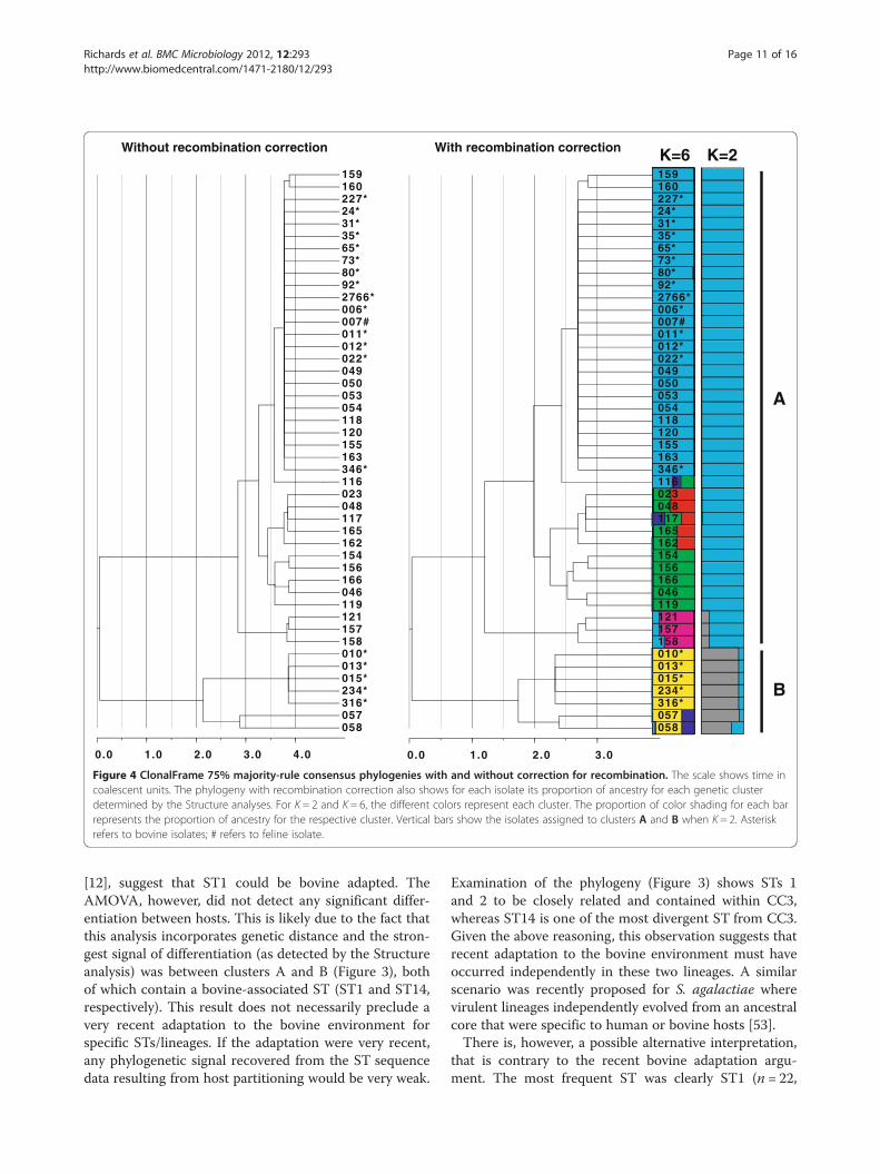
using a Bayesian phylogenetic approach (ClonalFrame,[68]) produced a well-supported phylogeny (Figure 3),with three independent runs of the Markov chain allproducing congruent topologies. Repeating the runswithout the recombination model (we assume no recom-bination) had no affect on the topology, but branchlengths did vary (Figure 4). The average total branchlength for the three phylogenies, not accounting forrecombination (15.9 coalescent time units), was slightlylarger than the average length of the three phylogenies

Table 1 Isolate screening data
ID Host/Tissue Herd/State/Country N1 N2 Ribotype ST CC
FSL Z3-022 Bovine Not available, Belgium 1 116-1000-4 1 3
IT-SCA-35 Bovine I-1, Italy 3 116-679-1 1 3
IT-SCA-65 Bovine I-2, Italy 3 116-679-1 1 3
IT-SCA-73 Bovine I-3, Italy 1 116-679-1 1 3
IT-SCA-31 Bovine I-4, Italy 1 116-1000-4 1 3
IT-SCA-92 Bovine I-5, Italy 3 116-1000-4 1 3
IT-SCA-80 Bovine I-6, Italy 2 116-1000-4 2 3
IT-SCA-24 Bovine I-7, Italy 3 116-679-1 1 3
FSL Z3-012* Bovine U-1, NY, USA 4 116-679-1 1 3
FSL Z3-006* Bovine U-2, NY, USA 4 116-679-1 1 3
R2-766 Bovine U-3, NY, USA 5 116-679-1 1 3
FSL Z3-227* Bovine U-4, NY, USA 11 116-679-1 1 3
FSL Z3-316 Bovine U-5, NY, USA 3 21 116-1180-4 14
FSL Z3-010 Bovine U-6, NY, USA 4 116-975-3 14
FSL Z3-346 Bovine U-7, NY, USA 2 116-679-1 1 3
FSL Z3-013* Bovine U-8, NY, USA 2 116-975-3 14
FSL Z3-015* Bovine U-9, NY, USA 1 12 116-975-3 14
FSL Z3-011* Bovine U-10, NY, USA 1 116-679-1 1 3
FSL Z3-234* Bovine U-10, NY, USA 2 116-975-3 14
FSL Z3-023* Canine, wound exudate Not available, Belgium 1000-5 11 2
FSL Z3-046* Canine, lip NY, USA 1168-1 9
FSL Z3-048 Canine, ear swab NY, USA 1000-5 11 2
FSL Z3-049 Canine, ear swab NY, USA 1000-4 1 3
FSL Z3-050 Canine, vaginal swab NY, USA 1000-4 1 3
FSL Z3-053 Canine, vaginal swab NY, USA 679-1 1 3
FSL Z3-054 Canine, vaginal swab NY, USA 679-1 1 3
FSL Z3-057 Canine, hock abscess NY, USA 975-3 15
FSL Z3-058 Canine, ear swab NY, USA 1171-2 6
FSL Z3-116 Canine, pharyngeal swab NY, USA 1168-5 8
FSL Z3-117 Canine, ear swab NY, USA 1000-5 12 2
FSL Z3-118 Canine, ear swab NY, USA 697-1 1 3
FSL Z3-119 Canine, ear swab NY, USA 1000-4 9 4
FSL Z3-120 Canine, vaginal swab NY, USA 679-1 1 3
FSL Z3-121* Canine, vaginal swab NY, USA 1173-7 4 1
FSL Z3-154* Canine, vaginal swab NY, USA 1173-8 10 4
FSL Z3-155 Canine, urine NY, USA 679-1 1 3
FSL Z3-156 Canine, throat NY, USA 1168-1 10 4
FSL Z3-157* Canine, pharyngeal swab NY, USA 1174-4 16 1
FSL Z3-158 Canine, pharyngeal swab NY, USA 1174-4 5 1
FSL Z3-159 Canine, eye NY, USA 679-1 3 3
FSL Z3-160 Canine, vaginal swab NY, USA 679-1 3 3
FSL Z3-162* Canine, ear swab NY, USA 1174-7 13 2
FSL Z3-163 Canine, dermis NY, USA 679-1 1 3
FSL Z3-165 Canine, vaginal swab NY, USA 1000-5 11 2
Richards et al. BMC Microbiology 2012, 12:293 Page 8 of 16http://www.biomedcentral.com/1471-2180/12/293

Table 1 Isolate screening data (Continued)
FSL Z3-166* Canine, pharyngeal swab NY, USA 1170-7 7
FSL Z3-007* Feline U-4, NY, USA 679-1 1 3
N1 = Number of isolates from each herd ribotyped (either individual cow or quarter milk sample). For each herd, only one isolate representing a distinct ribotypewas typed using MLST.N2 = Number of isolates from milking machine rubber liners or bulk tank milk.ST = sequence type.CC = clonal complex.1 Isolated from milking machine rubber liners.2 Isolated from bulk tank milk.* Isolate contains plasmid (see text).
Richards et al. BMC Microbiology 2012, 12:293 Page 9 of 16http://www.biomedcentral.com/1471-2180/12/293
that did account for recombination (14.2 coalescenttime units).The amount of recombination in bacteria can be quan-
tified using two ratios: (i) the ratio of the frequency atwhich recombination occurs relative to mutation (ρ/θ),and (ii) the ratio of the rates at which nucleotides be-come substituted as a result of recombination and muta-tion (r/m). The latter ratio accounts for length andnucleotide diversity of imported fragments and thereforecontains more information regarding the evolutionaryimpact of recombination [69]. Using ClonalFrame, wecalculated these ratios to be: ρ/θ = 0.1 and r/m = 1.5,with the latter ratio indicating that recombinationexceeded point mutation. Vos and Didelot [70] calcu-lated r/m for 48 diverse species of bacteria, and theirresults revealed a wide range of values (63.6 – 0.02). r/mfor S. canis ranked 25th in this distribution (approxi-mately in the middle). However, the average of the 48rates was 7.7, suggesting a below average rate of re-combination for S. canis when compared to these spe-cies of bacteria. When compared to the twoStreptococcus species in the distribution, S. canis wasmuch lower: S. pneumoniae = 23.1 (6th), S. pyogenes =17.2 (8th). Similar results were obtained when ρ/θ for S.canis was compared to other Streptococcus species: S.uberis = 17.2 [71], S. pneumoniae = 23.1 [72].
Table 2 Isolate diversity indices and summary statistics
n-RT RT RT-h n-ST
All 83 17 0.90 46
Bovine* 56 4 0.67 19
Canine 26 13 0.88 26
Feline 1 1 1
n-RT = number of isolates ribotyped.n-ST = number of isolates sequence typed.RT = number of ribotypes.RT-h = ribotype (gene) diversity.ST = number of STs.ST-h = ST (gene) diversity.θ = population parameter theta (per site).π = nucleotide diversity.plasmid = number of strains containing the plasmid.*The bovine isolates represent 18 distinct herds (farms). With one exception a singlMethods).
We expanded the evolutionary analysis by also ap-plying the parsimony-based approach e-BURST [73],which explores fine scale evolutionary relationshipsamong STs. The ClonalFrame phylogeny and e-BURSTresults were generally concordant regarding the group-ing of STs (Figure 3). The only discrepancy was ST7,which showed an intermediate relationship betweenSTs 9 and 10 in the phylogeny, but was not groupedwithin the same clonal complex (CC) as STs 9 and 10(ST7 was not grouped into any of the four clonalcomplexes).Population structure was further examined using the
Bayesian clustering approach implemented in the pro-gram Structure [74,75]. The number of clusters K wasestimated by calculating the ad hoc statistic ΔK, which isa measure of the second order rate of change of theprobability of the data L(K) for each value of K [76] (seeMethods for a full explanation of the approach). Theanalysis showed the optimum number of genetic clusters(K) to be two (A and B) (Figure 3 and Additional file 6).All four clonal complexes and ST8 were grouped intocluster A, whereas cluster B contained STs 6, 14, and 15.The ClonalFrame phylogeny showed CC1 to be the mostclosely related lineage to cluster B; concordantly, thislineage shared a small proportion (~12%) of its ancestrywith cluster B (Figure 4). Although ΔK indicated that
ST ST-h θ π plasmid
16 0.76 0.0127 0.0111 15
3 0.49 0.0089 0.0127 7
14 0.90 0.0139 0.0094 7
1 1
e ST was obtained from each herd (two STs were obtained from one herd) (see

0.4
054.vag.1
053.vag.1
346.NY.1
049.
ear.1
050.vag.1
022.
BEL
.1
118.ear.1120.vag.1155.uri.1
163.der.1
012.
NY.
1 006.NY.100
7.N
Y.1
011.
NY.
1 2766.NY.1
92.ITA.1
227.NY.124.ITA.131.ITA.135.ITA.1
65.ITA.1
80.ITA.2
73.ITA.1
160.vag.3
159.eye.3
116.thr.8
023.BEL.wou.11
048.ear.11
117.ear.12
165.vag.11162.ear.13
154.vag.10 119.
ear.
9
046.
lip.9
166.thr.7
156.thr.10
121.
vag.
4
010.
NY.14
158.
thr.5
157.
thr.1
6
013.N
Y.14
058.ear.15
057.leg.6
316.NY.14234.NY.14015.NY.14
1.0
1.0
1.0
1.0
1.01.0
0.98
1.01.0
CC2
CC3
CC4
CC1A
B
1.0
1.0
Figure 3 ClonalFrame 75% majority-rule consensus phylogeny (node posterior probabilities are at least 0.75). Posterior probabilities formajor lineages are shown at nodes. Dashed circles show each clonal complex (CC) and grey shading shows isolates assigned to the two clusters(A and B) determined by the Structure analysis. Taxa labels are colored as follows: red = canine isolate, blue = bovine isolate, green = feline isolate.The first number in the label shows isolate ID. For canine isolates, tissue source follows the isolate ID, which is followed by the ST. Tissue sourceabbreviations are as follows: thr = throat, vag = vaginal, uri = urine, der = dermis, wou = wound exudate. For bovine and feline isolates, the ID isfollowed by the geographic location of collection (ITA = Italy, BEL = Belgium, NY = New York state, USA). Strain 227.NY.1 (underlined) is the strainwho’s genome was sequenced in this study. Circles with white centers indicate those strains that contained the plasmid discussed in the text.The strain shaded in dark grey (166.thr.7) was grouped with CC4 members based on ClonalFrame analysis but it was not contained within CC4based on eBURST.
Richards et al. BMC Microbiology 2012, 12:293 Page 10 of 16http://www.biomedcentral.com/1471-2180/12/293
K was two and the Ln P(D) scores plateaued for Kvalues of two, three, and four (see Additional file 6),the Ln P(D) scores rose slightly after K = 4 and againplateaued starting with K = 6. This suggests a patternof hierarchal differentiation among isolates, with furthersubdivision present within clusters. Assuming K = 6 forthis additional subdivision, the assignment of individuals(proportion of ancestry) into these clusters delineatedisolates into groups concordant with the six majorlineages seen in the ClonalFrame phylogeny (Figure 4).Only three (1, 2, and 14) of the 16 STs were found in
bovines, and one of these (ST2) was a single locus
variant of the predominant ST in cattle (ST1). Conse-quently, there was a much higher diversity of STs foundin canine, producing a significant differentiation in thefrequency of STs between the two hosts. Previous studieshave shown the incidence of S. canis isolation frombovine to be rare [77-82]. This observation coupled withthe relatively low diversity of bovine STs suggests arecent adaptation to the bovine environment. Thus,the MLST data, the genomic features shared betweenS. canis and other bovine adapted Streptococcus speciesdiscussed earlier, and the epidemiological informationassociated with the original study regarding this strain
0.0 1.0 2.0 3.00.0 1.0 2.0 3.0 4.0
160
24*
157
92*
227*
316*
65*
154
121
117
013*015*
050
155
057
012*
054
046
023
158
058
80*
022*
165
156
159
163
007#
118
162
006*
31*
119
053
120
2766*
011*
049
116
048
166
234*
010*
35*
73*
346*
160
24*
157
92*
227*
316*
65*
154
121
117
013*015*
050
155
057
012*
054
046
023
158
058
80*
022*
165
156
159
163
007#
118
162
006*
31*
119
053
120
2766*
011*
049
116
048
166
234*
010*
35*
73*
346*
A
B
With recombination correctionWithout recombination correction K=6 K=2
Figure 4 ClonalFrame 75% majority-rule consensus phylogenies with and without correction for recombination. The scale shows time incoalescent units. The phylogeny with recombination correction also shows for each isolate its proportion of ancestry for each genetic clusterdetermined by the Structure analyses. For K = 2 and K = 6, the different colors represent each cluster. The proportion of color shading for each barrepresents the proportion of ancestry for the respective cluster. Vertical bars show the isolates assigned to clusters A and B when K = 2. Asteriskrefers to bovine isolates; # refers to feline isolate.
Richards et al. BMC Microbiology 2012, 12:293 Page 11 of 16http://www.biomedcentral.com/1471-2180/12/293
[12], suggest that ST1 could be bovine adapted. TheAMOVA, however, did not detect any significant differ-entiation between hosts. This is likely due to the fact thatthis analysis incorporates genetic distance and the stron-gest signal of differentiation (as detected by the Structureanalysis) was between clusters A and B (Figure 3), bothof which contain a bovine-associated ST (ST1 and ST14,respectively). This result does not necessarily preclude avery recent adaptation to the bovine environment forspecific STs/lineages. If the adaptation were very recent,any phylogenetic signal recovered from the ST sequencedata resulting from host partitioning would be very weak.
Examination of the phylogeny (Figure 3) shows STs 1and 2 to be closely related and contained within CC3,whereas ST14 is one of the most divergent ST from CC3.Given the above reasoning, this observation suggests thatrecent adaptation to the bovine environment must haveoccurred independently in these two lineages. A similarscenario was recently proposed for S. agalactiae wherevirulent lineages independently evolved from an ancestralcore that were specific to human or bovine hosts [53].There is, however, a possible alternative interpretation,
that is contrary to the recent bovine adaptation argu-ment. The most frequent ST was clearly ST1 (n = 22,

Richards et al. BMC Microbiology 2012, 12:293 Page 12 of 16http://www.biomedcentral.com/1471-2180/12/293
48% of isolates). This ST was also seen in all three hosts(canine, bovine, feline), including a wide range of caninetissue types (vaginal, ear, skin, urine, eye), and also allthree sampling locations (USA, Italy, and Belgium). Incontrast, with one exception, no other ST was seen inmore than one host or geographic location. The ex-ception was ST11, which was seen in both USA andBelgium. These observations suggest that ST1 is the mostancestral ST in the data set [83,84], and also possibly ageneralist, with the ability to infect different hosts andtissue types. Genomic comparisons showed that strainFSL S3-227 shared multiple mobile genetic elementswith S. agalactiae and S. dysgalactiae subsp. dysgalactiaestrains isolated from the bovine environment, with oneof these elements (the ICE) showing high sequence diver-gence. Although the ICE contained the Lac.2 operon,suggesting that this LGT may have contributed to bovineadaptation, the high divergence and multiple additionalLGTs suggest that S. canis ST1 may have had anextended association with the bovine environment, argu-ing against more recent adaptation. Consequently, ifST1’s lineage has possessed the ability to infect cowsfor an extended period of time, and is also the mostancestral with all lineages having descended from it, inorder for the ST14 lineage to have recently acquired theability to infect cows, all lineages intermediate betweenST1 and ST14 must have previously lost this ability. Thismight have occurred as a single event on the branchconnecting CC3 to ST8. Alternatively, all strains aregeneralist and the more recent lineages have simplyhad insufficient time to encounter the bovine environ-ment and/or that our sample size was too low to detecttheir presence.The distribution of the plasmid provides yet another
perspective. The plasmid has only been observed inone additional species: S. agalactiae (strain FSL-S3026[isolated from a bovine host], and strain NEM316[potential association with the bovine environment]).Therefore, it is possible that the plasmid was exchangedbetween S. canis and S. agalactiae in the bovine environ-ment, however, the plasmid appears randomly distributedamong S. canis isolates, regardless of host species orST. For example, (i) a Fisher exact test showed no sig-nificant difference in its distribution between bovineand canine isolates (P = 1.0), (ii) it was present in allclonal complexes and clusters, and (iii) it was presentin all three hosts including a wide range of caninetissue types (vaginal, ear, throat, lip). Consequently,the plasmid appears to have moved freely betweenbovine and canine environments, supporting the gen-eralist argument. An alternative explanation is that S.canis may have obtained the plasmid on independentoccasions from one or more different hosts. A simi-lar process involving various mobile genetic elements
has been observed for various Streptococcus species[17,85,86].
ConclusionCharacterization of the genome sequence for S. canisstrain FSL S3-227 detected a high diversity of virulencefactors. Approximately three quarters of the genes thatwere homologous to known Streptococcus virulence fac-tors are involved in invasion, evasion, and colonization,perhaps explaining S. canis's ability to infect a widerange of tissue types. Furthermore, the putative ancestralclonal complex (accounting for more than half of col-lected isolates) occurred in a wide range of tissue types,all hosts, and all geographic locations suggesting a wideand diverse niche. It has been demonstrated that thesource of bovine S. canis infection can be other farm-yard animals such as domestic cats [12]. Our results,revealed high genetic similarity among bovine, feline,and canine sourced isolates further supporting domesticfarm-yard animals as infection sources. Despite themodest level of recombination for S. canis when com-pared to other Streptococcus species, LGT is still clearlyan important evolutionary phenomenon in this speciesas evidenced by the multiple MGE present within itsgenome (i.e. plasmid, phage, and ICE) and the occur-rence of an integrative plasmid in approximately half ofthe collected isolates. Furthermore, the evidence forLGT between S. canis and two additional bovine mastitiscausing pathogens (S. agalactiae, and S. dysgalactiaesubsp. dysgalactiae) suggests a close association with thebovine environment for S. canis, with this LGT possiblycontributing to adaptation to this environment. Manyvirulence factors are also carried within these MGE,further highlighting the importance of these mobile ele-ments in the evolution of this pathogen. Furthermore,the high frequency of virulence factors within multipleMGE, coupled with LGT between S. canis and ahuman sourced bacteria (S. urinalis), suggests the pos-sibility for additional transport of virulence factors intothe human environment.
MethodsStrain selection, sequencing, and assemblyS. canis strain FSL Z3-227 was isolated from a compositemilk sample obtained from a cow with an intra-mammary infection. The sample was collected on the6th of April 1999 from a cow located in central NewYork State within a dairy herd experiencing an outbreakof S. canis induced mastitis. Bacterial culture and ribo-typing results indicated that a farm cat with chronicsinusitus was the likely source of the outbreak [12]. Util-izing a seven-gene MLST scheme developed here (seebelow), strain FSL Z3-227 was determined to be ST1.This ST was associated with multiple host species

Richards et al. BMC Microbiology 2012, 12:293 Page 13 of 16http://www.biomedcentral.com/1471-2180/12/293
(bovine, canine, feline). In addition, it was the mostcommon ST among bovine isolates and the only STto be found in all three countries represented in thestudy. Therefore, it was thought to have the potentialto have a broad complement of virulence factors,including those potentially associated with nicheadaptation in cattle, and was consequently selectedfor genome sequencing.Roche/454 pyrosequencing was used to determine the
genome sequence, and Newbler v1.1 (454 Life SciencesCorporation) was used to assemble the reads. Using re-striction enzyme BgIII, an optical map of the genomewas built by OpGen Technologies, Inc. (Madison, WI).Scaffold order and orientation was determined by align-ment to the optical map using Opgen Mapviewer. Smallinter and intra-scaffold gaps were closed by PCR andSanger sequencing. Seven larger gaps were closed usinglong range PCR and Illumina sequencing. Illumina readswere assembled using Velvet [87], and the optimum as-sembly was determined using the N50 statistic. Annota-tion of the genome assembly was performed usingthe NCBI Prokaryotic Genomes Automatic Annota-tion Pipeline (PGAAP) and Blast2GO v.2.5.0 (E valuecut-off = 1e-6 and minimum amino acid alignmentlength cut-off [hsp-length] = 33) [88] (annotations areshown in Additional file 2). This Whole GenomeShotgun project has been deposited at DDBJ/EMBL/GenBank under the accession AIDX00000000. Theversion described in this paper is the first version,AIDX01000000.
Homologous gene clusteringThe MCL algorithm [89] as implemented in theMCLBLASTLINE pipeline (available at http://micans.org/mcl) was used to delineate homologous proteinsequences among 214 Streptococcus genomes includingS. canis (see Additional file 3). Based on sequence simi-larity, the pipeline uses Markov clustering (MCL) toassign genes to homologous clusters. Similarity wasobtained from a reciprocal BLASTp within and be-tween all genome pairs using an E value cut-off of1e-5. The MCL algorithm was implemented using aninflation parameter of 1.8. Simulations have shown thisvalue to be generally robust to false positives andnegatives [90].
Virulence factorsAmino acid sequences for all S. canis CDS weresearched against the VFDB using BLASTp. We used anE value cut-off of 1e-5 and retained the single best hit.The search was refined by repeating the BLASTp searchagainst a database that contained only Streptococcusvirulence factors (88 genes).
Population geneticsIncluding the strain genome sequenced here, a total of83 S. canis isolates were obtained from bovine (n = 56),canine (n = 26), and feline (n = 1) hosts (Table 1). Isolatesof canine/feline origin included 25 canine isolates frompatients of Cornell University’s College of VeterinaryMedicine, Ithaca, NY, USA, one canine isolate fromBelgium, and one isolate from a cat living on a dairyfarm in upstate New York. The feline isolate was thelikely source of a mastitis outbreak at the same farm.Canine isolates from NY originated from dermis (n = 1),ear swabs (n = 7), eye (n = 1), hock abscess (n = 1), lip(n = 1), pharyngeal swabs (n = 5), urine (n = 1), and vaginalswabs (n = 8), and were collected from December 2003to May 2004. The canine isolate from Belgium originatedfrom wound exudate [1] and the feline isolate originatedfrom a nasal swab taken from a cat with chronic sinusitis[12]. Bovine isolates originated from one herd in Belgium(from mastitic milk; [1], seven dairy herds in Italy (16 iso-lates from bovine milk; collected in 2003 and 2004), and10 dairy herds in NY (two isolates from milking machineliners, one isolate from bulk tank milk, and 34 cow milkisolates; collected from 1999 to 2005). In addition tostrain FSL Z3-227, all 82 isolates were ribotyped usingthe commercial RiboPrinter system with EcoRI.Single isolates representing the ribotypes seen in each
herd (two isolates from the herd U-10 and a single iso-late from each of the remaining herds) (n = 19) werecombined with all canine/feline isolates (n = 27) and fur-ther screened using a seven housekeeping MLST schemewith PCR primers previously used for characterization ofS. pyogenes, S. pneumoniae, or S. uberis [91-95]. SeeAdditional file 7 for primer sequences and PCR profiles.MLST allele sequences were aligned using MAFFTv6.814b [96] as implemented in Geneious v5.1.2. Isolategenetic diversity indices were calculated using the pro-gram DNASP version 4.0 [97]. Diversity indices amongSTs were obtained by concatenating the seven alleles(4,014 bp). Diversity among ribotypes and STs was cal-culated using the formula for haplotype (gene) diversity[97]. Again using the concatenated allele sequences,population differentiation between bovine and caninegroupings of isolates (bovine = 19 canine = 26) was deter-mined by assessing the frequency distribution of STs(Fisher exact test) between the groups. Differentiationwas also determined by an AMOVA as implemented inArlequin v3.11 [98]. The AMOVA differs from the exacttest because in addition to assessing ST frequency distri-bution, it also considers genetic divergence among iso-late sequences in its determination of differentiation.With the exception of strain FSL Z3-227 (our genome
sequence), all isolates typed using the MLST scheme(n = 45) were also PCR screened for the presence of a55 CDS plasmid (see Results and discussion). Presence/

Richards et al. BMC Microbiology 2012, 12:293 Page 14 of 16http://www.biomedcentral.com/1471-2180/12/293
absence of the plasmid was determined using 25 primerpairs that were contiguous along the length of the plas-mid (see Additional file 8).Evolutionary relationships among STs were examined
using eBURSTv3 [73]. STs were grouped into clonal com-plexes and support for complex founders was estimatedusing 1000 bootstrap replicates. We used the most strin-gent (default) eBURST setting for grouping STs into acomplex, where STs within the same complex sharedidentical alleles at ≥ six of the seven loci with at least oneother member of the complex.Deeper evolutionary relationships (among clonal com-
plexes for example) were inferred using the Bayesianphylogenetic approach implemented in ClonalFramev1.1 [68]. This approach incorporates a model thatattempts to account for recombination. The Markov chainwas run with 1,000,000 iterations after an initial burn-in of50,000 iterations. Three independent runs were used toassess topological convergence. To assess the effect ofrecombination, all runs were repeated with the recom-bination rate parameter (R) held at zero (i.e. the effectof recombination on the topology was not accountedfor). We used ClonalFrame to calculate the recombin-ation ratios ρ/θ and r/m (average of the three runs).Isolates were clustered using the Bayesian approach
implemented in Structure v2.3.2 [74]. The number ofclusters K was estimated by calculating the ad hoc statis-tic ΔK [76]. ΔK was calculated for K = 1 through 10 using5 Markov chains for each value of K. The simulations ofEvanno et al. [76] showed that the highest value for ΔKreliably identified the optimum value of K. Chains wererun for 500,000 steps following an initial burn-in of100,000 steps, using the admixture ancestry and corre-lated allele frequency models. Once the optimum valueof K was identified, strains were assigned to clustersusing assignment coefficients (proportion of clustermembership) generated from an additional run utilizingthe linkage ancestry and correlated allele frequency mod-els. A study of recombinant bacterial populations showedthe linkage model of ancestry to produce the most accur-ate assignment scores in situations where there are mul-tiple linked loci along contiguous sections of DNA [75].The model assumes these sections, which could be re-combinant, to be discrete units of inheritance. Markovchains were run for 2,000,000 steps following an initialburn-in of 500,000 steps.
Additional files
Additional file 1: Streptococcus RefSeq genome summary statistics.
Additional file 2: S. canis annotation.
Additional file 3: Additional Streptococcus genomes.
Additional file 4: Insertion sites of putative integrative plasmid.
Additional file 5: S. canis isolate MLST allele data.
Additional file 6: Ln P(D) scores for Structure analysis.
Additional file 7: MLST PCR primer details.
Additional file 8: Putative integrative plasmid PCR primer details.
Competing interestsThe authors declare that they have no competing interests.
Authors’ contributionsVPR conducted data analysis and wrote the manuscript; MJS provided theconceptual framework, experimental design, and helped write themanuscript; PDPB and PL conducted laboratory work associated withgenome sequencing; TL conducted data analysis and genome assembly; BWconducted laboratory work associated with the survey of plasmiddistribution across canine and bovine isolates; LT, and PM conducted fieldwork associated with population genetics; RNZ conceived of the field andlaboratory work for population genetics, conducted MLST and ribotyping,and was involved in manuscript preparation. All authors read and approvedthe final manuscript.
AcknowledgementsWe would like to thank staff from Cornell University’s Quality Milk ProductionServices and Animal Health Diagnostic Centre for their contribution tosample and isolate collection. This study made use of PathogenTracker 2.0(www.microbtracker.net), developed by Martin Wiedmann. This work wassupported by the National Institute of Allergy and Infectious Disease, U.S.National Institutes of Health, under Grant No. AI073368 awarded to M.J.S.
Author details1Department of Population Medicine and Diagnostic Sciences, College ofVeterinary Medicine, Cornell University, Ithaca, NY 14853, USA. 2Quality MilkProduction Services, College of Veterinary Medicine, Cornell University, Ithaca,NY 14853, USA. 3Università degli Studi di Milano, Department of Health,Animal Science and Food Safety, Via Celoria 10, 20133 Milan, Italy. 4Currentaddress: Moredun Research Institute, Pentlands Science Park, Bush Loan,Penicuik and Royal (Dick) School of Veterinary Studies, University ofEdinburgh, Scotland, UK. 5Current address: Université de Lyon, UniversitéLyon 1, Centre National de la Recherche Scientifique, Ecologie desHydrosystèmes Naturels et Anthropisés, Villeurbanne, France. 6Currentaddress: Department of Plant Pathology & Plant-Microbe Biology, CornellUniversity, Ithaca, NY 14853, USA. 7Current address: 4055 McIntyre Road,Trumansburg, NY 14886, USA.
Received: 14 August 2012 Accepted: 6 December 2012Published: 18 December 2012
References1. Devriese LA, Hommez J, Kilpper-Balz R, Schleifer KH: Streptococcus canis sp.
nov.: a species of group G streptococci from animals. Int J Syst Bacteriol1986, 36(3):422–425.
2. Vandamme P, Pot B, Falsen E, Kersters K, Devriese LA: Taxonomic study ofLancefield streptococcal groups C, G, and L (Streptococcus dysgalactiae)and proposal of S. dysgalactiae subsp. equisimilis subsp. nov. Int J SystBacteriol 1996, 46(3):774–781.
3. Murase T, Morita T, Sunagawa Y, Sawada M, Shimada A, Sato K, Hikasa Y:Isolation of Streptococcus canis from a Japanese raccoon dog withfibrinous pleuropneumonia. Vet Rec 2003, 153(15):471–472.
4. Iglauer F, Kunstyr I, Morstedt R, Farouq H, Wullenweber M, Damsch S:Streptococcus canis arthritis in a cat breeding colony. J Exp Anim Sci 1991,34(2):59–65.
5. Pesavento PA, Bannasch MJ, Bachmann R, Byrne BA, Hurley KF: FatalStreptococcus canis infections in intensively housed shelter cats. VetPathol 2007, 44(2):218–221.
6. Kruger EF, Byrne BA, Pesavento P, Hurley KF, Lindsay LL, Sykes JE:Relationship between clinical manifestations and pulsed-field gel profilesof Streptococcus canis isolates from dogs and cats. Vet Microbiol 2010,146(1–2):167–171.
7. Matsuu A, Kanda T, Sugiyama A, Murase T, Hikasa Y: Mitral stenosis withbacterial myocarditis in a cat. J Vet Med Sci 2007, 69(11):1171–1174.

Richards et al. BMC Microbiology 2012, 12:293 Page 15 of 16http://www.biomedcentral.com/1471-2180/12/293
8. Sura R, Hinckley LS, Risatti GR, Smyth JA: Fatal necrotising fasciitis andmyositis in a cat associated with Streptococcus canis. Vet Rec 2008,162(14):450–453.
9. DeWinter LM, Prescott JF: Relatedness of Streptococcus canis from caninestreptococcal toxic shock syndrome and necrotizing fasciitis. Can J VetRes 1999, 63(2):90–95.
10. Hassan AA, Akineden O, Usleber E: Identification of Streptococcus canisisolated from milk of dairy cows with subclinical mastitis. J Clin Microbiol2005, 43(3):1234–1238.
11. Chaffer M, Friedman S, Saran A, Younis A: An outbreak of Streptococcuscanis mastitis in a dairy herd in Israel. N Z Vet J 2005, 53(4):261–264.
12. Tikofsky LL, Zadoks RN: Cross-infection between cats and cows: originand control of Streptococcus canis mastitis in a dairy herd. J Dairy Sci2005, 88(8):2707–2713.
13. Galperine T, Cazorla C, Blanchard E, Boineau F, Ragnaud JM, Neau D:Streptococcus canis infections in humans: retrospective study of54 patients. J Infect 2007, 55(1):23–26.
14. Lam MM, Clarridge JE 3rd, Young EJ, Mizuki S: The other group GStreptococcus: increased detection of Streptococcus canis ulcer infectionsin dog owners. J Clin Microbiol 2007, 45(7):2327–2329.
15. Whatmore AM, Engler KH, Gudmundsdottir G, Efstratiou A: Identification ofisolates of Streptococcus canis infecting humans. J Clin Microbiol 2001,39(11):4196–4199.
16. Bert F, Lambert-Zechovsky N: Septicemia caused by Streptococcus canis ina human. J Clin Microbiol 1997, 35(3):777–779.
17. Lefebure T, Richards VP, Lang P, Pavinski-Bitar P, Stanhope MJ: Generepertoire evolution of Streptococcus pyogenes inferred fromphylogenomic analysis with Streptococcus canis and Streptococcusdysgalactiae. PLoS One 2012, 7(5):e37607.
18. Jensen A, Kilian M: Delineation of Streptococcus dysgalactiae, itssubspecies, and its clinical and phylogenetic relationship toStreptococcus pyogenes. J Clin Microbiol 2012, 50(1):113–126.
19. Shinozaki-Kuwahara N, Takada K, Hirasawa M: Streptococcus ursoris sp. nov.,isolated from the oral cavities of bears. Int J Syst Evol Microbiol 2011,61(Pt 1):40–44.
20. Tapp J, Thollesson M, Herrmann B: Phylogenetic relationships andgenotyping of the genus Streptococcus by sequence determination of theRNase P RNA gene, rnpB. Int J Syst Evol Microbiol 2003, 53(Pt 6):1861–1871.
21. Suzuki H, Lefebure T, Hubisz MJ, Pavinski Bitar P, Lang P, Siepel A, StanhopeMJ: Comparative genomic analysis of the Streptococcus dysgalactiaespecies group: gene content, molecular adaptation, and promoterevolution. Genome Biol Evol 2011, 3:168–185.
22. Broyles LN, Van Beneden C, Beall B, Facklam R, Shewmaker PL, Malpiedi P,Daily P, Reingold A, Farley MM: Population-based study of invasivedisease due to beta-hemolytic streptococci of groups other than A andB. Clin Infect Dis 2009, 48(6):706–712.
23. DeWinter LM, Low DE, Prescott JF: Virulence of Streptococcus canis fromcanine streptococcal toxic shock syndrome and necrotizing fasciitis. VetMicrobiol 1999, 70(1–2):95–110.
24. Kanaya S, Yamada Y, Kudo Y, Ikemura T: Studies of codon usage and tRNAgenes of 18 unicellular organisms and quantification of Bacillus subtilistRNAs: gene expression level and species-specific diversity of codonusage based on multivariate analysis. Gene 1999,238(1):143–155.
25. Sharp PM, Bailes E, Grocock RJ, Peden JF, Sockett RE: Variation in thestrength of selected codon usage bias among bacteria. Nucleic Acids Res2005, 33(4):1141–1153.
26. Stothard P, Wishart DS: Circular genome visualization and explorationusing CGView. Bioinformatics 2005, 21(4):537–539.
27. Bhakdi S, Tranum-Jensen J, Sziegoleit A: Mechanism of membranedamage by streptolysin-O. Infect Immun 1985, 47(1):52–60.
28. Lang SH, Palmer M: Characterization of Streptococcus agalactiae CAMPfactor as a pore-forming toxin. J Biol Chem 2003, 278(40):38167–38173.
29. Bisno AL, Brito MO, Collins CM: Molecular basis of group A streptococcalvirulence. Lancet Infect Dis 2003, 3(4):191–200.
30. Panchaud A, Guy L, Collyn F, Haenni M, Nakata M, Podbielski A, Moreillon P,Roten CA: M-protein and other intrinsic virulence factors of Streptococcuspyogenes are encoded on an ancient pathogenicity island. BMC Genomics2009, 10:198.
31. Yang J, Liu Y, Xu J, Li B: Characterization of a new protective antigen ofStreptococcus canis. Vet Res Commun 2010, 34(5):413–421.
32. Nizet V, Beall B, Bast DJ, Datta V, Kilburn L, Low DE, De Azavedo JC: Geneticlocus for streptolysin S production by group A Streptococcus. InfectImmun 2000, 68(7):4245–4254.
33. Todd E: The differentiation of two distinct serologic varieties ofstreptolysin, streptolysin O and streptolysin S. J Pathol Bacteriol 1938,47:423–445.
34. Humar D, Datta V, Bast DJ, Beall B, De Azavedo JC, Nizet V: Streptolysin Sand necrotising infections produced by group G Streptococcus. Lancet2002, 359(9301):124–129.
35. Fuller JD, Camus AC, Duncan CL, Nizet V, Bast DJ, Thune RL, Low DE, DeAzavedo JC: Identification of a streptolysin S-associated gene cluster andits role in the pathogenesis of Streptococcus iniae disease. Infect Immun2002, 70(10):5730–5739.
36. Molloy EM, Cotter PD, Hill C, Mitchell DA, Ross RP: Streptolysin S-likevirulence factors: the continuing sagA. Nat Rev Microbiol 2011,9(9):670–681.
37. Koh TH, Sng LH, Yuen SM, Thomas CK, Tan PL, Tan SH, Wong NS:Streptococcal cellulitis following preparation of fresh raw seafood.Zoonoses Public Health 2009, 56(4):206–208.
38. Sun JR, Yan JC, Yeh CY, Lee SY, Lu JJ: Invasive infection with Streptococcusiniae in Taiwan. J Med Microbiol 2007, 56(Pt 9):1246–1249.
39. Facklam R, Elliott J, Shewmaker L, Reingold A: Identification andcharacterization of sporadic isolates of Streptococcus iniae isolated fromhumans. J Clin Microbiol 2005, 43(2):933–937.
40. Bekal S, Gaudreau C, Laurence RA, Simoneau E, Raynal L: Streptococcuspseudoporcinus sp. nov., a novel species isolated from the genitourinarytract of women. J Clin Microbiol 2006, 44(7):2584–2586.
41. Weinstein MR, Litt M, Kertesz DA, Wyper P, Rose D, Coulter M, McGeer A,Facklam R, Ostach C, Willey BM, et al: Invasive infections due to a fishpathogen, Streptococcus iniae. S. iniae Study Group. N Engl J Med 1997,337(9):589–594.
42. Kawamura Y, Hou XG, Sultana F, Miura H, Ezaki T: Determination of 16SrRNA sequences of Streptococcus mitis and Streptococcus gordonii andphylogenetic relationships among members of the genus Streptococcus.Int J Syst Bacteriol 1995, 45(2):406–408.
43. Jedrzejas MJ: Pneumococcal virulence factors: structure andfunction. Microbiol Mol Biol Rev 2001, 65(2):187–207. first page,table of contents.
44. Harvill ET, Preston A, Cotter PA, Allen AG, Maskell DJ, Miller JF: Multipleroles for Bordetella lipopolysaccharide molecules during respiratory tractinfection. Infect Immun 2000, 68(12):6720–6728.
45. Glaser P, Rusniok C, Buchrieser C, Chevalier F, Frangeul L, Msadek T, ZouineM, Couve E, Lalioui L, Poyart C, et al: Genome sequence of Streptococcusagalactiae, a pathogen causing invasive neonatal disease. Mol Microbiol2002, 45(6):1499–1513.
46. Chastanet A, Prudhomme M, Claverys JP, Msadek T: Regulation ofStreptococcus pneumoniae clp genes and their role in competencedevelopment and stress survival. J Bacteriol 2001, 183(24):7295–7307.
47. Blum G, Ott M, Lischewski A, Ritter A, Imrich H, Tschape H, Hacker J:Excision of large DNA regions termed pathogenicity islands from tRNA-specific loci in the chromosome of an Escherichia coli wild-typepathogen. Infect Immun 1994, 62(2):606–614.
48. Dobrindt U, Blum-Oehler G, Nagy G, Schneider G, Johann A, Gottschalk G,Hacker J: Genetic structure and distribution of four pathogenicity islands(PAI I(536) to PAI IV(536)) of uropathogenic Escherichia coli strain 536.Infect Immun 2002, 70(11):6365–6372.
49. Sampath J, Vijayakumar MN: Identification of a DNA cytosinemethyltransferase gene in conjugative transposon Tn5252. Plasmid 1998,39(1):63–76.
50. Saunders J, Saunders V: Bacterial transformation with plasmid DNA. InMethods in Microbiology Volume 21. Edited by Grinsted J, Bennett P. London:Academic Press; 1988.
51. Sumby P, Barbian KD, Gardner DJ, Whitney AR, Welty DM, Long RD, BaileyJR, Parnell MJ, Hoe NP, Adams GG, et al: Extracellular deoxyribonucleasemade by group A Streptococcus assists pathogenesis by enhancingevasion of the innate immune response. Proc Natl Acad Sci U S A 2005,102(5):1679–1684.
52. Richards VP, Lang P, Bitar PD, Lefebure T, Schukken YH, Zadoks RN,Stanhope MJ: Comparative genomics and the role of lateral genetransfer in the evolution of bovine adapted Streptococcus agalactiae.Infect Genet Evol 2011, 11(6):1263–1275.

Richards et al. BMC Microbiology 2012, 12:293 Page 16 of 16http://www.biomedcentral.com/1471-2180/12/293
53. Sørensen UB, Poulsen K, Ghezzo C, Margarit I, Kilian M: Emergence andGlobal Dissemination of Host-Specific Streptococcus agalactiae Clones.MBio 2010, 1(3).
54. Brochet M, Couve E, Zouine M, Vallaeys T, Rusniok C, Lamy MC, BuchrieserC, Trieu-Cuot P, Kunst F, Poyart C, et al: Genomic diversity and evolutionwithin the species Streptococcus agalactiae. Microbes Infect 2006,8(5):1227–1243.
55. Bisharat N, Crook DW, Leigh J, Harding RM, Ward PN, Coffey TJ, Maiden MC,Peto T, Jones N: Hyperinvasive neonatal group B Streptococcus has arisenfrom a bovine ancestor. J Clin Microbiol 2004, 42(5):2161–2167.
56. Canchaya C, Proux C, Fournous G, Bruttin A, Brussow H: Prophagegenomics. Microbiol Mol Biol Rev 2003, 67(2):238–276.
57. Lucchini S, Desiere F, Brussow H: Similarly organized lysogeny modules intemperate Siphoviridae from low GC content gram-positive bacteria.Virology 1999, 263(2):427–435.
58. Li J, Kasper DL, Ausubel FM, Rosner B, Michel JL: Inactivation of the alphaC protein antigen gene, bca, by a novel shuttle/suicide vector results inattenuation of virulence and immunity in group B Streptococcus. ProcNatl Acad Sci U S A 1997, 94(24):13251–13256.
59. Peltroche-Llacsahuanga H, Frye B, Haase G: Isolation of Streptococcus urinalisfrom a human blood culture. J Med Microbiol 2012, 61(Pt 5):740–742.
60. Collins MD, Hutson RA, Falsen E, Nikolaitchouk N, LaClaire L, Facklam RR: Anunusual Streptococcus from human urine, Streptococcus urinalis sp. nov.Int J Syst Evol Microbiol 2000, 50 Pt 3:1173–1178.
61. Rabel C, Grahn AM, Lurz R, Lanka E: The VirB4 family of proposed trafficnucleoside triphosphatases: common motifs in plasmid RP4 TrbE areessential for conjugation and phage adsorption. J Bacteriol 2003,185(3):1045–1058.
62. Haenni M, Saras E, Bertin S, Leblond P, Madec JY, Payot S: Diversity andmobility of integrative and conjugative elements in bovine isolates ofStreptococcus agalactiae, S. dysgalactiae subsp. dysgalactiae, andS. uberis. Appl Environ Microbiol 2010, 76(24):7957–7965.
63. Paoletti C, Foglia G, Princivalli MS, Magi G, Guaglianone E, Donelli G,Pruzzo C, Biavasco F, Facinelli B: Co-transfer of vanA and aggregationsubstance genes from Enterococcus faecalis isolates in intra- andinterspecies matings. J Antimicrob Chemother 2007, 59(5):1005–1009.
64. Ferretti JJ, McShan WM, Ajdic D, Savic DJ, Savic G, Lyon K, Primeaux C, SezateS, Suvorov AN, Kenton S, et al: Complete genome sequence of an M1 strainof Streptococcus pyogenes. Proc Natl Acad Sci U S A 2001,98(8):4658–4663.
65. Pryor SM, Cursons RT, Williamson JH, Lacy-Hulbert SJ: Experimentallyinduced intramammary infection with multiple strains of Streptococcusuberis. J Dairy Sci 2009, 92(11):5467–5475.
66. Zadoks RN, Schukken YH: Use of molecular epidemiology in veterinarypractice. Vet Clin North Am Food Anim Pract 2006, 22(1):229–261.
67. Excoffier L, Smouse PE, Quattro JM: Analysis of molecular variance inferredfrom metric distances among DNA haplotypes: Application to humanmitochondrial DNA restriction data. Genetics 1992, 131:479–491.
68. Didelot X, Falush D: Inference of bacterial microevolution usingmultilocus sequence data. Genetics 2007, 175(3):1251–1266.
69. Guttman DS, Dykhuizen DE: Clonal divergence in Escherichia coli as a resultof recombination, not mutation. Science 1994, 266(5189):1380–1383.
70. Vos M, Didelot X: A comparison of homologous recombination rates inbacteria and archaea. ISME J 2009, 3(2):199–208.
71. Lang P, Lefebure T, Wang W, Zadoks RN, Schukken Y, Stanhope MJ: Genecontent differences across strains of Streptococcus uberis identified usingoligonucleotide microarray comparative genomic hybridization. InfectGenet Evol 2009, 9(2):179–188.
72. Fraser C, Hanage WP, Spratt BG: Neutral microepidemic evolution ofbacterial pathogens. Proc Natl Acad Sci U S A 2005, 102(6):1968–1973.
73. Feil EJ, Li BC, Aanensen DM, Hanage WP, Spratt BG: eBURST: inferring patternsof evolutionary descent among clusters of related bacterial genotypes frommultilocus sequence typing data. J Bacteriol 2004, 186(5):1518–1530.
74. Pritchard JK, Stephens M, Donnelly P: Inference of population structureusing multilocus genotype data. Genetics 2000, 155(2):945–959.
75. Falush D, Stephens M, Pritchard JK: Inference of population structureusing multilocus genotype data: linked loci and correlated allelefrequencies. Genetics 2003, 164(4):1567–1587.
76. Evanno G, Regnaut S, Goudet J: Detecting the number of clusters ofindividuals using the software STRUCTURE: a simulation study. Mol Ecol2005, 14(8):2611–2620.
77. Persson Y, Nyman AK, Gronlund-Andersson U: Etiology and antimicrobialsusceptibility of udder pathogens from cases of subclinical mastitis indairy cows in Sweden. Acta Vet Scand 2011, 53:36.
78. Piepers S, De Meulemeester L, de Kruif A, Opsomer G, Barkema HW, DeVliegher S: Prevalence and distribution of mastitis pathogens insubclinically infected dairy cows in Flanders, Belgium. J Dairy Res 2007,74(4):478–483.
79. Sampimon O, Barkema HW, Berends I, Sol J, Lam T: Prevalence ofintramammary infection in Dutch dairy herds. J Dairy Res 2009, 76(2):129–136.
80. Petrovski KR, Heuer C, Parkinson TJ, Williamson NB: The incidence andaetiology of clinical bovine mastitis on 14 farms in Northland, NewZealand. N Z Vet J 2009, 57(2):109–115.
81. Guelat-Brechbuehl M, Thomann A, Albini S, Moret-Stalder S, Reist M,Bodmer M, Michel A, Niederberger MD, Kaufmann T: Cross-sectional studyof Streptococcus species in quarter milk samples of dairy cows in thecanton of Bern, Switzerland. Vet Rec 2010, 167(6):211–215.
82. Bengtsson B, Unnerstad HE, Ekman T, Artursson K, Nilsson-Ost M, Waller KP:Antimicrobial susceptibility of udder pathogens from cases of acuteclinical mastitis in dairy cows. Vet Microbiol 2009, 136(1–2):142–149.
83. Avise JC: Phylogeography. The history and formation of species. Cambridge,MA: Harvard University Press; 2000.
84. Templeton AR: Population genetics and microevolutionary theory. New Jersey:Wiley; 2006.
85. Delorme C, Poyart C, Ehrlich SD, Renault P: Extent of horizontal genetransfer in evolution of Streptococci of the salivarius group. J Bacteriol2007, 189(4):1330–1341.
86. Davies MR, Tran TN, McMillan DJ, Gardiner DL, Currie BJ, Sriprakash KS:Inter-species genetic movement may blur the epidemiology ofstreptococcal diseases in endemic regions. Microbes Infect 2005,7(9–10):1128–1138.
87. Zerbino DR, Birney E: Velvet: algorithms for de novo short read assemblyusing de Bruijn graphs. Genome Res 2008, 18(5):821–829.
88. Gotz S, Garcia-Gomez JM, Terol J, Williams TD, Nagaraj SH, Nueda MJ,Robles M, Talon M, Dopazo J, Conesa A: High-throughput functionalannotation and data mining with the Blast2GO suite. Nucleic Acids Res2008, 36(10):3420–3435.
89. van Dongen S: Graph clustering by flow simulation, University of Utrecht; 2000.90. Brohee S, van Helden J: Evaluation of clustering algorithms for protein-
protein interaction networks. BMC Bioinformatics 2006, 7:488.91. Enright MC, Spratt BG: A multilocus sequence typing scheme for
Streptococcus pneumoniae: identification of clones associated withserious invasive disease. Microbiology 1998, 144(Pt 11):3049–3060.
92. Enright MC, Spratt BG, Kalia A, Cross JH, Bessen DE: Multilocus sequencetyping of Streptococcus pyogenes and the relationships between emmtype and clone. Infect Immun 2001, 69(4):2416–2427.
93. Goh SH, Santucci Z, Kloos WE, Faltyn M, George CG, Driedger D,Hemmingsen SM: Identification of Staphylococcus species and subspeciesby the chaperonin 60 gene identification method and reversecheckerboard hybridization. J Clin Microbiol 1997, 35(12):3116–3121.
94. Ke D, Picard FJ, Martineau F, Menard C, Roy PH, Ouellette M, Bergeron MG:Development of a PCR assay for rapid detection of enterococci. J ClinMicrobiol 1999, 37(11):3497–3503.
95. Zadoks RN, Schukken YH, Wiedmann M: Multilocus sequence typing ofStreptococcus uberis provides sensitive and epidemiologically relevantsubtype information and reveals positive selection in the virulence genepauA. J Clin Microbiol 2005, 43(5):2407–2417.
96. Katoh K, Misawa K, Kuma K, Miyata T: MAFFT: a novel method for rapidmultiple sequence alignment based on fast Fourier transform. NucleicAcids Res 2002, 30(14):3059–3066.
97. Rozas J, Sánchez-DelBarrio J, Messegyer X, Rozas R: DNASP, DNApolymorphism analyses by the coalescent and other methods.Bioinformatics 2003, 19:2496–2497.
98. Excoffier L, Laval G, Schneider S: Arlequin (version 3.0): an integratedsoftware package for population genetics data analysis. Evol BioinformOnline 2005, 1:47–50.
doi:10.1186/1471-2180-12-293Cite this article as: Richards et al.: Genome characterization andpopulation genetic structure of the zoonotic pathogen, Streptococcuscanis. BMC Microbiology 2012 12:293.