geneticstfmanuscriptfeb15 · 1 recommendations of the international parkinson and movement disorder...
TRANSCRIPT

1
Recommendations of the International Parkinson and Movement Disorder
Society Task Force on Nomenclature of Genetic Movement Disorders
Connie Marras1 MD, PhD, Anthony Lang MD1, Bart P. van de Warrenburg,4 Carolyn Sue,3 Sarah J. Tabrizi MBChB, PhD,5 Lars Bertram MD,6,7 Katja Lohmann2 PhD, Saadet Mercimek-Mahmutoglu, MD, PhD,8 Alexandra Durr9, Vladimir Kostic10, Christine Klein2 MD,
1Toronto Western Hospital Morton and Gloria Shulman Movement Disorders Centre
and the Edmond J. Safra Program in Parkinson’s disease, University of Toronto,
Toronto, Canada
2Institute of Neurogenetics, University of Lübeck, Lübeck, Germany
3Department of Neurology, Royal North Shore Hospital and Kolling Institute of
Medical Research, University of Sydney, St Leonards, NSW 2065, Australia
4Department of Neurology, Donders Institute for Brain, Cognition, and Behaviour,
Radboud University Medical Centre, Nijmegen, The Netherlands
5Department of Neurodegenerative Disease, Institute of Neurology, University
College London, UK
6 Platform for Genome Analytics, Institute of Neurogenetics, University of Lübeck, Lübeck, Germany
7School of Public Health, Faculty of Medicine, Imperial College, London, UK
8Division of Clinical and Metabolic Genetics, Department of Pediatrics, University of
Toronto, The Hospital for Sick Children, Toronto, Canada
9 Sorbonne Université, UPMC Univ Paris 06, UM 75, ICM, F-75013 Paris, France;
Inserm, U 1127, ICM, F-75013 Paris, France; Cnrs, UMR 7225, ICM, F-75013 Paris,
France; ICM, Paris, F-75013 Paris, France; AP-HP, Hôpital de la Salpêtrière,
Département de Génétique et Cytogénétique, F-75013, Paris, France

2
10Institute of Neurology, School of Medicine University of Belgrade, Serbia
Corresponding author:
Christine Klein, MD
Institute of Neurogenetics
University of Luebeck
Ratzeburger Allee 160
23538 Luebeck
Germany
Email: [email protected]
Tel: +49-451-2903353
Fax: +49-451-2903355
Email addresses of authors
Katja Lohmann: [email protected]
Christine Klein: [email protected]
Anthony Lang: [email protected]
Connie Marras: [email protected]
Carolyn Sue: [email protected]
Sarah Tabrizi: [email protected]
Bart van de Warrenburg: [email protected]

3
Vladimir S. Kostić: [email protected]
Saadet Mahmutoglu: [email protected]
Alexandra Durr: [email protected]
Running title:
Word count text:
Abstract: 160
Title:
References:
Tables:
Figures:
Search terms:

4
Author Roles
1. Research project: A. Conception, B. Organization, C. Execution;
2. Manuscript: A. Writing of the first draft, B. Review and Critique;
Author contributions:
Connie Marras: 1A, B, C; 2A
Katja Lohmann: 1A, 1C, 2B
Anthony Lang: 1A, 1C, 2B
Christine Klein: 1A, B, C; 2B
Carolyn Sue: 1A, 1C, 2B
Bart van de Warrenburg: 1A, 1C; 2A, 2B
Vladimir Kostic: 1A, 1C, 2B
Sarah Tabrizi: 1C, 2B
Saadet Mahmutoglu: 1A, 2B
Alexandra Durr: 2B
Financial Disclosures of all authors (for the preceding 12 months)
Christine Klein
Stock Ownership in medically-
related fields None
Consultancies Medical advisor to Centogene
Advisory Boards None

5
Partnerships None
Honoraria None
Grants
The Hermann and Lilly Schilling Foundation;
the BMBF (01GI0201); the German
Research Foundation; the European
Community (FP7); intramural funds from the
University of Luebeck
Intellectual Property Rights None
Expert Testimony None
Employment University of Luebeck
Contracts None
Royalties None
Other None
Connie Marras
Stock Ownership in medically-
related fields None
Consultancies None
Advisory Boards None
Partnerships None
Honoraria Honoraria for teaching from EMD Serono
Grants The Michael J Fox Foundation, Canadian

6
Institutes of Health Research, National
Parkinson Foundation and the Parkinson
Society Canada; Site PI for clinical trial
sponsored by Allon Therapeutics
Intellectual Property Rights None
Expert Testimony None
Employment University Health Network
Contracts None
Royalties None
Other None
Sarah J Tabrizi
Sarah J Tabrizi
Stock Ownership in medically-
related fields
None
Consultancies
UCL Consultancy with Siena Biotech, Simon
Kucher Partnership, Roche, Takeda Pharmaceuticals
International, Novartis, Sanofi-Aventis, Isis
Pharmaceuticals Inc., GSK and TEVA
Pharmaceuticals. All honoraria paid for these
consultancies and advisory boards goes to University
College London, Sarah J Tabrizi’s employer.
Advisory Boards Isis Pharma, Roche, Siena Biotech, Teva
Pharma
Partnerships None

7
Honoraria None
Grants
EU FP7 Health call,
Medical Research Council UK,
CHDI Foundation,
Huntington Disease Association of the UK,
Dementia and Neurodegenerative Disease Network
UK,
European Huntington’s Disease Network,
UCL/UCLH Biomedical Research Centre
BBSRC
Intellectual Property Rights None
Expert Testimony None
Employment University College London
Contracts None
Royalties None
Other None
Carolyn Sue
Stock Ownership in medically-
related fields None
Consultancies None
Advisory Boards None
Partnerships None

8
Honoraria None
Grants
National Health and Medical Research
Council (of Australia), Australian Brain
Foundation, Hereditary Spastic Paraplegia
Foundation
Intellectual Property Rights None
Expert Testimony None
Employment Northern Sydney Local Health District
Contracts None
Royalties None
Other None
Anthony Lang
Stock Ownership in medically-
related fields None
Consultancies
Abbvie, Allon Therapeutics, Avanir
Pharmaceuticals, Biogen Idec, Boerhinger-
Ingelheim, Ceregene, Lilly, Medtronic,
Merck, Novartis, NeuroPhage
Pharmaceuticals, Teva and UCB
Advisory Boards None
Partnerships None

9
Honoraria Teva, UCB, AbbVie
Grants
Brain Canada, Canadian Institutes of Health
Research, Edmond J Safra Philanthropic
Foundation, Michael J. Fox Foundation,
National Parkinson Foundation, Ontario
Brain Institute, Parkinson Society Canada,
Tourette Syndrome Association, W. Garfield
Weston Foundation.
Intellectual Property Rights None
Expert Testimony Cases related to the welding industry.
Employment University of Toronto
Contracts None
Royalties Saunders, Wiley-Blackwell, Johns Hopkins
Press, Cambridge University Press.
Other None

10
Name: Katja Lohmann
Stock
Ownership in
medically-
related fields
None
Intellectual
Property
Rights
None
Consultancies None Expert
Testimony None
Advisory Boards None Employment University of Luebeck
Partnerships None Contracts None
Honoraria None Royalties None
Grants
German Research
Foundation, Dystonia
Coalition
Other None
Bart van de Warrenburg
Stock Ownership in medically-
related fields None
Consultancies None
Advisory Boards None
Partnerships None

11
Honoraria None
Grants
Gossweiler Foundation, Royal Dutch Society
for Physical Therapy, BBRMI-NL,
Wetenschapsfonds Dutch dystonia society,
Radboud University Medical Centre
Intellectual Property Rights None
Expert Testimony None
Employment Radboud University Medical Centre
Royalties None
Other None
Saadet Mercimek-Mahmutoglu
Stock Ownership in medically-related fields None
Consultancies None Advisory Boards None Partnerships None
Honoraria
None
Grants None Intellectual Property Rights None Expert Testimony None Employment The Hospital for Sick Children Contracts None Royalties None Other None
Alexandra Durr
Stock Ownership in medically-related fields None
Consultancies None

12
Advisory Boards None Partnerships None
Honoraria
Pfizer Inc.
Grants Pfizer Inc., Gossweiler Foundation, ULC, European Union
Intellectual Property Rights None Expert Testimony None
Employment Assistance Publique-Hôpitaux de Paris/Pierre et Marie Curie University
Contracts None Royalties None Other None
Vladimir S. Kostić
Stock Ownership in medically-related fields None
Consultancies None Advisory Boards Abvie (regional) Partnerships None
Honoraria
Novartis, Boehringer Ingelheim, Roche,
Lundbeck and Glaxo-Smith-Kline
Grants
Ministry of Education and Science, Republic of Serbia (project #ON175090)
Serbian Academy of Sciences and Arts (no 40070)
Intellectual Property Rights None Expert Testimony None Employment University of Belgrade Contracts None Royalties None Other None
Lars Bertram

13
Stock Ownership in medically-related fields None
Consultancies None Advisory Boards ADNI-2 Partnerships
Honoraria
Grants German Federal Ministry for Education and Research (BMBF), EU FP7, and the Cure Alzheimer's Fund.
Intellectual Property Rights Expert Testimony
Employment University of Luebeck, Germany
Imperial College London, UK
Contracts Royalties Other
14
Abstract
The system of assigning locus symbols to specify chromosomal regions that are
associated with a familial disorder has a number of problems when used as a
reference list of genetically determined disorders, including erroneously assigned
loci, duplicated loci, missing symbols, missing loci, unconfirmed loci, combining
causative genes and risk factor genes in the same list and discordance between
phenotype and list assignment. In this paper we report on the recommendations of
the International Parkinson and Movement Disorder Society (MDS) Task Force for
Nomenclature of Genetic Movement Disorders and present a system for naming
genetically determined movement disorders that addresses these problems. We
demonstrate how the system would be applied to currently known genetically
determined parkinsonism, dystonia, dominantly inherited ataxia, spastic paraparesis,
chorea, paroxysmal movement disorders, neurodegeneration with brain iron
accumulation and basal ganglia calcifications. This system provides a resource for
clinicians and researchers that, unlike the previous system, can be considered an
accurate and criterion-based list of confirmed genetically determined movement
disorders at the time it was last updated.

15
Introduction
The system of locus symbols (e.g. DYT1) was originally established to specify
chromosomal regions that had been linked to a familial disorder where the gene was
yet unknown.1 This system has been adopted by clinicians and researchers to
provide names for the condition, as well as the chromosomal region, and use of
these names is commonplace in medical parlance, particularly in the field of
movement disorders. However, as our techniques of genetic research and our
knowledge have evolved, a number of problems with the system of designating locus
symbols and with its use have arisen. These problems have been described
elsewhere2 but briefly, they include 1) an inability to distinguish disease–causing
genes from weaker genetic risk factors, 2) an inconsistent relationship between list
membership and movement disorder phenotype, 3) failure of some established
genetic movement disorders to be assigned a locus symbol, 4) more than one
symbol being assigned for the same disorder, 5) unconfirmed associations between
a gene or locus and a movement disorder, 6) erroneous labels resulting from
laboratory errors or mistakes of phenotypic assignment and 7) designating symbols
in the absence of any known locus or gene. Together, these problems make the
locus symbols unsuitable as a reference list of genetically determined movement
disorders. Unfortunately, it is currently used as such. This state of affairs was the
justification for the International Parkinson and Movement Disorder Society (MDS)
Task Force for Nomenclature of Genetic Movement Disorders. This report presents
a recommendation for a new system for naming of genetically determined movement
disorders by the Task Force.
The Task Force and its mandate

16
The MDS Task Force for the Nomenclature of Genetic Movement Disorders first
convened in May 2012. The mandate of the Task Force was to generate
recommendations for revising the system of naming of genetic movement disorders,
addressing the problems summarized above. The Task Force included clinical
neurologists and genetic experts covering the spectrum of movement disorders as
well as a metabolic geneticist (S M-M). Input was sought from experts in medical
fields other than movement disorders, where naming systems for genetically
determined disorders were in place (e.g. epilepsy). Editors of general medical and
neurology journals were also queried regarding their requirements from authors for
assigning names for newly discovered genetic conditions or their associated genes.
With this background, the Task Force agreed upon a set of rules that should govern
the naming based upon a set of previously published recommendations authored by
several members of the Task Force.2 These previously published recommendations
were developed into more concrete rules. We then proceeded to apply the rules to
classes of genetically determined movement disorders. The classes are
phenomenologically defined (e.g. ataxias, dystonias), or defined by a distinctive
imaging (e.g. basal ganglia calcification) or, theoretically, laboratory feature. To date
we have not found the need to classify on any laboratory features. The
recommendations and resulting lists have been made available to the membership of
the MDS through the Society’s website and feedback was solicited from the
membership. The recommendations were also shared with representatives from
GeneTests and OMIM, two compendia of genetic phenotypes for commentary and
suggestions.
Recommendations
Commented [CR1]: CM: Their input will be sought after
asking the MDS membership for their feedback.

17
1. Include only genes where genetic testing is possible.
Originally, locus symbols represented chromosomal regions. However, if we know
only the chromosomal region associated with a particular phenotype there are no
direct implications for diagnostic testing or for (basic) research applications.
Therefore, a disorder should only be listed once the causative gene is found. The
exception to this is when a founder haplotype is diagnostic, as in the case of X-linked
dystonia parkinsonism (“Lubag”). In this case, the disorder should be a member of
the list.
2. Replace number suffixes by the gene name.
We recommend that the symbol prefix be followed by the gene name (e.g. DYT-
SGCE [currently DYT11]). This naming system conveys the responsible gene and
maintains the connection between the phenotype (dystonia) and the gene.
Remembering a numerical designation (e.g. DYT1) is obviously easier than
remembering complex gene names. However, given the major sources of error and
confusion that have arisen in the numerical listing we feel that this new approach is
justified. In addition, the exponentially growing number of identified causative genes
will likely make remembering all but the most clinically important examples
impossible for most. Referring to reference tables will become increasingly
necessary, and with this vision, more informative names and rigorous review for
inclusion are preferred.
3. List disease-causing genes separately from risk factor genes.

18
A locus symbol prefix (e.g. PARK) would be conferred only upon disease-causing
genes (causing monogenic disorders) and not upon risk factors, recognizing the
diagnostic value of disease-causing mutations. The PD GENE website
(http://www.pdgene.org), developed by the Max Planck Institute for Molecular
Genetics, the Michael J. Fox Foundation and the Alzheimer Research Forum
provides a resource for cataloguing genetic risk factors for Parkinson’s disease and
for continuously evaluating them in an ongoing meta-analysis. When disease-
causing mutations and risk factors arise from the same gene (e.g. SNCA), such
genes should be represented on both lists.
We recognize that the distinction between disease-causing and risk-conferring is not
clear in many instances; rather, these attributes mark two ends of a continuum of
risk. Furthermore, the decision into which category a particular genetic variation falls
is complicated when penetrance varies by age, sex or ethnicity. As there is currently
no standard as to what level of penetrance of a mutation (or increase in risk) is
sufficient to consider a genetic mutation as being disease-causing, we have not
designated a specific threshold. Rather, we have accepted the designation (disease-
causing or risk factor) that prevails in the field for each gene. As discussed below, a
criterion-based method of making such distinctions would be of value to the field.
4. Raise the threshold of evidence before assigning locus symbols.
To avoid inaccuracies and redundancies that currently permeate the lists of locus
symbols, a level of evidence for genotype-phenotype association must be met prior
to conferring a place in the list. The US National Human Genome Research Institute

19
convened a working group to establish guidelines for investigating causality of
sequence variants in human disease.3 As outlined in the guidelines, four major
pieces of evidence lend support to causality: 1. The presence of the variant in
multiple unrelated affected individuals, 2. Evidence for segregation or statistical
association of the variant with disease 3. The variant should be conserved across
different species and 4. The variant should be predicted to alter the normal
biochemical effect of the gene product, if possible as supported by functional
evidence in human tissue or well-established cellular or animal models. Considering
their guidelines a gene-by-gene assessment of the sum of the evidence was
considered a most appropriate approach for deciding whether or not a gene warrants
a place on the lists.
5. Assign appropriate phenotype-prefix relationships.
For a gene to be a member of a particular phenotypic list, the phenotype (e.g.
dystonia in the case of DYTs) should be a consistent and prominent feature of the
disease linked to mutations in that gene. When more than one movement disorder is
a prominent and consistent feature, a double prefix could be assigned (e.g.
DYT/PARK-ATP1A3) and the symbol would belong to more than one list. Disorders
which can unusually present with an alternative movement disorder as the
predominant manifestation would appear cross-referenced to the list of the
alternative phenotype (e.g. SCA17 occasionally presenting as a choreic disorder,
thus it is cross-referenced to the chorea list) but the prefix would reflect the
phenotype that is consistent with in the majority of cases. When a genetic mutation
can unusually manifest with a movement disorder as the predominant manifestation
but the usual phenotype is not a movement disorder (e.g. C9orf72 mutations

20
presenting as a predominantly choreic disorder instead of the usual dementia-
predominant syndrome), we have not included these disorders on the list. The focus
of these lists is on disorders that have movement disorders as consistent and
predominant features.
Applying the recommendations
For each class of movement disorder we present the new list applying the principles
we have laid out above. For contrast we have provided, as supplementary material,
the list of existing locus symbols for each class of disorder, including a note in the
last column indicating the problems with the entry, where applicable. We have
included in the list of existing symbols those not listed by the Human Genome
Nomenclature Committee (http://www.genenames.org/), reflecting the reality that
many locus symbols have come into use bypassing this official channel. In our
revised system we have assigned symbols to disorders known to be genetically
determined but never assigned a locus symbol, in order to provide a complete list;
Wilson disease as an example. In the same spirit we have introduced to these lists a
number of pediatric metabolic disorders.
Genetically determined movement disorders
Genetically determined parkinsonism
A total of 20 genes and loci have been assigned a ‘PARK’ designation
(Supplementary Table 1). For five of these, the relationship is unconfirmed

21
(PARK3,5,11,13,18) and three fall into the ‘risk factor’ category (PARK10,12,16).
PARK1 and PARK4 are identical, both referring to the SNCA gene.
According to the revised system, there are eighteen confirmed monogenic conditions
where parkinsonism is a consistent and predominant feature (Table 1). In eight of
the forms of genetic parkinsonism mentioned below, dystonia is a prominent feature.
To guide clinicians we have divided these into three categories: 1) Those that are
associated with a clinical picture closely resembling that of idiopathic Parkinson
disease, 2) Those that present with parkinsonism similar to Parkinson disease but of
young onset and 3) Complex forms that have parkinsonism as a key clinical feature
but in addition present with atypical, multisystem features or other movement
disorders. We have provided references for the more complex disorders that may
not have been included in previous lists of this kind; for others we refer readers to a
recent review.4, 5 Of note, we have chosen not to include GBA as a monogenic
cause of parkinsonism in the PARK list but rather consider it a strong genetic risk
factor for Parkinson disease (similar to ApoE4 in Alzheimer disease) given its low,
age-dependent prevalence.6
Table 1: The proposed new list of hereditary parkinsonism
New
designation and
Phenotypic
subgroup
Clinical clues Inheri-
tance
Locus
symbol

22
Classical parkinsonism
PARK-SNCA Missense mutations cause classical
parkinsonism. Duplication or
triplication mutations in this gene
cause early onset parkinsonism with
prominent dementia.
AD PARK1
PARK-LRRK2 AD PARK8
PARK-VPS35 AD PARK17
Early onset parkinsonism
PARK-PARKIN* Often presents with dystonia, often in a
leg
AR PARK2
PARK-PINK1 Psychiatric features common AR PARK6
PARK-DJ1 AR PARK7
Atypical parkinsonism or complex phenotypes
PARK-ATP13A2 Kufor Rakeb syndrome; Juvenile or
early onset parkinsonism, vertical gaze
palsy, minifacial-faucial myoclonus,
pyramidal signs.
AR PARK9
NBIA/DYT/PARK
-PLA2G6
PLA2G6-associated
neurodegeneration (PLAN)
Iron accumulation: GP, SN in some;
AR NBIA2,
PARK14

23
adults may have striatal involvement;
½ INAD and majority of adult-onset
lack imaging BIA on MRI.
Infantile (INAD) phenotype:
Developmental delay, hypotonia,
ataxia, pyramidal signs, optic atrophy,
sensorimotor axonal neuropathy
seizures.
Adult phenotype: Dystonia-
parkinsonism, pyramidal signs,
cognitive, psychiatric features
PARK-FBX07 Early onset parkinsonism with
pyramidal signs
AR PARK15
PARK-DNAJC6 Occasional mental retardation and
seizures
AR PARK19
PARK-SYNJ1 May have seizures, cognitive decline,
abnormal eye movements, and
dystonia
AR PARK20
DYT/PARK-
ATP1A3**
Rapid onset dystonia-parkinsonism AD DYT12
DYT/PARK-
TAF1
Dystonia and parkinsonism X-
linked
DYT3

24
DYT/PARK-
GCH1
Guanine triphosphate cyclohydrolase deficiency:
Milder form: Childhood or adolescent
onset dopa-responsive dystonia, adult
onset parkinsonism
AD DYT5a
Severe form: generalized dystonia and
parkinsonism, infantile onset global
developmental delay, with or without
hyperphenylalaninemia7
AR none
DYT/PARK-TH Tyrosine hydroxylase deficiency:8
Mild form: dopa-responsive infantile to
early childhood onset dystonia
AR DYT5b
Severe form: infantile onset dystonia
and parkinsonism with truncal
hypotonia, global developmental delay
and parkinsonism
AR None
Very severe form: infantile onset
dystonia, oculogyric crises, severe
global developmental delay, truncal
hypotonia, parkinsonism
AR None
DYT/PARK-SPR Sepiapterin reductase deficiency,
infantile to childhood onset generalized
dystonia, parkinsonism, global
AR None

25
developmental delay, truncal
hypotonia, spasticity9
DYT/PARK-
QDPR
Dihydropterine reductase deficiency:
dystonia, parkinsonism, infantile onset
global developmental delay, truncal
hypotonia with
hyperphenylalaninemia10
AR None
DYT/PARK-PTS Pyruvoyl-tetrahydropterin synthase
deficiency: dystonia, parkinsonism,
usually neonatal onset irritability,
truncal hypotonia, infantile onset global
developmental delay with
hyperphenylalaninemia10
AR None
DYT/PARK-
SLC6A3
Dopamine transporter deficiency:
classical presentation with
parkinsonism-dystonia, infantile onset
global developmental delay, truncal
hypotonia,; atypical presentation with
juvenile onset parkinsonism11
AR None
DYT/PARK-
SLC30A10
Childhood onset dystonia or late onset
parkinsonism, hypermanganesemia,
polycythemia, and chronic liver
disease12
AR None
INAD: Infantile NeuroAxonal Dystrophy

26
*PARKIN has not been assigned a gene name thus the name for the protein product
is used instead.
** Mutations in this gene also causes alternating hemiplegia of childhood and
CAPOS (Cerebellar ataxia, pes cavus, optic atrophy and sensorineural hearing loss)
syndrome.
Genetically determined dystonia
There are currently 25 locus symbols with a numeric DYT designation
(Supplementary Table 2).13 However, a number of these currently await independent
confirmation (DYT2,3,4,7,13,15,16,17,20,21,23,24) and several others have been
shown to be erroneously designated (DYT14, 18 and 19). One of these (DYT22) has
never been linked to a locus, gene or clinical syndrome to our knowledge. Only eight
of these disorders appear on the newly proposed list (Table 2). In addition we have
conferred a ‘DYT’ prefix upon Wilson disease and Lesch Nyhan syndrome and a
number of other infantile and childhood onset disorders which were never previously
designated.
An international panel of dystonia experts recently developed a consensus update of
the definition and classification of dystonia. The two main axes of dystonia
classification currently considered most relevant are clinical and etiological.14 On
clinical grounds, the updated classification proposes characterization by age of
onset, body distribution, temporal pattern and association with additional features
(isolated or combined with other symptoms). Formerly, isolated dystonia was

27
referred to as primary dystonia. When additional features were primarily other
movement disorders this was referred to as dystonia plus, and is now referred to as
combined dystonia. When dystonia predominates the clinical picture but this occurs
in the context of a complex phenotype including symptoms other than movement
disorders (formerly secondary dystonia), this is now referred to as complex dystonia.
The proposed new list is thus divided into isolated, combined and complex dystonias,
following the suggested scheme. As almost all known forms of dystonia are inherited
in an autosomal dominant fashion, unlike in parkinsonism, mode of transmission
does not appear to be a useful feature to categorize familial dystonias.
Table 2: The proposed new list of isolated and combined hereditary dystonia
New designation
and phenotypic
subgroup
Clinical clues Inheritanc
e pattern
Locus
symbol
Isolated dystonias
DYT-TOR1A Early-onset generalized dystonia AD DYT1
DYT-THAP1 Adolescent-onset dystonia of
mixed type
AD DYT6
DYT-GNAL Adult onset cranial-cervical
dystonia
AD DYT25
DYT-PRKRA Rare form of usually generalized
dystonia, parkinsonism
inconsistent
AR DYT16

28
Combined dystonias (disorders where dystonia coexists with other
movement disorders and each are consistent and prominent characteristics
of the disorder)
DYT/PARK-GCH1 Guanine triphosphate cyclohydrolase deficiency
Milder form: Childhood or
adolescent onset dopa-responsive
dystonia, adult onset parkinsonism
AD DYT5a
Severe form: generalized dystonia
and parkinsonism, infantile onset
global developmental delay, with
or without hyperphenylalaninemia7
AR None
DYT/PARK-TH Tyrosine hydroxylase deficiency:8
Mild form: dopa-responsive
infantile to early childhood onset
dystonia
AR DYT5b
Severe form: infantile onset
dystonia and parkinsonism with
truncal hypotonia, global
developmental delay and
parkinsonism
AR None

29
Very severe form: infantile onset
dystonia, oculogyric crises, severe
global developmental delay,
truncal hypotonia, parkinsonism
AR None
DYT/PARK-
ATP1A3
Rapid-onset dystonia-
parkinsonism, chorea in later life**
AD DYT12
DYT/PARK-TAF1* Dystonia-parkinsonism X-linked DYT3
DYT-SGCE Myoclonus-dystonia AD DYT11
Complex dystonias (where dystonia dominates the clinical picture but this
occurs in the context of a complex phenotype including symptoms other
than movement disorders)
DYT/CHOR-HPRT Lesch-Nyhan Syndrome. Infantile
onset choreaathetosis, dystonia,
global developmental delay, self-
injuries behaviour
X-linked
recessive
None
DYT/CHOR-T2 Mitochondrial acetoacetyl-CoA
thiolase deficiency: metabolic
decompensation and basal ganglia
injury during acute stress resulting
in dystonia and chorea15
AR none
DYT/CHOR-GCDH Glutaric aciduria type I,
macrocephaly, metabolic
decompensation and basal ganglia
injury during acute stress resulting
AR None

30
in dystonia and chorea16
DYT-DDC Aromatic amino acid
decarboxylase deficiency: Infantile
onset generalized dystonia, may
have chorea, global developmental
delay, truncal hypotonia,
oculogyric crises17
AR None
DYT/PARK-
SLC30A10
Childhood onset dystonia or late
onset parkinsonism,
hypermanganesemia,
polycythemia, and chronic liver
disease12
AR None
DYT/PARK-SPR Sepiapterin reductase deficiency,
infantile to childhood onset
generalized dystonia,
parkinsonism, global
developmental delay, truncal
hypotonia, spasticity9
AR None
DYT/PARK-QDPR Dihydropteridine reductase
deficiency: infantile onset dystonia,
parkinsonism, global
developmental delay, truncal
hypotonia, with
hyperphenylalaninemia10
AR None

31
DYT/PARK-PTS Pyruvoyl-tetrahydropterin synthase
deficiency: premature delivery,
dystonia, parkinsonism, usually
neonatal onset irritability, truncal
hypotonia, infantile onset global
developmental delay, with
hyperphenylalaninemia10
AR None
DYT/PARK-
SLC6A3
Dopamine transporter deficiency
classical presentation with infantile
onset parkinsonism-dystonia,
global developmental delay,
truncal hypotonia. Atypical
presentation with juvenile onset
parkinsonism17
AR None
NBIA/DYT-PANK2
Pantothenate kinase- associated
neurodegeneration (PKAN)
Iron accumulation: GP - Eye of the
tiger,
Phenotype: Dystonia, spasticity,
parkinsonism, chorea, psychiatric
cognitive decline, gaze palsy,
pigmentary retinopathy
AR NBIA1
NBIA/DYT/PARK- PLA2G6-associated AR NBIA2,

32
PLA2G6
neurodegeneration (PLAN)
Iron accumulation: GP, SN in
some; adults may have striatal
involvement; ½ INAD and majority
of adult-onset lack imaging BIA on
MRI.
Infantile (INAD) phenotype:
Developmental delay, hypotonia,
ataxia, pyramidal signs, optic
atrophy, sensorimotor axonal
neuropathy seizures.
Adult phenotype: Dystonia-
parkinsonism, pyramidal signs,
cognitive, psychiatric features
PARK14
DYT-ATP7B Wilson disease: dystonia with
occasionally predominant chorea
and/or parkinsonism. Liver
disease.
AR None
DYT- SLC19A3 Biotin-responsive basal ganglia
disease, childhood onset dystonia,
confusion, generalized seizures,
ataxia, facial palsy,
ophthalmoplegia, dysphagia18
AR None

33
DYT-TIMM8A Mohr-Tranebjaerg syndrome,
infantile, childhood to adult onset
dystonia, deafness19
DYT-mtND6 Homoplasmic G14459A mutation:
Childhood onset dystonia, juvenile
onset subacute visual loss (Leber
hereditary optic neuropathy)20
Mitochondr
ial
None
INAD: infantile NeuroAxonal Dystrophy
*Due to a founder effect, genetic testing is possible. The pathogenicity of the TAF1
gene is not absolutely confirmed, however testing of selected variants in this gene is
sufficient for the diagnosis.
**Mutations in this gene also cause alternating hemiplegia of childhood and CAPOS
(Cerebellar ataxia, pes cavus, optic atrophy and sensorineural hearing loss)
syndrome.
Genetically determined paroxysmal movement disorders
There are a number of movement disorders whose symptoms occur episodically.
These include the paroxysmal dyskinesias and episodic ataxias. Although these
disorders could be incorporated into other lists following a phenomenologic
classification system, we have suggested that they be defined according to their
distinctive episodic nature. The movement disorders they display are often mixed,
and overlap in phenomenology is increasingly recognized.21 Therefore we have
proposed a new category of “Paroxysmal Movement Disorders, or PxMD”. The
paroxysmal dyskinesias22, 23 were previously designated “DYT” loci13 (see

34
Supplementary Table 2). The previous list of 7 episodic ataxias is shown in
Supplementary table 3.24 Four of these remain unconfirmed (EA3,4,5,7). Table 3
shows the proposed new list of paroxysmal movement disorders. We have conferred
a PxMD prefix upon SLC2A1 mutations (glucose transporter type 1 deficiency) even
though mutations in this gene more frequently cause a syndrome of seizures and
developmental delay that is not dominated by paroxysmal movement disorders. It is
a minority of cases that display a predominant paroxysmal ataxia, dystonia and/or
chorea.25 Because this is a major consideration in the differential diagnosis of
paroxysmal movement disorders and indeed the only gene known to be responsible
for the paroxysmal exertion-induced dyskinesia phenotype we felt that leaving this
out to be problematic from the perspective of a clinician considering a patient with a
paroxysmal exertion-induced dyskinesia. A disorder not included in the tables,
known as glycine encephalopathy, deserves mention. This disorder can be caused
by mutations in three different genes and can present with intermittent chorea
precipitated by intercurrent illness.26 Although intermittent chorea can be the
predominant phenotype in individuals with such mutations, these variants usually
present with developmental delay as the predominant symptom. Since intermittent
chorea is not a consistent phenotype it has not been given a place on the list of
genetically determined movement disorders.
Table 3: The proposed new list of paroxysmal movement disorders
New designation Clinical clues Inheri-
tance
Locus
symbol
Predominant dyskinesias

35
PxMD-PRRT2 Paroxysmal kinesigenic
dyskinesia, rarely
paroxysmal ataxia
AD DYT10 or
DYT19
PxMD-MR-1 Paroxysmal non-
kinesigenic dyskinesia
AD DYT8
PxMD-SLC2A1 Paroxysmal exertion-
induced dyskineisa*
AD DYT18/DYT9
Predominant ataxias
PxMD-KCNA1 Paroxysmal ataxia with
interictal myokymia
AD EA1
PxMD-CACNA1A Paroxysmal ataxia with variable seizures, vertigo, headache, weakness
AD EA2
PxMD-SLC1A3 Paroxysmal ataxia with seizures, migraine, alternating hemiplegia
AD EA6
PxMD-PDHA1 Pyruvate dehydrogenase E1-alpha deficiency with infantile to childhood onset episodic ataxia, global developmental delay27
X-linked None
PxMD-SLC6A19 Hartnup disorder with paroxysmal ataxia, cognitive dysfunction, skin rash, psychosis28
AR None
*A phenotype of infantile onset dystonia, chorea, global developmental delay,
epilepsy, acquired microcephaly and ataxia is the most common presentation of
mutations in this gene

36
Genetically determined dominant cerebellar ataxia
The dominant spinocerebellar ataxias (SCAs) have previously been (and are still
being) referred to as autosomal dominant cerebellar ataxias (ADCA). The problems
with the current SCA list are numerous, i.e. missing genes or unconfirmed
associations (SCA4,18,20,25,26,30,32,34,37,40), unidentified loci (SCA9), recessive
or congenital disorders (SCA24, 29), and allelic diseases (SCA4/SCA31,
SCA019/SCA22 and SCA15/SCA16). Supplementary Table 4 lists these locus
symbols and their current status29. Also, some dominantly inherited ataxias have not
been assigned a SCA locus, e.g. Dentato-rubro-pallidoluysian atrophy (DRPLA) and
dominant ataxia combined with narcolepsy and deafness due to DNMT1 mutations.
While some SCAs are pure cerebellar disorders, others present with a plethora of
other neurological symptoms, including other movement disorders. Occasionally,
individuals with an “SCA” can be affected by an ‘other’ movement disorder as the
only or clearly predominant disease feature.30 Examples of this are parkinsonism in
SCA2 and chorea in SCA17. These disorders are cross-referenced to the lists of the
alternative phenotype. We here propose a list for the dominant ataxias (Table 4), but
as next steps we need similar proposals for the recessive and congenital. We
suggest that this should be taken up by experts from within the ataxia field in close
collaboration with members of this task force.
Table 4: The proposed new list for the dominant spinocerebellar ataxias (SCAs)
New
designation
Clinical clues Inheritance Locus Symbol

37
and
phenotypic
subgroup
Pure or relatively pure ataxia
SCA-SPTBN2 Pure ataxia AD SCA5
SCA-CACNA1A Pure ataxia AD SCA6
SCA-TTBK2 Pure ataxia AD SCA11
SCA-PDYN Pure ataxia AD SCA23
SCA-ATXN8OS Relatively pure; pyramidal
signs, neuropsychiatric
features
AD SCA8
SCA-PPP2R2B Relatively pure; head and
hand tremor
AD SCA12
SCA-PRKCG Relatively pure; sometimes
other movement disorders
(dystonia, myoclonus)
AD SCA14
SCA-ITPR1 Relatively pure; myoclonus,
dystonia
AD SCA15/16
SCA-KCND3 Relatively pure; hand tremor,
peripheral neuropathy,
cognitive disturbances
AD SCA19/22

38
SCA-FGF14 Relatively pure; early-onset
hand tremor, orofacial
dyskinesia, behavioural
problems
AD SCA27
SCA-TGM6 Relatively pure; pyramidal
features, cervical dystonia
AD SCA35
SCA-ELOVL5 Relatively pure; neuropathy AD SCA38
Complex Ataxia (ataxias that can often have other neurological features)
SCA-ATXN1 Marked non-ataxia features;
can have dominant
choreapyramidal features,
peripheral neuropathy,
ophthalmoplegia
AD SCA1
SCA-ATXN2 Marked non-ataxia features,
can have predominant
parkinsonism or chorea;
neuronopathy, dementia,
myoclonus
AD SCA2
SCA-ATXN3 Marked non-ataxia features;
can have predominant
parkinsonism, dystonia,
chorea, spasticity,
neuropathy, lower motor
AD SCA3

39
neuron involvement
SCA-ATXN7 Marked visual loss AD SCA7
SCA-ATXN10 Seizures AD SCA10
SCA-TBP Marked non-ataxia features,
can present with predominant
chorea. May be HD-like
AD SCA17, HDL4
SCA-TMEM240 Cognitive impairment /
mental retardation
AD SCA21
SCA-AFG3L2 Ophthalmoparesis AD SCA28
SCA-BEAN1 Hearing loss, vertigo AD SCA31
SCA-NOP56 Motor neuron involvement AD SCA36
SCA-DNMT1 Sensorineural deafness,
narcolepsy, dementia
AD None
SCA-ATN1 Dentatorubropallidoluysian
atrophy (DRPLA):
Myoclonus, chorea,
parkinsonism, dementia,
supranuclear gaze palsy
AD None
SCA/HSP-
VAMP1
Spastic ataxia, supranuclear
upgaze limitation
AD SPAX1

40
SCA: spinocerebellar ataxia; AD: autosomal dominant
Genetically determined chorea
Chorea is a prominent clinical manifestation of Huntington disease (HD), and in four
look-alike disorders, upon which the prefix HDL (Huntington Disease-Like) has been
conferred (HDL1-4, Supplementary Table 5).31 The gene associated with the HDL3
locus is not yet known and HDL4 refers to the same gene as spinocerebellar ataxia
17, i.e. the TBP gene. There are a number of other diseases in which chorea is a
consistent and predominant feature, however, and these have never been unified
under a single naming system such as the DYTs, PARKs or SCAs. As a result, the
proposed list for choreas is an expanded one (Table 5). Because not all genetically
determined choreas have a phenotype akin to Huntington’s disease, we propose a
new prefix, CHOR.
Chorea can unusually be the predominant feature of several autosomal recessively
inherited ataxias, in particular ataxia telangiectasia, ataxia with oculomotor apraxia
types 1 and 2 and Friedreich’s ataxia. As such, these ataxic disorders merit cross-
referencing to the chorea list. However, given that we have not yet taken on the
nomenclature of autosomal recessively inherited ataxias we have not included them
yet. The list will be amended as we complete that work.
Chorea can be a feature in some SCAs (e.g. SCA1, 2, or 7) and is also part of the
phenotypic spectrum in cases with intracranial basal ganglia calcifications,
neurodegeneration with brain iron accumulation (NBIA), pontocerebellar hypoplasia
(PCH2), and several dystonias (e.g. DYT-TAF1). However, chorea is not a prominent

41
finding in these disorders therefore they are not included in the list of genetically
determined choreas. Chorea is often a prominent manifestation of paroxysmal
movement disorders but we have chosen to place these disorders on a separate list
because the paroxysmal nature of the movement disorder was felt to be a more
distinctive feature.
Table 5: The proposed new list of hereditary choreas
New designation Clinical clues Inheri-
tance
Locus
symbol
CHOR-HTT Huntington’s disease (HD):
Chorea and dementia, young
onset may have predominant
parkinsonism (Westphal
variant)
AD None
CHOR-PRNP HD-like phenotype, seizures
(variable)
AD HDL1
CHOR-JPH3 HD-like phenotype
To date only found in
patients of African descent
AD HDL2
CHOR-NKX2-1 Phenotypes
1. Brain–lung–
thyroid syndrome (50%):
AD
None

42
infantile onset global
developmental delay,
childhood onset chorea-
athetosis, hypothyroidism
and pulmonary dysfunction
2. Brain and thyroid disease
(30%): infantile onset global
developmental delay
childhood onset chorea-
athetosis, hypothyroidism
3. Isolated benign hereditary
chorea (13%)32
CHOR-VPS13A Chorea-Acanthocytosis; 33
Feeding dystonia, tics,
parkinsonism, seizures,
cognitive and behavioral
symptoms, neuropathy
Laboratory: Acanthocytosis
elevated creatine kinase and
liver enzymes
AR none
CHOR-XK McLeod syndrome;
Seizures, cognitive and
behavioral symptoms,
X-linked
recessive
none

43
neuromuscular involvement,
cardiomyopathy;
Laboratory: Acanthocytosis,
Kell antigen, elevated CK,
liver enzymes
Combined phenotypes: where chorea coexists with (an)other
movement disorder(s) as a prominent and consistent feature
DYT/CHOR-HPRT Lesch-Nyhan Syndrome.
Infantile onset global
developmental delay,
choreoathetosis,
dystonia, self-injuries
behaviour
X-linked
recessive
none
DYT/CHOR-T2 Mitochondrial acetoacetyl-CoA thiolase deficiency metabolic decompensation and basal ganglia injury during acute stress resulting in dystonia and chorea15
AR none
DYT/CHOR-GCDH Glutaric aciduria type I, macrocephaly, metabolic decompensation and basal ganglia injury during acute stress resulting in dystonia and chorea16
AR None
Hereditary spastic paraplegia

44
Hereditary spastic paraplegia (HSP) is a group of inherited disorders characterized
by “spasticity” or progressive stiffness of the limbs (usually the lower limbs more than
upper limbs) associated with hyper-reflexia and gait difficulties. Symptoms may begin
in early childhood through to late adulthood. Affected patients may develop signs of
spasticity only and are referred to as “pure” forms of HSP, whereas other patients
may have associated features such as muscle weakness or atrophy, ptosis and
ophthalmoplegia, thin corpus callosum, ataxia or cognitive impairment and are
referred to as “complicated’ or “complex” forms of HSP. A common complex
phenotype includes amyotrophy of the hands and has been called Silver syndrome.
Only three HSP genes have been reported to present with only pure HSP.
Distinguishing features in specific monogenic forms of HSP (eg thinning of the corpus
callosum in SPG11 and SPG15 or external ophthalmoplegia in SPG7) can provide
clinical clues to the precise genetic diagnosis, however variability of phenotypic
expression even within specific genetic forms of makes genetic counseling
challenging.
A total of 73 genes and loci have been reported to cause HSP and assigned an
“SPG” (spastic paraplegia) designation to date (Supplementary Table 6).34, 35 Thirty-
two HSP causing genes have been found in only single families and remain
unconfirmed
(SPG5B,14,16,19,24,25,27,29,32,34,36,37,38,40,41,42,44,52,53,56,59,60,61,63,64,
66,67,68,69,70,71,72). For seventeen, no gene has been unequivocally identified.
Two SPG designations refer to the same gene (SPG 45 and SPG65). Thus,
according to the revised system, there are only 40 confirmed monogenic forms of
hereditary spastic paraplegia. Three are transmitted following an X-linked recessive
trait, 27 are AR and 10 are AD. Given that most patients with HSP have common
clinical features of spasticity, and most have additional features, the spastic

45
paraplegias are most easily classified according to mode of inheritance. We
recommend the prefix HSP (and not SPG) to recognize the role of inheritance in this
group of disorders. A revised classification system is outlined in Table 6.
Table 6: The proposed new list of hereditary spastic paraplegias
New
designation
and
Inheritance
subgroup
Clinical clues Inheri-
tance
Old Locus
Symbol
Autosomal dominant forms
HSP-ATL1 Pure or complex; Silver-
syndrome, allelic with
hereditary sensory
neuropathy type 1, cerebral
palsy (infantile onset).
AD/AR SPG3A
HSP-SPAST Pure or complex; dementia,
epilepsy, Peripheral
neuropathy, tremor, ataxia,
TCC, cerebellar atrophy.
AD SPG4
HSP-NIPA1 Pure or complex; Peripheral
neuropathy, spinal cord
atrophy, spastic dysarthria,
AD SPG6

46
facial dystonia, atrophy of the
small hand muscles, upper
limb spasticity, epilepsy.
HSP-KIAA0196 Pure spastic paraplegia AD SPG8
HSP-KIF5A Pure or complex; allelic to
Charcot Marie Tooth
Neuropathy Type 2, Silver-
syndrome, mental
retardation, parkinsonism,
deafness, retinitis,
dysautonomia, sensory spinal
cord-like syndrome.
AD SPG10
HSP-RTN2 Pure spastic paraplegia AD SPG12
HSP-HSPD1 Pure or complex; dystonia. AD SPG13
HSP-BSCL2 Complex; Silver syndrome,
these mutations may also
cause distal hereditary
neuropathy type 5.
AD SPG17
HSP-REEP1 Pure or complex; distal motor
neuronopathy, axonal
Peripheral neuropathy, Silver-
like syndrome, cerebellar
AD SPG31

47
ataxia, tremor, dementia.
HSP-ZFYV327 Pure spastic paraplegia AD SPG33
Autosomal Recessive forms
HSP-CYP7B1 Pure or complex; white matter
lesions, optic atrophy,
cerebellar ataxia, sensory
ataxia.
AR SPG5A
HSP-SPG7 Pure or complex; optic atrophy,
cerebellar atrophy, dysarthria,
dysphagia, TCC, CPEO-like
phenotype, mitochondrial
abnormalities on muscle
biopsy.
AR/AD* SPG7
HSP-KIAA1840 Pure or complex; May cause
Kjellin syndrome; TCC, mental
retardation, sensory
neuropathy, amyotrophy,
dysarthria, nystagmus, ataxia,
parkinsonism, maculopathy,
white matter lesions.
Occasional parkinsonism.
AR SPG11
HSP-ZFYVE26 Complex; Kjellin syndrome.
TCC, WMLs, mental
AR SPG15

48
retardation, dysarthria,
pigmentary maculopathy,
peripheral neuropathy, distal
amyotrophy. Occasional
parkinsonism
HSP-ERLIN2 Complex; intellectual decline,
speech involvement, seizures,
congenital hip dislocation.
AR SPG18
HSP-SPARTIN Complex; Troyer-syndrome.
Early onset dysarthria, distal
muscle wasting with
contractures and cerebellar
signs in some. Delayed
cognition and dysmorphism.
AR SPG20
HSP-ACP33 Pure or complex; Mast
syndrome, Dementia,
cerebellar involvement,
dyskinesias, athetoid
movements, TCC, white matter
lesions.
AR SPG21
HSP-B4GALNT Complex; progressive
dysarthria, distal amyotrophy,
non-progressive cognitive
impairment, cerebellar signs,
AR SPG26
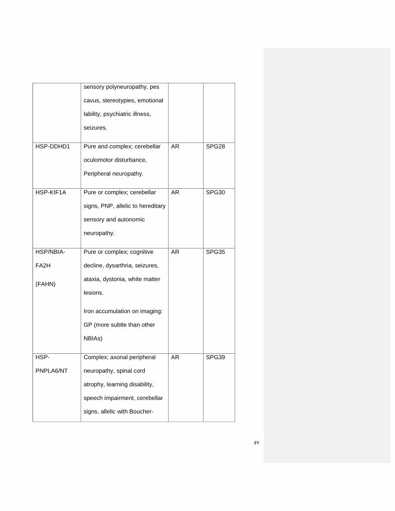
49
sensory polyneuropathy, pes
cavus, stereotypies, emotional
lability, psychiatric illness,
seizures.
HSP-DDHD1 Pure and complex; cerebellar
oculomotor disturbance,
Peripheral neuropathy.
AR SPG28
HSP-KIF1A Pure or complex; cerebellar
signs, PNP, allelic to hereditary
sensory and autonomic
neuropathy.
AR SPG30
HSP/NBIA-
FA2H
(FAHN)
Pure or complex; cognitive
decline, dysarthria, seizures,
ataxia, dystonia, white matter
lesions.
Iron accumulation on imaging:
GP (more subtle than other
NBIAs)
AR SPG35
HSP-
PNPLA6/NT
Complex; axonal peripheral
neuropathy, spinal cord
atrophy, learning disability,
speech impairment, cerebellar
signs, allelic with Boucher-
AR SPG39

50
Neuhäuser and Gordon
Holmes syndromes.
HSP/NBIA-
C19orf12
Mitochondrial membrane
protein-associated
neurodegeneration (MPAN)
Complex; Silver-syndrome.
Iron accumulation: GP –
hyperintense streaking of
medial medullary lamina
between GPi and GPe; SN.
AR SPG43
HSP-NT5C2 Complex; mental retardation,
ocular signs
AR SPG45
HSP-GBA2 Complex; mental impairment,
cataract, hypogonadism in
males, TCC and cerebellar
atrophy on brain imaging.36
AR SPG46
HSP-AP4B Complex; intellectual disability,
seizures, TCC, white matter
lesions.
AR SPG47
HSP-KIAA0415 Pure or complex; cervical cord
hyperintensities.
AR SPG48

51
HSP-TECPR2 Complex; severe intellectual
disability, fluctuating central
hypoventilation,
gastresophageal reflux
disease, awake apnea,
areflexia, dysmorphic features.
AR SPG49
HSP-APAM1 Complex; cerebral palsy,
intellectual disability, reduction
of cerebral white matter and
atrophy of the cerebellum.
AR SPG50
HSP-AP4E1 Complex; cerebral palsy,
intellectual disability and
microcephaly.
AR SPG51
HSP-DDHD2 Complex; mental retardation,
dysmorphism, short stature
and dysgenesis of the corpus
callosum.37
AR SPG54
HSP-C12orf65 Complex; optic atrophy,
peripheral neuropathy.
AR SPG55
HSP-KIF1C.
Allelic with
autosomal
recessive
Pure and complicated, chorea,
myoclonus, dysarthria,
developmental delay, mild
mental retardation, hypodontia,
AR SPG58

52
spastic ataxia at
the SAX2 locus.
ptosis, short stature,
sensorineural deafness, pes
planus, white matter lesions.
HSP-ERLIN1 Pure and complex; thoracic
kyphosis, borderline
intelligence.
AR SPG62
HSP-NT5C2 Complex; learning disability,
optic atrophy, squint,
glaucoma, congenital cataract,
TCC, white matter lesions,
cystic occipital leukomalacia.
AR SPG65
HSP-ALSIN Complex, generalized
dystonia, no speech
AR Alsin
HSP-SACSIN Spastic ataxia AR SACS
HSP- ALDH3A2 RM, ichtyosis, macular
dystrophy,
leukoencephalopathy
AR Sjögren-
Larsson
syndrome
HSP-BICD2 SMA like AR
X-linked recessive
HSP-L1CAM Complex; MASA-syndrome,
hydrocephalus, TCC.
XR SPG1
HSP-PLP1 Pure or complex; optic XR SPG2

53
Allelic with
Pelizaeus-
Merzbacher
disease.
atrophy, ataxia, nystagmus,
peripheral neuropathy,
aphasia, mental retardation.
HSP-SLC16A2 Complex; Allan-Herndon-
Dudley syndrome
XR SPG22
TCC=thinning of the corpus callosum, SACS=Spastic Ataxia of Charlevoix-
Saguenay, SMA=Spinal Muscular Atrophy
Silver syndrome: Complex HSP involving amyotrophy of the hand muscles
Kjellin syndrome: Complex HSP including thinning of the corpus callosum and central
retinal degeneration
* Note that some studies have suggested that some SPG7 mutations may have an
autosomal dominant effect, particularly autosomal dominant optic atrophy.
Primary Familial Brain Calcification
Primary familial brain calcification (PFBC) refers to genetically determined
calcification of various brain structures, notably but not exclusively the basal ganglia,
in the absence of a known metabolic, toxic, infectious or traumatic etiology. This
condition can be associated with various neurological symptoms, but frequently
movement disorders, including parkinsonism, dystonia, chorea, ataxia and tremor.
Other neurological and psychiatric symptoms and signs are also common. Locus
symbols for this condition use the acronym IBGC (Idiopathic Basal Ganglia
calcification) and IBGC1 through 4 have been assigned, but only one has a known

54
and independently confirmed gene, SLC20A2 (Supplementary Table 7).38, 39 PFBC
is a term that recognizes that the calcification can often extend well beyond the basal
ganglia to involve the dentate nucleus, cerebellar gyri, brain stem, centrum
semiovale, and subcortical white matter, and recognizes the genetic etiology of the
familial forms. Thus, we propose the prefix PFBC, consistent with the nomenclature
recently used by others.40 No distinguishing phenotypic features of PFBC-SLC20A2
compared with the SLC20A2 mutation negative patients with familial brain
calcification have yet been identified.38 Table 7 shows the proposed new list of
genetically determined primary familial brain calcification disorders. In this list we
have not included the disorder of progressive encephalopathy and spastic tetraplegia
due to mutations in the TREX1 gene despite the fact that it is associated with brain
calcification.41 This is because it does not primarily present with a movement
disorder.
Table 7: The proposed new list of primary familial brain calcification
New designation Clinical clues
Inheri-
tance
Locus
symbol
PFBC-SLC20A2 Various movement disorders,
cognitive dysfunction
AD IBGC3,
IBGC1
PFBC-PDGFRB Various movement disorders,
cognitive dysfunction
AD IBGC4
PFBC-PDGFB Various movement disorders,
cognitive dysfunction
AD IBGC5

55
Neurodegeneration with brain iron accumulation
Neurodegeneration with brain iron accumulation (NBIA) characterizes a number of
progressive neurological disorders with the hallmark of iron deposition on magnetic
resonance imaging (MRI) in several brain regions, most consistently the globus
pallidus.42 Most of these are genetically determined although some patients,
particularly with neurological symptoms beginning in mid-to-late adult life, have
sporadic disorders of uncertain origin. Movement disorders, particularly dystonia and
parkinsonism, dominate the clinical manifestations, but other common features
include spasticity, ataxia, cognitive and psychiatric disturbances, as well as
oculomotor abnormalities, optic nerve,retinal and peripheral nerve involvement.
To date NBIA as a locus symbol (i.e. numerical designation) has only been applied to
five disorders. However, up to nine distinct genetic disorders have been included
under the NBIA umbrella term. Other disorders in this group have either retained the
original disease name (e.g., aceruloplasminenemia, neuroferritinopathy) or, for more
recently described disorders, been labelled with an acronym combining the name of
the causative protein with “Associated Neurodegeneration” (i.e., PKAN for
pantothenate kinase associated neurodegeneration). In Supplementary Table 8 we
have provided a listing of all disorders that have been included under the umbrella
NBIA classification.43 Table 8 provides a new designation for each of these. Those
that present consistently with one or more specific movement disorder phenotypes
have been assigned a combined prefix (e.g. NBIA/DYT) and are included in the list
specific to that movement disorder.
.

56
Table 8: The proposed new list of Neurodegeneration with Brain Iron Accumulation
New
designa-
tion
Clinical clues Inheritanc
e pattern
Locus
symbol
NBIA/DYT
-PANK2
Pantothenate kinase associated
neurodegeneration (PKAN)
Iron accumulation: GP - Eye of the tiger,
Phenotype: Dystonia, spasticity,
parkinsonism, chorea, psychiatric cognitive
decline, gaze palsy, pigmentary retinopathy
AR NBIA1
NBIA/DYT
/PARK-
PLA2G6
PLA2G6-associated neurodegeneration
(PLAN)
Iron accumulation: GP, SN in some; adults
may have striatal involvement; ½ INAD and
majority of adult-onset lack imaging BIA on
MRI.
Infantile phenotype: Developmental delay,
hypotonia, ataxia, pyramidal signs, optic
atrophy, sensorimotor axonal neuropathy
seizures.
AR NBIA2,
PARK14

57
Adult phenotype: Dystonia-parkinsonism,
pyramidal signs, cognitive, psychiatric
features
NBIA-CP Aceruloplasminemia
Iron accumulation: More homogeneous
involvement of GP, caudate, putamen,
thalamus, red nucleus, dentate
Phenotype: Cognitive impairment, cranial
dyskinesia (incl blepharospasm), ataxia,
chorea, retinal degeneration, diabetes, liver
disease.
AR
NBIA-FTL Neuroferritinopathy
Iron accumulation: GP, caudate, putamen,
SN, red nucleus. Cystic BG changes –
pallidal necrosis.
Phenotype: Chorea, stereotypies, cranial
dyskinesia/dystonia, parkinsonism, ataxia,
cognitive and psychiatric dysfunction.
AD NBIA3
NBIA-
WDR45
Beta-propeller protein associated
neurodegeneration (BPAN – previously
SENDA*)
Iron accumulation: GP, SN (more than GP).
X-linked
dominant
NBIA5
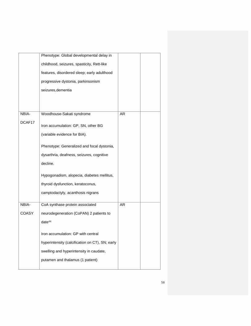
58
Phenotype: Global developmental delay in
childhood, seizures, spasticity, Rett-like
features, disordered sleep; early adulthood
progressive dystonia, parkinsonism
seizures,dementia
NBIA-
DCAF17
Woodhouse-Sakati syndrome
Iron accumulation: GP, SN, other BG
(variable evidence for BIA).
Phenotype: Generalized and focal dystonia,
dysarthria, deafness, seizures, cognitive
decline.
Hypogonadism, alopecia, diabetes mellitus,
thyroid dysfunction, keratoconus,
camptodactyly, acanthosis nigrans
AR
NBIA-
COASY
CoA synthase protein associated
neurodegeneration (CoPAN) 2 patients to
date44
Iron accumulation: GP with central
hyperintensity (calcification on CT), SN; early
swelling and hyperintensity in caudate,
putamen and thalamus (1 patient)
AR

59
Phenotype: Dystonia, spasticity, cognitive
impairment, bradykinesia, rigidity, motor
axonal neuropathy, obsessive compulsive
behavior, tics.
Members of other lists that have brain iron accumulation as a consistent
feature
HSP/NBIA-
FA2H
(FAHN)
Fatty acid hydroxylase associated
neurodegeneration (FAHN)
Iron accumulation: GP (more subtle than
other NBIAs)
Phenotype: Pure or complex; cognitive
decline, dysarthria, seizures, ataxia,
dystonia, white matter lesions.
AR SPG35
HSP/NBIA-
C19orf12
(MPAN)
Mitochondrial membrane protein
associated neurodegeneration (MPAN)
Iron accumulation: GP – hyperintense
streaking of medial medullary lamina
between GPi and GPe; SN.
Phenotype: Spasticity, dystonia,
parkinsonism, cognitive decline, psychiatric
AR NBIA4,
SPG43

60
abn, optic atrophy, motor axonopathy
Members of other lists that occasionally have brain iron accumulation on
imaging as a feature
PARK-
ATP13A2
Kufor Rakeb syndrome: Juvenile or early
onset parkinsonism, vertical gaze palsy,
minifacial-faucial myoclonus, pyramidal signs
AR PARK9
BIA = Brain iron accumulation, GP – globus pallidus, GPi = GP internal segment,
GPe = GP external segment, SN = substantia nigra, BG = basal ganglia, INAD =
Infantile neuroaxonal dystrophy; VSNGP = vertical supranuclear gaze palsy, Bx =
biopsy, ERG = electroretinogram
*SENDA: static encephalopathy of childhood with neurodegeneration in adulthood
Discussion
Challenges:
We present a new system of nomenclature for genetically determined movement
disorders that attempts to address many of the problems that have developed with
the previous system. In doing so we have tried to develop a system that is logical,
consistent, flexible to change and comprehensive. In our desire to be
comprehensive we have included a number of childhood onset metabolic disorders
that have heretofore been left out of the locus symbol naming system. Including

61
them will hopefully serve to increase awareness on the part of clinicians in adult
medicine of disorders that can affect adults that transition from pediatric care and
even occasionally present in young adulthood. We realize, however, several
challenges. First, it is impossible for us to be truly comprehensive by including all
genetically determined disorders that can, unusually, at some point in the course,
manifest predominantly as a movement disorder. However, we have tried to include
all of those disorders that have a movement disorder as a consistent and
predominant feature. At the same time, in circumstances where mutations in a
specific gene can cause more than one distinct phenotype and where one of those
phenotypes is a frequent cause of a particular movement disorder (e.g. SLC2A1
mutations causing infantile developmental delay and epilepsy OR paroxysmal
exertion-induced dystonia) we have chosen to retain it in the lists. This system is
bound to be associated with some ‘misclassification’ however, since for many
disorders there is insufficient knowledge on the frequency of movement disorders
compared with other phenotypes to render a decision on this basis. Second, we are
conscious of the fact that knowledge of the phenotypic spectrum of these disorders
will continuously evolve, and it will be necessary to change designations over time.
By avoiding a sequential nomenclature (e.g. numbers) we hope that this will be an
easier process than it has been in the past. The need to incorporate new knowledge
also implies that individuals will need to be dedicated to maintaining the lists
indefinitely – we anticipate that this task will fall to the MDS‘s Genetic Nomenclature
Task Force. Third, we are also conscious of the fact that many of the disorders
listed in these tables have well known names that will continue to be used, no matter
how logical our new system may be. It is not our intent, for example, to advocate that
Wilson disease hereafter be referred to as DYT-ATP7B. However, the new symbol
will serve to link the ATP7B gene to the phenotype of dystonia; cross-referencing to

62
other lists will acknowledge the combined movement disorders that are often a part
of this disorder and placing DYT-ATP7B on these lists will provide a more complete
genetic differential diagnosis to clinicians faced with a particular phenotype than has
been available in the past. Fourth, by attempting to avoid erroneous assignment of
causative mutations we have raised the challenge of establishing criteria that will
minimize false positive associations. One has to be very cautious about the nature
of variants found in a given gene, since except for recurrent mutations or repeat
expansions, the pathogenicity of a new missense variant is usually difficult to
establish. There are currently no error-proof criteria for establishing pathogenicity
and the system will have to be monitored for erroneous entries as new data becomes
available.
Next steps
Our work is not yet complete - it remains to compile lists for the recessively inherited
and congenital ataxias and for genetic causes of myoclonus. This will be taken on as
a next project of the task force, involving additional experts. In addition, it would be
useful to incorporate into the classification the underlying type of mutation to inform
genetic counseling issues such as instability during transmission (repeat expansion
disorders), heterozygous risk assessment (for example in mutations of SPG7 or
Parkin) and reduced penetrance (all dominant forms of inherited movements
disorders) or imprinting (for example in DYT-SGCE).
As mentioned above, a formal mechanism for incorporating new knowledge into the
naming system needs to be established and should include input from both clinicians

63
and geneticists. The next task of the Task Force will be to establish these
mechanisms.
In the process of developing these lists it became clear that there is no standard for
the field as to what we consider disease-causing vs risk-conferring. This is important
because one of the underlying principles of our naming system has been to restrict
the lists to genes that are disease-causing and not include genetic risk factors. Such
a standard would be helpful for communication and should be a task taken on by an
expert panel. Once accepted definitions are in place, there may be changes to our
lists.
Finally, this system can be easily applied to other neurological disorders and we
would encourage leaders in other medical fields to consider adopting a similar
system and avoid many of the problems that have beset the field of genetics in
movement disorders.
Acknowledgements:
CK is a recipient of a career development award from the Hermann and Lilly Schilling Foundation.
CM is a recipient of a New Investigator Award from the Canadian Institutes of Health Research.
CMS is a recipient of the National Health and Medical Research Council Clinical Practitioner Fellowship (#1008433)

64
References
1. Kramer PL, de Leon D, Ozelius L, et al. Dystonia gene in Ashkenazi Jewish
population is located on chromosome 9q32-34. Annals of Neurology 1990;27:114-
120.
2. Marras C, Lohmann K, Lang A, Klein C. Fixing the broken system of genetic
locus symbols: Parkinson disease and dystonia as examples. Neurology
2012;78:1016-1024.
3. MacArthur DG, Manolio TA, Dimmock DP, et al. Guidelines for investigating
causality of sequence variants in human disease. Nature 2014;508:469-476.
4. Bonifati V. Genetics of Parkinson's disease--state of the art, 2013.
Parkinsonism & Related Disorders 2014;20 Suppl 1:S23-28.
5. Trinh J, Farrer M. Advances in the genetics of Parkinson disease. Nat Rev
Neurol 2013;9:445-454.
6. Alcalay RN, Dinur T, Quinn T, et al. Comparison of Parkinson risk in
Ashkenazi Jewish patients with Gaucher disease and GBA heterozygotes. JAMA
Neurol 2014;71:752-757.
7. Opladen T, Hoffmann G, Horster F, et al. Clinical and biochemical
characterization of patients with early infantile onset of autosomal recessive GTP
cyclohydrolase I deficiency without hyperphenylalaninemia. Mov Disord 2011;26:157-
161.
8. Furukawa Y, Kish S. Tyrosine Hydroxylase Deficiency. In: Pagon RA, Adam
MP, Ardinger HH, et al., eds. Gene Reviews: University of Washingon, 2014.
9. Friedman J, Roze E, Abdenur JE, et al. Sepiapterin reductase deficiency: a
treatable mimic of cerebral palsy. Annals of Neurology 2012;71:520-530.

65
10. Opladen T, Hoffmann GF, Blau N. An international survey of patients with
tetrahydrobiopterin deficiencies presenting with hyperphenylalaninaemia. Journal of
Inherited Metabolic Disease 2012;35:963-973.
11. Sedel F, Saudubray JM, Roze E, Agid Y, Vidailhet M. Movement disorders
and inborn errors of metabolism in adults: a diagnostic approach. Journal of Inherited
Metabolic Disease 2008;31:308-318.
12. Tuschl K, Clayton PT, Gospe SM, Mills PB. Dystonia/Parkinsonism,
Hypermanganesemia, Polycythemia, and Chronic Liver Disease. In: Pagon RA,
Adam MP, Ardinger HH, et al., eds. GeneReviews. Seattle (WA): University of
Washington, 2012.
13. Klein C. Genetics in dystonia. Parkinsonism & Related Disorders 2014;20
Suppl 1:S137-142.
14. Albanese A, Bhatia K, Bressman SB, et al. Phenomenology and classification
of dystonia: a consensus update. Mov Disord 2013;28:863-873.
15. Mitchell GA FT. Inborn errors of ketone body metabolism. In: Scriver CR,
Beaudet AL, Sly WS, Valle D, eds. The metabolic and molecular bases of inherited
disease. New York: McGraw-Hill, 2001: 2327–2356.
16. Kolker S, Christensen E, Leonard JV, et al. Diagnosis and management of
glutaric aciduria type I--revised recommendations. Journal of Inherited Metabolic
Disease 2011;34:677-694.
17. Ng J, Zhen J, Meyer E, et al. Dopamine transporter deficiency syndrome:
phenotypic spectrum from infancy to adulthood. Brain 2014;137:1107-1119.
18. Tabarki B, Al-Hashem A, Alfadhel M. Biotin-Thiamine-Responsive Basal
Ganglia Disease. In: Pagon RA, Adam MP, Ardinger HH, et al., eds. GeneReviews.
Seattle (WA): University of Washington, 2013.

66
19. Ha AD, Parratt KL, Rendtorff ND, et al. The phenotypic spectrum of dystonia in
Mohr-Tranebjaerg syndrome. Mov Disord 2012;27:1034-1040.
20. Kim IS, Ki CS, Park KJ. Pediatric-onset dystonia associated with bilateral
striatal necrosis and G14459A mutation in a Korean family: a case report. J Korean
Med Sci 2010;25:180-184.
21. Gardiner AR, Bhatia KP, Stamelou M, et al. PRRT2 gene mutations: from
paroxysmal dyskinesia to episodic ataxia and hemiplegic migraine. Neurology
2012;79:2115-2121.
22. Bhatia KP. Paroxysmal dyskinesias. Mov Disord 2011;26:1157-1165.
23. Fernandez-Alvarez E, Perez-Duenas B. Paroxysmal movement disorders and
episodic ataxias. Handb 2013;112:847-852.
24. Jen JC. Hereditary episodic ataxias. Annals of the New York Academy of
Sciences 2008;1142:250-253.
25. Wang D, Pascual JM, De Vivo D. Glucose Transporter Type 1 Deficiency
Syndrome. In: Pagon RA, Adam MP, Ardinger HH, et al., eds. GeneReviews. Seattle
(WA): University of Washington, 2012.
26. Van Hove J, Coughlin C, Scharer G. Glycine Encephalopathy. In: Pagon RA,
Adam MP, Ardinger HH, et al., eds. GeneReviews. Seattle (WA): University of
Washington, 2013.
27. Patel KP, O'Brien TW, Subramony SH, Shuster J, Stacpoole PW. The
spectrum of pyruvate dehydrogenase complex deficiency: clinical, biochemical and
genetic features in 371 patients.[Republished from Mol Genet Metab. 2012
Jan;105(1):34-43; PMID: 22079328]. Mol Genet Metab 2012;106:385-394.
28. Seow HF, Broer S, Broer A, et al. Hartnup disorder is caused by mutations in
the gene encoding the neutral amino acid transporter SLC6A19. Nature Genetics
2004;36:1003-1007.

67
29. Verbeek DS, van de Warrenburg BP. The autosomal dominant cerebellar
ataxias. Seminars in Neurology 2011;31:461-469.
30. Van Gaalen J, Giunti P, van de Warrenburg BP. Movement disorders in
spinocerebellar ataxias. Mov Disord 2011;26:792-800.
31. Schneider SA, Walker RH, Bhatia KP. The Huntington's disease-like
syndromes: what to consider in patients with a negative Huntington's disease gene
test. Nat Clin Pract Neurol 2007;3:517-525.
32. Carre A, Szinnai G, Castanet M, et al. Five new TTF1/NKX2.1 mutations in
brain-lung-thyroid syndrome: rescue by PAX8 synergism in one case. Human
Molecular Genetics 2009;18:2266-2276.
33. Baeza AV, Dobson-Stone C, Rampoldi L, et al. Chorea-Acanthocytosis. In:
Pagon RA, Adam MP, Ardinger HH, et al., eds. GeneReviews. Seattle (WA):
University of Washington, 2014.
34. Fink JK. Hereditary spastic paraplegia: clinico-pathologic features and
emerging molecular mechanisms. Acta Neuropathol (Berl) 2013;126:307-328.
35. Salinas S, Proukakis C, Crosby A, Warner TT. Hereditary spastic paraplegia:
clinical features and pathogenetic mechanisms. Lancet Neurology 2008;7:1127-
1138.
36. Martin E, Schule R, Smets K, et al. Loss of function of glucocerebrosidase
GBA2 is responsible for motor neuron defects in hereditary spastic paraplegia.
American Journal of Human Genetics 2013;92:238-244.
37. Gonzalez M, Nampoothiri S, Kornblum C, et al. Mutations in phospholipase
DDHD2 cause autosomal recessive hereditary spastic paraplegia (SPG54). Eur J
Hum Genet 2013;21:1214-1218.
38. Hsu SC, Sears RL, Lemos RR, et al. Mutations in SLC20A2 are a major cause
of familial idiopathic basal ganglia calcification. Neurogenetics 2013;14:11-22.

68
39. Wang C, Li Y, Shi L, et al. Mutations in SLC20A2 link familial idiopathic basal
ganglia calcification with phosphate homeostasis. Nature Genetics 2012;44:254-256.
40. Sobrido MJ, Coppola G, Oliveira J, Hopfer S, Geschwind DH. Primary Familial
Brain Calcification. In: Pagon RA, Adam MP, Bird TD, Dolan CR, Fong CT, Stephens
K, eds. GeneReviews. Seattle Washington: University of Washington, Seattle, 2013.
41. Stephenson JB. Aicardi-Goutieres syndrome (AGS). Europ J Paediatr Neurol
2008;12:355-358.
42. Doorn JM, Kruer MC. Newly characterized forms of neurodegeneration with
brain iron accumulation. Current Neurology & Neuroscience Reports 2013;13:413.
43. Schneider SA, Hardy J, Bhatia KP. Syndromes of neurodegeneration with
brain iron accumulation (NBIA): an update on clinical presentations, histological and
genetic underpinnings, and treatment considerations. Mov Disord 2012;27:42-53.
44. Dusi S, Valletta L, Haack TB, et al. Exome sequence reveals mutations in CoA
synthase as a cause of neurodegeneration with brain iron accumulation. American
Journal of Human Genetics 2014;94:11-22.