genetic variations involved in vitamin e status
TRANSCRIPT

HAL Id: inserm-01761218https://www.hal.inserm.fr/inserm-01761218
Submitted on 8 Apr 2018
HAL is a multi-disciplinary open accessarchive for the deposit and dissemination of sci-entific research documents, whether they are pub-lished or not. The documents may come fromteaching and research institutions in France orabroad, or from public or private research centers.
L’archive ouverte pluridisciplinaire HAL, estdestinée au dépôt et à la diffusion de documentsscientifiques de niveau recherche, publiés ou non,émanant des établissements d’enseignement et derecherche français ou étrangers, des laboratoirespublics ou privés.
Distributed under a Creative Commons Attribution| 4.0 International License
Genetic Variations Involved in Vitamin E StatusPatrick Borel, Charles Desmarchelier
To cite this version:Patrick Borel, Charles Desmarchelier. Genetic Variations Involved in Vitamin E Status. InternationalJournal of Molecular Sciences, MDPI, 2016, 17 (12), pp.E2094. �10.3390/ijms17122094�. �inserm-01761218�

International Journal of
Molecular Sciences
Review
Genetic Variations Involved in Vitamin E StatusPatrick Borel *,† and Charles Desmarchelier †
NORT, Aix-Marseille Université, INRA, INSERM, 13005 Marseille, France; [email protected]* Correspondence: [email protected]; Tel.: +33-491-324-277† These authors contributed equally to this work.
Academic Editors: Maria Laura Colombo, Laura Di Renzo and Rafat A. SiddiquiReceived: 12 October 2016; Accepted: 9 December 2016; Published: 13 December 2016
Abstract: Vitamin E (VE) is the generic term for four tocopherols and four tocotrienols that exhibit thebiological activity of α-tocopherol. VE status, which is usually estimated by measuring fasting bloodVE concentration, is affected by numerous factors, such as dietary VE intake, VE absorption efficiency,and VE catabolism. Several of these factors are in turn modulated by genetic variations in genesencoding proteins involved in these factors. To identify these genetic variations, two strategies havebeen used: genome-wide association studies and candidate gene association studies. Each of thesestrategies has its advantages and its drawbacks, nevertheless they have allowed us to identify a list ofsingle nucleotide polymorphisms associated with fasting blood VE concentration and α-tocopherolbioavailability. However, much work remains to be done to identify, and to replicate in differentpopulations, all the single nucleotide polymorphisms involved, to assess the possible involvement ofother kind of genetic variations, e.g., copy number variants and epigenetic modifications, in order toestablish a reliable list of genetic variations that will allow us to predict the VE status of an individualby knowing their genotype in these genetic variations. Yet, the potential usefulness of this area ofresearch is exciting with regard to personalized nutrition and for future clinical trials dedicated toassessing the biological effects of the various isoforms of VE.
Keywords: single nucleotide polymorphisms; genetic polymorphism; bioavailability; absorption;tocopherol; nutrigenetics; genome-wide association studies; candidate gene association studies;antioxidant; oxidative stress
1. Vitamin E: The Major Lipid-Soluble Antioxidant in the Human Body
Vitamin E is the generic term for 8 natural isoforms that exhibit the biological activity ofα-tocopherol: four tocopherols (α, β, γ, and δ) and four tocotrienols (α, β, γ, and δ). In this review,when we use the abbreviation VE instead of vitamin E, we refer to all these isoforms. α-tocopherol isthe main vitamer found in the diet of Europeans, while γ-tocopherol is the main vitamer found in thediet of Americans, due to their higher consumption of soybean oil, which is a rich source of this vitamer.VE is present in numerous different foods but usually at low concentrations, except in vegetable oilsand nuts. Therefore, it is difficult to meet the recommended dietary allowances for VE without eatingthese VE-rich foods, which are also rich in fat. The average α-tocopherol intake of Americans is stillbelow the USA recommended dietary allowances (15 mg/day for people ≥14 years of age). In fact,about three-fourths of Americans (19–30 years of age) consume less than 10 mg/day [1]. In Europe,8% of men and 15% of women fail to meet 67% of the European recommended dietary allowances forα-tocopherol [2]. As a consequence, a recent systematic review of the literature reporting vitamin Eserum concentrations concluded that globally 13% of the subjects were below the functional deficiencythreshold concentration of 12 µmol/L, mostly for newborns and children, and that only 21% of thesubjects reached the threshold of 30 µmol/L, which is assumed to have beneficial effects on humanhealth [3].
Int. J. Mol. Sci. 2016, 17, 2094; doi:10.3390/ijms17122094 www.mdpi.com/journal/ijms

Int. J. Mol. Sci. 2016, 17, 2094 2 of 11
2. Vitamin E and Human Health
It is acknowledged that VE is the major lipid-soluble chain-breaking antioxidant found in thehuman body. Therefore, most studies dedicated to assess the biological effects of VE have focusedon its ability to quench free radicals. They have confirmed that several VE isomers play a key rolein the antioxidant defenses of our body. Nevertheless, some VE isomers have biological activitiesindependent of their antioxidant properties [4]. For example, it has been shown that some VE isoformscan modify gene expression [5,6], inhibit cell proliferation [7], and modulate platelet aggregation [8]and enzyme activity by binding to the enzyme cofactor binding site [9].
Since oxidative stress has been involved in the etiology of cardiovascular diseases and cancers,the association between VE intake or VE status and the incidence of these diseases, as well as thepotential benefit of VE supplementation on the incidence of these diseases, have been studied byseveral independent teams [10–12]. On the whole, although VE intake and VE status have beeninversely associated with the incidence of these diseases, most randomized controlled trials have failedto show a benefit of α-tocopherol supplementation on the incidence of these diseases [13,14]. Severalexplanations have been offered to account for this discrepancy, such as opposing functions of VEisoforms (high plasma γ-tocopherol concentrations oppose the benefit of α-tocopherol [15,16]. It hasalso been suggested that the high interindividual variability of α-tocopherol bioavailability may haveinterfered with the protective effects of α-tocopherol supplementation [17–19]. This variability is atleast partly due to genetic variations between individuals, and it is hence of paramount importance toidentify these in order to take into account this confounding variable in future studies on the effect ofVE intake or supplementation on various phenotypes.
3. Proteins Involved in Vitamin E Status
Fasting blood α-tocopherol concentration, measured in either plasma or serum, corrected ornot by blood lipids, is regarded as the acknowledged marker of VE status. Two complementaryapproaches have been used to identify genetic variations that can modulate this status. The firstone, in chronological order, is candidate gene association studies (CGAS’s). After an in silicosearch of candidate genes that are likely to affect α-tocopherol homeostasis because they are highlypolymorphic [20], it has enabled the identification of the first genetic variants associated with thevariability in α-tocopherol status [21,22]. Nevertheless, this approach has some drawbacks becausemany genetic variants with potential effect on VE status are left out of the analysis. Indeed, in CGAS’s,only a limited number of genetic variations, which are only present in the few genes selected bythe researcher, usually through their known or putative involvement in the studied phenotype, areinvestigated. Besides requiring a thorough knowledge of the proteins and hence the genes that canaffect the studied phenotype, selecting relevant genetic variants, i.e., genetic variants that significantlyaffect the activity or the expression of the candidate proteins, is also required. Although several toolsare available to predict the physiological consequences of genetic variants, it is not yet possible topredict them with 100% accuracy. Thanks to progress in affordable high throughput genotypingtechniques, genome-wide association studies (GWAS’s) have been increasing. One advantage ofGWAS’s is that they do not make any assumptions on the genes or genetic variations that can affect thestudied disease/phenotype—in this case, VE status. This allows researchers to identify associationsthat were not expected to be found. Nevertheless, GWAS’s also have their drawbacks. The mainone is that the statistical stringency used in this approach, which is compulsory in order to avoidfalse positive associations, usually leads to false negative associations, i.e., to reject genetic variantsthat are actually associated with the studied disease/phenotype. This can be a problem in polygenicphenotypes/traits, such as VE status, which is affected by numerous proteins. Another drawback ofGWAS’s is their cost. Since a very large number of genetic variations is investigated (typically >106),the sample size required to limit false positive associations is high (typically >10,000 subjects). This isnot necessarily an issue if the phenotype of interest is cheap to measure (e.g., one-point measurementsuch as fasting blood VE concentration), but this can become a limiting factor for more complex

Int. J. Mol. Sci. 2016, 17, 2094 3 of 11
phenotypes such as the evaluation of VE bioavailability, which requires a postprandial experiment in aclinical environment with several measurement points. Finally, the studied populations need to bewell characterized, obviously for the phenotype of interest but also for covariates that can influencethis phenotype. For example, in the case of GWAS’s on the variability of VE status, not knowing theVE intake of the studied group greatly reduces the number of SNPs associated with the phenotype ofinterest since a significant part of the variability cannot be accounted for.
VE status is affected by numerous factors, e.g., dietary VE intake, VE absorption efficiency, andVE catabolism, but VE bioavailability has been shown to be a key determinant thereof [23]. Moreover,the knowledge of an individual’s VE absorption capacity is far more relevant than the knowledge ofan individual’s fasting blood VE in order to provide personalized dietary recommendations. Since VEbioavailability is a phenotype that is relatively expensive to measure, the large sample size requiredto perform GWAS’s prevents the use of this approach to identify genetic variants associated with thevariability in VE bioavailability. On the other hand, CGAS’s might be a good alternative since thelimited number of genetic variants investigated does not require too large a sample size in order toobtain sufficient statistical power. It is thus important to provide a state of the art of the proteins thatare known, assumed, or reasonably hypothesized to modulate, directly or indirectly, the bioavailabilityof VE. Indeed, future CGAS’s should focus on genetic variants in or near the genes that encode theseproteins to increase their chance of finding significant associations. The last part of this chapter istherefore dedicated to listing these proteins by describing the fate of VE from the food matrix in whichit has been ingested to its site of catabolism.
It is not the aim of this review to describe in detail the metabolism of VE in the human digestivetract, and more details on this topic can be found in recent reviews [24,25]. Gut VE metabolism mightfirst involve gastric lipase, but there is no study dedicated to the assessment of whether this lipase isable to hydrolyze VE esters. Several studies have shown that α-tocopheryl esters are hydrolyzed tofree α-tocopherol in the duodenum by carboxyl ester hydrolase [26]. The metabolism of VE in the gutlumen can also involve other digestive enzymes, such as pancreatic lipase, trypsin, and α-amylase,which can facilitate the release of VE from the food matrix and thus facilitate its micellization and thusits absorption. Nevertheless, there is no study dedicated to assessing whether they have a significantrole in VE absorption.
After its release from the food matrix (usually dietary fat or a vegetable matrix), VE is incorporatedinto mixed micelles in the aqueous environment of the intestinal lumen. These lipid vehicles, whichare mostly made of bile salts, phospholipids, cholesterol, and the products of triglyceride lipolysis,carry VE to the apical membrane of the enterocyte where is it absorbed. Although it was thoughtthat VE uptake takes place by passive diffusion, several studies in cell cultures and in transgenic micehave demonstrated that proteins are implicated in this process [27]. To date, three proteins have beenshown to be involved, directly or indirectly, in α-tocopherol uptake at the apical side of the enterocyte:scavenger receptor class B member 1 (SR-BI) [28], NPC1 like intracellular cholesterol transporter 1(NPC1L1) [29,30], and CD36 molecule (CD36) [31]. After its uptake, VE has to reach the basolateralside of the enterocyte to be secreted in either chylomicrons or in intestinal HDL [32]. Surprisingly,there is no data yet on protein(s) involved in its intracellular transport. Yet, α-tocopherol is able tobind to bovine serum albumin [33]. Candidates could be Niemann–Pick type C1/C2 (NPC1/2) andsec14p-like proteins (encoded by transporter 1, ATP binding cassette subfamily B member (TAP1,2, and 3)), which transport α-tocopherol in other cells. It is assumed that most, if not all, VE isincorporated into chylomicrons, but it seems that a fraction of α-tocopherol is secreted in intestinalHDL via a basolateral membrane protein, ATP binding cassette subfamily A member 1 (ABCA1) [34].The incorporation of VE within chylomicrons likely depends on several proteins, including microsomaltriglyceride transfer protein (MTP) [32], which is required for the incorporation of triglycerides withinthe nascent chylomicrons. Chylomicron VE and intestinal-HDL VE are then transported to the liverand to other tissues. Apolipoproteins that belong to these lipoparticles, e.g., apoB48 and apoAI, as wellas proteins that are involved in the blood metabolism of these lipoparticles, e.g., lipoprotein lipase,
Int. J. Mol. Sci. 2016, 17, 2094 4 of 11
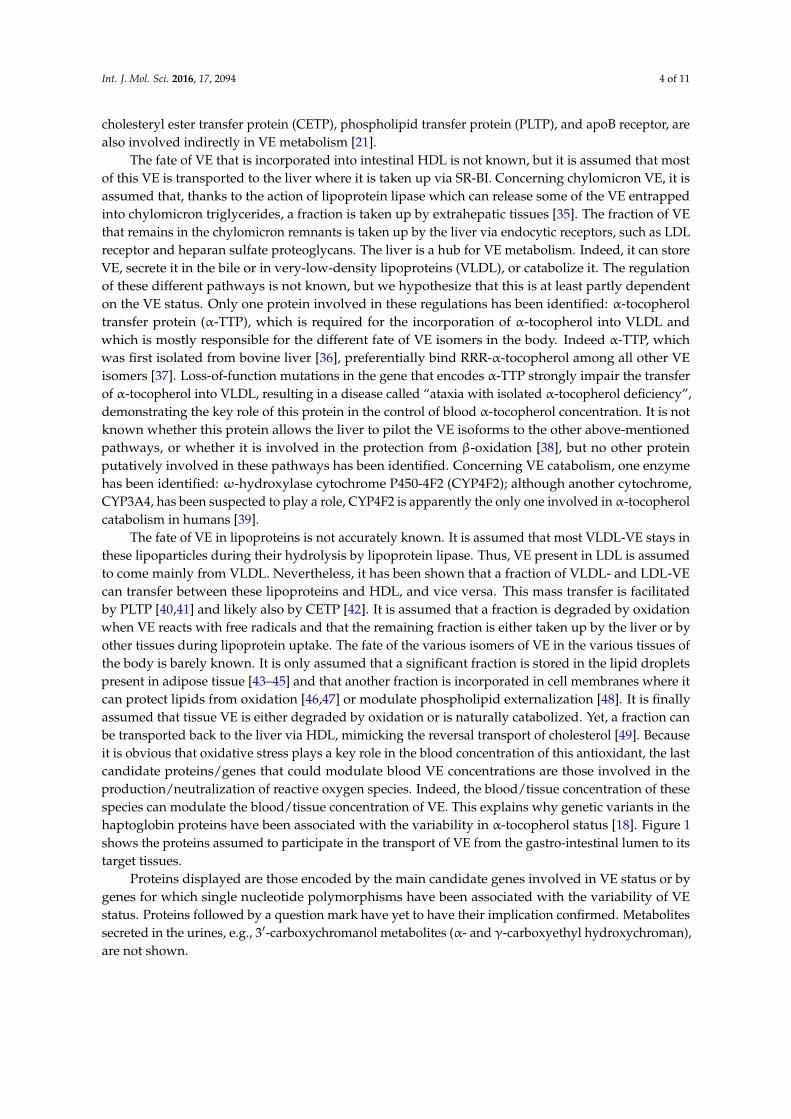
cholesteryl ester transfer protein (CETP), phospholipid transfer protein (PLTP), and apoB receptor, arealso involved indirectly in VE metabolism [21].
The fate of VE that is incorporated into intestinal HDL is not known, but it is assumed that mostof this VE is transported to the liver where it is taken up via SR-BI. Concerning chylomicron VE, it isassumed that, thanks to the action of lipoprotein lipase which can release some of the VE entrappedinto chylomicron triglycerides, a fraction is taken up by extrahepatic tissues [35]. The fraction of VEthat remains in the chylomicron remnants is taken up by the liver via endocytic receptors, such as LDLreceptor and heparan sulfate proteoglycans. The liver is a hub for VE metabolism. Indeed, it can storeVE, secrete it in the bile or in very-low-density lipoproteins (VLDL), or catabolize it. The regulationof these different pathways is not known, but we hypothesize that this is at least partly dependenton the VE status. Only one protein involved in these regulations has been identified: α-tocopheroltransfer protein (α-TTP), which is required for the incorporation of α-tocopherol into VLDL andwhich is mostly responsible for the different fate of VE isomers in the body. Indeed α-TTP, whichwas first isolated from bovine liver [36], preferentially bind RRR-α-tocopherol among all other VEisomers [37]. Loss-of-function mutations in the gene that encodes α-TTP strongly impair the transferof α-tocopherol into VLDL, resulting in a disease called “ataxia with isolated α-tocopherol deficiency”,demonstrating the key role of this protein in the control of blood α-tocopherol concentration. It is notknown whether this protein allows the liver to pilot the VE isoforms to the other above-mentionedpathways, or whether it is involved in the protection from β-oxidation [38], but no other proteinputatively involved in these pathways has been identified. Concerning VE catabolism, one enzymehas been identified: ω-hydroxylase cytochrome P450-4F2 (CYP4F2); although another cytochrome,CYP3A4, has been suspected to play a role, CYP4F2 is apparently the only one involved in α-tocopherolcatabolism in humans [39].
The fate of VE in lipoproteins is not accurately known. It is assumed that most VLDL-VE stays inthese lipoparticles during their hydrolysis by lipoprotein lipase. Thus, VE present in LDL is assumedto come mainly from VLDL. Nevertheless, it has been shown that a fraction of VLDL- and LDL-VEcan transfer between these lipoproteins and HDL, and vice versa. This mass transfer is facilitatedby PLTP [40,41] and likely also by CETP [42]. It is assumed that a fraction is degraded by oxidationwhen VE reacts with free radicals and that the remaining fraction is either taken up by the liver or byother tissues during lipoprotein uptake. The fate of the various isomers of VE in the various tissues ofthe body is barely known. It is only assumed that a significant fraction is stored in the lipid dropletspresent in adipose tissue [43–45] and that another fraction is incorporated in cell membranes where itcan protect lipids from oxidation [46,47] or modulate phospholipid externalization [48]. It is finallyassumed that tissue VE is either degraded by oxidation or is naturally catabolized. Yet, a fraction canbe transported back to the liver via HDL, mimicking the reversal transport of cholesterol [49]. Becauseit is obvious that oxidative stress plays a key role in the blood concentration of this antioxidant, the lastcandidate proteins/genes that could modulate blood VE concentrations are those involved in theproduction/neutralization of reactive oxygen species. Indeed, the blood/tissue concentration of thesespecies can modulate the blood/tissue concentration of VE. This explains why genetic variants in thehaptoglobin proteins have been associated with the variability in α-tocopherol status [18]. Figure 1shows the proteins assumed to participate in the transport of VE from the gastro-intestinal lumen to itstarget tissues.
Proteins displayed are those encoded by the main candidate genes involved in VE status or bygenes for which single nucleotide polymorphisms have been associated with the variability of VEstatus. Proteins followed by a question mark have yet to have their implication confirmed. Metabolitessecreted in the urines, e.g., 3′-carboxychromanol metabolites (α- and γ-carboxyethyl hydroxychroman),are not shown.

Int. J. Mol. Sci. 2016, 17, 2094 5 of 11Int. J. Mol. Sci. 2016, 17, 2094 5 of 11
Figure 1. Summary of the proteins involved in the variability of vitamin E (VE) status. LIPF (gastric lipase), CEH (carboxyl ester hydrolase), ASBT (human bile acid transporter), LPL (lipoprotein lipase), APOBR (apolipoprotein B receptor), ABCB1 (ATP binding cassette subfamily B member 1).
4. Genetic Variations Associated with the Variability in Vitamin E Status
As stated previously, both GWAS’s and CGAS’s have been applied to identify SNPs associated with fasting blood VE concentrations. Three GWAS’s have shown that a SNP in CYP4F2, a SNP in SCARB1, and a SNP near APOA1/C3/A4/A5 are associated with α-tocopherol status [50–52]. As mentioned above, CYP4F2 encodes for cytochrome P450 4F2, which catabolizes VE. SCARB1 encodes for SR-BI, which is a plasma membrane receptor for HDL and which has been involved in α-tocopherol uptake by several tissues [28,53]. It is therefore not surprising that genetic variants in these genes can modify blood VE concentration. Concerning the association of a SNP within the cluster APOA1/C3/A4/A5, which is in linkage disequilibrium with ZNF259 and BUD13 that have no known role in α-tocopherol metabolism, it is likely due to a genetic variant in APOA5, as suggested by Ferrucci et al. [52]. CGAS’s have confirmed the associations found in the GWAS’s [21,22,54–56]. Furthermore, they have showed that genetic variants in other genes, i.e., CD36 [57], CETP [21] and APOE [22], are also likely involved in VE status. Although, as explained above, the fact that these associations were not observed in GWAS’s suggests that their effect on VE status is moderate.
The following table (Table 1) summarizes all the genetic variants that have been associated with the variability in vitamin E status.
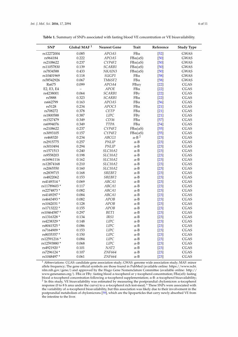
Table 1. Summary of SNPs associated with fasting blood VE concentration or VE bioavailability.
SNP Global MAF 1 Nearest Gene Trait Reference Study Type rs12272004 0.085 APOA5 FBα [52] GWAS rs964184 0.222 APOA5 FBα (αS) [50] GWAS
rs2108622 0.237 CYP4F2 FBα (αS) [50] GWAS rs11057830 0.139 SCARB1 FBα (αS) [50] GWAS rs7834588 0.433 NKAIN3 FBα (αS) [50] GWAS
Figure 1. Summary of the proteins involved in the variability of vitamin E (VE) status. LIPF (gastriclipase), CEH (carboxyl ester hydrolase), ASBT (human bile acid transporter), LPL (lipoprotein lipase),APOBR (apolipoprotein B receptor), ABCB1 (ATP binding cassette subfamily B member 1).
4. Genetic Variations Associated with the Variability in Vitamin E Status
As stated previously, both GWAS’s and CGAS’s have been applied to identify SNPs associatedwith fasting blood VE concentrations. Three GWAS’s have shown that a SNP in CYP4F2, a SNPin SCARB1, and a SNP near APOA1/C3/A4/A5 are associated with α-tocopherol status [50–52].As mentioned above, CYP4F2 encodes for cytochrome P450 4F2, which catabolizes VE. SCARB1encodes for SR-BI, which is a plasma membrane receptor for HDL and which has been involved inα-tocopherol uptake by several tissues [28,53]. It is therefore not surprising that genetic variants inthese genes can modify blood VE concentration. Concerning the association of a SNP within thecluster APOA1/C3/A4/A5, which is in linkage disequilibrium with ZNF259 and BUD13 that have noknown role in α-tocopherol metabolism, it is likely due to a genetic variant in APOA5, as suggestedby Ferrucci et al. [52]. CGAS’s have confirmed the associations found in the GWAS’s [21,22,54–56].Furthermore, they have showed that genetic variants in other genes, i.e., CD36 [57], CETP [21] andAPOE [22], are also likely involved in VE status. Although, as explained above, the fact that theseassociations were not observed in GWAS’s suggests that their effect on VE status is moderate.
The following table (Table 1) summarizes all the genetic variants that have been associated withthe variability in vitamin E status.
Int. J. Mol. Sci. 2016, 17, 2094 6 of 11
Table 1. Summary of SNPs associated with fasting blood VE concentration or VE bioavailability.
SNP Global MAF 1 Nearest Gene Trait Reference Study Type
rs12272004 0.085 APOA5 FBα [52] GWASrs964184 0.222 APOA5 FBα(αS) [50] GWASrs2108622 0.237 CYP4F2 FBα(αS) [50] GWAS
rs11057830 0.139 SCARB1 FBα(αS) [50] GWASrs7834588 0.433 NKAIN3 FBα(αS) [50] GWAS
rs10401969 0.118 SUGP1 FBα [58] GWASrs58542926 0.067 TM6SF2 FBα [58] GWAS
Rs675 0.099 APOA4 FBαγ [22] CGASE2, E3, E4 – APOE FBα [22] CGASrs4238001 0.064 SCARB1 FBγ [22] CGAS
rs5888 0.323 SCARB1 FBα [22] CGASrs662799 0.163 APOA5 FBα [56] CGAS
rs5128 0.234 APOC3 FBα [21] CGASrs708272 0.378 CETP FBα [21] CGASrs1800588 0.387 LIPC FBγ [21] CGASrs1527479 0.349 CD36 FBα [57] CGASrs6994076 0.349 TTPA FBα [54] CGASrs2108622 0.237 CYP4F2 FBα(αS) [55] CGASrs3093105 0.157 CYP4F2 FBα(αS) [55] CGASrs468320 0.234 ABCG1 α-B 2 [23] CGASrs2915775 0.257 PNLIP α-B [23] CGASrs3010494 0.294 PNLIP α-B [23] CGASrs1571513 0.240 SLC10A2 α-B [23] CGASrs9558203 0.198 SLC10A2 α-B [23] CGAS
rs16961116 0.162 SLC10A2 α-B [23] CGASrs12874168 0.210 SLC10A2 α-B [23] CGASrs2065550 0.160 SLC10A2 α-B [23] CGASrs2839715 0.168 SREBF2 α-B [23] CGASrs4822062 0.153 SREBF2 α-B [23] CGAS
rs4149314 * 0.069 ABCA1 α-B [23] CGASrs11789603 * 0.117 ABCA1 α-B [23] CGASrs2274873 * 0.082 ABCA1 α-B [23] CGASrs4149297 * 0.084 ABCA1 α-B [23] CGASrs4643493 * 0.082 APOB α-B [23] CGASrs1042031 * 0.128 APOB α-B [23] CGASrs1713222 * 0.155 APOB α-B [23] CGAS
rs10464587 * 0.297 BET1 α-B [23] CGASrs1316328 * 0.134 IRS1 α-B [23] CGASrs4238329 * 0.148 LIPC α-B [23] CGASrs8041525 * 0.086 LIPC α-B [23] CGASrs7164909 * 0.153 LIPC α-B [23] CGASrs8035357 * 0.150 LIPC α-B [23] CGAS
rs12591216 * 0.084 LIPC α-B [23] CGASrs12593880 * 0.068 LIPC α-B [23] CGASrs4921920 * 0.101 NAT2 α-B [23] CGASrs7296124 * 0.107 ZNF664 α-B [23] CGASrs1048497 * 0.061 ZNF664 α-B [23] CGAS1 Abbreviations: CGAS: candidate gene association study; GWAS: genome wide association study; MAF: minorallele frequency; The gene official symbols are those found in PubMed (available online: https://www.ncbi.nlm.nih.gov/gene/) and approved by the Hugo Gene Nomenclature Committee (available online: http://www.genenames.org/). FBα or FBγ: fasting blood α-tocopherol or γ-tocopherol concentration; FBα(αS): fastingblood α-tocopherol concentration following α-tocopherol supplementation; α-B: α-tocopherol bioavailability;2 In this study, VE bioavailability was estimated by measuring the postprandial chylomicron α-tocopherolresponse (0 to 8 h area under the curve) to a α-tocopherol rich test-meal; * These SNPs were associated withthe variability of α-tocopherol bioavailability, but this association was likely due to their involvement in thepostprandial metabolism of chylomicrons [59], which are the lipoparticles that carry newly absorbed VE fromthe intestine to the liver.
Int. J. Mol. Sci. 2016, 17, 2094 7 of 11
5. Genetic Variations Associated with the Variability in Vitamin E Bioavailability
Fasting blood α-tocopherol concentration, which is usually used to evaluate VE status, is affectedby numerous factors, such as dietary intake of VE, dietary intake of pro-oxidant compounds, andα-tocopherol metabolism within the body. Although the relative role of each of these factors isunknown, it is assumed that the effect of the absorption efficiency of VE is very important withregard to long-term VE status. This assumption is supported by the fact that a significant relationshipbetween α-tocopherol status and α-tocopherol bioavailability has been observed [23]. More precisely,in a recent clinical trial from our group where an α-tocopherol-rich meal, which provided VE as67 mg (100 IU) D-α-tocopheryl acetate, was given to a group of healthy subjects, the coefficient ofvariation of the α-tocopherol response to the test meal, i.e., the postprandial chylomicron α-tocopherolresponse, an acknowledged estimate of α-tocopherol bioavailability, was 81% [23]. In this study,the interindividual variability in α-tocopherol bioavailability was associated with a combination of28 SNPs in or near 11 candidate genes. Seven of these genes were involved in the postprandialchylomicron triacylglycerol response in the same group of subjects [59], which was not surprising,as most newly absorbed VE is carried from the intestine to the liver via chylomicrons. Four ofthese genes were specifically associated with α-tocopherol response, suggesting that they play aspecific role in α-tocopherol bioavailability. These genes were SLC10A2 (solute carrier family 10(sodium/bile acid cotransporter), member 2), PNLIP (pancreatic lipase), SREBF2 (sterol regulatoryelement binding transcription factor 2), and ABCG1 (ATP-binding cassette, sub-family G (WHITE),member 1). We hypothesize that genetic variations in SLC10A2 are associated with α-tocopherolbioavailability because either the protein encoded by this gene is involved in α-tocopherol transportacross membrane of the enterocyte, or because this protein is involved in bile salt absorption, whichhas an indirect effect on the absorption of VE incorporated in bile-salt containing micelles. Geneticvariations in PNLIP likely have an indirect effect on VE micellization because dietary VE is usuallyembedded in dietary fat and triglyceride hydrolysis by pancreatic lipase is assumed to facilitate therelease and the transfer of VE from the oil droplets of dietary lipid emulsions to mixed micelles. SREBF2encodes for a transcription factor that controls the expression of NPC1L1, among other genes. SinceNPC1L1 is involved in α-tocopherol uptake by the intestinal cell [29,30], it is likely that genetic variantsin SREBF2 can indirectly affect α-tocopherol absorption. ABCG1 encodes for a membrane transporterof various molecules across cellular membranes. Thus, the association observed between geneticvariants in this gene and α-tocopherol bioavailability suggests that this protein is able to transportα-tocopherol as well. This was confirmed recently by a study in ABCG1-deficient mice [60]. Althoughthere has only been one study dedicated to identifying the genetic variants involved in α-tocopherolbioavailability, and although we acknowledge that the results obtained should be confirmed in othergroups of subjects, we confidently conclude that it is likely that several SNPs in different genes havean effect of VE bioavailability. Moreover, although the effect of each SNP can be relatively low (e.g.,1%–2% of the variability in α-tocopherol bioavailability explained), their additive effect can have asignificant effect on bioavailability.
6. Other Genetic Variations Potentially Involved in Vitamin E Status
As stated in this review on the genetic variations involved in VE status, much work remainsto be done. Indeed, GWAS’s only allow us to identify SNPs associated with the VE status in largepopulations where covariates (e.g., VE intake and smoking status) are well measured while CGAS’scan miss out SNPs in genes not thought to affect this status. It can also be concluded that, since VEstatus is modulated by numerous genes, it is necessary to simultaneously know the effect of severalgenotypes in order to try to predict VE status with genotyping. It should also be reminded thatSNPs are not the only genetic variations that occur in DNA. Indeed, there are also copy numbervariants, insertion/deletion of certain base pairs, and epigenetic modifications, e.g., DNA methylation.A genetic score that would aim to predict VE status should therefore take into account all the geneticvariations that can have a significant impact on VE status. Furthermore, association studies have
Int. J. Mol. Sci. 2016, 17, 2094 8 of 11
to be performed in different populations to be sure that the associations are not specific to certainethnic groups.
In summary, there is now enough evidence to state that VE status is partly modulated by SNPs inseveral genes. Although much work remains to be done to obtain a combination of genetic variations(SNPs but also other kinds of genetic variations) that will allow us to confidently predict the VE statusof an individual by knowing his genotype at these variations, the potential usefulness of this area ofresearch is exciting with regard to personalized nutrition and for future clinical trials dedicated toassess the biological effects of VE. Nevertheless, it should be reminded that genetics only representsone of the factors that affect VE status, albeit stable over the lifespan, since other factors, such as VEdietary intake, dietary habits (e.g., consumption of other micronutrients) [61], oxidative stress (throughe.g., smoking), and age [62] also affect this status. Thus, a prediction of VE status should take intoaccount these variables as well.
Acknowledgments: This work was supported by the salaries paid to the authors by their employer (INRA: FrenchNational Institute for Agricultural Research). The authors did not receive funds to cover the costs of publishing inopen access.
Conflicts of Interest: The authors declare no conflict of interest.
References
1. McBurney, M.I. Majority of Americans not consuming vitamin E RDA. J. Nutr. 2011, 141, 1920. [CrossRef][PubMed]
2. Polito, A.; Intorre, F.; Andriollo-Sanchez, M.; Azzini, E.; Raguzzini, A.; Meunier, N.; Ducros, V.;O’Connor, J.M.; Coudray, C.; Roussel, A.M.; et al. Estimation of intake and status of vitamin A, vitamin Eand folate in older European adults: The ZENITH. Eur. J. Clin. Nutr. 2005, 59, S42–S47. [CrossRef] [PubMed]
3. Peter, S.; Friedel, A.; Roos, F.F.; Wyss, A.; Eggersdorfer, M.; Hoffmann, K.; Weber, P. A systematic reviewof global α-tocopherol status as assessed by nutritional intake levels and blood serum concentrations.Int. J. Vitam. Nutr. Res. 2016. [CrossRef] [PubMed]
4. Zingg, J.M.; Azzi, A. Non-antioxidant activities of vitamin E. Curr. Med. Chem. 2004, 11, 1113–1133.[CrossRef] [PubMed]
5. Landrier, J.F.; Gouranton, E.; Reboul, E.; Cardinault, N.; El Yazidi, C.; Malezet-Desmoulins, C.; Andre, M.;Nowicki, M.; Souidi, M.; Borel, P. Vitamin E decreases endogenous cholesterol synthesis and apo-AI-mediatedcholesterol secretion in Caco-2 cells. J. Nutr. Biochem. 2010, 21, 1207–1213. [CrossRef] [PubMed]
6. Zingg, J.M. Vitamin E: A role in signal transduction. Annu. Rev. Nutr. 2015, 35, 135–173. [CrossRef] [PubMed]7. Jiang, Q.; Wong, J.; Fyrst, H.; Saba, J.D.; Ames, B.N. γ-tocopherol or combinations of vitamin E forms induce
cell death in human prostate cancer cells by interrupting sphingolipid synthesis. Proc. Natl. Acad. Sci. USA2004, 101, 17825–17830. [CrossRef] [PubMed]
8. Mabile, L.; Bruckdorfer, K.R.; RiceEvans, C. Moderate supplementation with natural α-tocopherol decreasesplatelet aggregation and low-density lipoprotein oxidation. Atherosclerosis 1999, 147, 177–185. [CrossRef]
9. McCary, C.A.; Yoon, Y.; Panagabko, C.; Cho, W.; Atkinson, J.; Cook-Mills, J.M. Vitamin E isoforms directlybind PKCα and differentially regulate activation of PKCα. Biochem. J. 2012, 441, 189–198. [CrossRef][PubMed]
10. Pruthi, S.; Allison, T.G.; Hensrud, D.D. Vitamin E supplementation in the prevention of coronary heartdisease. Mayo Clin. Proc. 2001, 76, 1131–1136. [CrossRef] [PubMed]
11. Negis, Y.; Zingg, J.M.; Libinaki, R.; Meydani, M.; Azzi, A. Vitamin E and cancer. Nutr. Cancer 2009, 61,875–878. [CrossRef] [PubMed]
12. Papas, A.; Vos, E. Vitamin E, cancer, and apoptosis. Am. J. Clin. Nutr. 2001, 73, 1113. [PubMed]13. Traber, M.G. Does vitamin E decrease heart attack risk? Summary and implications with respect to dietary
recommendations. J. Nutr. 2001, 131, 395S–397S. [PubMed]14. Lee, I.M.; Cook, N.R.; Gaziano, J.M.; Gordon, D.; Ridker, P.M.; Manson, J.E.; Hennekens, C.H.; Buring, J.E.
Vitamin E in the primary prevention of cardiovascular disease and cancer: The women’s health study:A randomized controlled trial. JAMA 2005, 294, 56–65. [CrossRef] [PubMed]
Int. J. Mol. Sci. 2016, 17, 2094 9 of 11
15. Cook-Mills, J.; Gebretsadik, T.; Abdala-Valencia, H.; Green, J.; Larkin, E.K.; Dupont, W.D.; Shu, X.O.;Gross, M.; Bai, C.; Gao, Y.T.; et al. Interaction of vitamin E isoforms on asthma and allergic airway disease.Thorax 2016, 71, 954–956. [CrossRef] [PubMed]
16. Cook-Mills, J.M.; Avila, P.C. Vitamin E and D regulation of allergic asthma immunopathogenesis.Int. Immunopharmacol. 2014, 23, 364–372. [CrossRef] [PubMed]
17. Zingg, J.M.; Azzi, A.; Meydani, M. Genetic polymorphisms as determinants for disease-preventive effects ofvitamin E. Nutr. Rev. 2008, 66, 406–414. [CrossRef] [PubMed]
18. Blum, S.; Vardi, M.; Brown, J.B.; Russell, A.; Milman, U.; Shapira, C.; Levy, N.S.; Miller-Lotan, R.; Asleh, R.;Levy, A.P. Vitamin E reduces cardiovascular disease in individuals with diabetes mellitus and the haptoglobin2-2 genotype. Pharmacogenomics 2010, 11, 675–684. [CrossRef] [PubMed]
19. Milman, U.; Blum, S.; Shapira, C.; Aronson, D.; Miller-Lotan, R.; Anbinder, Y.; Alshiek, J.; Bennett, L.;Kostenko, M.; Landau, M.; et al. Vitamin E supplementation reduces cardiovascular events in a subgroup ofmiddle-aged individuals with both type 2 diabetes mellitus and the haptoglobin 2-2 genotype: A prospectivedouble-blinded clinical trial. Arterioscler. Thromb. Vasc. Biol. 2008, 28, 341–347. [CrossRef] [PubMed]
20. Doring, F.; Rimbach, G.; Lodge, J.K. In silico search for single nucleotide polymorphisms in genes importantin vitamin E homeostasis. IUBMB Life 2004, 56, 615–620. [CrossRef] [PubMed]
21. Borel, P.; Moussa, M.; Reboul, E.; Lyan, B.; Defoort, C.; Vincent-Baudry, S.; Maillot, M.; Gastaldi, M.;Darmon, M.; Portugal, H.; et al. Human fasting plasma concentrations of vitamin E and carotenoids, andtheir association with genetic variants in apo C-III, cholesteryl ester transfer protein, hepatic lipase, intestinalfatty acid binding protein and microsomal triacylglycerol transfer protein. Br. J. Nutr. 2009, 101, 680–687.[PubMed]
22. Borel, P.; Moussa, M.; Reboul, E.; Lyan, B.; Defoort, C.; Vincent-Baudry, S.; Maillot, M.; Gastaldi, M.;Darmon, M.; Portugal, H.; et al. Human plasma levels of vitamin E and carotenoids are associated withgenetic polymorphisms in genes involved in lipid metabolism. J. Nutr. 2007, 137, 2653–2659. [PubMed]
23. Borel, P.; Desmarchelier, C.; Nowicki, M.; Bott, R.; Tourniaire, F. Can genetic variability in α-tocopherolbioavailability explain the heterogeneous response to α-tocopherol supplements? Antioxid. Redox Signal.2015, 22, 669–678. [CrossRef] [PubMed]
24. Borel, P.; Preveraud, D.; Desmarchelier, C. Bioavailability of vitamin E in humans: An update. Nutr. Rev.2013, 71, 319–331. [CrossRef] [PubMed]
25. Schmolz, L.; Birringer, M.; Lorkowski, S.; Wallert, M. Complexity of vitamin E metabolism. World J. Biol.Chem. 2016, 7, 14–43. [CrossRef] [PubMed]
26. Desmarchelier, C.; Tourniaire, F.; Prévéraud, D.; Samson-Kremser, C.; Crenon, I.; Rosilio, V.; Borel, P. Thedistribution and relative hydrolysis of tocopheryl acetate in the different matrices co-existing in the lumenof the small intestine during digestion could explain its low bioavaialbility. Mol. Nutr. Food Res. 2013.[CrossRef] [PubMed]
27. Reboul, E.; Borel, P. Proteins involved in uptake, intracellular transport and basolateral secretion of fat-solublevitamins and carotenoids by mammalian enterocytes. Prog. Lipid Res. 2011, 50, 388–402. [CrossRef] [PubMed]
28. Reboul, E.; Klein, A.; Bietrix, F.; Gleize, B.; Malezet-Desmoulins, C.; Schneider, M.; Margotat, A.; Lagrost, L.;Collet, X.; Borel, P. Scavenger receptor class B type I (SR-BI) is involved in vitamin E transport across theenterocyte. J. Biol. Chem. 2006, 281, 4739–4745. [CrossRef] [PubMed]
29. Reboul, E.; Soayfane, Z.; Goncalves, A.; Cantiello, M.; Bott, R.; Nauze, M.; Terce, F.; Collet, X.; Comera, C.Respective contributions of intestinal Niemann-Pick C1-like 1 and scavenger receptor class B type I tocholesterol and tocopherol uptake: In vivo v. In vitro studies. Br. J. Nutr. 2012, 107, 1296–1304. [CrossRef][PubMed]
30. Narushima, K.; Takada, T.; Yamanashi, Y.; Suzuki, H. Niemann-Pick C1-like 1 mediates α-tocopheroltransport. Mol. Pharmacol. 2008, 74, 42–49. [CrossRef] [PubMed]
31. Goncalves, A.; Roi, S.; Nowicki, M.; Niot, I.; Reboul, E. Cluster-determinant 36 (CD36) impacts on vitamin Epostprandial response. Mol. Nutr. Food Res. 2014. [CrossRef] [PubMed]
32. Anwar, K.; Iqbal, J.; Hussain, M.M. Mechanisms involved in vitamin E transport by primary enterocytes andin vivo absorption. J. Lipid Res. 2007, 48, 2028–2038. [CrossRef] [PubMed]
33. Landrier, J.F.; Reboul, E.; Malezet-Desmoulin, C.; Lorec, A.M.; Ghiringhelli, O.; Borel, P. Comparison ofdifferent vehicles to study the effect of tocopherols on gene expression in Caco-2 cells. Free Radic. Res. 2008,42, 523–530. [CrossRef] [PubMed]

Int. J. Mol. Sci. 2016, 17, 2094 10 of 11
34. Reboul, E.; Trompier, D.; Moussa, M.; Klein, A.; Landrier, J.F.; Chimini, G.; Borel, P. ATP-binding cassettetransporter A1 is significantly involved in the intestinal absorption of α- and γ-tocopherol but not in that ofretinyl palmitate in mice. Am. J. Clin. Nutr. 2009, 89, 177–184. [CrossRef] [PubMed]
35. Sattler, W.; Levakfrank, S.; Radner, H.; Kostner, G.M.; Zechner, R. Muscle-specific overexpression oflipoprotein lipase in transgenic mice results in increased α-tocopherol levels in skeletal muscle. Biochem. J.1996, 318, 15–19. [CrossRef] [PubMed]
36. Stocker, A.; Zimmer, S.; Spycher, S.E.; Azzi, A. Identification of a novel cytosolic tocopherol-binding protein:Structure, specificity, and tissue distribution. IUBMB Life 1999, 48, 49–55. [CrossRef] [PubMed]
37. Panagabko, C.; Morley, S.; Hernandez, M.; Cassolato, P.; Gordon, H.; Parsons, R.; Manor, D.; Atkinson, J.Ligand specificity in the CRAL-TRIO protein family. Biochemistry 2003, 42, 6467–6474. [CrossRef] [PubMed]
38. Grebenstein, N.; Schumacher, M.; Graeve, L.; Frank, J. α-Tocopherol transfer protein is not required for thediscrimination against γ-tocopherol in vivo but protects it from side-chain degradation in vitro. Mol. Nutr.Food Res. 2014, 58, 1052–1060. [CrossRef] [PubMed]
39. Sontag, T.J.; Parker, R.S. Cytochrome p450 ω-hydroxylase pathway of tocopherol catabolism. Novelmechanism of regulation of vitamin E status. J. Biol. Chem. 2002, 277, 25290–25296. [CrossRef] [PubMed]
40. Jiang, X.C.; Tall, A.R.; Qin, S.; Lin, M.; Schneider, M.; Lalanne, F.; Deckert, V.; Desrumaux, C.; Athias, A.;Witztum, J.L.; et al. Phospholipid transfer protein deficiency protects circulating lipoproteins from oxidationdue to the enhanced accumulation of vitamin E. J. Biol. Chem. 2002, 277, 31850–31856. [CrossRef] [PubMed]
41. Huuskonen, J.; Olkkonen, V.M.; Jauhiainen, M.; Ehnholm, C. The impact of phospholipid transfer protein(PLTP) on HDL metabolism. Atherosclerosis 2001, 155, 269–281. [CrossRef]
42. Hacquebard, M.; Vandenbranden, M.; Malaisse, W.J.; Ruysschaert, J.M.; Deckelbaum, R.J.; Carpentier, Y.A.Vitamin E transfer from lipid emulsions to plasma lipoproteins: Mediation by multiple mechanisms. Lipids2008, 43, 663–671. [CrossRef] [PubMed]
43. El-Sohemy, A.; Baylin, A.; Ascherio, A.; Kabagambe, E.; Spiegelman, D.; Campos, H. Population-basedstudy of α- and γ-tocopherol in plasma and adipose tissue as biomarkers of intake in Costa Rican adults.Am. J. Clin. Nutr. 2001, 74, 356–363. [PubMed]
44. Schafer, L.; Overvad, K. Subcutaneous adipose-tissue fatty acids and vitamin E in humans: Relation to dietdiet and sampling site. Am. J. Clin. Nutr. 1990, 52, 486–490. [PubMed]
45. Parker, R.S. Carotenoid and tocopherol composition of human adipose tissue. Am. J. Clin. Nutr. 1988, 47,33–36. [PubMed]
46. Christen, S.; Woodall, A.A.; Shigenaga, M.K.; Southwell-Keely, P.T.; Duncan, M.W.; Ames, B.N. γ-Tocopheroltraps mutagenic electrophiles such as NOx and complements α-tocopherol: Physiological implications.Proc. Natl. Acad. Sci. USA 1997, 94, 3217–3222. [CrossRef] [PubMed]
47. Marquardt, D.; Williams, J.A.; Kucerka, N.; Atkinson, J.; Wassall, S.R.; Katsaras, J.; Harroun, T.A. Tocopherolactivity correlates with its location in a membrane: A new perspective on the antioxidant vitamin E. J. Am.Chem. Soc. 2013, 135, 7523–7533. [CrossRef] [PubMed]
48. Klein, A.; Deckert, V.; Schneider, M.; Dutrillaux, F.; Hammann, A.; Athias, A.; Le Guern, N.; Pais deBarros, J.P.; Desrumaux, C.; Masson, D.; et al. α-Tocopherol modulates phosphatidylserine externalization inerythrocytes: Relevance in phospholipid transfer protein-deficient mice. Arterioscler. Thromb. Vasc. Biol. 2006,26, 2160–2167. [CrossRef] [PubMed]
49. Hill, S.A.; McQueen, J. Reverse cholesterol transport—A review of the process and its clinical implications.Clin. Biochem. 1997, 30, 517–525. [CrossRef]
50. Major, J.M.; Yu, K.; Chung, C.C.; Weinstein, S.J.; Yeager, M.; Wheeler, W.; Snyder, K.; Wright, M.E.; Virtamo, J.;Chanock, S.; et al. Genome-wide association study identifies three common variants associated with serologicresponse to vitamin E supplementation in men. J. Nutr. 2012, 142, 866–871. [CrossRef] [PubMed]
51. Major, J.M.; Yu, K.; Wheeler, W.; Zhang, H.; Cornelis, M.C.; Wright, M.E.; Yeager, M.; Snyder, K.;Weinstein, S.J.; Mondul, A.; et al. Genome-wide association study identifies common variants associatedwith circulating vitamin E levels. Hum. Mol. Genet. 2011, 20, 3876–3883. [CrossRef] [PubMed]
52. Ferrucci, L.; Perry, J.R.; Matteini, A.; Perola, M.; Tanaka, T.; Silander, K.; Rice, N.; Melzer, D.; Murray, A.;Cluett, C.; et al. Common variation in the β-carotene 15,15′-monooxygenase 1 gene affects circulatinglevels of carotenoids: A genome-wide association study. Am. J. Hum. Genet. 2009, 84, 123–133. [CrossRef][PubMed]
Int. J. Mol. Sci. 2016, 17, 2094 11 of 11
53. Mardones, P.; Strobel, P.; Miranda, S.; Leighton, F.; Quinones, V.; Amigo, L.; Rozowski, J.; Krieger, M.;Rigotti, A. α-Tocopherol metabolism is abnormal in scavenger receptor class b type I (SR-BI)-deficient mice.J. Nutr. 2002, 132, 443–449. [PubMed]
54. Zanon-Moreno, V.; Asensio-Marquez, E.M.; Ciancotti-Oliver, L.; Garcia-Medina, J.J.; Sanz, P.;Ortega-Azorin, C.; Pinazo-Duran, M.D.; Ordovas, J.M.; Corella, D. Effects of polymorphisms in vitamin E-,vitamin C-, and glutathione peroxidase-related genes on serum biomarkers and associations with glaucoma.Mol. Vis. 2013, 19, 231–242. [PubMed]
55. Athinarayanan, S.; Wei, R.; Zhang, M.; Bai, S.; Traber, M.G.; Yates, K.; Cummings, O.W.; Molleston, J.; Liu, W.;Chalasani, N. Genetic polymorphism of cytochrome P450 4F2, vitamin E level and histological response inadults and children with nonalcoholic fatty liver disease who participated in PIVENS and TONIC clinicaltrials. PLoS ONE 2014, 9, e95366. [CrossRef] [PubMed]
56. Girona, J.; Guardiola, M.; Cabre, A.; Manzanares, J.M.; Heras, M.; Ribalta, J.; Masana, L. The apolipoproteinA5 gene—1131T→C polymorphism affects vitamin E plasma concentrations in type 2 diabetic patients.Clin. Chem. Lab. Med. 2008, 46, 453–457. [CrossRef] [PubMed]
57. Lecompte, S.; Szabo de Edelenyi, F.; Goumidi, L.; Maiani, G.; Moschonis, G.; Widhalm, K.; Molnar, D.;Kafatos, A.; Spinneker, A.; Breidenassel, C.; et al. Polymorphisms in the CD36/FAT gene are associated withplasma vitamin E concentrations in humans. Am. J. Clin. Nutr. 2011, 93, 644–651. [CrossRef] [PubMed]
58. Wood, A.R.; Perry, J.R.; Tanaka, T.; Hernandez, D.G.; Zheng, H.F.; Melzer, D.; Gibbs, J.R.; Nalls, M.A.;Weedon, M.N.; Spector, T.D.; et al. Imputation of variants from the 1000 Genomes Project modestly improvesknown associations and can identify low-frequency variant-phenotype associations undetected by HapMapbased imputation. PLoS ONE 2013, 8, e64343. [CrossRef] [PubMed]
59. Desmarchelier, C.; Martin, J.C.; Planells, R.; Gastaldi, M.; Nowicki, M.; Goncalves, A.; Valero, R.; Lairon, D.;Borel, P. The postprandial chylomicron triacylglycerol response to dietary fat in healthy male adultsis significantly explained by a combination of single nucleotide polymorphisms in genes involved intriacylglycerol metabolism. J. Clin. Endocrinol. Metab. 2014, 99, E484–E488. [CrossRef] [PubMed]
60. Olivier, M.; Bott, G.R.; Frisdal, E.; Nowick, M.; Plengpanich, W.; Desmarchelier, C.; Roi, S.; Quinn, C.M.;Gelissen, I.; Jessup, W.; et al. ABCG1 is involved in vitamin E efflux. Biochim. Biophys. Acta 2014, 1841,1741–1751. [CrossRef] [PubMed]
61. Reboul, E.; Thap, S.; Perrot, E.; Amiot, M.J.; Lairon, D.; Borel, P. Effect of the main dietary antioxidants(carotenoids, γ-tocopherol, polyphenols, and vitamin C) on α-tocopherol absorption. Eur. J. Clin. Nutr. 2007,61, 1167–1173. [CrossRef] [PubMed]
62. Borel, P.; Mekki, N.; Boirie, Y.; Partier, A.; Grolier, P.; Alexandre-Gouabau, M.C.; Beaufrere, B.; Armand, M.;Lairon, D.; Azais-Braesco, V. Postprandial chylomicron and plasma vitamin E responses in healthy oldersubjects compared with younger ones. Eur. J. Clin. Investig. 1997, 27, 812–821. [CrossRef]
© 2016 by the authors; licensee MDPI, Basel, Switzerland. This article is an open accessarticle distributed under the terms and conditions of the Creative Commons Attribution(CC-BY) license (http://creativecommons.org/licenses/by/4.0/).