genetic testing in steroid-resistant nephrotic … genetic testing in steroid-resistant nephrotic...
TRANSCRIPT

REVIEW
Genetic testing in steroid-resistant nephrotic syndrome: why, who,when and how?
Rebecca Preston1& Helen M. Stuart2,3 & Rachel Lennon1,4
Received: 21 April 2017 /Revised: 24 October 2017 /Accepted: 25 October 2017 /Published online: 27 November 2017
Abstract Steroid-resistant nephrotic syndrome (SRNS) is a common cause of chronic kidney disease in childhood and has asignificant risk of rapid progression to end-stage renal disease. The identification of over 50 monogenic causes of SRNS hasrevealed dysfunction in podocyte-associated proteins in the pathogenesis of proteinuria, highlighting their essential role inglomerular function. Recent technological advances in high-throughput sequencing have enabled indication-driven genetic paneltesting for patients with SRNS. The availability of genetic testing, combined with the significant phenotypic variability ofmonogenic SRNS, poses unique challenges for clinicians when directing genetic testing. This highlights the need for clearclinical guidelines that provide a systematic approach for mutational screening in SRNS. The likelihood of identifying a causativemutation is inversely related to age at disease onset and is increased with a positive family history or the presence of extra-renalmanifestations. An unequivocal molecular diagnosis could allow for a personalised treatment approach with weaning of immu-nosuppressive therapy, avoidance of renal biopsy and provision of accurate, well-informed genetic counselling. Identification ofnovel causative mutations will continue to unravel the pathogenic mechanisms of glomerular disease and provide new insightsinto podocyte biology and glomerular function.
Keywords Steroid-resistant nephrotic syndrome . Focal segmental glomerulosclerosis . Monogenic . Mutational screening .
Genetic testing
Introduction
Nephrotic syndrome (NS) comprises a heterogeneous group ofdisorders characterised by hypoalbuminaemia, oedema and hy-perlipidaemia. This primarily reflects dysfunction of the normally
size- and charge-selective glomerular filtration barrier (GFB),with resultant loss of protein into the urine. NS is the mostcommon glomerular disease of childhood, with an estimatedincidence of approximately 1–2 per 100,000 children [1, 2],accounting for approximately 10% of early-onset chronic kid-ney disease [3]. Classification is based on the response to treat-ment with glucocorticoids (Gc) as either steroid-sensitive(where Gc induces remission) or steroid-resistant NS (SRNS).Approximately 80% of paediatric NS cases respond to Gc, withthe remaining 20% being steroid-resistant [4]. SRNS may befurther characterised by renal histology, with the majority ofcases showing focal segmental glomerulosclerosis (FSGS) [5]and, to a lesser extent, minimal change disease (MCD) or dif-fuse mesangial sclerosis (DMS). Furthermore, SRNS may oc-cur as an isolated kidney disease or, less frequently, as asyndromic disorder associated with extra-renal manifestations.There is significant heterogeneity in the onset and clinicalcourse of SRNS, and neither the clinical features nor the histo-logical pattern predicts therapy response. However, SRNS ismore likely to show resistance to a range of immunosuppres-sive agents [6] and progress to end-stage renal disease (ESRD)at a faster rate [4, 7].
* Rachel [email protected]
1 Division of Cell Matrix Biology, Wellcome Trust Centre forCell-Matrix Research, School of Biological Sciences, Faculty ofBiology Medicine and Health, University of Manchester,Manchester, UK
2 Division of Evolution and Genomic Sciences, School of BiologicalSciences, Faculty of Biology, Medicine and Health, The Universityof Manchester, Manchester, UK
3 Manchester Centre for Genomic Medicine, St. Mary’s Hospital,Central Manchester Foundation NHS Trust, Manchester AcademicHealth Science Centre (MAHSC), Manchester, UK
4 Department of Paediatric Nephrology, Royal Manchester Childrens’Hospital, Central Manchester University Hospitals NHS FoundationTrust, Manchester Academic Health Science Centre,Manchester, UK
Pediatric Nephrology (2019) 34:195–210https://doi.org/10.1007/s00467-017-3838-6
# The Author(s) 2017. This article is an open access publication

The exponential discovery of genes implicated in SRNShas helped to build understanding about the molecular mech-anisms of glomerular filtration. Mutations in genes encodingpodocyte-associated proteins have been implicated in about30% of SRNS cases in children [8–10] (Table 1), and identi-fication of these monogenic defects has provided fundamentalinsights into the pathogenesis of SRNS. Importantly, mono-genic SRNS exhibits significant clinical and histological het-erogeneity, even with identical causative mutations, and isinitially indistinguishable from idiopathic NS. However, chil-dren with monogenic SRNS experience higher rates of resis-tance to immunosuppression and lower rates of disease reoc-currence after renal transplantation [6, 63].
With the ever-increasing number of genes implicated inSRNS and significant variability in clinical phenotype, clini-cians face difficulties when presented with a child withSRNS. Mutation detection in such patients allows for a morepersonalised treatment approach; that is, the possibility ofavoiding immunosuppressive therapy, thereby preventing asso-ciated side effects, and the potential to better predict post-transplant reoccurrence. A genetic diagnosis may also allowscreening for, and early management of, associated medicalconditions, such as glaucoma in Nail–Patella syndrome. In ad-dition, a molecular diagnosis offers scope for more accurategenetic counselling, risk stratification and prenatal diagnosisfor affected families. Currently, there are no clear guidelinesdetailing the clinical utilisation, relevance and cost-effectiveness of mutational screening for children with SRNS.Here, we discuss the most common causes of monogenicSRNS and link these causes to clinical phenotypes. We discussthe indications for genetic testing and propose a clinically usefulapproach for mutational screening in SRNS, with particularreference to who should undergo genetic testing, when thisshould be performed and how this should be carried out.
Podocyte biology
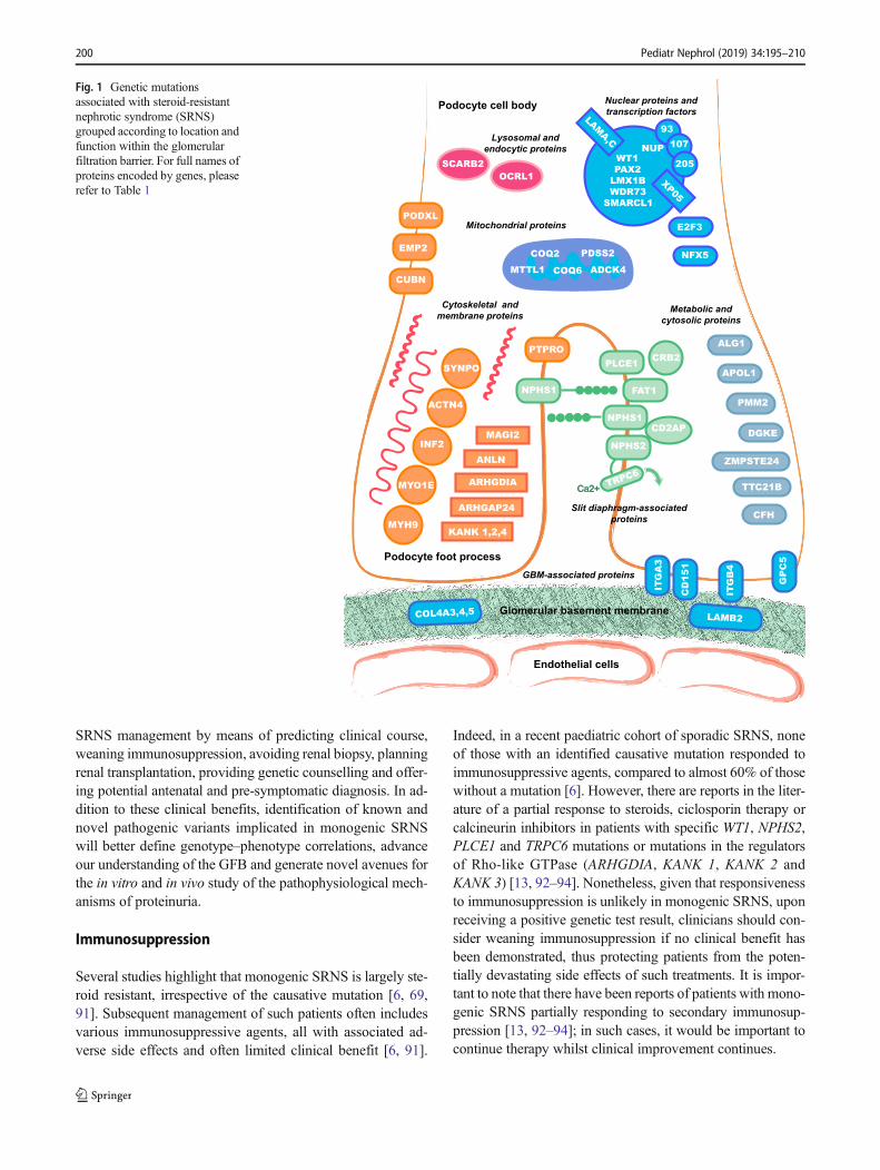
The GFB is composed of three interacting layers: thefenestrated endothelial cells, the glomerular basement mem-brane and the outer podocyte layer. Podocytes are highlyspecialised epithelial cells, and their interdigitating foot pro-cesses connect to form the slit diaphragm, a unique multi-protein cell junction structure which, through regulation ofpodocyte function, controls the ultrafiltration of molecules.Genetic advances in SRNS have unveiled dysfunction ofpodocyte- and slit diaphragm-associated proteins in the path-ogenesis of proteinuria, highlighting their importance in main-taining GFB integrity. The discovery began with genesencoding the slit diaphragm proteins nephrin (NPHS1) andpodocin (NPHS2) [11, 12]. Since then, linkage analysis andnext generation sequencing (NGS) have permitted the identi-fication of over 50 genes implicated in SRNS, and this number
continues to increase. Interestingly, the majority of encodedproteins map to distinct structural protein complexes and sig-nalling pathways within the podocyte (Fig. 1). A thoroughfunctional analysis of these proteins is beyond the scope ofthis review, but readers are directed elsewhere for a moredetailed evaluation [64].
Genotype–phenotype correlationsin monogenic SRNS
Monogenic SRNS can be inherited in an autosomal recessive,autosomal dominant or mitochondrial manner, and can occur asan isolated renal disease or as part of a multisystem disorder.Most cases of recessive disease are characterised by an earlyonset and high penetrance, and they are not infrequently asso-ciated with extra-renal malformations. In contrast, the maincauses of dominant disease are associated with a later onsetand incomplete penetrance, and the patients may remainasymptomatic. The genes associated with SRNS identified todate and their associated phenotypes are presented in Table 1. Inthe following sections, we outline the most common monogen-ic causes of SRNS, according to age of onset.
Congenital NS
Congenital NS (CNS), which presents within the first3 months of life, is commonly associated with causativemutations. Indeed, mutations have been identified in 75–100% of cases of CNS [8, 10, 65, 66]. Causative muta-tions appear to largely occur in one of five genes (NPHS1,NPHS2, WT1, LAMB2 and PLCE1). NPHS1, encodingnephrin, is the main gene implicated in CNS, and muta-tion is responsible for the autosomal recessive Finnishtype (CNF), which typically has a severe phenotype withmassive proteinuria and rapid progression to ESRD [11].However, the NPHS1 mutation detection rate remainshigh amongst non-Finnish cases of CNS [8, 10, 65].Mutations in the NPHS2 gene, encoding podocin, are alsoresponsible for a significant number of CNS cases, andthe phenotype varies from the severe CNF presentation tomilder disease with onset of proteinuria occurring laterthan in those with NPHS1 mutations [4, 66, 67].Mutations in the PLCE1, WT1 and LAMB2 genes havealso been detected in patients presenting with isolatedCNS; mutations in these genes will be discussed in moredetail in the next section.
Infantile and childhood NS
Monogenic NS, which presents in infancy (from 4 to12 months of life) and during childhood, is most commonlyattributed to mutations in the NPHS2 gene, encoding podocin
196 Pediatr Nephrol (2019) 34:195–210

[8, 68]. There is a recognised genotype to phenotype correla-tion that explains this phenotypic variability [12, 66, 69].Recent whole-exome sequencing performed on a paediatriccohort of SRNS patients revealed the mean age of onset asso-ciated with NPHS2mutations to be approximately 6 years [8].That said, it is clearly important to consider NPHS2 mutationas a cause for SRNS in a child of any age and, conversely, toexpect a low rate of NPHS2 mutations in certain ethnicgroups, namely Chinese, Japanese and Korean [10, 70].Additionally, NPHS1 mutations have been identified in in-fants and children presenting with SRNS, with the rarerhypomorphic mutations being associated with a milder late-onset phenotype [8, 71, 72]. Thus, mutations in this geneshould be considered in all paediatric age groups.
Mutations inPLCE1 (encoding phospholipase C epsilon-1)typically cause isolated DMS, with patients presenting withsevere, early-onset SRNS and rapid progression to ESRD[13]. Again, significant clinical heterogeneity exists, withPLCE1 mutations manifesting from birth and throughoutchildhood, with both FSGS or DMS found histologically[73, 74].
WT1 encodes Wilms’ tumour 1, a transcription factor andkey kidney development gene. Mutations in WT1 cause iso-lated and syndromic SRNS, which will be discussed later inthis review. Previous studies have estimatedWT1mutations toaccount for approximately 6% of sporadic SRNS cases inchildhood, manifesting at any age, depending on the underly-ing mutation [8, 66, 75, 76]. Genotype–phenotype correla-tions have been drawn from specificWT1mutations. Of these,certain mutations cause early-onset, severe disease with DMShistologically, or alternatively, late-onset SRNS with FSGShistologically and slower progression to ESRD [75].Interestingly, isolated SRNS may result from a wide rangeof WT1 sequence variations, which are associated with vari-able expression and incomplete penetrance [75]. It is impor-tant to stress thatWT1-related nephropathy may be an isolateddisease with no associated comorbidities or it may be an initialsymptom of syndromic SRNS with extra-renal features man-ifesting later.
Although mutations in TRPC6 and ACTN4 are typical-ly associated with autosomal dominant late-onset disease,as discussed below, there are reports of both infantile andchildhood-onset SRNS caused by mutations in thesegenes.
Late-onset NS
Autosomal dominant SRNS typically presents later in life, inadolescence or adulthood, and has significant phenotypic var-iability. The overall mutation detection rate remains substan-tial, approaching 25% in adolescence and 12% in adulthood,but it is lower than cases presenting earlier in childhood [8,65]. The main genes implicated in late-onset SRNS, which
presents in adolescence, include NPHS2, TRPC6, INF2 andACTN4 [31, 65, 68, 77, 78]. TRPC6, encodes a transient re-ceptor potential cation channel and was originally identified ascausing autosomal dominant FSGS, presenting in adolescenceand early adulthood and showing relatively rapid progressionto ESRD. Since then, TRPC6mutations have been implicatedin childhood-onset FSGS, and even SRNS presenting withinthe first year of life, with variable disease severity [8, 10, 77,79, 80]. Similarly, mutations in ACTN4, which encodes alpha-actinin 4, typically cause late-onset FSGS with slow progres-sion to ESRD [29, 81], but mutations in this gene have beenreported in children presenting with SRNS and rapid progres-sion to ESRD [8, 82]. Mutations in INF2, encoding invertedformin 2, were originally identified in patients with autosomaldominant SRNS, with age of onset ranging from adolescenceand throughout adulthood [31, 83]. Although INF2mutationstypically result in isolated FSGS, they have also been detectedin a subgroup of patients with associated Charcot–Marie–Tooth neuropathy [84]. Specific NPHS2 mutations may man-ifest late, in adolescence or adulthood; the common variantR229Q may result in late-onset SRNS in compound hetero-zygotes with specific second mutations [85].
Syndromic SRNS and mitochondrial disorders
Syndromic SRNS is associated with extra-renal manifes-tations and most commonly occurs due to mutations ingenes encoding nuclear proteins (WT1 , LMX1B ,SMARCL1, WDR73), glomerular basement membraneand adhesion components (LAMB2, ITGA3, ITGB4), actincytoskeleton components (MYH9) and lysosomal(SCARB2) and mitochondrial proteins (COQ2, COQ6,PDSS2, MTTL1, ADCK4). Table 2 lists the major extra-renal manifestations associated with gene defects causingsyndromic SRNS; if extra-renal manifestations are pres-ent, it is highly likely that a causative mutation will beidentified. A full description of syndromic SRNS is be-yond the scope of this review but readers are directed toseveral detailed reviews for a thorough discussion [5, 20,21, 44, 45, 50, 55, 84, 86, 87].
WT1 mutations are associated with a spectrum of signif-icant extra-renal manifestations, including urogenital ab-normalities and malignancy. Mutations in the KTS (spliceinsertion) site are associated with Frasier syndrome,characterised by childhood-onset SRNS, histologicallycharacterised FSGS, male-to-female sex reversal and in-creased risk of gonadoblastoma [18, 75]. Missense muta-tions in exons 8 and 9 (affecting the zinc finger domains)are associated with Denys–Drash syndrome, characterisedby infantile-onset SRNS, histologically characterisedDMS, sex reversal, gonadoblastoma and Wilms’ tumour[75]. Large genomic rearrangements disrupting WT1 andthe neighbouring PAX6 result in WAGR syndrome (Wilms’
Pediatr Nephrol (2019) 34:195–210 197

Table 1 Monogenic causes of steroid-resistant nephrotic syndrome identified to date, including details of associated clinical phenotype, most fre-quently observed renal histological lesion and likely mode of inheritance
Gene Protein Phenotype Mode ofinheritance
Histology Reference
Slit diaphragm-associated proteins
NPHS1 Nephrin CNS (Finnish type), SRNS (early onset) AR PTRD, PMS,FSGS, MCD
[11]
NPHS2 Podocin CNS, SRNS (early and late onset) AR FSGS, MCD [12]
PLCE1 Phospholipase C epsilon 1 CNS, SRNS (early onset) AR DMS, FSGS [13]
CD2AP CD2-associated protein SRNS AD, AR FSGS [14]
TRPC6 Transient receptor potential channel C6 SRNS (late onset) AD FSGS [15]
CRB2 Crumbs family member 2 SRNS AR FSGS [16]
FAT1 FAT atypical cadherin 1 FSGS, neurological involvement AR Variable [17]
Nuclear proteins and transcription factors
WT1 Wilms’ tumour protein 1 Denys Drash, Frasier, isolated SRNS +/−ambiguous genitalia
AD, AR FSGS, DMS [18, 19]
LMX1B LIM homeobox transcription factor 1β Nail-patella syndrome, isolated SRNS AD FSGS [20]
SMARCL1 SMARCA-like protein Schimke immuno-osseous dysplasia AR FSGS [21]
NUP93 Nuclear pore complex protein 93 SRNS AR FSGS [22]
NUP107 Nuclear pore complex protein 107 SRNS (early onset) AR FSGS [23]
NUP205 Nuclear pore complex protein 205 SRNS AR FSGS [22]
XPO5 Exportin 5 SRNS AR FSGS [22]
E2F3 E2F transcription factor FSGS, mental retardation (gene deletion) AD FSGS [24]
NXF5 Nuclear RNA export Factor 5 FSGS, co-segregating heart block XLR FSGS [25]
PAX2 Paired box protein 2 Isolated SRNS (adult-onset) AD FSGS [26]
LMNA Lamin A and C Familial partial lipodystrophy, FSGS AD FSGS [27]
WDR73 WD repeat domain 73 Galloway-Mowat syndrome AR FSGS, DMS [28]
Cytoskeletal, scaffold and membrane proteins
ACTN4 α-actinin 4 SRNS (late onset) AD FSGS [29]
MYH9 Myosin heavy chain 9, non-muscle MYH9-related disorders, SRNS AD FSGS [30]
INF2 Inverted formin 2 SRNS, Charcot-Marie-Tooth disease AD FSGS [31]
MYO1E Myosin 1E SRNS AR FSGS [32]
MAGI2 Membrane Associated Guanylate Kinase,inverted 2
CNS, SRNS AR MCD [33]
ANLN Anillin actin binding protein SRNS (adult-onset) AD FSGS [34]
ARHGAP24 Rho GTPase-activating protein 24 SRNS (adult-onset) AD FSGS [35]
ARHGDIA Rho GDP dissociation inhibitor alpha SRNS (CNS), seizures, cortical blindness AR FSGS [36]
KANK 1/2/ 4 Kidney ankyrin repeat-containing protein SRNS +/− haematuria AR FSGS [37]
SYNPO Synaptopodin FSGS AD FSGS [38]
PTPRO Protein-tyrosine phosphatase-R O SRNS (childhood onset) AR FSGS, MCD [39]
EMP2 Epithelial membrane protein 2 SRNS (childhood onset) AR FSGS [40]
APOL1 Apolipoprotein L1 Susceptibility to SRNS Biallelic FSGS [41]
CUBN Cubilin SRNS AR FSGS [42]
PODXL Podocalyxin FSGS AD FSGS [43]
Glomerular basement membrane-associated proteins
LAMB2 Laminin subunit β2 Pierson syndrome, isolated SRNS AR DMS, FSGS [44]
ITGB4 Integrin β4 Epidermolysis bullosa, SRNS, lungdisease
AR FSGS [45]
ITGA3 Integrin α3 Epidermolysis bullosa, SRNS, lungdisease
AR FSGS [46]
COL4A3/4/5 Type IV collagen α3, α4, α5 Alport syndrome AD, AR, XL FSGS [47]
198 Pediatr Nephrol (2019) 34:195–210

tumour, aniridia, genito-urinary abnormalities and mentalretardation) [75]. Although these genotype–phenotype cor-relations have been clearly described, it is important tonote that there remains significant phenotypic variabilitywith respect to extra-renal manifestations of WT1-associat-ed disease. Furthermore, histopathological heterogeneity isnoted even amongst carriers of the same genetic abnormal-ity [75].
LAMB2 and LMX1B mutations typically cause Piersonsyndrome and Nail–Patella syndrome, respectively, buthave also been identified in isolated congenital, infantileand childhood-onset SRNS [88, 89], and mutation in thesegenes should therefore be considered to be causative inthese age groups.
Isolated SRNSmay also be seen, although rarely, in certainmitochondrial cytopathies, including MELAS (mitochondrialmyopathy, encephalopathy, lactic acidosis and stroke-like ep-isodes) caused by MTTL1 mutations [54] and coenzyme Q10
deficiency [90]. Coenzyme Q10 deficiency due to mutations inCOQ2 and COQ6 may cause an isolated or syndromic
nephropathy [51]. Mutations in PDSS2 cause Leigh syndromebut may also cause isolated SRNS [52].
ADCK4 mutations have been found to cause isolatedSRNS with histologically characterised FSGS, manifestingfrom infancy through to early adulthood [53]. In contrast toprevious studies [9], in a large multicentre cohort of Chinesepaediatric patients with SRNS, ADCK4 was found to be themost commonly mutated causative gene, responsible forSRNS presenting as early as the congenital period, but mostfrequently during childhood [10].
Interestingly, there have been reports of patients with co-enzyme Q10 deficiency and ADCK4 mutation whereby inter-ventional treatment with COQ10 supplementation has beenshown to modify disease progression [53, 90].
Genetic testing; why?
Genetic testing in NS has important clinical and non-clinicalimplications. Confirmation of a genetic defect can personalise
Table 1 (continued)
Gene Protein Phenotype Mode ofinheritance
Histology Reference
GPC5 Glypican 5 NS (adult onset) Risk gene Variable [48]
CD151 CD151 antigen FSGS, bullous skin lesions, deafness AR FSGS [49]
Mitochondrial proteins
COQ2 Coenzyme Q2 CoQ10 deficiency, SRNS +/−encephalopathy
AR CG [50]
COQ6 Coenzyme Q6 CoQ10 deficiency, SRNS and deafness AR FSGS, DMS [51]
PDSS2 Prenyl-diphosphate synthase subunit 2 CoQ10 deficiency, SRNS, Leighsyndrome
AR FSGS [52]
ADCK4 AarF domain containing kinase 4 CoQ10 biosynthesis disruption AR FSGS [53]
MTTL1 Mitochondrial tRNA 1 MELAS, diabetes, deafness, SRNS Mitochondrial FSGS [54]
Lysosomal and endocytic proteins
SCARB2 Scavenger receptor class B, member 2 Action myoclonus-renal failure syn-drome
AR FSGS [55]
OCRL1 Oculocerebrorenal syndrome of Lowe Dent-2 disease, Lowe syndrome, SRNS XLR FSGS [56]
Metabolic and cytosolic proteins
ZMPSTE24 Zinc metallopeptidase STE24 Mandibuloacral dysplasia AR FSGS [57]
PMM2 Phosphomannomutase 2 Congenital defect of glycosylation AR CG [58]
ALG1 Asparagine-linked glycosylation 1 Congenital defect of glycosylation AR FSGS [59]
TTC21B Tetratricopeptide repeat protein 21B FSGS AR FSGS [60]
CFH Complement factor H SRNS AR FSGS [61]
DGKE Diacylglycerol kinase epsilon NS AR FSGS [62]
AD, Autosomal dominant; AR, autosomal recessive; CG, collapsing glomerulopathy; CNS, congenital nephrotic syndrome; DMS, diffuse mesangialsclerosis; FSGS, focal segmental glomerulosclerosis; MCD, minimal change disease; MELAS, Mitochondrial encephalomyopathy, lactic acidosis, andstroke-like episodes; NS, nephrotic syndrome; PMS, progressive mesangial sclerosis; PTRD, proximal tubule radial dilatation; SRNS, steroid-resistantnephrotic syndrome; XL, X-linked
Pediatr Nephrol (2019) 34:195–210 199
SRNS management by means of predicting clinical course,weaning immunosuppression, avoiding renal biopsy, planningrenal transplantation, providing genetic counselling and offer-ing potential antenatal and pre-symptomatic diagnosis. In ad-dition to these clinical benefits, identification of known andnovel pathogenic variants implicated in monogenic SRNSwill better define genotype–phenotype correlations, advanceour understanding of the GFB and generate novel avenues forthe in vitro and in vivo study of the pathophysiological mech-anisms of proteinuria.
Immunosuppression
Several studies highlight that monogenic SRNS is largely ste-roid resistant, irrespective of the causative mutation [6, 69,91]. Subsequent management of such patients often includesvarious immunosuppressive agents, all with associated ad-verse side effects and often limited clinical benefit [6, 91].
Indeed, in a recent paediatric cohort of sporadic SRNS, noneof those with an identified causative mutation responded toimmunosuppressive agents, compared to almost 60% of thosewithout a mutation [6]. However, there are reports in the liter-ature of a partial response to steroids, ciclosporin therapy orcalcineurin inhibitors in patients with specific WT1, NPHS2,PLCE1 and TRPC6 mutations or mutations in the regulatorsof Rho-like GTPase (ARHGDIA, KANK 1, KANK 2 andKANK 3) [13, 92–94]. Nonetheless, given that responsivenessto immunosuppression is unlikely in monogenic SRNS, uponreceiving a positive genetic test result, clinicians should con-sider weaning immunosuppression if no clinical benefit hasbeen demonstrated, thus protecting patients from the poten-tially devastating side effects of such treatments. It is impor-tant to note that there have been reports of patients with mono-genic SRNS partially responding to secondary immunosup-pression [13, 92–94]; in such cases, it would be important tocontinue therapy whilst clinical improvement continues.
Podocyte cell body
Podocyte foot process
Glomerular basement membrane
Lysosomal and endocytic proteins
Mitochondrial proteins
Nuclear proteins and transcription factors
Slit diaphragm-associated proteins
Metabolic and cytosolic proteins
Cytoskeletal and membrane proteins
COQ2 PDSS2
MTTL1 COQ6 ADCK4
WT1PAX2
LMX1BWDR73
SMARCL1
SCARB2
XP05
NUP
93107
205
PLCE1
NPHS1
NPHS2
CD2AP
NPHS1
KANK 1,2,4
ARHGAP24
ARHGDIA
ANLN
MYH9
MYO1E
INF2
ACTN4
ZMPSTE24
COL4A3,4,5 LAMB2
ITG
A3
ITG
B4
PMM2
TRPC6Ca2+
OCRL1
GBM-associated proteins
CD
151
FAT1
PTPRO
LAMA,C
CRB2
GP
C5
SYNPO
ALG1
APOL1
DGKE
TTC21B
CUBN
E2F3
NFX5
PODXL
EMP2
CFH
MAGI2
Endothelial cells
Fig. 1 Genetic mutationsassociated with steroid-resistantnephrotic syndrome (SRNS)grouped according to location andfunction within the glomerularfiltration barrier. For full names ofproteins encoded by genes, pleaserefer to Table 1
200 Pediatr Nephrol (2019) 34:195–210

Novel interventional therapy and monitoring
The discovery of rare monogenic causes of SRNS have revealeda small but significant cohort whose disease may be amenable tospecific interventional treatment, thereby avoiding lengthy im-munosuppression and delaying progression to ESRD. Patientswith disease-causing mutations in genes encoding enzymes ofthe coenzyme Q10 pathway (COQ2, COQ6 and ADCK4) and inthe CUBN gene may respond to treatment with coenzyme Q10
and vitamin B12, respectively. Likewise, patients withARHGDIA mutations, through modulation of Rac I–mineralo-corticoid interactions, could theoretically respond to eplenerone(a mineralocorticoid-receptor antagonist) [36].
As highlighted in Table 2, many syndromic forms of SRNShave associated medical problems that may benefit from earlyrecognition and management. An example is theWT1mutation,which can predispose to malignancy, and the detection of suchmutations should trigger monitoring for associated Wilms’ tu-mour and gonadoblastoma. Given the latter entity is largely as-sociated with sex reversal, a karyotype analysis should also beperformed, especially in phenotypically female patients present-ing with SRNS and primary amenorrhoea.
Renal biopsy
Renal histology has historically been utilised as a key diag-nostic and prognostic criterion for children with SRNS, but
emerging evidence reveals significant histological heteroge-neity amongst monogenic causes of SRNS, demonstratingthat biopsy findings may not correlate with genetic results[8, 95]. Furthermore, there does not appear to be a notabledifference in the frequency of histological lesions found inpatients with or without a recognised genetic cause [6]. Forthese reasons, in cases of primary SRNS, rapid genetic testinghas the potential to obviate the need for renal biopsy for diag-nostic purposes and serves as a less invasive diagnostic meth-od; this is of particular significance in the younger SRNScohort, for whom a genetic aetiology is more likely. In theevent of rapid genetic testing being inaccessible, renal histol-ogy may direct clinicians towards the most likely Bculprit^gene; for example, if DMS is detected in an infant presentingwith SRNS, it would be prudent to perform mutational anal-ysis on certain genes (LAMB2,WT1, NPHS1, PLCE1) prefer-entially over others. However, clinical indications for renalbiopsy do remain, such as atypical features suggestive of lu-pus nephritis, with histology providing useful information.Additionally, a histological diagnosis enables phenotype pat-terns to become better established and is therefore useful froma clinical research perspective.
Disease reoccurrence post-transplantation
There is a high risk of progression to ESRD in monogenicSRNS, with many patients requiring renal transplantation
Table 2 Syndromic steroid-resistant nephrotic syndrome andassociated extra-renalmanifestations
Gene Disease Extra-renal manifestations
WT1 Denys–Drash syndrome Urogenital abnormalities, ambiguous genitalia, nephroblastoma
Frasier syndrome Gonadoblastoma, male pseudohermaphroditism
LAMB2 Pierson’s syndrome Ocular abnormalities; microcoria
LMX1B Nail–Patella syndrome Skeletal defects, hypoplastic nails, absent patella, glaucoma
SMARCL1 Schimkeimmune-osseous dys-plasia
Spondyloepiphyseal dysplasia, T cell immunodeficiency, cerebralinfarcts, skin pigmentation
SCARB2 Action myoclonus renalfailure
Progressive myoclonic epilepsy, tremor, ataxia
COQ2 CoQ10 deficiency Progressive encephalomyopathy
COQ6 CoQ10 deficiency Sensorineural hearing loss
PDSS2 Leigh syndrome Hypotonia, ataxia, deafness, growth retardation
WDR73 Galloway-Mowatsyndrome
Microcephaly, psychomotor impairment, seizures, hypotonia
MTTL1 MELAS Myopathy, encephalopathy, lactic acidosis, stroke-like episodes,diabetes, deafness
ITGA3 Epidermolysis-associated Epidermolysis bullosa, interstitial lung disease
ITGB4 Epidermolysis-associated Epidermolysis bullosa, pyloric atresia
MYH9 MYH9-relatedsyndromes
Macrothrombocytopenia, mental retardation, sensorineuraldeafness, cataracts
INF2 Charcot–Marie–Tooth Chronic peripheral motor and sensory neuropathy
ZMPSTE24 Manibuloacral dysplasia Mandibular and clavicular hypoplasia, cutaneous atrophy,lipodystrophy, acro-oestolysis
For names of encoded proteins and associated histology, please consult Table 1
Pediatr Nephrol (2019) 34:195–210 201

[96]. Several studies have highlighted a low risk of diseasereoccurrence post-transplant when a genetic aetiology hasbeen confirmed [6, 8, 68, 69, 96, 97]. Conversely, there is ahigh risk of post-transplant disease reoccurrence in the idio-pathic group; this is postulated to be caused by circulatingfactors [98, 99]. Given that the likelihood of post-transplantreoccurrence is minimal for genetic SRNS; after excluding themutation in parents, a parental transplant can be planned, al-though in our experience this typically follows bilateral ne-phrectomy and an interval of time on dialysis. Post-transplantation prognosis is improved with living donor trans-plantation, which is associated with prolonged graft survivaland decreased rejection rates as compared to deceased donorkidney transplantation.
Genetic counselling
Genetic counselling prior to testing should ensure that familiesare informed regarding potential outcomes and limitations of thechosen genetic test, including the discovery of variants of un-known significance and the potential for incidental findings.Possible benefits, including changes to medical management,should be discussed, as well as potential harms, including priva-cy, legal and social implication. These subjects are covered in thenext section.
Making a molecular diagnosis has important implicationsfor a family. It may enable accurate discussion of recurrencerisk in future children and potential identification of pre-symptomatic individuals at risk [4]. Early referral to a clinicalgenetics service can facilitate identification of individuals atrisk and genetic testing of family members as well as counsel-ling in terms of family planning, prenatal diagnosis and pre-implantation genetic diagnosis. Genetic screening in unaffect-ed family members may be additionally important when plan-ning a living related donor (LRD) renal transplant, especiallyin the case of autosomal dominant disease. When inheritanceof a severe disease-causing mutation is likely, pre-symptomatic testing for proteinuria and genotyping at birthare avenues that should be discussed and offered to affectedfamilies. Ethical considerations of causing potentially unnec-essary anxiety should be addressed, and families should becounselled on the benefits and risks of such an approach; thatis, the benefits of providing a timely genetic diagnosis, andtherefore active clinical management, versus the risks of test-ing for a disease which may never manifest or be mild in theevent of incomplete penetrance or variable expression.Prenatal diagnosis could be offered in families with a knownrisk of severe NS, such as CNS, or in cases where elevatedalpha-fetoprotein levels have been detected in maternal serumor amniotic fluid. It can be used to allow the family to make aninformed decision about continuing a pregnancy or to allowpreparation both by the family and medical professionals forthe birth of an affected child.
Risks of genetic testing
In addition to the ethical and emotional considerations thatmust be addressed during genetic counselling, there are sev-eral other risks to the patient and their family who are consid-ering genetic testing or receiving a genetic diagnosis. In theUK, genetic testing is usually paid for by the NationalHealth Service, and in countries with a privatised medicalsystem, health insurance policies often cover the cost ofgenetic testing performed at the request of a doctor.However, this is not always the case, and in some situa-tions the significant cost, which can be over US$2000,must be covered by the family. Furthermore, upon receiv-ing a genetic diagnosis, the fear of insurance discrimina-tion and the associated costs of enhanced insurance pre-miums represent a significant emotional and financial bur-den. Although there is legislation in place which protectsthose with a genetic disease from discrimination by healthinsurers, this does not always extend to protect patientsfrom employment discrimination or the amplified costs oflife, disability and long-term care insurance [100]. As partof the genetic counselling process, these issues should bediscussed with affected families and informed consent ob-tained prior to genetic testing. A thorough discussion re-garding the barriers to genetic testing in public health isbeyond the scope of this review, but readers are directed toseveral useful articles for further information [100–104].
Genetic testing; who?
Having discussed the benefits of identifying a causativemutation in patients with SRNS, it is important to note thatthe overall burden of monogenic SRNS has yet to be fullydelineated. Recent evidence estimates that a geneticaetiology is detected in approximately 30% of cases. Anegative result does not exclude genetic disease as muta-tions may be missed, with sensitivity for genes covered bythe test depending on methodology and analysis used.Alternatively, a mutation may be present in a gene notcovered by the chosen test, for example a novel geneticassociation. This, combined with the profound clinicaland pathological heterogeneity of genetic and idiopathicSRNS, highlights that universal genetic testing in SRNSis inappropriate and unlikely to be cost-effective. Rather,mutational screening should be directed towards those inwhom a genetic aetiology is likely and should therefore bereserved for patients presenting with primary SRNS.
Indications for genetic testing
When accessible and affordable, mutational screening shouldbe performed in all children presenting with primary SRNS.
202 Pediatr Nephrol (2019) 34:195–210
Even in young adults, the likelihood of detecting a causativemutation remains substantial, and when cost allows, mutation-al screening should also be offered to this cohort. However,when such an inclusive approach is not possible, there arecertain indications in which a genetic cause for SRNS be-comes more likely, and mutational screening should be per-formed as a priority. Given that the likelihood of detecting acausative mutation is inversely related to age of disease onset[105], mutational screening becomes increasingly importantthe earlier the disease manifests. Genotype–phenotype corre-lations clearly demonstrate that mutations in recessive genesare more frequently implicated in early-onset disease and thatmutations in dominant genes are more frequently implicatedin adult-onset disease. Although there may not be an obviousfamily history in early-onset disease, a positive family historyin any age group indicates that monogenic SRNS is likely andshould trigger mutational screening. Additionally, the likeli-hood of finding causative recessive mutations correlates di-rectly with the degree of consanguinity [80]; thus, a history ofconsanguinity should prompt mutational screening. Finally,the presence of extra-renal manifestations suggestive of anunderlying genetic syndrome (Table 2) makes screening ofassociated genes advisable.
The clinical indications for genetic testing in SRNS can besummarized as follows:
& Congenital or infantile-onset NS& Childhood-onset NS& Family history of NS& Consanguinity& Extra-renal manifestations.
Genetic testing; when?
Before undertaking genetic testing for SRNS, it is importantthat the potential detection of a causative mutation is likely toaid in diagnosis, alter clinical management, inform likelyprognosis and provide information when stratifying risk forfamily members and delivering genetic counselling. Incongenital- and infantile-onset NS, genetic testing should beconsidered before commencing immunosuppressive therapyor performing renal biopsy. Similarly, when genetic testingcan be performed in a timely manner, early confirmation ofa genetic diagnosis in childhood-onset SRNS would minimisethe adverse effects of current therapies on the growing child.Pre-transplantation genetic testing will provide clinicians withinformation that may be helpful in predicting the risk of post-transplant reoccurrence and will therefore guide pre- and post-transplant management, especially when considering LRDkidney transplantation from family members.
To summarise, we suggest that genetic testing should beconsidered when important clinical decisions need to be maderegarding the need for renal biopsy, the intensity and durationof immunosuppression and pre-transplantation therapy, andwhen syndromic SRNS is suspected.
Genetic testing; how?
Traditionally, genetic testing in diagnostic laboratories hasemployed Sanger sequencing, frequently in association withexon copy number analysis, to assess specific disease-relatedgenes individually. In genetically heterogeneous disorders,with multiple causal genes, such as SRNS, this method canbe expensive and time-consuming owing to the cost of screen-ing multiple individual genes. The advent of high-throughputmassively parallel sequencing (NGS methods) allows for ahigher diagnostic yield, time savings and a reduction in cost[105, 106]. Typically, diagnostic laboratories utilise a targetedcapture of a ‘panel’ of genes of interest followed by sequenc-ing on an NGS platform. Sanger sequencing still plays animportant role for the confirmation of genetic variants identi-fied via NGS and filling in of regions of poor coverage. Thelimitations of Sanger sequencing include the need to ensureboth adequate coverage of regions of interest and adequateanalysis to detect copy number variants such as exonic dele-tions. As with most Sanger sequencing approaches, this meth-od will miss deep intronic or regulatory region variants unlessspecifically targeted.
Whole-exome sequencing (WES) or whole-genome se-quencing (WGS) employ NGS methods to attempt to se-quence the coding portion of the genome (the exome) or theentire genome, respectively. This approach is not limited toknown candidate genes and therefore has the ability to identifymutations in novel genes, thereby expanding the heterogene-ity of SRNS and enhancing our understanding of the patho-genesis and molecular mechanisms of proteinuria. WES isincreasingly being implemented in the clinical setting, but itswidespread application is limited by the amounts of data gen-erated and the requirements for robust bioinformatics supportand assessments of the pathogenicity of larger numbers ofvariants. When targeted capture utilised for WES gives suffi-cient coverage of the ‘Mendeliome’, an in silico panel ofgenes can be analysed to give similar results to a targetedcapture approach, whilst giving the flexibility to ‘open’ thedata if the initial analysis does not find a variant of interest,or in light of novel genetic associations. A similar approachcan be utilised with WGS, with the potential advantage ofimproved coverage, coverage of regulatory and intronic re-gions and improved analysis of copy number and structuralrearrangements, but with the disadvantages of increased costand substantially increased data and variant volume.
Pediatr Nephrol (2019) 34:195–210 203
WES and WGS are hampered by the fact that largenumbers of genetic variants are identified, including vari-ants of unknown significance and incidental or secondaryfindings. These findings raise a number of ethical and prac-tical issues relating to consent, data storage and analysis,all too extensive to cover here. The American College ofMedical Genetics and Genomics has published guidanceon reporting secondary findings.
When compared toWES orWGS, the cost-effectiveness ofNGS using a targeted gene panel analysis has greater clinicalapplication in SRNS, as it produces a more feasible dataset forbioinformatics analysis which is functionally interpretable in aclinical setting.
Application to SRNS
Currently, clinical phenotyping combined with targeted NGSpanel analysis is the most cost-effective and clinically usefulapproach for mutational screening in SRNS. This method en-ables clinicians to quantify and stratify likely response to im-munosuppression, rate of progression to ESRD and risk ofpost-transplant reoccurrence. Using NGS technology, mostmonogenic SRNS genes (approximately 40–50 genes per pan-el) can be analysed within 6 weeks and at a competitive pricecompared to Sanger methods [6, 107]. There are several com-mercial indication-driven SRNS gene panels currently in usearound the world, with many laboratories conducting entire ortargeted sequence analysis, antenatal testing and carrierscreening for SRNS genes. Indeed, an internet search (www.genetests.org) reveals at least 12 laboratories worldwideoffering extended NGS panels for SRNS with an averageturnaround of 3–6 weeks and associated cost ranging from$1000 to $2200. Comparatively, Sanger sequencing forindividual genes or small panels of genes (approximately 5
genes) has a slightly quicker turnaround of 2–4 weeks and,depending on the size of the gene, costs $450–$1000 perindividual gene.
In certain circumstances where NGS technology is inacces-sible or unaffordable, and a disease-causingmutation is highlylikely in a specific gene, as suggested by the presence of extra-renal manifestations or a positive family history, Sanger se-quencing methods remain an important diagnostic tool. It isimportant to stress that employing genotype–phenotype cor-relations alone to direct mutational screening using Sangermethods is only cost-effective, and clinically beneficial, pro-vided a causative mutation is identified early in the screeningprocess.
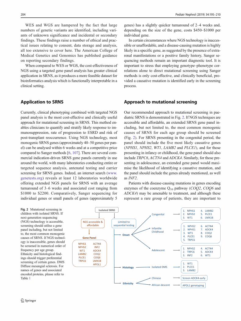
Approach to mutational screening
Our recommended approach to mutational screening in pae-diatric SRNS is demonstrated in Fig. 2. If NGS techniques areaccessible and affordable, an extended SRNS gene panel in-cluding, but not limited to, the most common monogeniccauses of SRNS for each age group should be screened(Fig. 2). For SRNS presenting in the congenital period, thepanel should include the five most likely causative genes(NPHS1, NPHS2, WT1, LAMB2 and PLCE1), and for thosepresenting in infancy or childhood, the gene panel should alsoinclude TRPC6, ACTN4 and ADCK4. Similarly, for those pre-senting in adolescence, an extended gene panel would maxi-mise the likelihood of identifying a causative mutation, andthe panel should include the genes already mentioned, as wellas INF2.
Patients with disease-causing mutations in genes encodingenzymes of the coenzyme Q10 pathway (COQ2, COQ6 andADCK4) may be amenable to treatment, and although theserepresent a rare group of patients, they are important to
Fig. 2 Mutational screening inchildren with isolated SRNS. Ifnext-generation sequencing(NGS) technology is accessible,screening should utilise a genepanel including, but not limitedto, the most common monogeniccauses of SRNS. If NGS technol-ogy is inaccessible, genes shouldbe screened in numerical order offrequency per age group.Ethnicity and histological find-ings should trigger preferentialscreening of certain genes. DMSDiffuse mesangial sclerosis. Fornames of genes and associatedencoded proteins, please refer toTable 1
204 Pediatr Nephrol (2019) 34:195–210
recognise and should therefore be included in the geneticscreening panel for isolated SRNS presenting in any paediatricage group, including the first year of life. This is especiallytrue for patients of Chinese, Japanese and Korean origin, asthere appears to be an increased frequency of the ADCK4mutation in these populations [10, 70]. Additionally, giventhat the APOL1 genotype represents a vulnerable populationwho present with more advanced disease [108], defining apatient’s APOL1 genotype has important clinical implications,and mutational screening of this gene should be included inthe gene panel, especially for patients of African descent.
If NGS technology is neither accessible nor affordable andclinicians are limited to sequential testing, genes should bescreened in numerical order, for each age group, as depictedin Fig. 2. If renal histology is available and reveals isolatedDMS, we suggest preferentially screening for mutations inWT1, PLCE1 and LAMB2. In the presence of extra-renal man-ifestations, it is highly likely that a causative mutation will bedetected, and genes should be selected for screening depend-ing on the specific phenotype identified (Table 2). In view ofthe significant complications associated with WT1 mutations,establishing a genetic diagnosis appears to be particularly im-portant; it is therefore advisable to routinely screen WT1 formutations in isolated SRNS.
With the advent of NGS methods targeting large panels ofgenes, novel pathogenic variants in genes that are more rarelyimplicated in paediatric SRNS are being identified. Most re-cently, mutations in SMARCAL1, CUBN, LMX1B, PODXLand the nucleoporin genes, NUP93 and NUP107, have beenfound to be causative of isolated SRNS presenting from in-fancy and throughout childhood [8, 10]. Moreover, mutationsin genes which are usually linked to another renal phenotype(DGKE,OCRL andCOL4A3) have been found to be causativeof isolated SRNS presenting in childhood [8]. These findingshighlight the importance of employing NGS technology in thediagnosis of monogenic SRNS through expanding currentknowledge in the pathogenesis and molecular basis of protein-uria. For this reason, extended gene panels and if appropriateresources are available, WES/WGS techniques, should beconsidered wherever possible.
Conclusion
Mutations of podocyte-associated genes account for approxi-mately 30% of paediatric cases of SRNS. The younger thechild at presentation, the higher the genetic diagnostic rate.Advances in genomic sequencing have improved our under-standing of the molecular basis of NS and of the genetic het-erogeneity of SRNS. A genetic diagnosis allows aBpersonalised^ approach when investigating and managingpatients and their families. That is, clinicians are more ableto make decisions about weaning immunosuppression,
avoiding renal biopsy and planning renal transplantation andgeneticists can offer more informed genetic counselling. In asmall but significant sub-group of patients with specific mu-tations, a genetic diagnosis may open avenues for interven-tional disease-modifying therapy and allow clinicians to detectand treat asymptomatic extra-renal manifestations early.
The clinical utility of such advances has been hindered by alack of clear guidelines pertaining to mutational screening inSRNS. There exists profound clinical and pathological het-erogeneity in monogenic SRNS, with mutations in thesame gene and even identical mutations resulting in signif-icant phenotypic variability. This heterogeneity renders se-quential mutational screening using traditional Sanger se-quencing a timely and costly process. Technological ad-vances in genomic sequencing have led to the developmentof commercial, indication-driven gene panels which simul-taneously sequence over 40 known SRNS-related genes.NGS panels are available worldwide, and with a similarturnaround time and slightly increased cost to traditionalmethods, they currently represent the most time- and cost-effective approach to mutational screening in SRNS. Wepropose that all children presenting with primary SRNS bescreened for monogenic causative mutations using an ex-tended gene panel; especially in cases of early-onset dis-ease and those with a positive family history or history ofconsanguinity. If such an inclusive approach is not possi-ble, we provide recommendations for sequential gene test-ing which direct the clinician towards the most frequentlyoccurring causative mutation per age group, depending onavailable histology, presence of extra-renal manifestationsand ethnicity.
NGS methods targeting large panels of genes, the wholeexome or genome have allowed the identification of novelpathogenic variants and also novel genetic associations impli-cated in paediatric SRNS. This in turn enables in vitro and invivo study of podocyte-associated proteins, further unravellingthe pathogenic pathways of SRNS and providing importanttherapeutic targets to guide advanced medical management ona gene-specific basis.
Key summary points
& Mutations of podocyte-associated proteins account for ap-proximately 30% of SRNS in childhood.
& The likelihood of detecting a causative mutation is in-versely related to age of disease onset.
& Monogenic SRNS displays significant phenotypic hetero-geneity in terms of associated renal histology and clinicalpresentation.
& A definitive molecular diagnosis has important clinicalimplications, allowing for a personalised treatmentapproach.
Pediatr Nephrol (2019) 34:195–210 205
9. Sadowski CE, Lovric S, Ashraf S, Pabst WL, Gee HY, Kohl S,Engelmann S, Vega-Warner V, Fang H, Halbritter J, Somers MJ,Tan W, Shril S, Fessi I, Lifton RP, Bockenhauer D, El-Desoky S,Kari JA, Zenker M, Kemper MJ, Mueller D, Fathy HM, SolimanNA, SRNS Study Group, Hildebrandt F (2015) A single-genecause in 29.5% of cases of steroid-resistant nephrotic syndrome.J Am Soc Nephrol 26:1279–1289
10. Wang F, Zhang Y, Mao J, Yu Z, Yi Z, Yu L, Sun J, Wei X, Ding F,Zhang H, Xiao H, Yao Y, Tan W, Lovric S, Ding J, Hildebrandt F(2017) Spectrum of mutations in Chinese children with steroid-resistant nephrotic syndrome. Pediatr Nephrol 32:1181–1192
11. Kestila M, Lenkkeri U, Mannikko M, Lamerdin J, McCready P,Putaala H, Ruotsalainen V, Morita T, Nissinen M, Herva R,Kashtan CE, Peltonen L, Holmberg C, Olsen A, Tryggvason K(1998) Positionally cloned gene for a novel glomerular protein—nephrin—is mutated in congenital nephrotic syndrome. Mol Cell1:575–582
12. Boute N, Gribouval O, Roselli S, Benessy F, Lee H,Fuchshuber A, Dahan K, Gubler MC, Niaudet P, Antignac C(2000) NPHS2, encoding the glomerular protein podocin, ismutated in autosomal recessive steroid-resistant nephrotic syn-drome. Nat Genet 24:349–354
13. Hinkes B, Wiggins RC, Gbadegesin R, Vlangos CN, Seelow D,Nurnberg G, Garg P, Verma R, Chaib H, Hoskins BE, Ashraf S,Becker C, Hennies HC, Goyal M, Wharram BL, Schachter AD,Mudumana S, Drummond I, Kerjaschki D, Waldherr R, DietrichA, Ozaltin F, Bakkaloglu A, Cleper R, Basel-Vanagaite L, PohlM,Griebel M, Tsygin AN, Soylu A, Muller D, Sorli CS, Bunney TD,Katan M, Liu J, Attanasio M, O'Toole JF, Hasselbacher K, MuchaB, Otto EA, Airik R, Kispert A, Kelley GG, Smrcka AV,Gudermann T, Holzman LB, Nurnberg P, Hildebrandt F (2006)Positional cloning uncovers mutations in PLCE1 responsible for anephrotic syndrome variant that may be reversible. Nat Genet 38:1397–1405
14. Lowik MM, Groenen PJ, Pronk I, Lilien MR, Goldschmeding R,Dijkman HB, Levtchenko EN, Monnens LA, van den Heuvel LP(2007) Focal segmental glomerulosclerosis in a patient homozy-gous for a CD2AP mutation. Kidney Int 72:1198–1203
15. Winn MP, Conlon PJ, Lynn KL, Farrington MK, Creazzo T,Hawkins AF, Daskalakis N, Kwan SY, Ebersviller S, BurchetteJL, Pericak-Vance MA, Howell DN, Vance JM, Rosenberg PB(2005) A mutation in the TRPC6 cation channel causes familialfocal segmental glomerulosclerosis. Science 308:1801–1804
16. Ebarasi L, Ashraf S, Bierzynska A, Gee HY, McCarthy HJ,Lovric S, Sadowski CE, Pabst W, Vega-Warner V, Fang H,Koziell A, Simpson MA, Dursun I, Serdaroglu E, Levy S,Saleem MA, Hildebrandt F, Majumdar A (2015) Defects ofCRB2 cause steroid-resistant nephrotic syndrome. Am JHum Genet 96:153–161
17. Gee HY, Sadowski CE, Aggarwal PK, Porath JD, Yakulov TA,Schueler M, Lovric S, Ashraf S, Braun DA, Halbritter J, Fang H,Airik R, Vega-Warner V, Cho KJ, Chan TA, Morris LG, Ffrench-Constant C, Allen N, McNeill H, Buscher R, Kyrieleis H, WallotM, Gaspert A, Kistler T, Milford DV, Saleem MA, Keng WT,Alexander SI, Valentini RP, Licht C, Teh JC, Bogdanovic R,Koziell A, Bierzynska A, Soliman NA, Otto EA, Lifton RP,Holzman LB, Sibinga NE, Walz G, Tufro A, Hildebrandt F(2016) FAT1 mutations cause a glomerulotubular nephropathy.Nat Commun 7:10822
18. Barbaux S, Niaudet P, GublerMC, Grunfeld JP, Jaubert F, KuttennF, Fekete CN, Souleyreau-Therville N, Thibaud E, Fellous M,McElreavey K (1997) Donor splice-site mutations in WT1 areresponsible for Frasier syndrome. Nat Genet 17:467–470
19. Pelletier J, Bruening W, Kashtan CE, Mauer SM, Manivel JC,Striegel JE, Houghton DC, Junien C, Habib R, Fouser L, FineRN, Silverman BL, Haber DA, Housman D (1991) Germline
206 Pediatr Nephrol (2019) 34:195–210
& Recent advances in high-throughput sequencing haverevolutionised genetic testing, and indication-driven genepanel analysis currently represents the most cost-effectiveapproach for mutational screening in SRNS.
& Identification of novel SRNS genes and causativemutations will further unravel the pathogenic path-ways of SRNS.
Acknowledgements R.P is an Academic Clinical fellow funded by theNational Institute for Health Research (NIHR), H.S. is an AcademicClinical Lecturer funded by NIHR and R.L. is supported by aWellcome Trust Senior Fellowship award (202860/Z/16/Z).
Compliance with ethical standards
Conflict of interest The authors declare no conflicts of interests.
Open Access This article is distributed under the terms of the CreativeCommons At t r ibut ion 4 .0 In te rna t ional License (h t tp : / /creativecommons.org/licenses/by/4.0/), which permits unrestricted use,distribution, and reproduction in any medium, provided you give appro-priate credit to the original author(s) and the source, provide a link to theCreative Commons license, and indicate if changes were made.
References
1. Wong W (2007) Idiopathic nephrotic syndrome in New Zealandchildren, demographic, clinical features, initial management andoutcome after twelve-month follow-up: results of a three-year na-tional surveillance study. J Paediatr Child Health 43:337–341
2. Filler G, Young E, Geier P, Carpenter B, Drukker A, Feber J(2003) Is there really an increase in non-minimal change nephroticsyndrome in children? Am J Kidney Dis 42:1107–1113
3. Smith JM, Stablein DM, Munoz R, Hebert D, McDonald RA(2007) Contributions of the transplant registry: the 2006 annualreport of the north American Pediatric renal trials and collabora-tive studies (NAPRTCS). Pediatr Transplant 11:366–373
4. Benoit G, Machuca E, Antignac C (2010) Hereditary nephroticsyndrome: a systematic approach for genetic testing and a reviewof associated podocyte gene mutations. Pediatr Nephrol 25:1621–1632
5. McCarthy HJ, Saleem MA (2011) Genetics in clinical practice:nephrotic and proteinuric syndromes. Nephron Exp Nephrol 118:e1–e8
6. Giglio S, Provenzano A, Mazzinghi B, Becherucci F, Giunti L,Sansavini G, Ravaglia F, Roperto RM, Farsetti S, Benetti E,Rotondi M, Murer L, Lazzeri E, Lasagni L, Materassi M,Romagnani P (2015) Heterogeneous genetic alterations in sporad-ic nephrotic syndrome associate with resistance to immunosup-pression. J Am Soc Nephrol 26:230–236
7. Hildebrandt F (2010) Genetic kidney diseases. Lancet 375:1287–1295
8. Bierzynska A, McCarthy HJ, Soderquest K, Sen ES, Colby E,Ding WY, Nabhan MM, Kerecuk L, Hegde S, Hughes D, MarksS, Feather S, Jones C, Webb NJ, Ognjanovic M, Christian M,Gilbert RD, Sinha MD, Lord GM, Simpson M, Koziell AB,Welsh GI, Saleem MA (2017) Genomic and clinical profiling ofa national nephrotic syndrome cohort advocates a precision med-icine approach to disease management. Kidney Int 91:937–947

mutations in theWilms' tumor suppressor gene are associated withabnormal urogenital development in Denys–Drash syndrome.Cell 67:437–447
20. Dreyer SD, Zhou G, Baldini A, Winterpacht A, Zabel B, Cole W,Johnson RL, Lee B (1998) Mutations in LMX1B cause abnormalskeletal patterning and renal dysplasia in Nail Patella syndrome.Nat Genet 19:47–50
21. Boerkoel CF, Takashima H, John J, Yan J, Stankiewicz P,Rosenbarker L, Andre JL, Bogdanovic R, Burguet A, CockfieldS, Cordeiro I, Frund S, Illies F, Joseph M, Kaitila I, Lama G,Loirat C, McLeod DR, Milford DV, Petty EM, Rodrigo F,Saraiva JM, Schmidt B, Smith GC, Spranger J, Stein A, ThieleH, Tizard J,Weksberg R, Lupski JR, Stockton DW (2002)Mutantchromatin remodeling protein SMARCAL1 causes Schimkeimmuno-osseous dysplasia. Nat Genet 30:215–220
22. Braun DA, Sadowski CE, Kohl S, Lovric S, Astrinidis SA,Pabst WL, Gee HY, Ashraf S, Lawson JA, Shril S, Airik M,Tan W, Schapiro D, Rao J, Choi WI, Hermle T, Kemper MJ,Pohl M, Ozaltin F, Konrad M, Bogdanovic R, Buscher R,Helmchen U, Serdaroglu E, Lifton RP, Antonin W,Hildebrandt F (2016) Mutations in nuclear pore genesNUP93, NUP205 and XPO5 cause steroid-resistant nephroticsyndrome. Nat Genet 48:457–465
23. Miyake N, Tsukaguchi H, Koshimizu E, Shono A, MatsunagaS, Shiina M, Mimura Y, Imamura S, Hirose T, Okudela K,Nozu K, Akioka Y, Hattori M, Yoshikawa N, Kitamura A,Cheong HI, Kagami S, Yamashita M, Fujita A, Miyatake S,Tsurusaki Y, Nakashima M, Saitsu H, Ohashi K, Imamoto N,Ryo A, Ogata K, Iijima K, Matsumoto N (2015) Biallelicmutations in nuclear pore complex subunit NUP107 causeearly-childhood-onset steroid-resistant Nephrotic syndrome.Am J Hum Genet 97:555–566
24. Izu A, Yanagida H, Sugimoto K, Fujita S, Sakata N, Wada N,Okada M, Takemura T (2011) Pathogenesis of focal segmentalglomerular sclerosis in a girl with the partial deletion of chromo-some 6p. Tohoku J Exp Med 223:187–192
25. Esposito T, Lea RA, Maher BH, Moses D, Cox HC, Magliocca S,Angius A, Nyholt DR, Titus T, Kay T, Gray NA, Rastaldi MP,Parnham A, Gianfrancesco F, Griffiths LR (2013) Unique X-linked familial FSGS with co-segregating heart block disorder isassociated with a mutation in the NXF5 gene. HumMol Genet 22:3654–3666
26. Kerti A, Csohany R, Wagner L, Javorszky E, Maka E, Tory K(2013) NPHS2 homozygous p.R229Q variant: potential modifierinstead of causal effect in focal segmental glomerulosclerosis.Pediatr Nephrol 28:2061–2064
27. Thong KM, Xu Y, Cook J, Takou A, Wagner B, Kawar B, OngAC (2013) Cosegregation of focal segmental glomerulosclerosisin a family with familial partial lipodystrophy due to a mutation inLMNA. Nephron Clin Pract 124:31–37
28. Colin E, Huynh Cong E, Mollet G, Guichet A, Gribouval O,Arrondel C, Boyer O, Daniel L, Gubler MC, Ekinci Z,Tsimaratos M, Chabrol B, Boddaert N, Verloes A, ChevrollierA, Gueguen N, Desquiret-Dumas V, Ferre M, Procaccio V,Richard L, Funalot B, Moncla A, Bonneau D, Antignac C(2014) Loss-of-function mutations in WDR73 are responsiblefor microcephaly and steroid-resistant nephrotic syndrome:Galloway–Mowat syndrome. Am J Hum Genet 95:637–648
29. Kaplan JM, Kim SH, North KN, Rennke H, Correia LA, TongHQ, Mathis BJ, Rodriguez-Perez JC, Allen PG, Beggs AH,Pollak MR (2000) Mutations in ACTN4, encoding alpha-actinin-4, cause familial focal segmental glomerulosclerosis.Nat Genet 24:251–256
30. Kopp JB, Smith MW, Nelson GW, Johnson RC, Freedman BI,Bowden DW, Oleksyk T, McKenzie LM, Kajiyama H, AhujaTS, Berns JS, BriggsW, ChoME, Dart RA, Kimmel PL, Korbet
SM, Michel DM, Mokrzycki MH, Schelling JR, Simon E,Trachtman H, Vlahov D, Winkler CA (2008) MYH9 is amajor-effect risk gene for focal segmental glomerulosclerosis.Nat Genet 40:1175–1184
31. Boyer O, Benoit G, Gribouval O, Nevo F, Tete MJ, Dantal J,Gilbert-Dussardier B, Touchard G, Karras A, Presne C, GrunfeldJP, Legendre C, Joly D, Rieu P, Mohsin N, Hannedouche T, MoalV, Gubler MC, Broutin I, Mollet G, Antignac C (2011) Mutationsin INF2 are a major cause of autosomal dominant focal segmentalglomerulosclerosis. J Am Soc Nephrol 22:239–245
32. Mele C, Iatropoulos P, Donadelli R, Calabria A,Maranta R, CassisP, Buelli S, Tomasoni S, Piras R, Krendel M, Bettoni S, Morigi M,Delledonne M, Pecoraro C, Abbate I, Capobianchi MR,Hildebrandt F, Otto E, Schaefer F, Macciardi F, Ozaltin F, EmreS, Ibsirlioglu T, Benigni A, Remuzzi G, Noris M, PodoNetConsortium (2011) MYO1E mutations and childhood familial fo-cal segmental glomerulosclerosis. N Engl J Med 365:295–306
33. Bierzynska A, Soderquest K, Dean P, Colby E, Rollason R, JonesC, Inward CD, McCarthy HJ, Simpson MA, Lord GM, WilliamsM, Welsh GI, Koziell AB, Saleem MA, NephroS, UK study ofNephrotic Syndrome (2017) MAGI2 mutations cause congenitalNephrotic syndrome. J Am Soc Nephrol 28:1614–1621
34. Gbadegesin RA, Hall G, Adeyemo A, Hanke N, Tossidou I,Burchette J, Wu G, Homstad A, Sparks MA, Gomez J, Jiang R,Alonso A, Lavin P, Conlon P, Korstanje R, Stander MC, ShamsanG, Barua M, Spurney R, Singhal PC, Kopp JB, Haller H, HowellD, Pollak MR, Shaw AS, Schiffer M, Winn MP (2014) Mutationsin the gene that encodes the F-actin binding protein anillin causeFSGS. J Am Soc Nephrol 25:1991–2002
35. Akilesh S, Suleiman H, Yu H, Stander MC, Lavin P, GbadegesinR, Antignac C, Pollak M, Kopp JB, Winn MP, Shaw AS (2011)Arhgap24 inactivates Rac1 in mouse podocytes, and a mutantfo rm is as soc ia t ed wi th fami l i a l foca l s egmen ta lglomerulosclerosis. J Clin Invest 121:4127–4137
36. Gee HY, Saisawat P, Ashraf S, Hurd TW, Vega-Warner V, Fang H,Beck BB, Gribouval O, Zhou W, Diaz KA, Natarajan S, WigginsRC, Lovric S, Chernin G, Schoeb DS, Ovunc B, Frishberg Y,Soliman NA, Fathy HM, Goebel H, Hoefele J, Weber LT, InnisJW, Faul C, Han Z, Washburn J, Antignac C, Levy S, Otto EA,Hildebrandt F (2013) ARHGDIA mutations cause nephrotic syn-drome via defective RHO GTPase signaling. J Clin Invest 123:3243–3253
37. Gee HY, Zhang F, Ashraf S, Kohl S, Sadowski CE, Vega-WarnerV, Zhou W, Lovric S, Fang H, Nettleton M, Zhu JY, Hoefele J,Weber LT, Podracka L, Boor A, Fehrenbach H, Innis JW,Washburn J, Levy S, Lifton RP, Otto EA, Han Z, Hildebrandt F(2015) KANK deficiency leads to podocyte dysfunction and ne-phrotic syndrome. J Clin Invest 125:2375–2384
38. Dai S, Wang Z, Pan X, Wang W, Chen X, Ren H, Hao C, Han B,Chen N (2010) Functional analysis of promoter mutations in theACTN4 and SYNPO genes in focal segmental glomerulosclerosis.Nephrol Dial Transplant 25:824–835
39. Ozaltin F, Ibsirlioglu T, Taskiran EZ, Baydar DE, Kaymaz F,Buyukcelik M, Kilic BD, Balat A, Iatropoulos P, Asan E,Akarsu NA, Schaefer F, Yilmaz E, Bakkaloglu A, PodoNetConsortium (2011) Disruption of PTPRO causes childhood-onset nephrotic syndrome. Am J Hum Genet 89:139–147
40. Gee HY, Ashraf S,Wan X, Vega-Warner V, Esteve-Rudd J, LovricS, Fang H, Hurd TW, Sadowski CE, Allen SJ, Otto EA, KorkmazE, Washburn J, Levy S, Williams DS, Bakkaloglu SA,Zolotnitskaya A, Ozaltin F, Zhou W, Hildebrandt F (2014)Mutations in EMP2 cause childhood-onset nephrotic syndrome.Am J Hum Genet 94:884–890
41. Genovese G, Friedman DJ, Ross MD, Lecordier L, Uzureau P,Freedman BI, Bowden DW, Langefeld CD, Oleksyk TK,Uscinski Knob AL, Bernhardy AJ, Hicks PJ, Nelson GW,
Pediatr Nephrol (2019) 34:195–210 207

Vanhollebeke B, Winkler CA, Kopp JB, Pays E, Pollak MR(2010) Association of trypanolytic ApoL1 variants with kidneydisease in African Americans. Science 329:841–845
42. Ovunc B, Otto EA, Vega-Warner V, Saisawat P, Ashraf S,Ramaswami G, Fathy HM, Schoeb D, Chernin G, Lyons RH,Yilmaz E, Hildebrandt F (2011) Exome sequencing revealscubilin mutation as a single-gene cause of proteinuria. J Am SocNephrol 22:1815–1820
43. Barua M, Shieh E, Schlondorff J, Genovese G, Kaplan BS, PollakMR (2014) Exome sequencing and in vitro studies identifiedpodocalyxin as a candidate gene for focal and segmentalglomerulosclerosis. Kidney Int 85:124–133
44. Zenker M, Aigner T, Wendler O, Tralau T, Muntefering H, FenskiR, Pitz S, Schumacher V, Royer-Pokora B, Wuhl E, Cochat P,Bouvier R, Kraus C, Mark K, Madlon H, Dotsch J, Rascher W,Maruniak-Chudek I, Lennert T, Neumann LM, Reis A (2004)Human laminin beta2 deficiency causes congenital nephrosis withmesangial sclerosis and distinct eye abnormalities. Hum MolGenet 13:2625–2632
45. Kambham N, Tanji N, Seigle RL, Markowitz GS, Pulkkinen L,Uitto J, D'Agati VD (2000) Congenital focal segmentalglomerulosclerosis associated with beta4 integrin mutation andepidermolysis bullosa. Am J Kidney Dis 36:190–196
46. Has C, Sparta G, Kiritsi D, Weibel L, Moeller A, Vega-Warner V,Waters A, He Y, Anikster Y, Esser P, Straub BK, Hausser I,Bockenhauer D, Dekel B, Hildebrandt F, Bruckner-Tuderman L,LaubeGF (2012) Integrin alpha3mutations with kidney, lung, andskin disease. N Engl J Med 366:1508–1514
47. Voskarides K, Damianou L, Neocleous V, Zouvani I,Christodoulidou S, Hadjiconstantinou V, Ioannou K, AthanasiouY, Patsias C, Alexopoulos E, Pierides A, Kyriacou K, Deltas C(2007) COL4A3/COL4A4 mutations producing focal segmentalglomerulosclerosis and renal failure in thin basement membranenephropathy. J Am Soc Nephrol 18:3004–3016
48. Okamoto K, Tokunaga K, Doi K, Fujita T, Suzuki H, Katoh T,Watanabe T, Nishida N, Mabuchi A, Takahashi A, Kubo M,Maeda S, Nakamura Y, Noiri E (2011) Common variation inGPC5 is associated with acquired nephrotic syndrome. NatGenet 43:459–463
49. Karamatic Crew V, Burton N, Kagan A, Green CA, Levene C,Flinter F, Brady RL, Daniels G, Anstee DJ (2004) CD151, the firstmember of the tetraspanin (TM4) superfamily detected on eryth-rocytes, is essential for the correct assembly of human basementmembranes in kidney and skin. Blood 104:2217–2223
50. Salviati L, Sacconi S, Murer L, Zacchello G, Franceschini L,Laverda AM, Basso G, Quinzii C, Angelini C, Hirano M, NainiAB, Navas P, DiMauro S, Montini G (2005) Infantile encephalo-myopathy and nephropathy with CoQ10 deficiency: a CoQ10-responsive condition. Neurology 65:606–608
51. Heeringa SF, Chernin G, Chaki M, Zhou W, Sloan AJ, Ji Z, XieLX, Salviati L, Hurd TW, Vega-Warner V, Killen PD, Raphael Y,Ashraf S, Ovunc B, Schoeb DS, McLaughlin HM, Airik R,Vlangos CN, Gbadegesin R, Hinkes B, Saisawat P, Trevisson E,DoimoM, Casarin A, Pertegato V, Giorgi G, Prokisch H, Rotig A,Nurnberg G, Becker C, Wang S, Ozaltin F, Topaloglu R,Bakkaloglu A, Bakkaloglu SA, Muller D, Beissert A, Mir S,Berdeli A, Varpizen S, Zenker M, Matejas V, Santos-Ocana C,Navas P, Kusakabe T, Kispert A, Akman S, Soliman NA, KrickS, Mundel P, Reiser J, Nurnberg P, Clarke CF, Wiggins RC, FaulC, Hildebrandt F (2011) COQ6 mutations in human patients pro-duce nephrotic syndrome with sensorineural deafness. J ClinInvest 121:2013–2024
52. Lopez LC, Schuelke M, Quinzii CM, Kanki T, Rodenburg RJ,Naini A, Dimauro S, Hirano M (2006) Leigh syndrome with ne-phropathy and CoQ10 deficiency due to decaprenyl diphosphate
synthase subunit 2 (PDSS2) mutations. Am J Hum Genet 79:1125–1129
53. Ashraf S, Gee HY, Woerner S, Xie LX, Vega-Warner V, Lovric S,Fang H, Song X, Cattran DC, Avila-Casado C, Paterson AD,Nitschke P, Bole-Feysot C, Cochat P, Esteve-Rudd J,Haberberger B, Allen SJ, Zhou W, Airik R, Otto EA, Barua M,Al-Hamed MH, Kari JA, Evans J, Bierzynska A, Saleem MA,Bockenhauer D, Kleta R, El Desoky S, Hacihamdioglu DO,Gok F, Washburn J, Wiggins RC, Choi M, Lifton RP, Levy S,Han Z, Salviati L, Prokisch H, Williams DS, Pollak M, ClarkeCF, Pei Y, Antignac C, Hildebrandt F (2013) ADCK4 mutationspromote steroid-resistant nephrotic syndrome throughCoQ10 bio-synthesis disruption. J Clin Invest 123:5179–5189
54. Yasukawa T, Suzuki T, Ueda T, Ohta S, Watanabe K (2000)Modification defect at anticodon wobble nucleotide of mitochon-drial tRNAs(Leu)(UUR) with pathogenic mutations of mitochon-drial myopathy, encephalopathy, lactic acidosis, and stroke-likeepisodes. J Biol Chem 275:4251–4257
55. Berkovic SF, Dibbens LM, Oshlack A, Silver JD, Katerelos M,Vears DF, Lullmann-Rauch R, Blanz J, Zhang KW, Stankovich J,Kalnins RM, Dowling JP, Andermann E, Andermann F, Faldini E,D'Hooge R, Vadlamudi L, Macdonell RA, Hodgson BL, BaylyMA, Savige J, Mulley JC, Smyth GK, Power DA, Saftig P, BahloM (2008) Array-based gene discovery with three unrelated sub-jects shows SCARB2/LIMP-2 deficiency causes myoclonus epi-lepsy and glomerulosclerosis. Am J Hum Genet 82:673–684
56. Kaneko K, Hasui M, Hata A, Hata D, Nozu K (2010) Focal seg-mental glomerulosclerosis in a boy with Dent-2 disease. PediatrNephrol 25:781–782
57. Agarwal AK, ZhouXJ, Hall RK, Nicholls K, Bankier A, Van EschH, Fryns JP, Garg A (2006) Focal segmental glomerulosclerosis inpatients with mandibuloacral dysplasia owing to ZMPSTE24 de-ficiency. J Investig Med 54:208–213
58. van der Knaap MS, Wevers RA, Monnens L, Jakobs C, Jaeken J,van Wijk JA (1996) Congenital nephrotic syndrome: a novel phe-notype of type I carbohydrate-deficient glycoprotein syndrome. JInherit Metab Dis 19:787–791
59. Kranz C, Denecke J, Lehle L, Sohlbach K, Jeske S, Meinhardt F,Rossi R, Gudowius S, Marquardt T (2004) Congenital disorder ofglycosylation type Ik (CDG-Ik): a defect of mannosyltransferase I.Am J Hum Genet 74:545–551
60. Huynh Cong E, Bizet AA, Boyer O, Woerner S, Gribouval O,Filhol E, Arrondel C, Thomas S, Silbermann F, Canaud G,Hachicha J, Ben Dhia N, Peraldi MN, Harzallah K, Iftene D,Daniel L, Willems M, Noel LH, Bole-Feysot C, Nitschke P,Gubler MC, Mollet G, Saunier S, Antignac C (2014) A homozy-gous missense mutation in the ciliary gene TTC21B causes famil-ial FSGS. J Am Soc Nephrol 25:2435–2443
61. Sethi S, Fervenza FC, Zhang Y, Smith RJ (2012) Secondary focaland segmental glomerulosclerosis associated with single-nucleotide polymorphisms in the genes encoding complement fac-tor H and C3. Am J Kidney Dis 60:316–321
62. Ozaltin F, Li B, Rauhauser A, An SW, Soylemezoglu O,Gonul II, Taskiran EZ, Ibsirlioglu T, Korkmaz E, BilginerY, Duzova A, Ozen S, Topaloglu R, Besbas N, Ashraf S,Du Y, Liang C, Chen P, Lu D, Vadnagara K, Arbuckle S,Lewis D, Wakeland B, Quigg RJ, Ransom RF, WakelandEK, Topham MK, Bazan NG, Mohan C, Hildebrandt F,Bakkaloglu A, Huang CL, Attanasio M (2013) DGKE vari-ants cause a glomerular microangiopathy that mimicsmembranoproliferative GN. J Am Soc Nephrol 24:377–384
63. Conlon PJ, Lynn K, Winn MP, Quarles LD, Bembe ML, Pericak-Vance M, Speer M, Howell DN (1999) Spectrum of disease infamilial focal and segmental glomerulosclerosis. Kidney Int 56:1863–1871
208 Pediatr Nephrol (2019) 34:195–210

64. Akchurin O, Reidy KJ (2015) Genetic causes of proteinuria andnephrotic syndrome: impact on podocyte pathobiology. PediatrNephrol 30:221–233
65. Santin S, Bullich G, Tazon-Vega B, Garcia-Maset R, Gimenez I,Silva I, Ruiz P, Ballarin J, Torra R, Ars E (2011) Clinical utility ofgenetic testing in children and adults with steroid-resistant ne-phrotic syndrome. Clin J Am Soc Nephrol 6:1139–1148
66. Hinkes BG, Mucha B, Vlangos CN, Gbadegesin R, Liu J,Hasselbacher K, Hangan D, Ozaltin F, Zenker M, Hildebrandt F,Arbeitsgemeinschaft fur Paediatrische Nephrologie Study Group(2007) Nephrotic syndrome in the first year of life: two thirds ofcases are caused by mutations in 4 genes (NPHS1, NPHS2, WT1,and LAMB2). Pediatrics 119:e907–e919
67. Koziell A, Grech V, Hussain S, Lee G, Lenkkeri U, Tryggvason K,Scambler P (2002) Genotype/phenotype correlations of NPHS1andNPHS2mutations in nephrotic syndrome advocate a function-al inter-relationship in glomerular filtration. Hum Mol Genet 11:379–388
68. Santin S, Tazon-Vega B, Silva I, Cobo MA, Gimenez I, Ruiz P,Garcia-Maset R, Ballarin J, Torra R, Ars E, FSGS Spanish StudyGroup (2011) Clinical value of NPHS2 analysis in early- andadult-onset steroid-resistant nephrotic syndrome. Clin J Am SocNephrol 6:344–354
69. Weber S, Gribouval O, Esquivel EL, Moriniere V, Tete MJ,Legendre C, Niaudet P, Antignac C (2004) NPHS2 mutation anal-ysis shows genetic heterogeneity of steroid-resistant nephroticsyndrome and low post-transplant recurrence. Kidney Int 66:571–579
70. Sako M, Nakanishi K, Obana M, Yata N, Hoshii S, Takahashi S,Wada N, Takahashi Y, Kaku Y, Satomura K, Ikeda M, Honda M,Iijima K, Yoshikawa N (2005) Analysis of NPHS1, NPHS2,ACTN4, and WT1 in Japanese patients with congenital nephroticsyndrome. Kidney Int 67:1248–1255
71. Philippe A, Nevo F, Esquivel EL, Reklaityte D, Gribouval O, TeteMJ, Loirat C, Dantal J, Fischbach M, Pouteil-Noble C, DecramerS, Hoehne M, Benzing T, Charbit M, Niaudet P, Antignac C(2008) Nephrin mutations can cause childhood-onset steroid-re-sistant nephrotic syndrome. J Am Soc Nephrol 19:1871–1878
72. Santin S, Garcia-Maset R, Ruiz P, Gimenez I, Zamora I, Pena A,Madrid A, Camacho JA, Fraga G, Sanchez-Moreno A, Cobo MA,Bernis C, Ortiz A, de Pablos AL, Pintos G, Justa ML, Hidalgo-Barquero E, Fernandez-Llama P, Ballarin J, Ars E, Torra R, FSGSSpanish Study Group (2009) Nephrin mutations cause childhood-and adult-onset focal segmental glomerulosclerosis. Kidney Int76:1268–1276
73. Gbadegesin R, Hinkes BG, Hoskins BE, Vlangos CN,Heeringa SF, Liu J, Loirat C, Ozaltin F, Hashmi S, Ulmer F,Cleper R, Ettenger R, Antignac C, Wiggins RC, Zenker M,Hildebrandt F (2008) Mutations in PLCE1 are a major causeof isolated diffuse mesangial sclerosis (IDMS). Nephrol DialTransplant 23:1291–1297
74. Ismaili K, Pawtowski A, Boyer O,Wissing KM, Janssen F, Hall M(2009) Genetic forms of nephrotic syndrome: a single-center ex-perience in Brussels. Pediatr Nephrol 24:287–294
75. Lipska BS, Ranchin B, Iatropoulos P, Gellermann J, Melk A,Ozaltin F, Caridi G, Seeman T, Tory K, Jankauskiene A,Zurowska A, Szczepanska M, Wasilewska A, Harambat J,Trautmann A, Peco-Antic A, Borzecka H, Moczulska A, SaeedB, Bogdanovic R, Kalyoncu M, Simkova E, Erdogan O, VrljicakK, Teixeira A, AzocarM, Schaefer F (2014) Genotype–phenotypeassociations in WT1 glomerulopathy. Kidney Int 85:1169–1178
76. Lipska BS, Iatropoulos P, Maranta R, Caridi G, Ozaltin F, AnaratA, Balat A, Gellermann J, Trautmann A, Erdogan O, Saeed B,Emre S, Bogdanovic R, AzocarM, Balasz-Chmielewska I, BenettiE, Caliskan S, Mir S, Melk A, Ertan P, Baskin E, Jardim H,Davitaia T, Wasilewska A, Drozdz D, Szczepanska M,
Jankauskiene A, Higuita LM, Ardissino G, Ozkaya O, Kuzma-Mroczkowska E, Soylemezoglu O, Ranchin B, Medynska A,Tkaczyk M, Peco-Antic A, Akil I, Jarmolinski T, Firszt-Adamczyk A, Dusek J, Simonetti GD, Gok F, Gheissari A,Emma F, Krmar RT, Fischbach M, Printza N, Simkova E, MeleC, Ghiggeri GM, Schaefer F (2013) Genetic screening in adoles-cents with steroid-resistant nephrotic syndrome. Kidney Int 84:206–213
77. Santin S, Ars E, Rossetti S, Salido E, Silva I, Garcia-Maset R,Gimenez I, Ruiz P, Mendizabal S, Luciano Nieto J, Pena A,Camacho JA, Fraga G, Cobo MA, Bernis C, Ortiz A, de PablosAL, Sanchez-Moreno A, Pintos G, Mirapeix E, Fernandez-LlamaP, Ballarin J, Torra R, Group FS, Zamora I, Lopez-Hellin J,Madrid A, Ventura C, Vilalta R, Espinosa L, Garcia C, MelgosaM, Navarro M, Gimenez A, Cots JV, Alexandra S, Caramelo C,Egido J, San Jose MD, de la Cerda F, Sala P, Raspall F, Vila A,Daza AM, Vazquez M, Ecija JL, Espinosa M, Justa ML, PovedaR, Aparicio C, Rosell J, Muley R, Montenegro J, Gonzalez D,Hidalgo E, de Frutos DB, Trillo E, Gracia S, de los Rios FJ(2009) TRPC6 mutational analysis in a large cohort of patientswith focal segmental glomerulosclerosis. Nephrol Dial Transplant24:3089–3096
78. Barua M, Brown EJ, Charoonratana VT, Genovese G, Sun H,Pollak MR (2013) Mutations in the INF2 gene account for a sig-nificant proportion of familial but not sporadic focal and segmen-tal glomerulosclerosis. Kidney Int 83:316–322
79. Heeringa SF, Moller CC, Du J, Yue L, Hinkes B, Chernin G,Vlangos CN, Hoyer PF, Reiser J, Hildebrandt F (2009) A novelTRPC6mutation that causes childhood FSGS. PLoS One 4:e7771
80. Sadowski CE, Lovric S, Ashraf S, Pabst WL, Gee HY, Kohl S,Engelmann S, Vega-Warner V, Fang H, Halbritter J, Somers MJ,Tan W, Shril S, Fessi I, Lifton RP, Bockenhauer D, El-Desoky S,Kari JA, Zenker M, Kemper MJ, Mueller D, Fathy HM, SolimanNA, Hildebrandt F (2015) A single-gene cause in 29.5% of casesof steroid-resistant nephrotic syndrome. J Am Soc Nephrol 26:1279–1289
81. Pollak MR, Alexander MP, Henderson JM (2007) A case of fa-milial kidney disease. Clin J Am Soc Nephrol 2:1367–1374
82. Weins A, Kenlan P, Herbert S, Le TC, Villegas I, Kaplan BS,Appel GB, Pollak MR (2005) Mutational and biological analysisof alpha-actinin-4 in focal segmental glomerulosclerosis. J AmSoc Nephrol 16:3694–3701
83. Brown EJ, Schlondorff JS, Becker DJ, Tsukaguchi H, Tonna SJ,Uscinski AL, Higgs HN, Henderson JM, Pollak MR (2010)Mutations in the formin gene INF2 cause focal segmentalglomerulosclerosis. Nat Genet 42:72–76
84. Boyer O, Nevo F, Plaisier E, Funalot B, Gribouval O, Benoit G,Huynh Cong E, Arrondel C, Tete MJ, Montjean R, Richard L,Karras A, Pouteil-Noble C, Balafrej L, Bonnardeaux A, CanaudG, Charasse C, Dantal J, Deschenes G, Deteix P, Dubourg O,Petiot P, Pouthier D, Leguern E, Guiochon-Mantel A, Broutin I,Gubler MC, Saunier S, Ronco P, Vallat JM, AlonsoMA, AntignacC, Mollet G (2011) INF2 mutations in Charcot-Marie-tooth dis-ease with glomerulopathy. N Engl J Med 365:2377–2388
85. Tory K,Menyhard DK,Woerner S, Nevo F, Gribouval O, Kerti A,Straner P, Arrondel C, Huynh Cong E, Tulassay T, Mollet G,Perczel A, Antignac C (2014) Mutation-dependent recessive in-heritance of NPHS2-associated steroid-resistant nephrotic syn-drome. Nat Genet 46:299–304
86. Chernin G, Vega-Warner V, Schoeb DS, Heeringa SF, Ovunc B,Saisawat P, Cleper R, Ozaltin F, Hildebrandt F, Members of theGPN Study Group (2010) Genotype/phenotype correlation in ne-phrotic syndrome caused by WT1 mutations. Clin J Am SocNephrol 5:1655–1662
87. Matejas V, Hinkes B, Alkandari F, Al-Gazali L, Annexstad E,Aytac MB, Barrow M, Blahova K, Bockenhauer D, Cheong HI,
Pediatr Nephrol (2019) 34:195–210 209

Maruniak-Chudek I, Cochat P, Dotsch J, Gajjar P, Hennekam RC,Janssen F, Kagan M, Kariminejad A, Kemper MJ, Koenig J,Kogan J, Kroes HY, Kuwertz-Broking E, Lewanda AF, MedeiraA, Muscheites J, Niaudet P, Pierson M, Saggar A, Seaver L, SuriM, Tsygin A, Wuhl E, Zurowska A, Uebe S, Hildebrandt F,Antignac C, Zenker M (2010) Mutations in the human lamininbeta2 (LAMB2) gene and the associated phenotypic spectrum.Hum Mutat 31:992–1002
88. Hasselbacher K, Wiggins RC, Matejas V, Hinkes BG, MuchaB, Hoskins BE, Ozaltin F, Nurnberg G, Becker C, Hangan D,Pohl M, Kuwertz-Broking E, Griebel M, Schumacher V,Royer-Pokora B, Bakkaloglu A, Nurnberg P, Zenker M,Hildebrandt F (2006) Recessive missense mutations inLAMB2 expand the clinical spectrum of LAMB2-associateddisorders. Kidney Int 70:1008–1012
89. Boyer O,Woerner S, Yang F, Oakeley EJ, Linghu B, Gribouval O,Tete MJ, Duca JS, Klickstein L, Damask AJ, Szustakowski JD,Heibel F, Matignon M, Baudouin V, Chantrel F, Champigneulle J,Martin L, Nitschke P, Gubler MC, Johnson KJ, Chibout SD,Antignac C (2013) LMX1B mutations cause hereditary FSGSwithout extrarenal involvement. J Am Soc Nephrol 24:1216–1222
90. Montini G,Malaventura C, Salviati L (2008) Early coenzymeQ10supplementation in primary coenzyme Q10 deficiency. N Engl JMed 358:2849–2850
91. Buscher AK, Kranz B, Buscher R, Hildebrandt F, Dworniczak B,Pennekamp P, Kuwertz-Broking E,Wingen AM, John U, KemperM, Monnens L, Hoyer PF, Weber S, Konrad M (2010)Immunosuppression and renal outcome in congenital and pediatricsteroid-resistant nephrotic syndrome. Clin J Am Soc Nephrol 5:2075–2084
92. Gellermann J, Stefanidis CJ, Mitsioni A, Querfeld U (2010)Successful treatment of steroid-resistant nephrotic syndrome asso-ciated with WT1 mutations. Pediatr Nephrol 25:1285–1289
93. Ruf RG, Lichtenberger A, Karle SM, Haas JP, Anacleto FE,Schultheiss M, Zalewski I, Imm A, Ruf EM, Mucha B, BaggaA, Neuhaus T, Fuchshuber A, Bakkaloglu A, Hildebrandt F,Arbeitsgemeinschaft Fur Padiatrische Nephrologie Study Group(2004) Patients with mutations in NPHS2 (podocin) do not re-spond to standard steroid treatment of nephrotic syndrome. JAm Soc Nephrol 15:722–732
94. Faul C, Donnelly M, Merscher-Gomez S, Chang YH, Franz S,Delfgaauw J, Chang JM, Choi HY, Campbell KN, Kim K,Reiser J, Mundel P (2008) The actin cytoskeleton of kidneypodocytes is a direct target of the antiproteinuric effect of cyclo-sporine A. Nat Med 14:931–938
95. Trautmann A, Bodria M, Ozaltin F, Gheisari A, Melk A, AzocarM, Anarat A, Caliskan S, Emma F, Gellermann J, Oh J, Baskin E,Ksiazek J, Remuzzi G, Erdogan O, Akman S, Dusek J, Davitaia T,Ozkaya O, Papachristou F, Firszt-Adamczyk A, Urasinski T, TestaS, Krmar RT, Hyla-Klekot L, Pasini A, Ozcakar ZB, Sallay P,Cakar N, Galanti M, Terzic J, Aoun B, Caldas Afonso A,
Szymanik-Grzelak H, Lipska BS, Schnaidt S, Schaefer F,PodoNet Consortium (2015) Spectrum of steroid-resistant andcongenital nephrotic syndrome in children: the PodoNet registrycohort. Clin J Am Soc Nephrol 10:592–600
96. Maas RJ, Deegens JK, van den Brand JA, Cornelissen EA,Wetzels JF (2013) A retrospective study of focal segmentalglomerulosclerosis: clinical criteria can identify patients at highrisk for recurrent disease after first renal transplantation. BMCNephrol 14:47
97. Weber S, Tonshoff B (2005) Recurrence of focal-segmentalglomerulosclerosis in children after renal transplantation: clinicaland genetic aspects. Transplantation 80:S128–S134
98. Konigshausen E, Sellin L (2016) Circulating permeability factorsin primary focal segmental Glomerulosclerosis: a review of pro-posed candidates. Biomed Res Int 2016:3765608
99. Wada T, Nangaku M (2015) A circulating permeability factor infocal segmental glomerulosclerosis: the hunt continues. ClinKidney J 8:708–715
100. Green RC, Lautenbach D, McGuire AL (2015) GINA, ge-netic discrimination, and genomic medicine. N Engl J Med372:397–399
101. Aswini YB, Varun S (2010) Genetics in public health: rarely ex-plored. Indian J Hum Genet 16:47–54
102. Spoonamore KG, Johnson NM (2016)Who pays? Coverage chal-lenges for cardiovascular genetic testing in U.S. patients. FrontCardiovasc Med 3:14
103. Trosman JR, Weldon CB, Douglas MP, Kurian AW, Kelley RK,Deverka PA, Phillips KA (2017) Payer coverage for hereditarycancer panels: barriers, opportunities, and implications for the pre-cision medicine initiative. J Natl Compr Cancer Netw 15:219–228
104. Robinson JO, Carroll TM, Feuerman LZ, Perry DL, Hoffman-Andrews L, Walsh RC, Christensen KD, Green RC, McGuireAL (2016) Participants and study Decliners' perspectives aboutthe risks of participating in a clinical trial of whole genome se-quencing. J Empir Res Hum Res Ethics 11:21–30
105. Lovric S, Ashraf S, TanW,Hildebrandt F (2016) Genetic testing insteroid-resistant nephrotic syndrome: when and how? NephrolDial Transplant 31:1802–1813
106. Brown EJ, Pollak MR, Barua M (2014) Genetic testing for ne-phrotic syndrome and FSGS in the era of next-generation sequenc-ing. Kidney Int 85:1030–1038
107. Ha TS (2017) Genetics of hereditary nephrotic syndrome: a clin-ical review. Korean J Pediatr 60:55–63
108. SampsonMG, Robertson CC,Martini S,Mariani LH, Lemley KV,Gillies CE, Otto EA, Kopp JB, Randolph A, Vega-Warner V,Eichinger F, Nair V, Gipson DS, Cattran DC, Johnstone DB,O'Toole JF, Bagnasco SM, Song PX, Barisoni L, Troost JP,Kretzler M, Sedor JR (2016) Integrative genomics identifies novelassociations with APOL1 risk genotypes in black NEPTUNE sub-jects. J Am Soc Nephrol 27:814–823
210 Pediatr Nephrol (2019) 34:195–210