genetic diversity and distribution of the ciguatera
TRANSCRIPT

Genetic Diversity and Distribution of the Ciguatera-Causing Dinoflagellate Gambierdiscus spp.(Dinophyceae) in Coastal Areas of JapanTomohiro Nishimura1,2, Shinya Sato3,4, Wittaya Tawong1,2, Hiroshi Sakanari1, Keita Uehara1, Md
Mahfuzur Rahman Shah5,6, Shoichiro Suda5, Takeshi Yasumoto7, Yohsuke Taira8, Haruo Yamaguchi1,
Masao Adachi1*
1 Faculty of Agriculture, Kochi University, Nankoku, Kochi, Japan, 2 The United Graduate School of Agricultural Sciences, Matsuyama, Ehime University, Ehime, Japan,
3 Royal Botanic Garden Edinburgh, Edinburgh, United Kingdom, 4 Cardiff University, Cardiff, Wales, United Kingdom, 5 Faculty of Science, University of the Ryukyus,
Nakagami District, Okinawa, Japan, 6 College of Ocean Science, Jeju National University, Jeju, South Korea, 7 National Research Institute of Fisheries Science, Yokohama,
Kanagawa, Japan, 8 Okinawa Institute of Science and Technology Evolutionary Systems Biology Unit, Kunigami District, Okinawa, Japan
Abstract
Background: The marine epiphytic dinoflagellate genus Gambierdiscus produce toxins that cause ciguatera fish poisoning(CFP): one of the most significant seafood-borne illnesses associated with fish consumption worldwide. So far, occurrencesof CFP incidents in Japan have been mainly reported in subtropical areas. A previous phylogeographic study of JapaneseGambierdiscus revealed the existence of two distinct phylotypes: Gambierdiscus sp. type 1 from subtropical andGambierdiscus sp. type 2 from temperate areas. However, details of the genetic diversity and distribution for JapaneseGambierdiscus are still unclear, because a comprehensive investigation has not been conducted yet.
Methods/Principal Finding: A total of 248 strains were examined from samples mainly collected from western and southerncoastal areas of Japan during 2006–2011. The SSU rDNA, the LSU rDNA D8–D10 and the ITS region were selected as geneticmarkers and phylogenetic analyses were conducted. The genetic diversity of Japanese Gambierdiscus was high since fivespecies/phylotypes were detected: including two reported phylotypes (Gambierdiscus sp. type 1 and Gambierdiscus sp. type2), two species of Gambierdiscus (G. australes and G. cf. yasumotoi) and a hitherto unreported phylotype Gambierdiscus sp.type 3. The distributions of type 3 and G. cf. yasumotoi were restricted to the temperate and the subtropical area,respectively. On the other hand, type 1, type 2 and G. australes occurred from the subtropical to the temperate area, with atendency that type 1 and G. australes were dominant in the subtropical area, whereas type 2 was dominant in thetemperate area. By using mouse bioassay, type 1, type 3 and G. australes exhibited mouse toxicities.
Conclusions/Significance: This study revealed a surprising diversity of Japanese Gambierdiscus and the distribution of fivespecies/phylotypes displayed clear geographical patterns in Japanese coastal areas. The SSU rDNA and the LSU rDNA D8–D10 as genetic markers are recommended for further use.
Citation: Nishimura T, Sato S, Tawong W, Sakanari H, Uehara K, et al. (2013) Genetic Diversity and Distribution of the Ciguatera-Causing DinoflagellateGambierdiscus spp. (Dinophyceae) in Coastal Areas of Japan. PLoS ONE 8(4): e60882. doi:10.1371/journal.pone.0060882
Editor: Brett Neilan, University of New South Wales, Australia
Received December 21, 2012; Accepted March 4, 2013; Published April 11, 2013
Copyright: � 2013 Nishimura et al. This is an open-access article distributed under the terms of the Creative Commons Attribution License, which permitsunrestricted use, distribution, and reproduction in any medium, provided the original author and source are credited.
Funding: This study was supported by a Grant-in-Aid from Food Safety Commission, Japan (NO. 0904). The URL is http://www.fsc.go.jp/english/index.html. Thefunders had no role in study design, data collection and analysis, decision to publish, or preparation of the manuscript.
Competing Interests: The authors have declared that no competing interests exist.
* E-mail: [email protected]
Introduction
Ciguatera fish poisoning (CFP) is one of the most significant
marine food-borne illnesses caused by eating reef fish, it affects
25,000–500,000 people annually around the world; and this is
endemic throughout the tropical and subtropical Pacific, Indian
Ocean and the tropical Caribbean Sea [1,2,3,4,5,6,7]. Common
symptoms of this syndrome include gastrointestinal, neurological
and cardiovascular disturbances [5,6,8]. In some cases, the
neurological disturbance usually resolves within weeks of onset,
although some symptoms related to the nervous system may persist
for months or years [5,8].
Occurrence of CFP incidents was rather rare in Japan. Until the
1980’s CFP incidents were restricted within the subtropical area
(Okinawa region) [9]; however, in recent years CFP incidents have
increasingly been reported not only from the subtropical area but
also from the temperate area (including the main islands of Japan,
Honshu, Shikoku and Kyushu regions) [10,11,12]. Thus, the
public health problem is becoming more serious along Japanese
coastal areas.
In the late 1970’s, a marine epiphytic dinoflagellate was
discovered and identified as a possible producer of the toxins
responsible for CFP [13] and later described as Gambierdiscus toxicus
Adachi and Fukuyo by morphological observations [14]. G. toxicus
could produce ciguatoxins (CTXs) which are transferred to
PLOS ONE | www.plosone.org 1 April 2013 | Volume 8 | Issue 4 | e60882

herbivorous and carnivorous fish via food chain [13]. The genus
Gambierdiscus may also produce other toxins such as maitotoxins
(MTXs), gambierol and gambieric acid [15].
Routine identification of Gambierdiscus species using light
microscopy (LM) and scanning electron microscopy (SEM) is very
difficult even for trained taxonomists because species descriptions
are based on subtle differences in the thecal plate morphology
[14,15,16,17,18,19,20] that are mostly quantitative and largely
overlap. Some early studies described three species of Gambierdiscus
based on SEM observations, that is, G. toxicus [14], G. belizeanus
[16], G. yasumotoi [17].
Molecular information has enabled us to assess the phylogenetic
relationship more objectively, and Gambierdiscus species are by no
means excluded from this technical advance [e.g. [20]]. All the
phylogenetic analyses of Gambierdiscus conducted so far have been
inferred from nuclear-encoded ribosomal RNA gene (rDNA) and
have shown that each species is partitioned into unique clades that
correspond with its morphological features [15,18,20]. In 1999,
Chinain et al. [18] described G. pacificus, G. australes and G.
polynesiensis by morphological and phylogenetic analysis of the D8–
D10 region of the nuclear large subunit (LSU) rDNA (LSU rDNA
D8–D10) for the first study introducing molecular phylogenetic
technique for the classification of Gambierdiscus species. In 2009,
Litaker et al. [20] described four new species, G. caribaeus, G.
carolinianus, G. carpenteri and G. ruetzleri, based on morphological
characteristics together with molecular phylogenies using the
nuclear small subunit (SSU) rDNA, the D1–D3 region of the LSU
rDNA (LSU rDNA D1–D3) and the LSU rDNA D8–D10. They
also performed a sequence analysis of the ITS1-5.8 S rDNA-ITS2
(ITS region) in order to discriminate G. ruetzleri from its sister
species G. yasumotoi as both species were the most closely related of
the Gambierdiscus species [20]. Furthermore, an amended descrip-
tion of G. toxicus with the designation of an epitype specimen was
provided [20]. In their subsequent paper in 2010, two phylotypes
(putative species), called Gambierdiscus ribotype 1 and Gambierdiscus
ribotype 2, were reported based on the LSU rDNA D8–D10
phylogeny from the Atlantic [21]. Coincidently in 2010 from
Japanese coastal areas in the Pacific, a further two putative
allopatric phylotypes were detected with the SSU rDNA and the
ITS region: Gambierdiscus sp. type 1 from the subtropical and
Gambierdiscus sp. type 2 from the temperate areas [22]. Recently in
2011, Fraga et al. [15] described G. excentricus by morphological
and phylogenic analyses of the LSU rDNA D1–D3 and the LSU
rDNA D8–D10. In 2012 from Jeju Island, Korea, Pacific, Jeong
et al. [23] reported the existence of a possibly cryptic species of ‘G.
caribaeus’ by using phylogenies of the SSU rDNA, the LSU rDNA
D1–D3 and the LSU rDNA D8–D10, and morphological
observations by SEM.
Gambierdiscus spp. distribute globally from the Atlantic (including
Gulf of Mexico and Caribbean Sea) to the Pacific (Australia to
Hawaii) [[21,24] and refs therein] as well as the Bay of Bengal,
India [25], Cau Island, Vietnam [26], Indonesia [27], La Union,
Philippines [28], Langkawi Island, Redang Island, Port Dickson,
Kota Kinabalu and Sipadan Island, Malaysia [29,30,31], Jeju
Island, Korea [23,32] and Taiwan [33]. Japanese coasts have also
been investigated over the past few decades and revealed that
Gambierdiscus spp. distribute widely from subtropical to temperate
areas [11,22,34,35,36,37,38]. Additionally, in previous studies in
Japan, ‘G. toxicus’ has been found from the temperate to
subtropical areas in Japanese coasts based on morphologically
observations [11,34,35,36,37].
In the present study, we apply the phylogeographic approach
with the genetic marker used by Litaker et al. [20] (the SSU
rDNA, the LSU rDNA D8–D10 and the ITS region) to have a
more complete picture of the diversity of Japanese Gambierdiscus
spp. We particularly placed emphasis on revealing the distribution
of each species and each phylotype, which we define here as a
monophyletic entity based on a molecular phylogeny that has not
been taxonomically described but has accumulated an equivalent
degree of sequence divergence with other described species. In
addition, toxicity was tested with mouse bioassay for a represen-
tative clone selected from each phylotype.
Results
Phylogenetic Analyses of the SSU and the LSU D8–D10The topologies of Bayesian inference (BI) tree for the SSU
rDNA and the LSU rDNA D8–D10 of Gambierdiscus spp. including
species/phylotypes from Japan (Figs. 1 and 2) were almost the
same and the same as that of the Maximum likelihood (ML) tree
for the LSU rDNA D8–D10 of Gambierdiscus spp. including
Japanese species/phylotypes (Fig. S1). The topologies of the
former BI trees were almost the same as that of BI tree of
Gambierdiscus spp. for the SSU rDNA reported by Litaker et al.
[20] and those for the LSU rDNA D8–D10 reported by Fraga
et al. [15] and Litaker et al. [20,21]. Discrepancies between the
ML and the BI trees were only found in the positions of
Gambierdiscus ribotype 2 and G. belizeanus (Fig. 2 and Fig. S1).
Therefore we used the BI trees for discussion of both the SSU
rDNA and the LSU rDNA D8–D10 phylogenies.
Both BI phylogenetic trees showed that, each Gambierdiscus
species/phylotype was clearly separated into five genetically
distinct clades I–V and each clade received the highest nodal
supports (Figs. 1 and 2), i.e. Bayesian posterior probability (pp)/
bootstrap value (bt) = (1.00/100) obtained by BI analysis and ML
analysis, respectively. In the SSU rDNA tree (Fig. 1), the major
clades I–V branched off one by one in a ladder-like fashion, viz.
(I(II(III(IV,V)))). The topology LSU rDNA D8–D10 tree (Fig. 2)
was substantially the same as that of the SSU rDNA tree
recovering five major clades, except for the position of clade II that
was sister to clade IV, viz., (I(III((II,IV)V))).
Clade I was comprised of G. yasumotoi (0.68/77 for the LSU
rDNA D8–D10 tree), G. ruetzleri (1.00/77 and 1.00/99 for the SSU
rDNA tree and for the LSU rDNA D8–D10 tree, respectively) and
two Japanese strains IR4 G and Go3 (0.58/39 for the SSU rDNA
tree, no the LSU rDNA D8–D10 sequence for the latter strain)
established in the present study (green letters), the both strains
were provisionally named as Gambierdiscus cf. yasumotoi in the
present study based on its gross morphology and sequence analysis
of the ITS region (see below, genetic distance section).
Clade II, in the SSU rDNA tree (Fig. 1), had G. carpenteri (1.00/
97) which diverged at first followed by the second divergence of G.
caribaeus (1.00/100) and Gambierdiscus sp. type 2 (1.00/100),
whereas, in the LSU rDNA D8–D10 tree (Fig. 2), the first
diverged species/phylotype was Gambierdiscus sp. type 2 (1.00/100)
and the second ones were G. carpenteri (1.00/87) and G. caribaeus
(1.00/95). Type 2, in the SSU rDNA tree (Fig. 1), was solely
comprised of Japanese strains including 14 strains established in
the present study (blue letters) and two strains (black letters) by
Kuno et al. [22].
Clade III had G. carolinianus (1.00/100 and 1.00/100) which was
sister to G. polynesiensis (1.00/94 and 1.00/97), Gambierdiscus
ribotype 1 (1.00/99 for the LSU rDNA D8–D10 tree) whose
SSU rDNA sequences were unavailable and Gambierdiscus sp. type
3 (1.00/100 and 1.00/100) including two strains (WI9 G and
WI11 G) established in the present study (orange letters).
Clade IV was exclusively consisted of strains of G. australes (1.00/
100 and 1.00/100), in the SSU rDNA tree (Fig. 1), among which
Diversity and Distribution of Gambierdiscus in JPN
PLOS ONE | www.plosone.org 2 April 2013 | Volume 8 | Issue 4 | e60882

seven strains were collected in the present study (purple letters) and
one strain (black letter) by Litaker et al. [20], and G. excentricus
(1.00/100) whose SSU rDNA sequences were unavailable in the
LSU rDNA D8–D10 tree (Figs. 1 and 2).
Clade V was the most diverse clade, containing five robust
phylotypes/species - Gambierdiscus ribotype 2 (1.00/100) diverged
at first followed by the second divergence of G. belizeanus (1.00/
100) in the SSU rDNA tree (Fig. 1), whereas the first diverged
Figure 1. Bayesian inference (BI) phylogeny of the SSU rDNA of Gambierdiscus species/phylotypes. Nodal supports are of Bayesianposterior probability (pp) and Bootstrap (bt) values obtained by BI analysis and Maximum likelihood (ML) analysis, respectively. Nodes with strongsupports (pp/bt = 1.00/100) are shown as thick lines. For sequences obtained via cloning, a variant ID, starting with C, is shown followed by strain ID(i.e. KW070922_1_C4). Sequences obtained in the present study are indicated in color. A moderately supported branch (1.00/84) indicated by anasterisk consists of the temperate strains, whereas the others in Gambierdiscus sp. type 1 are all of the subtropical origin.doi:10.1371/journal.pone.0060882.g001
Diversity and Distribution of Gambierdiscus in JPN
PLOS ONE | www.plosone.org 3 April 2013 | Volume 8 | Issue 4 | e60882

phylotype was G. belizeanus and the second one was Gambierdiscus
ribotype 2 in the LSU rDNA D8–D10 tree (Fig. 2). Subsequently,
Gambierdiscus sp. type 1 (1.00/100 and 1.00/93), in the SSU rDNA
tree (Fig. 1), contained 16 strains collected in the present study (red
letters) and two strains (black letters) by Kuno et al. [22], branched
off that was sister to G. pacificus (1.00/95 and 0.82/88) and G.
toxicus (1.00/100 and 1.00/97).
Within type 1, in the SSU rDNA tree, there was a distinct
phylogenetic structure corresponding to the geographic origin of
the strains, i.e. a moderately supported branch (1.00/84) indicated
Figure 2. Bayesian inference (BI) phylogeny of the D8–D10 region of the LSU rDNA of Gambierdiscus species/phylotypes. Nodalsupports are of Bayesian posterior probability (pp) and Bootstrap (bt) values obtained by BI analysis and Maximum likelihood (ML) analysis,respectively. Nodes with strong supports (pp/bt = 1.00/100) are shown as thick lines. For sequences obtained via cloning, a variant ID, starting with C,is shown followed by strain ID (i.e. T080908_1_C1). Sequences obtained in the present study are indicated in color.doi:10.1371/journal.pone.0060882.g002
Diversity and Distribution of Gambierdiscus in JPN
PLOS ONE | www.plosone.org 4 April 2013 | Volume 8 | Issue 4 | e60882
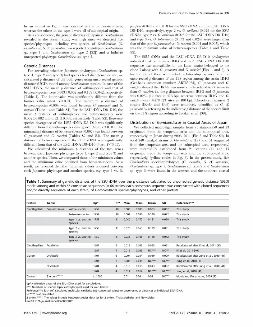
by an asterisk in Fig. 1 was consisted of the temperate strains,
whereas the others in the type 1 were all of subtropical origin.
As a consequence, the genetic diversity of Japanese Gambierdiscus
revealed in the present study was high since we detected five
species/phylotypes including two species of Gambierdiscus (G.
australes and G. cf. yasumotoi), two reported phylotypes (Gambierdiscus
sp. type 1 and Gambierdiscus sp. type 2 [22]) and a hitherto
unreported phylotype Gambierdiscus sp. type 3.
Genetic DistancesFor revealing whether Japanese phylotypes (Gambierdiscus sp.
type 1, type 2 and type 3) had species level divergence or not, we
calculated p distance of the both genes using uncorrected genetic
distance (UGD) model among Gambierdiscus species. In case of the
SSU rDNA, the mean p distance of within-species and that of
between-species were 0.00360.002 and 0.13960.042, respectively
(Table 1). The latter value was significantly different from the
former value (t-test, P,0.01). The minimum p distance of
between-species (0.004) was found between G. yasumotoi and G.
ruetzleri (Table 1 and Table S1). For the LSU rDNA D8–D10, the
mean p distance of within-species and between-species were
0.00260.002 and 0.12160.036, respectively (Table S2). Between-
species divergence of the LSU rDNA D8–D10 was significantly
different from the within-species divergence (t-test, P,0.01). The
minimum p distance of between-species (0.007) was found between
G. yasumotoi and G. ruetzleri (Tables S2 and S3). The mean p
distance of between-species of the SSU rDNA was significantly
different from that of the LSU rDNA D8–D10 (t-test, P,0.05).
We calculated the minimum p distances of the two genes
between each Japanese phylotype (type 1, type 2 and type 3) and
another species. Then, we compared those of the minimum values
and the minimum value obtained from between-species. As a
result, we revealed that the minimum values obtained between
each Japanese phylotype and another species, e.g. type 1 vs. G.
pacificus (0.040 and 0.018 for the SSU rDNA and the LSU rDNA
D8–D10, respectively), type 2 vs. G. caribaeus (0.028 for the SSU
rDNA), type 2 vs. G. carpenteri (0.025 for the LSU rDNA D8–D10)
and type 3 vs. G. polynesiensis (0.033 and 0.024), were larger than
that of the pair G. yasumotoi vs. G. ruetzleri (0.004 and 0.007), which
was the minimum value of between-species (Table 1 and Table
S2).
The SSU rDNA and the LSU rDNA D8–D10 phylogenies
indicated that our strains IR4G and Go3 (LSU rDNA D8–D10
sequence was unavailable for the latter strain) belonged to the
clade I along with G. yasumotoi and G. ruetzleri (Figs. 1 and 2). A
further test of their within-clade relationship by means of the
uncorrected p distance of the ITS region among the strain IR4G
(GenBank accession number: AB765925), G. yasumotoi and G.
ruetzleri showed that IR4G was more closely related to G. yasumotoi
than G. ruetzleri, i.e. the p distance between IR4G and G. yasumotoi
was 0.0252 (12 sites in 476 bp), whereas between IR4G and G.
ruetzleri was 0.0479 (23 sites in 480 bp). Therefore, Japanese 2
strains (IR4G and Go3) were tentatively identified as G. cf.
yasumotoi by referring to the indicative p distance of the species limit
on the ITS region according to Litaker et al. [39].
Distribution of Gambierdiscus in Coastal Areas of JapanWe collected macroalgal samples from 73 stations (50 and 23
originated from the temperate area and the subtropical area,
respectively) in Japan during 2006–2011 (Fig. 3 and Table S4). In
total 248 unialgal strains of Gambierdiscus (197 and 51 originated
from the temperate area and the subtropical area, respectively)
were successfully established from 24 stations (11 and 13
originated from the temperate area and the subtropical area,
respectively) (yellow circles in Fig. 3). In the present study, five
Gambierdiscus species/phylotypes (G. australes, G. cf. yasumotoi,
Gambierdiscus sp. type 1, Gambierdiscus sp. type 2 and Gambierdiscus
sp. type 3) were found in the western and the southern coastal
Table 1. Summary of genetic distances of the SSU rDNA over the p distance calculated by uncorrected genetic distance (UGD)model among and within 66 consensus sequences ( = 66 strains; each consensus sequence was constructed with cloned sequencesand/or directly sequence of each strain) of Gambierdiscus species/phylotypes, and other protists.
Protist Genus bp* n** Min. Max. Mean SD Reference***
Dinoflagellate Gambierdiscus within-species 1759 10 0.000 0.005 0.003 0.002 This study
between-species 1759 10 0.004 0.180 0.139 0.042 This study
type 1 vs. anotherspecies
1759 11 0.040 0.172 0.121 0.050 This study
type 2 vs. anotherspecies
1759 11 0.028 0.163 0.129 0.051 This study
type 3 vs. anotherspecies
1759 11 0.033 0.168 0.140 0.042 This study
Dinoflagellate Peridinium 1687 9 0.013 0.083 0.055 0.021 Recalculated after Ki et al., 2011 [40]
1687 9 0.015 0.085 NC**** NC**** Ki et al., 2011 [40]
Diatom Cyclotella 1704 6 0.004 0.034 0.019 0.009 Recalculated after Jung et al., 2010 [41]
1704 6 0.005 0.035 NC**** NC**** Jung et al., 2010 [41]
Discostella 1704 4 0.010 0.015 0.013 0.002 Recalculated after Jung et al., 2010 [41]
1704 4 0.011 0.017 NC**** NC**** Jung et al., 2010 [41]
Diatom 2 orders***** c. 1600 0.01 0.04 0.01 NC**** Moniz and Kaczmarska, 2009 [42]
bp*:Nucleotide bases of the SSU rDNA used for calculations.n**: Numbers of species (species/phylotypes) used for calculations.Reference***: Each ref. calculated molecular similarity (we converted values to uncorrected p distance) of individual SSU rDNA.NC****: Not calculated.2 orders*****: The values include between-species data set for 2 orders; Thalassiosirales and Naviculales.doi:10.1371/journal.pone.0060882.t001
Diversity and Distribution of Gambierdiscus in JPN
PLOS ONE | www.plosone.org 5 April 2013 | Volume 8 | Issue 4 | e60882

areas of Japan (areas B, C, D, E, F and G in Figs. 3 and 4). On the
other hand, our 248 strains contained no G. toxicus. No
Gambierdiscus cell was found in samples collected from all stations
of the Sea of Japan, except for KN, and some stations along Pacific
coast (gray circles in Fig. 3). Details of strains are summarized in
Table S5.
The species/phylotype composition plotted onto a map showed
a species/phylotype-specific pattern of their distribution (Fig. 4).
For example, G. cf. yasumotoi and type 3 were restricted in the
subtropical area (from the station OM and IR in areas E and G,
respectively) and the temperate area (WI in area B), respectively
(Fig. 4B). On the other hand, type 1, type 2 and G. australes were
widespread from the temperate area (areas B, C and D in Fig. 4) to
Figure 3. Map of research area. Location of each sampling station and its ID as well as presence (yellow circle) or absence (gray circle) ofGambierdiscus cells in the sample is shown. Each island is marked as A: Hokkaido, B: Honshu (main Isl.), C: Shikoku, D: Kyushu, E: Okinawa Island, F:Miyako Island, Ikema Island and Irabu Island (from right to left), G: Ishigaki Island and Iriomote Island (from right to left). A: the boreal area, B, C and D:the temperate area, E, F and G: the subtropical area.doi:10.1371/journal.pone.0060882.g003
Diversity and Distribution of Gambierdiscus in JPN
PLOS ONE | www.plosone.org 6 April 2013 | Volume 8 | Issue 4 | e60882

the subtropical area (areas E, F and G in Fig. 4) in the areas
studied (Table 2 and Table S4), with a tendency that type 1 and G.
australes were dominant in the subtropical area, whereas type 2 was
more abundant in the temperate area (Fig. 4A). Although type 2
exhibited a widespread distribution (Table 2, Fig. 4), they were
totally absent at the southern end of our study field (subtropical
island areas F and G in Fig. 4). Each sampling station usually held
1–3 species/phylotype (s), with an exceptional case in that four
species/phylotypes (type 1, type 2, type 3 and G. australes) were
recorded by during samplings on two different dates at a
temperate station WI (area B in Fig. 4B).
We compared the occurrence of each species/phylotype with
sea water temperature (SWT) measured when the samples were
taken. Tendencies that type 1 and G. australes were more abundant
in warmer water and that type 2 more commonly occurred in
cooler water were found (Table 2), e.g. type 1 cells were isolated
from 21.1uC to 32.4uC, G. australes cells were isolated from 19.1uCto 32.4uC, whereas type 2 cells were isolated from 17.2uC to
30.0uC (Table 2). We found no tendency of the occurrence of each
species/phylotype with salinity (Table 2).
Toxicity AnalysesThe toxicities in mice were tested by means of the dichloro-
methane soluble fraction (DSF, a fraction expected to contain
CTXs) and aqueous methanol soluble fraction (MSF, a fraction
expected to contain MTXs) using six strains representing four
species/phylotypes, viz., KW070922_1 (Gambierdiscus sp. type 1);
M080828_2 and T070411_1 (Gambierdiscus sp. type 2); WI9G and
WI11G (Gambierdiscus sp. type 3); S080911_1 (G. australes). The
results of mouse bioassay indicated the presence of toxicity in type
1, type 3 and G. australes but not in type 2 (Table 3 and Table S6).
In DSF toxicity, G. australes was the highest (67061024 MU 1,000
cells21) and type 1 also showed positive result (20 6 1024 MU
1,000 cells21). In MSF toxicity, both type 1 and G. australes showed
positive result (6761024 MU 1,000 cells21). Although one strain
of type 3 (WI11G) was positive for MSF toxicity (67 MU 1,000
cells21), another strain WI9G was negative (Table 3 and Table
S6).
Discussion
Genetic Diversity of Japanese GambierdiscusA previous phylogeographic study of Japanese Gambierdiscus
using the SSU rDNA and the ITS region revealed the existence of
two putative allopatric phylotypes: Gambierdiscus sp. type 1 and
Gambierdiscus sp. type 2 [22]. On the other hand, we detected five
species/phylotypes including the two reported phylotypes of
Gambierdiscus (type 1 and type 2 [22]), two species of Gambierdiscus
(G. australes and G. cf. yasumotoi) and a hitherto unreported
phylotype Gambierdiscus sp. type 3 from western and southern
coastal areas of Japan. Considering these results, the unexpectedly
high genetic diversity of Japanese Gambierdiscus, up to five species/
phylotypes in the present study, might simply be explained by the
intensiveness of sampling, viz. the more strains established the
more probability to encounter something new.
The minimum uncorrected p distances (0.028–0.040) of the
SSU rDNA obtained from the pairs between Japanese phylotypes
(type 1, type 2 and type 3) and another species, were larger than
that of the pair G. yasumotoi vs. G. ruetzleri (0.004) which was the
minimum value of between-species of Gambierdiscus. Furthermore,
these values were also larger than the minimum p distances
calculated for between-species from other protists, e.g. dinoflagel-
late Peridinium (between 9 species, 0.013) [40], diatom Cyclotella
(between 6 species, 0.004) [41], diatom Discostella (between 4
species, 0.010) [41] and 2 orders of diatoms (0.01) [42] (Table 1).
Therefore, the minimum p distances obtained from the pairs
between Japanese phylotypes and another species are most likely
indicative of species level divergence, and Japanese phylotypes will
deserve to be described as new species. Currently detailed
morphological investigations are ongoing to support the descrip-
tion of these phylotypes (Nishimura et al. unpubl.). It also should
be noted that the minimum p distances obtained from the pairs G.
yasumotoi vs. G. ruetzleri was the same as or lower than the values
obtained from other protists, and was close to those within-species
level divergence of Gambierdiscus species.
The mean p distance of the SSU rDNA obtained from between-
species (0.139) was larger than those of other protists, e.g.
dinoflagellate Peridinium (between 9 species, 0.055) [40], diatom
Cyclotella (between 6 species, 0.019) [41], diatom Discostella
(between 4 species, 0.013) [41] and 2 orders of diatoms (0.01)
[42]. These results suggest that the genetic diversity of Gambierdiscus
seems to be larger than other protists.
In relation to Gambierdiscus sp. type 2 from Japanese coastal
waters, G. caribaeus GCJJ1 strain from Jeju Island, Korea, Pacific
was reported to be closely related with a Japanese strain C-1,
which belongs to Gambierdiscus sp. type 2 [22], by Jeong et al. [23].
They also reported that the SSU rDNA sequence of Korean strain
GCJJ1, which has not been opened on DNA Data Bank of Japan
(DDBJ) yet, was 0.5% different (0.005 for uncorrected p distance in
the present study) from that of strain C-1 of Gambierdiscus sp. type
2, whereas the Korean strain GCJJ1 was 2.4–4.0% different
(0.024–0.040 for uncorrected p distance) from those of G. caribaeus
[23]. On the other hand, morphological observations using SEM
suggested that morphology of Korean strain GCJJ1 was similar to
those of Belize strains of G. caribaeus. Considering the similar
morphology and the molecular divergence between them, the
authors suggested that the Korean strain GCJJ1 may be a cryptic
species of G. caribaeus. Together with our results that the mean p
distance (0.13960.042) and the minimum p distance (0.004)
among Gambierdiscus species, the Korean strain GCJJ1 may belong
to Gambierdiscus sp. type 2 rather than G. caribaeus and the strain as
well as Japanese strains of type 2 are to be described as a new
species.
The SSU rDNA and the LSU rDNA D8–D10 phylogenies
indicated that our strains IR4G and Go3 (LSU rDNA D8–D10
sequence was unavailable for the latter strain) belonged to the
clade I, along with G. yasumotoi and G. ruetzleri. An empirical study
by Litaker et al. [39] proposed that uncorrected p distance
between 0.042 and 0.580 substitutions per site in the ITS region
are indicative of species level divergence of the ITS region based
on their observations among 14 genera of dinoflagellate. Recently
Litaker et al. [20] applied the idea with Gambierdiscus, where two
morphologically similar but barely and genetically distinguishable
species G. yasumotoi and G. ruetzleri had p = 0.07 for the ITS region.
As a result of our sequence analyses, we provisionally called our
strain IR4G as G. cf. yasumotoi, given the p distance of the ITS
region between IR4G and G. yasumotoi was 0.0252, whereas
between IR4G and G. ruetzleri was 0.0479. In the case of the SSU
rDNA sequences comparison, however, relationships within clade
I are still confusing, because the p distances obtained from the
pairs G. yasumotoi vs. G. ruetzleri was lower value comparing with
those of other protists, and the value was similar with within-
species level divergence of Gambierdiscus species. This indicates that
additional works are needed to confirm its taxonomical status.
Distribution of Gambierdiscus in Coastal Areas of JapanKuno et al. [22] revealed the existence of two allopatric
phylotypes: Gambierdiscus sp. type 1 from the subtropical area
Diversity and Distribution of Gambierdiscus in JPN
PLOS ONE | www.plosone.org 7 April 2013 | Volume 8 | Issue 4 | e60882

Diversity and Distribution of Gambierdiscus in JPN
PLOS ONE | www.plosone.org 8 April 2013 | Volume 8 | Issue 4 | e60882

and Gambierdiscus sp. type 2 from the temperate area. On the other
hand, we confirmed both of the two phylotypes occur from the
subtropical area to the temperate area. In addition to those
distributions, we found that G. australes occurs from the subtropical
area to the temperate area, whereas, G. cf. yasumotoi and
Gambierdiscus sp. type 3 were restricted to the subtropical area
and the temperate area, respectively. Additionally, we found a
tendency that type 1 and G. australes were dominant in the
subtropical area, whereas type 2 was dominant in the temperate
area. Considering those trends of the distribution of each species/
phylotype in Japan, different growth response of each species/
phylotype to various environmental parameters, such as SWT and
salinity so on, would determine those trends of the distribution.
Comparing those distributions with SWT when each cell was
isolated, type 1 and G. australes cells were isolated from warmer
waters, while type 2 cells were isolated from cooler waters.
Additionally, type 2 cells were isolated from KN (35u41931 N,
135u18916 E), in which Hatayama et al. [38] has also observed
Gambierdiscus cells. The sampling station is the most northerly
location in the world where Gambierdiscus spp. have been reported
so far. These results indicate that type 2 appears to prefer cooler
waters to warmer waters because of their growth response which
probably plays a crucial role in their predomination around the
temperate areas in Japan. To confirm this hypothesis, comparative
culture experiments with type 1, type 2, type 3 and G. australes are
currently ongoing (Yoshimatsu et al. unpubl.). Additionally, we
found no Gambierdiscus cell from Japanese northern stations (HO,
HF, IW, NS and NC), where the number of samples was poorly
represented in our collection. The absence of Gambierdiscus from
our northern samples could also be explained by their restricted
distribution in warm waters: so far there is no Gambierdiscus found
from boreal area.
ToxicityChinain et al. [18] applied mouse bioassay (MBA) for detection
of toxicities of four Gambierdiscus species (G. toxicus, G. pacificus, G.
polynesiensis and G. australes) isolated from French Polynesia, Pacific
and revealed that G. pacificus, G. polynesiensis and G. australes showed
both toxicities of DSF (a fraction expected to contain CTXs) and
MSF (a fraction expected to contain MTXs), whereas G. toxicus
showed only MSF toxicity. In the present study, MBA results of
Japanese Gambierdiscus revealed that three species/phylotypes had
toxicities (G. australes, Gambierdiscus sp. type 1 and Gambierdiscus sp.
type 3) and a phylotype (Gambierdiscus sp. type 2) was thought to be
non-toxic. Unfortunately, G. cf. yasumotoi could not be assessed for
its toxicity, because mass culturing was not successful. However a
strain of G. yasumotoi isolated from Singapore was found to be
potentially toxic [17].
In DSF toxicities among Japanese Gambierdiscus, the toxicity of
G. australes was stronger than that of type 1. Additionally, for type 3
which was revealed to be ‘‘non-toxic’’ for DSF in the present
study, a strain WI9G exhibited progressive paralysis of mice or
death of mice however the proportion that died did not exceed
50%. The DSF toxicity of Japanese G. australes was approx. 170
fold stronger than that of G. australes isolated from French
Polynesia [18]. This suggests that the toxicity of a Gambierdiscus
species is possibly variable being related with location where each
strain was isolated. Furthermore, the degree of DSF toxicity of
Japanese G. australes followed the toxicities of G. polynesiensis strains,
which are known as ‘super-toxic strains [43]’. The DSF toxicity of
type 1 followed that of Japanese G. australes. In MSF toxicities
among Japanese Gambierdiscus, the toxicities of G. australes, type 1
and type 3 were the same value, furthermore those toxicities were
stronger than those of G. toxicus, G. pacificus, G. polynesiensis and G.
australes isolated from French Polynesia [18]. Considering these
results, it was revealed that species/phylotypes which have
relatively strong DSF and MSF toxicities so far may distribute
around Japanese coastal areas.
Conclusion/Future ProspectsAs a result of phylogenies of the SSU rDNA and the LSU rDNA
D8–D10, we revealed that five species/phylotypes including two
reported phylotypes (Gambierdiscus sp. type 1 and Gambierdiscus sp.
type 2 [22]), two species of Gambierdiscus (G. australes and G. cf.
Figure 4. Geographic distribution of Gambierdiscus species/phylotypes. Enlargement of a part enclosed by an open square in Fig. 3. Thenumbers in each pie indicate the number of strains used for phylotyping. Each color in pies corresponds to a species/phylotype in phylogenetic treesin Figs. 1 and 2, i.e. red: Gambierdiscus sp. type 1, blue: Gambierdiscus sp. type 2, orange: Gambierdiscus sp. type 3, purple: G. australes, green: G. cf.yasumotoi. Each island is marked as B: Honshu (main Isl.), C: Shikoku, D: Kyushu, E: Okinawa Island, F: Miyako Island, Ikema Island and Irabu Island(from right to left), G: Ishigaki Island and Iriomote Island (from right to left). B, C and D: the temperate area, E, F and G: the subtropical area. Figure 4A:Total number of strains and composition of each species/phylotype. Figure 4B: Breakdown of species/phylotype composition from each samplingstation.doi:10.1371/journal.pone.0060882.g004
Table 2. Details of sampling stations where each Gambierdiscus species/phylotype was observed.
Species/phylotype SWT (6C) SalinityLatitude (samplingstation) Area Station
Gambierdiscus sp. type 1 21.1–32.4 29.2–33.0 24u26940 N (ISC)–33u27910 N (WI)
B, C, D,E, F, G
WI, M, T, S, K, KW, OI, GTN, OSS, U, G, MYA, MYI, ISC, ISB
Gambierdiscus sp. type 2 17.2–30.0 29.7–33.2 26u07958 N (ON)–32u52932 N (KN)
B, C, E KN, MME, WI, WK, M, T, S, KW, OI, OBS, U, ON
Gambierdiscus sp. type 3 22.2 ND* 33u27910 N (WI) B WI
Gambierdiscus australes 19.1–32.4 30.5–32.2 24u26940 N (ISC)–33u27910 N (WI)
B, C, E,F, G
WI, M, S, OSS, U, ON, G, OD, I, IR, ISC, ISB
Gambierdiscus cf. yasumotoi 24.8 ND* 24u25906 N (IR)–26u26941 N (OM)
E, G OM, IR
ND*: No data.doi:10.1371/journal.pone.0060882.t002
Diversity and Distribution of Gambierdiscus in JPN
PLOS ONE | www.plosone.org 9 April 2013 | Volume 8 | Issue 4 | e60882

yasumotoi) and a hitherto unreported phylotype Gambierdiscus sp.
type 3 distribute around Japanese coastal areas, especially western
and southern areas. Out of Japanese five species/phylotypes, the
three phylotypes (type 1, type 2 and type 3) that showed species
level genetic divergence by calculating uncorrected genetic
distance using these genes will deserve to be described as new
species. Also the distribution of Japanese Gambierdiscus was
revealed; G. cf. yasumotoi and type 3 distributed allopatrically to
the subtropical area and the temperate area, respectively. On the
other hand, type 1, type 2 and G. australes occurred from the
subtropical area to the temperate area, with a tendency that type 1
and G. australes were dominant in the subtropical area, whereas
type 2 was dominant in the temperate area. We revealed a
surprising diversity of Japanese Gambierdiscus, and the distribution
of five species/phylotypes displayed clear geographical patterns in
the subtropical and temperate areas. For revealing toxicities of
those species/phylotypes, MBA was conducted and revealed that
G. australes and type 1 had toxicities of DSF and MSF, and type 3
had only toxicity of MSF, whereas no toxicities of both fractions
were detected from type 2. In the DSF toxicities, G. australes was
stronger than that of type 1. So far, occurrences of CFP incidents
have been mainly reported from the subtropical area of Japan that
might be explained by the result obtained in the present study that
the toxic type 1 and G. australes are dominant in subtropical area,
viz. it is suggests that one of agents of CFP incidents occurred in
the subtropical area of Japan might be Gambierdiscus sp. type 1
and/or G. australes. Comparative LC-MS studies on extracts from
both cultured Gambierdiscus and ciguateric fish are required to
elucidate as to whether the occurrence of CFP is linked to the
toxins produced by Gambierdiscus.
As a whole, the SSU rDNA and the LSU rDNA D8–D10 as
genetic marker is recommended for further use, i.e. species/
phylotype-level comparison such as molecular systematic/taxon-
omy within the genus. Now we are trying to establish the
molecular-based monitoring system using quantitative polymerase
chain reaction (qPCR) assay for the detection of each toxic
species/phylotype of Gambierdiscus in Japanese coastal areas.
Materials and Methods
Ethics StatementNo specific permits were required for the described field studies.
No specific permission was required for any locations and activity.
The locations are not privately owned or protected in any way. No
activity during field study involved any endangered species or
protected species.
The Animal Use Protocol (AUP) for handling mouse described
here was approved by the Animal Ethics Committee (approval ID:
D-00068) of Kochi University.
Isolation and Establishment of Gambierdiscus StrainsMacroalgal substrata were collected mainly from the southern
part of Japan between 2006 and 2011. Details of the sampling
stations are shown in Table S4. In the laboratory the macroalgae
were placed in a plastic bottle and vigorously shaken to cause
epiphytes, including Gambierdiscus, to detach from the substrata.
The resultant suspension was sieved twice, firstly through 150 mm
and then through 20 mm Nitex meshes. Materials retained on the
second mesh filter were resuspended in filtered seawater.
Gambierdiscus cells placed in a 6 well/flat bottom microplate (Asahi
Glass, Tokyo, Japan) were isolated under an inverted microscope
referring to the morphological criteria of the genus Gambierdiscus
described by Adachi and Fukuyo [14]. All the clonal strains were
maintained with Daigo IMK (Nihon Pharmaceutical, Tokyo,
Japan), IMK/2, IMK/4 or soil-extract-added IMK/4 medium
using GF/F-filtered sea water (salinity of 3161) in polypropylene
(PP)-capped test tubes (256150 mm) with a flat bottom (Mar-
uemu, Osaka, Japan) containing 20 ml medium at 25uC, with
100 mmol photons?m–2?s–1 from cool-white tubes; the photoperiod
was 12:12 h L:D.
Table 3. Toxicities of Japanese Gambierdiscus species/phylotypes tested by a mouse bioassay. One MU is defined as the LD50 dosefor a 20-g mouse over 24 h.
Species/phylotype Strain DSF toxicity* MSF toxicity** Reference
(61024 MU/1,000 cells) (61024 MU/1,000 cells)
Gambierdiscus sp. type 1 KW070922_1 20 67 This study
Gambierdiscus sp. type 2 M080828_2 _*** _*** This study
T070411_1 _*** ND**** This study
Gambierdiscus sp. type 3 WI9G _*** _*** This study
WI11G _*** 67 This study
Gambierdiscus australes S080911_1 670 67 This study
RAV-92 4 0.2 Chinain et al., 1999 [18]
Gambierdiscus toxicus GTT-91 _*** 0.7 Chinain et al., 1999 [18]
REN-1 _*** 1.7 Chinain et al., 1999 [18]
TUR _*** 0.6 Chinain et al., 1999 [18]
Gambierdiscus pacificus HO-91 9 0.7 Chinain et al., 1999 [18]
Gambierdiscus polynesiensis TB-92 1500 0.1 Chinain et al., 1999 [18]
RG-92 800 0.06 Chinain et al., 1999 [18]
DSF*: Dichloromethane soluble fraction (DSF) toxicities correspond to CTXs toxicity.MSF**: Aqueous methanol soluble fractions (MSF) toxicities correspond to MTXs toxicity._***: Not detected.ND****: Not done.doi:10.1371/journal.pone.0060882.t003
Diversity and Distribution of Gambierdiscus in JPN
PLOS ONE | www.plosone.org 10 April 2013 | Volume 8 | Issue 4 | e60882

DNA Extraction, PCR, Cloning and SequenceTo amplify the SSU rDNA and the LSU rDNA D8–D10 we
performed direct PCR, which used intact cells for PCR template
instead of genomic DNA allowing us to skip the DNA extraction
process, in order to analyze large numbers of isolates quicker and
more efficiently. For harvesting cells for template, 1 ml of medium
containing Gambierdiscus cells was centrifuged to make a pellet,
which was subsequently washed twice with sterile water. A small
fraction was picked from the pellet by a micropipette and
transferred into a PCR tube that contained a 25-ml mixture as
below. In case direct PCR failed to amplify the SSU rDNA and/or
the LSU rDNA D8–D10, and the ITS region for IR4G, genomic
DNA was extracted from the strains using DNeasy Plant Mini Kit
(Qiagen, Valencia, CA, USA).
The SSU rDNA was amplified by using 50 mM of oligonucleotide
primers Dino5’UF [44] and 18 ScomR1 [45] (Table 4). PCR
reactions typically contained a 25-ml mixture: 0.5 ml of MightyAmp
DNA Polymerase (1.25 U/ml, Takara Bio, Shiga, Japan); primers as
above (7.5 pmol each); 12.5 ml of 26MightyAmp buffer (Mg2+,
dNTP plus) which contains magnesium chloride (4 mM) and
dNTPs (800 mM each). The PCR cycling comprised of an initial
2 min heating step at 98uC, followed by 35 cycles: 98uC for 10 sec,
68uC for 110 sec, and a final extension at 68uC for 5 min. The PCR
amplification of the SSU rDNA for strains from several sampling
stations (MME, OBS, OSS, OM, OD, MYA and MYI, Table S4)
was performed at University of the Ryukyus using primers and
methods described in Takano and Horiguchi [46]. The LSU rDNA
was amplified by using 50 mM of oligonucleotide primers FD8 and
RB [18] (Table 4). The PCR cycling comprised of an initial 2 min
heating step at 98uC, followed by 25 cycles: 98uC for 10 sec, 55uCfor 15 sec and 68uC for 40 sec, and a final extension at 68uC for
5 min. The ITS region was amplified by using 50 mM of
oligonucleotide primers ITSA [47] and D2C [48] (Table 4). The
PCR cycling comprised of an initial 2 min heating step at 98uC,
followed by 35 cycles: 98uC for 10 sec, 55uC for 15 sec and 68uC for
75 sec, and a final extension at 68uC for 5 min.
The quantity and length of products were examined by agarose
gel electrophoresis against known standards. Excess primers and
dNTPs were removed from PCR product using High Pure PCR
Cleanup Micro Kit (Roche, Tokyo, Japan). Each product was
sequenced directly or cloned into the T-vector pMD20 (TaKaRa
Bio, Shiga, Japan). Clones were screened for inserts by PCR
amplification with plasmid primers U19 and pUCM13R (Table 4).
BigDyeH Terminator v3.1 Cycle Sequencing Kit (Applied
Biosystems Japan, Chiba, Japan) was used for the sequence of
the PCR products and the clones. Primers and excess dye-labeled
nucleotides were removed using the Performa DTR V3 clean-up
system (Edge Biosystems, Gaithersburg, MD).
For easy and quick phylotyping of strains, approximately
500 bp including the most variable 300 bp region of the SSU
rDNA [44] were sequenced by internal sequencing primer G10R
[49] (Table 4). Sequencing products were run on an ABI PRISM
3100-Avant Genetic Analyzer (Applied Biosystems Japan, Chiba,
Japan). Forward and reverse reads were edited and aligned using
SeqMan (DNASTAR, Madison, WI). All the information of clonal
strains, including locality, water temperature and source sample
are listed in Table S5.
Phylogeny and Sequence AnalysesThe SSU rDNA and the LSU rDNA D8–D10 sequences were
aligned using ClustalW [50] with publicly available ones retrieved
from DNA Data Bank of Japan (DDBJ). In both rDNA datasets,
the 59 and 39 ends were manually aligned to truncate and refine
the both ends. Finally, ambiguously aligned positions were
excluded, resulting in 190 taxa/1757 nucleotides (32 bp–
1762 bp site of Pfiesteria piscicida, AY245693 followed the alignment
used in Litaker et al. [44]) and 136 taxa/846 nucleotides
(1980 bp–2978 bp site of Prorocentrum micans, X16108 followed
the alignment used in Lenaers et al. [51]) in the SSU rDNA and
the LSU rDNA D8–D10 dataset, respectively.
MrBayes 3.1.2 [52,53] was used for Bayesian inference (BI) to
estimate the posterior probability distribution using Metropolis-
Coupled Markov Chain Monte Carlo (MCMCMC) [53].
MCMCMC from a random starting tree were used in this
analysis with two independent runs and 1 cold and 3 heated chain
with temperature set 0.2. Trees were sampled every 100 th
generation. To increase the probability of chain convergence, we
sampled at least 10,000 trees after the standard deviation values of
the two runs dipped below 0.01 to calculate the posterior
probabilities. RAxML-VI-HPC, v7.0.4 [54] was used for Maxi-
mum likelihood (ML) analyses. We conducted a rapid Bootstrap
analysis and search for the best-scoring ML tree in one single run
with -f a option for 100 repeats. MrModeltest 2 [55] was used to
determine the most appropriate model of sequence evolution. The
best-fit model according to the Akaike Information Criterion (AIC)
was GTR+G, and this was used for BI and ML analyses.
With the ITS region we calculated the uncorrected genetic
distance (p) to consider substitutions, gaps and ambiguous bases for
calculations among G. yasumotoi, G. ruetzleri and Japanese strain
IR4G, in order to find the closest relative of these strains. The ITS
region sequences of G. yasumotoi and G. ruetzleri were obtained from
Fig. 74 in Litaker et al. [20]. A consensus of the ITS region
sequence for six separate clones (sequences obtained via cloning) of
IR4G was used for the analysis to reduce any bias due to the
inclusion of pseudogene sequences. Additionally we also calculated
p distance using uncorrected genetic distance (UGD) model (p-
distance model in MEGA 5.05 [56]) among Gambierdiscus species in
that strict consensus sequences were obtained both for each species
using the SSU rDNA and the LSU rDNA D8–D10 and revealed
the p distances of within-species and between-species. Then, we
calculated the p distances between each Japanese phylotype and
another species. All positions containing alignment gaps and
missing data were eliminated only in pairwise comparisons
(Pairwise deletion option in MEGA 5.05 [56]). For the UGD
model of the SSU rDNA and the LSU rDNA D8–D10 calculation
in the present study, using MEGA 5.05, gaps and ambiguous bases
were not considered.
Toxicity AnalysesSix strains representing four species/phylotypes, viz.,
KW070922_1 (Gambierdiscus sp. type 1); M080828_2 and
T070411_1 (Gambierdiscus sp. type 2); WI9G and WI11G
(Gambierdiscus sp. type 3); S080911_1 (G. australes), were maintained
in glass Petri dishes (SANSYO, Tokyo, Japan) filled with 20 mL of
Daigo IMK medium under culturing conditions described as
above. Cells in the late stationary phase (approx. 30 days) were
harvested by filtration using 20 mm Nitex mesh and/or centrifug-
ing. Toxic extracts were prepared by extraction from cell pellets,
by using the method modified after Chinain [18], thrice in
methanol (2 mL for a total biomass of 1.06106 cells) and thrice in
aqueous methanol (MeOH:H2O 9:1) (1 mL for a total biomass of
1.0 6106 cells) under sonication, for 15 min. each. After dividing
toxic extracts into the required number of specimens, the extract
was evaporated, a solvent partition was applied to the resulting
reside using 0.4 mL of dichloromethane (CH2Cl2) once and
0.2 mL of aqueous methanol (MeOH:H2O 6:4) twice. During
liquid-liquid partitions, the dichloromethane and aqueous meth-
anol phases, in which CTXs and MTXs are recovered respec-
Diversity and Distribution of Gambierdiscus in JPN
PLOS ONE | www.plosone.org 11 April 2013 | Volume 8 | Issue 4 | e60882

tively, were handled with extreme care in order to limit carry over
of MTXs into the dichloromethane phase, and vice versa. The
dichloromethane soluble fraction (DSF) and aqueous methanol
soluble fraction (MSF) were evaporated, respectively.
The DSF and MSF of six strains of Gambierdiscus sp. were tested
for their toxicity using mouse bioassay. Three or five mice (male,
ddY, 20 g; Japan SLC, Inc, Shizuoka, Japan) were intraperitone-
ally administered with a single dose of toxic extract dissolved in
500 ml of a 0.85% saline solution containing 1% Tween 60. Mice
were observed over 24 h and signs and time of death recorded.
Fractions were considered non-toxic if injection of a maximal dose
was not lethal. Total lethality was expressed in mouse units (MU)
1,000 cells21, one MU is defined as the i.p. LD50 dose for a 20 g
mouse over 24 h.
Supporting Information
Figure S1 Maximum likelihood (ML) phylogeny of theD8–D10 region of the LSU rDNA of Gambierdiscusspecies/phylotypes. Nodal supports are of ML analysis. Nodes
with strong supports (pp/bt = 1.00/100) are shown as thick lines.
For sequences obtained via cloning, a variant ID, starting with C,
is shown followed by strain ID (i.e. T080908_1_C1). Sequences
obtained in the present study are indicated in color.
(TIF)
Table S1 Estimation of genetic distances of the SSUrDNA over the p distance calculated by uncorrectedgenetic distance (UGD) model among and within 69
Table 4. Oligonucleotide primers used for PCR amplification and DNA sequencing.
Primer name Synthesis direction Sequence (5’–3’) Anneals to Reference
The ITS1-5.8S-ITS2rDNA
ITS A Forward GTA ACA AGG THT CCG TAG GT 22–41* Sato et al., 2011 [47]
type4ITSICF Forward for sequencing TTG TTG GTT TCC CCT CAA 83–102* This study
inner5.8S_F Forward for sequencing AAA TTG CAG AAT YCC GTG AG 306–325* This study
inner5.8S_R2 Reverse for sequencing TGA CTC ACG GRA TTC TGC 311–328* This study
type4ITSICR Reverse for sequencing GTC TGC CAG TGT CAC AAT GC 473–492* This study
ITS B Reverse for sequencing AKA TGC TTA ART TCA GCR GG 542–561* Sato et al., 2011 [47]
D2C Reverse CCT TGG TCC GTG TTT CAA GA 714–733** Scholin et al., 1994 [48]
The SSU rDNA
Dino5’UF Forward CAA CCT GGT GAT CCT GCC AGT 1–23*** Litaker et al., 2005 [49]
18ScomF1 Forward for sequencing GCT TGT CTC AAA GAT TAA GCC TAG C 32–56*** Zhang et al., 2005 [45]
18S_G53F Forward for sequencing TGC ATG TCT CAG CTT AAG TG 54–73*** This study
G10’F Forward for sequencing TGG AGG GCA AGT CTG GTG 549–566*** This study
18S_G623F Forward for sequencing GTT AAA AGG CTC GTA GTT GGA 618–638*** This study
G17’F Forward for sequencing ATA CCG TCM TAG TCT TAA CC 1007–1026*** Modified after Litaker et al., 2003 [44]
18S_G1249F Forward for sequencing GGA TTG ACA GAT TGA CAG CT 1232–1251*** This study
G18F Forward for sequencing CAA TAA CAG GTC TGT GAT GC 1426–1445*** Litaker et al., 2003 [44]
18S_G371R Reverse for sequencing ACC CTC ATC CTC CGT CAC CT 359–378*** This study
G10R Reverse for sequencing CCG CGG CTG CTG GCA CCA GAC 559–579*** Litaker et al., 2005 [49]
18S_G781R Reverse for sequencing AAA CAC CTG CTT TGA ACA C 768–786*** This study
G17’R Reverse for sequencing GTT TAT GGT TAA GAC TAK GAC GG 1010–1032*** This study
18S_G1301R Reverse for sequencing CAC TCC ACC AAC TAA GAA CG 1279–1303*** This study
G18R Reverse for sequencing GCA TCA CAG ACC TGT TAT TG 1426–1445*** Litaker et al., 2005 [49]
18S_G1781R Reverse for sequencing GAA ACC TTG TTA CGA CTT CT 1758–1777*** This study
18ScomR1 Reverse CAC CTA CGG AAA CCT TGT TAC GAC 1762–1785*** Zhang et al., 2005 [45]
The D8–D10 region of the LSU rDNA
FD8 Forward GGA TTG GCT CTG AGG GTT GGG 1980–2000** Chinain et al., 1999 [18]
GLD8_421F Forward for sequencing ACA GCC AAG GGA ACG GGC TT 2395–2414** This study
GLD8_677R Reverse for sequencing TGT GCC GCC CCA GCC AAA CT 2645–2664** This study
RB Reverse GAT AGG AAG AGC CGA CAT CGA 2905–2925** Chinain et al., 1999 [18]
T-Vector pMD20
U19 Forward GGT TTT CCC AGT CAC GAC G In the vector Applied Biosystems
pUCM13R Reverse CAG GAA ACA GCT ATG AC In the vector Promega
*: Annealing site in the ITS1-5.8S-ITS2 rDNA sequence of Gambierdiscus yasumotoi GYASU, GU968498 (Vandersea et al., 2010 Direct Submission).**: Annealing site in the LSU rDNA sequence of Prorocentrum micans, X16108 (Lenaers et al., 1989 [51]).***: Annealing site in the SSU rDNA sequence of Pfiesteria piscicida, AY245693 (Litaker et al., 2003 [44]).doi:10.1371/journal.pone.0060882.t004
Diversity and Distribution of Gambierdiscus in JPN
PLOS ONE | www.plosone.org 12 April 2013 | Volume 8 | Issue 4 | e60882

consensus sequences ( = 69 strains; each consensussequence was constructed with cloned sequences and/or directly sequence of each strain) of Gambierdiscusspecies/phylotypes.(XLS)
Table S2 Summary of genetic distances of the D8–D10region of the LSU rDNA over the p distance calculated byuncorrected genetic distance (UGD) model among andwithin 51 consensus sequences ( = 51 strains; eachconsensus sequence was constructed with cloned se-quences and/or directly sequence of each strain) ofGambierdiscus species/phylotypes.(XLS)
Table S3 Estimation of genetic distances of the D8–D10region of the LSU rDNA over the p distance calculated byuncorrected genetic distance (UGD) model among andwithin 61 consensus sequences ( = 61 strains; eachconsensus sequence was constructed with cloned se-quences and/or directly sequence of each strain) ofGambierdiscus species/phylotypes.(XLS)
Table S4 Details of macroalgal sample collection.(XLS)
Table S5 Details of Gambierdiscus spp. clonal strains.
(XLS)
Table S6 Details of toxicities of Japanese Gambierdis-cus species/phylotypes tested by a mouse bioassay. One
MU is defined as the LD50 dose for a 20-g mouse over 24 h (n = 3
or 5 in the present study).
(XLS)
Acknowledgments
We thank K. Ohnishi, Kochi University for allowing us to access his
facilities and technical help. We are grateful to H. Akino, T. Fujimoto, T.
Fukao, M. Furukawa, K. Ichimi, T. Ikeda, T. Ikegami, R. Ishimoto, H.
Iwamoto, M. Kanemaru, K. Kishimoto, S. Komatsu, S. Kondo, T.
Kusumoto, D. Muraoka, T. Murata, K. Nabeuchi, T. Nakamura, H.
Nishimura, Y. Nomiya, N. Oka, T. Okami, H. Shimada, Y. Tanimoto, K.
Teeyapon, K. Tose, M. Tsuda, H. Yamashita and T. Yoshikawa for
collecting macroalgal samples.
Author Contributions
Conceived and designed the experiments: TN MMR S. Sato S. Suda MA.
Performed the experiments: TN MMR. Analyzed the data: TN S. Sato
MA. Contributed reagents/materials/analysis tools: TN S. Sato WT HS
KU MMR S. Suda TY YT HY MA. Wrote the paper: TN S. Sato MA.
References
1. Ragelis EP (1984) Ciguatera seafood poisoning: overview. In: Ragelis EP, eds.
Seafood Toxins, American Chemical Society, Washington D.C. 22–36.
2. Bagnis R (1993) Ciguatera fish poisoning. In: Falconer IR, eds. Algal Toxins in
Seafood and Drinking Water, Academic Press, New York. 105–115.
3. Yasumoto T, Satake M (1996) Chemistry, etiology and determination methods
of ciguatera toxins. J Toxicol - Tox Rev 15: 91–107.
4. Fleming LE, Baden DG, Bean JA, Weisman R, Blythe DG (1998) Seafood toxin
diseases: issues in epidemiology and community outreach. In: Reguera B, BlancoJ, Fernandez MK, Wyatt T, eds. Harmful Algae, Xunta de Galicia and
Intergovernmental Oceanographic Commission of UNESCO. 245–248.
5. Lehane L, Lewis RJ (2000) Ciguatera: recent advances but the risk remains.
Int J Food Microbiol 61: 91–125.
6. Chinain M, Darius HT, Ung A, Fouc MT, Revel T, et al. (2010) Ciguatera risk
management in French Polynesia: The case study of Raivavae Island (Australes
Archipelago). Toxicon 56: 674–690.
7. Tester PA, Feldman RL, Nau AW, Kibler SR, Litaker RW (2010) Ciguatera fish
poisoning and sea surface temperatures in the Caribbean Sea and the WestIndies. Toxicon 56: 698–710.
8. Lewis RJ (2001) The changing face of ciguatera. Toxicon 39: 97–106.
9. Oshiro N, Yogi K, Asato S, Sasaki T, Tamanaha K, et al. (2010) Ciguatera
incidence and fish toxicity in Okinawa, Japan. Toxicon 56: 656–661.
10. Taniyama S (2008) The occurrence of palytoxin-like poisoning and ciguatera in
parts of the main land of Japan. Nippon Suisan Gakkaishi 74: 917–918. (InJapanese).
11. Ishikawa A, Kurashima A (2010) Occurrence of the toxic benthic dinoflagellateGambierdiscus toxicus in Ago Bay, central part of Japan. Bull Jpn Soc Fish
Oceanogr 74: 13–19. (In Japanese).
12. Oshiro N, Matsuo T, Sakugawa S, Yogi K, Matsuda S, et al. (2011) Ciguatera
fish poisoning on Kakeroma Island, Kagoshima Prefecture, Japan. Trop MedHealth 39: 53–57. (In Japanese).
13. Yasumoto T, Nakajima I, Bagnis R, Adachi R (1977) Finding of a dinoflagellateas a likely culprit of ciguatera. Bull Japan Soc Sci Fish 43: 1021–1026.
14. Adachi R, Fukuyo Y (1979) The thecal structure of a marine toxic dinoflagellateGambierdiscus toxicus gen. et sp. nov. collected in a ciguatera-endemic area. Bull
Japan Soc Sci Fish 45: 67–71.
15. Fraga S, Rodrıguez F, Caillaud A, Diogene J, Raho N, et al. (2011) Gambierdiscus
excentricus sp. nov. (Dinophyceae), a benthic toxic dinoflagellate from the Canary
Islands (NE Atlantic Ocean). Harmful Algae 11: 10–22.
16. Faust MA (1995) Observation of sand-dwelling toxic dinoflagellates (Dinophy-
ceae) from widely differing sites, including two new species. J Phycol 31: 996–1003.
17. Holmes MJ (1998) Gambierdiscus yasumotoi sp. nov. (Dinophyceae), a toxic benthicdinoflagellate from southeastern Asia. J Phycol 34: 661–668.
18. Chinain M, Faust MA, Pauillac S (1999) Morphology and molecular analyses ofthree toxic species of Gambierdiscus (Dinophyceae): G. pacificus, sp. nov., G.
australes, sp. nov., and G. polynesiensis, sp. nov. J Phycol 35: 1282–1296.
19. Richlen ML, Morton SL, Barber PH, Lobel PS (2008) Phylogeography,
morphological variation and taxonomy of the toxic dinoflagellate Gambierdiscus
toxicus (Dinophyceae). Harmful Algae 7: 614–629.
20. Litaker RW, Vandersea MW, Faust MA, Kibler SR, Chinain M, et al. (2009)
Taxonomy of Gambierdiscus including four new species, Gambierdiscus caribaeus,Gambierdiscus carolinianus, Gambierdiscus carpenteri and Gambierdiscus ruetzleri (Go-
nyaulacales, Dinophyceae). Phycologia 48: 344–390.
21. Litaker RW, Vandersea MW, Faust MA, Kibler SR, Nau AW, et al. (2010)Global distribution of ciguatera causing dinoflagellates in the genus Gambierdiscus.
Toxicon 56: 711–730.
22. Kuno S, Kamikawa R, Yoshimatsu S, Sagara T, Nishio S, et al. (2010) Geneticdiversity of Gambierdiscus spp. (Gonyaulacales, Dinophyceae) in Japanese coastal
areas. Phycol Res 58: 44–52.
23. Jeong HJ, Lim AS, Jang SH, Yih WH, Kang NS, et al. (2012) First report of theepiphytic dinoflagellate Gambierdiscus caribaeus in the temperate waters off Jeju
Island, Korea: Morphology and molecular characterization. J EukaryotMicrobiol 59: 637–650.
24. Parsons ML, Aligizaki K, Bottein MYD, Fraga S, Morton SL, et al. (2012)
Gambierdiscus and Ostreopsis: Reassessment of the state of knowledge of theirtaxonomy, geography, ecophysiology, and toxicology. Harmful Algae 14: 107–
129.
25. Naik RK, Hegde S, Anil AC (2011) Dinoflagellate community structure from thestratified environment of the Bay of Bengal, with special emphasis on harmful
algal bloom species. Environ Monit Assess 182: 15–30.
26. Roeder K, Erler K, Kibler S, Tester P, The HV, et al. (2010) Characteristicprofiles of Ciguatera toxins in different strains of Gambierdiscus spp. Toxicon 56:
731–738.
27. Caillaud A, Yasumoto T, Diogene J (2010) Detection and quantification of
maitotoxin-like compounds using a neuroblastoma (Neuro-2a) cell based assay.
Application to the screening of maitotoxin-like compounds in Gambierdiscus spp.Toxicon 56: 36–44.
28. Pocsidio GN, Dimaano LM (2004) Population densities of potentially toxic
epiphytic dinoflagellates in Lingsat Reef, La Union Province, Philippines.Philipp Agric Sci 87: 148–159.
29. Usup G, Leaw CP, Asmat A (2002) Increasing importance of harmful algalblooms in Malaysia. In: Proceedings of the Regional Symposium on
Environment and Natural Resources 10–11th April 2002 Malaysia. 144–153.
30. Mohammad-Noor N, Daugbjerg N, Moestrup Ø, Anton A (2004) Marineepibenthic dinoflagellates from Malaysia - a study of live cultures and preserved
samples based on light and scanning electron microscopy. Nord J Bot 24: 629–
690.
31. Leaw CP, Lim PT, Tan TH, Tuan-Halim TN, Cheng KW, et al. (2011) First
report of the benthic dinoflagellate, Gambierdiscus belizeanus (Gonyaulacales:Dinophyceae) for the east coast of Sabah, Malaysian Borneo. Phycol Res 59:
143–146.
32. Kim HS, Yih W, Kim JH, Myung G, Jeong HJ (2011) Abundance of epiphyticdinoflagellates from coastal waters off Jeju Island, Korea During Autumn 2009.
Ocean Sci J 46: 205–209.
33. Chou HN (2001) Algal biotoxin research, monitoring, and management inshellfish aquaculture of Taiwan. Aquaculture and fisheries resources manage-
ment. In: Proceedings of the Joint Taiwan-Australia Aquaculture and FisheriesResources and Management Forum 4. 277.
Diversity and Distribution of Gambierdiscus in JPN
PLOS ONE | www.plosone.org 13 April 2013 | Volume 8 | Issue 4 | e60882

34. Hara Y, Horiguchi T (1982) A floristic study of the marine microalgae along the
coast of the Izu Peninsula. Mem Natl Sci Mus 15: 99–108. (In Japanese).
35. Koike K, Ishimaru T, Murano M (1991) Distributions of benthic dinoflagellates
in Akajima Island, Okinawa, Japan. Bull Japan Soc Sci Fish 57: 2261–2264.
36. Fukuyo Y, Imai I, Kodama M, Tamai K (2002) Red tides and other harmful
algal blooms in Japan. In: Taylor FJR, Trainer VL, eds. Harmful algal blooms in
the PICES region of the North Pacific. PICES Scientific Report No. 23. North
Pacific Marine Sci Org (PICES), Canada. 7–20.
37. Omura T, Nagahama Y, Fukuyo Y (2010) Ciguatera causative species found in
main land of Japan. In: GEOHAB Meeting Honolulu, Hawaii, USA, 21–23
June 2010. Available: http://www.scor-int.org/GEOHAB_BHAB_Program_
Book.pdf. Accessed 13 November 2012.
38. Hatayama Y, Ishikawa A, Natsuike M, Takeichi Y, Ajisaka T, et al. (2011) First
report of the benthic dinoflagellate of the genus Gambierdiscus from western
Wakasa Bay in the Sea of Japan. Nippon Suisan Gakkaishi 77: 685–687. (In
Japanese).
39. Litaker RW, Vandersea MW, Kibler SR, Reece KS, Stokes NA, et al. (2007)
Recognizing dinoflagellate species using ITS rDNA sequences. J Phycol 43: 344–
355.
40. Ki JS, Park MH, Han MS (2011) Discriminative power of nuclear rDNA
sequences for the DNA taxonomy of the dinoflagellate genus Peridinium
(Dinophyceae). J Phycol 47: 426–435.
41. Jung SW, Han MS, Ki JS (2010) Molecular genetic divergence of the centric
diatom Cyclotella and Discostella (Bacillariophyceae) revealed by nuclear ribosomal
DNA comparisons. J Appl Phycol 22: 319–329.
42. Moniz MBJ, Kaczmarska I (2009) Barcoding diatoms: Is there a good marker?
Mol Ecol Resour 9: 65–74.
43. Chinain M, Darius HT, Ung A, Cruchet P, Wang Z, et al. (2010) Growth and
toxin production in the ciguatera-causing dinoflagellate Gambierdiscus polynesiensis
(Dinophyceae) in culture. Toxicon 56: 739–750.
44. Litaker RW, Vandersea MW, Kibler SR, Reece KS, Stokes NA, et al. (2003)
Identification of Pfiesteria piscicida (Dinophyceae) and Pfiesteria-like organisms
using internal transcribed spacer-specific PCR assays. J Phycol 39: 754–761.
45. Zhang H, Bhattacharya D, Lin S (2005) Phylogeny of dinoflagellates based on
mitochondrial cytochrome b and nuclear small subunit rDNA sequencecomparisons. J Phycol 41: 411–420.
46. Takano Y, Horiguchi T (2005) Acquiring scanning electron microscopical, light
microscopical and multiple gene sequence data from a single dinoflagellate cell.J Phycol 42: 251–256.
47. Sato S, Nishimura T, Uehara K, Sakanari H, Tawong W, et al. (2011)Phylogeography of Ostreopsis along west Pacific coast, with special reference to a
novel clade from Japan. PLoS ONE 6: e27983.
48. Scholin CA, Herzog M, Sogin M, Anderson DM (1994) Identification of group-and strain-specific genetic markers for globally distributed Alexandrium (Dino-
phyceae). II. Sequence analysis of a fragment of the LSU rRNA gene. J Phycol30: 999–1011.
49. Litaker RW, Steidinger KA, Mason PL, Landsberg JH, Shields JD, et al. (2005)The reclassification of Pfiesteria shumwayae (Dinophyceae): Pseudopfiesteria, gen.
nov. J Phycol 41: 643–651.
50. Thompson JD, Higgins DG, Gibson TJ (1994) CLUSTAL W: improving thesensitivity of progressive multiple sequence alignment through sequence
weighting, position-specific gap penalties and weight matrix choice. NucleicAcids Res 22: 4673–4680.
51. Lenaers G, Maroteaux L, Michot B, Herzog M (1989) Dinoflagellates in
evolution. A molecular phylogenetic analysis of large subunit ribosomal RNA.J Mol Evol 29: 40–51.
52. Huelsenbeck JP, Ronquist F (2001) MRBAYES: Bayesian inference ofphylogenetic trees. Bioinformatics 17: 754–755.
53. Ronquist F, Huelsenbeck JP (2003) MrBayes 3: Bayesian phylogenetic inferenceunder mixed models. Bioinformatics 19: 1572–1574.
54. Stamatakis A (2006) RAxML-VI-HPC: maximum likelihood-based phylogenetic
analyses with thousands of taxa and mixed models. Bioinformatics 22: 2688–2690.
55. Nylander JAA (2004) MrModeltest v2. Program distributed by the author.Evolutionary Biology Centre, Uppsala University.
56. Tamura K, Peterson D, Peterson N, Stecher G, Nei M, et al. (2011) MEGA5:
molecular evolutionary genetics analysis using maximum likelihood, evolution-ary distance, and maximum parsimony methods. Mol Biol Evol 28: 2731–2739.
Diversity and Distribution of Gambierdiscus in JPN
PLOS ONE | www.plosone.org 14 April 2013 | Volume 8 | Issue 4 | e60882