genetic diagnostic method - انجمن علمی دکترای علوم ... to generate a case report...
TRANSCRIPT

Genetic Diagnostic Method
Genetic disease and testing
• The patient’s journey…
• Types of genetic tests
• Techniques used in genetic testing
• Implications of genetic testing
• The future…

Inherited disease
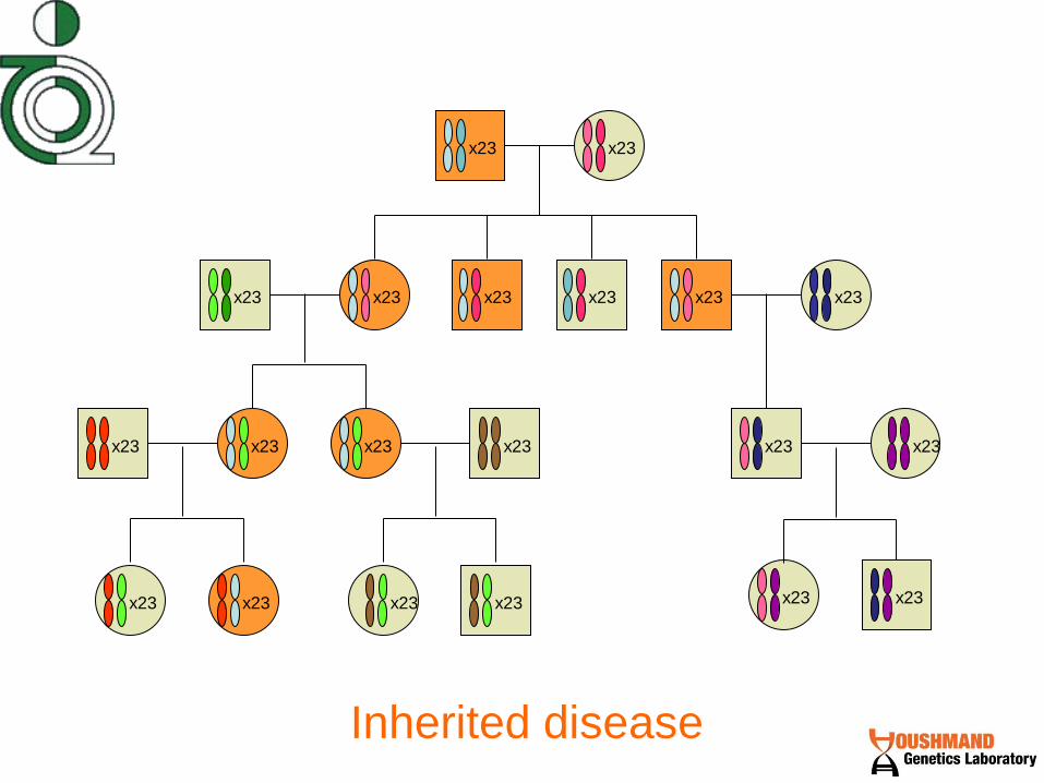
Inherited disease
x23 x23
x23 x23 x23 x23 x23 x23
x23 x23 x23 x23 x23 x23
x23 x23 x23 x23 x23 x23

Genetic disease

Symptoms

?

Predictive testing Tells a person if she carries a mutation that will cause, or put her at higher risk for, a disease later in life.
Newborn screening Detects common disorders in newborns, where immediate treatment can prevent dangerous symptoms
Carrier testing Tells a person whether or not he carries a mutation that could be passed on to his offspring. One can be a carrier, but not be at risk for a disease (as in recessive genes)
? ? ? ?
Genetic Testing

?
Physician
Genetic
Counselor
Test family
members with
disease
symptoms?
Test patient?

?
Treatment
Prevention
Reproduction
Physician
Genetic Counselor

Genetic
Counseling


Dr. M. Houshmand
Dr. M. Houshmand
Diagnostic

We now know how God
wrote the book of life

But do we know
How to read the book ?
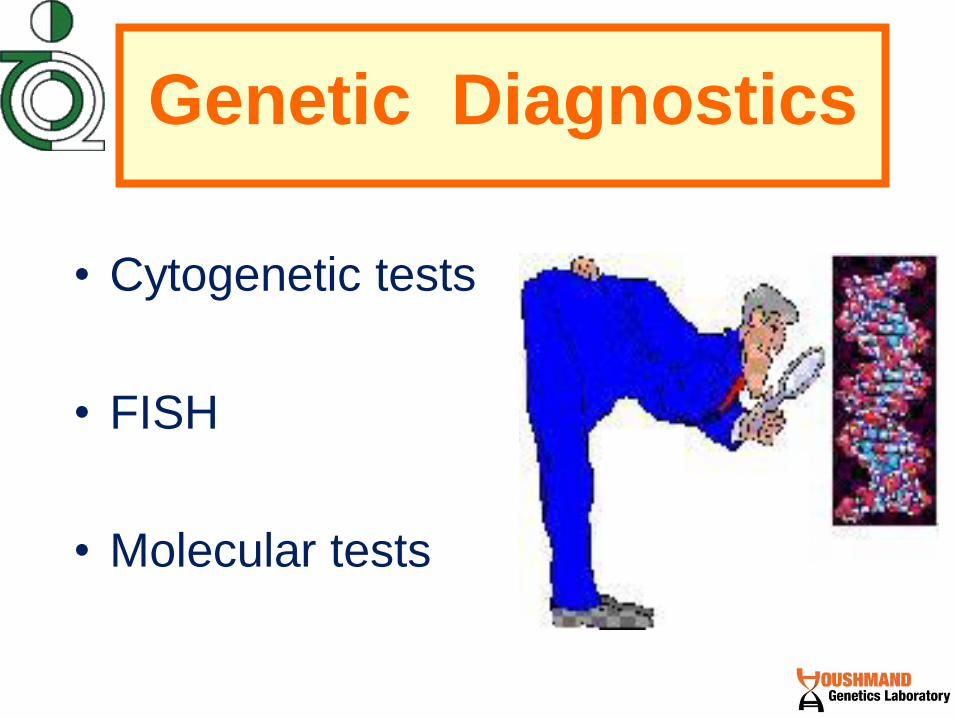
Genetic Diagnostics
• Cytogenetic tests
• FISH
• Molecular tests

DNA
RNA
Protein
Function
DNA
RNA
Protein
Function
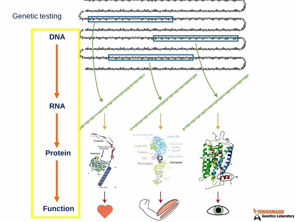
Genetic testing

DNA
Protein
Protein
Function
Levels of Genetic Testing
normal mutated
Analysis of whole
chromosomes – for large
changes; extra chromosome, very
large deletions or insertions
atcgatcgatcg atcgaAcgatcg Analysis of sequence – for small
changes; mutations in the
sequence, small deletions or
insertions
Analysis of protein shape – for
any change that may affect the
folding of the protein
Analysis of protein function – if
the functional protein is supposed
to make something, then some
tests can detect the presence or
absence of the product
X X

DNA
Protein
Protein
Function
normal mutated
Analysis of whole
chromosomes – for large
changes; extra chromosome, very
large deletions or insertions
atcgatcgatcg atcgaAcgatcg Analysis of sequence – for small
changes; mutations in the
sequence, small deletions or
insertions
Analysis of protein shape – for
any change that may affect the
folding of the protein
Analysis of protein function – if
the functional protein is supposed
to make something, then some
tests can detect the presence or
absence of the product
X X
Levels of Genetic Testing

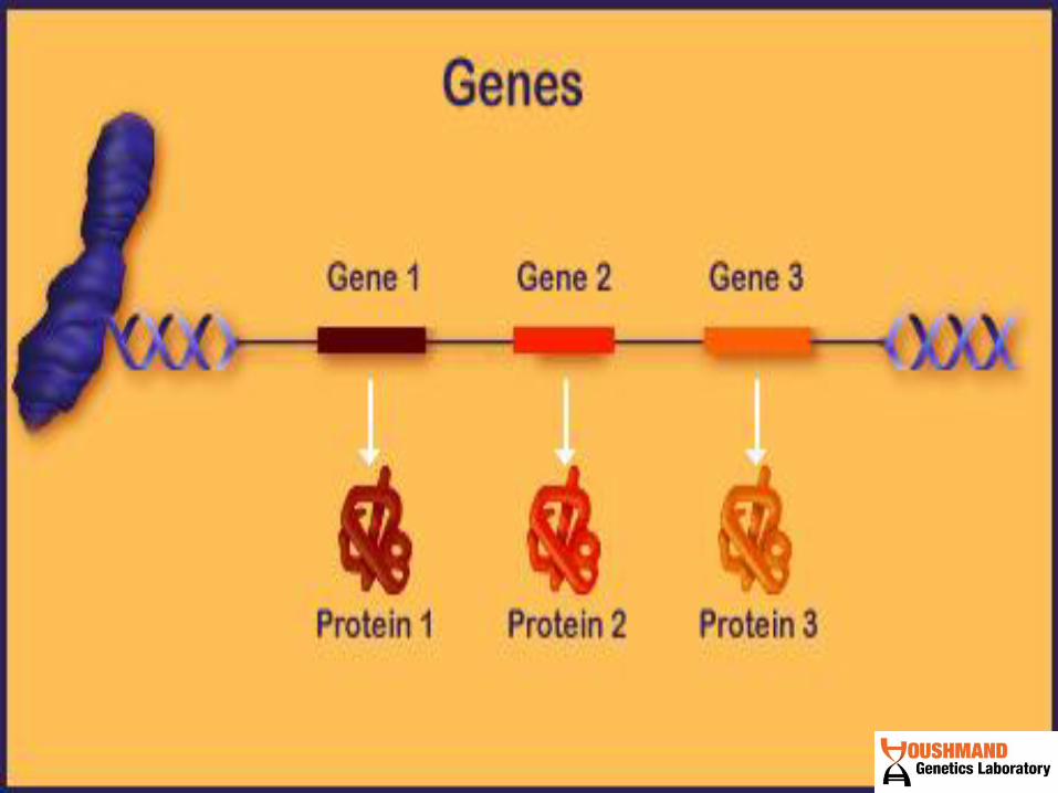
Molecular Biology Overview
Cell Nucleus
Chromosome
Protein
Graphics courtesy of the National Human Genome Research Institute
Gene (DNA) Gene (mRNA), single strand

Cytogenetic
Microscope
Molecular Cytogenetic
Biomedical Genetics
Molecular Genetic

Earth
Country
Chromosomal Abnormalities

Chromosome

Chromosome

1. Cells (from blood, amniotic fluid, or chorionic villus) are grown in culture. Mitogens may be required: lymphocytes require phytohemagglutinin
2. Colcemid stops cells at metaphase.
3. Hypotonic shock ruptures RBCs, swells lymphocytes.
4. Cells are fixed in MeOH/HOAc.
5. Chromosomes are spread on a slide.
6. Trypsinization and staining with Giemsa reveals G-bands.
7. The chromosome spread is photographed and arranged by type.
Karyotypes are made from metaphase chromosomes:

normal meiotic
division and
fertilization

Autosome
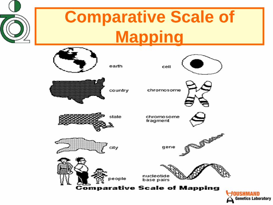
Comparative Scale of
Mapping

Earth

Country

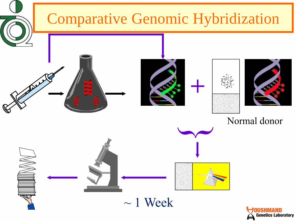
~ 1 Week

Chromosomes Preparation
• Any tissue with living nucleated cells which undergo division can be used for studying chromosomes.
• Most commonly circulating lymphocytes from peripheral blood are used.
• Samples for chromosomal analysis can be prepared relatively easily using:
- Skin
- Bone marrow
- Chorionic Villi
- Amniocytes

G-banding analysis

Chromosomal Abnormalities
• Mutations when involve large parts of the
chromosome, and when these are large
enough to be visible under the light
microscope, they are termed
CHROMOSOME ABERRATION.
• With light microscope, the smallest visible
addition or deletion from a chromosome is
about 4 Mbp, which involves many
contiguous genes.

Chromosomal Aberrations
Abnormalities of the chromosomes are usually
classified into:
I. Numerical abnormalities: where the somatic cells contain an abnormal number of normal chromosomes.
II. Structural aberrations: where the somatic cells contain one or more abnormal chromosomes.
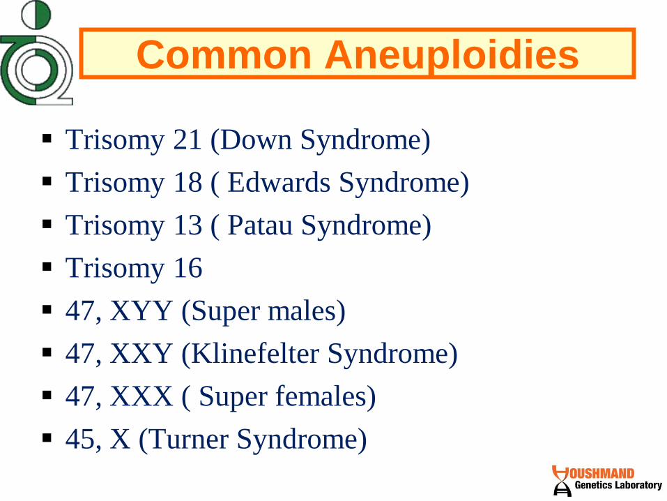
Common Aneuploidies
Trisomy 21 (Down Syndrome)
Trisomy 18 ( Edwards Syndrome)
Trisomy 13 ( Patau Syndrome)
Trisomy 16
47, XYY (Super males)
47, XXY (Klinefelter Syndrome)
47, XXX ( Super females)
45, X (Turner Syndrome)

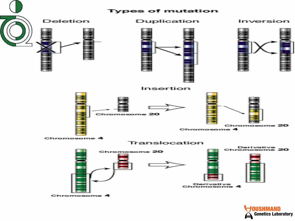
Structural Aberrations
• Structural chromosome rearrangements result
from chromosome breakage with subsequent
reunion in a different configuration.
• They can be balanced or unbalanced.

Structural Aberrations
Structural aberrations are subdivided into:
• Translocations
• Deletions
• Duplications
• Inversions
• Ring chromosomes
• Isochromosomes

Country
State

~ 1 Week
+
Fluorescence In Situ Hybridization
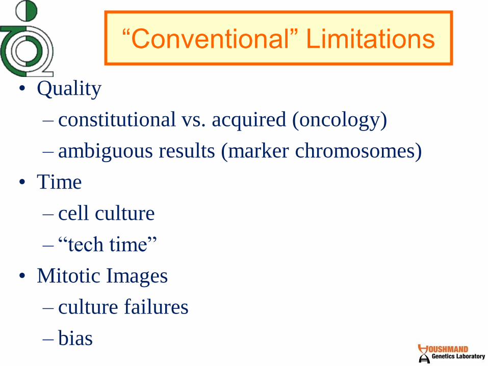
“Conventional” Limitations
• Quality
– constitutional vs. acquired (oncology)
– ambiguous results (marker chromosomes)
• Time
– cell culture
– “tech time”
• Mitotic Images
– culture failures
– bias
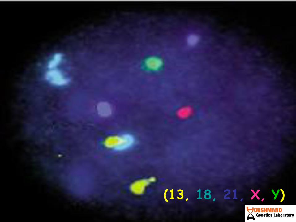
Mosaic +18 (13, 18, 21, X, Y)

Multiplex-FISH
~ 1 Week
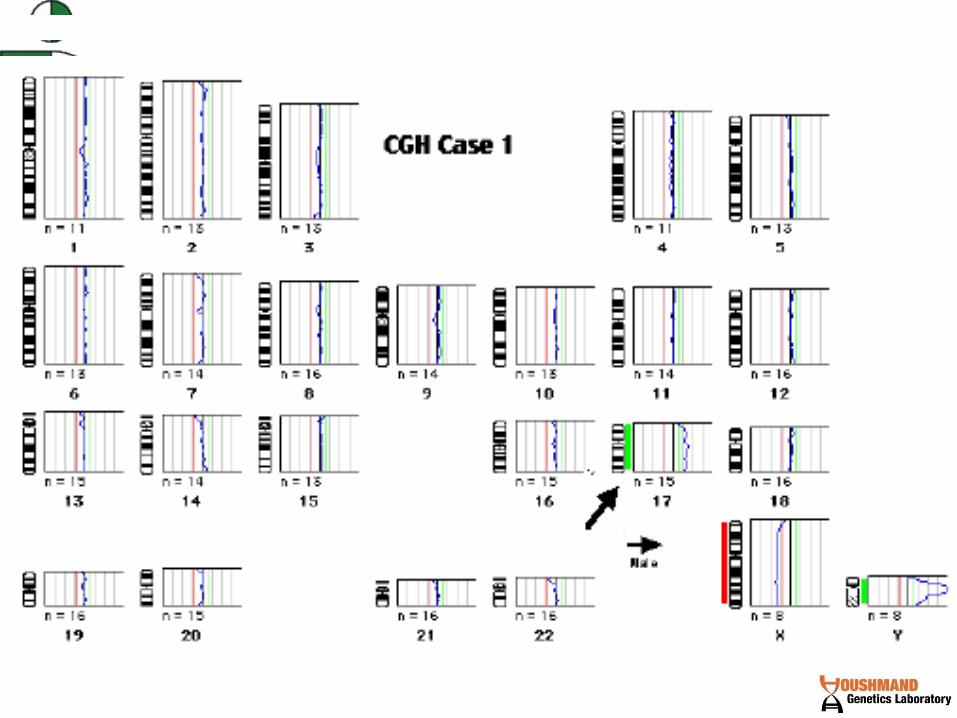
Comparative Genomic Hybridization
+
Normal donor

Array Comparative Genomic Hybridization
(aCGH)

Whole Genome Data Is Acquired
Patient below without any known genetic disease
All chromosomes but Y represented
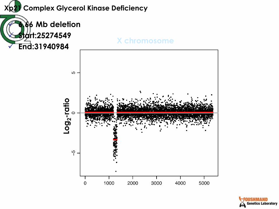
Xp21 Complex Glycerol Kinase Deficiency
Log
2-r
atio
X chromosome
6.66 Mb deletion
Start:25274549
End:31940984

ACMG Recommends Replacing Karyotyping with
Chromosomal Microarrays as 'First-Line' Postnatal
Test
Microarrays should be used instead of G-banded
karyotyping as the first test to detect genetic
abnormalities in postnatal evaluations, according to
the American College of Medical Genetics.
September 28, 2010
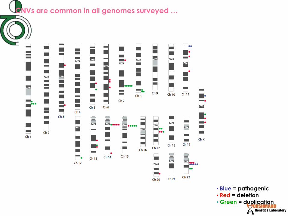
CNVs are common in all genomes surveyed …
• Blue = pathogenic
• Red = deletion
• Green = duplication
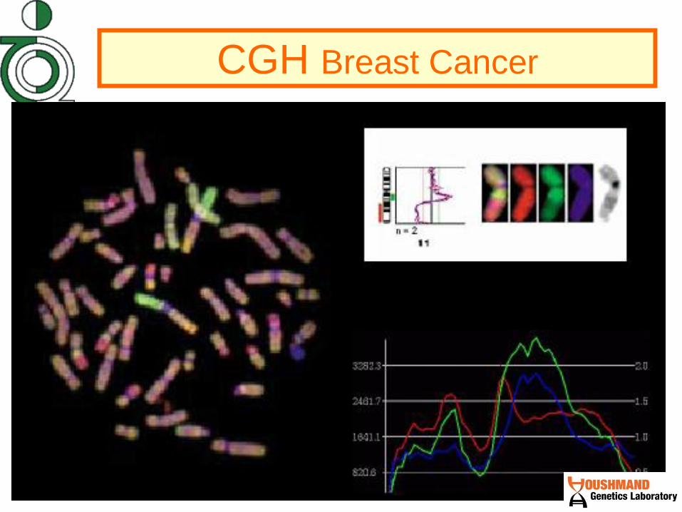
CGH Breast Cancer

~ 2 days
DNA Micro Arrays (FISH & Chips )
+ Normal donor
Manufactured DNA chip

Gene loss
Gene gain
“Chip” Technology

City

District

People

Normal, wild type Mutation inactivates enzyme

Normal

Nonsense Mutation


Missense Mutation


Framshif Mutation
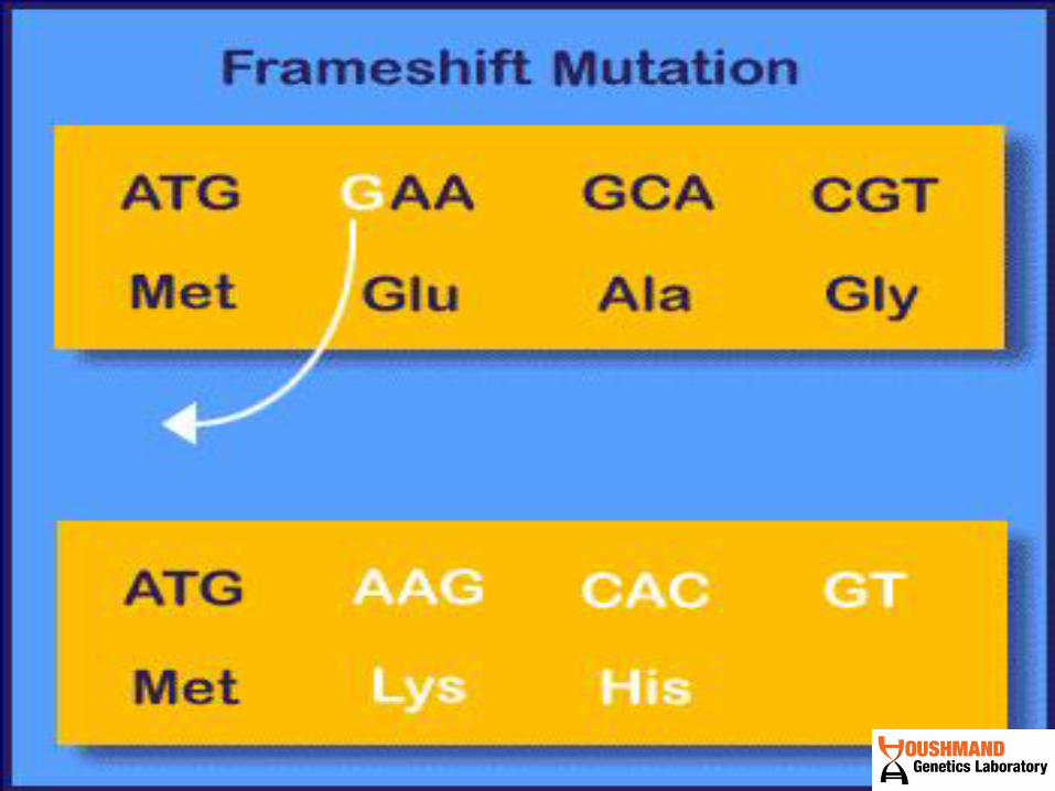
Mutation
Detections
Techniques
1993


PCR


Components of a PCR
Reaction
• Buffer (containing Mg++)
• Template DNA
• 2 Primers that flank the fragment of DNA to be amplified
• dNTPs
• Taq DNA Polymerase (or another thermally stable DNA polymerase)

PCR Melting
94 oC
Melting
94 oC
Annealing
Primers
50 oC
Extension
72 oC T
emp
erat
ure
100
0
50
T i m e
30x
5’ 3’
3’ 5’
3’ 5’
5’
5’ 3’ 5’
3’ 5’
5’
5’
5’
5’ 3’
3’ 5’
3’ 5’
5’ 3’
5’ 3’
5’

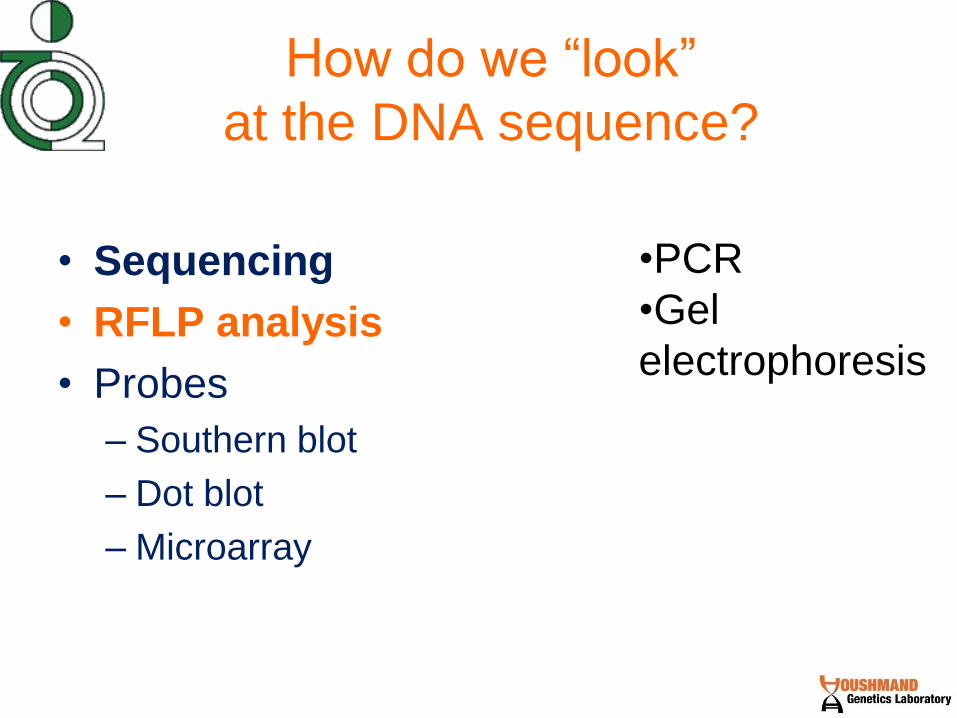
How do we “look”
at the DNA sequence?
• Sequencing
• RFLP analysis
• Probes
– Southern blot
– Dot blot
– Microarray
•Gel electrophoresis
•PCR
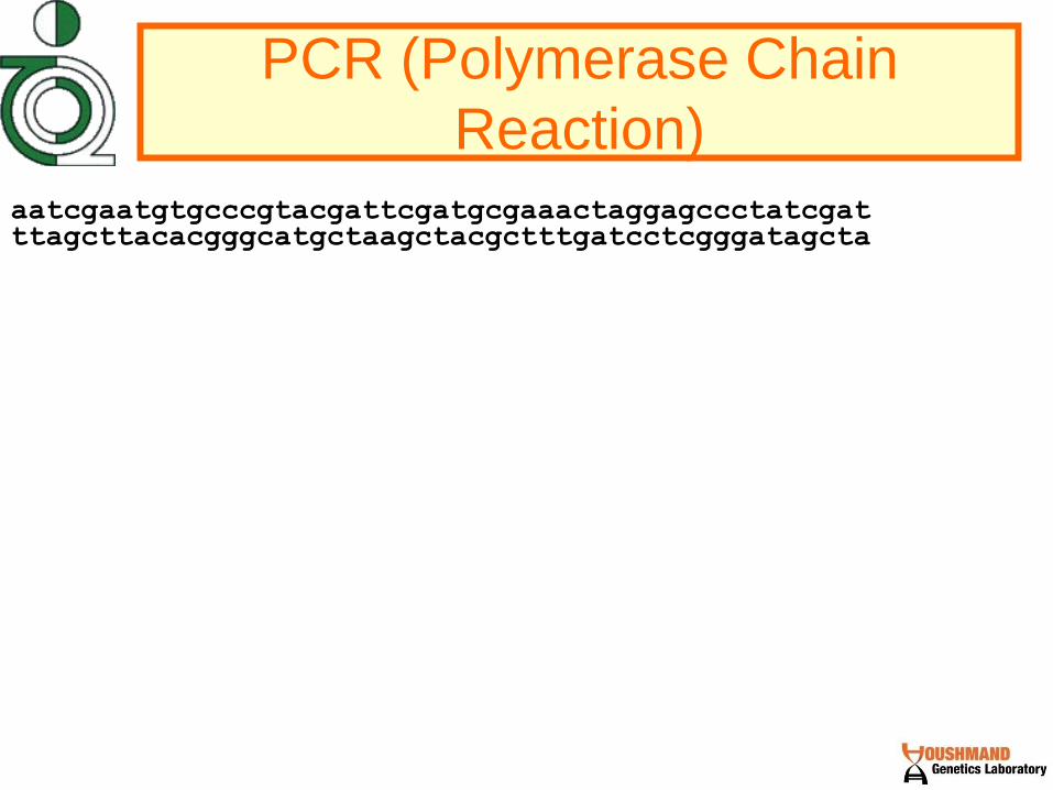
PCR (Polymerase Chain
Reaction)
aatcgaatgtgcccgtacgattcgatgcgaaactaggagccctatcgat ttagcttacacgggcatgctaagctacgctttgatcctcgggatagcta
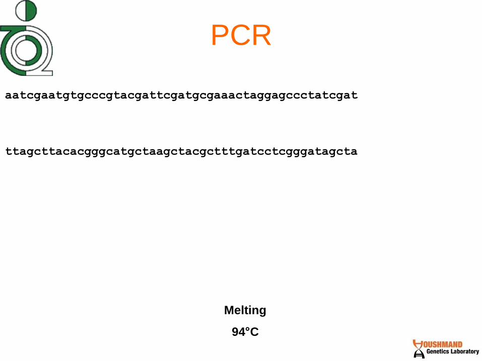
PCR
aatcgaatgtgcccgtacgattcgatgcgaaactaggagccctatcgat
ttagcttacacgggcatgctaagctacgctttgatcctcgggatagcta
Melting
94°C

PCR
aatcgaatgtgcccgtacgattcgatgcgaaactaggagccctatcgat
ttagcttacacgggcatgctaagctacgctttgatcctcgggatagcta
Annealing
58°C
cgggcat
aactagg

PCR
aatcgaatgtgcccgtacgattcgatgcgaaactaggagccctatcgat
ttagcttacacgggcatgctaagctacgctttgatcctcgggatagcta
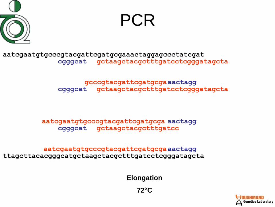
Elongation
72°C
cgggcat
aactagg aatcgaatgtgcccgtacgattcgatgcga
gctaagctacgctttgatcctcgggatagcta

PCR
aatcgaatgtgcccgtacgattcgatgcgaaactaggagccctatcgat
ttagcttacacgggcatgctaagctacgctttgatcctcgggatagcta
Melting
94°C
cgggcat
aactagg aatcgaatgtgcccgtacgattcgatgcga
gctaagctacgctttgatcctcgggatagcta

PCR
aatcgaatgtgcccgtacgattcgatgcgaaactaggagccctatcgat
ttagcttacacgggcatgctaagctacgctttgatcctcgggatagcta
Annealing
58°C
cgggcat
aactagg aatcgaatgtgcccgtacgattcgatgcga
gctaagctacgctttgatcctcgggatagcta
cgggcat
cgggcat
aactagg
aactagg
PCR
aatcgaatgtgcccgtacgattcgatgcgaaactaggagccctatcgat
ttagcttacacgggcatgctaagctacgctttgatcctcgggatagcta
Elongation
72°C
cgggcat
aactagg aatcgaatgtgcccgtacgattcgatgcga
gctaagctacgctttgatcctcgggatagcta
cgggcat
cgggcat
aactagg
aactagg
gcccgtacgattcgatgcga
gctaagctacgctttgatcctcgggatagcta
aatcgaatgtgcccgtacgattcgatgcga
gctaagctacgctttgatcc

PCR Makes a lot of copies of a specific portion of DNA

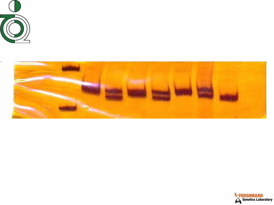
PCR test for deletion
Electrophoresis

DNA Between The Primers Doubles
With Each Thermal Cycle
0
Cycles
Number
1
3
8
2
4
1
2
4
16
5
32
6
64

More Cycles = More DNA Number of cycles
0 10 15 20 25 30
Size
Marker

Theoretical Yield Of PCR Theoretical yield = 2n x y
Where y = the starting
number of copies and
n = the number of thermal cycles
= 107,374,182,400
If you start with 100 copies, how many copies are
made in 30 cycles?
2n x y
= 230 x 100
= 1,073,741,824 x 100

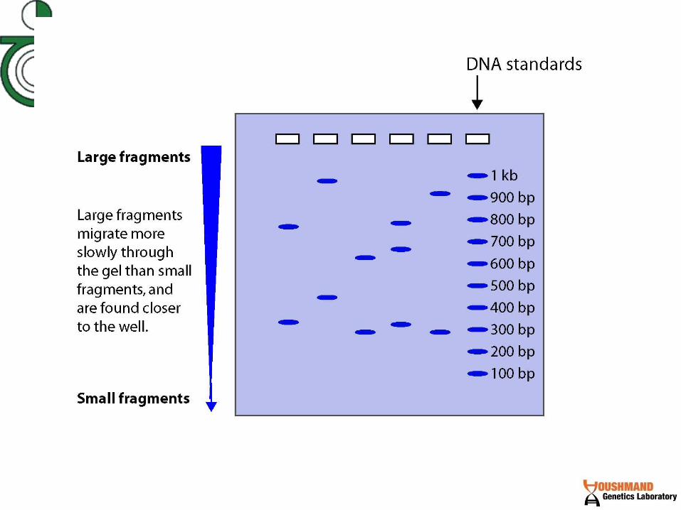
Gel Electrophoresis Separates DNA fragments on the basis of size
Large fragments take longer than
small fragments to migrate
through an agarose gel

DMD Multiplex deletion test
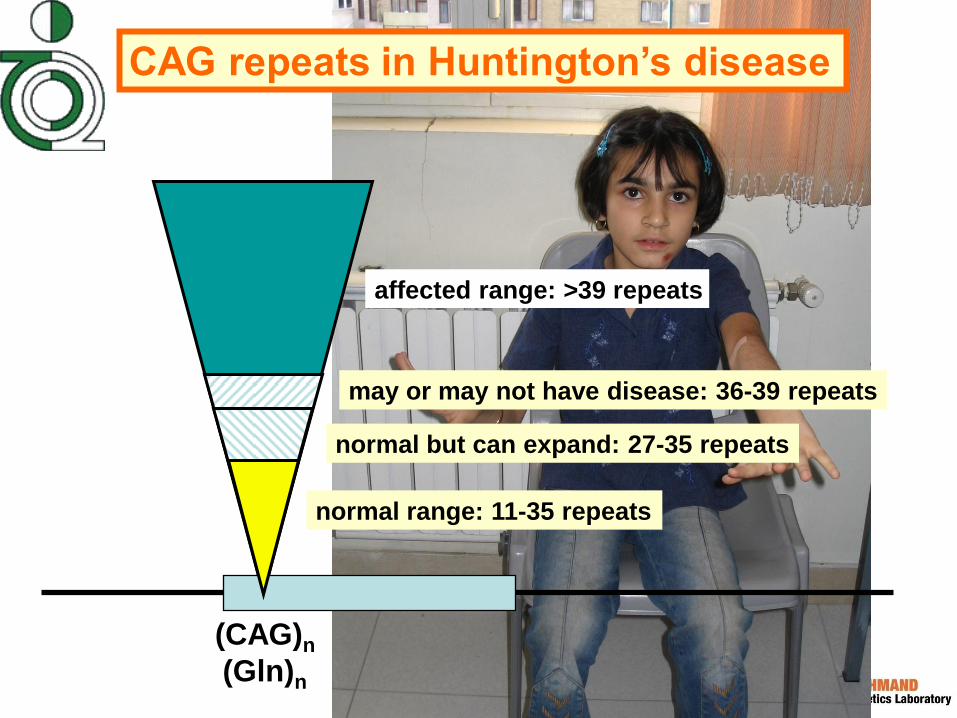
affected range: >39 repeats
CAG repeats in Huntington’s disease
normal range: 11-35 repeats
may or may not have disease: 36-39 repeats
normal but can expand: 27-35 repeats
(CAG)n
(Gln)n

Structure and inheritance of CAG repeats
in spinal and bulbar muscular atrophy
normal 13-28
affected 39-60
(CAG)n
(Gln)n
androgen receptor gene

Spinal Bulbar Muscular Atrophy
Kennedy Syndrome
Dr. Babak Zamani

Cancer
DMD

Paternity Testing

Forensic testing

Steps in Forensic DNA Analysis
DNA
Extraction
Multiplex PCR Amplification
Male: 13,14-15,16-12,13-10,13-15,16
Interpretation of Results
Sample Collection
& Storage
Buccal swab Blood Stain
DNA
Quantitation
Slot Blot 1 ng
0.3 ng
1 ng
1 ng
0.7 ng
0.5 ng
0.5 ng
No DNA
Usually 1-2 day process (a minimum of ~5 hours)
If a match occurs, comparison of
DNA profile to population allele
frequencies to generate a case
report with probability of a random
match to an unrelated individual
STR Typing
DNA separation and sizing
Tec
hn
olo
gy
B
iolo
gy
Ge
ne
tic
s
DNA
Database
Search
Collection
Extraction
Quantitation
STR Typing
Interpretation
of Results
Database Storage & Searching
Specimen Storage
Multiplex PCR
Calculation of
Match Probability
Steps Involved

Short Tandem Repeat (STR) Markers
STR repeat region
GATA GATA GATA GATA
PCR product size generated
DNA template
containing STR marker
Reverse
PCR primer
Forward
PCR primer
Fluorescent
dye
PCR Product Size (bp)
Allelic Ladder
Sample
#2
Sample
#1
TCCCAAGCTCTTCCTCTTCCCTAGATCAATACAGACAGA
AGACAGGTGGATAGATAGATAGATAGATAGATAGATAGA
TAGATAGATAGATATCATTGAAAGACAAAACAGAGATGG
ATGATAGATACATGCTTACAGATGCACAC
PCR primers anneal to unique sequences
bracketing the variable STR repeat region
= 11 GATA repeats (“11” is all that is reported)
The overall PCR product size is measured

CSF1PO
D5S818
D21S11
TH01
TPOX
D13S317
D7S820
D16S539 D18S51
D8S1179
D3S1358
FGA
VWA
13 Core U.S. STR Loci
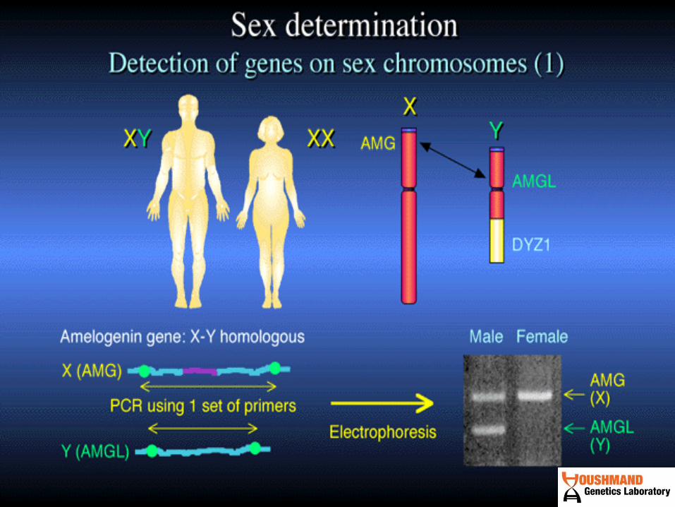
AMEL
AMEL
Sex-typing
Position of Forensic STR Markers
on Human Chromosomes
8 STR loci overlap between U.S. and Europe
1997

Family Inheritance of STR Alleles (D13S317)
Father
Child #1
Child #2
Child #3
Mother
PCR product size (bp)
11 14
11
12 14
8 14
12
12 8
PATERNITY TESTING

Why we use PCR?
+/- How
much?
(Qualitative)
(Quantitative)


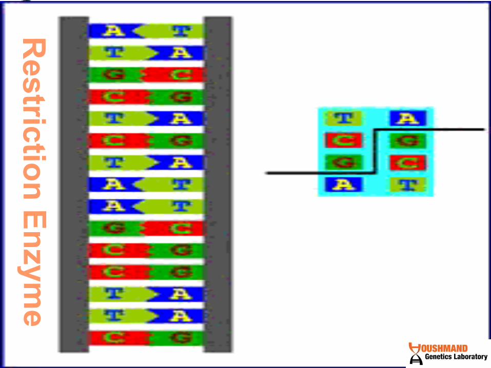

A restriction enzymes binds to DNA at a specific sequence and make a double-
stranded cut at or near that sequence.
Restriction endonucleases cut DNA
molecules at defined positions
How do we “look”
at the DNA sequence?
• Sequencing
• RFLP analysis
• Probes
– Southern blot
– Dot blot
– Microarray
•PCR
•Gel
electrophoresis

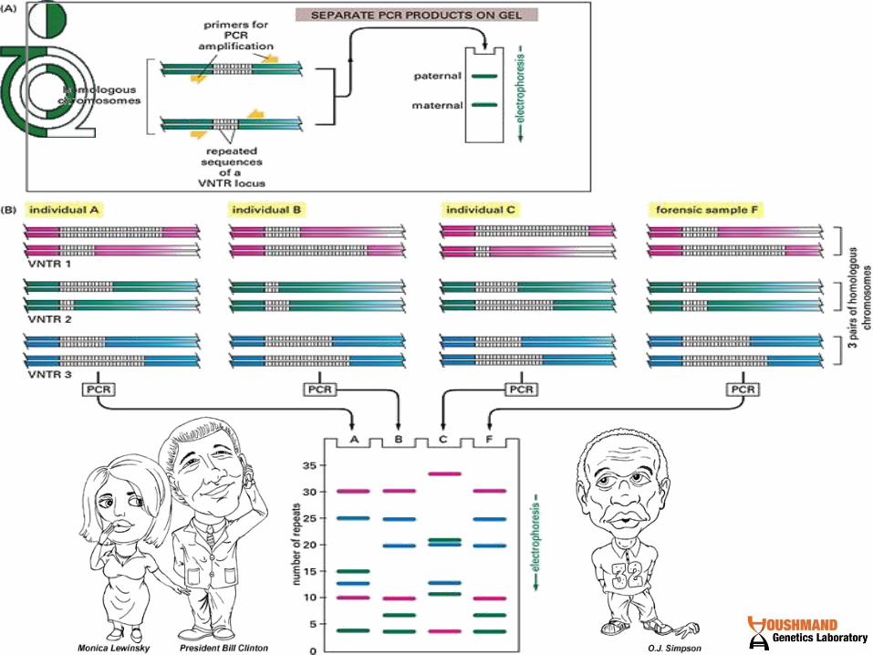
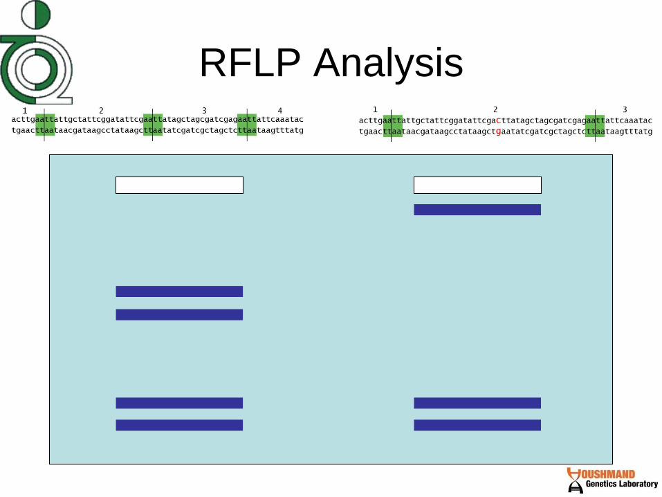
RFLP Analysis
RFLP (Restriction Fragment Length Polymorphism) analysis relies on
the use of restriction enzymes
These enzymes recognize specific DNA sequences (usually
palindromes) and cut them…

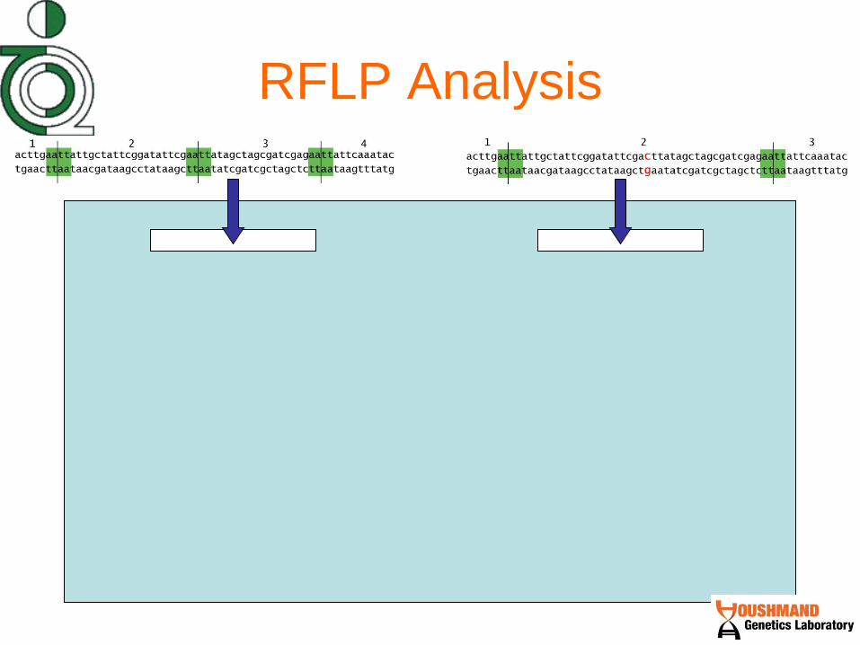
RFLP Analysis Restriction Fragment Length Polymorphism means that there are
polymorphisms (differences) between people in the number of restriction
sites (and therefore the length of the cut fragments). This person has 4
fragments after restriction digest.
This person has a mutation that eliminates one of the sites that the restriction
enzyme cuts at. Therefore, this person has 3 bands, one of them being much
larger than the rest. If this mutation was associated with a disease, a restriction
digest would show that a carrier of the mutation had 3 fragments instead of 4

RFLP Analysis

RFLP Analysis
Genomic DNA is very long and
contains a lot of restriction
enzyme cut sites
PCR makes many
copies of a small
region of DNA,
better for RFLP
analysis
RFLP Analysis

RFLP Analysis
RFLP Analysis
Issues with RFLP analysis
• Can not detect all mutations – the
mutation has to coincide with a
restriction enzyme cut site

F
V
LUUR
T
P
E
LCUN
SAGY
H
ND5ND6
ND4
ND4L
ND3R
COIII
G
KCOIID
SUCN
COI
AN
CY
W
ND2
I
M
Q
ND1
16S
12S
D-loopCyt.b
3460G-A
11778G->A
14484T->C
LHON
DDEE
LL
EE
TT
II
OO
NN
3243A->G
MELAS
CPEO
DDM
8344A->G
MERRF
tRNA-Ser mutations
DEAFNESS
cyt. b mutations
MYOGLOBINURIC
MYOPATHY
8993T->G
NARP/MILS
PEO, KSS, Pearson
1555A->G
DEAFNESS (aminoglycosides)
tRNA-Ile mutations
CARDIOPATHY
ATPase6/8
F
V
LUUR
T
P
E
LCUN
SAGY
H
ND5ND6
ND4
ND4L
ND3R
COIII
G
KCOIID
SUCN
COI
AN
CY
W
ND2
I
M
Q
ND1
16S
12S
D-loopCyt.b
3460G-A
11778G->A
14484T->C
LHON
DDEE
LL
EE
TT
II
OO
NN
3243A->G
MELAS
CPEO
DDM
8344A->G
MERRF
8344A->G
MERRF
tRNA-Ser mutations
DEAFNESS
cyt. b mutations
MYOGLOBINURIC
MYOPATHY
8993T->G
NARP/MILS
8993T->G
NARP/MILS
PEO, KSS, Pearson
1555A->G
DEAFNESS (aminoglycosides)
tRNA-Ile mutations
CARDIOPATHY
ATPase6/8
mtDNA Disorders


Introduction
The mobility in gel electrophoresis of double-stranded DNA's of a given
length is relatively independent of nucleotide sequence.

the mobility of single strands can
vary considerably as a result of only
small changes in nucleotide sequence.
SSCP techniques may be used as
Screening methods to point to the mutations

Most SSCP protocols are
designed to analyze the polymorphism at single loci. To
this end a specific pair of PCR
primers bracketing the target
region is used to amplify DNA from individuals

Gel Discussion • Number of bands in SSCP: 2 – 8
and even more.
• Reason: This maybe due to
different shapes and conformations
of single strands.

For avoiding of returning single strands to
double form, strands should be pushed
into gel rapidly .For this, initial 15 minutes
should have 350 voltage and then return it
to 60-120.
There is two methods for detecting a
mutation into a sample:
SSCP Up to 80% Sequencing Up to 99%

Principle of DHPLC
Rapid denaturation of DNA by heating, re-annealing by slow cooling.
Heteroduplexe
s form in the presence of two different alleles.
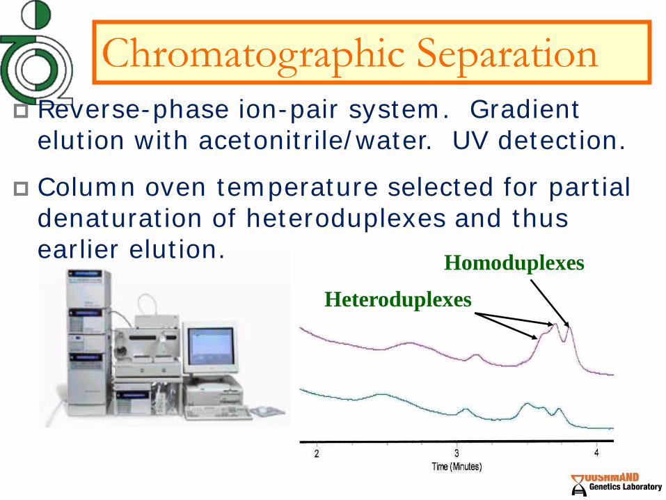
Homoduplexes
Heteroduplexes
Chromatographic Separation
Reverse-phase ion-pair system. Gradient elution with acetonitrile/water. UV detection.
Column oven temperature selected for partial denaturation of heteroduplexes and thus earlier elution.

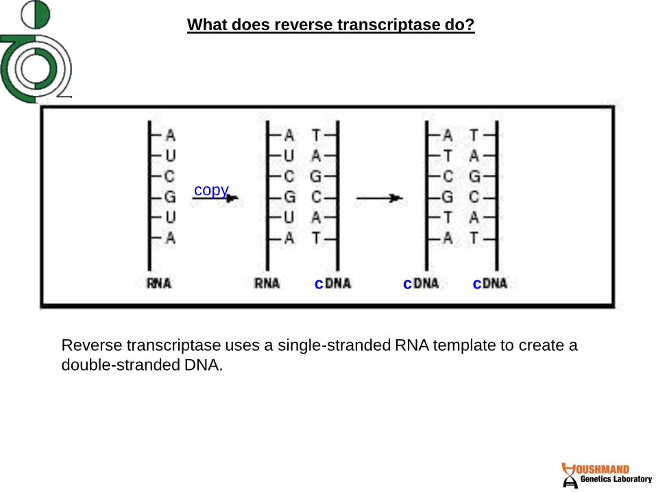
Reverse transcriptase uses a single-stranded RNA template to create a
double-stranded DNA.
What does reverse transcriptase do?
copy
c c c

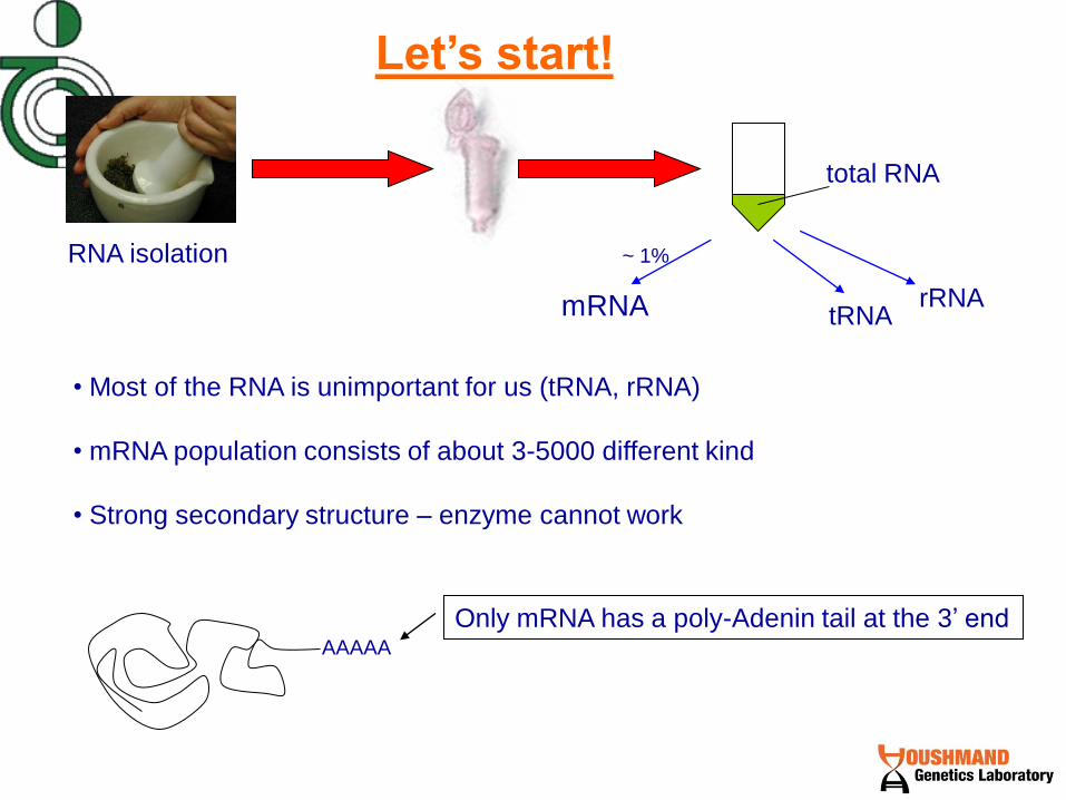
Let’s start!
total RNA
tRNA rRNA mRNA
~ 1%
• Most of the RNA is unimportant for us (tRNA, rRNA)
• mRNA population consists of about 3-5000 different kind
• Strong secondary structure – enzyme cannot work
AAAAA
Only mRNA has a poly-Adenin tail at the 3’ end
RNA isolation
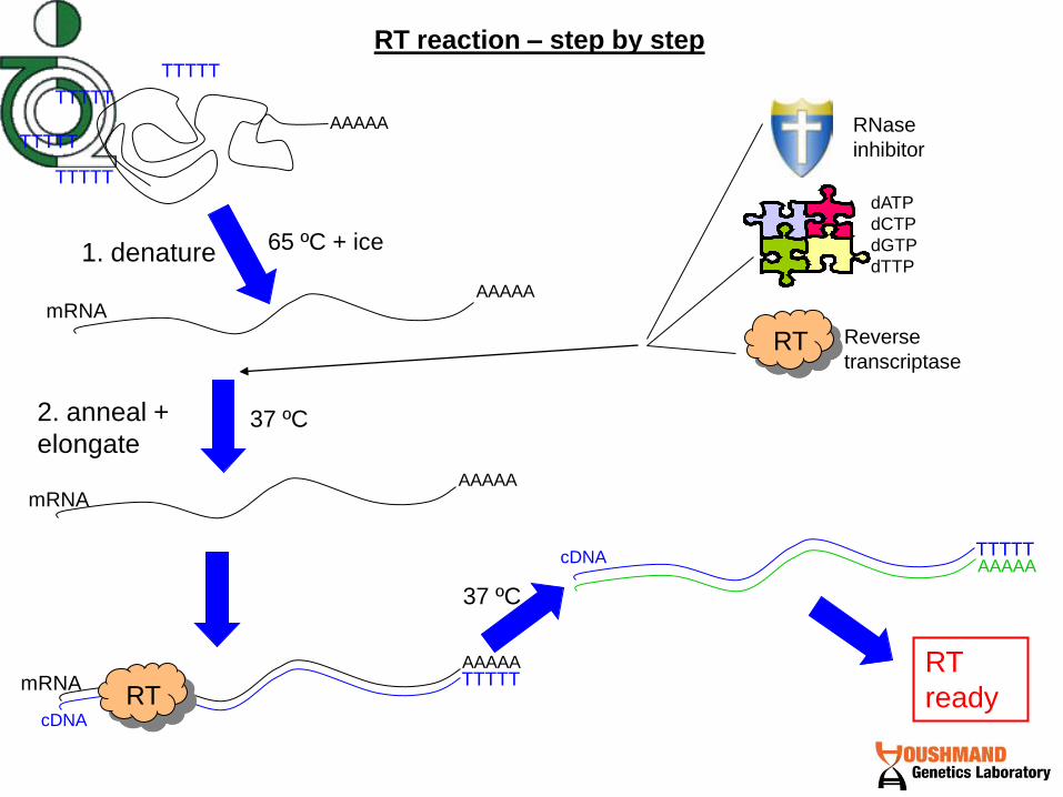
RT reaction – step by step
AAAAA
AAAAA
AAAAA
65 ºC + ice
37 ºC
TTTTT
TTTTT
TTTTT
TTTTT
Reverse
transcriptase
dATP
dCTP
dGTP
dTTP
RNase
inhibitor
mRNA
mRNA
1. denature
AAAAA TTTTT mRNA
2. anneal +
elongate
TTTTT cDNA AAAAA
37 ºC
RT
cDNA
RT
RT
ready

How to amplify our gene of interest from the cDNA “soup”?
TTTTT cDNA AAAAA
TTTTT AAAAA cDNA TTTTT
AAAAA cDNA
PCR
Gel
visualization
Gene
#2
750 bp
500 bp
Gene
#1
Gene-specific
primers

RT–PCR at the bench
total RNA +
oligodT
37 ºC – 1 hour
anneal +
elongate
65ºC – 10 min
denature
Add:
Enzyme
dNTPs
RNasin RT
ready
RT:
PCR:
DNA pol
dNTPs
primers
Buffer
MgCl2
95ºC
3 min
denature amplify
95ºC – 30 sec
55ºC – 30 sec
72ºC – 1 min
72ºC
10 min
finish
PCR
ready
template
Gel analysis
30 cycles

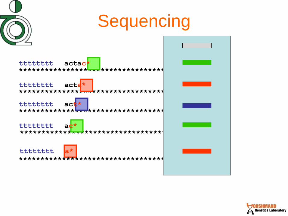
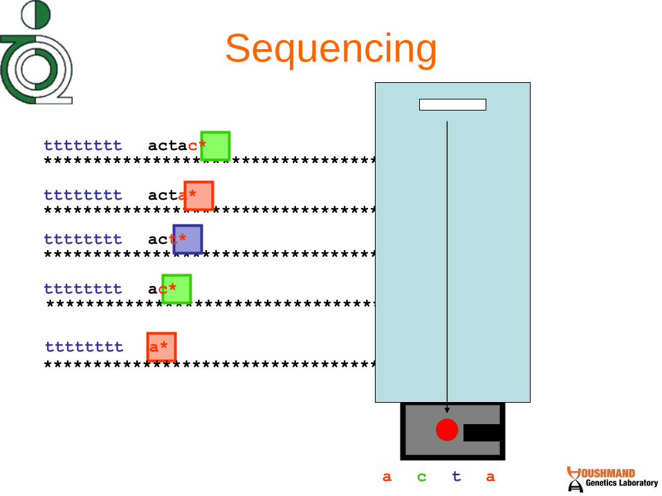
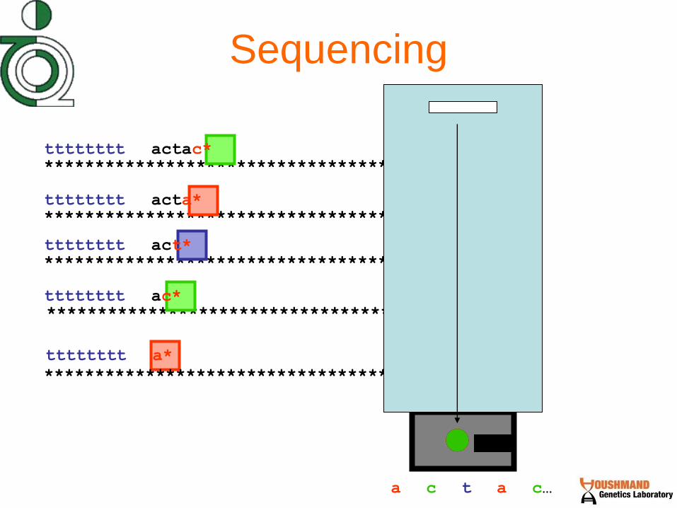
Sanger Sequencing

DNA Sequencing: Dideoxy
Method
• Modified sugars cause chain
termination because it lacks the 3’-OH
group, which is essential for
attachment of the next nucleotide in a
growing DNA strand
• The products of DNA synthesis are
then separated by electrophoresis. In
principle, the sequence can be read
directly from the gel

Analysis of Recombinant Clones: Thermal Cycle DNA Sequencing
O
H H
H OH
H
Base
H
PO4
O
H H
H H
H
Base
H
PO4
dNTP
ddNTP

How do we “look”
at the DNA sequence?
• Sequencing
• RFLP analysis
• Probes
– Southern blot
– Dot blot
– Microarray
•PCR
•Gel
electrophoresis

Sequencing
aaaaaaaatgatgatgatgatgatgatgatgatgatgatgatg
“Known gene sequence:”
*******************************************
Patient DNA sequence
How do we find out if the patient’s
sequence is different?

Sequencing
*******************************************
tttttttt actactactac*
tttttttt
tttttttt
tttttttt
*******************************************
*******************************************
*******************************************
******************************************* tttttttt
actac*
actactactactag*
actactactactacgactact*
actactactacta*

Sequencing
*******************************************
tttttttt a*
*******************************************
*******************************************
*******************************************
*******************************************
tttttttt actac*
tttttttt act*
tttttttt ac*
tttttttt acta*
a

Sequencing
*******************************************
tttttttt a*
*******************************************
*******************************************
*******************************************
*******************************************
tttttttt actac*
tttttttt act*
tttttttt ac*
tttttttt acta*
a c

Sequencing
*******************************************
tttttttt a*
*******************************************
*******************************************
*******************************************
*******************************************
tttttttt actac*
tttttttt act*
tttttttt ac*
tttttttt acta*
t a c
Sequencing
*******************************************
tttttttt a*
*******************************************
*******************************************
*******************************************
*******************************************
tttttttt actac*
tttttttt act*
tttttttt ac*
tttttttt acta*
t a c a
Sequencing
*******************************************
tttttttt a*
*******************************************
*******************************************
*******************************************
*******************************************
tttttttt actac*
tttttttt act*
tttttttt ac*
tttttttt acta*
t a c a c…

Sequencing
actactactactagtactactactactactactac
*******************************************
aaaaaaaatgatgatgatgatgatgatgatgatgatgatgatg
“Known gene sequence:”

Sequencing
actactactactagtactactactactactactac aaaaaaaatgatgatgatgatcatgatgatgatgatgatgatg
aaaaaaaatgatgatgatgatgatgatgatgatgatgatgatg
“Known gene sequence:”
Mutation

Issues with Sequencing
• Informative
• Time consuming
• Expensive


Electrophoresis
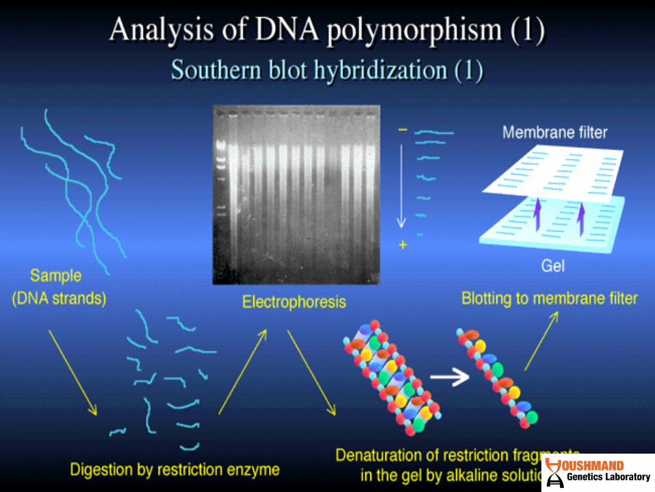

Southern Blot

Types of Blots
• Southern Blot – use DNA to probe DNA
• Northern Blot – use DNA to probe RNA
• Western Blot – use antibodies to probe
Protein
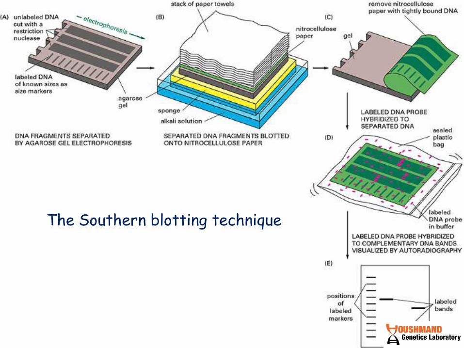
The Southern blotting technique

Southern Blot
Restriction enzyme
DNA of various sizes
Electrophorese on agarose gel
gel
Denature - transfer to filter paper.
blot
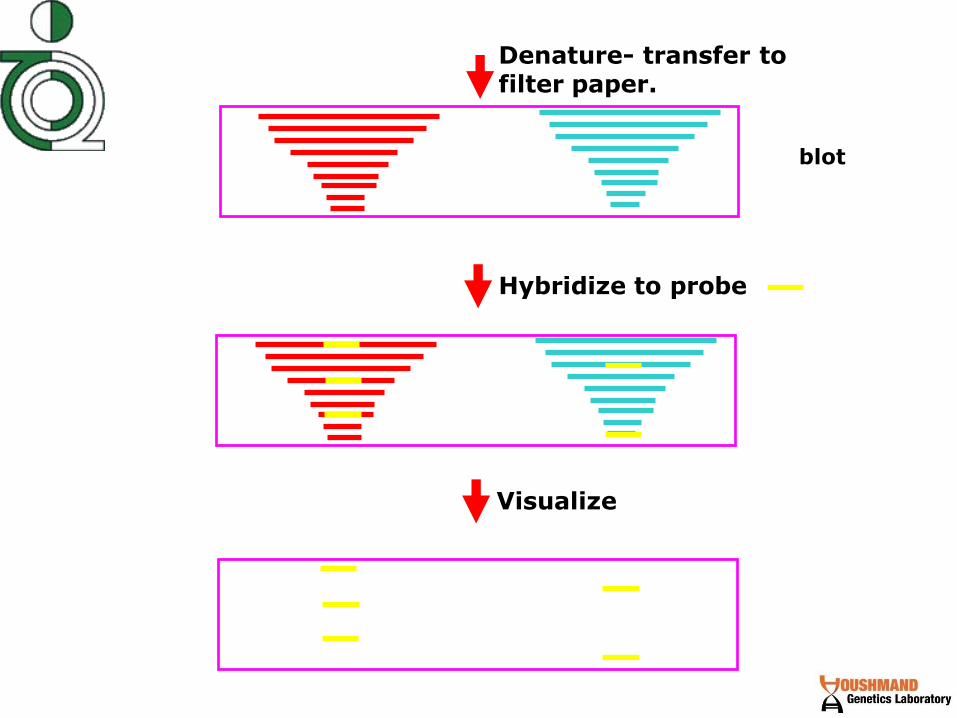
Hybridize to probe
Visualize
Denature- transfer to filter paper.
blot
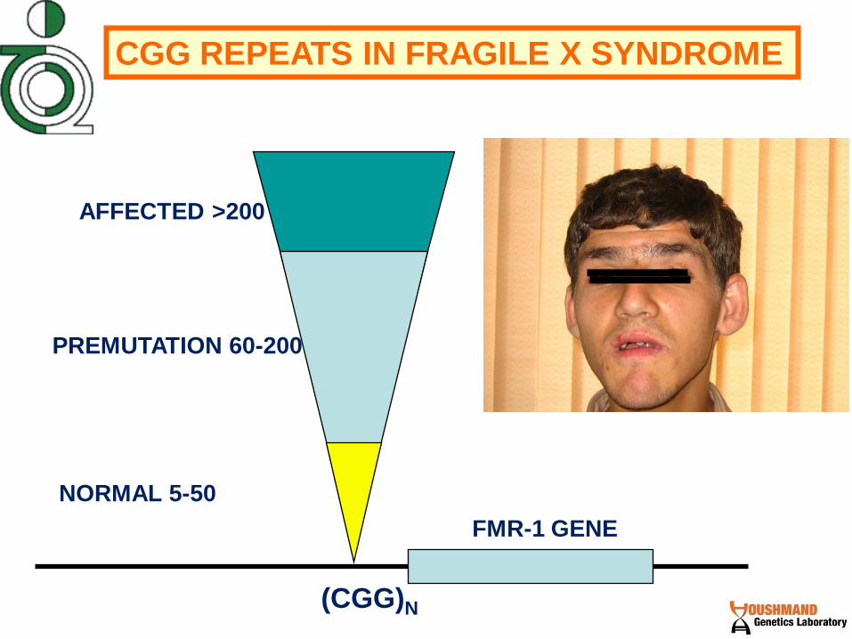
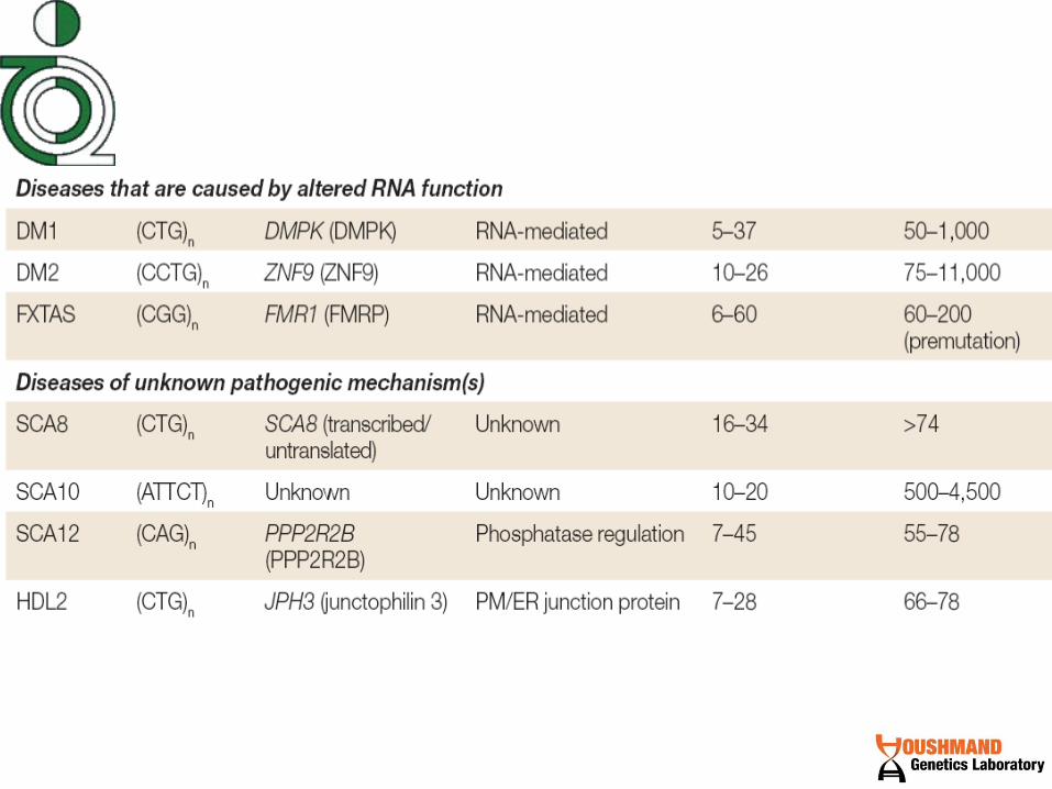
CGG REPEATS IN FRAGILE X SYNDROME
NORMAL 5-50
PREMUTATION 60-200
AFFECTED >200
(CGG)N
FMR-1 GENE

Structure and inheritance of CTG repeats
in myotonic dystrophy
normal 5-30
premutation 45-75
affected >75
(CTG)n
myotonic protein kinase gene

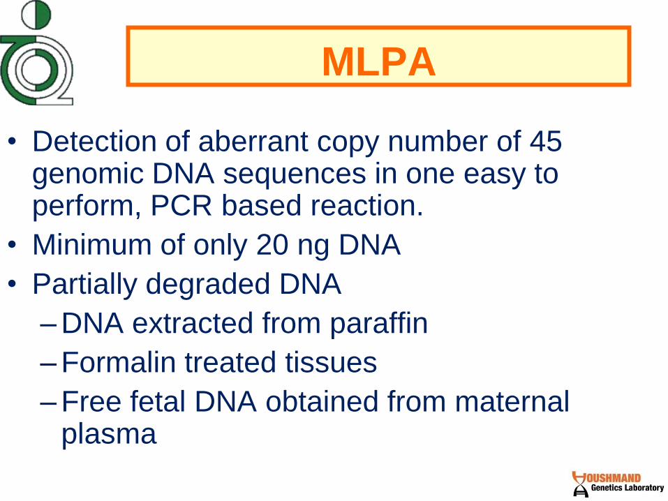
MLPA
• Detection of aberrant copy number of 45 genomic DNA sequences in one easy to perform, PCR based reaction.
• Minimum of only 20 ng DNA
• Partially degraded DNA
– DNA extracted from paraffin
– Formalin treated tissues
– Free fetal DNA obtained from maternal plasma

• Discriminates sequences that differ in only a single nucleotide.
• 45 different mRNAs
• To determine the methylation status of promoters
• Detection of known mutations and SNPs.

MLPA technique
1. Denaturation
2. Hybridization
3. Ligation
4. Amplification
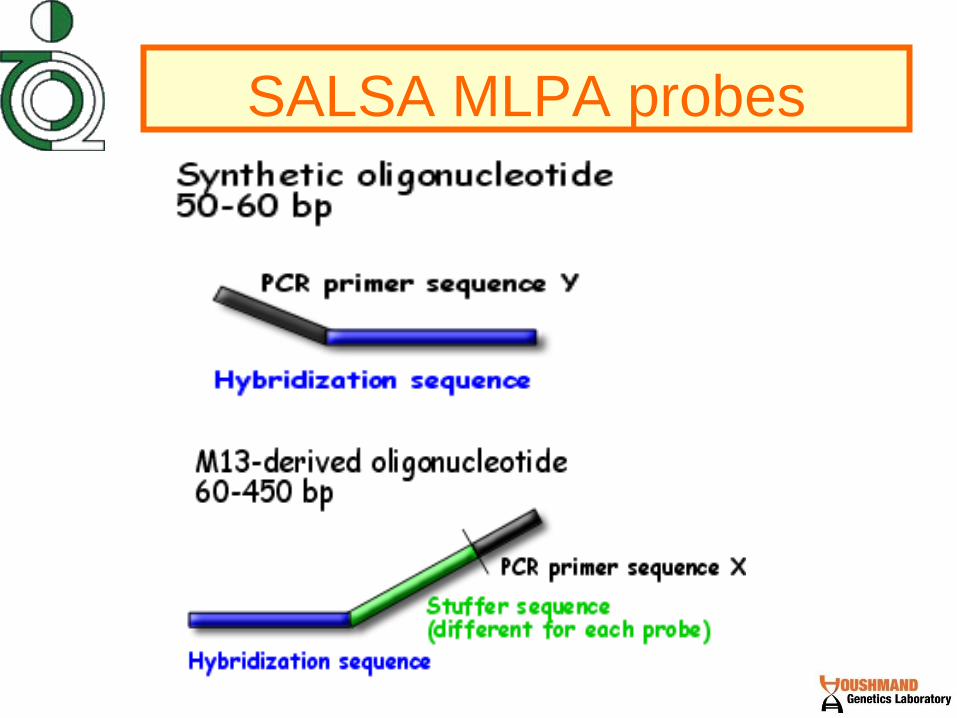
SALSA MLPA probes
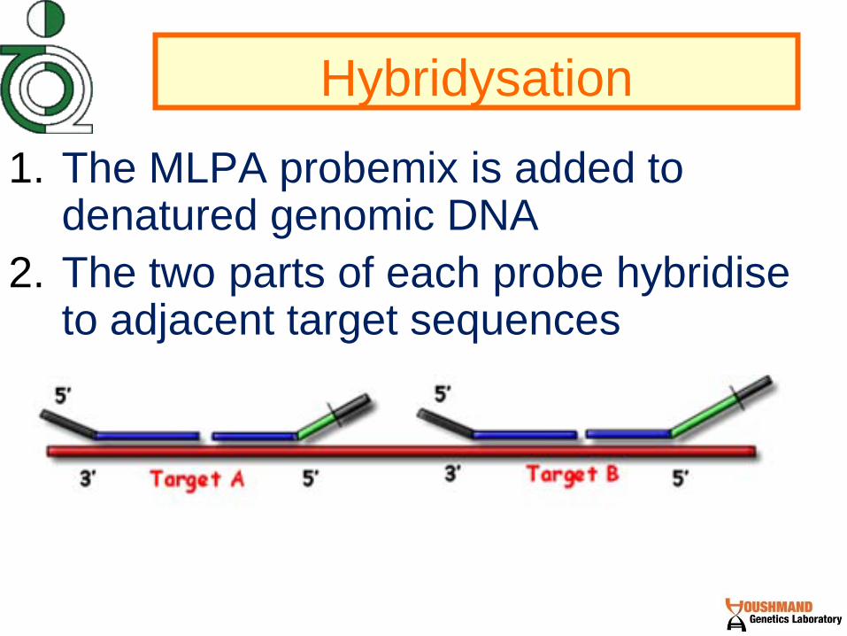
Hybridysation
1. The MLPA probemix is added to denatured genomic DNA
2. The two parts of each probe hybridise to adjacent target sequences

Ligation
3. Probes are ligated by a thermostable ligase

Amplification
4. A universal primer pair is used to amplify all ligated probes.
The amplification product of each probe has a unique length (130 480 bp).
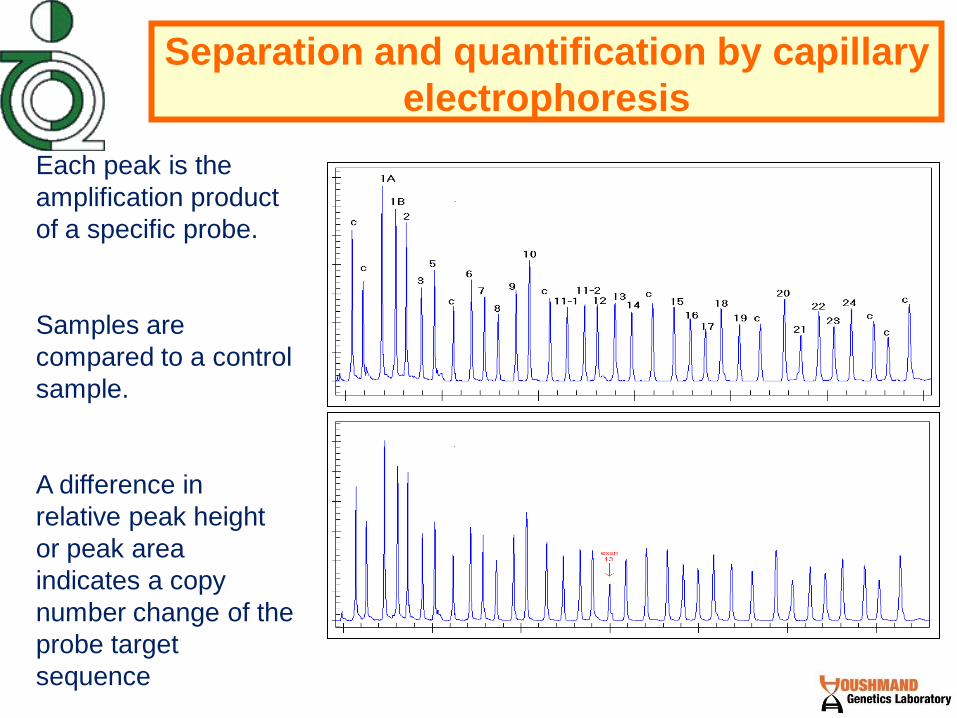
Separation and quantification by capillary
electrophoresis
Each peak is the
amplification product
of a specific probe.
Samples are
compared to a control
sample.
A difference in
relative peak height
or peak area
indicates a copy
number change of the
probe target
sequence
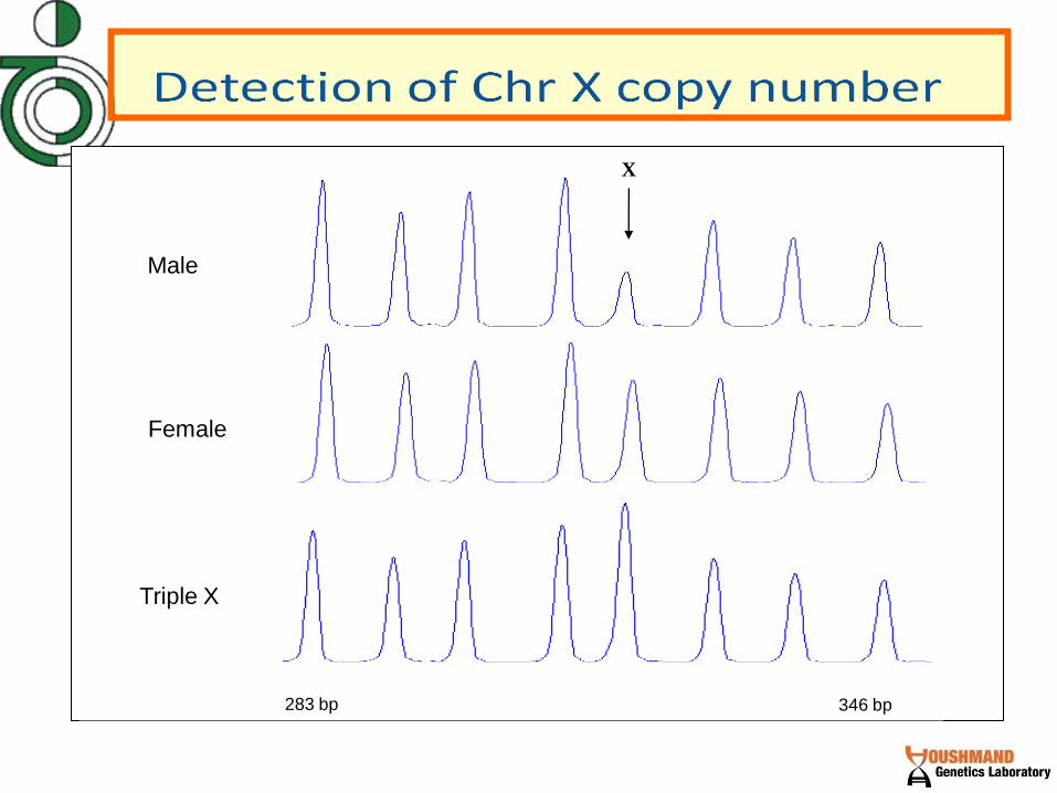
Triple X
Female
Male
283 bp 346 bp
X
control
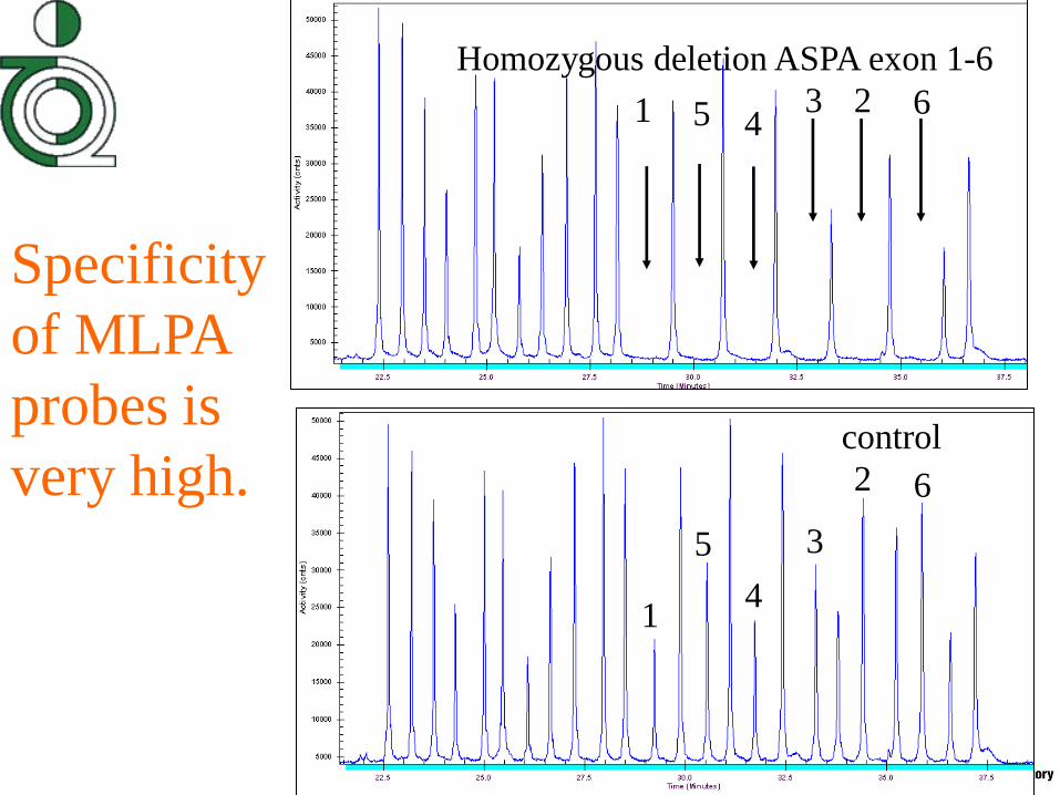
Homozygous deletion ASPA exon 1-6
1 5 4 3 2 6
1
5
4
3
2 6
Specificity
of MLPA
probes is
very high.

Heterozygous ASPA del. exon 1-6
Control
1
5
4
3
2 6
1 5 4 3 2 6
Reproducibilit
y of MLPA is
sufficient to
distinguish
homozygotes
and
heterozygotes.
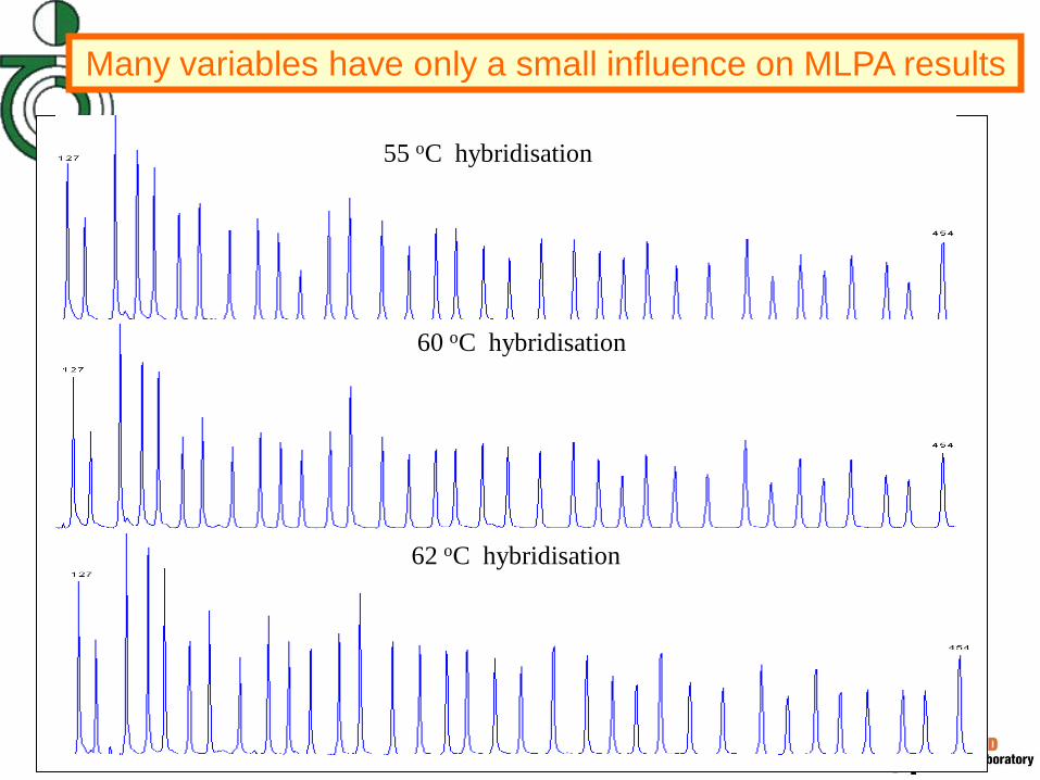
55 oC hybridisation
60 oC hybridisation
62 oC hybridisation
Many variables have only a small influence on MLPA results

10 ng. DNA
(less than recommended minimum amount)
50 ng. DNA
750 ng. DNA
Amount of DNA used has little
influence on MLPA results

Detection of point mutations
• Only perfectly matched probes will
be ligated Probes for common
mutations or SNP’s can be made
–Signal will only be present when the
mutation is present

Normal
Ligation of the two probe oligonucleotides
Amplification product
Mismatch at the probe ligation site
No ligation, no amplification product
MLPA discriminates sequences that differ
in only a single nucleotide and can be used
to detect known mutations.
Mismatch Perfect match

Advantages of MLPA
• Detection of copy number of 45 genomic DNA sequences in a simple to perform, PCR based, reaction.
• Requires only 20 ng human DNA
• Discriminates sequences that differ in only a single nucleotide.
• Only a thermocycler and a sequence type electrophoresis system are required.
• Identical protocol for many different applications.

• High throughput ; Results available within 24 hrs.
• Large lot sizes (> 100.000 reactions) are possible. All reagents have proved to be very stable.
• For each probe signal, presence of two specific oligonucleotides is required.
• All reagents are fluid: Simplified quality control.
• Including electrophoresis, total reagent costs are < EUR 15,- / reaction.

Disadvantages of MLPA
• Results depend on sample quality. For amniotic fluid samples we recommend to use cell lysates rather than purified DNA.
• Amniotic fluid samples that are contaminated with maternal blood can not be used.
• MLPA can not detect all triploidies.
• MLPA can not detect balanced translocations. More than 130 MLPA tests are currently available from MRC-Holland.

• MRC-Holland is research oriented and is not yet ISO certified.
• MLPA kits are not yet CE certified.
• Time consuming and difficult to develop new MLPA based assays.
MLPA products
• Detection of aneuploidy of chromosomes 13, 18, 21, X and Y
• Detection of large chromosomal deletions or duplications: DiGeorge syndrome, Williams syndrome, Spinal Muscular Atrophy (SMA), subtelomeric regions etc
• Detection of gains and losses of genes in cancer tissues: Her2-neu (ERBB2), TP53, MYC etc.
• Sequencing
• RFLP analysis
• Probes
– Dot blot
– Microarray
•PCR
•Gel
electrophoresis
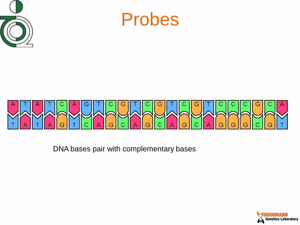
Probes
DNA bases pair with complementary bases

Probes
Probe
A probe is an a short fragment of DNA that is complementary to part
of a longer DNA sequence.
DNA strands can be
separated with high
heat.
As the temperature is
lowered, smaller
fragments that have
complementary
sequences (probes) will
base pair faster than the
long original strands of
DNA

Probes The ability of a probe to bind depends on:
*Its complementarity to the DNA strand it’s binding to…
-Single base pair differences can affect binding
depending on…
*The stringency of the binding conditions
-Temperature
-Buffer conditions
Low stringency
High stringency
• Sequencing
• RFLP analysis
• Probes
– Dot blot
– Microarray
•PCR
•Gel
electrophoresis

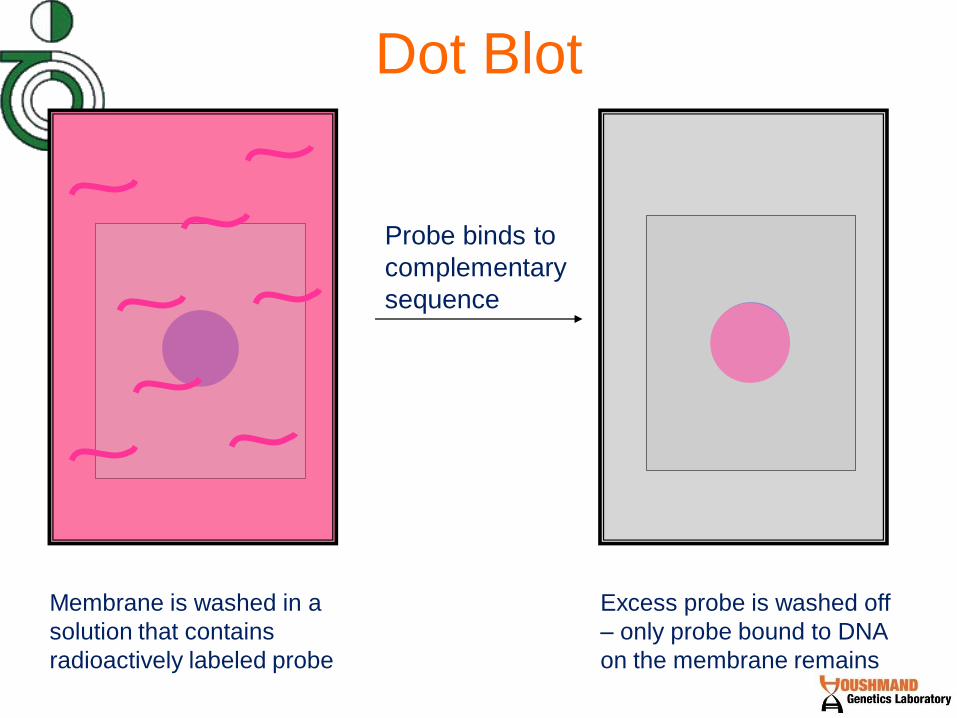
Dot Blot (A Southern blot without the gel…)
A patient’s DNA (genomic DNA or
PCR products) is denatured and
spotted to a membrane…

Dot Blot
Membrane is washed in a
solution that contains
radioactively labeled probe
Probe binds to
complementary
sequence
Dot Blot
Membrane is washed in a
solution that contains
radioactively labeled probe
Excess probe is washed off
– only probe bound to DNA
on the membrane remains
Probe binds to
complementary
sequence

Dot Blot
The membrane is exposed to
autoradiography film.. Therefore,
wherever there is radioactive probe, the
film will be exposed…

Dot Blot
The membrane is exposed to
autoradiography film..
Therefore, wherever there is
radioactive probe, the film will
be exposed…
And a black dot will be
developed where the probe
was…

Dot blot to diagnose cystic fibrosis (CF)
Normal
Normal
probe
Mutant
probe
CF patient
(homozygous mutant)
CF carrier
(heterozygous mutant)
DNA from a patient is spotted out twice, one will be used with a
probe complementary to the normal sequence, the other will be used
with a probe that is complementary to a mutated sequence.
Since a heterozygote has
one copy of the normal
gene and one copy of the
abnormal gene, both
probes can bind, but only
half as much binds,
making the dot lighter.
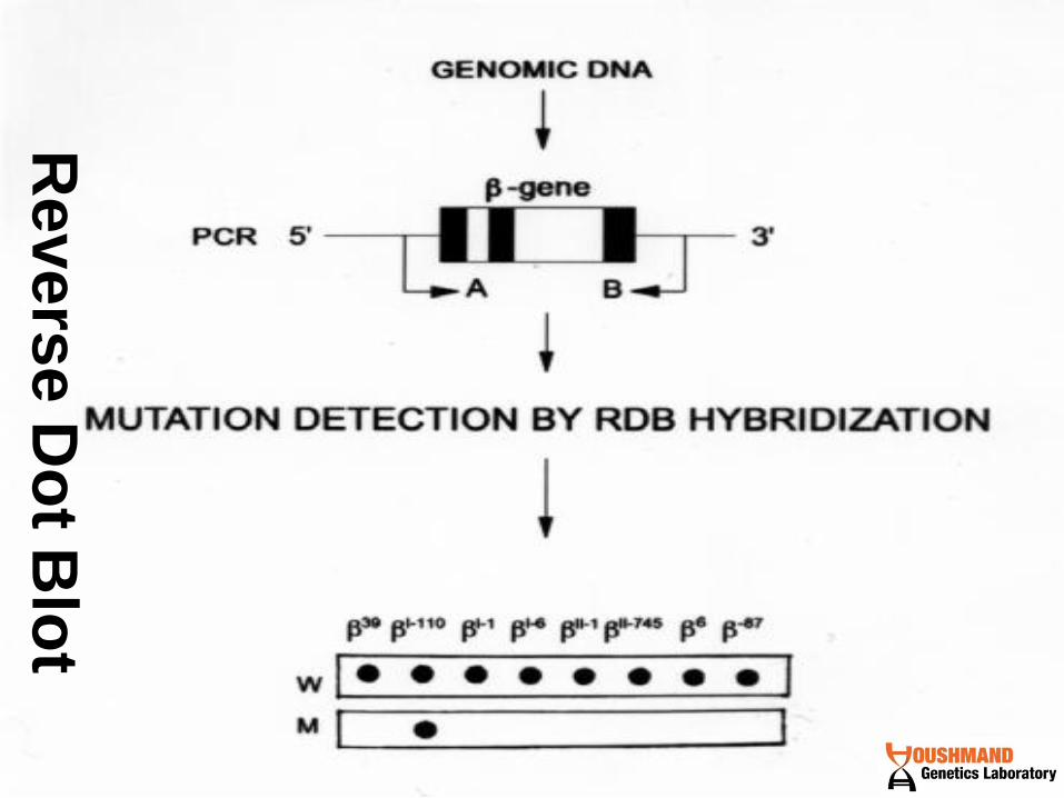
Revers
e D
ot B
lot

Issues with dot blots
• Can be used to test patient for
multiple mutations
• Single base pair mutations may be
hard to detect with dot blots


Functional genomics = The ability to perform
genome-wide patterns of gene expression
and the mechanisms by which gene
expression is coordinated.
DNA microarrays can be used to detect
differences in the levels gene expression in
different populations of cells on a genome-
wide level.

Reverse Transcriptase
in vitro transcription
mRNA
cDNA
Fragmentation of
cRNA
cRNA
GeneChip
Hybridization
This is what
we are doing
in this class
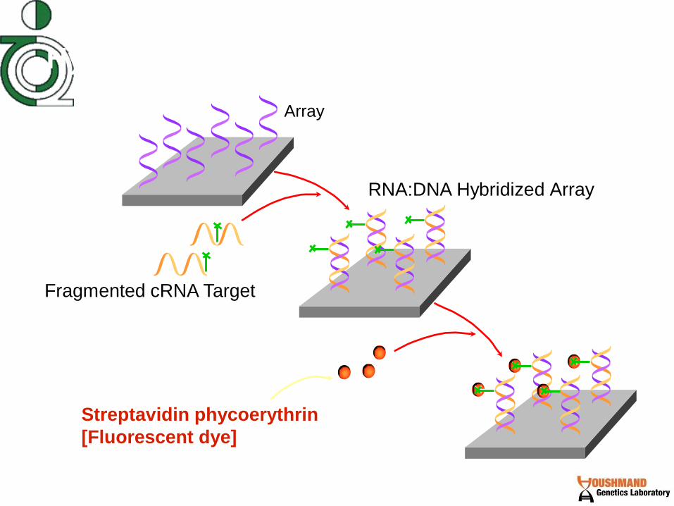
Hybridization and Staining
Array
Fragmented cRNA Target
RNA:DNA Hybridized Array
Streptavidin phycoerythrin
[Fluorescent dye]
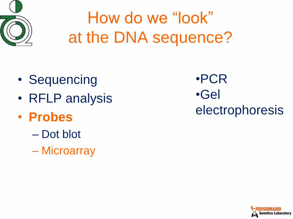
How do we “look”
at the DNA sequence?
• Sequencing
• RFLP analysis
• Probes
– Dot blot
– Microarray
•PCR
•Gel
electrophoresis

Microarray (High throughput dot blots. Allows for testing of many different mutations…)
Microarrays start
with a “chip” on
which is spotted
many different
probes for different
mutations. Each
dot represents a
different probe.
If this were a dot
blot, it would be
called a “reverse
dot blot” because
the PROBE is
spotted, rather than
the DNA.

Microarray (High throughput dot blots. Allows for testing of many different mutations…)
The chip is treated
much like a southern
or a dot blot, except it
is washed with a
patient’s DNA
(fragmented with
restriction enzymes,
denatured and
labeled with a
flourescent dye), and
it automated.

Microarray
Binds to a probe for a
CF mutation
Binds to a probe for a
colon-cancer
susceptibility mutation
Binds to a probe for a
breast-cancer
susceptibility mutation
If a patient’s DNA binds to spotted probes, a computer detects this by the
measuring the intensity of the flourescence emanating from that spot. A
homozygote for a CF mutation will have a more intense spot than a
heterozygote for the same mutation

Issues with Microarrays
• Expensive
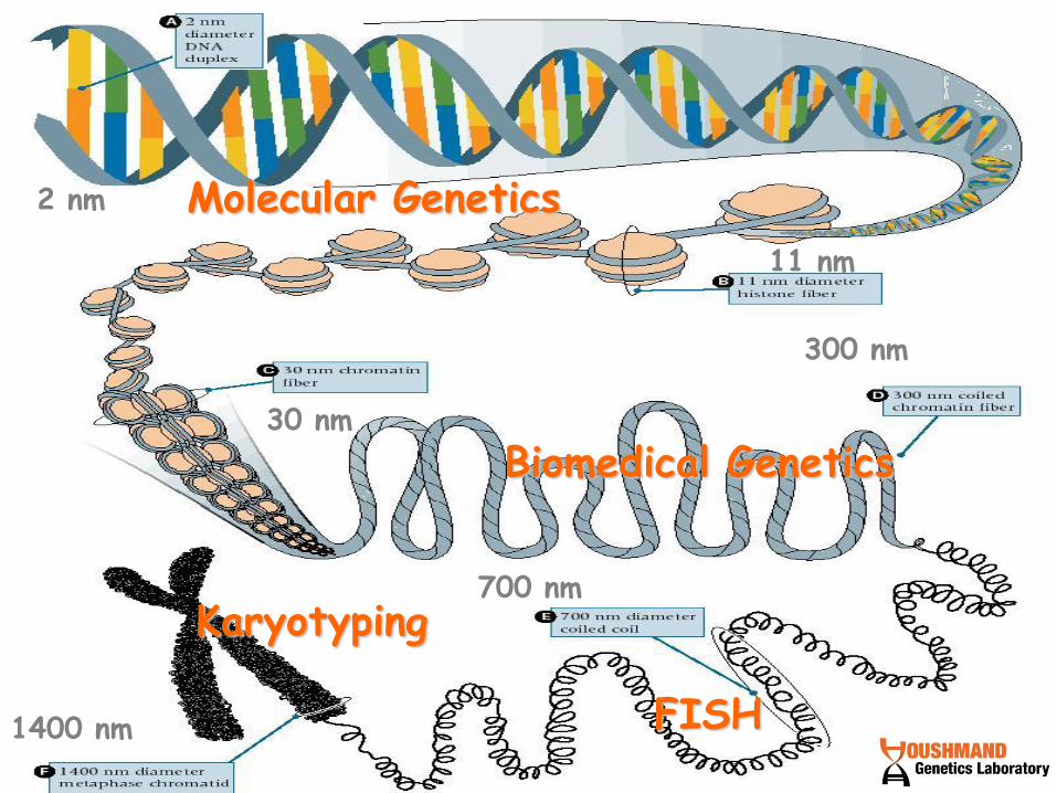
Karyotyping
FISH
Biomedical Genetics
Molecular Genetics
1400 nm
700 nm
300 nm
30 nm
11 nm
2 nm


Dr. M. Houshmand
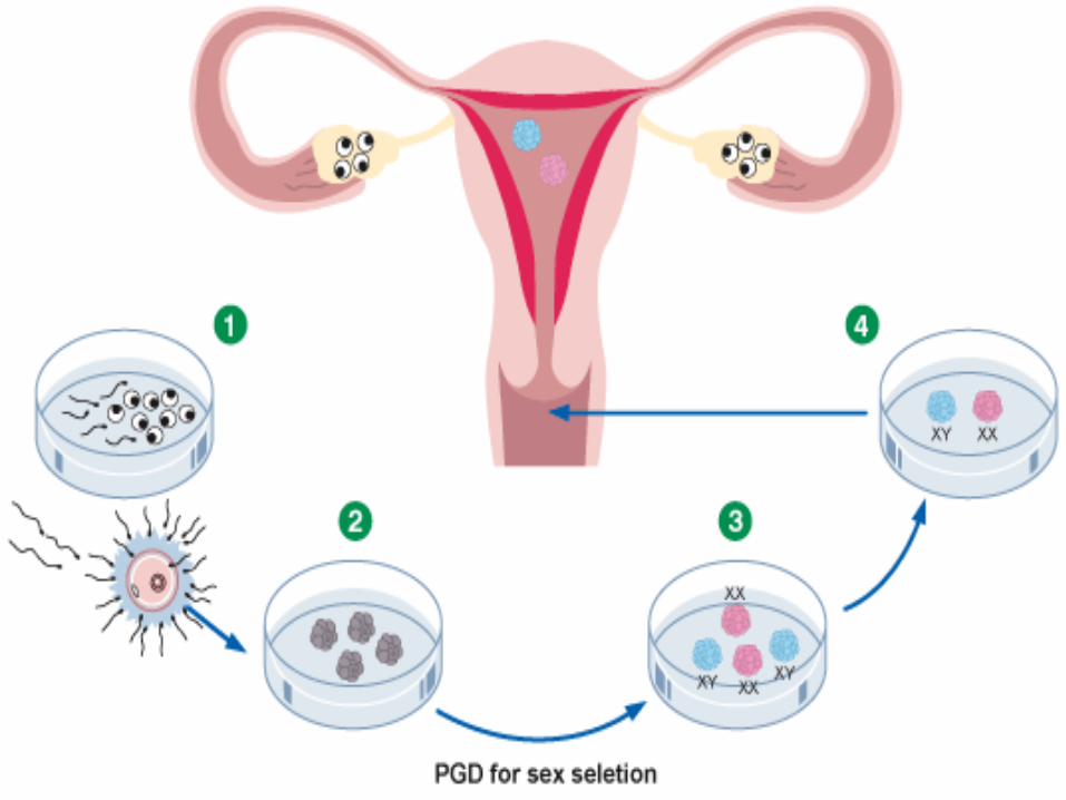
PGD


Achondroplasia
Adrenoleukodystrophy
Agammaglobulinemia
Alpha-1-Antitrypsin
Alpha Thalassemia
Alport Disease
Alzheimer's disease - Early onset (PSEN1-2)
Becker muscular dystrophy
Beta Thalassemia
Charcot Marie Tooth
Chromosomal aneuploidies by FISH
Cystic Fibrosis
Cruzon syndrome
Duchenne muscular dystrophy
Dystonia
Epidermolysis Bullosa
Fanconi Anemia
Familial adenomatous polyposis (FAP)
Familial dysautonomia
Fragile-X syndrome
Gaucher’s Disease
Glycogen storage disease
Hemophilia A and B
HLA typing
HSNF5 mutations
Huntington disease
Hurler syndrome
Incontinentia pigmentii
Kell disease
Marfan syndrome
MELAS
Multiple Endocrine Neoplasia Type II (MEN II)
Multiple Epiphysial Dysplasia
Myotonic Dystrophy
Myotubular myopathy
Neurofibromatosis type I
Neurofibromatosis type II
Norrie disease
Osteogenesis imperfecta I - IV
OTC Deficiency
P53 Oncogene
Phenylketonuria
Polycystic kidney disease (Autosomal Dominant
types I and II)
Retinitis Pigmentosa
SCA 6
Sickle Cell Anemia
Sonic hedgehog mutations
Spinal/Bulbar Muscular Atrophy
Spinal Muscular Atrophy
Tay-Sachs Disease
Translocations by FISH
Tuberous sclerosis
Von Hippel Lindau
Wiskott-Aldrich syndrome
X linked Disease by sexing
X-linked hydrocephalus
X-linked hyper IgM syndrome

Current Techniques Applied to
Molecular Pathology
(one gene – one disease)
• Southern blot
• Dot blot/Reverse dot blot
• Polymerase chain reaction
• SSCP/DGGE
• RT-PCR
• DNA sequencing
• TaqMan, real-time PCR
• Invader assay
• In situ hybridization

New Techniques Coming to
Molecular Pathology
(all genes – all diseases)
• Southern blot
• Dot blot/Reverse dot blot
• Polymerase chain reaction
• SSCP/DGGE
• RT-PCR
• DNA sequencing
• TaqMan, real-time PCR
• Invader assay
• In situ hybridization
• Microarray hybridization
• High-density microarray hybridization
• Array comparative genomic hybridization
• Whole-genome sequencing

Difficulties:
Allelic drop out
Contamination
Background for each
Disease
Materials and
Equipments
Information
Dr. M. Houshmand

Refferal Lab
User Lab 5
Service Lab 1
User Lab 4
User Lab 1
User Lab 3
User Lab 2 Service Lab 2
Service Lab 3
Service Lab 4
Service Lab 5

