genetic counseling for fetal anomalies · holt-oram syndrome •1 per 100,000 live births...
TRANSCRIPT

Genetic Counseling for Fetal Anomalies
Carin Lea Yates, MS, CGCGenetic Counselor
Henry Ford Hospital 18 March 2015

Objectives
• Explain basic genetic concepts.
• Describe the role of the genetic counselor in the prenatal setting.
• Identify when genetic counseling for fetal anomalies is indicated.

BASIC GENETIC CONCEPTS


Genetic Conditions
• Inherited
– Dominant
– Recessive
– X-linked
• De Novo
– Trisomies
– Single gene
GENETIC COUNSELING

Genetic Counseling
Interpretation of family and medical histories to assess the chance of disease occurrence or recurrence.
Education about inheritance, testing, management, prevention, resources and research.
Counseling to promote informed choices and adaptation to the risk or condition.
National Society of Genetic Counselors, 2005
Genetic counseling is the process of helping people understand and adapt to the medical, psychological and familial implications of genetic contributions to disease. This process integrates:

PRENATAL DIAGNOSIS


PRENATAL GENETIC COUNSELING

Common Indications for Genetic Counseling & Prenatal Diagnosis
• Advanced maternal age
• Previous pregnancy with de novo chromosome aneuploidy
• Parent with a structural chromosome abnormality
• Family history of a genetic condition that can be ruled out
• Family history of X-linked disorder with no specific prenatal diagnostic test
• Risk of a neural tube defect
• Abnormal maternal serum screen
• Abnormal cell free fetal DNA
• Abnormal ultrasound

Case Presentation
Lucy is a 32-year-old primigravida woman referred for ultrasound anomalies. Lucy had a routine second trimester ultrasound in her obstetrician’s office that suggested a fetal arm anomaly. She was referred to our center for further evaluation. Lucy underwent a level II ultrasound and fetal echocardiogram in our clinic where a unilateral (right) absence of the fetal radius and an atrial septal defect were detected.

Genetic Consultation
• Any pregnancy with fetal anomalies automatically referred for genetic counseling to discuss:
– Etiology of birth defect(s), if known
– Chance underlying genetic syndrome
– Benefits and limitations of genetic testing
– Coordination of genetic testing
– Management of pregnancy
– Prognosis
• Always have an “on-call” counselor

Information gathering
• Spoke to:
– Sonographer
– Maternal fetal medicine
– Pediatric cardiologist
• Viewed ultrasound pictures and ultrasound report
• Discussed case with OB Geneticist

Unilateral absence of the radius
Level II Ultrasound

Atrial septal defect (ASD)
Fetal Echocardiogram

Information Gathering

Differential Diagnosis
• More than one birth defect increases chance that fetus has a genetic condition
– Chromosome abnormality
– Single gene condition
– Multifactorial genetic syndrome
• Limb anomalies and heart defects
– Both “common”
– Dozens of defined genetic syndromes

Trisomy 13 or 18
• Multiple congenital anomalies
– Not always seen on ultrasound
• Severe mental retardation
• Most cases lethal

Holt-Oram syndrome
• 1 per 100,000 live births
• Autosomal dominant
– 85% de novo mutations
– Germline mosaicism
• Clinical features
– Cardiovascular abnormalities
– Bilateral asymmetric limb abnormalities

T Box Transcription Factor Gene (TBX5 gene)
• Nine coding exons
• Transcription factor
– Cardiac septation
– Forelimb outgrowth
• T-Box
– DNA binding region
– Highly conserved across species
• Over 30 mutations identified
– Reduced TBX5 dose
(Basson et al 1999)

Clinical Features Limb Abnormalities • Radial, thenar, or carpal
bones• From thumb hypoplasia to
phocomelia
Cardiovascular Abnormalities
• Most common ASD or VSD
• From asymptomatic conduction defects to multiple structural anomalies

Consultation• Obtained appropriate histories
– Medical history on both patient and partner
– Pregnancy history for patient
– Family history for both patient and partner

Consultation
• Discussed birth defects– Unable to determine etiology at this time
– Likely we will not during pregnancy
– Cannot predict if an other anomalies and/or mental retardation
• Options at this point– Continue: no additional evaluation
– Continue: genetic testing
– Termination

Consultation• Genetic testing options
– Chromosome analysis• Will not detect small structural changes or single gene conditions
– TBX5 sequencing• 35% of patients with HOS will show mutations in this gene

Consultation
• Psychological support
– Throughout consultation and afterwards
• Using counseling skills
• Identifying personal resources
• Referring couple to social worker specializing in pregnancy complications
• Availability of other professionals and support groups

Coordination of Testing
• Patient chose to do testing stepwise
–Genzyme Genetics
• Complete chromosome analysis
–TAT: 10 to 14 days
–GeneDx
• TBX5 gene sequencing
–TAT: 2 to 3 weeks


Genetic Test Results
• Provide accurate genetic counseling
– Etiology for birth defects
– Prognosis for this pregnancy
– Facilitate testing on parents for accurate recurrence risk assessment
• Referrals
– Orthopedic surgeon
– Support groups for Holt-Oram

Additional Genetic Testing
• Most likely de novo case– Unremarkable family history
– 85% cases are due to new mutation
• Parental testing available– If negative, still cannot rule out germline
mosaicism
• Future pregnancies– Preimplantation genetic diagnosis
– Prenatal diagnosis

Follow-Up Consultation
• When daughter was 8 months old, Lucy and her husband contacted me to facilitate genetic testing
• Neither had any clinical features of Holt-Oram syndrome

TBX5 genetic testing results

GENETIC COUNSELING FOR FETAL ANOMALIES

Ultrasound Results

Genetic Consultation
• Any pregnancy with fetal anomalies referred for genetic counseling to discuss:
– Etiology of birth defect(s), if known
– Chance underlying genetic syndrome
– Benefits and limitations of genetic testing
– Coordination of genetic testing
– Management of pregnancy
– Prognosis

“Soft Signs”
• Variations on ultrasound– NOT structural abnormalities– Detected with increased frequency due to
advanced ultrasound resolution
• Each soft sign is identified in 1-2% of normal pregnancies
• Each occurs more often in fetuses with a chromosome abnormality

Examples of Soft Signs
• Echogenic intracardiac focus (EIF)– Hyperechoic spot on the fetal heart
– Hypothesized to be calcium deposits in the muscle
– Associated with an increased risk for Down syndrome
• Choroid plexus cyst (CPC)– The choroid plexus is the portion of the brain
responsible for creating cerebral spinal fluid
– Associated with increased risk for Trisomy 18

EIF CPC

What if a soft sign is visualized?
• Are there other risk factors?
– Advanced maternal age?
– Multiple markers?
– Abnormal maternal serum screen result?
– Family history?
• Prenatal diagnostic testing
• If chromosomes are normal then the markers are considered normal variants


Cleft Lip & Cleft Palate

Cleft Lip & Cleft Palate
• Facial cleft involving the upper lip and/or palate, usually to the right or left of midline
• May occur as an isolated malformation or part of a multiple malformation syndrome
• Seen in 1:500 – 1:1000 livebirths
• Cleft lip and/or palate (M>F)
• Isolated cleft palate (F>M)

Cleft Lip & Cleft Palate
• Prenatal Testing / Management of Pregnancy
– Detailed medical history, exposure history, family history
– Detailed fetal ultrasound to look for additional abnormalities
– Fetal echocardiogram
– Fetal karyotype
• Consider additional genetic testing if findings are suggestive of specific genetic syndrome (FISH for 22q11.2 deletion)
– Serial ultrasounds for polyhydramnios
– Meet with neonatologist and pediatric surgeon prior to delivery

Cleft Lip & Cleft Palate• After Delivery
– Genetics evaluation to look for additional abnormalities
– Echocardiogram– Feeding difficulties – failure to gain
weight appropriately– Surgical repair of the defect– Hearing evaluation– Long term concerns include
appearance (and related psychological problems), dental abnormalities, speech disorders, and reduced body growth

Cleft Lip & Cleft Palate
• Recurrence Risks
– Aneuploidy: slightly higher than a woman’s age-related risk for aneuploidy
– Genetic syndrome: depends on the syndrome and type of inheritance, but may potentially be as high as 50%
– Isolated cleft lip and palate:
• Risk to offspring of affected: 4.3%
• Risk to siblings of affected: 4-10%

Cystic Kidneys
• Group of disorders ranging from:
– Solitary cysts
– Multicystic kidneys
– Polycystic kidneys
• Findings:
– Enlarged and hyperechogenic
– Uni- or bi-lateral involvement
– Absent or reduced levels of
amniotic fluid
– Small/absent bladder
• Genetic, developmental, and
systemic etiologies

Complications/Prognosis
• The cysts replace most of the normal structure of the kidneys reducing function
• Unilateral – Good prognosis if associated abnormalities are excluded and
amniotic fluid volume is preserved– Chronic concerns: high blood pressure, pain, infections, kidney
failure– Usually requires dialysis and/or transplant later in life
• Bilateral – Typically considered lethal condition because of oligohydramnios
leading to pulmonary hypoplasia– Need to make decisions about resuscitation at birth
• Depends on diagnosis; other associated abnormalities?– Many times, a diagnosis is not made prenatally

Differential Diagnoses/Ultrasound Findings
Disease Cysts (size, location,
and number)
AFI Associated
abnormalities
AD Polycystic KD Randomly distributed
cysts
Normal or
reduced
Uncommon
AR Polycystic KD Greatly enlarged kidneys
with no visible cysts
Reduced Uncommon
Trisomy 13 Enlarged echogenic
kidneys; randomly
dispersed small cysts
Normal or
reduced
Heart defects,
brain abnl., IUGR,
facial clefts
Meckel-Gruber
syndrome
Small cysts, same size,
scattered throughout
Normal or
reduced
Encephalocele,
polydactyly
Multicystic
Dysplastic Kidney
Cysts grouped at the
periphery of the kidney
If bilateral
then no fluid;
unilateral
then normal
GI-defects, other
urologic abnl.,
cranial defects

Ventral Wall DefectsOmphalocele & Gastroschisis
Fetal MRI: Omphalocele
Gastroschisis

Ventral Wall Defects
• Omphalocele is a transparent sac of amnion attached to the umbilical ring that contains herniated intestines
• Gastroschisis is the finding of exposed intestines that protrude through a defect that is typically located on the right side of the abdomen
• Both occur in 1:4000 births– Gastroschisis (M:F)
– Omphalocele (M1:F5)

Ventral Wall Defects
• Omphalocele– Associated abnormalities are
seen in ~66% of cases, including congenital heart defects, bladder exstrophy, imperforate anus, neural tube defects, cleft lip +/-cleft palate, and diaphragmatic hernia
– Approximately 25% have associated chromosomal abnormalities
– Other genetic syndromes

Ventral Wall Defects
• Gastroschisis– Primary concern: vascular compromise from kinking of the
blood vessels coming through the defect which can lead to necrosis
– Intestinal atresias and other GI disruptions are found in approximately 5-10% of cases
– Extraintestinal abnormalities occur in less than 5% of cases
– Usually not associated with chromosomal abnormalities or genetic syndromes
– Associated with young maternal age and maternal cigarette smoking
Ventral Wall Defects
• Prenatal Testing / Management of Pregnancy
– Omphalocele
• Detailed fetal ultrasound examination
• Fetal karyotype
• Fetal echocardiogram
• Consultation with neonatologist and pediatric surgeon
• Serial ultrasounds to monitor growth
• Preterm delivery and IUGR frequently complicate cases
• High rate of emergency C-sections due to fetal distress
• Delivery at hospital with tertiary-care center

Ventral Wall Defects
• Prenatal Testing / Management of Pregnancy
– Gastroschisis
• Detailed fetal ultrasound examination
• Fetal karyotype can be considered
• Fetal echocardiogram
• Consultation with neonatologist and pediatric surgeon
• Serial ultrasounds to detect thickening and/or dilation of the fetal bowel and assess fetal growth
• Preterm delivery and IUGR frequently complicate cases
• Delivery at tertiary-care center

Neural Tube Defects (NTDs)
• Population incidence: 1/1000 births• Failure of closure of the
neural tube @ ~28 days post conception• Includes
– Anencephaly– Spina bifida
• Myelomeningocele• Meningocele
• Caused by– Multifactorial inheritance– Diabetes– Medications (anti-seizure
medications)– Chromosomal or single gene
disorders


Anencephaly
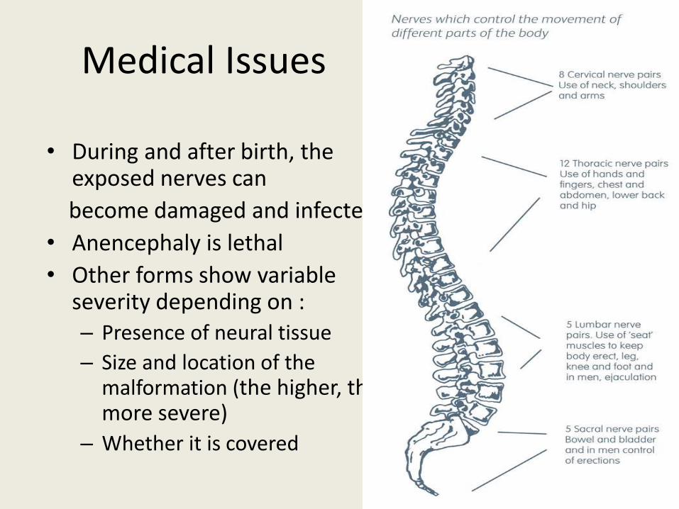
Medical Issues
• During and after birth, the exposed nerves can
become damaged and infected
• Anencephaly is lethal
• Other forms show variable severity depending on :– Presence of neural tissue
– Size and location of the malformation (the higher, the more severe)
– Whether it is covered

Medical Issues
• Spine:– Loss of sensation and
paralysis
– Loss of control of muscles served by damaged nerves
– Tethered cord
– Sexual dysfunction
• Brain– Hydrocephalus
– Chiari II malformation
– Learning disabilities
• Urologic issues:
– Loss of bowel and bladder control
– Kidney infections and damage
– Gastrointestinal issues
• Orthopedic issues
– Paralysis can lead to dislocated joints, misshapen bones, scoliosis

Diagnostic Options
• Ultrasound
– A detailed ultrasound at 18-20 weeks can detect 90% of open neural tube defects
– Anencephaly as early as 12 weeks
– Open defects are associated with specific head findings
• Lemon sign: 98% of fetuses with open defects when scanned <24 weeks and 13% of fetuses scanned after 24 weeks
• Banana sign: 95% of fetuses with open defects
• Ventriculomegaly
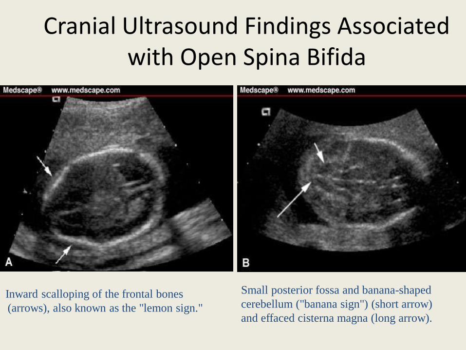
Cranial Ultrasound Findings Associated with Open Spina Bifida
Inward scalloping of the frontal bones
(arrows), also known as the "lemon sign."
Small posterior fossa and banana-shaped
cerebellum ("banana sign") (short arrow)
and effaced cisterna magna (long arrow).
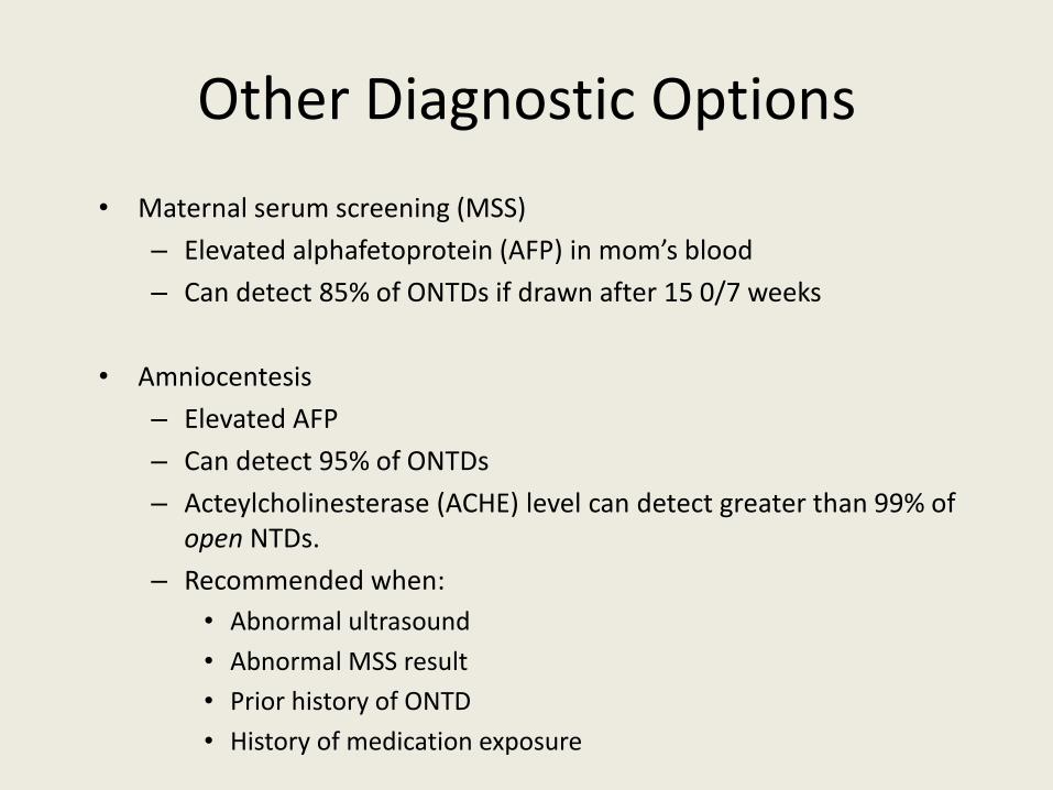
Other Diagnostic Options
• Maternal serum screening (MSS)
– Elevated alphafetoprotein (AFP) in mom’s blood
– Can detect 85% of ONTDs if drawn after 15 0/7 weeks
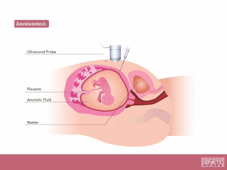
• Amniocentesis
– Elevated AFP
– Can detect 95% of ONTDs
– Acteylcholinesterase (ACHE) level can detect greater than 99% of open NTDs.
– Recommended when:
• Abnormal ultrasound
• Abnormal MSS result
• Prior history of ONTD
• History of medication exposure

Pregnancy Management
• Continuation versus termination• Follow-up ultrasounds to
watch progression• Fetal MRI• Fetal echocardiogram• Discuss delivery plan• Surgery
– Earlier the better (before 25 weeks)
– Cannot restore lost function but can prevent further damage
– Risks to mother and fetus

Medical Management/Prognosis
• Prevent infection2
• Surgery2
• Shunt to correct hydrocephalus
• Long-term multidisciplinary care (neurosurgeon, orthopedic surgeon, urologist, dietician, physical therapist)
• Assistive devices
• Early developmental intervention
• Prognosis is variable but better when children get early intervention

Recurrence Risks
• Recurrence depends on the cause
– Genetic condition: risk as high as 50%
• Other abnormalities?
• Family history?
– Chromosomal: risk <1%
– Isolated: 3-5% risk to siblings
• Recurrence risks for all types
• Folic Acid supplementation can reduce the risk of NTDs by 50%

Thank You
Elizabeth Cameron, MS, CGC
Lauren Mohnach, MS, CGC
For use of their slides on fetal anomalies.