gene expression profiling of clear cell papillary renal cell carcinoma: comparison with clear cell...
TRANSCRIPT

Gene expression profiling of clear cellpapillary renal cell carcinoma: comparisonwith clear cell renal cell carcinoma andpapillary renal cell carcinomaKevin E Fisher1, Qiqin Yin-Goen1, Dianne Alexis1, Joseph S Sirintrapun2,William Harrison2, R Benjamin Isett3, Michael R Rossi3, Carlos S Moreno1,3,Andrew N Young1 and Adeboye O Osunkoya1,3,4
1Department of Pathology, Emory University School of Medicine, Atlanta, GA, USA; 2Department ofPathology, Wake Forest University School of Medicine, Winston-Salem, NC, USA; 3Emory WinshipCancer Institute, Atlanta, GA, USA and 4Department of Urology, Emory University School of Medicine,Atlanta, GA, USA
Clear cell papillary renal cell carcinoma is a distinct variant of renal cell carcinoma that shares some overlapping
histological and immunohistochemical features of clear cell renal cell carcinoma and papillary renal cell
carcinoma. Although the clear cell papillary renal cell carcinoma immunohistochemical profile is well described,
clear cell papillary renal cell carcinoma mRNA expression has not been well characterized. We investigated the
clear cell papillary renal cell carcinoma gene expression profile using previously identified candidate genes. We
selected 17 clear cell papillary renal cell carcinoma, 15 clear cell renal cell carcinoma, and 13 papillary renal cell
carcinoma cases for molecular analysis following histological review. cDNA from formalin-fixed paraffin-
embedded tissue was prepared. Quantitative real-time PCR targeting alpha-methylacyl coenzyme-A racemase
(AMACR), BMP and activin membrane-bound inhibitor homolog (BAMBI), carbonic anhydrase IX (CA9),
ceruloplasmin (CP), nicotinamide N-methyltransferase (NNMT), schwannomin-interacting protein 1 (SCHIP1),
solute carrier family 34 (sodium phosphate) member 2 (SLC34A2), and vimentin (VIM) was performed. Gene
expression data were normalized relative to 28S ribosomal RNA. Clear cell papillary renal cell carcinoma expressed
all eight genes at variable levels. Compared with papillary renal cell carcinoma, clear cell papillary renal cell
carcinoma expressed more CA9, CP, NNMT, and VIM, less AMACR, BAMBI, and SLC34A2, and similar levels of
SCHIP1. Compared with clear cell renal cell carcinoma, clear cell papillary renal cell carcinoma expressed slightly
less NNMT, but similar levels of the other seven genes. Although clear cell papillary renal cell carcinoma exhibits a
unique molecular signature, it expresses several genes at comparable levels to clear cell renal cell carcinoma
relative to papillary renal cell carcinoma. Understanding the molecular pathogenesis of clear cell papillary renal cell
carcinoma will have a key role in future sub-classifications of this unique tumor.Modern Pathology advance online publication, 26 July 2013; doi:10.1038/modpathol.2013.140
Keywords: clear cell; clear cell papillary; gene expression; molecular; papillary; renal cell carcinoma
Clear cell papillary renal cell carcinoma is amorphologically and immunohistochemically dis-tinct renal epithelial neoplasm originally describedas a ‘clear cell papillary renal cell carcinoma of end-stage kidneys’.1 Since its initial description, it is
apparent that although clear cell papillary renal cellcarcinoma has a propensity to arise in the setting ofend-stage kidney disease, these tumors can occur denovo in normal kidneys. Histologically, clear cellpapillary renal cell carcinoma is characterized bypapillary infoldings composed almost entirely ofcells with clear cytoplasm and polarized low-gradenuclei. Immunohistochemically, clear cell papillaryrenal cell carcinoma expresses cytokeratin 7 andcarbonic anhydrase IX (CA9) but does not expressalpha methyl-coenzyme-A racemase (AMACR),CD10, or transcription factor E3 (TFE3).2 Clear cell
Correspondence: Dr AO Osunkoya, MD, Department of Pathology,Emory University School of Medicine, Room H174, 1364 CliftonRoad NE, Atlanta, GA 30322, USA.E-mail: [email protected] 1 February 2013; revised 13 June 2013; accepted 14 June2013; published online 26 July 2013
Modern Pathology (2013), 1–9
& 2013 USCAP, Inc. All rights reserved 0893-3952/13 $32.00 1
www.modernpathology.org

papillary renal cell carcinoma does not harbor thechromosome 3p deletions seen in clear cell renalcell carcinoma or demonstrate trisomies 7 and 17, orloss of Y that is characteristic of papillary renalcell carcinoma.3 Clearly, clear cell papillaryrenal cell carcinoma is a distinct tumor that sharessome morphological, immunohistochemical, andmolecular features of both clear cell renal cellcarcinoma and papillary renal cell carcinoma, butfew studies have examined the molecular geneexpression profile of these neoplasms. Althoughclear cell papillary renal cell carcinoma may haveoverlapping morphological features with clearcell renal cell carcinoma and papillary renal cellcarcinoma (especially those with clear cell features)in areas, clear cell papillary renal cell carcinomaand papillary renal cell carcinoma are moreaggressive tumors. Herein, using real-time (RT)-PCR, we targeted eight previously identifiedcandidate genes (Table 1) to assess their expressionlevels in clear cell papillary renal cell carcinoma,clear cell renal cell carcinoma, and papillary renalcell carcinoma. We selected a panel of four candi-date clear cell renal cell carcinoma markers and fourpapillary renal cell carcinoma markers, which hadbeen identified in previous expression-profilingstudies from our group. These markers were chosento study whether the novel subtype clear cellpapillary renal cell carcinoma was distinct from orsimilar to clear cell renal cell carcinoma or papillaryrenal cell carcinoma in terms of gene expression.
Materials and methods
Case Selection and Analysis
Seventeen cases of clear cell papillary renal cellcarcinoma, 15 cases of clear cell renal cell carcinoma,and 13 cases of papillary renal cell carcinoma wereselected for analysis. All cases were re-reviewed by aurological pathologist. The diagnosis of clear cellpapillary renal cell carcinoma was based on classicmorphological features and corresponding immuno-histochemical profile (positive cytokeratin 7 expres-sion and negative CD10, P504S/AMACR and TFE3expression). Tumor grading and staging was based on
the standard Fuhrman nuclear-grading system andthe American Joint Committee on Cancer, 7th editiontumor-node-metastasis staging system, respec-tively.4,5 This study was completed following theguidelines of and with approval from theinstitutional review board of our institution.
Quantitative RT-PCR
Quantitative RT-PCR experiments were performedin duplicate on total RNA isolated from formalin-fixed paraffin-embedded tissue from each specimen.Unstained histological sections were deparaffinizedwith ethanol and xylene. The tumor cells weremicrodissected with a sterile scalpel. Tissue wasdigested in buffer containing proteinase K at 60 1Covernight, and total RNA was isolated by phenolchloroform extraction and subjected to DNase treat-ment. RNA quality and quantity were assessed withBioanalyzer (Agilent Technologies, Santa Clara, CA,USA). Up to 3mg of RNA was used for first-strandcDNA synthesis with Superscript III (Invitrogen/Life Technologies, Grand Island, NY, USA). PCR wascarried out with sybrogreen master mix (AppliedBiosystems/Life Technologies) in the 96-well formatusing ABI PRISM 7500 Fast RT-PCR system. PCRruns were analyzed by using Relative QuantificationManager (Applied Biosystems) software. RelativemRNA expression levels of eight genes (AMACR,BAMBI, CA9, CP, NNMT, SCHIP1, SLC34A2, andVIM, see Table 1) were assessed. These genes wereselected from previous data generated by ourlaboratory as markers of clear cell renal cellcarcinoma and papillary renal cell carcinoma.6
Data Analysis
Gene expression data were normalized relative to thegeometric mean of the 28S ribosomal RNA house-keeping gene. mRNA extracted from normal kidneywas used as a reference standard. For each tumor, therelative mean Ct of each gene was calculated asfollows: (value well 1þ value well 2)/2. The tumorDCt was calculated as follows: gene tested relativemean Ct� 28SRNA relative mean Ct. The DDCt was
Table 1 Genes overexpressed in clear cell renal cell carcinoma and papillary renal cell carcinoma
SymbolOverexpressed inPRCC vs CCRCC UniGene GeneBank Name
AMACR PRCC Hs.508343 NM_014324 Alpha-methylacyl coenzyme-A racemaseBAMBI PRCC Hs.533336 NM_012342 BMP and activin membrane-bound inhibitor homologCA9 CCRCC Hs.63287 NM_001216 Carbonic anhydrase IXCP CCRCC Hs.558314 NM_000096 CeruloplasminNNMT CCRCC Hs.503911 NM_006169 Nicotinamide N-methyltransferaseSCHIP1 PRCC Hs.134665 NM_014575 Schwannomin-interacting protein 1SLC34A2 PRCC Hs.479372 NM_006424 Solute carrier family 34 (sodium phosphate) member 2VIM CCRCC Hs.455493 NM_003380 Vimentin
Abbreviations: CCPRCC, clear cell papillary renal cell carcinoma; PRCC, papillary renal cell carcinoma.
Gene expression profiling of CCPRCC
2 KE Fisher et al
Modern Pathology (2013), 1–9

calculated as follows: tumor DCt�normal kidneyDCt. Fold-change of mRNA was calculated as2(�DDCt). Average fold-change was calculated byaveraging the fold-change of the total cases for eachtumor type.
Statistics
For differential mRNA expression data, values foreach tumor type were compared using the Wilcoxonrank-sum test. For comparison of mean-normalizedmRNA expression, a two-tailed Student’s t-testassuming equal variances was performed. The meanmRNA fold-change in expression of each genebetween each tumor histological subtype was ana-lyzed. Statistical significance was set at Po0.05.
Results
Case Selection and Analysis
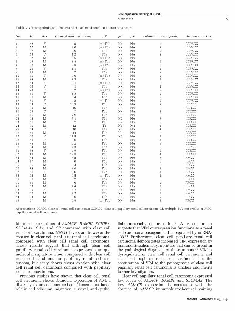
Overall, 45 tumors from 28 male and 17 femalepatients with a mean age of 53 years (range 29–78years) were included in the study. Eight male and 9female patients, with a mean age of 53 years (range29–73 years), had clear cell papillary renal cellcarcinoma (Figures 1a–d), 9 male and 6 femalepatients, with a mean age of 58 years (range 40–78years), had clear cell renal cell carcinoma, and 11male and 2 female patients, with a mean age of 59years (range 40–74 years), had papillary renal cellcarcinoma. Fifteen of the 17 cases (88%) of clear cellpapillary renal cell carcinoma were identifiedduring screening of the patients for renal insuffi-ciency. The average maximum tumor dimension ofthe 45 tumors was 5.8 cm (2.5 cm for clear cellpapillary renal cell carcinoma, 8.7 cm for clear cellrenal cell carcinoma, and 6.4 cm for papillary renalcell carcinoma). Thirty cases of renal cell carcinomawere pathological stage pT1 (17 clear cell papillaryrenal cell carcinoma, 2 clear cell renal cell carcino-ma, and 11 papillary renal cell carcinoma), 3 clearcell renal cell carcinoma were in pT2, 11 renal cellcarcinoma were in pT3 (11 clear cell renal cellcarcinoma and 2 papillary renal cell carcinoma),and 1 clear cell renal cell carcinoma was in pT4.Multifocality was seen in nine clear cell papillaryrenal cell carcinoma and two papillary renal cellcarcinoma. Lymph node metastases were seen intwo clear cell renal cell carcinoma patients, and nopatient had evidence of distant metastases at thetime of surgical resection. Seventeen cases of clearcell papillary renal cell carcinoma, 4 clear cell renalcell carcinoma, and 7 papillary renal cell carcinomashowed nuclear features consistent with Fuhrmannuclear grade 2; 7 clear cell renal cell carcinoma and5 papillary renal cell carcinoma were classified asFuhrman nuclear grade 3. The clinicopathologicalcharacteristics of the 45 cases are summarized inTable 2.
Quantitative RT-PCR, Data Analysis and Statistics
The differential mRNA expression (�DDCt) of thefour papillary renal cell carcinoma genes (AMACR,BAMBI, SCHIP1, and SLC34A2) and the four clearcell renal cell carcinoma genes (CA9, NNMT, CP, andVIM) in each of the 45 individual renal cellcarcinomas is shown in Figures 2a and b, respec-tively. The individual �DDCt values for the papil-lary renal cell carcinoma genes and the clear cellrenal cell carcinoma genes were normalized using alog2 transformation (2(�DDCt), Figures 3 and 4,respectively), and the results were averaged to givean overall mean-normalized mRNA expressionvalue for the entire cohort of clear cell papillaryrenal cell carcinoma, papillary renal cell carcinomaand clear cell renal cell carcinoma (Figure 5). Thedata points were statistically analyzed using theWilcoxon rank-sum test (Figure 2) or Student’s t-test(Figure 5).
When comparing clear cell papillary renal cellcarcinoma with papillary renal cell carcinoma, clearcell papillary renal cell carcinoma were character-ized by overexpression of CA9, ceruloplasmin (CP),and vimentin (VIM; P¼ 0.001, 0.003, and o0.001,respectively, by Wilcoxon rank-sum test; P¼ 0.56,0.031, and 0.020, respectively, by Student’s t-test)and relative overexpression of CP and nicotinamideN-methyltransferase (NNMT) when examined indi-vidually (P¼ 0.001 and o0.001, respectively, byWilcoxon rank-sum test). The transformed andaveraged values for CP and NNMT approachedstatistical significance (P¼ 0.056 and 0.068, respec-tively, by Student’s t-test). Clear cell papillary renalcell carcinoma showed relative underexpression ofAMACR, BAMBI, and SLC34A2 (Po0.001, o0.001,and ¼ 0.014, respectively, by Wilcoxon rank-sumtest and P¼ 0.005, o0.001, and ¼ 0.001, respec-tively, by Student’s t-test). Clear cell papillary renalcell carcinoma and papillary renal cell carcinomaexpressed comparable levels of schwannomin-inter-acting protein 1 (SCHIP1; approached statisticalsignificance by Wilcoxon rank-sum test but notstatistically significant by Student’s t-test). Thestatistical analyses of the genes expressed in clearcell papillary renal cell carcinoma vs papillary renalcell carcinoma are summarized in Table 3.
When comparing clear cell papillary renal cellcarcinoma with clear cell renal cell carcinoma, clearcell papillary renal cell carcinoma expressedslightly lower levels of NNMT (approached statis-tical significance by Wilcoxon rank-sum test(P¼ 0.041) but not significant by Student’s t-test).No statistically significant differences (by Wilcoxonrank-sum test or Student’s t-test) in AMACR, BAMBI,SCHIP1, SLC34A2, CA9, CP, or VIM expression wereseen between clear cell papillary renal cell carcino-ma and clear cell renal cell carcinoma. Thestatistical analyses of the genes expressed in clearcell papillary renal cell carcinoma vs clear cell renalcell carcinoma are summarized in Table 3.
Modern Pathology (2013), 1–9
Gene expression profiling of CCPRCC
KE Fisher et al 3

When comparing clear cell renal cell carcinomawith papillary renal cell carcinoma, clear cell renalcell carcinoma expressed relatively higher amountsof CA9, CP, NNMT, and VIM (P¼ 0.002, 0.037,o0.001, and o0.001, respectively, by Wilcoxonrank-sum test; P¼ 0.023, 0.007, o0.001, and 0.009,respectively, by Student’s t-test). Papillary renal cellcarcinoma expressed relatively higher amounts ofAMACR (P¼ 0.001 by Wilcoxon rank-sum test andP¼ 0.036 by Student’s t-test) and BAMBI (BMP andactivin membrane-bound inhibitor homolog;Po0.001 by Wilcoxon rank-sum test and Student’st-test). Papillary renal cell carcinoma demonstratedrelative overexpression of SLC34A2 (P¼ 0.033 byWilcoxon rank-sum test), and the relative over-expression approached statistical significance whenthe values were transformed and averaged (P¼ 0.066by Student’s t-test). Papillary renal cell carci-noma expressed slightly higher levels of SCHIP1compared with clear cell renal cell carcinoma(approached statistical significance by Wilcoxonrank-sum test (P¼ 0.065) but not significant by
Student’s t-test). These results are consistent withour previous work.6 Statistical analysis of thegenes expressed in papillary renal cell carcinomavs clear cell renal cell carcinoma is summarized inTable 4.
Discussion
Clear cell papillary renal cell carcinoma is amorphologically and immunohistochemically un-ique subset of renal cell carcinoma that do notharbor consistent chromosomal imbalances or VHLmutations according to the few molecular studiesreported in the literature.7,8 To our knowledge, nostudies have examined the mRNA expression ofdistinct papillary renal cell carcinoma or clear cellrenal cell carcinoma genes in clear cell papillaryrenal cell carcinoma. Our data show that clear cellpapillary renal cell carcinoma expresses abundantCA9, CP, NNMT, and VIM compared with papillaryrenal cell carcinoma, and somewhat similar but not
Figure 1 Clear cell papillary renal cell carcinoma.(a) Clear cell papillary renal cell carcinoma (hematoxylin and eosin (H&E), lowmagnification). (b) Clear cell papillary renal cell carcinoma (H&E high magnification). (c) Positive cytokeratin 7 (CK7) expression in clearcell papillary renal cell carcinoma. (d) Negative AMACR/P504S expression in clear cell papillary renal cell carcinoma.
Modern Pathology (2013), 1–9
Gene expression profiling of CCPRCC
4 KE Fisher et al
identical expressions of AMACR, BAMBI, SCHIP1,SLC34A2, CA9, and CP compared with clear cellrenal cell carcinoma. NNMT levels are however de-creased in clear cell papillary renal cell carcinoma,compared with clear cell renal cell carcinoma.These results suggest that although clear cellpapillary renal cell carcinoma expresses a uniquemolecular signature when compared with clear cellrenal cell carcinoma or papillary renal cell car-cinoma, it clearly shows closer overlap with clearcell renal cell carcinoma compared with papillaryrenal cell carcinoma.
Previous studies have shown that clear cell renalcell carcinoma shows abundant expression of VIM, adiversely expressed intermediate filament that has arole in cell adhesion, migration, survival, and epithe-
lial-to-mesenchymal transition.9 A recent reportsuggests that VIM overexpression functions as a renalcell carcinoma oncogene and is regulated by miRNA-138.10 Furthermore, clear cell papillary renal cellcarcinoma demonstrates increased VIM expression byimmunohistochemistry, a feature that can be useful inthe pathological diagnosis of these tumors.11 VIM isdysregulated in clear cell renal cell carcinoma andclear cell papillary renal cell carcinoma, but thecontribution of VIM to the pathogenesis of clear cellpapillary renal cell carcinoma is unclear and meritsfurther investigation.
Clear cell papillary renal cell carcinoma expressedlow levels of AMACR, BAMBI, and SLC34A2. Thelow AMACR expression is consistent with theabsence of AMACR immunohistochemical staining
Table 2 Clinicopathological features of the selected renal cell carcinoma cases
No. Age Sex Greatest dimension (cm) pT pN pM Fuhrman nuclear grade Histologic subtype
1 52 F 5 (m) T1b Nx NA 2 CCPRCC2 57 M 3.6 (m) T1a Nx NA 2 CCPRCC3 47 M 0.9 T1a Nx NA 2 CCPRCC4 58 F 1.2 T1a Nx NA 2 CCPRCC5 32 M 3.5 (m) T1a Nx NA 2 CCPRCC6 45 M 1.8 (m) T1a Nx NA 2 CCPRCC7 66 M 2.5 (m) T1a Nx NA 2 CCPRCC8 29 F 1.1 T1a Nx NA 2 CCPRCC9 49 M 2 T1a Nx NA 2 CCPRCC10 66 F 0.9 (m) T1a Nx NA 2 CCPRCC11 44 M 2.5 T1a Nx NA 2 CCPRCC12 64 F 2.1 (m) T1a Nx NA 2 CCPRCC13 66 F 1 T1a Nx NA 2 CCPRCC14 73 F 3.2 (m) T1a Nx NA 2 CCPRCC15 60 F 1.3 T1a Nx NA 2 CCPRCC16 39 M 5.4 T1b Nx NA 2 CCPRCC17 59 F 4.8 (m) T1b Nx NA 2 CCPRCC18 64 F 10.5 T2b Nx NA 2 CCRCC19 60 M 16 T3c Nx NA 3 CCRCC20 55 F 7 T1b Nx NA 2 CCRCC21 46 M 7.9 T3b N0 NA 3 CCRCC22 48 M 5.1 T3a N2 NA 3 CCRCC23 51 M 7 T3b Nx NA 2 CCRCC24 60 M 12 T4 N1 M1 4 CCRCC25 54 F 10 T2a N0 NA 4 CCRCC26 66 M 14 T3b N0 NA 3 CCRCC27 60 F 11.5 T2b N0 NA 3 CCRCC28 40 F 6 T3b N0 NA 4 CCRCC29 78 M 5.2 T3b Nx NA 3 CCRCC30 54 M 2.3 T1a Nx NA 3 CCRCC31 62 F 4.5 T3b Nx NA 2 CCRCC32 75 M 12.5 T3b N0 NA 4 CCRCC33 65 M 6.5 T3a Nx NA 3 PRCC34 47 M 6 T1b Nx NA 3 PRCC35 56 M 6.4 T1b Nx NA 3 PRCC36 74 M 4.8 T1b Nx NA 2 PRCC37 51 F 26 T3a Nx NA 4 PRCC38 64 M 4.5 (m) T1b Nx NA 2 PRCC39 56 M 3.2 T1a Nx NA 3 PRCC40 69 M 6 T1b Nx NA 2 PRCC41 65 M 2.4 T1a Nx NA 2 PRCC42 40 F 3.7 T1a Nx NA 2 PRCC43 60 M 2.2 T1a Nx NA 3 PRCC44 64 M 6 T1b Nx NA 2 PRCC45 57 M 5.9 (m) T1b Nx NA 2 PRCC
Abbreviations: CCRCC, clear cell renal cell carcinoma; CCPRCC, clear cell papillary renal cell carcinoma; M, multiple; NA, not available; PRCC,papillary renal cell carcinoma.
Modern Pathology (2013), 1–9
Gene expression profiling of CCPRCC
KE Fisher et al 5

described in this tumor.3,12,13AMACR, BAMBI, andSLC34A2 are genes that are expressed at higher levelsin papillary renal cell carcinoma, suggesting thatclear cell papillary renal cell carcinoma is molecu-larly distinct from papillary renal cell carcinoma.However, SCHIP1 was expressed at a similar levelbetween the two tumor types; therefore, some mole-cular overlap does exist. Future studies are needed tocharacterize additional papillary renal cell carcinomagenes that are differentially expressed compared withclear cell papillary renal cell carcinoma in order tobetter characterize the distinct molecular phenotypeof clear cell papillary renal cell carcinoma comparedwith papillary renal cell carcinoma.
Clear cell papillary renal cell carcinoma alsoexpressed high levels of CP and CA9 similar to thelevels seen in clear cell renal cell carcinoma. CP isan acute-phase reactant protein that is expressed inmany inflammatory situations, and serum CP pro-tein levels are elevated in patients with renal cellcarcinoma.6 CA9 is a hypoxia-inducible proteinoverexpressed in clear cell renal cell carcinoma
secondary to VHL dysregulation, but Rohan et al.8
showed that CA9 upregulation in clear cell papillaryrenal cell carcinoma occurs via a VHL-independentmechanism.8,14 These observations suggest thatalthough clear cell papillary renal cell carcinomaoverexpresses CA9, the pathway for itsoverexpression is likely different than that of clearcell renal cell carcinoma. This supports thehypothesis that although clear cell papillary renalcell carcinoma share some molecular features withclear cell renal cell carcinoma, unique molecularpathway alterations exist.
Clear cell papillary renal cell carcinoma aretypically described as low-grade malignancies.7 Inour study, clear cell papillary renal cell carcinomatended to be smaller, multifocal, and of lowerpathological stage and grade, which was similar tothe clinicopathological features seen in papillaryrenal cell carcinoma (Table 2). These clinicopatho-logical features were not shared with clear cell renalcell carcinoma; clear cell renal cell carcinoma werelarger, all unifocal, and most were in pathological
-3-2-101234567
-6
-4
-2
0
2
4
6 BAMBICCPRCC vs. PRCC: p < 0.001 CCRCC vs. PRCC: p < 0.001
-4-3-2-101234
CCRCC vs. PRCC: p = 0.065
-6
-4
-2
0
2
4
6
8 CCPRCC vs. PRCC: p = 0.001CCRCC vs. PRCC: p = 0.002
CA9
-6-4-202468
10 CCPRCC vs. PRCC: p = 0.003CCRCC vs. PRCC: p < 0.001
CP
AMACRa
CCPRCC vs. PRCC: p < 0.001 CCRCC vs. PRCC: p = 0.001
b
-2
0
2
4
6
8
10
12
CCPRCC vs. PRCC: p < 0.001CCPRCC vs. CCRCC: p = 0.041 CCRCC vs. PRCC: p = 0.037
SCHIP1 NNMT
-2-10123456
CCPRCC vs. PRCC: p < 0.001CCRCC vs. PRCC: p < 0.001
CCPRCC (n=17) CCRCC (n=15) PRCC (n=13)Genes overexpressed in clear cell renal cell carcinoma
VIM
-12-10-8-6-4-20246
Genes overexpressed in papillary renal cell carcinoma
CCPRCC vs. PRCC: p = 0.014CCRCC vs. PRCC: p = 0.033
SLC34A2
CCPRCC (n=17) CCRCC (n=15) PRCC (n=13)
Figure 2 (a, b) Differential mRNA expression (�DDCt) in renal cell carcinoma. Total RNA was isolated from formalin-fixedparaffin-embedded tissue, and mRNA levels for AMACR, BAMBI, SCHIP1, SLC34A2, CA9, CP, NNMT, and VIM were assessed. Geneexpression data were normalized relative to the geometric mean of the 28S ribosomal RNA (28SRNA) housekeeping gene. mRNA fromnormal kidney was used as a reference standard. Differential mRNA expression (�DDCt) is shown. Each bar is an individual tumor.P-values are calculated by Wilcoxon rank-sum test.
Modern Pathology (2013), 1–9
Gene expression profiling of CCPRCC
6 KE Fisher et al

0
20
40
60
80
0
6
12
18
24
30
0
3
6
9
12
15
0
3
6
9
12CCPRCC (n=17) CCRCC (n=15) PRCC (n=13)
CCPRCC (n=17) CCRCC (n=15) PRCC (n=13)
CCPRCC (n=17) CCRCC (n=15) PRCC (n=13)
CCPRCC (n=17) CCRCC (n=15) PRCC (n=13)
Genes overexpressed in papillary renal cell carcinoma
AMACR
BAMBI
SCHIP1
SLC34A2
Figure 3 Normalized 2(�DDCt)mRNA expression of the papillary renal cell carcinoma genes. Total RNA was isolated from formalin-fixedparaffin-embedded tissue, and mRNA levels for AMACR, BAMBI, SCHIP1, and SLC34A2 were assessed. Each bar represents the log2 genemRNA expression level of a single tumor.
CCPRCC (n=17) PRCC (n=13)
CCPRCC (n=17) CCRCC (n=15) PRCC (n=13)
CCPRCC (n=17) CCRCC (n=15) PRCC (n=13)
CCPRCC (n=17) CCRCC (n=15) PRCC (n=13)
0
15
30
45
60
Genes overexpressed in clear cell renal cell carcinoma
CCRCC (n=15)
0
60
120
180
240
300
0
300
600
900
1200
0
10
20
30
40
50
CA9
CP
NNMT
VIM
Figure 4 Normalized 2(�DDCt)mRNA expression of the clear cell renal cell carcinoma genes. Total RNA was isolated from formalin-fixedparaffin-embedded tissue, and mRNA levels for CA9, CP, NNMT, and VIM were assessed. Each bar represents the log2 gene mRNAexpression level of a single tumor.
Modern Pathology (2013), 1–9
Gene expression profiling of CCPRCC
KE Fisher et al 7
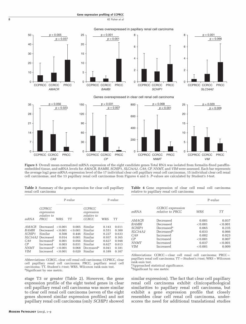
stage T3 or greater (Table 2). However, the geneexpression profile of the eight tested genes in clearcell papillary renal cell carcinoma was more similarto clear cell renal cell carcinoma (seven of the eightgenes showed similar expression profiles) and notpapillary renal cell carcinoma (only SCHIP1 showed
similar expression). The fact that clear cell papillaryrenal cell carcinoma exhibit clinicopathologicalsimilarities to papillary renal cell carcinoma, butexhibit a gene expression profile that closelyresembles clear cell renal cell carcinoma, under-scores the need for additional translational studies
BAMBI SCHIP1 SLC34A2AMACR
0
5
10
15
20
25
CCPRCC CCRCC PRCC
CA9 CP NNMT VIM
0
7
14
21
28
35
CCPRCC CCRCC PRCC
Genes overexpressed in papillary renal cell carcinoma
Genes overexpressed in clear cell renal cell carcinoma
p = 0.005p = 0.066p = 0.037
p < 0.001p < 0.001
p = 0.001
p = 0.020p = 0.009 p < 0.001
p = 0.031p = 0.007
p = 0.056p = 0.023
0
10
20
30
40
50
CCPRCC CCRCC PRCC0
2
4
6
8
CCPRCC CCRCC PRCC
0
30
60
90
120
150
CCPRCC CCRCC PRCC0
200
400
600
800
CCPRCC CCRCC PRCC
p = 0.068
0
5
10
15
20
25
CCPRCC CCRCC PRCC
0
2
4
6
8
CCPRCC CCRCC PRCC
Figure 5 Overall mean-normalized mRNA expression of the eight candidate genes.Total RNA was isolated from formalin-fixed paraffin-embedded tissue, and mRNA levels for AMACR, BAMBI, SCHIP1, SLC34A2, CA9, CP, NNMT, and VIM were assessed. Each bar representsthe average log2 gene mRNA expression level of the 17 individual clear cell papillary renal cell carcinomas, 15 individual clear cell renalcell carcinomas, and the 13 papillary renal cell carcinomas from Figures 4 and 5. P-values are calculated by Student’s t-test.
Table 3 Summary of the gene expression for clear cell papillaryrenal cell carcinoma
P-value P-value
mRNA
CCPRCCexpressionrelative toPRCC WRS TT
CCPRCCexpressionrelative toCCRCC WRS TT
AMACR Decreased o0.001 0.005 Similar 0.143 0.611BAMBI Decreased o0.001 o0.001 Similar 0.551 0.300SCHIP1 Similar 0.508 0.446 Similar 0.227 0.633SLC34A2 Decreased 0.014 0.001 Similar 0.937 0.165CA9 Increaseda 0.001 0.056 Similar 0.627 0.948CP Increased 0.003 0.031 Similar 0.627 0.613NNMT Increaseda o0.001 0.068 Decreaseda 0.041 0.181VIM Increased o0.001 0.020 Similar 0.189 0.107
Abbreviations: CCRCC, clear cell renal cell carcinoma; CCPRCC, clearcell papillary renal cell carcinoma; PRCC, papillary renal cellcarcinoma; TT, Student’s t-test; WRS, Wilcoxon rank-sum test.aSignificant by one metric.
Table 4 Gene expression of clear cell renal cell carcinomarelative to papillary renal cell carcinoma
P-value
mRNACCRCC expressionrelative to PRCC WRS TT
AMACR Decreased 0.001 0.037BAMBI Decreased o0.001 o0.001SCHIP1 Decreaseda 0.065 0.235SLC34A2 Decreasedb 0.033 0.066CA9 Increased 0.002 0.023CP Increased o0.001 0.007NNMT Increased 0.037 o0.001VIM Increased o0.001 0.009
Abbreviations: CCRCC¼ clear cell renal cell carcinoma; PRCC¼papillary renal cell carcinoma; TT¼Student’s t-test; WRS¼Wilcoxonrank-sum test.aApproached statistical significance.bSignificant by one metric
Modern Pathology (2013), 1–9
Gene expression profiling of CCPRCC
8 KE Fisher et al
regarding the behavior of clear cell papillary renalcell carcinoma.
We analyzed the changes in mRNA expression byboth nonparametric (Wilcoxon rank-sum test) andparametric (Student’s t-test) means partly owing tothe small number of samples, but also owing to thelarge intersample variation for each RNA expressionvalue. Overall, there was good concordance betweenthe statistical values obtained. When comparingclear cell papillary renal cell carcinoma withpapillary renal cell carcinoma, discordant statisticalsignificance was only seen with NNMT and CA9mRNA; both were significant by Wilcoxon rank-sumtest and approached significance by Student’s t-test(Figures 2 and 5, and Table 3). When comparingclear cell papillary renal cell carcinoma with clearcell renal cell carcinoma, there was a statisticallysignificant difference in NNMT expression byWilcoxon rank-sum test but not by Student’s t-test.
The nonparametric Wilcoxon rank-sum test is muchless sensitive to outliers than the parametric Student’st-test, but the Student’s t-test is a more conventionalmethod to assess log2-transformed expression data.There are several potential outlying data points thatmay have skewed the Student’s t-test results (Figures 3and 4). However, we do not believe that thisdiscordance in statistical significance between theWilcoxon rank-sum test and Student’s t-test compro-mises our primary conclusions, but rather reinforcesthe need to validate these data with additional tumorsamples. Statistical discordance was also seen withSCHIP1 (approached significance by Wilcoxonrank-sum test but not Student’s t-test) and SLC34A2mRNA (significant by Wilcoxon rank-sum test butapproached significance by Student’s t-test) whencomparing clear cell renal cell carcinoma withpapillary renal cell carcinoma (Figures 3 and 4, andTable 4). However, previous studies with moresamples clearly validated this association, furthersupporting the conclusion that a larger n is neededto mitigate the influence of outlying data points.6
In conclusion, our study demonstrates that clearcell papillary renal cell carcinoma exhibits a uniquemolecular signature when compared with clear cellrenal cell carcinoma and papillary renal cellcarcinoma, and although this signature is distinct,it expresses several genes at comparable levels toclear cell renal cell carcinoma relative to papillaryrenal cell carcinoma. Understanding the molecularpathogenesis of clear cell papillary renal cellcarcinoma will have a key role in future sub-classifications of this unique tumor.
Disclosure/conflict of interest
The authors declare no conflict of interest.
References
1 Tickoo SK, Deperalta-Venturina MN, Harik LR, et al.Spectrum of epithelial neoplasms in end-stage renaldisease: an experience from 66 tumor-bearing kidneyswith emphasis on histologic patterns distinct fromthose in sporadic adult renal neoplasia. Am J SurgPathol 2006;30:141–153.
2 Williamson SR, Eble JN, Cheng L, et al. Clear cellpapillary renal cell carcinoma: differential diagnosisand extended immunohistochemical profile. ModPathol 2013;26:697–708.
3 Gobbo S, Eble JN, Grignon DJ, et al. Clear cell papillaryrenal cell carcinoma: a distinct histopathologic andmolecular genetic entity. Am J Surg Pathol 2008;32:1239–1245.
4 Fuhrman SA, Lasky LC, Limas C. Prognostic signifi-cance of morphologic parameters in renal cell carci-noma. Am J Surg Pathol 1982;6:655–663.
5 Edge SB, Byrd DR, Compton CC, et al. Kidney, In EdgeSB, Byrd DR, Compton CC, et al. (eds.) AJCC CancerStaging Manual, 7th edn. Springer: New York, NY,USA, 2009, pp 479–489.
6 Osunkoya AO, Yin-Goen Q, Phan JH, et al. Diagnosticbiomarkers for renal cell carcinoma: selection usingnovel bioinformatic systems for microarray data ana-lysis. Hum Pathol 2009;40:1671–1678.
7 Adam J, Couturier J, Molinie V, et al. Clear-cellpapillary renal cell carcinoma: 24 cases of a distinctlow-grade renal tumour and a comparative genomichybridization array study of seven cases. Histopatho-logy 2011;58:1064–1071.
8 Rohan SM, Xiao Y, Liang Y, et al. Clear-cell papillaryrenal cell carcinoma: molecular and immunohistochem-ical analysis with emphasis on the von Hippel-Lindaugene and hypoxia-inducible factor pathway-relatedproteins. Mod Pathol 2011;24:1207–1220.
9 Satelli A, Li S. Vimentin in cancer and its potential as amolecular target for cancer therapy. Cell Mol Life Sci2011;68:3033–3046.
10 Yamasaki T, Seki N, Yamada Y, et al. Tumor suppres-sive microRNA138 contributes to cell migration andinvasion through its targeting of vimentin in renal cellcarcinoma. Int J Oncol 2012;41:805–817.
11 Bhatnagar R, Alexiev BA. Renal-cell carcinomas inend-stage kidneys: a clinicopathological study withemphasis on clear-cell papillary renal-cell carcinomaand acquired cystic kidney disease-associated carci-noma. Int J Surg Pathol 2012;20:19–28.
12 Aydin H, Chen L, Cheng L, et al. Clear cell tubulopa-pillary renal cell carcinoma: a study of 36 distinctivelow-grade epithelial tumors of the kidney. Am J SurgPathol 2010;34:1608–1621.
13 Kuroda N, Shiotsu T, Kawada C, et al. Clear cellpapillary renal cell carcinoma and clear cell renal cellcarcinoma arising in acquired cystic disease of thekidney: an immunohistochemical and genetic study.Ann Diagn Pathol 2011;15:282–285.
14 Neal C, Michael MZ, Rawlings LH, et al. The VHL-dependent regulation of microRNAs in renal cancer.BMC Med 2010;8:64.
Modern Pathology (2013), 1–9
Gene expression profiling of CCPRCC
KE Fisher et al 9