geldomainsintheplasmamembraneof saccharomyces · pdf filein saccharomyces cerevisiae, we found...
TRANSCRIPT
Gel Domains in the Plasma Membrane of SaccharomycescerevisiaeHIGHLY ORDERED, ERGOSTEROL-FREE, AND SPHINGOLIPID-ENRICHED LIPID RAFTS*□S
Received for publication, June 28, 2010, and in revised form, November 30, 2010 Published, JBC Papers in Press, December 2, 2010, DOI 10.1074/jbc.M110.154435
Francisco Aresta-Branco‡, Andre M. Cordeiro‡, H. Susana Marinho‡§, Luísa Cyrne‡§, Fernando Antunes‡§,and Rodrigo F. M. de Almeida‡1
From the ‡Centro de Química e Bioquímica e §Departamento de Química e Bioquímica, Faculdade de Ciencias da Universidade deLisboa, Ed. C8, Campo Grande, 1749-016, Lisboa, Portugal
The plasma membrane of Saccharomyces cerevisiae wasstudied using the probes trans-parinaric acid and diphenyl-hexatriene. Diphenylhexatriene anisotropy is a good reporterof global membrane order. The fluorescence lifetimes of trans-parinaric acid are particularly sensitive to the presence andnature of ordered domains, but thus far they have not beenmeasured in yeast cells. A long lifetime typical of the gel phase(>30 ns) was found in wild-type (WT) cells from two differentgenetic backgrounds, at 24 and 30 °C, providing the first directevidence for the presence of gel domains in living cells. To un-derstand their nature and location, the study of WT cells wasextended to spheroplasts, the isolated plasma membrane, andliposomes from total lipid and plasma membrane lipid extracts(with or without ergosterol extraction by cyclodextrin). It isconcluded that the plasma membrane is mostly constituted byordered domains and that the gel domains found in living cellsare predominantly at the plasma membrane and are formed bylipids. To understand their composition, strains with muta-tions in sphingolipid and ergosterol metabolism and in theglycosylphosphatidylinositol anchor remodeling pathway werealso studied. The results strongly indicate that the gel domainsare not ergosterol-enriched lipid rafts; they are mainly com-posed of sphingolipids, possibly inositol phosphorylceramide,and contain glycosylphosphatidylinositol-anchored proteins,suggesting an important role in membrane traffic and signal-ing, and interactions with the cell wall. The abundance of thesphingolipid-enriched gel domains was inversely related to thecellular membrane system global order, suggesting their in-volvement in the regulation of membrane properties.
It is becoming clear (1–4) that the biophysical properties ofthe lipid moiety of the plasma membrane of eukaryotic cellsand their modulation by lipid-protein and protein-proteininteractions are involved and stimulate/respond to all sorts ofsignals and alterations of physiological conditions. For exam-ple, during cellular adaptation to hydrogen peroxide (H2O2)in Saccharomyces cerevisiae, we found that, together with al-
terations in the plasma membrane permeability to H2O2 (5,6), there are altered patterns in protein expression and in thelipid composition of the plasma membrane (7) and, concomi-tantly, its biophysical properties (8). However, the physiologi-cal presence and role of stable domains formed on the solebasis of specific lipid-lipid interactions are still debatable (9).Yeast cells have been used to address important questions
regarding membrane microdomain organization and func-tion. In this respect, they exhibit several advantages, such as amuch simpler lipidome than mammalian cells and a consider-ably lower number of genes involved in lipid metabolic path-ways (10, 11). Two different types of domains, thought to bethe equivalent of liquid ordered domains in higher eu-karyotes, have been observed in the plasma membrane of S.cerevisiae as follows: membrane domains enriched in sterolscalled MCC2 for “membrane compartments occupied byCan1p” and domains called MCP for “membrane compart-ments occupied by Pma1p,” which are possibly sphingolipid-rich domains (12, 13). Recently, another type of compartmenthas been proposed (14). S. cerevisiae is thus highly suited tostudy the biophysical properties of different membrane com-partments. To address that subject, the plasma membrane ofS. cerevisiae was labeled with either of the following: 1) 1,6-diphenyl-1,3,6-hexatriene (DPH), one of the most commonlyused membrane probes and known to distribute evenlyamong most membrane domains, thus giving an indication ofthe average or global order of the lipid bilayer (15); or 2)trans-parinaric acid (t-PnA), which is one of the few probesthat partitions preferentially into ordered domains, where itdisplays increased fluorescence quantum yield (16), thus be-ing especially sensitive to changes in the amount and compo-sition of those ordered domains (17). For both probes, thechromophore is buried in the hydrophobic core of the lipidbilayer, providing direct information on acyl chain packing.Thus, the results can be interpreted without taking into con-sideration biophysical complexities on the lipid-water inter-face/lipid headgroup region or interactions with extra mem-brane molecules or domains of membrane proteins. The
* This work was supported by Grants PPCDT/BIA-MIC/59925/2004 andPTDC/QUI-BIQ/104311/2008 from Fundacao para a Ciencia e a Tecnolo-gia, Portugal.
□S The on-line version of this article (available at http://www.jbc.org) con-tains supplemental Figs. S1–S5 and Tables S1 and S2.
1 To whom correspondence should be addressed. Tel.: 351-217-500-924;Fax: 351-217-500-888; E-mail: [email protected].
2 The abbreviations used are: MCC, membrane compartment occupied byCan1p; MCP, membrane compartment occupied by Pma1p; DPH, 1,6-diphenyl-1,3,6-hexatriene; t-PnA, trans-parinaric acid; GPI, glycosylphos-phatidylinositol; POPC, 1-palmitoyl-2-oleoyl-sn-glycero-3-phosphocho-line; TMA, trimethylammonium; MLV, multilamellar vesicle; IPC, inositolphosphorylceramide.
THE JOURNAL OF BIOLOGICAL CHEMISTRY VOL. 286, NO. 7, pp. 5043–5054, February 18, 2011© 2011 by The American Society for Biochemistry and Molecular Biology, Inc. Printed in the U.S.A.
FEBRUARY 18, 2011 • VOLUME 286 • NUMBER 7 JOURNAL OF BIOLOGICAL CHEMISTRY 5043
by guest on May 2, 2018
http://ww
w.jbc.org/
Dow
nloaded from

probe t-PnA presents the additional benefit of displaying along lifetime component in its fluorescence intensity decaythat is characteristic of the type of ordered domains beingdetected, namely gel ordered (if clearly above 30 ns) or liquidordered (below 30 ns) domains (18–21). This probe has beenused to characterize membrane domains in different types ofmammalian cells (22, 23) but has never been used in yeastcells.The results obtained in this study show that the yeast
plasma membrane contains a significant fraction of gel-likehighly ordered sphingolipid-enriched microdomains, with acomposition independent of sterol content, and possibly in-volving glycosylphosphatidylinositol (GPI)-anchored proteins.Those domains cannot be detected by changes in the globalorder of the membrane, because the latter is inversely relatedto the abundance of the former.
EXPERIMENTAL PROCEDURES
Materials and Strains—The S. cerevisiae strains used inthis work are indicated in Table 1 and were obtained fromEUROSCARF (Frankfurt, Germany) and also were the kindgifts from Prof. A. Conzelmann, University of Fribourg, Swit-zerland, and Prof. H. Riezman, University of Geneva, Switzer-land. Deletion of the genes in the mutant strains was checkedby PCR amplification using the appropriate primers.Yeast extract, bactopeptone, yeast nitrogen base, and agar
were from Difco. Yeast lytic enzyme (Arthrobacter luteus) waspurchased from ICN Biomedicals (Aurora, OH). t-PnA andDPH were purchased from Invitrogen, and Ludox (colloidalsilica diluted to 50 weight % in water) and ergosterol werepurchased from Sigma. 1-Palmitoyl-2-oleoyl-sn-glycero-3-phosphocholine (POPC) and phytoceramide from S. cerevi-siae (N-octadecanoyl(2S,3S,4R)-2-amino-1,3,4-octadecanet-riol) were obtained from Avanti Polar Lipids (Alabaster, AL).Organic solvents were analytical grade (lipid extraction) andspectroscopic grade (lipid and probe stock solutions) fromMerck.Media and Growth Conditions—Unless otherwise indi-
cated, S. cerevisiae cells were inoculated at an A600 of 0.15 andcultured in synthetic complete medium (SC) containing 6.8%(w/v) yeast nitrogen base, 2% (w/v) glucose, and the aminoacids as indicated in Ref. 6. Cells were grown overnight at30 °C with shaking at 160 rpm and reinoculated into SC me-dium at an A600 of �0.15. After incubation for 5–6 h, cells inexponential phase were harvested at an A600 � 0.6.Intact Cells Preparation—Harvested cells (WT, erg6�,
scs7�, per1�, and erg2�erg6�) were washed twice with sterilewater and then suspended in buffer A (100 mM sodium dihy-drogenophosphate, 100 mM sodium chloride, 1 mM EDTA,
pH 7.4). The probes DPH and t-PnA were added from a con-centrated ethanol stock solution to the cells to a final concen-tration of 2 �M and incubated for 20 and 5 min, respectively,at room temperature (24 °C) or at 30 °C.Spheroplast Preparation—Harvested cells were washed
once with sterile water and washed once with buffer B (25 mM
Tris-HCl, pH 7.5). The pellet was then suspended in buffer Dto an A600 � 0.5. Hydrolysis was made by adding Yeast LyticEnzyme (250 �l of a 1.25 mg/ml solution for each 10 ml ofculture) to the culture and incubating for 90 min at 35 °C. Cellwall digestion by zymolyase was checked by measuring thedecrease of absorbance at 600 nm of cell suspensions. After acentrifugation (5 min at 300 � g), the pellet containingspheroplasts was resuspended with buffer C, and addition ofthe probes was as described above.Total Lipid Extraction—S. cerevisiae cells were inoculated
at an A600 of 0.075 and were harvested at A600 � 0.3. Theywere then centrifuged at 7200 � g for 5 min, washed oncewith 0.4 M sucrose in buffer C (25 mM imidazole HCl, pH 7.0),resuspended in 0.4 M sucrose in buffer C with protease inhibi-tor PMSF, and lysed by vortexing three times with glass beadsfor 2 min, alternated with 2 min on ice. Immediately after,lipids were extracted using the Folch method (24).Plasma Membrane Isolation and Lipid Extraction—The
isolation of the plasma membrane was carried out accordingto Ref. 25 with some minor modifications (7). Briefly, cells(300 A600) were harvested by centrifugation at 5000 � g for 5min and washed once with 0.4 M sucrose in buffer C (25 mM
imidazole HCl, pH 7.0). Cells were resuspended in 0.4 M su-crose in buffer C, containing a mixture of protease inhibitors(100 mM PMSF, 1 mg/ml leupeptin, 0.15 mg/ml benzamidine,and 0.1 mg/ml pepstatin), and lysed by vortexing with glassbeads. After a low speed centrifugation at 530 � g for 20 min,the supernatant was recovered and centrifuged at 22,000 � gfor 30 min to obtain a pellet containing a crude membraneextract. The crude membrane extract was resuspended in 2ml of buffer C with the protease inhibitor mixture, applied ontop of a discontinuous sucrose gradient (constituted by 12 mlof three layers of 2.25, 1.65, and 1.1 M sucrose in buffer C),and centrifuged in a Beckman SW 28 rotor at 22,000 rpm(90,000 � g) for 18 h. Purified plasma membranes were ob-tained from the interface between the 2.25 and 1.65 M sucroselayers. The pure plasma membranes were resuspended inbuffer C and centrifuged at 30,000 � g for 40 min. The finalpellet was resuspended either in buffer C or in buffer D(buffer C plus glycerol (1:1, v/v)) when stored at �80 °C.Plasma membrane lipids were extracted from purified plasmamembranes in buffer C using the Folch method (24).
TABLE 1Yeast strains used in this study
Strain name Genetic background/genotype Source
BY4741 (wild-type) MATa his3�1 leu2�0met15�0 ura3�0 EUROSCARFerg6� BY4741;MATa his3�1 leu2�0met15�0 ura3�0 YML008c::kanMX4 EUROSCARFper1� BY4741;MATa his3�1 leu2�0met15�0 ura3�0 YCR044c::kanMX4 EUROSCARFscs7� BY4741;MATa his3�1 leu2�0met15�0 ura3�0 YMR272c::kanMX4 EUROSCARFRH3435 (wild type) MATa his4 ura3 lys2 leu2 can1 bar1 H. RiezmanRH3616 (erg2�erg6�) MATa erg2�::URA3 erg6� ura3 leu2 can1 bar1 H. Riezman
Sterol-independent Lipid Rafts in Yeast Plasma Membrane
5044 JOURNAL OF BIOLOGICAL CHEMISTRY VOLUME 286 • NUMBER 7 • FEBRUARY 18, 2011
by guest on May 2, 2018
http://ww
w.jbc.org/
Dow
nloaded from

Liposome Preparation—Lipid stock solutions were made inchloroform except for phytoceramide, which was dissolved inchloroform/methanol (2:1, v/v). Multilamellar vesicles (MLV)of binary and ternary mixtures of POPC with ergosteroland/or phytoceramide or of lipid extracts from plasma mem-brane fractions and whole cells were prepared in buffer Awith either t-PnA or DPH, as described previously (15, 21).Total lipid concentration in MLV suspensions was 0.2 mM inthe binary mixtures to avoid incomplete hydration of cera-mide and 0.4 mM for the ternary mixtures. The probe/lipidratio was 1:200 in ergosterol-containing systems to minimizesterol autofluorescence to negligible levels. In the case of er-gosterol-containing samples, in all steps of the experimentalprocedure the solutions and suspensions were bubbled withN2 (g) and at all times the samples were protected from light.The absence of oxidation products of ergosterol was con-firmed by inspection of the ultraviolet-visible absorptionspectra.Liposomes Reconstituted from Plasma Membrane Lipids
without Ergosterol—The removal of ergosterol through incu-bation with methyl-�-cyclodextrin followed the proceduredescribed in Ref. 26, with minor modifications. Briefly, theliposome suspensions were incubated with 20 mM methyl-�-cyclodextrin for 30 min at room temperature with shaking.They were then centrifuged at 15,000 � g for 45 min. Theprecipitated liposomes were resuspended in buffer A and la-beled with t-PnA as described above for untreated liposomes.Fluorescence Measurements and Data Analysis—Fluores-
cence measurements were carried out on a Horiba Jobin YvonFL-1057 Tau 3 spectrofluorometer. The experiments werecarried out at 24 or 30 °C in a temperature-controlled samplecompartment with magnetic stirring.For steady-state measurements, the excitation and emission
wavelengths were 358 and 430 nm for DPH and 320 and 404nm for t-PnA. The steady-state anisotropy (r) was calculatedaccording to Equation 1,
r � �IVV � G � IVH�/�IVV � 2G � IVH� (Eq. 1)
in which G is the instrumental correction factor (15). Sub-scripts V and H represent the vertical and horizontal orienta-tions of the polarizers, and the order of the subscripts corre-sponds to the excitation and emission. An adequate blank wassubtracted from each intensity reading.For time-resolved measurements by the single photon
counting technique, nanoLED N-320 (Horiba Jobin Yvon) wasused for the excitation of t-PnA, and emission wavelength was404 nm. Ludox was used as the scatterer to obtain the instru-mental response function. The program TRFA data processorversion 1.4 (Minsk, Belarus) was used for the analysis of theexperimental fluorescence decays. A global analysis methodwas applied to completely separate probe emission from theautofluorescence of the cells. The decays were analyzed byfitting a sum of exponentials as shown in Equation 2,
I�t� � �i � 1
n
� iexp��t/�i� (Eq. 2)
where �i and �i are the normalized amplitude and lifetime ofcomponent i, respectively. The mean fluorescence lifetimewas obtained through Equation 3,
�� � �� i� i2/�� i� i (Eq. 3)
The quality of fit was judged by a reduced 2 value close to 1and random distribution of weighted residuals and residualsautocorrelation (see supplemental Fig. S1, A and B forexamples).Labeling and fluorescence measurements did not affect cell
viability determined using trypan blue (data not shown). Thereadings of DPH fluorescence anisotropy were stable at least5–20 min after probe addition (data not shown).
RESULTS
Plasma Membrane of S. cerevisiae Contains Highly OrderedLipid Domains—The plasma membrane of S. cerevisiaeWTcells was labeled with t-PnA, and the fluorescence decay ofthe probe was obtained. As can be seen in Fig. 1A, a long life-time component of 41 ns was present (see also supplementalFig. S1A). This long component is a fingerprint of a gel phase,i.e. a solid ordered phase. This gel lipid phase is not com-monly found in living cells, especially eukaryotic cells, underphysiological conditions (27–30). Although the amplitude ofthis lifetime component was small (Fig. 1B), the contributionto the total fluorescence intensity decay was very significant,due to the long lifetime value, and the average fluorescencelifetime was also among the highest found in cells labeled witht-PnA (Fig. 1C). To confirm that the long component was dueto the probe being stably incorporated in the plasma mem-brane, the fluorescence decay of t-PnA labeling WT cells wasobtained for different incubation times (2, 5, and 15 min).Although a very small decrease of the mean fluorescence life-time could be noted (supplemental Fig. S2A), no significantdifferences were found in the long component lifetime (sup-plemental Fig. S2C). At short incubation times, the probe wasstill incorporating in the plasma membrane, as shown by thetime-dependent steady-state fluorescence intensity (supple-mental Fig. S2B), which reached a plateau at �5–10 min afterprobe addition. This is the minimum incorporation timefound for single bilayer systems, such as unilamellar lipo-somes or platelets (23, 31). Thus, the presence of the long life-time component of t-PnA at short incubation times (supple-mental Fig. S2C) indicates that this long lifetime reflects aproperty of the plasma membrane and not of intracellularmembranes or structures (see also text accompanying supple-mental Fig. S2). To rule out any temperature effect, controlswere also performed where all the procedures and measure-ments were carried out at 30 °C, and the results are shown insupplemental Fig. S3. The long component is still present(supplemental Fig. S3, panel A), and is slightly shorter (37 ns)than at 24 °C. This was expected because, for a solid ordereddomain, the maximum degree of order or rigidity of the lipidbilayer decreases with temperature. However, the amplitude(supplemental Fig. S3, panel B) is the same at both tempera-tures, i.e. the relative abundance of the domains remains thesame at 24 and 30 °C.
Sterol-independent Lipid Rafts in Yeast Plasma Membrane
FEBRUARY 18, 2011 • VOLUME 286 • NUMBER 7 JOURNAL OF BIOLOGICAL CHEMISTRY 5045
by guest on May 2, 2018
http://ww
w.jbc.org/
Dow
nloaded from
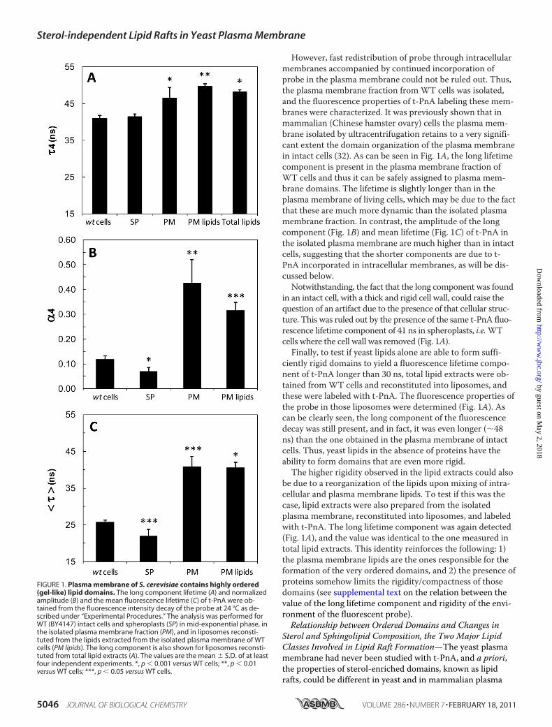
However, fast redistribution of probe through intracellularmembranes accompanied by continued incorporation ofprobe in the plasma membrane could not be ruled out. Thus,the plasma membrane fraction fromWT cells was isolated,and the fluorescence properties of t-PnA labeling these mem-branes were characterized. It was previously shown that inmammalian (Chinese hamster ovary) cells the plasma mem-brane isolated by ultracentrifugation retains to a very signifi-cant extent the domain organization of the plasma membranein intact cells (32). As can be seen in Fig. 1A, the long lifetimecomponent is present in the plasma membrane fraction ofWT cells and thus it can be safely assigned to plasma mem-brane domains. The lifetime is slightly longer than in theplasma membrane of living cells, which may be due to the factthat these are much more dynamic than the isolated plasmamembrane fraction. In contrast, the amplitude of the longcomponent (Fig. 1B) and mean lifetime (Fig. 1C) of t-PnA inthe isolated plasma membrane are much higher than in intactcells, suggesting that the shorter components are due to t-PnA incorporated in intracellular membranes, as will be dis-cussed below.Notwithstanding, the fact that the long component was found
in an intact cell, with a thick and rigid cell wall, could raise thequestion of an artifact due to the presence of that cellular struc-ture. This was ruled out by the presence of the same t-PnA fluo-rescence lifetime component of 41 ns in spheroplasts, i.e.WTcells where the cell wall was removed (Fig. 1A).Finally, to test if yeast lipids alone are able to form suffi-
ciently rigid domains to yield a fluorescence lifetime compo-nent of t-PnA longer than 30 ns, total lipid extracts were ob-tained fromWT cells and reconstituted into liposomes, andthese were labeled with t-PnA. The fluorescence properties ofthe probe in those liposomes were determined (Fig. 1A). Ascan be clearly seen, the long component of the fluorescencedecay was still present, and in fact, it was even longer (�48ns) than the one obtained in the plasma membrane of intactcells. Thus, yeast lipids in the absence of proteins have theability to form domains that are even more rigid.The higher rigidity observed in the lipid extracts could also
be due to a reorganization of the lipids upon mixing of intra-cellular and plasma membrane lipids. To test if this was thecase, lipid extracts were also prepared from the isolatedplasma membrane, reconstituted into liposomes, and labeledwith t-PnA. The long lifetime component was again detected(Fig. 1A), and the value was identical to the one measured intotal lipid extracts. This identity reinforces the following: 1)the plasma membrane lipids are the ones responsible for theformation of the very ordered domains, and 2) the presence ofproteins somehow limits the rigidity/compactness of thosedomains (see supplemental text on the relation between thevalue of the long lifetime component and rigidity of the envi-ronment of the fluorescent probe).Relationship between Ordered Domains and Changes in
Sterol and Sphingolipid Composition, the Two Major LipidClasses Involved in Lipid Raft Formation—The yeast plasmamembrane had never been studied with t-PnA, and a priori,the properties of sterol-enriched domains, known as lipidrafts, could be different in yeast and in mammalian plasma
FIGURE 1. Plasma membrane of S. cerevisiae contains highly ordered(gel-like) lipid domains. The long component lifetime (A) and normalizedamplitude (B) and the mean fluorescence lifetime (C) of t-PnA were ob-tained from the fluorescence intensity decay of the probe at 24 °C as de-scribed under “Experimental Procedures.” The analysis was performed forWT (BY4147) intact cells and spheroplasts (SP) in mid-exponential phase, inthe isolated plasma membrane fraction (PM), and in liposomes reconsti-tuted from the lipids extracted from the isolated plasma membrane of WTcells (PM lipids). The long component is also shown for liposomes reconsti-tuted from total lipid extracts (A). The values are the mean S.D. of at leastfour independent experiments. *, p � 0.001 versus WT cells; **, p � 0.01versus WT cells; ***, p � 0.05 versus WT cells.
Sterol-independent Lipid Rafts in Yeast Plasma Membrane
5046 JOURNAL OF BIOLOGICAL CHEMISTRY VOLUME 286 • NUMBER 7 • FEBRUARY 18, 2011
by guest on May 2, 2018
http://ww
w.jbc.org/
Dow
nloaded from

membrane. Therefore, it was also hypothesized that the longfluorescence lifetime could be due to incorporation of theprobe into ergosterol-enriched ordered domains. However,the 41-ns component was also present in t-PnA fluorescencedecay when the probe was labeling the plasma membrane oferg6� cells (Fig. 2A), which have deficient lipid rafts (33).These cells synthesize zymosterol and cholesta-5,7,24-trienolinstead of ergosterol (see supplemental Table S1). It is thusconcluded that gel-like domains are present in the plasmamembrane of WT cells and spheroplasts and of erg6� cells.Moreover, they should have similar compositions becausetheir rigidity is the same, as explained in the supplementaltext.erg6� cells, unlike WT cells, do not possess one major ste-
rol. Therefore, experiments were also carried out witherg2�erg6� cells, which accumulate only the non-raft-pro-moting zymosterol (see also supplemental Table S1) (34). Be-cause these mutant cells were obtained from a different ge-netic background than the BY4741 cells, the correspondingWT cells (RH3435) were also analyzed. A long lifetime com-ponent of 42 ns was detected in RH3435 cells, which is notsignificantly different from the one found in the BY4741 WTstrain (Fig. 2A). The amplitude and mean fluorescence life-time were also identical in both WT strains (Fig. 2, B and C).This result shows that the very ordered domains detected byt-PnA are not specific of one genetic background but are ageneral feature in the organization of S. cerevisiae plasmamembrane. Regarding erg2�erg6� cells, a long lifetime com-ponent (�30 ns) in t-PnA fluorescence decay was also de-tected (Fig. 2A). Because erg2�erg6� cells have no sterols withordering effect, the ordered domains have to be formed byother classes of lipids and should not correspond to a liquidordered phase. It should be noted that while in erg6� cells, thesphingolipid composition remains very similar to WT cells,and in erg2�erg6� cells, in addition to a very different sterolprofile, there are profound changes in sphingolipid composi-tion (supplemental Table S2).If the domains detected by the long component of t-PnA
are sterol-depleted, they should be mainly composed bysphingolipids, because these are the other membrane lipidsusually associated with membrane ordered domains.Therefore, a major change in sphingolipid compositionshould lead to an alteration in the value of t-PnA long life-time component. Thus, scs7� cells, which lack �-hydroxy-lation of sphingolipid-associated fatty acids and have asphingolipid profile very different from WT cells (supple-mental Table S2) (35), were also studied. Those cells have asterol composition that is identical to the respective WT cells(supplemental Table S1). It was found that the long compo-nent of t-PnA was significantly higher in scs7� cells whencompared with WT cells (Fig. 2A). This constitutes strongevidence that the domains detected by t-PnA are mainlyformed by sphingolipids.On another hand, the amplitude of the long lifetime com-
ponent is proportional to the amount of the ordered domainsrelative to the whole membrane (16, 18, 22, 36, 37). From Figs.1B and 2B, it is thus clear that the relative abundance of thehighly ordered domains was significantly different in the
plasma membrane of WT cells and spheroplasts and of erg6�and scs7� cells. These differences are also reflected on thevalues obtained for the average fluorescence lifetime of t-PnA(Figs. 1C and 2C). Therefore, changes in cell physiology lead
FIGURE 2. Rigidity and abundance of gel domains in the plasma mem-brane of S. cerevisiae may change with mutations involving sterol,sphingolipid, and GPI anchor biosynthetic pathways. The long compo-nent lifetime (A) and normalized amplitude (B) and the mean fluorescencelifetime (C) of t-PnA were obtained from the fluorescence intensity decay ofthe probe at 24 °C as described under “Experimental Procedures.” The anal-ysis was performed for mid-exponential phase WT (BY4741), erg6�, scs7�,and per1�, WT (RH3435), and erg2�erg6� cells. The values are the mean S.D. of at least four independent experiments. *, p � 0.001 versus WT cells;***, p � 0.05 versus WT cells.
Sterol-independent Lipid Rafts in Yeast Plasma Membrane
FEBRUARY 18, 2011 • VOLUME 286 • NUMBER 7 JOURNAL OF BIOLOGICAL CHEMISTRY 5047
by guest on May 2, 2018
http://ww
w.jbc.org/
Dow
nloaded from

to small alterations in the abundance of the gel-like domainsin yeast plasma membrane.S. cerevisiae Plasma Membrane Behavior as a Whole Can
Be Differentiated from the Highly Ordered Domains—To un-derstand how the whole membrane system may respond tocellular alterations (absence of ergosterol or absence of cellwall and concomitant decreased fraction of highly ordereddomains), the membranes of S. cerevisiae were labeled withDPH, a probe that is sensitive to its global properties, ratherthan to a particular kind of domain (see also supplementalFig. S4). DPH is a rod-shaped probe that incorporates in themembrane with its long axis parallel to the acyl chain pali-sade, and its steady-state fluorescence anisotropy is a wellestablished parameter to report on the alterations undergonein the global order of the membrane (15, 23, 31, 38). As can beseen in Fig. 3, the steady-state anisotropy of DPH was signifi-cantly increased in erg6� cells, scs7� cells, and spheroplastsfromWT cells, when compared with intact WT cells, i.e. theglobal order was higher in the cells that contain less highlyordered gel-like domains. The outcome of the cellular adapta-tion to the absence of ergosterol in the composition of theplasma membrane is such that there are less sphingolipid do-mains and a higher global order of the membrane.Ergosterol is among the set of special sterol molecules able
to induce the increase of the membrane order and the con-comitant formation of lipid rafts. Because erg6� cells accumu-late zymosterol, which does not have the ordered domain-forming ability of ergosterol, (39) and cholesta-5,7,24-trienol,a lower order of the membrane bilayer was anticipated inerg6� cells when compared with WT cells. However, the aver-age order of the plasma membrane in erg6� cells was in-creased when compared with WT cells (Fig. 3), as observedpreviously (8).In spheroplasts, however, ergosterol was present, and at the
same time, the highly ordered domains were less abundant.
Spheroplasts displayed the highest average order (Fig. 3), andthus it seems that the lower abundance of the highly ordereddomains increases the average order of the membranes. Evenin erg2�erg6� cells, which have a much lower order of thegel-like domains and a very high abundance of the nonorder-ing zymosterol, the fluorescence anisotropy of DPH was simi-lar to WT cells (supplemental Fig. S5). Altogether, these re-sults point toward a compensating effect from lipids withordering ability other than sterols. As will be discussed below,erg2�erg6� cells have a very increased content of mannosy-lated sphingolipids as compared with itsWT strain (supple-mental Table S2).Moreover, the abundance of the long component in scs7�
cells was smaller than in WT cells (Fig. 2B), leading to a meanfluorescence lifetime of t-PnA that is similar in both WT andmutant cells (Fig. 2C). As concluded above, a lower abun-dance of highly ordered domains increases the average orderof the membrane. Therefore, it can be anticipated that inscs7� cells the global membrane order was higher than in WTcells. The fact that the fluorescence anisotropy of DPH label-ing scs7� cells was higher than in WT cells (Fig. 3) confirmsthis prediction.Highly Ordered Domains in S. cerevisiae Plasma Membrane
Are Sphingolipid-enriched Gel-like Domains—To further clar-ify the composition and nature of the highly ordered domainsresponsible for the presence of a 41-ns lifetime component inthe fluorescence decay of t-PnA labeling the plasma mem-brane of yeast cells, studies in membrane model systems com-prising fungal lipid mixtures with well defined compositionswere also conducted (Fig. 4). Although the fluorescence decayof t-PnA has been previously studied in synthetic saturatedphosphatidylcholine and in mammalian-sphingolipid-con-taining membranes (17, 21), no studies have been performedin the presence of fungal sphingolipids. In Fig. 4A, the value ofthe longest lifetime component detected in t-PnA fluores-cence decay in mixtures of POPC/ergosterol and in mixturesof POPC/yeast phytoceramide is shown. POPC is one of themost abundant phospholipids in the plasma membrane of S.cerevisiaeWT cells (35), whereas ergosterol is the major ste-rol. Phytoceramide is the backbone of all the major complexsphingolipids in yeast plasma membrane (35, 40). In addition,a thorough study of the lipid composition of yeast subcellularmembranes using ESI-MS/MS clearly shows that the plasmamembrane of S. cerevisiae contains a significant quantity ofphytoceramide (41). It can be clearly seen in Fig. 4A that evenfor a very high concentration of ergosterol, corresponding tothe lipid bilayer being totally in the liquid ordered phase (42,43), the longest lifetime component obtained for the fluores-cence decay of t-PnA was between 12 and 22 ns. Conversely,in POPC/phytoceramide mixtures, a small amount of phyto-ceramide was enough to cause a remarkable increase of thelongest component of t-PnA fluorescence decay. For molefractions of phytoceramide above 20%, the value of that com-ponent was stabilized at �41 ns. The amplitude of the longcomponent (inset to the right of Fig. 4A) increased with phy-toceramide mole fraction, i.e. with the mole fraction of the gelphase, particularly between 5 and 30 mol % phytoceramide,highlighting the sensitivity of the probe to the presence of gel
FIGURE 3. Global order of the cell membrane system of S. cerevisiae ishigher in spheroplasts and deletion mutant strains of sphingolipid andsterol metabolism than in WT intact cells. The DPH steady-state fluores-cence anisotropy at 24 °C was obtained as described under “ExperimentalProcedures” for WT (BY4741) intact cells and spheroplasts (SP) and forerg6�, scs7�, and per1� intact cells in mid-exponential phase. The valuesare the mean S.D. of at least five independent experiments. *, p � 0.001versus WT cells.
Sterol-independent Lipid Rafts in Yeast Plasma Membrane
5048 JOURNAL OF BIOLOGICAL CHEMISTRY VOLUME 286 • NUMBER 7 • FEBRUARY 18, 2011
by guest on May 2, 2018
http://ww
w.jbc.org/
Dow
nloaded from

phase when this phase does not constitute the majority of themembrane, as should be the case for the membranes of livingcells. Thus, these results show that yeast phytoceramide, butnot ergosterol, forms a gel phase that gives t-PnA its charac-teristic long lifetime component. The amplitude of the longestcomponent of the probe decay in POPC/ergosterol wassmaller and does not show a well defined trend. This is be-cause it is not the very long component that can be specifi-cally attributed to the gel phase.We have confirmed that the POPC/phytoceramide binary
system undergoes at least one gel/fluid phase transition andobtained a melting temperature for such domains of �42 °C(Fig. 4B). To do so, we have measured the steady-state fluo-rescence anisotropy of t-PnA as a function of temperature.Although this parameter is a weighted sum of all the environ-ments felt by the probe, it is a very straightforward measure ofthe presence of ordered domains, and it allows us to distin-guish a gel/fluid transition (a sharp decrease of anisotropy)versus the progressive melting of liquid ordered domains andtemperature-induced disordering of the lipid bilayer (a slowdecrease of the anisotropy upon raising temperature). In addi-
tion, the huge difference between the long lifetime compo-nent of ergosterol-enriched and phytoceramide-enrichedmembrane model systems highlights the sensitivity of t-PnAto the nature of the ordered domains formed by fungal lipids.DPH anisotropy, on another hand, shows variation profilesthat are much more similar (supplemental Fig. S4) among thePOPC/ergosterol and POPC/phytoceramide mixtures. Thefact that DPH anisotropy increased with both ergosterol andphytoceramide is another indication that a reduction of theamplitude of the long lifetime component in yeast cells leadsto a significant ordering of the remainder of the membrane,because the diminished abundance of sphingolipid-enricheddomains per se, should lead to a decrease of DPH anisotropy.To confirm that phytoceramide-enriched gel domains are
still formed in the presence of ergosterol, ternary mixtures ofPOPC/ergosterol/phytoceramide were also prepared and la-beled with t-PnA. Phytoceramide was used at a constant 1:5phytoceramide/(POPC ergosterol) ratio, which corre-sponds to a mole fraction of 17 mol %, because it is on theorder of the sphingolipid concentrations found in yeast whenconsidering sphingolipids, ergosterol, and phospholipids (11,
FIGURE 4. Lipids from the plasma membrane of S. cerevisiae and phytoceramide form gel domains at physiological temperatures. A, phytoceramide,but not ergosterol, when mixed with POPC promotes the formation of a gel phase and the appearance of a long component �30 ns in t-PnA fluorescencedecay: long component lifetime �i of t-PnA fluorescence intensity decay incorporated in MLVs composed of mixtures of POPC/phytoceramide (gray circles)and POPC/ergosterol (open circles) at 24 °C (see inset for normalized amplitudes). B, steady-state fluorescence anisotropy of t-PnA incorporated in MLVs ofPOPC/phytoceramide (80:20 mol/mol) as a function of temperature. C, ergosterol at high concentrations abolishes the phytoceramide-enriched gel phase:long component of t-PnA fluorescence decay at 24 °C incorporated in MLVs composed of 1:5 (mol/mol) mixtures of phytoceramide/(POPC ergosterol) invarying proportions. The dashed line indicates the 30-ns value above which gel domains are known to be present. D, plasma membrane lipids of S. cerevisiaeundergo a gel/fluid transition at high temperatures: steady-state fluorescence anisotropy of t-PnA labeling liposomes reconstituted from isolated plasmamembrane lipid extracts of mid-exponential WT (BY4147) cells as a function of temperature. E, steady-state fluorescence anisotropy of t-PnA (black bars)versus DPH (white bars) for WT cells in mid-exponential phase and liposomes reconstituted from isolated plasma membrane lipid extracts (PM lipids) andfrom total lipid extracts of WT (BY4147) cells. A and C, X stands for mole fraction; the lines are merely to guide the eye; in �i, i � 3 or 4, depending on the mix-ture. B and D, straight lines are linear fits to the data points. They are used to determine the initial and the final temperatures of the gel/fluid transition,pointed by the arrows, from the intercept of the line with the steepest slope with the lines at lower and at higher temperatures, respectively. All panels: thevalues are the mean S.D. of at least four independent experiments. *, p � 0.001 versus t-PnA; **, p � 0.001 versus WT cells; ***, p � 0.05 versus t-PnA; #,p � 0.05 versus WT cells.
Sterol-independent Lipid Rafts in Yeast Plasma Membrane
FEBRUARY 18, 2011 • VOLUME 286 • NUMBER 7 JOURNAL OF BIOLOGICAL CHEMISTRY 5049
by guest on May 2, 2018
http://ww
w.jbc.org/
Dow
nloaded from

44). Also, the long component of t-PnA fluorescence decay(Fig. 4A), average lifetime, and anisotropy (data not shown)attains the highest value at �20 mol % phytoceramide, butstill the system is close enough to the gel/fluid coexistenceboundary so that the fluorescence of t-PnA will be sensitive toeven a partial solubilizing effect of ergosterol. In addition,studying this system allows us to compare its behavior with ananalogous system composed of the mammalian lipids POPC/ceramide/cholesterol previously studied by us (21) and to as-sess which sterol has a stronger ability to solubilize a gelphase. From Fig. 4C, it can be clearly observed that a sphingo-lipid-enriched gel phase persists until ergosterol concentra-tions reach �30 mol % (in relation to POPC), because thelong lifetime component of t-PnA remains longer than 30 ns.This concentration is similar to the ergosterol/phospholipidratio found in the plasma membrane of the S. cerevisiaeBY4741 WT strain used in this work and also other WTstrains (7, 41). In addition, the sphingolipids are highly en-riched at the plasma membrane, i.e. they are present at a mo-lar ratio higher than the one used in this experiment. Thus,although ergosterol has some ability to solubilize phytoceram-ide-enriched gel domains, the results suggest that at least asmall fraction of those domains should persist at physiologicalconcentrations of those lipids. Similarly, in the ternary mix-ture of mammalian lipids POPC/ceramide/cholesterol, for 17mol % of ceramide the gel phase was completely solubilizedfor �30 mol % cholesterol (21). Therefore, the ability of themain mammalian sterol to abolish gel phases was similar tothat of ergosterol.If the lipids from the yeast plasma membrane are able to
form a gel phase at room temperature, then it should be pos-sible to detect a gel/fluid transition at this temperature orhigher in liposomes made from such lipids. Thus, liposomesfrom plasma membrane lipids were prepared and labeled witht-PnA, and the fluorescence anisotropy of the probe wasmeasured as a function of temperature (Fig. 4D). It is clearthat a gel/fluid transition that starts at temperatures higherthan �42 °C was present. The shape of the curve on that tem-perature range was remarkably similar to a recent study withDPH of the transition temperature of inositol phosphorylcer-amide (IPC) purified from S. cerevisiae (45). In the same work,it is shown that the melting temperature of IPC is higher thanthat of a major mammalian sphingolipid (45). Because we alsoobtained a very high gel/fluid transition temperature for lipo-somes reconstituted from S. cerevisiae plasma membrane lip-ids (Fig. 4D), and ergosterol and cholesterol showed a similarability to abolish gel domains, the fact that thus far all studieswith t-PnA in mammalian cells always yielded a long compo-nent clearly shorter than 30 ns, i.e. a gel phase was not de-tected, is probably a consequence of the lower melting tem-perature of their main sphingolipids, as compared withS. cerevisiae.Additional evidence that t-PnA is detecting gel domains in
both cells and liposomes reconstituted from plasma mem-brane or total lipids stems from the large difference observedbetween the fluorescence anisotropy of t-PnA and DPH. In apure gel, a pure liquid ordered or a pure liquid disorderedphase, both probes have similar fluorescence anisotropy val-
ues (21, 36). In the case of liquid disordered/liquid ordereddomains, the anisotropy of t-PnA is only slightly larger thanthat of DPH because the quantum yield of t-PnA is moder-ately higher in this phase. However, in the presence of a gelphase, and as described in the Introduction, the quantumyield of t-PnA increases abruptly and its preference for gelphases is also highly marked. This is the only case where thefluorescence anisotropy of t-PnA can display a value typical ofa gel phase (close to 0.3 or higher), whereas the anisotropy ofDPH is clearly that of a mixture between ordered and disor-dered phases (close to 0.2 or lower). This marked difference ofbehavior has been previously used by us to characterize thepresence of multiple phases in complex mixtures of mamma-lian lipids (20, 21, 36). In Fig. 4E, the anisotropy at 24 °C oft-PnA and DPH in WT cells, in liposomes reconstituted fromplasma membrane lipids, and from total lipids is compared.In every case, there was a large difference between the aniso-tropy of the two probes, and thus in all of them a gel phaseshould be present. The comparison was also made forerg2�erg6� cells in supplemental Fig. S5. Even for erg2�erg6�cells, where the value of the long lifetime component (32 ns)is on the border of the fingerprint of the gel phase, the hugedifference among DPH and t-PnA anisotropy confirms that itis due to a gel phase. In addition, and as stated before, thelong component is present in WT cells at 30 °C (supplementalFig. S3), and moreover, the anisotropy of t-PnA at 30 °C inWT and scs7� cells is also very high,3 indicating that at opti-mal growth temperature the gel domains are present.A final confirmation that ergosterol is not required for the
formation of the highly ordered domains detected by t-PnAwas obtained from an experiment in which ergosterol wasremoved from liposomes made of plasma membrane lipidsthrough incubation with methyl-�-cyclodextrin (Fig. 5). Re-
3 A. F. Fernandes, L. Cyrne, R. F. M. de Almeida, F. Antunes, and H. S.Marinho, unpublished observations.
FIGURE 5. Ergosterol is not required for the presence of gel domainsformed from S. cerevisiae plasma membrane lipids. The long componentlifetime of t-PnA fluorescence intensity decay at 24 °C (mean S.D.) isshown for liposomes reconstituted from lipids extracted from the isolatedplasma membrane (PM) fraction of WT (BY4741) cells in mid-exponentialphase, untreated (n � 5) or treated (n � 3) with methyl-�-cyclodextrin, asdescribed under “Experimental Procedures.”
Sterol-independent Lipid Rafts in Yeast Plasma Membrane
5050 JOURNAL OF BIOLOGICAL CHEMISTRY VOLUME 286 • NUMBER 7 • FEBRUARY 18, 2011
by guest on May 2, 2018
http://ww
w.jbc.org/
Dow
nloaded from

moving ergosterol did not affect the presence of the longcomponent but rather leads to a slight increase of its lifetime,possibly due to an added segregation of the lipids with theability to form a gel phase that otherwise would be in liquidordered domains stabilized by the raft-promoting ergosterol.GPI-anchored Proteins Can Be Accommodated in Highly
Rigid Sphingolipid-enriched Domains—One of the questionsthat is pertinent to ask refers to which kind of proteins areable to incorporate in the gel-like domains detected by at-PnA lifetime component �40 ns. In fact, for most trans-membrane proteins, the energetic penalty associated with thedisruption of strong lipid-lipid interactions will prevent a sig-nificant incorporation on those domains (9). However, GPI-anchored proteins possess a lipid anchor, which in yeastmature into two types of lipid moiety. One type is phytocer-amide-based, i.e. presents the same backbone as sphingolip-ids, and the acyl chain is also the one commonly found inyeast sphingolipids, i.e. the C26:0 chain (hydroxylated or not).The other type is glycerophospholipid-based, with a very longchain saturated fatty acid at the sn-2 position. Because thesn-1 position is usually acylated with a saturated fatty acid,and the phytoceramide long chain base is also saturated, bothtypes of anchors are expected to accommodate in sphingolip-id-enriched gel-like domains (46). To investigate this possibil-ity, per1� cells, which lack one enzyme of the GPI anchor re-modeling pathway (47), were also studied. The GPI anchor inper1� cells is not reacylated and remains with an unsaturatedacyl chain at the sn-2 position. In addition, the ceramide-typeanchor is not synthesized (47). The unsaturated lipid anchorwould, in principle, be much less efficiently accommodated insphingolipid-enriched gel domains. The per1� cells were la-beled with t-PnA, and the value of the long lifetime compo-nent is shown in Fig. 2A. The long lifetime component wasincreased in comparison with all cell types, with the excep-tion of scs7� cells. In addition, the abundance of the do-mains was not significantly different from WT cells (Fig.2B), and the net result was a longer mean fluorescence life-time (Fig. 2C). If the major difference in per1� cells is thatGPI-anchored proteins are not incorporated in gel do-mains, then these domains may become more compact,because even though the GPI anchor is saturated, the pres-ence of the sugar residues in the headgroup of the attachedprotein may limit the extent of packing.Finally, if the abundance of the domains is inversely related
to the global order of the membrane, as proposed above, thenno significant changes would be expected for the DPH aniso-tropy in per1� cells in comparison with the WT cells. Thiswas indeed observed when per1� cells were labeled with DPH(Fig. 3).
DISCUSSION
This study presents evidence supporting the recently devel-oped notion that S. cerevisiae plasma membrane containssphingolipid-enriched domains, with either a very low or nosterol content. Furthermore, these domains can be biophysi-cally described as gel-like or solid ordered domains, as op-posed to the “typical” lipid rafts, which are described as liquidordered domains. In addition, the longest values were ob-
served in liposomes reconstituted from plasma membranelipids and total lipid extracts, i.e. in the absence of proteins.This suggests that the rigidity of the domains is limited by thepresence of proteins and that their formation is based on thetendency of certain lipids to laterally segregate.The probe t-PnA has been used previously to detect or-
dered domains in mammalian cells. The long componentfound had values shorter than 21 ns, which correspond tocholesterol-enriched liquid ordered domains (37). To the bestof our knowledge, this is the first time that this probe is usedto label yeast plasma membrane, and also the first time that along component typical of the gel phase (�30 ns) is found inliving cells. Its presence was detected in two different WTstrains (Fig. 2) and in one of these cases at both 24 and 30 °C(supplemental Fig. S3), which is the optimal growth tempera-ture of the strain, indicating that this may be a general resultfor this type of organism.Based on the biophysical evidence presented in this study,
it is likely that the sphingolipid-enriched domains detected byt-PnA are related to the plasma membrane domain MCP,which comprises a large portion of the membrane. MCP andMCC cover �80% of the plasma membrane. In this work, itwas found that the plasma membrane isolated from S. cerevi-siae is overall highly ordered (anisotropy of DPH higher than0.2) and contains a significant fraction of liquid ordered andgel domains (anisotropy of t-PnA higher than 0.3) (Fig. 4E).Mannosylated species, which comprise the majority of the
sphingolipids of the S. cerevisiae plasma membrane (48), dueto their large headgroup, are expected to be less tightlypacked than IPC or phytoceramide. In addition, they are, inprinciple, more suitable to stabilize liquid ordered domainsthrough interaction with sterols by protecting their hydro-phobic ring system from water, because the headgroup ofthese molecules is merely a hydroxyl moiety. In erg6� cells,there is only a very slight increase in mannosyl inositol phos-phorylceramide levels when compared with WT cells (2), al-though its biosynthetic rate is significantly slowed down (49).In erg6� cells, there is no alteration of the rigidity of sphingo-lipid-enriched domains, only a slightly smaller fraction (Fig. 2,A and B, respectively). Similarly, sphingolipid synthesis/com-position of the elongase system mutant elo3�, which containsC22:0/24:0 acyl chain sphingolipids, instead of C26:0, is notaffected by the activity of Erg6p (50). However, in erg2�erg6�cells, most sphingolipids are mannosylated, at the expense ofIPC, and the gel domains are considerably less tightly packedthan in the other cell types studied, because the long compo-nent of t-PnA decreases from �40 to 32 ns. Considering thatthe gel/fluid phase transition detected in the plasma mem-brane lipid extracts (Fig. 4D) is similar to the one recently ob-tained for S. cerevisiae IPC (45), it is reasonable to expect thatthe nonmannosylated sphingolipids are the major compo-nents of the solid ordered domains of WT cells. It will be in-teresting in the future to study the biophysical properties andliquid ordered versus gel phase stability of IPC versusmanno-sylated IPC, in the absence and presence of several yeast ste-rols, to clarify the differences in sphingolipid compositionsbetween the liquid ordered and gel domains in yeast plasmamembrane. An additional physiological significance comes
Sterol-independent Lipid Rafts in Yeast Plasma Membrane
FEBRUARY 18, 2011 • VOLUME 286 • NUMBER 7 JOURNAL OF BIOLOGICAL CHEMISTRY 5051
by guest on May 2, 2018
http://ww
w.jbc.org/
Dow
nloaded from

from the fact that changes in sphingolipid composition seemto compensate for lethality produced by changes in sterolcomposition and abundance (51).In all single mutant strains analyzed in this study, an inverse
relation between sphingolipid-enriched domains abundance andglobal order of the membranes was observed (Fig. 3 versus Fig.2B). However, measurement of membrane order with the probetrimethylammonium-DPH (TMA-DPH), which labels exclu-sively the plasmamembrane surface due to its TMAmoiety (52),indicates that there is a decrease of its order in erg6� cells, andno alteration in scs7� cells, when compared withWT cells (2). Adifferent behavior of DPH and TMA-DPH is better explained bya distinct change in the domain structure of intracellular andplasmamembranes. In themutants described above, the reducedabundance of sphingolipid domains in the plasmamembranemay be explained by an accumulation of these lipids in intracel-lular membranes, which becomemore ordered but are still be-low the concentration threshold necessary to segregate into geldomains. In the case of erg6� cells, the anisotropy of TMA-DPHis probably smaller due both to the absence of ergosterol andreduced sphingolipid domains in the plasmamembrane. In thecase of scs7� cells, this latter effect is compensated by their in-creased rigidity due to the absence of the hydroxyl group insphingolipid-associated fatty acids, and the order of the plasmamembrane as detected by TMA-DPH is virtually the same as inWT cells.In spheroplasts fromWT cells, the average order of the
bilayer is higher than in intact WT cells (Fig. 3), but the abun-dance of sphingolipid domains is smaller (Fig. 1B). A possibleexplanation for these observations may be a cell wall-inducedstabilization of the sphingolipid-enriched domains in theplasma membrane. The removal of the cell wall destabilizesthose domains, and they may be partially mixed in the bulkmembrane or internalized. As a consequence, the bulk mem-brane is sphingolipid-enriched when compared with intactcells, and this in turn leads to an increased average order ofthe whole membrane (Scheme 1). It may be speculated that
this could be a feedback-like mechanism, by which the celluses the high transition temperature sphingolipids accumu-lated in highly ordered domains to impart a higher mechani-cal resistance to the plasma membrane as a response to cellwall removal or damage. Alternatively, this could be a way oftriggering signaling mechanisms. In fact, it cannot be ruledout that the removal of the cell wall is activating stress re-sponse pathways that may also alter the plasma membrane.Cell wall stabilization of sphingolipid-enriched domains mayinvolve GPI-anchored proteins, because the sorting of theseproteins is sphingolipid-dependent (53, 54), and many ofthem are targeted to the cell wall (55). However, this hypothe-sis seems unlikely because per1� cells show the same abun-dance in sphingolipid domains as the WT cells. Noteworthy isthe existence of immobile membrane domains that are stabi-lized by interaction with the cell wall and that contain trans-membrane proteins intimately involved in sphingolipid me-tabolism, the Sur7p family cortical patches (56).The study of per1� cells suggests that the sphingolipid-
enriched domains contain a significant amount of GPI-an-chored proteins. Thus, it is possible that these domains areinvolved in GPI-anchored protein traffic and signaling, be-cause their low abundance combined with special packingproperties should lead to a high and stable membrane com-partmentalization of any molecules that accumulate withinthose domains. Several findings concerning GPI-anchoredproteins may relate to this. Sphingolipids, but not ergosterol,are indispensable both for GPI-anchored protein transportfrom the endoplasmic reticulum to the Golgi and for the sta-bilization of their association with membranes (53, 54, 57).GPI-anchored proteins are transported from the endoplasmicreticulum to the Golgi in particular types of vesicles that areceramide-enriched, which probably contain a significant frac-tion of gel-like domains as a consequence of their composi-tion. These domains may be delivered to the plasma mem-brane. It has been suggested that Gas1p, a GPI-anchoredprotein, would partition into such domains that are not “thetypical lipid-raft, i.e. a sphingolipid-rich but ergosterol-poordomain” (50). The formation of this particular type of domainin yeast, but not in mammals, can also help to explain differ-ences encountered in the transport of GPI-anchored proteinsin the two groups of organisms.The presence of the very long chain fatty acid C26:0 in the
sphingolipids of S. cerevisiae should be related to certain es-sential functions, because mutant cells with sphingolipidscontaining shorter acyl chains are less resistant, e.g. to ther-mal stress (40). The first biological function associated withthose sphingolipids was signal transduction (44). More re-cently, they have been implicated in cellular growth, endocy-tosis, and in the vesicular transport of GPI-anchored proteinsfrom the endoplasmic reticulum to the Golgi complex (58).There is no direct evidence that sphingolipid functions aredependent on their segregation into a particular type of lipiddomains such as gel domains. However, there are importantpieces of evidence suggesting that the biophysical nature orstructural properties of the sphingolipid domains are veryimportant. Candidate genes for the regulation of sphingolipid-enriched gel domains should include those involved in cer-
SCHEME 1. Depiction of the proposed model explaining the inverse rela-tionship between the abundance of highly ordered sphingolipid-enricheddomains and the global order of the membrane. Top represents the lipidcomponents of the plasma membrane of WT or per1� cells, and the bottommay represent the plasma membrane of WT spheroplasts or the cell membranesystem of erg6� or scs7� intact cells. Sphingolipids are indicated with their po-lar heads in gray and glycerophospholipids in black. Top, high abundance ofsphingolipid-enriched domains in the plasma membrane, with concomitantsphingolipid depletion in the remainder of the plasma membrane and/or othermembranes. Bottom, decreased gel domain abundance implies that the sphin-golipids are more scattered through the disordered domains of the plasmamembrane and/or intracellular membranes, leading to an increased globalorder of the cell membrane system.
Sterol-independent Lipid Rafts in Yeast Plasma Membrane
5052 JOURNAL OF BIOLOGICAL CHEMISTRY VOLUME 286 • NUMBER 7 • FEBRUARY 18, 2011
by guest on May 2, 2018
http://ww
w.jbc.org/
Dow
nloaded from

amide synthesis, such as LCB1, LAG1, and LAC1. In fact,lcb1� cells are unviable unless they carry the semi-dominantSLC1–1 mutation, which carries a Q44L substitution in theSlc1 protein, a 1-acyl-sn-glycerol-3-phosphate acyltransferase.This mutation allows cells to survive by catalyzing the incor-poration of C26:0 fatty acids into the sn-2 position of glycero-lipids, which then mimic yeast ceramides and serve as sub-strates for the enzymes that add the polar headgroups foundin yeast sphingolipids (59). The lethal double mutationlag1�lac1� is rescued by overexpression of the YDC1 gene.This gene codes for a reverse ceramidase that catalyzes thesynthesis of unusual glycerophospholipids of the phospha-tidylinositol family, which are sometimes mannosylated andalso contain very-long-chain fatty acids that are exclusivelyfound in sphingolipids in WT cells (60). C26:0-fatty acyl glyc-erolipids can replace sphingolipid functions (61). These un-usual glycerophospholipids can, in principle, partially mimicthe biophysical properties of the WT sphingolipids, includingthe tendency to segregate into gel-like domains. A role for thevery long chain fatty acid containing phosphoinositol in stabi-lizing highly curved membrane domains in nuclear mem-branes has also been suggested (62). In the proposed model,the clustering of this type of lipid would occur in a sterol-in-dependent manner.The domains detected in this study may be involved in cel-
lular processes related to plasma membrane dynamics. In fact,the plasma membrane of S. cerevisiae cells adapted to H2O2has reduced levels of the hydroxylated very long chain fattyacid 2-OH-C26:0 present in the sphingolipids when com-pared with control cells (7) and simultaneously has a lowerpermeability to H2O2 and several alterations of the membraneorder, including an increased fluorescence anisotropy of DPH(8) and a higher quantum yield of t-PnA (7).Recently, cholesterol-independent and sphingolipid-en-
riched microdomains containing signaling molecules havebeen identified in the plasma membrane of mammalian cells(3). In addition, two mammalian raft receptors have been lo-calized to distinct plasma membrane domains (63). Such do-mains may well be the mammalian counterparts of the yeastdomains identified in this study. It was also found that dihy-drosphingomyelin has an increased ability to form gel phasedomains when mixed with cholesterol and mammalian phos-pholipids when compared with sphingomyelin with the sameacyl chain (64). Dihydrosphingomyelin has a sphingoid back-bone that is closer to yeast sphingolipids than sphingomyelin.In addition, the rigidity of the ordered domains was affectedby a small alteration of the long chain base near the head-group region, as was observed in this study for scs7� cells(Fig. 2A).The mechanism of formation, dynamics, and physiologi-
cal relevance of the gel-like sphingolipid-enriched domainsin yeast remains open. However, this study paves the wayfor future developments. Furthermore, because these do-mains have a less elusive nature and are more stable thanthe liquid ordered-like lipid rafts, they offer the possibilityto clarify many open questions in this rapidly expandingfield.
Acknowledgments—We thank Prof. Andreas Conzelmann for thekind gift of per1� cells and for helpful discussions. We also thankProf. Howard Riezman for the kind gift of erg2�erg6� cells.
REFERENCES1. Opekarova, M., Malínska, K., Novakova, L., and Tanner, W. (2005) Bio-
chim. Biophys. Acta 1711, 87–952. Guan, X. L., Souza, C. M., Pichler, H., Dewhurst, G., Schaad, O., Kaji-
wara, K., Wakabayashi, H., Ivanova, T., Castillon, G. A., Piccolis, M.,Abe, F., Loewith, R., Funato, K., Wenk, M. R., and Riezman, H. (2009)Mol. Biol. Cell 20, 2083–2095
3. Hofman, E. G., Ruonala, M. O., Bader, A. N., van den Heuvel, D., Voort-man, J., Roovers, R. C., Verkleij, A. J., Gerritsen, H. C., and van BergenEn Henegouwen, P. M. (2008) J. Cell Sci. 121, 2519–2528
4. Grossmann, G., Opekarova, M., Malinsky, J., Weig-Meckl, I., and Tan-ner, W. (2007) EMBO J. 26, 1–8
5. Sousa-Lopes, A., Antunes, F., Cyrne, L., and Marinho, H. S. (2004) FEBSLett. 578, 152–156
6. Branco, M. R., Marinho, H. S., Cyrne, L., and Antunes, F. (2004) J. Biol.Chem. 279, 6501–6506
7. Pedroso, N., Matias, A. C., Cyrne, L., Antunes, F., Borges, C., Malho, R.,de Almeida, R. F., Herrero, E., and Marinho, H. S. (2009) Free Radic.Biol. Med. 46, 289–298
8. Folmer, V., Pedroso, N., Matias, A. C., Lopes, S. C., Antunes, F., Cyrne,L., and Marinho, H. S. (2008) Biochim. Biophys. Acta 1778, 1141–1147
9. Lingwood, D., and Simons, K. (2010) Science 327, 46–5010. Goffeau, A., Barrell, B. G., Bussey, H., Davis, R. W., Dujon, B., Feldmann,
H., Galibert, F., Hoheisel, J. D., Jacq, C., Johnston, M., Louis, E. J.,Mewes, H. W., Murakami, Y., Philippsen, P., Tettelin, H., and Oliver,S. G. (1996) Science 274, 546, 563–567
11. Ejsing, C. S., Sampaio, J. L., Surendranath, V., Duchoslav, E., Ekroos, K.,Klemm, R. W., Simons, K., and Shevchenko, A. (2009) Proc. Natl. Acad.Sci. U.S.A. 106, 2136–2141
12. Malínska, K., Malínsky, J., Opekarova, M., and Tanner, W. (2003)Mol.Biol. Cell 14, 4427–4436
13. Malinska, K., Malinsky, J., Opekarova, M., and Tanner, W. (2004) J. CellSci. 117, 6031–6041
14. Berchtold, D., and Walther, T. C. (2009)Mol. Biol. Cell 20, 1565–157515. de Almeida, R. F., Fedorov, A., and Prieto, M. (2003) Biophys. J. 85,
2406–241616. Sklar, L. A., Hudson, B. S., and Simoni, R. D. (1977) Biochemistry 16,
819–82817. de Almeida, R. F., Loura, L. M., and Prieto, M. (2009) Chem. Phys. Lipids
157, 61–7718. Reyes Mateo, C., Brochon, J. C., Pilar Lillo, M., and Ulises Acuna, A.
(1993) Biophys. J. 65, 2237–224719. Reyes Mateo, C., Ulises Acuna, A., and Brochon, J. C. (1995) Biophys. J.
68, 978–98720. Silva, L. C., de Almeida, R. F., Castro, B. M., Fedorov, A., and Prieto, M.
(2007) Biophys. J. 92, 502–51621. Castro, B. M., Silva, L. C., Fedorov, A., de Almeida, R. F., and Prieto, M.
(2009) J. Biol. Chem. 284, 22978–2298722. Schroeder, F. (1983) Eur. J. Biochem. 132, 509–51623. Mateo, C. R., Lillo, M. P., Gonzalez-Rodríguez, J., and Acuna, A. U.
(1991) Eur. Biophys. J. 20, 41–5224. Folch, J., Lees, M., and Sloane Stanley, G. H. (1957) J. Biol. Chem. 226,
497–50925. Panaretou, B., and Piper, P. (2006)Methods Mol. Biol. 313, 27–3226. Lagane, B., Gaibelet, G., Meilhoc, E., Masson, J. M., Cezanne, L., and
Lopez, A. (2000) J. Biol. Chem. 275, 33197–3320027. Welti, R., Rintoul, D. A., Goodsaid-Zalduondo, F., Felder, S., and Silbert,
D. F. (1981) J. Biol. Chem. 256, 7528–753528. Ragoonanan, V., Malsam, J., Bond, D. R., and Aksan, A. (2008) Biochim.
Biophys. Acta 1778, 2283–229029. Wolf, D. E. (1995)Mol. Membr. Biol. 12, 101–10430. Schulthess, G., and Hauser, H. (1995)Mol. Membr. Biol. 12, 105–112
Sterol-independent Lipid Rafts in Yeast Plasma Membrane
FEBRUARY 18, 2011 • VOLUME 286 • NUMBER 7 JOURNAL OF BIOLOGICAL CHEMISTRY 5053
by guest on May 2, 2018
http://ww
w.jbc.org/
Dow
nloaded from

31. de Almeida, R. F., Loura, L. M., Fedorov, A., and Prieto, M. (2005) J. Mol.Biol. 346, 1109–1120
32. Scolari, S., Engel, S., Krebs, N., Plazzo, A. P., De Almeida, R. F., Prieto,M., Veit, M., and Herrmann, A. (2009) J. Biol. Chem. 284, 15708–15716
33. Bagnat, M., Keranen, S., Shevchenko, A., Shevchenko, A., and Simons,K. (2000) Proc. Natl. Acad. Sci. U.S.A. 97, 3254–3259
34. Heiderpriem, R. W., Livant, P. D., Parish, E. J., Barbuch, R. J., Broaddus,M. G., and Bard, M. (1992) J. Steroid Biochem. Mol. Biol. 43, 741–743
35. Guan, X. L., and Wenk, M. R. (2006) Yeast 23, 465–47736. Castro, B. M., de Almeida, R. F., Silva, L. C., Fedorov, A., and Prieto, M.
(2007) Biophys. J. 93, 1639–165037. Schroeder, F., Goetz, I. E., and Roberts, E. (1984) J. Neurochem. 43,
526–53938. Lentz, B. R. (1988) inMembrane “Fluidity” from Fluorescence Anisotropy
Measurements (Loew, M. L., ed) pp. 13–42, CRC Press, Inc., Boca Ra-ton, FL
39. Megha Bakht, O., and London, E. (2006) J. Biol. Chem. 281,21903–21913
40. Dickson, R. C. (2008) J. Lipid Res. 49, 909–92141. Schneiter, R., Brugger, B., Sandhoff, R., Zellnig, G., Leber, A., Lampl, M.,
Athenstaedt, K., Hrastnik, C., Eder, S., Daum, G., Paltauf, F., Wieland,F. T., and Kohlwein, S. D. (1999) J. Cell Biol. 146, 741–754
42. Silva, L., Coutinho, A., Fedorov, A., and Prieto, M. (2006) Biophys. J. 90,3625–3631
43. Hsueh, Y. W., Chen, M. T., Patty, P. J., Code, C., Cheng, J., Frisken, B. J.,Zuckermann, M., and Thewalt, J. (2007) Biophys. J. 92, 1606–1615
44. van der Rest, M. E., Kamminga, A. H., Nakano, A., Anraku, Y., Poolman,B., and Konings, W. N. (1995)Microbiol. Rev. 59, 304–322
45. Klose, C., Ejsing, C. S., García-Saez, A. J., Kaiser, H. J., Sampaio, J. L.,Surma, M. A., Shevchenko, A., Schwille, P., and Simons, K. (2010) J. Biol.Chem. 285, 30224–30232
46. Bosson, R., and Conzelmann, A. (2007) Biochem. Soc. Symp. 74,199–209
47. Fujita, M., Umemura, M., Yoko-o, T., and Jigami, Y. (2006)Mol. Biol.Cell 17, 5253–5264
48. Leber, A., Fischer, P., Schneiter, R., Kohlwein, S. D., and Daum, G.(1997) FEBS Lett. 411, 211–214
49. Swain, E., Baudry, K., Stukey, J., McDonough, V., Germann, M., andNickels, J. T., Jr. (2002) J. Biol. Chem. 277, 26177–26184
50. Eisenkolb, M., Zenzmaier, C., Leitner, E., and Schneiter, R. (2002)Mol.Biol. Cell 13, 4414–4428
51. Valachovic, M., Bareither, B. M., Shah Alam Bhuiyan, M., Eckstein, J.,Barbuch, R., Balderes, D., Wilcox, L., Sturley, S. L., Dickson, R. C., andBard, M. (2006) Genetics 173, 1893–1908
52. Abe, F., and Hiraki, T. (2009) Biochim. Biophys. Acta 1788, 743–75253. Watanabe, R., Funato, K., Venkataraman, K., Futerman, A. H., and Riez-
man, H. (2002) J. Biol. Chem. 277, 49538–4954454. Heese-Peck, A., Pichler, H., Zanolari, B., Watanabe, R., Daum, G., and
Riezman, H. (2002)Mol. Biol. Cell 13, 2664–268055. De Sampaïo, G., Bourdineaud, J. P., and Lauquin, G. J. (1999)Mol. Mi-
crobiol. 34, 247–25656. Young, M. E., Karpova, T. S., Brugger, B., Moschenross, D. M., Wang,
G. K., Schneiter, R., Wieland, F. T., and Cooper, J. A. (2002)Mol. Cell.Biol. 22, 927–934
57. Gaigg, B., Timischl, B., Corbino, L., and Schneiter, R. (2005) J. Biol.Chem. 280, 22515–22522
58. Obeid, L. M., Okamoto, Y., and Mao, C. (2002) Biochim. Biophys. Acta1585, 163–171
59. Lester, R. L., Wells, G. B., Oxford, G., and Dickson, R. C. (1993) J. Biol.Chem. 268, 845–856
60. Cerantola, V., Guillas, I., Roubaty, C., Vionnet, C., Uldry, D., Knudsen, J.,and Conzelmann, A. (2009)Mol. Microbiol. 71, 1523–1537
61. Gaigg, B., Toulmay, A., and Schneiter, R. (2006) J. Biol. Chem. 281,34135–34145
62. Schneiter, R., Brugger, B., Amann, C. M., Prestwich, G. D., Epand, R. F.,Zellnig, G., Wieland, F. T., and Epand, R. M. (2004) Biochem. J. 381,941–949
63. Asanov, A., Zepeda, A., and Vaca, L. (2010) Biochim. Biophys. Acta1801, 147–155
64. Vieira, C. R., Munoz-Olaya, J. M., Sot, J., Jimenez-Baranda, S., Izquierdo-Useros, N., Abad, J. L., Apellaniz, B., Delgado, R., Martinez-Picado, J.,Alonso, A., Casas, J., Nieva, J. L., Fabrias, G., Manes, S., and Goni, F. M.(2010) Chem. Biol. 17, 766–775
Sterol-independent Lipid Rafts in Yeast Plasma Membrane
5054 JOURNAL OF BIOLOGICAL CHEMISTRY VOLUME 286 • NUMBER 7 • FEBRUARY 18, 2011
by guest on May 2, 2018
http://ww
w.jbc.org/
Dow
nloaded from

Fernando Antunes and Rodrigo F. M. de AlmeidaFrancisco Aresta-Branco, André M. Cordeiro, H. Susana Marinho, Luísa Cyrne,
RAFTSORDERED, ERGOSTEROL-FREE, AND SPHINGOLIPID-ENRICHED LIPID
: HIGHLYSaccharomyces cerevisiaeGel Domains in the Plasma Membrane of
doi: 10.1074/jbc.M110.154435 originally published online December 2, 20102011, 286:5043-5054.J. Biol. Chem.
10.1074/jbc.M110.154435Access the most updated version of this article at doi:
Alerts:
When a correction for this article is posted•
When this article is cited•
to choose from all of JBC's e-mail alertsClick here
Supplemental material:
http://www.jbc.org/content/suppl/2010/12/02/M110.154435.DC1
http://www.jbc.org/content/286/7/5043.full.html#ref-list-1
This article cites 63 references, 30 of which can be accessed free at
by guest on May 2, 2018
http://ww
w.jbc.org/
Dow
nloaded from