gauges biologic rationale of esthetic crown lengthening
TRANSCRIPT

See discussions, stats, and author profiles for this publication at: https://www.researchgate.net/publication/51574919
Biologic rationale of esthetic crown lengthening using innovative proportion
gauges
Article in The International journal of periodontics & restorative dentistry · September 2011
Source: PubMed
CITATIONS
19READS
1,276
1 author:
Paul Fletcher
Stony Brook University
25 PUBLICATIONS 1,357 CITATIONS
SEE PROFILE
All content following this page was uploaded by Paul Fletcher on 27 December 2017.
The user has requested enhancement of the downloaded file.
The International Journal of Periodontics & Restorative Dentistry
© 2011 BY QUINTESSENCE PUBLISHING CO, INC. PRINTING OF THIS DOCUMENT IS RESTRICTED TO PERSONAL USE ONLY.. NO PART OF MAY BE REPRODUCED OR TRANSMITTED IN ANY FORM WITHOUT WRITTEN PERMISSION FROM THE PUBLISHER.

Volume 31, Number 5, 2011
523
Biologic Rationale of Esthetic Crown Lengthening Using Innovative Proportion Gauges
Paul Fletcher, DDS* Periodontal crown lengthening is performed to reduce excess tissue and encompasses the recontouring and removal of gingiva or gingiva and bone to increase the length of the clinical crown when inadequate solid tooth structure is available to place a crown margin with proper re-tention and resistance form. Most often this is a result of extensive car-ies, crown fracture, faulty pre-existing margins, or failed restorations. Addi-tionally, it is performed to correct gingival asymmetries and to reposi-tion the dentogingival complex as an adjunct to esthetic restorative procedures. In conjunction with an increase in clinical crown length, the procedure involves a concurrent in-crease in the biologic crown length, defined as the distance from the margin of sound tooth structure to the crest of bone.1 Rosenberg et al2 stated that this therapeutic modality was performed predominantly to fulfill the requirements of the restor-ative dentist related to esthetics, marginal seal, retention, and form and function, and it was integral in determining the success of the de-finitive restoration.
Research shows that practitioners tend to underestimate the amount of tooth structure that must be exposed during a crown lengthening procedure. In the anterior portion of the mouth, this can lead to biologic width problems and subsequent cosmetic issues. This paper presents a biologically based, step-by-step approach to periodontal esthetic crown lengthening. Using a series of innovative measuring gauges, the ideal clinical crown length of a tooth as well as the proper occlusogingival placement of the interproximal papilla will be determined based on established, documented tooth proportion relationships. The biologic crown length of the tooth, defined as the distance from the incisal edge to the bone crest, will subsequently be determined as a function of the clinical crown length, with the ultimate goals being adequate tooth structure for the placement of a restorative margin, establishment of a healthy dentogingival complex, and the placement of an esthetically pleasing
definitive restoration. (Int J Periodontics Restorative Dent 2011;31:523–532.)
* Clinical Associate Professor, Department of Periodontology, Columbia University College of Dental Medicine, New York, New York; Formerly, Clinical Associate Professor, Department of Periodontology and Implant Dentistry, New York University College of Dentistry, New York, New York. Correspondence to: Dr Paul Fletcher, Specialized Dentistry of New York, 150 E. 58th Street, Suite 3200, New York, NY 10155; fax: 212-754-6753; email: [email protected].
© 2011 BY QUINTESSENCE PUBLISHING CO, INC. PRINTING OF THIS DOCUMENT IS RESTRICTED TO PERSONAL USE ONLY.. NO PART OF MAY BE REPRODUCED OR TRANSMITTED IN ANY FORM WITHOUT WRITTEN PERMISSION FROM THE PUBLISHER.

The International Journal of Periodontics & Restorative Dentistry
524
There is a lack of consensus re-garding the amount of tooth struc-ture that must be exposed above the crest of bone for restorative purposes.3–8 This paper presents a biologically based, step-by-step approach to periodontal esthetic crown lengthening using a series of innovatively designed, color-coded measurement gauges. The ideal clinical crown length of a tooth will be determined based on estab-lished tooth proportion relation-ships. The proper biologic length of the crown will subsequently be determined as a function of the clinical length, as will the proper occlusogingival placement of the interproximal papilla. The presence of adequate tooth structure for the placement of a restorative margin, establishment of a healthy dento-gingival complex, and the subse-quent fabrication of a well-fitting, esthetically pleasing definitive res-toration are the ultimate goals.
The recognition and identifi-cation of the components of bio-logic width have been defined over time.1–4,9,10 Gargiulo et al9 used hu-man autopsy material to record an average measurement of 2.73 mm, comprising 0.97 mm for the epitheli-al attachment, 1.07 mm for the con-nective tissue, and 0.69 mm for the sulcus depth. In clinical studies, the superosseous keratinized gingiva in the esthetic zone was found to aver-age 3.6 mm,11 with a probing depth of 3.0 mm on the midfacial aspect of the maxillary central incisors and 3.0 to 4.5 mm interproximally.12
These findings were confirmed histologically, and, additionally, a
postsurgical reduction of the com-ponents of the attachment appa-ratus was demonstrated.13–15 The junctional epithelium will migrate to the osseous crest if the root planing extends to that level, but when supercrestal fibers from the periodontal ligament are retained, they may reconnect with the con-nective tissue fibers from the flap.16
When all attachment fibers are removed during surgery, 0.4 to 1.0 mm of crestal resorption occurs, exposing ligament fibers that will mesh with connective tissue fibers from the inner aspect of the flap to establish a new supracrestal connec-tive tissue fiber barrier beneath the reformed junctional epithelium.13,17 This connective tissue fiber barrier prevents further apical migration of the junctional epithelium, and its blood supply supports the newly formed attachment apparatus.
The consensus of opinion is that at least 2 mm of exposed tooth structure is needed for crown re-tention.18 Adding this number to a mean junctional epithelial length of 0.97 mm and a mean connec-tive tissue fiber barrier of 1.07 mm, it would seem that, ideally, at least 4.0 mm of superosseous tooth structure must be available for the placement of a restorative crown margin and for the establishment of an attachment apparatus. If less is present, retention may be com-promised or the biologic width im-pinged upon. In the esthetic zone, additional considerations must be addressed. If a crown margin is exposed, the restoration is usually considered to be less than ideal.
Additionally, a noticeable “reddish” inflammatory hue in the tissue will also detract from the final result.
Gingival inflammation tends to increase the deeper a crown mar-gin is placed within the sulcus.19,20 While controversy exists as to how far subgingivally a crown margin should end, 0.5 to 1.0 mm seems to be agreed upon, unless the tissue biotype is extremely thin.4,19,21–23 Since gingival recession may occur as a result of trauma, tissue biotype, manipulation during crown prepa-ration, root prominence, and ongo-ing passive eruption, it would seem desirable to place a crown margin in the esthetic zone as far intrasul-cularly as is clinically acceptable.
Esthetic crown lengthening
A primary objective of contempo-rary dentistry is to optimize the re-sults for patients desiring esthetic periodontal restorative treatment. While a myriad of restorative tech-niques are used to enhance the overall quality of the definitive res-toration, obtaining a clinical crown length that remains stable and an understanding of the management of the interdental tissue, including the changes that occur in the pa-pilla in response to alterations in tooth contour and contact position, are also imperative.
© 2011 BY QUINTESSENCE PUBLISHING CO, INC. PRINTING OF THIS DOCUMENT IS RESTRICTED TO PERSONAL USE ONLY.. NO PART OF MAY BE REPRODUCED OR TRANSMITTED IN ANY FORM WITHOUT WRITTEN PERMISSION FROM THE PUBLISHER.

Volume 31, Number 5, 2011
525
Case report
A 29-year-old man was not pleased with the appearance of his smile and desired to modify the size and shape of his maxillary incisors (Fig 1). A comprehensive periodontal and re-storative evaluation was performed. It was determined that periodon-tal esthetic crown lengthening sur-gery was indicated, followed by the
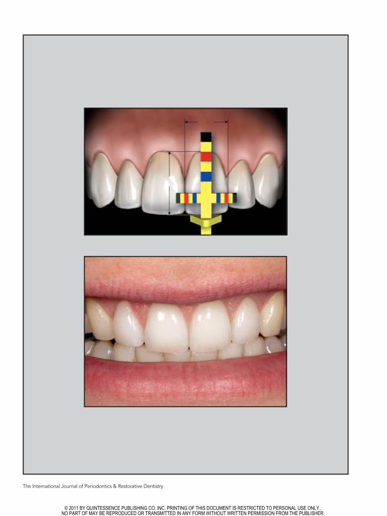
esthetic restoration of the four maxil-lary incisors. Chu Aesthetic Gauges (Hu-Friedy) (Figs 2a to 2c) were used as guides in establishing the correct occlusogingival clinical dimension of the tooth as a function of its width and the correct papilla position as a function of its length. Additionally, the gauges determined the correct biologic length of each crown as a function of the clinical length.
Fig 1 Initial clinical appearance of the patient. Fig 2a T-Bar Proportion Gauge. The widths of the color markings on the horizontal arm are 75% to 80% of the lengths of the corre-sponding color markings on the vertical arm.
Fig 2b (left) Crown lengthening gauge (Biologic Periogauge). The color markings on the longer arm are 3 mm greater than the same color markings on the shorter arm.
Fig 2c (above) Papilla Tip Gauge.
8.5 mm
11 mm
10.5 mm6.5 mm
14 mm11 mm
© 2011 BY QUINTESSENCE PUBLISHING CO, INC. PRINTING OF THIS DOCUMENT IS RESTRICTED TO PERSONAL USE ONLY.. NO PART OF MAY BE REPRODUCED OR TRANSMITTED IN ANY FORM WITHOUT WRITTEN PERMISSION FROM THE PUBLISHER.
The International Journal of Periodontics & Restorative Dentistry
526
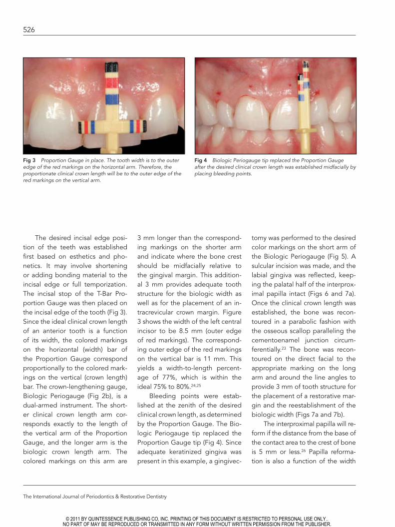
The desired incisal edge posi-tion of the teeth was established first based on esthetics and pho-netics. It may involve shortening or adding bonding material to the incisal edge or full temporization. The incisal stop of the T-Bar Pro-portion Gauge was then placed on the incisal edge of the tooth (Fig 3). Since the ideal clinical crown length of an anterior tooth is a function of its width, the colored markings on the horizontal (width) bar of the Proportion Gauge correspond proportionally to the colored mark-ings on the vertical (crown length) bar. The crown-lengthening gauge, Biologic Periogauge (Fig 2b), is a dual-armed instrument. The short-er clinical crown length arm cor-responds exactly to the length of the vertical arm of the Proportion Gauge, and the longer arm is the biologic crown length arm. The colored markings on this arm are
3 mm longer than the correspond-ing markings on the shorter arm and indicate where the bone crest should be midfacially relative to the gingival margin. This addition-al 3 mm provides adequate tooth structure for the biologic width as well as for the placement of an in-tracrevicular crown margin. Figure 3 shows the width of the left central incisor to be 8.5 mm (outer edge of red markings). The correspond-ing outer edge of the red markings on the vertical bar is 11 mm. This yields a width-to-length percent-age of 77%, which is within the ideal 75% to 80%.24,25
Bleeding points were estab-lished at the zenith of the desired clinical crown length, as determined by the Proportion Gauge. The Bio-logic Periogauge tip replaced the Proportion Gauge tip (Fig 4). Since adequate keratinized gingiva was present in this example, a gingivec-
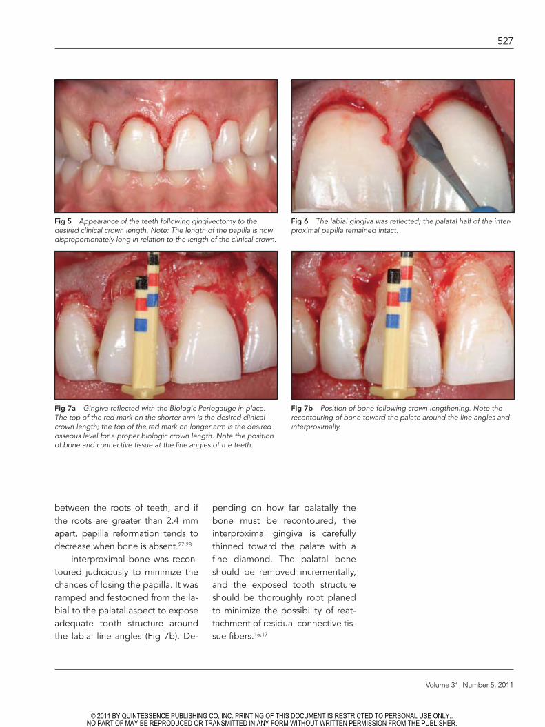
tomy was performed to the desired color markings on the short arm of the Biologic Periogauge (Fig 5). A sulcular incision was made, and the labial gingiva was reflected, keep-ing the palatal half of the interprox-imal papilla intact (Figs 6 and 7a). Once the clinical crown length was established, the bone was recon-toured in a parabolic fashion with the osseous scallop paralleling the cementoenamel junction circum-ferentially.23 The bone was recon-toured on the direct facial to the appropriate marking on the long arm and around the line angles to provide 3 mm of tooth structure for the placement of a restorative mar-gin and the reestablishment of the biologic width (Figs 7a and 7b).
The interproximal papilla will re-form if the distance from the base of the contact area to the crest of bone is 5 mm or less.26 Papilla reforma-tion is also a function of the width
Fig 3 Proportion Gauge in place. The tooth width is to the outer edge of the red markings on the horizontal arm. Therefore, the proportionate clinical crown length will be to the outer edge of the red markings on the vertical arm.
Fig 4 Biologic Periogauge tip replaced the Proportion Gauge after the desired clinical crown length was established midfacially by placing bleeding points.
© 2011 BY QUINTESSENCE PUBLISHING CO, INC. PRINTING OF THIS DOCUMENT IS RESTRICTED TO PERSONAL USE ONLY.. NO PART OF MAY BE REPRODUCED OR TRANSMITTED IN ANY FORM WITHOUT WRITTEN PERMISSION FROM THE PUBLISHER.
Volume 31, Number 5, 2011
527
between the roots of teeth, and if the roots are greater than 2.4 mm apart, papilla reformation tends to decrease when bone is absent.27,28
Interproximal bone was recon-toured judiciously to minimize the chances of losing the papilla. It was ramped and festooned from the la-bial to the palatal aspect to expose adequate tooth structure around the labial line angles (Fig 7b). De-
pending on how far palatally the bone must be recontoured, the interproximal gingiva is carefully thinned toward the palate with a fine diamond. The palatal bone should be removed incrementally, and the exposed tooth structure should be thoroughly root planed to minimize the possibility of reat-tachment of residual connective tis-sue fibers.16,17
Fig 5 Appearance of the teeth following gingivectomy to the desired clinical crown length. Note: The length of the papilla is now disproportionately long in relation to the length of the clinical crown.
Fig 6 The labial gingiva was reflected; the palatal half of the inter-proximal papilla remained intact.
Fig 7b Position of bone following crown lengthening. Note the recontouring of bone toward the palate around the line angles and interproximally.
Fig 7a Gingiva reflected with the Biologic Periogauge in place. The top of the red mark on the shorter arm is the desired clinical crown length; the top of the red mark on longer arm is the desired osseous level for a proper biologic crown length. Note the position of bone and connective tissue at the line angles of the teeth.
© 2011 BY QUINTESSENCE PUBLISHING CO, INC. PRINTING OF THIS DOCUMENT IS RESTRICTED TO PERSONAL USE ONLY.. NO PART OF MAY BE REPRODUCED OR TRANSMITTED IN ANY FORM WITHOUT WRITTEN PERMISSION FROM THE PUBLISHER.
The International Journal of Periodontics & Restorative Dentistry
528
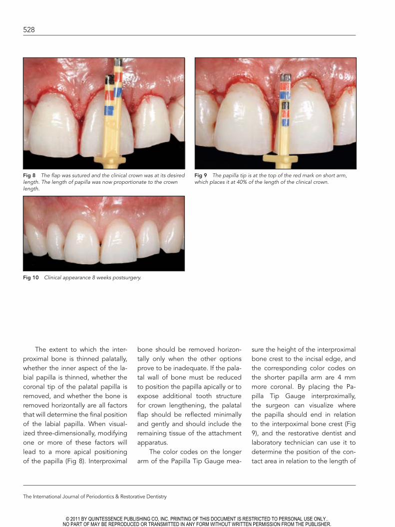
The extent to which the inter-proximal bone is thinned palatally, whether the inner aspect of the la-bial papilla is thinned, whether the coronal tip of the palatal papilla is removed, and whether the bone is removed horizontally are all factors that will determine the final position of the labial papilla. When visual-ized three-dimensionally, modifying one or more of these factors will lead to a more apical positioning of the papilla (Fig 8). Interproximal
bone should be removed horizon-tally only when the other options prove to be inadequate. If the pala-tal wall of bone must be reduced to position the papilla apically or to expose additional tooth structure for crown lengthening, the palatal flap should be reflected minimally and gently and should include the remaining tissue of the attachment apparatus.
The color codes on the longer arm of the Papilla Tip Gauge mea-
sure the height of the interproximal bone crest to the incisal edge, and the corresponding color codes on the shorter papilla arm are 4 mm more coronal. By placing the Pa-pilla Tip Gauge interproximally, the surgeon can visualize where the papilla should end in relation to the interpoximal bone crest (Fig 9), and the restorative dentist and laboratory technician can use it to determine the position of the con-tact area in relation to the length of
Fig 8 The flap was sutured and the clinical crown was at its desired length. The length of papilla was now proportionate to the crown length.
Fig 9 The papilla tip is at the top of the red mark on short arm, which places it at 40% of the length of the clinical crown.
Fig 10 Clinical appearance 8 weeks postsurgery.
© 2011 BY QUINTESSENCE PUBLISHING CO, INC. PRINTING OF THIS DOCUMENT IS RESTRICTED TO PERSONAL USE ONLY.. NO PART OF MAY BE REPRODUCED OR TRANSMITTED IN ANY FORM WITHOUT WRITTEN PERMISSION FROM THE PUBLISHER.

Volume 31, Number 5, 2011
529
the clinical crown. Since the length of the papilla has been found to be approximately 40% to 50% of the length of the tooth,29 the papilla should only be shortened 0.4 to 0.5 mm for every 1.0 mm that it is dis-proportionate to the length of the clinical crown to maintain the prop-er crown-papilla ratio.
Healing was rapid and un-eventful (Fig 10), and the dento-gingival complex consisted of a gingival sulcus, junctional epithe-
lium, and connective tissue fiber barrier populating the apical 3 mm of supraosseous tooth structure. While osseous remodeling contin-ues histologically for longer than 12 months, soft tissue healing is most-ly completed after 8 weeks.30 What must be assessed at this point is the maturation of the gingiva and the stability of the position of the gingival margin. If the gingival contour has stabilized and a crown margin is atraumatically placed in-
tracrevicularly, the definitive resto-rations can be placed successfully within 8 to 12 weeks.31 The restor-ative dentist should have more than adequate tooth structure for the placement of an intracrevicular crown margin, there should be min-imal coronal migration of the buc-cal gingiva, and the papilla should fill the embrasure space (Fig 11).
Fig 11a Definitive restorations in place.
Fig 11b There was no coronal migration of the gingiva on the direct facial aspect.
Fig 11c The papilla now ends at the top of the blue line on the papilla gauge, and the interproximal gingiva migrated coronally to fill the embrasure space.
a
cb
© 2011 BY QUINTESSENCE PUBLISHING CO, INC. PRINTING OF THIS DOCUMENT IS RESTRICTED TO PERSONAL USE ONLY.. NO PART OF MAY BE REPRODUCED OR TRANSMITTED IN ANY FORM WITHOUT WRITTEN PERMISSION FROM THE PUBLISHER.

The International Journal of Periodontics & Restorative Dentistry
530
Discussion
Crestal bone must often be removed when performing esthetic crown lengthening to provide adequate tooth structure for the establishment of an attachment apparatus and the placement of an intracrevicular crown margin. Since acrylic tem-plates are often imprecise32 and it has been shown that less than 3 mm of tooth structure is routinely re-moved during crown lengthening procedures,33 an objective measur-ing device would be a valuable aid in assuring sufficient tooth structure is exposed, as well as in establishing a clinical crown with an ideal width-to-length proportional relationship.34
Clinicians will, on occasion, at-tempt to construct a crown with less than 3 mm of available subgingival tooth structure, resulting in the crown margin being placed within the connective tissue attachment or at the bone crest. Theoretically, suf-ficient horizontal bone resorption will subsequently occur for a more apical establishment of the attach-ment apparatus. While resorption of this type often occurs on the direct buccal aspect, where the buccal plate of bone tends to be thin, it is less predictable at the line angles and interproximally. It is here, where the roots are relatively far apart, that the bone thickens as it contours into the embrasures and vertical trough-ing can occur because of inade-quate crestal resorption. The crown margin subsequently becomes a chronic irritant, and the characteris-tic inflammatory appearance of bio-logic width impingement occurs.
Similarly, a gingivectomy to es-tablish a desired clinical crown length, followed by thorough root planing without flap elevation, may remove all supracrestal fibers and stimulate adequate crestal resorp-tion for the successful placement of a subgingival crown margin. Coro-nal gingival rebound and biologic width problems tend to be more frequent occurrences, though, and experience has shown the stability of the definitive restoration to be less predictable than when the gin-giva is flapped and osseous recon-touring performed.31,35,36
A minimum of 2 to 5 mm of ker atinized tissue is required for gingival health.37,38 In the highlight-ed case, where the patient had a healthy, fibrous biotype and lami-nate veneers were being placed, it was felt that a minimum of 2 to 3 mm of keratinized tissue would be adequate for gingival health. A gingivectomy was subsequently performed to expose the needed additional tooth structure as op-posed to using an intrasulcular inci-sion to reposition the tissue apically.
While the amount of bone that must be removed to provide appro-priate biologic crown length may, on occasion, appear visually strik-ing, tooth mobility does not be-come an issue when adequate bone is present originally. When bone lev-els are compromised initially and re-cession has not occurred, apical positioning of the buccal gingiva or a gingivectomy may be adequate to achieve the desired result.
Management of the papilla is an important aspect of esthetic
© 2011 BY QUINTESSENCE PUBLISHING CO, INC. PRINTING OF THIS DOCUMENT IS RESTRICTED TO PERSONAL USE ONLY.. NO PART OF MAY BE REPRODUCED OR TRANSMITTED IN ANY FORM WITHOUT WRITTEN PERMISSION FROM THE PUBLISHER.

Volume 31, Number 5, 2011
531
crown lengthening. As shown in the present case, the interproximal tissue will proliferate coronally and the papilla will reform as long as the distance from the bone crest to the base of the contact area is 5 mm or less12,26 and the interradic-ular distance between the teeth is 2.4 mm or less (Fig 11c).27,28 Any minor residual interproximal spac-ing that may remain can be elimi-nated by positioning the contact area of the definitive restoration further apically.
Conclusions
The esthetic restorative process re-quires the participation of both the periodontist and restorative dentist. Proper modification of the height and contour of the gingiva, as well as the length of the clinical and bio-logic crown, is intrinsic in obtaining an optimal final result. The dynam-ics of hard and soft tissue wound healing are topics the periodontist is intimately familiar with. A tech-nique for crown lengthening was presented that uses a unique set of measuring devices to objectively define the ideal clinical length of an anterior tooth as a function of its width. The gauges then provide the surgeon with a guide that de-termines precisely how much bone must be removed from a biologic standpoint to ultimately produce a restoration of the highest clinical quality. Perhaps, most importantly, periodontal esthetic restorative therapy and the gauges foster a col-laborative interaction between the
periodontist and restorative dentist. It is this ongoing communication directed toward a jointly achieved, successful, final result that is the foundation of a mutually rewarding, long-term, working relationship.
Acknowledgment
The author expresses his appreciation to Dr Dennis P. Tarnow for his review of the manuscript.
References
1. Chu SJ, Hochman MN, Fletcher P. A biologic approach to aesthetic crown lengthening: Part II—Interdental con-siderations. Pract Proced Aesthet Dent 2008;20:529–536.
2. Rosenberg ES, Garber DA, Evian CI. Tooth lengthening procedures. Compend Contin Educ Gen Dent 1980;1:161–172.
3. Ingber J, Rose LF, Coslet JG. The “biolog-ic width”—A concept in periodontics and restorative dentistry. Alpha Omegan 1977; 70:62–65.
4. Nevins M, Skurow HM. The intracrevicular restorative margin, the biologic width, and the maintenance of the gingival margin. Int J Periodontics Restorative Dent 1984; 4:30–49.
5. Assif D, Pilo R, Marshak B. Restoring teeth following crown lengthening pro-cedures. J Prosthet Dent 1991;65:62–64.
6. Becker W, Ochsenbein C, Becker BE. Crown lengthening: The periodontal-restorative connection. Compend Contin Educ Dent 1998;19:239–240, 242.
7. Fugazzotto PA. Periodontal restorative in-terrelationships: The isolated restoration. J Am Dent Assoc 1985;110:915–917.
8. Wagenberg BD, Eskow RN, Langer B. Exposing adequate tooth structure for restorative dentistry. Int J Periodontics Restorative Dent 1989;9:322–331.
9. Gargiulo AW, Wentz FM, Orban B. Dimen-sions and relations of the dentogingival junction in humans. J Periodontol 1961; 32:261–267.
© 2011 BY QUINTESSENCE PUBLISHING CO, INC. PRINTING OF THIS DOCUMENT IS RESTRICTED TO PERSONAL USE ONLY.. NO PART OF MAY BE REPRODUCED OR TRANSMITTED IN ANY FORM WITHOUT WRITTEN PERMISSION FROM THE PUBLISHER.

The International Journal of Periodontics & Restorative Dentistry
532
10. Vacek JS, Gher ME, Assad DA, Rich-ardson AC, Giambarresi LI. The di-mensions of the human dentogingival junction. Int J Periodontics Restorative Dent 1994;14:154–165.
11. Perez JR, Smukler H, Nunn ME. Clinical dimensions of superosseous gingivae in healthy periodontium. J Periodontol 2008;79:2267–2272.
12. Kois JC. Altering gingival levels: The re-storative connections, Part I: Biologic vari-ables. J Esthet Restor Dent 1994;6:3–9.
13. Oakley E, Rhyu IC, Karatzas S, Gandini-Santiago L, Nevins M, Caton J. Forma-tion of the biologic width following crown lengthening in nonhuman pri-mates. Int J Periodontics Restorative Dent 1999;19:529–541.
14. Caton J, Nyman S. Histometric evalua-tion of periodontal surgery. III. The effect of bone resection on the connective tis-sue attachment level. J Periodontol 1981; 52:405–409.
15. Stahl SS, Froum SJ, Kushner L. Periodon-tal healing following open debridement flap procedures. II. Histological observa-tions. J Periodontol 1982;53:15–21.
16. Stahl SS. Healing following simulated fi-ber retention procedures in rats. J Peri-odontol 1977;48:67–73.
17. Carnevale GC, Sterrantino SF, Di Febo G. Soft and hard tissue wound healing fol-lowing tooth preparation to the alveolar crest. Int J Periodontics Restorative Dent 1983;6:36–53.
18. Libman WJ, Nichols JI. Load fatigue of teeth restored with cast posts and cores and complete crowns. Int J Prosthodont 1995;8:155–161.
19. Newcomb GM. The relationship between the location of subgingival crown margins and gingival inflammation. J Periodontol 1974;45:151–154.
20. Flores-de-Jacoby L, Ziafiropoulos GG, Ciancio S. The effect of crown margin lo-cation on plaque and periodontal health. Int J Periodontics Restorative Dent 1989; 9:197–205.
21. Block PL. Restorative margins and peri-odontal health: A new look at an old perspective. J Prosthet Dent 1987;57: 683–689.
22. Minkler JS. Simplified full coverage prep-arations. Dent Clin North Am 1965;25: 355–372.
23. Kois JC. The restorative-periodontal in-terface: Biological parameters. Periodon-tol 2000 1996;11:29–38.
24. Magne P, Gallucci GO, Belser UC. Ana-tomic crown/length ratios of unworn and worn maxillary teeth in white subjects. J Prosthet Dent 2003;89:453–461.
25. Sterrett JD, Oliver T, Robinson F, Fortson W, Knaak B, Russel CM. Width/length ratios of normal clinical crowns of the maxillary anterior dentition in man. J Clin Periodontol 1999;26:153–157.
26. Tarnow DP, Magner AW, Fletcher P. The effect of the distance from the contact point to the crest of bone on the pres-ence or absence of the interproximal dental papilla. J Periodontol 1992;63: 995–996.
27. Cho HS, Jang HS, Kim DK, et al. The ef-fect of interproximal distance between roots on the existence of interdental pa-pillae according to the distance from the contact point to the alveolar crest. J Peri-odontol 2006;77:1651–1657.
28. Martegani P, Silvestri M, Mascarello F, et al. Morphometric study of the inter-proximal unit in the esthetic region to correlate anatomic variables affecting the aspect of soft tissue embrasure space. J Periodontol 2007;78:2260–2265.
29. Chu SJ, Tarnow DP, Tan JHP, Stappert CFJ. Papilla proportions in the maxillary anterior dentition. Int J Periodontics Re-storative Dent 2009;29:385–393.
30. Wilderman MN, Pennel BM, King K, Bar-ron JM. Histogenesis of repair following osseous surgery. J Periodontol 1970;41: 551–565.
31. Lanning SK, Waldrop TC, Gunsolley JC, Maynard JG. Surgical crown lengthening: Evaluation of biological width. J Peri-odontol 2003;74:468–474.
32. Walker M, Hansen P. Template for surgi-cal crown lengthening: Fabrication tech-nique. J Prosthodont 1998;7:265–267
33. Herrero F, Scott JB, Maropis PS, Yukna RA. Clinical comparison of desired versus actual amount of surgical crown length-ening. J Periodontol 1995;66:568–571.
34. Chu SJ, Fletcher P, Mieleszko AJ. Clini-cal application of innovative measure-ment gauges for predictable correction of tooth size/proportion and gingival architecture discrepancies. Quintessence Dent Technol 2009;32:63–76.
35. Deas, DE, Moritz AJ, McDonnell HT, Powell CA, Mealey BL. Osseous surgery for crown lengthening: A 6-month clinical study. J Periodontol 2004;75:1288–1294.
36. Pontoriero R, Carnevale G. Surgical crown lengthening: A 12-month clinical wound healing study. J Periodontol 2001; 72:841–848.
37. Lang NP, Löe H. The relationship be-tween the width of keratinized gingiva and gingival health. J Periodontol 1972; 43:623–627.
38. Maynard JG Jr, Wilson RD. Physiologic dimensions of the periodontium sig-nificant to the restorative dentist. J Peri-odontol 1979;50:170–174.
© 2011 BY QUINTESSENCE PUBLISHING CO, INC. PRINTING OF THIS DOCUMENT IS RESTRICTED TO PERSONAL USE ONLY.. NO PART OF MAY BE REPRODUCED OR TRANSMITTED IN ANY FORM WITHOUT WRITTEN PERMISSION FROM THE PUBLISHER. View publication statsView publication stats