gap junctions in hematopoietic stroma control proliferation and
TRANSCRIPT

Anais da Academia Brasileira de Ciências (2004) 76(4): 743-756(Annals of the Brazilian Academy of Sciences)ISSN 0001-3765www.scielo.br/aabc
Gap junctions in hematopoietic stroma control proliferationand differentiation of blood cell precursors
ESTEVÃO BODI1, SANDRA P. HURTADO2, MARCELO A. CARVALHO2
RADOVAN BOROJEVIC2,3 and ANTÔNIO C. CAMPOS DE CARVALHO1
1Instituto de Biofísica Carlos Chagas Filho, CCS, Bl. G, Universidade Federal do Rio de JaneiroCidade Universitária, Ilha do Fundão, 21949-900 Rio de Janeiro, RJ, Brasil
2Departamento de Histologia e Embriologia, Instituto de Ciências BiomédicasUniversidade Federal do Rio de Janeiro, 21949-900 Rio de Janeiro, RJ, Brasil
3PABCAM, Hospital Universitário Clementino Fraga FilhoUniversidade Federal do Rio de Janeiro, Cidade Universitária, Ilha do Fundão
21941-970 Rio de Janeiro, RJ, Brasil
Manuscript received on April 7, 2004; accepted for publication on May 26, 2004;
contributed by Antonio C. Campos-de-Carvalho*
ABSTRACT
We examined gap junction communication in an in vitro model of hematopoiesis, using the murine bone
marrow stroma cell line S-17, and primary cultures of murine marrow-derived blood cell precursors. S-17 cells
express several connexins, the major one being connexin 43. Connexin expression and formation of functional
gap junctions is modulated by stroma cell density. Transfection of S-17 cells with a vector containing connexin
43 sense or anti-sense sequences increased or decreased, respectively, connexin 43 synthesis and intercellular
dye coupling. Under these conditions, modulation of gap junction-mediated communication modified the
growth pattern of stroma itself, as well as the ability of the stroma to sustain hematopoiesis. Increased
connexin 43 expression was associated with a delay in differentiation of blood cells, resulting in increased
production of hematopoietic precursors, while decreased connexin 43 expression elicited an accelerated
differentiation of myeloid blood cell precursor cells. These results suggest that connexin-mediated coupling
in the stroma modulates the ratio between proliferation and differentiation of hematopoietic precursors. We
therefore propose that increased gap junction communication in the stroma elicits an enhanced production
of immature bone marrow cells through the delay in their terminal differentiation, inducing consequently an
extended proliferation period of blood cell precursors.
Key words: bone marrow, hematopoiesis, connexins, Gap junctions, S-17 cells.
INTRODUCTION
Gap junctions are specialized cell membrane struc-
tures that form ion channels interconnecting the cy-
toplasm of adjacent cells. The intercellular chan-
nels are formed by apposition of hemi-channels or
*Member Academia Brasileira de CiênciasCorrespondence to: Antonio Carlos Campos de CarvalhoE-mail: [email protected]
connexons, each one made of six protein sub-units
belonging to the connexin family (Makowski et al.
1977). Nineteen members of the family have been
cloned in rodents, and named according to the
molecular weights in kDa of the proteins predicted
from their cDNAs, e.g. connexin 43, or Cx43 (Man-
they et al. 1999). Molecules of up to 1 kDa can pass
through gap junctions, thus allowing for the rapid
An Acad Bras Cienc (2004) 76 (4)

744 ESTEVÃO BODI ET AL.
diffusion of metabolites or second messengers be-
tween cells of a given tissue. The control of inter-
cellular communication mediated by gap junctions
is achieved by processes such as trans-junctional po-
tential, intracellular calcium and pH levels, as well
as connexin phosphorylation. Despite the fact that
gap junctions are usually not present in circulating
blood cells, connexin expression is known to oc-
cur in leukocytes during inflammatory reactions in
blood vessels (Saez et al. 2000), and gap junctions
have been described in the hematopoietic bone mar-
row (Rosendaal et al. 1991, Rosendaal 1995).
Hematopoiesis is a highly ordered biological
phenomenon. Approximately 2 × 10¹¹ blood cells
belonging to several lineages are produced each day
in a normal adult human. Each cell lineage has a rel-
atively independent pattern of production, which can
promptly respond to specific peripheral demands.
All the lineages are derived from a small population
of totipotent hematopoietic stem cells, which follow
a programmed progressive restriction of their pro-
liferating capacity, ultimately generating the termi-
nally differentiated mature blood cells. In chrono-
logical terms, the early part of the proliferation cas-
cade involves the generation of totipotent or multi-
potent cells, while the later one produces the most
differentiated cells (Johnson 1984). At each step of
this cascade, hematopoietic cells respond in a differ-
ent manner to various growth factors, cytokines, or
inhibitors of proliferation and differentiation present
in the bone marrow environment. Consequently,
chronological order of hematopoiesis is spatially or-
dered, providing a defined microenvironment to the
cells that are at a specific developmental stage, in-
ducing the next step of their commitment and dif-
ferentiation (Metcalf 1993).
It has been shown that the intramedullar hema-
topoietic space is divided in logettes, organized
around a central vascular loop, and delimited by the
bone tissue covered by endosteum (Charbord et al.
1996). The most immature cells are located close
to the endosteum, and the maturation is concomi-
tant with their migration towards the central region,
in which they reach the blood vessels and are even-
tually released into the systemic circulation (Lord
1997). This spatial order requires the presence of in-
ternal gradients, recognized and followed by blood
cell precursors during their proliferation and matu-
ration. These gradients are of short range, since the
distance from the endosteum to the central blood
vessel is not large, and they have to be modulated
in accordance with the increased or decreased pe-
ripheral demand for one or another cell type. Since
the spatial organization of the hematopoietic envi-
ronment is dependent upon the local connective tis-
sue stroma, its cells are integrated into multicellular
units that have to be recognized and correctly in-
terpreted by blood precursors along their migration,
proliferation and differentiation.
Recent studies have presented morphological
evidence for the presence of gap junctions in the
membranes of both stroma and blood cell lineages.
Cytochemical analyses of bone marrow have consis-
tently shown the presence of the Cx43 in the stroma
and Cx37 in bone marrow blood vessels (Krenacs
and Rosendaal 1998). Intercellular transfer of dyes
such as Lucifer Yellow (LY) among stroma cells,
and more rarely between the stroma and the blood
cell precursors, have been demonstrated, indicating
the presence of functional gap junctions (Rosendaal
et al. 1991, Dorshkind et al. 1993). In humans
Cx43 has been shown to mediate coupling between
bone marrow stromal cells and CD34+ blood cell
precursors (Durig et al. 2000). Observations, both
in vivo and in vitro, have suggested that Cx43 ex-
pression can be low during the steady state produc-
tion of blood cells in adults, but its high expres-
sion is critical for periods of enhanced blood pro-
duction in embryogenesis and regeneration after cy-
toablative treatments (Rosendaal 1995, Krenacs and
Rosendaal 1998, Rosendaal et al. 1994). Studies
on cell lines derived from Cx43 knockout animals
that were transfected with Cx43 cDNA confirmed
the supportive role for gap junction communication
in hematopoiesis (Cancelas et al. 2000). In another
study hematopoiesis in mice knockout for Cx43 was
shown to be deficient in both lymphopoiesis and
myelopoeisis (Montecino-Rodriguez et al. 2000).
An Acad Bras Cienc (2004) 76 (4)

CONNEXIN 43 IN HEMATOPOIESIS 745
Notwithstanding the description of connexins and
functional gap junctions in hematopoietic tissues,
and altered hematopoiesis in knockout mice, the pre-
cise role played by gap junction communication in
hematopoiesis has been elusive. The caveats of the
proposal that gap junctions participate in the control
of hematopoiesis are (a) that stroma cells can regu-
late the degree of their gap junction-mediated com-
munication in response to external demands, and (b)
that modulation of gap junction-mediated intercel-
lular communication can induce altered patterns of
hematopoietic cell proliferation and differentiation.
We therefore undertook this study to address the is-
sue of whether specifically modulating gap junction
expression in stroma cells resulted in altered blood
cell production.
MATERIALS AND METHODS
Cell Cultures
The S-17 murine bone marrow stroma cell line
was established and described by Dorshkind et al.
(1986). Cells were obtained from the Rio de Janeiro
Cell Bank (PABCAM, Federal University of Rio de
Janeiro) and used with authorization obtained from
Dr. K. Dorshkind. S-17 cells were maintained in
Dulbecco’s Modified Minimum Essential Medium
(DMEM) (GIBCO-BRL, Gaithersburg, MD) sup-
plemented with 10% fetal bovine serum (FBS)
(Cultilab, Campinas, SP, Brazil) without antibiotics.
Bone marrow hematopoietic cells were harvested
by flushing the femoral cavity of inbred C57BL/10J
mice with the culture medium. Cells were gently
dissociated with a Pasteur pipette, plated in plas-
tic Petri dishes (Nunc, Roskilde, Denmark), and
incubated at 37◦C. After 2 hours, non-adherent
cells were harvested, quantified and used for fur-
ther experiments. Confluent S-17 and transfected
S-17 cells plated over plastic coverslips in 24-well
tissue culture plates (Nunc), were co-cultured with
105 hematopoietic non-adherent cells per well ob-
tained from the same mice. Hematopoietic cells
were harvested daily, and cytosmears were prepared
by cyto-centrifugation. The cells were fixed with
methanol and stained by standard May-Grünwald
and Giemsa (MGG) solutions (Gabe 1968). The
stroma cells remaining attached on coverslips were
fixed and stained as described.
Dye Transfer Experiments
Cells were plated in 35 mm Petri dishes, and injected
with LY, as previously described (Froes et al. 1999).
Two minutes after the injection into one cell, the
adjacent cells that received the dye were observed
under fluorescence microscopy and counted. All dye
transfer experiments used cells at the same passage
and at the same stage of confluence.
Immunocytochemistry
Cells were plated in 24-well plates, over 13 mm glass
coverslips. The cells were fixed in 70% ethanol at
−20◦C for 20 minutes. Non-specific binding was
blocked using a solution of 0.1% bovine serum al-
bumin (BSA) in phosphate-buffered saline (PBS).
Cells were treated with a polyclonal anti-Cx43 an-
tibody directed to residues 346-360 of the Cx43 se-
quence (kindly supplied by E. Hertzberg – Albert
Einstein College of Medicine, New York, USA),
diluted 1:250 in blocking solution containing BSA
(5%) in PBS, at 37◦C for 1 hour. Cells were subse-
quently incubated with fluoresceine isothiocyanate
(FITC)-conjugated anti-rabbit IgG (Sigma Chemi-
cal Co, St. Louis, MO) diluted 1:500 in PBS under
the same conditions. Mouse heart tissue was used
as a positive control for Cx43 labeling and liver was
used as negative control.
Reverse Transcriptase – Polymerase Chain
Reaction (RT-PCR) Analysis
Total RNA was extracted from 106 cells using TRI-
ZOL (GIBCO-BRL) according to the manufactur-
er’s protocol. RNA (1µg) was reverse-transcribed
using a cDNA synthesis kit (Pharmacia, Uppsala,
Sweden), at 37◦C for 1 hour. The cDNAs were am-
plified using 22 cycles for β-actin and 25 cycles for
the connexins (30 seconds at 94◦C, 90 seconds at
58◦C, 2 minutes at 72◦C, and finally 10 minutes at
An Acad Bras Cienc (2004) 76 (4)

746 ESTEVÃO BODI ET AL.
TABLE I
Connexins Primers for RT-PCR experiments.
The position of the primer in mRNA is displayed between brackets.
Foward Reverse RT-PCR Product
Cx26 (284) Cx26 (586) 302 pb
5’tggcctaccggaagacacga 5’caaattccagacacagagat
Cx 30.3 (343) Cx 30.3 (783) 440 pb
5’gccctgtacagcaacctgag 5’gtcatggatacacacctgca
Cx 31 (257) Cx 31 (694) 373 pb
5’gtccctctatgctggtcatc 5’ctcgctaatcctgtggaag
Cx 31.1 (328) Cx 31.1 (630) 366 pb
5’gtgaaggttacctttacccg 5’tctttgctaggagagttggc
Cx 32 (376) Cx 32 (712) 336 pb
5’cacatctcagggacactgtg 5’ttgtattcaggtgagaggcg
Cx 33 (378) Cx 33 (819) 441 pb
5’ggcgcttgcagaaacataca 5’caccgggactacctgatcac
Cx 37 (391) Cx 37 (1504) 1113 pb
5’gaacatcagatggccaagat 5’ggatcataaacagtggaact
Cx 40 (291) Cx 40 (1021) 730 pb
5’gcacactgtgcgcatgcagg 5’ctgctggccttactaaggcg
Cx 43 (514) Cx 43 (1087) 573 pb
5’atccagtggtacatctatgg 5’ctgctggctctgctggaagg
Cx 45 (265) Cx 45 (855) 590 pb
5’gtgatgtacctgggatatgc 5’cttagcattggacagctcag
Cx 46 (375) Cx 46 (925) 550 pb
5’ggaaccaatgcgtacaggga 5’cctctgccttgcgcatcgtt
Cx 50 (301) Cx 50 (853) 593 pb
5’cgcatggaggagaagcgcaa 5’gggctggtctccaccat
72◦C). Products were separated by electrophore-
sis in 2% agarose gels and stained with Ethidium
Bromide. A mock-RT-PCR reaction, in which the
reverse transcriptase was omitted during the prepa-
ration of cDNA from the cell RNA, was used as the
internal negative control for the potential presence
of the genomic DNA. This reaction was always neg-
ative and it is not displayed in the Figures.
The sequences for connexin primers are given
in Table I. Specific primer sequences were used for
each connexin tested. Their specificity was checked
by running controls for each primer with all con-
nexin cDNAs used.
In semi-quantitative PCR experiments 1µg
cDNA was used in 10-fold serial dilutions (1:1000;
1:100; 1:10 and 1:1). PCR products were quantified
by densitometry and Cx43 expression was compared
to that of β-actin.
Western-blots
Cells grown to confluence in 60 mm Petri dishes
were harvested in 1 mM NaH2CO3 buffer (pH 8.3)
containing 2 mM phenylmethylsuphonyl fluoride
(PMSF), 1 mM Na3VO4 and 5 mM ethylenediami-
netetraacetic acid (EDTA). The sample was soni-
cated, protein content was estimated by Bradford’s
An Acad Bras Cienc (2004) 76 (4)
CONNEXIN 43 IN HEMATOPOIESIS 747
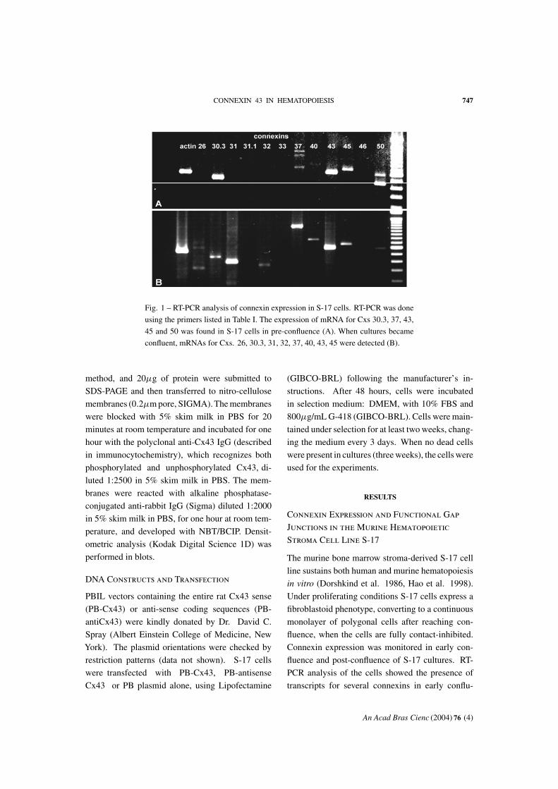
Fig. 1 – RT-PCR analysis of connexin expression in S-17 cells. RT-PCR was done
using the primers listed in Table I. The expression of mRNA for Cxs 30.3, 37, 43,
45 and 50 was found in S-17 cells in pre-confluence (A). When cultures became
confluent, mRNAs for Cxs. 26, 30.3, 31, 32, 37, 40, 43, 45 were detected (B).
method, and 20µg of protein were submitted to
SDS-PAGE and then transferred to nitro-cellulose
membranes (0.2µm pore, SIGMA). The membranes
were blocked with 5% skim milk in PBS for 20
minutes at room temperature and incubated for one
hour with the polyclonal anti-Cx43 IgG (described
in immunocytochemistry), which recognizes both
phosphorylated and unphosphorylated Cx43, di-
luted 1:2500 in 5% skim milk in PBS. The mem-
branes were reacted with alkaline phosphatase-
conjugated anti-rabbit IgG (Sigma) diluted 1:2000
in 5% skim milk in PBS, for one hour at room tem-
perature, and developed with NBT/BCIP. Densit-
ometric analysis (Kodak Digital Science 1D) was
performed in blots.
DNA Constructs and Transfection
PBIL vectors containing the entire rat Cx43 sense
(PB-Cx43) or anti-sense coding sequences (PB-
antiCx43) were kindly donated by Dr. David C.
Spray (Albert Einstein College of Medicine, New
York). The plasmid orientations were checked by
restriction patterns (data not shown). S-17 cells
were transfected with PB-Cx43, PB-antisense
Cx43 or PB plasmid alone, using Lipofectamine
(GIBCO-BRL) following the manufacturer’s in-
structions. After 48 hours, cells were incubated
in selection medium: DMEM, with 10% FBS and
800µg/mL G-418 (GIBCO-BRL). Cells were main-
tained under selection for at least two weeks, chang-
ing the medium every 3 days. When no dead cells
were present in cultures (three weeks), the cells were
used for the experiments.
RESULTS
Connexin Expression and Functional Gap
Junctions in the Murine Hematopoietic
Stroma Cell Line S-17
The murine bone marrow stroma-derived S-17 cell
line sustains both human and murine hematopoiesis
in vitro (Dorshkind et al. 1986, Hao et al. 1998).
Under proliferating conditions S-17 cells express a
fibroblastoid phenotype, converting to a continuous
monolayer of polygonal cells after reaching con-
fluence, when the cells are fully contact-inhibited.
Connexin expression was monitored in early con-
fluence and post-confluence of S-17 cultures. RT-
PCR analysis of the cells showed the presence of
transcripts for several connexins in early conflu-
An Acad Bras Cienc (2004) 76 (4)

748 ESTEVÃO BODI ET AL.
ence (Fig. 1A) and at confluence (Fig. 1B). Semi-
quantitative RT-PCR indicated up-regulation of
Cx43 message after confluence (Fig. 2). Western-
blot and immunofluorescence also showed the pres-
ence of Cx43 (Fig. 3). Western-blotting confirmed
up-regulation of total Cx43 content in post-confluent
cell cultures (Fig. 3A). The latter method indicated
that in early confluence low levels of Cx43 were es-
sentially present in the cytoplasm (Fig. 3B), whilst
after two days of confluence, the typical punctuate
distribution along the cell membranes at the inter-
face between adjacent cells was observed (Fig. 3C).
The presence of other connexins identified by RT-
PCR could not be detected by use of available anti-
bodies (anti-Cx26, Cx32 and 37 antibodies, data not
shown).
cDNA dilutions
0.8
0.6
0.4
0.2
0.000.1 0.01 0.1 1
CX
43 r
atio
Fig. 2 – Semi-quantitative RT-PCR for Cx43, using serial ten
fold dilutions (1:1000, 1:100, 1:10 and 1) of 1µg cDNA. Results
represent the ratio between Cx43 and β-actin expression in S17
cells, in early-confluent cultures (•) and in full confluence (�).
The presence of functional gap junctions was
monitored by LY transfer. In agreement with the
expression pattern of Cx43, dye transfer was very
low in early confluence (Fig. 4A,B), and high after
the cells had reached confluence (Fig. 4C,D).
Biological Function of Cx43 Gap Junctions
in the Hematopoietic Stroma
In order to establish the biological role of gap junc-
tion mediated cell coupling in the hematopoietic
stroma, we proceeded to transfect S-17 cells with
the PBIL vector, the vector containing the sense se-
quence corresponding to Cx43 mRNA, or the vector
containing the anti-sense sequence. The expression
of Cx43 in the three transfected cell lines was mon-
itored by dye coupling and immuno-labeling tech-
niques. Western blot analysis showed the expected
decrease in Cx43 expression in cells transfected with
anti-sense sequence, and an increase in cells trans-
fected with the sense vector (Fig. 5).
Gap junction mediated coupling, as determined
by intercellular LY transfer, was in agreement with
Cx43 expression (Fig. 6), showing that increased
or decreased availability of Cx43, in the sense and
anti-sense transfected cells, correlated with the pres-
ence of functional gap junctions. S-17 cells trans-
fected with plasmid only exhibit coupling compara-
ble to untransfected cells, and intermediate between
anti-sense and sense transfected cells (histograms in
Fig. 6). We were thus able to monitor whether mod-
ulation of coupling and connexin-43 expression had
an effect on the in vitro growth pattern of S-17 cells.
Plasmid-transfected cells followed a growth pattern
similar to control cells. Cx43 antisense-transfected
cells had a tendency to spread on the plastic sub-
strate, while sense-transfected cells grew in a typi-
cal pattern of ‘‘hills-and-valleys’’, characteristic of
myofibroblasts, in which cells show a tendency to
adhere to each other more than to the culture dish
surface (Fig. 7). Primary cultures of both human
and murine bone marrow stroma cells display this
pattern of growth.
Effect of Gap Junction-mediated Stroma
Cell Coupling on Hematopoiesis
Hematopoietic stroma controls the proliferation and
differentiation of hematopoietic precursors through
cell-cell contacts involving adhesion, as well as jux-
tacrine and/or paracrine stimulation of membrane-
An Acad Bras Cienc (2004) 76 (4)

CONNEXIN 43 IN HEMATOPOIESIS 749
B C
A
Fig. 3 – (A) Expression of Cx43 in S-17 cells: total protein extracted from cells (15µg) was
loaded in each lane and the blot was probed with a polyclonal antibody specific for Cx43 (residues
346-360). Lane a shows S-17 cells in pre-confluence, lane b in confluence, lane c is a negative
control using mouse liver tissue and d a positive control using mouse brain tissue. Observe the
lower expression of Cx43 in the pre-confluent S-17 cultures and the higher expression in confluent
cells. (B) Immunocytochemistry, using the Cx43 antibody, shows the diffuse labeling of Cx43
in the cytoplasm of pre-confluent S17 cells and (C) punctuate pattern of labeling at appositional
membrane areas in confluent cultures of S17 cells. Magnification = × 400.
receptors on the target cells. The capacity of S-17
cells to sustain hematopoiesis was monitored by co-
culture with freshly harvested non-adherent murine
bone marrow cells. This cell fraction contains hema-
topoietic precursors in different stages of commit-
ment and differentiation, which respond to stimula-
tion either by further proliferation or by expression
of the terminally differentiated phenotype. In blood
cells, the full differentiation is associated with the
cessation of proliferation. Under the experimental
conditions used in our study only myelopoiesis is
favored.
Control cultures over the normal S-17 cells
and cultures over S-17 cells transfected with plas-
mid only showed a similar ability to sustain myelo-
poiesis. The general pattern of myelopoiesis over
the S17 stroma transfected with plasmid contain-
ing the anti-sense sequence for Cx43, with a de-
creased gap junction-mediated coupling, was char-
acterized by and accelerated terminal differentiation
of myeloid cells (Fig. 8). Mature granulocytes and
macrophages derived from the donor bone marrow
could be observed for up to 24h after co-culture
(Fig. 8A). Myeloid cells were still dividing, form-
ing small clusters of cells whose central region
contained cells with a relatively high nucleo/cyto-
plasmic ratio, and the cytoplasm with high affinity
for MGG stains characteristic of myeloid precur-
sors. From the second day on (Figs. 8B and C), most
myeloid cells acquired a lower nucleus/cytoplas-
mic ratio and lower affinity for MGG stains, char-
acteristic of the macrophage morphology in vitro.
Some cells with macrophagic morphology were still
able to divide (Fig. 8B, arrow). From the third day
of culture (Figs. 8D and E) all the cells differenti-
ated forming small (< 20 cells) clusters or isolated
mono-macrophagic cells, with the morphological
conversion into macrophages with a low nucleus-
An Acad Bras Cienc (2004) 76 (4)

750 ESTEVÃO BODI ET AL.
0
20
40
60
80
100
0-2 3-5 6-8 9-11 12-14 15-17 18-20 21-
# coupled cells
% o
f in
ject
ed c
ells
E
Fig. 4 – Functional coupling between S-17 cells in early-confluence (A and B) and in post-
confluence (C and D). The level of coupling in S-17 cells was determined by dye-transfer ex-
periments using Lucifer Yellow (LY). S-17 cells in early-confluence displaying a fibroblastoid
morphology are not coupled – LY is retained in the cell in which it was injected (marked with
asterix). In post-confluent cultures cells show a polygonal form, vacuolated cytoplasm (D), and a
significant degree of gap-junction coupling as indicated by the spread of LY to adjacent cells (C).
Magnification = × 320. (E) Histogram showing degree of coupling in post-confluent cultures (filled
bars) and early confluent cultures (hatched bar).
An Acad Bras Cienc (2004) 76 (4)

CONNEXIN 43 IN HEMATOPOIESIS 751
a b c d e f g
- 43kDa
Fig. 5 – Expression of Cx43 in S-17 cells transfected with sense Cx43, anti-sense Cx43 DNA
and plasmid only. Proteins were extracted (20µg total protein) after cells reached confluence.
Western blot of mouse brain (positive control) is shown in lane a, S-17 cells transfected with the
anti-sense Cx43 is shown in the lane b, and sense Cx43 in lane c. In another experiment S-17
cells were transfected with plasmid only (lane d), and with anti-sense Cx43 (lane e). Mouse liver
was used as a negative control in lane f, and mouse brain as positive control in lane g.
Fig. 6 – Functional coupling among confluent cultures of S-17 cells transfected with plasmid only (A and B), anti-sense (C and D) and
with sense (E and F) for Cx43. Cells transfected with anti-sense Cx43 exhibit low coupling level. S-17 cells transfected with Cx43 in
sense orientation show enhanced coupling. Histograms of the degree of coupling are displayed in right panel.
An Acad Bras Cienc (2004) 76 (4)

752 ESTEVÃO BODI ET AL.
Fig. 7 – Morphology of S-17 cells used as a stroma layer for co-
cultures with non-adherent bone marrow cells, as observed after
harvesting the bone marrow cells. A. S-17 cells transfected with
the plasmid without any insert. B. S-17 cells transfected with the
plasmid containing the anti-sense sequence for Cx43. C. S-17
cells transfected with the plasmid containing the sense sequence
for Cx43. Note the typical ‘‘hills and valleys’’ pattern of growth
in C (arrows). Final magnification × 120.
cytoplasmic ratio and a vacuolated cytoplasm. From
the fifth day on (Figs. 8F and H) all the cells were
differentiated into mature vacuolated macrophages,
remaining either isolated or still gathered in small
groups. Apoptotic figures were frequent, and low
cell proliferation was concomitant with the in-
creased cell debris, characteristic of senescent cul-
tures (Fig. 8H).
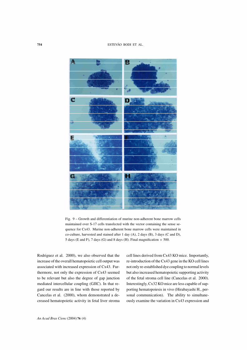
The effect of increased gap junction-mediated
coupling was monitored in cultures of myeloid cells
over the stroma of S-17 cells transfected with the
plasmids containing sense Cx43 DNA (Fig. 9). The
S17 cells transfected with the sense DNA delayed
myeloid cell differentiation, and after 24h of cul-
ture clusters of immature myeloid cells were found
without any morphological signs of differentiation
(Fig. 9A).After 48 and 72h of culture (Figs. 9B to D)
the size of the clusters increased due to the contin-
uous proliferation of myeloid cells, forming giant
colonies of undifferentiated cells that grew up for
over six days in culture (Figs. 9E and F). From this
moment on, their differentiation into macrophages
proceeded from the periphery to the center of the
colonies. Maturing macrophages could be clearly
observed at the periphery (Fig. 9G), or progressively
detached and after eight days the large colonies frag-
mented into smaller clusters that still contained
blasts in the center. On the eight-day, granulocytes
whose life in culture is inferior to 24h were still
found, indicating the sustained production of GM-
CSF, and indicating that the manipulation of Cx43
expression did not inhibit the fundamental ability of
S-17 cell to sustain the full differentiation of myeloid
lineages.
DISCUSSION
In the present study, we have shown, in an in vitro
experimental model of hematopoiesis, that a bone
marrow stroma cell line expresses several connex-
ins, the major one being Cx43, and forms function-
ally competent gap junctions. We also demonstrated
that the two conditions required to causally correlate
the presence of functional gap junctions to the con-
trol of hematopoiesis were present, namely (a) mod-
ulation of connexin expression in the stroma was
biologically relevant for stroma growth pattern and
function, and (b) in vitro hematopoiesis was mod-
ulated by connexin expression and the presence of
functional gap junction channels.
An Acad Bras Cienc (2004) 76 (4)

CONNEXIN 43 IN HEMATOPOIESIS 753
Fig. 8 – Growth and differentiation of murine non-adherent bone marrow cells
maintained over S-17 cells transfected with the vector containing the antisense
sequence for Cx43. Murine non-adherent bone marrow cells were maintained
in co-culture, harvested and stained after 1 day (A), 2 days (B), 3 days (C and
D), 4 days (E), 5 days (F), 6 days (G) and 7 days (H). Arrow head in A points
to granulocytes, arrows in A and B indicate macrophage-like cells in division,
arrows in G and H indicate apoptotic cells. Final magnification × 500.
Cx43 expression in stroma cells was modulated
both at the level of transcription and translation and
this modulation was dependent upon cell density.
Parallel studies have shown that hormones, such as
corticoids, and vitamin A, agents known to control
myeloid cell differentiation, also modulate connexin
expression in the hematopoietic stroma (Hurtado et
al. 2004). In our experimental model, the level of
stroma cell coupling was determinant of the pattern
of hematopoietic cell proliferation and differentia-
tion. In agreement with in vivo data obtained by the
group of Rosendaal (Rosendaal 1995, Krenacs and
Rosendaal 1998, Dorshkind et al. 1993), and with
observations on Cx43 knockout mice (Montecino-
An Acad Bras Cienc (2004) 76 (4)
754 ESTEVÃO BODI ET AL.
Fig. 9 – Growth and differentiation of murine non-adherent bone marrow cells
maintained over S-17 cells transfected with the vector containing the sense se-
quence for Cx43. Murine non-adherent bone marrow cells were maintained in
co-culture, harvested and stained after 1 day (A), 2 days (B), 3 days (C and D),
5 days (E and F), 7 days (G) and 8 days (H). Final magnification × 500.
Rodriguez et al. 2000), we also observed that the
increase of the overall hematopoietic cell output was
associated with increased expression of Cx43. Fur-
thermore, not only the expression of Cx43 seemed
to be relevant but also the degree of gap junction
mediated intercellular coupling (GJIC). In that re-
gard our results are in line with those reported by
Cancelas et al. (2000), whom demonstrated a de-
creased hematopoietic activity in fetal liver stroma
cell lines derived from Cx43 KO mice. Importantly,
re-introduction of the Cx43 gene in the KO cell lines
not only re-established dye coupling to normal levels
but also increased hematopoietic supporting activity
of the fetal stroma cell line (Cancelas et al. 2000).
Interestingly, Cx32 KO mice are less capable of sup-
porting hematopoiesis in vivo (Hirabayashi H., per-
sonal communication). The ability to simultane-
ously examine the variation in Cx43 expression and
An Acad Bras Cienc (2004) 76 (4)

CONNEXIN 43 IN HEMATOPOIESIS 755
the GJIC is important given the growing evidence
for connexin mediated effects that are not depen-
dent on cell-to-cell communication (Goldberg et al.
2000). Our in vitro system, using the sense and
anti-sense strategy to up or down-regulate Cx43 ex-
pression and GJIC clearly indicates that Cx43 mod-
ulation of hematopoiesis is dependent on stroma
cell-to-cell communication. It remains to be seen if
stroma-hematopoietic junctional communication is
also modulated under our experimental conditions.
At any rate, our results suggest that modulation of
stroma GJIC affects primarily hematopoietic cell
differentiation, speeding it when stroma cells are
poorly coupled and delaying it when stroma cells
are highly coupled. High proliferation rates in this
later case would ensue, since each additional divi-
sion between the early committed precursors and
terminally differentiated blood cells doubles the to-
tal cell output.
Our findings are also in accordance with the
proposal that the subendosteal layer and the adjacent
reticular cells are integrated through gap junctions
(Rosendaal et al. 1991). In this region are located
the earliest blood cell precursors that are maintained
in the undifferentiated state. In order to achieve their
full maturation, the cells have to leave the suben-
dosteal environment, and migrate towards the cen-
tral perivascular part of the bone marrow, in which
there are no gap junctions and which favor the cell
differentiation (Rosendaal et al. 1991). Gap junc-
tions may mediate the integration of hematopoietic
microenvironments, and give a molecular support
to the concept of intrinsic spatial gradients in the
bone marrow, in which the cells are exposed to the
favorable condition for proliferation or differentia-
tion at the specific stage of their differentiation, for
a limited period of time.
The question of how gap junctions determine
these microenvironments and the involved signals
remains open, and this question is the subject of on-
going studies. At any rate, the data presented sug-
gests that hematopoiesis may be controlled through
two parallel and probably complementary mecha-
nisms. In the first one, the systemic factors and hor-
mones brought into the bone marrow by blood cir-
culation, as well as the locally produced cytokines,
act directly on hematopoietic cells proper, through
interaction with the corresponding receptors. In the
second one, hematopoiesis can respond to peripheral
demands or suffer intrinsic modifications indirectly,
through modulation of cell coupling in the stroma,
and the consequent modification of blood precursor
adhesion, proliferation and differentiation.
ACKNOWLEDGMENTS
This work was supported by grants from Programade Apoio a Núcleos de Excelência (PRONEX),Conselho Nacional de Desenvolvimento Cien-tífico e Tecnológico (CNPq), Financiadora de Es-tudos e Projetos (FINEP), Fundação de Amparo àPesquisa do Estado do Rio de Janeiro (FAPERJ),Fundação Universitária José Bonifácio (FUJB) andthe International Centre for Genetic Engineering andBiotechnology, Trieste. Estevão Bodi had a fellow-ship from Fundação deAmparo à Pesquisa do Estadode São Paulo (FAPESP) (97/04863-9).
RESUMO
Investigamos a comunicação intercelular mediada porjunções comunicantes em um modelo in vitro de hemato-poiese, usando uma linhagem celular murina de estromade medula óssea, S-17, e culturas primárias de precursoreshematopoiéticos murinos. As S-17 expressam diversasconexinas, sendo a principal a conexina43. A expressãode conexinas e a formação de canais funcionais são modu-ladas pela densidade das células de estroma. A transfecçãode células S-17 com um vetor contendo seqüências sensoou anti-senso de conexina43 aumenta ou diminui, respec-tivamente, a síntese de conexina43 e o acoplamento inter-celular. Nestas condições, a modulação da comunicaçãomediada pelas junções comunicantes modifica o padrãode crescimento das células do próprio estroma, bem comoa capacidade do estroma para sustentar a hematopoiese.O aumento na expressão de conexina43 resulta em umretardo na diferenciação das células sanguíneas, e no au-mento da produção de precursores hematopoiéticos, en-quanto a diminuição na expressão da conexina43 resultanuma diferenciação acelerada dos precursores mielóides.Estes resultados sugerem que o acoplamento mediado porconexina nas células de estroma modula a razão entre pro-liferação e diferenciação dos precursores hematopoiéti-
An Acad Bras Cienc (2004) 76 (4)

756 ESTEVÃO BODI ET AL.
cos. Propomos portanto, que o aumento da comunicaçãomediada por junções comunicantes no estroma elicita umaprodução aumentada de células imaturas de medula óssea,através de um retardo em sua diferenciação terminal, in-duzindo consequentemente um período de proliferaçãoprolongado dos precursores hematopoiéticos.
Palavras-chave: medula óssea, hematopoiese, conexi-nas, junções comunicantes, células S-17.
REFERENCES
Cancelas JA, Koevoet WLM, de Koning AE, MayenAEM, Rombouts EJC and Pleomacher RE. 2000.Connexin-43 gap junctions are involved in multi-connexin-expressing stromal support of hemopoieticprogenitors and stem cells. Blood 96: 498–505.
Charbord P, Tavian M, Humeau L and PéaultB. 1996. Early ontogeny of the human marrowfrom long bones: an immunohistochemical study ofhematopoiesis and its microenvironment. Blood 87:4109–4119.
Dorshkind K, Johnson A, Collins L, Keller GM andPhillips RA. 1986. Generation of purified stromalcell cultures that suport lymphoid and myeloid pre-cursors. J Immunol Methods 89: 37–47.
Dorshkind K, Green L, Godwin A and FletcherWH. 1993. Connexin 43 type gap junctions mediatecommunications between bone marrow stromal cells.Blood 82: 38–45.
Durig J, Rosenthal C, Halfmeyer K, WiemannM, Novotny J, Bingmann D, Duhrsen U andSchirrmacher K. 2000. Intercellular communica-tion between bone marrow stromal cell and CD34+haematopoietic progenitor cells is mediated by con-nexin 43-type gap junctions. Br J Haematol 111:416–425.
Froes MM, Correia AHP, Garcia-Abreu J, Spray DC,Carvalho ACC and Moura Neto V. 1999. Gapjunctional coupling between neurons and astrocytesin primary central nervous system cultures. Proc NatlAcad Sc. USA 96: 7541–7546.
Gabe M. 1968. Techniques Histologiques, ed. Masson,Paris.
Goldberg GS, Bechberger JF, Tajima Y, Merrit M,Omori Y, Gawinowicz MA, Narayanan R, TanY, Yamasaki H, Naus CC, Tsuda H and Nichol-son BJ. 2000. Connexin43 supresses MFG-E8 whileinducing contact inhibition of glioma cells. CancerRes 60: 6018–6026.
Hao GL, Smogorzewska EM, Barsky LW and CrooksGM. 1998. In vitro identification of single CD34+
CD38– cells with both lymphoid and myeloid poten-tial. Blood 91: 4145–4151.
Hurtado S, Balduino A, Bodi E, El-Cheikh MC,Campos de Carvalho AC and Borojevic R. 2004.Connexin expression and gap-junction-mediated cellinteractions in an in vitro model of haemopoieticstroma. Cell Tissue Res 316: 65–76.
Johnson GR. 1984. Hematopoietic multipotential stemcells in culture. Clin. Haematol. 13: 309–327.
Krenacs T and Rosendaal M. 1998. Connexin43 gapjunctions in normal, regenerating, and cultured bonemarrow and in human leukemias: their possible in-volvement in blood formation. Amer J Pathol 152:993–1004.
Lord B. 1997. Biology of the hematoopoietic stem cell.In: Potten CS. (Ed.) Stem cells. Academic Press,London, pp. 401–422.
Makowski L, Caspar DLD, Phillips WC and Goode-nough DA. 1977. Gap junctions structure II: Anal-ysis of the X-ray diffraction data. J Cell Biol 74:629–645.
Manthey D, Bakauskas F, Lee CG, Kozak CA andWillecke K. 1999. Molecular cloning and func-tional expression of the mouse gap junction geneconnexin 57 in human HeLa cells. J Biol Chem 274:14716–14723.
Metcalf D. 1993. Hematopoietic regulators: redun-dance or subtlety? Blood 82: 3515–3523.
Montecino-Rodriguez E, Leathers H and Dorsh-kind K. 2000. Expression of connexin 43 (Cx43)is critical for normal hematopoiesis. Blood 96:917–924.
Rosendaal M. 1995. Gap junctions in blood formingtissues. Microscop. Res Techn 31: 396–407.
Rosendaal M, Gregan A and Green CR. 1991. Directcell-cell communication in the blood forming system.Tissue Cell 23: 457–470.
Rosendaal M, Green CR, Rahman A and Morgan D.1994. Up-regulation of the connexin 43 gap junctionnetwork in haematopoietic tissue before the growthof stem cell. J Cell Sci 107: 29–37.
Saez JC, Branes MC, Corvalan LA, Egenin EA,Gonzalez H, Martines AD and Palisson F. 2000.Gap junctions in cells of the immune system: struc-ture, regulation and possible functional roles. Braz JMed Biol Res 33: 447–455.
An Acad Bras Cienc (2004) 76 (4)