fungi at the edge of life: cryptoendolithic black fungi ... fungi at the... · studies in mycology...
TRANSCRIPT
STUDIES IN MYCOLOGY 51: 1–32. 2005.
1
Fungi at the edge of life: cryptoendolithic black fungi from Antarctic desert
L. Selbmann1, G.S. de Hoog2,3, A. Mazzaglia4, E.I. Friedmann5 and S. Onofri1*
1Dipartimento di Scienze Ambientali, Università degli Studi della Tuscia, Largo dell’Università, Viterbo, Italy 2Centraalbureau voor Schimmelcultures, P.O. Box 85167, NL-3508 AD, Utrecht, The Netherlands 3Institute for Biodiversity and Ecosystem Dynamics, University of Amsterdam, Kruislaan 315, NL-1098 SM Amsterdam, The Netherlands 4Dipartimento di Difesa delle Piante, Università degli Studi della Tuscia, Via S. Camillo de Lellis, Viterbo, Italy 5NASA Ames Research Center, Moffett Field, CA 94035-1000, U.S.A. *Correspondence: S. Onofri, [email protected] Abstract: Twenty-six strains of black, mostly meristematic fungi isolated from cryptoendolithic lichen dominated communi-ties in the Antarctic were described by light and Scanning Electron Microscopy and sequencing of the ITS rDNA region. In addition, cultural and temperature preferences were investigated. The phylogenetic positions of species recognised were determined by SSU rDNA sequencing. Most species showed affinity to the Dothideomycetidae and constitute two main groups referred to under the generic names Friedmanniomyces and Cryomyces (gen. nov.), each characterised by a clearly distinct morphology. Two species could be distinguished in each of these genera. Six strains could not be assigned to any taxonomic group; among them strain CCFEE 457 belongs to the Hysteriales, clustering together with Mediterranean marble-inhabiting Coniosporium species in an approximate group with low bootstrap support. All strains proved to be psychrophiles with the only exception for the strain CCFEE 507 that seems to be mesophilic- psychrotolerant. All had very thick melanised cell walls, the ability to produce exopolysaccharides and to grow meristematically. They are thought to be well adapted to the harsh environment of the Antarctic cold Desert. Hypotheses concerning their origin and evolution are put forward.
Taxonomic novelties: Cryomyces antarcticus Selbmann, de Hoog, Mazzaglia, Friedmann & Onofri gen. sp. nov., C. minteri Selbmann, de Hoog, Mazzaglia, Friedmann & Onofri sp. nov., Friedmanniomyces simplex Selbmann, de Hoog, Mazzaglia, Friedmann & Onofri sp. nov. Key words: Antarctica, black yeasts, cryptoendolithic community, Dothideales, extremophiles, Hysteriales, ITS, licheni-colous fungi, meristematic fungi, microcolonial fungi, phylogeny, SSU, taxonomy.
INTRODUCTION Antarctic rock-inhabiting microorganisms live at the absolute edge of life under the most extreme condi-tions known on Earth. They live between the limit of adaptability and near-death, barely surviving and rarely reproducing (Friedmann & Weed 1987). The environmental conditions of the cold deserts in the McMurdo Dry Valleys (Fig. 1B–D) are extremely stressful to microbial growth. These conditions have been postulated to resemble those prevailing at early Mars, combining extremely low temperatures with extreme dryness and high solar irradiation (Onofri et al. 2004). Rocks have a prominent role as substrata in such cold deserts (Nienow & Friedmann 1993). When conditions at the rock surface are prohibitive for microbial life, Antarctic microbial communities form a particular cryptoendolithic ecotype (Golubic et al. 1981), i.e. condensed growth in microscopic niches under the surface of porous rock (Nienow & Fried-mann 1993). The reasons for growth inside rock
rather than colonising its surface lies in the signifi-cant microclimatic changes occurring over a distance of a few millimetres within the rock substratum (Friedmann & Weed 1987). Average Winter tem-peratures at rock surfaces generally lie 2 °C below the outside air temperature, ranging in 1985 from –47.2 °C to –19.4 °C on Linnaeus Terrace (McMurdo Dry Valleys; Friedmann et al. 1987), but in Summer the rock surface temperature may reach 20 °C above that of outside air. Large differences in microclimatic conditions are noted with exposition. Wide thermal fluctuations at exposed sites contribute to the practi-cally sterile conditions at the outside, the communi-ties being inside the rock where the nanoclimate supplies milder and more stable conditions (Nienow & Friedmann 1993). Antarctic cryptoendolithic microorganisms consti-tute very simple communities comprising only a few species (Nienow & Friedmann 1993). The most common and extensively studied is the “lichen-dominated community” in sandstone (Friedmann 1982). This community colonises porous rocks up to

SELBMANN ET AL.
2
about 10 mm deep under the rock crust. Microbial growth leads to variously coloured bands (Fig. 1F). This stratification is maintained because each micro-bial type has different physiological requirements and abilities (e.g., exposure to solar radiation) and/or produces different antibiotic substances (Ocampo-Friedmann & Friedmann 1993). The community is organised as follows. Immediately under the silici-fied, reddish brown crust (1) a black speckled zone (2) is observed, followed by a white band (3), a green band (4) and sometimes a blue-green band. Fungi and chlorophycean algae, together forming a lichen association, occupy the black (2) and the white (3) zones. The fungal hyphae in the dark zone are melan-ised, while in the white zone they are colourless. The hyaline fungi and part of the pigmented ones proba-bly represent different morphotypes of one and the same lichen species, in which the pigmentation expressed in the upper layer is supposed to be a response to higher light intensities. The cryptoen-dolithic growth is a remarkable morphological adap-tation adopted by lichens to extreme conditions. They loose their typical morphology and enter the porosity of rocks to protect themselves to the hostile outside climate. Nevertheless, they preserve the ability to form a thallus with characteristic morphology as soon as they reach protected niches such as small depres-sions or crevices on rock surfaces, where epilithic structures can be observed. Thick-walled and dark-pigmented, non-lichenised fungi grow mixed with the lichen-forming fungi in the black zone (2). These fungi, often showing meristematic growth in vitro, are isolated as regular members of the lichen domi-nated cryptoendolithic community. The green (4) zone is characterised by the presence of non-lichenised algae, among which the endemic chloro-phycean alga Hemichloris antarctica is predominant, and several of the most extremophilic prokaryotes known to date, viz. the cyanobacteria Chroococ-cidiopsis sp. and Gloeocapsa sp. (Friedmann 1982). A further band has been occasionally observed when Chroococcidiopsis sp. form a separate zone below Hemichloris antarctica. The enzymatic abilities tested in some cryptoen-dolithic fungi were found to be different among species, so that micro- or nano-niches occupied by these fungi do not completely overlap (M. Fenice, pers. comm.). A conspicuous sign of microbial colonisation macroscopically visible on the rock surface is the characteristic mosaic-like pattern of biogenous weathering (Fig. 1E). Microbial activity strongly contributes to the morphology of rocks and landscape (Fig. 1C–D). A large number of fungal species, including yeasts, has been recorded from Antarctica, colonising nearly all terrestrial environments (Onofri 1999), but the best adapted to the harshest conditions seem to be the melanised fungi exhibiting meristematic growth.
Such fungi are also found in other environments where they are subjected to extreme conditions. Black meristematic microfungi, producing slowly expanding cauliflower-like colonies, are morphologi-cally barely differentiated. Their phylogeny is quite diverse, and they have been assigned to three differ-ent ascomycete orders: Dothideales, Chaetothyriales and Pleosporales (Sterflinger et al. 1999). They are commonly isolated from rocks in hot climates (Wol-lenzien et al. 1997), marble monuments (Sterflinger & Krumbein 1997), hypersaline environments (Zalar et al. 1999a) and other substrates that are hard to colonise. Some of them, belonging to the order Chaetothyriales, are encountered as opportunists on humans. The skin disease chromoblastomycosis has a meristematic invasive phase, the muriform cells (Matsumoto et al. 1984). The natural niche of these species is in the spines of cactus plants in semi-arid climates (Zeppenfeldt et al. 1994). In humans the thick melanised cell wall is supposed to protect the fungus from phagocytic action by oxygen radicals (Schnitzler et al. 1999). The ability to grow meris-tematically, ensuring to the colonies an optimal surface/volume ratio (Wollenzien et al. 1995), is hypothesized to enhance the ability to survive under conditions of stress such as high or low temperature, low water availability (Wollenzien et al. 1995), acidity, nutrient deficiency (Sterflinger et al. 1999), high UV exposure (Urzì et al. 1995) or high salt concentration (Zalar et al. 1999c). In some fungi meristematic growth could be induced by acidifica-tion of the culture medium (Mendoza et al. 1993). Furthermore, black fungi that preserve meristematic growth as a stable character are reported to tolerate desiccation, UV exposure (Wollenzien et al. 1995) and high temperature (Sterflinger 1998) better than lichens. Meristematic black fungi from the Antarctic cryptoendolithic communities are fascinating and poorly known microorganisms, adapted to environ-mental conditions that are even more hostile than those of the remaining rock-inhabiting fungi. Al-though their actual limits of survival are not well known, they seem to be, both from morphological and physiological points of view, very well adapted to withstand unfavourable conditions. This is exem-plified by the fact that they express melanised thick walls as a stable character, allowing them to resist dryness and the UV irradiation that reaches them through the translucent crystals of orthoquarzite in sandstone. All exhibit a highly reduced morphology with scarce differentiation. Cryptoendolithic black fungi frequently produce exopolysaccharides outside the hyphae. Similarly, exopolysaccharides were observed surrounding the multicellular conidia (or bulbils) produced by Friedmanniomyces endolithicus Onofri (Onofri et al. 1999). These substances may protect microfungal structure from desiccation and

CRYPTOENDOLYTHIC BLACK FUNGI FROM ANTARCTIC DESERT
3
freeze damages (Selbmann et al. 2002). Microbe-enclosing polymer layers (biofilms) have recently been described in Antarctic endolithic microecosys-tems. They create physical and chemical conditions at a nano-scale, that are often much less severe than those of the external environment (De los Rìos et al. 2003). None of the black Antarctic fungi has been hith-erto identified, except for F. endolithicus, described as new genus and new species based mainly on
morphological observations (Onofri et al. 1999). The phylogenetically isolated position of the genus was recently confirmed by molecular investigations (Onofri et al. 2002). During 18 Antarctic expeditions, organised by the U.S. National Science Foundation (NSF) and the Italian National Program for Antarctic Research (PNRA), quite a large number of meris-tematic fungi have been isolated from rocks collected in different sites in Victoria Land, Antarctica.
A B
C D
E1
2
3
4
F
1 cm
Fig. 1. Landscapes at sample locations. A. Timber Peak, Northern Victoria Land, Antarctica. B. Layered Beacon sandstone and dolerite in the University Valley, Southern Victoria Land, Antarctica. C, D. Sandstone sculptured by the activity of cryptoen-dolithic microrganisms in Battleship Promontory. E. Patchwork-like effect of sandstone surface resulting from biogenous weathering. F. Typical stratification in sandstone colonised by lichen dominated community: (1) silicified, reddish brown crust; (2) black zone colonised by lichenised and non-lichenised fungi; (3) white zone colonised by lichenised fungi and lichenised algae; (4) green zone colonised by non-lichenised algae and cyanobacteria.

SELBMANN ET AL.
4
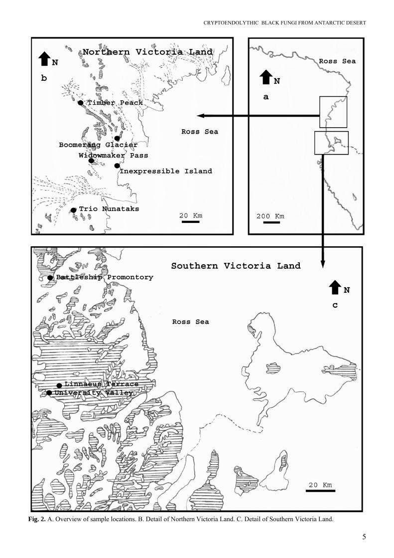
Because of their inability to produce well-defined, recognisable structures, their identification has been pending, awaiting an approach with molecular tools. In the present paper 26 strains of black fungi from cryptoendolithic microbial lichen dominated commu-nities were sequenced for ITS and SSU rDNA frag-ments, and morphologically described and illustrated, in order to clarify their taxonomy, phylogeny and ecology. MATERIALS AND METHODS Sampling sites Antarctic rock samples were collected from Linnaeus Terrace (McMurdo Dry Valleys), University Valley (McMurdo Dry Valleys), Battleship Promontory (Southern Victoria Land), Inexpressible Island, the promontory between Widowmaker Pass and Olson Nunatak, Boomerang Glacier, and Timber Peak (Northern Victoria Land, Antarctica). Essential data on each site are reported below and summarised in Table 1. Maps shown in Figs 2A–C indicate the precise provenance of each sample from which strains were isolated. Linnaeus Terrace: 77°36' S 161°05' E is an elevated ridge of weathered Beacon Sandstone approximately 1.5 km in length and 1 km in width. It is located at the east end of the Asgaard Range, 1.5 km north of Oliver Peak at an elevation of about 1600 m. The air temperature ranges from –20 to –45 °C in Winter, the mean remaining below zero in Summer (Nienow & Friedmann 1993). Battleship Promontory: 76°55' S 160°55' E is a Beacon sandstone promontory which rises from the floor of Alatna Valley near its head, in Victoria Land, 900–1000 m a.s.l. The name was suggested by Parker Calkin, U.S. geologist who made stratigraphic studies in the valley in the 1960–61 season. University Valley: 77°52' S 160°40' E is a valley about 1.5 km long at an elevation of about 1650 m, lying next NE of Farnell Valley in the Beacon Valley area of Victoria Land. Named in January 1962 by USARP (United States Antarctic Research Program) researchers Heinz Janetschek and Fiorenzo Ugolini after their respective university affiliation, Leopold-Franzens-Universität at Innsbruck, Austria, and Rutgers University at New Brunswick, New Jersey, U.S.A. Inexpressible Island: 74°54' S 163°39' E is an island, 10 km long, in Terra Nova Bay, Victoria Land, lying close south of the Northern Foothills at the outer edge of the Nansen Ice Sheet. First explored by the
Northern Party of the BrAE (British Antarctic Expe-dition), 1910–13, and called “Southern Foothills” in contrast to the Northern Foothills. The name was changed to Inexpressible Island by the party after spending a very unpleasant winter on half rations in a cave in a snowdrift on the Island. Widowmaker Pass: 74°55' S 162°20' E is a heavily crevassed and therefore dangerous pass leading from Larsen Glacier to Reeves Glacier, between Mount Janetschek and Mount Gerlache in Victoria Land. It was attributed this expressive name by the NZGSAE (New Zealand Geological Survey Antarctic Expedi-tion), 1962–63. Olson Nunatak: 74°55' S 162°28' E is a bare rock nunatak lying at the south side of the terminus of Reeves Glacier, 6 km north of the summit of Mount Gerlache, in Victoria Land. Boomerang Glacier: 74°33' S 163°54' E is a gently curving glacier, 16 km long, draining southward from Mount Dickason in the Deep Freeze Range to enter Browning Pass, at the north side of Nansen Ice Sheet in Victoria Land. Discovered by the Northern Party of the BrAE, 1910–13, and named by them because of its shape. Timber Peak: 74°10' S 162°23' E is a high peak (3070 m) above Priestley Glacier, on the south side. The peak is about 3 km WNW of the summit of Mount New Zealand in the Eisenhower Range, Victoria Land. The Southern Party of the NZGSAE (1962–63) gave this name because petrified sections of tree branches were found in sandstone deposits at this point. Sampling and isolation Samples of colonised rock were aseptically taken off with a sterile chisel and placed in sterilised plastic bags. The bags were preserved until the isolation in the refrigerator at –20 °C. Strains were isolated by directly plating fragments of colonised rock on Petri dishes containing MYEA (malt extract 2 %, yeast extract 0.2 %, agar 1.5 %). Plates were incubated at 10 °C and growth was inspected monthly. Colonies were subcultured on MYEA slants. Strains are listed in Table 1 in which data for each are specified. Morphology Light microscopic observations were carried out using slide cultures on MEA (2 % malt extract agar) incubated for 10 wks and mounted in lactophenol. Samples for scanning electron microscopic observa-tions were prepared according with methods de-scribed by Onofri et al. (1980).
CRYPTOENDOLYTHIC BLACK FUNGI FROM ANTARCTIC DESERT
5
Fig. 2. A. Overview of sample locations. B. Detail of Northern Victoria Land. C. Detail of Southern Victoria Land.
SELBMANN ET AL.
6
Morphological terminology A diagrammatic overview of the terminology used in descriptions is given in Fig. 3. Thallus expansion is either by the blastic or the thallic mode. Blastic devel-opment includes directional growth of hyphae and budding cells, presumably guided by a spindle pole body (SPB); blastoconidia may be produced from a narrow base (Fig. 3G). Polar expansion is subse-quently arrested, and eventually thallic processes take over, by either isodiametrical or unilateral swelling (Fig. 3D–E), without any sign of the presence of an SPB. If this shift in development takes place in a single hyphal cell, conversion leads to the formation of a chlamydospore. Alternatively, each cell of the hyphal stretch swells similarly; this leads to a torulose hypha (Fig. 3C). All single cells of the torulose hy-phae may disarticulate schizolytically, and thus indi-vidual arthroconidia are liberated (Fig. 3I). Single cells may also germinate in a uni- or multipolar fash-ion while shedding off the remains of a dark, thick mother cell wall (Fig. 3J). A similar series of events is observed with single cells. Except for budding at a narrow base, isodiametric swelling is possible (Fig. 3F). Such cells may germinate by germination as described above. Alternatively, the cell continues its isodiametric expansion and eventually develops compartments by transverse, longitudinal and oblique septation (Fig. 3H). The meristematic clump of cells expands further, eventually locally sheds off the mother cell wall and falls apart into separate cells or smaller cell clumps (Fig. 3K), which each may resume growth in an identical manner. Physiology Growth on agar media: Cultural characteristics and growth velocity was recorded on PDA (potato dex-trose agar), MEA (malt extract agar), CZA (Czapek dox agar) and OA (oatmeal agar). Plates were incu-bated at 10 °C and inspected monthly. Tests were performed in triplicate. Acid production: The ability to produce acids was tested both on solid media and in liquid culture. Tests on solid media were carried out on chalk test medium (glucose 50 g/L, CaCO3 5 g/L, yeast extract 5 g/L, agar 20 g/L) prepared in Petri dishes. Plates were incubated at 10 °C and inspected monthly. Tests were performed in triplicate. Tests in liquid culture were performed in the following medium [(NH4)2SO4 1 g/L, KH2PO4.7H2O 1 g/L, FeSO4.7H2O 0.01 g/L, KCl 0.5 g/L, yeast extract 1 g/L, glucose 30 g/L] (Thauresia & McNeil 1994) using 250 mL flasks. Cultures were incubated at 15 °C under continual shaking at 180 r.p.m. for 2 mo. Aliquots of 5 mL were taken aseptically from culture broths every 15 d, filtered and pHs of supernatants were recorded.
Oxalic acid production was measured using the Boe-hringer Mannheim kit R-Biopharm GmbH, D 64293 Darmstadt, Germany. The organic acid production was also investigated as follows: culture broths were strongly acidified (pH 1) by addition of 1N HCl and organic acids were extracted in the organic phase using ethyl acetate and successively dried in vacuo. Samples were sylilated using NO-bistrimethyl-silylacetamide and analysed by GC/MS using a HP 5890 gas chromatograph equipped of a BPX-5 / SGE 30 m x 0.25 mm column and connected to a mass spectrometer VG TS-250. The analytic conditions were: 40 °C for 6 min and 6 °C.min-1 until 200 °C for 5 min. Temperature injector was 200 °C. EPS production: The ability to produce EPS was tested in liquid culture using the following medium (NaNO3 3 g/L, KH2PO4 1 g/L, MgSO4 0.5 g/L, KCl 0.5 g/L, Yeast Extract 1 g/L, Glucose 30 g/L) (Compere & Griffith 1978) using 250 mL flasks. Cultures were incubated at 15 °C at 180 r.p.m. for 2 mo. Cultural broth were sampled every 15 d filtered and EPS were precipitated 1:2 v/v in EtOH. The EPS were filtered using GF/C filters (Whatman Interna-tional Ltd Maidstone, U.K.), dried and weighted. Temperature relationships: Temperature preferences were tested by inoculating strains on MEA in Petri dishes and incubating at 0, 5, 10, 15, 20, 25, 30 and 35 °C and the diameter of the colonies was recorded. Tests were performed in duplicate. DNA extraction Fungi were grown on agar slants (MEA) for about 6 mo at 10 °C. The DNA extraction was carried out on a mycelial fragment using the Nucleospin Plant kit (Macherey-Nagel, Düren, Germany). PCR conditions PCR reactions were performed using the Ready-to-Go PCR-Beads Kit (Amersham Pharmacia Biotech, Piscataway, New Jersey, U.S.A.). In each 25 μL reaction tube 5 pmol of each primer and 40 ng on template DNA were added. The amplification was carried out using MiniCycler™ (MJ Research, Inc Waltham, Massachusetts U.S.A.) equipped with heated lid. First denaturation step was 95 °C for 30 s, annealing was 55 °C for 30 s, extension 72 °C for 30 s. Cycles were repeated 35 times, with a last extension 72 °C for 5 min. The products were purified using Nucleospin Extract kit (Macherey-Nagel, Düren, Germany). Primers NS1, NS2, NS3, NS4, NS5, NS8, ITS1, ITS4 (White et al. 1990), SR10R (Bruns et al. 1992) and ITS5 ITS4a (Larena et al. 1999) were employed.

CRYPTOENDOLYTHIC BLACK FUNGI FROM ANTARCTIC DESERT
7
Polar growthPolar growth
IsodiametricIsodiametric andand unilateral swellingunilateral swelling
Arthric secessionArthric secession MeristematicMeristematic growthgrowth
D EC
I
F
H
K
A B
G
J
Fig. 3. Diagram of developmental possibilities in meristematic fungi; ontogenetic options indicated by dotted lines. A, B. Polar growth, either hyphal (A) or budding (B). C–E. Isodiametric expansion, either in hyphal cell (C–D), unilateral in hyphal cell (E) or in yeast cell (F). G–H. Cell maturation, either by a blastic (G) or a thallic (H) process. I–K. Secession, either by arthric disarticulation (I), inflation and germination (J) or inflation and multilateral disarticulation (K). Sequencing, alignment and phylogenetic tree reconstruction Sequencing reactions were performed by the dide-oxynucleotide Sanger (1977) method using the TF Big Dye Terminator 1,1 RR Applied Biosystems kit. Fragments were analysed using an ABI 310 Genetic Analyser (Applied Biosystems). Sequence assembly was done using the Chromas programme (v. 1.45 1996–1998, Conor McCarthy School of Health Sci-ence, Griffith University, Southport, Queensland, Australia). Sequences of ITS were aligned in BioNu-merics v. 3.0 (Applied Maths, Kortrijk, Belgium) and
18S sequences with ARB beta-package (v. 22-08-2003) developed by W. Ludwig et al. (2004; www.mikro.biologie.tu-muenchen.de/pub/ ARB). The SSU alignment spanned positions 141–2512, which corresponds to 1515 bp with reference to Saccharo-myces cerevisiae. Trees based on SSU sequences were reconstructed with the Parsimony option in ARB. ITS trees were reconstructed using the Treecon software package (Van de Peer & De Wachter 1994) with the neighbour-joining algorithm with Kimura 2 correction with 100 bootstrap replications.

SELBMANN ET AL.
8
Table 1. List of strains of Antarctic black meristematic fungi analysed.
GenBank Species Isolation No. Accession No. Groups* Sample Location
SSU ITS
Cryomyces minteri
967-26c CCFEE 5187T = CBS 116302
-/A Soil “Cirque 2”, Battleship Promontory, Alatna Valley, McMurdo Dry Valleys, Southern Victoria Land, Antarc-tica on 22 Dec 1996
DQ066714 DQ028270
Cryomyces antarcticus
F3 A801-11 CCFEE 453 3/A Sandstone Linnaeus Terrace, McMurdo Dry Valleys, Southern Victoria Land, Antarctica on 1980-81
Cryomyces antarcticus
F6 A801-30A CCFEE 456 3/A Sandstone McMurdo Dry Valleys, Southern Victoria Land, Antarc-tica on 1980-81
Cryomyces antarcticus
F44 A812-H5b
CCFEE 534T = CBS 116301
3/A Soil Linnaeus Terrace, McMurdo Dry Valleys, Southern Victoria Land, Antarctica on 1981-82
DQ066713 DQ028269
Cryomyces antarcticus
F45 A812-H41c
CCFEE 535 3/A Soil Linnaeus Terrace, McMurdo Dry Valleys, Southern Victoria Land, Antarctica on 1981-82
Cryomyces antarcticus
F46 A812-H59a
CCFEE 536 -/A Soil Linnaeus Terrace, McMurdo Dry Valleys, Southern Victoria Land, Antarctica on 1981-82
Cryomyces antarcticus
F57 A801-3aFF2
CCFEE 690 -/A Sandstone McMurdo Dry Valleys, Southern Victoria Land, Antarc-tica on 1980-81
Cryomyces antarcticus
F24 A801-30aFF2
CCFEE 514 3/A Sandstone Linnaeus Terrace, McMurdo Dry Valleys, Southern Victoria Land, Antarctica on 1980-81
Cryomyces antarcticus
F25 A801-3aFF4
CCFEE 515 3/A Sandstone Linnaeus Terrace, McMurdo Dry Valleys, Southern Victoria Land, Antarctica on 1980-81
Friedmanniomy-ces endolithicus
967-58b CCFEE 5208R -/C Sandstone Promontory between Widowmaker Pass and Olson Nunatak, 200 m a.s.l., Northern Victoria Land, Antarctica on Jan 14 1997
Friedmanniomy-ces endolithicus
967-39a CCFEE 5195 -/C Granite or pegmatite
Close to Boomerang Glacier, Northern Victoria Land, Antarctica on Jan 4 1997
Friedmanniomy-ces endolithicus
967-21a CCFEE 5180 -/C Sandstone Battleship Promontory, McMurdo Dry Valleys, Southern Victoria Land, Antarctica on Dec 26 1996
Friedmanniomy-ces endolithicus
967-43a CCFEE 5199 -/C Sandstone Trio Nunataks, Northern Victoria Land, Antarctica on Jan 7 1997
Friedmanniomy-ces endolithicus
F33 A801-H59a
CCFEE 670 -/C Sandstone McMurdo Dry Valleys, Southern Victoria Land, Antarc-tica on 1980-81

CRYPTOENDOLYTHIC BLACK FUNGI FROM ANTARCTIC DESERT
9
Table 1. Contd.
GenBank Species Isolation No. Accession No. Groups* Sample Location
SSU ITS Friedmanniomy-ces endolithicus
F32 A801-147 CCFEE 522 5/C Sandstone Linnaeus Terrace, McMurdo Dry Valleys, Southern Victoria Land, Antarctica on 1980-81
DQ066715 DQ028272
Friedmanniomy-ces endolithicus
F34 A801-25 CCFEE 524 -/C Soil Linnaeus Terrace, McMurdo Dry Valleys, Southern Victoria Land, Antarctica on 1980-81
Friedmanniomy-ces endolithicus
967-38a CCFEE 5001 -/C Sandstone (rock pow-der)
Timber Peak, Northern Victoria Land, Antarctica on Jan 4 1997
Friedmanniomy-ces endolithicus
967-38g1 CCFEE 5193 -/C Sandstone (rock pow-der)
Timber Peak, Northern Victoria Land, Antarctica on Jan 4 1997
Friedmanniomy-ces simplex
967-24a CCFEE 5184T = CBS 116775
-/C Sandstone Battleship Promontory, McMurdo Dry Valleys, Southern Victoria Land, Antarctica on Dec 27 1996
DQ066716 DQ028271
Unidentified Dothideales
967-22a CCFEE 5018 -/B Soil “Cirque 1”, Battleship Promontory, Alatna Valley, McMurdo Dry Valleys, Southern Victoria Land, Antarc-tica, close to the camp, at the base of a vertical slope of Beacon sandstone, E-exposed on Dec 19 1996
Unidentified Dothideales
F12 A790-20 CCFEE 502 5/E Sandstone Linnaeus Terrace, McMurdo Dry Valleys, Southern Victoria Land, Antarctica on 1979-80
Unidentified Dothideales
F17 A801- 8bFF16
CCFEE 507 2/F Soil Linnaeus Terrace, McMurdo Dry Valleys, Southern Victoria Land, Antarctica on 1980/81
Unidentified Dothideales
F1 A801-23 CCFEE 451 4/G Sandstone Linnaeus Terrace, McMurdo Dry Valleys, Southern Victoria Land, Antarctica on 1980/81
Unidentified Dothideales
A967-21-1 CCFEE 5211 -/H Sandstone Battleship Promontory, McMurdo Dry Valleys, Southern Victoria Land, Antarctica on Dec 26 1996
Unidentified Dothideales
967-16a CCFEE 5176 -/M Weathered granite
Inexpressible Island, Northern Victoria Land, Antarctica on 22 Dec 1996
Unidentified Dothideales
F7 A801-45 CCFEE 457 5/D Sandstone University Valley, McMurdo Dry Valleys, Southern Victoria Land, Antarctica on 1980/81
Abbreviation used: T = ex-type strain; R = representative strain; CBS = Centraalbureau voor Schimmelcultures; CCFEE = Culture Collection of Fungi from Extreme Environments. *Groups = comparison between groups organised on the basis of molecular results (numbers, this study) and on the basis of morphological observation (letters, Ocampo-Friedmann & Friedmann 1993).
SELBMANN ET AL.
10
RESULTS Physiology None of the strains produced acid on chalk agar, not even strain CCFEE 670, Friedmanniomyces en-dolithicus that produced many crystals when culti-vated on MEA, probably due to insufficient growth on this medium. No traces of oxalic acid in liquid cul-tures have been detected using the kit. The inability to produce organic acids was confirmed with GC/MS analyses where no significant amount of organic acids was recorded in the cultural broth even in samples showing low pH values. Temperature relations are given in Table 2. All strains tested can be referred to as being psychrophilic, in accordance with the defini-tion given by van Uden (1984) and Vishniac (1987). This definition is somewhat wider than the one used in bacteriology where a species is considered to be psychrophilic when it is able to grow in the range 0–20 °C (Morita 1975), but for yeasts and other eukaryotic microorganisms the upper temperature limit for psychrophily is maintained at 25 °C (van Uden 1984; Vishniac 1987). Most of the strains had an optimum growth temperature in the range of 10–20 °C, and in many cases there was still well detectable growth at 0 °C. Strains of Cryomyces showed opti-mum growth at 15–20 °C. Members of the genus Friedmanniomyces showed very slow growth at the optimum temperature between 10–15 °C. An excep-tion was CCFEE 507, having an optimum in the range of 15–25 °C. Since it was also very well able to grow at low temperatures and still expanded at 0 °C, it matched with the definition of mesophilic psychrotol-erance (Zucconi et al. 1996). Growth velocities on different agar media are given in Table 2. Strains tested were able to grow well on MEA and PDA. Cryomyces and particularly Friedmanniomyces strains generally showed poor or no growth on CZA. SSU phylogeny Figure 4 shows a phylogenetic tree of 93 ascomycetes that could be confidently aligned to the Antarctic black fungi analysed, mainly comprising members of the order Dothideales, Capnodiales, Jahnulales, Mycocaliciales, Myriangiales, Hysteriales, and Chae-tothyriales. The selection comprised other fungi from rock, epiphytic species, halo- and acidophilic species and phytopathogens, but also species known to occur on humans. The Antarctic strains reported in this paper were included in different orders of the Dothideomycetidae. One group (C) comprised strains CCFEE 5187, 453, 456, 534, 535, 536, 690, 514 and 515. A second group (A), comprising strains CCFEE 5208, 5195, 5180, 5199, 670, 522, 524, 5001, 5193 and 5184, contained the the strain CCFEE 5208, morphologically identical
to Friedmanniomyces endolithicus Onofri previously described from rock (Onofri et al. 1999). This group was found to be phylogenetically relatively close to the lichenicolous fungus Hobsonia santessonii Lowen & Hawksw. and a strain deposited as Mycocalicium victoriae (C. Knight ex F. Wilson) Tibell. Strain CCFEE 457 clustered with members of the order Hysteriales [Glyphium elatum (Grev.) H. Zogg] and to a lesser extent to the Chaetothyriales, forming a clade with some Coniosporium species from rock in the Mediterranean. The latter group was found to be paraphyletic to the family Herpotrichiellaceae (Cap-ronia, with Exophiala anamorphs). All remaining strains proved to be unrelated to (A) and (C) (Fried-manniomyces), but no sequence identity could be found. Strain CCFEE 5176 had an unresolved posi-tion. Strain CCFEE 502 had a Mycocalicium victoriae strain as its nearest neighbour, which was paraphyletic to the Friedmanniomyces / Hobsonia group. Strains CCFEE 507 and 5211 / 5018 were remote from each other and did not bear any similarity to a described teleomorph species. CCFEE 451 was relatively close to Coccodinium bartschii A. Massal. The topology of the SSU rDNA tree was main-tained with Neighbour-joining (data not shown) as well as with parsimony (Fig. 4) algorithms, with the exception of a cloud containing members of Xylaria-les, Jahnulales and Pleosporales. Robustness was observed despite relatively low bootstrap values. ITS sequence data Sequences of most strains proved to be reproducible, with the exception of isolates morphologically attrib-uted to Cryomyces. The difficulties concerned both the amplification and the sequencing steps. Very often it was not possible to obtain good amplification of some fragment of SSU and, more frequently, of ITS portions and the reaction needed to be repeated many times, purifying bands from agarose gels and chang-ing primers, before obtaining good results. This seemed to be unrelated to the GC content since the problems were not solved using DMSO in the ampli-fication mixture (Winship 1989). Furthermore, se-quencing reactions gave sometime partially over-lapped electrophorograms in some positions or they were unreadable at all. Different runs of the same isolate sometimes produced well-readable electrophorograms, which were, however, up to 16.8 % different from each other. A mixture of different strains at the moment of DNA extraction was excluded on the basis of the good quality of the different runs. The differences were far beyond heterogeneity expected in heterothallic strains. Strain CCFEE 456 of Cryomyces antarcticus repeat-edly yielded three different sequences, while CCFEE 514 of Cryomyces antarcticus produced two types.
CRYPTOENDOLYTHIC BLACK FUNGI FROM ANTARCTIC DESERT
11
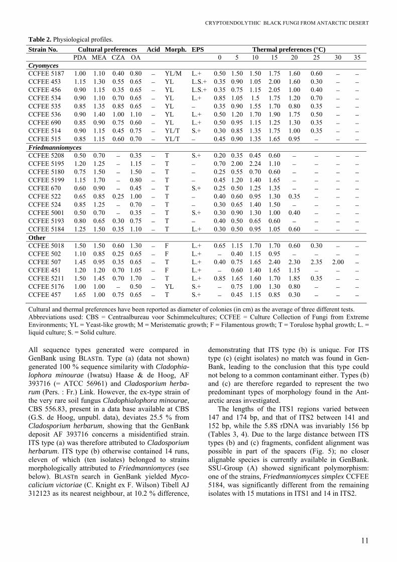
Table 2. Physiological profiles. Strain No. Cultural preferences Acid Morph. EPS Thermal preferences (°C) PDA MEA CZA OA 0 5 10 15 20 25 30 35 Cryomyces CCFEE 5187 1.00 1.10 0.40 0.80 − YL/M L.+ 0.50 1.50 1.50 1.75 1.60 0.60 − − CCFEE 453 1.15 1.30 0.55 0.65 − YL L.S.+ 0.35 0.90 1.05 2.00 1.60 0.30 − − CCFEE 456 0.90 1.15 0.35 0.65 − YL L.S.+ 0.35 0.75 1.15 2.05 1.00 0.40 − − CCFEE 534 0.90 1.10 0.70 0.65 − YL L.+ 0.85 1.05 1.5 1.75 1.20 0.70 − − CCFEE 535 0.85 1.35 0.85 0.65 − YL − 0.35 0.90 1.55 1.70 0.80 0.35 − − CCFEE 536 0.90 1.40 1.00 1.10 − YL L.+ 0.50 1.20 1.70 1.90 1.75 0.50 − − CCFEE 690 0.85 0.90 0.75 0.60 − YL L.+ 0.50 0.95 1.15 1.25 1.30 0.35 − − CCFEE 514 0.90 1.15 0.45 0.75 − YL/T S.+ 0.30 0.85 1.35 1.75 1.00 0.35 − − CCFEE 515 0.85 1.15 0.60 0.70 − YL/T − 0.45 0.90 1.35 1.65 0.95 − − − Friedmanniomyces CCFEE 5208 0.50 0.70 − 0.35 − T S.+ 0.20 0.35 0.45 0.60 − − − − CCFEE 5195 1.20 1.25 − 1.15 − T − 0.70 2.00 2.24 1.10 − − − − CCFEE 5180 0.75 1.50 − 1.50 − T − 0.25 0.55 0.70 0.60 − − − − CCFEE 5199 1.15 1.70 − 0.80 − T − 0.45 1.20 1.40 1.65 − − − − CCFEE 670 0.60 0.90 − 0.45 − T S.+ 0.25 0.50 1.25 1.35 − − − − CCFEE 522 0.65 0.85 0.25 1.00 − T − 0.40 0.60 0.95 1.30 0.35 − − − CCFEE 524 0.85 1.25 − 0.70 − T − 0.30 0.65 1.40 1.50 − − − − CCFEE 5001 0.50 0.70 − 0.35 − T S.+ 0.30 0.90 1.30 1.00 0.40 − − − CCFEE 5193 0.80 0.65 0.30 0.75 − T − 0.40 0.50 0.65 0.60 − − − − CCFEE 5184 1.25 1.50 0.35 1.10 − T L.+ 0.30 0.50 0.95 1.05 0.60 − − − Other CCFEE 5018 1.50 1.50 0.60 1.30 − F L.+ 0.65 1.15 1.70 1.70 0.60 0.30 − − CCFEE 502 1.10 0.85 0.25 0.65 − F L.+ − 0.40 1.15 0.95 − − − − CCFEE 507 1.45 0.95 0.35 0.65 − T L.+ 0.40 0.75 1.65 2.40 2.30 2.35 2.00 − CCFEE 451 1.20 1.20 0.70 1.05 − F L.+ − 0.60 1.40 1.65 1.15 − − − CCFEE 5211 1.50 1.45 0.70 1.70 − T L.+ 0.85 1.65 1.60 1.70 1.85 0.35 − − CCFEE 5176 1.00 1.00 − 0.50 − YL S.+ − 0.75 1.00 1.30 0.80 − − − CCFEE 457 1.65 1.00 0.75 0.65 − T S.+ − 0.45 1.15 0.85 0.30 − − −
Cultural and thermal preferences have been reported as diameter of colonies (in cm) as the average of three different tests. Abbreviations used: CBS = Centraalbureau voor Schimmelcultures; CCFEE = Culture Collection of Fungi from Extreme Environments; YL = Yeast-like growth; M = Meristematic growth; F = Filamentous growth; T = Torulose hyphal growth; L. = liquid culture; S. = Solid culture. All sequence types generated were compared in GenBank using BLASTn. Type (a) (data not shown) generated 100 % sequence similarity with Cladophia-lophora minourae (Iwatsu) Haase & de Hoog, AF 393716 (= ATCC 56961) and Cladosporium herba-rum (Pers. : Fr.) Link. However, the ex-type strain of the very rare soil fungus Cladophialophora minourae, CBS 556.83, present in a data base available at CBS (G.S. de Hoog, unpubl. data), deviates 25.5 % from Cladosporium herbarum, showing that the GenBank deposit AF 393716 concerns a misidentified strain. ITS type (a) was therefore attributed to Cladosporium herbarum. ITS type (b) otherwise contained 14 runs, eleven of which (ten isolates) belonged to strains morphologically attributed to Friedmanniomyces (see below). BLASTn search in GenBank yielded Myco-calicium victoriae (C. Knight ex F. Wilson) Tibell AJ 312123 as its nearest neighbour, at 10.2 % difference,
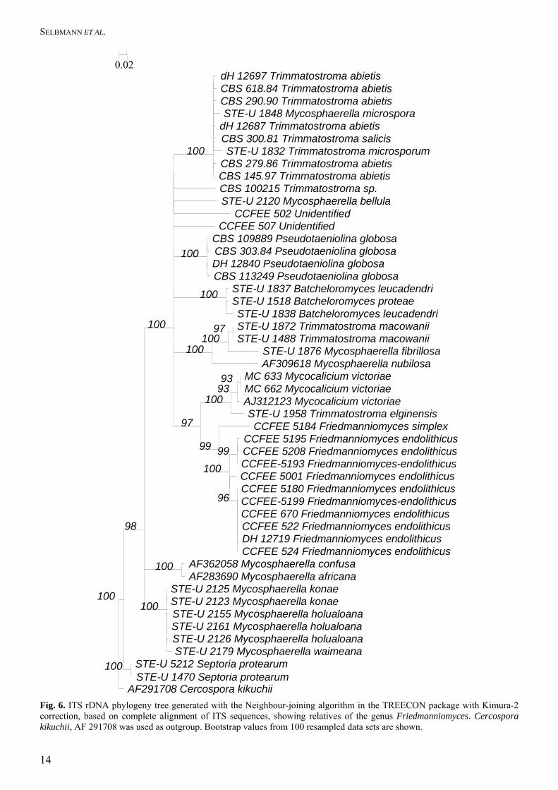
demonstrating that ITS type (b) is unique. For ITS type (c) (eight isolates) no match was found in Gen-Bank, leading to the conclusion that this type could not belong to a common contaminant either. Types (b) and (c) are therefore regarded to represent the two predominant types of morphology found in the Ant-arctic areas investigated. The lengths of the ITS1 regions varied between 147 and 174 bp, and that of ITS2 between 141 and 152 bp, while the 5.8S rDNA was invariably 156 bp (Tables 3, 4). Due to the large distance between ITS types (b) and (c) fragments, confident alignment was possible in part of the spacers (Fig. 5); no closer alignable species is currently available in GenBank. SSU-Group (A) showed significant polymorphism: one of the strains, Friedmanniomyces simplex CCFEE 5184, was significantly different from the remaining isolates with 15 mutations in ITS1 and 14 in ITS2.

SELBMANN ET AL.
12
Dothideales
Dothideales
DothidealesDothideales
E
D
A
B
C
F
G
DothidealesDothidealesDothideales
XylarialesJahnulales
Pleosporales
I
II
I
II
Dothideomycetes
Capnodiales
Dothideomycetes
Myriangiales
ChaetothyrialesChaetothyriales
Dothideales
Hysteriales
Mycocaliciales
Dothideales
Fig. 4. SSU rDNA Phylogeny generated with the ARB package using parsimony with 1000 replications. Bootstrap values are indicated with the branches. The chaetothyrialean representatives are taken as outgroup. Published ordinal relation-ships of teleomorph species are mentioned in bold. Asterisks: rock-inhabiting strains. Arrows: Antarctic cryptendolithic strains. A–G: groups recognised, with subgroups I and II. The genera Cryomyces and Friedmanniomyces are highlighted.

CRYPTOENDOLYTHIC BLACK FUNGI FROM ANTARCTIC DESERT
13
CCFEE 5187 C. minteri
CCFEE 5184 F. simplex CCFEE 5001 F. endolithicus
CCFEE 456 C. antarcticus
CCFEE 514 F. endolithicus
CCFEE 453 C. antarcticusCCFEE 534 C. antarcticus
CCFEE 670 F. endolithicus
CCFEE 535 C. antarcticus
CCFEE 5199 F. endolithicus
CCFEE 690 C. antarcticus
CCFEE 507 Unidentified
AJ312123 Mycocalicium victoriae
CCFEE 5193 F. endolithicus
CCFEE 524 F. endolithicus
CCFEE 522 F. endolithicusCCFEE 5180 F. endolithicus
CCFEE 5195 F. endolithicusCCFEE 5208 F. endolithicus
MC662 Mycocalicium victoriaeMC633 Mycocalicium victoriae
CBS 100215 Trimmatostroma sp. Mc 754 Coniosporium sp.
CBS 109889T P. globosaCBS 303.84 P. globosa
CCFEE 536 C. antarcticusCCFEE 515 C. antarcticus
0.02
Fig. 5. ITS rDNA phylogeny generated with the Neighbour-joining algorithm in the TREECON package based on partial alignment of ITS sequences (portions 1–75; 144–167; 179–377; 516–532) including species of Cryomyces and Friedmanniomyces.
The remaining ITS polymorphism in group (A) was not consistent, as similar mutations were found in both subunits. The entire ITS region could be aligned with Myco-calicium victoriae and Pseudotaeniolina globosa De Leo et al. (Fig. 6), which are, however, significantly different. SSU Group (C) showed some polymor-phism: isolate of Cryomyces minteri CCFEE 5187 differed consistently from remaining strains of cluster C in 12 unique positions in ITS1 and 4 in ITS2. Alignment of ITS regions was not possible for the unidentified strains; in fact the percentage of dissimi-larity with the closest neighbours ranged between 6 % and 15.9 %. The only exception was the strain CCFEE 451 showing 97.4 % similarity with another unidenti-fied strain, AY559327, isolated from limestone, Mallorca. The nearest neighbours and the relative percentages of ITS dissimilarity for the unidentified strains are summarised in Table 5. Descriptions of species isolated A list of species with their sample location is given in Table 1. In total 26 isolates were acquired, belonging to ten different taxonomic entities which are described below.
Table 3. Differences in ITS sequences between the two species Cryomyces antarcticus and C. minteri. Positions Locations C. antarcticus C. minteri Length 22 ITS1 C/G C 32 ITS1 A G 36 ITS1 C/T C 38 ITS1 C A 41 ITS1 T C 47 ITS1 T C 58 ITS1 C T 79 ITS1 G A 80 ITS1 T C 94 ITS1 C T 115 ITS1 A G 130 ITS1 T C 136 ITS1 C T 157 ITS1 G A
Cryomyces antarcticus 155 bp–174 bp Cryomyces minteri 173 bp
297 5.8s C T 318 5.8s C T
Cryomyces antarcticus 156 bp Cryomyces minteri 156 bp
409 ITS2 T C 426 ITS2 T C 443 ITS2 C T 449 ITS2 T - 476 ITS2 T/- -
Cryomyces antarcticus 149 bp–152 bp Cryomyces minteri 151 bp
SELBMANN ET AL.
14
AF291708 Cercospora kikuchii
CCFEE 507 Unidentified
CCFEE 5184 Friedmanniomyces simplex
CBS 100215 Trimmatostroma sp.
CCFEE 5001 Friedmanniomyces endolithicus
STE-U 1832 Trimmatostroma microsporum
CCFEE 5180 Friedmanniomyces endolithicus
CBS 300.81 Trimmatostroma salicis
CCFEE-5199 Friedmanniomyces-endolithicus
dH 12687 Trimmatostroma abietis
STE-U 1958 Trimmatostroma elginensis
AF309618 Mycosphaerella nubilosa
CCFEE 670 Friedmanniomyces endolithicus
STE-U 2155 Mycosphaerella holualoana
STE-U 1848 Mycosphaerella microspora
AJ312123 Mycocalicium victoriae
CCFEE-5193 Friedmanniomyces-endolithicus
CCFEE 522 Friedmanniomyces endolithicus
STE-U 1876 Mycosphaerella fibrillosa
STE-U 2161 Mycosphaerella holualoana
STE-U 1837 Batcheloromyces leucadendri
CBS 290.90 Trimmatostroma abietis
dH 12697 Trimmatostroma abietis CBS 618.84 Trimmatostroma abietis
CBS 279.86 Trimmatostroma abietis CBS 145.97 Trimmatostroma abietis
STE-U 2120 Mycosphaerella bellula CCFEE 502 Unidentified
CBS 109889 Pseudotaeniolina globosa CBS 303.84 Pseudotaeniolina globosa DH 12840 Pseudotaeniolina globosa CBS 113249 Pseudotaeniolina globosa
STE-U 1518 Batcheloromyces proteae STE-U 1838 Batcheloromyces leucadendri STE-U 1872 Trimmatostroma macowanii STE-U 1488 Trimmatostroma macowanii
MC 633 Mycocalicium victoriae MC 662 Mycocalicium victoriae
CCFEE 5195 Friedmanniomyces endolithicusCCFEE 5208 Friedmanniomyces endolithicus
DH 12719 Friedmanniomyces endolithicus CCFEE 524 Friedmanniomyces endolithicus
AF362058 Mycosphaerella confusa AF283690 Mycosphaerella africana
STE-U 2125 Mycosphaerella konae STE-U 2123 Mycosphaerella konae
STE-U 2126 Mycosphaerella holualoana STE-U 2179 Mycosphaerella waimeana
STE-U 5212 Septoria protearum STE-U 1470 Septoria protearum
100
98
100
97
99
100
100
96
100
100
100
100
93
99
100
100
97
93
100
100
0.02
Fig. 6. ITS rDNA phylogeny tree generated with the Neighbour-joining algorithm in the TREECON package with Kimura-2 correction, based on complete alignment of ITS sequences, showing relatives of the genus Friedmanniomyces. Cercospora kikuchii, AF 291708 was used as outgroup. Bootstrap values from 100 resampled data sets are shown.

CRYPTOENDOLYTHIC BLACK FUNGI FROM ANTARCTIC DESERT
15
Table 4. Differences in ITS sequences between the species Friedmanniomyces endolithicus and F. simplex.
ITS position Region F. endolithicus F. simplex Fragment lengths
3 ITS1 T C 18 ITS1 A/G G 21 ITS1 T C 38 ITS1 T - 41 ITS1 C/T T 46 ITS1 G A 52 ITS1 -/T T 53 ITS1 -/G G 60 ITS1 T C 66 ITS1 C T 76 ITS1 T C 79 ITS1 G - 80 ITS1 G C 84 ITS1 T G 86 ITS1 C/-/G C 87 ITS1 -/G G 96 ITS1 A G 97 ITS1 A G 98 ITS1 T/C T 105 ITS1 G C 106 ITS1 C/T A 113 ITS1 T C 141 ITS1 A/G A
F. endolithicus 147–149 bp F. simplex 148 bp
260 5.8S A/G A 271 5.8S T C 280 5.8S A G
F. endolithicus 156 bp F. simplex 156 bp
344 ITS2 C/G C 346 ITS2 - G 347 ITS2 - C 348 ITS2 -/T T 349 ITS2 T/C C 351 ITS2 C G 353 ITS2 T/G C 356 ITS2 G A 370 ITS2 - A 371 ITS2 T C 376 ITS2 T C 391 ITS2 G/A G 424 ITS2 T C 429 ITS2 A/G G 433 ITS2 C/T C 442 ITS2 - C 443 ITS2 T/C T 445 ITS2 A - 447 ITS2 A S 448 ITS2 -/C - 449 ITS2 C - 451 ITS2 A/C T
F. endolithicus 148–149 bp F. simplex 151 bp
SELBMANN ET AL.
16
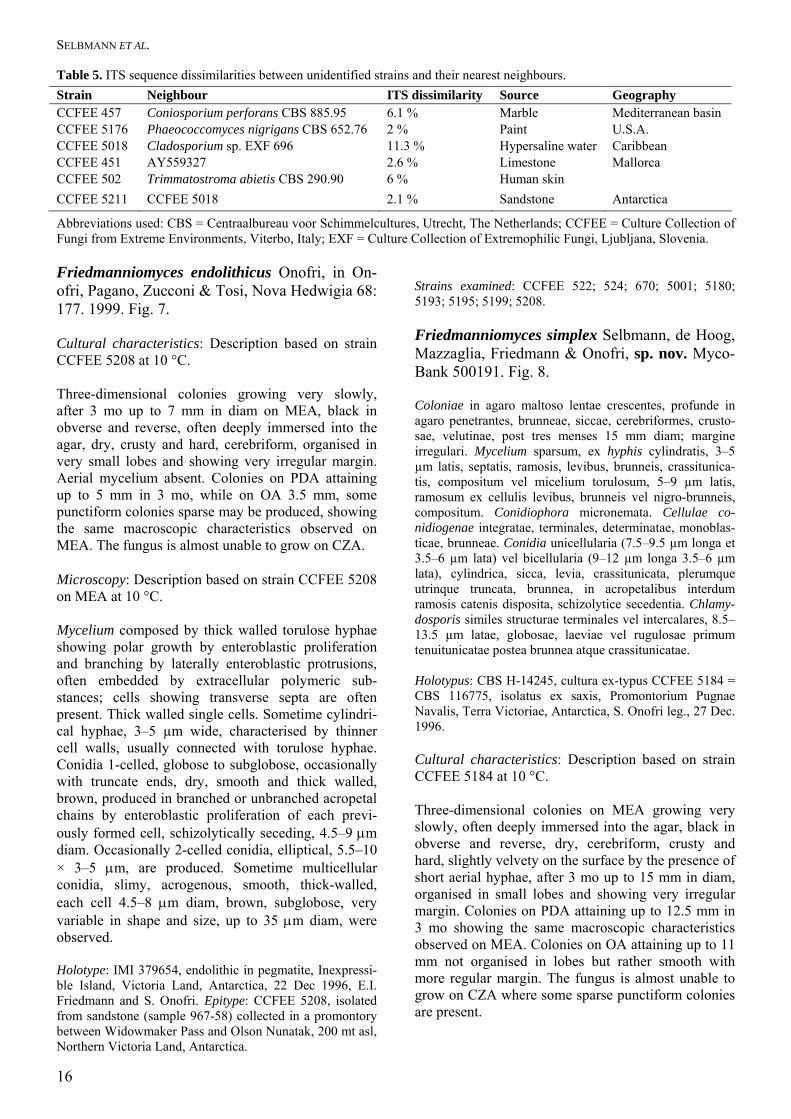
Table 5. ITS sequence dissimilarities between unidentified strains and their nearest neighbours. Strain Neighbour ITS dissimilarity Source Geography CCFEE 457 Coniosporium perforans CBS 885.95 6.1 % Marble Mediterranean basin CCFEE 5176 Phaeococcomyces nigrigans CBS 652.76 2 % Paint U.S.A. CCFEE 5018 Cladosporium sp. EXF 696 11.3 % Hypersaline water Caribbean CCFEE 451 AY559327 2.6 % Limestone Mallorca CCFEE 502 Trimmatostroma abietis CBS 290.90 6 % Human skin CCFEE 5211 CCFEE 5018 2.1 % Sandstone Antarctica
Abbreviations used: CBS = Centraalbureau voor Schimmelcultures, Utrecht, The Netherlands; CCFEE = Culture Collection of Fungi from Extreme Environments, Viterbo, Italy; EXF = Culture Collection of Extremophilic Fungi, Ljubljana, Slovenia. Friedmanniomyces endolithicus Onofri, in On-ofri, Pagano, Zucconi & Tosi, Nova Hedwigia 68: 177. 1999. Fig. 7. Cultural characteristics: Description based on strain CCFEE 5208 at 10 °C. Three-dimensional colonies growing very slowly, after 3 mo up to 7 mm in diam on MEA, black in obverse and reverse, often deeply immersed into the agar, dry, crusty and hard, cerebriform, organised in very small lobes and showing very irregular margin. Aerial mycelium absent. Colonies on PDA attaining up to 5 mm in 3 mo, while on OA 3.5 mm, some punctiform colonies sparse may be produced, showing the same macroscopic characteristics observed on MEA. The fungus is almost unable to grow on CZA. Microscopy: Description based on strain CCFEE 5208 on MEA at 10 °C. Mycelium composed by thick walled torulose hyphae showing polar growth by enteroblastic proliferation and branching by laterally enteroblastic protrusions, often embedded by extracellular polymeric sub-stances; cells showing transverse septa are often present. Thick walled single cells. Sometime cylindri-cal hyphae, 3–5 µm wide, characterised by thinner cell walls, usually connected with torulose hyphae. Conidia 1-celled, globose to subglobose, occasionally with truncate ends, dry, smooth and thick walled, brown, produced in branched or unbranched acropetal chains by enteroblastic proliferation of each previ-ously formed cell, schizolytically seceding, 4.5–9 μm diam. Occasionally 2-celled conidia, elliptical, 5.5–10 × 3–5 μm, are produced. Sometime multicellular conidia, slimy, acrogenous, smooth, thick-walled, each cell 4.5–8 μm diam, brown, subglobose, very variable in shape and size, up to 35 μm diam, were observed. Holotype: IMI 379654, endolithic in pegmatite, Inexpressi-ble Island, Victoria Land, Antarctica, 22 Dec 1996, E.I. Friedmann and S. Onofri. Epitype: CCFEE 5208, isolated from sandstone (sample 967-58) collected in a promontory between Widowmaker Pass and Olson Nunatak, 200 mt asl, Northern Victoria Land, Antarctica.
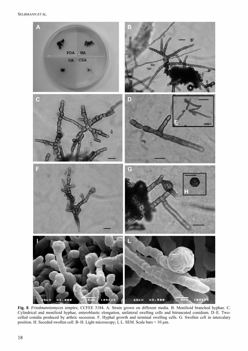
Strains examined: CCFEE 522; 524; 670; 5001; 5180; 5193; 5195; 5199; 5208. Friedmanniomyces simplex Selbmann, de Hoog, Mazzaglia, Friedmann & Onofri, sp. nov. Myco-Bank 500191. Fig. 8. Coloniae in agaro maltoso lentae crescentes, profunde in agaro penetrantes, brunneae, siccae, cerebriformes, crusto-sae, velutinae, post tres menses 15 mm diam; margine irregulari. Mycelium sparsum, ex hyphis cylindratis, 3–5 µm latis, septatis, ramosis, levibus, brunneis, crassitunica-tis, compositum vel micelium torulosum, 5–9 µm latis, ramosum ex cellulis levibus, brunneis vel nigro-brunneis, compositum. Conidiophora micronemata. Cellulae co-nidiogenae integratae, terminales, determinatae, monoblas-ticae, brunneae. Conidia unicellularia (7.5–9.5 µm longa et 3.5–6 µm lata) vel bicellularia (9–12 µm longa 3.5–6 µm lata), cylindrica, sicca, levia, crassitunicata, plerumque utrinque truncata, brunnea, in acropetalibus interdum ramosis catenis disposita, schizolytice secedentia. Chlamy-dosporis similes structurae terminales vel intercalares, 8.5–13.5 µm latae, globosae, laeviae vel rugulosae primum tenuitunicatae postea brunnea atque crassitunicatae. Holotypus: CBS H-14245, cultura ex-typus CCFEE 5184 = CBS 116775, isolatus ex saxis, Promontorium Pugnae Navalis, Terra Victoriae, Antarctica, S. Onofri leg., 27 Dec. 1996. Cultural characteristics: Description based on strain CCFEE 5184 at 10 °C. Three-dimensional colonies on MEA growing very slowly, often deeply immersed into the agar, black in obverse and reverse, dry, cerebriform, crusty and hard, slightly velvety on the surface by the presence of short aerial hyphae, after 3 mo up to 15 mm in diam, organised in small lobes and showing very irregular margin. Colonies on PDA attaining up to 12.5 mm in 3 mo showing the same macroscopic characteristics observed on MEA. Colonies on OA attaining up to 11 mm not organised in lobes but rather smooth with more regular margin. The fungus is almost unable to grow on CZA where some sparse punctiform colonies are present.

CRYPTOENDOLYTHIC BLACK FUNGI FROM ANTARCTIC DESERT
17
Fig. 7. Friedmanniomyces endolithicus. A. CCFEE 5208 grown on different media. B. CCFEE 670, hyphal growth, terminal swelling cells and EPS. C–D. CCFEE 5208, light microscopy of monilioid hyphae and clumps of cells, with cells showing transverse septa (D). E–F. CCFEE 5208, SEM of monilioid hyphae (E), and multicellular conidium (F). G–H. EPS formation in CCFEE 5001 (G) and CCFEE 5208 (H), observed with SEM. Scale bars = 10 µm.
SELBMANN ET AL.
18
Fig. 8. Friedmanniomyces simplex, CCFEE 5184. A. Strain grown on different media. B. Monilioid branched hyphae. C. Cylindrical and monilioid hyphae, enteroblastic elongation, unilateral swelling cells and bitruncated conidium. D–E. Two-celled conidia produced by arthric secession. F. Hyphal growth and terminal swelling cells. G. Swollen cell in intercalary position. H. Seceded swollen cell. B–H. Light microscopy; I, L. SEM. Scale bars = 10 µm.

CRYPTOENDOLYTHIC BLACK FUNGI FROM ANTARCTIC DESERT
19
Microscopy: Description based on strain CCFEE 5184 on MEA at 10 °C. Mycelium sparse, composed by cylindrical hyphae 3–5 µm wide, septate, branching by laterally enteroblas-tic protrusions, smooth, brown, thick walled or show-ing thinner cell wall when young. Often mycelium showing a slight torulose organisation, 5–9 µm wide, branched, smooth, brown to dark brown with dark-ened septa, often originating cylindrical hyphae. Conidiophores micronematous. Conidiogenous cells integrated, terminal, determinate, monoblastic, brown, thick-walled. 1-celled conidia 7.5–9.5 µm long and 3.5–6 µm wide or 2-celled conidia 9–12 µm long and 3.5–6 µm wide, cylindrical, dry, smooth, thick-walled, usually with truncated ends, brown, produced in branched or unbranched acropetal chains by entero-blastic proliferation of each previously formed cell, schizolytically seceding. Chlamydospore-like struc-tures produced by cell swelling in intercalary or more frequently in terminal position, 8.5–13.5 µm diam wide, globose, smooth or slightly roughened thin-walled at the first stage of formation turning slightly darker brown and thick-walled later. Holotype: CBS-H 14245, culture ex-type CCFEE 5184 = CBS 116775 (sample 967-24), Battleship Promontory, Victoria Land, Antarctica, from sandstone, S. Onofri. Friedmanniomyces simplex possesses conidio-phores micronematous, conidiogenous cells inte-grated, terminal, determinate, monoblastic, brown, thick-walled, and conidia 1- or 2-celled, smooth, thick walled usually with truncated ends, brown, produced in branched or unbranched acropetal chains by en-teroblastic proliferation of each previously formed cell, schizolytically seceding. In this respect it is similar to F. endolithicus, from which differs in slightly velvety colonies, conidia cylindrical or sub-globose, instead of globose to subglobose, and in lacking multicellular conidia. For its differences and similarities from the type species of Friedmanniomy-ces, F. simplex is here proposed as a new species in that genus. It was confirmed by molecular data. Cryomyces Selbmann, de Hoog, Mazzaglia, Friedmann & Onofri, gen. nov. MycoBank 500192. Ad fungos imperfectos, hyphomycetes pertinens. Coloniae in agaro maltoso lentae crescentes, compactae, cerebrifor-mes, nigrae. Mycelium, si adsit, meristematicum vel ex hyphis brevibus, septatis, interdum ramosis, brunneis, crassitunicatis, plus minusve torulosis, compositum. Hy-phae torulosae et mycelium meristematicum mutant in conidia unicellularia, globosa, sicca, acrogena ubi in acropetalibus torulosisque catenis disposita sunt, valde
crassi-tunicata, incrustata, schizolytice secedentia, entero-blastice germinantes. Teleomorphosis ignota. Species typica: Cryomyces antarcticus Selbmann, de Hoog, Mazzaglia, Friedmann & Onofri, sp. nov. Imperfect fungi, hyphomycetes. Colonies growing very slowly in MEA, compact, cerebriform, black. Mycelium, when present, meristematic or composed by short hyphae septate, scarcely branched, brown, thick walled, showing torulose organisation. Torulose hyphae and meristematic mycelium often resolved into 1-celled conidia globose, dry, acrogenous when organised in torulose acropetal chains, very thick walled, coated by fragmented incrustations, produced by schyzolitic secession, enteroblastically germinat-ing. Teleomorph: Unknown, phylogenetic affinity to the ascomycete order Dothideales. Cryomyces conidia are similar to chlamydospore-like structures described in Sarcinomyces (Her-manides-Nijhof 1977), but Cryomyces lacks conidia produced by annellated conidiogenous cells. Cryomyces antarcticus Selbmann, de Hoog, Mazzaglia, Friedmann & Onofri, sp. nov. Myco-Bank 500193. Fig. 9. Coloniae in agaro maltoso nigrae, compactae, cerebrifor-mes, lente crescentes, post tres menses usque ad 11 mm diam, margine irregulari, primum madidae, butyraceae, deinde siccae, induratae et friabiles. Mycelium, si adsit, rare meristematicum vel saepius ex hyphis brevibus, septatis, interdum ramosis, brunneis, crassitunicatis, plus minusve torulosis, 4–9 µm latis, compositum. Hyphae torulosae et mycelium meristematicum mutant in conidia unicellularia, plusminusve globosa, sicca, brunnea vel nigro-brunnea, valde crassitunicata, incrustata, rugosa, acrogena ubi in acropetalibus torulosis catenis disposita sunt, 4–12 μm diam, schizolytice secedentia. Holotypus: CBS H-14246, cultura ex-typus CCFEE 534 = CBS 116301, Linnaeus Terrace, Terra Victoriae, Antarc-tica, isolatus ex saxis, 1982, E.I. Friedmann leg. Cultural characteristics: Description based on strain CCFEE 534 at 15 °C. Three-dimensional colonies on MEA, black in ob-verse and reverse, compact, cerebriform, growing slowly, after 3 mo up to 11 mm in diam, lobed with irregular margin, initially glistening, moist, buttery becoming later crusty and hard, brittle in texture. Colonies on PDA attaining up to 9 mm in 3 mo, showing the same macroscopic characteristics ob-served on MA. Colonies on OA attaining up to 6 mm, organised in small lobes with very irregular margin.

SELBMANN ET AL.
20
Fig. 9. Cryomyces antarcticus. A. CCFEE 534 grown on different media. B, C. CCFEE 453, yeast-like organisation (B) and thick-walled, enteroblastically germinating mother cell (C). D. CCFEE 534, yeast-like organisation and thick-walled, cross-decorated enteroblastically germinating mother cell. E. CCFEE 515, monilioid hyphae. F–H. CCFEE 534, yeast-like organisa-tion and enteroblastic germination (F), monilioid hyphae (G) and seceded cells showing scars (H). B–E. Light microscopy; F–H. SEM. Bars indicate 10 µm.
CRYPTOENDOLYTHIC BLACK FUNGI FROM ANTARCTIC DESERT
21
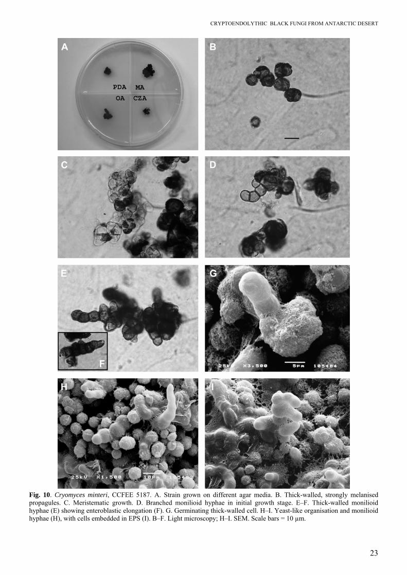
Colonies on CZA attaining up to 7 mm in 3 mo, flat with very irregular margin, diffusing black-brown pigment in the agar. Microscopy: Description based on strain CCFEE 534 on MEA at 10 °C. Mycelium often not observed, where present rarely meristematic or more frequently composed of short hyphae, closely septate, rarely branched, brown, thick walled, torulose, 4–9 μm wide. Torulose hypae and meristematic mycelium often resolved into 1-celled conidia globosa, dry, brown to dark brown, very thick walled, coated by fragmented incrustations, roughed, acrogenous when produced in torulose chains, 4–12 μm diam, produced by schyzolitic secession. Holotype: CBS-H 14246, culture ex-type CCFEE 534 = CBS 116301, Linnaeus Terrace, McMurdo Dry Valleys, Southern Victoria Land, Antarctica, on sandstone, E.I. Friedmann. Strains examined: CCFEE 453; 456; 514; 515; 534; 535; 536; 690. Cryomyces minteri Selbmann, de Hoog, Maz-zaglia, Friedmann & Onofri, sp. nov. MycoBank 500194. Fig. 10. Etymology: dedicated to David Minter, who first observed a strain from Antarctic rocks morphologically belonging to an undescribed fungal group. Coloniae in agaro maltoso nigrae, compactae, cerebrifor-mes, lente crescentes, post tres menses usque ad 11 mm diam, margine irregulari, primum madidae, butyraceae, deinde siccae, induratae friabilesque. Mycelium meris-tematicum vel rarius ex hyphis brevibus, 6–8 µm latis, septatis, interdum ramosis, primum dilute brunneis deinde brunneis vel nigro-brunneis, crassitunicatis, plus minusve torulosis, compositum. Mycelium meristematicum atque hyphae torulosae, mutant in conidia unicellularia, plusmi-nusve globosa, sicca, brunnea vel nigro-brunnea, valde crassitunicata, incrustata, rugosa, 7–11 μm diam, schizolytice secedentia. Holotypus: CBS H-14247, cultura ex-typus CCFEE 5187 = CBS 116302, Promontorium Pugnae Navalis, Terra Victo-riae, Antarctica, isolatus ex saxis, 28 Dec. 1996, S. Onofri leg. Cultural characteristics: Description based on strain CCFEE 5187 at 15 °C. Three-dimensional colonies on MEA black in obverse and reverse, compact, cerebriform, growing slowly, after 3 mo up to 11 mm in diam, lobed with irregular margin, initially glistening, moist, buttery becoming later crusty and hard, brittle in texture. Colonies on PDA after 3 mo attaining up to 10 mm, with the same
macroscopic characteristics observed on MEA. Colo-nies on OA attaining up to 8 mm, more flat with more regular margin. Colonies on CZA attaining up to 4 mm in 3 mo, organised in very small lobes and show-ing a very irregular margin. Microscopy: Description based on strain CCFEE 5187 on MEA at 15 °C. Mycelium often meristematic or, more rarely, com-posed by short hyphae 6–8 µm wide septate, scarcely branched, slightly pigmented when young and becom-ing brown to very dark brown later, thick-walled with torulose organisation. Meristematic mycelium and torulose hypae often resolved into 1-celled conidia globosa, dry, brown to dark brown, very thick walled, coated by fragmented incrustations, roughed, 7–11 μm diam, produced by schizolytic secession. Large amount of extracellular polymeric substances are present. Holotype: CBS H-14247; culture ex-type CCFEE 5187 = CBS 116302, Battleship Promontory, Victoria Land, Antarctica, weathered rocks (sample 967-26), 28 Dec. 1996, S. Onofri leg. Insufficiently characterised strains A number of single strains were not well characterised morphologically and were found to take isolated positions in SSU and ITS phylogeny. This would necessitate the description of separate genera for most of them. The present authors prefer to refrain from introduction of formal categories for these fungi until better insight is available into their phylogeny and taxonomic circumscription. An overview is presented in Fig. 11. Strain CCFEE 451 (Fig. 11A). Slow growing three-dimensional colonies, after 3 mo up to 10.5–12 mm on MEA, PDA and OA, 7 mm on CZA. Olive to greyish above, dark below, greyish brown above when cultivated on CZA, compact cushion shaped and felty in appearance with regular margin. Flat colonies when grown on CZA or OA. Hyphae deeply penetrating into the agar. Mycelium composed of thin walled cylindrical hyphae becoming later pale brown from 1.2 to 2.4 μm wide. Occasionally more or less spheri-cal, smooth- and thin-walled, terminal or intercalary swelling cells up to 7 μm in diam were observed, which may become delimited by a septum and turn slightly darker brown. These swellings do not secede. Strain CCFEE 457 (Fig. 11B). Three-dimensional cauliflower-like colonies, after 3 mo up to 16.5 mm on PDA, 10 mm on MEA and 6–7 mm on OA and CZA. Black above and below, compact, slightly felty with very irregular margin. Hyphae deeply penetrating into the agar. Mycelium composed by monilioid basically unbranched hyphae with rhomboidal cells often showing a transversal septum in the central part.
SELBMANN ET AL.
22
Sometime lateral budding cells are present. Conidia, with or without septum, are produced by arthric secession. Strain CCFEE 507 (Fig. 11C). Three-dimensional cauliflower-like colonies, after 3 mo up to 16.5 mm on PDA, 10 mm on MEA and 6–7 mm on OA and CZA. Black above and below compact slightly felty with very irregular margin. Hyphae deeply penetrating into the agar. Mycelium composed basically of cylin-drical hyphae often showing enlarged cells producing chlamydospores by isodiametric or unilateral swell-ing. Occasionally monilioid hyphae were formed generating conidia by arthric secession. Hyphae often embedded in large amounts of EPS. Strain CCFEE 5211 (Fig. 11D). Three-dimensional cauliflower-like colonies on both MEA and PDA, almost flat on OA. After 3 mo up to 15 mm on MEA and PDA, 17 mm on OA and 6–7 mm on CZA. Black above and below compact slightly felty on MEA and PDA, glistening on OA. Margin very irregular on MEA and CZA, regular on PDA and OA. Hyphae deeply penetrating into the agar. Mycelium is com-posed only by highly branched monilioid hyphae. Conidia are produced by arthric secession. Strain CCFEE 5018 (Fig. 11E). Slow growing cauliflower-like colonies, after 3 mo up to 13–15 mm on MEA, PDA and OA, 6 mm on CZA. From deep brown to black above, dark below, compact, lobated and felty in appearance with irregular margin on MEA, PDA and CZA, regular on OA. Rather flat colonies when grown on OA. Hyphae deeply penetrat-ing into the agar. Mycelium formed by regularly septated hyphae with slightly swollen and very regular cells with darkened septa producing single cells by arthric secession. Strain CCFEE 502 (Fig. 11F). Three-dimensional colonies on both MEA and PDA, almost completely flat on OA. After 3 mo up to 8.5 mm on MEA, 11 mm on PDA, 6.5 mm on OA and practically unable to grow on CZA. Deep brown above and black below compact slightly felty. Margin regular on MEA, regular and partially immersed on OA, very irregular and immersed on PDA. Hyphae deeply penetrating into the agar. Mycelium composed by cylindrical hyphae often thick-walled, strongly melanised and closely septate in the terminal part. Swollen cells and monilioid hyphae were also observed. Strain CCFEE 5176 (Figs 11G–H). Slow growing three-dimensional colonies, after 3 mo up to about 10–15 mm on MEA and PDA, 6 mm on OA, and unable to grow on CZA. Dark above and below, glistening, soft and elastic in texture with irregular margin. Yeast-like organisation, thick walled and strongly melanised cells producing new cells by budding in one or less frequently in two or more
different positions; cells often remaining coupled surrounded by large amount of EPS. DISCUSSION Seven clusters (A–G) could be observed on the basis of SSU phylogeny, of which order relationships were surmised on the basis of teleomorph relationships, as indicated in Fig. 4. The tree was rooted with group (G) containing members of Chaetothyriales: Capronia moravica (Petr.) E. Müller et al. (Untereiner et al. 1995), C. dactylotricha Untereiner et al. (Untereiner 1995) and Ceramothyrium linnaeae (Dearn.) S. Hughes (Con-stantinescu et al. 1989). This order is known to com-prise numerous species that are opportunists on hu-man patients. The teleomorphs are otherwise mainly found on plants and mushrooms (de Hoog 1999; Untereiner et al. 1999). The Mediterranean rock-inhabiting Coniosporium species have been associated with the order Chaetothyriales (Sterflinger et al. 1999), but appear more closely linked to Glyphium elatum (Grev. : Fr.) Zogg (Goree 1974), a member of Hysteriales which is known to have Trimmatostroma-like anamorphs (Sutton 1970). Glyphium species are mostly found on dead wood and branches (Sutton 1970). Strain CCFEE 457 from Antarctic rock pro-duces monilioid hyphae with arthric secession and leading to spherical cells similar to those of rock-inhabiting Coniosporium species (Sterflinger et al. 1997) and thus might be assigned to this genus. In its ITS sequence it is 6.1 % different from the ex-type strain of C. perforans, CBS 885.95 (Table 5). Re-cently, Phaeococcomyces chersonesos Bogomolova & Minter was described from marble in Crimea, Ukraine (Bogomolova & Minter 2003) which bears much similarity to Coniosporium. Group (F) contains Myriangium duriaei Mont. & Berk. that is classified in the order Myriangiales of the Dothideomycetes (Lumbsch & Lindemuth 2001). The order is not central in this subclass, and found remote from the Dothideales in the present paper. Group (F) does not contain any strains from the Antarctic. Cladosporium herbarum (Pers.) Link : Fr. (with Davidiella teleo-morph; Braun et al. 2003) is a sister group of this order. Cluster (F) and relatives thus marks the Do-thideomycetidae in the SSU phylogeny. The Dothideales sensu stricto are aggregated in groups (D) and (E). Group (E) is the complex of Aureobasidium pullulans (De Bary) Arn. This species is a typical coloniser of moist surfaces, both of plants and of inert materials.
CRYPTOENDOLYTHIC BLACK FUNGI FROM ANTARCTIC DESERT
23
Fig. 10. Cryomyces minteri, CCFEE 5187. A. Strain grown on different agar media. B. Thick-walled, strongly melanised propagules. C. Meristematic growth. D. Branched monilioid hyphae in initial growth stage. E–F. Thick-walled monilioid hyphae (E) showing enteroblastic elongation (F). G. Germinating thick-walled cell. H–I. Yeast-like organisation and monilioid hyphae (H), with cells embedded in EPS (I). B–F. Light microscopy; H–I. SEM. Scale bars = 10 µm.

SELBMANN ET AL.
24
Fig. 11. Unidentified meristematic species from Antarctic rock. A. CCFEE 451, SEM of hyphal growth and swelling cell at terminal position. B. CCFEE 457, SEM of monilioid hyphae and conidia produced by arthric secession. C. CCFEE 507, SEM of monilioid, cylindrical hyphae and EPS. D. CCFEE 5211, light microscopy of monilioid, branched hyphae and bitruncated conidia. E. CCFEE 5018, light microscopy of monilioid hyphae. F. CCFEE 502, SEM of monilioid and cylindrical hyphae. G, H. CCFEE 5176, coupled cells, observed with scanning (G) and light microscope (H). Scale bar = 10 µm.

CRYPTOENDOLYTHIC BLACK FUNGI FROM ANTARCTIC DESERT
25
A teleomorph Discosphaerina fulvida (F.R. Sander-son) Sivanesan has been associated to this species on the basis of ITS sequence similarity (Yurlova et al. 1999). The group further contains Delphinella strobili-gena (Desm.) Sacc. ex E. Müll. & Arx, Dothidea insculpta Wallr. and D. hippophaes (Passerini) Fuckel (Froideveaux 1972). They are typically found on dead twigs and as necrotrophs on woody plant remains and leaves (Froideveaux 1972; Barr 1972). Sydowia polyspora (Bref. & v. Tavel) E. Müll., the teleomorph of Hormonema dematioides Lagerb. & Melin is also found in this group; it is restricted to branches and leaves of Gymnosperms (Barr 1972). Group (E) does not contain any strains from the Antarctic. Group (D) is the group comprising e.g. Guignardia and Botryosphaeria, having mostly coelomycetous anamorphs (Van der Aa 1973). The numerous species known in these genera (Punithalingam 1974; Denman et al. 2000, Okane et al. 2001, Smith & Stanosz 2001, Zhou & Stanosz 2001) are plant pathogens (Farr et al. 1989). Also Scytalidium dimidiatum (Penzig) Sutton & Dyko, having a coelomycetous synanamorph in Nattrassia mangiferae (H. Syd. & Syd.) Sutton & Dyko (Sutton & Dyko 1989) is found in this group. It is a plant pathogen that also causes infections on human skin (de Hoog et al. 2000). Group (D) does not contain any strains from the Antarctic. The Dothideales of groups (D) and (E) are flanked by strains that are unresolved and showed relatively large distances to any of the sequences included in the tree, although alignment was still confident. Among these is Antarctic strain CCFEE 5176. Its unestab-lished position is illustrated by the neighbouring tropical species being Aliquandostipite khaoyaeinsis Inderbitzin, a member of the order Jahnulales (Inder-bitzin et al. 2001). The nearest neighbour of CCFEE 5176 at 2 % ITS distance is Phaeococcomyces nigri-cans (Rich & Stern) de Hoog, CBS 652.76 (Table 5), the phylogenetic position of which is unknown. Mor-phologically CCFEE 5176 is a thick-walled black yeast, as is P. nigricans. The Mediterranean rock-inhabiting species Sarcinomyces petricola Wollenzien & de Hoog (Wollenzien et al. 1997) is also unre-solved. Group (C) containing the Antarctic cryptendolithic genus Cryomyces is paraphyletic to Phaeotheca fissurella Sigler et al. (Zalar et al. 1999b), but at a very low bootstrap support. P. fissurella is known from a single strain on Pinus contorta in Canada (Sigler et al. 1981). Its nearest neighbour is Com-minutispora agavaciencis Ramaley (Ramaley 1996), having a peculiar anamorph of the Phaeotheca-type (Zalar et al. 1999b). Thus the genus Cryomyces seems to be ecologically as well as phylogenetically a mav-erick, without any obvious direct ancestor. This is underlined in the ITS data, where Cryomyces could not confidently be aligned to any fungus at all, only a
partial alignment being allowed (Fig. 5). Nevertheless C. antarcticus was consistently present in a restricted Antarctic environment with reproducible choice of habitat. The species thus must have been present in similar environments long before the Antarctic conti-nent reached its current position and cooled off, and subsequently adapted to the climate of Southern Victoria Land. The environment of the unknown plesiomorph may be located in high mountain tops of Andes, Alps or Himalaya, which hitherto have not been investigated for fungal life. This hypothesis implies that Cryomyces might be among the older genera of the Dothideomycetidae. Groups (A) and (B) are poorly resolved. Closest teleomorph species to Group (B) is Raciborskiomyces longisetosus (Volkart) M.E. Barr (Barr 1997); it belongs to the family Pseudoperisporiaceae of the Dothideomycetidae (Kirk et al. 2001) but was not clearly assigned to any specific order. The species is an epiphyte on woody or herbaceous plants. Group (B-I) contains another black epiphytic and epilithic species, Trimmatostroma abietis Butin & Pehl (Butin et al. 1996). A further Trimmatostroma species, T. salinum Zalar et al., which is halotolerant (1999c), is found at a considerable SSU distance (Fig. 4) and its ITS region could not be aligned with confidence (G.S. de Hoog, unpublished data). Judging from ITS data, Trimmatostroma abietis appears to be identical (or very close; Taylor et al. 2003) to T. microsporum Joanne E. Taylor & Crous (Taylor & Crous 2000), the anamorph of Mycosphaerella microspora Joanne E. Taylor & Crous. This is a member of the family Dothideaceae (Dothideales). Also some Mycosphae-rella species are fairly well alignable with ITS is used (Crous et al. 2001). In general, black epiphytic (and weakly pathogenic) on evergreen plants (Crous et al. 2004) and epilithic species seem to be mixed in the tree. Two unidentified Antarctic strains, CCFEE 5211 and 5018, were located near Cladosporium / Davidi-ella, outside the Dothideales. Both had monilioid hyphae with arthric secession; they could not be identified with certainty. Coccodinium bartschii in group (B–I) is a member of the order Capnodiales according to Kirk et al. (2001) but was assigned to the Dothideales by Winka et al. (1998) on the basis of SSU sequence data. In the present SSU tree so many taxa have been added that this relationship has become less apparent. The group B-II comprises a remarkable number of species origi-nating from stone, such as Capnobotryella renispora J. Sugiyama (Titze & de Hoog 1990), Pseudotaenio-lina globosa De Leo et al. (De Leo et al. 2003), as well as an unidentified Trimmatostroma species (CBS 100215) and T. abietis (CBS 618.84). The halophilic fungus Hortaea werneckii (Horta) Nishimura & Miyaji (Zalar et al. 1999a) is distantly related. The group also comprised two Antarctic rock fungi: CCFEE 451 and 507. CCFEE 451 is filamentous with

SELBMANN ET AL.
26
chlamydospore-like swellings that do not secede, while in CCFEE 507 additional monilioid hyphae falling apart into separate cells were observed. It is difficult to assign these cultures to any known hy-phomycete genus. Mycocalicium victoriae (C. Knight ex F. Wilson) Tibell, CBS 109863 in Group (A) has been assigned to the order Mycocaliciales even though Sikaroodi et al. (2001) placed Hobsonia santessonii Lowen & D. Hawksw. in the order Dothideales on the basis of SSU rDNA sequence data. Note that M. albonigrum (Nyl.) Tibell seems totally unrelated (Fig. 4). One would suppose that a misidentification or -isolation may have taken place, but both species are represented by several SSU and/resp. ITS sequences (Figs 4, 6). SSU group (A) consists of two clearly separate subunits, one of which (A-II) is the acidophilic genus ’Acido-myces’ (invalidly described by Baker et al. 2004). Subgroup (A–I) has strain CCFEE 502, a filamentous fungus without any obvious characteristics but mor-phologically different from Friedmanniomyces, in a basal position, and includes Mycocalicium victoriae and the lichenicolous species Hobsonia santessonii. The latter species occurs on the thalli of Peltigera scabrosa Th. Fr. on the ground in acid heathlands of northern Scandinavia (Lowen et al. 1986). The ulti-mate group comprises the Antarctic cryptendolithic genus Friedmanniomyces, with two species. The phylogenetic relationship of this group with a licheni-colous fungus (Lawrey & Diederich 2003) is signifi-cant since the Antarctic fungi tested have been iso-lated from lichen-dominated communities. Taking the poorly resolved groups of strains in groups (A) and (B) together, it may be remarked that the cryptendolithic genus Friedmanniomyces, colonis-ing substrates characterised by high salinity (Nishi-yama 1977) and low pH (de los Rios et al. 2003), is flanked by extremotolerant strains and species, grow-ing either in acidic or in salt environments, or are rock-inhabiting. Though these habitats have only scarcely been investigated, it seems likely that related species are rather widely distributed in suitable habi-tats. The phylogeny of Friedmanniomyces suggests that the Antarctic continent, before it moved to its current position by plate tectonics, harboured a fungal diversity similar to that found in other parts of the world, such as the Mediterranean, that are predisposed to endure extreme temperatures. Subsequently natural selection may have taken place leading to a small number of cold-tolerant species. This would mean that the phylogenetic history and adaptation of Friedman-niomyces began with the geographic isolation in the South pole and the cooling of Antarctica, in a period between about 60 and 30 million years ago. This is in contrast to Cryomyces, that had remained without any nearest neighbour with similar ecology. Thus it was already considerably distant before the cooling of Antarctica, and thus is supposed to have been pre-
existent as a psychrotolerant, cryptendolithic species. It now may occupy similar niches on nunataks and high mountain tops, which have presently not been investigated for the presence of cryptendolithic fungi. During glacial periods its distribution may have been extensive, while during interglacial periods it with-drew to refugia such as the Antarctic. An alternative hypothesis for both genera would be wind-dispersal of extant species from adjacent regions with temperate climates. This is possible over long distances for several groups of cryptogams, which has lead to similarities in floras on land masses that are located downwind (Muňoz et al. 2004). Colonisation of the harsh Antarctic environment would then be of very recent date. However, the considerable sequence deviation of both Friedmanniomyces and Cryomyces from known taxa suggests their local evolution as endemic Antarctic species. This matches with the supposition that cryptendothic fungi are dispersed inside scales of rock that have detached as a result of biogenous weathering (Friedmann 1982) and thus are dispersed only very locally. None of the taxa which are phylogenetically close to the Antarctic strains is lichenised; only one (Hobsonia santessonii) is lichenicolous. Gargas et al. (1995) suggested that lichenisation was acquired repeatedly, while Lutzoni et al. (2001) proved that it is an ancestral condition that has been repeatedly lost in the course of evolution, via lichenicolous behav-iour. By colonisation the no-longer lichenised fungi continue to take advantage of the alga or cyanobacte-rium without having to find a specific free-living photobiont with which to form a lichen thallus de novo each generation (Lutzoni et al. 2001). It is re-markable, however, that the cryptendolithic fungi studied here, consistently associated in the lichen-dominated community (Friedmann 1982), all are non-lichenised, although their habitat would certainly be favourable for such a life style. Their mode of nutri-tion is unknown. It is possible that some are licheni-colous, but at present no evidence is available. Never-theless consistency is observed in the main clades, as groups A, B and C show an overrepresentation of rock-inhabiting strains, either cryptendolithic species from the Antarctic or species causing biopitting in hot climates (Wollenzien et al. 1995, Sterflinger et al. 1997). More simple factors such as the pH of the rock might be an additional factor determining evolution, as the acidophilic genera Hobsonia and Acidomyces are directly adjacent to Friedmanniomyces. Cryptoen-dolithic black fungi have been isolated from the lichen-dominated community, while they seem to be absent from other kinds of cryptoendolithic communi-ties in the Antarctic. Therefore an association with lichens has been surmised (Nienow & Friedmann 1993). This relationship seems to be confirmed by our data, strains of Friedmanniomyces being phylogeneti-cally related (ranging from 98.82 and 99.17 % similar-

CRYPTOENDOLYTHIC BLACK FUNGI FROM ANTARCTIC DESERT
27
ity in SSU) to the lichenicolous fungus Hobsonia santessonii (Sikaroodi et al. 2001). Most of the Antarctic meristematic fungi analysed had previously been assigned to different, unnamed groups on the basis of morphological characteristics (Ocampo-Friedmann & Friedmann 1993). Table 1 lists that classification in comparison with results obtained by SSU rDNA sequencing in the present study. The genus Cryomyces proved to be morpho-logically recognisable by its peculiar yeast-like or-ganisation of the thallus. In contrast, most other Antarctic black meristematic fungi were identifiable on the basis of molecular studies only. Strains CCFEE 457, 502, and 522, for example, had grouped together in morphological Friedmann’s group 5, but were clearly separable in the SSU tree (Fig. 4). Strain CCFEE 522 belongs to the genus Friedmanniomyces, strain CCFEE 457 belongs to a different order. The majority of the meristematic Antarctic rock fungi are members of the Dothideomycetidae. This is not surprising since particularly the orders Dothidea-les and Capnodiales contain numerous stress-tolerant fungi with a focus on habitats with low water activity, such as colonisation of exposed inert surfaces, or halotolerance (de Hoog 1999). Many members have high degrees of morphological plasticity (Figueras et al. 1996, Yoshida et al. 1996) including yeast-like and meristematic ecotypes. Some of them, such as Phaeo-theca and Trimmatostroma maintain also a meris-tematic growth form during their entire life cycle, and in this respect they are similar to Friedmanniomyces and Cryomyces. Phaeotheca is an osmotolerant genus (Zalar et al. 1999b), while Trimmatostroma has been reported from hypersaline environments (Zalar et al. 1999c) in addition to wood and rock. It may be con-cluded that the reduction of cellular surface and increase of wall thickness are advantageous for the survival of both types of environmental stress. Other genera, such as Aureobasidium, Hormonema and Hortaea, are able to produce meristematic synana-morphs in addition to yeast-like morphology. Hortaea presently contains two distinct species, one halotoler-ant (Zalar et al. 1999a) and the other acidotolerant (Hölker et al. 2004). Hormonema is an opportunistic plant pathogen mainly found in the phyllosphere (Funk 1985). The surface of the leaf is slightly os-motic by the presence of sugary compounds but is low in nitrogen. The ecological similarity of this environ-ment to inert surfaces such as stone is exemplified by Trimmatostroma abietis Butin & Pehl, which is found on conifer needles as well as on rock (Butin et al. 1996). Aureobasidium pullulans (De Bary) Arn. an extremely common coloniser of moist surfaces, sug-ary leaves etc. (Yurlova et al. 1999), and converts from a yeast-like to its meristematic growth form in times of nutritional stress. All these fungi are phy-logenetically rather remote from each other within the order Dothideales. This strongly suggests that stress-
tolerance is an ecological mainstay in the entire order, being expressed differently in the individual species, but provoking a general response of meristematic growth and melanin production in all members of this ecological series. The fact that melanised meristematic cells are well-suited for survival of low water activity and exposure to drought on inert surfaces is further under-lined by the fact that similar stress-tolerant ecotypes produced in other orders of the fungal Kingdom have similar morphological traits. One Antarctic strain, CCFEE 457, clustered basally to the order Chaetothy-riales in the Coniosporium clade. The exact phyloge-netic position of this group is not well defined. It contains exclusively rock-inhabiting meristematic black fungi, isolated from monuments in the Mediter-ranean basin (Sterflinger et al. 1997, 1999). Several species are involved in biopitting of marble, which is a similar process of rock degradation as observed in the McMurdo Dry Valleys, Antarctica, but at an extremely high rather than at a low temperature. During the hot and dry Mediterranean Summers the Coniosporium species outcompete the lichens and contaminants that had accumulated and expanded during the moist and moderately warm Winter season, and the MCF then become the preponderant form of fungal life in these micro-habitats (Wollenzien et al. 1995). The endemic character of species in the genus Cryomyces in Southern Victoria Land is particularly unexpected, because there is no a priori reason why highly tolerant fungi should be less widespread than other genera, such as Friedmanniomyces. Strains of Cryomyces have thus far exclusively been isolated from samples collected from Linnaeus Terrace (C. antarcticus: CCFEE 514, 515, 534, 535, 536, 453), which is one of the most extreme locations of the McMurdo Dry Valleys, and from Battleship Promon-tory (C. minteri: CCFEE 5187). Both species of Cryomyces thus have a very limited distribution. A possible explanation is that sandstone in Northern Victoria Land is not as common as in the McMurdo Dry Valleys and rock substrata in that area are mostly represented by compact and scarcely porous volcanic and metamorphic rocks. Therefore cryptoendolithic colonisation can be largely affected by the scarce presence of suitable rocks for the cryptoendolithic microbial settlement. Unexpectedly the Antarctic black fungi show optimal growth at temperatures significantly higher than those to which they usually are exposed in their natural environment. The same phenomenon was observed previously in other microorganisms of the Antarctic cryptoendolithic community, although the net photosynthetic optimum for the community falls within the range of temperatures prevailing in nature (Nienow & Friedmann 1993). Thus the community as a whole is well adapted to low temperatures, even

SELBMANN ET AL.
28
though the individual members are not. The black fungi from the Antarctic show the ability to grow at a wide range of temperatures, which allow them to tolerate not only the extremes, but also the strong daily and seasonal thermal fluctuations characterising the environment of the McMurdo Dry Valleys. Fur-thermore, the ability of many Antarctic black fungi to grow around zero degrees allows them to grow during the period of highest photosynthetic activity of the community. The presence of the mesophilic-psychrotolerant strain CCFEE 507 in the Antarctic cryptoendolithic communities, with an optimum growth temperature of 25 °C, is remarkable. But also this fungus appeared to be able to grow at 0 °C. For that reason it may be considered to be particularly specialised to tolerate strong thermal fluctuations, being able to maintain a certain metabolic activity at low as well as relatively high temperature. Cryomyces species are able to grow at zero degrees but otherwise at a relatively wide range of temperatures (Table 2), which would allow them to thrive in a wider diversity of environments, or, more likely, enables to cope with the extreme shifts in temperature. Their unique and peculiar morphology underlines their suitability to inhabit extreme conditions: they mostly exhibit a very simple, yeast-like organisation with single cells which are rapidly transformed into strongly melanised, thick-walled propagules which probably are highly resistant to UV exposure and dehydration. Such a structure seems particularly suitable to survive in the prohibi-tive environmental conditions characterising Linnaeus Terrace. Germination and production of vegetative forms probably take place only during the less pro-hibitive conditions of the Antarctic Summer. There are at least three possible explanations for the endemic character of Cryomyces, the first of which (1) is a sampling effect mentioned above. Otherwise (2) the geographical isolation might be a consequence of a high level of specialisation to the extreme environment rather than simple tolerance, leading to inability to compete with fungi encountered in areas with milder conditions. Or, finally (3), it might be a founder effect, Cryomyces being phyloge-netically younger than Friedmanniomyces. The con-finement to Southern Victoria Land would then sug-gest a low rate of dispersion. This option seems less likely, given the strong local winds which transport rock dust over longer distances within the Antarctic. Also the phylogenetic distance of Cryomyces to any other fungus, suggesting an ancestral position as explained above, favours hypothesis (2). Strains belonging to Friedmanniomyces seem to be more widespread than Cryomyces, although more statistical evidence is needed. They colonise different localities in Northern Victoria Land and reach until ice-free zones situated in Southern Victoria Land, such as Battleship Promontory (CCFEE 5108, 5184) and Linnaeus Terrace (CCFEE 524). Their apparently
ubiquitous distribution in ice-free zones of the entire Victoria Land might be a consequence of a more effective dispersal mechanism than Cryomyces. Since meristematic fungi lack any specialised structures enhancing dispersal, they must be distributed pas-sively by the same dust storms as supposed for Cryo-myces. Particularly for Cryomyces this hypothesis seems to be supported by the finding in Battleship Promontory, a site located distantly from Linnaeus Terrace, of the only strain belonging to a different Cryomyces species. Probably the low rate of disper-sion enhances geographic isolation with accumulating genetic divergence. Phylogenetic data suggest a process of natural selection since the emergence of the Antarctic climate, as explained above. Friedmannio-myces species do not have a yeast-like organisation, but rather produce a more complicated, multicellular thallus. Highly resistant propagules as observed in Cryomyces are absent, but their lower thermal prefer-ences would suggest a good chance of survival in the most extreme zones of the McMurdo Dry Valleys like Linnaeus Terrace, despite a lower degree of morpho-logical adaptation. Our cryptoendolithic black fungi proved to be unable to produce extracellular acidic compounds under tested experimental conditions, although the possibility of acid production in nature due to respira-tion cannot be excluded. A chemical action on the rocks cannot be excluded since some strains proved to be able to decrease the pH in culture broths, possibly as a consequence of electrolyte assimilation rather than organic acid production. Similar features were observed also in other mi-crocolonial rock fungi (Wollenzien et al. 1995). Alternatively we may suppose that Antarctic cryp-toendolithic black fungi may enter pre-existing air-spaces inside the rocks, and subsequently are able to create spaces mechanically, as has been suggested for Mediterranean rock fungi (Sterflinger 2000). All Antarctic cryptoendolithic fungi tested show extremely slow growth, even under optimal condi-tions. The most promising survival strategy is the ability to produce structurally simple and highly resistant structures that allow them to conclude their life cycle during the very short time window when suitable conditions are prevailing. Rapid expansion is not necessary to have good chance of survival in the Antarctic rock of the Ross Desert, as there is little competition with other fungi due to the low biodiver-sity. This sheds some light on possible speciation processes in meristematic fungi. Some rapidly repro-ducing fungi are known to transform to a slow-growing meristematic form under conditions of stress, such as Exophiala dermatitidis (Kano) de Hoog at ultra-low pH (de Hoog et al. 1994). Several strains of this species maintain their slowly expanding, meris-tematic morphology, and are non-revertable. The same is noted even more frequently in the related
CRYPTOENDOLYTHIC BLACK FUNGI FROM ANTARCTIC DESERT
29
species E. phaeomuriformis (Matsumoto et al.) Matos et al. (Matsumoto et al. 1986). Meristematic and yeast-like / filamentous strains are identical in their rDNA sequence data (Matos et al. 2002). In other fungi some distance is noted between the counterparts. This is the case in, for example, the basidiomycetous yeast Trichosporon asteroides (Rischin) Ota with its meristematic form Fissuricella filamenta (Arnold & Ahearn) Pore et al. (Guého et al. 1992) and in the pleosporalean hyphomycete Alternaria infectoria Simmons and its meristematic counterpart Botryomy-ces caespitosus de Hoog & Rubio (de Hoog et al. 1997). This recurrent phenomenon of fixed meris-tematic mutants drifting away from their ancestral form is remarkable and might trigger processes of sympatric speciation. However, the fact that we wit-ness this type of speciation repeatedly within only a limited number of fungal orders suggests that drift is possible only in groups that are already able to shift their modus of growth from longitudinal to isodiamet-ric, which is linked to their thriving in dynamic eco-logical niches involving stages of environmental hostility. This ability is particularly found in the Dothideales, where psychrotolerant (but not thermo-tolerant), acidotolerant, halotolerant, xerotolerant next to epiphytic, epilithic and endolithic species are found. To some extent similar ecological traits are noted in the Chaetothyriales, harbouring thermotoler-ant and endolithic species, or these traits in combina-tion (Sterflinger et al. 1999). The evolutionary emer-gence of the cryptendolithic fungi most likely took place after sympatric speciation processes, by muta-tional shifts to one of the stages in a dynamic life cycle. This focus and subsequent loss of the remaining parts of the life cycle may have become necessary during a gradual change of the Antarctic climate. Another strategy is represented by the ability to produce antibiotic substances that allow the fungi to compete with other components of rock-inhabiting communities (algae, cyanobacteria and bacteria) and to contribute to maintenance of the stratification of the community (Ocampo-Friedmann & Friedmann 1993). As reported in Table 2, all fungi tested were able to grow on natural media (MEA, PDA, OA) but fre-quently unable to grow in synthetic, chemically de-fined medium (CZA). Apparently some essential growth factors are present in the media based on raw extracts. The ability to produce a certain amount of extracel-lular polysaccharides is another adaptation to extreme conditions, protecting the fungi against cycles of desiccation, freezing and thawing. Furthermore the compounds may contribute to create more suitable physical and chemical conditions in the microbial biofilm, which are much less severe than those of the external environment (De los Rìos et al. 2003). In conclusion, meristematic black fungi in the Antarctic are not only able to tolerate high levels of
stress, but they are even specialised to do so. Their simple morphology, scarcely differentiated structure and reduced life cycle make them particularly suited to live in the harsh environment of the McMurdo Dry Valleys. Notably they are UV-resistant by their cells being thick-walled and strongly melanised. Very similar behaviour has emerged in different orders of the fungal Kingdom, and in the Dothideales repeat-edly in independent phylogenetic lines, and therefore one might suppose that this type of behaviour is a general adaptation of fungal life to the conditions prevailing on the Antarctic. The fungi analysed in this study are among the very few fungi known to be endemic to Antarctica. Since such morphology and life cycles are found also in algae and cyanobacteria (Nienow & Friedmann 1993) we might conclude that this type of life cycle is an expression of optimal extremophily, and thus that if there has never been life on the planet Mars, where similar conditions prevailed during its early history, the life forms would probably look and behave similarly (Onofri et al. 2004). Eu-karyotic microorganisms show limits of growth and survival similar to those of Prokaryotes, allowing their survival under Martian conditions (Horneck 2000). There are also some endemic species in the genus Thelebolus (order Thelebolales; de Hoog et al. 2005) which are structurally much more complicated than the cryptendolithic fungi, but these possibly have a life cycle involving passage through warm-blooded animals. Almost all cryptendolithic fungi are psychro-philic but able to grow in a very wide range of tem-peratures, which suggests that the organisms are able to survive in a very variable environment character-ised by dramatic thermal fluctuations. They can be classified as generalists type-2 of Vincent (2000), i.e., having a very successful strategy of acclimation to changing conditions. The cryptendoliths thus can be characterised as the most extremotolerant fungi on Earth. They must have metabolic systems and en-zymes that still are active at very low temperatures. ACKNOWLEDGEMENTS This work has been conducted in the framework of the Italian National Program for Antarctic Research (PNRA). The authors would like to thank U.S. National Science Foundation (NSF) for financial support and assistance. Dr. D.W. Minter (CABI Bioscience U.K.) is acknowledged for his contributions to the morphological study of the organ-isms and Drs A. Aptroot (CBS, The Netherlands) and D.L. Hawksworth (Universidad Complutense, Madrid, Spain) for advice concerning lichenised fungi. We are indebted to these scientists and to Dr K. Sterflinger (University of Agricultural Sciences ACBR, Vienna, Austria) for critically reading the manuscript. A.H.G. Gerrits van den Ende, M. Reubzaet and A. Cegcel are thanked for technical assistance and S. Pagano for isolating some of the strains studied in this work.

SELBMANN ET AL.
30
REFERENCES Baker BJ, Lutz MA, Dawson SC, Bond, PL, Banfield JF
(2004). Metabolically active eukaryotic communities in extremely acidic mine drainage. Applied and Environ-mental Microbiology 70: 6264–6271.
Barr ME (1972). Preliminary studies on the Dothideales in temperate North America. Contributions from the Uni-versity of Michigan Herbarium 9: 523–638.
Barr ME (1997). Notes on some 'dimeriaceous' fungi. Mycotaxon 64: 149–171.
Bogomolova EV, Minter DW (2003). A new microcolonial rock-inhabiting fungus from marble in Chersonesos (Crimea, Ukraine). Mycotaxon 86: 195–204.
Braun U, Crous PW, Dugan F, Groenewald JZ & Hoog GS de (2003). Phylogeny and taxonomy of Cladosporium-like hyphomycetes, including Davidiella gen. nov., the teleomorph of Cladosporium s. str. Mycological Pro-gess 2: 3-18.
Bruns TD, Vilgalys R, Barns SM, Gonzalez D, Hibbett DS, Lane DJ, Simon L, Stickel S, Szaro TM, Weisburg WG (1992). Evolutionary relationships within the fungi: analyses of nuclear small subunit rRNA sequences. Mo-lecular Phylogenetics and Evolution 1: 231–241.
Butin H, Pehl L, Hoog, GS de, Wollenzien U (1996). Trimmatostroma abietis sp. nov. (hyphomycetes) and related species. Antonie van Leeuwenhoek 69: 203–209.
Compere AL, Griffith WL (1978). Production of high viscosity glucans from hydrolyzed cellulosics. Devel-opments in Industrial Microbiology Series 19: 601–607.
Constantinescu O, Holm K, Holm L (1989). Teleomorph-anamorph connections in Ascomycetes. 1–3. Stanhugh-esia (Hyphomycetes) new genus, the anamorph of Ce-ramothyrium. Studies in Mycology 31: 69–84.
Crous PW, Denman S, Taylor JE, Swart L & Palm ME (2004). Cultivation and diseases of Proteaceae: Leuca-dendron, Leucospermum and Protea. CBS Biodiversity Series 2: 1–226.
Crous PW, Hong L, Wingfield BD, Wingfield MJ (2001). ITS rDNA phylogeny of selected Mycosphaerella spe-cies and their anamorphs occurring on Myrtaceae. My-cological Research 105: 425–431.
Denman S, Crous PW, Taylor JE, Kang J-C, Pascoe I, Wingfield MJ (2000). An overview of the taxonomic history of Botryosphaeria, and a re-evaluation of its anamorphs based on morphology and ITS rDNA phy-logeny. Studies in Mycology 45: 129–140.
Farr DF, Bills GF, Chamuris GP, Rossman AY (1989). Fungi on Plants and Plant Products in the United States. APS Press, St. Paul, U.S.A.
Figueras MJ, Hoog GS de, Takeo K, Guarro J (1996). Stationary phase development of Trimmatostroma abi-etis. Antonie van Leeuwenhoek 69: 783–791.
Friedmann EI (1982). Endolithic microorganisms in the Antarctic cold desert. Science 215: 1045−1053.
Friedmann EI, McKay CP, Nienow JA (1987). The cryp-toendolithic microbial environment in the Ross Desert of Antarctica: Satellite-transmitted countinous Nano-climate Data, 1984 to 1986. Polar Biology 7: 273−287.
Friedmann EI, Weed R (1987). Microbial trace-fossil formation, biogenous and abiotic weathering in the Ant-arctic cold desert. Science 236: 703−705.
Froideveaux L (1972). Contribution à l'étude des Dothiora-cées (Ascomycètes). Nova Hedwigia 23: 679–734.
Funk A (1985). Foliar fungi on Western trees. Canadian Forestry Service, Victoria, Canada.
Gargas A, DePriest PT, Grube M, Tehler A (1995). Multi-ple origins of lichen symbioses in fungi suggested by SSU rDNA phylogeny. Science 268: 1492–1495.
Golubic S, Friedmann EI, Schneider J (1981). The lithobi-ontic ecological niche, with special reference to micro-organisms. Journal of Sedimentary Petrology 51: 475−478.
Goree H (1974). Glyphium in western Canada and United States. Canadian Journal of Botany 52: 1265–1269.
Guého E, Smith MTh, Hoog GS de, Billon-Grand G, Christen R, Batenburg-van der Vegte WH (1992). Con-tributions to a revision of the genus Trichosporon. An-tonie van Leeuwenhoek 61: 289–316.
Hermanides-Nijhof EJ (1977). Aureobasidium and allied genera. Studies in Mycology 14: 144–176.
Hölker U, Bend J, Pracht R, Müller T, Tetsch T, Höfer M, Hoog GS de (2004). Hortaea acidophila, a new acid-tolerant black yeast from lignite. Antonie van Leeuwen-hoek 86: 287–294.
Hoog GS de, ed. (1999). Ecology and evolution of black yeasts and their relatives. Studies in Mycology 43: 1–208.
Hoog GS de, Göttlich E, Platas G, Genilloud O, Leotta G, Brummelen J van (2005). Evolution and ecology of the genus Thelebolus in Antarctica. Studies in Mycology 51: 33–76
Hoog GS de, Guarro J, Figueras MJ, Gené J (2000). Atlas of Clinical Fungi. 2nd ed. Centraalbureau voor Schimmelcultures, Utrecht, The Netherlands & Univer-sitat Rovira i Virgili, Reus, Spain.
Hoog GS de, Takeo K, Yoshida S, Göttlich E, Nishimura K, Miyaji M (1994). Pleoanamorphic life cycle of Exo-phiala (Wangiella) dermatitidis. Antonie van Leeuwen-hoek 65: 143–153.
Hoog GS de, Uijthof JMJ, Gerrits van den Ende AHG, Figge MJ, Weenink XO (1997). Comparative rDNA di-versity in medically significant fungi. Microbiology and Culture Collections 13: 39–48.
Horneck G (2000). The microbial world and the case for Mars. Planetary and Space Science 48: 1053–1063.
Inderbitzin P, Landvik S, Abel-Wahab MA, Berbee ML (2001). Aliquandostipitaceae, a new family for two new tropical ascomycetes with unusually wide hyphae and dimorphic ascomata. American Journal of Botany 88: 52–61.
Kirk PM, Cannon PF, David JC, Stalpers JS (2001). Ains-worth & Bisby's Dictionary of the Fungi, 9th. ed. CAB International, Oxon, U.K.
Larena I, Salazar O, Gonzalez V, Julian MC, Rubio V (1999). Design of a primer for ribosomal DNA internal trascribed spacer with enhanced specifity for ascomy-cetes. Journal of Biotechnology 75: 187–194.
Lawrey JD, Diederich P (2003). Lichenicolous fungi: interactions, evolution and biodiversity. The Bryologist 106: 80–120.
Leo F de , Urzì C, Hoog GS de (2003). A new meristematic fungus, Pseudotaeniolina globosa. Antonie van Leeuwenhoek 83: 351–360.
CRYPTOENDOLYTHIC BLACK FUNGI FROM ANTARCTIC DESERT
31
Lowen R, Brady BL, Hawksworth DL, Paterson RRM (1986). Two new lichenicolous species of Hobsonia. Mycologia 78: 842–846.
Ludwig W, Strunk O, Westram R, Richter L, Meier H, Yadhukumar, Buchner A, Lai T, Steppi S, Jobb G, Forster W, Brettske I, Gerber S, Ginhart AW, Gross O, Grumann S, Hermann S, Jost R, Konig A, Liss T, Lussmann R, May M, Nonhoff B, Reichel B, Strehlow R, Stamatakis A, Stuckmann N, Vilbig A, Lenke M, Ludwig T, Bode A, Schleifer KH. (2004). ARB: a soft-ware environment for sequence data. Nucleic Acids Re-search 32: 1363–1371.
Lumbsch HT, Lindemuth R (2001). Major lineages of Dothideomycetes (Ascomycota) inferred from SSU and LSU rDNA sequences. Mycological Research 105: 901–908.
Lutzoni F, Pagel M, Reeb V (2001). Major lineages are derived from lichen symbiotic ancestors. Nature 411: 937–940.
Matsumoto T, Matsuda T, McGinnis MR, Ajello L (1984). Clinical and mycological spectra of Wangiella dermati-tidis infections. Mycoses 36: 145–155.
Matsumoto T, Padhye AA, Ajello L, McGinnis MR (1986). Sarcinomyces phaeomuriformis: a new dematiaceous hyphomycete. Journal of Medical and Veterinary My-cology 24: 395–400.
Matos T, Haase G, Gerrits van den Ende AHG, Hoog GS de (2002). Molecular diversity of oligotrophic and neuro-tropic members of the black yeast genus Exophiala, with accent on E. dermatitidis. Antonie van Leeuwen-hoek 89: 293–303.
Mendoza L, Karuppayil SM, Szaniszlo PJ. (1993). Calcium regulates in vitro dimorphism in chromoblastomycotic fungi. Mycoses 36: 157–164.
Morita RY (1975). Psychrophilic bacteria. Bacteriological Reviews 39: 144–167.
Muňoz J, Felicísimo ÁM, Cabezas F, Burgaz AR, Martinez I (2004). Wind as a long-distance dispersal vehicle in the Southern hemisphere. Science 304: 1144–1147.
Nienow JA, Friedmann EI (1993). Terrestrial lithophytic (rock) communities. In: Antarctic Microbiology (Fried-mann EI, ed.) Wiley-Liss, New York, U.S.A: 343−412.
Nishiyama T (1977). Studies on evaporite minerals from Dry Valley, Victoria Land, Antarctica. Antarctic Re-search 58: 171–185.
Ocampo-Friedmann R, Friedmann EI (1993). Biologically active substances produced by antarctic cryptoendolithic fungi. Antarctic Journal of the United States 28: 252−254.
Okane I, Nakagiri A, Ito T (2001). Identity of Guignardia sp. inhabiting ericaceous plants. Canadian Journal of Botany 79: 101–109.
Onofri S (1999). Antarctic microfungi. In: Enigmatic Microorganisms and Life in Extreme Environment (Seckbac J, ed). Kluwer Academic, Dordrecht, The Netherlands: 323−336.
Onofri S, Castagnola M, Rossi Espagnet S (1980). L’impiego della microscopia elettronica a scansione in micologia. Micologia Italiana 1: 29−32.
Onofri S, Pagano S, Zucconi L, Tosi S (1999). Friedmanniomyces endolithicus (Fungi, Hyphomycetes), anam.-gen. and sp.nov., from continental Antarctica. Nova Hedwigia 68: 175−181.
Onofri S, Selbmann L, Hoog GS de (2002). Molecular taxonomy of cryptoendolithic black yeasts from Continental Antarctica. 10th International Congress of Mycology (Abstracts), IUMS World Congresses “The World of Microbes”, Paris: 28.
Onofri S, Selbmann L, Zucconi L, Pagano S (2004). Ant-arctic microfungi as models for exobiology. Planetary and Space Science 52: 229–237.
Peer Y van de, Wachter R de (1994). Treecon for Windows: a software package for the construction and drawing of evolutionary trees for the Microsoft Windows environ-ment. Computers in Applied Bioscience 10: 569–570.
Punithalingam E (1974). Studies on Sphaeropsidales in culture. II. Mycological Papers 136: 1–63.
Ramaley AW (1996). Comminutispora gen. nov. and its Hyphospora gen. nov. anamorph. Mycologia 88: 132–136.
Ríos A de los , Wierzchos J, Snacho LG, Ascaso C (2003). Acid microenvironments in microbial biofilms of Ant-arctic endolithic microecosystems. Environmental Mi-crobiology 5: 231–237.
Sanger F, Nicklen S, Coulson AR (1977). DNA sequencing with chain-terminating inhibitors. Proceedings of the National Academy of Sciences of the USA 74: 5463−5467.
Schnitzler N, Peltroche-Llacsahuanga H, Bestier N, Zün-dorf J, Lütticken R, Haase G (1999) Effect of melanin and carotenoids of Exophiala (Wangiella) dermatitidis on phagocytosis, oxidative burst, and killing by human neutrophils. Infection and Immunity 67: 94–101.
Selbmann L, Onofri S, Fenice M, Federici F, Petruccioli M (2002). Production and structural characterization of the exopolysaccharide of the Antarctic fungus Phoma her-barum CCFEE 5080. Research in Microbiology 153: 585−592.
Sigler L, Tsuneda A, Carmichael JW (1981). Phaeotheca and Phaeosclera, two new genera of dematiaceous hy-phomycetes and a redescription of Sarcinomyces Lind-ner. Mycotaxon 17: 449–467.
Sikaroodi M, Lawrey JD, Hawksworth DL, DePriest PT (2001). The phylogenetic position of selected licheni-colous fungi: Hobsonia, Illosporium, and Marchandio-myces. Mycological Research 105: 453−460.
Smith DR & Stanosz GR (2001). Molecular and morpho-logical differentiation of Botryosphaeria dothidea (ana-morph Fusicoccum aesculi) from some other fungi with Fusicoccum anamorphs. Mycologia 93: 505–515.
Sterflinger K (1998). Temperature and NaCl-tolerance of rock-inhabiting meristematic fungi. Antonie van Leeuwenhoek 74: 271–281.
Sterflinger K (2000). Fungi as geologic agents. Geomicrobiology Journal 17: 97–124.
Sterflinger K, Krumbein WE (1995). Multiple stress factors affecting growth of rock inhabiting black fungi. Botanica Acta 108: 467−538.
Sterflinger K, Krumbein WE (1997). Dematiaceous fungi as a major agent for biopitting on Mediterranean mar-bles and limestones. Geomicrobiological Journal 14: 219−230.
Sterflinger K, Baere R de, Hoog GS de, Wachter R de, Krumbein WE, Haase G (1997). Coniosporium per-forans and Coniosporium apollinis, two new rock-inhabiting fungi isolated from marble in the Sanctuary

SELBMANN ET AL.
32
of Delos (Cyclades, Greece). Antonie van Leeuwenhoek 72: 349−363.
Sterflinger K, Hoog GS de, Haase G (1999). Phylogeny and ecology of meristematic ascomycetes. Studies in Mycol-ogy 43: 5–22.
Sutton BC (1970). Glyphium, leptothecium (Earle) comb. nov., G. schizosporum (Maire) Zogg, and their imper-fect states. Transactions of the British Mycological So-ciety 54: 255–264.
Sutton BC & Dyko BJ (1989). Revision of Hendersonula. Mycological Research 93: 466–488.
Taylor JE, Crous PW (2000). Fungi occurring on Pro-teaceae: new anamorphs for Teratosphaeria, My-cosphaerella and Lembosia, and other fungi associated with leaf spots and cankers of proteaceous hosts. Myco-logical Research 104: 618–636.
Taylor JE, Groenewald JZ, Crous PW (2003). A phyloge-netic analysis of Mycosphaerellaceae leaf spot patho-gens of Proteaceae. Mycological Research 107: 653–658.
Thauresia S, McNeil B (1994). Physiochemical factors affecting the formation of the biological response modi-fier scleroglucan. Journal of Chemical Technology & Biotechnology 59: 157–163.
Titze A, Hoog GS de (1990). Capnobotryella renispora on roof tile. Antonie van Leeuwenhoek 60: 35–42.
Untereiner WA (1995). Fruiting studies in species of Capronia (Herpotrichiellaceae). Antonie van Leeuwenhoek 68: 3–17.
Untereiner WA, Gerrits van den Ende AHG, de Hoog GS (1999). Nutritional physiology of species of Capronia. Studies in Mycology 43: 98–106.
Untereiner WA, Strauss NA, Malloch D (1995). A molecu-lar-morphotaxonomic approach to the systematics of the Herpotrichiellaceae and allied black yeasts. Mycologi-cal Research 99: 897–913.
Urzì C, Wollenzien U, Criseo G, Krumbein WE (1995). Biodiversity of the rock inhabiting microflora with spe-cial reference to black fungi and black yeasts. In: Mi-crobial Diversity and Ecosystem Function (Allsopp D, Colwell RR, Hawksworth DL, eds). CAB International, Wallington, U.K.: 289–302.
Van der Aa (1973). Studies in Phyllosticta I. Studies in Mycology 5: 1–110.
Van Uden N (1984). Temperature profiles of yeasts. Ad-vances in Microbiological Physiolology 25: 195−251.
Vincent W (2000). Evolutionary origins of Antarctic mycobiota: invasion, selection and endemism. Antarctic Science 12: 374–385.
Vishniac HS (1987). Psychrophily and systematics of yeast-like fungi. In: The expanding Realm of Yeast-like Fungi (Hoog GS de, Smith MTh, Weijman ACM, eds). Studies in Mycology 30: 389−402.
White TJ, Bruns T., Lee SB, Taylor JW (1990). Amplifica-tion and direct sequencing of fungal ribosomal RNA genes for Phylogenetics. In: PCR Protocols, A guide to methods and applications (Innis MA, Gelfand DH, Sninsky JJ, White TJ, eds). Academic, San Diego, U.S.A.: 315–322.
Winka K, Eriksson O, Bång Å (1998). Molecular evidence for recognising the Chaetothyriales. Mycologia 90: 822–830.
Winship PR (1989). An improved method for directly sequencing PCR amplified material using dimethylsul-foxide. Nucleid Acids Research 17: 1266.
Wollenzien U, Hoog GS de, Krumbein WE, Urzì C (1995). On the isolation of microcolonial fungi occurring on and in marble and other calcareous rocks. Science of the To-tal Environment 167: 287−294.
Wollenzien U, Hoog GS de, Krumbein WE, Uijthof JMJ (1997). Sarcinomyces petricola, a new microcolonial fungus from marble in the Mediterranean basin. Antonie van Leeuwenhoek 71: 281–288.
Yoshida S, Takeo K, Hoog GS de, Nishimura K, Miyaji M (1996). A new type of growth exhibited by Trimma-tostroma abietis. Antonie van Leeuwenhoek 69: 211–215.
Yuan ZQ, Mohammed C (1997). Two types of coelomy-cetes with appendaged conidia on stems of eucalypts from Australia. Mycological Research 101: 1531–1534.
Yurlova NA, Hoog GS de, Gerrits van den Ende AHG (1999). Taxonomy of Aureobasidium and allied genera. Studies in Mycology 43: 63–69.
Zalar P, Hoog GS de, Gunde-Cimerman N (1999a). Ecol-ogy of halotolerant dothideaceous black yeasts. Studies in Mycology 43: 38–48.
Zalar P, Hoog GS de, Gunde-Cimerman N (1999b). Taxon-omy of the endoconidial black yeast genera Phaeotheca and Hyphospora. Studies in Mycology 43: 49–56.
Zalar P, Hoog GS de, Gunde-Cimerman N (1999c). Trim-matostroma salinum, a new species from hypersaline water. Studies in Mycology 43: 57–62.
Zeppenfeldt G, Richard-Yegres N, Yegres F, Hernández R (1994). Cladosporium carrionii: hongo dimórfico en cactáceas de la zona endémica para la cromomicosis en Venezuela. Revista Iberoamericana de Micologia 11: 61–63.
Zhou S, Stanosz GR (2001). Relationships among Botryos-phaeria species and associated anamorphic fungi in-ferred from the analyses of ITS and 5.8S rDNA se-quences. Mycologia 93: 516–527.
Zucconi L, Pagano S, Fenice M, Selbmann L, Tosi S, Onofri S (1996). Growth temperature preferences of fungal strains from Victoria Land, Antarctica. Polar Bi-ology 16: 53–61.