fungal infection of mantis shrimp (oratosquilla oratoria ... filefungal infection of mantis shrimp...
TRANSCRIPT

Fungal Infection of Mantis Shrimp (Oratosquilla oratoria)Caused by Two Anamorphic Fungi Found in Japan
Pham Minh Duc Æ Kishio Hatai Æ Osamu Kurata ÆKozue Tensha Æ Uchida Yoshitaka Æ Takashi Yaguchi ÆShun-Ichi Udagawa
Received: 7 June 2008 / Accepted: 4 December 2008 / Published online: 24 January 2009
� Springer Science+Business Media B.V. 2009
Abstract Two fungal pathogens of the mantis
shrimp (Oratosquilla oratoria) in Yamaguchi and
Aichi Prefectures, Japan are described as the new
species Plectosporium oratosquillae and Acremonium
sp. (a member of the Emericellopsis marine clade).
Both fungi infect the gills of the mantis shrimp, which
become brown or black due to melanization. The
former species is characterized by its slow growth on
artificial seawater yeast extract peptone glucose
(PYGS) agar, pale yellow to pale orange or grayish
yellow colonies, short cylindrical solitary phialides
with a wavy tip, and one-celled ellipsoidal conidia.
Although lacking the two-celled conidia demonstrated
by the type species Plectosporium tabacinum, the
taxonomic placement of the new species was con-
firmed by DNA sequence analysis of the internal
transcribed spacer (ITS) region of ribosomal DNA
(ITS1, 5.8S rDNA and ITS2). Acremonium sp., the
other causal pathogen, differs from P. oratosquillae by
its fast growth on PYGS agar, pale orange to salmon-
colored colonies, long, slender conidiophores consist-
ing of solitary phialides with tips lacking an undulate
outline, and typically cylindrical conidia. Analysis of
ITS and b-tubulin gene sequences placed this fungus
within the phylogenetically distinct Emericellopsis
(anam. Acremonium) marine clade. Various physio-
logical characteristics of both pathogens were also
investigated. This is the first report of a fungal infection
found on the mantis shrimp in Japan.
Keywords Acremonium sp. � Fungal infection �Mantis shrimp � Oratosquilla oratoria �Plectosporium oratosquillae
Introduction
The mantis shrimp (Oratosquilla oratoria) is an
economically important and delicious culinary crusta-
cean species. One of the famous Japanese ‘sushi
dishes’ is made from the meat of the mantis shrimp.
This shrimp is found on mud in the coastal areas of
Japan and is the most dominant species, occupying
P. M. Duc � K. Hatai (&) � O. Kurata
Laboratory of Fish Diseases, Nippon Veterinary and Life
Science University, 1-7-1 Kyonan-cho, Musashino,
180-8602 Tokyo, Japan
e-mail: [email protected]
P. M. Duc
College of Aquaculture and Fisheries, Cantho University,
Cantho, Vietnam
K. Tensha � U. Yoshitaka
Yamaguchi Prefectural Fisheries Research Center Inland
Sea Division, Aio-Futoshima, Yamaguchi, Japan
T. Yaguchi
Medical Mycology Research Center, Chiba University,
1-8-1 Inohana, Chuo-ku, 260-8673 Chiba, Japan
S.-I. Udagawa
Tama Laboratory, Japan Food Research Laboratories,
6-11-10 Nagayama, Tama-shi, 206-0025 Tokyo, Japan
123
Mycopathologia (2009) 167:229–247
DOI 10.1007/s11046-008-9174-4

21.5% by biomass of the 20 most abundant species.
Production of the shrimp met with a decline from 1991
to 1999, and continues to decrease due to environmen-
tal variations [1, 2] and other harmful factors.
In marine environments, some genera of patho-
genic anamorphic fungi cause serious diseases of
various aquatic animals. In crustaceans, Fusarium
species cause black gill disease in the kuruma prawn
(Penaeus japonicus) in Japan [3–7], lobster (Homa-
rus americanus) in the USA [8] and tiger shrimp
(Penaeus monodon) cultured in Vietnam [9]. Other
anamorphic fungi are also pathogenic to crayfish
[10], red sea bream [11], ayu fry [12], and young
striped jack fish [13]. However, there are no reports
of fungal infections in mantis shrimps. Consequently,
we attempted to isolate the fungal pathogens from
wild mantis shrimp, collected from April 2005 to
December 2006 in Yamaguchi and Aichi Prefectures,
Japan and describe here the identification of the
isolates which infected the gills of this shrimp.
Materials and Methods
Mantis Shrimp Samples
Mantis shrimp of 17–30 g in body weight were
sampled monthly from April 2005 to December 2006
by trawl net in Yamaguchi and Aichi Prefectures,
along the Pacific side of central Japan. For each
collection, there were 4–23 mantis shrimp with a
fungal infection resulting in gill lesions (Figs. 1, 2).
Histological Findings
Shrimp tissues infected with fungi were fixed in 10%
phosphate buffered formalin solution. The gills,
muscle, and intestine specimens were embedded in
paraffin, sectioned at 3–4 lm, and stained using the
periodic acid Schiff (PAS).
Isolation of Fungi
Infected gills were washed three times in sterile
physiological saline (0.85% NaCl) and inoculated on
plates with artificial seawater yeast extract peptone
glucose (PYGS) agar [0.125% Bacto peptone,
0.125% Bacto yeast extract, 0.3% glucose, 1.2%
Difco agar, and 3.8% artificial sea salt (Aqua-
Ocean�, Japan Pet Drugs, Tokyo)]. Ampicillin and
streptomycin sulphate (500 lg ml-1) were added to
the medium to inhibit bacterial growth. Plates were
incubated for 2–4 days at 25�C and then subcultured
onto fresh PYGS agar plates. The single spore culture
method of Ho and Ko [14] was applied to obtain pure
isolates. The isolates were maintained at 25�C on
PYGS agar for subsequent experiments.
Identification of Fungi
The strains studied are listed in Tables 1, 2. Refer-
ence strains are named according to their original
designation in culture collections.
Morphology
Morphological characterizations of the isolates were
made from colonies grown on PYGS agar, potato-
dextrose agar (PDA, Nissui Phamaceutical Co. Ltd.,
Tokyo, Japan), potato-carrot agar (PCA), and Sa-
broud glucose agar (SGA, Nissui) after incubation at
20, 25, and 37�C. Slide cultures were also used to
examine microscopic features. For the observation of
conidial structures on slides, materials were stained
with cotton blue in lactic acid or B solution (Fungal
Flora Y Co., Ltd., Japan, commercial product for
fungal observations under a fluorescent microscope).
Capitalized color names and references in the
descriptions are those of Kornerup and Wanscher
[15]. For scanning electron microscope (SEM)
observations, blocks of agar (2–5 mm2) were cut
from sporulating colonies of the isolates and fixed in
2% glutaraldehyde in 0.1 M phosphate buffer and 1%
osmium tetroxide, dehydrated in a graded ethanol
series, then critical-point dried (JEOL JFD-310).
Specimens were mounted and sputter-coated with
silver (JFC-1600 Auto Fine Coaster) and examined
using SEM (JEOL JSM-6380 LV, Japan).
DNA Analysis and Sequencing
DNA was prepared using Gentorukun (Takara Bio Inc.,
Ltd., Otsu, Japan) from approximately 100-ll volume
of fungal mass cultured at 25�C for 14 days on PDA
slants. The ITS region (ITS1, 5.8S rDNA and ITS2)
and b-tubulin gene were sequenced directly from PCR
products using the primer pairs of ITS1 and ITS4 [16],
230 Mycopathologia (2009) 167:229–247
123

and Bt2a and Bt2b [17], respectively. PCR products
were sequenced using the BigDye Terminator Cycle
Sequencing Ready Reaction Kit (Applied Biosystems,
Foster City, CA, USA) and the ABI PRISM 3130
Genetic Analyzer (Applied Biosystems), according to
the manufacturer’s instructions.
Molecular Phylogenetic Analysis
DNA sequences were edited using ATGC ver. 4
sequence assembly software (Genetyx Co., Ltd.,
Tokyo, Japan), and alignment of the sequences was
performed using ClustalX software [18]. For
neighbor-joining (NJ) analysis [19], the distances
between sequences were calculated using Kimura’s
two-parameter model [20]. A bootstrap was con-
ducted with 1,000 replications [21].
Physiology
Effect of Culture Media on Mycelial
Growth of Isolates
Mycelial growth of the isolates was evaluated on seven
media: PYGS agar, PDA, oatmeal agar (OA, Difco
Laboratories, Detroit, MI, USA), SGA, cornmeal agar
Figs. 1-8 Mantis shrimp
with naturally fungal
infection. Fig. 1 Gills
showed brown
discoloration, bar = 2 cm;
Fig. 2 Gills disappeared due
to fungal infection,
bar = 1 cm; Fig. 3Numerous conidia of fungal
group B inside the gill
lamella, bar = 10 lm;
Fig. 4 Hyphae and conidial
germination (arrow) of
fungal group A inside the
gill lamella; Fig. 5 Hyphae
and conidia (arrow) and
fungal hyphae of group B
were encapsulated (arrow
head) at base of gill, PAS;
Fig. 6 Conidia of group B
invaded in the gill lamella,
PAS; Fig. 7 Hyphae of
fungal group A were
encapsulated (arrow) at
base of gill, PAS; Fig. 8Numerous hyphae (arrow
head) and conidia (arrow)
of fungal group A invaded
in the gill lamella, PAS
Mycopathologia (2009) 167:229–247 231
123

(CMA, Nissui), malt extract agar (MEA, 3% Fluka
malt extract, 0.5% Bacto peptone, 1.5% Difco agar),
and mycosel agar (MSA, Eiken Chemicals Co. Ltd.,
Tokyo, Japan). Plates containing each of the seven
media were inoculated according to Kim et al. [22], as
follows: about 5-mm diameter agar plugs were
removed with a sterile cork borer No. 2 from the
leading edges of pre-cultured colonies, and one such
plug was placed in the center of each 90-mm Petri dish
containing a medium. The colony diameter in each
plate was measured after incubation for 7 or 15 days at
25�C.
Effect of Temperature on Mycelial Growth of Isolates
The effect of temperature on mycelial growth was
evaluated on PYGS agar. Inoculated plates, as
described above, were incubated at 5, 10, 15, 20, 25,
30, 35, and 40�C. The colony diameter on each plate
was measured after incubation for 8 or 15 days.
Table 1 List of isolates from diseased mantis shrimp and the representative strains in this study (isolate group A)
Species, strain No. Accession No. ITS Substrate Location Date of isolation
Plectosporium sp.
NJM 0573 AB425973a Mantis shrimp (Oratosquilla oratoria) Yamaguchi, Japan Dec. 2005
NJM 0662 AB425974a Mantis shrimp (O. oratoria) Yamaguchi, Japan Apr. 2006
NJM 0665 AB425975a Mantis shrimp (O. oratoria) Yamaguchi, Japan May 2006
NJM 0667 AB425976a Mantis shrimp (O. oratoria) Aichi, Japan Apr. 2006
NJM 0678 AB425977a Mantis shrimp (O. oratoria) Aichi, Japan Sep. 2006
DAR76524 EU480709b
DAR76526 EU480705b
DAR76527 EU480691b
RM1-12 DQ993622c Marine sponge (Suberites zeteki)
142C EU480698b
Plectosporium alismatis (Oudem.) W.M. Pitt, W. Gams & U. Braun
CBS 113363 (DR76493) AY572016b Leaf spost (Alisma plantago-aquatica) The Netherlands
Gams 1B AY572020d
Gams 2A AY572015d
RH 01 AY258151d
RH 62 AY258150d
P. delsorboi Antignani & W. Gams
CBS 116708e DQ825986b Leaf spost (Curcuma alismatifola) Italy
P. tabacinum (J.F.H. Beyma) M.E. Palm, W. Gams & Nirenberg
NBRC 9985 AB425978a Violet (Vio odorata) Egypt
NBRC 30005 AB425979a River bottom mud
NRRL 20430 AF176952d
RH 126 AF258149d
00017 AJ246154d
380408 AJ492873d
(none number) DQ493934c
Out group
Verticillium nigrescens
IMI 044575 AJ292440
UHMH 6687 AF108473
V. albo-atrum
ATCC 44943 X60705
a This study; b Ash et al. [23]; c Wang et al. [24] ; d Pitt et al. [26]; e Zare et al. [25]
232 Mycopathologia (2009) 167:229–247
123

Table 2 List of isolates from diseased mantis shrimp and the representative strains in this study (isolate group B)
Species, strain No. Accession No. of GenBank Substrate Location Date of isolation
ITS b-tubulin
Acremonium sp.
NJM 0567 AB425971a AB436949a Mantis shrimp (O. oratoria) Yamaguchi, Japan May 2005
NJM 0672 AB425972a AB436950a Aichi, Japan Sep. 2006
A. fuci Summerbell, Zuccaro & W. Gams
CBS 112868 AY632653b AY632690b Fucus serratus, leaf Germany
A. potronii Vuill.
CBS 378.70F AY632655b AY632691b Salty soil France
A. tunakii
CBS 111360 AY632654b AY632689b
Emericellopsis donezkii Beliakova
CBS 489.71 AY632658b AY632674b Water (river) Ukraine
E. humicola (Cain). C. Gilman
CBS 180.56 AY632659b AY632675b Peat soil Canada
E. glabra (J.F.H. Beyma) Backus & Orpurt
CBS 119.40 AY632657b AY63673b Soil Netherlands
E. maritima Beljakova
CBS 491.71 AY632670b AY632686b Coastal seawater Ukraine
E. microspora Backus & W. Gams
CBS 380.62 AY632663b AY632679b Wet prairie soil USA
E. minima Stolk
CBS 871.68 AY632660b AY632676b Wheat field soil Germany
CBS 111361 AY632661b AY632677b Brown algae (Fucus serratus) Germany
CBS 190.55 AY632669b AY632685b Mangrove soil Mozambique
E. pallida Beljakova
CBS 624.73 AY632667b AY632683b Organic soil Canada
E. robusta Emden & W. Gams
CBS 489.73 AY632664b AY632680b Agriculture soil Netherlands
E. salmosynnemata Grosklags & Swift
CBS 382.62 AY632666b AY632682b Soil Belgium
E. stolkiae D.E. Dacidson & M. Christensen
CBS 159.71 AY632668b AY632684b Mud in saline lake USA
E. synnematicola P.N. Mathur & Thirumalachar
CBS 176.60 AY632665b AY632681b Soil India
E. terricola J.F.H. Beyma
CBS 229.59 AY632662b AY632678b Dried river mud Netherlands
Stanjeminium frisellum W. Gams, Schroers & M. Christensen
CBS 655.79 AY632671b AY632687b Soil from grassland USA
S. ochroroseum W. Gams & M. Christensen
CBS656.79 AY632672b AY632688b Soil from grassland USA
Out group
Bionectria aureofulvella
CBS 195.93 AF358226b AF358181b
Mycopathologia (2009) 167:229–247 233
123

Effect of Concentration of Artificial Seawater
on Mycelial Growth of Isolates in Group A
To examine the effect of the concentration of artificial
seawater (ASW) on mycelial growth of the isolates
(Plectosporium oratosquillae, see Results), the saline
component of PYGS agar was amended with ASW at
various concentrations (v/v): 100, 75, 50, 25, 12.5, and
0%. The salinity of ASW was prepared as 38%.
Inoculated plates, as described above, were incubated
at 25�C. The colony diameter in each plate was
measured after incubation for 15 days.
Effect of pH on Mycelial Growth of Isolates
The effect of pH on mycelial growth was evaluated in
sterile PYGS broth, in which the pH was adjusted to a
predetermined level by the addition of 1N solution of
HCl or NaOH and filtrated through a cellulose aseptic
filter (0.45-lm pore size; Toyo Roshi Kaisha Ltd.,
Tokyo, Japan) under aseptic conditions. The pH of
the medium was adjusted to 3.0, 4.0, 5.0, 6.0, 7.0, 8.0,
9.0, 10.0, 11.0, and 12.0. Each medium treatment
(5 ml) was poured into a sterile test tube. As with the
agar plates, inoculated test tubes were incubated at
25�C. The mycelial growth in each test tube was
evaluated by visual comparison with a PYGS broth
control (pH 7.0). The results from pH manipulation
were recorded as: (0) no growth; (1) weak growth; (2)
moderate growth; and (3) abundant growth. The
inoculations that did not grow at the lowest and
highest pH levels were washed in sterile distilled
water and then placed onto fresh PYGS agar to
determine whether the fungus was still viable.
Measurement of Growth Experiments
All experiments were carried out with three replicates
per treatment. The colony diameter on each plate was
measured by a vernier caliper after appropriate
incubation. The radial growth rate of each isolate
was calculated to give the final diameter value.
Results
Histopathology
The external clinical symptoms of a mantis shrimp
with fungal infection were brown or black discolor-
ation due to melanization and gill disappearance due
to deliquescence. Hyphae with septa and conidia
were observed in the infected gills under a micro-
scope (Figs. 3, 4). The results of histological
examination showed that fungal elements were
present in the gills (Figs. 5, 6). Fungal hyphae were
encapsulated in base of gills (Figs. 7, 8).
Mycology
Phylogenetic Analysis
Two groups of fungal strains were isolated coinci-
dentally from mantis shrimp samples: Group A
contained five isolates, NJM 0573, 0662, 0665,
0667, and 0678, which were easy to recognize in
culture by their slow growth on all media, and group
B contained two isolates, NJM 0567 and 0672,
differing from A by their fast growth and an
Acremonium-like colony appearance. The ITS
sequences for all the seven NJM isolates, and the
b-tubulin gene of the two NJM isolates in group B
were determined.
The sequences of the ITS region on the five NJM
isolates in group A were completely identical. Com-
parison of the ITS sequence by Blast searching (http://
www.ncbi.nlm.nih.gov) showed 100% similarity to a
sequence from Plectosporium sp. RM1-12, and the
Table 2 continued
Species, strain No. Accession No. of GenBank Substrate Location Date of isolation
ITS b-tubulin
B. oblongispora
CBS 100258 AF358248b AF358169b
B. samuelsii
CBS 699.97 AF358236b AF358190b
a This study; b Zuccaro et al. [27]
234 Mycopathologia (2009) 167:229–247
123

remaining highest matches were to sequences from
other Plectosporium sp. The phylogenic relationships
of the ITS region for the five NJM isolates and a total of
22 strains of related species of Plectosporium and
Verticillium (including an outgroup strain) [23–26]
were performed by NJ analysis (Fig. 9). The isolates of
Plectosporium were divided into two major clades; one
included the five NJM isolates (group A), Plectospo-
rium sp. RM1-12, and P. tabacinum isolates, and the
other included P. alismatis isolates and P. delsorboi
isolates. In the former, the five NJM isolates, the three
DAR isolates and the isolate RM1-12 formed a
monophyletic clade, with a sister group comprising
P. tabacinum supported by high bootstrap values, and
this topology was similar to that of Wang et al. [24]. On
the other hand, the latter were separated into P. al-
ismatis isolates and P. delsorboi isolates, which was in
accordance with the grouping of Zare et al. [25].
Therefore the five NJM isolates, three DAR isolates
and RM1-12 were distinguishable from other species
of Plectosporium on phylogeny.
The sequences of the ITS region and the b-tubulin
gene of the two NJM isolates in group B were
completely identical. The identity of the two isolates
on the ITS region was compared by Blast searching
which showed relative sequence homology to E.
maritima CBS 491.71 (100%), and NBRC 9603
(100%), E. minima CBS 190.55 (99.1%), E. pallida
CBS 624.73 (99.1%) and NBRC 9815 (99.1%), E.
stolkiae CBS 159.71 (98.7%), A. potronii CBS
379.70F (100%) and A. fuci CBS 112868 (98.0%).
Therefore the phylogenic relationships of the ITS
region for them and a total of 22 strains of
Emericellopsis, one of the teleomorphic genera found
in terrestrial and marine inhabitants, Acremonium
potronii, A. fuci, and Bionectia (including an out-
group strain) [27] were inferred from NJ analysis
(Fig. 10). In the tree, the strains of Emeriellopsis and
relative were divided into the four clades: clade A
comprising E. donezkii, E. humicola, E. microspora,
E. minima, E. robusta, E. stolkiae, and A. tubakii;
clade B comprising E. maritime, E. minima, E. pallid,
E. stolkiae, A. fuci, A. potronii, and the NJM isolates;
and two minor clades C and D.
In the b-tubulin gene analysis, the two isolates had
relative sequence homologies to E. maritima CBS
491.71 (97.2%), E. minima CBS 190.55 (98.2%),
E. stolkiae CBS 159.71 (98.2%), A. potronii CBS
379.70 (97.2%), and A. fuci CBS 112868 (96.0%),
and the NJ tree (Fig. 11) distributed the same
topology as that from the ITS region analysis.
Based on the sequences of the ITS region and the
b-tubulin gene, the two NJM isolates showed strong
similarities to E. maritima, E. minima ss.str. (CBS
V. albo-atrum ATCC 44943 (out group)V. nigrescens IMI44575
V. nigrescens UHMH6687Gams2ARH01CBS113363
Gams1BRH62
CBS11670800017RH126
380408NRRL20430NBRC998540658
NBRC30005148A
142CDAR76527DAR76526DAR76524
RM1-12NJM 0678NJM 0667NJM 0665NJM 0662NJM 0573
0.01
Plectosporium sp.
P. tabacinum
P. delsorboi
P. alismatis
52
7169
58
77
8897
84
98
8979
94
99
100
Fig. 9 Neighbor-joining
tree based on sequences of
the ITS region from 27
members of Plectosporiumsp. and relatives; neighbor-
joining algorithm with
1,000 bootstrap replicates
(values[50 are shown with
branches). Verticilliumalbo-atrum ATCC 44943
was taken as an outgroup
Mycopathologia (2009) 167:229–247 235
123

C
B
A
D
Clade(Zuccaro et al., 2004)
B. samuelsii CBS 699.97B. aureofulvella CBS 195.93
B. oblongispora CBS 10028598E. synnematicola CBS 176.60E. salmosynnemata CBS 382.62
S. grisellum CBS655.79S. ochroroseum CBS656.79A. fuci CBS 112868E. stolkiae CBS 159.71E. pallida CBS 624.73
E. minima CBS 190.55A. potronii CBS 379.70FE. maritima CBS 491.71Acremonium sp. NJM 0672Acremonium sp. NJM 0567
76
60
E. glabra CBS 119.40E. humicola CBS 180.56
E. donezkii CBS489.71E. terricola CBS 229.59
99
A. tubakii CBS111360E. minima CBS 871.68E. minima CBS 111361
E. robusta CBS 489.73E. salmosynnemata CBS 380.62
71
76
52
54
83
100
100
51
100
0.01
Fig. 10 Neighbor-joining
tree based on sequences of
the ITS region from 24
members of Emericellopsissp. and relatives; neighbor-
joining algorithm with
1,000 bootstrap replicates
(values[50 are shown with
branches). Bionectiraaureofulvella CBS 195.93,
B. oblongispora CBS
100285 and B. samuelsiiCBS 699.97 were taken as
an outgroup
B. samuelsii CBS 699.97B. aureofulvella CBS 195.93
B. oblongispora CBS 100285100E. salmosynnemata CBS 382.62
E. synnematicola CBS 176.60S. grisellum CBS655.79
S. ochroroseum CBS656.79E. pallida CBS 624.73
A. potronii CBS 379.70FE. stolkiae CBS 159.71
A. fuci CBS 112868E. minima CBS 190.55E. maritima CBS 491.71
Acremonium sp. NJM 0672Acremonium sp. NJM 0567
62
10087
53
100
100
100
86
E. humicola CBS 180.56E. donezkii CBS489.71E. glabra CBS 119.40
100
E. terricola CBS 229.59A. tubakii CBS111360
E. salmosynnemata CBS 380.62E. robusta CBS 489.73E. minima CBS 111361E. minima CBS 871.6898
84
99
100
0.01
C
B
A
D
Clade(Zuccaro et al., 2004)
Fig. 11 Neighbor-joining
tree based on sequences of
the b-tubulin gene from 24
members of Emericellopsissp. and relatives; neighbor-
joining algorithm with
1,000 bootstrap replicates
(values [ 50 are shown
with branches). Bionectiraaureofulvella CBS 195.93,
B. oblongispora CBS
100285 and B. samuelsiiCBS 699.97 were taken as
an outgroup
236 Mycopathologia (2009) 167:229–247
123

190.55, ex-type and unrelated to the other isolates), and
A. potronii. However, the isolates did not produce
ascospores on any of the tested media, so they were
phylogenetically considered as Acremonium species
with affinity to the Emericellopsis marine clade.
Sequence data for the seven NJM isolates and 2
NBRC strains of closely related species analyzed in
this study were submitted to the DNA Data Bank of
Japan (DDBJ) and the accession numbers were listed
in Tables 1, 2. The alignment was deposited in
TreeBASE (http://www.treebase.org/) with study
accession number S2236.
Specific Identification of Isolates in Group A
Based on the phylogenetic analysis of ITS sequences,
the five isolates of group A in this study were
provisionally identified as a Plectosporium species.
Upon further mycological examination on PYGS agar,
the five isolates NJM 0573, 0662, 0665, 0667, and 0678
showed the same colony characteristics: initially dull
white or slightly yellowish, moist, flat or slightly
sulcate, becoming pale orange or grayish yellow in
surface color and covered with trailing and interlacing
hyphae with age. The isolates failed to grow at
temperatures of 37�C and above. Examination of the
slide culture preparations revealed conidiogenous cells
that developed as discrete short phialides with a wavy
tip. The conidiogenous cells also reduced to phialidic
protrusions that developed as small pegs from the sides
of hyphae. Conidia were one-celled, hyaline, ellipsoi-
dal or cylindrical to obovoid, and smooth-walled.
A morphological comparison of isolate NJM 0662
with living cultures of two representative strains of
P. tabacinum, NBRC 9985 (also as ATCC 24393, IMI
151458, CBS 367.73) and NBRC 30005, revealed that
it belonged to a separate taxon, classifiable in the
genus Plectosporium in Table 3, [28, 29]. Isolates
NJM 0573, 0662, 0665, 0667, and 0678 were distinct
from three known species: P. tabacinum, P. alismatis
(Oudem.) W.M. Pitt et al., and P. delsorboi Antignani
& W. Gams [26, 29]. We propose that the new taxon,
P. oratosquillae, be established for these strains.
Plectosporium oratosquillae Pham Minh Duc, T.
Yaguchi & Udagawa, sp. nov. (Figs. 12, 13, 14, 15,
16, 17, 18).
Etymology. The specific epithet refers to the host.
Coloniae in agaro ‘‘PYGS’’ lente crescentes, posto
14 dies 20�C, 10–13 mm diam attingentes, planae vel
leviter sulcatae, ex mycelio vegetativo submerso
constantes, funiculosae, dilute flavae ad dilute auran-
tiacae; conidiogenesis moderata ad abunda; reversum
incoloratum vel dilute flavum. Mycelio ex hyphis
hyalinis ad dilute flavo-brunneis, ramosis, levibus,
septatis, 1–4 lm diam composito. Conidiophora ex
hyphis submersis vel superficialibus oriunda, solitar-
ia, simplicia vel parce ramosa, levia, hyalina.
Cellulae conidiogenae phialidicae, hyalinae, rectae
vel saepe sinuosae, cylindraceae vel subulatae,
6–18 9 2–4 lm, leves vel parum asperae, basaliter
septatae, collari minuto cylindraceo usque ad 2 lm
longo terminatae; tubi phialidici laterales, brevi-cyl-
indracei, 3–4 9 1.5–2 lm, saepe basi septo nulli.
Conidia unicellularia, hyalina, ellipsoidea vel cylindr-
acea vel obovoidea, interdum curvata, 3–10 9
2–4 lm, levia, guttulata, in capitulis mucidis connexa.
Chlamydosporae absentes.
Colonies on PYGS agar growing slowly, attaining
a diameter of 10–13 mm in 14 days at 20�C, flat or
slightly sulcate, consisting of a submerged vegetative
mycelium, with surface developing of trailing and
interlacing hyphae or ropes of hyphae, pale yellow
(4A3) to pale orange (5A3); conidiogenesis moderate
to abundant; exudate none; odor musty; reverse
uncolored or pale yellow (4A3). Colonies on
potato-carrot agar (PCA) growing more restrictedly,
attaining a diameter of 15–16 mm in 21 days at 20�C,
flat, thin, often centrally moist, with white cottony
aerial mycelium in marginal areas, grayish yellow
(4B4) to partially grayish orange (5B5); conidiogen-
esis light; reverse pale yellow (4A3). Colonies on
Sabouraud glucose agar (SGA) growing more
restrictedly, cottony, compact textured, rosy vina-
ceous; conidiogenesis sparse; reverse rosy buff.
Mycelium composed of hyaline to pale yellowish
brown, branched, smooth-walled, septate, 1–4 lm
diameter hyphae. Conidiophores arising from sub-
merged or superficial hyphae, solitary, unbranched or
sparingly branched, smooth-walled, hyaline. Conid-
iogenous cells phialidic, hyaline, straight or sinuous,
cylindrical or subulate, 6–18 9 2–4 lm, smooth-
walled or slightly roughened, basally septate, with a
wavy tip, collarette cylindrical, up to 2-lm deep,
sometimes inconspicuous; short phialidic protrusions
arising laterally from intercalary cells of hyphae as
small pegs, often lacking a basal septum, 3–4 9 1.5–
2 lm. Conidia 1-celled, hyaline, pale yellowish
brown in mass, ellipsoidal, cylindrical to obovoid,
Mycopathologia (2009) 167:229–247 237
123

sometimes curved, 3–10 9 2–4 lm, smooth-walled,
guttulate, accumulated in a slimy head at the tips of
phialides. Chlamydospores lacking.
Holotype: Dried colonies of IFM 56765 (NJM
0662), after 14 days growth on PYGS agar, deposited
in the herbarium of Medical Mycology Research
Center (MMRC), Chiba University, Chiba, Japan;
isolated from diseased mantis shrimp (Oratosquilla
oratoria), Yamaguchi Pref., Japan, April 2006.
Specific Identification of Isolates in Group B
Although the failure of teleomorph development in
culture was probably a result of unfavorable experi-
mental conditions, phylogenetic analysis of ITS and b-
tubulin sequences of the two isolates, NJM 0567 and
NJM 0672, was identified to Acremonium sp. (the
Emericellopsis marine clade). Their cultures on PYGS
agar developed rapidly, with salmon-colored (Fig. 19)
or pale orange (5A3) (Fig. 20), colonies attaining a
diameter of 70 mm after 10 days at 20�C. Subcultures
on PDA produced more or less floccose and rather
smaller colonies, attaining a diameter of 20 mm in
7 days at 20�C. Conidia were one-celled, hyaline,
cylindrical to ellipsoidal, straight or slightly curved,
smooth-walled and measured 4–10 9 2.5–3 lm
(Fig. 21), gathering in colorless slime at the tips of the
phialides. Arising from the hyphae, conidiophores were
macronematous and micronematous, simple or basiton-
ously branched, septate, smooth-walled, 28–45 9
2–3 lm, and borne a terminal phialide (Figs. 22, 23).
The mycelium was composed of hyaline, branched,
septate, smooth, 1–4-lm-wide hyphae (Fig. 24). Phia-
lide sometimes has septate (Fig. 25) and phialide apex
with a visible collarette (Fig. 26).
Isolates were deposited as Acremonium sp. in the
culture collection of the Medical Mycology Research
Center, Chiba University, as IFM 56765.
Physiology
Effect of Culture Media on Mycelial Growth
of Isolates
The radial mycelial growth rates of isolates in
P. oratosquillae and Acremonium sp. were evidently
affected by culture media (Table 4). PYGS agar was
apparently the most adequate for growth of all seven
isolates tested. Besides the exceptional cases of more
trace growth of the two isolates NJM 0662 and NJM
0678 on conventional media such as PDA, OA, and
CMA, isolates in P. oratosquillae failed to grow on
Table 3 Morphological comparison of P. oratosquillae with known Plectosporium species
Feature P. oratosquillae P. tabacinum P. alismatisa P. delsorboia
Colony diameter on
PYGSA at 20�C, 14 d
10–13 mm 42–48 mm ND ND
Condiogenesis on
PYGSA
Heavy Light ND ND
Colony diameter on
PDA at 25�C, 14 d
7–12 mm 58–65 mm 31–37 mm 63–91 lm
Conidiogenesis on PDA Light Heavy Heavy Heavy
Phialide length 6–18 lm 4–30 lm 12–35 lm 30–50 lm
Conidial shape Ellipsoidal Fusiform with rounded
tapering ends
Fusiform with strongly
tapering ends
Ellipsoidal
Conidial septation 0-Septate 0-1-Septate (1-septate
often [ 50%)
Mostly 1-septate Mostly 1-septate
Conidial size 3-10 9 2-4 lm 8-12 9 2-3 lm (1-septate) 13-19.5 9 2.5-3.5 lm 6-13 9 3-3.5 lm
Chlamydospores Absent Absent Present Absent
Pathogenicity Marine mantis
shrimp
Plants of various kinds and
cray fish (fresh-water fish)
Plants of Alismataceae Plants of
Zingiberaceae
Distribution Japan all over the world Africa, Asia, Europe, North
America, Oceania
Asia, Italy
a Data of P. alismatis and P. delsorboi from Pitt and Gams [28] and Antignani et al. [29], respectively
ND No data
238 Mycopathologia (2009) 167:229–247
123

Figs. 12–18Morphological
characteristics of isolates in
the group A. Fig. 12 Colony
of NJM 0662 on PYGS agar
at 25�C for 1 month;
Fig. 13 Colony of NJM
0667 on PYGS agar at 25�C
for 25 days; Fig. 14Phialide apex aggregating
elongate conidia, SEM;
Fig. 15 Short conidiophore,
slide cultured, then stained
with cotton blue, under a
light microscope,
bar = 10 lm; Fig. 16Elongate phialides and two
short phialidic protrusions
(arrows), slide cultured,
then stained with cotton
blue, under a light
microscope; Fig. 17Elongate conidiophore,
SEM; Fig. 18 Phialide apex
with collarette, SEM,
bar = 2 lm
Mycopathologia (2009) 167:229–247 239
123

MEA, MSA, and SGA, which are usually adequate
for most pathogenic and potentially pathogenic
fungus cultures. This finding on the vegetative
growth indicates that P. oratosquillae is an obligate
marine fungus that can complete its life cycle only in
marine environments. There is significant difference
Figs. 19–26Morphological
characteristics of isolates in
the group B. Figs. 19 and 20Colonies of NJM 0567 and
0672, both on PYGS agar at
25�C for 7 days; Fig. 21Obovoid to cylindrical
conidia, slide cultured, then
stained with B solution
(fungal flora Y company,
Japan), under a fluorescent
microscope, bar = 10 lm;
Fig. 22 Erect conidiophores
and conidia aggregating in a
slimy head on phialide
apex, slide cultured, then
stained with B solution
(fungal flora Y company,
Japan), under a fluorescent
microscope, bar = 10 lm;
Fig. 23 Branched
conidiophore, slide
cultured, then stained with
B solution (fungal flora Y
company, Japan), under a
fluorescent microscope,
bar = 10 lm; Fig. 24Hyphae aggregating in a
strand with septa, slide
cultured, then stained with
B solution (fungal flora Y
company, Japan), under a
fluorescent microscope,
bar = 8 lm; Fig. 25Phialides with septa
(arrows), SEM,
bar = 10 lm; Fig. 26Phialide apex with a visible
collarette (arrow), SEM,
bar = 5 lm
240 Mycopathologia (2009) 167:229–247
123

in the growth response of Acremonium sp. isolates
between PYGS agar and conventional media, and the
salinity influence of seawater is obvious.
Effect of Temperature on Mycelial Growth of Isolates
The rates of radial mycelial growth of the seven
isolates on PYGS agar are presented in Table 5. Most
isolates in P. oratosquillae grow slowly at 10�C and
rapidly at 20–25�C, but the decline in growth on each
side of the optimum temperature is unequal. They
have a very slow decline in their ability to growth at
10–15�C, whereas mycelial growth was hardly
observed at 30�C. The two isolates of Acremonium
sp. grow rapidly at 20–30�C, and the rates of radial
mycelial growth of the seven isolates on PYGS agar
were temperature dependent.
Effect of Concentrations of Artificial Seawater
on Mycelial Growth of Isolates in P. oratosquillae
The rate of radial mycelial growth of all the five
isolates belonging to P. oratosquillae followed sim-
ilar trends in response to changes in seawater
concentrations (Table 6). In comparison with maxi-
mum growth on 100% seawater agar, only slight
decreases in colony diameter were observed at 75%
seawater agar (its estimated salinity: 28.5%). There
is a general coincidence between salinity tolerance in
culture of the isolates and their occurrence in sea
environments: seawater from the Inland Sea, one of
the sampling locations of diseased mantis shrimp, has
a salinity range from 28.5 to 33.5%.
Effect of Initial pH on Mycelial Growth of Isolates
The effects of different pH levels on the vegetative
growth of isolates in P. oratosquillae and Acremonium
Table 4 Effect of culture media on mycelial growth of
isolates
Medium Colony diameter (mm)
Plectosporium oratosquillae Acremonium sp.
0573 0662 0665 0667 0678 0567 0672
PYGSA 17 16 9 16 9 60 49
PDA – 1 – – 1 27 28
MEA – – – – – 24 21
OA – 2 – 1 2 25 20
CMA – – – 1 2 31 26
SGA – – – – – 16 15
MSA – – – – – 8 3
PYGSA Peptone yeast extract glucose seawater agar, PDApotato dextrose agar, MEA malt extract agar, OA oatmeal agar,
CMA cormeal agar, SGA Sabroud glucose agar, MSA mycosel
agar
Plectosporium oratosquillae: Colony diameter was measured
at 25�C, 15 d after inoculation
Acremonium sp.: Colony diameter was measured at 25�C, 7 d
after inoculation
–: No growth
Table 5 Effect of temperature on mycelial growth of isolates
on PYGS agar
Temperature
(�C)
Colony diameter (mm)
Plectosporium oratosquillae Acremoniumsp.
0573 0662 0665 0667 0678 0567 0672
5 1 2 – – – 12 1
10 5 8 3 3 1 24 10
15 11 13 5 13 4 37 17
20 17 15 7 16 6 52 31
25 18 16 9 18 8 66 48
30 3 1 5 – 2 40 34
35 – – – – – 4 16
40 – – – – – – –
Plectosporium oratosquillae: Colony diameter was measured
at 25�C, 15 d after inoculation
Acremonium sp.: Colony diameter was measured at 25�C, 8 d
after inoculation
–: \1 mm in diameter or no growth
Table 6 Effect of artificial seawater (ASW) concentration on
mycelial growth of Plectosporium oratosquillae isolates
ASW Colony diameter (mm)
(%, v/v) 0573 0662 0665 0667 0678
100 17 16 9 16 10
75 17 14 6 14 9
50 9 11 5 9 7
25 6 7 3 2 4
12.5 5 2 2 1 2
0 – 1 – – –
Medium: Amended PYGS agar diluted by distilled water with a
range from 75 to 0% ASW concentration; Salinity of 100%
ASW was 38%
Colony diameter was measured at 25�C, 15 d after inoculation
–: \1 mm in diameter or no growth
Mycopathologia (2009) 167:229–247 241
123
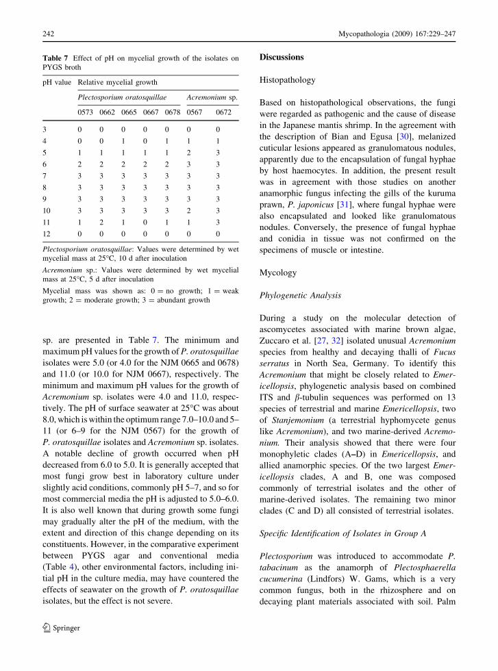
sp. are presented in Table 7. The minimum and
maximum pH values for the growth of P. oratosquillae
isolates were 5.0 (or 4.0 for the NJM 0665 and 0678)
and 11.0 (or 10.0 for NJM 0667), respectively. The
minimum and maximum pH values for the growth of
Acremonium sp. isolates were 4.0 and 11.0, respec-
tively. The pH of surface seawater at 25�C was about
8.0, which is within the optimum range 7.0–10.0 and 5–
11 (or 6–9 for the NJM 0567) for the growth of
P. oratosquillae isolates and Acremonium sp. isolates.
A notable decline of growth occurred when pH
decreased from 6.0 to 5.0. It is generally accepted that
most fungi grow best in laboratory culture under
slightly acid conditions, commonly pH 5–7, and so for
most commercial media the pH is adjusted to 5.0–6.0.
It is also well known that during growth some fungi
may gradually alter the pH of the medium, with the
extent and direction of this change depending on its
constituents. However, in the comparative experiment
between PYGS agar and conventional media
(Table 4), other environmental factors, including ini-
tial pH in the culture media, may have countered the
effects of seawater on the growth of P. oratosquillae
isolates, but the effect is not severe.
Discussions
Histopathology
Based on histopathological observations, the fungi
were regarded as pathogenic and the cause of disease
in the Japanese mantis shrimp. In the agreement with
the description of Bian and Egusa [30], melanized
cuticular lesions appeared as granulomatous nodules,
apparently due to the encapsulation of fungal hyphae
by host haemocytes. In addition, the present result
was in agreement with those studies on another
anamorphic fungus infecting the gills of the kuruma
prawn, P. japonicus [31], where fungal hyphae were
also encapsulated and looked like granulomatous
nodules. Conversely, the presence of fungal hyphae
and conidia in tissue was not confirmed on the
specimens of muscle or intestine.
Mycology
Phylogenetic Analysis
During a study on the molecular detection of
ascomycetes associated with marine brown algae,
Zuccaro et al. [27, 32] isolated unusual Acremonium
species from healthy and decaying thalli of Fucus
serratus in North Sea, Germany. To identify this
Acremonium that might be closely related to Emer-
icellopsis, phylogenetic analysis based on combined
ITS and b-tubulin sequences was performed on 13
species of terrestrial and marine Emericellopsis, two
of Stanjemonium (a terrestrial hyphomycete genus
like Acremonium), and two marine-derived Acremo-
nium. Their analysis showed that there were four
monophyletic clades (A–D) in Emericellopsis, and
allied anamorphic species. Of the two largest Emer-
icellopsis clades, A and B, one was composed
commonly of terrestrial isolates and the other of
marine-derived isolates. The remaining two minor
clades (C and D) all consisted of terrestrial isolates.
Specific Identification of Isolates in Group A
Plectosporium was introduced to accommodate P.
tabacinum as the anamorph of Plectosphaerella
cucumerina (Lindfors) W. Gams, which is a very
common fungus, both in the rhizosphere and on
decaying plant materials associated with soil. Palm
Table 7 Effect of pH on mycelial growth of the isolates on
PYGS broth
pH value Relative mycelial growth
Plectosporium oratosquillae Acremonium sp.
0573 0662 0665 0667 0678 0567 0672
3 0 0 0 0 0 0 0
4 0 0 1 0 1 1 1
5 1 1 1 1 1 2 3
6 2 2 2 2 2 3 3
7 3 3 3 3 3 3 3
8 3 3 3 3 3 3 3
9 3 3 3 3 3 3 3
10 3 3 3 3 3 2 3
11 1 2 1 0 1 1 3
12 0 0 0 0 0 0 0
Plectosporium oratosquillae: Values were determined by wet
mycelial mass at 25�C, 10 d after inoculation
Acremonium sp.: Values were determined by wet mycelial
mass at 25�C, 5 d after inoculation
Mycelial mass was shown as: 0 = no growth; 1 = weak
growth; 2 = moderate growth; 3 = abundant growth
242 Mycopathologia (2009) 167:229–247
123

et al. [33], Domsch et al. [34] reviewed the distribu-
tion and pathogenicity of this fungus, which has been
reported from a diverse range of hosts, including
basil, chard, cucumber, melon, peanuts, potato,
pumpkin, sugar beet, sunflower, tobacco, tomato,
etc. Smith-Kopperl et al. [35] also reported that P.
tabacinum was a pathogen of an invasive aquatic
weed (Hydrilla verticillata) in Florida, USA. Sur-
prisingly, it was isolated as a gill pathogen of crayfish
(Austropotamobius pallipes), native to British fresh-
waters [10]. A second species, P. alismatis (Oudem.)
W.M. Pitt, W. Gams & U. Braun with more falcate
conidia, is commonly found on leaf spots of Alisma
and other genera of the Alismataceae [26], and a third
species, P. delsorboi Antignani et al. [29], with
straight conidia, attacks the inflorescence of Curcuma
(Zingiberaceae).
In Japan, P. tabacinum has been isolated from the
aquatic sediment of a lake [36] and is more widely
known as a blight pathogen of pumpkin leaves and
stems, garden ranunculus seedlings, radish roots, etc.
[37, 38]. However, to our knowledge, there is no
previous report of the isolation of Plectosporium
species from a marine environment.
Specific Identification of Isolates in Group B
In the monographs of the Cephalosporium-like
hyphomycetes, by Gams [39, 40], Acremonium
species were classified into three sections: sect.
Acremonium with conidiophores consisting of
unbranched phialides without a visible periclinal
wall thickening and with collarette at the apex; sect.
Nectrioidea with generally wider hyphae and often
repeatedly branched conidiophores, the phialides
often undulate in the upper part, with a progressive
periclinal wall thickening and sometimes with a
narrow collarette; and sect. Gliomastix (often treated
as a separate genus) with usually pigmented conidia.
Acremonium has frequently been associated with
the teleomorphic species of Emercicellopsis (Bionec-
triaceae, Hypocreales) and several nectriaceous
genera. The Emercicellopsis species appear to be
primarily soil saprophytes that are characterized by
non-ostiolate globose ascomata with transparent
peridia but appearing dark because of the pigmented
ascospores within eight-spored and globose asci, and
one-celled, ovoid to ellipsoidal, dark-walled, wing-
like ornamented ascospores. They can be isolated
from various environments worldwide, including
cultivated and forest soils, rhizospheres, mycorrhizae,
and freshwater-, estuarine- and marine sediments
[33]. During a survey of fungi associated with mud in
marine, brackish lake and freshwater habitats in
Japan, Tubaki [41] noted that E. humicola, E.
microspora, and E. minima were isolated frequently,
and suggested that aquatic sediments might be
characteristic environments for some members of
this genus.
In the Acremonium anamorphs of Emericellopsis,
rather complex conidiophores occur that are unlike
other species of Acremonium. The branched conidio-
phores suggest Acremonium sect. Nectriodea, but
hyphae and conidiophores are thin-walled. The
morphological features of the two isolates in Group
B have a branching phialide (conidiophore) structure
and relatively large ellipsoidal conidia that are nearly
identical to those of the Emericellopsis anamorphs.
Furthermore, with the exception of ascomatal pro-
duction, sizes of the phialides and conidia of the
isolates suggest a possible relationship to E. minima
ss.str.; viz., the phialides of E. minima are 20–
30 9 2.0–2.5 lm and the conidia are 4–10 9 2.0–
3.5 lm (fide Stolk, [42]), whereas in the isolates,
phialides are 28–45 9 2.0–3.0 lm and conidia are 4–
10 9 2.5–3.0 lm.
In contrast, two marine-derived Acremonium,
A. potronii and A. fuci, are distinguished by differ-
ences in phialide and conidium morphology. In A.
potronii, phialides are 11–27 9 1.0–2.0 lm, and
conidia are obovoid, 2.1–4.0(–5.0) 9 1.3–2.5
(-3.0) lm [39, 40]. In A. fuci, phialides are 7.5–
23.5 9 1.0–2.2 lm, and conidia are obovoid to
broadly ellipsoidal, 10–15 9 3.0–6.0 lm [32].
Physiology
Effect of Culture Media on Mycelial Growth
of Isolates
These results might be important when considering
the ecological distribution of P. oratosquillae and
Acremonium sp. (the Emericellopsis marine clade).
According to Jones and Jennings [43] in their
comparative study on vegetative growth of ten
marine, one freshwater and two terrestrial species in
media made up with distilled water and seawater,
indicated that the response of the marine fungi varied
Mycopathologia (2009) 167:229–247 243
123

among species, and in only one instance was the dry
weight of vegetative mycelium significantly greater
in a distilled water medium. The non-marine fungi all
produced significantly greater dry weights of vege-
tative mycelium in the distilled water medium.
However, Jones and Jennings stated that a simple
comparison of growth in distilled water with seawater
media did not give a complete picture of the growth
of fungi under saline conditions. They further
suggested that the close association of marine fungi
with the sea cannot be ascribed alone to the influence
of seawater on vegetative growth. Jones studies some
of the factors influencing biodiversity of marine fungi
[44]. He found that salinity was important in affecting
species composition and that marine fungi could have
either a wide salinity tolerance (such as Asteromyces
cruciatus, salinity 0–100% sea water) or a narrow
salinity tolerance (such as Althrornia crouchi, salinity
40–100% sea water).
On the other hand, there is good evidence that
certain species may require seawater for fruiting. The
effects of salinity on sporulation and spore germina-
tion of a range of terrestrial, freshwater, and marine
fungi were investigated by Byrne and Jones [45, 46].
Their results showed that reproduction and spore
germination of the marine fungi tested were markedly
affected by high salinities. A similar trend was found
in our description for P. oratosquillae, which only
produced abundant conidia in the presence of
seawater, which supports the point that the fungus
is obligatorily marine.
Effect of Temperature on Mycelial Growth of Isolates
Boyd and Kohlmeyer [47] reported the growth of
three marine hyphomycetes on PYGS medium:
Asteromyves cruciatus Moreau & Moreau ex Henne-
bert, Sigmoidea marina Haythorn & Jones, and
Varicosporina ramulosa Meyers & Kohlm. Their
study demonstrated that growth tests of temperature
tolerance could be used to indicate possible geo-
graphic distribution patterns of marine fungi. For
example, V. ramulosa is a tropical–subtropical spe-
cies and its maximum growth occurs between 35 and
40�C, whereas A. cruciatus and S. marina are
temperate water species; the former species is
observed in the North Atlantic and North and South
Pacific and grows at 10�C, but with optimum growth
at 20�C and no growth at 35�C.
The data on temperature requirements for opti-
mum growth of Group A isolates showed that
P. oratosquillae is evidently a member of a temperate
water species. In fact, the occurrence of diseased
mantis shrimp was found to be in the temperate
seawater zone of the Pacific side of central Japan,
where the temperatures of bottom seawater in the
crustacean habitats (at Tokyo Bay) ranged from
12.7�C (the lowest in March) to 22.3�C (the highest
in September) [48]. Although the range of Group B
isolates extends further toward the subtropical zones
than that of P. oratosquillae, our results suggest that
Acremonium sp. (the Emericellopsis marine clade) is
also a temperate water species.
Effect of Concentrations of Artificial Seawater
on Mycelial Growth of Isolates in P. oratosquillae
Marine fungi are considered to be species that are
capable of indefinitely repeating their lifecycle in the
fully saline conditions of the sea, but which may also
continue to develop in other conditions, including
those of fluctuating salinity in estuaries. Tubaki and Ito
[49], surveyed fungi grown on submerged balsa wood
and bamboo blocks in brackish estuarine habitats
(salinities of their locations: 1.6–11.0%) in Japan.
They encountered 24 species of ascomycetous and
anamorphic fungi and examined the mycelial growth
of 17 brackish water-inhabiting species at different
concentrations of natural seawater in laboratory cul-
ture. They found that most of the brackish water-
inhabiting fungi exhibited a broad tolerance to
decreasing salinities, with vegetative growth and
reproduction the most markedly affected; 10 species
responded favorably to salinities above 10% seawater,
with maximum growth generally at 30–50% seawater,
and three species were characteristic in approximately
equal growth from 30 to 100% seawater. Moreover, in
the case of three true marine fungi, such as Corollos-
pora maritima (Linder) Kohlm., Leptosphaeria orae-
maris Linder, and Torpedospora radiata Meyers,
maximum growth occurred in 70 to 100% seawater.
Thus, the effects of salinities above 70% seawater on
the maximum growth of marine fungi could account
for their distribution in the higher salinity regions of an
estuary or in the sea. The vegetative growth of
P. oratosquillae exhibited a similar response to that
of these three marine fungi, because the colony
development of isolates in Group A was severely
244 Mycopathologia (2009) 167:229–247
123

inhibited at low salinity (12.5–25% seawater agar)
whereas they exhibited optimum mycelial growth at
75–100% seawater (almost normal seawater). This
may be important when considering the epidemic
distribution of the mantis shrimp pathogen.
Effect of Initial pH on Mycelial Growth of Isolates
The two isolates of Acremonium showed similar
trends in their response to pH changes as
P. oratosquillae (Table 7), but their visible growth
was observed over a wide pH range. They were also
more tolerant to high temperature (Table 5), suggest-
ing that this fungus is cosmopolitan, not only as a
pathogen of mantis shrimp but also as a saprophyte in
aquatic and terrestrial environments.
It is difficult to adequately state the general
influence of pH on fungal ecology, particularly as
its effects are modified by so many other environ-
mental factors. For example, although there is some
evidence that alkaline and acidic soils contain
different fungal communities, this does not always
correlate with the pH-growth responses of represen-
tative species of these communities in laboratory
experiments. Reports on the distribution of P. tabac-
inum were related almost exclusively to cultivated
land [33]. By means of distribution studies using
alkaline and slightly acidic media [i.e., alkaline CMA
(pH 9.7) and CMA (pH 6.0)], Nagai et al. [50] noted
that P. tabacinum was isolated only on alkaline CMA
from weakly acidic forest soil (pH 5.0–6.0) in Japan.
Even though most plant pathogens grow best in
media with an initial pH of 5.0–6.5, and mycelial
growth of P. tabacinum occurred over a range of pH
4–8 [51], Nagai et al. confirmed that optimum growth
of the Japanese isolate of P. tabacinum occurred at
pH 7.0 on MEA. They also showed that many
Acremonium species were isolated from Indonesian
alkaline soil, and the growth patterns of these
alkalophilic isolates were the same as that of the
Acremonium isolate from Japanese acidic soil.
Conclusion
The Japanese mantis shrimp (O. oratoria) lives on the
mud of the lower intertidal zone in coastal waters
around Japan. The crustacean is usually found in
U-shaped burrows in soft mud, swimming along the
surface of the intercoastal waterway at night.
Since the 1980s, significant case reports on the
occurrence of black gill disease and disease-associ-
ated mortality of the mantis shrimp have increased
throughout the Pacific coast side of Japan. Even
though there has been considerable concern over the
disease, the cause has yet to be unequivocally
identified. Thus, two hyphomycetes have been iso-
lated from the diseased samples, collected from
Yamaguchi and Aichi Prefectures on the Pacific coast
side of Japan and are taxonomically described as
etiological agents: P. oratosquillae and Acremonium
sp. (a member of the Emericellopsis marine clade).
P. oratosquillae, a new obligate marine fungus, is
characterized by very slow growth on PYGS agar
with optimum growth at 20–25�C, while the culture
of Acremonium sp. displays some different responses
to temperature (optimum: 20–30�C), salinity toler-
ance, and acidity (optimum pH: 5–10). There are no
substantial ecological data on P. oratosquillae in the
coastal zone [52], but numerous fungi, including
Emericellopsis species, are more common in the
coastal sediments of high organic matter than in those
of lower organic content [36, 43].
Acknowledgments Pham Minh Duc would like to thank the
Vietnamese government through ‘‘Project 322’’ for financial
support and the opportunity for his PhD thesis study in Japan.
References
1. Kodama K, Aoki I, Shimizu M. Long-term changes in the
assemblage of demersal fishes and invertebrates in relation to
environmental variations in Tokyo Bay. Jpn Fish Manag Ecol.
2002;9:303–13. doi:10.1046/j.1365-2400.2002.00313.x.
2. Kodama K, Shimizu T, Aoki I. Possible factors causing the
fluctuation of the recruitment of Japanese mantis shrimp
Oratosquilla oratoria in Tokyo Bay. Bull Kanagawa Pre-
fect Fish Res Inst. 2003;8:71–6 (in Japanese).
3. Ishikawa Y. A fungus caused black gill condition in cultured
kuruma prawn. Fish Pathol. 1968;3:34–49 (in Japanese).
4. Egusa S, Ueda T. A Fusarium sp. associated with black gill
diseases of the kuruma prawn, Penaeus japonicus Bate.
Bull Jpn Soc Sci Fish. 1972;38:1253–60.
5. Hatai K, Egusa S. Studies on the pathogenic fungus asso-
ciated with black gill disease of kuruma prawn, Penaeusjaponicus II: some of the note on the BG-Fusarium. Fish
Pathol. 1978;12:225–31 (in Japanese).
6. Momoyama K. Distribution of the hyphae in kuruma
prawn, Penaeus japonicus infected with Fusarium solani.Fish Pathol. 1987;22:15–23 (in Japanese).
7. Khoa LV, Hatai K, Yuasa A, Sawada K. Morphology and
molecular phylogeny of Fusarium solani isolated from
kuruma prawn, Penaeus japonicus with black gills. Fish
Pathol. 2005;40:103–9.
Mycopathologia (2009) 167:229–247 245
123
8. Lightner DV, Fontaine CT. A mycosis of the American lob-
ster Homarus americanus caused by Fusarium sp. J Invertebr.
1975;25:239–45. doi:10.1016/0022-2011(75)90074-9.
9. Khoa LV, Hatai K, Aoki T. Fusarium incarnatum isolated
from black tiger shrimp, Penaeus monodon Fabricius, with
black gill disease cultured in Vietnam. J Fish Dis.
2004;27:507–15. doi:10.1111/j.1365-2761.2004.00562.x.
10. Alderman DJ, Polglase JL. Fusarium tabacinum (Beyma)
Gams as a gill parasite in the crayfish, Austropotamobiuspallipes Lereboullet. J Fish Dis. 1985;8:249–52. doi:
10.1111/j.1365-2761.1985.tb01222.x.
11. Hatai K, Kubota S, Kida N, Udagawa S. Fusarium oxy-sporum in red sea beam, Pagrus sp. J Wildlife Dis.
1986;22:570–1.
12. Hatai K, Fujimaki Y, Egusa S. A visceral mycosis in ayu
fry, Plecoglossus altivelis Temminck & Schlegel, caused
by a species of Phoma. J Fish Dis. 1986;9:111–6. doi:
10.1111/j.1365-2761.1986.tb00989.x.
13. Munchan C, Kurata O, Hatai K, Hashiba N, Nakaoka N,
Kawakami H. Mass mortality of young striped jack,
Pseudocaranx dentex caused by a fungus Ochroconishumicola. Fish Pathol. 2006;41:179–82. doi:10.3147/jsfp.
41.179.
14. Ho WC, Ko WH. A simple method for obtaining single-
spore isolates of fungi. Bot Bull Acad Sin. 1997;38:41–4.
15. Kornerup A, Wanscher JH. Methuen handbook of colour.
2nd ed. London: Eyre Methuen; 1978.
16. White TJ, Bruns TD, Lee SB, Taylor JW. Amplification
and direct sequencing of fungal ribosomal DNA for phy-
logenetics. In: Innis MA, Gelfan DH, Sninsky JJ, White TJ,
editors. PCR protocols: a guide to the methods and appli-
cation. San Diego: Academic Press; 1990. p. 315–22.
17. Glass NL. Donaldson GC: development of primer sets
designed for use with the PCR to amplify genes from fil-
amentous ascomycetes. Appl Environ Microbiol.
1995;61:1323–30.
18. Thompson JD, Gibson TJ, Plewniak , Jeanmpugin F,
Higgins DG. The clustal X windows interface: flexible
strategies for multiple sequence alignment aided by quality
analysis tools. Nucleic Acids Res. 1997;24:4876–82. doi:
10.1093/nar/25.24.4876.
19. Saitou N, Nei M. The neighbor-joining method: a new
method for reconstructing phylogenetic trees. Mol Biol
Evol. 1987;4:406–25.
20. Kimura M. A simple method for estimation evolutionary
rate of base substitutions through comparative studies of
nucleotide sequences. J Mol Evol. 1980;16:111–20. doi:
10.1007/BF01731581.
21. Felsenstein J. Confidence limits on phylogenies: an
approach using the bootstrap. Evolution. 1985;39:783–91.
doi:10.2307/2408678.
22. Kim YK, Xiao CL, Rogers JD. Influence of culture media
and environmental factors on mycelial growth and pycnidial
production of Sphaeropsis pyriputrescens. Mycologia.
2005;97:25–32. doi:10.3852/mycologia.97.1.25.
23. Ash GJ, Chung YR, Mckenzie C, Cother EJ. A phyloge-
netic and pathogenic comparison of potential biocontrol
agents for weeds in the family Alismataceae from Australia
and Korea. Aust Plant Pathol. 2008;37:402–5. doi:10.1071/
AP08023.
24. Wang G, Li Q, Zhu P. Phylogenetic diversity of culture-
able fungi associated with the Hawaiian Sponges Suberiteszeteki and Gelliodes fibrosa. Antonie Van Leeuwenhoek.
2008;93:163–74. doi:10.1007/s10482-007-9190-2.
25. Zare R, Gams W, Starink-Willemse M, Summerbell RC. Gi-bellulopsis, a suitable genus for Verticillium nigrescens, and
Musicillium, a new genus for V. theobromae. Nova Hedwig.
2007;85:463–89. doi:10.1127/0029-5035/2007/0085-0463.
26. Pitt WM, Goodwin SB, Ash GJ, Cother NJ, Cother EJ.
Plectosporium alismatis comb. nov. a new placement for
the Alismataceae pathogen Rhynchosporium alismatis.
Mycol Res. 2004;108:775–80. doi:10.1017/S095375620
4000541.
27. Zuccaro A, Summerbell RC, Gams W, Schroers H-J.
Mitchell JI. A new Acremonium species associated with
Fucus spp., and it affinity with a phylogenetically distinct
marine Emericellopsis clade. Stud Mycol. 2004;50:
283–97.
28. Pitt WM, Gams W. A redescription of Plectosporium al-ismatis (hyphomycetes with glomerellaceous affinities).
Nova Hedwig. 2005;81:311–23. doi:10.1127/0029-5035/
2005/0081-0311.
29. Antignani V, Gams W, Marziano F. Plectosporium del-sorboi n. sp., a pathogen of Curcuma, Zingiberaceae. Nova
Hedwig. 2008;86:209–14. doi:10.1127/0029-5035/2008/
0086-0209.
30. Bian BZ, Egusa S. Histopathology of black gill disease
caused by Fusarium solani (Martius) infection in the ku-
ruma prawn, Penaeus japonicus Bate. J Fish Dis.
1981;4:195–201. doi:10.1111/j.1365-2761.1981.tb01126.x.
31. Khoa LV, Hatai K. First case of Fusarium oxysporuminfection in culture kuruma prawn, Penaeus japonicus in
Japan. Fish Pathol. 2005;40:195–6.
32. Zuccaro A, Schulz B, Mitchell JI. Molecular detection of
ascomycetes associated with Fucus serratus. Mycol Res.
2003;107:1451–66. doi:10.1017/S0953756203008657.
33. Palm ME, Gams W, Nirenberg HI. Plectosporium, a new
genus for Fusarium tabacinum, the anamorph of Plec-tosphaerella cucumerina. Mycologia. 1995;87:397–406.
doi:10.2307/3760837.
34. Domsch KH, Gams W, Anderson T-H. Compendium of
soil fungi. 2nd ed. Eching: IHW Verlag; 2007.
35. Smith-Kopperl ML, Charudattan R, Berher RD. Plectos-porium tabacinum, a pathogen of the invasive aquatic weed
Hydrilla verticillata in Florida. Plant Dis. 1999;83:24–8.
doi:10.1094/PDIS.1999.83.1.24.
36. Tubaki K, Ito T. Descriptive catalogue of IFO fungus
collection IV. IFO Res Commun. 1975;7:113–42.
37. Sato T, Inaba T, Mori M, Watanabe K, Tomioka K, Ha-
maya E. Plectosporium blight of pumpkin and ranunculus
caused by Plectosporium tabacinum. J Gen Plant Pathol.
2005;71:127–32. doi:10.1007/s10327-004-0173-0.
38. Sato T, Takeuchi J, Nagao H, Tomioka K. Studies on
phytopathological, morphological and molecular varia-
tions. In: Kurisaki J et al, editors. Genetic and functional
diversity of agriculture microorganisms. Proceedings on
12th NIAS Int Workshop Genetic Resources. Tsukuba,
Japan: Nat Inst Agrobiol Sci; 2005b. p. 113–4.
39. Gams W. Cephalosporium-artige Schimmelpilze (Hypho-
mycetes). Stuttgart, Germany:G. Fischer; 1971.
246 Mycopathologia (2009) 167:229–247
123

40. Gams W. Cephalosporium-like Hyphomycetes. Sugadaira,
Japan: Hyphomycete course, Mycol Soc Jpn; 1997.
41. Tubaki K. Aquatic sediment as a habitat of Emericellopsis,with a description of an undescribed species of Cephalospo-rium. Mycologia. 1973;65:938–41. doi:10.2307/3758530.
42. Stolk AC. Emericellopsis minima sp.nov. and Westerdyk-ella ornata gen.nov., sp.nov. Trans Br Mycol Soc.
1955;38:419.
43. Jones EBG, Jennings DH. The effect of salinity on the
growth of marine fungi in comparison with non-marine
species. Trans Br Mycol Soc. 1964;47:619–25.
44. Jones EBG. Marine fungi: some factors influencing bio-
diversity. Fungal Divers. 2000;4:53–73.
45. Byrne P, Jones EBG. Effect of salinity on spore germina-
tion of terrestrial and marine fungi. Trans Br Mycol Soc.
1975;64:497–503.
46. Byrne PJ, Jones EBG. Effect of salinity on the reproduc-
tion of terrestrial and marine fungi. Trans Br Mycol Soc.
1975;65:185–200.
47. Boyd PE, Kohlmeyer J. The influence of temperature on the
seasonal and geographic distribution of three marine fungi.
Mycologia. 1982;74:894–902. doi:10.2307/3792718.
48. Kodama K, Shiraishi H, Morita M, Horiguchi T. Verifi-
cation of lipofuscin-based crustacean ageing: seasonality
of lipofuscin accumulation in the stomatopod Oratosquillaoratoria in relation to water temperature. Mar Biol.
2006;150:131–40. doi:10.1007/s00227-006-0337-x.
49. Tubaki K, Ito T. Fungi inhabiting in brackish water. Rep
Tottori Mycol Inst. 1973;10:523–39.
50. Nagai K, Sakai T, Rantiatmodjo RM, Suzuki K, Gams W,
Okada G. Studies on the distribution of alkalophilic and
alkali-tolerant soil fungi I. Mycoscience. 1995;36:247–56.
doi:10.1007/BF02268598.
51. Zhang W, Sulz M, Bailey KL. Growth and spore produc-
tion of Plectosporium tabacinum. Can J Bol. 2001;79:
1297–306. doi:10.1139/cjb-79-11-1297.
52. Bliss DE, Provenzano AJ Jr. Pathobiology. In: The biology
of crustacea. Vol. 6. Academic Press; 1983.
Mycopathologia (2009) 167:229–247 247
123