functional topography of the brain the massive
TRANSCRIPT

Full Terms & Conditions of access and use can be found athttp://www.tandfonline.com/action/journalInformation?journalCode=cphp20
Download by: [University Of Maryland] Date: 09 July 2017, At: 18:49
Philosophical Psychology
ISSN: 0951-5089 (Print) 1465-394X (Online) Journal homepage: http://www.tandfonline.com/loi/cphp20
The Massive Redeployment Hypothesis and theFunctional Topography of the Brain
Michael L. Anderson
To cite this article: Michael L. Anderson (2007) The Massive Redeployment Hypothesisand the Functional Topography of the Brain, Philosophical Psychology, 20:2, 143-174, DOI:10.1080/09515080701197163
To link to this article: http://dx.doi.org/10.1080/09515080701197163
Published online: 23 Apr 2007.
Submit your article to this journal
Article views: 187
View related articles
Citing articles: 36 View citing articles

Philosophical PsychologyVol. 20, No. 2, April 2007, pp. 143–174
The Massive RedeploymentHypothesis and the FunctionalTopography of the Brain
Michael L. Anderson
This essay introduces the massive redeployment hypothesis, an account of the functional
organization of the brain that centrally features the fact that brain areas are typically
employed to support numerous functions. The central contribution of the essay is to
outline a middle course between strict localization on the one hand, and holism on the
other, in such a way as to account for the supporting data on both sides of the argument.
The massive redeployment hypothesis is supported by case studies of redeployment, and
compared and contrasted with other theories of the localization of function.
Keywords: Brain Imaging; fMRI; Functional Localization; Holism; Localization;
Massive Redeployment Hypothesis; Modularity
1. Introduction
The brain, it needs hardly be said, has many functions. Even focusing on the so-called
cortical functions—and thus leaving out the largely unconscious, automatic, ‘‘lower’’
functions like homeostatic regulation, balance, and the like—leaves quite a large
number of things for the brain to do, from moving one’s fingers (say, to turn the
pages of this essay), to reading and understanding these sentences (because of which
the pages will soon need turning). Naturally, there is a great deal of interest in how
the brain does these things, and that has generally involved interest in where the brain
does them. This interest in the functional topography of the brain is at least as old as
Gall’s (1798) phrenology, and has undergone a recent resurgence, due in large
Michael L. Anderson, is Assistant Professor of Cognitive Science in the Department of Psychology at Franklin &
Marshall College, and Visiting Assistant Professor at the Institute for Advanced Computer Studies at the
University of Maryland, College Park, where he is also a member of the Graduate Faculty in the Program in
Neuroscience and Cognitive Science. Correspondence to: Michael L. Anderson, Department of Psychology,
Franklin & Marshall College, Lancaster, PA 17604, USA. Email: [email protected]
ISSN 0951-5089 (print)/ISSN 1465-394X (online)/07/020143-32 � 2007 Taylor & Francis
DOI: 10.1080/09515080701197163

measure to advances in various imaging technologies. The main purpose of the
current essay is to outline an idea regarding what we should generally expect the
functional topography of the brain to be, an idea I call the massive redeployment
hypothesis (MRH).In the next section, I will introduce MRH and two views serving opposing
contrasts to it: what I take to be the current, orthodox position on the functional
topography of the brain, which I call strict localization, as well as the main alternative
to this view, functional holism (Lloyd, 2000; Uttal, 2001). As will become clear, MRH
offers a middle ground between strict localization on the one hand, and holism on
the other, in such a way that offers an explanation of (or at least does some justice to)
the evidence on both sides of the debate. x3 will provide some evidence for and
illustrations of MRH, in the form of three case studies of the redeployment of brain
areas in different functions. x4 outlines some more broad-based evidence for MRH,
by way of an empirical review of 135 brain-imaging experiments, and x5 will provide
a detailed account of MRH. Finally, x6 will briefly discuss some of the implications
of and possible objections to MRH.
2. Cognitive Functions and Brain Areas
Roughly speaking, a cognitive function is a process of cognitive or psychological
interest (e.g., that supports or causes some cognitive effect like intentionally moving
one’s fingers, recognizing a face, or remembering a list of words) that can be specified
in terms of inputs, outputs, and the (often mysterious) transformation of the former
into the later. In this I am following Fodor (2000) in the pragmatic definition of
a (cognitive) function as whatever appears in one of the boxes in a psychologist’s
diagram of cognitive processing.1 The project of functional topography, simply put,
is to map these boxes onto brain areas. In imaging studies, this generally involves
getting subjects to engage in some cognitive task, and seeing what ‘‘lights up’’—i.e.,
finding brain areas, the activation of which is reliably and uniquely correlated to
the cognitive activity in question, once noise and background processing are
subtracted out.
So far, so good. What the brain topographer wants to know is which brain areas
participate in which functions. Let us say a brain area participates in a function if
activity or processing in that area supports the transformation of inputs to outputs
that define the function. A given function may (and typically does) have more than
one participant; among these, there may be those the failure of which would cause
the failure of the function; let us call these the necessary participants.2 In addition,
there may be areas that participate in only one function; let us call these the exclusive
participants. Finally, let us call the collection of participants in a certain function
a functional complex.
Now, let us assume, for the sake of the discussion, that the brain topographer is
interested only in the necessary participants in cognitive function. And let us assume
further that many functions have exclusive participants (something I think is possibly
144 M. L. Anderson

true, but need not be). It does not follow from these assumptions that most or all of
the necessary participants in a function are exclusive participants, nor that most or all
functions have exclusive participants. Yet these are clearly the predominant working
assumptions of brain topography; let’s call it the strict localization hypothesis.
2.1. Strict Localization and Scientific Practice
Do working scientists actually accept the strict localization hypothesis? One might be
prepared to doubt it, if only in light of the caution most scientists employ when
stating their conclusions. Consider the following, multiply hedged passage from
Grasby et al. (1993): ‘‘The brain areas identified in these comparisons define a
number of the neuroanatomical components of a distributed system for signal
processing and storage relevant to auditory-verbal memory function’’ (p. 1). And yet
the impression that they are in fact committed to strict localization is encouraged
by the many available brain maps—such as might be found in any text of cognitive
neuroscience—showing the functional breakdown of the brain in terms of neat,
contiguous, non-overlapping brain areas. And, of course, for every cautious
statement can be found a corresponding less cautious one. The following are cited
by Lloyd (2000):
The implications of these results are discussed, and it is argued that they areconsistent with localization of a lexicon for spoken word recognition in the middlepart of the left superior and middle temporal gyri, and a lexicon for written wordrecognition in the posterior part of the left middle temporal gyrus. (Howard et al.,1992, p. 1769)These data localize the vigilance aspects of normal human attention to sensorystimuli . . . (Pardo, Raichle, & Fox, 1991, p. 61)
Indeed, even when there are controversies regarding the function of a given area—
e.g., as in the ongoing controversy about how to explain the fact that the so-called
‘‘fusiform face area’’ (Allison et al., 1994; Haxby et al., 1994; Kanwisher, McDermott,
& Chun, 1997; McCarthy, Puce, Gore, & Allison, 1997) seems to be involved in
processing stimuli other than faces (Gauthier, Skudlarski, Gore, & Anderson, 2000;
Tarr & Gauthier, 2000)—the arguments are typically directed not toward identifying
the multiplicity of functions in which the area(s) of interest participate, but rather
toward properly defining the unique function in which a given area participates.
Moreover, and perhaps more tellingly, even if few scientists would embrace the
strict localization hypothesis when it is put starkly before them, their methodology is
at least somewhat predisposed to produce data consistent with it. What I mean is
this: a typical design for an imaging experiment involves subjects who perform a
number of trials of an assigned task, along with control trials that generally involve
the subject engaging in a different but closely related task (but may also consist of
resting periods, e.g., a low-level reference). The experimenters image the brain during
all these periods, and the data analysis involves comparing—within subjects, between
subjects, or both—on-task activations with control activations, and subtracting out
what is common; the images that result are difference images. I intend no broad
Philosophical Psychology 145

attack on this method,3 nor will I suggest that the results garnered from its use are in
any way invalid. That such experiments tell us something important is not in
question. There is a serious question, however, regarding how to interpret fMRI data,
and what kinds of conclusions they in fact support.4 In my view, the brain areas that
are shown to be active in the on-task trials are indeed participants, and in most cases
necessary participants, in the task in question. However, it also appears that necessary
but nonexclusive participants will often be, or are at least at risk of being, subtracted
out (and the more closely related the experimental and control task, the higher the
risk is likely to be).Why this is so is easy to see. Imagine that Table 1 represents the average5 level
of activity in five brain areas, in the task and control conditions.A brain image created using the subtraction method (based on the difference line
in Table 1) would strongly suggest that area A was the brain area responsible for the
main experimental task (and perhaps that area C was involved in the control task, or
inhibited by the experimental task). But note the high activation in area B for both
task and control. There are a number of possible explanations for this: e.g., that area
B has generally high activation having nothing to do with either the task or the
control; that area B has high activation because it participates in the processing of the
inputs for both the task and control; or that area B is a necessary (but nonexclusive)
participant in both the experimental and the control task. In the first two cases,
it would be both legitimate and desirable to subtract out the activation of area B; but
in the third case the effect of subtraction is the loss of valuable information, and
potentially misleading support for strict localization.
This worry is not merely abstract, but reflects an interpretive choice faced by
working scientists, one that also imposes an interpretive constraint. In a review of
work on the role of prefrontal cortex in long-term memory, in which both semantic
and episodic memory was being investigated, Randy Buckner (1996) writes:
The data can be discussed and interpreted in one of two ways, both of which arecorrect (Figure 1). One way to examine brain areas active during semantic andepisodic retrieval tasks is to compare the tasks with a low-level control task thatdoes not make any memory demands. Such comparisons reveal all the brain areasactive during the tasks—those which overlap as well as those which are unique tothe two tasks. Alternatively, if one wanted to isolate brain areas specific to episodicretrieval, one might compare it with a similar task that required only semanticretrieval. This approach would isolate areas selective for episodic retrieval but
Table 1. Imaginary brain area activations for task and controlconditions.
Brain area A B C D E
ActivationTask 5 6 2 1 0Control 2 6 5 1 0Difference 3 0 �3 0 0
146 M. L. Anderson

would not necessarily reveal all the areas activated by the episodic retrieval task.Areas shared in common by both the semantic and the episodic retrieval taskswould be missed. There are tradeoffs between the two kinds of comparisons. Well-controlled comparisons serve to better isolate cognitive processes and, presumably,isolate brain areas differentially involved in those processes. Such comparisons,however, potentially miss important brain areas being activated by the task, simplybecause the reference control task is also activating those brain areas. Comparisonsinvolving low-level reference tasks identify more completely the brain pathwaysactivated but are difficult to interpret because they are underconstrained. (p. 154)
So difference images, especially when they involve subtractions of closely related tasks
(as in Figure 1), are likely to remove the necessary, nonexclusive participants in the
task. Thus, we need to be mindful of this possibility, and not conclude that a given
difference image necessarily reveals the only participants in a given task or function.
But can we nevertheless conclude, from subtraction images produced by narrowly
focused, well-designed studies, that the areas shown to be active are the exclusive
participants in the task under consideration? Buckner appears to endorse this
reading, noting that the B minus A image allows one to ‘‘isolate brain areas specific
to episodic retrieval’’ (Buckner, 1996, p. 154).6
By way of returning to the question with which this section began—whether
working scientists accept the strict localization hypothesis—it is worth noticing that,
even while sounding a cautionary note regarding the interpretation of imaging data,
Buckner is nevertheless willing to describe the B minus A image as revealing the brain
areas specific to, or specialized for, episodic retrieval. And as can be seen by again
glancing at the localization quotes above, Buckner is far from alone in a tendency
to interpret such data as revealing the exclusive participants in the tasks under
consideration. The answer to the question would seem to be ‘‘yes.’’ Still, whether any
Figure 1. Representative activations are displayed for two different memory tasks (fromBuckner, Petersen, et al., 1995), one relying on semantic retrieval (left section, labeled A)and one relying on episodic retrieval (middle section, labeled B). (Reprinted withpermission).
Philosophical Psychology 147

given scientist believes in strict localization is less important than whether strict
localization is in fact justified by the data. And as we will see in the next section, even
highly focused, well-designed studies in no way establish strict localization, and,
taking a somewhat broader view, one can easily find independent reasons to doubt it.
2.2. Redeployment in the Brain
So, why can’t we conclude from the studies summarized above that a particular area
of right prefrontal cortex is specific to (an exclusive participant in) episodic retrieval?
The answer is simple: because there are in fact many other functions in which the
right prefrontal cortex participates, including the management of anxiety (Wang
et al., 2005) and the generation of hypotheses in unconstrained situations (Vartanian
& Goel, 2005). Indeed, the areas identified as participating in these three quite
different functions have significant overlap.7 There appear to be areas of right
prefrontal cortex, then, that participate in at least three significantly different
functions: episodic retrieval, anxiety management, and hypothesis generation. Thus,
while difference images can show areas that participate in one task and not another,
they cannot show that the area is limited to that task—and, in point of fact, the
area under discussion is not limited to the tasks highlighted by any one these studies.
As I would like to put it, in the course of its normal operation, this area of right
prefrontal cortex is redeployed to support (perhaps many) different functions.As we will see in much more detail below, right prefrontal cortex is in no way
unique in this regard. But even from what little we have seen so far, the outlines of an
alternative to strict localization should be clear. It is widely recognized that cognitive
functions typically have several necessary participants. But the data we have reviewed
above also suggests that individual brain areas can be participants in several cognitive
functions. That is, (i) a typical cognitive function requires the participation of more
than one brain area, and (ii) each brain area may be a participant—may be
redeployed—in support of other cognitive functions. Let us call any position on
brain function that accepts (i) and (ii) above a redeployment hypothesis. In contrast,
we’ll call any position that denies (ii) a localization hypothesis.
The main purpose of this essay is to defend redeployment, and question
localization, as the fundamental organizational principle of the brain. However,
before getting to the evidence for redeployment, we need to get a bit clearer about the
principles defining redeployment generally, because (ii)—that individual brain areas
can be participants in more than one cognitive function—admits of at least two
interpretations. On one interpretation, the redeployed brain area does the same thing
(at some level of description) in each instance of redeployment, and differences in
function are the result of differences in the structure and dynamics of the functional
complex as a whole. On the other interpretation, by contrast, the reason it is possible
for brain areas to be redeployed to support different cognitive functions is because
the area does something different in each case of redeployment; in becoming part of
a different functional network, the area becomes a nondissociable part of the
overall implementation of the network’s function. This latter interpretation is the
148 M. L. Anderson
defining claim of functional holism, and so as the last step in situating the position
being offered here, we should try to say specifically how MRH (or any RH) differs
from holism.
2.3. Holism
Anyone familiar with the case for functional holism will have noticed that many
of the arguments I have used above are quite similar to those deployed by the
holists. Given that we both object to strict localization, this should not be too
surprising. Thus, e.g., Dan Lloyd (2000) questions the plausibility of localization by
criticizing the subtractive method along the same lines as I have above, and by
performing a review of 35 PET experiments, each investigating a different cognitive
function, from movement to memory to language understanding. He shows that each
of the 35 different tasks activated, on average, 3.3 different Brodmann areas; likewise,
each of the 31 Brodmann areas participated, on average, in 3.4 different tasks.
He writes:
[Brain] areas are often involved across stimulus and response types, subsuming avariety of perceptual and cognitive tasks. This multifunctionality of the averageBrodmann area undermines modularity. Each area has more than one job toperform . . . . [Thus], any hypothesis of functional localization must be severelyhedged. For each conclusion of the form, ‘‘Subnet S computes function f,’’ we mustsubstitute ‘‘Subnet S computes function f, among others.’’ This is not a trivialemendation. (Lloyd, 2000, p. 98)
One weakness of this study is that each Brodmann area is relatively large,8 and it
could well be that the areas of activation in the same Brodmann area do not in fact
overlap (especially given the small number of experiments reviewed). Likewise,
the study does not appear to control for laterality (e.g., whether the Brodmann area is
in the right or left hemisphere). Nevertheless, the review is striking, and certainly
defies the spirit of strict localization—and its essential findings are confirmed by the
larger and more carefully designed empirical review discussed in x4.
Similarly, discussing a delayed-response experiment by Jacobsen (1935), Uttal
(2001) writes:
My contention is that the brain localization aspects of this experiment shouldbe interpreted differently from the way they usually are. The prefrontal regionshould be considered, not as the ‘‘locus’’ of the mechanisms responsible for thedelayed-response behaviour, but rather as one possible region among manywhose manipulations may affect, control, or influence that particular behaviour.(pp. 17–18)
But while we all reject strict localization, Uttal and Lloyd appear to endorse holism as
the proper alternative. As mentioned already above, I define holism with respect to
brain function as the claim that the same region of the brain can do different things
at different times. One can imagine a radical holism, whereby everything the brain
does is done with all the brain—along the lines Lashley’s (1950) mass action
hypothesis—but probably no one would defend this position.9 The real question is
Philosophical Psychology 149

whether a more limited form of holism—some parts of the brain do different things
some of the time, but no part does all things at all times—is the right alternative to
localization. Both Uttal and Lloyd appear to think so. Uttal is a bit difficult to pin
down in this regard, as he spends most of his book arguing that we have very
little evidence for localization—because of numerous technical, epistemic, and
methodological limitations that (he argues) are too often played down or ignored
when interpreting neuroscientific data—and much less time offering a specific
alternative. It is clear that he does not believe that the brain is an equipotent mass,
but while he thinks there is some functional organization to the brain (no part of
the brain does everything, nor is everything done with all the brain), he also seems
to think that the brain does a lot of things with a lot of its parts. He writes,
for instance:
An alternative, and perhaps more realistic, point of view to the notion of isolatablecognitive-neural modules postulates a complex mind-brain system instantiated as aunified entity in which the various parts interact too strongly to be isolated. Thatthey cannot be isolated from each other has to do with their fundamental nonlinearnature and heavy interconnectedness, and not with inadequate research tools orincomplete data. (Uttal, 2001, p. 204)
Likewise, Dan Lloyd (2000) offers a theory of sparsely distributed networks,
whereby ‘‘. . . anatomically defined brain regions are multifunctional. A region may
be recruited to join a subnetwork to compute one function, and later recruited to
a different subnet to compute a different function’’ (p. 95). If his hypothesis is
that brain regions do different things (compute different functions) at different
times, then his position, too, is a form of holism. Nevertheless, a note of caution
is in order here: it could be that Lloyd’s claim is not that the region computes a
different function in each of the subnetworks of which it is a part, but rather that
the subnetwork computes a different function, utilizing the resources of the region
(which does the same thing in each subnetwork).10 If this is his position, then
Lloyd may be offering a redeployment hypothesis, and not a form of (limited)
holism. However, insofar as Lloyd is committed to a connectionist architecture,
it is not at all clear how it would be possible for him to make this latter claim.
In a given connectionist network, is not generally possible to specify what a given
part is doing, never mind claim that it is doing the same thing when the part
is included in different networks. A network generally processes as a whole.
Still, I must acknowledge the possibility that our views are more similar than I am
allowing here.
One reason for the uncertainty of interpretation here is that the localization-
holism debate has generally been presented in terms of a choice between whether
cognitive functions are typically instantiated by a few and closely grouped neural
participants, or by many and widely distributed ones, and although both Uttal and
Lloyd make an effort to frame the debate in somewhat different terms, they
nevertheless largely follow this convention. By this measure, Lloyd, Uttal, and I are all
150 M. L. Anderson

holists—and yet this is pretty clearly not the right distinguishing factor between
localization and holism, for as Mundale (2002) persuasively argues, the belief that
cognitive functions typically have many and widely distributed participants is
perfectly compatible with localization.11 Thus, to help further clarify these issues,
I am explicitly identifying two other questions that can be asked:
1. Are brain areas largely dedicated to (exclusive participants in) the cognitive
function(s) in which they participate?2. When a brain area participates in more than one cognitive function, is it doing
the same thing in each case?
The believer in localization answers ‘‘yes’’ to both questions (although [2] does not
really arise), whereas the holist answers ‘‘no’’.
In contrast with both localization and holism (limited or otherwise), aredeployment hypothesis splits the difference, answering ‘‘no’’ to (1), and ‘‘yes’’ to
(2). That is, a redeployment hypothesis claims that parts of the brain are specialized, in
that they do the same thing each time they are activated. However, the thing that they
do—the function they compute or transformation they effect—does not line up with
any specific cognitive function. Rather, brain areas must work in concert with other
areas to do anything interesting, and are generally deployed in many different
functional complexes, which do many different (interesting) things. The main
motivating reason for rejecting holism is that it seems that one can offer an
evolutionary reason for redeployment as an architectural feature of the brain only if
brain areas do roughly the same thing for each of the functional complexes in which
they participate. As new brain functions develop, one might well expect opportunistic
reuse of existing functional components,12 but it seems that this would only be effective
insofar as the existing components already did something that could easily become auseful part of a functional complex supporting the new function. Too little initial
compatibility would make the incorporation of existing components into a new
functional complex quite puzzling, and too much alteration in the functional structure
of the existing component could cause problems with the other functions it supports.
For imagine that component c computes function f, and that it does this because
it and its participants compose a circuit of a particular description. If that’s the
right explanation—that a component and an area do what they do in virtue of theirphysical layout, the nature of each part and the details of their relations—then we
have an easy sort of story to tell about how component d, which shares some of c’s
participants, can compute a different function, g, just so long as each participant,
although doing the same thing, is put into such relations with other participants so
as to produce a different outcome. This technique of design reuse is employed every
day in building new generations of electronic hardware and software by layering
new functions over old. But a cardinal rule of re-use is never to break prior
functionality (backwards compatibility), and one obeys this rule by not changing
prior implementations. For suppose that one can get function g to work by
changing the role of participant a, shared by component c. By hypothesis, this
means changing its physical/functional properties, thereby altering the functional
Philosophical Psychology 151

characteristics of component c, which introduces the possibility that it will
no longer compute function f (or that it will fail in circumstances where it did not
fail before). There can be little doubt that such fitness regressions sometimes occur
(they certainly occur in technology), and may even remain if the benefits are
greater than the loss, but this can hardly constitute the evolutionary norm. Some
of the examples I will detail below, such as the use of a phonological loop to
support working memory, offer a nice illustration of the redeployment of existing
components to support a novel task, in such a way that little or no modification
of that original function is required (it might be, e.g., that covert or silent
rehearsal was a later adaptation of existing function, developed to support
memory).
This brings us back to the main task of this essay—to introduce and support MRH.
MRH, obviously, is a redeployment hypothesis, but what makes it massive
(as opposed, perhaps to mild, moderate, meek, or modest)? There are three things
that distinguish a massive redeployment hypothesis from its alternatives. First are its
expectations regarding the degree to which redeployment is used in the brain: MRH
predicts that nonexclusive participation will turn out to be the norm when it comes
to the functional topography of the brain (a more moderate hypothesis might predict
occasional instances of redeployment). Another factor is whether redeployment
respects the traditional boundaries between cognitive domains (e.g., perception,
motor control, language, memory, etc.). MRH predicts significant redeployment
both within and between cognitive domains. The final distinguishing feature of MRH
is a proposal for a specific, 3-tier architecture for the functional topography of the
brain. As this proposal would make little sense without first introducing a great deal
of empirical evidence, it is to this evidence that we now turn. We will return to the
details of MRH in x5.
3. Case Studies for Massive Redeployment
Here I will discuss three different instances involving the apparent redeployment
of brain areas to support multiple functions. The case studies both provide some
evidentiary support for MRH and, perhaps more importantly, illustrate how such
redeployment works, and why it might have evolved.
3.1. The Organization of M1
The first case study I would like to discuss involves the organization of the primary
motor cortex (M1). One of the brain maps with which nearly everyone is familiar
is the motor homunculus (see Figure 2).
The somatotopic organization of M1 has long been part of the standard account
of its functional topography. In its classic form, Penfield’s homunculus specified
distinct, non-overlapping regions for motor control down to the level of individual
fingers and joints. It is a clear product of the strict localization hypothesis.13
However, over the past few decades, evidence has been mounting that the areas of
152 M. L. Anderson

M1 controlling the various body parts are in fact distributed and overlapping.
Recently, Marc Schieber (2001) has reviewed this evidence, and found six factors
constraining the somatotopic organization in M1:
1) Convergent output from a large M1 territory controls any particular body part,joint, or muscle. 2) Divergent output of many single M1 neurons reaches multiplespinal motoneuron pools. 3) Horizontal connections interlink the cortex throughouta major body part region. 4) Widely distributed activity appears in a major bodypart region whenever any smaller body part is moved. 5) Partial inactivation of amajor region affects multiple smaller body parts simultaneously. 6) Plasticity limitsthe degree to which control of a specific body part can be assigned to a particularpiece of cortex. (p. 2125, original emphasis)
For the purposes of this essay, I will be focusing on findings (1), (2) and (4). Findings
(3), (5) and (6), while compatible with MRH and interesting in their own right,
nevertheless have implications somewhat orthogonal to the main elements of MRH
I am trying to support.The clear implication of convergence is that there are multiple, not necessarily
spatially contiguous areas that share in the motor control of a given muscle or body
part. Using intracortical microstimulation (ICMS), a technique that limits the
possibility that the stimulus will accidentally spread to larger areas of cortex,
Asanuma & Rosen (1972) found multiple small areas controlling the same movement
of, or contracting the same muscle in, a monkey’s forelimb. Moreover, they found
Figure 2. ‘‘Motor homunculus’’ from Penfield & Rasmussen (1950).
Philosophical Psychology 153
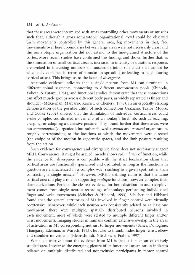
that these areas were intermixed with areas controlling other movements or muscles
such that, although a gross somatotopic organizational trend could be observed(arm movements controlled by this general area, leg movements in that, face
movements over here), boundaries between large areas were not necessarily clear, andthe somatotopic organization did not extend to the fine-grained structure of the
cortex. More recent studies have confirmed this finding, and shown further that, asthe stimulation of small cortical areas is increased in intensity or duration, responses
are evoked in increasing numbers of muscles or joints (an effect that cannot beadequately explained in terms of stimulation spreading or leaking to neighbouringcortical areas). This brings us to the issue of divergence.
Anatomic evidence indicates that a single neuron from M1 can terminate indifferent spinal segments, connecting to different motoneuron pools (Shinoda,
Yokota, & Futami, 1981), and functional studies demonstrate that these connectionscan affect muscle groups across different body parts, as widely separated as finger and
shoulder (McKiernan, Marcario, Karrier, & Chenery, 1998). In an especially strikingdemonstration of the possible utility of such connections Graziano, Taylor, Moore,
and Cooke (2002) showed that the stimulation of individual cortical areas couldevoke complex coordinated movements of a monkey’s forelimb, such as reaching,grasping, or adopting a defensive posture. They found further that these areas were
not somatotopically organized, but rather showed a spatial and postural organization,roughly corresponding to the locations at which the movements were directed
(the endpoint of the motion in egocentric space), and the limb posture resultingfrom the action.
Such evidence for convergence and divergence alone does not necessarily suggestMRH. Convergence, it might be argued, merely shows redundancy of function, while
the evidence for divergence is compatible with the strict localization claim thatcortical areas are functionally specialized and dedicated, so long as the functions in
question are characterized in a complex way: reaching to a given spot, rather thancontracting a single muscle.14 However, MRH’s defining claim is that the samecortical area can play a role in supporting multiple functions, however complex their
characterizations. Perhaps the clearest evidence for both distribution and redeploy-ment comes from single neuron recordings of monkeys performing individuated
finger and wrist movements (Schieber & Hibbard, 1993). Schieber and Hibbardfound that the general territories of M1 involved in finger control were virtually
coextensive. Moreover, while each neuron was consistently related to at least onemovement, there were multiple, spatially distributed neurons involved in
each movement, most of which were related to multiple different finger and/orwrist movements. Imaging studies in humans confirm extensive overlap in the areasof activation in M1 corresponding not just to finger movements (Sanes, Donoghue,
Thangaraj, Edelman, & Warach, 1995), but also to thumb, index finger, wrist, elbowand shoulder movements (Kleinschmidt, Nitschke, & Frahm, 1997).
What is attractive about the evidence from M1 is that it is such an extensivelystudied area. Insofar as the emerging picture of its functional organization indicates
reliance on multiple, distributed and nonexclusive participants in motor control
154 M. L. Anderson

functions, then given the extent of the evidence, the hypothesis needs to be taken
quite seriously. On the other side of the coin, given that the evidence is restricted to
M1 and motor control, extensive redeployment might not seem all that surprising.
The support for MRH coming from the study of M1, while strong, is also somewhat
narrow. Thus, the next two case studies showcase some rather more radical and
surprising instances of apparent redeployment. The evidence for these examples is
somewhat less strong, but the implications are far broader.
3.2. Sensorimotor Coding in Working Memory
One instance of redeployment on which there has been a fair amount of work is in
the apparent use of sensorimotor resources to support working memory. As the
evidence has been reviewed in detail by Margaret Wilson (2001), I’ll only provide a
brief summary. The experiments in question typically involve the presentation
of multiple items (words or letters) either visually or auditorily, with the task being to
remember these items in order. The question of interest is what kind of processing
supports this ability, and there is a great deal of evidence supporting some version of
the Baddeley and Hitch model of working memory, which posits that working
memory has both verbal and visuospatial components, among others (Baddeley,
1986, 1995; Baddeley & Hitch, 1974, 1994). Basically, the Baddeley and Hitch model
says that one strategy for remembering such lists involves (silently) saying them to
one’s self (producing a ‘‘phonological loop’’), which engages brain areas typically
used both in speech production and in audition. Another strategy for remembering
words is the visual representation of their form or meaning (especially for abstract
nouns). Wilson notes that this latter strategy is not particularly effective for
maintaining an ordered list, and that therefore a strategy involving some version of
the phonological loop is more typically employed.A pattern of findings supports the existence of a phonological loop, a strategy that
engages both inner ‘‘speaking’’ and inner ‘‘hearing’’ to support working memory.
First, there is poor recall of similar sounding terms; second, there is poor recall of
longer words; third, there is poor recall if the subject is made to speak during the
maintenance period; and fourth, there is poor recall when the subject is exposed
to irrelevant speech during the maintenance period. Moreover, imaging studies have
found that such memory tasks cause activation in areas typically involved in speech
production (Broca’s area, left premotor cortex, left supplementary motor cortex,
and right cerebellum) and in phonological storage (left posterior parietal cortex)
(Awh et al., 1996). Imaging data also tends to support the use of sensorimotor
strategies in visuospatial working memory, showing activation of right hemisphere,
including areas of visual and prefrontal cortex (Smith, 2000).
Although these findings will not be at all surprising to anyone who has ever tried to
remember multiple things, only to be foiled by having to say, or listen to, something
unrelated, the broad implications are nevertheless significant. As Wilson (2001)
writes, in this case it appears that:
Philosophical Psychology 155

. . . sensorimotor processes are run covertly to assist with the representation andmanipulation of information, in the temporary absence of task-relevant inputor output. Such an arrangement would make sense, given our evolutionary heritagefrom creatures whose neural resources were devoted largely to perceptual andmotor processes. Indeed, given that we have such resources, it would be odd if wedid not exploit them whenever possible to assist in off-line cognitive processing.(pp. 44–45)
3.3. The Use of Motor Simulations in Language Understanding
Finally, the last case I would like to consider is an even more striking example of the
redeployment of resources in apparently disparate functions: the action-sentence
compatibility effect (Glenberg & Kaschak, 2002), which suggests the involvement
of the motor system in language understanding. To demonstrate this interesting
interaction between comprehension and motor control, Glenberg and Kaschak asked
subjects to indicate whether a given sentence made sense or not by making a response
requiring a movement either toward or away from their bodies (e.g., reaching for a
button). They found that response times were longer in cases where the required
movement ran counter to a movement suggested by the sentence itself (e.g., where
the response required a movement toward the body, and the sentence, e.g., ‘‘Close
the drawer’’ indicated a movement away from the body, or vice-versa). This was true
even when the ‘‘movement’’ indicated by the sentence was abstract, as in the transfer
of information from one party to another (e.g., ‘‘You told Ann about the party’’).
A general explanation of this effect is that the comprehension of the sentences
involved a motor simulation of the action they describe, thus ‘‘priming’’ the system
to move in one way, rather than another. More particularly, Glenberg and Kaschak
posit that understanding language involves combining the affordances of the sentence
elements, and judging the ‘‘doability’’ of the action corresponding to the meshed
set of affordances. A doable action indicates a comprehensible sentence.These results are intriguing and highly suggestive, yet, as Glenberg and Kaschak
readily admit, there is much more work to be done.
In summary, our results demonstrate that the understanding of imperative,double-object and dative constructions is grounded in action. Given that languagealmost certainly arose to facilitate coordination of action, it is not surprising thatthere is an observable remnant of that history. The results also raise the intriguingpossibility that much, if not all, language comprehension is similarly grounded.Although substantial work needs to be done to secure that possibility, that workmay well be rewarded by an account of language and meaning firmly anchored inhuman experience. (2002, p. 564)
One kind of evidence that is currently missing for this effect is neural imaging data.
To help address this lacuna, I hope in the near future to run an MEG experiment
featuring the Glenberg-Kaschak task. MEG evidence, especially given its temporal
resolution, might help rule out the most obvious alternate explanation of the data,
that it is a post-understanding simulation of the action that is interfering with the
156 M. L. Anderson

response, rather than a simulation implicated in the understanding itself. Although
it is true that it is difficult to use this alternative to explain the effect in the case ofabstract transfers (for there is little reason to believe that a post-understanding
simulation of abstract transfers would implicate movements toward or away fromthe subject, even if it involved simulating the actions used in the transfer, such as
speaking), MEG data might help settle the matter.There are nevertheless other kinds of evidence available that appear to support the
general finding that motor control and language understanding are intertwined withone another. For instance, patient KJ-1360, who has a lesion in left premotor cortex,shows an impairment in verb retrieval, but has otherwise normal linguistic abilities
(Damasio & Tranel, 1993). Studies by Martin and colleagues (Martin, Haxby,Lalonde, Wiggs, & Ungerleider, 1995; Martin, Wiggs, Ungerlieder, & Haxby, 1996)
confirm this basic finding that areas associated with motor control are involvedin verb retrieval, and also show that naming colors and animals involved visual
processing areas, suggesting that language use and comprehension involves the reuseof many other areas of the brain besides motor areas, and, moreover, that this
redeployment is content specific, with verbs reusing motor control areas, and certainnouns like animal and color names reusing visual processing resources. That thereis a large amount of redeployment of sensory processing areas in linguistic and
conceptual tasks is another striking case of redeployment worth pursuing in its ownright (Barsalou, 1999), but we will focus here on the relation between language use
and motor areas.One particularly interesting part of the brain in this regard is Broca’s area
(left Brodmann areas 44 and 45). Broca’s area has long been associated with languageprocessing, but what has recently begun to emerge is its functional complexity
(Hagoort, 2005). For instance, it has been shown that Broca’s area is involved inmany different action-related tasks, including movement preparation (Thoenissen,
Zilles, & Toni, 2002), action sequencing (Nishitani, Schurmann, Amunts, & Hari,2005), action recognition (Decety et al., 1997; Hamzei et al., 2003, Nishitani et al.,2005), imagery of human motion (Binkofski et al., 2000), and action imitation
(Nishitani et al., 2005). In other words, language processing involves (much) morethan one region of the brain, and the regions of the brain associated with language
processing are involved in many other tasks, of which we have listed just a few. Note,however, that it does not appear to be the case that brain areas are redeployed
haphazardly; rather, the contributions they make are useful in more than onesituation. In the case of Broca’s area, it is not surprising that an area of the brain
that plays a role in action sequencing would be useful in language processingand production, since this, too, requires action sequencing. Likewise, that verbretrieval/comprehension would involve motor simulation is unsurprising, so long as
we suppose that our ability to understand verbs is closely connected to ourexperience of acting in the world.
Returning, then, to our central theme, a main distinguishing feature of MRH is theclaim that the functional complexes of the brain make heavy use of nonexclusive
participants, not just within, but across classically specified domains. The three case
Philosophical Psychology 157

studies above offer some evidence for this claim—and, just as importantly, help to
illustrate what redeployment does for the brain, and why it makes sense as an
organizational principle. But it must of course be admitted that this evidence in no
way proves MRH, and certainly does not establish redeployment as the norm. Thus,
in the next section we turn to a different kind of evidence that can help do just that.
4. Further Evidence for MRH
The evidence for MRH is in no way restricted to the few brain areas or cognitive
functions listed above. In fact, a recent empirical review by Cabeza & Nyberg (2000)
strongly suggests rather rampant redeployment to be the norm. Cabeza and Nyberg
survey 275 fMRI and PET experiments, arranging them by task category (attention,
perception, imagery, language, working memory, episodic memory encoding,
episodic memory retrieval, etc.). For each task, they catalog the participants in that
task from a list of 31 different brain areas (28 Brodmann, and three subcortical
areas), each divided into four different parts: left lateral, right lateral, left medial and
right medial. Although Cabeza and Nyberg do not do any statistical analysis of this
data (their primary interest is in examining/establishing the consistency of findings
across different experiments on similar tasks), the results of even a simple analysis are
striking.
For simplicity and brevity, I focus here on only four of the ten categories of tasks
surveyed: attention, perception, imagery, and language. The data on the other task
categories is consistent with what I report here. Cabeza and Nyberg looked at 39
attention-related tasks, 42 perception-related tasks, 18 imagery-related tasks, and 36
language-related tasks, for a total of 135 tasks in these four categories. The attention
tasks included things like tone detection and Stroop tasks (naming colored words);
perception tasks included such things as object identification and facial recognition;
the various imagery tasks include mental rotation and landmark visualization; and
the language tasks included reading out loud and silently, lexical decision tasks
(discriminating words from nonwords), and the like.As mentioned already above, Cabeza and Nyberg divided each brain area into
4 parts; however, their coding scheme forces a decision between lateral and medial
activation, such that it is not possible to show a left medial and a left lateral activation
in a given area for a given task. Instead, the possible activations for each brain area
are left lateral (LL), right lateral (RL), bilateral lateral (BL); left medial (LM), right
medial (RM), bilateral medial (BM). Thus, for instance, they list the following
activations for a task involving hearing words vs. a resting condition (Muller et al.,
1997): an LL activation in Brodmann area 47, and BL activations in areas 21 and 22.
For the purposes of counting participants in a task, I treated bilateral activations of
an area as two participants, one left and one right (medial or lateral). Thus, the
language task above would have five participants, three LL participants (areas 47, 21
and 22) and two RL participants (areas 21 and 22). For the purposes of counting
redeployments (areas activated by more than one task), I matched LL activations
158 M. L. Anderson

in an area to other LL activations of that area, as well as to BL activations, and
I matched RL activations in an area to other RL activations of that area, as well as to
BL activations. I followed the same procedure for medial activations. I did not match
bilateral activations to each other.
The data show that, on average, each of the 135 tasks has 5.97 participants
(SD¼ 4.80), with somewhat more than that for the language tasks and slightly less for
attention and perception (see Table 2). More importantly, they show that each area
was typically a participant in more than one task, although interestingly, there is a
significant difference between medial and lateral activations in this regard. Thus, each
LM area that was a participant in at least one task was on average a participant in 3.87
different tasks (SD¼ 3.34); likewise, each RM area that was a participant in at
least one task was, on average, a participant in 3.29 tasks (SD¼ 1.77). In contrast
(and providing incredibly striking evidence against strict localization), each LL area
that was a participant in at least one task was a participant in an average of 14.29
different tasks (SD¼ 9.20), and each RL area was a participant in 10.41 (SD¼ 7.96).
Put differently, an average LL area participated in nearly one in nine (10.6%) of the
tasks studied, and the average RL area participated in one in thirteen (7.7%).15
The participation of areas in multiple tasks was not restricted to only closely
related tasks. In fact, of the 28 LL areas that participated in at least one task in one of
the four task categories, 26 (93%) were also participants in at least one other task in a
different task category. Moreover, 23 of those areas (82%) participated in tasks in at
least three categories, and 15 (54%) participated in tasks in all four categories. The
numbers are similar for RL activations, and while the numbers for medial activations
Table 2. Average number of participants per task, by taskcategory.
Task Category Average number of participants per task
Attention 5.26, SD 4.23Perception 4.88, SD 3.55Imagery 6.39, SD 3.29Language 7.81, SD 6.56Total 5.97, SD 4.80
Table 3. The number of brain regions activated (out of 31), with activations in at leastthe number of task categories listed, out of the four categories surveyed.
Number of areas with activations in at least:
Activation Type 1 task category 2 task categories 3 task categories 4 task categories
Right Lateral 29 26 22 11Left Lateral 28 26 23 15Right Medial 14 10 6 0Left Medial 15 9 6 2
Philosophical Psychology 159

were somewhat lower, the data overall undermine strict localization, and strongly
suggest widespread redeployment throughout the cortex (see Tables 3 and 4).It appears to be the norm not just that a given task has many participants, but that
each participant contributes to many distinct and very different cognitive tasks.
To underline this last point, and to give some more detailed sense of the distribution
of activations across task categories, Tables 5–8 (given in the Appendix) show the
number of activations, arranged by area and task category and normalized with
respect to the number of tasks in each category. Although the data is consistent with
brain areas having some categorical preferences or tendencies, there is nevertheless in
nearly all cases a significant distribution of activation across multiple task categories.
Thus, e.g., an average of 46% of LL activations and 40% of RL activations occur in
task categories other than the category with the highest number of activations for
a given area (the numbers are 28% and 29% for LM and RM, respectively), and
in 15 of the 28 LL areas with at least one activation, the task categories with the
highest number of activations accounted for less than 51% of the total activations
for that area.
Finally, there were nine tasks of the 135 examined that activated a total of ten areas
not activated by any other task. Since other tasks not examined might also activate
these areas, we cannot conclude on this basis alone that these nine tasks have
exclusive participants—and, in fact, eight of these ten areas are known to be involved
in tasks in categories not surveyed here. Even if the remaining two areas turn out
to be exclusive to their two tasks, this still would mean that fewer than 1.5% of
the tasks examined had exclusive participants. Only one of these two tasks had a
single participant, not activated by any other task; the other had 8 other participants,
none of which were exclusive.Overall, the picture could hardly be clearer: nearly every brain area participates in
multiple cognitive functions, and each cognitive function utilizes many participants,
very few of which are exclusive. The data on brain function is decidedly not consistent
with the strict localization hypothesis.16
Table 4. The number of brain regions activated (out of 31), with activations in exactlythe number of task categories listed, out of the four categories surveyed.
Number of areas with activations in exactly:
Activation Type 1 task category 2 task categories 3 task categories 4 task categories
Right Lateral 3 4 11 11Left Lateral 2 3 8 15Right Medial 4 4 6 0Left Medial 6 3 4 2
160 M. L. Anderson

5. Massive Redeployment and the Meaning of Function
Now it is certainly possible, when faced with such data, to try to maintain strict
localization—most or all participants are exclusive to a task, and most or all tasks
have exclusive participants—by redefining the function of a given area to encompass
and account for the various tasks that it seems to support. As was mentioned already,
this has been one strategy employed in trying to account for the participation of the
fusiform face area in recognition tasks not involving faces,17 and there may well be
future proposals for the ‘‘true’’ function of right prefrontal cortex (and each of the
31 other areas surveyed) that account for its participation in the various tasks listed
above. Insofar as this move involves a recognition that cognitive functions, as defined
by psychology, are unlikely to map one-to-one with particular brain functions,
defined by the specific, identifiable mechanisms (integration, filtering, and other such
transforms) implemented in our neural architecture, then I think it can be counted
as a step forward (Bechtel, 2002, 2005a, 2005b). On the other side of the coin,
unless this move is accompanied by a call to redefine cognitive functions in terms of
area-specific brain functions (i.e., to question the validity of the ontology of cognitive
psychology, a move for which I currently see no compelling grounds), we would still
be left with a many-to-many mapping of cognitive functions to brain areas, and this
is a significant step away from strict localization.Since we have just introduced a new term—brain function—let us step back for a
bit to examine it. As stated briefly, above, I am using the term to refer to a specific,
identifiable mechanism implemented in the brain for transforming signals and/or
information from one form to another. Signal processing functions might include
filtering, smoothing, integrating, enhancing, shifting waveform or periodicity,
interpolating, and the like. Information-processing functions might be described
in mathematical terms (adding, subtracting) or in terms of data processing (store,
search, sort). There may also be another, more appropriate vocabulary for brain
functions, not yet discovered, developed or widely accepted. In point of fact, we can
have little confidence that the field of cognitive science has defined a reliable list of
the various functions (elementary operations) implemented in the brain, and my
point here is not to recommend any such list, but only to give the reader some notion
of the sort of thing I mean by the term brain function. The real issue, for my purposes
here, is to come to some understanding of how these functions are likely to relate
to cognitive functions, on the one hand, and to brain areas on the other.
So, let me suggest first of all that, given what has been shown and argued so far,
there can be little doubt that it will be a very rare to have a single brain function
implement a single cognitive function. Likewise, it will be rare for a single cognitive
function to be the only user of a given brain function.18 Thus the arrangement here is
likely to be many-many. Now, as I noted above, we cannot be confident that we know
what the various brain functions are—we have not yet discovered the basic functional
vocabulary of the brain. So, one approach to developing this vocabulary is to define a
brain function as whatever it is that some brain area is discovered to do. In my view,
defining into existence a one-to-one correspondence between function and area is
Philosophical Psychology 161

too dogmatic, especially when the evidence seems to indicate that brain areas don’t
always do things by themselves, but generally act in concert with other areas.19
Moreover, we need to be aware of the fact that it is the purpose of many brain areas,
not to actually participate in any of the processing leading to a cognitive result,
but rather to inhibit or suppress other areas; just because an area becomes active
during a task, as shown by some imaging method, it cannot be concluded that this
area is a direct contributor to the processing required by the task.20 It’s role, instead,
might be inhibitory—a very necessary role, to be sure, since often an area will be
inhibiting another area that itself normally inhibits the very processing necessary in
the current task—but of course this sense of ‘function’ is not the same as the that
under discussion.21 In any case, it further shows that brain function, in that sense,
cannot be strictly tied to brain area, but that a brain function is in fact often the
product of cooperation between pieces of brain matter (whether they are spatially
contiguous or not) playing distinctly different roles.
To try to avoid confusion, and to follow the lead of Bechtel (2002), let us call
a brain function in the sense defined above (a specific, identifiable mechanism
implemented in the brain for transforming signals and/or information from one
form to another) a component function, and use the term role, or area role, for
whatever it is determined that a particular, contiguous bit of brain matter actually
does. To introduce equivalent vocabulary to refer to anatomy instead of function,
let’s call the entirety of participants in a cognitive function a functional complex, and
the sum of the participants that implement a component function a component; an
area will still be called an area. Relating all these levels of anatomy and function,
we can say that so long as it seems possible (a) that some number of brain areas could
cooperate to implement some component function, (b) that the component function
in question need not be identical to any cognitive function, and (c) that the
individual area roles need not be identical to the component function (as in the case
of components that require more than one brain area for processing, and/or involve
inhibitory areas), then a more flexible approach to the functional topography of the
brain is called for, one that neither insists that cognitive functions map to single
Arearole
Arearole
Arearole
Arearole
Arearole
Arearole
Arearole
Componentfunction
Componentfunction
Componentfunction
Componentfunction
Cognitivefunction
Cognitivefunction
Cognitivefunction
Figure 3. A 3-tier architecture showing a many-to-many relationship between each level.
162 M. L. Anderson

component functions, nor requires that component functions map to single brain
areas, but allows for many-to-many relations between all three levels (see Figure 3).
Saying the same thing with our anatomical vocabulary, we should expect each
functional complex to have more than one component, each of which in turn will
involve more than one area; likewise, we should expect areas to be members of more
than one component, and components to be members of more than one functional
complex, and we should not expect that such cross-participation will respect
traditional functional-anatomical boundaries (see Figure 4).22 This, stated in the
most detailed form that will be offered in this paper, is the massive redeployment
hypothesis.
6. Implications, Objections, and Replies
I will now address some of the implications of MRH, thereby further clarifying the
position, by answering some of the questions most likely to occur to the reader.
Figure 4. Anatomical illustration of 3-tier architecture, with many-to-many relationshipsbetween levels.
Philosophical Psychology 163

6.1. Since You Claim That Brain Areas Have Unique Roles, Why Isn’t This Just
Strict Localization by Another Name?
First, and most simply, because area roles are not likely to be very interesting
by themselves; they only start to have much functional or cognitive significance in
combination with other brain areas to form components and functional complexes,the elements of which might be widely scattered, and participants in multiple
components and complexes. Second, because localization has typically meant being
able to assign domain-specific roles to brain areas, and the prospect for this looksvery dim. And finally, what may be among the most important practical implications
of MRH, because if MRH is right, then one cannot determine what any brain area
does by looking at its participation in (activation by) any individual task or taskcategory, as been the normal practice. Rather, one must begin to look at the
participation of brain areas across multiple tasks in multiple domains, and try to
discern what role it could be playing to account for its functional promiscuity.It must be admitted that this deeply violates the spirit of localization as it has been
traditionally understood. Nevertheless, it is true that MRH is compatible with an
anemic version of localization that claims simply that individual brain areas do something, and the same thing, however low-level, simple, or cognitively uninteresting,
whenever they are activated.
6.2. Are You Denying Modularity?
No, although I must add the usual caveat that it depends what one means by
‘modularity’. MRH is compatible with a limited form of functional localization(see x6.1), insofar as localization does not require all localized participants in a brain
function to be exclusive participants in that function. Since I am obviously denying
the strict localization hypothesis, I also deny any version of modularity that requiresit. What I am proposing is an hypothesis regarding the functional organization of
the brain that posits overlapping functional complexes; i.e., I expect the entities that
implement different cognitive and brain functions to share functional elements(‘‘participants’’). Any version of modularity on which modules could be components
or functional complexes (or even organized groups of functional complexes) could
thereby be compatible with MRH.23
One aspect of the modularity hypothesis that, in its strong from, sits uneasily with
MRH is the claim that modules are domain specific. I have reviewed evidence that
motor areas are participants in language-related and memory-related tasks, and thatlanguage-related areas are participants in motor-related tasks. One result of this,
as Prinz (2005) notes, is that focal brain lesions can produce deficits across multiple
domains, and genetic language disorders often manifest nonlinguistic problems.Moreover, it appears that most brain areas participate in functions across several task
categories. Thus, although functional complexes might be domain specific asorganized, they are not thereby composed of domain-specific parts. This also casts
into doubt the oft-made claim for modularity that many modules can be run
simultaneously, helping avoid bottlenecks and computational constraints. The more
164 M. L. Anderson

overlap between modules, the less likely they can be active simultaneously, a fact that
accords well with what we know about interference between different cognitive tasks.
Although strong interpretations of localization, domain specificity, and simultaneous
operation are not strictly speaking necessary to the modularity hypothesis, Prinz
argues that insofar as we have to weaken our interpretation of its various tenets,
we should discard the hypothesis, and focus on functional decomposition more
broadly construed. This may well be the right position, but I’ll not pursue the matter
further here.
6.3. Can’t There be Parts of the Brain that Conform to the StrictLocalization Hypothesis?
Of course there can be some brain areas that are best understood in terms of strict
localization of function. But the evidence is that redeployment is the norm.
Two specific (and to my mind novel and exciting) predictions of MRH would be:
(a) that evolutionarily older cognitive functions would tend to use fewer and
more tightly grouped participants, whereas newer functions would use more, and
more widely scattered ones; and (b) that older cortical areas will be participants
in more, and more diverse, cognitive functions, and newer areas the opposite.
These are empirical questions, and will eventually be settled by the data (Anderson,
2007).
6.4. Why Didn’t You Discuss Topic Y?
There are a large number of topics, the discussion of which could conceivably
have enhanced this essay. Here is a partial list: the various conceptions and
critiques of functional localization (see, e.g., Bechtel & Richardson, 1993; Mundale,
2002; Zola-Morgan, 1995); the modularity hypothesis, massive or otherwise (e.g.,
Carruthers, 2003, 2005, 2006; Fodor, 1983, 2000; Pinker, 2005; Prinz, 2005; Sperber,
2002); the organization of other well-studied brain regions (see, e.g., Bechtel, 2001;
Dagher, Owen, Boecker, & Brooks, 1999); evolutionary accounts of cognition (see
Barkow, Cosmides, & Tooby, 1992; Sperber, 1996); embodied cognition (see
Anderson, 2003; Clark, 1997, 1998; Wilson, 2002).24 However, it was my hope
to keep this essay focused on the relatively narrow task of introducing MRH, and
offering some evidence in support of it. In maintaining this focus I have had
to sacrifice some breadth.
7. Conclusion
This essay introduced the massive redeployment hypothesis, an account of the
functional organization of the brain that gives pride of place to the fact that brain
areas are typically employed to support numerous functions, with little respect for
traditional categorical boundaries. Although I think that the three case studies in
which there appears to be redeployment of brain areas to support very different
Philosophical Psychology 165

functions, together with the empirical review that suggests such redeployment is the
norm, strongly support MRH, this is not likely to be, nor is it intended as, the last
word on brain organization. Still, an hypothesis can be prove useful even (or perhaps
especially) in the course of being disproved and discarded, insofar as it offers a way
to help (re-)organize old data and interpret new information, and may suggest novel
experimental inquiries. I hope for no more than this from MRH.
Acknowledgments
Thanks are due to the participants of the workshop for early career researchers,
sponsored by the McDonnell Project in Philosophy and the Neurosciences, and
especially to John Bickle, Valerie Gray Hardcastle, Ben Hardy, David Kaplan,
Anthony Landreth, and Bill Seeley. Without the discussions we had, I probably would
not have thought to write this essay. Thanks are also due to William Bechtel, Peter
Carruthers, Tony Chemero, and Dan Lloyd for helpful advice in revising and greatly
improving the presentation.
Notes
[1] Many theorists of embodied cognition (EC) might object immediately to this definition,since, if cognition is embodied, then cognitive functions are certainly not definable simplyin terms of neural inputs and outputs. (Note that it follows immediately that cognitivefunctions are not localizable, and the game is over before it begins.) But even on the ECmodel, there is something that the brain is doing in the course of contributing to a cognitivefunction, and even if we believe that isolating this bit of processing leaves us with somethingquite incomplete from the standpoint of defining a cognitive function, it is still a legitimatequestion to ask whether and to what degree the brain’s contribution is itself localizable.I will be arguing for a particular understanding of functional localization that denies strictlocalization, but does not embrace holism (as many EC theorists seem wont to do).However, the evidence with respect to localization of function is such that should givecomfort to EC theorists, since it indicates that higher-order cognitive functions are veryoften supported by brain areas typically involved in tasks like motor control and sensoryprocessing. See Anderson (in press) for further discussion.
[2] There is a complication here that is worth noting, but does not require solving: redundancyin some brain areas, and cooperation among others, may mean that actually specifying thenecessary participants would be logically complex; e.g., it could be that what is necessaryis either area A or areas (B & C) together, or some such. But for our purposes, we will notneed to spell out any such relations; it is enough to agree that there is indeed some set ofnecessary participants for each brain function, however difficult to specify their form andidentity. Note further that the necessary participants do not exhaust the necessary conditions,or even the necessary brain processing, for the success of the function. In the case ofunderstanding a sentence, for instance, there is a great deal of processing necessary toprepare the inputs to the comprehension function (if such there be), the failure of whichwould prevent the success of the comprehension function. Because we are focusing on theprocesses supporting the post-input transformation, these sorts of issues are not material.
[3] Although it is worth noting that there are other sorts of analytical methods available,e.g., multi-variate methods including multiple regression, discriminant analysis, and
166 M. L. Anderson

principle component analysis, all of which are designed to reveal the multiple contributorsto a given effect or outcome. It is likely that imaging experiments would be designedsomewhat differently from the usual ‘‘blocked’’ designs, were the data to be analysed usingmulti-variate methods.
[4] And it should also be stressed that this issue is not specific to imaging studies, but is facedwhenever one is engaged in comparative studies, i.e., in producing analyses of differences.
[5] Activation results are typically averaged over many trials, thereby filtering out noise.[6] Later he uses a more cautious formulation: ‘‘allows one to observe brain areas specialized
for the processes that differ between the two tasks’’ (Buckner, 1996, p. 155)[7] To be precise, the activations for anxiety management and hypothesis generation do not
overlap with the activations shown in Figure 1—they overlap in the X and Y coordinates,but, being near Z¼�20� 4, are considerably below the horizontal section shown in thefigure, which is somewhere in the neighbourhood of Z¼ 12 in the Talairach and Tournoux(1988) coordinate system. However, they do overlap with some of the activations reportedin the papers from which Buckner gathered the data for his survey of memory retrieval(see, e.g., Andreasen et al., 1995). Thus the statement that these three functions haveoverlapping brain activations is true. There is also another complication here that should benoted, but does not affect the conclusions. The papers cited in the Buckner survey reportcoordinates based on the Talairach and Tournoux (1988) atlas; the more recent work I amciting uses MNI coordinates (Evans et al., 1993). I have compared these two systems usingthe transforms described by Matthew Brett (2006).
[8] Although, as Lloyd points out, the spatial accuracy of PET is not so great that it is aninappropriate choice.
[9] Although some of the quantum theories of mind may come close to espousing a radicalholism, at a different level of organization.
[10] The text is somewhat equivocal with respect to these possibilities. Here is a fuller quotation:‘‘Third, a brain might be constructed of sparsely distributed networks. Here anatomicallydefined brain regions are multifunctional. A region may be recruited to join a subnetworkto compute one function, and later recruited to a different subnet to compute a differentfunction. Thus, subnetworks would overlap in their anatomy. This form of distributionis sparse, however, insofar as particular brain regions are not omnifunctional. That is, eachfunction is computed by a subset of regions, rather than the whole brain. The engagedsubnetworks overlap, but the adaptability of each region is limited to a fixed list offunctions’’ (Lloyd, 2000, p. 95).
[11] My arguments for MRH are largely compatible with those in Mundale (2002) for a limitedform of localization. Mundale argues that the localization hypothesis allows for thepossibility that brain functions utilize multiple participants in complex combinations—furthermore, she implies that this is what localization has meant all along. However,while allowing for the possibility of multiple necessary participants in a given brain function,her argument does not address the difference between necessary and exclusive participation,and thus she does not discuss the possibility of a form of localization in which the (localized)participants in one function also participate in other functions. Someone else theorizing inthe same ballpark as MRH is Jesse Prinz (2005), who centrally features evidence that brainregions are used in multiple functions in his critique of modularity. I thus expect he wouldbe sympathetic to the account offered here, but Prinz (2005) does not develop such anaccount.
[12] This is one reason there is (or we should expect there will turn out to be) so muchredeployment of sensorimotor resources, as these are some of the oldest. For somediscussion of this issue, and its relevance to embodied cognition, see Anderson (in press).
[13] This is a potentially interesting case study of the effect of prior assumptions in science,because both Penfield and Rasmussen (1950) and Woolsey et al. (1952) admit that thepictures they offered did not reflect the complexity of the data, and the degree to which their
Philosophical Psychology 167

evidence indicated a great deal of overlap of the areas controlling different body parts.
It took a certain amount of interpretive effort to make their findings consistent with
strict localization—e.g., focusing only on the primary movement evoked by the electro-
stimulation, and discounting some movements as the effect of spreading activation from the
primary target site. This is not to say that these interpretive moves were unreasonable
or illegitimate. See Schieber (2001) for some of the interesting details.[14] It might be thought that I am understating the case in saying that evidence like that
presented in Graziano et al. (2002) is compatible with strict localization; rather it seems to
positively cry out for such interpretation. After all, stimulation of each of the cortical
sites studied did exactly one thing, which was to move the monkey’s arm to a spatially
well-defined posture. I have two responses to this claim. The first involves a clarification of
the scope of MRH: the hypothesis does not amount to the claim that no part of the brain is
organized according to strict localization; the part of the cortex studied by Graziano et al.
may turn out to be one of those areas. However, MRH predicts (and the evidence presented
here strongly suggests) that strict localization will not turn out to be the norm of functional
organization. This brings us to the second response, which is to point out that the electrical
stimulation of brain areas is a highly unnatural event. It does not follow from the fact that
the stimulation of an area provokes a given movement that this movement is evoked in the
course of an organism’s natural behaviour by the selective activation of that same brain area.
To demonstrate this latter claim would require observing brain activation during behaviour
elicited under the control of the organism, and the evidence gathered this way suggests a
more complex picture, involving both distributed activation in the control of individual
movements, and redeployment of single areas for the control of different movements.[15] Cabeza and Nyberg (2000), whose work was the basis of this analysis, do notice that there is
apparently a great deal of redeployment in the brain, but they decline to offer any hypotheses
about the significance of this finding. Interestingly, they observe that researchers tend to
interpret activations in terms of the domain within which they are working: ‘‘Area 7
activations, for instance, were usually attributed to attentional processes in attention studies,
to perceptual processes in perception studies, to working memory processes in working
memory studies, and so on’’ (p. 31). This tendency would serve to mask the prevalence
of redeployment from those disinclined to look for it.[16] Note that the data presented do not rule out holism as an alternative. I have already
indicated why I favour redeployment over holism (see x2.3), but it must be admitted that,
until we have much better awareness of what individual brain areas contribute to cognitive
functions (something that imaging data alone will not provide), it will be difficult to
definitively rule out holism as an alternative explanation of the functioning of the brain.[17] It has been proposed that the fusiform face area is in fact involved in automatic visual
processing tasks correlated with expertise, and this is why it is involved in face recognition
(Tarr & Gauthier, 2000).[18] According to MRH, this will be true for both tokens and types of brain functions.[19] It goes without saying that different brain areas might implement the same, or a very similar,
brain function.[20] This is yet another reason to be cautions about concluding that the active brain areas in
an imaging experiment are those responsible, or specialized for, the task being examined.[21] Inhibition is an incredibly important mechanism in the brain. Common uses include
inhibition of inhibition (thereby releasing some function normally inhibited),
lateral inhibition to emphasize contrast borders, and inhibition that can toggle the function
of a component between two possibilities (saccade left vs. saccade right).[22] It appears that this MRH architecture is a somewhat more flexible version of what was
proposed by Bechtel (2002) and Petersen and Fiez (1993), with the difference being that they
seem to imply that component functions will be strictly localized (whereas I am claiming
168 M. L. Anderson

that only area roles will be). Still, the current proposal is clearly compatible with this work inspirit. For instance, Petersen and Fiez (1993) write:
The areas involved in performing a particular task are distributed in differentlocations in the brain, but the processing involved in task performance is notdiffusely distributed among them. Each area makes a specific contribution tothe performance of the task, and the contribution is determinedby where the area resides within its richly connected, parallel, distributedhierarchy. (pp. 513–514)
Mesulam (1990, 1998) also suggests a position similar to MRH: ‘‘Many cortical nodes arelikely to participate in the function of more than one network. Conceivably, top-downconnections from transmodal areas could differentially recruit such a cortical node into theservice of one network or another’’ (1998, p. 1040). However, Mesulam’s development anddefense of this common basic idea is significantly different from that offered here. Detailingthose differences, and the respective strengths and weaknesses of each approach, while apotentially worthwhile task, is well beyond the scope of the present paper.
[23] Another possibility is a version of modularity on which modules were identified withthe individual participants in functional complexes. But this would make for some verylow-level, uninteresting modules, and so is not likely to be an attractive option forthe modularity theorist.
[24] For a brief account of the relation between embodied cognition and the redeploymenthypothesis, see Anderson (in press).
References
Allison, T., Ginter, H., McCarthy, G., Nobre, A. C., Puce, A., Luby, M., & Spencer, D. D. (1994).Face recognition in human extrastriate cortex. Journal of Neurophysiology, 71, 821–825.
Anderson, M. L. (2003). Embodied cognition: A field guide. Artificial Intelligence, 149, 91–103.Anserson, M. L. (2007). Evolution of cognitive funtion via redeployment of brain areas. The
Neuroscientist, 13, 13–21.Anderson, M. L. (in press). How to study the mind: An introduction to embodied cognition.
In F. Santoianni & C. Sabatano (Eds.), Brain development in learning environments: Embodiedand perceptual advancements.
Andreasen, N. C., O’Leary, D. S., Arndt, S., Cizadlo, T., Hurtig, R., Rezai, K., et al. (1995).Short-term and long-term verbal memory: A positron emission tomography study. Proceedingsof the National Academy of Science, 92, 5111–5115.
Asanuma, H., & Rosen, I. (1972). Topographical organization of cortical efferent zones projectingto distal forelimb muscles in the monkey. Experimental Brain Research, 14, 143–156.
Awh, E., Jonides, J., Smith, E. E., Schumacher, E. H., Koeppe, R. A., & Katz, S. (1996). Dissociationof storage and rehearsal in verbal working memory: Evidence from positron emissiontomography. Psychological Science, 7, 25–31.
Baddeley, A. D. (1986). Working memory. Oxford, England: Oxford University Press.Baddeley, A. D. (1995). Working memory. In M. S. Gazzaniga (Ed.), The cognitive neurosciences
(pp. 755–764). Cambridge, MA: MIT Press.Baddeley, A. D., & Hitch, G. (1974). Working memory. In G. H. Bower (Ed.), The psychology of
learning and motivation (pp. 647–667). Hillsdale, NJ: Erlbaum.Baddeley, A. D., & Hitch, G. (1994). Developments in the concept of working memory.
Neuropsychology, 8, 485–493.Barkow, J., Cosmides, L., & Tooby, J. (1992). The adapted mind: Evolutionary psychology and the
generation of culture. New York: Oxford University Press.Barsalou, L. (1999). Perceptual symbol systems. Behavioural and Brain Sciences, 22, 577–660.
Philosophical Psychology 169

Bechtel, W. (2001a). The compatibility of complex systems and reduction: A case analysis ofmemory research. Minds and Machines, 11, 483–502.
Bechtel, W. (2001b). Decomposing and localizing vision: An exemplar for cognitive neuroscience.In W. Bechtel, P. Mandik, J. Mundale, & R. S. Stuffelbeam (Eds.), Philosophy and theneurosciences: A reader (pp. 225–249). Oxford, England: Basil Blackwell.
Bechtel, W. (2002). Decomposing the mind-brain: A long-term pursuit. Brain and Mind, 3,229–242.
Bechtel, W. (2005a). The challenge of characterizing operations in the mechanisms underlyingbehaviour. Journal of the Experimental Analysis of Behaviour, 84, 313–325.
Bechtel, W. (2005b). Mental mechanisms: What are the operations? In B. G. Bara, L. Barsalou, &M. Bucciarelli (Eds.), Proceedings of the 27th annual meeting of the Cognitive Science Society(pp. 208–213). Mahwah, NJ: Lawrence Erlbaum Associates.
Bechtel, W., & Abrahamsen, A. (2005). Explanation: A mechanistic alternative. Studies in Historyand Philosophy of the Biological and Biomedical Sciences, 36, 421–441.
Bechtel, W., & Richardson, R. C. (1993). Discovering complexity: Decomposition and localization asscientific research strategies. Princeton, NJ: Princeton University Press.
Binkofski, F., Amunts, K., Stephan, K. M., Posse, S., Schormann, T., Freund, H.-J., et al. (2000).Broca’s region subserves imagery of motion: A combined cytoarchitectonic and fMRI study.Human Brain Mapping, 11, 273–285.
Brett, M. (2006). The MNI brain and the Talairach atlas. CBU Imaging Wiki. Retrieved, August 25,2006, from http://imaging.mrc-cbu.cam.ac.uk/imaging/MniTalairach
Buckner, R. L. (1996). Beyond HERA: Contributions of specific prefrontal brain areas to long-termmemory retrieval. Psychonomic Bulletin and Review, 3, 149–158.
Buckner, R. L., Petersen, S. E., Ojemann, J. G., Miezin, F. M., Squire, L. R., & Raichle, M. E. (1995).Functional anatomical studies of explicit and implicit memory retrieval tasks. Journal ofNeuroscience, 15, 12–29.
Cabeza, R., & Nyberg, L. (2000). Imaging cognition II: An empirical review of 275 PET and fMRIstudies. Journal of Cognitive Neuroscience, 12, 1–47.
Carruthers, P. (2003). Moderately massive modularity. In A. O’Hear (Ed.), Minds and persons(pp. 67–89). Cambridge, England: Cambridge University Press.
Carruthers, P. (2005). The case for massively modular models of mind. In R. Stainton (Ed.),Contemporary debates in cognitive science (pp. 205–225). Oxford, England: Blackwell.
Carruthers, P. (2006). Simple heuristics meet massive modularity. In P. Carruthers, S. Laurence, &S. Stich (Eds.), The innate mind: Vol. 2. Culture and cognition (pp. 181–196). New York: OxfordUniversity Press.
Clark, A. (1997). Being there: Putting brain, body, and world together again. Cambridge, MA:MIT Press.
Clark, A. (1998). Embodied, situated, and distributed cognition. In W. Bechtel & G. Graham (Eds.),A companion to cognitive science (pp. 506–517). Malden, MA: Blackwell.
Dagher, A., Owen, A., Boecker, H., & Brooks, D. (1999). Mapping the network for planning. Brain,122, 1973–1987.
Damasio, A. R., & Tranel, D. (1993). Nouns and verbs are retrieved with differently distributedneural systems. Proceedings of the National Academy of Sciences of the USA, 90, 4957–4960.
Decety, J., Grezes, J., Costes, N., Perani, D., Jeannerod, M., Procyk, E., et al. (1997). Brain activityduring observation of actions. Influence of action content and subject’s strategy. Brain, 120,1763–1777.
Evans, A. C., Collins, D. L., Mills, S. R., Brown, E. D., Kelly, R. L., & Peters, T. M. (1993).3D statistical neuroanatomical models from 305 MRI volumes. In L. A. Klaisner (Ed.),Proceedings of the IEEE: Nuclear Science Symposium and Medical Imaging Conference (Vol. 3,pp. 1813–1817). Piscataway, NJ: Institute of Electrical and Electronics Engineers.
Fodor, J. (1983). The modularity of mind. Cambridge, MA: MIT Press.Fodor, J. (2000). The mind doesn’t work that way. Cambridge, MA: MIT Press.
170 M. L. Anderson

Gall, F. J. (1798). Letter from Dr. F. J. Gall, to Joseph Fr[eiherr] von Retzer, upon the functionsof the brain, in man and animals. Der neue Teutsche Merkur, 3, 311–332.
Gauthier, I., Skudlarski, P., Gore, J. C., & Anderson, A. W. (2000). Expertise for cars and birdsrecruits brain areas involved in face recognition. Nature Neuroscience, 3, 191–197.
Glenberg, A., & Kaschak, M. (2002). Grounding language in action. Psychonomic Bulletin andReview, 9, 558–565.
Grasby, P. M., Frith, C. D., Friston, K. J., Bench, C., Frackowiak, R. S., & Dolan, R. J. (1993).Functional mapping of brain areas implicated in auditory—verbal memory function. Brain,116, 1–20.
Graziano, M. S. A., Taylor, C. S. R., Moore, T., & Cooke, D. F. (2002). The cortical control ofmovement revisited. Neuron, 36, 1–20.
Hagoort, P. (2005). On Broca, brain and binding. Trends in Cognitive Sciences, 9, 416–423.Hamzei, F., Rijntjes, M., Dettmers, C., Glauche, V., Weiller, C., & Buchel (2003). The human action
recognition system and its relationship to Broca’s area: An fMRI study. Neuroimage, 19,637–644.
Haxby, J. V., Horwitz, B., Ungerleider, L. G., Maisog, J. M., Pietrini, P., & Grady, C. L. (1994).The functional organization of human extrastriate cortex: A PET-RCBF study of selectiveattention to faces and locations. Journal of Neuroscience, 14, 6336–6353.
Howard, D., Patterson, K., Wise, R., Brown, W., Friston, K., Weiller, C., & Frackowiak, R. (1992).The cortical localization of the lexicons. Brain, 115, 1769–1782.
Jacobsen, C. F. (1935). Functions of the frontal association areas in primates. Archives of Neurologyand Psychiatry, Chicago, 33, 558–569.
Kanwisher, N., McDermott, J., & Chun, M. M. (1997). The fusiform face area: A module in humanextrastriate cortex specialized for face perception. Journal of Neuroscience, 17, 4302–4311.
Kleinschmidt, A., Nitschke, M. F., & Frahm, J. (1997). Somatotopy in the human motor cortexhand area: A high-resolution functional MRI study. European Journal of Neuroscience, 9,2178–2186.
Lashley, K. S. (1950). In search of the engram. Symposia of the Society for Experimental Biology, 4,454–482.
Lloyd, D. (2000). Terra cognita: From functional neuroimaging to the map of the mind. Brain andMind, 1, 93–116.
Martin, A., Haxby, J. V., Lalonde, F. M., Wiggs, C. L., & Ungerleider, L. G. (1995). Discrete corticalregions associated with knowledge of color and knowledge of action. Science, 270, 102–105.
Martin, A., Wiggs, C. L., Ungerleider, L. G., & Haxby, J. V. (1996). Neural correlates of category-specific knowledge. Nature, 379, 649–652.
McCarthy, G., Puce, A., Gore, J. C., & Allison, T. (1997). Face-specific processing in the humanfusiform gyrus. Journal of Cognitive Neuroscience, 9, 605–610.
McIntosh, A. R. (1999). Mapping cognition to the brain through neural interactions. Memory,7, 523–548.
McKiernan, B. J., Marcario, J. K., Karrier, J. H., & Chenery, P. D. (1998). Corticomotoneuronalpostspike effects in shoulder, elbow, wrist, digit, and intrinsic hand muscles during a reachand prehension task. Journal of Neurophysiology, 21, 1132–1139.
Mesulam, M.-M. (1990). Large–scale neurocognitive networks and distributed processing forattention, language, and memory. Annals of Neurology, 28, 597–613.
Mesulam, M.-M. (1998). From sensation to cognition. Brain, 121, 1013–1052.Muller, R. A., Rothermel, R. D., Behen, M. E., Muzik, O., Mangner, T. J., & Chugani, H. T. (1997).
Receptive and expressive language activations for sentences: A PET study. Neuroreport, 8,3767–3770.
Mundale, J. (2002). Concepts of localization: Balkanization in the brain. Brain and Mind, 3,313–330.
Nishitani, N., Schurmann, M., Amunts, K., & Hari, R. (2005). Broca’s region: From action tolanguage. Physiology, 20, 60–69.
Philosophical Psychology 171

Pardo, J., Raichle, M., & Fox, P. (1991). Localization of a human system for sustained attention
by positron emission tomography. Nature, 349, 61–63.Penfield, W., & Rasmussen, T. (1950). The cerebral cortex of man. New York: MacMillan.Petersen, S. E., & Fiez, J. A. (1993). The processing of single words studied with positron emission
tomography. Annual Review of Neuroscience, 16, 509–530.Pinker, S. (2005). So how does the mind work? Mind and Language, 20, 1–24.Prinz, J. (2005). Is the mind really modular? In R. Stainton (Ed.), Contemporary debates in cognitive
science (pp. 22–36). New York: Blackwell.Sanes, J. N., Donoghue, J. P., Thangaraj, V., Edelman, R. R., & Warach, S. (1995). Shared neural
substrates controlling hand movements in human motor cortex. Science, 268, 1775–1777.Schieber, M. H. (2001). Constraints on somatotopic organization in the primary motor cortex.
Journal of Neurophysiology, 86, 2125–2143.Schieber, M. H., & Hibbard, L. S. (1993). How somatotopic is the motor cortex hand area? Science,
261, 489–492.Shinoda, Y., Yokota, J., & Futami, T. (1981). Divergent projection of individual corticospinal axons
to motoneurons of multiple miscles in the monkey. Neuroscience Letters, 96, 7–12.Smith, E. E. (2000). Neural bases of working memory. Current Directions in Psychological Science,
9, 45–49.Sperber, D. (1996). Explaining culture: A naturalistic approach. London: Blackwell.Sperber, D. (2002). In defense of massive modularity. In E. Dupoux (Ed.), Language, brain and
cognitive development (pp. 44–57). Cambridge, MA: MIT Press.Tarr, M. J., & Gauthier, I. (2000). FFA: A flexible fusiform area for subordinate-level visual
processing automatized by expertise. Nature Neuroscience, 3, 764–769.Thoenissen, D., Zilles, K., & Toni, I. (2002). Differential involvement of parietal and precentral
regions in movement preparation and motor intention. Journal of Neuroscience, 22, 9024–9034.Uttal, W. R. (2001). The new phrenology: The limits of localizing cognitive processes in the brain.
Cambridge, MA: MIT Press.Vartanian, O., & Goel, V. (2005). Task constraints modulate activation in right ventral lateral
prefrontal cortex. NeuroImage, 27, 927–933.Wang, J., Rao, H., Wetmore, G. S., Furlan, P. M., Korczykowski, M., Dinges, D. F., et al. (2005).
Perfusion functional MRI reveals cerebral blood flow pattern under psychological stress.
Proceedings of the National Academy of Sciences USA, 102, 17804–17809.Wilson, M. (2001). The case for sensorimotor coding in working memory. Psychonomic Bulletin
and Review, 8, 44–57.Wilson, M. (2002). Six views of embodied cognition. Psychonomic Bulletin and Review, 9, 625–636.Woolsey, C. N., Settlage, P. H., Meyer, D. R., Sencer, W., Hamuty, T. P., & Travis, A. M. (1952).
Patterns of localization in precentral and ‘‘supplementary’’ motor areas and their relation to the
concept of a premotor area. Research Publications—Association for Research in Nervous and
Mental Disease, 30, 238–264.Zola-Morgan, S. (1995). Localization of brain function: The legacy of Franz Joseph Gall
(1758–1828). Annual Review of Neuroscience, 18, 359–383.
172 M. L. Anderson

Table
6.Rightlateralactivationsbytask
category
andbrain
area,norm
alized
withrespectto
thenumber
oftasksin
each
category.
Brain
areas
Task
category
109
4611
4745
446
84
3224
2331
740
3938
ins
4222
2120
mt
3719
1817
bg
thcb
Attention
2.15
6.46
5.38
1.08
3.23
1.08
5.38
7.54
2.15
0.00
1.08
0.00
0.00
0.00
10.779.69
3.23
0.00
1.08
2.15
4.31
0.00
1.08
1.08
1.08
5.38
9.69
0.00
1.08
0.00
3.23
Perception3.00
1.00
2.00
5.00
3.00
4.00
1.00
3.00
0.00
0.00
0.00
0.00
0.00
0.00
6.00
4.00
1.00
0.00
1.00
1.00
0.00
2.00
1.00
5.00
18.0020.0010.001.00
1.00
1.00
3.00
Imagery
0.00
4.67
2.33
0.00
0.00
7.00
2.33
2.33
4.67
2.33
0.00
0.00
0.00
0.00
4.67
4.67
2.33
0.00
4.67
0.00
2.33
2.33
2.33
7.00
7.00
9.33
7.00
4.67
0.00
0.00
0.00
Language
0.00
1.17
0.00
1.17
4.67
2.33
0.00
4.67
0.00
3.50
0.00
0.00
0.00
1.17
1.17
1.17
1.17
3.50
3.50
5.83
16.3311.670.00
1.17
4.67
4.67
5.83
3.50
4.67
4.67
8.17
Average
1.29
3.32
2.43
1.81
2.72
3.60
2.18
4.38
1.71
1.46
0.27
0.00
0.00
0.29
5.65
4.88
1.93
0.88
2.56
2.25
5.74
4.00
1.10
3.56
7.69
9.85
8.13
2.29
1.69
1.42
3.60
SD
1.53
2.69
2.22
2.19
1.96
2.56
2.34
2.32
2.22
1.75
0.54
0.00
0.00
0.58
3.97
3.55
1.05
1.75
1.82
2.55
7.28
5.21
0.96
2.93
7.29
7.07
2.04
2.16
2.05
2.22
3.38
Appendix:
Table
5.Leftlateralactivationsbytask
category
andbrain
area,norm
alized
withrespectto
thenumber
oftasksin
each
category.
Brain
areas
Task
category
109
4611
4745
446
84
3224
2331
740
3938
ins
4222
2120
mt
3719
1817
bg
thcb
Attention
0.00
1.08
3.23
1.08
2.15
2.15
3.23
7.54
3.23
2.15
1.08
0.00
0.00
0.00
10.77
8.62
3.23
0.00
2.15
1.08
3.23
3.23
0.00
1.08
4.31
8.62
7.54
0.00
2.15
2.15
4.31
Perception
2.00
0.00
3.00
2.00
7.00
3.00
2.00
1.00
2.00
0.00
0.00
0.00
0.00
0.00
6.00
3.00
1.00
0.00
6.00
0.00
0.00
6.00
1.00
5.00
11.0017.007.00
3.00
1.00
0.00
6.00
Imagery
7.00
7.00
4.67
0.00
2.33
4.67
4.67
14.004.67
7.00
0.00
0.00
0.00
0.00
14.0016.334.67
0.00
7.00
0.00
4.67
9.33
4.67
4.67
9.33
11.677.00
0.00
0.00
0.00
0.00
Language
1.17
4.67
1.17
1.17
11.6711.677.00
5.83
3.50
3.50
0.00
0.00
0.00
0.00
0.00
2.33
9.33
4.67
7.00
11.6726.8319.835.83
3.50
8.17
9.33
9.33
1.17
4.67
4.67
4.67
Average
2.54
3.19
3.02
1.06
5.79
5.37
4.22
7.09
3.35
3.16
0.27
0.00
0.00
0.00
7.69
7.57
4.56
1.17
5.54
3.19
8.68
9.60
2.88
3.56
8.20
11.657.72
1.04
1.96
1.71
3.74
SD
3.08
3.23
1.44
0.82
4.52
4.32
2.15
5.37
1.09
2.94
0.54
0.00
0.00
0.00
6.09
6.49
3.52
2.33
2.31
5.68
12.26
7.26
2.81
1.78
2.84
3.79
1.11
1.42
2.01
2.22
2.60

Table
8.Rightmedialactivationsbytask
category
andbrain
area,norm
alized
withrespectto
thenumber
oftasksin
each
category.
Brain
areas
Task
category
109
4611
4745
446
84
3224
2331
740
3938
ins
4222
2120
mt
3719
1817
bg
thcb
Attention
0.00
1.08
0.00
0.00
0.00
0.00
0.00
1.08
1.08
0.00
5.38
3.23
1.08
0.00
0.00
0.00
0.00
0.00
0.00
0.00
0.00
0.00
0.00
0.00
0.00
0.00
0.00
1.08
0.00
3.23
1.08
Perception
0.00
1.00
0.00
1.00
0.00
0.00
0.00
0.00
0.00
0.00
0.00
0.00
1.00
1.00
0.00
0.00
0.00
0.00
0.00
0.00
0.00
0.00
0.00
0.00
0.00
0.00
0.00
0.00
1.00
1.00
0.00
Imagery
0.00
0.00
0.00
0.00
0.00
0.00
0.00
4.67
0.00
0.00
0.00
0.00
2.33
2.33
2.33
0.00
0.00
0.00
0.00
0.00
0.00
0.00
0.00
0.00
0.00
0.00
0.00
4.67
7.00
0.00
2.33
Language
0.00
0.00
0.00
0.00
0.00
0.00
0.00
3.50
1.17
0.00
2.33
0.00
0.00
1.17
0.00
0.00
0.00
0.00
0.00
0.00
0.00
0.00
0.00
0.00
0.00
0.00
2.33
1.17
0.00
1.17
1.17
Average
0.00
0.52
0.00
0.25
0.00
0.00
0.00
2.31
0.56
0.00
1.93
0.81
1.10
1.13
0.58
0.00
0.00
0.00
0.00
0.00
0.00
0.00
0.00
0.00
0.00
0.00
0.58
1.73
2.00
1.35
1.14
SD
0.00
0.60
0.00
0.50
0.00
0.00
0.00
2.15
0.65
0.00
2.55
1.62
0.96
0.96
1.17
0.00
0.00
0.00
0.00
0.00
0.00
0.00
0.00
0.00
0.00
0.00
1.17
2.03
3.37
1.36
0.95
Table
7.Leftmedialactivationsbytask
category
andbrain
area,norm
alized
withrespectto
thenumber
oftasksin
each
category.
Brain
areas
Task
category
109
4611
4745
446
84
3224
2331
740
3938
ins
4222
2120
mt
3719
1817
bg
thcb
Attention
1.08
2.15
0.00
0.00
0.00
0.00
0.00
1.08
0.00
0.00
4.31
3.23
1.08
1.08
0.00
0.00
0.00
0.00
0.00
0.00
0.00
0.00
0.00
0.00
0.00
0.00
1.08
1.08
0.00
6.46
1.08
Perception
0.00
0.00
0.00
1.00
0.00
0.00
0.00
0.00
0.00
0.00
1.00
1.00
0.00
0.00
0.00
0.00
0.00
0.00
0.00
0.00
0.00
0.00
0.00
1.00
0.00
0.00
1.00
1.00
0.00
0.00
1.00
Imagery
0.00
0.00
0.00
0.00
0.00
0.00
0.00
2.33
0.00
0.00
0.00
2.33
0.00
2.33
0.00
0.00
0.00
0.00
0.00
0.00
0.00
0.00
0.00
0.00
0.00
0.00
0.00
0.00
0.00
0.00
2.33
Language
2.33
0.00
0.00
0.00
0.00
0.00
0.00
3.50
1.17
0.00
8.17
4.67
0.00
3.50
0.00
0.00
0.00
0.00
0.00
0.00
0.00
0.00
0.00
0.00
0.00
0.00
0.00
1.17
1.17
1.17
2.33
Average
0.85
0.54
0.00
0.25
0.00
0.00
0.00
1.73
0.29
0.00
3.37
2.81
0.27
1.73
0.00
0.00
0.00
0.00
0.00
0.00
0.00
0.00
0.00
0.25
0.00
0.00
0.52
0.81
0.29
1.91
1.69
SD
1.11
1.08
0.00
0.50
0.00
0.00
0.00
1.52
0.58
0.00
3.69
1.54
0.54
1.52
0.00
0.00
0.00
0.00
0.00
0.00
0.00
0.00
0.00
0.50
0.00
0.00
0.60
0.54
0.58
3.09
0.75
174 M. L. Anderson