functional selection of hepatitis c virus envelope e2 ...aac.asm.org/content/54/8/3355.full.pdf ·...
TRANSCRIPT

ANTIMICROBIAL AGENTS AND CHEMOTHERAPY, Aug. 2010, p. 3355–3364 Vol. 54, No. 80066-4804/10/$12.00 doi:10.1128/AAC.01357-09Copyright © 2010, American Society for Microbiology. All Rights Reserved.
Functional Selection of Hepatitis C Virus Envelope E2-BindingPeptide Ligands by Using Ribosome Display�
Fang Chen,1† Yinglan Zhao,1† Min Liu,1‡ Dongqing Li,1‡ Hongyan Wu,1‡§ Haidan Chen,1Yongzhe Zhu,2 Fengling Luo,1 Jin Zhong,3 Yidan Zhou,4
Zhongtian Qi,2 and Xiao-Lian Zhang1*State Key Laboratory of Virology and Department of Immunology and Hubei Province Key Laboratory of Allergy and
Immune-Related Diseases, Wuhan University School of Medicine, Wuhan 430071, People’s Republic of China1;Department of Microbiology, Second Military Medical University, Shanghai 200433, People’s Republic of China2;
The Unit of Viral Hepatitis, Institut Pasteur of Shanghai, Shanghai 200025, People’s Republic of China3; andJianghan University, College of Life Science, Department of Biotechnology,
Wuhan 430056, People’s Republic of China4
Received 27 September 2009/Returned for modification 7 December 2009/Accepted 29 April 2010
Small peptides that inhibit the hepatitis C virus (HCV) at the stage of viral entry have the potential toserve as attractive antiviral drugs. Ribosome display is a cell-free system for in vitro selection of peptidesfrom large random peptide libraries. Thus, we utilized a ribosome display library technique for affinityselection of HCV envelope protein E2-binding peptide ligands. Through 13 rounds of selection, theribosome display system generated high-affinity 12-mer peptides, and the selected peptide PE2D (MARHRNWPLVMV) demonstrated the highest specificity and affinity to the HCV E2 protein. Furthermore,amino acids 489 to 508 (YPPRPCGIVPAKSVCGPVYC) of E2 were identified as crucial for binding toPE2D. The selected peptides, especially PE2D, not only dramatically blocked E2 protein binding tohepatocytes but also dramatically inhibited HCV cell culture (HCVcc) entry into hepatocytes. HCVcc andHCV particles from HCV patient serum samples could also be specifically captured using PE2D. Our studydemonstrates that the newly selected peptide ligand PE2D holds great promise for developing a newmolecular probe, a therapeutic drug specifically for HCV, or an early-diagnostic reagent for HCV surfaceenvelope antigen E2.
Hepatitis C virus (HCV) is a major cause of chronic liverdisease worldwide, and infection with this virus frequently re-sults in cirrhosis and hepatocellular carcinoma (6, 9). Humansand chimpanzees are the only species that are susceptible toHCV infection. The lack of a small-animal model and difficul-ties in propagating HCV in cell culture have hampered HCVresearch. However, the production of infectious HCV in tissueculture was recently established from a cloned JFH-1 viralgenome and has since been widely applied in many labs aroundthe world (35, 51, 60). This new technique has played animportant role in HCV-related research and drug discovery.
HCV is a member of the Flaviviridae family of viruses andencodes two envelope glycoproteins, E1 and E2, which formthe major antigenic components of the virion (34). E1 and E2may play a crucial role in the initiation of infection by medi-ating the interaction between the virus and the host cell mem-brane (17, 34). While E2 is thought to initiate viral attachment,E1 may be involved in virus-cell membrane fusion (17, 47). E2has been proposed to be responsible for recognition and bind-
ing with cellular receptors (10, 18, 20, 21, 22, 54). HCV infec-tion is dependent on at least three coreceptors: CD81 (3, 20,38), scavenger receptor BI (SR-BI) (3, 21, 22), and claudin-1(CLDN1) (15, 25, 48). Recently, occludin (OCLN) was re-ported as a new HCV entry factor required for infection ofcells (44). In addition, CD81 has been identified as a criticalcoreceptor for HCV particle entry (25, 31, 38, 41, 54).
To date, the only available therapy for HCV infection isalpha interferon (IFN-�), either alone or in combination withribavirin (37, 45). Such treatments are expensive, show lowresponse rates, and carry the risk of significant side effects.Consequently, the development of effective drugs against HCVinfection remains a high priority.
Early detection of HCV infection in HCV- or HIV-infectedpatients has significant implications for patient management.However, the currently recommended serological screeningstrategy for identifying anti-HCV antibodies is unable to detectplasma donations that are anti-HCV negative and HCV RNApositive during the preseroconversion window period (PWP)(30). Another method for detecting HCV RNA is reversetranscription-PCR (RT-PCR), which is not feasible due to itshigh cost (12); it is not used routinely in diagnostic laborato-ries, especially in developing countries. No current method isavailable to measure HCV surface antigens in serum. Moresensitive and less expensive assays for the early diagnosis ofHCV antigens are needed.
In recent years, a number of different methods have beendeveloped to screen peptide or protein libraries for specific
* Corresponding author. Mailing address: Department of Immunol-ogy, Wuhan University School of Medicine, Donghu Road 165, Wuhan430071, People’s Republic of China. Phone: 86-27-68759986. Fax: 86-27-87336380. E-mail: [email protected].
† Fang Chen and Yinglan Zhao are co-primary authors.‡ Min Liu, Dongqing Li, and Hongyan Wu are co-secondary au-
thors.§ Present address: Department of Immunology, Three Gorges Uni-
versity, Hubei, Yichang, People’s Republic of China.� Published ahead of print on 17 May 2010.
3355
on July 10, 2018 by guesthttp://aac.asm
.org/D
ownloaded from
target molecules (39). The majority of these methods, such asphage display (14, 50), cell surface display (5), plasmid display(11), and the yeast two-hybrid system (16), are termed in vivosystems because living cells are involved in these processes oflibrary generation or screening. Therefore, the complexities oflibraries are limited by transformation efficiency to about 109
sequences. This limitation could be resolved for in vitro displaytechnologies such as ribosome displays (19, 23, 24, 26, 27, 28,29, 32) by introducing a cell-free translation system. Since thissystem does not require transformation, large libraries can beprepared and utilized for selection. This technique allows amuch larger sampling of random sequences (1013) than hasbeen previously possible. Ribosome display is an in vitro cell-free system of coupling individual nascent proteins (pheno-types) to their corresponding mRNAs (genotypes) via the for-mation of stable protein-ribosome-mRNA complexes. Thistechnique permits the selection of a functional nascent proteinby iterative cycles of panning and RT-PCR amplification invitro. Furthermore, diversifications can be convenient. Randompeptide libraries displayed on the ribosome are increasingly beingused for the in vitro selection of biologically relevant macromol-ecules, including epitopes, antagonists, enzymes, and cell surfacereceptors (1, 13, 36, 52, 55, 58). Therefore, we applied a ribosomedisplay system to select peptides that could bind to the HCV E2envelope protein.
Small peptides that inhibit the virus at the stage of viralentry, for example, by blocking the interactions between theviral envelope glycoprotein and the cellular receptor or core-ceptor, are attractive antiviral drugs. In the present study, wehave identified peptide ligands that specifically bind to theHCV E2 envelope protein by using this ribosome display tech-nology and examined the binding affinities and functions of theselected peptides.
MATERIALS AND METHODS
Materials. Escherichia coli DH5� and E. coli BL21(DE3) were used in thisstudy according to procedures previously described (53). Human hepatocellularliver carcinoma (Huh-7.5.1) cells were cultured in RPMI 1640 medium with 10%(vol/vol) fetal bovine serum (HyClone). HCV cell culture (HCVcc)–JFH-1 (anE2 genotype 2a isolate) and Huh-7.5.1 cells were derived from the Huh-7.5 cellline, which was kindly provided by Wen-Zhe Ho from the University of Pen-nsylvania School of Medicine (56). Cloned E1E2 genes of HCV genotypes 1through 6, in pcDNA3 expression vectors, were kindly provided by Jonathan K.Ball from the University of Nottingham (41). The CD81 recombinant protein wasprepared according to a previously published paper (7). Recombinant E2–glu-tathione S-transferase (E2-GST) and GST proteins and anti-E2 polyclonal an-tibody were prepared as previously described (33). CD81 (GenBank accessionnumber BC093047), SR-B1 (GenBank accession number BC112037), CLDN1(GenBank accession number BC012471), and OCLN (GenBank accession num-ber BC029886) were cloned into a pLenti 6 (Invitrogen) expression vector.Anti-human SR-B1 monoclonal antibody (MAb) (BD Pharmingen), anti-humanCD81 MAb 5A6 (Santa Cruz Biotechnology), rabbit anti-human CLDN1 (CellSignaling), and anti-human OCLN (Invitrogen) were used in this study.
Serum samples. HCV patient serum samples or healthy donor serum sampleswere collected from 2007 to 2009 at the Zhongnan Hospital of Wuhan Univer-sity, Wuhan, China. All patients were negative for antibodies against HIV,hepatitis B virus (HBV), and hepatitis D virus. The study was approved by thelocal ethics committee, and all patients and controls in the study gave informedconsent before blood donation.
Construction of the ribosome display library. A ribosome display library wasconstructed according to previously described methods (53). The random oligo-nucleotide template was synthesized as a single-stranded 90-mer with the fol-lowing sequence: 5�-ACCGGATCCATGATGATGATGATGATGGCCGGA(NNM)12GCTAGCCATGGATATATGTCC-3�, where the central (NNM)12
represents the random oligonucleotides: M is either A or C, and N is either A,
T, G, or C. The italics represent the BamHI site, the bold represents the startcodon, and the underlined text represents the Shine-Dalgarno (SD) se-quence. The template for transcription/translation was synthesized by tworounds of PCR.
In vitro-coupled transcription/translation and affinity selection. In vitro-cou-pled transcription/translation in an E. coli S30 system was performed as previ-ously described (53). The random DNA template, amino acid mixture, S30premix, and S30 extract were incubated at 30°C for 2.5 h. The synthetic DNAlibrary encoding random peptide sequences was used to generate ribosomecomplexes.
Peptides that bound to the HCV E2 protein were selected via the ribosomedisplay library. The coupled transcription/translation product was incubated withE2-GST-Sepharose 4B by gently shaking at 4°C for 1 h. The trapped polysomeswere disrupted with EDTA to release bound mRNA, which was then convertedinto cDNA by RT-PCR. After being washed three times with ice-cold buffer (50mM Tris acetate [pH 7.5], 150 mM NaCl, 1% [vol/vol] Tween 20, and 5 mMmagnesium acetate), the retained ribosomal complexes were dissociated withice-cold elution buffer (50 mM Tris acetate [pH 7.5], 150 mM NaCl, and 10 mMEDTA) for 10 min at 4°C by gentle shaking. The released mRNA was recoveredusing an RNaid kit. Purified RNA was subsequently used for RT-PCR. Fromround three to round six, RNA pools were first bound to GST-Sepharose 4B toremove nonspecifically bound RNA, and then the unbound RNA was bound toE2-GST-Sepharose 4B compounds.
Cloning, sequencing, and synthesis of selected peptides. After selection, RT-PCR products were digested with PstI and BamHI and then subcloned intopUC19 vectors (PstI and BamHI digestion), which were then transformed into E.coli DH5� cells. Plasmid DNA was isolated from individual clones, purified, andanalyzed by sequencing. Individual peptide sequences were obtained and thensynthesized.
SPR affinity assay. Surface plasmon resonance (SPR)—a label-free, biosen-sor-based system—has been widely used to study biomolecular interactions inreal time by providing high-quality kinetic and affinity data as well as rapid activeconcentration determination. We analyzed the interactions between the E2 pro-tein and selected peptides by the SPR technique using a Biacore 1000 biosensor,according to the previously published method (40). The values of the associationrate (Kon) and the dissociation rate (Koff) were calculated from the forward andreverse reactions of the optimally fitted binding curves, respectively.
ELISA. For analysis of the binding affinities of different concentrations ofPE2D with the E2-GST or GST proteins, an indirect enzyme-linked immunosor-bent assay (ELISA) method was used. ELISA (96-well) plates were coated with0.3 nM E2-GST or GST protein at 4°C for 7 h. The plates were then blocked with2% bovine serum albumin (BSA) at 37°C for 2 h. After the plates were washedwith phosphate-buffered saline (PBS) containing 0.1% Tween 20 three times,different concentrations of biotin-labeled PE2D peptides were added to eachwell. After incubation at 37°C for 30 min, 100 �l of horseradish peroxidase(HRP)-streptavidin (1:1,000) was added and incubated at 37°C for 1 h. Finally,the samples were developed with a substrate solution containing o-phenylenedi-amine. The absorbance of each sample was measured at 450 nm. All data wereanalyzed for statistical significance.
For analysis of the PE2D binding site on the E2 protein, 15 E2 fragments (P1through P15) that spanned different regions of E2 were synthesized by HDBiosciences Co. Ltd. The ELISA plates were coated with 0.3 nM each E2fragment (P1 to P15, 100 �l per well) at 4°C for 7 h. After the plates were blockedwith 2% BSA and washed with PBS, 250 nM biotin-labeled PE2D was added toeach well. The same steps as those described above were then followed.
For the virus capture assays, both indirect and sandwich ELISA methods wereused. In the indirect ELISA, some 96-well plates were coated with HCV infec-tious particles (3 � 105 virus particles in 180 �l per well) in carbonate buffer, pH9.6, for 7 h at 4°C and blocked with 1% BSA at 4°C overnight. After incubation,the plates were washed, and different concentrations of biotin-labeled PE2Dwere added to each well. Each concentration was tested in triplicate. The samesteps as those described above were then followed.
In the sandwich ELISA, 96-well plates were coated with 0.3 nM PE2D (100�l per well) and blocked with 2% BSA at 4°C overnight. A total of 100 �l ofHCV patient serum samples (n � 50, both HCV antibody and HCV RNApositive) or healthy donor serum samples (n � 55, both HCV antibody andHCV RNA negative) were added and incubated at 37°C for 30 min. Theplates were then extensively washed to remove unbound viral particles, rabbitanti-E2 polyclonal antibody (1:150) was added to each well, and the plateswere incubated at 37°C for 1 h. The plates were then washed again, andHRP-labeled streptavidin was added. The same steps as those describedabove were then followed.
3356 CHEN ET AL. ANTIMICROB. AGENTS CHEMOTHER.
on July 10, 2018 by guesthttp://aac.asm
.org/D
ownloaded from
Flow cytometry. To analyze the blocking effects of the selected peptides(PE2A, PE2B, PE2C, and PE2D) on the HCV E2 binding ability to Huh-7.5.1cells, 250 nM peptides, 50 IU heparin, 120 nM CD81, or their mixture wasincubated with 0.25 �g E2 protein in a final volume of 100 �l PBS buffer at 37°Cfor 1 h. They were then mixed with 1 � 106 Huh-7.5.1 cells and further incubatedat 37°C for 30 min. The cells were then washed in PBS and stained in 100 �l ofPBS containing 1% BSA and anti-E2 polyclonal antibody for 30 min. The cellswere pelleted, washed, and resuspended in PBS containing 1% BSA and phy-coerythrin (PE)-labeled goat anti-rabbit IgG antibody. After incubation, the cellswere washed, and cell-bound E2 was analyzed using flow cytometry.
The binding affinities of E2 to target cells by flow cytometry were obtained bymonitoring the mean fluorescence intensity of target cells bound to the fluores-cein isothiocyanate (FITC)-labeled anti-E2 IgG as previously described (8, 49).The equation Y � BmaxX/(Kd � X) (Origin Pro 7.5) was used to calculate thetarget antigen E2 binding equilibrium dissociation constant Kd, where Bmax is themaximum percentage of fluorescence, Y is the mean percentage of fluorescence,and X is the ligand E2 protein concentration expressed in moles.
Fluorescence microscopy analysis of the blocking effects of peptides on HCVccinfection. Huh-7.5.1 cells were cultured in 6-well plates at a concentration of4.5 � 105 cells/well with 200 �l (7 � 107 copies) of JFH-1 HCVcc and 250 nMPE2D, PE2D scramble mutant (PE2D-mut), or 10,000 IU/well IFN-�. Themixtures were incubated at 37°C overnight. Cells were washed with diethylpyrocarbonate (DEPC)-treated PBS to remove HCVcc in the supernatant andthen fixed with 4% paraformaldehyde. The cells were then washed with PBScontaining 0.2% BSA. FITC-labeled anti-rabbit IgG and rabbit anti-E2 poly-clonal antibodies were added to the Huh-7.5.1 cells, incubated for 4 h, andwashed with PBS three times. Cells were then permeabilized with buffers con-taining 0.5% Triton X-100, 0.1% SDS, and 50 mmol/liter Tris (pH 8.0) andstained with 100 �g/ml of the DNA-binding dye propidium iodide (PI) (Sigma)at room temperature for 5 min. The cells were washed three times with PBS.Imaging of the cells on the cover slide was performed with a confocal fluores-
cence microscope (Leica DM RXA) under 488-nm excitation light (green forFITC and red for PI).
Real-time quantitative RT-PCR analysis of the inhibitory effects of peptideson HCVcc infection. Huh-7.5.1 cells were cultured in 6-well plates at a concen-tration of 4.5 � 105 cells/well with different final concentrations of PE2D orPE2D-mut or with 10,000 IU/well IFN-�, along with 200 �l (7 � 107 copies) ofJFH-1 HCVcc. The mixtures were incubated at 37°C overnight. Cells werewashed with DEPC-treated PBS to remove any HCVcc in the supernatant, andthe total RNA was extracted using Trizol reagent (Invitrogen Life Technologies)and reverse transcribed using the first-strand cDNA synthesis kit (Fergment).The HCV primers were designed based on the HCV sequence (GenBank ac-cession number M67463) selected within the 5� noncoding region (NCR) of theHCV genome according to a previous publication (56). RNA was quantifiedusing RT-PCR and the QuantiTect SYBR green PCR handbook kit (Qiagen).
Statistical analysis. Differences were considered to be statistically significantat P values of �0.05. Data were analyzed using the Student t test and thenonparametric method with SPSS software.
RESULTS
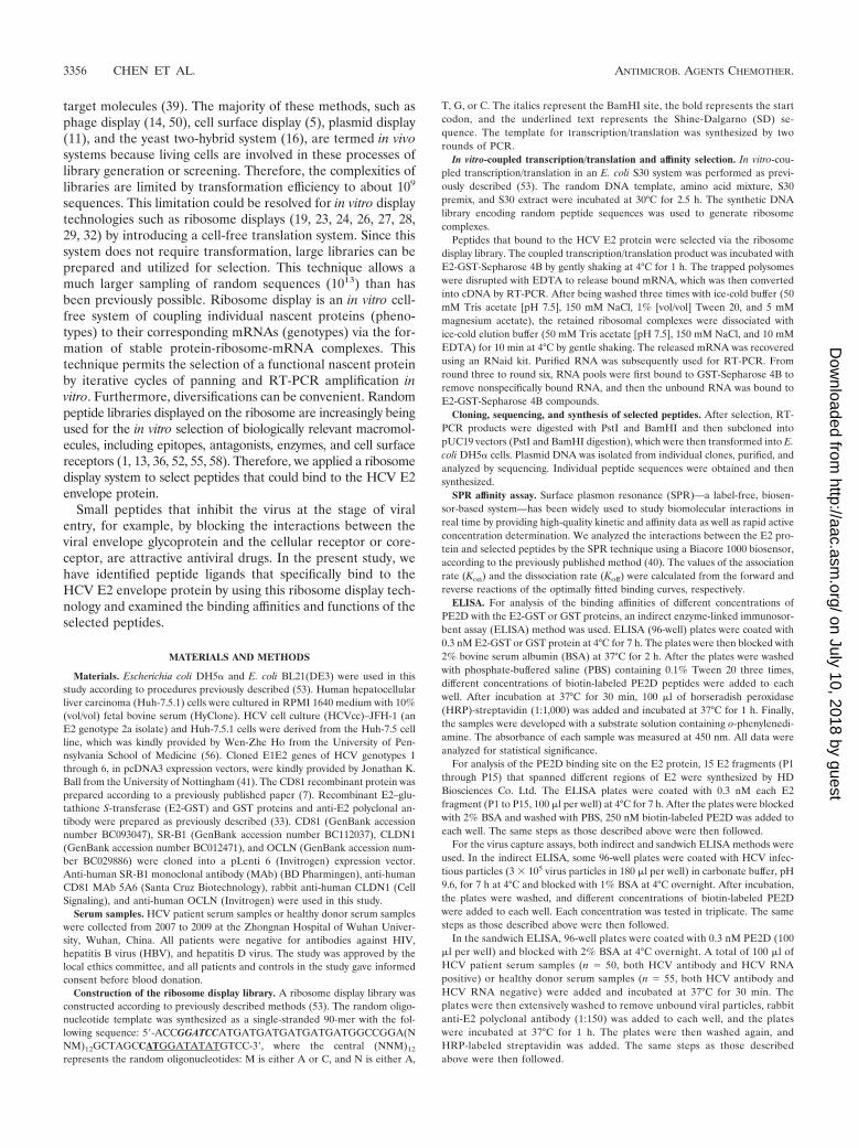
Selection of peptides that bind to HCV E2 by ribosomedisplay. First, E2-GST and GST proteins were purified andidentified by SDS-PAGE (Fig. 1A) and Western blot analysis(Fig. 1B). To isolate peptides that specifically bound to theHCV E2 protein, we utilized E2-GST-Sepharose 4B as a se-lection target and GST-Sepharose 4B as a counterselectiontarget. The ribosome display system with a distinct random12-mer peptide library was utilized (53), and mRNA-carryingpeptide products were bound to E2-GST-Sepharose 4B beads(Fig. 1C). After a 12-cycle selection, the 12th-pool clones fromRNA that bound to E2-GST-Sepharose 4B beads were reversetranscribed by RT-PCR and cloned into pUC19 vectors. Thirtyindividual clones were selected for DNA sequencing and thentranslated into 12-mer amino acid sequences. The results of anNCBI BLAST analysis showed that the four selected 12-merpeptides have protein-binding potentials, and they were thenfurther selected for chemical synthesis and named PE2A,PE2B, PE2C, and PE2D (Table 1).
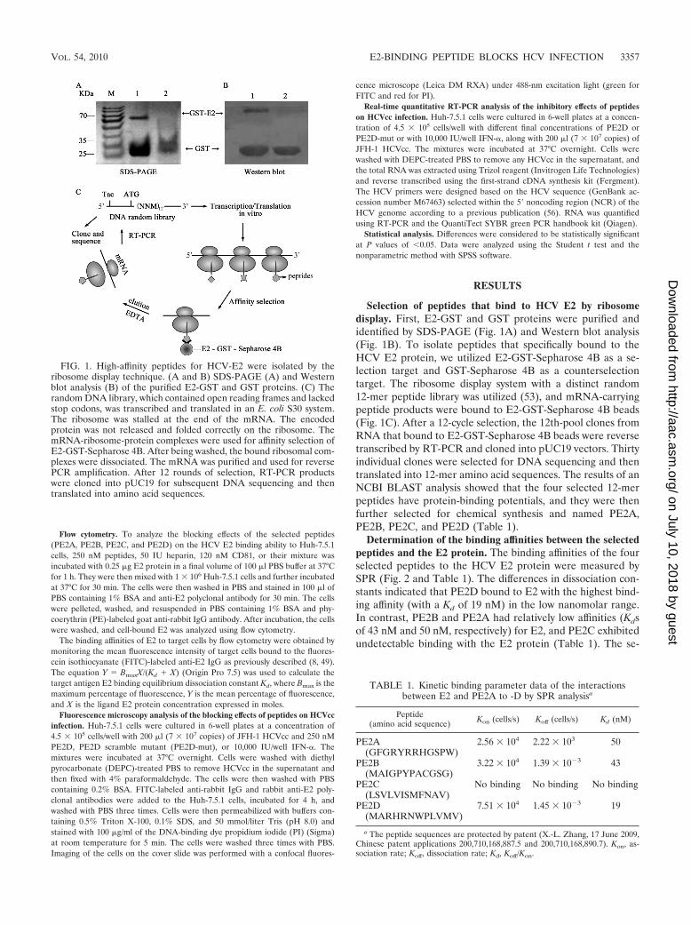
Determination of the binding affinities between the selectedpeptides and the E2 protein. The binding affinities of the fourselected peptides to the HCV E2 protein were measured bySPR (Fig. 2 and Table 1). The differences in dissociation con-stants indicated that PE2D bound to E2 with the highest bind-ing affinity (with a Kd of 19 nM) in the low nanomolar range.In contrast, PE2B and PE2A had relatively low affinities (Kdsof 43 nM and 50 nM, respectively) for E2, and PE2C exhibitedundetectable binding with the E2 protein (Table 1). The se-
FIG. 1. High-affinity peptides for HCV-E2 were isolated by theribosome display technique. (A and B) SDS-PAGE (A) and Westernblot analysis (B) of the purified E2-GST and GST proteins. (C) Therandom DNA library, which contained open reading frames and lackedstop codons, was transcribed and translated in an E. coli S30 system.The ribosome was stalled at the end of the mRNA. The encodedprotein was not released and folded correctly on the ribosome. ThemRNA-ribosome-protein complexes were used for affinity selection ofE2-GST-Sepharose 4B. After being washed, the bound ribosomal com-plexes were dissociated. The mRNA was purified and used for reversePCR amplification. After 12 rounds of selection, RT-PCR productswere cloned into pUC19 for subsequent DNA sequencing and thentranslated into amino acid sequences.
TABLE 1. Kinetic binding parameter data of the interactionsbetween E2 and PE2A to -D by SPR analysisa
Peptide(amino acid sequence) Kon (cells/s) Koff (cells/s) Kd (nM)
PE2A(GFGRYRRHGSPW)
2.56 � 104 2.22 � 103 50
PE2B(MAIGPYPACGSG)
3.22 � 104 1.39 � 10�3 43
PE2C(LSVLVISMFNAV)
No binding No binding No binding
PE2D(MARHRNWPLVMV)
7.51 � 104 1.45 � 10�3 19
a The peptide sequences are protected by patent (X.-L. Zhang, 17 June 2009,Chinese patent applications 200,710,168,887.5 and 200,710,168,890.7). Kon, as-sociation rate; Koff, dissociation rate; Kd, Koff/Kon.
VOL. 54, 2010 E2-BINDING PEPTIDE BLOCKS HCV INFECTION 3357
on July 10, 2018 by guesthttp://aac.asm
.org/D
ownloaded from

quence of binding affinities was as follows: PE2D (Kd, 19nM) PE2B (Kd, 43 nM) PE2A (Kd, 50 nM) PE2C(undetectable Kd). Usually, the peptide with the lowest Kd
displays the highest binding affinity (40, 53). Therefore, PE2Dwas chosen for further characterization, as it had the highestE2 binding affinity based on dissociation constants.
An ELISA analysis showed that PE2D bound to E2-GST ina peptide concentration-dependent manner (Fig. 2B). How-ever, different concentrations of PE2D did not have any de-tectable binding affinities to the GST control protein (Fig. 2B).However, different concentrations of scrambled PE2D (PE2D-mut) had a significantly lower binding affinity to E2-GST thanPE2D (Fig. 2B).
The selected peptide PE2D specifically targets HCV parti-cles. To determine whether PE2D could bind to HCV parti-cles, virus capture assays were performed by both indirect andsandwich ELISA methods. In the indirect ELISA, virus sam-ples from HCVcc or control cell culture medium were used tocoat ELISA plates and incubated with different concentrationsof biotin-labeled PE2D, followed by extensive washing to re-move unbound peptide. The peptide-bound viral particles onthe ELISA plate were revealed using HRP-labeled streptavi-din, as described in Materials and Methods. The resultsshowed that PE2D bound to HCVcc in a concentration-depen-dent manner (Fig. 3A), while it did not bind to the control cellculture medium (Fig. 3A). These results are consistent with thebinding affinities of PE2D with the different concentrations ofE2 protein shown in Fig. 2B. However, the different concen-trations of PE2D-mut had significantly lower binding abilitiesto HCVcc than PE2D (Fig. 3B).
In the sandwich ELISA, PE2D was adsorbed to the ELISAplate, and serum samples from 50 HCV patients and 55 healthydonors were added and incubated. The PE2D-bound viral par-ticles were revealed following the addition of anti-E2 poly-clonal antibody and HRP-labeled IgG. As shown in Fig. 3B,biotin-labeled PE2D could bind to HCV particles in HCVpatient samples with much higher affinities than it could tohealthy donor samples (P � 0.05). These data suggest that theselected peptide PE2D specifically targets HCV particles andcould be used to diagnose early HCV infection by detecting
HCV surface antigen in sera that is present during the earlystage or before seroconversion.
Identification of the PE2D peptide-binding site on the E2protein. To further identify the PE2D peptide-binding site onE2, 15 peptide fragments (P1 to P15), which were based on thegenotype 1a E2 amino acid sequence and secondary structure,were synthesized (Fig. 4A). In the indirect ELISAs, each E2fragment (P1 to P15) was used to coat ELISA plates andincubated with biotin-labeled PE2D. The biotin-PE2D-boundE2 fragments in the ELISA plate were revealed by the HRP-labeled streptavidin, as described in Materials and Methods.Of all the E2 fragments tested, the results showed that PE2Dbound most strongly to the E2-P7 fragment sequence (YPPR
FIG. 2. Binding affinity analysis of the interaction between selectedpeptides and the HCV E2 protein. (A) A typical time profile of theSPR signal. The SPR analysis was performed as described in Materialsand Methods. The data demonstrate the interaction between E2 andthe four peptides, PE2A to -D. PE2C has the lowest binding affinity.(B) The indirect ELISA method was used to analyze the bindingaffinities of different doses of PE2D and PE2D-mut with the E2-GSTor GST proteins. OD, optical density.
FIG. 3. Virus capture assays. (A) Binding affinity analysis of theinteraction between different doses of PE2D and HCVcc (3 � 105 virusparticles in 180 �l per well) in 96-well plates by indirect ELISA. Thedata shown are the means standard errors of the mean (SEM) fromsix separate experiments. (B) Serum samples from both HCV patientsand healthy donors were analyzed by the sandwich ELISA. HCVparticles from HCV patient serum samples could be specifically cap-tured using peptide PE2D. Peptide PE2D-bound viral particles in theELISA plate were revealed by addition of anti-E2 polyclonal antibodyand HRP-labeled IgG. The absorbance of each sample was measuredat 450 nm. Data were analyzed with the nonparametric statisticalanalysis method.
FIG. 4. Identification of the PE2D peptide-binding site of the E2protein. (A) Diagram of the 15 E2 peptide fragments (P1 to P15) thatspanned different regions of E2.
3358 CHEN ET AL. ANTIMICROB. AGENTS CHEMOTHER.
on July 10, 2018 by guesthttp://aac.asm
.org/D
ownloaded from
PCGIVPAKSVCGPVYC) (Fig. 4B). The binding affinity be-tween PE2D and P7 was 2- to 20-fold higher than those be-tween PE2D and the other 14 peptides (Fig. 4A). While PE2Aexhibited much lower binding affinities to each E2 peptide (P1to P15) of E2, it still exhibited binding affinities that werehigher than those of the PBS control, while PE2D-mut showedsignificantly lower binding affinities to each E2 peptide (P1 toP15) than PE2D (Fig. 4B).
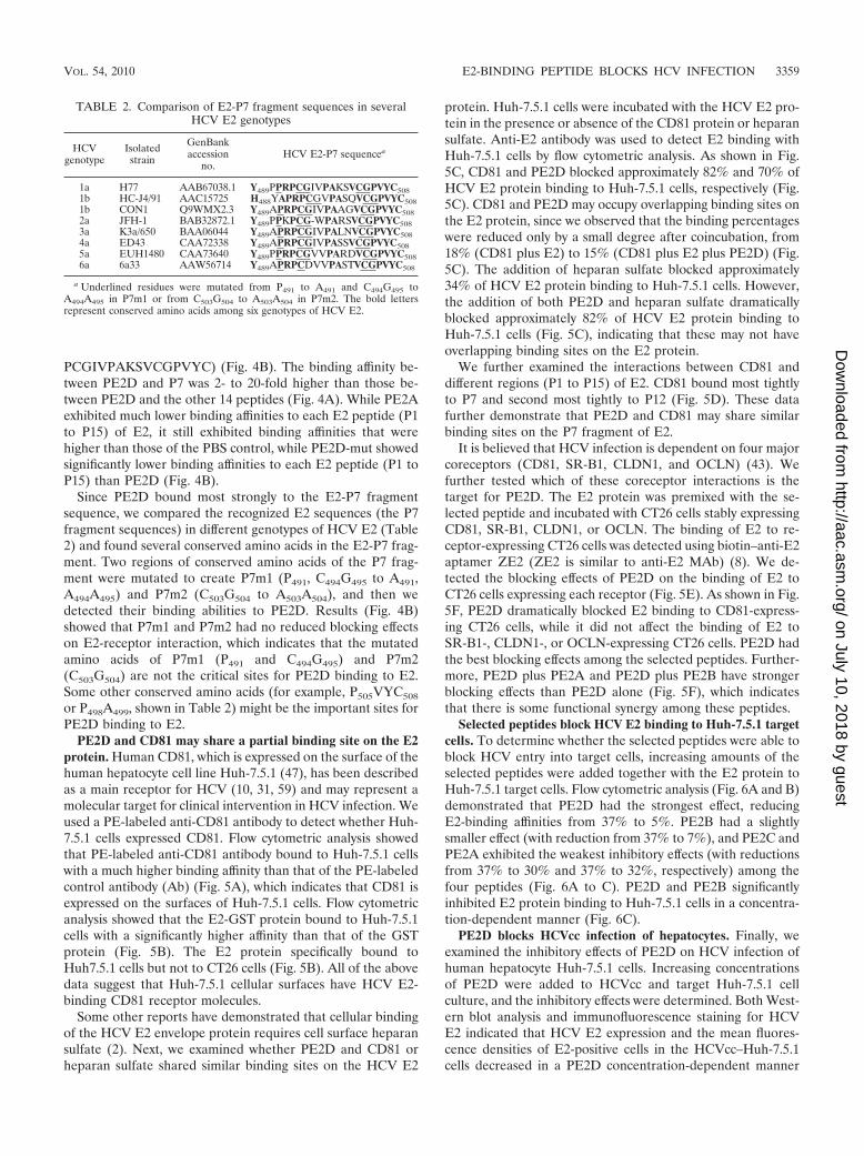
Since PE2D bound most strongly to the E2-P7 fragmentsequence, we compared the recognized E2 sequences (the P7fragment sequences) in different genotypes of HCV E2 (Table2) and found several conserved amino acids in the E2-P7 frag-ment. Two regions of conserved amino acids of the P7 frag-ment were mutated to create P7m1 (P491, C494G495 to A491,A494A495) and P7m2 (C503G504 to A503A504), and then wedetected their binding abilities to PE2D. Results (Fig. 4B)showed that P7m1 and P7m2 had no reduced blocking effectson E2-receptor interaction, which indicates that the mutatedamino acids of P7m1 (P491 and C494G495) and P7m2(C503G504) are not the critical sites for PE2D binding to E2.Some other conserved amino acids (for example, P505VYC508
or P498A499, shown in Table 2) might be the important sites forPE2D binding to E2.
PE2D and CD81 may share a partial binding site on the E2protein. Human CD81, which is expressed on the surface of thehuman hepatocyte cell line Huh-7.5.1 (47), has been describedas a main receptor for HCV (10, 31, 59) and may represent amolecular target for clinical intervention in HCV infection. Weused a PE-labeled anti-CD81 antibody to detect whether Huh-7.5.1 cells expressed CD81. Flow cytometric analysis showedthat PE-labeled anti-CD81 antibody bound to Huh-7.5.1 cellswith a much higher binding affinity than that of the PE-labeledcontrol antibody (Ab) (Fig. 5A), which indicates that CD81 isexpressed on the surfaces of Huh-7.5.1 cells. Flow cytometricanalysis showed that the E2-GST protein bound to Huh-7.5.1cells with a significantly higher affinity than that of the GSTprotein (Fig. 5B). The E2 protein specifically bound toHuh7.5.1 cells but not to CT26 cells (Fig. 5B). All of the abovedata suggest that Huh-7.5.1 cellular surfaces have HCV E2-binding CD81 receptor molecules.
Some other reports have demonstrated that cellular bindingof the HCV E2 envelope protein requires cell surface heparansulfate (2). Next, we examined whether PE2D and CD81 orheparan sulfate shared similar binding sites on the HCV E2
protein. Huh-7.5.1 cells were incubated with the HCV E2 pro-tein in the presence or absence of the CD81 protein or heparansulfate. Anti-E2 antibody was used to detect E2 binding withHuh-7.5.1 cells by flow cytometric analysis. As shown in Fig.5C, CD81 and PE2D blocked approximately 82% and 70% ofHCV E2 protein binding to Huh-7.5.1 cells, respectively (Fig.5C). CD81 and PE2D may occupy overlapping binding sites onthe E2 protein, since we observed that the binding percentageswere reduced only by a small degree after coincubation, from18% (CD81 plus E2) to 15% (CD81 plus E2 plus PE2D) (Fig.5C). The addition of heparan sulfate blocked approximately34% of HCV E2 protein binding to Huh-7.5.1 cells. However,the addition of both PE2D and heparan sulfate dramaticallyblocked approximately 82% of HCV E2 protein binding toHuh-7.5.1 cells (Fig. 5C), indicating that these may not haveoverlapping binding sites on the E2 protein.
We further examined the interactions between CD81 anddifferent regions (P1 to P15) of E2. CD81 bound most tightlyto P7 and second most tightly to P12 (Fig. 5D). These datafurther demonstrate that PE2D and CD81 may share similarbinding sites on the P7 fragment of E2.
It is believed that HCV infection is dependent on four majorcoreceptors (CD81, SR-B1, CLDN1, and OCLN) (43). Wefurther tested which of these coreceptor interactions is thetarget for PE2D. The E2 protein was premixed with the se-lected peptide and incubated with CT26 cells stably expressingCD81, SR-B1, CLDN1, or OCLN. The binding of E2 to re-ceptor-expressing CT26 cells was detected using biotin–anti-E2aptamer ZE2 (ZE2 is similar to anti-E2 MAb) (8). We de-tected the blocking effects of PE2D on the binding of E2 toCT26 cells expressing each receptor (Fig. 5E). As shown in Fig.5F, PE2D dramatically blocked E2 binding to CD81-express-ing CT26 cells, while it did not affect the binding of E2 toSR-B1-, CLDN1-, or OCLN-expressing CT26 cells. PE2D hadthe best blocking effects among the selected peptides. Further-more, PE2D plus PE2A and PE2D plus PE2B have strongerblocking effects than PE2D alone (Fig. 5F), which indicatesthat there is some functional synergy among these peptides.
Selected peptides block HCV E2 binding to Huh-7.5.1 targetcells. To determine whether the selected peptides were able toblock HCV entry into target cells, increasing amounts of theselected peptides were added together with the E2 protein toHuh-7.5.1 target cells. Flow cytometric analysis (Fig. 6A and B)demonstrated that PE2D had the strongest effect, reducingE2-binding affinities from 37% to 5%. PE2B had a slightlysmaller effect (with reduction from 37% to 7%), and PE2C andPE2A exhibited the weakest inhibitory effects (with reductionsfrom 37% to 30% and 37% to 32%, respectively) among thefour peptides (Fig. 6A to C). PE2D and PE2B significantlyinhibited E2 protein binding to Huh-7.5.1 cells in a concentra-tion-dependent manner (Fig. 6C).
PE2D blocks HCVcc infection of hepatocytes. Finally, weexamined the inhibitory effects of PE2D on HCV infection ofhuman hepatocyte Huh-7.5.1 cells. Increasing concentrationsof PE2D were added to HCVcc and target Huh-7.5.1 cellculture, and the inhibitory effects were determined. Both West-ern blot analysis and immunofluorescence staining for HCVE2 indicated that HCV E2 expression and the mean fluores-cence densities of E2-positive cells in the HCVcc–Huh-7.5.1cells decreased in a PE2D concentration-dependent manner
TABLE 2. Comparison of E2-P7 fragment sequences in severalHCV E2 genotypes
HCVgenotype
Isolatedstrain
GenBankaccession
no.HCV E2-P7 sequencea
1a H77 AAB67038.1 Y489PPRPCGIVPAKSVCGPVYC5081b HC-J4/91 AAC15725 H488YAPRPCGVPASQVCGPVYC5081b CON1 Q9WMX2.3 Y489APRPCGIVPAAGVCGPVYC5082a JFH-1 BAB32872.1 Y489PPKPCG-WPARSVCGPVYC5083a K3a/650 BAA06044 Y489APRPCGIVPALNVCGPVYC5084a ED43 CAA72338 Y489APRPCGIVPASSVCGPVYC5085a EUH1480 CAA73640 Y489PPRPCGVVPARDVCGPVYC5086a 6a33 AAW56714 Y489APRPCDVVPASTVCGPVYC508
a Underlined residues were mutated from P491 to A491 and C494G495 toA494A495 in P7m1 or from C503G504 to A503A504 in P7m2. The bold lettersrepresent conserved amino acids among six genotypes of HCV E2.
VOL. 54, 2010 E2-BINDING PEPTIDE BLOCKS HCV INFECTION 3359
on July 10, 2018 by guesthttp://aac.asm
.org/D
ownloaded from

(Fig. 7A and B). Immunofluorescence microscopy demon-strated that HCV was detected in the cytoplasm of these cells(Fig. 7C) and the fluorescence densities were decreased uponaddition of PE2D or IFN-� (Fig. 7C), while addition of thePE2D mutant had no or much less effect (Fig. 7A to D).Results similar to these were obtained with real-time quanti-tative RT-PCR analysis (Fig. 7E). In addition, both Westernblot and real-time RT-PCR analyses showed that the inhibitory
effects of 350 nM PE2D were virtually equivalent to those of10,000 IU of IFN-� (Fig. 7A and E). These data illustrate thatPE2D specifically inhibits HCV infection of human hepato-cytes.
HCV has six major genotypes and numerous subtypes basedon its positive-stranded RNA genome (20). HepG2 cells stablyexpressing the E1E2 proteins encoded by the E1E2 genes(from genotypes 1 to 6) were established by G418 selection,
FIG. 5. PE2D can partially page HCV E2 binding with the CD81 receptor. (A) CD81 molecules are expressed on the cellular surface ofHuh-7.5.1 cells, as demonstrated by flow cytometric analysis with PE-labeled anti-CD81 antibody. (B) HCV E2 had a much higher binding affinityfor Huh-7.5.1 cells than for CT26 cells by flow cytometric analysis. E2-GST or GST proteins (0.25 �g) were preincubated with 1 � 106 Huh-7.5.1or CT26 cells, respectively. Anti-GST antibody and FITC–anti-IgG were added and analyzed by flow cytometric analysis. (C) PE2D and CD81 mayshare a partial binding site on the E2 protein. Huh-7.5.1 cells mixed with PE2D, CD81, heparin, CD81 plus PE2D, or heparin plus PE2D wereincubated with E2 protein at 37°C for 1 h, as described in Materials and Methods, and then cell-bound E2 was detected by flow cytometric analysiswith rabbit anti-E2 polyclonal antibody stained with PE-conjugated rabbit IgG. The data are shown as the means SEM from three separateexperiments. (D) The binding affinities of GST-CD81 for the different E2 fragments (P1 to P15) were measured using ELISA. The data are shownas means SEM from six separate experiments. (E) Determination of CD81, SR-B1, CLDN1, or OCLN expression in the CD81-, SR-B1-,CLDN1-, or OCLN-transfected CT26 cells by Western blot analysis, using anti-CD81 MAb 5A6, anti-SR-B1 MAb, anti-CLDN1, or anti-OCLN,respectively. (F) Comparison of the blocking effects of PE2D, PE2B, PE2A, or PE2D-mut on the E2 receptor interactions. The E2 protein (1�g/100 �l) was premixed with the selected peptide (100 pmol/100 �l) and incubated with CT26 cells (1 � 105) stably expressing CD81, SR-B1,CLDN1, or OCLN in 96-well plates. The binding of E2 to receptor-expressing CT26 cells was detected using 50 nM biotin-anti-E2 aptamer ZE2,followed by the addition of HRP-streptavidin. The absorbance of each sample was measured at 450 nm.
3360 CHEN ET AL. ANTIMICROB. AGENTS CHEMOTHER.
on July 10, 2018 by guesthttp://aac.asm
.org/D
ownloaded from

and E2 expressions from these cells were determined by West-ern blot analysis (Fig. 8A). Different genotypes of HepG2 cellsstably expressing the E1E2 gene were used to coat ELISAplates. We utilized ELISA to determine which genotypes pos-sessed E1E2 that could be bound by PE2D. The results showedthat the sequence of binding affinities of PE2D to E2 of eachgenotype was as follows: genotype 2a 1a, 1b 3a 5a 6a 4a, and the binding affinities of PE2D with genotypes 1a, 1b,and 2a were 2- to 3-fold higher than those of genotypes 3a, 4a,5a, and 6a (Fig. 8B). This result suggests that HCV genotypes1 and 2 could be most effectively blocked by PE2D.
DISCUSSION
Ribosome display has been used for the selection of peptideligands (19, 32, 52) and proteins in vitro (4, 13, 24). One of thekey advantages of ribosome display is that the diversity of thelibrary is not limited by the transformation efficiency of bacte-rial cells (23). Additionally, ribosome display has been used toimprove the affinity (4, 32) and the stability (28) of existingprotein ligands. Using ribosome display, this study successfullygenerated 12-mer peptides that bound specifically to the HCVE2 protein. To our knowledge, this is the first time this findinghas been demonstrated. We applied the SPR sensor system
(Table 1 and Fig. 2A), ELISA (Fig. 2B and 3), and flow cyto-metric analysis methods (Fig. 5 and 6) to investigate the kineticparameters and binding affinities of the selected peptides tothe HCV E2 protein and HCV particles. Notably, we identifiedthe strongest binding and interaction between the selectedpeptide PE2D and the E2 protein. The results of an NCBIBLAST analysis further demonstrated that the PE2D sequencewas 75% identical (8/12 amino acid residues) to laminin(GenBank accession number EAW 75372) amino acids 3357 to3368 (LARHRNWPSLSM [underlined letters represent iden-tical residues]). These data illustrate that PE2D most likely hasthe binding domain of the laminin-alpha5 chain domain, whichfunctions normally for protein binding (57). Because the mo-lecular weight of laminin is much larger than that of PE2D,whether laminin can bind to E2 and block E2 binding to itsreceptors needs to be further investigated.
Recently, several reports showed that HCV cell entry is a mul-tistep process (43). It is believed that the initial capture of HCVparticles by glycosaminoglycans and/or lipoprotein receptors isfollowed by coordinated interactions with the SR-BI, the CD81tetraspanin, the tight junction protein CLDN1, and/or OCLN.The different regions, structures, and conformations of E2 mightbe involved in binding to different coreceptors.
FIG. 6. Inhibitory effects of the selected peptides on E2 protein binding to Huh-7.5.1 cells. (A) Huh-7.5.1 cells (1 � 106) were incubated with0.25 �g E2-GST, GST alone, or E2-GST plus the 250 nM peptides (PE2A to D), followed by staining with anti-GST antibody. Cell-bound GST-E2was detected with rabbit polyclonal anti-GST antibody stained with anti-rabbit FITC-IgG. Binding was analyzed by flow cytometric analysis. Thedata shown are representative of five experiments. (B) Statistical analysis of the E2-GST or GST binding percentages of Huh-7.5.1 cells asdetermined by flow cytometry. The data shown are the means SEM from four separate experiments. (C) Cell-bound E2 was examined using flowcytometry following the addition of different concentrations of PE2D, PE2A, or PE2B. The inhibitory percentages of selected peptides on theHuh-7.5.1 cell-bound E2 were calculated. All data shown are means SEM from four separate experiments.
VOL. 54, 2010 E2-BINDING PEPTIDE BLOCKS HCV INFECTION 3361
on July 10, 2018 by guesthttp://aac.asm
.org/D
ownloaded from

Several conserved residues in HCV E2 for CD81 bindingwere identified as W420, Y527, W529, G530, and D535 (41).Three putative CD81 interaction sites on HCV E2 have alsobeen previously identified: region 1, amino acids 474 to 492(YANGSGLDERPYCWHYPPR) (47); region 2, amino acids522 to 551 (SGAPTYSWGANDTDVFVLNNTRPPLGNWFG) (47); and region 3, amino acids 612 to 619 (PYRLWHYPC) (47). We found that PE2D binds to amino acids 489 to508 of E2 (Fig. 4A and B), and CD81 binds most tightly withthe P7 fragment (amino acids 489 to 508) of E2 as well (Fig.5D). These residues partly overlap with the previously identi-
fied CD81-binding region 1 (amino acids 474 to 492) of E2 (47)and are close to the CD81-binding conserved residues W420and Y527 and region 2 (amino acids 522 to 551) of E2 (41, 47).These data demonstrate that PE2D and CD81 share an over-lapping binding site on the P7 fragment of E2 and that PE2Dcan partially block HCV E2 binding with the CD81 receptorand subsequent viral entry by binding to these residues of E2.
Finally, using Western blot, real-time RT-PCR, and immu-nofluorescence staining analyses, we found that the selectedpeptides, especially PE2D, bound to E2 and HCV particles ina concentration-dependent manner (Fig. 3A) and that PE2D,
FIG. 7. PE2D dramatically inhibits HCVcc infection of Huh-7.5.1 cells. (A) Western blot analysis of HCVcc incubated with differentconcentrations of PE2D or PE2D-mut in Huh-7.5.1 cells with anti-E2 antibody. The �-actin gene, a housekeeping gene with constant expression,was used as an internal control. (B) The differences in fluorescence of HCVcc incubated with different concentrations of PE2D or PE2D-mut inHuh-7.5.1 cells are shown as the means SEM from three separate experiments. ffu, focus-forming units. (C) Confocal fluorescence microscopicanalysis of immunofluorescent detection of E2 in HCVcc-labeled Huh-7.5.1 cells. Cell nuclei were stained with the red fluorescent dye PI, andHCV was stained with green FITC-conjugated anti-E2 antibody. The illustrated data are representative of three separate experiments. (D) Quan-titation (means SEM from three separate experiments) of the intracellular HCV E2 mean fluorescence intensities (MFI) shown in panel C.(E) Levels of intracellular HCV-E2 RNA were measured by real-time RT-PCR analysis. HCV-E2 RNA levels were normalized based on GAPDH(glyceraldehyde-3-phosphate dehydrogenase) mRNA levels assessed by real-time RT-PCR. The data shown are the means SEM from threeindependent experiments.
3362 CHEN ET AL. ANTIMICROB. AGENTS CHEMOTHER.
on July 10, 2018 by guesthttp://aac.asm
.org/D
ownloaded from

but not its mutant, significantly inhibits HCVcc binding andentry into human hepatocyte Huh-7.5.1 cells (Fig. 7). Thesedata suggest that PE2D may also hold potential for use as adrug against HCV infection; this potential of PE2D is strength-ened by its small molecular weight (12-mer), its low immuno-genicity, and the ease of its chemical synthesis in large quan-tities at a relatively low cost. The newly selected peptides,especially PE2D, are worthy of further clinical and basic re-search.
The limitations of the methods of detection of anti-HCVantibodies or HCV RNA in serum that are currently used forthe diagnosis of HCV infection have enhanced efforts to find arapid, simple, sensitive, and specific alternative diagnostic ap-proach to detect viral antigens. Similar to the detection of anHBV surface antigen for the diagnosis of HBV infection, theselected E2-binding peptides may be helpful for the diagnosisof early HCV infection through a simple ELISA method todetect HCV E2 envelope surface antigen. Although HCV E2contains approximately 1 or 2 amino acid sequence hypervari-able regions (HVR), most of the E2 residues and the chemi-cophysical properties and conformation of HVR are highlyconserved (42, 46). Our experimental results showed thatPE2D specifically targets HCV particles and could also be usedto diagnose early HCV infection by detecting HCV surfaceantigen in sera (Fig. 3A and B), especially for HCV genotypes1 and 2 (Fig. 8B). Therefore, PE2D holds great potential forthe detection of HCV surface antigen in both clinical applica-tions and basic research.
In summary, our data demonstrate that the selected pep-tides, especially PE2D, hold great promise for developing newmolecular probes, as therapeutic drugs, or as early diagnosticreagents targeting HCV. The selected peptides can also serve
as tools for analyzing HCV-host cell interactions both in vitroand in vivo.
ACKNOWLEDGMENTS
This work was supported by grants from the National Natural Sci-ence Foundation of China (20532020, 30670098, and 30870122), the973 Program of China (2006CB504300 and 2009CB522507), the Na-tional Special Fund of China for the Important Infectious Diseases(2008ZX10003-005 and 2008ZX10002-013), and the Hubei ProvinceNatural Science Foundation (2006ABD007).
The authors declare that there are patent applications for this workand for peptide sequences (X.-L. Zhang, 17 June 2009, Chinese patentapplications 200,710,168,887.5 and 200,710,168,890.7).
REFERENCES
1. Amstutz, P., P. Forrer, C. Zahnd, and A. Pluckthun. 2001. In vitro displaytechnologies: novel developments and applications. Curr. Opin. Biotechnol.12:400–405.
2. Barth, H., C. Schafer, M. I. Adah, F. Zhang, R. J. Linhardt, H. Toyoda, A. K.Toyoda, T. Toida, T. H. Kuppevelt, E. Depla, F. von Weizsacker, H. E. Blum,and T. F. Baumert. 2003. Cellular binding of hepatitis C virus envelopeglycoprotein E2 requires cell surface heparan sulfate. J. Biol. Chem. 278:41003–41012.
3. Bartosch, B., A. Vitelli, C. Granier, C. Goujon, J. Dubuisson, S. Pascale, E.Scarselli, R. Cortese, A. Nicosia, and F. L. Cosset. 2003. Cell entry ofhepatitis C virus requires a set of co-receptors that include the CD81 tet-raspanin and the SR-B1 scavenger receptor. J. Biol. Chem. 278:41624–41630.
4. Binz, H. K., P. Amstutz, A. Kohl, M. T. Stumpp, C. Briand, P. Forrer, M. G.Grutter, and A. Pluckthun. 2004. High-affinity binders selected from de-signed ankyrin repeat protein libraries. Nat. Biotechnol. 22:575–582.
5. Boder, E. T., and K. D. Wittrup. 1997. Yeast surface display for screeningcombinatorial polypeptide libraries. Nat. Biotechnol. 15:553–557.
6. Boyer, N., and P. Marcellin. 2000. Pathogenesis, diagnosis and managementof hepatitis C. J. Hepatol. 32:98–112.
7. Cao, J., P. Zhao, X. H. Miao, L. J. Zhao, L. J. Xue, and Z. Z. Qi. 2003. Phagedisplay selection on whole cells yields a small peptide specific for HCVreceptor human CD81. Cell Res. 13:473–479.
8. Chen, F., Y. Hu, D. Li, H. Chen, and X. L. Zhang. 2009. CS-SELEX gener-ates high-affinity ssDNA aptamers as molecular probes for hepatitis C virusenvelope glycoprotein E2. PLoS One 4:e8142.
9. Choo, Q. L., G. Kuo, A. J. Weiner, L. R. Overby, D. W. Bradley, and M.Houghton. 1989. Isolation of a cDNA clone derived from a blood-bornenon-A, non-B viral hepatitis genome. Science 244:359–362.
10. Cocquerel, L., C. Voisset, and J. Dubuisson. 2006. Hepatitis C virus entry:potential receptors and their biological functions. J. Gen. Virol. 87:1075–1084.
11. Cull, M. G., J. F. Miller, and P. J. Schatz. 1992. Screening for receptorligands using large libraries of peptides linked to the C terminus of the lacrepressor. Proc. Natl. Acad. Sci. U. S. A. 89:1865–1869.
12. De Cock, L., V. Hutse, and R. Vranckx. 2005. Correlation between detectionof antibodies against hepatitis C virus in oral fluid and hepatitis C virus RNAin serum. Eur. J. Clin. Microbiol. Infect. Dis. 24:566–568.
13. Dower, W. J., and L. C. Mattheakis. 2002. In vitro selection as a powerfultool for the applied evolution of proteins and peptides. Curr. Opin. Chem.Biol. 6:390–398.
14. Dunn, I. S. 1996. Phage display of proteins. Curr. Opin. Biotechnol. 7:547–553.
15. Evans, M. J., T. von Hahn, N. M. Tscherne, A. J. Svder, M. Panis, B. Wolk,T. Hatziioannou, J. A. McKeating, P. D. Bieniasz, and C. M. Rice. 2007.Claudin-1 is a hepatitis C virus co-receptor required for a late step in entry.Nature 446:801–805.
16. Fields, S., and O. Song. 1989. A novel genetic system to detect protein-protein interactions. Nature 340:245–246.
17. Flint, M., and J. A. McKeating. 2000. The role of the hepatitis C virusglycoproteins in infection. Rev. Med. Virol. 10:101–117.
18. Flint, M., T. von Hahn, J. Zhang, M. Farquhar, C. T. Jones, P. Balfe, C. M.Rice, and J. A. McKeating. 2006. Diverse CD81 proteins support hepatitis Cvirus infection. J. Virol. 80:11331–11342.
19. Gersuk, G. M., M. J. Corey, F. Corey, J. E. Stray, G. H. Kawasaki, and R. L.Vessella. 1997. High-affinity peptide ligands to prostate-specific antigen iden-tified by polysome selection. Biochem. Biophys. Res. Commun. 232:578–582.
20. Gottwein, J. M., T. K. Scheel, T. B. Jensen, J. B. Lademann, J. C. Prentoe,M. L. Knudsen, A. M. Hoegh, and J. Bukh. 2009. Development and char-acterization of hepatitis C virus genotype 1–7 cell culture systems: role ofCD81 and scavenger receptor class B type I and effect of antiviral drugs.Hepatology 49:364–377.
21. Grove, J., S. Nielsen, J. Zhong, M. F. Bassendine, H. E. Drummer, P. Balfe,and J. A. McKeating. 2008. Identification of a resident in hepatitis C virus E2
FIG. 8. Different genotypes of E2 detected by PE2D. (A) Deter-mination of E2 protein expressions of different genotypes of HepG2cells stably expressing the E1E2 gene with anti-E2 antibody by West-ern blot analysis. HepG2 cells were used as the control. (B) Differentgenotypes of E2 were detected by PE2D. Data shown are the means SEM from three independent experiments.
VOL. 54, 2010 E2-BINDING PEPTIDE BLOCKS HCV INFECTION 3363
on July 10, 2018 by guesthttp://aac.asm
.org/D
ownloaded from

glycoprotein that determines scavenger receptor BI and CD81 receptordependency and sensitivity to neutralizing antibodies. J. Virol. 82:12020–12029.
22. Grove, J., T. Huby, Z. Stamataki, T. Vanwolleghem, P. Meuleman, M. Far-quhar, A. Schwarz, M. Moreau, J. S. Owen, G. Leroux-Roels, P. Balfe, andJ. A. McKeating. 2007. Scavenger receptor BI and BII expression levelsmodulate hepatitis C virus infectivity. J. Virol. 81:3162–3169.
23. Hanes, J., and A. Pluckthun. 1997. In vitro selection and evolution of func-tional proteins by using ribosome display. Proc. Natl. Acad. Sci. U. S. A.94:4937–4942.
24. Hanes, J., C. Schatzel, A. Knappik, and A. Pluckthun. 2000. Picomolaraffinity antibodies from a fully synthetic naive library selected and evolved byribosome display. Nat. Biotechnol. 18:1287–1292.
25. Harris, H. J., M. J. Farquhar, C. J. Mee, C. Davis, G. M. Reynolds, A.Jennings, K. Hu, F. Yuan, H. Deng, S. G. Hubscher, J. H. Han, P. Balfe, andJ. A. McKeating. 2008. CD81 and claudin 1 coreceptor association: role inhepatitis C virus entry. J. Virol. 82:5007–5020.
26. He, M., and M. J. Taussig. 2007. Rapid discovery of protein interactions bycell-free protein technologies. Biochem. Soc. Trans. 35:962–965.
27. Irving, R. A., G. Coia, A. Roberts, S. D. Nuttall, and P. J. Hudson. 2001.Ribosome display and affinity maturation: from antibodies to single V-domains and steps towards cancer therapeutics. J. Immunol. Methods248:31–45.
28. Jermutus, L., A. Honegger, F. Schwesinger, J. Hanes, and A. Pluckthun.2001. Tailoring in vitro evolution for protein affinity or stability. Proc. Natl.Acad. Sci. U. S. A. 98:75–80.
29. Keefe, A. D. 2001. Protein selecting using mRNA display. Curr. Protoc. Mol.Biol. Unit 24.5, p. 24.5.1–24.5.34. doi:10.1002/0471142727.mb2405s53.
30. Kita, M., M. Deguchi, M. Kagita, N. Yoshioka, E. Kobayashi, M. Watanabe,S. Asari, K. Yamanaka, and Y. Iwatani. 2009. Clinical utility and character-istics of nine anti-HCV antibody screening reagents used in Japan. Clin. Lab.55:9–22.
31. Koutsoudakis, G., E. Herrmann, S. Kallis, R. Bartenschlager, and T. Pi-etschmann. 2007. The level of CD81 cell surface expression is a key deter-minant for productive entry of hepatitis C virus into host cells. J. Virol.81:588–598.
32. Lamla, T., and V. A. Erdmann. 2003. Searching sequence space for high-affinity binding peptides using ribosome display. J. Mol. Biol. 329:381–388.
33. Li, P., Q. Wan, Y. Feng, M. Liu, J. G. Wu, X. W. Chen, and X. L. Zhang.2007. Engineering of N-glycosylation of hepatitis C virus envelope proteinE2 enhances T cell responses for DNA immunization. Vaccine 25:1544–1551.
34. Lindenbach, B. D., and C. M. Rice. 2003. Molecular biology of flaviviridae.Adv. Virus Res. 59:23–61.
35. Lindenbach, B. D., M. J. Evans, A. J. Syder, B. Wolk, T. L. Tellinghuisen,C. C. Liu, T. Maruyama, R. O. Hynes, D. R. Burton, J. A. McKeating, andC. M. Rice. 2005. Complete replication of hepatitis C virus in cell culture.Science 309:623–626.
36. Mattheakis, L. C., R. R. Bhatt, and W. J. Dower. 1994. An in vitro polysomedisplay system for identifying ligands from very large peptide libraries. Proc.Natl. Acad. Sci. U. S. A. 91:9022–9026.
37. McHutchison, J. G., S. C. Gordon, E. R. Schiff, M. L. Shiffman, W. M. Lee,V. K. Rustgi, Z. D. Goodman, M. H. Ling, S. Cort, and J. K. Albrecht. 1998.Interferon alpha-2b alone or in combination with ribavirin as initial treat-ment for chronic hepatitis C. Hepatitis Interventional Therapy Group.N. Engl. J. Med. 339:1485–1492.
38. Meuleman, P., J. Hesselgesser, M. Paulson, T. Vanwolleghem, I. Desombere,H. Reiser, and G. Leroux-Roles. 2008. Anti-CD81 antibodies can prevent ahepatitis C virus infection in vivo. Hepatology 48:1761–1768.
39. Mondon, P., O. Dubreuil, K. Bouayadi, and H. Kharrat. 2008. Humanantibody libraries: a race to engineer and explore a large diversity. Front.Biosci. 13:1117–1129.
40. Murphy, M., L. Jason-Moller, and J. Bruno. 2006. Using Biacore to measurethe binding kinetics of an antibody-antigen interaction. Curr. Protoc. ProteinSci. Chapter 19, Unit 19.14. doi:10.1002/0471142301.ps1914s45.
41. Owsianka, A. M., J. M. Timms, A. W. Tarr, R. J. Brown, T. P. Hickling, A.Szwejk, K. Bienkowska-Szewczyk, B. J. Thomson, A. H. Patel, and J. K. Ball.2006. Identification of conserved residues in the E2 envelope glycoprotein of
the hepatitis C virus that are critical for CD81 binding. J. Virol. 80:8695–8704.
42. Penin, F., C. Combet, G. Germanidis, P. O. Frainais, G. Deleage, and J. M.Pawlotsky. 2001. Conservation of the conformation and positive charges ofhepatitis C virus E2 envelope glycoprotein hypervariable region 1 points toa role in cell attachment. J. Virol. 75:5703–5710.
43. Pietschmann, T. 2009. Virology: final entry key for hepatits C virus. Nature457(7231):797–798.
44. Ploss, A., M. J. Evans, V. A. Gaysinskaya, M. Panis, H. You, Y. P. de Jong,and C. M. Rice. 2009. Human occludin is a hepatitis C virus entry factorrequired for infection of mouse cells. Nature 457(7231):882–886.
45. Poynard, T., P. Marcellin, S. S. Lee, C. Niederau, G. S. Minuk, and G. Ideo.1998. Randomised trial of interferon alpha2b plus ribavirin for 48 weeks orfor 24 weeks versus interferon alpha2b plus placebo for 48 weeks for treat-ment of chronic infection with hepatitis C virus. International HepatitisInterventional Therapy Group. Lancet 352:1426–1432.
46. Roccasecca, R., H. Ansuini, A. Vitelli, A. Meola, E. Scarselli, S. Acali, M.Pezzanera, B. B. Ercole, J. McKeating, A. Yagnik, A. Lahm, A. Tramontano,R. Cortese, and A. Nicosia. 2003. Binding of the hepatitis C virus E2 glyco-protein to CD81 is strain specific and is modulated by a complex interplaybetween hypervariable regions 1 and 2. J. Virol. 77:1856–1867.
47. Rothwangl, K. B., B. Manicassamy, S. L. Uprichard, and L. Rong. 2008.Dissecting the role of putative CD81 binding regions of E2 in mediatingHCV entry: putative CD81 binding region 1 is not involved in CD81 binding.Virol. J. 5:46.
48. Schwarz, A. K., J. Grove, K. Hu, C. J. Mee, P. Balfe, and J. A. McKeating.2009. Hepatoma cell density promotes claudin-1 and scavenger receptor BIexpression and hepatitis C virus internalization. J. Virol. 83:12407–12414.
49. Shangguan, D. H., Y. Li, Z. W. Tang, Z. C. Cao, H. W. Chen, P. Mal-likaratchy, K. Sefah, C. J. Yang, and W. Tan. 2006. Aptamers evolved fromlive cells as effective molecular probes for cancer study. Proc. Natl. Acad. Sci.U. S. A. 103:11838–11843.
50. Smith, G. P., and V. A. Petrenko. 1997. Phage display. Chem. Rev. 97:391–410.
51. Wakita, T., T. Pietschmann, T. Kato, T. Date, M. Miyamoto, Z. Zhao, K.Murthy, A. Habermann, H. G. Krausslich, M. Mizokami, R. Bartenschlager,and T. J. Liang. 2005. Production of infectious hepatitis C virus in tissueculture from a cloned viral genome. Nat. Med. 11:791–796.
52. Weichhart, T., M. Horky, J. Sollner, S. Gangl, T. Henics, E. Nagy, A. Meinke,A. von Gabain, C. M. Fraser, S. R. Gill, M. Hafner, and U. von Ahsen. 2003.Functional selection of vaccine candidate peptides from Staphylococcus au-reus whole-genome expression libraries in vitro. Infect. Immun. 71:4633–4641.
53. Wu, H. Y., X.-L. Zhang, Q. Pan, and J. Wu. 2005. Functional selection of atype IV pili-binding peptide that specifically inhibits Salmonella typhi adhe-sion to/invasion of human monocytic cells. Peptide 26:2057–2063.
54. Wunschmann, S., J. D. Medh, D. Klinzmann, W. N. Schmidt, and J. T.Stapleton. 2000. Characterization of hepatitis C virus (HCV) and HCV E2interactions with CD81 and the low-density lipoprotein receptor. J. Virol.74:10055–10062.
55. Yang, L. M., J. L. Wang, L. Kang, S. Gao, Y. H. Liu, and T. M. Hu. 2008.Construction and analysis of high-complexity ribosome display random pep-tide libraries. PLoS One 3:e2092.
56. Yang, J. H., J. P. Lai, S. D. Douglas, D. Metzger, X. H. Zhu, and W.-Z. Ho.2002. Real-time RT-PCR for quantitation of hepatitis C virus RNA. J. Virol.Methods 102:119–128.
57. Yu, H., and J. F. Talts. 2003. Beta1 integrin and alpha-dystroglycan bindingsites are localized to different laminin-G-domain-like (LG) modules withinthe laminin alpha5 chain G domain. Biochem. J. 371(Pt. 2):289–299.
58. Zahnd, C., P. Amstutz, and A. Pluckthun. 2007. Ribosome display: selectingand evolving proteins in vitro that specifically bind to a target. Nat. Methods4:269–279.
59. Zhang, J., G. Randall, A. Higginbottom, P. Monk, C. M. Rice, and J. A.McKeating. 2004. CD81 is required for hepatitis C virus glycoprotein-medi-ated viral infection. J. Virol. 78:1448–1455.
60. Zhong, J., P. Gastaminza, G. Cheng, S. Kapadia, T. Kato, D. R. Burton, S. F.Wieland, S. L. Uprichard, T. Wakita, and F. V. Chisari. 2005. Robusthepatitis C virus infection in vitro. Proc. Natl. Acad. Sci. U. S. A. 102:9294–9299.
3364 CHEN ET AL. ANTIMICROB. AGENTS CHEMOTHER.
on July 10, 2018 by guesthttp://aac.asm
.org/D
ownloaded from