functional medicine approach to osteoporosis prevention ... · osteoporosis by dexa • white, 118...
TRANSCRIPT

Functional Medicine Approach To OsteoporosisPrevention & Treatment
Elizabeth M. Board, MD
The views and opinions expressed herein are solely those of the presenter and do not necessarily represent those of Genova Diagnostics. Thus, Genova Diagnostics does not accept liability for consequences of any actions taken on the basis of the information provided.

Lahnor Powell, ND, MPHMedical Education Specialist for Genova Diagnostics

Elizabeth Board, MD, DABA, IFMCPFounder of Atlanta Functional Medicine, Pain Physician, IFMCP
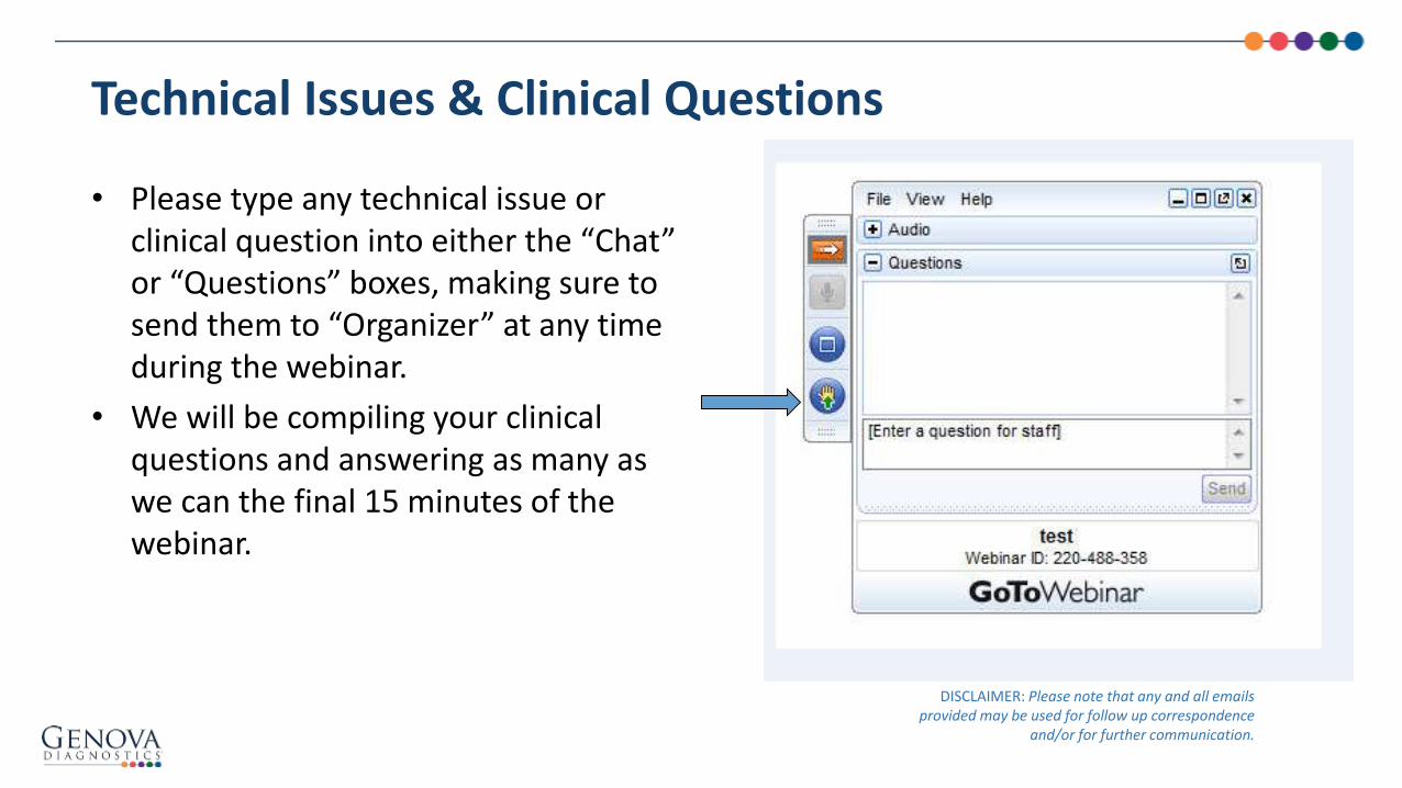
Technical Issues & Clinical Questions
• Please type any technical issue or clinical question into either the “Chat” or “Questions” boxes, making sure to send them to “Organizer” at any time during the webinar.
• We will be compiling your clinical questions and answering as many as we can the final 15 minutes of the webinar.
DISCLAIMER: Please note that any and all emails provided may be used for follow up correspondence
and/or for further communication.

Need More Resources? Ensure you have an account!

Elizabeth M. Board, MD
The views and opinions expressed herein are solely those of the presenter and do not necessarily represent those of Genova Diagnostics. Thus, Genova Diagnostics does not accept liability for consequences of any actions taken on the basis of the information provided.
Functional Medicine Approach To OsteoporosisPrevention & Treatment

• Review a rational approach to the prevention and treatment of osteoporosis
• Introduce functional lab testing that can identify causes and contributors to osteoporosis
• Explain how functional lab testing can provide a powerful educational platform that enhances patient comprehension and improves patient compliance
Objectives for This Presentation

• By 2020, 12.3 million individuals in the United States aged >50 years are expected to have osteoporosis
• Osteoporotic fractures, particularly hip fractures, are associated with limitations in ambulation, chronic pain and disability, loss of independence, and decreased quality of life
• 21% to 30% of patients who experience a hip fracture die within 1 year
• The prevalence of primary osteoporosis increases with age and differs by race/ethnicity
• With the aging of the US population, the potential preventable burden is likely to increase in future years
Prevalence of the Problem
Curry SJ, et al. JAMA. 2018;319(24):2521-31.

Impact of Osteoporosis
Watts NB, et al. Endocr Pract. 2010;16(6):1016-19.
American Association Of Clinical Endocrinologists Medical Guidelines For Clinical Practice For The Diagnosis And Treatment Of Postmenopausal Osteoporosis: Executive Summary Of Recommendations
• Incidences of new diagnoses in U.S. women based on recent statistics (2004 to 2006)

• Osteoporosis is defined as a “progressive systemic skeletal disease characterized by low bone mass and microarchitectural deterioration of bone tissue, with a consequent increase in bone fragility and susceptibility to fracture.”
• In 2005 the estimated cost of osteoporosis was 17 billion We expect this to increase as the
population of those over 60 increases
Osteoporosis Financial Impact
Watts NB, et al. Endocr Pract. 2010;16(suppl3):1–37.

• 64 yo nulliparous, postmenopausal female presents with osteoporosis by DEXA
• White, 118 lbs, BMI <20 on diclofenac and migraine medications
• Began anti-inflammatory diet, multivitamin designed to build bone
• Anti-inflammatory medical food with turmeric
• Vitamin D3 and K2, increased omega-3 fatty acids
• Walk every day
• Stress reduction, breathing exercises
• Decrease diclofenac
• Began on estradiol and progesterone
• Sleep improved with progesterone and magnesium
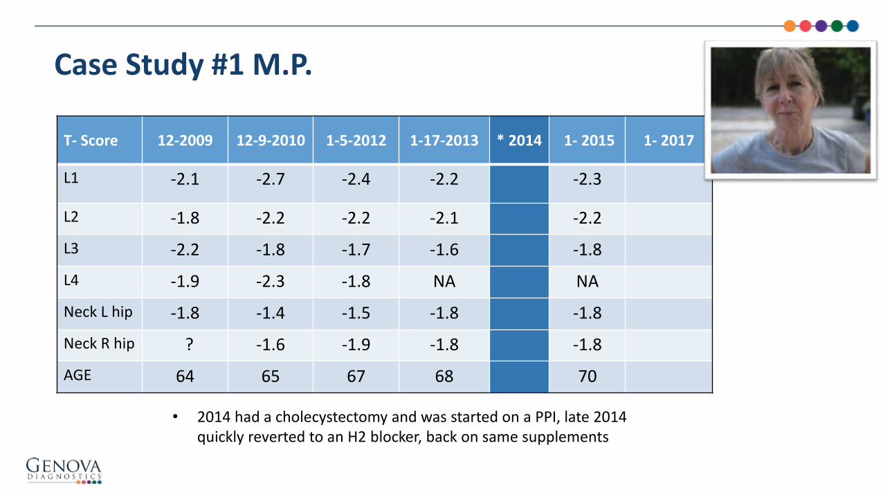
Case Study #1 M.P.

Case Study #1 M.P.
T- score 12-2009 12-9-2010 1-5-2012 1-17-2013 * 2014 1- 2015 1- 2017
L1 -2.1 -2.7 -2.4 -2.2
L2 -1.8 -2.2 -2.2 -2.1
L3 -2.2 -1.8 -1.7 -1.6
L4 -1.9 -2.3 -1.8 NA
Neck L hip -1.8 -1.4 -1.5 -1.8
Neck R hip ? -1.6 -1.9 -1.8
AGE 64 65 67 68
• 2014 had a cholecystectomy and was started on a PPI, stopped the bone building multivitamin supplements

Case Study #1 M.P.
2014
• She is taking Prilosec for nondysplastic Barrett’s She has recently undergone a
Cholecystectomy
• Has lost 10 lbs, BMI 18.5

Case Study #1 M.P.
?

Case Study #1 M.P.
Case Study #1 M.P.
T- Score 12-2009 12-9-2010 1-5-2012 1-17-2013 * 2014 1- 2015 1- 2017
L1 -2.1 -2.7 -2.4 -2.2 -2.3
L2 -1.8 -2.2 -2.2 -2.1 -2.2
L3 -2.2 -1.8 -1.7 -1.6 -1.8
L4 -1.9 -2.3 -1.8 NA NA
Neck L hip -1.8 -1.4 -1.5 -1.8 -1.8
Neck R hip ? -1.6 -1.9 -1.8 -1.8
AGE 64 65 67 68 70
• 2014 had a cholecystectomy and was started on a PPI, late 2014 quickly reverted to an H2 blocker, back on same supplements

2015
• Transitioned from Prilosec to Zantac
• Restarted her bone building multivitamin
• Still on BHRT
Case Study #1 M.P.

Case Study #1 M.P.
• Currently on Zantac
• Recent cholecystectomy
• Requiring more vitamin D
• On BHRT

Case Study #1 M.P.
• Her gastroenterologist advised against treatment for H. pylori
• We added digestive enzymes, bile acids, Ca D Glucarate, Sacc boulardi, bone building MVI
• Added probiotics
?

Case Study #1 M.P.
T- score 12-2009 12-9-2010 1-5-2012 1-17-2013 * 2014 1- 2015 1- 2017
L1 -2.1 -2.7 -2.4 -2.2 -2.3 -2.1
L2 -1.8 -2.2 -2.2 -2.1 -2.2 -2.1
L3 -2.2 -1.8 -1.7 -1.6 -1.8 -1.7
L4 -1.9 -2.3 -1.8 NA NA NA
Neck L hip -1.8 -1.4 -1.5 -1.8 -1.8 -1.7
Neck R hip ? -1.6 -1.9 -1.8 -1.8 -1.8
AGE 64 65 67 68 70 72
• 2014 had a cholecystectomy and was started on a PPI, quickly reverted to an H2 blocker

Gut Absorption & Digestion
• PPIs
• Hydrochloric acid deficiency
• Acute pancreatitis
• Cholestasis
• Malabsorption
• GI infections
• Celiac disease
• SIBO
Use of proton pump inhibitors is associated with fractures in young adults: a population-based study
RESULTS: We identified 124,799 cases and 605,643 controls. The adjusted odds ratio for the risk of fracture associated with PPI exposure was 1.13 (95% CI 0.92 to 1.39) among children aged < 18 years old and 1.39 (95% CI 1.26 to 1.53) among young adults aged 18-29 years old. In young
adults but not children we observed a dose-response effect with increased total exposure to PPIs (p for trend <0.001).
Freedberg DE, et al. Osteoporos Int. 2015;26(10):2501-07.
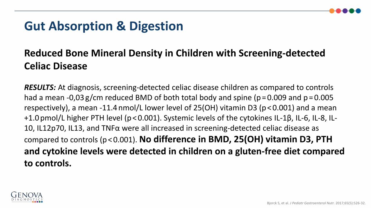
Reduced Bone Mineral Density in Children with Screening-detected Celiac Disease
RESULTS: At diagnosis, screening-detected celiac disease children as compared to controls had a mean -0,03 g/cm reduced BMD of both total body and spine (p = 0.009 and p = 0.005 respectively), a mean -11.4 nmol/L lower level of 25(OH) vitamin D3 (p < 0.001) and a mean +1.0 pmol/L higher PTH level (p < 0.001). Systemic levels of the cytokines IL-1β, IL-6, IL-8, IL-10, IL12p70, IL13, and TNFα were all increased in screening-detected celiac disease as
compared to controls (p < 0.001). No difference in BMD, 25(OH) vitamin D3, PTH and cytokine levels were detected in children on a gluten-free diet compared to controls.
Gut Absorption & Digestion
Bjorck S, et al. J Pediatr Gastroenterol Nutr. 2017;65(5):526-32.

Physiologic, pathophysiologic, and pharmacologic regulation of gastric acid secretion
RECENT FINDINGS: Gastric acid kills microorganisms, assists digestion, and facilitates absorption of
iron, calcium, and vitamin B12. The main stimulants of acid secretion are the hormone gastrin,
released from antral G cells; paracrine agent histamine, released from oxyntic enterochromaffin-like cells; and neuropeptide acetylcholine, released from antral and oxyntic intramural neurons. Gastrin is also a trophic hormone that participates in carcinogenesis. Helicobacter pylori may increase or decrease acid secretion depending upon the acuity and predominant anatomic focus of infection; most
patients manifest hypochlorhydria. Although generally considered well tolerated, concerns have been raised regarding associations between PPI use and dementia, kidney disease, myocardial infarction, pneumonia, osteoporosis, dysbiosis, small bowel injury, micronutrient deficiency, and fundic gland polyps.
Gut Absorption & Digestion
Schubert ML, et al. Curr Opin Gastroenterol. 2017;33(6):430-38.

Relationship between Helicobacter pylori infection and bone mineral density: a retrospective cross-sectional studyBACKGROUND: Helicobacter pylori (H. pylori) infection can induce individual inflammatory and immune reactions which associated with extra-digestive disorders. Our aim is to investigate the association between H. pylori infection and bone mineral density.METHODS: This retrospective cross-sectional study was performed by using the data from the health examination database in a medical center of southern Taiwan in 2013. We investigated the relationship between sex, age, body mass index (BMI), waist circumstance, lipid profile, H. pylori infection, the findings of upper gastrointestinal endoscopy and bone mineral density (BMD). Because of nonrandomized assignment and strong confounding effect of age on BMD, the 1:1 propensity score match was applied for age adjustment. The simple and multiple stepwise logistic regression analysis were performed to assess the risk factors of decreased BMD in these well-balanced pairs of participants.RESULTS: Of the 867 subjects in final analysis with the mean age of 55.9 ± 11.3 years, 381 (43.9%) subjects had H. pylori infection, and 556 (64.1%) subjects had decreased BMD. In decreased BMD group, the portion of woman was higher than a normal BMD group (37.2% versus 29.6%,P = 0.023), the age was significantly older (59.4 ± 9.8 versus 49.8 ± 11.3, p < 0.001) and BMI was significantly lower (24.7 ± 3.5 versus 25.4 ± 3.7, p = 0.006) than the normal BMD group. The prevalence of H. pylori infection was 39.9% and 46.2% in the normal BMD group and the decreased BMD group respectively (P = 0.071). The multivariate analysis which was used for these possible risk factors showed that only advanced age (OR 1.09, 95% CI 1.08-1.11, P < 0.001), and low BMI (OR 0.91, 95% CI 0.87-0.95, P < 0.001) were independently significantly associated with decreased BMD in this nonrandomized study. In the propensity score-matched participants, the multiple stepwise logistic regression analysis revealed H. pylori infection (OR 1.62, 95% CI 1.12-2.35, P = 0.011) and low BMI (OR 0.92, 95% CI 0.87-0.97, P = 0.001) were independently significantly associated with decreased BMD.
CONCLUSION: H. pylori infection and low BMI were independently significantly associated with decreased BMD in selected propensity score-matched populations after age adjustment.
Gut Absorption & Digestion
Pan BL, et al. BMC Gastroenterol. 2018;18(1):54-60.

Case Study #2 L.B.
• CC: hypothyroidism, natural pain relief for lumbar back pain, unable to lose weight, constipation, insomnia, recently post-menopausal, Tamoxifen for Breast cancer 18mo ago
• Fam Hx:– Mother: osteoarthritis, hypothyroid,
obesity, hypertension
– Maternal aunt: GERD, obesity, HTN, hypothyroidism, IBS
– Father: schizophrenia
– Maternal grandmother: breast cancer
• Timeline:– Born in Georgia– 4: Allergy to kale and mustard greens– 12: Menarche– 20s-30s: smoker– 39: Dieting, lost 50 lbs eventually gained it back– 40: Miscarriage. Otherwise nulliparous.– 49: Dieting, lost 50 lbs but gained it back– 42: Hysterectomy (ovaries retained)– 53: Fall without fracture, multiple close family members died
resulting in STRESS– 54: Breast cancer diagnosis– 55: Lumpectomy, tamoxifen, DEXA found to be “normal”– 55-56: Increasing lumbar back pain, scar pain, cannot exercise– 56: “Post Menopausal” based on hormones 6 months ago

• PRIOR LABS: Early 2017: low WBC; normal liver enzymes; TSH was 3.3 in 2003, 4.3 in 2005. Bone density scan in 2015 was normal. High B12; ALP 40
• Current LIFESTYLE: Poor sleep. Can’t exercise due to back pain. Non GMO, organic, no soy, no gluten, no foods in mustard family, Paleo in 2015, began ketogenic 9 mo ago losing 50 pounds. Recent stress with deaths of friends/family. Friend and spouse very supportive
Case Study #2 L.B.
• July 2017: hsCRP: 5.7, MTHFR: compound heterozygote, ESR: 4, ANA (-), 25 OH VitD: 47, coQ10 2.46, midday cortisol: 16.3, HbA1C: 5.4, FFA: elevated, ALP: 33, TSH:0.97, free T4: 1.37, freeT3: 2.2, reverse T3: 19
• Causes of Decreased ALK PHOS: Deficiency of Zn, Vit C, B6, B9, B12, Mag, Phos; Hypochlorhydria, Hypothyroidism, Celiac
• Now post menopausal with h/o breast cancer
• Recent 50 lb weight loss as she changed to ketogenic diet
• Decreased exercise
• DEXA from 2015 “normal bone density” Rec: calcium and vitamin D

DEXA 2017

Treatment Plan in 2015:
• “Calcium and Vitamin D”
Treatment Plan in 2017:
• “Oscal and Vit D”
• Ingredients: Calcium Carbonate, Corn Syrup Solids , Contains Less Than 2% of Talc, Corn Starch, Sodium Starch Glycolate , Polysorbate 80, Polyvinyl Alcohol, Polyethylene Glycol 3350, Titanium Dioxide, Yellow 5 Lake, Blue 1 Lake, Calcium Stearate, Methylparaben and Propylparaben (Preservative), Gelatin, Sucrose, Cholecalciferol (Vitamin D3), Di-Alpha Tocopherol
• 500mg Calcium carbonate and 200IU of Vitamin D3
Case Study #2 L.B.

Treatment Questions and Considerations:• Her sleep was improved with magnesium and resolution of pain following
the use of low dose naltrexone. So now she plans to begin an increase in exercise but what kind?
• How much did not being able to exercise impact her bone loss?
Case Study #2 L.B.
• On a ketogenic diet, can she be missing key ingredients necessary for bone growth?
• Is her high protein diet resulting in too much acid?
• Is her digestion and absorption adequate especially with a low alkaline phosphatase? Which
minerals might she be missing?
• Are her low free T3 and mildly elevated reverse T3 impacting her decreasing bone density?
• Can her hormones be further optimized safely, or is it necessary in order to build bone?

Vitamin D, Calcium, or Combined Supplementation for the Primary Prevention of Fractures in Community-Dwelling Adults: Evidence Report and Systematic Review for the US Preventive Services Task Force
OBJECTIVE: To update the evidence for benefits and harms of vitamin D, calcium, or combined supplementation for the primary prevention of fractures in community-dwelling adults to inform the US Preventive Services Task Force.DATA SOURCES: PubMed, EMBASE, Cochrane Library, and trial registries through March 21, 2017; references; and experts. Surveillance continued through February 28, 2018.STUDY SELECTION: English-language randomized clinical trials (RCTs) or observational studies of supplementation with vitamin D, calcium, or both among adult populations; studies of populations that were institutionalized or had known vitamin D deficiency, osteoporosis, or prior fracture were excluded.DATA EXTRACTION AND SYNTHESIS: Dual, independent review of titles/abstracts and full-text articles and study quality rating using predefined criteria. Random-effects meta-analysis used when at least 3 similar studies were available.MAIN OUTCOMES AND MEASURES: Incident fracture, mortality, kidney stones, cardiovascular events, and cancer.RESULTS: Eleven RCTs (N = 51 419) in adults 50 years and older conducted over 2 to 7 years were included. Compared with placebo, supplementation with vitamin D decreased total fracture incidence (1 RCT [n = 2686]; absolute risk difference [ARD], -2.26% [95% CI, -4.53% to 0.00%]) but had no significant association with hip fracture (3 RCTs [n = 5496]; pooled ARD, -0.01% [95% CI, -0.80% to 0.78%]). Supplementation using vitamin D with calcium had no effect on total fracture incidence (1 RCT [n = 36 282]; ARD, -0.35% [95% CI, -1.02% to 0.31%]) or hip fracture incidence (2 RCTs [n = 36 727]; ARD from the larger trial, -0.14% [95% CI, -0.34% to 0.07%]). The evidence for calcium alone was limited, with only 2 studies (n = 339 total) and very imprecise results. Supplementation with vitamin D alone or with calcium had no significant effect on all-cause mortality or incident cardiovascular disease; ARDs ranged from -1.93% to 1.79%, with CIs consistent with no significant differences. Supplementation using vitamin D with calcium was associated with an increased incidence of kidney stones (3 RCTs [n = 39 213]; pooled ARD, 0.33% [95% CI, 0.06% to 0.60%]), but supplementation with calcium alone was not associated with an increased risk (3 RCTs [n = 1259]; pooled ARD, 0.00% [95% CI, -0.87% to 0.87%]). Supplementation with vitamin D and calcium was not associated with an increase in cancer incidence (3 RCTs [n = 39 213]; pooled ARD, -1.48% [95% CI, -3.32% to 0.35%]).
CONCLUSION AND RELEVANCE: Vitamin D supplementation alone or with calcium was not associated with reduced fracture incidence among community-dwelling adults without known vitamin D deficiency, osteoporosis, or prior fracture. Vitamin D with calcium was associated with an increase in the incidence of kidney stones.
Right Raw Materials:
Kahwati LC, et al. .JAMA. 2018;319(15):1600-12.

RAW MATERIALS
Phytonutrients
Calcium
Phosphorus
Magnesium
Vitamin K
Vitamin A
Vitamin C
Vitamin D
Vitamin B:- B2
- B6
- B9
- B12
All The Right Raw Materials:
Boron
Manganese
Zinc
Potassium
Copper
Silica
Selenium
Fe
Amino Acids
Omega 3 FA

Luteolin attenuates glucocorticoid-induced osteoporosis by regulating ERK/Lrp-5/GSK-3β signaling pathway in vivo and in vitro
ABSTRACT: Glucocorticoid-induced osteoporosis (GIO) is a secondary osteoporosis with extensive use of glucocorticoids (GCs). GCs can increase bone fragility and fracture via inhibiting osteoblastic proliferation and differentiation. Luteolin (LUT), a kind of plant flavonoid, has been reported to exhibit the antioxidant activity, but the effects of LUT on GIO still remain unclear. This study aimed to investigate the effects of LUT on GIO both in vivo and in vitro and elaborate the potential molecular mechanisms. LUT increased the superoxide dismutase activity, glutathione level and decreased reactive oxygen species (ROS) level and lactate dehydrogenase release in GIO. Meanwhile, LUT decreased caspase-3, caspase-9, and Bax protein expressions and increased Bcl-2 protein expression in GIO. LUT increased the ratio of osteoprotegerin (OPG)/receptor activator of nuclear factor-κB Ligand (RANKL) messenger RNA (mRNA) expression and mRNA expression levels of osteogenic markers, including runt-related transcription factor 2, osterix, collagen type I, and osteocalcin. LUT also enhanced the extracellular signal-regulated kinases (ERK) phosphorylation, glycogen synthase kinase 3β (GSK-3β) phosphorylation, mRNA expression levels of lipoprotein-receptor-related protein 5 (Lrp-5) and β-catenin. Further study revealed that Lrp-5 small interfering RNA (siRNA )and ERK-siRNA reduced the effects of LUT on GSK-3β phosphorylation, alkaline phosphatase (ALP) activity and the ratio of OPG/RANKL mRNA expression. Moreover, ERK-siRNA decreased Lrp-5
mRNA expression in vitro. These results indicated that LUT promoted proliferation by attenuating oxidative stress and promoted osteoblastic differentiation by regulating the ERK/Lrp-5/GSK-3β
pathway in GIO.
Right Raw Materials
Jing Z, et al. J Cell Physiol. 2018;1-19.

Right Raw Materials
Calcium:
• Calcium carbonate not well absorbed unless acid present
• Chelated calcium like calcium citrate and calcium malate
• Beware of sources that may contain lead (dolomite, oyster shell and bone meal)
• Daily dose 1000 mg
Vitamin D3:
• Use the active form
• Dose varies depending on genetics and BMI
• Monitor levels every 3 months – 6 months
B Vitamins: Homocysteine metabolism, all B’s but especially, B2, B6, B12
Magnesium:
• 60% is stored in bone
• Supports production of hydroxyapatite and bone marrow stromal cells as well as 1,25(OH)2D vitamin synthesis
• Deficiency via hypocalcemia elevates parathyroid hormone synthesis and subsequently osteoclast activity
• Osteoblastic number and activity declines during magnesium deficiency and leads to decreased trabecular volume and alteration of bone microarchitecture in a way similar to osteomalacia
• Daily dose 320-420 mg
• Hypermagnesemia (e.g. chronic renal failure) disturbs calcium/magnesium ratio, which may lead to a defect in mineralization and osteoblasts differentiation

Vitamin K: • K1 is found in leafy greens K2 created by microbiota
• K2 activates osteocalcin and attracts calcium to be anchored into hydroxyapatite crystals that form bone
• Activates matrix Gla protein keeps calcium out of arteries
• MK-4: 10-45 mg and MK-7: 90-200 mcg longer acting
• Decreased dosages if on blood thinners
Boron: • Stimulates bone growth & bone metabolism
• Boron activates 1,25(OH)2D3 production & increases bone mineralization especially for trabecular bone microarchitecture and cortical bone strength
• Daily dose to support bone health is 3 mg
Right Raw Materials
Pizzorno L, Wright J. Mount Jackson, VA: Axios Press; 2013.
Zofkova I, et.al. Physiol Res. 2017;66(3):391-402.

Copper: • Necessary for bone growth regulation and skeletal development by inducing lysine crosslinks in
collagen and elastin
• Limits osteoclastic activation by free radicals and decreases osteoclastic bone resorption directly
• Dose for bone health 0.9 mg/day
Iron: • Stimulates synthesis of bone matrix via lysyl hydroxylase and activates 25-hydroxycholecalciferol
hydroxylase to support mineralization of bone matrix through vitamin D
• In excess, ferric ion activates osteoclastic differentiation
• Daily dose varies
Manganese: • Positively modulates RANKL/OPG ratio in the process of bone formation, determining thickness of
trabecular bone area and increasing trabecular number
• Daily dose for bone health is 1.8 – 2.3 mg
Right Raw Materials
Zofkova I, et.al. Physiol Res. 2017;66(3):391-402.

Selenium: • Restores antioxidant capacity in bone cells and inhibits NF-κB – RANKL axis and osteoclast
differentiation. In high doses, selenium induces apoptosis of mature osteoclasts
• Dose is at least 55 μg/day
Zinc: • Positively influences the strength, flexibility and architecture of bone
• It increases osteoblastic activity and promotes synthesis of collagen while inhibiting osteoclasticbone resorption
• Additional benefit for osteoblasts that have been exposed toxic elements, such as lead or cadmium and excessive alcohol
• 15 mg is necessary to increase bone density balanced with 1 mg copper daily
Right Raw Materials
Zofkova I, et.al. Physiol Res. 2017;66(3):391-402.

Strontium ranelate as a possible disease-modifying osteoarthritis drug: a systematic review
ABSTRACT: Considering that osteoarthritis (OA) is the most prevalent joint disease worldwide, multiple pharmacological treatments have been proposed to alter the articular structure with potential benefit in the progression of the disease. The so-
called disease-modifying OA drugs have been frequently investigated but conclusive findings are rare. Strontium ranelate (SrRan) is a drug usually prescribed to treat osteoporosis, with proven effects in decreasing the risk of fractures and possible effect in reducing the progression of OA. The objective of this review was to demonstrate the current panorama of knowledge on the use of SrRan in clinical and experimental models, clarifying its mechanisms of action and describing possible anti-nociceptive and anti-inflammatory effects. The systematic review was based on the PRISMA statement and included articles that are indexed in scientific databases. Fifteen studies were included: seven pre-clinical and eight clinical studies. Despite the
limited number of studies, the results suggest a positive effect of SrRan in patients with OA, through changes in functional capacity and reduction of progression of morphological parameters and joint degradation, with moderate quality of evidence for those clinical outcomes. Novel studies are necessary to elucidate the molecular targets of SrRan, focusing on anti-inflammatory effects and histological changes promoted by SrRan, which seemed to reduce the progression of OA in the experimental and clinical studies.
Right Raw Materials
Rodrigues TA, et al. Braz J Med Biol Res. 2018;51(8):e7440.

A New Insight to Bone Turnover: Role of ω-3 Polyunsaturated Fatty Acids
“In conclusion, strong evidences featured in the scientific literature available to date support the benefits of ω-3 fatty acids in bone health. Several mechanisms have been proposed (affecting bone formation, bone resorption, serum calcium and vitamin D, oxidative stress, and inflammatory mediators). However, neither the exact benefit nor the exact mechanism of action of essential fatty acids has been determined yet.“
Right Raw Materials: OMEGA 3 FA
Kajarabille N, et al. Scientific World Journal. 2013;2013:589641.

• Gut: digestion and absorption
• Right raw materials
A Functional Approach Treatment Plan

• 75 y/o white female with a past history of shoulder and wrist fractures and osteoporosis
• At 71 her PCP stopped her BHRT • Insomnia• Frozen shoulder• Constipation • vaginal dryness• Weight gain• Wrist fracture• Restarted her BHRT, 2 yrs later had inflammation via
thermogram• Complete hormones through Genova• Began DIM, more magnesium, methylated B vitamins,
corrected Constipation, turmeric• Inflammation resolved• Normal mammogram
Case Study #3 F.D.
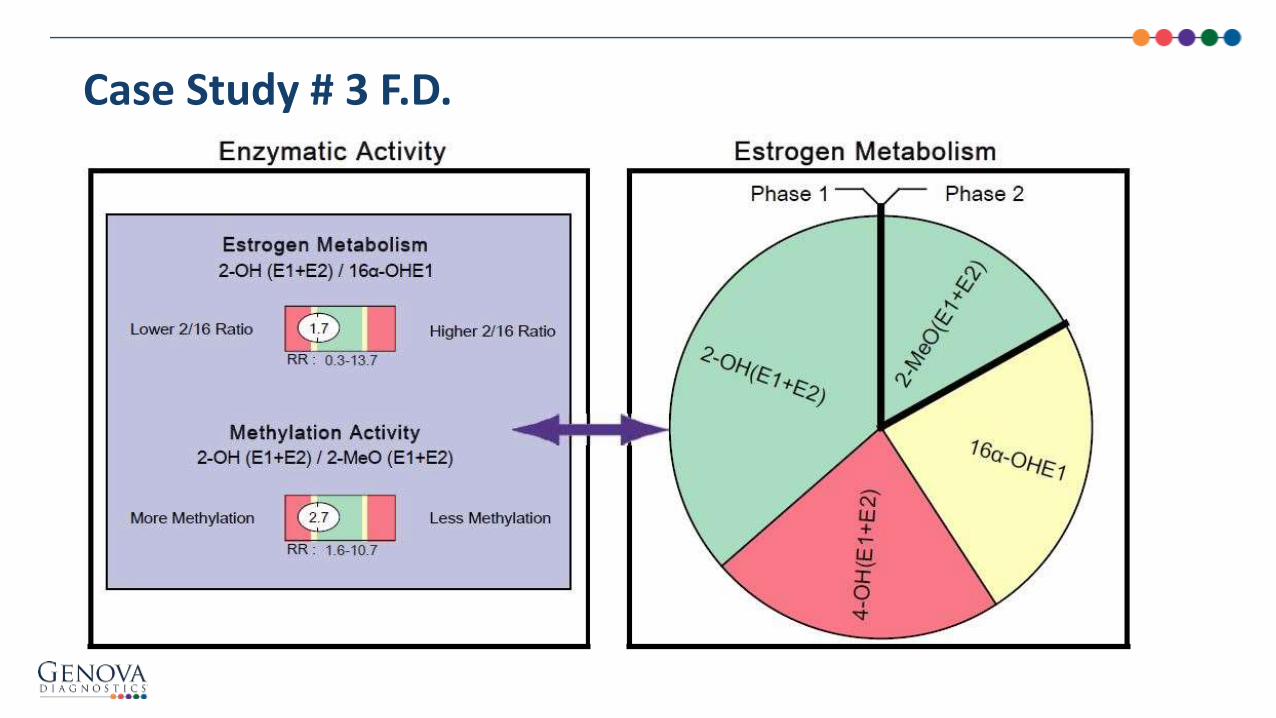
Case Study # 3 F.D.

Menopausal hormone therapy for primary prevention: why the USPSTF is wrong
• When these women fail to use MHT, in addition to an increase in mortality risk, they also experience accelerated risks for major diseases including CVD, osteoporosis, dementia, mood disorders, and sexual dysfunction
• Unlike bisphosphonates, which have been associated with excessive bone mineralization, estrogen facilitates normal bone architecture
• Effects of osteoporosis medications on bone quality. There is no question that estrogen is an effective and metabolically appropriate preventive strategy for osteoporotic fractures, and that osteoporosis is a chronic disease with tremendous impact in postmenopausal women
Optimization of Hormones
Langer RD, et al. Climacteric. 2017;20(5):402-13.
Benhamou CL. Joint Bone Spine. 2007;74(1):39-47.

Negative spinal bone mineral density changes and subclinical ovulatory disturbances--prospective data in healthy premenopausal women with regular menstrual cycles
ABSTRACT: Subclinical ovulatory disturbances (anovulation or short luteal phases within normal-length menstrual cycles) indicate
lower progesterone-to-estrogen levels. Given that progesterone plays a bone formation role, subclinical ovulatory disturbances may be associated with bone loss or less than expected bone gain… a meta-analysis of prospective studies in healthy premenopausal women to determine the overall relationship of subclinical ovulatory disturbances to change in bone mineral density. Two reviewers independently identified from serial literature searches 6 studies meeting inclusion criteria: a 2-year study in 114 young adult women, 2006-2009, Vancouver, Canada; a 2-year study in 189 premenopausal women, 2000-2005, Toronto, Canada; a single-cycle study in 14 young women, 1996-1997, Melbourne, Australia; an 18-month study in 53 women, 1990-1995, Santa Clara, California; a 4-year study in 27 women, 1988-1995, Vancouver, Canada; and a 1-year study in 66 women, 1985-1988, Vancouver, Canada. This meta-analysis included a combined sample size of 473 observations in 436 premenopausal women studied over 1-4 years and aged 14-47 years. The percentage of women with ovulatory disturbances varied significantly from 13% to 82%. Women with more frequent ovulatory disturbances had more negative percentage changes in spine bone mineral density (weighted mean difference = -0.86; P = 0.040) for random-effects analysis. There was significant heterogeneity among these 6
studies (I(2) = 80%). In summary, these data show that regularly menstruating women with more frequent ovulatory disturbances experience more negative changes in bone (approximately -0.9% per year). These cycles with silent estrogen/progesterone imbalance may be clinically important.
Optimization of Hormones
Li D, et al. Epidemiol Rev. 2014;36:137-47.

80% of our bone mass develops BEFORE the age of 18. Bone mass then begins to slope downward at the age of 35 for women and 45 for men
Anorexia:
– Adolescents have decreased rates of bone accrual raising “concerns of suboptimal peak bone mass and future bone health”
– Rec regain weight and menstrual function
– Bisphosphonates generally are not recommended in women of child bearing age due to their long half-life and potential for teratogenicity
– Risedronate did improve hip and vertebral bone density in women with AN
Optimization of Hormones
Misra M, et al. J Endocrinol. 2014;1(3):R163-76.

Maor G, et al. J Bone Miner Res. 2002;17:1034–43.
Hormone Effect
SEX HORMONES Estrogens Decreases production & activation of osteoclasts, (to a lesser extent) stimulates osteoblasts
Progesterone Boosts production and activation of osteoblasts (which have progesterone receptors)
Testosterone Deficiency in men = osteoporosis, in women and men helps maintain muscle mass
Cortisol Long term elevations reduce calcium absorption from gut and inhibits osteoblasts
DHEA Precursor to androgens and ultimately estrogens
Thyroid Hormones Euthyroid to balance bone remodeling cycle, hyper and hypothyroidism lead to osteoporosis
Parathyroid Hormone Signals osteoclasts to break down bone, increase calcium in blood and stimulate kidneys to reabsorb calcium
Calcitonin Decreases calcium absorption in gut, signals the kidneys to release calcium in the urine, keeps calcium in the bone and retards activity of osteoclasts
Calcitriol (active form Vit. D) Increases calcium absorption of gut, reabsorbs calcium by the kidneys
IGF-1 Increases, maintains muscle mass and stimulates osteoblasts to build bone
Leptin Increases linear growth and skeletal mass as observed in childhood obesity

• Gut: digestion and absorption
• Right raw materials
• Optimize Hormones
A Functional Approach Treatment Plan
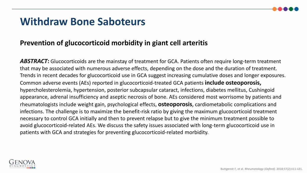
Prevention of glucocorticoid morbidity in giant cell arteritis
ABSTRACT: Glucocorticoids are the mainstay of treatment for GCA. Patients often require long-term treatment that may be associated with numerous adverse effects, depending on the dose and the duration of treatment. Trends in recent decades for glucocorticoid use in GCA suggest increasing cumulative doses and longer exposures.
Common adverse events (AEs) reported in glucocorticoid-treated GCA patients include osteoporosis,hypercholesterolemia, hypertension, posterior subcapsular cataract, infections, diabetes mellitus, Cushingoid appearance, adrenal insufficiency and aseptic necrosis of bone. AEs considered most worrisome by patients and
rheumatologists include weight gain, psychological effects, osteoporosis, cardiometabolic complications and infections. The challenge is to maximize the benefit-risk ratio by giving the maximum glucocorticoid treatment necessary to control GCA initially and then to prevent relapse but to give the minimum treatment possible to avoid glucocorticoid-related AEs. We discuss the safety issues associated with long-term glucocorticoid use in patients with GCA and strategies for preventing glucocorticoid-related morbidity.
Withdraw Bone Saboteurs
Buttgereit F, et al. Rheumatology (Oxford). 2018;57(2):ii11-ii21.

Associations of urinary polycyclic aromatic hydrocarbons with bone mass density and osteoporosis in U.S. adults, NHANES 2005-2010
ABSTRACT: Polycyclic aromatic hydrocarbons (PAHs) are environmental endocrine disruptors, which may modify the bone mineralization. However, epidemiological evidences on this issue were scant. We aimed to investigate the associations of PAHswith bone mass density (BMD) and osteoporosis based on a nationally-representative sample from general U.S.POPULATION: Data utilized were extracted from the 2005-2010 National Health and Nutrition Examination Survey (NHANES). Nine urinary PAHs (U-PAHs) metabolites were measured as exposure biomarkers. Associations of specific U-PAHs with BMD and osteoporosis were estimated by multivariable adjusted linear regression models and logistic regression models, respectively. Compared with women at the first tertiles, those at the third tertiles of 1-Hydroxynapthalene, 2-Hydroxyfluorene, 3-Hydroxyphenanthrene, 2-Hydroxyphenanthrene and 9-Hydroxyfluorene had significantly decreased BMD levels [coefficient (β) = -0.023 to -0.014, p < 0.05] or increased likelihoods of osteoporosis [odds ratios (ORs) = 1.86 to 3.36, p < 0.05] at different bone sites. Whereas, elevated BMD levels (β = 0.021, p < 0.05) at trochanter and decreased likelihoods of osteoporosis (OR = 0.33, p < 0.05) at intertrochanter were observed among women at the second tertiles of 1-Hydroxypyrene and 2-Hydroxynapthalene, respectively. Similar results were found for all the population, i.e., combination of men and women. Most of the significant associations disappeared among adult men only. Furthermore, Associations between U-PAHs and BMD were stronger for postmenopausal women when compared with premenopausal group. In conclusion, associations of U-PAHs with BMD and osteoporosis varied by specific U-PAHs and bone sites, as well as menopausal status and genders in U.S. adults.
Withdraw Bone Saboteurs
Guo J, et al. Medicine (Baltimore). 2018;97(17):e0532.
PAHs are released from burning coal, oil, gasoline, trash, tobacco, and wood

• “With respect to lead and menopause, most research, including an earlier study in the Nurses’ Health Study cohort (Korrick et al. 2002), has focused on the effects of menopause on blood lead levels (Garrido Latorre et al. 2003; Jackson et al. 2010; Nash et al. 2004; Potula and Kaye 2006; Vahter et al. 2004). Release of lead from bone to blood as a consequence of increased bone turnover following menopause has been proposed as a mechanism that may explain cross-sectional associations between menopause and blood lead levels.”
• Higher lead levels were associated with early menopause
Rising Lead Levels During Menopause
Ki-Do E, et al. Environ Health Perspect. 2014;122(3):229–34.

Lead: • Disrupts enzyme function and negatively impacts osteoblasts
• >90% stored in bone and released into blood stream during periods of bone remodeling
• Khalil et.al. (2008) showed that increases in circulation lead levels as low as 8mcg/dl decreases hip BMD and increases risk of falls and fractures
• Deficiencies in Zn, Ca, Fe and Mg exacerbate lead toxicity
Pesticides: Exposure decreases BM in multiple species
Mercury: Indirectly by uncoupling bone remodeling mechanisms, replacing Zn, neurotoxicity, reduce hormone effectiveness.
Sunscreen chemicals
Exposure to Endocrine Disrupting Chemicals
Glyphosate: • Glyphosate has been shown to severely deplete Mn levels in plants
• Chondroitin sulfate synthesis depends on Mn, and its deficiency leads to osteoporosis and osteomalacia
Withdraw Bone Saboteurs
Samsel A, et al. Surg Neurol Int. 2015;6:45-71.
Quig D. Altern Med Rev. 1998;3(4):262-70.
Khalil N, et al. J Bone Miner Res. 2008;23(9):1417-25.

Withdraw Bone Saboteurs
NutrEval of a 52 yo Perimenopausal female, postmenopausal at the time of the fourth blood draw, began hormones at the time of the 7th blood draw
BHRT
Perimenopausal

Cadmium• Stimulates the formation and activation of osteoclasts
• Limits the osteoblast ability to form bone collagen
• Half life of 20-40 years
• Stored mainly in the kidneys and liver
• Fe and Zn deficiency increase vulnerability to cadmium toxicity
• Disrupts the production of progesterone by the ovary
• Women with a urinary cadmium level of 0.5-1 mcg/gram of creatine had a 43% increased chance of hip osteoporosis compared with women <0.5 mcg/g creatinine
Withdraw Bone Saboteurs
Zhang W, et al. Toxicology. 2007;239(3):204-12.
Gallagher CM, et al. Environ Health Perspect. 2008;116(10):1338-43.
• High protein, high acid
• Processed foods
• Coffee
• Sugar
• Alcohol
• Stress
• Tobacco
• Sodas with phosphoric acid
• Medications: PPIs, steroids, etc.
Withdraw Bone Saboteurs

• Gut: digestion and absorption
• Right raw materials
• Optimize Hormones
• Withdraw Bone Saboteurs
A Functional Approach Treatment Plan

• Most of us are sedentary at work only or at work and home
• Bedridden, not moving against gravity = up to 1% of bone mineral loss per week!
• With weight bearing exercise can gain 1-3% per year
• Strengthens muscles: improves balance & stability
Type of Exercise:– Dynamic rather than static
– Vary routine
– Weight bearing against gravity
– Stretch to maintain flexibility
– Balance work
– Posture
– Focus on extension and avoid flexion
– Strengthen pelvic and abdominal stabilizing muscles which attach to vertebral spine
Stimulate Bone Growth
Pizzorno L, Wright J. Mount Jackson, VA: Axios Press; 2013.

Whole-body vibration training and bone health in postmenopausal women: A systematic review and meta-analysis
BACKGROUND: The aims of the present systematic review and meta-analysis were to evaluate published, randomized controlled trials that investigate the effects on whole-body vibration (WBV) training on total, femoral neck, and lumbar spine bone mineral density (BMD) in postmenopausal women, and identify the potential moderating factors explaining the adaptations to such training.METHODS: From a search of electronic databases (PubMed, Web of Science, and Cochrane) up until September 2017, a total 10 studies with 14 WBV groups met the inclusion criteria. Three different authors tabulated, independently, the selected indices inidentical predetermined forms. The methodological quality of all studies was evaluated according to the modified PEDro scale. For each trial, differences within arms were calculated as mean differences (MDs) and their 95% confidence intervals between pre- and postintervention values. The effects on bone mass between exercise and control groups were also expressed as MDs. Both analyses were performed in the total sample and in a specific class of postmenopausal women younger than 65 years of age (excluding older women).RESULTS: The BMD of 462 postmenopausal women who performed WBV or control protocol was evaluated. Significant pre-post improvements in BMD of the lumbar spine were identified following WBV protocols (P = .03). Significant differences in femoralneck BMD (P = .03) were also found between intervention and control groups when analyzing studies that included postmenopausal women younger than 65 years.
CONCLUSION: WBV is an effective method to improve lumbar spine BMD in postmenopausal and older women and to enhance femoral neck BMD in postmenopausal women younger than 65 years.
Stimulate Bone Growth
Marin-Cascales E, et al. Medicine (Baltimore). 2018;97(34):e11918.
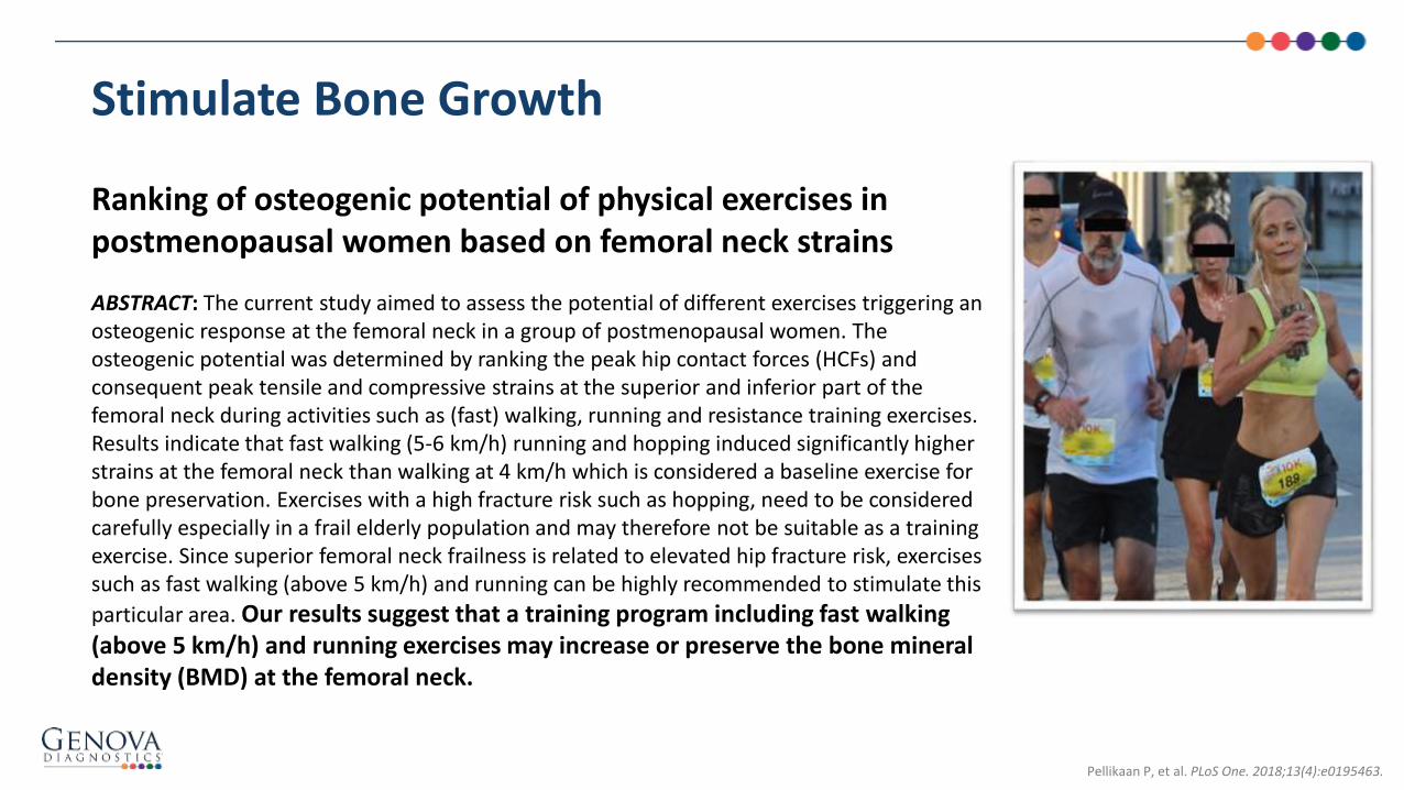
Ranking of osteogenic potential of physical exercises in postmenopausal women based on femoral neck strains
ABSTRACT: The current study aimed to assess the potential of different exercises triggering an osteogenic response at the femoral neck in a group of postmenopausal women. The osteogenic potential was determined by ranking the peak hip contact forces (HCFs) and consequent peak tensile and compressive strains at the superior and inferior part of the femoral neck during activities such as (fast) walking, running and resistance training exercises. Results indicate that fast walking (5-6 km/h) running and hopping induced significantly higher strains at the femoral neck than walking at 4 km/h which is considered a baseline exercise for bone preservation. Exercises with a high fracture risk such as hopping, need to be considered carefully especially in a frail elderly population and may therefore not be suitable as a training exercise. Since superior femoral neck frailness is related to elevated hip fracture risk, exercises such as fast walking (above 5 km/h) and running can be highly recommended to stimulate this
particular area. Our results suggest that a training program including fast walking (above 5 km/h) and running exercises may increase or preserve the bone mineral density (BMD) at the femoral neck.
Stimulate Bone Growth
Pellikaan P, et al. PLoS One. 2018;13(4):e0195463.

• Gut: digestion and absorption
• Right raw materials
• Optimize hormones
• Withdraw bone saboteurs
• Stimulate bone growth
A Functional Approach Treatment Plan

Effect of a balance-training programme on postural balance, aerobic capacity and frequency of falls in women with osteoporosis: A randomized controlled trial
OBJECTIVE: To investigate the effect of a 12-month complex balance-training programme on static and dynamic postural balance, aerobic capacity and frequency of falls in women with established osteoporosis.DESIGN: Randomized controlled trial in which the intervention group was assigned a 12-month exercise programme (3 times a week for 30 min) and the control group had no intervention.SUBJECTS: A total of 100 osteoporotic women with at least one previous fracture.METHODS: Performance-based Timed Up and Go (TUG), Berg Balance Scale (BBS) and stabilometric platform tests were used to evaluate balance. Aerobic capacity was measured by bicycle ergometry. Frequency of falls was assessed using a falls diary.RESULTS: After 1 year, there was a statistically significant difference between the improvement achieved in the intervention and control groups on the performance-based TUG, BBS and stabilometric platform tests (p < 0.05). Mean metabolic equivalent (MET) value decreased in the intervention group, from 4.91 to 3.82 (a significant difference from the change achieved in the control group; p = 0.05). Relative risk of falls was 0.534 at 1 year (p = 0.17).
CONCLUSION: The 12-month balance-training program significantly improved postural balance and increased aerobic capacity in women with established osteoporosis.
Balance and Fall Prevention
Miko I, et al. J Rehabil Med. 2018;50(6):542-47.

Balance and Fall PreventionOver 90% of hip fractures are a result of falls
Modifiable Factors To Reduce Falls:
• Impaired gait and balance
• Impaired mobility and disability
• Neuromuscular or musculoskeletal disorders
• Age
• Impaired vision
• Neurological, heart disorders
• History of falls
• Medication decreasing blood sugar or blood pressure
• Cognitive impairment
PT after fracture: “At all times, increased strength may prevent falls by improving confidence and coordination as well as maintaining bone mass by stimulating bone formation and by decreasing bone resorption and by preserving muscle strength.”
“Large trials have shown that it is possible to reduce falls, but randomized studies have not shown any significant decrease in fracture risk. Some randomized trials have shown that wearing hip protectors can markedly reduce hip fracture risk, particularly in the elderly living in nursing homes. A meta-analysis of well-conducted randomized controlled trials, however, casts some doubt about the anti-fracture efficacy.”
Kanis JA, et al. Osteoporos Int. 2013;24(1):23-57.
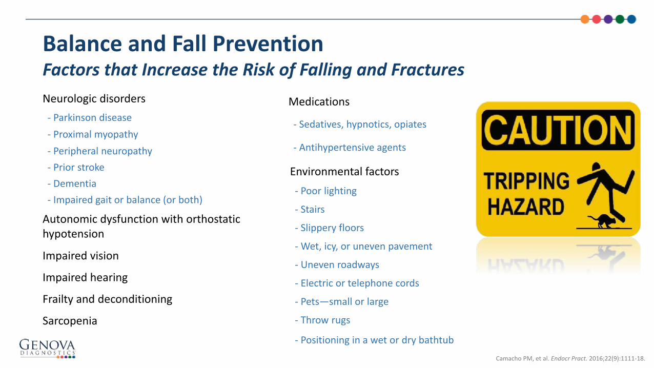
Balance and Fall PreventionFactors that Increase the Risk of Falling and Fractures
Neurologic disorders
- Parkinson disease
- Proximal myopathy
- Peripheral neuropathy
- Prior stroke
- Dementia
- Impaired gait or balance (or both)
Autonomic dysfunction with orthostatic hypotension
Impaired vision
Impaired hearing
Frailty and deconditioning
Sarcopenia
Medications
- Sedatives, hypnotics, opiates
- Antihypertensive agents
Camacho PM, et al. Endocr Pract. 2016;22(9):1111-18.
Environmental factors
- Poor lighting
- Stairs
- Slippery floors
- Wet, icy, or uneven pavement
- Uneven roadways
- Electric or telephone cords
- Pets—small or large
- Throw rugs
- Positioning in a wet or dry bathtub

• Gut: digestion and absorption
• Right raw materials
• Optimize hormones
• Withdraw bone saboteurs
• Stimulate bone growth
• Balance & fall prevention
A Functional Approach Treatment Plan

Case Study #5 D.C.
• 63 yo petite female, French artist who teaches meditation• CC: recent 2013 DEXA: osteoporosis• Rec’d Prolia she cannot afford this medication
Timeline:• Childhood: Eczema, h/o anemia
• 20-60: exposure to cadmium
• 53: Menopause
• 59: gum surgery
• 61: DEXA 2011: T scores: Vertebral -1.6 L femur -2.1 R femur -2.2– Began Premarin, vit D and calcium
• 61: Began 18 months of PPI
• 63: Stopped dairy and fatty meals and was able to wean off PPI
• 63: DEXA 2013: T scores: vertebral -1.8 ( L4 -3.2) L femur -2.6

Case Study #5 D.C.C#2 D.C.
• Lifestyle:– Vegan
– Walk/jog/aerobics 4 x a week
– Pilates & yoga
• Treatment Plan:– GI: gut restoration, HCL trial, digestive enzymes
– Raw materials: calcium, strontium, boron, methylated all B vitamins, phosphorus, magnesium, fish oil
– Optimize hormones: DHEA, estriol, progesterone
– Withdraw the saboteurs: decrease caffeine to one or less cups of coffee per day, minimize alcohol, meditation for stress
– Stimulate bone growth: weight bearing, continue yoga and pilates
– Balance: yoga, balance exercises

Case Study #5 D.C.
T score 2011T score 2013
Discovery C S/N 47894T score 2015
Discovery C S/N 47894T score 2017
Discovery C S/N 47894
L3 -1.7 -1.1 -1.1
L1-L4 -1.6 -1.8 -1.2 -1.2
Neck femur -2.1 -2.6 - 2.3 -2.2
Total Hip -1.6 -1.6 -1.6
No fractures and the patient is thrilled

Questions?
Explore
WWW.GDX.NET for more information and
educational resources, including…
LEARN GDX – Brief video modulesLIVE GDX – Previous webinar recordings
GI University – Focused learning modules
Conferences – Schedule of events we attend
Test Menu – Detailed test profile information________
MY GDX – Order materials and get results
Elizabeth Board, MDPresenter
Lahnor Powell, ND, MPHModerator

US Client Services: 800-522-4762
UK Client Services: 020.8336.7750
Please schedule a complimentary appointment with one of our Medical Education Specialists for questions related to:
– Diagnostic profiles featured in this webinar
– How Genova’s profiles might support patients in your clinical practice
– Review a profile that has already been completed on one of your patients
We look forward to hearing from you!
Additional Questions?

October 24, 2018
Register for upcoming LIVEGDX Webinars online at WWW.GDX.NET
Upcoming LIVEGDX Webinar Topics
The views and opinions expressed herein are solely those of the presenter and do not necessarily represent those of Genova Diagnostics. Thus, Genova Diagnostics does not accept liability for consequences of any actions taken on the basis of the information provided.

Elizabeth M. Board, MD
The views and opinions expressed herein are solely those of the presenter and do not necessarily represent those of Genova Diagnostics. Thus, Genova Diagnostics does not accept liability for consequences of any actions taken on the basis of the information provided.
Functional Medicine Approach To OsteoporosisPrevention & Treatment