functional dairy products 2003 mattila-sandholm & saarela
TRANSCRIPT
Functional dairyproducts
Edited byTiina Mattila-Sandholm and Maria Saarela
CRC PressBoca Raton Boston New York Washington, DC
Cambridge England

Published by Woodhead Publishing Limited, Abington Hall, AbingtonCambridge CB1 6AH, Englandwww.woodhead-publishing.com
Published in North America by CRC Press LLC, 2000 Corporate Blvd, NWBoca Raton FL 33431, USA
First published 2003, Woodhead Publishing Ltd and CRC Press LLC© 2003, Woodhead Publishing LtdThe authors have asserted their moral rights.
This book contains information obtained from authentic and highly regarded sources.Reprinted material is quoted with permission, and sources are indicated. Reasonableefforts have been made to publish reliable data and information, but the authors and thepublishers cannot assume responsibility for the validity of all materials. Neither theauthors nor the publishers, nor anyone else associated with the publication, shall be liablefor any loss, damage or liability directly or indirectly caused or alleged to be caused bythis book.
Neither this book nor any part may be reproduced or transmitted in any form or by anymeans, electronic or mechanical, including photocopying, microfilming and recording, orby any information storage or retrieval system, without permission in writing from thepublishers.
The consent of Woodhead Publishing and CRC Press does not extend to copying forgeneral distribution, for promotion, for creating new works, or for resale. Specificpermission must be obtained in writing from Woodhead Publishing or CRC Press forsuch copying.
Trademark notice: Product or corporate names may be trademarks or registered trade-marks, and are used only for identification and explanation without intent to infringe.
British Library Cataloguing in Publication DataA catalogue record for this book is available from the British Library.
Library of Congress Cataloging in Publication DataA catalog record for this book is available from the Library of Congress.
Woodhead Publishing ISBN 1 85573 584 9 (book) 1 85573 691 8 (e-book)CRC Press ISBN 0-8493-1743-6CRC Press order number: WP1743
Cover design by The ColourStudioTypeset by Ann Buchan (Typesetters), Middx, EnglandPrinted by TJ International, Padstow, Cornwall, England
Contributors
Chapter 1M. Saxelin*, R. Korpela and A.
Mäyrä-MäkinenValio Ltd, R&DMeijeritie 4 APO Box 3000039 HelsinkiFinland
Tel: +358 10381 3111Fax: +358 10381 3019Email: [email protected]
Chapter 2C. Gill* and I. RowlandNorthern Ireland Centre for Diet and
Health (NICHE)University of UlsterColeraine CampusCromore RoadColeraineCo. LondonderryBT52 1SAUK
Tel: +44 (0) 28 7032 4675Email: [email protected]
Chapter 3
J. Lovegrove* and K. JacksonSchool of Food BiosciencesThe University of ReadingPO Box 226WhiteknightsReadingRG6 6APUK
Tel: +44 (0) 118 378 8700Fax: +44 (0) 118 378 0080Email: [email protected]
Chapter 4
R. WoodMineral Bioavailability LabUSDA HNRCA at Tufts University711 Washington StBostonMA 02111USA
Email: [email protected]
*Indicates main point of contact
xii Contributors
Chapter 5
P.V. KirjavainenDepartment of Biochemistry and
Food Chemistry and FunctionalFoods Forum
University of TurkuFIN-20014Finland
Tel: +358 2 333 6861Fax: +358 2 333 6860Email: [email protected]
Chapter 6
H. GillInstitute of Food, Nutrition and
Human HealthMassey UniversityPalmerston NorthNew Zealand
Email: [email protected]
Chapter 7
F. ShanahanUniversity College CorkClinical Sciences BuildingCork University HospitalCorkIreland
Tel: +353 21 490 1226Fax: +353 21 434 5300Email: [email protected]
Chapter 8
R.J. FitzGerald*Life Sciences Department,University of LimerickLimerickIreland
Tel: +353 61 202 598Fax: +353 61 331 490Email: [email protected]
H. MeiselInstitut für Chemie und Technologie
der MilchPO Box 60 69D-24121 KielGermany
Email: [email protected]
Chapter 9G. Boehm* and B. StahlInfant Nutrition ResearchNumico Research GermanyMilupa GmbH & Co. KGBahnstrasse 14–3061381 FriedrichsdorfGermany
Tel: +49 6172 991320Fax: +49 6172 991862Email: [email protected]
Chapter 10R. Fondén*Arla Foods ICSSE 10546 StockholmSweden
Email: [email protected]
M. Saarela, J. Mättö and T. Mattila-Sandholm
VTT BiotechnologyTietotie 2, EspooP.O.Box 1500FIN-02044 VTTFinland
Tel: +358-9-456 4466Fax: +358-9-455 2103
Contributors xiii
Chapter 11S. Gnädig, Y. Xue, O. Berdeaux, J.M.
Chardigny and J-L. Sebedio*Institut National de la Recherche
AgronomiqueUnité de Nutrition LipidiqueBP 86510 -17 rue Sully21065 Dijon Cedex - France
Tel: +33 (0) 3 80 69 31 23Fax: +33 (0) 3 80 69 32 23Email: [email protected]
Chapter 12R. RastallSchool of Food BiosciencesThe University of ReadingPO Box 226WhiteknightsReadingRG6 6APUK
Tel: +44 (0) 118 9316726Fax: +44 (0) 118 9310080Email: [email protected]
Chapter 13A.C. Ouwehand* and S. SalminenUniversity of TurkuDepartment of Biochemistry and
Food ChemistryFIN 20014 TurkuFinland
Tel: +358 2 333 6894Fax: +358 2 333 6884Email: [email protected]
Chapter 14P. MarteauEuropean Hospital Georges
Pompidou and Paris V University
20 Rue Leblanc75908 ParisCedex 15France
Email: [email protected]
Chapter 15
L. LähteenmäkiSensory Quality and Food ChoiceGroup ManagerVTT BiotechnologyPO Box 1500FIN 02044 VTTFinland
Tel. +358 9 456 5965Fax +358 9 455 2103Email: [email protected]
Chapter 16
T. Mattila-Sandholm,* L.Lähteenmäki and M. Saarela
VTT BiotechnologyPO Box 1500FIN 02044 VTTFinland
Tel: +358-50-5527243Email: [email protected]
Chapter 17L. HoolihanNutrition Research SpecialistDairy Council of California222 Martin # 155Irvine, CA 92612USA
Tel: 0001 949 756 7892Fax: 001 949 756 7896Email: [email protected]
• W ell-documented strains
• Studies conducted by leaders in their respectivefields
• Strains available in deep frozen and dried forms for use infermented products and nutritional supplements
• Consumer-friendly brand
Read more about these premium probiotics atwww.howaru.com
Visit www.enteromix.com tofind out more aboutDanisco’s world-class health and nutrition unit in Finland
Premium probiotics from Danisco
Danisco A/SDK-8220 BrabrrrdDenmarkTelephone:+45 89 43 50 00Telefax: +45 86 25 10 77E-mail:[email protected] w w w.danisco.com
It’s a question of healthIt’s a question of health
Premium probiotics from Danisco
• Well-documented strains
• Studies conducted by leaders in their respective fields
• Strains available in deep frozen and dried forms for use infermented products and nutritional supplements
• Consumer-friendly brand
Read more about these premium probiotics atwww.howaru.com
Visit www.enteromix.com to find out more aboutDanisco’s world-class health and nutrition unit in Finland
Danisco A/SDK-8220 BrabrandDenmarkTelephone: +45 89 43 50 00Telefax: +45 86 25 10 77E-mail: [email protected] www.danisco.com
• Well-documented strains
• Studies conducted by leaders in their respective fields
• Strains available in deep frozen and dried forms for use infermented products and nutritional supplements
• Consumer-friendly brand
Read more about these premium probiotics atwww.howaru.com
Visit www.enteromix.com to find out more aboutDanisco’s world-class health and nutrition unit in Finland
Contents
List of contributors . . . . . . . . . . . . . . . . . . . . . . . . . . . . . . . . . . . . . . . . . . . . . . xi
1 Introduction: classifying functional dairy products . . . . . . . . . . . . . . . 1M. Saxelin, R. Korpela and A. Mäyrä-Mäkinen, Valio Ltd, Finland1.1 Introduction . . . . . . . . . . . . . . . . . . . . . . . . . . . . . . . . . . . . . . . . . . . 11.2 Composition of milk . . . . . . . . . . . . . . . . . . . . . . . . . . . . . . . . . . . . 11.3 Fermented milk products . . . . . . . . . . . . . . . . . . . . . . . . . . . . . . . . . 21.4 What do we mean by functional dairy products? . . . . . . . . . . . . . . 51.5 Examples of functional dairy products: gastrointestinal health and
general well-being . . . . . . . . . . . . . . . . . . . . . . . . . . . . . . . . . . . . . . 61.6 Examples of functional dairy products: cardiovascular health . . . 101.7 Examples of functional dairy products: osteoporosis and other
conditions . . . . . . . . . . . . . . . . . . . . . . . . . . . . . . . . . . . . . . . . . . . 131.8 Future trends . . . . . . . . . . . . . . . . . . . . . . . . . . . . . . . . . . . . . . . . . 141.9 Sources of further information and advice: links . . . . . . . . . . . . . 151.10 References . . . . . . . . . . . . . . . . . . . . . . . . . . . . . . . . . . . . . . . . . . . 15
Part I The health benefits of functional dairy products . . . . . . . . . . . . . 17
2 Cancer . . . . . . . . . . . . . . . . . . . . . . . . . . . . . . . . . . . . . . . . . . . . . . . . . . . . 19C. Gill and I. Rowland, University of Ulster, UK2.1 Introduction . . . . . . . . . . . . . . . . . . . . . . . . . . . . . . . . . . . . . . . . . . 192.2 The relationship between diet and cancer . . . . . . . . . . . . . . . . . . . 192.3 Colon carcinogenesis . . . . . . . . . . . . . . . . . . . . . . . . . . . . . . . . . . . 212.4 Colorectal cancer and dairy products . . . . . . . . . . . . . . . . . . . . . . 232.5 Calcium . . . . . . . . . . . . . . . . . . . . . . . . . . . . . . . . . . . . . . . . . . . . . 232.6 Casein . . . . . . . . . . . . . . . . . . . . . . . . . . . . . . . . . . . . . . . . . . . . . . 262.7 Whey . . . . . . . . . . . . . . . . . . . . . . . . . . . . . . . . . . . . . . . . . . . . . . . 28
vi Contents
2.8 Conjugated linoleic acid . . . . . . . . . . . . . . . . . . . . . . . . . . . . . . . . 282.9 Sphingolipids . . . . . . . . . . . . . . . . . . . . . . . . . . . . . . . . . . . . . . . . . 292.10 Prebiotics and probiotics . . . . . . . . . . . . . . . . . . . . . . . . . . . . . . . . 302.11 Mechanisms of anticarcinogenicity and antigenotoxicity for
probiotics and prebiotics . . . . . . . . . . . . . . . . . . . . . . . . . . . . . . . . 412.12 Future trends . . . . . . . . . . . . . . . . . . . . . . . . . . . . . . . . . . . . . . . . . 422.13 Sources of further information and advice . . . . . . . . . . . . . . . . . . 432.14 Acknowledgement . . . . . . . . . . . . . . . . . . . . . . . . . . . . . . . . . . . . . 442.15 References . . . . . . . . . . . . . . . . . . . . . . . . . . . . . . . . . . . . . . . . . . . 44
3 Coronary heart disease . . . . . . . . . . . . . . . . . . . . . . . . . . . . . . . . . . . . . . 54J. Lovegrove and K. Jackson, The University of Reading, UK3.1 Introduction . . . . . . . . . . . . . . . . . . . . . . . . . . . . . . . . . . . . . . . . . . 543.2 Risk factors in coronary heart disease . . . . . . . . . . . . . . . . . . . . . . 573.3 Relevant lipid particles . . . . . . . . . . . . . . . . . . . . . . . . . . . . . . . . . 613.4 Diet and coronary heart disease . . . . . . . . . . . . . . . . . . . . . . . . . . 673.5 The effects of probiotics on coronary heart disease . . . . . . . . . . . 743.6 The effects of prebiotics on coronary heart disease . . . . . . . . . . . 803.7 The effects of synbiotics on coronary heart disease . . . . . . . . . . . 853.8 Future trends . . . . . . . . . . . . . . . . . . . . . . . . . . . . . . . . . . . . . . . . . 863.9 Sources of further information and advice . . . . . . . . . . . . . . . . . . 873.10 References . . . . . . . . . . . . . . . . . . . . . . . . . . . . . . . . . . . . . . . . . . . 87
4 Osteoporosis . . . . . . . . . . . . . . . . . . . . . . . . . . . . . . . . . . . . . . . . . . . . . . . 94R. Wood, Tufts University, USA4.1 Introduction . . . . . . . . . . . . . . . . . . . . . . . . . . . . . . . . . . . . . . . . . . 944.2 The epidemiology of osteoporosis . . . . . . . . . . . . . . . . . . . . . . . . 944.3 Dairy products, calcium intake and calcium absorption . . . . . . . . 984.4 Dairy products and osteoporosis . . . . . . . . . . . . . . . . . . . . . . . . . 1004.5 Future trends: genetic markers of osteoporosis risk . . . . . . . . . . 1014.6 Future trends: redefining a nutritional prescription for optimal
bone health . . . . . . . . . . . . . . . . . . . . . . . . . . . . . . . . . . . . . . . . . 1024.7 Sources of further information and advice . . . . . . . . . . . . . . . . . 1044.8 References . . . . . . . . . . . . . . . . . . . . . . . . . . . . . . . . . . . . . . . . . . 104
5 Probiotics and the management of food allergy . . . . . . . . . . . . . . . . . 108P.V. Kirjavainen, University of Turku, Finland5.1 Introduction . . . . . . . . . . . . . . . . . . . . . . . . . . . . . . . . . . . . . . . . . 1085.2 The mechanisms and symptoms of food allergy . . . . . . . . . . . . . 1095.3 The prevalence of food allergy . . . . . . . . . . . . . . . . . . . . . . . . . . 1135.4 Probiotics and food allergy: the clinical evidence . . . . . . . . . . . 1145.5 Mechanisms of action: gut microbiota composition and food
allergies . . . . . . . . . . . . . . . . . . . . . . . . . . . . . . . . . . . . . . . . . . . . 1165.6 Infant development and allergic sensitisation . . . . . . . . . . . . . . . 1195.7 Selecting the right probiotic . . . . . . . . . . . . . . . . . . . . . . . . . . . . 1235.8 Conclusion and future trends. . . . . . . . . . . . . . . . . . . . . . . . . . . . 125
Contents vii
5.9 Sources of further information and advice . . . . . . . . . . . . . . . . . 1255.10 References . . . . . . . . . . . . . . . . . . . . . . . . . . . . . . . . . . . . . . . . . . 126
6 Dairy products and the immune function in the elderly . . . . . . . . . 132H. Gill, Massey University, Palmerston North, New Zealand6.1 Introduction . . . . . . . . . . . . . . . . . . . . . . . . . . . . . . . . . . . . . . . . . 1326.2 The immune system . . . . . . . . . . . . . . . . . . . . . . . . . . . . . . . . . . . 1336.3 Immunosenescence . . . . . . . . . . . . . . . . . . . . . . . . . . . . . . . . . . . 1346.4 Nutrition and immune function in the elderly . . . . . . . . . . . . . . . 1376.5 Bovine milk and immunomodulation . . . . . . . . . . . . . . . . . . . . . 1396.6 Milk proteins . . . . . . . . . . . . . . . . . . . . . . . . . . . . . . . . . . . . . . . . 1396.7 Antibodies and other protective agents in milk . . . . . . . . . . . . . . 1456.8 Fermented dairy products and probiotic LAB . . . . . . . . . . . . . . 1486.9 Immunomodulatory effects of fermented milk products
and LAB . . . . . . . . . . . . . . . . . . . . . . . . . . . . . . . . . . . . . . . . . . . 1496.10 Future trends . . . . . . . . . . . . . . . . . . . . . . . . . . . . . . . . . . . . . . . . 1576.11 References . . . . . . . . . . . . . . . . . . . . . . . . . . . . . . . . . . . . . . . . . . 158
7 The therapeutic use of probiotics in gastrointestinal inflammation . . . 169F. Shanahan, University College Cork, Ireland7.1 Introduction . . . . . . . . . . . . . . . . . . . . . . . . . . . . . . . . . . . . . . . . . 1697.2 Bacteria in the gut . . . . . . . . . . . . . . . . . . . . . . . . . . . . . . . . . . . . 1707.3 Studying gut flora . . . . . . . . . . . . . . . . . . . . . . . . . . . . . . . . . . . . 1707.4 Gut flora and intestinal function . . . . . . . . . . . . . . . . . . . . . . . . . 1717.5 Gut immune function . . . . . . . . . . . . . . . . . . . . . . . . . . . . . . . . . . 1737.6 Microbial subversion of intestinal immunosensory function . . . 1747.7 Bacterial translocation . . . . . . . . . . . . . . . . . . . . . . . . . . . . . . . . . 1747.8 Intestinal bacteria and IBD . . . . . . . . . . . . . . . . . . . . . . . . . . . . . 1757.9 Modifying the gut flora: probiotics in practice . . . . . . . . . . . . . . 1777.10 Future trends . . . . . . . . . . . . . . . . . . . . . . . . . . . . . . . . . . . . . . . . 1787.11 Sources of further information and advice . . . . . . . . . . . . . . . . . 1797.12 Acknowledgement . . . . . . . . . . . . . . . . . . . . . . . . . . . . . . . . . . . . 1797.13 References . . . . . . . . . . . . . . . . . . . . . . . . . . . . . . . . . . . . . . . . . . 179
Part II Functional dairy ingredients . . . . . . . . . . . . . . . . . . . . . . . . . . . . 185
8 Caseinophosphopeptides (CPPs) as functional ingredients . . . . . . . . 187R.J. FitzGerald, University of Limerick, Ireland, andH. Meisel, Institut für Chemie und Technologie der Milch, Germany8.1 Introduction . . . . . . . . . . . . . . . . . . . . . . . . . . . . . . . . . . . . . . . . . 1878.2 Structural characteristics and production of CPPs . . . . . . . . . . . 1888.3 CPPs and mineral (calcium) bioavailability . . . . . . . . . . . . . . . . 1908.4 Human studies with CPPs . . . . . . . . . . . . . . . . . . . . . . . . . . . . . . 1918.5 Effect of CPPs on mineral uptake in specific cell systems . . . . . 1928.6 Cytomodulatory effects . . . . . . . . . . . . . . . . . . . . . . . . . . . . . . . . 193
viii Contents
8.7 Safety assessment of CPPs . . . . . . . . . . . . . . . . . . . . . . . . . . . . . 1948.8 Potential ingredient applications of CPPs . . . . . . . . . . . . . . . . . . 1958.9 Summary and future trends . . . . . . . . . . . . . . . . . . . . . . . . . . . . . 1978.10 References . . . . . . . . . . . . . . . . . . . . . . . . . . . . . . . . . . . . . . . . . . 198
9 Oligosaccharides . . . . . . . . . . . . . . . . . . . . . . . . . . . . . . . . . . . . . . . . . . 203G. Boehm and B. Stahl, Numico Research Germany, Germany9.1 Introduction . . . . . . . . . . . . . . . . . . . . . . . . . . . . . . . . . . . . . . . . . 2039.2 Structural aspects of free oligosaccharides . . . . . . . . . . . . . . . . . 2049.3 Physiological functions of dietary oligosaccharides . . . . . . . . . . 2209.4 Effect on intestinal flora: prebiotic role . . . . . . . . . . . . . . . . . . . 2219.5 Effect on intestinal infections and mineral absorption . . . . . . . . 2249.6 Effect on the immune system and other physiological effects . . 2279.7 Analytical methods . . . . . . . . . . . . . . . . . . . . . . . . . . . . . . . . . . . 2289.8 Future trends . . . . . . . . . . . . . . . . . . . . . . . . . . . . . . . . . . . . . . . . 2339.9 Acknowledgements . . . . . . . . . . . . . . . . . . . . . . . . . . . . . . . . . . . 2339.10 References . . . . . . . . . . . . . . . . . . . . . . . . . . . . . . . . . . . . . . . . . . 233
10 Lactic acid bacteria (LAB) in functional dairy products . . . . . . . . . 244R. Fondén, Arla Foods ICS, Sweden, M. Saarela, J. Mättö andT. Mattila-Sandholm, VTT Biotechnology, Finland10.1 Introduction . . . . . . . . . . . . . . . . . . . . . . . . . . . . . . . . . . . . . . . . . 24410.2 Production of dairy products using LAB . . . . . . . . . . . . . . . . . . 24610.3 Dairy products with probiotic LAB . . . . . . . . . . . . . . . . . . . . . . 24810.4 The health benefits of probiotic LAB . . . . . . . . . . . . . . . . . . . . . 25010.5 Enhancing the viability and stability of LAB . . . . . . . . . . . . . . . 25210.6 Enhancing the functionality of LAB . . . . . . . . . . . . . . . . . . . . . . 25410.7 Future trends . . . . . . . . . . . . . . . . . . . . . . . . . . . . . . . . . . . . . . . . 25610.8 Sources of further information and advice . . . . . . . . . . . . . . . . . 25710.9 References . . . . . . . . . . . . . . . . . . . . . . . . . . . . . . . . . . . . . . . . . . 257
11 Conjugated linoleic acid (CLA) as a functional ingredient . . . . . . . . 263S. Gnädig, Y. Xue, O. Berdeaux, J.M. Chardigny and J-L. Sebedio,Institut National de la Recherche Agronomique, France11.1 Introduction . . . . . . . . . . . . . . . . . . . . . . . . . . . . . . . . . . . . . . . . . 26311.2 Natural sources of CLA . . . . . . . . . . . . . . . . . . . . . . . . . . . . . . . . 26311.3 Commercial production of CLA . . . . . . . . . . . . . . . . . . . . . . . . . 26711.4 Analytic methods . . . . . . . . . . . . . . . . . . . . . . . . . . . . . . . . . . . . . 27211.5 The influence of processing on the CLA content of dairy
products . . . . . . . . . . . . . . . . . . . . . . . . . . . . . . . . . . . . . . . . . . . 27311.6 Functional benefits of CLA: cancer . . . . . . . . . . . . . . . . . . . . . . 27411.7 Multisite anticarcinogenesis . . . . . . . . . . . . . . . . . . . . . . . . . . . . 27511.8 Multistage anticarcinogenesis . . . . . . . . . . . . . . . . . . . . . . . . . . . 27711.9 Mechanisms of CLA anticarcinogenesis . . . . . . . . . . . . . . . . . . . 27811.10 Functional benefits of CLA: lipid and protein metabolism. . . . . 281
Contents ix
11.11 The process of CLA metabolism . . . . . . . . . . . . . . . . . . . . . . . . . 28411.12 Functional benefits of CLA: atherosclerosis . . . . . . . . . . . . . . . . 28611.13 Functional benefits of CLA: immune function . . . . . . . . . . . . . . 28711.14 Functional benefits of CLA: diabetes . . . . . . . . . . . . . . . . . . . . . 28811.15 Conclusion and future trends . . . . . . . . . . . . . . . . . . . . . . . . . . . . 28911.16 References . . . . . . . . . . . . . . . . . . . . . . . . . . . . . . . . . . . . . . . . . . 289
Part III Product development . . . . . . . . . . . . . . . . . . . . . . . . . . . . . . . . . 299
12 Enhancing the functionality of prebiotics and probiotics . . . . . . . . . 301R. Rastall, The University of Reading, UK12.1 Introduction . . . . . . . . . . . . . . . . . . . . . . . . . . . . . . . . . . . . . . . . . 30112.2 The functional enhancement of prebiotics . . . . . . . . . . . . . . . . . 30112.3 Targeted prebiotics . . . . . . . . . . . . . . . . . . . . . . . . . . . . . . . . . . . 30512.4 Current manufacturing technologies for prebiotics . . . . . . . . . . . 30712.5 Emerging manufacturing technologies for second generation
prebiotics . . . . . . . . . . . . . . . . . . . . . . . . . . . . . . . . . . . . . . . . . . . 30912.6 The functional enhancement of probiotics . . . . . . . . . . . . . . . . . 31112.7 Conclusion and future trends . . . . . . . . . . . . . . . . . . . . . . . . . . . . 31112.8 References . . . . . . . . . . . . . . . . . . . . . . . . . . . . . . . . . . . . . . . . . . 312
13 Safety evaluation of probiotics . . . . . . . . . . . . . . . . . . . . . . . . . . . . . . 316A.C. Ouwehand and S. Salminen, University of Turku, Finland13.1 Introduction . . . . . . . . . . . . . . . . . . . . . . . . . . . . . . . . . . . . . . . . . 31613.2 Key safety issues . . . . . . . . . . . . . . . . . . . . . . . . . . . . . . . . . . . . . 31713.3 Identifying probiotic strains . . . . . . . . . . . . . . . . . . . . . . . . . . . . 31913.4 Potential risk factors: acute toxicity . . . . . . . . . . . . . . . . . . . . . . 32013.5 Potential risk factors: microbial metabolism . . . . . . . . . . . . . . . . 32013.6 Potential risk factors: microbial properties and binding . . . . . . . 32313.7 Other potential risk factors . . . . . . . . . . . . . . . . . . . . . . . . . . . . . 32513.8 Post-marketing surveillance . . . . . . . . . . . . . . . . . . . . . . . . . . . . 32713.9 Safety issues for new generation probiotics . . . . . . . . . . . . . . . . 32713.10 The safety of animal probiotics . . . . . . . . . . . . . . . . . . . . . . . . . . 32813.11 The current regulatory context . . . . . . . . . . . . . . . . . . . . . . . . . . 32913.12 Conclusion and future trends . . . . . . . . . . . . . . . . . . . . . . . . . . . . 33113.13 Sources for further information and advice . . . . . . . . . . . . . . . . 33213.14 References . . . . . . . . . . . . . . . . . . . . . . . . . . . . . . . . . . . . . . . . . . 332
14 Clinical trials . . . . . . . . . . . . . . . . . . . . . . . . . . . . . . . . . . . . . . . . . . . . . 337P. Marteau, Paris V University, France14.1 Introduction . . . . . . . . . . . . . . . . . . . . . . . . . . . . . . . . . . . . . . . . . 33714.2 Setting up a clinical trial: protocols . . . . . . . . . . . . . . . . . . . . . . . 33714.3 Statistical analysis . . . . . . . . . . . . . . . . . . . . . . . . . . . . . . . . . . . . 34014.4 Ethical issues . . . . . . . . . . . . . . . . . . . . . . . . . . . . . . . . . . . . . . . . 34114.5 Managing a clinical trial . . . . . . . . . . . . . . . . . . . . . . . . . . . . . . . 343
x Contents
14.6 Assessing the validity of a clinical trial . . . . . . . . . . . . . . . . . . . . 34314.7 Sources of further information and advice . . . . . . . . . . . . . . . . . 34414.8 References . . . . . . . . . . . . . . . . . . . . . . . . . . . . . . . . . . . . . . . . . . 344
15 Consumers and functional foods . . . . . . . . . . . . . . . . . . . . . . . . . . . . . 346L. Lähteenmäki, VTT Biotechnology, Finland15.1 Functional foods and consumers . . . . . . . . . . . . . . . . . . . . . . . . . 34615.2 The role of health in food choice . . . . . . . . . . . . . . . . . . . . . . . . 34715.3 Nutritional guidelines and health claims . . . . . . . . . . . . . . . . . . . 34915.4 Consumers, claims and carrier products . . . . . . . . . . . . . . . . . . . 35015.5 Consumer attitudes to functional foods . . . . . . . . . . . . . . . . . . . . 35415.6 Future trends . . . . . . . . . . . . . . . . . . . . . . . . . . . . . . . . . . . . . . . . 35615.7 Sources of further information and advice . . . . . . . . . . . . . . . . . 35715.8 References . . . . . . . . . . . . . . . . . . . . . . . . . . . . . . . . . . . . . . . . . . 357
16 European research in probiotics and prebiotics: the PROEUHEALTH
cluster . . . . . . . . . . . . . . . . . . . . . . . . . . . . . . . . . . . . . . . . . . . . . . . . . . . 359T. Mattila-Sandholm, L. Lähteenmäki and M. Saarela, VTTBiotechnology, Finland16.1 Introduction: research projects within the PROEUHEALTH
cluster . . . . . . . . . . . . . . . . . . . . . . . . . . . . . . . . . . . . . . . . . . . . . 35916.2 Developing research tools: MICROBE DIAGNOSTICS . . . . . . 36416.3 Understanding mechanisms of actions: DEPROHEALTH,
PROPATH and EU MICROFUNCTION . . . . . . . . . . . . . . . . . . 36516.4 Investigating effects on health: PROGID, CROWNALIFE and
PROSAFE . . . . . . . . . . . . . . . . . . . . . . . . . . . . . . . . . . . . . . . . . . 36816.5 Probiotic and prebiotic technologies: PROTECH . . . . . . . . . . . . 37116.6 Consumers and the perceived health benefits of probiotics . . . . 37216.7 Conclusions and future trends . . . . . . . . . . . . . . . . . . . . . . . . . . . 37416.8 References . . . . . . . . . . . . . . . . . . . . . . . . . . . . . . . . . . . . . . . . . . 375
17 The market for functional dairy products: the case of the UnitedStates . . . . . . . . . . . . . . . . . . . . . . . . . . . . . . . . . . . . . . . . . . . . . . . . . . . 378L. Hoolihan, Dairy Council of California, USA17.1 Introduction . . . . . . . . . . . . . . . . . . . . . . . . . . . . . . . . . . . . . . . . . 37817.2 Drivers of the functional foods market . . . . . . . . . . . . . . . . . . . . 37917.3 The growth of the functional foods market in the US . . . . . . . . . 38017.4 The regulatory context in the US . . . . . . . . . . . . . . . . . . . . . . . . 38117.5 The potential for functional dairy foods in the US . . . . . . . . . . . 38417.6 Future trends . . . . . . . . . . . . . . . . . . . . . . . . . . . . . . . . . . . . . . . . 38617.7 Sources of further information and advice . . . . . . . . . . . . . . . . . 38817.8 References . . . . . . . . . . . . . . . . . . . . . . . . . . . . . . . . . . . . . . . . . . 388
Index . . . . . . . . . . . . . . . . . . . . . . . . . . . . . . . . . . . . . . . . . . . . . . . . . . . . 390
1
Introduction: classifying functional dairyproductsM. Saxelin, R. Korpela and A. Mäyrä-Mäkinen, Valio Ltd, Finland
1.1 Introduction
Dairy products form the major part of functional foods. To understand theirsuccess it is important to know that milk is a natural and highly nutritive part of abalanced daily diet. Designing and developing functionality in dairy-based productssimply means modifying and/or enriching the healthy nature of the original base.This chapter is a brief introduction to the composition of milk and the nature offermented milk products. It also gives a few definitions and introduces some of thefunctional dairy products on the market. The purpose of this chapter is not toevaluate the quality and depth of the science behind each product: some of theseproducts are tested in their final state, while the functionality of others is based onaccepted knowledge of a particular compound. At the same time, this chapteroffers some ‘good guesses’ as to the potential development of functional dairyfoods in the future.
1.2 Composition of milk
The milks of various mammalian species differ in the amount and type of theircomponents. This review focuses on cows’ milk and those products of whichcows’ milk forms a prominent ingredient. Cows’ milk is mainly composed ofwater, with approximately 4.8% lactose, 3.2% protein, 3.7% fat, 0.19% non-protein nitrogen and 0.7% ash. The principal families of proteins in milk arecaseins, whey proteins and immunoglobulins. About 80% of proteins are caseins(Banks and Dalgleish, 1990).
2 Functional dairy products
Caseins (αs1
-, αs2
-, β- and κ-) and whey proteins differ in their physiological and
biological properties. Caseins form complexes called micelles with calcium.Globular α-lactalbumin and β-lactoglobulin are the main whey proteins. Theyconstitute 70–80% of the total whey proteins, the remainder being immunoglobulins,glycomacropeptide, serum albumin, lactoferrin and numerous enzymes. Some ofthe biological properties of milk proteins are shown in Table 1.1. Milk proteins area rich source of precursors of biologically active peptides. Bioactive peptides areformed by the enzymatic hydrolysis of proteins or by the proteolytic activity oflactic acid bacteria in microbial fermentations. Many of the peptides survivethrough the intestinal tract. Bioactive peptides are also formed in vivo by theenzymatic hydrolysis of the digestive enzymes. Table 1.2 shows some bioactivepeptides derived from milk proteins, and also their functions.
Milk fat is a complex of lipids, and exists in microscopic globules in an oil-in-water emulsion in milk. The majority of milk lipids are triglycerides or the estersof fatty acids combined with glycerol (97–98%), and the minority are phospholipids(0.2–1%), free sterols (0.2–0.4%) and traces of free fatty acids. About 62% of milkfat is saturated, 30% monounsaturated (oleic acid), 4% polyunsaturated and 4% ofminor types of fatty acids (Miller et al., 2000).
Lactose is the principal carbohydrate in milk. It is a disaccharide formed fromgalactose and glucose. Lactose forms about 54% of the total non-fat milk solids. Italso provides 30% of the energy of milk. In addition to high-value protein, milkalso provides vital minerals and vitamins. It is an important source of minerals, inparticular of calcium, phosphorus, magnesium, potassium and trace elements suchas zinc. In many countries, especially in Europe, milk is the principal source ofcalcium, providing about 60–80% of the total calcium intake. Calcium formssoluble complexes with milk protein, casein, and phosphorus, and is easilyabsorbed. Milk contains all the vitamins known to be essential to humans.Vitamins A, D, E and K are associated with the fat component of milk. In northerncountries where there is a shortage of sunshine in winter, milk and milk fat hastraditionally been the major source of vitamin D. Milk also provides water-solublevitamins (ascorbic acid, thiamin, riboflavin, niacin, pantothenic acid, vitamin B6,folate and vitamin B12) in variable quantities (Miller et al., 2000).
1.3 Fermented milk products
The Scandinavian countries have a long tradition of using fermented dairyproducts. In the old days, the seasonal variation in milk production led the farms topreserve milk for the cold winter in the forms of butter and its by-product,buttermilk, as well as other traditional fermented milk products (Leporanta, 2001).Later, the industrial production of these products began, and selected product-specific starter cultures became commercially available. The consumption of milkand fermented milks in selected countries in Europe and some other countries isshown in Fig. 1.1. Cultured buttermilks, or fermented milk products as they arealso called, are primarily consumed plain, but flavoured varieties are available,
Introduction: classifying functional dairy products 3
Table 1.1 Biological activities of major cows’ milk proteins (Korhonen et al., 1998)
Protein Suggested functions Concentration (g/l)
Caseins (α, β and κ) Iron carrier (Ca, Fe, Zn, Cu) 28Precursors of bioactive peptides
α-Lactalbumin Lactose synthesis in mammary 1.2gland, Ca carrier,immunomodulation,anticarcinogenic
β-Lactoglobulin Retinol carrier, fatty acids binding, 1.3possible antioxidant
Immunoglobulins A, M and G Immune protection 0.7Glycomacropeptide Antiviral, antibacterial, bifidogenic 1.2
Releases protein to cause satiety?Lactoferrin Toxin binding 0.1
Antimicrobial, antiviralImmunomodulationAnticarcinogenicAntioxidativeIron absorption
Lactoperoxidase Antimicrobial 0.03Lysozyme Antimicrobial, synergistic with 0.0004
immunoglobulins and lactoferrin
Table 1.2 Bioactive peptides derived from cows’ milk proteins (Korhonen et al., 1998;Clare and Swaisgood, 2000)
Bioactive peptides Protein precursor Bioactivity
Casomorphins α- and β-Casein Opioid agonistsα-Lactorphin α-Lactalbumin Opioid agonistsβ-Lactorphin β-Lactoglobulin Opioid agonistsLactoferroxins Lactoferrin Opioid antagonistsCasoxins κ-Casein Opioid antagonistsCasokinins α- and β-Casein AntihypertensiveCasoplatelins κ-Casein, transferrin AntithromboticCasecidin α- and β-Casein AntimicrobialIsracidin α-Casein AntimicrobialImmunopeptides α- and β-Casein ImmunostimulantsPhosphopeptides α- and β-Casein Mineral carriersLactoferricin Lactoferrin AntimicrobialGlycomacropeptide Caseins Anti-stress
too. Mesophilic Lactococcus lactis subsp. lactis/cremoris/diacetylactis andLeuconoctoc cremoris strains are used for fermentation at 20–30 °C for 16–20 h.Starter cultures other than mesophilic lactococci/leuconostoc can also be used forthe fermentation of milk drinks. There are products on the market which arefermented with a special strain of lactobacilli (e.g. L. casei) or a mixture of severallactobacilli, lactococci and other genera/species. For example kefir, a traditional
4 Functional dairy products
Fig. 1.1 The consumption of milk drinks and fermented products and totalconsumption of liquid milk in selected countries. #Data not available for fermented
products.
fermented milk drink originating from the Balkans, is produced by a starter culturecontaining various species of Lactococcus, Leuconostoc, Lactobacillus,Acetobacter and yeasts, giving the product its special flavour and aroma.
The health effects of fermented milk products became known through theworks of Professor Elie Metchnikoff (Pasteur Institute, Paris), who about ahundred years ago discovered that the secret of the long life of Bulgarian peasantslay in their high consumption of a fermented milk product, yoghurt. Since the1950s, the flavouring of yoghurt with fruits has increased consumption radically.Today yoghurt is of ever-increasing popularity and there are various types ofyoghurt on the market. All yoghurts have this in common: that the milk isfermented with Streptococcus thermophilus and Lactobacillus delbrueckii subsp.bulgaricus, which grow in synergy in milk. The fermentation is carried out at 30–43 °C for 2.5–20 h. The selection of the starter culture strains defines thefermentation time and thus the structure and flavour of the final product. Fruitpreparations may then be added to the fermented milk base before packaging.
Quark-based products (fresh cheeses, etc.) are also made with microbialfermentations of milk, but the whey is separated after milk coagulation. Theproduction processes vary, but many products contain live lactic acid bacteria.Matured cheeses are formed if coagulated milk protein and milk fat are furtherprocessed by pressing, salting and maturing in a cool temperature for variousperiods of time.
All fermented milk products contain live lactic acid bacteria, unless they arepasteurised after fermentation. In 2000 the total consumption of fermented milksand yoghurts in the EU was about 6.35 million tonnes (Bulletin of the International
Introduction: classifying functional dairy products 5
Dairy Federation 368, 2001). That means a total consumption of more than 1020
colony-forming units (cfu) of lactic acid bacteria. Consumption varies consider-ably according to country, the highest being in the Nordic countries and theNetherlands. Since Metchnikoff’s time, fermented milks have been thought tooffer health benefits. The addition of selected, well-documented health-effectivestrains (probiotics) to the fermentations is an easy and natural way of enhancing thefunctionality of these products. When one considers the healthy nature of milk,consumed on a daily basis, it is hardly surprising that the major part of functionalfoods is dairy based.
1.4 What do we mean by functional dairy products?
Functional foods are not defined in the EU directives. Some countries (e.g. the UK,Sweden, Finland) have national rules (guidelines on health claims) for the interpre-tation of the current legislation (Directives 65/65/EEC and 2000/13/EC) in relationto health claims, but as more products are advertised and marketed across borders,harmonisation at the EU level is needed (Smith, 2001). A draft proposal (workingdocument Sanco/1832/2002) is under discussion. In Finland new guidelines werelaunched in June 2002. The European Functional Food Science Programme,funded by the European Union and led by the International Life Sciences Institute(ILSI), defines functional foods as follows (Diplock, 1999):
A food can be regarded as ‘functional’ if it is satisfactorily demonstratedto affect beneficially one or more target functions in the body, beyondadequate nutritional effects in a way that is relevant to either an improvedstate of health and well-being and/or reduction of risk of disease.
What is actually meant by ‘satisfactorily demonstrated’? One of the interpretationsis that a food product can be called functional only if its health benefit has beenshown in the consumption of a normal daily dose of the final product, or aneffective dose of the ingredient is used and the impact of the food matrix is known.There is a general consensus that, in order to be ‘satisfactorily demonstrated’, atleast two high-quality human intervention studies must have been completed.
Dairy foods can be divided into three groups:
• Basic products (milk, fermented milks, cheeses, ice cream, etc.).• Added-value products, in which the milk composition has been changed, e.g.
low-lactose or lactose-free products, hypoallergenic formulae with hydrolysedprotein for milk-hypersensitive infants, milk products enriched with Ca, vitamins,etc. Primarily, these products are targeted at specific consumer groups, and,depending on individual opinions, are included or not in the functional foodcategory.
• Functional dairy products with a proven health benefit. Products are based onmilk that is enriched with a functional component, or the product is based oningredients originating from milk. The most common functional dairy products
6 Functional dairy products
are those with probiotic bacteria, quite frequently enriched with prebioticcarbohydrates.
1.5 Examples of functional dairy products: gastrointestinalhealth and general well-being
1.5.1 Probiotic productsProbiotic bacteria are live microbial strains that, when applied in adequate doses,beneficially affect the host animal by improving its intestinal microbial balance.Probiotic foods are food products that contain a living probiotic ingredient in anadequate matrix and in sufficient concentration, so that after their ingestion, thepostulated effect is obtained, and is beyond that of usual nutrient suppliers (deVrese and Schrezenmeir, 2001).
It is clear, then, that the tradition of fermented dairy products is long, and tomake these products ‘functional’ is a natural and fairly simple concept (Lourens-Hattingh and Viljoen, 2001). The probiotic strains used in dairy products mostcommonly belong to Lactobacillus and Bifidobacterium genera (see Table 1.3).The characteristics of probiotic strains vary, and each strain has to be studiedindividually. The primary requirement of a probiotic strain is that it should beadequately identified with methods based on genetics, and that the strain should bedefined in the text of the product package. This makes it possible to analyse thescientific data behind any claims made.
Some probiotic strains are sufficiently proteolytic to grow excellently in milk,but others need growth stimulants. Those that do not ferment lactose needmonosaccharides (Saxelin et al., 1999; de Vrese and Schrezenmeir, 2001). Some-times the texture or the taste of a milk product fermented with a probiotic does notmeet with consumer approval or is technologically impractical. For this reason itis common to use probiotic strains together with standard starter cultures (yoghurt,mesophilic, etc). Probiotics can be added before the fermentation of the milk, orpart of the milk can be fermented separately with the probiotic strain and the twoparts mixed after the fermentations. Alternatively, a probiotic strain can be addedto the fermented product after fermentation. Sometimes the milk is not fermentedat all.
Table 1.3 The most common species of bacteria used in probiotic dairy foods
• Lactobacillus acidophilus group: • Bifidobacterium lactisL. acidophilus, L. johnsonii, • B. bifidumL. gasseri, L. crispatus • B. infantis
• L. casei/paracasei • B. breve• L. rhamnosus • B. animalis• L. reuteri • B. adolescentis• L. plantarum
Introduction: classifying functional dairy products 7
The level of a probiotic strain has to be stable and viable during the shelf-life ofthe product. There are reports showing that this is not always the case (Shah, 2000).However, research on the subject has changed the situation and will furtherimprove the quality of probiotic products. Today most of the defined probioticstrains used in dairy products have good storage stability. As to the testing offunctionality, the easiest method is to develop one type of product and to test itshealth benefits. Multinational companies often operate in several countries withthe same product image marketed under the same trade mark. The small bottle – the‘daily dose’ concept – is a good example of this. Identical bottles of Yakult (withthe Lactobacillus casei Shirota strain) or those of Danone Actimel (with theL. casei Imunitass strain DN 114 001) are marketed with the same product conceptand the same marketing message all over the world.
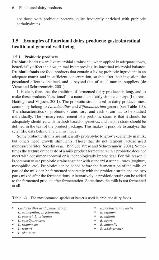
However, to meet consumers’ demands for probiotic foods in different countries,different types of products are also needed. One way to meet this challenge is to tryto define an effective daily dose to be used in various types of products. Forexample, Lactobacillus rhamnosus GG is used in Finland in cultured buttermilks,‘sweet’ milk, yoghurts, fermented whey-based drinks, set-type fermented milks(‘viili’), cheeses, juices, and mixtures of milk and juice. It is not reasonable orscientifically interesting to repeat clinical studies with all the different types,especially when the overall claims to be used in marketing are the same generallevel. Milk is a protective food matrix for probiotics and improves the survival ofthe strain in the intestine. As can be seen in Fig. 1.2, if one wishes to re-isolate the
Fig. 1.2 The recovery of Lactobacillus GG in faecal samples during daily consumption ofdifferent product forms. The daily dose of the probiotic strain (log cfu) per serving
and the level in stool samples (log cfu/g wet mass) are indicated in the vertical axis.
8 Functional dairy products
strain in stool samples during daily consumption, much lower doses of LactobacillusGG can be used in milk or cheese than in capsules or in powders.
The most common probiotic dairy products worldwide are various types ofyoghurt, other fermented dairy products (e.g. cultured buttermilks in Finland),various lactic acid bacteria drinks (‘Yakult-type’) and mixtures of probiotic(fermented) milks and fruit juice. Probiotic cheeses, both fresh and ripened, havealso been launched recently. From January 2000 to May 2002, 25 functionalcheeses were launched in Europe, 19 of which, it was claimed, contained an activeculture or a probiotic strain (Mintel’s Global New Products Database;www.gnpd.com). In addition to everyday products, probiotics are also used inindulgence products, e.g. ice creams.
Probiotic dairy foods (with certain specific strains) are known to relieveintestinal discomfort, prevent diarrhoea and improve recovery. However, nocountry will accept this claim, as it is too medical for use in the marketing of food.The most common health claim used for probiotic dairy foods may be ‘improvesnatural defence systems’, but as far as we know, the science behind that statementis not officially evaluated in any country for any product. In Japan, wherefunctional food legislation is organised best, the package claims for the acceptedFood for Specified Health Users (FOSHU) regulation lactobacilli products arethat they balance gastrointestinal functions. Recently a claim that a yoghurtproduct enriched with a strain of L. gasseri suppressed Helicobacter pylori (onecause of peptic ulcers) was also accepted. There are other products that supposedlysuppress the growth and activity of H. pylori, both in Europe and in the KoreanRepublic.
1.5.2 Prebiotic and synbiotic dairy productsPrebiotics are non-digestible food ingredients that beneficially affect the host byselectively stimulating the growth and/or activity of one or a limited number ofbacteria in the colon. Prebiotic foods are food products that contain a prebioticingredient in an adequate matrix and in sufficient concentration, so that after theiringestion, the postulated effect is obtained, and is beyond that of usual nutrientsuppliers. Synbiotics are mixtures of pro- and prebiotics that beneficially affectthe host by improving the survival and implantation of selected live microbialstrains in the gastrointestinal tract (de Vrese and Schrezenmeir, 2001).
In contrast to probiotics, which introduce exogenous bacteria into the humanintestine, prebiotics stimulate the preferential growth of a limited number ofbacteria already existing in a healthy, indigenous microbiota. The clue to prebioticcompounds is that they are not digested in the upper gastrointestinal tract, becauseof the inability of the digestive enzymes to hydrolyse the bond between themonosaccharide units. They act as soluble fibres and are digested in the colon,enhancing microbial activity and stimulating the growth mainly of bifidobacteriaand lactobacilli. Consumption of higher doses may encourage the formation of gas,flatulence and intestinal discomfort. The end-products in the gut fermentation aremainly short chain fatty acids (acetic, propionic and butyric acid), lactic acid,
Introduction: classifying functional dairy products 9
hydrogen, methane and carbon dioxide. Short chain fatty acids, especially butyricacid, are known to act as an energy source for enterocytes (Wollowski et al., 2001).The main dairy products enriched with prebiotics are yoghurts and yoghurt drinks,but spreads, fresh cheeses and milks are also on the market.
Galactooligosaccharide, a milk-based prebiotic, is derived from lactose by theβ-galactosidase enzyme. It is a natural prebiotic of human breast milk, andfacilitates the growth of bifidobacteria and lactobacilli in breast-fed infants.Galactooligosaccharides are commercially used principally in Japan and otherparts of Asia.
In Europe inulin and fructooligosaccharides are widely used in various functionalfoods, including dairy-based products. Inulin is a group of fructose polymerslinked by β(2–1) bonds that limit their digestion by enzymes in the upper intestine.Their chain lengths range from 2 to 60. Oligofructose is any fructose oligosaccharidecontaining two to ten monosaccharide units linked with glycosidic linkage. Bothinulin and fructooligosaccharides (oligofructoses) are extracted from plant material(e.g. chicory) or synthesised from sucrose. The role of inulin and the oligofructosesin a food matrix is bi-functional. They do not increase the viscosity of a milkproduct but give a richer texture to liquid products and spreads.
1.5.3 Low-lactose and lactose-free milk productsIn the human intestine lactose is hydrolysed by a lactase enzyme developed in thebrush border of the small intestine. When a person has a lactase deficiency andlactose causes intestinal discomfort and other symptoms, this is called lactoseintolerance, and is quite common in most parts of the world. The incidence oflactose intolerance is low only in the Nordic countries, the British Isles, Australiaand New Zealand. Most people can drink one glass of milk (~10 g lactose) in asingle dose taken with a meal, without suffering symptoms, but not a 50 g doseingested on an the empty stomach, the dose used in lactose tolerance tests.
There is a general consensus of opinion that probiotic dairy products alleviatelactose intolerance. This is true of all fermented dairy products, especially yoghurt,owing to the β-galactosidase activity of the yoghurt culture and the higherconsistency of fermented milks compared with ordinary milk. However, a muchmore sophisticated and efficient way of reducing symptoms caused by lactose is tohydrolyse it in the milk enzymatically. In long-life milks the enzyme is generallyadded to the milk after sterilisation, and the product is released for sale after acertain period, when the level of lactose has decreased. In fermented milks theenzyme is added before fermentation or at the same time as the culture. If addedwith the culture, the enzyme must be active in acidic conditions. In Finland, ValioLtd has a large range of lactose-hydrolysed (HYLA®) milk products, altogetheraround 80 varieties.
The hydrolysis of lactose changes the taste of the milk, making it sweeter,because glucose and galactose are sweeter than lactose. This is an accepted fact infermented milk products, especially if they are additionally sweetened. However,this sweetness is not popular in milk for drinking, and thus milk consumption
10 Functional dairy products
drops. Recently, this problem, too, has been solved. In 2001 Valio Ltd launcheda lactose-free milk in which the lactose has been completely removed physically.The sweetness has been restored to its normal level and the taste is that of normalfresh milk.
1.5.4 OthersSphingolipids contain compounds such as ceramides, sphingomyelin, cerebrosides,sulphatides and gangliocides. Sphingolipids are found in millk, butter and cheese– approximately 2 mg/100 g milk. Because they exist in cell membranes ratherthan in fat droplets, they are found in fat-free, low-fat as well as in full-fat dairyproducts. In vitro and experimental studies indicate that sphingolipids influencecell regulation, and thus carcinogenesis and tumour formation (Miller et al., 2000).In 2000, a yoghurt brand called ‘Inpulse’ was launched in Belgium (BüllengerButterei). The low-fat product was said to be rich in natural milk lecithin(45 mg/100 g) and sphingolipids (phospholipids 144 mg/100 g). A variety launchedsince then contains 0.6 g fat, 115 mg phospholipids, 36 mg phosphatidylcholineand 18.4 mg sphingolipids. The information on the product declares that ‘lecitinand sphingolipids are biomembranes, which re-establish the biological equilib-rium of the cells, protect against bacterial infections and help digestion’.
1.6 Examples of functional dairy products: cardiovascularhealth
Coronary heart disease (CHD) is a serious form of cardiovascular disease and themost common – the leading cause of death in developed industrialised countries.Many risk factors, both genetic and environmental, contribute to the developmentof coronary heart disease. The three most important modifiable risk factors for thisare cigarette smoking, high blood pressure and high blood cholesterol levels,particularly of low-density lipoprotein (LDL) cholesterol. Other risk factors likelyto contribute to the risk of CHD are diabetes, physical inactivity, low high-densitylipoprotein (HDL) cholesterol, high blood triglyceride levels, and obesity. Oxidativestress, homocysteine, lipoprotein and psychosocial factors may also increase therisk. To choose a healthy, low-fat diet with high levels of fruits and vegetables, anactive lifestyle and no smoking seems to reduce the risk of heart diseases. Theinclusion of semi-skimmed or non-fat milk products in an otherwise healthy dietadds many essential vitamins, not to mention milk calcium, which has a vital rolein controlling blood pressure (Miller et al., 2000). Milk products specificallydeveloped to reduce dietary risk factors are already on the market.
1.6.1 Products for controlling hypertensionThere are a few products on the market for lowering blood pressure. Several milkpeptides are known to have an inhibitory effect on the angiotensin converting
Introduction: classifying functional dairy products 11
enzyme (ACE inhibition). ACE is needed for converting angiotensin I to angiotensinII, increasing blood pressure and aldersterone, and inactivating the depressoraction of bradykinin. ACE inhibitors derived from caseins are called casokinins,and they are derived from the tryptic digestion of bovine β- and κ-caseins. In twocommercial products, these peptides are isoleucine–proline–proline and valine–proline–proline, which are formed from β-casein by the fermentation of milk withLactobacillus helveticus. The L. helveticus bacterium is generally used in cheese-making and the fermentation is a normal dairy process. The Calpis Amiel drink(Japan) is a sterile product, without living bacterial cells. The fermented milk drinkEvolus, more recently developed by Valio Ltd (Finland), contains, in addition tothe active tripeptides, living bacterial cells and an improved composition ofminerals (Ca, K, Mg). Both products have been tested in animal studies withspontaneously hypertensive rats (Sipola, 2002) and in clinical human trials(Hata et al., 1996; Seppo et al., 2002). The Japanese product has official FOSHUstatus.
In Finland there is a cheese on the market that has been shown to have ACEinhibitory activity (Festivo cheese, Agricultural Research Centre, Jokioinen,Finland). The bioactive peptides are shown to be αs
1-casein N-terminal peptides
but the researchers thought that they might be too long to be absorbed intact in theintestine. The quantity also varied during the maturation and age of the cheese, andthe effect of the cheese on human blood pressure remains to be tested (Ryhänen etal., 2001). Another idea, not yet commercially launched in dairy products, is basedon whey proteins that are hydrolysed so that the whey protein isolate has an ACEinhibitory activity (Davisco Foods International Inc., USA). The effect of thisproduct seems to be much faster than those based on the tripeptides, but themechanism is not yet known (Pins and Keenan, 2002).
1.6.2 Products for controlling cholesterolNatural cows’ milk fat contains high levels of saturated fatty acids. Replacing theconsumption of full-cream milk with semi-skimmed or non-fat milk will reducethe intake of saturated fatty acids. Sometimes it is not enough just to reduce theintake of saturated fats and cholesterol, since most cholesterol is synthesisedwithin our own bodies. On the other hand, plant sterols and stanols have long beenknown to reduce the assimilation of dietary cholesterol. Since the mid-1990s therehave been products enriched with plant stanols specially targeted at those peoplewith (moderately) high cholesterol levels. A few years later plant sterols were alsoaccepted as food ingredients by the EU Novel Foods legislation, and now the Foodand Drug Administration in the USA has also accepted plant sterols and stanols.Sterols are building blocks of the cell membranes in both plant and animal cells.Isolated plant stanols, hydrated forms of sterols, are crystallised particles. Theyeffectively bind cholesterol and are not absorbed by the human body. Esterifiedplant stanols are fat-soluble and easy to use as a food ingredient. Intestinal enzymeshydrolyse the ester bond and the insoluble stanol is free to bind cholesterol and tobe secreted. Basically, the effect of plant sterols is based on the same mechanism.
12 Functional dairy products
Several milk-based functional foods including plant sterols or stanols arecommercially available. They all are semi-skimmed or non-fat products. Prod-ucts containing Benecol (Raisio Benecol Ltd, Finland), the only plant stanolester ingredient, are on the market in several countries. In some products the‘effective daily dose’ has to be collected from several servings (e.g. Benecolmilk, yoghurt, various spreads in the UK), in some other countries the dose iscontained in one serving (e.g. Valio Benecol yoghurt in Finland). Plant sterolsare also added to functional milk products, especially to milk (e.g. MastelloneHnos SA, Argentina). In March 2001, Marks & Spencer launched a range of 20products, including yoghurt, enriched with soy proteins (& More brand, UK).The daily consumption of 25 g soy proteins has been shown to lower chole-sterol by 10%.
The safety risk of overdosing with plant sterols and stanols has been thesubject of discussion by the scientific committee on food of the EuropeanCommission. The consumption of this kind of product requires a fairly goodknowledge on the part of the consumer, as she or he has to be familiar with theproducts with the compound and also to know the quantity of the active ingre-dient in various products. For that reason the labelling must be informativeenough.
Matured cheeses contain quite high levels of milk fats. Replacing milk fat withvegetable oil can reduce the intake of saturated fatty acids. In Finland there arecheeses on the market in which milk fat has been replaced by rapeseed oil (Juliaand Julius with 17% and 25% rapeseed oil, respectively; Kyrönmaan Osuusmeijeri,Finland). The products, when included daily in a low-fat diet, reduced bloodcholesterol statistically significantly (Karvonen et al., 2002).
1.6.3 Omega-3 fatty acidsThere are two major classes of polyunsaturated fatty acids: omega-3 fatty acidsfound in fish oils and as a minor constituent of some vegetable oils, and omega-6fatty acids, which include the essential fatty acid linoleic acid, found in vegetableoils such as corn, sunflower and soybean. Omega-3 fatty acids are said tocontribute to the good functioning of the cardiovascular system, on the basis ofvarious physiological effects. Before omega-3 fatty acids could be added to milkproducts, the fishy taste and odour had to be disguised and the easy oxidation of theoil overcome. It took several years before these problems were solved, butnowadays there are a few suppliers selling good-quality fish-oils to be added tomilk. The pioneer in launching an omega-3-enriched milk was the Italian dairycompany Parmalat. Its ‘Plus Omega 3’ milk was launched in 1998 and is a semi-skimmed milk enriched with 80 mg omega-3. It is recommended for use by allhealth-conscious consumers in a dose of half a litre per day (Mellentin andHeasman, 1999). Since then other producers all over the world have followed withtheir own omega-3-enriched milks. Milk is often also enriched with the antioxidativevitamins A, C and E.
Introduction: classifying functional dairy products 13
1.7 Examples of functional dairy products: osteoporosis andother conditions
The cause of osteoporosis, as with other chronic diseases, is multifactorial,involving both genetic and environmental factors. An accumulation of scientificevidence indicates that a sufficient intake of calcium throughout one’s life offersprotection against osteoporosis. The bone mass reaches its peak when a person is30 years of age and then the density decreases with age, especially after themenopause. The fortification of semi-skimmed and non-fat milk with vitamin D isimportant, as this vitamin is essential to improve calcium absorption and is alsoremoved when fat is removed. Milk is the richest source of calcium. There areseveral milks and milk products enriched with calcium, and both inorganic andmilk-based calcium (e.g. TruCal, Glanbia Ingredients Inc.) are used. The absorptionof calcium may be enhanced with bioactive milk proteins. Caseino-phosphopeptides(CPPs) are known to increase the solubility of calcium, but controversy exists as towhether CPPs enhance calcium absorption in the body. The authors do not knowof any commercial applications of CPPs in dairy products.
1.7.1 Products for enhancing immune functionsSome of the probiotic dairy products have been shown to enhance immunefunctions and thus to reduce the risk of infection. Milk contains naturalimmunoglobulins, which can be isolated and concentrated, either from normalmilk or from colostrum, which contains a high proportion of them. There are milk-based products on the market in which the product is further enriched withimmunoglobulins. In the USA and Australia, Lifeway Foods is marketing kefirunder the brand name Basic Plus. The product is said to be probiotic, although theprobiotic strains are not specified. The active ingredient, an extract of colostrum,has been developed by GalaGen Inc. and is targeted at maintaining intestinal healthand the natural microbiota. Basic Plus was launched in 1998 and is the first dairy-based food supplement sold in the USA in the refrigerated sections of health foodand grocery stores.
Milk immunoglobulins are used in new drinks in the USA under the brand nameof ‘NuVim’. The production of immunoglobulins is boosted in a selected herd inNew Zealand by an immune stimulant, and isolated under carefully controlledconditions in order to preserve the micronutrients. The product is said to be lactose-free and fat-free, to have beneficial effects on the immune system and to improvethe health of muscles and joints (Heasman and Mellentin, 2002).
1.7.2 Milks to help with sleeping problemsMelatonin is a hormone that controls the body’s day and night rhythm. Thesecretion of melatonin is high in early childhood and decreases rapidly withageing. Stress conditions and age cause a lowering of the level of melatonin. It issecreted at nights in both humans and bovines. The concentration at night in cows’
14 Functional dairy products
milk is about four times higher than in milk collected during the day. The firstproduct based on a standardised milking system at night was launched in Finlandin 2000 (Yömaito, Ingman Foods Ltd). Since no human trials have been publishedso far, the company does not make any health claims. In spring 2002 an organicmilk, ‘Slumbering Bedtime Milk’ (Red Kite Farm, UK), was launched in the UK.It is said to contain higher levels of melatonin than ordinary milk. The companysays that the level of melatonin in the milk complements that of the human bodyand the drink will not induce drowsiness if drunk during the day, or the followingmorning if drunk at night/late in the evening.
1.8 Future trends
Research and discussion on pro- and prebiotics have encouraged basic research inthe field of the intestinal microbial flora and its metabolism. This has also led toimproved research funding from public resources, both nationally and from theEuropean Union. Not enough is known of the composition and metabolism of thebacteria in the intestines in health and disease. Also the knowledge on the role ofthe microbiota in the development and function of immune response needs moreinvestigation. Development and improvements in research methods, and in vitro,ex vivo and in vivo models, have provided important information on the mechanismsbehind the effects, and new biomarkers to be followed in human studies. The morewe know about the composition and function of the intestinal microbiota, thegreater the potential to develop functional foods for targeted consumer groups.Considering the healthy population there may be potential to develop targetedproducts for different age groups. In the reduction of risk and treatment of variousdiseases, pro- and prebiotics have resulted in promising benefits. However, it isimportant to understand the mechanisms behind the effects. When the mechanismsare known, it will be also possible to control the activity or the dose of the effectivecompounds. We also need official definitions of functional foods, and relevantregulation of physiological claims and health claims. The production of functionalfoods that have to follow the rules of production of medicines is hardly in theinterest of normal dairy companies. It may be unrealistic to apply the same rules tomedicines as to everyday foods with a short shelf-life.
Milk is a rich source of nutritive compounds which can be enriched and/orfurther modified. Milk fat does not consist merely of saturated fatty acids, but alsoof monounsaturated and polyunsaturated fatty acids. The role of conjugatedlinoleic acid (CLA) in preventing the risk of certain diseases, and in particular, theproblem of how to increase its quantity in milk has evoked wide interest amongseveral research groups. Milk proteins and bioactive peptides may supply newproducts to help protect against several common health risk factors. There arebioactive peptides potentially to be used to give satiety or to better tolerate stress.Lactose derivatives can be used as soluble fibre to relieve constipation and tomodulate the intestinal flora. Milk minerals can be used to replace sodium in salt,supporting a healthy diet for avoiding hypertension. Milk components are natural,
Introduction: classifying functional dairy products 15
and applications for novel foods are seldom needed. There is also a huge selectionof lactic acid bacteria used for milk fermentations, which have a long tradition ofsafe use. Genetically modified strains may be needed for special purposes, thoughperhaps not in products for the general public.
In developing functional dairy products, various groups of experts are needed.The basis must be in the scientific research of effects, requiring medical experts,nutritionists and microbiologists. Food technologists are needed for productdevelopment, process technologists and biotechnologists for processing thecompounds, chemists to analyse the compounds and, finally, experts for marketingthe products. Marketing is a big challenge, as it has to tell the public about thehealth benefits in such a simple way that every layperson understands. Medical andnutritional messages need to be simplified. It is important to remember thatfunctional dairy products are mainly for supplying nutritive foods for everydayconsumption. Nutrimarketing is also needed to explain research results to health-care professionals and to convince them of the benefits of functional foods.
1.9 Sources of further information and advice: links
www.gnpd.comwww.new-nutrition.comwww.scirus.comwww.just-food.comwww.ifis.orghttp://www.foodlineweb.co.ukwww.fst.ohio-state.edu/People/HARPER/Functional-foods/Functional-Foods.htmwww.valio.comwww.benecol.comwww.daviscofood.comwww.kefir.comwww.ific.orgwww.effca.comwww.usprobiotics.orgwww.elintarvikevirasto.fi/english
1.10 ReferencesBANKS W and DALGLEISH D G (1990), ‘Milk and milk processing’ in Robinson R K, Dairy
Mircobiology, Volume 1, The Microbiology of Milk, second edition, London, ElsevierScience Publishers Ltd, 1–35.
CLARE D A and SWAISGOOD H E (2000), ‘Bioactive milk peptides: a prospectus’, J Dairy Sci,83, 1187–1195.
DE VRESE M and SCHREZENMEIR J (2001), ‘Pro and prebiotics’, Innov Food Technol, May/June, 49–55.
DIPLOCK A T (1999), ‘Scientific concepts of functional foods in Europe: Consensusdocument’, Br J Nutr, 81(Suppl 1), S1–S27.
16 Functional dairy products
HATA Y, YAMAMOTO M, OHNI M, NAKAJIMA K, NAKAMURA Y and TAKANO T (1996), ‘Aplacebo-controlled study of the effect of sour milk on blood pressure in hypertensivesugjects’, Am J Clin Nutr, 64, 767–771.
HEASMAN M and MELLENTIN J (2002), ‘New NuVim prepares to be swallowed up’, NNB,7(8), 29–30.
KARVONEN H M, TAPOLA N S, UUSITUPA M I and SARKKINEN E S (2002), ‘The effect ofvegetable oil-based cheese on serum total and lipoprotein lipids’, Eur J Clin Nutr, 56,1094–1101.
KORHONEN H , PIHLANTO-LEPPÄLÄ A, RANTAMÄKI P and TUPASELA T (1998), ‘Impact ofprocessing on bioactive proteins and peptides’, Trends Food Sci Technol, 8, 307–319.
LEPORANTA K (2001), ‘Developing fermented milks into functional foods’, Innov FoodTechnol, 10, 46–47.
LOURENS-HATTINGH A and VILJOEN B C (2001), ‘Yoghurt as probiotic carrier food’, IntDairy J, 11, 1–17.
MELLENTIN J and HEASMAN M (1999), ‘Functional foods are dead. Long live functionalfoods’, NNB, 4(7), 16–19.
MILLER G D, JARVIS J K and MCBEAN L D (2000), Handbook of Dairy Foods and Nutrition,second edition, Boca Raton, London, New York, Washington DC, CRC Press.
PINS J and KEENAN J M (2002), ‘The antihypertensive effects of a hydrolysed whey proteinisolate supplement (BioZate1®)’, Cardiovasc Drugs Ther, 16 (Suppl 1), 68.
RYHÄNEN E-L, PIHLANTO-LEPPÄLÄ A and PAHKALA E (2001), ‘A new type of ripened, low-fat cheese with bioactive properties’, Int Dairy J, 11, 441–447.
SAXELIN M, GRENOW B, SVENSSON U, FONDEN R, RENIERO R and MATTILA-SANDHOLM T(1999), ‘The technology of probiotics’, Trends Food Sci Technol, 10, 387–392.
SEPPO L, JAUHIAINEN T, POUSSA T and KORPELA R (2002), ‘A fermented milk, high inbioactive peptides, has a blood pressure lowering effect in hypertensive subjects’, Am JClin Nutr, in press.
SHAH N P (2000), ‘Probiotic bacteria: selective enumeration and survival in dairy foods’, JDairy Sci, 83(4), 894–907.
SIPOLA M (2002), ‘Effects of milk products and milk protein-derived peptides on bloodpressure and arterial function in rats’, PhD Thesis, Institute of Biomedicine/ Pharmacology,University of Helsinki; electronic PDF version: http://ethesis.helsinki.fi/julkaisut/laa/biola/vk/sipola/.
SMITH J (2001), ‘Defining health claims for Europe’, Funct Foods Nutraceut, November/December, 12.
WOLLOWSKI I, RECHKEMMER G and POOL-ZOBEL B L (2001), ‘Protective role of probioticsand prebiotics in colon cancer’, Am J Clin Nutr, 73(2 Suppl), 451S–455S.

Part I
The health benefits of functional dairyproducts
2
CancerC. Gill and I. Rowland, University of Ulster, UK
2.1 Introduction
Colorectal cancer (CRC) is the fourth most common cause of cancer-relatedmortality in the world. It is estimated that up to 15% of all colorectal cancer is dueto genetic predisposition, with a further 60% due to sporadic tumours that appearto develop from adenomatous polyps. Adenomas and carcinomas develop througha stepwise accumulation of somatic mutations, termed the ‘adenoma–carcinomasequence’. Diet has major implications for the aetiology of the disease, with thoserich in vegetables associated with protection.
Worldwide, milk and dairy products contribute approximately 5% of totalenergy. But among the traditionally pastoral people of China, India, Africa andNorthern Europe, dairy and milk products supply approximately 10% total energyand 15–25% dietary protein and fat intake. Dairy products have been tentativelysuggested to play a protective role in the prevention of CRC. This chapter willexamine both experimental and epidemiological data for dairy products and theirsignificant components (including calcium, casein and conjugated linoleic acid) todetermine if a basis for the protective hypothesis exists. The emerging evidence fora protective role for fermented milks, probiotics, prebiotics and other functionaldairy products is reviewed.
2.2 The relationship between diet and cancerCancer is a significant global public health problem. Yearly 10.1 million newcancer cases are diagnosed, with a further 6.2 million people losing their livesworldwide. This disease accounts for 25% of deaths in countries with a western-ised lifestyle (IARC, 2000). Colorectal cancer is the fourth most frequent cause of
20 Functional dairy products
cancer-related mortality in the world: approximately 944,000 new cases werediagnosed globally in 2000 and this accounts for 9.2% of all new cancer cases(IARC, 2000). Within New Zealand, Australia, North America and Europe it is thesecond most prevalent cancer after lung/breast (Boyle and Langman, 2000):363,000 new cases were reported in Europe in 2000 and it affects 6% of men andwomen by age 75, in almost equal proportion. Generally the incidence andmortality of the disease are escalating (Cummings et al., 1996; Boyle and Langman,2000). The Modena colorectal registry (Italy) reported a 12.2% increase in incidentrates from 1985 to 1997 and other European studies have reported a similar trend(Johansen et al., 1993; Kemppainen et al., 1997).
Worldwide incident rates show an approximate 20-fold variation, with thedeveloped world suffering the highest rates and India one of the lowest (Ferlay etal., 2001). Rates even within countries may vary, as in India where the strictlyvegetarian Janists have a lower rate of colorectal cancer than the westernised Parsipopulation (ICS, 1985). These fluctuations are generally attributed to both geneticfactors and environmental factors, especially diet. Migrant studies (Japan to theUSA, Eastern Europe to North America) give additional support to the role ofenvironmental factors in the aetiology of colorectal malignancies, with reportedincident rates of migrants and their descendants reaching those of the host country,sometimes within one generation (WCRF, 1997). The highest rates of colorectalcancer are currently seen within Hawaiian Japanese men with an incidence of 53.5per 100,000 (IARC, 1997).
Evidence suggests that diet plays an important role in the aetiology of colorectalcancer. However, identifying conclusively which constituents (e.g. vegetables,meat, fibre, fat, micronutrients) exert an effect on risk has been more problematicdue to inconsistent data (for a detailed review of the epidemiological studies seePotter, 1999). The 1997 World Cancer Research Fund report concluded that theevidence (mainly from case-control studies) for diets rich in vegetables protectingagainst colorectal cancer was convincing, but that the effect of fruits could not bejudged because the data are limited and contradictory. Data from prospectivestudies are less convincing than case-control studies (Bingham, 2000). Diets highin fibre were reported to possibly reduce the risk of colorectal cancer, withsuggested protective mechanisms including toxin adsorption/dilution (WCRF,1997; AGA, 2000). Furthermore several micronutrients including carotenoids,ascorbate and folate have been examined epidemiologically to account for theprotective effect associated with vegetables, but the results have frequently beenincongruous, and coupled with the paucity of data, no strong associations wereobserved (Giovannucci et al., 1993; Slattery et al., 1997). Studies examining theeffect of meat consumption (especially red and processed meats) on colorectalcancer have collectively produced neither strong nor consistent findings, but it isbelieved that the weight of evidence points towards a slighty elevated risk (WCRF,1997; Norat and Riboli, 2001), although the mechanisms by which meat affectscolon carcinogenesis remains unclear. High saturated/animal fat intake may berelated to elevated risk (Potter, 1999; Zock, 2001) but does not appear to contributeto the risk associated with meat consumption (Giovannucci and Goldin, 1997).
Cancer 21
A slightly elevated risk in beer drinkers versus abstainers was first reported byStocks (1957); since then alcohol has been suspected as a risk factor for colorectalneoplasms. Studies on the topic have provided contentious results as detailed in areview by Potter et al. (1993). Overall, however, raised alcohol consumptionprobably increases the risk of cancers of the colon and rectum and this associationis related to total ethanol intake rather than the type of alcoholic drink (WCRF,1997). Despite the weight of the epidemiological evidence for diet playing animportant role in colorectal cancer risk, definitive evidence for causal associationis lacking owing to the difficulties of conducting dietary intervention studies. It isnecessary to look elsewhere for stronger criteria of such a link. The next sectionprovides a brief summary of the processes of carcinogenesis in the colon.
2.3 Colon carcinogenesis
Almost 70% of colorectal malignancies appear to be restricted to the left largebowel (descending) between the lower rectum and the splenic fissure, thoughcuriously this subsite distribution appears to be undergoing a proximal shifttowards the right large bowel (ascending), for reasons unknown (Faivre et al.,1989; Ponz de Leon and Roncucci, 2000). The colonic microarchitecture ischaracterised by crypts, which are approximately 50 cells in depth. The normalreplicative dynamics and structure of these crypts ensure that both stem cells andimmediate daughter cells replicate in the lowest region. When the immediatedaughter cells divide and migrate they give rise to all the cells that line the crypt.Eventually these cells will reach the surface, by which stage they are fullydifferentiated columnar epithelial cells, covered with microvilli, intimately con-nected via numerous tight junctions and involved in water and electrolyte transport.The constant outward movement of cells from the crypts should ensure that nointeraction occurs between the luminal environment and replicating cells; thus anymutagens should then only affect the differentiated colonocytes and effectivelyhave no impact upon the integrity of the crypt cell population (Potter, 1999).
The nature of the microarchitecture was used by Potter to argue that the firstmutagenic event occurring to a progenitor cell must be a blood-borne agent ratherthan luminal. Further it was suggested that, for luminal constituents to play any rolein carcinogenesis, a pre-existing polyp must be in contact with the faecal stream.Alternatively, Shih et al. (2001) recently postulated that the development of anadenomatous polyp may proceed from a top-down mechanism, whereby the inter-cryptal cells are transformed presumably from a luminal agent and the alteredmucosae spread laterally and downward to form new crypts, which connect to andeventually replace existing crypts. Other authors who follow the more classicalmechanism for colorectal carcinogenesis, offer the assertion that a luminal agentcould provide the ‘first hit’ (mutation) if a focal failure in the epithelial barrieroccurred as a result of insult (physical/chemical) or of failure in terminaldifferentiation. An agent could then affect directly or indirectly cells in the cryptand lead to the formation of a polyp (Bruce et al., 2000). Mutation of the APC gene
22 Functional dairy products
(adenomatous polyposis coli) permits an adenomatous polyp to develop, and sucha formation is considered an important predisposing risk factor for CRC. However,this does not mean that all polyps become malignancies (only about 5%) nor doesit prohibit the possibility that de novo colorectal tumorigenesis may occur (Owen,1996). Adenomas are well-demarcated clumps of epithelial dysplasia, classifiedinto three histological types – tubulovillous, tubular and villous – which increasein prevalence with age, being present in 24–40% of people over 50 years old (Ponzde Leon and Roncucci, 2000).
Approximately 15% of all colorectal cancer is due to genetic predisposition,with a further 60% the result of sporadic tumours that appear to develop fromadenomatous polyps. Adenomas and carcinomas develop through a stepwiseaccumulation of somatic mutations (Fig. 2.1). While the precise sequence ofgenetic events is not completely understood, it involves inactivation of varioustumour-suppressing genes (e.g. APC, p53), activation mutations in proto-oncogenes(e.g. K-ras, c-myc), and loss of function in DNA repair genes (e.g. hMLH1,hMSH2). This archetypal multi-step model has been termed the ‘adenoma–carcinoma sequence’ (Vogelstein et al., 1988; Fearon and Vogelstein, 1990). Fordetailed information on the genetic events and pathways to colorectal cancer thereader is referred to Potter (1999), Chung (2000), Ponz de Leon and Percesepe(2000) and Souza (2001).
Fig. 2.1 The adenoma–carcinoma sequence. Inactivation of the APC gene facilitates thedevelopment of a hyperplastic epithelium. Activation of oncogenic ras occurs in earlyadenomas. Deletion of 18q is found in dysplastic late adenomas. Ultimately, with theinactivation of p53, colorectal cancer develops. Components of the faecal stream are
believed to modulate colorectal carcinogenesis at various stages.
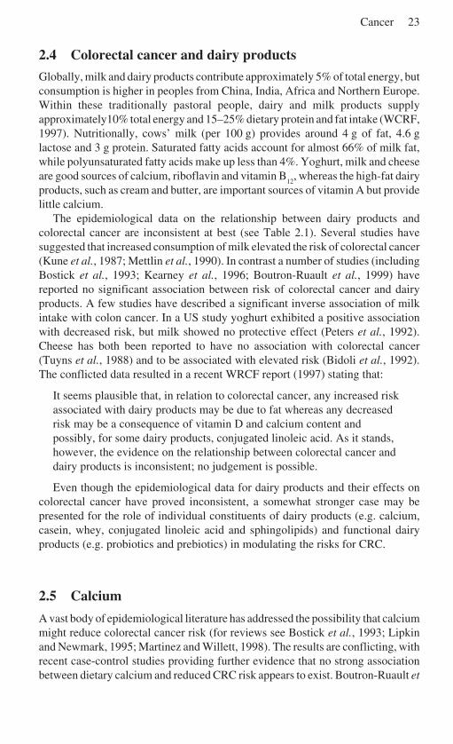
Cancer 23
2.4 Colorectal cancer and dairy products
Globally, milk and dairy products contribute approximately 5% of total energy, butconsumption is higher in peoples from China, India, Africa and Northern Europe.Within these traditionally pastoral people, dairy and milk products supplyapproximately10% total energy and 15–25% dietary protein and fat intake (WCRF,1997). Nutritionally, cows’ milk (per 100 g) provides around 4 g of fat, 4.6 glactose and 3 g protein. Saturated fatty acids account for almost 66% of milk fat,while polyunsaturated fatty acids make up less than 4%. Yoghurt, milk and cheeseare good sources of calcium, riboflavin and vitamin B
12, whereas the high-fat dairy
products, such as cream and butter, are important sources of vitamin A but providelittle calcium.
The epidemiological data on the relationship between dairy products andcolorectal cancer are inconsistent at best (see Table 2.1). Several studies havesuggested that increased consumption of milk elevated the risk of colorectal cancer(Kune et al., 1987; Mettlin et al., 1990). In contrast a number of studies (includingBostick et al., 1993; Kearney et al., 1996; Boutron-Ruault et al., 1999) havereported no significant association between risk of colorectal cancer and dairyproducts. A few studies have described a significant inverse association of milkintake with colon cancer. In a US study yoghurt exhibited a positive associationwith decreased risk, but milk showed no protective effect (Peters et al., 1992).Cheese has both been reported to have no association with colorectal cancer(Tuyns et al., 1988) and to be associated with elevated risk (Bidoli et al., 1992).The conflicted data resulted in a recent WRCF report (1997) stating that:
It seems plausible that, in relation to colorectal cancer, any increased riskassociated with dairy products may be due to fat whereas any decreasedrisk may be a consequence of vitamin D and calcium content andpossibly, for some dairy products, conjugated linoleic acid. As it stands,however, the evidence on the relationship between colorectal cancer anddairy products is inconsistent; no judgement is possible.
Even though the epidemiological data for dairy products and their effects oncolorectal cancer have proved inconsistent, a somewhat stronger case may bepresented for the role of individual constituents of dairy products (e.g. calcium,casein, whey, conjugated linoleic acid and sphingolipids) and functional dairyproducts (e.g. probiotics and prebiotics) in modulating the risks for CRC.
2.5 Calcium
A vast body of epidemiological literature has addressed the possibility that calciummight reduce colorectal cancer risk (for reviews see Bostick et al., 1993; Lipkinand Newmark, 1995; Martinez and Willett, 1998). The results are conflicting, withrecent case-control studies providing further evidence that no strong associationbetween dietary calcium and reduced CRC risk appears to exist. Boutron-Ruault et
24 Functional dairy products
Table 2.1 Colon cancer and dairy products
Study design/size Location Food Association with Authorcolorectal cancer risk
Cohort, 461,443, USA Milk products No significant Phillips and146 colon cancer association Snowdondeaths (1985)Case-control, Greece Milk products No significant Manousos100ca/100 Hc association et al. (1983)Cohort, 9959 Sweden Milk No significant Jarvinen et al.M + F, 76 (2001)colorectal cancerCase-control, France Dairy products No significant Boutron-ca 171/pc 309 Ruault et al.
(1999)Cohort, 9,159 M USA Cheese, whole No significant Kampman etand 8,585 F, 331M milk, skim milk, association al. (1994a)+ 350F colorectal fermented dairypolyp casesCohort, 12,852, Nether- Fermented milk, No significant Kampman et215 colon cancer lands hard cheese, association al. (1994b)
unfermentedmilkcalcium
Cohort, 35,216, USA Dairy products No significant Bostick et al.212 colon cancer association (1993)Case-control, USA Dairy products No significant Peters et al.746 ca/746 pc association (1992)Case-control, Singapore Milk products No significant Lee et al.132 ca/426 hc calcium association (1989)Case-control, Japan Dairy products No significant Tajima and93 ca/93 hc association Tominaga
(1985)Cohort 47,935, USE Milk, fermented No significant Kearney et al.203 colon cancer dairy association (1996)Case-control, Spain Dairy, calcium No significant Benito et al.286ca/295 pc and association (1990)203 hcCase-control, Italy Milk, cheese, No significant Negri et al.558 ca/1032 hc calcium association (1990)Case-control, USA Whole milk, Significant positive Mettlin et al.3334 cancer, 2% milk association (1990)504 colon ca/ Skim milk No significant1300 pc associationCase-control, 392 Australia Milk Significant positive Kune et al.ca/727 hc and pc (1987)Cohort, 27,111, Finland Calcium Significant inverse Pietinen et al.185 colon cancer association (1999)Case-control,424 USA Dairy products Significant inverse Shannon et al.ca/414 hc and pc association (1996)Case-control, 210 USA Cheese Significant inverse Young anddistal and 152 association (distal Wolf (1988)proximal colon colon only)ca/618 pc
Cancer 25
Table 2.1 cont’d
Study design/size Location Food Association with Authorcolorectal cancer risk
Case-control, USA Dairy Significant inverse Slattery et al.231 ca/391 pc association (1988)Case-control, Nether- Milk Significant inverse Kampman et232 ca, 259 pc lands association al. (1994c)
Skim milk, No significant,fermented associationdairy, cheese
Case control, France Milk Significant inverse Macquart-399 ca/399 hc trend Moulin et al.
cheese, butter No significant (1986)association
ca cases, pc population controls, hc hospital controls, M male, F female.
al. (1999) reported no association between the factors in a French case-controlstudy, as did a Swedish case-control study, although it suggested that dietaryvitamin D might reduce the risk of colorectal cancer (Pritchard et al., 1996). InFinnish men consuming a diet high in fat, meat and fibre and low in vegetables,high calcium intake was associated with lowered risk of colorectal cancer (Pietinenet al., 1999). Further case-control studies on fermented dairy products andmilk indicated that total dietary calcium was positively but non-significantlyassociated with colon cancer risk after adjustment for confounding factors(Kampman et al., 1994c; Ma et al., 2001). Similarly a prospective study examiningthe influence of dairy foods reported a non-significant inverse association betweenthe intake of calcium from food/supplements and colon cancer risk (Kearney et al.,1996).
Notwithstanding the burgeoning press on the apparent lack of effect of calcium,sufficient studies have reported a positive association to suggest that calcium mayin some manner protect against CRC (for reviews see Holt, 1999; Mobarhan, 1999;Kampman et al., 2000). Recently, a case-control study by Marcus and Neucomb(1998) reported a protective effect of higher levels of calcium intake and CRC riskin women. Calcium supplementation and consumption of total low-fat dairyproducts were inversely associated with colon cancer risk in both men and women(Kampman et al., 2000).
In contrast to conflicting epidemiological studies on calcium and colon cancer,experimental studies in animals and humans more regularly report a beneficialrole. Two recent studies on calcium supplementation and colorectal adenomarecurrence have shown significant, though moderate, reduction in the risk, with athird reporting a modest but non-significant reduction (Baron et al., 1999; Bonithon-Kopp et al., 2000; Wu et al., 2002). Wu et al. (2002) reported reduced risk only fordistal colon cancer and suggested that the observed risk pattern was consistentwith a threshold effect, indicating that calcium intake beyond moderate level
26 Functional dairy products
(700 mg/d) may not be associated with a further reduction in risk. High calciumintake may offer a protective effect from fat-induced promotion of carcinogenesisby binding cytotoxic bile and fatty acids (Kleibeuker et al., 1996a) or by reducingproliferation in the upper part of the colonic crypt (Bostick et al., 1995). Proliferativezone expansion in the colonic crypts and an increased rate of epithelial prolifera-tion are often viewed as an early step in carcinogenesis. Stimulation of proliferativeactivity in colonic epithelial may in part be mediated by chemical or physicalcytotoxic mechanisms, such that epithelial damage induced by these sourceswould increase cell loss at the epithelial surface. This would result in a compensa-tory increase in the mitotic activity of the crypts, thus elevating the risk forcolorectal cancer. Such considerations led to the development of assays to assesscytotoxic activity in faecal water (aqueous phase faeces) towards colon cells invitro (Rafter et al., 1987). It is thought that bile acids, especially secondarybile acids, make a major contribution to faecal water cytotoxicity (Rafter et al.,1987). Dietary calcium reduces the cytotoxicity of faecal water, presumably byprecipitating soluble bile acids (Van-der-Meer et al., 1991, 1997; Govers et al.,1993; Lapre et al., 1993; Sesink et al., 2001). Interestingly a recent study showedthat a shift from a dairy-rich diet (high fat, high risk) to a dairy-free diet (low fat,low risk) showed an increase in cytotoxicity of the faecal water, possibly as a resultof, among other things, decreased calcium (Glinghammar et al., 1997).
The formation of aberrant crypt foci (ACF) represent putative preneoplasticlesions that are induced in the colon of carcinogen-treated rodents and are presentin humans with a high risk for developing colon cancer (Pretlow et al., 1992; Bird,1995). ACFs are characterised by an increase in the size of the crypts, the epitheliallining and the pericryptal zone. The prevention of azoxymethane (AOM)-inducedACF is used as a bioassay in animal studies, and has demonstrated the positivechemoprotective effects of dietary calcium supplementation (various concentra-tions) and the efficacy of different calcium salts (lactate, phosphate, glucurate)(Wargovich et al., 1990, 2000; Pereira et al., 1994; Li et al., 1998). An ACF studyusing 1,2-dimethylhydrazine (DMH) reported that low dietary calcium lactatesupplementation inhibited colorectal carcinogenesis, and changed tumour locationtowards the distal colon (Vinas-Salas et al., 1998). Calcium also appears to induceapoptosis in normal mouse distal colonic epithelium without affecting cellproliferation. Such a mechanism may contribute to calcium’s putativechemopreventive role in colorectal carcinogenesis (Penman et al., 2000).Considering these data as a whole, it appears likely that calcium has a promisingrole to play in protecting against colorectal cancer, but the strength of that role isas yet undetermined.
2.6 Casein
The casein content of milk represents approximately 80% of the milk proteins, inthe form of α-, β- and κ-casein. Caseins are mostly phosphate-conjugated proteinswith low solubility with the degree of calcium binding by the protein being
Cancer 27
proportional to the phosphate content. No epidemiological data currently exist forcasein: several animal studies indicated that dietary supplementation with caseinreduced the risk of colon cancer. Casein diets provided a greater protective effectagainst the development of DMH-induced intestinal tumours in rats than did redmeat or soybean diets, as evidenced by a reduced tumour incidence (McIntosh etal., 1995). Further, a casein–wheat diet resulted in a decreased tumour mass indexbut not tumour burden in DMH-induced rats when compared with a chickpea–wheat diet (McIntosh et al., 1998). In vitro casein appears to exert an antimutageniceffect (Yoshida and Ye-Xiuyun, 1992; van-Boekel et al., 1993), which may beenhanced by enzymatic digestion (Goeptar et al., 1997). The converse, however,is true if the protein is thermolysed. Casein cooked at 180 °C and fed as asupplement promotes the growth of ACFs in AOM-induced animals and appearsto do so in a dose-dependent and thermolysis time-dependent manner (Zhang et al.,1992). The thermolysis of casein reduces its digestibility, so more protein reachesthe colon and consequently raises colonic protein fermentation, and therefore theconcentration of the potential tumour promoter ammonia. However, the effect ofthermolysed casein on colorectal carcinogenesis does not appear to be mediated bythe protein’s fermentation products (Corpet et al., 1995), neither is the promotingeffect associated with the formation of heterocyclic amines (Corpet and Cassand,1995). Molecules are created during the cooking of protein-rich foods, whichwhen activated become mutagens and carcinogens capable of producing tumoursin various tissues both in rodents and non-human primates (Nagao and Sugimura,1993). The promotion effect of cooked casein has been suggested as the result ofmucosal abrasion due to the physical properties of the protein, as coarse cookedcasein resulted in significantly more ACFs than fine ground (Corpet and Chatelin-Pirot, 1997).
Although the direct effects of casein on colorectal carcinogenesis appearnegative, data suggest that the protein and its degradation products may help todecrease the risk of colorectal cancer through indirectly affecting calcium. Severalanimal supplementation studies have reported that casein phosphopeptides (CPPs)(created by proteolytic degradation of α- and β-casein) significantly increasecalcium and zinc absorption (Lee et al., 1983; Saito et al., 1998). CPPs have alsobeen reported to help overcome the inhibitory effect of phytate on zinc and calciumabsorption (Hansen et al., 1996) and to improve the absorption of these metalsfrom rice-based cereals but not wholegrain cereal in humans (Hansen et al., 1997).CPPs appear to alter calcium absorption not by influencing membrane-boundreceptors or ion channels, but rather by acting as calcium carriers (ionophores)across the membrane (Ferraretto et al., 2001). However, one recent study hasindicated that CPPs had no effect, whereas casein enhanced calcium absorption(Bennett et al., 2000).
Evidence for casein’s anticancer activity is accumulating, as are the data for aneffect on calcium absorption. High levels of calcium may be protective forcolorectal cancer. If calcium’s effect on colonic mucosal proliferation is in part adirect mechanism as suggested by Nobre-Leitao et al. (1995), rather than the effectbeing wholly mediated through binding of cytotoxic bile acids, then the ability of
28 Functional dairy products
casein and/or its phosphopeptides to enhance the metal’s absorption may beimportant, especially in conjunction with the protein’s apparent antimutagenicactivity.
2.7 Whey
Whey proteins constitute a further 18% of the milk proteins, principallyα-lactoglobulin, α-lactalbumin and bovine serum albumin. Whey proteins areglobular in nature, more water-soluble than caseins and subject to heat denatura-tion. In comparison to casein, whey protein appears to exhibit a more direct effecton colorectal carcinogenesis. Animal studies examining DMH- and AOM-inducedtumour formation have reported that whey protein supplementation inhibitsincidence and growth of chemically induced tumours and as a result may reducethe risk of developing colorectal cancer (Bounous et al., 1988; Papenburg et al.,1990; Hakkak et al., 2001). Current studies are attempting to attribute theanticarcinogenic effects of whey protein to specific group members, i.e. bovinelactoferrin inhibits carcinogenesis in AOM-treated rats with no overt toxicity(Tsuda et al., 2000), while bovine α-lactalbumin exerts an antiproliferative effecton human colon adenocarcinoma cell lines (Caco2, Ht-29) (Sternhagen andAllen, 2001).
2.8 Conjugated linoleic acid
Conjugated linoleic acid (CLA) is a collective term for positional and geometricisomers of linoleic acid that are formed during the process of biohydrogenationof polyunsaturated fatty acids in ruminant animals. This reaction is catalysed byenzymes present in anaerobic microorganisms in the rumen, and therefore milk,dairy products and ruminant meat fat represent the richest sources of this fattyacid. The concentrations of CLA in dairy products range from 2.9 to 8.92 mg/gfat with the most abundant isomer 9, trans-11 CLA, contributing approximately73–93% of the total amount (MacDonald, 2000). CLA has been shown unequivo-cally to inhibit carcinogenesis in animal models (NRC, 1996), protecting against7,12-dimethylbenz[a]anthracene (DMBA)-induced mouse epidermal tumours (Haet al., 1987), benzo[a]pyrene-induced mouse forestomach neoplasia (Ha et al.,1990), DMBA and methylnitroso urea-induced rat mammary tumours (Ip et al.,1994; Banni et al., 1999). While a lot of data confirm the beneficial role of CLA,only a few studies exist to support a role in protecting against colorectal carcino-genesis. Dietary supplementation with CLA protected against2-amino-3-methylimidazo[4,5-f] quinoline (IQ) adduct formation in the rat colon,and also significantly inhibited ACF formation in AOM tumour-induced animals(Liew et al., 1995). A later study reported that under certain conditions, dietaryCLA lowered IQ and 2-amino-1-methyl-6-phenylimidazo[4,5-6]pyridine (PhIP)
Cancer 29
adduct formation, in various rat tissues including the colon (Josyula and Schut,1998). Physiological concentrations of CLA inhibited proliferation of colorectalcancer cells in vitro (Shultz et al., 1992). Although anti-carcinogenic effects ofCLA are dramatic, little information exists on the mechanisms of action. CLAanticancer activity may result from its potential function as an antioxidant or bymodifying synthesis of eicosanoids, which are associated with tumour promotion(Banni et al., 1995; Sebedio et al., 1997). The activation of PPARgamma (peroxi-some proliferator-activated receptors) by CLA is also suggested to account for adegree of CLA’s broad anticarcinogenic activity, as agonists of PPARgamma arereported to promote apoptosis and inhibit clonal expansion both in vitro and invivo (McCarty, 2000). For further information on CLA the reader is referred toreviews by Parodi (1997), McGuire and McGuire (1999) and Lawson et al.(2001). Although currently no direct evidence exists that CLA protects againstcolon cancer in humans, the wealth of data suggests that CLA may contribute tothe potential anticancer effect of dairy food and therefore cannot be ignored.
2.9 Sphingolipids
Sphingolipids are situated in plasma membranes, lipoproteins and lipid-richstructures. They are components of most food types, but are most prevalent in eggs,soybeans and dairy products with concentrations ranging from 1 to 2 g/kg (Vesperet al., 1999). The molecules are characterised by the presence of a sphingoid basebackbone which has numerous configurations depending upon many factors,including degree of saturation, alkyl chain length and branching of specific chains(for review see Schmelz et al. (1996). In addition to the structural function ofsphingolipids in membranes, these molecules and their active metabolites (ceramideand sphinogosine) play important roles in cell regulation and transmembranesignalling (for reviews see Riboni et al., 1997; Kolesnick and Kronke, 1998).Ceramide and sphingosine affect apopotosis, differentiation and cell growth in anumber of cell types (Hannun and Obeid, 1995; Sweeney et al., 1998). Dietarysphingomyelin (a major sphingolipid) decreases the incidence of ACF formation(70%) and the ratio of adenoma to adenocarcinoma in colons of DMH-treated mice(Dillehay et al., 1994; Schmelz et al., 1996). Other major sphingolipids, e.g.glucosylceramide and lactosylamide, have anti-cancer effects comparable to thoseof sphingomyelin (Schmelz et al., 2000). The activity of these molecules may bemediated through the active metabolites, as sphingolipids are hydrolysed through-out the gastrointestinal tract. To that end, recent work has centred on functionalisinga ceramide analogue by attaching D-glucuronic acid, so that the compoundbecomes a substrate for Escherichia coli β-glucuronidase and colonic digestion,thus facilitating a more colon-specific delivery for the effects of the anticancermetabolite (Schmelz et al., 1999). Clearly there is evidence to support a protectivedietary effects of sphingolipids against colorectal carcinogenesis, but as theseassociations are based on only a few studies, the need for more investigations onboth animals and humans is evident.
30 Functional dairy products
2.10 Prebiotics and probiotics
The relationship between colorectal cancer, diet and gut microflora is complex andintimate. The substances entering the colon from the ileum and the residentmicroflora represent key determinants of colon physiology. These, together withthe innate biology of the colon (e.g. epithelium, motility), are pertinent to colorectalcarcinogenesis. The concentration of bacteria resident in the colon increasesdistally with an estimated 300–400 different cultivatable species (belonging to 190genera) resident within a healthy adult colon (Holzapfel et al., 1998). About ahundred of these species are resident in the human colon at concentrations around1011 bacteria per gram. The anaerobes, bifidobacteria, eubacteria and bacteroides,and clostridia account for more than 99% of those species present in the colon.Once microflora is established, little qualitative variation in the compositionoccurs over time (Kleibeuker et al., 1996b), although there is extensive evidencethat the metabolic activity of the microflora can be modulated by diet, especiallynon-digestible carbohydrates (fibre, oligosaccharides) (Rowland et al., 1985;Rowland, 1988). With the capacity for the microflora to modulate colonic con-ditions established, it becomes obvious why analysing their dynamic interactionwith the colonic environment and mucosa is of such importance in terms ofcolorectal cancer and why there is currently intense interest in dietary modulationof microflora with food ingredients such as pro- and prebiotics (reviewed in Burnsand Rowland, 2000).
The original definition of a probiotic was ‘a live microbial feed supplementwhich beneficially affects the host animal by improving its intestinal microbialbalance’ (Fuller, 1989). Recent definitions are more general, omitting the aspect ofintestinal balance to encompass more directly effects on the immune system.Salminen et al. (1998) define a probiotic as ‘a live microbial food ingredient thatis beneficial to health’. For the most part probiotics are members of two genera oflactic acid-producing bacteria (LAB), Bifidobacterium and Lactobacillus, butEnterococcus and Saccharomyces are also used. Many of the bacteria used forprobiotic preparations have been isolated from human faecal samples to maximisethe chance of compatibility with the human gut and thereby enhance their probabilityof survival. The poor survival of ingested probiotics in the inimical environment ofthe stomach and small intestine has led to interest in the use of prebiotics.
A prebiotic is a ‘non-digestible food ingredient that beneficially affects the hostby selectively stimulating the growth and/or activity of one or a limited number ofbacteria in the colon that have the potential to improve host health’ (Gibson andRoberfroid, 1995). Various poorly digested carbohydrates fall into the category ofprebiotics including certain resistant starches and fibres (Silvi et al., 1999), but themost extensively described prebiotics are the non-digestible oligosaccharides(NDOs). These are low molecular weight carbohydrates with two to ten degrees ofpolymerisation, which are poorly digested in the human intestine, thus reaching thecolon largely unaltered and can act as a substrate for the colonic microflora. NDOsappear to specifically stimulate certain bifidobacteria and lactobacilli, often at theexpense of other microflora components such as clostridia, E. coli and bacteroides
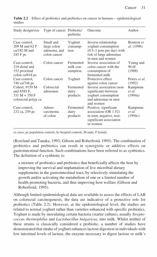
Cancer 31
Table 2.2 Effect of probiotics and prebiotics on cancer in humans – epidemiologicalstudies
Study design/size Type of cancer Probiotic/ Outcome Authorprebiotic
Case control, Small and Yoghurt Inverse relationship Boutron et209 M and 62 F large colon consump- yoghurt consumption al. (1996)ca/182 M and adenoma, and tion (0.5–1 pots per day) with245 F pc colon cancer risk of large adenomas
in men and womenCase-control, Colon cancer Fermented Inverse association of Young and210 distal and milk con- colon cancer with the Wolf152 proximal sumption consumption of (1988)colon ca/618 pc fermented milkCase-control, Colon cancer Yoghurt Protective effect Peters et al.746 ca/746 pc against colon cancer (1992)Cohort, 9159 M Colorectal Fermented Inverse association (non- Kampmanand 8585 F, adenomas dairy significant) between et al.331 M + 350 F products yoghurt consumption (1994a)colorectal polyp ca and adenomas in men
and womenCase-control, Adeno- Fermented Positive, significant Kampman232 ca, 259 pc carcinoma dairy association (OR 1.52) et al.
of colon products in men; negative, non- (1994c)significant associationin women
ca cases, pc population controls, hc hospital controls, M male, F female.
(Rowland and Tanaka, 1993; Gibson and Roberfroid, 1995). The combination ofprobiotics and prebiotics can result in synergistic or additive effects ongastrointestinal function. Such combinations have been referred to as synbiotics.The definition of a synbiotic is:
a mixture of probiotics and prebiotics that beneficially affects the host byimproving the survival and implantation of live microbial dietarysupplements in the gastrointestinal tract, by selectively stimulating thegrowth and/or activating the metabolism of one or a limited number ofhealth-promoting bacteria, and thus improving host welfare (Gibson andRoberfroid, 1995).
Although limited epidemiological data are available to assess the effects of LABon colorectal carcinogenesis, the data are indicative of a protective role forprobiotics (Table 2.2). However, at the epidemiological level, the studies arerelated to normal yoghurt rather than varieties enhanced with specific probiotics.Yoghurt is made by inoculating certain bacteria (starter culture), usually Strepto-coccus thermophilus and Lactobacillus bulgaricus, into milk. Whilst neither ofthese strains is classically considered a probiotic, a number of studies havedemonstrated that intake of yoghurt enhances lactose digestion in individuals withlow intestinal levels of lactase, the enzyme necessary to digest lactose or milk’s
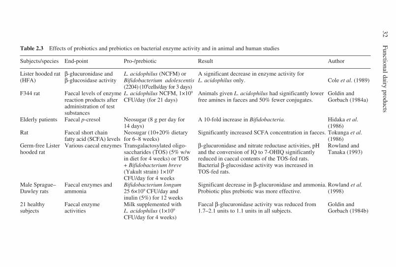
32Functional dairy products
Table 2.3 Effects of probiotics and prebiotics on bacterial enzyme activity and in animal and human studies
Subjects/species End-point Pro-/prebiotic Result Author
Lister hooded rat β-glucuronidase and L. acidophilus (NCFM) or A significant decrease in enzyme activity for(HFA) β-glucosidase activity Bifidobacterium adolescentis L. acidophilus only. Cole et al. (1989)
(2204) (109cells/day for 3 days)F344 rat Faecal levels of enzyme L. acidophilus NCFM, 1×10⁹ Animals given L. acidophilus had significantly lower Goldin and
reaction products after CFU/day (for 21 days) free amines in faeces and 50% fewer conjugates. Gorbach (1984a)administration of testsubstances
Elderly patients Faecal p-cresol Neosugar (8 g per day for A 10-fold increase in Bifidobacteria. Hidaka et al.14 days) (1986)
Rat Faecal short chain Neosugar (10+20% dietary Significantly increased SCFA concentration in faeces. Tokunga et al.fatty acid (SCFA) levels for 6–8 weeks) (1986)
Germ-free Lister Various caecal enzymes Transgalactosylated oligo- β-glucuronidase and nitrate reductase activities, pH Rowland andhooded rat saccharides (TOS) (5% w/w and the conversion of IQ to 7-OHIQ significantly Tanaka (1993)
in diet for 4 weeks) or TOS reduced in caecal contents of the TOS-fed rats.+ Bifidobacterium breve Bacterial β-glucosidase activity was increased in(Yakult strain) 1×10⁹ TOS-fed rats.CFU/day for 4 weeks
Male Sprague– Faecal enzymes and Bifidobacterium longum Significant decrease in β-glucuronidase and ammonia. Rowland et al.Dawley rats ammonia 25 6×10⁹ CFU/day and Probiotic plus prebiotic was more effective. (1998)
inulin (5%) for 12 weeks21 healthy Faecal enzyme Milk supplemented with Faecal β-glucuronidase activity was reduced from Goldin andsubjects activities L. acidophilus (1×10⁹ 1.7–2.1 units to 1.1 units in all subjects. Gorbach (1984b)
CFU/day for 4 weeks)
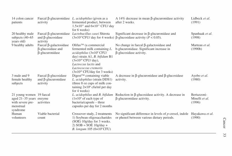
Cancer
33
14 colon cancer Faecal β-glucuronidase L. acidophilus (given as a A 14% decrease in mean β-glucuronidase activity Lidbeck et al.patients activity fermented product, between after 2 weeks. (1991)
1.5×10¹¹ and 6×10¹¹ CFU/ dayfor 6 weeks)
20 healthy male Faecal β-glucuronidase Lactobacillus casei Shirota Significant decrease in β-glucuronidase and Spanhaak et al.subjects (40–65 and β-glucosidase (3×10¹¹CFU/ day for 4 weeks) β-glucosidase activity (P < 0.05). (1998)years old) activity9 healthy adults Faecal β-galactosidase Olifus (a commercial No change in faecal β-galactosidase and Marteau et al.
β-glucosidase and fermented milk containing L. b-glucuronidase. Significant increase in (1990b)β-glucuronidase acidophilus (3×10⁹ CFU/ β-glucosidase activity.
day) strain A1, B. bifidum B1(3×10¹⁰ CFU/ day),Lactoccus lactis andLactococcus cremoris(3×10¹⁰ CFU/day for 3 weeks)
3 male and 9 Faecal β-glucosidase Digest™ containing viable A decrease in β-glucuronidase and β-glucosidase Ayebo et al.female healthy and β-glucuronidase L. acidophilus (strain DDS1) activity. (1980)subjects activity (three 8 oz cups of milk con-
taining 2×10⁶ cfu/ml per dayfor 4 weeks)
21 young women 19 faecal L. acidophilus and B. bifidum Reduction in β-glucosidase activity. A decrease in Bertazzoni-aged 21–35 years enzyme (1×10⁹ of each type of β-glucuronidase activity. Minelli et al.with severe pre- activities bacteria/capsule – three (1996)menstrual capsules per day for 2 monthssyndromeHuman Viable bacterial Crossover study, 2 treatments No significant difference in levels of p-cresol, indole Hayakawa et al.volunteers count 1) Soybean oligosaccharides or phenol between various dietary periods. (1990)
(SOE) 10g/day for 3 weeks.2) SOB = SOE 10g/day +B. longum 105 (6×10⁹ CFU)
34 Functional dairy products
sugar (i.e., a condition called lactase nonpersistence) (Marteau, 1990a). The bene-ficial effect may be explained by the ability of the starter cultures used in themanufacture of yoghurt with live, active cultures to produce the enzyme lactase,which digests the lactose in yoghurt. Yoghurt without live cultures, unfermentedor sweet acidophilus milk, and cultured buttermilk is tolerated about the same asmilk by individuals with lactase nonpersistence. To that end these active culturesin the yoghurt beneficially affect the host, and it has been reported that the bacteriaused to produce yoghurt can inactivate carcinogens and prevent DNA damage inthe colon of rats (Wollowski, 1999). For these reasons we shall consider the abovebacteria as probiotic. A case-control study by Boutron et al. (1996) showed asignificant inverse relationship between consumption of moderate amounts ofyoghurt (half to one pot per day) and the risk of large colonic adenomas in bothwomen and men. Further case-control studies have provided additional evidenceof inverse associations of colorectal cancer risk and consumption of fermenteddairy products (Young and Wolf, 1988) and yoghurt (Peters et al., 1992). Kampmanet al. (1994a) reported a non-significant inverse relationship between colonicadenomas and yoghurt consumption. This finding, however, was not corroboratedin a further case-control study in the Netherlands of colorectal cancer risk andfermented dairy products, which revealed a small significant positive associationin men (odds ratio, OR 1.52) and a small, non-significant inverse association inwomen (OR 0.77) (Kampman et al., 1994c).
The data generated from probiotic and prebiotic human intervention prospectiveand animal studies suggest a beneficial role for probiotics and serve to strengthenthe information provided by the population-based trials. The intervention studiesused to evaluate the ability of probiotics to prevent cancer in humans have beenbased largely upon the use of biomarkers for assessing cancer risk (reviewed inGill and Rowland, 2002). Owing to their non-invasive nature, markers in faecesand urine have been mostly used. Faecal markers are composed of two maincategories: those examining the activity of bacterial enzymes or bacterial metabolites(e.g. IQ activation, β-glucuronidase and ammonia production) and those based onbioassays on faecal water and metabolites. The aqueous phase of human faeces(faecal water) is considered to be an important source of modulators and inducersfor colorectal carcinogenesis and methods exist for assessing biological activitiesrelated to cancer risk in such samples.
Copious toxic, carcinogenic and endogenously produced compounds, e.g.steroids, are metabolised in the liver and conjugated to glucuronic acid by phase IIenzymes, before being excreted via the bile into the small intestine. In the colon, thebacterial enzyme β-glucuronidase can hydrolyse these conjugates, thereby releas-ing the parent compound or its hepatic metabolite. In the case of carcinogens andmutagens, the activity of β-glucuronidase in the colon may enhance the probabilityof tumour induction. For example, the colon carcinogen DMH is metabolised in theliver, small amounts of the procarcinogenic conjugate of the activated metabolitemethylazoxymethanol (MAM) are excreted in the bile. Hydrolysis of the conjugateby colonic bacteria releases MAM in to the colon. Germ-free animals treated withDMH or MAM result in fewer colon tumours than do conventional animals (Reddy
Cancer 35
et al., 1974). Both animal and human studies have frequently shown that consump-tion of prebiotics and probiotics can decrease β-glucuronidase activity. Thesestudies are summarised in Table 2.3 and some examples are discussed in moredetail below. In a conventional rat study (F344), Lactobacillus acidophilus(109–1010 CFU/day) supplementation (21 days) of a high meat diet (72% beef)significantly reduced the activity of faecal β-glucuronidase and nitroreductase (by40–50%) (Goldin and Gorbach, 1976). The modulating effect of the Lactobacillusstrain was dependent on the type of basal diet fed – no significant effects were seenwhen the rats were fed a grain-based diet. In a similar study in human floraassociated (HFA) rats, Cole et al. (1989) demonstrated a significant reduction in β-glucuronidase and β-glucosidase activities when L. acidophilus was fed for threedays, with the effect evident for seven days post-dose.
Lactobacillus acidophilus supplementation (fermented milk) of the diets of 14colon cancer patients for a period of six weeks resulted in a 14% decrease inβ-glucuronidase activity and a reduction in total bile acids and deoxycholic acid of15% and 18%, respectively. Coincident with these effects were alterations in thefaecal microflora, as E. coli numbers were depressed and lactobacilli augmented.Similar results were obtained by Spanhaak et al. (1998) who reported a significantdecrease in the activity of faecal β-glucuronidase and β-glucosidase activity in agroup of 20 healthy male subjects given L. casei Shirota (approximately 1011 CFU/day for a four-week test period).
More direct evidence for protective properties of probiotics and prebioticsagainst cancer has been obtained by assessing the ability of cultures to preventmutations and DNA damage in cell cultures or in animals (Table 2.4). The AmesSalmonella assay has been used to study the effect of lactic acid bacteria (LAB) onthe induction of mutations by a wide variety of model carcinogens in vitro. Thecarcinogens used include N-methylnitrosourea (MNU), N-nitrosocompounds,N-methyl-N-nitro-N-nitrosoguanidine (MNNG), heterocyclic amines (e.g. IQ andrelated compounds) and aflatoxin B1. In general the results indicate that thevarious LAB can inhibit genotoxicity of dietary carcinogens in vitro. The degree ofinhibition was strongly species-dependent, e.g. Pool-Zobel et al. (1993) demon-strated that L. confusus and L. sake had no effect on the mutagenic activity ofnitrosated beef, whereas L. casei and L. lactis diminished the effect by over 85%.
It would appear probably these results, in conjunction with similar results byother workers, are a consequence of binding of the mutagens by the LAB (Zhangand Ohta, 1991; Bolognani et al., 1997). Whether such a mechanism operates invivo is questionable, since binding appears not to affect uptake of carcinogens fromthe gut, neither does it have any apparent effect in vivo on mutagenicity in the liver,and further the process appears to be highly pH-dependent and easily reversed(Bolognani et al., 1997).
Using the comet assay (single cell microgel electrophoresis technique), Pool-Zobel et al. (1996) investigated the ability of a range of species of LAB to inhibitDNA damage in the colon mucosa of rats treated with the carcinogens MNNG orDMH. All the strains of lactobacilli and bifidobacteria tested – L. acidophilus(isolated from a yoghurt), L. confusus DSM 20196, L. gasseri P76, B. longum and
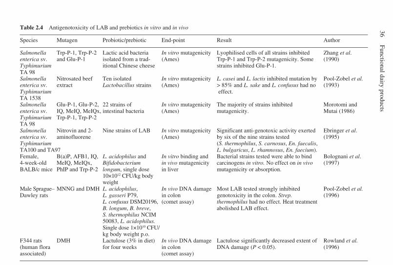
36Functional dairy products
Table 2.4 Antigenotoxicity of LAB and prebiotics in vitro and in vivo
Species Mutagen Probiotic/prebiotic End-point Result Author
Salmonella Trp-P-1, Trp-P-2 Lactic acid bacteria In vitro mutagenicity Lyophilised cells of all strains inhibited Zhang et al.enterica sv. and Glu-P-1 isolated from a trad- (Ames) Trp-P-1 and Trp-P-2 mutagenicity. Some (1990)Typhimurium itional Chinese cheese strains inhibited Glu-P-1.TA 98Salmonella Nitrosated beef Ten isolated In vitro mutagenicity L. casei and L. lactis inhibited mutation by Pool-Zobel et al.enterica sv. extract Lactobacillus strains (Ames) > 85% and L. sake and L. confusus had no (1993)Typhimurium effect.TA 1538Salmonella Glu-P-1, Glu-P-2, 22 strains of In vitro mutagenicity The majority of strains inhibited Morotomi andenterica sv. IQ, MeIQ, MeIQx, intestinal bacteria (Ames) mutagenicity. Mutai (1986)Typhimurium Trp-P-1, Trp-P-2TA 98Salmonella Nitrovin and 2- Nine strains of LAB In vitro mutagenicity Significant anti-genotoxic activity exerted Ebringer et al.enterica sv. aminofluorene (Ames) by six of the nine strains tested (1995)Typhimurium (S. thermophilus, S. carnosus, En. faecalis,TA100 and TA97 L. bulgaricus, L. rhamnosus, En. faecium).Female, B(a)P, AFB1, IQ, L. acidophilus and In vitro binding and Bacterial strains tested were able to bind Bolognani et al.4-week-old MeIQ, MeIQx, Bifidobacterium in vivo mutagenicity carcinogens in vitro. No effect on in vivo (1997)BALB/c mice PhIP and Trp-P-2 longum, single dose in liver mutagenicity or absorption.
10×1012 CFU/kg bodyweight
Male Sprague– MNNG and DMH L. acidophilus, In vivo DNA damage Most LAB tested strongly inhibited Pool-Zobel et al.Dawley rats L. gasseri P79, in colon genotoxicity in the colon. Strep. (1996)
L. confusus DSM20196, (comet assay) thermophilus had no effect. Heat treatmentB. longum, B. breve, abolished LAB effect.S. thermophilus NCIM50083, L. acidophilus.Single dose 1×10¹⁰ CFU/kg body weight p.o.
F344 rats DMH Lactulose (3% in diet) In vivo DNA damage Lactulose significantly decreased extent of Rowland et al.(human flora for four weeks in colon DNA damage (P < 0.05). (1996)associated) (comet assay)
Cancer 37
B. breve – prevented MNNG-induced DNA damage when given at a dose of 1010
CFU/kg body mass, 8 h before the carcinogen. In the majority of cases the DNAdamage was reduced to a level similar to that in control animals. Streptococcusthermophilus NCIM 50083 was less effective than the other LAB strains. Theprotective effect was dose-dependent: diminished doses of L. acidophilus (50%and 10% of the original dose) were less effective in inhibiting MNNG-inducedDNA damage. The importance of viable cells was also emphasised as heat-treatment of L. acidophilus abolished its antigenotoxic potential. Similar resultswere obtained when the LAB strains were tested in rats given DMH as the DNAdamaging agent. Again, all the lactobacilli and bifidobacteria strongly inhibitedDNA damage in the colon mucosa, whereas S. thermophilus was much lesseffective with only one of three strains tested exhibiting any protection againstDNA damage.
A range of animal studies have been carried out using DMH or AOM todetermine the effects of specific probiotics and prebiotics on ACF formation andtumour incidence (Table 2.5). In the most recent work by Tavan et al. (2002) it wasreported that rats treated with various heterocyclic aromatic amines (HAAs) andsupplemented with a range of dairy products (including non-fermented milk(30%), Bifidobacterium animalis DN-173010 fermented milk (30%) or Strepto-coccus thermophilus DN-001158 fermented milk (30%)) showed a 66%, 96% and93% decrease in ACF incidence respectively. Further, intermediate biomarkersshowed that there was a reduction in HAA metabolism, faecal mutagenicity andcolon DNA lesions. Kulkarni and Reddy (1994) reported 50% inhibition of ACFformation in AOM tumour-induced male F344 rats fed Bifidobacterium longum inthe diet (1.5% and 3% of a lyophilised culture containing 2×10¹⁰ CFU/g) for fiveweeks. As dietary treatments were started five weeks prior to administration of thecarcinogen dose results do not allow deductions to be made about the stage ofcarcinogenesis affected.
An analogous study carried out by Challa et al. (1997) observed a 23% decreasein total colonic ACF and a 28% reduction in total AC in rats given a diet containing0.5% B. longum BB536 (1×10⁸ CFU/g of feed). Animals were fed the experimentaldiet before treatment with AOM and throughout the experiment. Abdelali et al.(1995) compared the effects of Bifidobacterium sp. Bio Danone strain 173010administered in diet and also fed as a fermented milk product. The amounts oforganisms consumed were similar (6×10⁹ CFU/day) in both experimental variants.The LAB was given four weeks before DMH and continued for a further fourweeks before ACF assessment. The bifidobacteria-fermented milk appeared to beslightly less effective in reducing ACF than the dietary bifidobacteria (49% and61% reduction, respectively). Interestingly, however, skim milk alone reducedACF numbers by 51%. In AOM-treated Sprague–Dawley rats, supplementation ofB. longum 25 (6×10⁹cfu/day) for 12 weeks, caused a significant reduction (26%)in total ACF by comparison to control animals (Rowland et al., 1998). The changeswere reported in only small ACF (one to three AC per focus). Given that theprobiotic treatment began one week after the carcinogen exposure, the resultsindicate an effect on the early promotional phase of carcinogenesis.
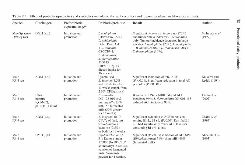
38Functional dairy products
Table 2.5 Effect of probiotics/prebiotics and synbiotics on colonic aberrant crypt foci and tumour incidence in laboratory animals
Species Carcinogen Pre/probiotic Probiotic/prebiotic Result Authorexposure stage*
Male Sprague– DMH (s.c.) Initiation and L.acidophilus Significant decrease in tumour no. (70%) McIntosh et al.Dawley rats promotion (Delvo Pro LA-1) and tumour mass index for L. acidophilus (1999)
L. acidophilus only. Tumour incidence decreased in largeDelvo Pro LA-1 intestine L.acidophilus (25%); L. acidophilus+ B. animalis + B. animalis (20%); L. rhamnosus (20%);CSCC1941; S. thermophilus (10%).L. rhamnosus;S. thermophilus DD145(1010 CFU/g, 1%dietary intake for36 weeks)
Male AOM (s.c.) Initiation and Lyophilised Significant inhibition of total ACF Kulkarni andF344 rats promotion B. longum (1.5% (P < 0.01). Significant reduction in total AC Reddy (1994)
and 3% dietary for per colon (P < 0.001).13 weeks (suppl. from2 1010 CFU/g stock)
Male HAA Initiation and B. animalis B. animalis DN-173-010 reduced ACF Tavan et al.F344 rats mixture promotion DN-173-010 or S. incidence 96%. S. thermophilus DN-001-158 (2002)
IQ, MeIQ, thermophilus DN- reduced ACF incidence 93%.phIP(1:1:1 ratio) 001-158 fermented
milk (30% dietaryfor 15 weeks)
Male AOM (s.c.) Initiation and B. longum (1×10⁸ Significant reduction in ACF in rats con- Challa et al.F344 rats promotion CFU/g of feed, rats suming BI, L, BI + L (P, 0.05). Rats fed BI (1997)
fed ad libitum) + L had significantly fewer ACF than ratslactulose (2.5%) consuming BI or L alone.or both for 13 weeks
Male DMH (i.p.) Initiation and Bifidobacterium sp. Significant (P < 0.05) inhibition of AC: 61% Abdelali et al.F344 rats promotion Bio Danone strain (Bifidobacterium) 51% (skim milk) 49% (1995)
173010 (6×10⁹ CFU/ (fermented milk).animal/day) in cell sus-pension or fermentedmilk. Skim milkpowder for 4 weeks).
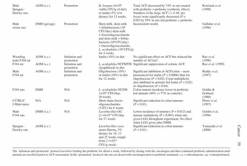
Cancer
39Male AOM (s.c.) Promotion B. longum (4×10⁸ Total ACF decreased by 74% in rats treated Rowland et al.Sprague– viable CFU/g of diet) with probiotic + prebiotic (synbiotic effect). (1998)Dawley rats or inulin (5% w/w Numbers of the large ACF ( > 4 AC per
dietary) for 12 weeks. focus) were significantly decreased (P <0.05) by 59% in rats fed probiotic + prebiotic.
Male DMH (gavage) Promotion Skim milk, skim milk Inconsistent results Gallaher et al.wistar rats + bifidobacteria (108 (1996)
CFU/day) skim milk+ fructooligosaccharideand skim milk + bifido-bacteria (108 CFU/day)+ fructooligosaccharide;L. acidophilus (106 CFU/g)for 4 weeks
Weanling AOM (s.c.) Initiation and Inulin (10%) in diet No significant effect on ACF but reduced the Rao et al.male F344 rat promotion number of AC/cm2. (1998)F344 rat AOM (s.c.) Initiation and L. acidophilus NCFMTM Significant suppression of colonic ACF. Rao et al. (1999)
promotion (lyophilised) in dietMale AOM (s.c.) Initiation and Oligofructose (10%) Significant inhibition of ACF/colon – more Reddy et al.F344 rat promotion or inulin (10%) in diet pronounced for inulin (P < 0.0006) than for (1997)
for 12 weeks oligofructose (P < 0.02). Crypt multiplicityalso inhibited in animals fed inulin (P < 0.02)or oligofructose (P < 0.04).
F344 rats DMH N/A L. acidophilus NCFM Colon tumour incidence lower in probiotic Goldin &1×10¹⁰ CFU/day, fed animals (40% vs 77% in controls). Gorbach20 weeks (1980)
C57BL/C N/A N/A Short chain fructo- Significant reduction in colon tumours Pierre et al.J-Min/+mice oligosaccharides (P < 0.01). (1997)
(5.8%) for 6 weeksMale DMH (s.c.) N/A Lactobacillus GG Lower incidence of tumours (P < 0.012) and Goldin et al.F344 rats (2–4×10¹⁰ CFU/day tumour multiplicity (P < 0.001) when rats (1996)
for 27 weeks given LGG throughout experiment. No effectwhen LGG given after DMH.
Sprague– AOM (s.c.) Lactobacillus casei Significant reduction in colon tumours Yamazaki et al.Dawley rats strain Shirota, 5% (P < 0.01). (2000)
dietary for 18, 12and 25 weeks (suppl.from 2 1×10¹⁰
CFU/g stock)
The ‘initiation and promotion’ protocol involves feeding the probiotic for about a week, followed by dosing with the carcinogen and then continued probiotic administration untilanimals are sacrificed prior to ACF assessment. In the ‘promotion’ protocol, the rats are dosed with carcinogen prior to probiotic treatment. s.c. = subcutaneous, i.p. = interperitoneal.
40 Functional dairy products
Alone, prebiotics appear to give inconsistent results on carcinogen-inducedACF induction, which may be partly a consequence of differences in treatmentregimes and carcinogens used. For example Rao et al. (1998) reported that 10%dietary inulin had no significant effect on total ACF in colon, or their multiplicity,in F344 rats, even though curiously a significant reduction in ACF/cm2 of colonwas reported. A study on Bifidobacterium spp. and fructo-oligosaccharides (FOS)(2% in diet) gave inconsistent results with only one out of three experimentsshowing a reduction in DMH-induced ACF (Gallaher et al., 1996). Reddy et al.(1997) compared long (inulin) and short chain (FOS) oligosaccharides incorpo-rated at a level of 10% in diet on AOM-induced ACF in rats. The NDOs were fedprior to carcinogen treatment and, throughout the experiment, significant decreasesin total ACF were reported at approximately 35% and 25%, respectively. Thedecreases observed were almost exclusively in the smaller ACF (< 3 AC per focus)and inhibition by inulin appeared to be more pronounced than that of FOS.Rowland et al. (1998) reported similar results, when inulin (5% in diet) was givena week after AOM dose. A decrease of 41% in small ACF was observed, but inulinappeared to have no effect on large ACF.
An investigation of the effect of L. acidophilus NCFM on colon tumour incid-ence in DMH-treated rats, showed a reduction in colon cancer incidence (40% vs77% in controls) in animals receiving L. acidophilus 1×10¹⁰ CFU/day after 20weeks. No difference was apparent at 36 weeks, suggesting that the lactobacilli hadincreased the latency period, or induction time, for tumours (Goldin and Gorbach,1980). Administration of dietary B. longum (0.5% lyophilised B. longum in diet,1×1010 CFU/day) to male rats significantly decreased the formation of IQ-inducedliver and colon tumours and the multiplicity of tumours in liver, colon and smallintestine (Reddy and Rivenson, 1993; Rumney et al., 1993). The percentageincidence of tumours was also decreased by 100% in colon and 80% in liver. Infemale rats, dietary supplementation with Bifidobacterium cultures also decreasedthe IQ induced mammary carcinogenesis to 50% and liver carcinogenesis to 27%of that on the control diet, but the differences were not significant. There were,however, significant changes in tumour multiplicity in the mammary gland.McIntosh et al. (1999) reported that freeze-dried L. acidophilius (1010 CFU/g, 1%of dietary intake ) fed daily was protective, as indicated by a 70% decrease intumour numbers and a large decrease in tumour mass in the large intestine ofDMH-treated Sprague–Dawley rats. A number of other LAB strains tested alsodecreased non-significantly tumour burden relative to control animals includingL. rhamnosus (GG) (20% reduction), B. animalis CSSC 1941 + L. acidophiliusDelvo Pro LA-1 (20% reduction) and S. thermophilius DD145 (10% reduction).Bolognani et al. (2001) report that background diet fed to rats influenced theprotective effects of probiotics, such that probiotics reduced ACF incidence onlywhen rats were fed a high-fat (20%) diet.
The Min mouse model recently developed provides animals, which are hetero-zygous for a nonsense mutation of the Apc gene, the murine homologue of APC.These mice develop spontaneous adenomas throughout the small intestine andcolon within a few weeks and have been used for testing of chemopreventive
Cancer 41
agents targeted against cancerous lesions. In one such study Min mice were fedvarious diets containing resistant starch, wheat bran or FOS (5.8% in diet) for sixweeks. Tumour numbers remained unchanged from the control (fibre-free diet) inthe mice fed either resistant starch or wheat bran, but a significant decrease in colontumours was observed in rats receiving the diet supplemented with FOS. Further-more 40% of the FOS-fed animals (n = 10) were totally free of colon tumours(Pierre et al., 1997).
2.11 Mechanisms of anticarcinogenicity and antigenotoxicityfor probiotics and prebiotics
2.11.1 Binding of carcinogensThere are numerous reports describing the binding or adsorption in vitro by LABand other intestinal bacteria, of a range of food-borne carcinogens including theheterocyclic amines, the fungal toxin aflatoxin B1, benzo(a)pyrene and the foodcontaminant AF2 (Morotomi and Mutai, 1986; Zhang et al., 1990; Zhang andOhta, 1991; Orrhage et al., 1994; Bolognani et al., 1997). Several of these studiesreported a concomitant decrease in mutagenicity. The extent of the binding wasdependent on the mutagen and bacterial strain used. In general the least bindingwas seen with aflatoxin B1 and AF2 and the most with the heterocyclic amines.The adsorption appeared to be a physical phenomenon, chiefly due to a cationexchange mechanism.
Whilst binding represents a credible mechanism for the inhibition of muta-genicity and genotoxicity by LAB in vitro, it does not appear to have any influencein vivo. Bolognani et al. (1997) reported that simultaneous administration to miceof LAB with various carcinogens did not affect absorption of the compounds fromthe gastrointestinal tract, nor did it affect the in vivo mutagenicity of the carcino-gens in the liver. These results are contradictory to those of Zhang and Ohta (1993),who found that absorption from the rat small intestine of Trp-P-1 was significantlyinhibited by co-administration of freeze-dried LAB. The latter study wasconfounded, however, by the use of rats that had been starved for four days, whichwould induce severe physiological and nutritional stresses on the animals.
2.11.2 Effects on bacterial enzymes, metabolite productionThe studies listed in Table 2.3 demonstrate that the increase in concentration ofLAB as a consequence of consumption of LAB and/or prebiotics leads to reduc-tions in certain bacterial enzymes ostensibly involved in activation or synthesis ofcarcinogens, genotoxins and tumour promoters. Such effects would appear to bedue to the low specific activity of these enzymes in LAB (Saito et al., 1992). Thesechanges in enzyme activity or metabolite concentration have been suggested to beresponsible for the reduced level of tumours or preneoplastic lesions seen incarcinogen-treated rats administered pro- and prebiotics (Rumney et al., 1993;
42 Functional dairy products
Rowland et al., 1998). Even though a causal link has not been demonstrated, thisremains a likely hypothesis.
2.11.3 Stimulation of protective enzymesNumerous food-borne carcinogens such as heterocyclic amines and polycyclicaromatic hydrocarbons, are known to be conjugated to glutathione (GSH), whichappears to result in inactivation. The enzyme involved, glutathione-s-transferase(GST), is found in the liver and in other tissues including the gut. Challa et al.(1997) in a study of the effect of lactulose and B. longum BB536 on AOM-inducedACF in the colon, showed that GST activity in the colonic mucosa was inverselyrelated to the ACF numbers. The prebiotic-resistant starch has also been shown toincrease colonic GST when incorporated into diets of rats (Treptow van Lishaut etal., 1999). Such a mechanism of protection would be effective against a wide rangeof dietary carcinogens.
2.11.4 Increase in immune responseAn alternative mechanism suggested by Perdigon et al. (1998) to account for LABanti-tumour activity is that of reducing the inflammatory immune response. In astudy of tumour growth in DMH-treated mice, yoghurt was observed to suppressthe inflammatory immune response with an increase in CD4+ T lymphocytes andin IgA-secreting cells. In those animals, a distinct reduction in tumours wasobserved. The study is consistent with the work of Schiffrin et al. (1996) andMarteau et al. (1997) who have provided evidence of immune system modulationin human subjects consuming probiotics. The observed changes were increases inlevels of antibody-secreting cells and elevated phagocytic activity of monocytesand granulocytes.
Taken as a whole, studies in in vitro systems and in a wide range of animalmodels provide considerable evidence that LAB, and to a lesser extent prebiotics,have the potential to decrease colon cancer risk. The evidence from humans is lesspersuasive, but nonetheless is suggestive of a cancer-preventing effect of fer-mented foods.
2.12 Future trends
No doubt exists that in the last few decades we have greatly expanded ourknowledge about the causes and risk factors associated with colorectal cancer.However, it is painfully obvious from the conflicting epidemiological informationprovided on dairy products that population-based studies must be more selective inthe factors they seek to examine, if a clearer and more consistent picture is to becreated. In addition, greater regard must be given to the means of dietary assess-ment applied to studies as recent trials have indicated that some methods of dietaryassessment are associated with very large measurement error, which obscures
Cancer 43
relationships between disease and diet within single homogeneous populations.The introduction of biomarkers to calibrate measurement error is a criticaldevelopment in the struggle to improve accuracy of large-scale prospective studiesinvestigating the interactions of diet and disease (S.A. Bingham, pers. comm.). Toestablish causal relationships between diet and colorectal cancer risk and toidentify more precisely the dietary components involved, human interventiontrials are required. The problem with human intervention studies is that cancer is animpractical end-point in terms of numbers, cost, study duration and ethicalconsiderations. The long lag phase (up to 20 years) between exposure to acarcinogenic event and appearance of tumours is a particular problem. Anotherstrategy is the use of intermediate end-point biomarkers of cancer, which may becellular, molecular, biochemical or rooted in pathologic change (e.g. faecal water,recurrence of polyps, epithelial markers). Biomarkers have been developed froman understanding of the sequence of events leading to colonic cancer, the biologyof normal mucosa and the factors associated with changes symptomatic ofprogression towards cancer and the manifestation of cancer itself. The specificadvantages of biomarkers are that they represent short/intermediate term end-points, which facilitate intervention on a reasonable time scale and that ethicalapproval is more readily obtainable for biomarker studies as they are minimallyinvasive, with measurements occurring on accessible material (e.g. faeces andsmall biopsies). Preferably, biomarkers should be reproducible, sensitive andrigorously validated, although this is not the case with all biomarkers in the cancerarea. Biomarkers ought to be correlated or causally linked with cancer andconsequently of biological significance. Accordingly, validation of a biomarker iscritical to its application as a research tool, as such an apposite response from themarker is required when assayed in healthy individuals on low-risk and high-riskdiets for colorectal cancer or in cancer patients (e.g. familial adenomatouspolyposis).
At present a wide range of potential biomarkers exists to augment investigationsinto the activity of specific foods, compounds and metabolites in colorectal cancer.The majority of these assays still require further validation before they can be usedwith complete confidence and compromises must be made in terms of the strengthof the causal information provided and study complexity. Clearly, we now needcarefully controlled intervention studies in human subjects using biomarkers ofcancer risk in order to strengthen the evidence for the role of functional dairyproducts. Such studies, if conducted correctly, should help to expose dietaryinteractions at vital stages in the carcinogenesis process and within a relativelyshort time begin to provide a more accurate and reliable picture.
2.13 Sources of further information and advice
For further information on the topic of colorectal cancer, the reader is directed toa number of sources within the IARC website including www-dep.iarc.fr/globocan/globocan.html (globocan 2000) and www-dep.iarc.fr/dataava/infodata.html. These
44 Functional dairy products
sources provide excellent data on a wide variety of cancer statistics. To consider ingreater detail the epidemiology related to colon cancer and diet, the reader isadvised to examine a number of reviews quoted within the body of this chapter.These references represent comprehensive insight given by authors includingPotter, Slattery and Bingham.
2.14 Acknowledgement
This chapter has been carried out with the financial support from the Commissionof the European Communities, specific RTD programme ‘Quality of Life Manage-ment of Living Resources’, QLK-2000-00067 ‘Functional foods, gut microfloraand healthy ageing’. It does not necessarily reflects its views and in no wayanticipates the Commission’s future policy in this area.
2.15 ReferencesABDELALI H, CASSAND P, SOUSSOTTE V, DAUBEZE M, BOULEY C and NARBONNE J F (1995)
Effect of dairy products on initiation of precursor lesions of colon cancer in rats. Nutritionand Cancer 24, 121–132.
AGA (2000) American Gastroenterologists Association Technical Review: Impact of dietaryfiber on colon cancer occurence. Gastroenterology 118, 1235–1257.
AYEBO AD, ANGELO I D and SHAHANI K M (1980) Effect of ingesting Lactobacillusacidophilius milk upon faecal flora and enzyme activity in humans. Milchwissenschaft 35,730–733.
BANNI S, DAY B W, EVANS R W, CORONGIU F P and LOMBARDI B (1995) Detection ofconjugated diene isomers of linoleic acid in liver lipids of rats fed a choline-devoid dietindicated that the diet does not cause lipoperoxidation. Journal of Nutritionant Biochem-istry 6, 281–289.
BANNI S, ANGIONI E, CASU V, MELIS M P, CARTA G, CORONGIU F P, THOMPSON H and IP C(1999) Decrease in linoleic acid metabolites as a potential mechanism in cancer riskreduction by conjugated linoleic acid. Carcinogenesis 20, 1019–1024.
BARON J A, BEACH M, MANDEL J S, VAN STOLK R U, HAILE R W, SANDLER R S, ROTHSTEIN R,SUMMERS R W, SNOVER D C, BECK G J, BOND J H and GREENBERG E R (1999) Calciumsupplements for the prevention of colorectal adenomas. Calcium Polyp Prevention StudyGroup. New England Journal of Medicine 340, 101–107.
BENITO E, OBRADOR A, STIGGELBOUT A, BOSCH F X, MULET M, MUNOZ N and KALDOR J (1990)A population-based case-control study of colorectal cancer in Majorca. I. Dietary factors.International Journal of Cancer/Journal International Du Cancer 45, 69–76.
BENNETT T, DESMOND A, HARRINGTON M, MCDONAGH D, FITZGERALD R, FLYNN A andCASHMAN K D (2000) The effect of high intakes of casein and casein phosphopeptide oncalcium absorption in the rat. British Journal of Nutrition 83, 673–680.
BERTAZZONI-MINELLI E, BENINI A, VICENTINI L, ANDREOLI E, OSELLADORE M and CERUTTI R(1996) Effect of Lactobacillius acidophilius and Bifidobacterium bifidum administrationon colonic microbiotia and its metabolic activity in premenstrual syndrome. MicrobiolEcology in Health and Disease 9, 247–260.
BIDOLI E, FRANCESCHI S, TALAMINI R, BARRA S and LA VECCHIA C (1992) Food consumptionand cancer of the colon and rectum in north-eastern Italy. International Journal of Cancer/Journal International Du Cancer 50, 223–229.
Cancer 45
BINGHAM S A (2000) Diet and colorectal cancer prevention. Biochemical Society Trans-actions 28, 12–16.
BIRD R P (1995) Role of aberrant crypt foci in understanding the pathogenesis of colon cancer.Cancer Letters 93, 55–71.
BOLOGNANI F, RUMNEY C J and ROWLAND I R (1997) Influence of carcinogen binding bylactic acid-producing bacteria on tissue distribution and in vivo mutagenicity of dietarycarcinogens. Food and Chemical Toxicology 35, 535–545.
BOLOGNANI F, RUMNEY C, POOL ZOBEL B L and ROWLAND I (2001) Effect of lactobacilli,bifidobacteria and inulin on the formation of aberrant crypt foci in rats. European Journalof Nutrition 40, 293–300.
BONITHON-KOPP C, KRONBORG O, GIACOSA A, RATH U and FAIVRE J (2000) Calcium and fibresupplementation in prevention of colorectal adenoma recurrence: a randomised inter-vention trial. The Lancet 356, 1300–1306.
BOSTICK R M, POTTER J D, SELLERS T A, MCKENZIE D R, KUSHI L H and FOLSOM A R (1993)Relation of calcium, vitamin D, and dairy food intake to incidence of colon cancer amongolder women. The Iowa Women’s Health Study. American Journal of Epidemiology 137,1302–1317.
BOSTICK R M, FOSDICK L, WOOD J R, GRAMBSCH P, GRANDITS G A, LILLEMOE T J, LOUIS T Aand POTTER J D (1995) Calcium and colorectal epithelial cell proliferation in sporadicadenoma patients: a randomized, double-blinded, placebo-controlled clinical trial. Jour-nal of the National Cancer Institute 87, 1307–1315.
BOUNOUS G, PAPENBURG R, KONGSHAVN P A, GOLD P and FLEISZER D (1988) Dietary wheyprotein inhibits the development of dimethylhydrazine induced malignancy. Clinical andInvestigative Medicine/Medecine Clinique et Experimentale 11, 213–217.
BOUTRON M C, FAIVRE J, MARTEAU P, COUILLAULT C, SENESSE P and QUIPOURT V (1996)Calcium, phosphorus, vitamin D, dairy products and colorectal carcinogenesis: a Frenchcase – control study. British Journal of Cancer 74, 145–151.
BOUTRON RUAULT M C, SENESSE P, FAIVRE J, CHATELAIN N, BELGHITI C and MEANCE S (1999)Foods as risk factors for colorectal cancer: a case-control study in Burgundy (France).European Journal of Cancer Prevention 8, 229–235.
BOYLE P and LANGMAN J S (2000) ABC of colorectal cancer – Epidemiology. British MedicalJournal 321, 805–808.
BRUCE W R, GIACCA A and MEDLINE A (2000) Possible mechanisms relating diet and risk ofcolon cancer. Cancer Epidemiology Biomarkers & Prevention 9, 1271–1279.
BURNS A J and ROWLAND I R (2000) Anti-carcinogenicity of probiotics and prebiotics.Current Issues in Intestinal Microbiology 1, 13–24.
CHALLA A, RAO D R, CHAWAN C B and SHACKELFORD L (1997) Bifidobacterium longum andlactulose suppress azoxymethane-induced colonic aberrant crypt foci in rats. Carcinogen-esis 18, 517–521.
CHUNG D C (2000) The genetic basis of colorectal cancer: insights into critical pathways oftumorigenesis. Gastroenterology 119, 854–865.
COLE C B, FULLER R and CARTER S M (1989) Effect of probiotic supplements of Lactobacillusacidophilus and Bifidobacterium adolescentis 2204 on β-glucosidase and β-glucuroni-dase activity in the lower gut of rats associated with a human faecal flora. MicrobialEcology in Health and Disease 2, 223–225.
CORPET D E and CASSAND P (1995) Lack of aberrant crypt promotion and of mutagenicity inextracts of cooked casein, a colon cancer-promoting food. Nutrition and Cancer 24, 249–256.
CORPET D E and CHATELIN-PIROT V (1997) Cooked casein promotes colon cancer in rats, maybe because of mucosal abrasion. Cancer Letters 114, 89–90.
CORPET D E, YIN Y, ZHANG X M, REMESY C, STAMP D, MEDLINE A, THOMPSON L, BRUCE W Rand ARCHER M C (1995) Colonic protein fermentation and promotion of colon carcinogen-esis by thermolyzed casein. Nutrition and Cancer 23, 271–281.
CUMMINGS J H, BEATTY E R, KINGMAN S M, BINGHAM S A and ENGLYST H N (1996) Digestion
46 Functional dairy products
and physiological properties of resistant starch in the human large bowel. British Journalof Nutrition 75, 733–747.
DILLEHAY D L, WEBB S K, SCHMELZ E M and MERRILL A H (1994) Dietary sphingomyelininhibits 1,2-dimethylhydrazine-induced colon cancer in CF1 mice. Journal of Nutrition124, 615–620.
EBRINGER L, FERENCIK M, LAHITOVA N, KACANI L and MICHALKOVA D (1995) Anti-mutagenic and immuno-stimulatory properties of lactic acid bacteria. World Journal ofMicrobiology and Biotechnology 11, 294–298.
FAIVRE J, BEDENNE L, BOUTRON M C, MILAN C, COLLONGES R and ARVEUX P (1989)Epidemiological evidence for distinguishing subsites of colorectal cancer. Journal ofEpidemiology and Community Health 43, 356–361.
FEARON E R and VOGELSTEIN B (1990) A genetic model for colorectal tumorigenesis. Cell61, 759–767.
FERLAY J, BRAY F, PISANI P and PARKIN D M (2001) GLOBOCAN 2000: Cancer Incidence,Mortality and Prevalence Worldwide, Lyon: IARC Press.
FERRARETTO A, SIGNORILE A, GRAVAGHI C, FIORILLI A and TETTAMANTI G (2001) Caseinphosphopeptides influence calcium uptake by cultured human intestinal HT-29 tumorcells. Journal of Nutrition 131, 1655–1661.
FULLER R (1989) Probiotics in man and animals.Journal of Applied Bacteriology 66, 365–378.GALLAHER D D, STALLINGS W H, BLESSING L L, BUSTA F F and BRADY L J (1996) Probiotics,
cecal microflora, and aberrant crypts in the rat colon. Journal of Nutrition 126, 1362–1371.
GIBSON G R and ROBERFROID M B (1995) Dietary modulation of the human colonicmicrobiota: introducing the concept of prebiotics. Journal of Nutrition 125, 1401–1412.
GILL CIR and ROWLAND I (2002) Diet and cancer: assessing the risk. British Journal ofNutrition 88 (suppl. 1), 51–516.
GIOVANNUCCI E and GOLDIN B (1997) The role of fat, fatty acids, and total energy intake inthe etiology of human colon cancer. American Journal of Clinical Nutrition 66, 1564S–1571S.
GIOVANNUCCI E, STAMPFER M J, COLDITZ G A, RIMM E B, TRICHOPOULOS D, ROSNER B A,SPEIZER F E and WILLETT W C (1993) Folate, methionine, and alcohol intake and risk ofcolorectal adenoma. Journal of the National Cancer Institute 85, 875–884.
GLINGHAMMAR B, VENTURI M, ROWLAND I R and RAFTER J J (1997) Shift from a dairyproduct-rich to a dairy product-free diet: influence on cytotoxicity and genotoxicity offecal water – potential risk factors for colon cancer. American Journal of ClinicalNutrition 66, 1277–1282.
GOEPTAR A R, KOEMAN J H, VAN BOEKEL M A J S and ALINK G M (1997) Impact of digestionon the antimutagenic activity of the milk protein casein. Nutrition Research 17, 1363–1379.
GOLDIN B R and GORBACH S L (1976) The relationship between diet and rat fecal bacterialenzymes implicated in colon cancer. Journal of the National Cancer Institute 57, 371–375.
GOLDIN B R and GORBACH S L (1980) Effect of Lactobacillus acidophilus dietary supple-ments on 1,2-dimethylhydrazine dihydrochloride-induced intestinal cancer in rats. Journalof the National Cancer Institute 64, 263–265.
GOLDIN B R and GORBACH S L (1984a) Alterations of the intestinal microflora by diet, oralantibiotics, and lactobacillus: decreased production of free amines from aromatic nitrocompounds, azo dyes, and glucuronides. Journal of the National Cancer Institute 73, 689–695.
GOLDIN B R and GORBACH S L (1984b) The effect of milk and lactobacillus feeding on humanintestinal bacterial enzyme activity. American Journal of Clinical Nutrition 39, 756–761.
GOLDIN B R, GUALTIERI L J and MOORE R P (1996) The effect of Lactobacillus on theinitiation and promotion of DMH-induced intestinal tumors in the rat. Nutrition andCancer 25, 197–204.
Cancer 47
GOVERS M J, LAPRE J A, DE VRIES H T and VAN DER MEER R (1993) Dietary soybean proteincompared with casein damages colonic epithelium and stimulates colonic epithelialproliferation in rats. Journal of Nutrition 123, 1709–1713.
HA Y L, GRIMM N K and PARIZA M W (1987) Anticarcinogens from fried ground beef: heat-altered derivatives of linoleic acid. Carcinogenesis 8, 1881–1887.
HA Y L, STORKSON J and PARIZA M W (1990) Inhibition of benzo(a)pyrene-induced mouseforestomach neoplasia by conjugated dienoic derivatives of linoleic acid. Cancer Research50, 1097–1101.
HAKKAK R, KOROURIAN S, RONIS M J, JOHNSTON J M and BADGER T M (2001) Dietary wheyprotein protects against azoxymethane-induced colon tumors in male rats. CancerEpidemiology Biomarkers & Prevention 10, 555–558.
HANNUN Y A and OBEID L M (1995) Ceramide: an intracellular signal for apoptosis. Trendsin Biochemical Sciences 20, 73–77.
HANSEN M, SANDSTROM B and LONNERDAL B (1996) The effect of casein phosphopeptideson zinc and calcium absorption from high phytate infant diets assessed in rat pups andCaco-2 cells. Pediatric Research 40, 547–552.
HANSEN M, SANDSTROM B, JENSEN M and SORENSEN S S (1997) Casein phosphopeptidesimprove zinc and calcium absorption from rice-based but not from whole-grain infantcereal. Journal of Pediatric Gastroenterology and Nutrition 24, 56–62.
HAYAKAWA K, MIZUTANI J, WADA K, MASAI T, YOSHIHARA I and MITSUOKA T (1990) Effectsof soybean oligosaccharides on human faecal microflora. Microbial Ecology in Healthand Disease 3, 293–303.
HIDAKA H, EIDA T, TAKIZAWA T, TOKUNGA T and TASHIRO Y (1986) Effects of fructo-oligosaccharides on intestinal flora and human health. Bifidobacteria Microflora 5, 37–50.
HOLT P R (1999) Dairy foods and prevention of colon cancer: human studies. Journal of theAmerican College of Nutrition 18, 379S–391S.
HOLZAPFEL W H, HABERER P, SNEL J, SCHILLINGER U and HUIS IN’T VELD J H (1998) Overviewof gut flora and probiotics. International Journal of Food Microbiology 41, 85–101.
IARC (1997) Cancer Incidence in Five Continents, Volume VII. France: IARC ScientificPublications, i–xxxiv, 1–1240.
ICS (1985) Cancer incidence in Greater Bombay, by religion and sex 1973–1978. Bombay:Indian Cancer Society.
IP C, SINGH M, THOMPSON H J and SCIMECA J A (1994) Conjugated linoleic acid suppressesmammary carcinogenesis and proliferative activity of the mammary gland in the rat.Cancer Research 54, 1212–1215.
JARVINEN R, KNEKT P, HAKULINEN T and AROMAA A (2001) Prospective study on milkproducts, calcium and cancers of the colon and rectum. European Journal of ClinicalNutrition 55, 1000–1007.
JOHANSEN C, MELLEMGAARD A, SKOV T, KJAERGAARD J and LYNGE E (1993) Colorectalcancer in Denmark 1943–1988. International Journal of Colorectal Disease 8, 42–47.
JOSYULA S and SCHUT H A (1998) Effects of dietary conjugated linoleic acid on DNA adductformation of PhIP and IQ after bolus administration to female F344 rats. Nutrition andCancer 32, 139–145.
KAMPMAN E, GIOVANNUCCI E, VAN’T VEER P, RIMM E, STAMPFER M J, COLDITZ G A, KOK F Jand WILLETT W C (1994a) Calcium, vitamin D, dairy foods, and the occurrence ofcolorectal adenomas among men and women in two prospective studies. AmericanJournal of Epidemiology 139, 16–29.
KAMPMAN E, GOLDBOHM R A, VAN DEN BRANDT P A and VAN’T VEER P (1994b) Fermenteddairy products, calcium, and colorectal cancer in The Netherlands Cohort Study. CancerResearch 54, 3186–3190.
KAMPMAN E, VAN’T VEER P, HIDDINK F J, VAN AKEN-SCHNEIJDER P, KOK F J and HERMUS R J(1994c) Fermented dairy products, dietary calcium and colon cancer: a case-control studyin The Netherlands. International Journal of Cancer/Journal International Du Cancer 59,170–176.
48 Functional dairy products
KAMPMAN E, SLATTERY M L, CAAN B and POTTER J D (2000) Calcium, vitamin D, sunshineexposure, dairy products and colon cancer risk (United States). Cancer Causes & Control11, 459–466.
KEARNEY J, GIOVANNUCCI E, RIMM E B, ASCHERIO A, STAMPFER M J, COLDITZ G A, WING A,KAMPMAN E and WILLETT W C (1996) Calcium, vitamin D, and dairy foods and theoccurrence of colon cancer in men. American Journal of Epidemiology 143, 907–917.
KEMPPAINEN M, RAIHA I and SOURANDER L (1997) A marked increase in the incidence ofcolorectal cancer over two decades in southwest Finland. Journal of Clinical Epidemiol-ogy 50, 147–151.
KLEIBEUKER J H, MULDER N H, CATS A, VAN DER MEER R and DE VRIES E G (1996a) Calciumand colorectal epithelial cell proliferation. Gut 39, 774–775.
KLEIBEUKER J H, NAGENGAST F M and VAN DER MEER R (1996b) Carcinogenesis in the colon.In Prevention and Early Detection of Colorectal Cancer (G P Young, P Rozen and BLevin, editors). London, Philadelphia, Toronto, Sydney, Tokyo: WB Saunders CompanyLtd, 46–62.
KOLESNICK R N and KRONKE M (1998) Regulation of ceramide production and apoptosis.Annual Review of Physiology 60, 643–665.
KULKARNI N and REDDY B S (1994) Inhibitory effect of Bifidobacterium longum cultures onthe azoxymethane-induced aberrant crypt foci formation and fecal bacterial beta-glucuro-nidase. Proceedings of the Society for Experimental Biology and Medicine 207, 278–283.
KUNE S, KUNE G A and WATSON L F (1987) Case-control study of dietary etiological factors:the Melbourne Colorectal Cancer Study. Nutrition and Cancer 9, 21–42.
LAPRE J A, DE VRIES H T and VAN DER MEER R (1993) Cytotoxicity of fecal water is dependenton the type of dietary fat and is reduced by supplemental calcium phosphate in rats.Journal of Nutrition 123, 578–585.
LAWSON R E, MOSS A R and GIVENS D I (2001) The role of dairy products in supplyingconjugated linoleic acid to man’s diet: a review. Nutrition Research Reviews 14, 153–172.
LEE H P, GOURLY L, DUFFY S W, ESTEVE J, LEE J and DAY H E (1989) Colorectal cancer anddiet in an Asian population: a case-control study among Singapore Chinese. InternationalJournal of Cancer 43, 1007–1016.
LEE Y S, NOGUCHI T and NAITO H (1983) Intestinal absorption of calcium in rats given dietscontaining casein or amino acid mixture: the role of casein phosphopeptides. BritishJournal of Nutrition 49, 67–76.
LI H, SHIMURA H, AOKI Y, DATE K, MATSUMOTO K, NAKAMURA T and TANAKA M (1998)Hepatocyte growth factor stimulates the invasion of gallbladder carcinoma cell lines invitro. Clinical and Experimental Metastasis 16, 74–82.
LIDBECK A, GELTNER ALLINGER U, ORRHAGE K M, OTTOVA L, BRISMAR B, GUSTAFSSON J A,RAFTER J J and NORD C E (1991) Impact of Lactobacillus acidophilus supplements on thefaecal microflora and soluble faecal bile acids in colon cancer patients. Microbial Ecologyin Health and Disease 4, 81–88.
LIEW C, SCHUT H A, CHIN S F, PARIZA M W and DASHWOOD R H (1995) Protection of conjugatedlinoleic acids against 2-amino-3-methylimidazo[4,5-f]quinoline-induced colon carcino-genesis in the F344 rat: a study of inhibitory mechanisms. Carcinogenesis 16, 3037–3043.
LIPKIN M and NEWMARK H (1995) Calcium and the prevention of colon cancer. Journal ofCellular Biochemistry Supplement 22, 65–73.
MA J, GIOVANNUCCI I, POLLAK M, CHAN M J, GAZIANO M, WILLET W and STAMPFER M J (2001)Milk intake, circulating levels of insulin-like growth factor-I, and risk of colorectal cancerin men. Journal of the National Cancer Institute 93, 1330–1336.
MACDONALD H B (2000) Conjugated linoleic acid and disease prevention: a review of currentknowledge. Journal of the American College of Nutrition 19, 111S–118S.
MACQUART-MOULIN G, RIBOLI E, CORNEE J, CHARNAY B, BERTHEZENE P and DAY N (1986)Case-control study on colorectal cancer and diet in Marseilles. International Journal ofCance/Journal International Du Cancer 38, 183–191.
MANOUSOS O, DAY N E, TRICHOPOULOS D, GEROVASSILIS F, TZONOU A and POLYCHRONO-
Cancer 49
POULOU A (1983) Diet and colorectal cancer: a case-control study in Greece. InternationalJournal of Cancer/Journal International Du Cancer 32, 1–5.
MARCUS P M and NEWCOMB P A (1998) The association of calcium and vitamin D, and colonand rectal cancer in Wisconsin women. International Journal of Epidemiology 27, 788–793.
MARTEAU P, FLOURIE B, POCHART P, CHASTANG C, DESJEUX J F and RAMBAUD J C (1990a)Effect of the microbial lactase (EC 3, 2, 1, 23), activity in yoghurt on the intestinalabsorption of lactose: an in vivo study in lactase-deficient humans, The British Journal ofNutrition, 64, 71–79.
MARTEAU P, POCHART P, FLOURIE B, PELLIER P, SANTOS L, DESJEUX J F and RAMBAUD J C(1990b) Effect of chronic ingestion of a fermented dairy product containing Lactobacillusacidophilus and Bifidobacterium bifidum on metabolic activities of the colonic flora inhumans. American Journal of Clinical Nutrition 52, 685–688.
MARTEAU P, MINEKUS M, HAVENAAR R and HUIS IN’T VELD J H (1997) Survival of lactic acidbacteria in a dynamic model of the stomach and small intestine: validation and the effectsof bile. Journal of Dairy Science 80, 1031–1037.
MARTINEZ M E and WILLETT W C (1998) Calcium, vitamin D, and colorectal cancer: a reviewof the epidemiologic evidence. Cancer Epidemiology, Biomarkers and Prevention 7,163–168.
MCCARTY M F (2000) Activation of PPARgamma may mediate a portion of the anticanceractivity of conjugated linoleic acid. Medical Hypotheses 55, 187–188.
MCGUIRE M A and MCGUIRE M K (1999) Conjugated linoleic acid (CLA): a ruminant fatty acidwith beneficial effects on human health. Proceedings of the American Society of AnimalScience 1–8.
MCINTOSH G H, REGESTER G O, LE LEU R K, ROYLE P J and SMITHERS G W (1995) Dairy proteinsprotect against dimethylhydrazine-induced intestinal cancers in rats. Journal of Nutrition125, 809–816.
MCINTOSH G H, WANG Y H and ROYLE P J (1998) A diet containing chickpeas and wheat offersless protection against colon tumors than a casein and wheat diet in dimethylhydrazine-treated rats. Journal of Nutrition 128, 804–809.
MCINTOSH G H, ROYLE P J and PLAYNE M J (1999) A probiotic strain of L. acidophilus reducesDMH-induced large intestinal tumors in male Sprague–Dawley rats. Nutrition andCancer 35, 153–159.
METTLIN C J, SCHOENFELD E R and NATARAJAN N (1990) Patterns of milk consumption andrisk of cancer. Nutrition and Cancer 13, 89–99.
MOBARHAN S (1999) Calcium and the colon: recent findings. Nutrition Reviews 57, 124–126.MOROTOMI M and MUTAI M (1986) In vitro binding of potent mutagenic pyrolysates to
intestinal bacteria. Journal of the National Cancer Institute 77, 195–201.NAGAO M and SUGIMURA T (1993) Carcinogenic factors in food with relevance to colon
cancer development. Mutation Research 290, 43–51.NEGRI E, LA VECCHIA C, D’AVANZO B and FRANCESCHI S (1990) Calcium, dairy products, and
colorectal cancer. Nutrition and Cancer 13, 255–262.NOBRE-LEITAO C, CHAVES P, FIDALGO P, CRAVO M, GOUVEIA-OLIVEIRA A, FERRA M A and
MIRA F C (1995) Calcium regulation of colonic crypt cell kinetics: evidence for a directeffect in mice. Gastroenterology 109, 498–504.
NORAT T and RIBOLI E (2001) Meat consumption and colorectal cancer: a review ofepidemiologic evidence. Nutrition Reviews 59, 37–47.
NRC (1996) Carcinogens and Anticarcinogens in the Human Diet. The National Academyof Sciences. Washington DC: National Academy Press.
ORRHAGE K, SILLERSTROM E, GUSTAFSSON J A, NORD C E and RAFTER J (1994) Binding ofmutagenic heterocyclic amines by intestinal and lactic acid bacteria. Mutation Research311, 239–248.
OWEN D A (1996) Flat adenoma, flat carcinoma, and de novo carcinoma of the colon. Cancer77, 3–6.
50 Functional dairy products
PAPENBURG R, BOUNOUS G, FLEISZER D and GOLD P (1990) Dietary milk proteins inhibit thedevelopment of dimethylhydrazine-induced malignancy. Tumour Biology: the Journal ofthe International Society for Oncodevelopmental Biology and Medicine 11, 129–136.
PARODI P W (1997) Cows’ milk fat components as potential anticarcinogenic agents. Journalof Nutrition 127, 1055–1060.
PENMAN I D, LIANG Q L, BODE J, EASTWOOD M A and ARENDS M J (2000) Dietary calciumsupplementation increases apoptosis in the distal murine colonic epithelium. Journal ofClinical Pathology 53, 302–307.
PERDIGON G, VALDEZ J C and RACHID M (1998) Antitumour activity of yoghurt: study ofpossible immune mechanisms. Journal of Dairy Research 65, 129–138.
PEREIRA M A, BARNES L H, RASSMAN V L, KELLOFF G V and STEELE V E (1994) Use ofazoxymethane-induced foci of aberrant crypts in rat colon to identify potential cancerchemopreventive agents. Carcinogenesis 15, 1049–1054.
PETERS R K, PIKE M C, GARABRANT D H and MACK T M (1992) Diet and colon cancer in LosAngeles County, California. Cancer Causes and Controls 3, 457–473.
PHILLIPS R L and SNOWDON D A (1985) Dietary relationships with fatal colorectal canceramong Seventh-Day Adventists. Journal of the National Cancer Institute 74, 307–317.
PIERRE F, PERRIN P, CHAMP M, BORNET F, MEFLAH K and MENANTEAU J (1997) Short-chainfructo-oligosaccharides reduce the occurrence of colon tumors and develop gut-associ-ated lymphoid tissue in Min mice. Cancer Research 57, 225–228.
PIETINEN P, MALILA N, VIRTANEN M, HARTMAN T J, TANGREA J A, ALBANES D and VIRTAMO J
(1999) Diet and risk of colorectal cancer in a cohort of Finnish men. Cancer Causes &Control 10, 387–396.
PONZ DE LEON M and PERCESEPE A (2000) Pathogenesis of colorectal cancer. Digestive andLiver Disease 32, 807–821.
PONZ DE LEON M and RONCUCCI L (2000) The cause of colorectal cancer. Digestive and LiverDisease 32, 426–439.
POOL-ZOBEL B L, MUNZNER R and HOLZAPFEL W H (1993) Antigenotoxic properties of lacticacid bacteria in the S. typhimurium mutagenicity assay. Nutrition and Cancer 20, 261–270.
POOL-ZOBEL B L, NEUDECKER C, DOMIZLAFF I, JI S, SCHILLINGER U, RUMNEY C, MORETTI M,VILARINI I, SCASSELLATI-SFORZOLINI R and ROWLAND I (1996) Lactobacillus- andbifidobacterium-mediated antigenotoxicity in the colon of rats. Nutrition and Cancer 26,365–380.
POTTER J D (1999) Colorectal cancer: molecules and populations. Journal of the NationalCancer Institute 91, 916–932.
POTTER J D, SLATTERY M L, BOSTICK R M and GAPSTUR S M (1993) Colon cancer: a reviewof the epidemiology. Epidemiologic Reviews 15, 499–545.
PRETLOW T P, O’RIORDAN M A, PRETLOW T G and STELLATO T A (1992) Aberrant crypts inhuman colonic mucosa: putative preneoplastic lesions. Journal of Cellular BiochemistrySupplement 16G, 55–62.
PRITCHARD R S, BARON J A and GERHARDSSON DE VERDIER M (1996) Dietary calcium,vitamin D, and the risk of colorectal cancer in Stockholm, Sweden. Cancer Epidemiology,Biomarkers and Prevention 5, 897–900.
RAFTER J J, CHILD P, ANDERSON A M, ALDER R, ENG V and BRUCE W R (1987) Cellular toxicityof fecal water depends on diet. American Journal of Clinical Nutrition 45, 559–563.
RAO C V, CHOU D, SIMI V, KU H and REDDY B S (1998) Prevention of colonic aberrant cryptfoci and modulation of large bowel microbial activity by dietary coffee fiber, inulin andpectin. Carcinogenesis 19, 1815–1819.
RAO C V, SANDERS M E, INDRANIE C, SIMI B and REDDY B S (1999) Prevention of colonicpreneoplastic lesions by the probiotic Lactobacillus acidophilus NCFMTM in F344 rats.International Journal of Oncology 14, 939–944.
REDDY B S and RIVENSON A (1993) Inhibitory effect of Bifidobacterium longum on colon,mammary, and liver carcinogenesis induced by 2-amino-3-methylimidazo[4,5-f]quinoline,a food mutagen. Cancer Research 53, 3914–3918.
Cancer 51
REDDY B S, WEISBURGER J H and WYNDER E L (1974) Fecal bacterial beta-glucuronidase:control by diet. Science 183, 416–417.
REDDY B S, HAMID R and RAO C V (1997) Effect of dietary oligofructose and inulin on colonicpreneoplastic aberrant crypt foci inhibition. Carcinogenesis 18, 1371–1374.
RIBONI L, VIANI P, BASSI R, PRINETTI A and TETTAMANTI G (1997) The role of sphingolipidsin the process of signal transduction. Progress in Lipid Research 36, 153–195.
ROWLAND I R (1988) Factors affecting metabolic activity of the intestinal microflora. DrugMetabolism Reviews 19, 243–261.
ROWLAND I R and TANAKA R (1993) The effects of transgalactosylated oligosaccharides ongut flora metabolism in rats associated with a human faecal microflora. Journal of AppliedBacteriology 74, 667–674.
ROWLAND I R, MALLETT A K and WISE A (1985) The effect of diet on the mammalian gut floraand its metabolic activities. Critical Reviews in Toxicology 16, 31–103.
ROWLAND I R, BEARNE C A, FISCHER R and POOL-ZOBEL B l (1996) The effect of lactulose onDNA damage induced by DMH in the colon of human flora-associated rats. Nutrition andCancer 26, 37–47.
ROWLAND I R, RUMNEY C J, COUTTS J T and LIEVENSE L C (1998) Effect of Bifidobacteriumlongum and inulin on gut bacterial metabolism and carcinogen-induced aberrant crypt fociin rats. Carcinogenesis 19, 281–285.
RUMNEY C J, ROWLAND I R, COUTTS T M, RANDERATH K, REDDY R, SHAH A B, ELLUL A andO’NEILL I K (1993) Effects of risk-associated human dietary macrocomponents onprocesses related to carcinogenesis in human-flora-associated (HFA) rats. Carcinogen-esis 14, 79–84.
SAITO Y, TAKANO T and ROWLAND I R (1992) Effects of soybean oligosaccharides on thehuman gut microflora in in vitro culture. Microbial Ecology in Health and Disease 5, 105–110.
SAITO Y, LEE Y S and KIMURA S (1998) Minimum effective dose of casein phosphopeptides(CPP) for enhancement of calcium absorption in growing rats. International Journal forVitamin and Nutrition Research 68, 335–340.
SALMINEN S, BOULEY C, BOUTRON-RUAULT M C, CUMMINGS J H, FRANCK A, GIBSON G R,ISOLAURI E, MOREAU M C, ROBERFROID M and ROWLAND I (1998) Functional food scienceand gastrointestinal physiology and function. British Journal of Nutrition 80 Suppl 1,S147–171.
SCHIFFRIN E, ROCHAT F, LINK-AMSTER H, AESCHLIMANN J and DONNET-HUGHES A (1996)Immunomodulation of blood cells following ingestion of lactic acid bacteria. Journal ofDairy Science 78, 491–497.
SCHMELZ E M, DILLEHAY D L, WEBB S K, REITER A, ADAMS J and MERRILL A H (1996)Sphingomyelin consumption suppresses aberrant colonic crypt foci and increases theproportion of adenomas versus adenocarcinomas in CF1 mice treated with 1,2-dimethylhydrazine: implications for dietary sphingolipids and colon carcinogenesis.Cancer Research 56, 4936–4941.
SCHMELZ E M, BUSHNEV A S, DILLEHAY D L, SULLARDS M C, LIOTTA D C and MERRILL A H(1999) Ceramide-beta-D-glucuronide: synthesis, digestion, and suppression of earlymarkers of colon carcinogenesis. Cancer Research 59, 5768–5772.
SCHMELZ E M, SULLARDS M C, DILLEHAY D L and MERRILL A H (2000) Colonic cellproliferation and aberrant crypt foci formation are inhibited by dairy glycosphingolipidsin 1,2-dimethylhydrazine-treated CF1 mice. Journal of Nutrition 130, 522–527.
SEBEDIO J L, JUANEDA P, DOBSON G, RAMILISON I, MARTIN J D and CHARDIGNY J M (1997)Metabolites of conjugated isomers of linoleic acid (CLA) in the rat. Biochimica etBiophysica Acta 1345, 5–10.
SESINK A L, TERMONT D S, KLEIBEUKER J H and VAN DER MEER R (2001) Red meat and coloncancer: dietary haem-induced colonic cytotoxicity and epithelial hyperproliferation areinhibited by calcium. Carcinogenesis 22, 1653–1659.
SHANNON J, WHITE E, SHATTUCK A L and POTTER J D (1996) Relationship of food groups and
52 Functional dairy products
water intake to colon cancer risk. Cancer Epidemiology, Biomarkers and Prevention 5,495–502.
SHIH I M, WANG T L, TRAVERSO G, ROMANS K, HAMILTON S R, BEN-SASSON A, KINZLER K Wand VOGELSTEIN B (2001) Top-down morphogenesis of colorectal tumors. Proceedingsof the National Academy of Sciences of the United States of America 98, 2640–2645.
SHULTZ T D, CHEW B P, SEAMAN W R and LUEDECKE L O (1992) Inhibitory effect of conjugateddienoic derivatives of linoleic acid and beta-carotene on the in vitro growth of humancancer cells. Cancer Letters 63, 125–133.
SILVI S, RUMNEY C J, CRESCI A and ROWLAND I R (1999) Resistant starch modifies gutmicroflora and microbial metabolism in human flora-associated rats inoculated withfaeces from Italian and UK donors. Journal of Applied Microbiology 86, 521–530.
SLATTERY M L, SORENSON A W and FORD M H (1988) Dietary calcium intake as a mitigatingfactor in colon cancer. American Journal of Epidemiology 128, 504–514.
SLATTERY M L, POTTER J D, COATES A, MA K N, BERRY T D, DUNCAN D M and CAAN B J (1997)Plant foods and colon cancer: an assessment of specific foods and their related nutrients(United States). Cancer Causes and Control 8, 575–590.
SOUZA R F (2001) Review article: a molecular rationale for the how, when and why ofcolorectal cancer screening. Alimentary Pharmacology & Therapeutics 15, 451–462.
SPANHAAK S, HAVENAAR R and SCHAAFSMA G (1998) The effect of consumption of milkfermented by Lactobacillus casei strain Shirota on the intestinal microflora and immuneparameters in humans. European Journal of Clinical Nutrition 52, 899–907.
STERNHAGEN L G and ALLEN J C (2001) Growth rates of a human colon adenocarcinoma cellline are regulated by the milk protein alpha-lactalbumin. Advances in ExperimentalMedicine and Biology 501, 115–120.
STOCKS P (1957) Cancer Incidence in North Wales and Liverpool Region in Relation to Habitand Environment. British Imperial Cancer Campaign, 35th Annual Report, 1.
SWEENEY E A, INOKUCHI J and IGARASHI Y (1998) Inhibition of sphingolipid inducedapoptosis by caspase inhibitors indicates that sphingosine acts in an earlier part of theapoptotic pathway than ceramide. Federation of European Biochemical Societies Letters425, 61–65.
TAJIMA K and TOMINAGA S (1985) Dietary habits and gastro-intestinal cancers: a comparativecase-control study of stomach and large intestinal cancers in Nagoya, Japan. JapaneseJournal of Cancer Research: Gann 76, 705–716.
TAVAN E, CAYUELA C, ANTOINE J, TRUGMAN G, CHAUGIER C and CASSAND P (2002) Effectsof dairy products on heterocyclic aromatic amine-induced rat colon carcinogenesis.Carcinogenesis 23, 477–483.
TOKUNGA T, OKU T and HOYSOYA N (1986) Influence of chronic intake of new sweetenerfructooligosaccharide (Neosugar) on growth and gastrointestinal function of the rat.Journal of Nutritional Science and Vitaminology 32, 111–121.
TREPTOW VAN LISHAUT S, RECHKEMMER G, ROWLAND I, DOLARA P and POOL-ZOBEL B L(1999) The carbohydrate crystalean and colonic microflora modulate expression ofglutathione S-transferase subunits in colon of rats. European Journal of Nutrition 38, 76–83.
TSUDA H, SEKINE K, USHIDA Y, KUHARA T, TAKASUKA N, IIGO M, HAN B S and MOORE M A(2000) Milk and dairy products in cancer prevention: focus on bovine lactoferrin.Mutation Research 462, 227–233.
TUYNS A J, KAAKS R and HAELTERMAN M (1988) Colorectal cancer and the consumption offoods: a case-control study in Belgium. Nutrition and Cancer 11, 189–204.
VAN BOEKEL M A, WEERENS C N, HOLSTRA A, SCHEIDTWEILER C E and ALINK G M (1993)Antimutagenic effects of casein and its digestion products. Food and Chemical Toxicol-ogy 31, 731–737.
VAN DER MEER R, LAPRE J A, GOVERS M J and KLEIBEUKER J H (1997) Mechanisms of theintestinal effects of dietary fats and milk products on colon carcinogenesis. Cancer Letters114, 75–83.
Cancer 53
VAN DER MEER R, TERMONT D S and DE VRIES H T (1991) Differential effects of calcium ionsand calcium phosphate on cytotoxicity of bile acids. American Journal of Physiology 260,G142–147.
VESPER H, SCHMELZ E M, NIKOLOVA-KARAKASHIAN M N, DILLEHAY D L, LYNCH D V andMERRILL A H (1999) Sphingolipids in food and the emerging importance of sphingolipidsto nutrition. Journal of Nutrition 129, 1239–1250.
VINAS-SALAS J, BIENDICHO-PALAU P, PINOL-FELIS C, MIGUELSANZ-GARCIA S and PEREZ-HOLANDA S (1998) Calcium inhibits colon carcinogenesis in an experimental model in therat. European Journal of Cancer 34, 1941–1945.
VOGELSTEIN B, FEARON E R, HAMILTON S R, KERN S E, PREISINGER A C, LEPPERT M,NAKAMURA Y, WHITE R, SMITS A M and BOS J L (1988) Genetic alterations duringcolorectal-tumor development. New England Journal of Medicine 319, 525–532.
WARGOVICH M J, ALLNUTT D, PALMER C, ANAYA P and STEPHENS L C (1990) Inhibition of thepromotional phase of azoxymethane-induced colon carcinogenesis in the F344 rat bycalcium lactate: effect of simulating two human nutrient density levels. Cancer Letters 53,17–25.
WARGOVICH M J, JIMENEZ A, MCKEE K, STEELE V E, VELASCO M, WOODS J, PRICE R, GRAY Kand KELLOFF G J (2000) Efficacy of potential chemopreventive agents on rat colon aberrantcrypt formation and progression. Carcinogenesis 21, 1149–1155.
WCRF (1997) Diet, Nutrition and the Prevention of Cancer: A Global Perspective. Washing-ton DC: World Cancer Research Fund/American Institute.
WOLLOWSKI I JI S T, BAKALINSKY A T, NEUDECKER C and POOL-ZOBEL B L (1999), Bacteriaused for the production of yoghurt inactivate carcinogens and prevent DNA damage in thecolon of rats. J. Nutr., 129, 77.
WU K, WILLETT W C, FUCHS C S, COLDITZ G A AND GIOVANNUCCI E L (2002) Calcium intakeand risk of colon cancer in women and men. Journal of the National Cancer Institute 94,437–446.
YAMAZAKI K, TSUNODA A, SIBUSAWA M, TSUNODA Y, KUSANO M, FUKUCHI K, YAMANAKAM, KUSHIMA M, NOMOTO K and MOROTOMI M (2000) The effect of an oral administrationof Lactobacillus casei strain Shirota on azoxymethane-induced colonic aberrant crypt fociand colon cancer in the rat. Oncology Reports 7, 977–982.
YOSHIDA S and YE XIUYUN (1992) The binding ability of bovine milk caseins to mutagenicheterocyclic amines. Journal of Dairy Science 75, 958–961.
YOUNG T B and WOLF D A (1988) Case-control study of proximal and distal colon cancer anddiet in Wisconsin. International Journal of Cancer/Journal International Du Cancer 42,167–175.
ZHANG X B and OHTA Y (1991) In vitro binding of mutagenic pyrolyzates to lactic acidbacterial cells in human gastric juice. Journal of Dairy Science 74, 752–757.
ZHANG X B and OHTA Y (1993) Microorganisms in the gastrointestinal tract of the rat preventabsorption of the mutagen-carcinogen 3-amino-1,4-dimethyl-5H-pyrido(4,3-b)indole.Canadian Journal of Microbiology 39, 841–845.
ZHANG X B, OHTA Y and HOSONO A I (1990) Antimutagenicity and binding of lactic acidbacteria from a Chinese cheese to mutagenic pyrolyzates. Journal of Dairy Science 73,2702–2710.
ZHANG X M, STAMP D, MINKIN S, MEDLINE A, CORPET D E, BRUCE W R and ARCHER M C (1992)Promotion of aberrant crypt foci and cancer in rat colon by thermolyzed protein. Journalof the National Cancer Institute 84, 1026–1030.
ZOCK P L (2001) Dietary fats and cancer. Current Opinion in Lipidology 12, 5–10.
3
Coronary heart diseaseJ. Lovegrove and K. Jackson, The University of Reading, UK
3.1 Introduction
Coronary heart disease (CHD) is one of the major causes of death in adults in theWestern world. Although there has been a trend over the past 15 years towards areduction in the rates of CHD in the major industrialised nations (including theUSA, Australia and the UK) due to better health screening, drug treatment,smoking and dietary advice, there has been an alarming increase in CHD in EasternEurope.
CHD is a condition in which the main coronary arteries supplying the heart areno longer able to supply sufficient blood and oxygen to the heart muscle (myocar-dium). The main cause of the reduced flow is an accumulation of plaques, mainlyin the intima of arteries, a disease known as atherosclerosis. This is a slow butprogressive disease which usually begins in childhood, but does not usuallymanifest itself until later life. Depending on the rate of the narrowing of the arteriesand its ultimate severity, four syndromes may occur during the progression ofCHD. These include angina pectoris, unstable angina pectoris, myocardial infarctionand sudden death. Angina pectoris literally means a ‘strangling sensation in thechest’ and is often provoked by physical activity or stress. This pain, which oftenradiates from the chest to the left arm and neck, is caused by reduced blood flow inthe coronary artery but this pain fades quite quickly when the patient is rested.Unstable angina pectoris occurs as the condition worsens and pain is experiencednot only during physical activity but also during rest. This type of angina is thoughtto involve rupture or fissuring of a fixed lesion which, when untreated, leads to anacute myocardial infarction. The condition is responsible for a large proportion ofdeaths from CHD and occurs as a result of prolonged occlusion of the coronaryartery leading to death of some of the heart muscle. Disturbances to the contraction
Coronary heart disease 55
of the heart can lead to disruption in the electrical contraction of heart muscleand the heart may go into an uncoordinated rhythm (ventricular fibrillation) ormay completely stop. Sudden death from a severe myocardial infarctionoccurs within one hour of the attack and is generally associated with advancedatherosclerosis.
A number of risk factors known to predispose an individual to CHD have beencategorised into those that are not modifiable, such as age, sex, race and familyhistory, and those that are modifiable, such as hyperlipidaemia (high levels oflipids (fat) in the blood), hypertension (high blood pressure), obesity, cigarettesmoking and lack of exercise. Epidemiological studies examining CHD risks indifferent populations have observed a positive correlation between an individual’sfasting cholesterol level (i.e. cholesterol level before eating a meal), especiallyelevated levels of low density lipoprotein (LDL) cholesterol and development ofCHD.1 Low levels of high density lipoprotein (HDL) cholesterol have been shownto be another risk factor for CHD since an inverse relationship exists with CHDdevelopment. This lipoprotein is involved in reverse cholesterol transport, carry-ing cholesterol from cells to the liver for removal from the body and a low level ofHDL cholesterol reflects an impairment in this process.
A recently recognised risk factor for CHD is an elevated level of triacylglycerol(TAG; major fat in the blood) in both the fasted and fed (postprandial) state and thestrength of this association has been demonstrated in a number of observationstudies.2 The atherogenic lipoprotein phenotype (ALP) is a newly recognisedcondition that describes a collection of abnormalities of both classical and newlydefined risk factors which predisposes an apparently healthy individual to anincreased CHD risk.3 The lipid abnormalities of the ALP are discussed in moredetail in section 3.3.3. The development of techniques by which to characterise theLDL subclass of individuals with CHD and those without CHD has enabled theprevalence of those with ALP to be identified in the population. Heritability studieshave revealed that 50% of the variability in expression is due to genetic factors,with the remainder being associated with environmental influences such as diet,smoking and a sedentary lifestyle.
Individuals with CHD and those with a high risk of developing the condition aretreated in a number of ways to help lower their LDL cholesterol and TAGconcentrations while elevating their HDL cholesterol. Many lines of evidencesuggest that adverse dietary habits are a contributory factor in CHD and so the firstline of treatment for individuals with moderately raised cholesterol (5.5–7.0 mmol/l) and/or TAG (1.5–3.0 mmol/l) levels is to modify their diet. This is implementedby reducing the percentage of dietary energy derived from fat to approximately30%, with a reduction of saturated fatty acids (SFAs) to 10% of the dietary energyderived from fat. Candidate fats for the replacement of a large proportion of SFAsinclude polyunsaturated fatty acids (PUFAs) of the n-6 (linoleic) series andmonounsaturated fatty acids (MUFAs). Although both PUFAs (n-6 series) andMUFAs have been shown to decrease total cholesterol and LDL cholesterol levels,MUFAs have also been shown to maintain or increase HDL cholesterol concentra-tions when they are used to substitute SFAs compared with n-6 PUFAs. PUFAs of
56 Functional dairy products
the n-3 series (eicosapentaneoic acid (EPA) and docosahexaneoic acid (DHA))have been shown to reduce plasma TAG levels in both the fed and fasted states andreduce thrombosis when added to the diet, but they do not usually reduce total andLDL cholesterol levels.
Individuals with cholesterol levels above 8.0 mmol/l and/or TAG levels above3.0 mmol/l require not only dietary modification but also lipid-lowering drugs tohelp reduce their disorder. Drugs that are available are effective in a number ofways, including the following:
• reduced synthesis of very low density lipoprotein (VLDL) and LDL (e.g.nicotinic acid)
• enhanced VLDL clearance (e.g. benzafibrate)• enhanced LDL clearance (e.g. cholestyramine) and hydroxy-methyl-glutaryl
CoA (HMG-CoA) reductase inhibition (e.g. simvasatin).
A recently available drug, fenofibrate, has been shown not only to reduce cholesteroland TAG levels, but it also increases HDL cholesterol concentrations and is ableto shift the LDL class profile away from the more atherogenic small dense LDL.These drugs are more aggressive and, although they cause greater reductions inlipid levels compared with dietary modification, they are often associated withunpleasant side-effects. Therefore this supports the need for effective dietarystrategies that can reduce circulating lipid levels in both the fed and fasted state andwhich offer long-term efficacy comparable with most effective drug treatments.One dietary strategy that has been proposed to benefit the lipid profile involves thesupplementation of the diet with prebiotics, probiotics and synbiotics, which aremechanisms to improve the health of the host by supplementation and/or fortifica-tion of certain health-promoting gut bacteria.
A prebiotic is a non-viable component of the diet that reaches the colon in anintact form and which is selectively fermented by colon bacteria such asbifidobacteria and lactobacilli. The term ‘prebiotic’ refers to non-digestibleoligosaccharides derived from plants and also synthetically producedoligosaccharides. Animal studies have shown that dietary supplementation withprebiotics markedly reduced circulating TAG and, to a lesser extent, cholesterolconcentrations. The generation of short chain fatty acids (SCFAs) during fermen-tation of the prebiotic by the gut microflora has been proposed to be one of themechanisms responsible for their lipid-lowering effects via inhibition of enzymesinvolved in de novo lipogenesis. Of the human studies conducted to date, therehave been inconsistent findings with respect to changes in lipid levels, although onthe whole there have been favourable outcomes.
A probiotic is a live microbial feed supplement, which beneficially affects thehost animal by improving its intestinal microbial balance and is generally fer-mented milk products containing lactic acid bacteria such as bifidobacteria and/orlactobacilli. The putative health benefits of probiotics include improved resistanceto gastrointestinal infections, reduction in total cholesterol and TAG levels andstimulation of the immune system.4 A number of mechanisms have been proposedto explain their putative lipid-lowering capacity and these include a ‘milk factor’,
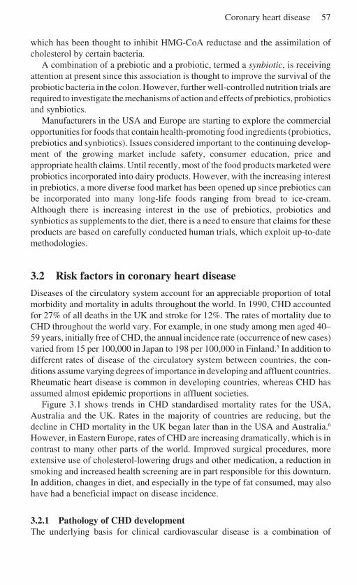
Coronary heart disease 57
which has been thought to inhibit HMG-CoA reductase and the assimilation ofcholesterol by certain bacteria.
A combination of a prebiotic and a probiotic, termed a synbiotic, is receivingattention at present since this association is thought to improve the survival of theprobiotic bacteria in the colon. However, further well-controlled nutrition trials arerequired to investigate the mechanisms of action and effects of prebiotics, probioticsand synbiotics.
Manufacturers in the USA and Europe are starting to explore the commercialopportunities for foods that contain health-promoting food ingredients (probiotics,prebiotics and synbiotics). Issues considered important to the continuing develop-ment of the growing market include safety, consumer education, price andappropriate health claims. Until recently, most of the food products marketed wereprobiotics incorporated into dairy products. However, with the increasing interestin prebiotics, a more diverse food market has been opened up since prebiotics canbe incorporated into many long-life foods ranging from bread to ice-cream.Although there is increasing interest in the use of prebiotics, probiotics andsynbiotics as supplements to the diet, there is a need to ensure that claims for theseproducts are based on carefully conducted human trials, which exploit up-to-datemethodologies.
3.2 Risk factors in coronary heart disease
Diseases of the circulatory system account for an appreciable proportion of totalmorbidity and mortality in adults throughout the world. In 1990, CHD accountedfor 27% of all deaths in the UK and stroke for 12%. The rates of mortality due toCHD throughout the world vary. For example, in one study among men aged 40–59 years, initially free of CHD, the annual incidence rate (occurrence of new cases)varied from 15 per 100,000 in Japan to 198 per 100,000 in Finland.5 In addition todifferent rates of disease of the circulatory system between countries, the con-ditions assume varying degrees of importance in developing and affluent countries.Rheumatic heart disease is common in developing countries, whereas CHD hasassumed almost epidemic proportions in affluent societies.
Figure 3.1 shows trends in CHD standardised mortality rates for the USA,Australia and the UK. Rates in the majority of countries are reducing, but thedecline in CHD mortality in the UK began later than in the USA and Australia.6
However, in Eastern Europe, rates of CHD are increasing dramatically, which is incontrast to many other parts of the world. Improved surgical procedures, moreextensive use of cholesterol-lowering drugs and other medication, a reduction insmoking and increased health screening are in part responsible for this downturn.In addition, changes in diet, and especially in the type of fat consumed, may alsohave had a beneficial impact on disease incidence.
3.2.1 Pathology of CHD developmentThe underlying basis for clinical cardiovascular disease is a combination of
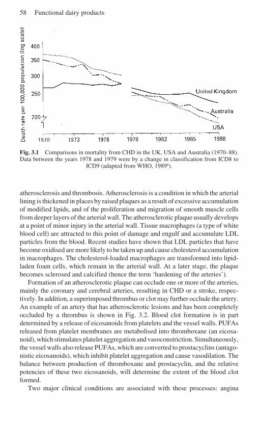
58 Functional dairy products
Fig. 3.1 Comparisons in mortality from CHD in the UK, USA and Australia (1970–88).Data between the years 1978 and 1979 were by a change in classification from ICD8 to
ICD9 (adapted from WHO, 19896).
atherosclerosis and thrombosis. Atherosclerosis is a condition in which the arteriallining is thickened in places by raised plaques as a result of excessive accumulationof modified lipids, and of the proliferation and migration of smooth muscle cellsfrom deeper layers of the arterial wall. The atherosclerotic plaque usually developsat a point of minor injury in the arterial wall. Tissue macrophages (a type of whiteblood cell) are attracted to this point of damage and engulf and accumulate LDLparticles from the blood. Recent studies have shown that LDL particles that havebecome oxidised are more likely to be taken up and cause cholesterol accumulationin macrophages. The cholesterol-loaded macrophages are transformed into lipid-laden foam cells, which remain in the arterial wall. At a later stage, the plaquebecomes sclerosed and calcified (hence the term ‘hardening of the arteries’).
Formation of an atherosclerotic plaque can occlude one or more of the arteries,mainly the coronary and cerebral arteries, resulting in CHD or a stroke, respec-tively. In addition, a superimposed thrombus or clot may further occlude the artery.An example of an artery that has atherosclerotic lesions and has been completelyoccluded by a thrombus is shown in Fig. 3.2. Blood clot formation is in partdetermined by a release of eicosanoids from platelets and the vessel walls. PUFAsreleased from platelet membranes are metabolised into thromboxane (an eicosa-noid), which stimulates platelet aggregation and vasoconstriction. Simultaneously,the vessel walls also release PUFAs, which are converted to prostacyclins (antago-nistic eicosanoids), which inhibit platelet aggregation and cause vasodilation. Thebalance between production of thromboxane and prostacyclin, and the relativepotencies of these two eicosanoids, will determine the extent of the blood clotformed.
Two major clinical conditions are associated with these processes: angina
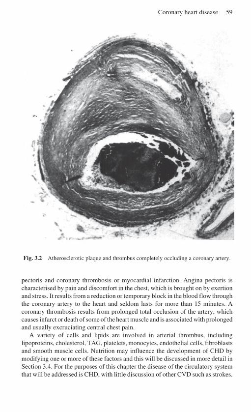
Coronary heart disease 59
Fig. 3.2 Atherosclerotic plaque and thrombus completely occluding a coronary artery.
pectoris and coronary thrombosis or myocardial infarction. Angina pectoris ischaracterised by pain and discomfort in the chest, which is brought on by exertionand stress. It results from a reduction or temporary block in the blood flow throughthe coronary artery to the heart and seldom lasts for more than 15 minutes. Acoronary thrombosis results from prolonged total occlusion of the artery, whichcauses infarct or death of some of the heart muscle and is associated with prolongedand usually excruciating central chest pain.
A variety of cells and lipids are involved in arterial thrombus, includinglipoproteins, cholesterol, TAG, platelets, monocytes, endothelial cells, fibroblastsand smooth muscle cells. Nutrition may influence the development of CHD bymodifying one or more of these factors and this will be discussed in more detail inSection 3.4. For the purposes of this chapter the disease of the circulatory systemthat will be addressed is CHD, with little discussion of other CVD such as strokes.
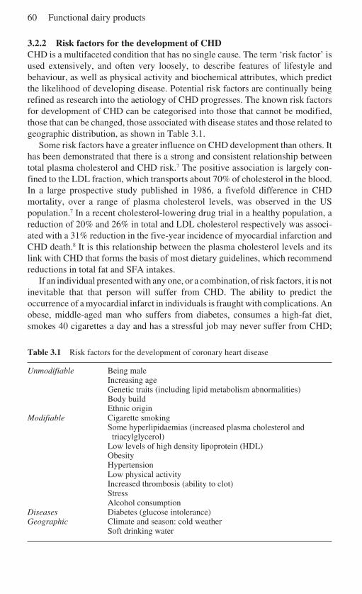
60 Functional dairy products
3.2.2 Risk factors for the development of CHDCHD is a multifaceted condition that has no single cause. The term ‘risk factor’ isused extensively, and often very loosely, to describe features of lifestyle andbehaviour, as well as physical activity and biochemical attributes, which predictthe likelihood of developing disease. Potential risk factors are continually beingrefined as research into the aetiology of CHD progresses. The known risk factorsfor development of CHD can be categorised into those that cannot be modified,those that can be changed, those associated with disease states and those related togeographic distribution, as shown in Table 3.1.
Some risk factors have a greater influence on CHD development than others. Ithas been demonstrated that there is a strong and consistent relationship betweentotal plasma cholesterol and CHD risk.7 The positive association is largely con-fined to the LDL fraction, which transports about 70% of cholesterol in the blood.In a large prospective study published in 1986, a fivefold difference in CHDmortality, over a range of plasma cholesterol levels, was observed in the USpopulation.7 In a recent cholesterol-lowering drug trial in a healthy population, areduction of 20% and 26% in total and LDL cholesterol respectively was associ-ated with a 31% reduction in the five-year incidence of myocardial infarction andCHD death.8 It is this relationship between the plasma cholesterol levels and itslink with CHD that forms the basis of most dietary guidelines, which recommendreductions in total fat and SFA intakes.
If an individual presented with any one, or a combination, of risk factors, it is notinevitable that that person will suffer from CHD. The ability to predict theoccurrence of a myocardial infarct in individuals is fraught with complications. Anobese, middle-aged man who suffers from diabetes, consumes a high-fat diet,smokes 40 cigarettes a day and has a stressful job may never suffer from CHD;
Table 3.1 Risk factors for the development of coronary heart disease
Unmodifiable Being maleIncreasing ageGenetic traits (including lipid metabolism abnormalities)Body buildEthnic origin
Modifiable Cigarette smokingSome hyperlipidaemias (increased plasma cholesterol and
triacylglycerol)Low levels of high density lipoprotein (HDL)ObesityHypertensionLow physical activityIncreased thrombosis (ability to clot)StressAlcohol consumption
Diseases Diabetes (glucose intolerance)Geographic Climate and season: cold weather
Soft drinking water
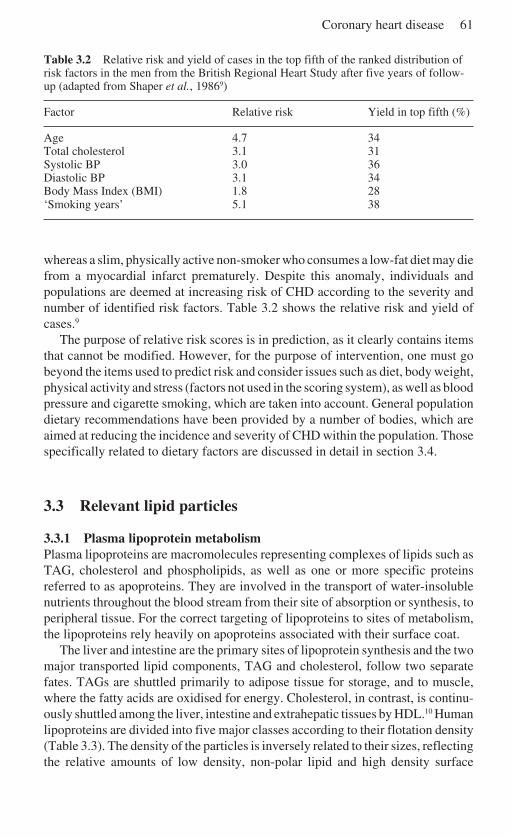
Coronary heart disease 61
Table 3.2 Relative risk and yield of cases in the top fifth of the ranked distribution ofrisk factors in the men from the British Regional Heart Study after five years of follow-up (adapted from Shaper et al., 19869)
Factor Relative risk Yield in top fifth (%)
Age 4.7 34Total cholesterol 3.1 31Systolic BP 3.0 36Diastolic BP 3.1 34Body Mass Index (BMI) 1.8 28‘Smoking years’ 5.1 38
whereas a slim, physically active non-smoker who consumes a low-fat diet may diefrom a myocardial infarct prematurely. Despite this anomaly, individuals andpopulations are deemed at increasing risk of CHD according to the severity andnumber of identified risk factors. Table 3.2 shows the relative risk and yield ofcases.9
The purpose of relative risk scores is in prediction, as it clearly contains itemsthat cannot be modified. However, for the purpose of intervention, one must gobeyond the items used to predict risk and consider issues such as diet, body weight,physical activity and stress (factors not used in the scoring system), as well as bloodpressure and cigarette smoking, which are taken into account. General populationdietary recommendations have been provided by a number of bodies, which areaimed at reducing the incidence and severity of CHD within the population. Thosespecifically related to dietary factors are discussed in detail in section 3.4.
3.3 Relevant lipid particles
3.3.1 Plasma lipoprotein metabolismPlasma lipoproteins are macromolecules representing complexes of lipids such asTAG, cholesterol and phospholipids, as well as one or more specific proteinsreferred to as apoproteins. They are involved in the transport of water-insolublenutrients throughout the blood stream from their site of absorption or synthesis, toperipheral tissue. For the correct targeting of lipoproteins to sites of metabolism,the lipoproteins rely heavily on apoproteins associated with their surface coat.
The liver and intestine are the primary sites of lipoprotein synthesis and the twomajor transported lipid components, TAG and cholesterol, follow two separatefates. TAGs are shuttled primarily to adipose tissue for storage, and to muscle,where the fatty acids are oxidised for energy. Cholesterol, in contrast, is continu-ously shuttled among the liver, intestine and extrahepatic tissues by HDL.10 Humanlipoproteins are divided into five major classes according to their flotation density(Table 3.3). The density of the particles is inversely related to their sizes, reflectingthe relative amounts of low density, non-polar lipid and high density surface

62 Functional dairy products
protein present. The two largest lipoproteins contain mainly TAG within their corestructures. These are chylomicrons (CMs), secreted by the enterocytes (cells of thesmall intestine), and VLDL, secreted by the hepatocytes (liver cells). Intermediatedensity lipoprotein (IDL) contains appreciable amounts of both TAG and choles-terol esters in their core. The two smallest classes, LDL and HDL, containcholesterol esters in their core structures and the mature forms of these particles arenot secreted directly from the liver but are produced by metabolic processes withinthe circulation. LDLs are produced as end products of the metabolism of VLDL,whereas components of HDL are secreted with CMs and VLDL. The lipidmetabolic pathways can be divided into the exogenous and endogenous cycles,which are responsible for the transport of dietary and hepatically derived lipid,respectively.11
Exogenous pathwayFollowing the digestion of dietary fat in the small intestine, long chain fatty acidsare absorbed by the enterocyte. The nascent CM particle consists of a core of TAG(84–9% of the mass), cholesterol ester (3–5%) and surface free cholesterol (1–2%)and on the surface, phospholipids (7–9%) and apoproteins (1.5–2.5%).12 Follow-ing secretion, the TAG component of the CM particle is hydrolysed by lipoproteinlipase (LPL) bound to the luminal surface of the endothelial cells in adipose tissueand muscle. Approximately 70–90% of the TAG is removed to produce a choles-terol ester-rich lipoprotein particle termed a CM remnant.13 As the core of the CMremnant particle becomes smaller, surface materials, phospholipids, cholesteroland apolipoproteins are transferred to HDL to maintain stability of the particle.Uptake of these particles probably requires interaction with hepatic lipase (HL),which is situated on the surface of the liver. HL further hydrolyses the TAG andphospholipid components of the CM remnant before uptake by a receptor-mediated process in the liver.14 The remnants are endocytosed and catabolised inlysosomes from which cholesterol can enter metabolic pathways in hepatocytes,including excretion into the bile (Fig. 3.3).
Endogenous pathwayVLDL provides a pathway for the transport of TAG from the liver to the peripheralcirculation. Two subclasses of VLDL are released from the liver, VLDL
1 (large
TAG-rich lipoprotein) and VLDL2 (smaller, denser particles). LPL in the capillary
bed extracts TAG from the secreted VLDL1 but less efficiently when compared to
CMs.15 LDL receptors on the surface of the liver recognise the VLDL2 particles
and mediate the endocytosis of a fraction of these particles. Prolonged residency ofsome VLDL particles in the plasma results in further metabolism by LPL, and tosome extent HL, to form the higher density IDL. HL, on the surface of hepatocytes,further hydrolyses IDL with eventual formation of LDL.16 LDL is a heterogeneouspopulation consisting of larger LDL species (LDL-I and LDL-II subclass) andsmaller denser LDL particles (LDL-III). LDL can be taken up by LDL receptorspresent on the surface of hepatocytes and on extrahepatic cells.
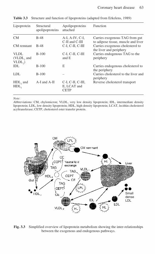
Coronary heart disease 63
Table 3.3 Structure and function of lipoproteins (adapted from Erkelens, 1989)
Lipoprotein Structural Apolipoproteins Functionapolipoproteins attached
CM B-48 A-I, A-IV, C-I, Carries exogenous TAG from gutC-II and C-III to adipose tissue, muscle and liver
CM remnant B-48 C-I, C-II, C-III Carries exogenous cholesterol tothe liver and periphery
VLDL B-100 C-I, C-II, C-III Carries endogenous TAG to the(VLDL
1 and and E periphery
VLDL2)
IDL B-100 E Carries endogenous cholesterol tothe periphery
LDL B-100 – Carries cholesterol to the liver andperiphery
HDL2 and A-I and A-II C-I, C-II, C-III, Reverse cholesterol transport
HDL3
E, LCAT andCETP
Note:Abbreviations: CM, chylomicron; VLDL, very low density lipoprotein; IDL, intermediate densitylipoprotein; LDL, low density lipoprotein; HDL, high density lipoprotein; LCAT, lecithin cholesterolacyltransferase; CETP, cholesterol ester transfer protein.
Fig. 3.3 Simplified overview of lipoprotein metabolism showing the inter-relationshipsbetween the exogenous and endogenous pathways.
SmalldenseLDL
64 Functional dairy products
Reverse cholesterol transportA function of HDL is to trigger the flux of cholesterol from peripheral cells andfrom membranes undergoing turnover to the liver for excretion. The process ofreverse cholesterol transport is mediated by an enzyme, lecithin cholesterolacyltransferase (LCAT), bound to species of HDL particles. It acts by trappingcholesterol into the core of the nascent HDL following interaction of this particlewith a cell surface protein. The cholesterol is transferred to HDL
3, which in turn is
converted to HDL2. This particle can follow one of two pathways (direct or
indirect) to deliver cholesterol to the liver.17 In the direct pathway, the HDL2 is
removed via receptor-mediated endocytosis by the LDL receptor or via selectiveuptake of cholesterol ester from the HDL particle by the liver. The cholesterol estertransfer protein (CETP) is involved in the indirect pathway and transfers choles-terol ester to lower density lipoproteins (CMs and VLDL), in return for TAG, andis followed by their uptake by the liver (neutral lipid exchange) (Fig. 3.3).
The balance between the forward (exogenous and endogenous pathways) andthe reverse pathway, which is tightly regulated by the secretion rates of thelipoproteins, determines the concentration of cholesterol in the plasma.
3.3.2 Classical lipid risk factors in coronary heart diseaseMany epidemiological studies have shown a positive correlation between fastingtotal cholesterol levels, especially LDL cholesterol levels and CHD mortality.1
Accumulation of LDL in the plasma leads to a deposition of cholesterol in thearterial wall, a process that involves oxidative modification of the LDL particles.The oxidised LDL is taken up by macrophages, which eventually become foamcells and forms the basis of the early atherosclerotic plaque. It has been estimatedthat every 1% increase in LDL cholesterol level leads to a 2–3% increase in CHDrisk.18 The role of cholesterol lowering as a public health strategy in the primaryprevention of CHD was unequivocally supported by the findings of the WestScotland Heart Study.8 This lipid-lowering trial involved 6,595 middle-aged menwith moderately raised cholesterol levels (5.8–8.0 mmol/l) but without any historyof CHD. The study showed a 22% reduction in overall mortality and a 33%reduction in cardiovascular mortality in men receiving the active drug (prevastatin)compared with a placebo, in a five-year follow-up. Total cholesterol levels werereduced by 20%, with LDL cholesterol levels being reduced by 26% on the activedrug treatment.
HDL cholesterol levels may influence the relationship between total cholesterollevels and CHD risk. A strong inverse relationship between fasting plasma HDLlevels and the risk of development of CHD has been reported.18 On average, HDLcholesterol levels are higher in women than in men. Factors that may lead toreduced HDL cholesterol levels include smoking, low physical activity anddiabetes mellitus; whereas those that increase levels include moderate alcoholconsumption. The Münster Heart Study, carried out between 1979 and 1991,investigated cardiovascular risk factors, stroke and mortality in people at work.Examination of fasting lipid parameters at the beginning of the study and with a
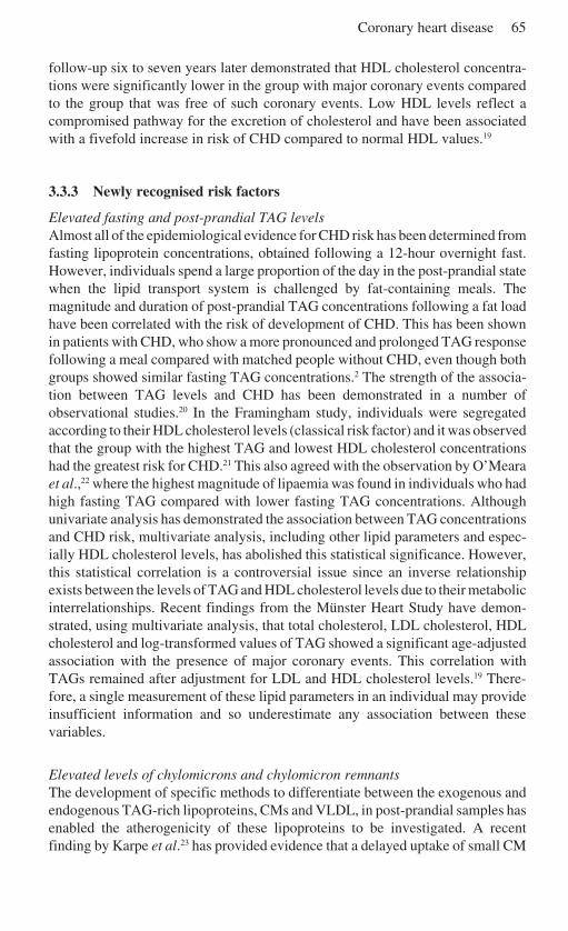
Coronary heart disease 65
follow-up six to seven years later demonstrated that HDL cholesterol concentra-tions were significantly lower in the group with major coronary events comparedto the group that was free of such coronary events. Low HDL levels reflect acompromised pathway for the excretion of cholesterol and have been associatedwith a fivefold increase in risk of CHD compared to normal HDL values.19
3.3.3 Newly recognised risk factors
Elevated fasting and post-prandial TAG levelsAlmost all of the epidemiological evidence for CHD risk has been determined fromfasting lipoprotein concentrations, obtained following a 12-hour overnight fast.However, individuals spend a large proportion of the day in the post-prandial statewhen the lipid transport system is challenged by fat-containing meals. Themagnitude and duration of post-prandial TAG concentrations following a fat loadhave been correlated with the risk of development of CHD. This has been shownin patients with CHD, who show a more pronounced and prolonged TAG responsefollowing a meal compared with matched people without CHD, even though bothgroups showed similar fasting TAG concentrations.2 The strength of the associa-tion between TAG levels and CHD has been demonstrated in a number ofobservational studies.20 In the Framingham study, individuals were segregatedaccording to their HDL cholesterol levels (classical risk factor) and it was observedthat the group with the highest TAG and lowest HDL cholesterol concentrationshad the greatest risk for CHD.21 This also agreed with the observation by O’Mearaet al.,22 where the highest magnitude of lipaemia was found in individuals who hadhigh fasting TAG compared with lower fasting TAG concentrations. Althoughunivariate analysis has demonstrated the association between TAG concentrationsand CHD risk, multivariate analysis, including other lipid parameters and espec-ially HDL cholesterol levels, has abolished this statistical significance. However,this statistical correlation is a controversial issue since an inverse relationshipexists between the levels of TAG and HDL cholesterol levels due to their metabolicinterrelationships. Recent findings from the Münster Heart Study have demon-strated, using multivariate analysis, that total cholesterol, LDL cholesterol, HDLcholesterol and log-transformed values of TAG showed a significant age-adjustedassociation with the presence of major coronary events. This correlation withTAGs remained after adjustment for LDL and HDL cholesterol levels.19 There-fore, a single measurement of these lipid parameters in an individual may provideinsufficient information and so underestimate any association between thesevariables.
Elevated levels of chylomicrons and chylomicron remnantsThe development of specific methods to differentiate between the exogenous andendogenous TAG-rich lipoproteins, CMs and VLDL, in post-prandial samples hasenabled the atherogenicity of these lipoproteins to be investigated. A recentfinding by Karpe et al.23 has provided evidence that a delayed uptake of small CM
66 Functional dairy products
remnants is associated with the progression of atherosclerosis. The abnormalclearance of CMs and CM remnants after a fat load have been implicated directlyand/or indirectly with the presence of CHD. This may reflect the ability of CMremnants to infiltrate the arterial wall directly. The mechanism whereby CMremnants provide the building blocks of arterial lesions are thought to occur by oneof two processes. First, the CM remnants may bind and penetrate the arterialsurfaces (in just the same way as plasma LDL), therefore the rate of atherogenesisshould be proportional to the plasma remnant concentrations. Second, CMs may beabsorbed and then degraded to remnants on the arterial surface.24 In each instance,the reaction leading to the endocytosis of remnants by smooth muscle cells maytake place at sites where local injury has removed the endothelium.
The magnitude of post-prandial lipaemia is dependent on a number of factorsincluding rates of clearance by peripheral tissue and receptor-mediated uptake ofthe remnants by the liver. Defects in any of these processes will cause anaccumulation of CMs and their remnants which in turn can influence endogenouslipoproteins known to be atherogenic. In particular the transfer of TAG from CMsto LDL and HDL and a reciprocal transfer of cholesterol esters to the CMs byCETP may increase the atherogenic lipid profile. This is known as neutral lipidexchange. The transfer of cholesterol ester to CMs and VLDL makes themresistant to lipase action which impedes the normal metabolism of these TAG-richlipoproteins. The cholesterol ester-enriched CM remnants are then able to be takenup by the macrophages in the arterial lesion. LDL and HDL, on the other hand,become suitable substrates for LPL and HL, causing a reduction in the size of theseparticles. This results in the development of an atherogenic lipoprotein profile inwhich the TAG-rich lipoproteins become cholesterol ester-enriched, LDL size isreduced and HDL cholesterol levels are reduced. The small dense nature of theLDL makes it unrecognisable by the LDL receptors in the liver and so makes it afavourable candidate for uptake by scavenger receptors present on macrophages inthe arterial lesion.25
Elevated levels of small dense LDL (LDL-III)It has long been established that elevated circulating LDL cholesterol levelsrepresent a major risk factor for the development of CHD. Recently, with the useof density gradient ultracentrifugation techniques, LDL has been separated intothree major subclasses. These are subdivided as light large LDL (LDL-I), inter-mediate size LDL (LDL-II) and small dense LDL (LDL-III).26 In healthynormolipidaemic males, a preponderance of LDL-II are seen, with only a smallpercentage of LDL-III being present.
A number of case control and cross-sectional studies has examined the relation-ship between LDL subclass and risk of CHD.27 In studies in men with CHD, orthose who had survived a myocardial infarction, it was demonstrated that smalldense LDL-III was more common in the men with CHD than without. In 1988,Austin et al.26 proposed that a preponderance of LDL-III in young men wasassociated with a threefold increase in CHD. However, more recently, a greaterrelative risk of CHD associated with raised LDL-III has been proposed.28 The
Coronary heart disease 67
increased atherogenicity of small dense LDL-III is thought to be due to the increasedresidency of these particles in the circulation due to their slow uptake by the LDLreceptor in the liver and peripheral tissues. This allows time for the small dense LDLto infiltrate into the intima of the arterial wall where it is thought that these particlesare retained by extracellular matrix components before oxidation of the LDLparticles occur. The modified small dense LDL-III is then taken up by the scavengerreceptor on macrophages leading to the subsequent formation of foam cells.29
Atherogenic lipoprotein phenotypeThe ALP is a collection of lipid abnormalities, which confers an increased risk ofCHD upon normal, healthy individuals. It has been proposed that 30–35% ofmiddle-aged men in the Western world may be affected.3 In the fasting state, thisphenotype is characterised by a moderately raised TAG concentration (1.5 to 2.3mmol/l), low HDL concentration (less than 1 mmol/l) and a predominance of smalldense LDL-III (greater than 35% of LDL mass) and HDL particles. It is importantto note that total cholesterol levels are usually in the normal range or are onlymoderately raised. The atherogenic nature of the ALP may arise from the impair-ment of the removal of TAG-rich lipoproteins (CMs and VLDL) leading to theconversion of small, atherogenic cholesterol enriched remnants, LDL and HDL byneutral lipid exchange. It is generally considered that this collection of lipidabnormalities is closely associated with the insulin resistance syndrome.30
3.4 Diet and coronary heart disease
There is a substantial, diverse and generally consistent body of evidence linkingdiet with cardiovascular disease. The evidence is most extensive for the relation-ship between CHD and dietary fat. Diet is believed to influence the risk of CHDthrough its effects on certain risk factors described in Table 3.1, for example bloodlipids, blood pressure and probably also through thrombogenic mechanisms. Morerecently, evidence suggests a protective role for dietary antioxidants such asvitamins E and C and carotenes, possibly through a mechanism that prevents theoxidation of LDL cholesterol particles.
3.4.1 Dietary lipids and CHD riskEpidemiological and clinical evidence clearly shows that the likelihood of deathfrom CHD is directly related to the circulating level of total cholesterol (and morespecifically LDL cholesterol). More recent evidence suggests that an exaggeratedpost-prandial TAG response to fat-containing meals is also a significant risk factorfor CHD.2 Numerous studies have shown that the kinds and amounts of fat in thediet significantly influence plasma cholesterol levels. In the Seven CountriesStudy, mean concentrations of cholesterol of each group were highly correlatedwith percentage energy derived from SFAs, and even more strikingly related to aformula that also took into account the intake of PUFAs (see Fig. 3.4).5
68 Functional dairy products
Fig. 3.4 Relationship of mean serum cholesterol concentration of the cohorts of SevenCountries Study to fat composition of the diet expressed in the multiple regress equation,including intakes of saturated and polyunsaturated fatty acids (B = Belgrade, C = Crevalcore,D = Dalmatia, E = East Finland, G = Corfu, J = Ushibuka, K = Crete, M = Montegiorgio, N= Zutphen, R = Roma, S = Slavonia, T = Tanushimara, U = USA, V = Velika Kisna, W =
West Finland, Z = Zregnjamin). (Adapted from Keys, 1980.5)
Since this research, numerous other studies have supported the relationshipbetween dietary intake of saturates and the raised plasma cholesterol levels.However, not all SFAs are equally potent in raising plasma cholesterol. Palmiticacid (16:0), the principal SFA in most diets, and myristic acid (14:0) are the mosteffective at elevating cholesterol; whereas stearic acid (18:0), lauric acid (12:0)and medium chain fatty acids (8:0 and 10:0) have little effect on plasma cholesterollevels.31 The cholesterol response to a particular SFA may depend in part on theTAG structure,32 and in part on LDL receptor activity.33 The mechanism that isresponsible for the increase in plasma cholesterol due to SFAs is at present unclear,but a reduction in the LDL receptor activity is one possibility. It has beenrecommended that the average contribution of SFAs to dietary energy be reducedto no more than 10%.34 The current UK dietary intake is shown in Table 3.4. Aconsiderable reduction from current levels of intake of dietary SFAs (average33%) would be required to meet this recommendation.35
The effect of the ingestion of different fatty acids on plasma cholesterol levelsis varied and is summarised in Table 3.5. Substitution of SFAs by MUFAs or n-6PUFAs significantly reduces LDL cholesterol levels, although n-6 PUFA are moreeffective in this respect. There has been doubt as to whether MUFAs are effectivein cholesterol-lowering or whether the observed decrease in plasma cholesterolwas simply due to a replacement of SFAs. Some studies have suggested that the
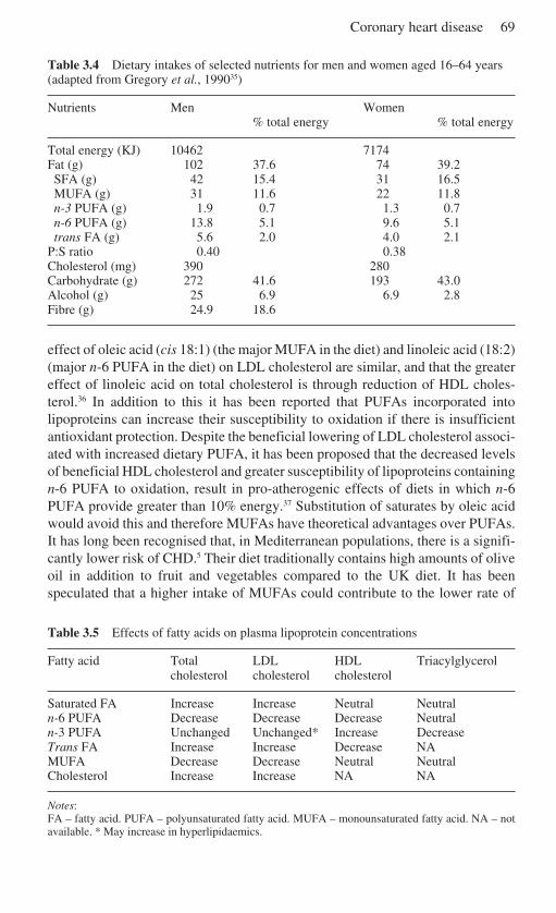
Coronary heart disease 69
Table 3.4 Dietary intakes of selected nutrients for men and women aged 16–64 years(adapted from Gregory et al., 199035)
Nutrients Men Women% total energy % total energy
Total energy (KJ) 10462 7174Fat (g) 102 37.6 74 39.2SFA (g) 42 15.4 31 16.5MUFA (g) 31 11.6 22 11.8n-3 PUFA (g) 1.9 0.7 1.3 0.7n-6 PUFA (g) 13.8 5.1 9.6 5.1trans FA (g) 5.6 2.0 4.0 2.1
P:S ratio 0.40 0.38Cholesterol (mg) 390 280Carbohydrate (g) 272 41.6 193 43.0Alcohol (g) 25 6.9 6.9 2.8Fibre (g) 24.9 18.6
effect of oleic acid (cis 18:1) (the major MUFA in the diet) and linoleic acid (18:2)(major n-6 PUFA in the diet) on LDL cholesterol are similar, and that the greatereffect of linoleic acid on total cholesterol is through reduction of HDL choles-terol.36 In addition to this it has been reported that PUFAs incorporated intolipoproteins can increase their susceptibility to oxidation if there is insufficientantioxidant protection. Despite the beneficial lowering of LDL cholesterol associ-ated with increased dietary PUFA, it has been proposed that the decreased levelsof beneficial HDL cholesterol and greater susceptibility of lipoproteins containingn-6 PUFA to oxidation, result in pro-atherogenic effects of diets in which n-6PUFA provide greater than 10% energy.37 Substitution of saturates by oleic acidwould avoid this and therefore MUFAs have theoretical advantages over PUFAs.It has long been recognised that, in Mediterranean populations, there is a signifi-cantly lower risk of CHD.5 Their diet traditionally contains high amounts of oliveoil in addition to fruit and vegetables compared to the UK diet. It has beenspeculated that a higher intake of MUFAs could contribute to the lower rate of
Table 3.5 Effects of fatty acids on plasma lipoprotein concentrations
Fatty acid Total LDL HDL Triacylglycerolcholesterol cholesterol cholesterol
Saturated FA Increase Increase Neutral Neutraln-6 PUFA Decrease Decrease Decrease Neutraln-3 PUFA Unchanged Unchanged* Increase DecreaseTrans FA Increase Increase Decrease NAMUFA Decrease Decrease Neutral NeutralCholesterol Increase Increase NA NA
Notes:FA – fatty acid. PUFA – polyunsaturated fatty acid. MUFA – monounsaturated fatty acid. NA – notavailable. * May increase in hyperlipidaemics.
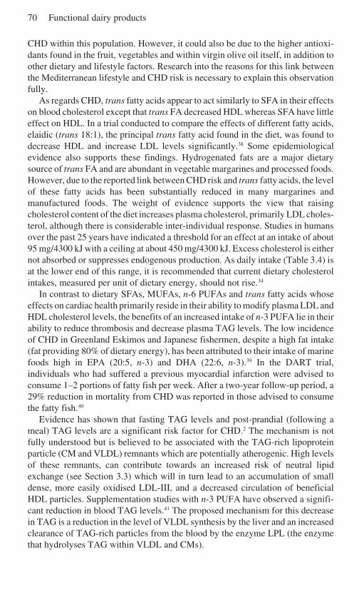
70 Functional dairy products
CHD within this population. However, it could also be due to the higher antioxi-dants found in the fruit, vegetables and within virgin olive oil itself, in addition toother dietary and lifestyle factors. Research into the reasons for this link betweenthe Mediterranean lifestyle and CHD risk is necessary to explain this observationfully.
As regards CHD, trans fatty acids appear to act similarly to SFA in their effectson blood cholesterol except that trans FA decreased HDL whereas SFA have littleeffect on HDL. In a trial conducted to compare the effects of different fatty acids,elaidic (trans 18:1), the principal trans fatty acid found in the diet, was found todecrease HDL and increase LDL levels significantly.38 Some epidemiologicalevidence also supports these findings. Hydrogenated fats are a major dietarysource of trans FA and are abundant in vegetable margarines and processed foods.However, due to the reported link between CHD risk and trans fatty acids, the levelof these fatty acids has been substantially reduced in many margarines andmanufactured foods. The weight of evidence supports the view that raisingcholesterol content of the diet increases plasma cholesterol, primarily LDL choles-terol, although there is considerable inter-individual response. Studies in humansover the past 25 years have indicated a threshold for an effect at an intake of about95 mg/4300 kJ with a ceiling at about 450 mg/4300 kJ. Excess cholesterol is eithernot absorbed or suppresses endogenous production. As daily intake (Table 3.4) isat the lower end of this range, it is recommended that current dietary cholesterolintakes, measured per unit of dietary energy, should not rise.34
In contrast to dietary SFAs, MUFAs, n-6 PUFAs and trans fatty acids whoseeffects on cardiac health primarily reside in their ability to modify plasma LDL andHDL cholesterol levels, the benefits of an increased intake of n-3 PUFA lie in theirability to reduce thrombosis and decrease plasma TAG levels. The low incidenceof CHD in Greenland Eskimos and Japanese fishermen, despite a high fat intake(fat providing 80% of dietary energy), has been attributed to their intake of marinefoods high in EPA (20:5, n-3) and DHA (22:6, n-3).39 In the DART trial,individuals who had suffered a previous myocardial infarction were advised toconsume 1–2 portions of fatty fish per week. After a two-year follow-up period, a29% reduction in mortality from CHD was reported in those advised to consumethe fatty fish.40
Evidence has shown that fasting TAG levels and post-prandial (following ameal) TAG levels are a significant risk factor for CHD.2 The mechanism is notfully understood but is believed to be associated with the TAG-rich lipoproteinparticle (CM and VLDL) remnants which are potentially atherogenic. High levelsof these remnants, can contribute towards an increased risk of neutral lipidexchange (see Section 3.3) which will in turn lead to an accumulation of smalldense, more easily oxidised LDL-III, and a decreased circulation of beneficialHDL particles. Supplementation studies with n-3 PUFA have observed a signifi-cant reduction in blood TAG levels.41 The proposed mechanism for this decreasein TAG is a reduction in the level of VLDL synthesis by the liver and an increasedclearance of TAG-rich particles from the blood by the enzyme LPL (the enzymethat hydrolyses TAG within VLDL and CMs).

Coronary heart disease 71
Fatty acids from the n-3 PUFA series also have a beneficial effect onthrombogenesis (blood clot formation). The utilisation of n-3 PUFAs instead of n-6 PUFAs in the production of eicosanoids (substances involved with the formationof blood clotting) can significantly reduce the rate of blood clot formation and thusreduce the risk of myocardial infarction. In addition, n-3 PUFA ingestion mayresult in the reduction in cardiac arrhythmias.42 Sudden cardiac death is a seriousproblem in Western countries and no drug treatment, to date, has had anysignificant effect on incidence. Epidemiological studies have shown a reducedincidence of sudden death with n-3 PUFA intake, and animal and limited humanevidence also suggests an anti-arrhythmic effect.43
At the present time most of the national and international guidelines forpopulation intakes of dietary fat are based on the known adverse effects of SFAsin raising blood cholesterol levels with some consideration given to possiblebenefits of n-3 PUFAs on thrombosis. Table 3.6 shows the dietary guidelines of theEuropean Union44 and those of the UK Committee of Medical Aspects of FoodPolicy.34 These recommendations are aimed at both the food industry and thegeneral population. Due to high SFA intake compared with those recommended,which reflect consumer resistance to these recommendations, future recommenda-tions may take greater account of the benefits of substituting SFAs with MUFAs,and the overall significance to human health of the absolute and relative amountsof n-6: n-3 PUFAs.
3.4.2 Carbohydrate intake and CHD riskRecommendations for reductions in total fat in the diet have important implicationsfor dietary carbohydrate intake. There is little variation in the proportion of energyderived from dietary protein; therefore there is a reciprocal relationship between
Table 3.6 Recommended daily intakes of dietary fat and fatty acids of the EEC (EU)and UK and the average daily consumption of these nutrients (% dietary energy)
Total fat SFA PUFA MUFA Trans FA
RecommendationsEuropean 20–301 101 0.52 (n-3) NR NR(EEC, 1992) 2.02 (n-6)UK <35 <10 0.2g/day (n-3) NR 2(DH, 1994) 5.0 (n-6)Current intakeUK (1990) 39 16 5 (n-6) 12 2USA (1985) 37 13 7 (n-6) 13 –France (1985–7) 36 15 6 (n-6) 13 –Germany (1988) 40 15 6 (n-6) 12 –Netherlands (1988) 40 16 7 (n-6) 15 –
Notes:1. Ultimate goal. 2. Population reference intake. NR – no recommendations.
72 Functional dairy products
the contributions of dietary fat and carbohydrate to energy. The metabolic effectsof exchanging carbohydrate for fat depend mainly on the degree of substitution.Diets where about 60% of food energy is derived from carbohydrate are associatedwith lower HDL levels and higher TAG levels, and despite lower LDL levels havebeen suggested to be associated with a higher risk of CHD.45, 46 However, a smallerincrease in dietary carbohydrate levels to accommodate a reduction in dietary fatto 30% of energy has been reported to result in a small rise in TAG levels and nofall in HDL levels,46 resulting in an overall positive benefit in CHD risk.
3.4.3 Non-starch polysaccharides and CHD riskFour prospective studies have shown an inverse relationship between dietary‘fibre’ intake and CHD. The studies have varied in the source of ‘fibre’ that wasfound to be effective. Morris et al.47 found that cereal fibre was inversely related tocases of CHD, whereas others found that vegetable sources of fibre were associ-ated with decreased risk.48 On the contrary, a two-year intervention study in menwho had suffered a previous heart attack, found no effect of increasing cereal‘fibre’ intake on subsequent risk of mortality from cardiovascular disease.40 Theeffect of a number of soluble NSPs which are selectively digested by the colonicgut microflora (classed as prebiotics) on the blood levels of cholesterol andespecially LDL cholesterol will be discussed in detail in section 3.6.
3.4.4 Antioxidant nutrients and CHD riskThe cells of the body are under constant attack by activated oxygen species, whichare produced naturally in the body. The protection of cells from the detrimentaleffects of these species is due to defence mechanisms, component parts of whichare, or are derived from, the micronutrients called ‘antioxidants’. These include theessential trace elements selenium, zinc and magnesium, and vitamin C and thevarious forms of vitamin E (the tocopherols and the tocotrienols). In addition,betacarotene, and other carotenoids, such as lutein and lycopene (present in largeamounts in tomatoes) and flavonoids, present in red wine, tea and onions, probablyalso play a role.
There is a large body of evidence supporting a protective effect of the antioxi-dant vitamins E and C.49 There are, however, inconsistencies in the amount of theseantioxidants associated with reduced risk of CHD; this is especially the case forvitamin E. One explanation for these discrepancies is that vitamin E may be amarker for a diet that contains other dietary constituents (such as carotenoids andflavonoids) which have antioxidant potential. When vitamin E is taken in isolationas a supplement, higher levels might be necessary to achieve the same effect asdiets that contain combinations of antioxidants. No specific recommendations onthe levels of antioxidant intake have been given but a diet rich in vegetable and fruitand containing nuts and seeds is recommended.50 The beneficial effects of red wineand olive oil on CHD risk could in part be attributed to antioxidants such asflavonoids and polyphenols contained within these foods.

Coronary heart disease 73
3.4.5 Sodium and potassium and CHD riskSodium intake appears to be an important determinant of blood pressure in thepopulation as a whole, at least in part by influencing the rise in blood pressure withage.51, 52 A diet lower in common salt and higher in potassium would be expectedto result in lower blood pressure and a smaller rise in blood pressure with age. Saltis the predominant source of sodium in the diet, with manufactured foods contrib-uting to 65–85% of the total salt ingested. Blood pressure is an important risk factorfor the development of CHD and strokes. It has been recommended that thepopulation should reduce its salt intake by 3 g/day by reducing salt at the table andalso the consumption of processed foods. Moreover, higher potassium levelswithin the diet are believed to reduce the blood pressure and foods such as fruit andvegetables, which contain this mineral, should be taken in higher amounts.
3.4.6 Alcohol and CHD riskThe debate surrounding the benefits of alcohol consumption and, more specifi-cally, red wine consumption and the risk of CHD is one that has been running formany years. There is evidence that high consumption of alcohol is related toincreased mortality, especially from CHD. However, alcohol consumption appearsto be associated with relatively low risk of CHD across a variety of studypopulations. The benefit that is associated with alcohol consumption is almostentirely due to the increased levels of HDL cholesterol which is associated withreduced CHD risk.53 However, other factors such as lower platelet activity,reducing the risk of thrombosis, and antioxidant properties of some drinks such asred wine have also been suggested. The proposed benefit of red wine over otherdrinks would only explain the potential increase in antioxidant levels as the effectof alcohol on HDL and haemostatic factors have been attributed to alcohol itself.A consumption of two units (one unit is equivalent to 8 g alcohol) a day of alcoholis believed to be beneficial in the risk of CHD, but the debate continues.54
3.4.7 Coffee and CHD riskFor over 20 years it has been suspected that caffeine or coffee consumption maycontribute to the development of CHD, but the evidence remains inconsistent.There is little evidence that caffeine itself has any relation to CHD risk but there areother components of coffee, which might in part account for some observedassociations. The Scandinavian practice of boiling coffee during its preparationappears to generate a hypercholesterolaemic fraction. Significant levels of thesecompounds have also been found in cafeteria coffee which have been found toincrease plasma LDL cholesterol levels.55 The relevance of this to the UK popu-lation, whose consumption of boiled coffee is very low, is unclear.
3.4.8 Diet and CHD riskThe diet is one of the modifiable risk factors associated with CHD risk.
74 Functional dairy products
Recommending reducing total fat (especially saturated fat), increasing NSP intakeand consumption of fruit and vegetables is advice that is likely to be associatedwith overall benefits on health. However, there is great inertia for dietary change,and many new products that claim to reduce the risk of CHD and other chronicdiseases, without altering lifestyle factors such as diet, clearly attract a great dealof attention.
3.5 The effects of probiotics on coronary heart disease
Current dietary strategies for the prevention of CHD advocate adherence to lowfat/low saturated fat diets.34 Although there is no doubt that, under experimentalconditions, low fat diets offer an effective means of reducing blood cholesterolconcentrations, on a population basis they appear to be less effective, largely dueto poor compliance attributed to low palatability and acceptability of these diets tothe consumer. Due to the low consumer compliance, attempts have been made toidentify other dietary components that can reduce blood cholesterol levels. Thesehave included investigations into the possible hypocholesterolaemic properties ofmilk products, usually in a fermented form. The role of fermented milk products ashypocholesterolaemic agents in humans is still equivocal, as the studies performedhave been of varying quality, design and statistical analysis with incompletedocumentation being the major limitation of most studies. However, since 1974when Mann and Spoerry56 showed an 18% fall in plasma cholesterol after feeding4–5 litres of fermented milk per day for three weeks to Masai warriors, there hasbeen considerable interest in the effect of probiotics on human lipid metabolism.56
3.5.1 Evidence from the MasaiAs epidemiological evidence suggested that an environmental agent that contrib-uted to hypocholesterolaemia had been introduced into the Western world around1900, Mann and Spoerry intended to investigate the effect of an exogenoussurfactant material, known to be hypercholesterolaemic in animals.56 The authors’original intention was to feed 4–5 litres of pasteurised European milk fermentedwith a wild culture of Lactobacillus to 24 male Masai warriors, for six days out ofevery week. However, demand rose to over 8.3 litres per day (23,000 kJ/day) andan exercise programme was started to prevent weight gain. However, eight of thesubjects gained considerable weight (> 5 lb) after feigning injury to avoid thisexercise. The eight subjects who gained weight surprisingly had a significant fall(18.2%) in serum cholesterol of 0.73 mmol/l. The total group’s mean cholesterollevel also significantly decreased (9.8%), as shown in Table 3.7. Although themarked reductions in serum cholesterol were striking, considering the weightincrease (usually associated with a raised cholesterol concentration), this study isnow considered simply as a curiosity. Due to the introduction of an exerciseprogramme and the inability to control food intake, the work cannot lend much tothe causality of the findings.
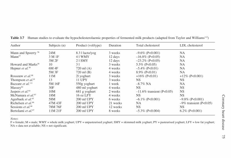
Coronary heart disease
75
Table 3.7 Human studies to evaluate the hypocholesterolaemic properties of fermented milk products (adapted from Taylor and Williams112)
Author Subjects (n) Product (vol/type) Duration Total cholesterol LDL cholesterol
Mann and Spoerry 56 24M 8.3 l lacto/yog 3 weeks –9.6% (P<0.001) NAMann57 3 M 1F 4 l WMY 12 days –16.8% (P<0.05) NA
3M 2F 2 l SMY 12 days –23.2% (P<0.05) NAHoward and Marks58 10 3 l 3 weeks 5.5% (P<0.05) NAHepner et al.59 6M 4F 720 ml (A) 4 weeks –5.4% (P<0.01) NA
5M 3F 720 ml (B) 4 weeks 8.9% (P<0.01) NARossouw et al.68 11M 2l yoghurt 3 weeks +16% (P<0.01) +12% (P<0.001)Thompson et al.61 13 11 UPY 3 weeks NS NSBazzare et al.111 5M 16F 550g yoghurt 1 week –8.7% NA NAMassey62 30F 480 ml yoghurt 4 weeks NS NSJaspers et al.64 10M 681 g yoghurt 2 weeks –11.6% transient (P<0.05) NSMcNamara et al.63 18M 16 oz LFY 4 weeks NS NSAgerbaek et al.65 58M 200 ml UPY 6 weeks –6.1% (P<0.001) –9.8% (P<0.001)Richelsen et al.66 47M 43F 200 ml UPY 21 weeks NA –9% transient (P<0.05)Sessions et al.60 78M 76F 200 ml UPY 12 weeks NS NSBertolami et al.67 11M 21F 200 ml UPY 8 weeks –5.3% (P<0.004) 6.2% (P<0.001)
Notes:F = female; M = male; WMY = whole milk yoghurt; UPY = unpasteurised yoghurt; SMY = skimmed milk yoghurt; PY = pasteurised yoghurt; LFY = low fat yoghurt;NA = data not available; NS = not significant.
76 Functional dairy products
3.5.2 Evidence for the ‘milk factor’As a follow-up to the Masai trial, Mann fed a small group of US volunteers (n = 4)4 litres per day of yoghurt (microbiological activity unspecified) over a 12-dayperiod and reported a significant fall of 37% in serum cholesterol values (however,the tabulated data indicated only a 16.8% fall).57 When intake of the yoghurt wasreduced to 2 litres, the hypocholesterolaemic effect was maintained, although anintake of 1 litre per day resulted in a return to baseline cholesterol levels. The rateof cholesterol biosynthesis was monitored by measuring the specific activity ofplasma digitonin-precipitated sterols, two hours after a pulse of [14C] acetate. A28% fall in acetate incorporation was reported by 16 days after a 12-day ingestionof the high dose of yoghurt (4 litres per day). Mann proposed the presence of a‘milk factor’ to explain the fall in serum cholesterol, such as a 3HMG-CoAreductase inhibitor.57 Investigating possible candidates for the ‘milk factor’,Howard and Marks fed lactose ± Ca/Mg, cheese whey or yoghurt to volunteersover a two-week period.58 The yoghurt, but not the lactose ± Ca/Mg or cheesewhey, significantly reduced plasma cholesterol by 5.5%. However, this trialwas subject to the same problems of lack of dietary control with substantialchanges from the volunteers’ habitual diet resulting in a number of confoundingfactors.
Most of the early studies introduced confounding factors due to the lack ofcontrol of the subjects’ diet. Hepner et al.59 performed a study that attempted tocontrol for these. This was a cross-over study in which 720 ml of yoghurt and750 ml milk were given to the subjects for a four-week period. Significantreductions in plasma cholesterol were observed after the first week of both supple-mentation periods.59 The observation that cholesterol levels can significantly fallafter acceptance onto a study has been well documented. This is probably due to aconscious or even unconscious modification of the diet by the volunteer due to anawareness of dietary assessment. In an attempt to reduce this, baseline run inperiods are essential.60 Of the early negative studies that have been published,those of Thompson et al.,61 Massey62 and McNamara et al.63 incorporated a run inperiod. The study performed by McNamara et al.63 was one of the more carefullydesigned studies. They investigated the effects of the ingestion of 480 ml unspeci-fied yoghurt and reported no significant cholesterol reduction. This was awell-controlled study, which included a three-week run-in period and four-weekintervention period, and investigated the effect of 16 oz of a low fat yoghurt(unspecified microbiological nature) and a non-fermented milk concentrate (as acontrol). Dietary intake and body weight remained constant and there was nochange in serum cholesterol, LDL, HDL or TAG levels.63 From the studiesmentioned above, it can be concluded that there is little evidence that fermentedmilk products affect serum lipid parameters per se.
3.5.3 Probiotic effect on lipid parametersHepner et al.59 were the first to attempt to discern whether the presence of livebacteria was important for the reported affects of yoghurt on lipid parameters. The
Coronary heart disease 77
aim of the study was to compare the effects of 750 ml pasteurised and unpasteur-ised yoghurt, using milk as a placebo. After a 12-week intervention period, alltreatments significantly reduced plasma cholesterol levels, with milk resulting in alesser reduction. Unfortunately, the nutritional and microbiological content of theproducts used was not reported, which severely hampers comparison with otherstudy data. Thompson et al.61 assessed a wide range of milk-based productsincluding milk laced with Lactobacillus acidophilus (titre 1.3 × 10⁷ counts/ml),buttermilk (a milk product fermented with Streptococcus cremoris and Strepto-coccus lactis – titre 6.4 × 10⁸ counts/ml) and a yoghurt (fermented withLactobacillus bulgaricus and Streptococcus thermophilus – titre 1.2 × 10⁹ counts/ml). One litre of supplement was fed for a three-week period, but no significantchange was reported in serum total cholesterol, LDL or HDL. The possibleimportance of variation in yoghurt cultures stimulated Jaspers et al.64 to assess theeffect of 681 g/day of three strains of a yoghurt fermented with a 1:1 ratio ofLactobacillus bulgaricus and Streptococcus thermophilus. Two strains (CH-I andCH-II) were taken separately over a 14-day period and two batches of a third strain(SH-IIIA and SH-IIIB) were taken separately over 14 days and 7 days, which ranconsecutively, with a 21-day ‘washout period’ between each of the interventionperiods. Body weight remained constant in the subjects and there were onlydifferences in the dietary intakes in minerals and vitamins. Significant falls inserum total and LDL cholesterol levels occurred after one week with one strain(CH-II) and two weeks with SH-IIIA strain. These transient changes could beexplained by the effect of commencing a study as discussed previously, or thiscould be a true difference between the efficacious properties of different strainsand indeed different types of probiotics.
Agerbaek et al.65 tested the effect of 200 ml per day of a yoghurt that containedEnterococcus faecium which was shown to have hypocholesterolaemic propertieswhen tested on animals. The study was a parallel design, and the active yoghurtwas tested against identical yoghurt that had been chemically fermented with anorganic acid (delta-gluco-lactone). The intervention period was for a six-weekperiod in 58 middle-aged men with moderately raised cholesterol levels (5.0–6.5mmol/l). They observed a 9.8% reduction in LDL cholesterol levels (P < 0.001) forthe live yoghurt group, which was sustained over the intervention period (Table3.7). This was a well-controlled study, which excluded many variables such as age,sex and body weight. However, an unforeseen skew in the randomisation resultedin a significantly different baseline total and LDL cholesterol levels in the twogroups. The fall in these parameters observed in the live yoghurt group could beascribed to a regression towards the mean. Another study performed using thesame yoghurt and a similar design for a longer period (six months) was carried outin 87 men and women aged 50–70 years.66 It was reported that at 12 weeks therewas a significant drop in LDL cholesterol levels in the group taking the activeyoghurt. These reductions were not sustained and this was partly explained by areduction in the titre of the yoghurt at 12 weeks. At the end of the study, a non-significant reduction in LDL or total cholesterol levels was observed between thetwo groups. A recent publication investigating the same product used a 200 g/day
78 Functional dairy products
ingestion of the yoghurt, for an eight-week period in a randomised double-blindplacebo controlled trial, with 32 patients who had mild to moderatehypercholesterolaemia. The patients were asked to follow a lipid-lowering dietfor eight weeks and were then given the test or control product for two eight-weekperiods. The results showed a significant reduction of 5.3% (P = 0.004) fortotal cholesterol and a 6.2% (P = 0.01) reduction in LDL cholesterol levels afterthe active product. However, the authors did question whether the averagereduction of approximately 5% for total and LDL cholesterol was clinicallyimportant.67
A similar trial of the same product containing E. faecium was conducted in 160middle-aged men and women with moderately raised cholesterol.60 The study wasa randomised, double-blind, multi-centre, placebo-controlled parallel study.Volunteers consumed 200 ml per day of either the active or chemically fermentedyoghurt for a 12-week period. Stratified randomisation was used to ensure that thegroups were comparable for age, sex, body mass index (BMI) and baseline fastingcholesterol levels. The importance of not changing their dietary habits and lifestyleduring the study was emphasised and adherence to the protocol confirmed bydietary assessment. Due to the importance of the titre of the bacterial content of theyoghurt, this was monitored throughout the study. The levels were found to be nolower than 1 × 10⁶ counts/ml at any time tested. During the two-week run-in period,both groups showed significant reductions in blood cholesterol levels (P<0.05),but thereafter there was no further change in either of the groups or between thegroups at any of the time points. These data are consistent with the conclusionsdrawn by Rossouw et al.,68 which indicated that apparent effects of some probioticson blood cholesterol levels may be attributed to reductions in blood lipids observedin subjects who commence an intervention trial. While these reductions are wellrecognised but are difficult to prevent, they highlight the importance of theinclusion of a run-in period within such studies.
3.5.4 Possible mechanisms of actionBefore the possible mechanisms are considered, it is important to highlight that,since viable and biological active micro-organisms are usually required at thetarget site in the host, it is essential that the probiotics not only have the character-istics that are necessary to produce the desired biological effects, but also have therequired viability and are able to withstand the host’s natural barriers againstingested bacteria. The classic yoghurt bacteria, Streptococcus thermophilus andLactobacillus bulgaricus, are technologically effective, but they do not reach thelower intestinal tract in a viable form. Therefore, intrinsic microbiological properties,such as tolerance to gastric acid, bile and pancreatic juice are important factorswhen probiotic organisms are considered.69
The mechanism of action of probiotics on cholesterol reduction is unclear, butthere are a number of proposed possibilities. These include physiological actionsof the end products of fermentation SCFAs, cholesterol assimilation, deconjugationof bile acids and cholesterol binding to bacterial cell walls. The SCFAs that are
Coronary heart disease 79
produced by the bacterial anaerobic breakdown of carbohydrate are acetic, propi-onic and butyric. The physiological effects of these are discussed in more detail insection 3.6.3.
It has been well documented that microbial bile acid metabolism is a peculiarprobiotic effect involved in the therapeutic role of some bacteria. The deconjugationreaction is catalysed by conjugated bile acid hydrolase enzyme, which is producedexclusively by bacteria. Deconjugation ability is widely found in many intestinalbacteria including genera Enterococcus, Peptostreptococcus, Bifidobacterium,Fusobacterium, Clostridium, Bacteroides and Lactobacillus.70 This reactionliberates the amino acid moiety and the deconjugated bile acid, thereby reducingcholesterol reabsorption, by increasing faecal excretion of the deconjugated bileacids. Many in vitro studies have investigated the ability of various bacteria todeconjugate a variety of different bile acids. Grill et al.71 reported Bifidobacteriumlongum as the most efficient bacterium when tested against six different bile salts.Another study reported that Lactobacillus species had varying abilities todeconjugate glycocholate and taurocholate.72 Studies performed on in vitroresponses are useful but in vivo studies in animals and humans are required todetermine the full contribution of bile acid deconjugation to cholesterol reduction.Intervention studies on animals and ileostomy patients have shown that oraladministration of certain bacterial species led to an increased excretion of free andsecondary bile salts.73, 74
There is also some in vitro evidence to support the hypothesis that certainbacteria can assimilate (take up) cholesterol. It was reported that L. acidophilus75
and Bif. bifidum76 had the ability to assimilate cholesterol in in vitro studies, butonly in the presence of bile and under anaerobic conditions. However, despitethese reports there is uncertainty whether the bacteria are assimilating cholesterolor whether the cholesterol is co-precipitating with the bile salts. Studies have beenperformed to address this question. Klaver and Meer77 concluded that the removalof cholesterol from the growth medium in which L. acidophilus and aBifidobacterium sp. were growing was not due to assimilation, but due to bacterialbile salt deconjugase activity. The same question was addressed by Tahri et al.,78
with conflicting results, and they concluded that part of the removed cholesterolwas found in the cell extracts and that cholesterol assimilation and bile aciddeconjugase activity could occur simultaneously.
The mechanism of cholesterol binding to bacterial cell walls has also beensuggested as a possible explanation for hypocholesterolaemic effects of probiotics.Hosona and Tono-oka79 reported Lactococcus lactis subsp. biovar had the highestbinding capacity for cholesterol of bacteria tested in the study. It was speculatedthat the binding differences were due to chemical and structural properties of thecell walls, and that even killed cells may have the ability to bind cholesterol in theintestine. The mechanism of action of probiotics on cholesterol reduction could beone or all of the above mechanisms with the ability of different bacterial species tohave varying effects on cholesterol lowering. However, more research is requiredto elucidate fully the effect and mechanism of probiotics and their possiblehypocholesterolaemic action.
80 Functional dairy products
3.6 The effects of prebiotics on coronary heart disease
3.6.1 PrebioticsIn recent years, there has been increasing interest in the important nutritional roleof prebiotics as functional food ingredients. This interest has been derived fromanimal studies that showed markedly reduced TAG and total cholesterol levelswhen diets containing significant amounts of a prebiotic (oligofructose (OFS))were fed. A prebiotic is defined as ‘a non-digestible food ingredient that benefi-cially affects the host by selectively stimulating the growth and/or the activity ofone or a number of bacteria in the colon, that has the potential to improve health’.80
Prebiotics, most often referred to as non-digestible oligosaccharides, are extractedfrom natural sources (e.g. inulin and OFS) or synthesised from disaccharides (e.g.transgalacto-oligosaccharides). The most commonly studied of the prebioticsinclude inulin and OFS which are found in many vegetables, including onion,asparagus, Jerusalem artichoke and chicory root (Dysseler and Hoffem).81 Theseconsist of between 2 and 60 fructose molecules joined by β2–1 osidic linkages,which, due to the nature of this type of linkage, escape digestion in the uppergastrointestinal tract and remain intact but are selectively fermented by colonicmicroflora. Inulin is currently found as a food ingredient of bread, baked goods,yoghurt and ice-cream because it displays gelling and thickening properties andhelps to improve the mouth feel and appearance of lower energy products.81 InEurope, the estimated intake of inulin and OFS is between 2 g and 12 g per day.82
3.6.2 The effect of prebiotics on lipid metabolism in humansSeveral studies that have investigated the effects of prebiotics on fasting plasmalipids have generated inconsistent findings (Table 3.8). In studies with individualswith raised blood lipids, three studies showed significant decreases in fasting totaland LDL cholesterol, with no significant changes in TAG levels,83–85 whereas onerecent study in type II diabetics did not observe any change in cholesterol levels.86
In normolipidaemic volunteers, only one study has demonstrated significantchanges in both fasting TAG (–27%) and total cholesterol (–5%) levels withinulin87 with another study showing a significant decrease only in TAG levels.88
However, Luo et al.89 and Pedersen et al.90 have reported no effect of OFS or inulintreatment on plasma lipids levels in young healthy subjects. In a group of middle-aged men and women, lower plasma TAG levels were observed at eight weekscompared with a placebo.91 It was suggested that the lack of lipid lowering notedin the studies of Pedersen et al.90 and Luo et al.89 may be due to insufficient durationof supplementation. In the study by Jackson et al.,91 follow-up blood samples weretaken four weeks after completion of the inulin supplementation period by whichtime the concentrations of TAG had returned to baseline values, supporting theconclusion that inulin feeding may have been responsible. These findings are inline with observed effects of inulin on lipid levels in animals in which thepredominant effect is on TAG rather than cholesterol concentrations.
Raised post-prandial TAG concentrations have also been recognised as a risk
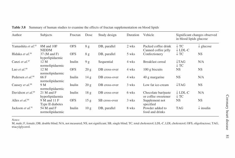
Coronary heart disease
81
Table 3.8 Summary of human studies to examine the effects of fructan supplementation on blood lipids
Author Subjects Fructan Dose Study design Duration Vehicle Significant changes observedin blood lipids glucose
Yamashita et al.83 8M and 10F OFS 8 g DB, parallel 2 wks Packed coffee drink ↓ TC ↓ glucoseNIDDM Canned coffee jelly ↓ LDL-C
Hidaka et al.84 37 (M and F) OFS 8 g DB, parallel 5 wks Confectionery ↓ TC NShyperlipidaemic
Canzi et al.87 12 M Inulin 9 g Sequential 4 wks Breakfast cereal ↓TAG N/Anormolipidaemic ↓ TC
Luo et al.89 12 M OFS 20 g DB cross-over 4 wks 100 g biscuits NS NSnormolipidaemic
Pedersen et al.90 66 F Inulin 14 g DB cross-over 4 wks 40 g margarine NS N/Anormolipidaemic
Causey et al.88 9 M Inulin 20 g DB cross-over 3 wks Low fat ice-cream ↓TAG NSnormolipidaemic
Davidson et al.85 21 M and F Inulin 18 g DB cross-over 6 wks Chocolate bar/paste ↓ LDL-C N/Ahyperlipidaemia or coffee sweetener ↓ TC
Alles et al.86 9 M and 11 F OFS 15 g SB cross-over 3 wks Supplement not NS NSType II diabetes specified
Jackson et al.91 54 M and F Inulin 10 g DB, parallel 8 wks Powder added to TAG ↓ insulinnormolipidaemic food and drinks
Notes:M, male; F, female; DB, double blind; N/A, not measured; NS, not significant; SB, single blind; TC, total cholesterol; LDL-C, LDL cholesterol; OFS, oligofructose; TAG,triacylglycerol.
82 Functional dairy products
factor for CHD.2 Data from studies in rats have shown a 40% reduction inpostprandial TAG concentrations when diets containing 10% OFS (w/v) werefed.92 However, very little information regarding the effect of prebiotics onpostprandial lipaemia in human subjects are available, although one recent studyin middle-aged subjects has shown no effect of inulin treatment on post-prandialTAG levels.93
The marked reduction in fasting lipid levels, notably TAGs, observed in animalstudies have not been consistently reproduced in human subjects. Only two studiesin normolipidaemic subjects have shown significant reductions in fasting TAGlevels with inulin,87, 88 with one study showing a significant effect of inulintreatment over time on fasting TAG levels compared with the placebo group.91 Theamount of fructans used in the human studies in Table 3.8 varies between 9 g and20 g and this amount is small compared to that which is used in animal studies (50–200 g per kg of rat chow of OFS),94 which is equivalent to a dose in humans ofapproximately 50–80 g of OFS/inulin per day. The prebiotic nature of OFS andinulin restricts its dosage in humans to 15–20 g per day since doses greater than thiscan cause gastrointestinal symptoms such as stomach cramps, flatulence anddiarrhoea.90 It is not known whether, at the levels used in human studies, significanteffects would be observed in animals.95
The types of food vehicles used to increase the amount of OFS/inulin in the dietdiffer. In the case of Luo et al.,89 100 g of biscuits were eaten every day and forPedersen et al.,90 40 g of margarine was consumed which may have contributed tothe negative findings in blood lipids. In the case of Davidson et al.,85 significantchanges in total and LDL cholesterol levels were observed over six weeks withinulin in comparison with the placebo (sugar). The percentage change in each ofthe lipid parameters was calculated over each of the six-week treatment periodsand, unexpectedly, there was an increase in total cholesterol, LDL cholesterol andTAG during the placebo phase. Non-significant falls in these variables wereobserved during inulin treatment and so when the net changes in the variables werecalculated (change during inulin minus change during control treatment) therewere significant differences in total and LDL cholesterol between the two treat-ments. The authors attributed the increase in total and LDL cholesterol levels in theplacebo phase to be due to the increased intake of SFAs in the chocolate productsthat were used as two of the vehicles in the study.85 In later studies, the use of inulinin its powder form enabled it to be added to many of the foods eaten in the subjects’normal diet without any need for dietary advice, thus avoiding changes in bodyweight. Since inulin has water binding properties, in its powder form it could beadded to orange juice, tea, coffee, yoghurt and soup.91
The significant relationship between subjects’ initial TAG concentration andpercentage change in TAG levels over the eight-week study demonstrated byJackson et al.91 lends support to the hypothesis that initial TAG levels could beimportant in determining the degree of the TAG response to inulin. The lack ofresponse in some individuals may be as a result of them being less responsive toinulin, variations in their background diet, or non-compliance with the studyprotocol. Speculation as to possible reasons for variability in response would be
Coronary heart disease 83
aided by a better understanding of the mechanism of action of inulin on plasmaTAG levels.
The length of the supplementation period used in the studies in Table 3.8 maybe another factor for inconsistent findings in changes in TAG levels in humansubjects with inulin. The studies were conducted over two to eight weeks, withsignificant effects occurring in only two studies conducted over three to fourweeks.87, 88 The lack of lipid lowering noted in the studies of Alles et al.,86
Pedersen et al.90 and Luo et al.89 may be due to insufficient duration of supplemen-tation. In the study of Jackson et al.,91 a trend for TAG lowering on inulin treatmentwas seen some time between four and eight weeks and this reflects the time neededfor the composition of the gut microflora to be modified. A four-week wash-outseemed to be sufficient for the TAG concentrations to return to baseline values.This may provide an explanation for the significant findings in TAG levels in thestudies of Canzi et al.87 and Causey et al.88 who used 3–4 week sequential andcross-over designs with very short wash-out periods.
While some of the studies, to date, support beneficial effects of inulin on plasmaTAG, the findings are by no means consistent and more work is required to provideconvincing evidence of the lipid-lowering consequences of prebiotic ingestion.
3.6.3 Mechanism of lipid lowering by prebioticsPrebiotics have been shown to be an ideal substrate for the health-promotingbacteria in the colon, notably bifidobacteria and lactobacilli.96 During the fermen-tation process a number of byproducts are produced including gases (H
2S, CO
2, H
2,
CH4), lactate and SCFAs (acetate, butyrate and propionate). The SCFAs, acetate
and propionate enter the portal blood stream, where they are utilised by the liver.Acetate is converted to acetyl CoA in the liver and acts as a lipogenic substrate forde novo lipogenesis, whereas propionate has been reported to inhibit lipidsynthesis.97, 98 Butyrate, on the other hand, is taken up by the large intestinal cells(colonocytes) and has been shown to protect against tumour formation in the gut.99
The type of SCFAs that are produced during the fermentation process is dependenton the microflora which can be stimulated by the prebiotic. Inulin has been shownto increase both acetate and butyrate levels, whereas synthetically producedprebiotics, for example galacto-oligosaccharides, increase the production of acetateand propionate and xylo-oligosaccharides increase acetate only.99
Inulin and OFS have been extensively studied to determine the mechanism ofaction of prebiotics in animals. Early in vitro studies using isolated rat hepatocytessuggested that the hypolipaemic action of OFS was associated with the inhibitionof de novo cholesterol synthesis by the SCFA propionate following impairment ofacetate utilisation by the liver for de novo lipogenesis.98 This is in agreement withhuman studies in which rectal infusions of acetate and propionate resulted inpropionate inhibiting the incorporation of acetate into TAGs released from theliver.100 Fiordaliso et al.101 demonstrated significant reductions in plasma TAGs,phospholipids and cholesterol in normolipidaemic rats fed a rat chow diet contain-ing 10% (w/v) OFS. The TAG-lowering effect was demonstrated after only one
84 Functional dairy products
week of OFS and was associated with a reduction in VLDL secretion. TAG andphospholipids are synthesised in the liver by esterification of fatty acids andglycerol-3-phosphate before being made available for assembly into VLDL,suggesting that the hypolipidaemic effect of OFS may be occurring in the liver.The reduction observed in cholesterol levels in the rats was only demonstratedafter long-term feeding (16 weeks) of OFS. Recent evidence has suggested that theTAG lowering effect of OFS occurs via reduction in VLDL TAG secretion fromthe liver due to the reduction in activity of all lipogenic enzymes (acetyl-CoAcarboxylase, fatty acid synthase, malic enzyme, ATP citrate lyase and glucose- 6-phosphate dehydrogenase), and in the case of fatty acid synthase, via modificationof lipogenic gene expression (see Fig. 3.5).102
3.6.4 The effect of prebiotics on glucose and insulin levelsVery little is known about the effects of prebiotics on fasting insulin and glucoselevels in humans. Of the supplementation studies conducted in humans, a signifi-cant reduction in glucose was observed in NIDDM subjects83 and a trend for areduction in glucose was observed in hyperlipidaemic subjects84 with OFS.However, a recent study has reported no effect of OFS on blood glucose levels intype II diabetics.86 A significant reduction in insulin levels was observed in healthymiddle-aged subjects with inulin, although this was not accompanied by changesin plasma glucose levels.91 The effect of the ingestion of acute test meals containingOFS on blood glucose, insulin and C-peptide levels in healthy adults showed atrend for a lower glycaemic response and peak insulin levels following the OFSenriched meals.103
The mechanism of action of prebiotics on lowering glucose and insulin levelshas been proposed to be associated with the SCFAs, especially propionate. Asignificant reduction in post-prandial glucose concentrations was observedfollowing both acute and chronic intakes of propionate-enriched bread.104 Theeffect of propionate intake on post-prandial insulin levels was not investigated. Arecent animal study has shown an attenuation of both post-prandial insulin andglucose levels following four weeks of feeding with OFS. These effects wereattributed to the actions of OFS on the secretion of the gut hormones, glucose-dependent insulinotropic polypeptide (GIP) and glucagon-like peptide 1 (GLP-1).92
These hormones are secreted from the small intestine (GIP) and the terminal ileumand colon (GLP-1) and contribute to the secretion of insulin following a meal in thepresence of raised glucose levels.105
In summary, the mechanisms of action of prebiotics, especially inulin and OFS,have been determined largely from animal studies. Present data suggest inhibitionof de novo lipogenesis as the primary mode of action of prebiotics in mediatingtheir lipid-lowering effects via down-regulation of the enzymes involved. If this isthe case, more modest or inconsistent effects might be expected in humans, inwhom de novo lipogenesis is extremely low, or variable depending on theirbackground diet. In animal studies, rats are fed a diet that is low in fat and high incarbohydrate and so de novo lipogenesis is an up-regulated pathway in these
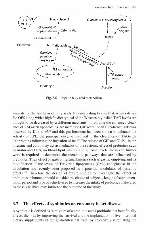
Coronary heart disease 85
Fig. 3.5 Hepatic fatty acid metabolism.
animals for the synthesis of fatty acids. It is interesting to note that, when rats arefed OFS along with a high fat diet typical of the Western-style diet, TAG levels arethought to be decreased by a different mechanism involving the enhanced clear-ance of TAG-rich lipoproteins. An increased GIP secretion in OFS-treated rats wasobserved by Kok et al.92 and this gut hormone has been shown to enhance theactivity of LPL, the principal enzyme involved in the clearance of TAG-richlipoproteins following the ingestion of fat.106 The release of GIP and GLP-1 in theintestine and colon may act as mediators of the systemic effect of prebiotics suchas inulin and OFS, on blood lipid, insulin and glucose levels. However, furtherwork is required to determine the metabolic pathways that are influenced byprebiotics. Their effect on gastrointestinal kinetics such as gastric emptying and itsmodification of the levels of TAG-rich lipoproteins (CMs) and glucose in thecirculation has recently been proposed as a potential modulator of systemiceffects.107 Therefore the design of future studies to investigate the effect ofprebiotics in humans should consider the choice of subjects, length of supplemen-tation period and type of vehicle used to increase the intake of prebiotics in the diet,as these variables may influence the outcome of the study.
3.7 The effects of synbiotics on coronary heart disease
A synbiotic is defined as ‘a mixture of a prebiotic and a probiotic that beneficiallyaffects the host by improving the survival and the implantation of live microbialdietary supplements in the gastrointestinal tract, by selectively stimulating the
86 Functional dairy products
growth and/or by activating the metabolism of one or a limited number of healthpromoting bacteria’.108 The use of synbiotics as functional food ingredients is anew and developing area and very few human studies have been performedlooking at their effect on risk factors for CHD. Research conducted so far withsynbiotics have looked at their effect on the composition of the gut microflora. Inone study in healthy subjects, a fermented milk product containing a Bifidobacteriumspp. with or without 18 g of inulin was given daily for 12 days.109 The authorsconcluded that the administration of the fermented milk product (probiotic)substantially increased the proportion of bifidobacteria in the gut, but that thisincrease was not enhanced by the addition of 18 g of inulin. The composition of thegut microflora was then assessed two weeks after completing the supplementationperiod and it was found that subjects who received the fermented milk product andinulin maintained their bifidobacterial population in the gut compared with thesubjects receiving the fermented milk product only. Although a synergistic effecton bifidobacteria in the gut was not observed with the synbiotic, these resultssuggest that either there was better implantation of the probiotic or a prebioticeffect on indigenous bifidobacteria.109 Maintenance of high numbers ofbifidobacteria in the gut may be beneficial in terms of healthy gut function;however, its effect on the lowering of blood lipid levels remains to be investigated.A more recent study has shown that a lower dose of prebiotic (2.75 g) added to alactobacillus fermented milk was able to increase significantly the number ofbifidobacteria when fed over a seven-week period in healthy human subjects.108 Ifthis effect was a result of the synbiotic product used in this study, the use of lowerdoses of prebiotics that can be used in synbiotic preparations should help to reducegastrointestinal complaints observed when a prebiotic is used alone and improvethe acceptability of these types of products by the general public.
3.8 Future trends
In recent years, a number of food manufacturers in the USA and Europe have beeninterested in the commercial opportunities for foodstuffs containing health-promoting probiotics and prebiotics. These food ingredients have received attentionfor their beneficial effects on the gut microflora and links to their systemic effectson the lowering of lipids known to be risk factors for CHD, notably cholesterol andTAG. Early attention was given to the incorporation of probiotics into dairyproducts such as fermented milk products (Yakult and Actimel Orange milk drink)and yoghurts, whose market is currently estimated at US$2 billion.110 Prebioticsare currently gaining interest and this has opened up the market away from thedairy industry to other areas of the food industry since prebiotics can be baked intobread, cereals, cakes, biscuits and even added to soups. Synbiotics have alsogenerated interest with some food manufacturers who are exploiting the effects ofcombining a prebiotic with a probiotic (Symbalance and PROBIOTIC plusoligofructose).
While consumers are interested in the concept of improving their health and
Coronary heart disease 87
well-being through diet, this is not quite so straightforward as originally thought.Recent bad press regarding food safety and the introduction of GM foods has madethe public sceptical about new ingredients in foods. For progress to be made, theconsumers need to be educated about the various health benefits and how they willbe able to use these products in their own diet without adverse consequences.Although the introduction of probiotics onto the supermarket shelves are slowlybeing accepted in the UK population, carefully controlled nutrition studies need tobe carried out to determine the beneficial effects of prebiotics, probiotics andsynbiotics before substantial health claims can be made.
To make these foods attractive to the consumer, the products need to be pricedin such a way that they are accessible to the general public. The low doses ofprebiotics and probiotics needed to help maintain a healthy gut microflora shouldbe made available to the general public, whereas products that contain higheramounts of prebiotics in order to help reduce blood lipids will need to be restrictedin order for the appropriate population group to be targeted.
3.9 Sources of further information and advice
3.9.1 General biochemistry and metabolic regulation text booksDEVLIN T M Textbook of Biochemistry with Clinical Correlations, 3rd edn, New York, John
Wiley, 1992.FRAYN K N Metabolic Regulation: A Human Perspective, London, Portland Press, 1996.
3.9.2 Published reports on dietary intake and coronary heart diseaseDepartment of Health, Nutritional Aspects of Cardiovascular Disease, Report on Health and
Social Subjects, no. 46, London, HMSO.
3.9.3 Prebiotic, probiotic and synbiotic informationADAMS C A ‘Nutricines’, Food Components in Health and Nutrition, Nottingham, Notting-
ham University Press, 1999.HASLER C M and WILLIAMS C M ‘Prebiotics and probiotics: where are we today?’, BJN, 80,
supp. 2.LEEDS A R and ROWLAND I R Gut Flora and Health: Past, Present and Future, International
Congress and Symposium Series 219, Royal Society of Medicine, 1996.SADLER M J and SALTMARSH M Functional Foods: The Consumer, the Products and the
Evidence, Bath, Bookcraft, 1997.
3.10 References1 KEYS A ‘Coronary artery disease in seven countries’, Circulation (1970), 41, I1–I211.2 PATSCH J R, MEISENBÖCK G, HOPFERWEISER T, MULHBERGER V, KNAPP E, DUNN J K,
GOTTO A M and PATSCH W. ‘Relation of triglyceride metabolism and coronary arterydisease: studies in the postprandial state’, Arterioscler Thromb (1992), 12, 1336–1345.
88 Functional dairy products
3 GRIFFIN B A and ZAMPELAS A ‘Influence of dietary fatty acids on the atherogeniclipoprotein phenotype’, Nut Res Rev (1995), 8, 1–26.
4 GIBSON G R and BEAUMONT A ‘An overview of human colonic bacteriology in healthand disease’. In A R Leeds and I R Rowland (eds), pp. 3–11, Gut Flora and Health:Past, Present and Future, International Congress and Symposium Series 219, London,Royal Society of Medicine (1996).
5 KEYS A Seven Countries: A Multivariate Analysis of Death and Coronary HeartDisease, Harvard University Press, Cambridge, MA (1980).
6 WORLD HEALTH ORGANISATION, ‘World Health Statistics Annual 1989’, Geneva,WHO (1989).
7 MARTIN M J, BROWNER W S, WENTWORTH J, HULLEY S B. and KULER LH. ‘Serumcholesterol, blood pressure and mortality: implications from a cohort of 361,662 men’,Lancet (1986) 2, 933–936.
8 SHEPHERD J, COBBE S M, FORD I, ISLES C G, LORIMER A R, MACFARLANE P W, MCKILLOPJ H. and PACKARD C J ‘Prevention of coronary heart disease with pravastatin in men withhypercholesterolaemia’, New Engl J Med (1995) 333(20), 1301–1307.
9 SHAPER A G, POCOCK S J, PHILIPS A N and WALKER M ‘Identifying men at high risk ofheart attacks – strategy for use in general practice’, BMJ (1986) 293 (6545), 474–479.
10 FIELDING P E and FIELDING C J ‘Dynamics of lipoprotein transport in the circulatorysystem’. In D E Vance and J Vance (eds), pp. 427–459, Biochemistry of Lipids,Lipoproteins and Membranes, Amsterdam, Elsevier (1991).
11 SCHNEIDER W J ‘Removal of lipoproteins from plasma’. In D E Vance and J Vance(eds), pp. 461–487, Biochemistry of Lipids, Lipoproteins and Membranes, Amster-dam, Elsevier (1991).
12 DEVLIN T M Textbook of Biochemistry with Clinical Correlations, 3rd edn, New York,John Wiley (1992).
13 REDGRAVE T G Gastrointestinal Physiology IV, Baltimore, University Park Press(1983).
14 BRASAEMLE D L, CORNLEY-MOSS K and BENSADOUN A ‘Hepatic lipase treatment ofchylomicron remnants increases exposure of apolipoprotein E’, J Lipid Res (1993) 34,455–463.
15 KARPE F and HULTIN M ‘Endogenous triglyceride-rich lipoproteins accumulate in ratplasma when competing with a triglyceride emulsion for a common lipolytic pathway’,J Lipid Res (1995) 36, 1557–1566.
16 GRIFFIN B A and PACKARD C J ‘Metabolism of VLDL and LDL subclasses’, Curr OpinLipidol (1994) 5, 200–206.
17 TALL A R ‘Plasma cholesterol ester transfer protein’, J Lipid Res (1993) 27, 361–367.18 GENSINI G F, COMEGLIO M and COLELLA A ‘Classical risk factors and emerging elements
in the risk profile for coronary artery disease’, Eur Heart J (1998) 19 (supp. A), A52–A61.
19 ASSMANN G, CULLEN P and SCHULTE H ‘The Münster Heart Study (PROCAM): Resultsof follow-up at 8 years’, Eur Heart J (1998) 19 (supp. A), A2–A11.
20 AUSTIN M A ‘Plasma triglyceride and coronary artery disease’, Arterioscl Thromb(1991) 11, 2–14.
21 WILSON P W F ‘Established risk factors and coronary artery disease: the Framinghamstudy’, Am J Hypertens (1994) 7, 7S–12S.
22 O’MEARA N M, LEWIS G F, CABANA V G, IVERIUS P H, GETZ G S and POLONSKY K S ‘Roleof basal triglyceride and high density lipoprotein in determination of postprandial lipidand lipoprotein responses’, J Clin Endocrin Metab (1991) 75, 465–471.
23 KARPE F, STEINER G, UFFELMAN K, OLIVECRONA T and HAMSTEN A ‘Postprandiallipoproteins and progression of coronary heart disease’, Atherosclerosis (1994) 106,83–97.
24 ZILVERSMIT D ‘Atherogenesis: a postprandial phenomenon’, Circulation (1979) 60,473–485.
Coronary heart disease 89
25 MEISENBÖCK G and PATSCH J R ‘Coronary artery disease: synergy of triglyceride-richlipoproteins and HDL’, Cardiovascular Risk Factors (1991) 1, 293–299.
26 AUSTIN M A, BRESLOW J L, HENNEKENS C H, BURLING J E, WILLETT W C and KRAUSS R M
‘Low density lipoprotein subclass patterns and risk of myocardial infarction’, JAMA(1988) 260, 1917–1921.
27 AUSTIN M A, HOKANSON J E and BRUNZELL J D ‘Characterisation of lowdensitylipoprotein subclasses: methodological approaches and clinical relevance’, Curr OpinLipidol (1994) 5, 395–403.
28 GRIFFIN B A ‘Low density lipoprotein heterogeneity’, Ballière’s Clinical Endocrinologyand Metabolism (1995) 9, 687–703.
29 CHAPMAN M J, GUÉRIN M and BRUCKERT E ‘Atherogenic, dense lowdensity lipoproteins:pathophysiology and new therapeutic approaches’, Eur Heart J (1998) 19 (supp. A),A24–A30.
30 REAVEN G M, CHEN Y-D I, JEPPENSEN J, MAHEUX P and KRAUSS R M ‘Insulin resistanceand hyperinsulinaemia in individuals with small, dense, low density lipoproteinparticles’, J Clin Invest (1993) 92, 141–146.
31 MENSINK R P ‘Effects of individual saturated fatty acids on serum lipids and lipoproteinconcentrations’, Am J Clin Nutr (1993) 57(s), 711S–714S.
32 HEGSTED D M, ANSMAN L M, JOHNSON J A and DALLAL G E ‘Dietary fat and serum lipids:an evaluation of the experimental data’, Am J Clin Nutr (1993) 57 875–883.
33 HAYES K C and KHOSLA P ‘Dietary fatty acids thresholds and cholesterolaemia’,FASEB J (1992) 6, 2600–2607.
34 DEPARTMENT OF HEALTH, Nutritional Aspects of Cardiovascular Disease: Report ofthe Cardiovascular Review Group Committee on Medical Aspects of Food Policy,London, HMSO (1994). Report on Health and Social Subjects 46.
35 GREGORY J, FOSTER K, TYLER H and WISEMAN M The Dietary and Nutritional Surveyof British Adults: A Survey of the Dietary Behaviour, Nutritional Status and BloodPressure of Adults aged 16 to 64 living in Great Britain’, Office of Population Censusand Surveys, Social Survey Division, London, HMSO (1990).
36 GRUNDY S M, NIX D, WHELAN M F and FRANKLIN L ‘Comparison of three cholesterollowering diets in normolipidaemic men’, JAMA (1986) 256, 2351.
37 BERRY E M ‘The effects of nutrients on lipoprotein susceptibility to oxidation’, CurrentOpinions in Lipidology (1992) 3, 5–11.
38 MENSINK R P and KATAN M B ‘Effect of dietary trans fatty acids in high density and lowdensity lipoprotein cholesterol levels in healthy subjects’, N Engl J Med (1990) 323,439–445.
39 DYBERG J and BANG H O ‘Haemostatic function and platelet polyunsaturated fatty acidsin Eskimos’, Lancet (1979) 2, 433–435.
40 BURR M L, FEHILY A M, GILBERT J F et al. ‘Effects of changes of fat, fish and fibre intakeson frequency of myocardial infarctions: DART Study’, Lancet (1989) 2, 757–762.
41 HARRIS W S ‘n-3 Fatty acids and serum lipoproteins: human studies’, Am J Clin Nutr(1997) 65 (S), 1645S–1654S.
42 SISCOVICK D S, RAGHUNATHAN T E, KING I and WEINMAN S ‘Dietary intake and cellmembrane levels of long chain n-3 polyunsaturated fatty acids and the risk of primarycardiac arrest’, JAMA (1996) 274, 1363–1367.
43 DE DECKERE E A M, KORVER O, VERSCHUREN P M and KATAN M B ‘Health aspects of fishand n-3 polyunsaturated fatty acids from plant and marine origin’, Euro J Clin Nutr(1998) 52, 749–753.
44 EEC SCIENTIFIC COMMITTEE FOR FOOD, Reference Nutrient Intakes for the EuropeanCommunity, Brussels, EC (1992).
45 MENSINK R P and KATAN M B ‘Effects of monosaturated fatty acids versus complexcarbohydrates on high density lipoproteins in healthy men and women’, Lancet (1987)1, 122–125.
90 Functional dairy products
46 GRUNDY S M ‘Comparison of monounsaturated fatty acids and carbohydrates forlowering plasma cholesterol’, N Engl J Med (1986) 314, 745–748.
47 MORRIS J N, MARR J W and CLAYTON D G ‘Diet and heart: a postscript’. BMJ (1977) 2,1307–1314.
48 KUSHI L H, LEW R A, STARE F J et al., ‘Diet and 20-year mortality from coronary heartdisease: the Ireland–Boston Diet–Heart Study’, N Engl J Med (1985) 312, 811–818.
49 RIEMERSMA R A, WOOD D A, MACINTYRE C C, ELTON R A, GEY K F and OLIVER M F ‘Riskof angina pectoris and plasma concentrations of vitamin A, C and E and carotene’,Lancet (1991) 337, 1–5.
50 STEPHENS N G, PARSONS A, SCHOFIELD P M, KELLY F and CHEESEMAN K ‘Randomisedcontrolled trial of vitamin E in patients with coronary disease: Cambridge HeartAntioxidant Study (CHAOS)’ (1996) Lancet, 348, 781–786.
51 ELLIOT P, DYER A and STAMLER J ‘Correcting for regression dilution in INTERSALT’,Lancet (1993) 342, 1123.
52 LAW M R, FROST C D and WALD N J ‘By how much does dietary salt reduction lowerblood pressure? I-III- Analysis of observational data among populations’, BMJ (1991)302, 811–824.
53 GAZIANO J M, BURING J E, BRESLOW J L, GOLDHABER S Z, ROSNER B , VANDENBURGH
M, WILLIET W C and HENNEKENS C H ‘Moderate alcohol intake, increased levels of highdensity lipoprotein and its subfractions, and decreased risk of myocardial infarction’,New Eng J Med (1993) 329, 1829–1834.
54 LAW M and WALD N ‘Why heart disease mortality is low in France: the time lagexplanation’, BMJ (1999) 318, 1471–1480.
55 URGERT R, MEYBOOM S, KUILMAN M, REXWINKEL H, VISSERS M N, KLERK M and KATAN
M B ‘Comparison of effect of cafetiere and filter coffee on serum concentrations of liveraminotransferases and lipids: six month randomised controlled trial’, BMJ (1996) 313,1362–1366.
56 MANN G V and SPOERRY A ‘Studies of a surfactant and cholesteremia in the Maasai’,Am J Clin Nutr (1974) 27, 464–469.
57 MANN G V ‘A factor in yoghurt which lowers cholesteremia in man’, Atherosclerosis(1977) 26, 335–340.
58 HOWARD A N and MARKS J ‘Effect of milk products on serumcholesterol’, Lancet(1979) II, 957.
59 HEPNER G, FRIED R, ST JEOR S, FUSETTI L and MORIN R ‘Hypocholesterolemic effect ofyogurt and milk’, Am J Clin Nutr (1979) 32, 19–24.
60 SESSIONS V A, LOVEGROVE J A, TAYLOR G R J, DEAN T S, WILLIAMS C M, SANDERS T AB,MACDONALD I and SALTER A ‘The effects of a new fermented milk product on totalplasma cholesterol: LDL cholesterol and apolipoprotein B concentrations in middleaged men and women’, Proceedings of the Nutrition Society (1997) 56, 120A.
61 THOMPSON L U, JENKINS D J A, AMER V, REICHERT R, JENKINS A and KAMULSKY J ‘Theeffect of fermented and unfermented milks on serum cholesterol’, Am J Clin Nutr(1982) 36, 1106–1111.
62 MASSEY L K ‘Effect of changing milk and yogurt consumption on human nutrient intakeand serum lipoproteins’, J Dairy Sci (1984) 67, 255–262.
63 MCNAMARA D J, LOWELL A E and SABB J E ‘Effect of yogurt intake on plasma lipid andlipoprotein levels in normolipidemic males’, Atherosclerosis (1989) 79, 167–171.
64 JASPERS D A, MASSEY L K and LUEDECKE L O ‘Effect of consuming yogurts preparedwith three culture strains on human serum lipoproteins’, J Food Sci (1984) 49, 1178–1181.
65 AGERBAEK M, GERDES L U and RICHELSEN B ‘Hypocholesterolaemic effects of a newproduct in healthy middle-aged men’, Eur J Clin Nutr (1995) 49, 346–352.
66 RICHELSEN B, KRISTENSEN K and PEDERSEN S B ‘Long-term (6 months) effect of a newfermented milk product on the level of plasma lipoproteins: a placebo-controlled anddouble blind study’, Eur J Clin Nutr (1996) 50, 811–815.
Coronary heart disease 91
67 BERTOLAMI M C ‘Evaluation of the effects of a new fermented milk product (Gaio) onprimary hypercholesterolemia’, Eur J Clin Nutr (1999) 53, 97–101.
68 ROSSOUW J E, BURGER E-M, VAN DER VYVER P and FERREIRA J J ‘The effect of skim milkyoghurt, and full cream milk on human serum lipids’, Am J Clin Nutr (1981) 34, 351–356.
69 HUIS IN’T VELD J H J and SHORTT C ‘Selection criteria for probiotic microorganisms’.In A.R. Leeds and I.R. Rowland (eds), pp. 27–36, Gut Flora and Health: Past, Presentand Future, International Congress and Symposium Series No. 219, London, NewYork, Royal Society of Medicine Press (1996).
70 HYLEMOND P B ‘Metabolism of bile acids in intestinal microflora’. In H. Danielson andJ. Sjovall (eds), pp. 331–343, Sterols and Bile Acids, New York, Elsevier Science(1985).
71 GRILL J P, MANGINOT-DURR C, SCHNEIDER F and BALLONGUE J ‘Bifidobacteria andprobiotic effects: action of Bifidobacterium species on conjugated bile salts’, CurrMicrobiol (1995) 31, 23–27.
72 GILLILand S E, NELSON C R and MAXWELL C ‘Assimilation of cholesterol by Lactoba-cillus acidophilus’, Appl Environ Microbiol (1985) 49(2), 377–381.
73 DE SMET I, DE BOEVER P and VERSTRAETE W ‘Cholesterol lowering in pigs throughenhanced bacterial bile salt hydrolase activity’, BJN (1998) 79, 185–194.
74 MARTEAU P, GERHARDT M F, MYARA A, BOUVIER E, TRIVIN F and RAMBAUD J C‘Metabolism of bile salts by alimentary bacteria during transit in human smallintestine’, Microbiol Ecol in Health and Disease (1995) 8, 151–157.
75 GILLILand S E, NELSON C R and MAXWELL C ‘Assimilation of cholesterol by Lactoba-cillus acidophilus’, Appl and Environ Microbiol (1985) 49 (2), 377–381.
76 RASIC J L, VUJICIC I F, SKRINJAR M and VULIC M ‘Assimilation of cholesterol by somecultures of lactic acid bacteria and bifidobacteria’, Biotech Lett (1992) 14 (1), 39–44.
77 KLAVER F A M and VAN DER MEER R ‘The assumed assimilation of cholesterol bylactobacilli and Bifidobacterium bifidum is due to their bile salt-deconjugating activ-ity’, Appl Environ Microbiol (1993) 59(4), 1120–1124.
78 TAHRI K, CROCIANI J, BALLONGUE J and SCHNEIDER F ‘Effects of three strains ofbifidobacteria on cholesterol’, Lett Appl Microbiol (1995) 21, 149–151.
79 HOSONO A and TONO-OKA T ‘Binding of cholesterol with lactic acid bacterial cells’,Milchwissenschaft (1995) 50(20), 556–560.
80 GIBSON G R and ROBERFROID M B ‘Dietary modulation of the human colonic microbiota:introducing the concept of prebiotics’, J Nutr (1995) 125, 1401–1412.
81 DYSSELER P and HOFFEM D ‘Inulin, an alternative dietary fibre: properties andquantitative analysis’, Eur J Clin Nutr (1995) 49, S145–S152.
82 VAN LOO J, COUSSEMENT P, DE LEENHEER L, HOEBREGS H and SMITS G ‘On the presenceof inulin and oligofructose as natural ingredients in the Western diet’, Crit Rev FoodSci and Nutr (1995) 35, 525–552.
83 YAMASHITA K, KAWAI K and ITAKURA M ‘Effects of fructo-oligosaccharides on bloodglucose and serum lipids in diabetic subjects’, Nutr Res (1984) 4, 961–966.
84 HIDAKA H, TASHIRO Y and EIDA T ‘Proliferation of bifidobacteria by oligosaccharidesand their useful effect on human health’, Bifidobacteria Microflora (1991) 10, 65–79.
85 DAVIDSON M H, SYNECKI C, MAKI K C and DRENNEN K B ‘Effects of dietary inulin inserum lipids in men and women with hypercholesterolaemia’, Nutr Res (1998) 3, 503–517.
86 ALLES M S, DE ROOS N M, BAKX J C, VAN DE LISDONK E, ZOCK P L and HAUTVAST J GA J ‘Consumption of fructooligosaccharides does not favourably affect blood glucoseand serum lipid concentrations in patients with type 2 diabetes’, Am J Clin Nutr (1999)69, 64–69.
87 CANZI E, BRIGHENTI F, CASIRAGHI M C, DEL PUPPO E and FERRARI A ‘Prolongedconsumption of inulin in ready to eat breakfast cereals: effects on intestinal ecosystem,bowel habits and lipid metabolism’, Cost 92. Workshop on Dietary Fibre andFermentation in the Colon, Helsinki (1995).
92 Functional dairy products
88 CAUSEY J L, GALLAHER D D and SLAVIN J L ‘Effect of inulin consumption on lipid andglucose metabolism in healthy men with moderately elevated cholesterol’, FASEB(1998) 12, 4737.
89 LUO J, RIZKALLA S W, ALAMOWITCH C, BOUSSAIRI A, BLAYO A, BARRY J-L, LAFFITTEA, GUYON F, BORNET F R J and SLAMA G ‘Chronic consumption of short-chainfructooligosaccharides by healthy subjects decreased basal hepatic glucose productionbut no effect on insulinstimulated glucose metabolism’, Am J Clin Nutr (1996) 63, 939–945.
90 PEDERSEN A, SANDSTRÖM B and VAN AMELSVOORT J M M ‘The effect of ingestion ofinulin on blood lipids and gastrointestinal symptoms in healthy females’, BJN (1997)78, 215–222.
91 JACKSON K G, TAYLOR G R J, CLOHESSY A M and WILLIAMS C M ‘The effect of the dailyintake of inulin on fasting lipid, insulin and glucose concentrations in middle-aged menand women’, BJN (1999) 82, 23–30.
92 KOK N N, MORGAN L M, WILLIAMS C M, ROBERFROIOD M B, THISSEN J-P and DELZENNE
N M ‘Insulin, glucagon-like peptide 1, glucose dependent insulinotropic polypeptideand insulin-like growth factor 1 as putative mediators of the hypolipidemic effect ofoligofructose in rats’, J Nutr (1998) 128, 1099–1103.
93 WILLIAMS C M ‘Effects of inulin on blood lipids in humans’, J Nutr (1998) 7, S1471–S1473.
94 ROBERFROID M ‘Dietary fiber, inulin and oligofructose: a review comparing theirphysiological effects’, Crit Rev Food Sci and Nutr (1993) 33, 102–148.
95 DELZENNE N M, KOK N, FIORDALISO M-F, DEBOYSER D M, GOETHALS F M andROBERFROID R M ‘Dietary fructo-oligosaccharides modify lipid metabolism’, Am JClin Nutr (1993) 57, 820S.
96 GIBSON G R and MCCARTNEY A L ‘Modification of gut flora by dietary means’, BiochemSoc Trans (1998) 26, 222–228.
97 WOLEVER T M S, BRIGHENTI F, ROYALL D, JENKINS A L and JENKINS D J A ‘Effect of rectalinfusion of short chain fatty acids in human subjects’, Am J Gastroenterol (1989) 84,1027–1033.
98 DEMIGNÉ C, MORAND C, LEVRAT M-A, BESSON C, MOUNDRAS C and RÉMÉSEY C ‘Effectof propionate on fatty acid and cholesterol synthesis and on acetate metabolism inisolated rat hepatocytes’, BJN (1995) 74, 209–219.
99 VAN LOO J, CUMMINGS J, DELZENNE N, ENGLYST H, FRANCK A, HOPKINS M, KOK N,MACFARLANE G, NEWTON D, QUIGLEY M, ROBERFROID M, VAN VLIET T and VAN DENHEUVEL E ‘Functional food properties of nondigestible oligosaccharides: a consensusreport from the ENDO project (DGXII AIRII-CT94-1095)’, BJN (1999) 81, 121–132.
100 WOLEVER T M S, SPADAFORA P J, CUNNANE S C and PENCHARZ P B ‘Propionate inhibitsincorporation of colonic [1,2-13C]acetate into plasma lipids in humans’, Am J ClinNutr (1995) 61, 1241–1247.
101 FIORDALISO M-F, KOK N, DESAGER J-P, GOETHALS F, DEBOYSER D, ROBERFROID R M andDELZENNE N ‘Dietary oligofructose lowers triglycerides, phospholipids and choles-terol in serum and very low density lipoproteins in rats’, Lipids (1995) 30, 163–167.
102 DELZENNE N M and KOK N ‘Effect of non-digestible fermentable carbohydrates onhepatic fatty acid metabolism’, Biochem Soc Trans (1998) 26, 228–230.
103 RUMESSEN J J, BODE S, HAMBERG O and GUDMAND-HOYER E ‘Fructans of the Jerusalemartichokes: intestinal transport, absorption, fermentation, and influence on bloodglucose, insulin, and C-peptide responses in healthy subjects’, Am J Clin Nutr (1990)52, 675–681.
104 TODESCO T, RAO A V, BOSELLO O and JENKINS D J A ‘Propionate lowers blood glucoseand alters lipid metabolism in healthy subjects’, Am J Clin Nutr (1991) 54, 860–865.
105 MORGAN L M ‘The role of gastrointestinal hormones in carbohydrate and lipidmetabolism and homeostasis: effects of gastric inhibitory polypeptide and glucagon-like peptide-1’, Biochem Soc Trans (1998) 26, 216–222.
Coronary heart disease 93
106 KNAPPER J M, PUDDICOMBE S M, MORGAN L M and FLETCHER J M ‘Investigations intothe actions of glucose dependent insulinotrophic polypeptide and glucagon-likepeptide-1 (7-36) amide on lipoprotein lipase activity in explants of rat adipose tissue’,J Nutr (1995) 125, 183–188.
107 DELZENNE N ‘The hypolipidaemic effect of inulin: when animal studies help toapproach the human problem’, BJN (1999) 82, 3–4.
108 ROBERFROID M B ‘Prebiotics and synbiotics: concepts and nutritional properties’, BJN(1998) 80 (supp. 2), S197–S202.
109 BOUHNIK Y, FLOURIE B, RIOTTOT M, BISETTI N, GAILING M, GUIBERT A, BORNET F andRAMBAUD J ‘Effect of fructo-oligosaccharides ingestion on fecal bifidobacteria andselected metabolic indexes of colon carcinogenesis in healthy humans’, Nutrition andCancer (1996) 26, 21–29.
110 YOUNG J ‘European market developments in prebiotic- and probioticcontainingfoodstuffs’, BJN (1998) 80 (supp. 2), S231–S233.
111 BAZARRE T L, WU S L and YUHAS J A ‘Total and HDL-cholesterol concentrationfollowing yoghurt and calcium supplementation’, Nutritional Reports International(1983) 28, 1225–1232.
112 TAYLOR G B J and WILLIAMS C M ‘Effect of probiotics and prebiotics on blood lipids’,BJN (1998) 80 (supp. 5), S225–S230.
4
OsteoporosisR. Wood, Tufts University, USA
4.1 Introduction
Osteoporosis is a metabolic bone disease characterized by a decrease in bone mass(osteopenia) with decreased bone mineral density (BMD) and enlargement of bonespaces producing porosity and fragility, which leads to an increased risk of bonefracture. There are many different etiologies of osteoporosis, but the most commonis due to a universal age-dependent loss of bone in adult populations.
Proposed risk factors for osteoporosis include genetic, nutritional, lifestyle andendocrine factors. The role of genetic factors is illustrated by differences in BMDbetween races and sexes, and familial similarities in BMD. Nutritional factorsknown to influence the risk of osteoporosis include low calcium intake, highalcohol intake, high caffeine and high sodium intake. Lifestyle factors that cancontribute to osteoporosis include cigarette smoking and low physical activity.An important endocrine factor is estrogen deficiency (postmenopausal andoophorectomized women).
4.2 The epidemiology of osteoporosis
Osteoporosis is the most prevalent metabolic bone disease in the United States andin other developed countries. Osteoporosis is a cross-racial and emerging globalpublic health problem that especially affects older women, but also needs to berecognized as very common in older men. Osteoporosis-related skeletal fractures,especially of the hip, are a significant cause of morbidity and mortality in theelderly in both the USA and elsewhere. The prevalence of a vertebral fracture inwomen aged 65 years in the USA has been estimated to be 27% (Cooper et al.,
Osteoporosis 95
1993). Among elderly persons in the USA who sustain a hip fracture, 27% enter anursing home for the first time within a year of the event (Cooper, 1997). Beyondthe evident personal pain and suffering as well as family disruption brought aboutby these bone fractures, the added financial burden to the national health-care billis estimated to be 13 billion dollars per year in the USA (Ray et al., 1997). Giventhe rising number of elderly around the globe, development of an effectiveosteoporosis prevention program is a key factor in the management of this growingpublic health problem.
There are many environmental and genetic factors that have been shown to beassociated with an increased risk of osteoporotic fracture. A very importantdeterminant of the risk of fracture is low BMD. The prevalence of low femoralBMD of a representative sample of the elderly (50 years and older) in the USA hasbeen reported from the NHANES III survey (Looker et al., 1995, 1997). Based onthis sample, the number of older women in the USA with osteoporosis (defined asa bone mineral density <2.5 SD below the mean BMD of young adult non-Hispanicwhite women) was 6 million, while 1 million men could be classified withosteoporosis using the same young female BMD cut-off value. Thus, althoughosteoporosis is clearly more common in older women than older men, there is stilla substantial number of older men affected by this disease. Moreover, osteoporosisis a universal phenomenon of aging affecting all races, although currently morewhite women than blacks or Hispanics in the USA have osteoporosis (Looker etal., 1995). In the next 50 years, the proportion of elderly minority members in theUS population will double, leading to a proportionate increase in the osteoporoticburden in these groups. Likewise, by the year 2050, there will be an estimated 6.3million osteoporotic fractures worldwide and 50% of these will occur in Asia.Racial differences in hip fracture incidence are substantial. Differences in bonemass between the races, such as between whites and blacks, probably have animportant effect on the risk of bone fracture. However, other more subtle geneticfactors may also play a significant role in determining the risk of osteoporoticfracture at any given level of BMD.
4.2.1 Osteoporosis in African–AmericansIt is generally appreciated that black populations have high bone mineral densityand a low prevalence of osteoporotic fracture, despite often low dietary calciumintakes. This observation clearly indicates that there is more to the osteoporosisequation than calcium intake alone. There is very little systematic information onbone mineral density and fracture risk in African populations, but some informa-tion is available on African–American populations, which will be highlighted hereto indicate the potential impact of racial differences on bone health. On the whole,African–American (black) women have more dense bones than white women(Looker et al., 1995). As a consequence, the risk of osteoporosis is lower in blackwomen. Differences in bone mass between black and white men are also evident.For example, whole body bone mineral content is greater in black than in whitemen, even after controlling for lean body mass and height (Barondess et al., 1997).
96 Functional dairy products
Because of these racial differences resulting in higher average BMD, it is often notappreciated that osteoporosis is also an issue in black populations, albeit to less ofan extent than in white populations. For example, according to data from theNHANES III survey, there are approximately 1.7 million black women in the USAwith low femoral bone mineral density indicative of either osteopenia or a moresevere stage of bone loss, osteoporosis (Looker et al., 1995).
The question arises whether racial differences in bone mineral density reflectdifferences in the attainment of peak bone mass or differences in the rates of boneloss with aging. Although this question has not been fully settled, it appears thatdifferences in peak bone mass may be particularly important. In a relatively smallstudy of bone loss (Luckey et al., 1996) in African–American and white womenconducted in 122 white and 121 African–American women, BMD was measuredat six-month intervals in the forearm and spine over a three- to four-year period.These investigators found that, in early menopause, bone loss in the forearm wasfaster in white than in black women. A similar trend was also evident in the spine.This apparent difference in the loss of bone density in the spine of –2.2% per yearin whites compared with –1.3% per year in blacks was not, however, statisticallysignificant. In women more than five years postmenopausal, bone loss did notdiffer by ethnic group. These findings are consistent with the idea that the higherbone mineral density evident in blacks may largely reflect the attainment of agreater peak bone mass ending in early adulthood, although there could be somecontribution of slower rates of estrogen-dependent bone loss in the early post-menopausal years in black women. In another small study, Perry and colleagues(1996) also concurred in the finding of generally similar rates of bone loss in blackand white women.
African–American populations have lower rates of osteoporotic fracture thanage-matched white populations. A study in the Washington DC area of the USA(Farmer et al., 1984) used the National Hospital Discharge Survey for the years1974 to 1979 and found that the incidence of hip fracture for white women was1.5 times greater than for black females after age 40. Likewise, a retrospectivestudy from 1987 to 1989 among Tennessee Medicaid enrollees in the USA, age 65years and more, indicated that the incidence of non-vertebral fractures in blackswas only half of that in whites, and differences in the fracture rates were evident in13 different fracture sites examined (Griffin et al., 1992). Overall, findings inblacks support the notion that high BMD is associated with a low rate of bonefracture.
4.2.2 Osteoporosis in Asian populationsThe complexity of predicting osteoporotic bone fracture risk from calcium intakeor BMD measures alone is illustrated by comparing bone fracture risk in Asiancompared with white populations. Calcium intake is low in Asian populations, andmany Asians have low BMD. In a study of BMD of Hong Kong Chinese it wasobserved that women aged 60 and above have mean BMD 30% lower than youngwomen (Ho et al., 1999). Moreover, the age-dependent increase in prevalence of
Osteoporosis 97
spinal osteoporosis increased dramatically from 10% in the 50–59 age group to45% in the 60–69 group, and over half of the women over 70 had osteoporosis ofthe hip. Comparison of trabecular BMD, measured by quantitative computedtomography, between Japanese women and American women 20–85 years oldfound that BMD was lower in the Asian women and that this difference becamegreater with age (Ito et al., 1997). In premenopausal women, these investigatorsfound that BMD began to decrease at the age of 20, whereas in American womenpeak bone mass was sustained until 35. In late postmenopausal women, the rate ofbone loss was increased in the Japanese women, –2.2% vs –1.4%.
However, despite generally lower BMD, hip fracture rates are substantiallylower in Asian countries than in the USA (Ho et al., 1993). These differencesprobably represent real racial differences in propensity to fracture rather thanenvironmental factors or unintended data collection bias because rates of hipfracture are also lower in Chinese–Americans, Japanese–Americans and Korean–Americans compared with USA whites (Lauderdale et al., 1997).
Why is the general notion that lower BMD is associated with higher rates ofosteoporotic bone fracture not evident in the case of Asian populations? Onepossibility is racial (genetic) differences in bone architecture that influence thepropensity to fracture. It has been shown for example that Asian women haveshorter hip axis lengths than non-Asian women (Cummings et al., 1994). How-ever, it needs to be pointed out that, despite a substantially lower risk of sustaininga hip fracture in Asian populations, because of its huge population it has beenprojected that 50% of all hip fractures in the world will occur in Asia during thenext century (Lau and Cooper, 1996).
4.2.3 Osteoporosis in Hispanic populationsOwing to the changing demographic profiles in various parts of the globe, steepincreases in hip fracture incidences are expected in the future in both Asia andLatin America (Cooper et al., 1992). BMD of Hispanics is similar to non-Hispanicwhites. For example, there is no difference in BMD in Puerto Rican women in theproximal femur compared with a standard mixed US population (Haddock et al.,1996). In the USA, many of the studies of BMD and bone fracture have focused onMexican–Americans, the largest Hispanic group. Bone mineral content of Mexican–American children aged 3–18 is not different from that of non-Hispanic whites(Ellis et al., 1997).
However, despite similarities in BMD, available data on skeletal fracture ratesin Mexican–Americans indicate that they are less likely to sustain a hip fracture.Hip fracture rates and vertebral fracture rates in Mexican–Americans are aboutone-half those seen in non-Hispanic whites (Bauer and Deyo, 1987; Bauer, 1988).The factors influencing lower rates of skeletal fracture in Hispanic populations arepoorly defined. Nevertheless, observations of the discordance between BMD andfracture risk in Hispanic and Asian populations compared with white populationssuggest that a clear understanding of the factors that influence bone fracture riskwill be complex.
98 Functional dairy products
4.3 Dairy products, calcium intake and calcium absorption
Among the nutritional factors known to influence the risk of low BMD, low dietarycalcium intakes have received considerable research and public attention. Ingeneral, low BMD in older persons may result from a lower rate of net bone accrual(peak bone mass) achieved during childhood and young adulthood and/or arelatively increased rate of bone loss during aging. Dietary calcium intake has beenshown to influence both of these determinants of bone mass. Therefore, it isgenerally believed that a high calcium intake throughout the entire lifespan willachieve the greatest benefit in ameliorating the risks of osteoporosis. The Food andNutrition Board of the Institute of Medicine in the United States reflected thisnotion in their recommendations for the need of a high ‘Adequate Intake’ level fordietary Ca in all age groups. The current ‘adequate intake’ levels for dietarycalcium in the USA are shown in Table 4.1. On the other hand, actual intakes ofcalcium in the general population are relatively low when considered in the contextof recommended calcium intakes to maintain optimal bone health. As shown inTable 4.2, mean calcium intakes of various age–sex groups adjusted for day-to-dayvariation as determined by the 1994 USDA Continuing Survey of Food Intakes byIndividuals fall considerably below recommended calcium intake levels. Sincedairy products are the main source of calcium intake in the US population, lowcalcium intakes largely reflect relatively low dairy food intakes. Despite highdietary calcium needs in older people, consumption of milk-based foods in theUSA decreases as people age. Clearly, there is a great need for nutrition educationto convince large segments of the adult population that they need to alter theireating patterns to provide additional calcium intake. In addition, efforts by the foodindustry to provide more alternative calcium-fortified foods for groups who preferto minimize or not to consume milk-based dairy products are needed to fill thecalcium intake gap.
As a rule of thumb, about 20–30% of dietary calcium is absorbed. At low dietarycalcium intakes, the majority of calcium absorbed is through a vitamin D-dependent pathway. At high dietary calcium intakes, most of the calcium that isabsorbed is via a vitamin D-independent concentration-dependent pathway (Sheikhet al., 1988). The determinants of intestinal calcium absorption, especially anygenetic components, are poorly understood (Wood, 2000). The bioavailability ofcalcium can vary considerably depending on the dietary source of the calcium.High dietary fiber intake reduces the bioavailability of dietary calcium (Allen,1982; Knox et al., 1991). In general, a plant-based diet provides insufficientbioavailable dietary calcium to meet current recommendations (Weaver et al.,1999). It should be noted that, despite the fact that calcium bioavailability fromsome vegetables can be high (Heaney and Weaver, 1990), when one considers thegenerally low calcium density (calcium per serving) of many plant foods, itbecomes impractical in the absence of calcium supplementation to fully meet one’scalcium needs from non-dairy food sources alone.
It has been known for a long time that the absorption efficiency of a low doseof calcium decreases as a function of age (Bullamore et al., 1970). This probably
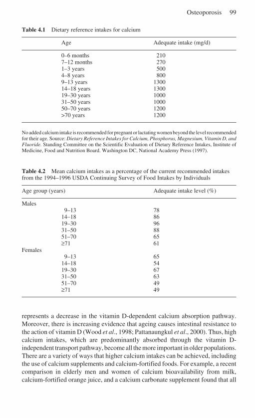
Osteoporosis 99
Table 4.1 Dietary reference intakes for calcium
Age Adequate intake (mg/d)
0–6 months 2107–12 months 2701–3 years 5004–8 years 8009–13 years 130014–18 years 130019–30 years 100031–50 years 100050–70 years 1200>70 years 1200
No added calcium intake is recommended for pregnant or lactating women beyond the level recommendedfor their age. Source: Dietary Reference Intakes for Calcium, Phosphorus, Magnesium, Vitamin D, andFluoride. Standing Committee on the Scientific Evaluation of Dietary Reference Intakes, Institute ofMedicine, Food and Nutrition Board. Washington DC, National Academy Press (1997).
Table 4.2 Mean calcium intakes as a percentage of the current recommended intakesfrom the 1994–1996 USDA Continuing Survey of Food Intakes by Individuals
Age group (years) Adequate intake level (%)
Males9–13 78
14–18 8619–30 9631–50 8851–70 65≥71 61
Females9–13 65
14–18 5419–30 6731–50 6351–70 49≥71 49
represents a decrease in the vitamin D-dependent calcium absorption pathway.Moreover, there is increasing evidence that ageing causes intestinal resistance tothe action of vitamin D (Wood et al., 1998; Pattanaungkul et al., 2000). Thus, highcalcium intakes, which are predominantly absorbed through the vitamin D-independent transport pathway, become all the more important in older populations.There are a variety of ways that higher calcium intakes can be achieved, includingthe use of calcium supplements and calcium-fortified foods. For example, a recentcomparison in elderly men and women of calcium bioavailability from milk,calcium-fortified orange juice, and a calcium carbonate supplement found that all
100 Functional dairy products
three calcium-rich products provided similar levels of bioavailable calcium (Martiniand Wood, 2002). An important point is that we must find more effective ways notonly to get more high-calcium products developed, but also to convince adultconsumers to include them in their diets.
4.4 Dairy products and osteoporosis
With the apparent exception of intermittent parathyroid hormone (Neer et al.,2001), treatments of osteoporosis do not result in increased BMD. Rather, mostagents used to treat osteoporosis are aimed at slowing the rate of bone loss, therebypreserving what BMD is left. In this regard, high dietary calcium intakes have aweak anti-bone resorptive effect and slow the rate of bone loss in elderly persons,especially in the presence of adequate vitamin D intakes (Dawson-Hughes et al.,1990, 1997; Dawson-Hughes, 1991). In perimenopausal women, the rapid declinein bone mass that occurs during the first 5 years after menopause cannot beprevented even with calcium supplementation supplying 1000 to 2000 mg Ca/d(Elders et al., 1991). Although several studies have shown that high calciumintakes can increase BMD in young children, it is less certain whether thebeneficial effects of calcium are merely to promote an earlier achievement of aperson’s genetically determined peak bone mass or actually enhances BMD.Regardless, it needs to be more generally recognized by the public that thecessation of high calcium intakes will probably result in a loss of accrued BMD(Lee et al., 1996).
4.4.1 Racial differences in calcium intakeVariations in calcium intake across populations largely reflect differences in dairyfood consumption. Populations living in areas where traditionally cows have beenkept for producing milk have relatively high dietary intakes of calcium-rich milkand cheese. Milk consumption varies markedly between countries. High milkintakes are evident in North America and northern Europe and low intakes aregenerally found in Asia.
4.4.2 Milk as a dietary source of calciumSince vitamin D-fortified milk provides both calcium and vitamin D in highamounts, it is a convenient food to achieve or maintain optimal nutritional bonehealth. The high calcium density of milk and milk-based food products can be animportant source of dietary calcium during childhood and early adulthood, a timewhen the achievement of peak bone mass is determined.
One would expect that it might be relatively easy to demonstrate that high intakeof dairy foods correlates with better bone health. However, this has not been thecase, and a recent review of these studies indicates that the majority of studies thathave investigated this question have found no effect of dairy food intake on bone
Osteoporosis 101
health (Weinsier and Krumdieck, 2000). One point of concern regarding thepotential of milk to enhance bone density is the relatively high protein content ofmilk, since a high protein intake increases urinary calcium loss (Kerstetter andAllen, 1990). However, in the elderly recent evidence suggests that increasingcalcium intake by increasing the consumption of milk compared with othercalcium-rich foods does not cause a greater increase in urinary calcium excretion(Martini and Wood, 2002).
4.5 Future trends: genetic markers of osteoporosis risk
Developments in the early part of the 21st century will probably centre around anincreased understanding of the functional role of specific genes in the developmentof complex chronic diseases, including osteoporosis. In addition, nutritionalsciences will further our understanding of the role of specific nutrients andfunctional food components in optimal bone health. It is likely that these findingswill be translated into the development of novel milk-based products to promotebone health. Some of these products may be targeted to populations that areparticularly at increased risk of developing osteoporosis.
Random mutations in DNA occur relatively frequently in the evolution of anorganism. Some of these mutations are carried forward in the evolutionarydevelopment of a species. Comparing the genetic sequence of a person’s DNAwith those of others reveals that it contains many mutations in various genes. Someof these genetic variations are of practical physiological significance and somemay prove useful as genetic markers of future disease risk. In these cases, thepromise of genetic testing is that early identification of these disease-risk biomarkerswill lead to early intervention to prevent or retard the development of the disease.Currently, these genetic markers are often measured as restriction fragment lengthpolymorphisms (RFLPs) that are caused by random mutations in DNA that lead tovariations in the ability of specific endonuclease enzymes to sever a givensequence of DNA. The aftermath of cutting a piece of DNA with these restrictionenzymes then results in DNA fragments of various physical lengths that indicatethe presence or absence of a given string of DNA code. Since mutations occurrandomly throughout the genome, and do not necessarily occur within the protein-coding region of a gene, these RFLPs do not necessarily have a functionalconsequence to the gene in which they are detected. Nevertheless, they canpotentially be useful as a marker of disease risk in certain circumstances, presum-ably because they are linked to other genetic mutations that are physiologicallyimportant. The advent of high-throughput genotyping methodologies will open upthe prospect of screening a large number of these genetic polymorphisms initiallyfor research purposes to identify disease relationships, and eventually to provideeach person and their medical care providers with a disease–risk profile based oncombinations of various gene markers.
Some examples of genetic markers that have been investigated to date inassociation studies of BMD or osteoporosis are the RFLPs in the vitamin D
102 Functional dairy products
receptor (VDR) gene (using BsmI, TaqI, ApaI and FokI restriction enzymes), theestrogen receptor gene (using XbaI and PvuII restriction enzymes) and nucleotiderepeat polymorphisms in the Sp1 binding site in the collagen type I alpha I genepromoter (Wood and Fleet, 1998). Mutations in the Sp1 binding site of the collagengene promoter could influence the regulation of collagen expression. Collagen isproduced by bone osteoblast cells and provides the organic matrix into whichinorganic calcium and phosphate are laid down to mineralize bone. In the case ofthe Bsm, Taq and Apa VDR RFLPs, there are no associated changes in the aminoacid composition of the VDR protein, yet some studies suggest that there is arelation to BMD (Cooper and Umbach, 1996). However, a FokI VDR RFLP isassociated with the expression of a shortened form of the VDR lacking the three N-terminal amino acids (Gross et al., 1996). The FokI VDR polymorphism resultsfrom a C-to-T transition (ACG is mutated to ATG) and creates an initiation codon(ATG) that is three codons proximal to a downstream start site usually used fortranslation of the VDR protein. The presence of the FokI site, designated byconvention as lower case f, indicates a genetic sequence that creates an newinitiation start site, allowing protein translation to initiate from the first ATG. Theallele lacking the site (designated F), results in initiation of translation from themore distal second ATG site. Thus, protein translation products from thesedifferent alleles differ by three amino acids with the f variant elongated because ofthe creation of an earlier start site (Gross et al., 1996). Moreover, the differinglength of the two VDR protein variants has a physiological consequence. The ffFokI polymorphism in the VDR gene has been associated with a 13% lower lumbarspine BMD and a greater rate of bone loss in the femoral neck (–4.7% vs –0.5%;ff vs FF) in postmenopausal Mexican–American women (Gross et al., 1996).Likewise, in Caucasian premenopausal women (Harris et al., 1997), the ff geno-type is associated with a 4% lower total body BMD and 12% lower femoral neckBMD. However, not all studies have found an influence of the FokI genotype(Eccleshall et al., 1997). Nevertheless, these studies are harbingers of things tocome. One day, when we have a far greater understanding of both the functionalconsequences and usefulness of various genetic markers of osteoporosis risk andthe influence of various environmental factors on the expression of the phenotype,we may be in a position to read our genetic barcode to determine our nutritionalrequirements and disease–risk profile, thereby allowing us to design a moreoptimal program to maintain better health.
4.6 Future trends: redefining a nutritional prescription foroptimal bone health
As our knowledge of nutrients and functional food components increases in thefuture, we will have an increased understanding of the nutritional ingredientsneeded for optimal bone health. To illustrate this potential trend, consider the caseof vitamin K. Epidemiological studies have implicated vitamin K as a potentially
Osteoporosis 103
important dietary factor that can affect BMD and the risk of bone fracture(Vermeer et al., 1995). There is accumulating evidence that several vitamin K-dependent proteins (osteocalcin, matrix Gla protein, protein S, gas6) could play animportant role in bone and cartilage metabolism (Ferland, 1998).
Currently, very few data exist on the adequacy of vitamin K status of differentage groups in relation to bone health (Binkley and Suttie, 1995). Most of theevidence used to support a relationship between vitamin K and bone is based onepidemiologic associations between a biomarker of vitamin K status (percentageof undercarboxylated osteocalcin) and BMD or risk of hip fracture. Assessment ofthe percentage of undercarboxylated osteocalcin is considered a sensitive measureof vitamin K nutritional status (Sokoll and Sadowski, 1996). Szulc and colleagues(1993) reported that circulating undercarboxylated osteocalcin was higher inelderly women and was a significant risk factor of hip fractures. In a three-yearfollow-up of the same cohort, these investigators confirmed that undercarboxylatedosteocalcin was still predictive of hip fracture after controlling for age andparathyroid hormone (PTH) (Szulc et al., 1996). In a 22-month prospective cohortstudy of 7598 elderly women, undercarboxylated osteocalcin, a measure ofvitamin K status, was predictive of hip fracture risk independent of femoral neckBMD, whereas total serum osteocalcin (a measure of bone turnover) was not(Vergnaud et al., 1997). Knapen et al. (1989) recently reported a significantassociation between percentage of undercarboxylated osteocalcin and BMD amongwomen within one to ten years of menopause, but not among other age groups.Significant associations between vitamin K intake or serum vitamin K levels andBMD have also been reported, although not consistently.
Kanai et al. (1997) reported that postmenopausal women with reduced BMDhad significantly lower plasma vitamin K than postmenopausal women withnormal bone density. In the Nurses’ Health Study, women in the second quintile ofvitamin K intake (dietary intakes of 107–142 µg/d) were at 40% lower risk of hipfracture compared with women in the lowest quintile of intake (Feskanich et al.,1999). Vitamin K intakes among elderly men and women in the FraminghamOsteoporosis Study (Framingham, Massachusetts) were not associated with BMDbut did relate to fracture rate (Booth et al., 2000). The above-mentioned epidemio-logical studies are highly suggestive of a role of vitamin K status on calcium andbone metabolism.
Currently, there is very little information concerning the effect of vitamin Ksupplementation on calcium and bone metabolism. Douglas et al. (1995) demon-strated in 20 postmenopausal women that 14 days of vitamin K supplementationincreased carboxylation of osteocalcin, a risk factor for hip fracture. Orimo et al.(1992) reported increases in bone mass among 272 postmenopausal womenfollowing supplementation with menaquinones (vitamin K
2) for 24–48 weeks. No
long-term (>12 months) clinical trials of the effects of vitamin K1 (phylloquinone)
supplementation on BMD or bone fracture have been reported. Ultimately, we willneed to await the final outcomes of ongoing clinical trials of vitamin K supplemen-tation and bone loss to strengthen our convictions concerning the importance ofhigh vitamin K intakes in protecting bone mass and its possible role in preventing
104 Functional dairy products
osteoporosis. However, the highly suggestive data of the relationship betweenvitamin K status and risk of osteoporotic fracture suggest that it may be animportant new nutrient in the armamentarium to combat osteoporosis. Milk may bethe ideal vehicle to fortify with vitamin K in the future.
4.7 Sources of further information and advice
National Osteoporosis Foundation1232 22nd Street N.W.Washington DC 20037-1292, USAInternet address: www.nof.org
National Institutes of HealthOsteoporosis and Related Bone Diseases National Resource Center1232 22nd Street, N.W.Washington DC 20037-1292, USAInternet address: www.osteo.org
Osteoporosis Society of Canada33 Laird DriveToronto, Ontario M4G 3S9CanadaInternet address: www.osteoporosis.ca
National Osteoporosis SocietyCamertonBath BA2 0PJ, UKInternet address: www.nos.org.uk
Osteoporosis AustraliaLevel 1, 52 Parramatta RoadForest Lodge NSW 2037GPO Box 121 Sydney NSW 2001AustraliaInternet address: www.osteoporosis.org.au
NIH Consensus StatementsOsteoporosis Prevention, Diagnosis, and TherapyMarch 27–29, 2000Vol. 17, No. 1Internet address: www.consensus.nih.gov/cons/111/111_intro.htm
4.8 ReferencesALLEN L (1982), ‘Calcium bioavailability and absorption: a review’, Am J Clin Nutr, 35, 783–
808.
Osteoporosis 105
BARONDESS D A, NELSON D A and SCHLAEN S E (1997), ‘Whole body bone, fat, and lean massin black and white men’, J Bone Miner Res, 12(6), 967–971.
BAUER R L (1988), ‘Ethnic differences in hip fracture: a reduced incidence in MexicanAmericans’, Am J Epidemiol, 127(1), 145–149.
BAUER R L and DEYO R A (1987), ‘Low risk of vertebral fracture in Mexican Americanwomen’, Arch Intern Med, 147(8), 1437–1439.
BINKLEY N C and SUTTIE J W (1995), ‘Vitamin K nutrition and osteoporosis’, J Nutr, 125(7),1812–1821.
BOOTH S, TUCKER K, CHEN H, HANNAN M, GAGNON D, CUPPLES L, WILSON P, ORDOVAS J,SCHAEFER E, DAWSON-HUGHES B and KIEL D (2000), ‘Dietary vitamin K intakes areassociated with hip fracture but not with bone mineral density in elderly men and women’,Am J Clin Nutr, 71, 1201–1208.
BULLAMORE J, WILKINSON R, GALLAGHER J, NORDIN B and MARSHALL D (1970), ‘Effect ofage on calcium absorption’, Lancet, 2(672), 535–537.
COOPER, C (1997), ‘The crippling consequences of fractures and their impact on quality oflife’, Am J Med, 103(2A), 12S–17S; discussion 17S–19S.
COOPER C, CAMPION G and MELTON L J D (1992), ‘Hip fractures in the elderly: a world-wideprojection’, Osteoporos Int, 2(6), 285–289.
COOPER C, ATKINSON E J, JACOBSEN S J, O’FALLON W M and MELTON L J D (1993),‘Population-based study of survival after osteoporotic fractures’, Am J Epidemiol, 137(9),1001–1005.
COOPER G S and UMBACH D M (1996), ‘Are vitamin D receptor polymorphisms associatedwith bone mineral density? A meta-analysis [see comments]’, J Bone Miner Res, 11(12),1841–1849.
CUMMINGS S R, CAULEY J A, PALERMO L, ROSS P D, WASNICH R D, BLACK D and FAULKNERK G (1994), ‘Racial differences in hip axis lengths might explain racial differences in ratesof hip fracture. Study of Osteoporotic Fractures Research Group’, Osteoporos Int, 4(4),226–229.
DAWSON-HUGHES B (1991), ‘Calcium supplementation and bone loss: a review of controlledclinical trials’, Am J Clin Nutr, 54(1 Suppl), 274S–280S.
DAWSON-HUGHES B, DALLAL G E, KRALL E A, SADOWSKI L, SAHYOUN N and TANNENBAUM S(1990), ‘A controlled trial of the effect of calcium supplementation on bone density inpostmenopausal women’, N Engl J Med, 323(13), 878–883.
DAWSON-HUGHES B, HARRIS S S, KRALL E A and DALLAL G E (1997), ‘Effect of calcium andvitamin D supplementation on bone density in men and women 65 years of age or older’,N Engl J Med, 337(10), 670–676.
DOUGLAS A S, ROBINS S P, HUTCHISON J D, PORTER R W, STEWART A and REID D M (1995),‘Carboxylation of osteocalcin in post-menopausal osteoporotic women following vitaminK and D supplementation’, Bone, 17(1), 15–20.
ECCLESHALL T, GARNERO P, GROSS P, DELMAS P and FELDMAN D (1997), ‘The start codonpolymorphism of the vitamin D receptor gene is not associated with bone mineral densityand bone turnover markers in premenopausal women’, J Bone Mineral Metab, 12(Suppl1), S370.
ELDERS P J, NETELENBOS J C, LIPS P, VAN GINKEL F C, KHOE E, LEEUWENKAMP O R, HACKENGW H and VAN DER STELT P F (1991), ‘Calcium supplementation reduces vertebral bone lossin perimenopausal women: a controlled trial in 248 women between 46 and 55 years ofage’, J Clin Endocrinol Metab, 73(3), 533–540.
ELLIS K J, ABRAMS S A and WONG W W (1997), ‘Body composition of a young, multiethnicfemale population’, Am J Clin Nutr, 65(3), 724–731.
FARMER M E, WHITE L R, BRODY J A and BAILEY K R (1984), ‘Race and sex differences in hipfracture incidence’, Am J Public Health, 74(12), 1374–1380.
FERLAND G (1998), ‘The vitamin K-dependent proteins: an update’, Nutr Rev, 56(8), 223–230.
FESKANICH D, WEBER P, WILLETT W C, ROCKETT H, BOOTH S L and COLDITZ G A (1999),
106 Functional dairy products
‘Vitamin K intake and hip fractures in women: a prospective study’, Am J Clin Nutr, 69(1),74–79.
GRIFFIN M R, RAY W A, FOUGHT R L and MELTON L J, 3rd (1992), ‘Black–white differencesin fracture rates’, Am J Epidemiol, 136(11), 1378–1385.
GROSS C, ECCLESHALL T R, MALLOY P J, V ILLA M L, MARCUS R and FELDMAN D (1996), ‘Thepresence of a polymorphism at the translation initiation site of the vitamin D receptor geneis associated with low bone mineral density in postmenopausal Mexican–Americanwomen’, J Bone Miner Res, 11(12), 1850–1855.
HADDOCK L, ORTIZ V, VAZQUEZ M D, AGUILO F, BERNARD E, AYALA A and MEJIAS N (1996),‘The lumbar and femoral bone mineral densities in a normal female Puerto Ricanpopulation’, P R Health Sci J, 15(1), 5–11.
HARRIS S S, ECCLESHALL T R, GROSS C, DAWSON-HUGHES B and FELDMAN D (1997), ‘Thevitamin D receptor start codon polymorphism (FokI) and bone mineral density inpremenopausal American black and white women’, J Bone Miner Res, 12(7), 1043–1048.
HEANEY R P and WEAVER C M (1990), ‘Calcium absorption from kale’, Am J Clin Nutr, 51(4),656–657.
HO S C, BACON W E, HARRIS T, LOOKER A and MAGGI S (1993), ‘Hip fracture rates in HongKong and the United States, 1988 through 1989’, Am J Public Health, 83(5), 694–697.
HO S C, LAU E M, WOO J, SHAM A, CHAN K M, LEE S and LEUNG P C (1999), ‘The prevalenceof osteoporosis in the Hong Kong Chinese female population’, Maturitas, 32(3), 171–178.
ITO M, LANG T F, JERGAS M, OHKI M, TAKADA M, NAKAMURA T, HAYASHI K and GENANT HK (1997), ‘Spinal trabecular bone loss and fracture in American and Japanese women’,Calcif Tissue Int, 61(2), 123–128.
KANAI T, TAKAGI T, MASUHIRO K, NAKAMURA M, IWATA I and SAJI F (1997), ‘Serum vitaminK level and bone mineral density in post-menopausal women’, Int J Gynaecol Obstet,56(1), 25–30.
KERSTETTER J and ALLEN L (1990), ‘Dietary protein increases urinary calcium’, J Nutr, 120,134–136.
KNAPEN M H, HAMULYAK K and VERMEER C (1989), ‘The effect of vitamin K supplementa-tion on circulating osteocalcin (bone Gla protein) and urinary calcium excretion’, AnnIntern Med, 111(12), 1001–1005.
KNOX T A, KASSARJIAN Z, DAWSON-HUGHES B, GOLNER B B, DALLAL G E, ARORA S andRUSSELL R M (1991), ‘Calcium absorption in elderly subjects on high- and low-fiber diets:effects of gastric acidity’, Am J Clin Nutr, 53, 1480–1486.
LAU E M and COOPER C (1996), ‘The epidemiology of osteoporosis. The oriental perspectivein a world context’, Clin Orthop, 323, 65–74.
LAUDERDAL D S, JACOBSEN S J, FURNER S E, LEVY P S, BRODY J A and GOLDBERG J (1997),‘Hip fracture incidence among elderly Asian–American populations’, Am J Epidemiol,146(6), 502–509.
LEE W T, LEUNG S S, LEUNG D M and CHENG J C (1996), ‘A follow-up study on the effects ofcalcium-supplement withdrawal and puberty on bone acquisition of children’, Am J ClinNutr, 64(1), 71–77.
LOOKER A C, JOHNSTON JR C C, WAHNER H W, DUNN W L, CALVO M S, HARRIS T B, HEYSE SP and LINDSAY R L (1995), ‘Prevalence of low femoral bone density in older U.S. womenfrom NHANES III’, J Bone Miner Res, 10(5), 796–802.
LOOKER A C, ORWOLL E S, JOHNSTON JR C C, LINDSAY R L, WAHNER H W, DUNN W L, CALVOM S, HARRIS T B and HEYSE S P (1997), ‘Prevalence of low femoral bone density in olderU.S. adults from NHANES III’, J Bone Miner Res, 12(11), 1761–1768.
LUCKEY M M, WALLENSTEIN S, LAPINSKI R and MEIER D E (1996), ‘A prospective study ofbone loss in African–American and white women – a clinical research center study’, J ClinEndocrinol Metab, 81(8), 2948–2956.
MARTINI L and WOOD R (2002), ‘Relative bioavailability of calcium-rich dietary sources inthe elderly’, Am J Clin Nutr, 76, 1345–50.
NEER R M, ARNAUD C D, ZANCHETTA J R, PRINCE R, GAICH G A, REGINSTER J Y, HODSMAN A
Osteoporosis 107
B, ERIKSEN E F, ISH-SHALOM S, GENANT H K, WANG O and MITLAK B H (2001), ‘Effect ofparathyroid hormone (1-34) on fractures and bone mineral density in postmenopausalwomen with osteoporosis’, N Engl J Med, 344(19), 1434–1441.
ORIMO H, SHIRAKI M, FUJITA T, ONONMURA T and INOUE K (1992), ‘Clinical evaluation ofmenatetrenone in the treatment of involutional osteoporosis: a double-blind multicentercomparative study with 1-hydroxy D3’, J Bone Miner Res, 7(Suppl 1), S122.
PATTANAUNGKUL S, RIGGS B L, YERGEY A L, VIEIRA N E, O’FALLON W M and KHOSLA S (2000),‘Relationship of intestinal calcium absorption to 1,25-dihydroxyvitamin D [1,25(OH)2D]levels in young versus elderly women: evidence for age-related intestinal resistance to1,25(OH)2D action’, J Clin Endocrinol Metab, 85(11), 4023–4027.
PERRY H M 3RD, HOROWITZ M, MORLEY J E, FLEMING S, JENSEN J, CACCIONE P, MILLER D K,KAISER F E and SUNDARUM M (1996), ‘Aging and bone metabolism in African Americanand Caucasian women’, J Clin Endocrinol Metab, 81(3), 1108–1117.
RAY N F, CHAN J K, THAMER M and MELTON L J 3RD (1997), ‘Medical expenditures for thetreatment of osteoporotic fractures in the United States in 1995: report from the NationalOsteoporosis Foundation’, J Bone Miner Res, 12(1), 24–35.
SHEIKH M, RAMIREZ A, EMMETT M, SANTA ANA C and FORDTRAN J (1988), ‘Role of vitaminD-dependent and vitamin D-independent mechanisms in absorption of food calcium’, JClin Invest, 81, 126–132.
SOKOLL L J and SADOWSKI J A (1996), ‘Comparison of biochemical indexes for assessingvitamin K nutritional status in a healthy adult population’, Am J Clin Nutr, 63(4), 566–573.
SZULC P, CHAPUY M C, MEUNIER P J and DELMAS P D (1993), ‘Serum undercarboxylatedosteocalcin is a marker of the risk of hip fracture in elderly women’, J Clin Invest, 91(4),1769–1774.
SZULC P, CHAPUY M C, MEUNIER P J and DELMAS P D (1996), ‘Serum undercarboxylatedosteocalcin is a marker of the risk of hip fracture: a three year follow-up study’, Bone,18(5), 487–488.
VERGNAUD P, GARNERO P, MEUNIER P J, BREART G, KAMIHAGI K and DELMAS P D (1997),‘Undercarboxylated osteocalcin measured with a specific immunoassay predicts hipfracture in elderly women: the EPIDOS Study’, J Clin Endocrinol Metab, 82(3), 719–724.
VERMEERC, JIE K S and KNAPEN M H (1995), ‘Role of vitamin K in bone metabolism’, AnnuRev Nutr, 15, 1–22.
WEAVER C M, PROULX W R and HEANEY R (1999), ‘Choices for achieving adequate dietarycalcium with a vegetarian diet’, Am J Clin Nutr, 70(3 Suppl), 543S–548S.
WEINSIER R L and KRUMDIECK C L (2000), ‘Dairy foods and bone health: examination of theevidence’, Am J Clin Nutr, 72(3), 681–689.
WOOD R J (2000), ‘Searching for the determinants of intestinal calcium absorption’, Am J ClinNutr, 72(3), 675–676.
WOOD R J and FLEET J C (1998), ‘The genetics of osteoporosis: vitamin D receptorpolymorphisms’, Annu Rev Nutr, 18, 233–258.
WOOD R K, FLEET J C, CASHMAN K, BRUNS M E and DELUCA H F (1998), ‘Intestinal calciumabsorption in the aged rat: evidence of intestinal resistance to 1,25-dihydroxyvitamin D’,Endocrinology, 139(9), 3843–3848.
5
Probiotics and the management of foodallergyP.V. Kirjavainen, University of Turku, Finland
5.1 Introduction
A fundamental ability of the immune system is the recognition of self from harmfulforeign substances. This task is particularly challenging in the gut where thegastrointestinal immune system must be able to find and distinguish the antigens ofinvading pathogens from the constant and enormous load of food antigens,environmental antigens and antigens originating from the resident microbiota. Theimmune system must be able to mount sufficient responses to eliminate theintruding pathogens while concomitantly restraining reactions to billions of harmlessor even beneficial microorganisms and allowing the entry of nutrient molecules.The development of the immune system including the normal formation of oraltolerance is essentially dependent on microbial stimulus encountered at gutmucosa in early infancy (Bauer et al., 1966; MacDonald and Carter, 1979;Pulverer et al., 1990; Sudo et al., 1997). In food allergies the formation ofimmunological tolerance to some food components is either not achieved or notmaintained.
Prenatally the gastrointestinal tract is sterile but is postpartum rapidly colonisedby thousands of bacteria originating from the mother, other external contacts (e.g.nurses) and the surrounding environment (Kirjavainen and Gibson, 1999). By theend of the first week of life, there are already millions of bacteria in a gram offaeces. Thereafter the number gradually increases and an adult colon is estimatedto be inhabited by as many as 1014 total bacterial cells belonging to at least 500different culturable species and weighing up to 2 kilograms (Kirjavainen andGibson, 1999). As this burden of microbial antigens is separated from the body byonly a single epithelial cell layer, it is not surprising that microbial stimulus has a
Probiotics and the management of food allergy 109
major influence on gut-associated immunological homeostasis and gut barrierintegrity. Indeed, it is because of the antigens of the microbiota that the fullymatured gut-associated lymphoid tissue is our largest immunological organ con-taining a greater number of T cells than the rest of the body combined (Brandtzaeget al., 1998). The close host–microbe interaction provides the rationale for the viewthat the quality of microbial stimulus to which an atopic individual is exposed to,i.e. via the resident gut microbiota, sporadic infections, or other external bacteriasuch as probiotics, may affect the process of allergic sensitisation and expressionof allergic symptoms (Strachan, 1989; Kalliomäki, 2001a, b; Kirjavainen et al.,2001, 2002).
5.2 The mechanisms and symptoms of food allergy
By definition food allergies are immunologically mediated hypersensitivityresponses to food antigens (Johansson et al., 2001). Reactions that do not involveimmunological mechanism are beyond the scope of this chapter. These reactionsare referred to as non-allergic food hypersensitivities and include, for example,deficiencies in enzyme or transportation functions as in lactase deficiency andglucose/galactose malabsorption, respectively. Typically the immunologicalmechanism in food allergies is an immediate, IgE-mediated type I response,although in delayed symptoms immune complex-mediated type III and cell-mediated type IV responses may also be involved.
It is thought that IgE-mediated allergies can develop only in people with geneticpredisposition to IgE type responsiveness. This genetic predisposition, known asatopy, may affect arguably 30–50% of the world population (Jarvis and Burney,1998). The expression of atopy (i.e. the atopic phenotype) is characterised by highserum total IgE concentration and the presence of IgE antibodies specific toordinarily harmless environmental antigens.
The IgE-mediated responses are characteristically divided in two phases:
• the induction phase during which the sensitisation to an allergen occurs;• the effector phase during which the clinical manifestations of allergy are
triggered and expressed (see Fig. 5.1).
It should be noted, however, that allergic sensitisation does not automatically leadto the development of allergic disease and expression of symptoms. Approximately12–23% of children may be sensitised without symptoms. Furthermore, in earlychildhood the sensitisation is usually transient as up to 70% of sensitisations tofood allergens appearing at the age of 1 have disappeared at the age of 6 years(Kulig et al., 1999). These transient sensitisations to foods are commonlysymptomless (van Asperen and Kemp, 1989).
5.2.1 Genetic predispositionAtopy has shown to be associated with several chromosomal loci, including
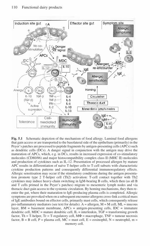
110 Functional dairy products
Fig. 5.1 Schematic depiction of the mechanism of food allergy. Luminal food allergensthat gain access or are transported to the basolateral side of the epithelium (primarily) in thePeyer’s patches are processed to peptide fragments by antigen-presenting cells (APCs) suchas dendritic cells (DCs). A danger signal in conjunction with the antigen may drive thematuration of APCs, which, e.g. in DCs, results in increased expression of co-stimulatorymolecules (CD80/86) and major histocompatibility complex class II (MHC II) moleculesand production of cytokines such as IL-12. Presentation of processed allergen by matureAPC results in differentiation of naïve T-helper cells to T-cell subsets with characteristiccytokine production patterns and consequently differential immunoregulatory effects.Allergic sensitisation may occur if the stimulatory conditions during the antigen presenta-tion promote type 2 T-helper cell (Th2) activation: T-cell contact together with Th2cytokines may induce heavy-chain switching in IgM-bearing B cells, which then (as all Band T cells primed in the Peyer’s patches) migrate to mesenteric lymph nodes and viathoracic duct gain access to the systemic circulation. By homing mechanisms, they then re-enter the gut, where their maturation to IgE-producing plasma cells is completed. Allergicsymptoms are provoked when on a subsequent encounter allergens cross-link a critical massof IgE antibodies bound on effector cells, primarily mast cells, which consequently releasepro-inflammatory mediators (see text for details). A = allergen, M = M cell, ML = mucouslayer, BM = basement membrane, APCs = antigen-presenting cells, IDC = immaturedendritic cell, MDC = mature dendritic cell, IL = interleukin, TGF = transforming growthfactor, Th = T-helper, Tr = T-regulatory cell, MΦ = macrophage, TNF = tumour necrosisfactor, B = B cell, P = plasma cell, MC = mast cell, E = eosinophil, N = neutrophil, m =
memory cell.
Probiotics and the management of food allergy 111
chromosomes 5q31–33, 6p21, 12q and HLA-D region 11q13 (Barnes and Marsh,1998; Barnes, 2000). Genes within these loci associated with atopy include IL-4(the primary cytokine promoting type 2 T-helper cell (Th2) function), IFN-γ (a Th1function promoting cytokine), IL-10 (powerful anti-inflammatory but also Th2-function maintaining cytokine), CD14 (a receptor predominantly involved in therecognition of pathogen associated molecular patterns), TGF-β (powerful anti-inflammatory cytokine), TNF (proinflammatory cytokine family), RANTES(chemotactic molecule attracting, e.g. eosinophiles, basophiles and monocytes),the 4-series leukotrienes, the β-subunit of high-affinity IgE receptor (FcεRIβ), β2-adrenergic receptor (ADRβ2) and high-affinity IL-4 receptor (IL-4R).
5.2.2 Induction phase – allergic sensitisationThe food allergens are usually proteins (Ebo and Stevens, 2001). These can enterfrom the luminal side into the basolateral side of the epithelium through damagedepithelial cell layer or via active uptake by enterocytes or so-called M cells that arespecialised to actively sample the gut contents by transporting macromoleculesand particles from the lumen into organised lymphoid follicles known as Peyer’spatches (MacDonald, 1998; see Fig. 5.1). In here these antigens are endocytosedby antigen-presenting cells, chiefly dendritic cells. Dendritic cells beneath theepithelium may also sample antigens directly by opening tight junctions betweenthe epithelial cells and protruding its arms to the luminal side (Rescigno et al.,2001). There is growing recognition that in addition to the contact with the antigen,activation of the antigen-presenting cells requires a danger signal indicative of hostdamage including molecules and molecular structures, released or produced bycells undergoing stress or abnormal cell death (Gallucci and Matzinger, 2001). Inthe case of allergens, their possible proteolytic activities may cause such signals.
The activated antigen-presenting cells present peptide fragments derived fromthe antigen to naïve CD4+ T-helper (Th) cells which consequently differentiate toeffector cells (see Fig. 5.1). Antigens originating from pathogens tend to trigger theTh cell differentiation into Th1 cells capable of producing IFN-γ that activatesmacrophages and is thus a critical cytokine in mucosal host defence againstinvasive pathogens. The normal response to food antigens is the formation of oraltolerance, i.e. establishment and maintenance of unresponsiveness to antigens onthe mucosal surfaces. Such unresponsiveness can be accomplished by three knownmechanisms: (1) deletion of the effector cells by apoptosis, (2) rendering theeffector cells anergic, i.e. functionally non-responsive, or (3) by active suppressiondue to generation of regulatory T cells. The regulatory T cells include Th3 cellsproducing high amounts of transforming growth factor-β (TGF-β), T regulatorytype 1 (Tr1) cells producing high amounts of IL-10 and CD4+ CD25+ anergic T-cell population inhibiting the proliferation of responding T cells via cell–cellinteractions (MacDonald, 1998; Groux, 2001; Maloy and Powrie, 2001).
The dose of the allergen is one determinant of the induced mechanism (Stroberet al., 1998). Tolerance to food antigens, which are usually encountered inrelatively low numbers, is thought to be formed primarily via the regulatory T cells
112 Functional dairy products
and the cytokines TGF-β and IL-10. TGF-β is a powerful immuno-suppressivecytokine that suppresses the acquisition of effector functions of naïve T cells. Thusunder the presence of TGF-β, Th cells differentiate into pluripotent cells that areincapable of secreting effector cytokines immediately on activation but remaincapable of differentiating into any effector T-cell subset once TGF-β is no longerpresent (Sad and Mosmann, 1994). TGF-β also prevents the maturation of den-dritic cells, which as immature are capable of endocytosis but are not able toprocess an antigen. Thus in the presence of TGF-β, dendritic cells cannot stimulateT cells and may induce tolerance (Gorelik and Flavell, 2002). The immuno-suppressive properties of IL-10 are similar and it plays particularly important rolein the differentiation of the regulatory T cells (Moore et al., 2001). Tolerance tofood antigens may also develop in the absence of danger signals stimulating theexpression of co-stimulatory molecules on the antigen-presenting cells, as it hasbeen suggested that antigen presentation by such cells results in the developmentof clonal anergy.
When allergic sensitisation occurs, the presentation of an allergen that shouldbe harmless to the host does not result in the formation of oral tolerance but in thedifferentiation of the naïve Th cells into Th2 memory cells. The reasons for thisimmunological dysfunction are not fully understood but it probably reflectsinherited aberrancies, e.g. in cytokine production and receptor expression associ-ated with the atopic genotype. In addition, the quality of microbial exposure mayplay a key role here as discussed below in detail. The maturation of B cells to IgE-producing plasma cells is stimulated by contact with the Th2 cells in the presenceof cytokines IL-4 and IL-13, produced by these cells.
5.2.3 Effector phase – expression of clinical symptomsThe symptomatic phase of allergies is triggered when on a re-encounter theallergen is recognised by the allergen-specific IgE antibodies (see Fig. 5.1). IgEbinds to the high-affinity IgE receptor (FcεRI) on mast cells, basophiles anddendritic cells and low-affinity IgE receptors (FcεRII, CD23) on monocytes ormacrophages and lymphocytes. A critical mass of allergen cross-linked IgEmolecules bound on an effector cell results in the activation of that cell andinitiation of an inflammatory cascade. Activation of the key participants in allergicinflammation, tissue mast cells, results (1) in immediate release of their granulescontaining, e.g. histamine, proteolytic enzymes and chemotactic factors attractingneutrophiles and eosinophiles to the site, (2) within 5–30 minutes in the productionof lipid mediators such as leukotrien D
4 and prostaglandin D
2,, and (3) within hours
in the production of interleukins such as strongly proinflammatory TNF-α, IgEproduction-stimulating IL-4 and eosinophil proliferation-stimulating IL-5 (Broide,2001). The activation of blood basophils results in similar response includingrelease of histamine by the degranulation and production of cytokines TNF-α andIL-4 (Platts-Mills, 2001). These inflammatory activities result in the clinicalsymptoms of the allergic disease due to vasodilation, increased vascularpermiability, inflammation, cell death and consequently tissue damage. Mast cell
Probiotics and the management of food allergy 113
mediators can also contribute to late phase responses occurring four to eight hoursafter an immediate response and characterised by high eosinophilic infiltration.
The clinical symptoms of food allergy can be cutaneous, respiratory,gastrointestinal and cardiovascular. The cutaneous symptoms include pruritis,atopic dermatitis/eczema, urticaria, angioedema and non-specific erythrema.Gastrointestinal symptoms include nausea, vomiting, diarrhoea, abdominal cramps,bloating and gas and sometimes colic-like symptoms in children, usually inresponse to cows’ milk. The respiratory symptoms such as sneezing, coughing,wheezing or asthma are less common and rare as the sole symptoms. Thecardiovascular symptoms including hypotension, shock and cardiac dysrhythmiasare associated with more severe cases and may cause death.
5.3 The prevalence of food allergy
The double-blind placebo-controlled food challenge is the golden standard for thediagnosis of food hypersensitivities. A positive skin prick test with the suspectedallergen and/or the presence of immunoglobulin E antibodies specific to theallergen are indicative of an IgE-mediated mechanism and thus true food allergy.
Food allergies are a particular problem for infants and young children in whomthe prevalence has been estimated to vary between 0.3 and 8% (Macdougall et al.,2002). Most commonly children are allergic to cows’ milk (2–3% of all children;Host et al., 1995) but allergies to egg white, wheat and soy are also common.Multiple food allergies in infants may be associated with nutritional repercussionsand growth retardation (Isolauri et al., 1998). Fatal and severe food allergicreactions have also been documented in children and their incidence is thought tobe rising. However, based on a recent retrospective analysis of fatalities in theUnited Kingdom, the risk that a child would die from a food allergic reaction wasestimated to be as low as 1 in 800,000 per year (Macdougall et al., 2002).
In adults, the prevalence of food allergies is relatively low. Although 3.8–25%of the general population perceive they are intolerant to foods, food allergy can beconfirmed with food challenge and skin prick testing only in 0.8 to 2.4% of thesubjects (Woods et al., 2002). There is some indication that the number of cases offood allergy-induced anaphylactic shocks is increasing (Wuthrich and Ballmer-Weber, 2001). Adults tend to be allergic to fish, crustaceans, peanuts and tree nuts.However, increasing number of foods has been reported to be provoking allergiesincluding tropical fruits, sesame seeds, psyllium, spices and condiments. These arecommonly cross-allergies to an allergen derived from another source such aspollen or natural rubber latex (Ebo and Stevens, 2001). The triggering foods alsodiffer between countries, probably in reflection of food consumption.
It remains arguable whether food allergies are increasing, although a dramaticincrease in some other allergic diseases in the Western countries is evident. In theUnited States the prevalence of atopic eczema, the predominant atopic manifesta-tion in the first years of life, increased from 3% in the 1960s to 10% in the 1990s(Horan et al., 1992; United States Centers for Disease Control, 1998). Birth cohort
114 Functional dairy products
surveys in the United Kingdom have shown that the prevalence of atopic eczemain children aged under 5 years has increased from 5% in people born in 1946 to 7%and 12%, respectively, for people born in 1958 and 1970 (Jarvis and Burney,1998). Notably, in 39–63% of all infants and young children atopic eczema istriggered by one or more challenge-confirmed food allergies (Sampson, 1992;Isolauri and Turjanmaa, 1996; Burks et al., 1998).
The reasons for the increasing prevalence of allergies in the westernisedcountries are not known. In recent years most attention has received the hygienehypothesis that was originally proposed by Strachan over a decade ago (Strachan,1989). This hypothesis links the increasing prevalence with reduced microbialexposure early in life. He based this claim on observations that the prevalence ofhay fever was inversely related to the number of children in household. Since then,a substantial amount of experimental data has provided mechanistic rationale toexplain how the quality of early microbial stimulus and function could affect theexpression of atopy and the development of allergic diseases.
5.4 Probiotics and food allergy: the clinical evidence
5.4.1 Reduction of the risk of developing allergic diseaseThere are two studies indicating that probiotics may have potential in reducing therisk of the development of allergies when administered in early infancy. In the first,the effects of neonatal administration of non-enteropathogenic Escherichia coliwere followed by a questionnaire. The results showed that the administration wasassociated with reduced susceptibility for consequent development of allergicdiseases. Subjects born prematurely and full-term were followed separately. Of thepremature subjects, the proportion that was reported to have an allergic disease at10 years of age was 60% smaller in those challenged to E. coli (n = 77) than in thecontrols (n = 55; 21% vs 53%). Of the subjects born full-term, the proportion ofallergic subjects at 20 years of age was 29% smaller in the E. coli-challenged group(n = 151) than in the controls (n = 179; 36% vs 51%) (Lodinová-
•Zádníková and
Cukrowská, 1999). In the second study, Lactobacillus rhamnosus strain GG(ATCC 53103) was given in double-blind placebo-controlled manner for two tofour weeks to pregnant mothers from families where the mother herself, the fatherand/or the siblings of the unborn child had an atopic disease. After the birth, theprobiotic/placebo was administered either to the mother or to the child for sixmonths. Remarkably, in the children who were exposed to the probiotic during theperinatal period the occurrence of atopic eczema at two years old was 50% lowerthan in the placebo group (15/64 [23%] vs 31/68 [46%]; Kalliomäki et al., 2001b).
5.4.2 Management of allergic manifestationsThe first implication of the potential of using bacterial stimulus in the managementof the symptoms of food allergies was reported in 1985 by Loskutova. In that studythe administration of a mixture containing Propionibacterium sp. and Lactobacillus
Probiotics and the management of food allergy 115
acidophilus hastened the disappearance of food allergy manifestations. A yearlater Ciprandi and co-workers reported that patients (n = 20) receiving adjunctivetreatment with Bacillus subtilis spores resulted in significant reduction of thefrequency and severity of the symptoms of adults with urticaria/angioedemamanifestations from food allergy (Ciprandi et al., 1986). However, the safety ofusing such spores in immunocompromised patients has been brought into aquestion (Oggioni et al., 1998). In a more recent partially blinded study by Trappand co-workers (1993), volunteers given yoghurt had decreased concentrations ofIgE in the serum and a lower frequency of allergies. Unfortunately, microbialcharacterisation of the yoghurt was not reported. Wheeler and co-workers (1997)studied the effect of yoghurt on cellular, humoral and phagocytic function in adultswith atopic allergy. Consumption of yoghurt, fermented with Lactobacillusbulgaricus and Streptococcus thermophilus, induced no significant changes in anyof the immune parameters investigated.
More prominent results were observed when infants with atopic eczema andcows’ milk allergy were given an extensively hydrolysed whey formula supple-mented with the Lactobacillus GG (Majamaa and Isolauri, 1997). In contrast to theplacebo group (n = 14), in the infants who received the probiotic supplementation(n = 13) a significant improvement of clinical symptoms and alleviation ofintestinal inflammation associated with food allergy was observed. These effectswere later confirmed with another study population (n = 27) and the results werecomparable whether Lactobacillus GG or Bifidobacterium lactis strain Bb-12 wasused as supplementation (Isolauri et al., 2000). With both strains the control ofatopic eczema was achieved after two months of therapy, while in the placebogroup it took six months. The effects on inflammatory markers were similarbetween the two strains: both treatments reduced the serum concentrations ofsoluble CD14 associated with chronic inflammation and urinary concentrations ofeosinophil protein X (EPX) reflecting allergic inflammation. No effects wereobserved on the concentrations of cell surface molecules or chemotactic factors.Interestingly, the effects on the production of the anti-inflammatory cytokineTGF-β1 were opposite with the different probiotics, bifidobacteria-treatmentresulting in decreased and lactobacilli-treatment increased serum TGF-β1 concen-tration. It has been later shown that feeding Lactobacillus GG to children expressingatopy results in induction of IL-10, another strongly immune suppressive cytokine(Pessi et al., 2000). The anti-inflammatory action of Lactobacillus GG is alsosupported by a study where the administration of this strain to adults with cows’milk allergy was shown to down-regulate the overactive phagocytic functionsassociated with allergic inflammation (Pelto et al., 1998).
Recently, the anti-allergenic effects of Lactobacillus GG were tested in a trialwith teenagers and young adults who were allergic to birch pollen and apples(Helin et al., 2002). The subjects received the probiotic for a total of 5.5 monthsprior, during and after the birch pollen season (n = 18). The treatment did notalleviate their symptoms or reduce their use of medication during the birch-pollenseason or the subsequent months as compared to the placebo group (n = 18), nordid the treatment significantly affect the symptoms caused by apple in the oral
116 Functional dairy products
challenge tests. This controversy to the positive effects of this strain in themanagement of cows’ milk allergy and atopic eczema in infants demonstrates thelimitations of a single probiotic strain and regimen in the management of differentallergies during different periods of life.
5.5 Mechanisms of action: gut microbiota composition andfood allergies
The mechanistic rationale for the probiotic therapy in the management of allergiesis provided by the fundamental role of microbial stimulus and function in thenormal development and regulation of immunological functions and gut barrier.The functions relevant in the context of allergies include stimulatory effects onimmune functions that are antagonistic to Th2 activity, promotion of oral toleranceinduction, anti-inflammatory action and effects reducing and modifying the host’sexposure to allergens (see Fig. 5.2). The quality of microbial stimulus has beenshown to influence functions in the both phases of allergy, i.e. induction andeffector phase, and thus both phases are also rational targets of probiotic therapy.
5.5.1 Aberrancies in the composition of the gut microbiota associatedwith allergiesThe predominant site for host–microbe interaction is in the gut and thus thecompositional development of gut microbiota has been suggested to be the keydeterminant in whether or not the atopic genotype will be fully expressed and thusaffect the development of allergic diseases. Therefore, determining the character-istics in the normal microbial compositions in the gut, and in the compositionsassociated with the expression of allergies, may help in understanding the aetiol-ogy of allergic diseases and point to the potential microbial targets of a probiotic(and prebiotic) therapy. Moreover, assessing the properties of the key microbesfound may reveal direct immunological treatment targets and thus theimmunomodulatory properties that a probiotic strain should have.
A study by Russian scientists in 1982 represents the first clear suggestion thatallergies are associated with compositional aberrancy in the gut microbiota(Shaternikov et al., 1982). This study reported two years later in English was basedon their assessment of 60 under 1-year-old infants with food allergy and atopiceczema. In this study it was claimed that the severity of the disease was in directcorrelation with the stage of aberrancy in the faecal microbiota. This aberrancy wascharacterised as low prevalence of bifidobacteria and lactobacilli and highprevalence of Enterobactericeae, pathogenic species of staplylococci and strepto-cocci as well as Candida species (Kuvaeva et al., 1984). Very similar observationson older subjects (10–45 years) were reported by Ionescu and co-workers (1986)another two years later: compared with healthy subjects (n = 21), those with atopiceczema (n = 58) had lower prevalence of lactobacilli, bifidobacteria and enterococcispecies but higher prevalence of Klebsiellae, Proteus, Staphylococcus aureus,
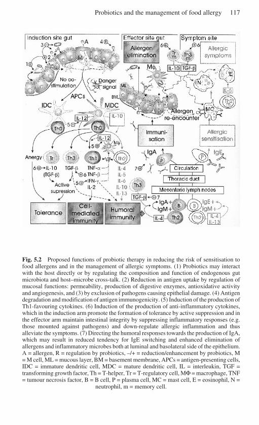
Probiotics and the management of food allergy 117
Fig. 5.2 Proposed functions of probiotic therapy in reducing the risk of sensitisation tofood allergens and in the management of allergic symptoms. (1) Probiotics may interactwith the host directly or by regulating the composition and function of endogenous gutmicrobiota and host–microbe cross-talk. (2) Reduction in antigen uptake by regulation ofmucosal functions: permeability, production of digestive enzymes, antioxidative activityand angiogenesis, and (3) by exclusion of pathogens causing epithelial damage. (4) Antigendegradation and modification of antigen immunogenicity. (5) Induction of the production ofTh1-favouring cytokines. (6) Induction of the production of anti-inflammatory cytokines,which in the induction arm promote the formation of tolerance by active suppression and inthe effector arm maintain intestinal integrity by suppressing inflammatory responses (e.g.those mounted against pathogens) and down-regulate allergic inflammation and thusalleviate the symptoms. (7) Directing the humoral responses towards the production of IgA,which may result in reduced tendency for IgE switching and enhanced elimination ofallergens and inflammatory microbes both at luminal and basolateral side of the epithelium.A = allergen, R = regulation by probiotics, –/+ = reduction/enhancement by probiotics, M= M cell, ML = mucous layer, BM = basement membrane, APCs = antigen-presenting cells,IDC = immature dendritic cell, MDC = mature dendritic cell, IL = interleukin, TGF =transforming growth factor, Th = T-helper, Tr = T-regulatory cell, MΦ = macrophage, TNF= tumour necrosis factor, B = B cell, P = plasma cell, MC = mast cell, E = eosinophil, N =
neutrophil, m = memory cell.
118 Functional dairy products
Clostridium innocuum and Candida species. In 1990, Ionescu and co-workerspublished these results in detail (1990a) and very similar findings from a comparisonof the faecal microbiota of 30 healthy subjects and 110 subjects with atopic eczema(1990b).
Unfortunately, it was a decade later that the topic received wider acknowledge-ment. A study by Bjorksten and co-workers (1999) provided confirmation for theprevious findings by demonstrating that colonisation by lactobacilli was less commonin both Estonian and Swedish 2-year-old children with food allergies (n = 27) than inthe age-compatible healthy children (n = 36), while the opposite was true for coliformsand S. aureus. Bacteroides were shown to make up a larger proportion of themicrobiota in healthy than in allergic infants. We compared the microbiota of healthyinfants (n = 10) with that of infants with early onset atopic eczema and suspected cows’milk allergy (n = 27) during full breast-feeding. Klebsiellae species tended to be foundmore frequently and Streptococcus species less frequently in the faeces of allergicinfants than in those of healthy infants (Kirjavainen et al., 2001). Furthermore, thepredominant anaerobic and facultatively anaerobic microbiota of allergic infants wascharacterised by lower prevalence of Gram-positive species. Lower prevalence ofbifidobacteria was restricted to those allergic infants with gastrointestinal symptomsand higher prevalence of Bacteroides to infants whose cows’ milk allergy was laterconfirmed by challenge. The connection between Bacteroides and cows’ milk allergyis supported by our recently published data showing a positive correlation betweenfaecal Bacteroides numbers and serum total IgE concentration within infants whowere highly sensitive to cows’ milk proteins as indicated by intolerance to extensivelyhydrolysed whey formula (Kirjavainen et al., 2002). The faecal numbers of E. colicorrelated similarly with total IgE in all the allergic infants, which is in accordance withthe association suggested by the earlier studies between the high prevalence ofcoliforms and allergy.
Most interestingly, two studies have demonstrated differences in the faecalmicrobiota prior to the beginning of the expression of atopy and the developmentof allergic diseases, suggesting that these differences may not be secondary to thedisease. In a study by our group some of the predominant microbial genera wereenumerated with fluorescently labelled 16S RNA specific oligonucleotide probes(Kalliomäki et al., 2001a). The neonatal faecal microbiota preceding the expres-sion of atopy (n = 12) was characterised by the presence of lower numbers ofbifidobacteria and higher numbers of bacteria belonging to the Clostridiumhistolyticum group than in the faeces of infants who did not express atopy (n = 17)(Kalliomäki et al., 2001a). Bjorksten and co-workers (2001) demonstrated similartrends with plate culture methods with few additions. They found that bifidobacteriaand enterococci were less frequently present and lactobacilli more frequentlypresent in the neonatal faecal microbiota of infants who at 2 years had atopiceczema (n = 18) than in the microbiota of symptomless infants (n = 26). Laterduring the first year, a higher prevalence of S. aureus, higher numbers of clostridiaand lower numbers of Bacteroides were other characteristics of the faecal microbiotaof the infants symptomatic at 2 years. Nevertheless, further studies are required toconfirm the causality between the microbial aberrancies in the gut microbiota and
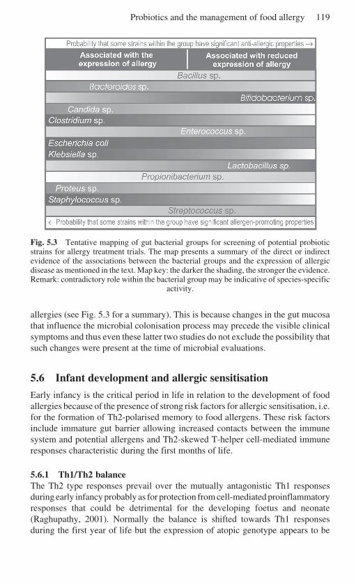
Probiotics and the management of food allergy 119
Fig. 5.3 Tentative mapping of gut bacterial groups for screening of potential probioticstrains for allergy treatment trials. The map presents a summary of the direct or indirectevidence of the associations between the bacterial groups and the expression of allergicdisease as mentioned in the text. Map key: the darker the shading, the stronger the evidence.Remark: contradictory role within the bacterial group may be indicative of species-specific
activity.
allergies (see Fig. 5.3 for a summary). This is because changes in the gut mucosathat influence the microbial colonisation process may precede the visible clinicalsymptoms and thus even these latter two studies do not exclude the possibility thatsuch changes were present at the time of microbial evaluations.
5.6 Infant development and allergic sensitisation
Early infancy is the critical period in life in relation to the development of foodallergies because of the presence of strong risk factors for allergic sensitisation, i.e.for the formation of Th2-polarised memory to food allergens. These risk factorsinclude immature gut barrier allowing increased contacts between the immunesystem and potential allergens and Th2-skewed T-helper cell-mediated immuneresponses characteristic during the first months of life.
5.6.1 Th1/Th2 balanceThe Th2 type responses prevail over the mutually antagonistic Th1 responsesduring early infancy probably as for protection from cell-mediated proinflammatoryresponses that could be detrimental for the developing foetus and neonate(Raghupathy, 2001). Normally the balance is shifted towards Th1 responsesduring the first year of life but the expression of atopic genotype appears to be
120 Functional dairy products
associated with defects such as compromised IFN-γ production that disable thenormal maturation of the Th1 function (Prescott et al., 1999). Therefore, in theseinfants the immune system remains Th2 polarised and the high-risk window forallergic sensitisation is widened.
The bacteria encountered after birth are thought to be the major trigger for theshift away from the predominant Th2 responsiveness, as there appears to be auniversal tendency for bacteria to stimulate the Th cell differentiation into Th1effector cells. However, the responses triggered by the bacteria may be variable;e.g. an in vitro study suggests that Gram-positive bacteria tend to be more potentinducers of IL-12 than their Gram-negative counterparts, which were shown toinduce more of IL-10 (Hessle et al., 2000). The induction of IL-12 and IL-10production in vitro has been demonstrated to vary also between different lacto-bacillus strains (von der Weid et al., 2001; Christensen et al., 2002). IL-12 isproduced by, for example, dendritic cells and macrophages and is a key cytokinepromoting the Th cell differentiation into Th1 cells. IL-10 is considered to be oneof the most critical cytokines responsible for maintaining Th2 bias (Raghupathy,2001); however, its strong anti-inflammatory properties are thought to preventallergic sensitisation and responses (van den Biggerlaar et al., 2000; Holt, 2000;Wills-Karp et al., 2001; see Fig. 5.2).
Differences in the pathogen-associated molecular patterns (PAMPs) in bacteriamay account for the variable innate immune responses mounted towards them.PAMPs are motifs that are not present on host cells but have been conservedinvariant within a microorganism of given class due to their essential physiologicalfunction (Medzhitov, 2001) and as such are good targets for innate recognition.PAMPs include bacterial cell-wall components such as lipopolysaccharides (LPS)of Gram-negative bacteria and lipoteichoic acids of Gram-positive bacteria andunmethylated CpG DNA (Aderem and Ulevitch, 2000). A new family of essentialpattern recognition receptors was discovered a few years ago: the Toll-likereceptors (TLRs) expressed on antigen-presenting cells. Currently ten differentToll-like receptors have been found and they all are specialised for the recognitionof different PAMPs, e.g. co-operation between TLR2 and TLR6 have beendemonstrated to be essential for the cell activation in response to, for example,peptidoglycan from Gram-positive bacteria and lipoarabinomannan frommycobacterial cell wall, TLR4 (with the aid of another pattern recognition receptor,CD14) in response to, for example, LPS from Gram-negative bacteriaand lipoteichoic acids from Gram-positive bacteria, and TLR9 in response tounmethylated CpG DNA (Ozinsky et al., 2000; Tapping et al., 2000; Takeuchiand Akira, 2001). Signalling downstream of these TLRs may result in activationof nuclear factor-κB (NF-κB) or mitogen-activated protein (MAP) kinases (c-JunN-terminal kinase (JNK) and p38). These mediate the activation of genes in thenucleus encoding for proinflammatory cytokines, such as IL-12, chemokines andtheir receptors as well as other factors associated with dendritic cell maturation(Medzhitov, 2001).
The risk of allergic sensitisation may be strongly influenced by the ongoingrecognition of bacteria by Toll-like receptors. Studies in mice with compromised
Probiotics and the management of food allergy 121
Toll-mediated signalling capacity indicate that antigen-specific Th1 responses tofood allergens are dependent on simultaneously induced Toll-mediated activities,while similar dependency was not observed in Th2 responses (Schnare et al.,2001). Furthermore, deficient signalling via Toll-like receptors may promoteallergic sensitisation as re-exposing the mice to the allergen enhanced the produc-tion of IL-13 by T cells, a cytokine capable of inducing isotype class-switching ofB cells to produce IgE (Schnare et al., 2001). There is also increasing body ofevidence that the downstream signalling may occur through different pathwayswhen triggered by microbes. For example it is known that signalling via all TLRscan involve the adaptor protein MyD88, but signalling via TLR4 (and possibly viaTLR3, which recognises viral double stranded DNA), but not TLR2 or TLR9, mayalso trigger the MyD88-independent pathway (Medzhitov, 2001). These differentpathways have been shown to have different effects on dendritic cell activationwhich may be critical concerning the differentiation of Th cells, since signals viathe MyD88-independent pathway do not trigger the production of IL-12(Medzhitov, 2001). Since TLR4 is likely to be more important in the recognitionof Gram-negative than Gram-positive bacteria, the probability that it activates theMyD88-independent pathway may also be higher than with Gram-positive speciesand this may contribute to the suggested differences in the IL-12 productionbetween Gram-positive and Gram-negative bacteria. The importance of Toll-likereceptors for Th maturation and the possibility that bacteria are recognisedvariably by the different TLRs provide the rationale for suggestions that qualitativedifferences in the gut microbiota could be reflected to the Th1/Th2 balance andthereby allergic sensitisation.
5.6.2 Formation of oral toleranceThe importance of microbial stimulus for the normal formation of oral tolerance isbest demonstrated in a study by Sudo and co-workers (1997). They demonstratedthat the Th2-mediated humoral responses but not Th1-mediated responses ofgerm-free mice were unsusceptible to the induction of oral tolerance but after themice were colonised with a single Bifidobacterium infantis strain such tolerancewas inducible. The explanation for this could be related to the necessity of the Toll-like signalling for the development of Th1 function as described above. The effectmay also reflect microbial induction of cytokine production promoting activesuppression. The possibility of such activity by lactic acid bacteria is supported byat least two studies. Firstly, by the aforementioned observation that Lactobacillus66 treatment of allergic infants resulted in increased serum concentrations of TGF-β1 and, secondly, by an in vitro demonstration showing that different Lactobacillusstrains provided different degree of stimulus for the formation of low proliferatingTh cell population producing TGF-β and IL-10 (Von der Weid et al., 2001).
5.6.3 InflammationInflammatory responses play central role in allergic diseases. In the effector arm of
122 Functional dairy products
allergy it is the inflammatory responses triggered by the allergen–IgE complex thatcause the symptoms. In the inductor arm, inflammation can enhance the exposureto potential allergens by increasing the mucosal permeability and promote thematuration of dendritic cells and modulate the Th1/Th2/T regulatory cell balance(see Fig. 5.1; Crowe, 1999; Kirjavainen et al., 1999; Nagler-Anderson and Shi,2001). The gut microbes and, as such, probiotics can affect the inflammatoryresponses in various ways and in the both arms of the allergic disease (see Fig. 5.2).
As described above there is a universal tendency to mount Th1-mediatedproinflammatory responses to bacteria. Such reactions would be wasteful anddetrimental if they were mounted against the innumerable commensal gut mi-crobes and therefore the immune system has adapted to recognise and tolerate theharmless components of the gut microbiota while monitoring its composition viaactive uptake and mounting responses when potentially dangerous organisms areencountered (Neish et al., 2000; Nagler-Anderson, 2001; Hajjar et al., 2002).Pathogens may have a dual role concerning allergic inflammation. On the onehand, as they do not induce tolerance, they may have greater potential to stimulatethe immunity away from the Th2-type responses than commensal bacteria towardswhich tolerance exists (Kirjavainen et al., 1999; Neish et al., 2000). Conversely,by triggering inflammatory responses or by toxin formation such bacteria mayincrease gut permeability and thus the exposure to potential allergens and the riskfor allergic sensitisation (McDonel, 1979; Deitch et al., 1991; Duchmann et al.,1995; Obiso et al., 1995; Crowe, 1999; Madsen et al., 1999b; Lapa e Silva et al.,2000; Neish et al., 2000). Restraining the potentially inflammatory microbiotacould therefore provide a potential target for probiotic therapy (Kirjavainen et al.,1999; Madsen et al., 1999a). Indeed, in our recent study it was shown that theallergic inflammation alleviating effects of bifidobacterial supplementation duringweaning were accompanied by restraining action on Bacteroides and E. colimicrobiota, the numbers of which correlated directly with the serum total IgEconcentrations during breast-feeding (Kirjavainen et al., 2002).
Bifidobacteria and lactobacilli are antagonistic against many pathogens due tocompetitive exclusion, production of antimicrobial components (Gibson andWang, 1994; Lievin et al., 2000) as well as via stimulation of the IgA production(Moreau et al., 1978; Kaila et al., 1992; Yasui et al., 1992; Majamaa et al., 1995).The latter effect can reduce the intestinal inflammation also by enhancing theexclusion of allergens and again reduce the risk of hypersensitivity reactions tosuch allergens in already sensitised subjects (Takahashi et al., 1998; Isolauri et al.,1993). As discussed above, specific probiotics may also reduce wide range ofinflammatory responses by stimulating the production of IL-10 and TGF-β andtherefore antigen non-specific bystander suppression (Pessi et al., 2000; Isolauri etal., 2000).
The degradation process is another defining parameter in relation to theinflammatory responsiveness mounted towards dietary antigens (Isolauri et al.,1993; Barone et al., 2000). Most antigens encountered are already processed whenthey contact the mucosal surface. Proteases produced by intestinal microbiotacontribute to the processing of food antigens in the gut and modify their
Probiotics and the management of food allergy 123
immunogenicity. Such an activity may also be used in probiotic therapyas demonstrated by finding that, in contrast to purified casein, which up-regulatesIL-4 and INF-γ production in infants with cows’ milk allergy, caseins degradedby probiotic bacterium-derived, Lactobacillus GG, enzymes were further shownto down-regulate IL-4 production with no effect on INF-γ release (Sütas et al.,1996).
5.7 Selecting the right probiotic
Further clinical studies with larger study populations are required to confirm thebeneficial effects of probiotics in the management of food allergy. However, takentogether, the scant but prominent clinical demonstrations and the substantialnumber of experimental data providing the mechanistic rationale for bacterialtherapy, the great potential of such an approach is undeniable. Therefore, inaddition to the extended clinical studies with the current most studied probiotics,such as Lactobacillus GG and Bifidobacterium Bb-12, efforts should be focusedon optimising the treatments, i.e. finding the best regimen and strains. This goalcan be efficiently reached only by first assessing the specific properties that aprobiotic strain should have so that it is able to carry out the desired effects. Wehave recently redefined probiotics as ‘specific live or inactivated microbial culturesthat have documented targets in reducing the risk of human disease or in theirnutritional management’, to address the importance of specific targets for specificstrains (Isolauri et al., 2002). Previously the screening for potential probioticstrains for clinical trials has not been focused enough on the disease-specificproperties but more on selecting strains that have already been shown to havepotential in the treatment of some other disease. These strains are commonlycharacterised by properties that are universally considered to be desirable forprobiotic action, such as strong in vitro adhesion and viability, but on the otherhand shown to not be always mandatory (Ouwehand and Salminen, 1998; Isolauriet al., 2002). Such selection practice has been well justified for the preliminarystudies and the reason is clear: little is known about specific properties ofprobiotics for specific diseases.
5.7.1 Microbiota-oriented approachOne approach to the identification of the desired properties has been the characteri-sation of important bacterial groups in the natural prevention of allergies bycomparing the gut microbial composition in healthy and allergic individuals and inconnection to the expressed phenotype of the disease. These data are somewhatvariable, but there are also very clear common trends that do not appear to beassociated with certain countries or time. Such trends include desirability of infantmicrobiota robust in bifidobacteria and enterococci and low in at least certainspecies of coliforms, clostridia and S. aureus. Data concerning lactobacilli andbacteroides are variable. In Fig. 5.3 these characteristics have been summarised in
124 Functional dairy products
conjunction with the effects of allergy treatment trials with different probioticstrains. The variability in the results may reflect the differences in the study designs(particularly in the selection criteria for the study populations), but also thatdifferent species and strains within these genera have radically different effects inrespect to the disease. The latter notion is supported by a recent study byOuwehand and co-workers (2001) demonstrating that different bifidobacteriaspecies tended to prevail in the microbiota of allergic infants (n = 7) compared withhealthy infants (n = 6). Further and more detailed species- and even strain-specificcharacterisation of gut microbial composition in association with allergies iswarranted.
5.7.2 Allergic immune phenotype-oriented approachAnother logical approach to studying the desired properties of probiotics is tocharacterise the immunological aberrancies associated with the development andmanifestations of allergic disease and to investigate which properties in bacteriacould stimulate counterbalancing responses. A good example of such an approachis a subsequent study by He and co-workers (2002) demonstrating that thebifidobacteria species prevailing in healthy infants were more potent inducers ofIL-10 than the species more commonly found in the faeces of allergic infants,which conversely appeared to be more potent inducers of the proinflammatorycytokines tumour necrosis factor (TNF)-α, IL-6 and IL-12. As IL-10 may be oneof the key cytokines promoting the formation of oral tolerance, and also key anti-inflammatory cytokine in restraining allergic responses, the characterisation of thecomponents responsible for these differences could reveal a desirable property ofa probiotic strain intended for the management of allergies. The recent advances inthe understanding of bacterial recognition by the innate immune system, inparticular the discovery of the Toll-like receptors and their specialised role in therecognition of different PAMPs, as well as improved understanding of the signaltransduction pathways, may provide the focus points for such assessments.
The immunological aberrancies associated with allergies can also be looked atfrom a slightly different perspective: it is possible that the immunological aberran-cies associated with the expression of atopic genotype set special requirements formicrobial stimulus. In other words, exposure to the same bacteria does notnecessarily result in similar immunological responses in individuals that expressatopy and those that do not. An example of such aberrancy is low CD14 expressionassociated with atopy, which may compromise the bacterial recognition andtherefore, for example, the ability of bacteria to promote the development of Th2antagonising Th1 function. We have previously hypothesised that it may bepossible to overcome this defect by quantitatively strong enough exposure toGram-positive bacteria, the recognition of which appears to be less dependent onCD14 than that of Gram-negative bacteria. This suggestion is in agreement withour observation, which tentatively suggests that the early colonisation by Gram-positive bacteria may have been poor in infants who have developed an atopicdisease (Kirjavainen et al., 2001).
Probiotics and the management of food allergy 125
5.7.3 Engineering the desirable probiotic strainOnce the key microbial functions and components in respective to the managementof allergies have been identified, it may be possible to genetically implant theseproperties to bacterial strains that are otherwise most suitable for probiotic use, e.g.from the industrial point of view. It is also possible to genetically engineerrecombinant bacteria with desired function such as production of anti-inflammatorycytokines IL-10 and TGF-β (Steidler et al., 2000; Gao et al., 2001). However, suchapproaches possess many unanswered questions of safety. These include theproblems with preventing the spread of the genetically introduced propertybetween other members of the microbiota and to microbiota of other individuals.A less controversial approach would be to screen for microorganisms with thedesirable properties, e.g. for one strongly inducing IL-10 production, and then ifnecessary modify its technological properties without genetic engineering. Thismight be accomplished, for example, by exposing the microorganism to sublethalstress such as acidic conditions or heat, in order to improve the survival propertiesin the gastrointestinal tract and tolerance to stress, and thereby also to provideimproved competitiveness against pathogens in the intestinal milieu (Klaenhammerand Kullen, 1999).
5.8 Conclusion and future trends
Probiotics have been successfully used in the risk reduction and management ofallergic diseases but extended trials must be conducted before recommendationscan be given for clinical practice. There are extensive experimental and clinicaldata on the effects by which probiotics may, by immunological or non-immunological means, reduce the risk of allergic sensitisation and development ofallergic disease and, on the other hand, alleviate the symptoms in already allergicsubjects. These mechanisms include immunomodulatory effects promoting Th2antagonistic Th1 functions and the formation of oral tolerance, enhanced antigenelimination and down-regulated inflammatory responses, e.g. via stimulation ofanti-inflammatory cytokines IL-10 and TGF-β, antagonism against pathogenicmicrobiota and effects seen as enhanced elimination, modified degradation,permeation and presentation of food antigens. Particular efforts should be made tocharacterise the properties in bacteria that are responsible for these mechanisms tofacilitate the selection of optimal probiotics and optimise the treatment regimen.
5.9 Sources of further information and advice
In the beginning of this chapter, the basics of food allergies were briefly revised.A more comprehensive overview of the current understanding of food allergies,food allergens and their relationship with the gastrointestinal immune system hasbeen recently published (Burks, 2002; Brandtzaeg, 2002; Samuel et al., 2002). Forin-depth understanding of food allergies, their mechanisms, diagnosis and
126 Functional dairy products
treatments the reader is advised to consult a textbook such as Food Allergy andIntolerance by Brostoff and Challacombe (2002). Good sources for the up-to-the-minute information on food allergies are the specialised journals such as Journalof Allergy and Clinical Immunology, Clinical and Experimental Allergy, Allergyand Current Opinion in Allergy and Clinical Immunology.
The gastrointestinal immune system and the formation of oral tolerance arekeystones in the aetiology of food allergies. There are number of good reviewscovering these topics including those by Nagler-Anderson (2001), a very nicelywritten overview of the both subjects, Nagler-Anderson and Shi (2001), a compre-hensive review on oral tolerance, Groux (2001) and Maloy and Powrie (2001),which review the current knowledge on regulatory T cells in detail. The review byGorelik and Flavell (2002) presents comprehensively the relevant information onthe importance of the key cytokine TGF-β and that by Wills-Karp and co-workers(2001) presents allergy related information on IL-10. An excellent referencesource for information on different cytokines is the Cytokines Online PathfinderEncyclopaedia in the Internet (Ibelgaufts, 2002).
The mechanisms of probiotic therapies are based on the normal host–microbeinteractions. Further discussion concerning such interactions can be found fromreviews including those of Hooper and Gordon (2001) and MacDonald andPettersson (2000). Toll-like receptors and the signal transduction pathways theyactivate are the foundation for innate immune responses to bacteria and a numberof excellent reviews can be found including those by Medzhitov (2001) andTakeuchi and Akira (2001).
5.10 ReferencesADEREM A and ULEVITCH R J (2000), ‘Toll-like receptors in the induction of the innate
immune response’, Nature, 406(6797), 782–787.BARNES K C (2000), ‘Atopy and asthma genes-where do we stand?’, Allergy, 55(9), 803–817.BARNES K C and MARSH D G (1998), ‘The genetics and complexity of allergy and asthma’,
Immunol Today, 19(7), 325–332.BARONE K S, REILLY M R, FLANAGAN M P and MICHAEL J G (2000), ‘Abrogation of oral
tolerance by feeding encapsulated antigen’, Cellular Immunol, 199, 65–72.BAUER H, PARONETTO F, BURNS W A and EINHEBER A (1966), ‘The enhancing effect of the
microbial flora on macrophage function and the immune response. A study in germfreemice’, J Exp Med, 123, 1013–1024.
BJORKSTEN B, NAABER P, SEPP E and MIKELSAAR M (1999), ‘The intestinal microflora inallergic Estonian and Swedish 2-year-old children’, Clin Exp Allergy, 29(3), 342–346.
BJORKSTEN B, SEPP E, JULGE K, VOOR T and MIKELSAAR M (2001), ‘Allergy development andthe intestinal microflora during the first year of life’, J Allergy Clin Immunol, 108(4), 516–520.
BRANDTZAEG P (2002), ‘Current understanding of gastrointestinal immunoregulation and itsrelation to food allergy’, Ann NY Acad Sci, 964, 13–45.
BRANDTZAEG P, FARSTAD I N and HELGELAND L (1998), ‘Phenotypes of T cells in the gut’,Chem Immunol, 71, 1–26.
BROIDE D H (2001), ‘Molecular and cellular mechanisms of allergic disease’, J Allergy ClinImmunol, 108 (2 Suppl), S65–71.
Probiotics and the management of food allergy 127
BROSTOFF J and CHALLACOMBE S J, EDS (2002), Food Allergy and Intolerance, W. B.Saunders, London.
BURKS W A, JAMES J M, HIEGEL A, WILSON G, WHEELER J G, JONES S M and ZUERLEIN N (1998),‘Atopic dermatitis and food hypersensitivity reactions’, J Pediatr, 132(1), 132–136.
BURKS W (2002), ‘Current understanding of food allergy’, Ann NY Acad Sci, 964, 1–12.CHRISTENSEN H R, FROKIAER H and PESTKA J J (2002), ‘Lactobacilli differentially modulate
expression of cytokines and maturation surface markers in murine dendritic cells’,J Immunol, 168(1), 171–178.
CIPRANDI G, SCORDAMAGLIA A, RUFFONI S, PIZZORNO G and CANONICA G W (1986), ‘Effectsof an adjunctive treatment with Bacillus subtilis for food allergy’, Chemioterapia, 5(6),408–410.
CROWE S E (1999), ‘Food allergies – immunophysiology aspects’, in Ogra P L, Mestecky J,Lamm M E, Strober W, Bienenstock J and McGhee J R, Mucosal Immunology, San Diego,Academic Press, 1129–1139.
DEITCH E A, SPECIAN R D and BERG R D (1991), ‘Endotoxin-induced bacterial translocationand mucosal permeability: role of xanthine oxidase, complement activation, and macro-phage products’, Crit Care Med, 19(6), 785–791.
DUCHMANN R, KAISER I, HERMANN E, MAYET W, EWE K and MEYER ZUM BUSCHENFELDE K H(1995), ‘Tolerance exists towards resident intestinal flora but is broken in activeinflammatory bowel disease (IBD)’, Clin Exp Immunol, 102, 448–455.
EBO D G and STEVENS W J (2001), ‘IgE-mediated food allergy–extensive review of theliterature’, Acta Clin Belg, 56(4), 234–247.
GALLUCCI S and MATZINGER P (2001), ‘Danger signals: SOS to the immune system’, CurrOpin Immunol, 13, 114–119.
GAO C F, KONG X T, GRESSNER A M and WEISKIRCHEN R (2001), ‘The expression andantigenicity identification of recombinant rat TGF-beta1 in bacteria’, Cell Res, 11(2), 95–100.
GIBSON G R and WANG X (1994), ‘Regulatory effects of bifidobacteria on the growth of theother colonic bacteria’, J Appl Bacteriol, 77, 412–420.
GORELIK L and FLAVELL R A (2002), ‘Transforming growth factor-beta in T-cell biology’,Nature Rev Immunol, 2(1), 46–53.
GROUX H (2001), ‘An overview of regulatory T cells’, Microbes Infect, 3(11), 883–889.HAJJAR A M, ERNST R K, TSAI J H, WILSON C B and MILLER S I (2002), ‘Human Toll-like
receptor 4 recognizes host-specific LPS modifications’, Nat Immunol, 3(4), 354–359.HE F, MORITA H, HASHIMOTO H, HOSODA M, KURISAKI J, OUWEHAND A C, ISOLAURI E, BENNO
Y and SALMINEN S (2002), ‘Intestinal Bifidobacterium species induce varying cytokineproduction’, J Allergy Clin Immunol, 109(6), 1035–1036.
HELIN T, HAAHTELA S and HAAHTELA T (2002), ‘Effect of oral treatment with an intestinalbacterial strain, Lactobacillus rhamnosus (ATCC 53103), on birch-pollen allergy: aplacebo-controlled double-blind study’, Allergy, 57(3), 243–246.
HESSLE C, ANDERSSON B and WOLD A E (2000), ‘Gram-positive bacteria are potent inducersof monocytic interleukin-12 (IL-12) while Gram-negative bacteria preferentially stimu-late IL-10 production’, Infect Immun, 68(6), 3581–3586.
HOLT P (2000), ‘Parasites, atopy, and the hygiene hypothesis: resolution of a paradox?’,Lancet, 356(9243), 1699–1701.
HOOPER L V and GORDON J I (2001), ‘Commensal host–bacterial relationships in the gut’,Science, 292(5519), 1115–1118.
HORAN R F, SCHNEIDER L C and SHEFFER A L (1992), ‘Allergic disorders and mastocytosis’,JAMA, 268, 2858–2868.
HOST A, JACOBSEN H P, HALKEN S and HOLMENLUND D (1995), ‘The natural history of cows’milk protein allergy/intolerance’, Eur J Clin Nutr, 49(1), S13–18.
IBELGAUFTS H (2002), COPE Cytokines Online Pathfinder Encyclopaedia [Online], Available:http://www.copewithcytokines.de/ [accessed 13 Aug. 2002].
IONESCU G, RADOVICIC D, SCHULER R, HILPERT R, NEGOESCU A, PREDA I, JURTHI E, ITZE W
128 Functional dairy products
and ITZE L (1986), ‘Changes in fecal microflora and malabsorption phenomena suggestinga contaminated small bowel syndrome in atopic eczema patients’, Microecology andTherapy, 16, 273.
IONESCU G, KIEHL R, ONA L and SCHULER R (1990a), ‘Abnormal fecal microflora andmalabsorption phenomena in atopic eczema patients’, J Adv Med, 3(2), 71–91.
IONESCU G, KIEHL R, WICHMANN-KUNZ F and LEIMBECK R (1990b), ‘Immunobiologicalsignificance of fungal and bacterial infections in atopic eczema’, J Adv Med, 3(1), 47–58.
ISOLAURI E and TURJANMAA K (1996), ‘Combined skin prick and patch testing enhancesidentification of food allergy in infants with atopic dermatitis’, J Allergy Clin Immunol,97(1 Pt 1), 9–15.
ISOLAURI E, MAJAMAA H, ARVOLA T, RANTALA I, VIRTANEN E and ARVILOMMI H (1993),‘Lactobacillus casei strain GG reverses increased intestinal permeability induced by cowmilk in suckling rats’, Gastroenterology, 105(6), 1643–1650.
ISOLAURI E, SUTAS Y, SALO M K, ISOSOMPPI R and KAILA M (1998), ‘Elimination diet in cows’milk allergy: risk for impaired growth in young children’, J Pediatr, 132(6), 1004–1009.
ISOLAURI E, ARVOLA T, SUTAS Y, MOILANEN E and SALMINEN S (2000), ‘Probiotics in themanagement of atopic eczema’, Clin Exp Allergy, 30(11), 1604–1610.
ISOLAURI E, RAUTAVA S, KALLIOMÄKI M, KIRJAVAINEN P and SALMINEN S (2002), ‘Role ofprobiotics in food hypersensitivity’, Curr Opin Allergy Clin Immunol, 2, 263–271.
JARVIS D and BURNEY P (1998), ‘ABC of allergies. The epidemiology of allergic disease’,BMJ, 316(7131), 607–610.
JOHANSSON S G, HOURIHANE J O, BOUSQUET J, BRUIJNZEEL-KOOMEN C, DREBORG S,HAAHTELA T, KOWALSKI M L, MYGIND N, RING J, VAN CAUWENBERGE P, VAN HAGE-HAMSTEN M and WUTHRICH B (2001), ‘A revised nomenclature for allergy. An EAACIposition statement from the EAACI nomenclature task force’, Allergy, 56(9), 813–824.
KAILA M, ISOLAURI E, SOPPI E, VIRTANEN E, LAINE S and ARVILOMMI H (1992), ‘Enhancementof the circulating antibody secreting cell response in human diarrhea by a humanLactobacillus strain’, Pediatr Res, 32, 141–144.
KALLIOMAKI M, KIRJAVAINEN P, EEROLA E, KERO P, SALMINEN S and ISOLAURI E (2001a),‘Distinct patterns of neonatal gut microflora in infants in whom atopy was and was notdeveloping’, J Allergy Clin Immunol, 107(1), 129–134.
KALLIOMAKI M, SALMINEN S, ARVILOMMI H, KERO P, KOSKINEN P and ISOLAURI E (2001b),‘Probiotics in primary prevention of atopic disease: a randomised placebo-controlledtrial’, Lancet, 357(9262), 1076–1079.
KIRJAVAINEN P V and GIBSON G R (1999), ‘Healthy gut microflora and allergy: factorsinfluencing development of the microbiota’, Ann Med, 31(4), 288–292.
KIRJAVAINEN P V, APOSTOLOU E, SALMINEN S J and ISOLAURI E (1999), ‘New aspects ofprobiotics – a novel approach in the management of food allergy’, Allergy, 54(9), 909–915.
KIRJAVAINEN P V, APOSTOLOU E, ARVOLA T, SALMINEN S J, GIBSON G R and ISOLAURI E(2001), ‘Characterizing the composition of intestinal microflora as a prospective treat-ment target in infant allergic disease’, FEMS Immunol Med Microbiol, 32(1), 1–7.
KIRJAVAINEN P V, ARVOLA T, SALMINEN S J and ISOLAURI E (2002), ‘Aberrant compositionof gut microbiota of allergic infants: a target of bifidobacterial therapy at weaning?’, Gut,51(1), 51–55.
KLAENHAMMER T R and KULLEN M J (1999), ‘Selection and design of probiotics’, Int J FoodMicrobiol, 50(1–2), 45–57.
KULIG M, BERGMANN R, KLETTKE U, WAHN V, TACKE U and WAHN U (1999), ‘Natural courseof sensitization to food and inhalant allergens during the first 6 years of life’, J Allergy ClinImmunol, 103(6), 1173–1179.
KUVAEVA I B, ORLOVA N G, VESELOVA O L, KUZNEZOVA G G and BOROVIK T E (1984),‘Microecology of the gastrointestinal tract and the immunological status under foodallergy’, Nahrung, 28(6–7), 689–693.
LAPA E SILVA J R, POSSEBON DA SILVA M D, LEFORT J and VARGAFTIG B B (2000), ‘Endotoxins,asthma, and allergic immune responses’, Toxicology, 152, 31–35.

Probiotics and the management of food allergy 129
LIEVIN V, PEIFFER I, HUDAULT S, ROCHAT F, BRASSART D, NEESER J R and SERVIN A L (2000),‘Bifidobacterium strains from resident infant human gastrointestinal microflora exertantimicrobial activity’, Gut, 47(5), 646–652.
LODINOVÁ-V
ZÁDNÍKOVÁ R and CUKROWSKÁ B (1999), ‘Influence of oral colonization of theintestine with a non-enteropathogenic E. coli strain after birth on the frequency ofinfectious and allergic diseases after 10 and 20 years’, Immunol Lett, 69, 64.
LOSKUTOVA I E (1985), ‘Effectiveness of using Maliutka and Malysh adapted propionic–acidophilus mixtures in the combined treatment of congenital hypotrophy’, Vopr-Pitan,May–June, 17–20.
MACDONALD T T (1998), ‘T cell immunity to oral allergens’, Curr Opin Immunol, 10(6), 620–627.
MACDONALD T T and CARTER P B (1979), ‘Requirement for a bacterial flora before micegenerate cells capable of mediating the delayed hypersensitivity reaction to sheep redblood cells’, J Immunol, 122, 2624–2629.
MACDONALD T T and PETTERSSON S (2000), ‘Bacterial regulation of intestinal immuneresponses’, Inflamm Bowel Dis, 6(2), 116–122.
MACDOUGALL C F, CANT A J and COLVER A F (2002), ‘How dangerous is food allergy inchildhood? The incidence of severe and fatal allergic reactions across the UK and Ireland’,Arch Dis Child, 86(4), 236–239.
MADSEN K L, DOYLE J S, JEWELL L D, TAVERNINI M M and FEDORAK R N (1999a),‘Lactobacillus species prevents colitis in interleukin 10 gene-deficient mice’, Gastroen-terology, 116(5), 1107–1114.
MADSEN K L, MALFAIR D, GRAY D, DOYLE J S, JEWELL L D and FEDORAK R N (1999b),‘Interleukin-10 gene-deficient mice develop a primary intestinal permeability defect inresponse to enteric microflora’, Inflamm Bowel Dis, 5(4), 262–270.
MAJAMAA H and ISOLAURI E (1997), ‘Probiotics: a novel approach in the management of foodallergy’, J Allergy Clin Immunol, 99, 179–185.
MAJAMAA H, ISOLAURI E, SAXELIN M and VESIKARI T (1995), ‘Lactic acid bacteria in thetreatment of acute rotavirus gastroenteritis’, J Pediatr Gastroenterol Nutr, 20, 333–338.
MALOY K J and POWRIE F (2001), ‘Regulatory T cells in the control of immune pathology’,Nat Immunol, 2(9), 816–822.
MCDONEL J L (1979), ‘The molecular mode of action of Clostridium perfringens entero-toxin’, Am J Clin Nutr, 32(1), 210–208.
MEDZHITOV R (2001), ‘Toll-like receptors and innate immunity’, Nature Rev Immunol, 1(2),135–145.
MOORE K W, DE WAAL MALEFYT R, COFFMAN R L and O’GARRA A (2001) ‘Interleukin-10 andthe interleukin-10 receptor’, Annu Rev Immunol, 19, 683–765.
MOREAU M C, DUCLUZEAU R, GUY-GRAND D and MULLER M C (1978), ‘Increase in thepopulation of duodenal IgA plasmocytes in axenic mice monoassociated with differentliving or dead bacterial strains of intestinal origin’, Infect Immun, 21, 532–539.
NAGLER-ANDERSON C and SHI H N (2001), ‘Peripheral nonresponsiveness to orally admin-istered soluble protein antigens’, Crit Rev Immunol, 21(1–3), 121–131.
NAGLER-ANDERSON C (2001), ‘Man the barrier! Strategic defences in the intestinal mucosa’,Nature Rev Immunol, 1(1), 59–67.
NEISH A S, GEWIRTZ A T, ZENG H, YOUNG A N, HOBERT M E, KARMALI V, RAO A S and MADARAJ L (2000), ‘Prokaryotic regulation of epithelial responses by inhibition of IºB-±ubiquitination’, Science, 289, 1560–1563.
OBISO R J JR, LYERLY D M, VAN TASSELL R L and WILKINS T D (1995), ‘Proteolytic activityof the Bacteroides fragilis enterotoxin causes fluid secretion and intestinal damage invivo’, Infect Immun, 63(10), 3820–3826.
OGGIONI M R, POZZI G, VALENSIN P E, GALIENI P and BIGAZZI C (1998), ‘Recurrent septicemiain an immunocompromised patient due to probiotic strains of Bacillus subtilis’, J ClinMicrobiol, 36, 325–326.
OUWEHAND A C and SALMINEN S J (1998), ‘The effects of cultured milk products with viable
130 Functional dairy products
and non-viable bacteria’, Int Dairy J, 8, 749–758.OUWEHAND A C, ISOLAURI E, HE F, HASHIMOTO H, BENNO Y and SALMINEN S (2001),
‘Differences in Bifidobacterium flora composition in allergic and healthy infants’,J Allergy Clin Immunol, 108(1), 144–145.
OZINSKY A, UNDERHILL D M, FONTENOT J D, HAJJAR A M, SMITH K D, WILSON C B, SHROEDER L
and ADEREM A (2000), ‘The repertoire for pattern recognition of pathogens by the innateimmune system is defined by cooperation between Toll-like receptors’, Proc Natl AcadSci USA, 97(25), 13766–13771.
PELTO L, ISOLAURI E, LILIUS E M, NUUTILA J and SALMINEN S (1998), ‘Probiotic bacteriadown-regulate the milk-induced inflammatory response in milk-hypersensitive subjectsbut have an immunostimulatory effect in healthy subjects’, Clin Exp Allergy, 28(12),1474–1479.
PESSI T, SÜTAS Y, HURME M and ISOLAURI E (2000), ‘Interleukin-10 generation in atopicchildren following oral Lactobacillus rhamnosus GG’, Clin Exp Allergy, 30, 1804–1808.
PLATTS-MILLS T A (2001), ‘The role of immunoglobulin E in allergy and asthma’, Am JRespir Crit Care Med, 164(8 Pt 2), S1–5.
PRESCOTT S L, MACAUBAS C, SMALLACOMBE T, HOLT B J, SLY P D and HOLT P G (1999),‘Development of allergen-specific T-cell memory in atopic and normal children’, Lancet,353(9148), 196–200.
PULVERER G, KO H L, ROSZKOWSKI W, BEUTH J, YASSIN A and JELJASZEWICZ J (1990),‘Digestive tract microflora liberates low molecular weight peptides with immunotriggeringactivity’, Int J Med Microbiol, 272, 318–327.
RAGHUPATHY R (2001), ‘Pregnancy: success and failure within the Th1/Th2/Th3 paradigm’,Semin Immunol, 13, 219–227.
RESCIGNO M, URBANO M, VALZASINA B, FRANCOLINI M, ROTTA G, BONASIO R, GRANUCCI F,KRAEHENBUHL J P and RICCIARDI-CASTAGNOLI P (2001), ‘Dendritic cells express tightjunction proteins and penetrate gut epithelial monolayers to sample bacteria’, NatImmunol, 2(4), 361–367.
SAD S and MOSMANN T R (1994), ‘Single IL-2-secreting precursor CD4 T cell can developinto either Th1 or Th2 cytokine secretion phenotype’, J Immunol, 153(8), 3514–3522.
SAMPSON H A (1992), ‘The immunopathogenic role of food hypersensitivity in atopicdermatitis’, Acta Derm Venereol Suppl (Stockh), 176, 34–37.
SAMUEL B, LEHRER S B, AYUSO R and REESE G (2002), ‘Current understanding of foodallergens’, Ann NY Acad Sci, 964, 69–85.
SCHNARE M, BARTON G M, HOLT A C, TAKEDA K, AKIRA S and MEDZHITOV R (2001), ‘Toll-like receptors control activation of adaptive immune responses’, Nat Immunol, 2(10),947–950.
SHATERNIKOV V A, KUVAEVA I D, LADODO K S, ORLOVA N G and VESELOVA O L (1982),‘General and local humoral immunity and intestinal microflora in children with skinmanifestations of food allergy’, Vopr-Pitan, Sep–Oct, 51–56.
STEIDLER L, HANS W, SCHOTTE L, NEIRYNCK S, OBERMEIER F, FALK W, FIERS W and REMAUT E
(2000), ‘Treatment of murine colitis by Lactococcus lactis secreting interleukin-10’,Science, 289(5483), 1352–1355.
STRACHAN D P (1989), ‘Hay fever, hygiene, and household size’, BMJ, 299(6710), 1259–1260.
STROBER W, KELSALL B and MARTH T (1998), ‘Oral tolerance’, J Clin Immunol, 18(1), 1–30.SUDO N, SAWAMURA S, TANAKA K, AIBA Y, KUBO C and KOGA Y (1997), ‘The requirement
of intestinal bacterial flora for the development of an IgE production system fullysusceptible to oral tolerance induction’, J Immunol, 159, 1739–1745.
SÜTAS Y, SOPPI E, KORHONEN H, SYVAOJA E L, SAXELIN M, ROKKA T and ISOLAURI E (1996),‘Suppression of lymphocyte proliferation in vitro by bovine caseins hydrolysed withLactobacillus GG-derived enzymes’, J Allergy Clin Immunol, 98, 216–224.
TAKAHASHI T, NAKAGAWA E, NARA T, YAJIMA T and KUWATA T (1998), ‘Effects of orally
Probiotics and the management of food allergy 131
ingested Bifidobacterium longum on the mucosal IgA response of mice to dietaryantigens’, Biosci Biotechnol Biochem, 62, 10–15.
TAKEUCHI O and AKIRA S (2001), ‘Toll-like receptors; their physiological role and signaltransduction system’, Int Immunopharmacol, 1(4), 625–635.
TAPPING R I, AKASHI S, MIYAKE K, GODOWSKI P J and TOBIAS P S (2000), ‘Toll-like receptor4, but not toll-like receptor 2, is a signaling receptor for Escherichia and Salmonellalipopolysaccharides’, J Immunol, 165(10), 5780–5787.
TRAPP C L, CHANG C C, HALPERN G M, KEEN C L and GERSCHWIN M E (1993), ‘The influenceof chronic yoghurt consumption on population of young and elderly adults’, Int JImmunother, 9, 53–64.
UNITED STATES CENTERS FOR DISEASE CONTROL (1998), ‘Forecasted state-specific estimatesof self-reported asthma prevalence – 1998’, Morbidity and Mortality, 47, 1022–1025.
VAN ASPEREN P P and KEMP A S (1989), ‘The natural history of IgE sensitisation and atopicdisease in early childhood’, Acta Paediatr Scand, 78(2), 239–245.
VAN DEN BIGGELAAR A H, VAN REE R, RODRIGUES L C, LELL B, DEELDER A M, KREMSNER P G
and YAZDANBAKHSH M (2000), ‘Decreased atopy in children infected with Schistosomahaematobium: a role for parasite-induced interleukin-10’, Lancet, 356(9243), 1723–1727.
VON DER WEID T, BULLIARD C and SCHIFFRIN E (2001), ‘Induction by a lactic acid bacteriumof a population of CD4+ T cells with low proliferative capacity that produce transforminggrowth factor β and interleukin-10’, Clin Diagn Lab Immunol, 8, 695–701.
WHEELER J G, BOGLE M L, SHEMA S J, SHIRREL M A, STINE K C, PITTLER A J, BURKS A W andHELM R M (1997), ‘Impact of dietary yoghurt on immune function’, Am J Med Sci, 313,120–123.
WILLS-KARP M, SANTELIZ J and KARP C L (2001), ‘The germless theory of allergic disease:revisiting the hygiene hypothesis’, Nature Rev Immunol, 1(1), 69–75.
WOODS R K, STONEY R M, RAVEN J, WALTERS E H, ABRAMSON M and THIEN F C (2002),‘Reported adverse food reactions overestimate true food allergy in the community’, EurJ Clin Nutr, 56(1), 31–6.
WUTHRICH B and BALLMER-WEBER B K (2001), ‘Food-induced anaphylaxis’, Allergy,56(Suppl 67), 102–104.
YASUI H, NAGAOKA N, MIKE A, HAYAKAWA K and OHWAKI M (1992), ‘Detection ofBifidobacterium strains that induce large quantities of IgA’, Microb Ecol Health Dis, 5,155–162.
6
Dairy products and the immune functionin the elderlyH. Gill, Massey University, Palmerston North, New Zealand
6.1 Introduction
The population of elderly persons is growing rapidly in all countries. According tolatest WHO estimates there are about 600 million people aged 60 years or older inthe world and this figure is expected to rise to over 2 billion (20% of the worldpopulation) by the year 2050 (United Nations, 1999; WHO, 1999). Furthermore,the number of people over the age of 80 is expected to more than triple, and thenumber of centenarians is projected to increase by 15-fold within the next 50 years.This increase in life expectancy has been the result of improvements in diet, health-care and living conditions.
Ageing is associated with a decline or deterioration in many physiologicalfunctions including the immune system (Wick and Grubeck-Loebenstein, 1997).Decline in immune function with age is termed as immunosenescence and is foundin all mammalian species. Immunosenescence is deemed to be responsible forenhanced morbidity and mortality in the aged. Epidemiological and clinical studieshave revealed that elderly persons suffer more often from infectious diseases(respiratory tract and urinary tract infections, gastroenteritis, tuberculosis, endo-carditis and septicaemia), cancers and autoimmune and atopic reactions than theiryounger counterparts (Kelley and Bendich, 1996). As a result, the care of elderlyaccounts for a significantly higher percentage of health-care costs relative to theirpopulation. For example, 11% of the elderly population use over 40% of health-care resources in the US (Chandra, 1989).
The effective means to correct this age-related immune dysfunction are highlysought and the years since 1980 have seen enormous interest in both fundamentaland applied research that is aimed at improving geriatric health. It has been
Dairy products and the immune function in the elderly 133
suggested that dietary supplementation could be used to improve and maintainimmune health in the elderly. Being a non-invasive, cost-effective and practicalmeans of health improvement, it is of special interest to both consumers and thehealth-care providers. The challenge, therefore, is to identify nutrients/foodcomponents or develop products that are able to improve immune health. Thischapter will outline scientific evidence for immunological changes associated withageing and then discuss evidence, both scientific and clinical, regarding the use ofmilk or milk products (including fermented products) for optimizing immunehealth in the elderly.
6.2 The immune system
To fully appreciate the significance of age-related alterations in immune function tohuman health it is important to understand how the immune system works. Theimmune system comprises a network of organs and cells that are strategicallydistributed throughout the body. The major function of the immune system is todefend the body against invasion by pathogenic organisms and against thedevelopment and metastasis of cancers. These protective effects are mediated byinnate (non-specific) and adaptive (specific) immune systems. The major effectorsof non-specific immunity include polymorphonuclear cells (PMN), mononuclearphagocytes, natural killer cells (NK cells) and the complement system. Theseresponses represent the first line of host defence and operate non-selectively againstforeign and abnormal antigens/materials. Inflammation is a form of non-specifichost immune response where both humoral and cellular factors act in concert.
The specific immunity comprises antibody- and cell-mediated responses and ischaracterized by its specificity and memory; the responses are prompt and ofgreater intensity. Antibody-mediated immunity is accomplished by the productionof specific antibodies by plasma cells (mature B cells). T cells are the centralcomponent of the cell-mediated immunity. T cells of the CD4+ phenotype arehelper/inducer cells whereas CD8 cells are suppressor/cytotoxic cells. These sub-populations of T cells act in concert to modulate and regulate the immuneresponses. Furthermore, CD4+ cells can be divided into Th1 and Th2 cells based ontheir cytokine profile (Mosmann and Coffma, 1998; Ginaldi et al., 1999). Th1 cellsproduce IFN-γ, IL-2, IL-12 and L-18 and are vital for cell-mediated immunity. Onthe other hand, Th2 cells produce IL-4, IL-5, IL-6, IL-10 and IL-13 and promoteantibody production including class switching from IgM to IgG1 and IgE; activationof Th2-type responses is associated with atopy. The development of an effectiveimmune response requires a balance between Th1 and Th2 subsets andinappropriately skewed responses are associated with pathology (Crohn’s disease,food allergies, diabetes, etc.).
Which of these mechanisms are activated depends on the nature of the disease-causing agent. Primarily, extracellular bacteria and viruses are eliminated/controlledby phagocytic cells, complement and antibodies. Intracellular bacteria, parasitesand fungi are controlled by the activation of T cells and the secretion of cytokines
134 Functional dairy products
such as IFN-γ that activate monocytes/macrophages. Cytotoxic T cells and NKcells eliminate cancerous cells and intracellular viruses. Most, if not all, immuno-competent cells and molecules produced by them have overlapping functions.Thus, an effective host response is dependent upon the successful interactionbetween various arms/components of the innate and specific immune systems. Inthe case of an over-activated immune system, such as in allergies and autoimmunediseases, modulation and regulation of the immune system are important in theresolution of the disease state.
6.3 Immunosenescence
Ageing is associated with profound and complex changes in immune function.Whether altered capability of the immune system with ageing simply reflects ageneral deterioration of the immune system or reshaping/realignment of theimmune system is a subject of active debate (Weksler, 1995). A careful evaluationof the literature suggests that immunosenescence probably represents a combina-tion of both, a decline and an active realignment of important functions. Theevidence for the latter is more compelling as some immune responses are decreased,some increased, while others remain unchanged in the aged (Weksler, 1995). Anincreased prevalence of autoimmune, atopic and inflammatory reactions and thereduced ability of the elderly to mount effective immune responses to antigenicchallenge further reflect this paradigm (Goodwin, 1995; Franceschi et al., 1995).Studies on carefully selected healthy individuals have also showed that ageingdoes not affect immunocompetence in all individuals. Some centenarians werefound to exhibit several immune functions comparable to young individuals(Franceschi et al., 1995).
6.3.1 Cell-mediated immunityT-cell function is more profoundly affected than humoral immunity by ageing(Franceschi et al., 1995; Doria and Frasca, 1997; Lesourd, 1997). Aged individu-als have more immature (CD2+ CD3+) and fewer mature (CD3+) T cells (Lesourdand Mazari 1999). The absolute number of CD4 cells is slightly increased, whilethe number of CD8 cells is variously reported as normal, decreased or increased(Ginaldi et al., 2001; Gill et al., 2001b). T-cell proliferation responses, whethermeasured in vitro as a response to mitogens or antigen stimulation, mixed leuco-cyte reaction or in vivo as a delayed-type hypersensitivity (DTH) response to recallantigens (bacterial and fungal products and dinitrochlorobenzene (DNCB)) aresignificantly reduced (reviewed by Lesourd, 1997; Ginaldi et al., 2001). T-cellhelper activity for proliferation and maturation of B cells, and the generation ofcytotoxic effectors also declines with age (reviewed in Miller, 1996). A decreasein the expression of CD28, an important co-stimulatory molecule, in the elderly hasalso been observed (Pawelec et al., 1995). The ability of clonal cells to undergoclonal expansion is also reduced (Tyan, 1981).
Dairy products and the immune function in the elderly 135
Ageing is also accompanied by a progressive shift in T cells from naïve(CD25RA+) to memory cells (CD45RO+) (Lesourd and Meaume, 1994); this naïveto memory cell shift is observed in both CD4 and CD8 T cells (Ernst et al., 1990a,b). The expression of CD3 is down-regulated on both naïve and memory cells(Ginaldi et al., 2001).
Alterations in both functional and proliferative responses of naïve and memoryT cells have been reported during senescence (Ginaldi et al., 2000). A majorconsequence of these changes is that elderly are less able to mount an effectiveimmune response to novel pathogens, and are less responsive to immunizationstrategies; an effective immune response to novel antigens or pathogens dependson the availability and optimal functional capacity of naïve T cells. Thymicinvolution has been deemed responsible for dramatic decline in T-cell function(Chandra et al., 1997a, b).
The cytokine network also exhibits profound alterations with age. The produc-tion of some cytokines is increased while the production and utilization of othersis decreased (Kubo and Cinader, 1990; Green-Johnson et al., 1991; Ernst et al.,1995; Cakman et al., 1996). In general, the cytokine production shifts from Th1 toTh2 type. Compared with young individuals, the production of IL-2 and IFN-γ isreduced and the production of IL-4 and IL-10 is increased in the aged. Thesechanges are thought to be due to a shift in T cells from naïve to memory phenotype;naïve T cells preferentially produce IL 2 and IFN-γ, while memory T cells produceIL-4 and IL-10 (Ernst et al., 1990a, b).
The use of immune status as an indicator of general health (survival and diseaserisk) has been the subject of many studies. It has been suggested that a cluster ofimmune parameters including low CD4+ T cell count, high CD8+ T cell count, lowB-cell numbers, low IL-2 production and low T-cell proliferation could haveprognostic relevance to high subsequent mortality in the very old people; subjectsthat developed this high-risk profile showed poorer survival than those who did not(Ferguson et al., 1995). Anergy in skin delayed-type hypersensitivity responses torecall antigens (Wayne et al., 1990), and a decline in the numbers of both CD4+ andCD16+ (NK cells) at baseline has also been associated with increased mortality insubsequent years (Pawelec and Solana, 1997); individuals in the lowest quartile ofCD4+ and CD16+ cell counts had about tenfold increased risk of dying in the firsttwo years of follow-up compared with those in the highest quartile; low CD4+ orCD16+ cell counts carried a twofold risk.
6.3.2 Humoral immunityAgeing affects both quality and quantity of antibody responses (LeMaoult et al.,1997). The concentration of IgG in the serum is reduced while that of IgA isincreased (Chandra, 1997a, b). The ability to mount antibody responses to newantigens and pathogens is significantly reduced (Moulias et al., 1985). Boosterresponses remain unaffected, but it takes longer to reach peak levels. Antibodyspecificity shifts from foreign to auto, antibody isotype from IgG to IgM andantibody affinities from high to low (LeMaoult et al., 1997). The number of B cells
136 Functional dairy products
is not reduced but the frequency of B cells responsive to some, but not all, antigensis reduced (Miller, 1996). As a result, the elderly respond poorly to most vaccines.Memory responses induced by vaccination are also of short duration in oldcompared with young subjects (Poweres and Belshe, 1993). These changes inimmunocompetence are attributed to impaired T-cell function in the elderly.
6.3.3 Innate immunityThe number of NK cells increases with ageing (Ales-Martinez et al., 1988;Lesourd and Meaume, 1994). NK cells derived from the elderly (SENIEUR)exhibit increased expression of activation surface markers CD95 and HLA-DR,and reduced IL-2 induced proliferation and the expression of CD69, comparedwith cells from young donors. Studies on NK cell function have producedconflicting results (Lesourd, 1997; Chandra, 1989; Gill et al., 2001b). A reductionin NK cell activity (Facchini et al., 1987), and a reduced lytic activity on a per-cellbasis in a subset of NK cells (recognized by GL 183 and EB 6) has been reportedin the aged individuals (Franceschi et al., 1995; Pawelec et al., 1995). However,there is also evidence that the elderly subjects selected on the basis of strict health-performance criteria do not show reduction in NK cell numbers or activity(Franceshi et al., 1995; Ligthart et al., 1989; Lesourd and Meaume, 1994). Thesignificance of these differences to health maintenance remains unclear. A recentstudy has shown that a persistently low NK cell activity is a predictor of impendingmorbidity (Franceschi et al., 1995).
A body of evidence suggests that the function of macrophages and granulocytesis also impaired in the elderly (Chandra, 1989). The number of macrophagesremains unchanged. However, more macrophages are required to elicit an optimalT-cell response to mitogens (Rich et al., 1993). Reduced responsiveness of agedmice to pneumococcal vaccine is attributed to a defective macrophage function(Garg et al., 1996). Organ-specific defects in macrophage function have also beenreported in the aged (Higashimoto et al., 1994). The ability of mononuclear cellsfrom healthy elderly subjects and centenarians to produce proinflammatorycytokines such as IL-1, IL-6, IL-8 and TNF-α is enhanced (Rich et al., 1993).However, the spontaneous release of IL-8 is decreased in the elderly comparedwith younger counterparts (Clark and Peterson, 1994). Monocytes from aged miceproduce less hydrogen peroxide (Ding et al., 1994) and higher amounts ofprostaglandin E2 (Hayek et al., 1997). Prostaglandin E
2 (PGE) is known to have a
suppressive effect on T-cell proliferation and IL-2 production, and lymphocytesfrom aged subjects exhibit enhanced sensitivity to PGE (Goodwin, 1992).
The numbers of neutrophil and neutrophil precursors are not affected duringageing, but the proliferative responses of neutrophil precursor cells to G-CSF arelowered. Elderly patients with sepsis often fail to mount leucocytosis (Corberandet al., 1981; McLaughlin et al., 1986). Polymorphonuclear cells derived from theelderly show reduced migration ability. The capacity to phagocytose opsonizedbacteria, yeast or zymosan particles is also reduced. Studies on the microbicidalactivity of neutrophils from the elderly have produced conflicting data. However,
Dairy products and the immune function in the elderly 137
a majority of the studies show an age-related decline in the microbicidal mechanismsof neutrophils against yeast and bacteria (Fulop et al., 1986; Lipschitz et al., 1988;Esperaza et al., 1996). Whether these altered response arise in neutrophil progenitorsor reflect the influence of cytokines on mature neutrophils in the circulation is notknown (Butcher et al., 2000).
The classical and complement activation pathways appear to be maintained/preserved in the elderly. Ageing has little affect on antigen-processing pathways(Lesourd and Mazari, 1997). However, studies in old mice have shown that theability of follicular dendritic cells to process and present antigen-containingimmune complexes and the transport of antigens into lymph node germinal centresby migrating dendritic cells are impaired (Homes et al., 1984). The significance ofthese deficits in the generation of immune responses is not known.
Neutrophils and macrophages play an important role in mediating protectionagainst bacterial and fungal infections, which are largely responsible for increasedmorbidity and mortality in the elderly. Thus reduced neutrophil supply and/orfunction may account for increased susceptibility of the elderly to infection(Butcher et al., 2000). NK cells play an important role in protection againstneoplasms and the control of viral infections, as well as in the regulation of cell-mediated immunity via the secretion of interferons. Deficient NK cell functionmay account, at least in part, for enhanced susceptibility of the elderly to viralinfections and cancers.
6.4 Nutrition and immune function in the elderly
Nutrition is a critical determinant of immunocompetence and malnutrition is themost common cause of immunodeficiency worldwide (Chandra, 1989, 1995). Inthe elderly, malnutrition and ageing exert a cumulative effect on immunocompe-tence and contribute significantly to increased morbidity and mortality (Lesourd,1995; Heuser and Adler, 1997; Ginaldi et al., 2001). Both protein energy malnutri-tion (PEM) and micronutrient (such as Zn, Se and vitamins (vitamin B6, folic acid,vitamin E)) deficiencies are common in the aged population. At least one-third of theelderly in developed countries exhibit nutritional deficiencies of some kind (Halleret al., 1996); up to 18% of apparently healthy elderly (Lesourd et al., 1998) and one-third of low-income elderly are reported to have vitamin B6 deficiency. In anotherstudy, Chandra (1992) reported vitamin E deficiency in 8.3% of healthy elderlysubjects who lived in their own homes. Lack of variety, reduced energy intake, thepresence of malabsorption in some individuals and drug–nutrient interaction are themajor factors contributing to nutritional deficiencies in the aged population. Thephysically isolated, those living alone, the socially isolated, those with sensory ormental impairment or with chronic systemic diseases, the very poor and the very richshow the highest risk of malnutrition (Chandra, 1989).
Both PEM and micronutrient deficits are known to impair immune responsiveness(Chandra, 1985; Lesourd, 1995, 1997; Rasmussen et al., 1996; Buzina-Suboticanecet al., 1998). The magnitude of immunological impairment depends on the type of
138 Functional dairy products
nutrient involved, its interaction with other nutrients, the severity of deficiency, thepresence of concomitant infection and the age of the subject (Chandra, 1997a,b).Specific deficits produce specific effects. For example, low protein intake isassociated with a sharp drop in immunity in aged animals and humans (Lipschitz etal., 1988; Lesourd et al., 1998; Chandra, 1990; Lesourd, 1990a, b, 1995; Lesourdand Meaume, 1994). PEM reduces both specific and non-specific immune re-sponses. Lymphocyte counts (CD3), the proportions of CD4+ and CD8+ T cells andthe ratio of CD4+ to CD8+ T cells are markedly reduced. Lymphocyte proliferationresponses and DTH responses to recall and new antigens are impaired. Theproduction of several cytokines such as IL-1, IL-2 and IFN-γ is also diminished. Theefficacy of vaccines is significantly reduced. Antibody levels, sero-conversion rateas well as antibody affinity are reduced following vaccination in the elderly withPEM. A high level (about 75%) of unresponsiveness to vaccines in hospitalizedelderly patients is related to undernutrition (Lesourd, 1997). PEM has little effect onantibody responses to T cell-independent antigens or antigens administered withadjuvants. However, the affinity of antibodies is reduced in patients with PEM. Theconcentration of secretory IgA to viral vaccines is also decreased. Phagocytic cellfunction and complement activity are also reduced. The magnitude of immunedeficiency is correlated with the degree of nutritional deficit. Elderly subjects withreduced serum albumin levels (<30 g/l) show lymphopaenia and reduced bloodCD4 counts (<400/mm3) similar to that found in HIV infections (Lesourd, 1990a, b).Similarly, Zn deficiency reduces peripheral blood lymphocyte counts, T-lymphocyteproliferation responses, IL-2 production and T-cell cytotoxicity (Keen and Gershwin,1990; Prasad et al., 1997; Dardenne et al., 1982).
Several recent studies have demonstrated that supplementation with single ormultiple nutrients could be used to restore, at least partly, diminished immuno-competence in the institutionalized aged patients as well as self-sufficient, apparentlyhealthy, elderly (reviewed by Chandra, 1989, 1995; Lesourd, 1997; High, 1999).Lesourd (1995) reported enhanced antibody responses to tetanus toxoid vaccine inundernourished elderly subjects following supplementation with 2580 kJ/daycomplete dietary formula (ready to eat). Similarly, supplementation with a completedietary formula containing 1600–2100 kJ/day was found to correct protein energy-associated defects in cell-mediated immunity (lymphocyte proliferation to mitogensand DTH to recall antigens) in undernourished elderly patients (Lesourd et al.,1990). Chandra (1992) reported the immunity-enhancing (immune function andclinical end-points) effect of multivitamin and mineral supplementation (at a levelsimilar to the recommended daily allowance, RDA) in the elderly patients. After 12months, the supplemented group showed significant enhancement in percentage orproportion of CD4+ cells, NK cell activity, proliferation responses to T cellmitogens, IL-2 production and IL-2 receptor expression. The incidence of infection-related illness was also substantially reduced in the supplemented group; the meannumber of days of illness due to infection in supplemented group was 23 vs 48 inthe placebo group. Antibiotic usage in supplemented group (18 days) was alsolower than the placebo group (32 days).
In institutionalized elderly subjects, supplementation with defined oligovitamins
Dairy products and the immune function in the elderly 139
(vitamins A, C and E) increased numbers of circulating CD4+ and CD8+ T cells andproliferation responses to mitogens (Penn et al., 1991). Zn supplementation hasalso been shown to increase the number of CD3+, CD4+, CD16 and CD56lymphocytes, NK cell activity and DTH responses to recall antigens (Bogden et al.,1990; Fortes et al., 1998; High, 1999). Similarly supplementation with very highlevels of vitamin E has been reported to enhance cellular and humoral immuneresponses in the aged (Meydani et al., 1997). Together, these observations provideevidence that supplementation could be used to delay, prevent or restore age-related immune dysfunction and thus reduce morbidity and mortality due toimmune-related diseases. However, the challenge is to identify nutrients/productsthat are able to provide these benefits.
6.5 Bovine milk and immunomodulation
Bovine milk contains a variety of proteins, carbohydrates, lipids, vitamins, mineralsand growth factors. In addition to meeting the growth and developmental needs(nutritional requirements) of neonates, milk plays an important role in protectingthe neonate until its own immune system becomes fully competent. Milk mediatesits protective function by two distinct ways: first by providing passive protectionthrough the transfer of molecules such as antibodies, and secondly by regulatingthe development of an effective and a balanced immune system.
The immunomodulatory effects of milk are attributed to a wide range of milkcomponents, including whey proteins, caseins, lipids, hormones and growthfactors (Gill, 2000; Cross and Gill, 2000). In addition, milk proteins contain a hostof immunoactive peptides that can be released following enzymatic hydrolysis invitro, commercial processing or normal digestion (Gill, 2000). Furthermore,fermented milk products contain lactic acid bacteria (LAB) that are able toinfluence a range of host immune responses (Gill, 1998; Erickson and Hubbard,2000; Wold, 2001; Isolauri et al., 2001; Gill and Cross, 2002).
Several in vitro and in vivo studies have shown that milk products (includingfermented products) and milk components are able to influence aspects of bothspecific and non-specific host immune responses. These have been reviewed indetail by Gill (1998, 2000), Gill et al. (2000), Cross and Gill (2000), Erickson andHubbard (2000), Korhonen et al. (2000); Wold (2001) and Isolauri et al. (2001).This review will focus only on observations derived from animal models or humanstudies. Immunomodulatory effects of fermented dairy products and probioticLAB will be discussed in a separate section.
6.6 Milk proteins
6.6.1 Humoral immune responseThe ability of milk proteins to enhance antibody responses to both T cell-dependent and independent antigens is well documented. Studies by Bounous and
140 Functional dairy products
colleagues (Bounous and Kongshawn, 1989; Bounous et al., 1985, 1988) showedthat dietary whey protein concentrate (WPC) is able to enhance antibody responsesto systemic immunization with sheep red blood cells (a T cell-dependent antigen)and trinitrophenylated (TNP)-ficoll (a T cell-independent antigen); antibodyresponses to sheep red blood cells (SRBC) among WPC-fed mice were almost fivetimes higher than mice fed equivalent diets of similar nutritional efficiencycontaining casein, soy, wheat, corn protein, egg albumin, beef/fish protein orPurina mouse chow. The immune-enhancing effect was manifested after twoweeks of feeding and persisted for at least eight weeks (as long as the dietarytreatment continued) (Bounous et al., 1988).
Mixing whey protein with other protein sources (soy protein or casein) alsoresulted in enhanced antibody responses (Bounous et al., 1988). WPC has alsobeen found to enhance humoral responses to SRBC in CBA/N × DBA/2J F1 micecarrying the ‘xid defect’ (an accessory cell–cell interaction defect) (Bounous et al.,1985). The enhancing effect of dietary whey on serum antibody responses toovalbumin has also been reported (Wong and Watson, 1995; Gill et al., 2002b).
Recent studies have provided evidence that milk proteins are also able toenhance mucosal antibody responses to orally administered antigens and vaccines.Mice fed diets containing whey protein concentrate, for 4–12 weeks, showedsignificantly higher mucosal antibody responses to ovalbumin, cholera toxin,polio and ’flu vaccine compared with mice fed normal chow (Gill et al., 2002a, b,c, d; Low et al., 2001). Potentiation of antibody responses occurred irrespective ofwhen immunizations commenced (at the beginning, during the early stages orduring the middle stages) during the dietary regimen (Low et al., 2001). Inclusionof WPC into follow-on milk was also able to enhance both humoral and systemicantibody responses to orally administered antigens (Gill et al., 2002b, c, d).
Whey contains a complex mixture of major and minor proteins. Diets contain-ing individual major whey proteins have also been reported to increase systemicand mucosal antibody responses. Bounous and Kongshavan (1989) and Gill et al.(2002a) reported significantly higher serum and/or mucosal antibody responses inmice fed diets containing β-lactoglobulin, α-lactalbumin, BSA, lactoperoxidase orimmunoglobulin than mice fed other protein sources. Mice fed lactoferrin (bLF)hydrolysates were also reported to exhibit higher concentrations of antigen-specific IgA antibody responses in bile and intestinal contents, following oralimmunization with cholera toxin than mice fed control diets (Miyauchi et al.,1997). The ability of caseinoglycopeptides (CGP), derived from κ-casein, tomodulate antigen-specific serum antibody responses has also been demonstrated(Monnai et al., 1998); CGP intake suppressed IgG antibody responses to orallyadministered antigens.
Bounous and Gold (1989) reported that immunoenhancing ability of WPC wascontingent upon its undenatured state and is significantly greater for WPCs thatcontain higher concentrations of bovine Ig and serum albumin. However, recentstudies in our laboratory have shown that immunomodulatory properties of WPCare resistant to commercial-type processing. Both native (commercially produced)and reconstituted (separated into major protein fractions and then recombined)
Dairy products and the immune function in the elderly 141
WPC were found to be effective at enhancing a range of humoral and cellularimmune responses (Gill et al., 2002a).
The effect of whey proteins on humoral immunity in human subjects has notbeen investigated. However, these results indicate that WPC or whey fractionsenriched for various whey proteins could be effectively used as dietary adjuvantsto enhance responses to vaccines in population groups, such as elderly, that showreduced ability to respond optimally to various vaccines.
6.6.2 Cellular immune responsesCell-mediated immune responses play an important role in host protection againstintracellular bacteria, viral infections and cancers. The ability of milk componentsto modulate cell-mediated immune responses has been the subject of several invitro studies (Cross and Gill, 2000; Gill et al., 2000). However, only a few studieshave examined the effect of milk products on cell-mediated immune responses invivo. Ingestion of bovine milk whey proteins, for five to eight weeks, was found toenhance footpad delayed-type hypersensitivity responses and in vitro concanavalinA-induced spleen cell proliferation in mice (Wong and Watson, 1995).Enhancement of lymphocyte proliferative responses to T-cell mitogens in mice fedWPC or increasing concentrations of whey protein hydrolysates, primed withMycobacterium bovis BCG, one week prior to the assessment of immune function,has also been observed (Bounous et al., 1983). In a later study, however, Bounousand Kongshavan (1985) found that whey protein had little effect on cell-mediatedimmunity as measured by graft vs host rejection, DTH and proliferation responsesto T-cell mitogens. Whether it was due to different strains of mice, length of dietarysupplementation/or methods employed is not known. Recent studies in ourlaboratory have shown that diets containing WPC are able to significantly enhancelymphocyte proliferative responses to T and B cell mitogens in mice (Gill et al.,2002a,b,c). Fractionation of WPC further revealed that the lactoperoxidase-enriched fraction was able to significantly enhance both T and B cell mitogenesis,while the β-lactoglobulin- and immunoglobulin-enriched fractions were effectiveat enhancing mitogen-stimulated T-cell proliferation only (Gill et al., 2002a).Casein-derived peptides have also been found to have enhancing effect onlymphocyte function. Spleen cells from mice fed κ-casein-derived CGP hadsignificantly higher proliferative responses to stimulation with T-cell mitogens,phytohaemagglutinin (PHA); responses to B cell mitogens were not affected(Monnai et al., 1989).
The ability of WPC to influence T-cell populations has also been reported. Micefed undenatured whey protein concentrate for four weeks exhibited higher numbersof L3T4+ cells (helper cells) and a higher ratio of L3T4+/Lyt-2+ cells (helper:suppressor) compared with mice fed an isocaloric casein diet (Bounous and Gold,1993; Bounous et al., 1993). A significant increase in total white blood cells, CD4+
and CD8+ lymphocyte counts, and Con A-stimulated IFN-γ production by spleencells in mice fed whey, compared with mice fed other proteins, have also beenobserved (Ford et al., 2001). Ishida et al. (1992a) reported that oral administration
142 Functional dairy products
of immune milk (from cows immunized with human intestinal bacteria) was alsoeffective at enhancing cell-mediated immunity in mice; the mitogenic responses ofmesenteric lymph node cells and the redirected cytotoxic activity of interepitheliallymphocytes to P815 tumour cells with anti-CD3mAb were significantly higher inmice fed immune milk compared with the control group.
Several minor whey components (such as lactoferrin, lactoperoxidase, peptides/growth factors) have also been found to enhance cell-mediated immunity. Forexample, Zimecki and Kruzel (2000) reported a dose-dependent enhancement ofdelayed-type hypersensitivity responses to a range of antigens (ovalbumin, BCGand SRBC) in mice, following oral or parenteral administration of bLF. In tumour-bearing mice, oral administration of bovine LF was reported to inhibit experimentalmetastasis and augment the numbers of CD4+, CD8 +and NK cells (asialoGM1+
cells) in the spleen and peripheral blood of tumour-bearing mice; the cytotoxicactivities of these cells against Yac-1 and colon 26 carcinoma were also enhanced(Wang et al., 2000; Tsuda et al., 2002). Furthermore, the numbers of CD4+ andCD8+ cells, and the production of IL-18, interferon-γ and caspase-1 in the intestinalepithelium, were also increased in bLF-fed mice.
In cancer patients, intake of whey proteins has been reported to increase thenumber of blood leucocytes (Kennedy et al., 1995). Feeding diets containing milkproteins has also been shown to delay thymic involution. Rats fed whey protein-based diets for five weeks (from 6 to 11 weeks of age) had significantly greaterthymic weight than rats fed a soy protein or normal diet (Register et al., 1996);thymic involution usually occurs between 8 and 15 weeks of age in rats. Anincrease in thymic weight and cellularity in mice fed whey protein was also notedby Parker and Goodrum (1990). Although the effect of dietary supplementation onimmune function was not investigated in these studies, the results provide putativeevidence that whey proteins may be able to slow the age-associated alterations inimmune function.
Oral administration of immune milk has also been found to inhibit age-relateddecline in immune function (Ishida et al., 1992b). Eighteen-month-old mice, fedimmune milk, exhibited significantly higher redirected cytotoxicity of intra-epithelial lymphocytes (IEL) and lymphocyte proliferation response to mitogenicor alloantigenic stimulation compared with the control group. Production ofantigen-specific antibodies by spleen cells, following immunization, was alsohigher in mice fed immune milk. In fact, the responses at 18 months of age were notdifferent from those observed at 2 months of age. In addition, a lower level ofautoantibodies was observed at 8 and 18 months of age in the immune milk-fedgroup compared with the control group.
Previous studies have shown that a decline in immune capability of ageing miceis associated with a significant decrease in intracellular glutathione levels and thatthis immune deficit could be corrected with supplementation with glutathione(Furukawa et al., 1987). Bounous et al. (1988) reported that dietary whey proteinswere able to significantly increase the level of tissue glutathione levels in agedmice. Therefore, it is likely that whey proteins may help preserve immuno-competence in the aged by maintaining high glutathione levels and thus enhance
Dairy products and the immune function in the elderly 143
host resistance to infectious diseases and cancers. Enhanced resistance to infectionand tumour development, and improved survival rate in aged mice fed wheyprotein-based diets, compared with controls fed the nutritionally equivalent Purinachow, has been reported previously (Gold et al., 1989).
The mechanisms by which milk-derived components stimulate immunity arenot known. It has been suggested that the immunoenhancing ability of wheyproteins is related to its unique amino acid profile. Substitution of intact proteinswith an equivalent free amino acid mixture, which duplicated the specific aminoacid profile of the various proteins, was found to enhance humoral immunity, butelicited a lower response compared with the intact proteins (Bounous andKongshavn, 1989; Bounous and Gold, 1989). Whey proteins have also beenreported to enhance immune function by increasing levels of glutathione inimmunocompetent cells (Bounous and Gold, 1989). It is also likely that some milkcomponents exert their influence by directly interacting with immunocompetentcells; the presence of receptors for a number of milk-proteins/peptides on immuno-competent cells have been demonstrated (Gill and Rutherfurd, 1998).
6.6.3 Innate immune responsesConsumption of bovine milk-based diets has been found to potentiate phagocyticcapacity of both neutrophils and monocytes/macrophages in animal models. Gilland colleagues observed significantly higher phagocytic activity of peripheralblood leucocytes and peritoneal macrophages in mice fed whey protein for fourweeks, compared with those fed mouse chow (Gill et al., 2002a,b,c). Oraladministration of lactoferrin, lactoperoxidase-enriched whey fractions or milkfatconjugated linoleic acid (CLA) has also been reported to enhance phagocyticcapacity of mouse blood leucocytes and peritoneal macrophages (Zhao, 1999; Gillet al., 2002a). An association between enhanced phagocytic cell function andincreased resistance to Klebsiella and Salmonella of mice fed WPC-based diets hasalso been suggested (Bounous et al., 1981). Intake of immune milk has also beenreported to enhance phagocytic cell function and protect mice against the lethaleffects of radiation: the bactericidal activity of mesenteric lymph node (MLN) cellsfrom the irradiated mice given immune milk was significantly higher than thosefrom the mice given control milk prior to irradiation and post-irradiation (Ishida etal., 1992a).
The ability of milk and milk products to enhance PMN and monocyte functionin the aged individuals has also been demonstrated. Bosche and Plat (1995)reported enhanced phagocytic capacity of blood leucocytes in elderly subjects thatconsumed 0.5 litres of skim milk/day for four weeks. Subjects with higherpathological values (TLC, C-reactive protein, erythrcyte sedimentation rate (ESR))showed the highest increase. Significantly enhanced microbicidal capacity ofblood neutrophils in healthy elderly subjects following regular consumption oflactose-hydrolysed milk (2 glasses/day) for a period of six weeks has also beenreported (Fig. 6.1; Arunachalam et al., 2000). The improvements in bacterial-killing ability of granulocytes became obvious three weeks after continuous
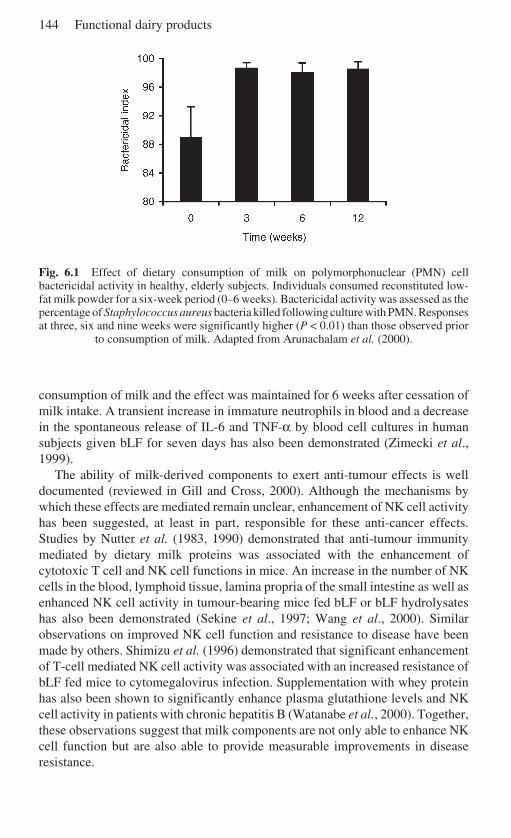
144 Functional dairy products
Fig. 6.1 Effect of dietary consumption of milk on polymorphonuclear (PMN) cellbactericidal activity in healthy, elderly subjects. Individuals consumed reconstituted low-fat milk powder for a six-week period (0–6 weeks). Bactericidal activity was assessed as thepercentage of Staphylococcus aureus bacteria killed following culture with PMN. Responsesat three, six and nine weeks were significantly higher (P < 0.01) than those observed prior
to consumption of milk. Adapted from Arunachalam et al. (2000).
consumption of milk and the effect was maintained for 6 weeks after cessation ofmilk intake. A transient increase in immature neutrophils in blood and a decreasein the spontaneous release of IL-6 and TNF-α by blood cell cultures in humansubjects given bLF for seven days has also been demonstrated (Zimecki et al.,1999).
The ability of milk-derived components to exert anti-tumour effects is welldocumented (reviewed in Gill and Cross, 2000). Although the mechanisms bywhich these effects are mediated remain unclear, enhancement of NK cell activityhas been suggested, at least in part, responsible for these anti-cancer effects.Studies by Nutter et al. (1983, 1990) demonstrated that anti-tumour immunitymediated by dietary milk proteins was associated with the enhancement ofcytotoxic T cell and NK cell functions in mice. An increase in the number of NKcells in the blood, lymphoid tissue, lamina propria of the small intestine as well asenhanced NK cell activity in tumour-bearing mice fed bLF or bLF hydrolysateshas also been demonstrated (Sekine et al., 1997; Wang et al., 2000). Similarobservations on improved NK cell function and resistance to disease have beenmade by others. Shimizu et al. (1996) demonstrated that significant enhancementof T-cell mediated NK cell activity was associated with an increased resistance ofbLF fed mice to cytomegalovirus infection. Supplementation with whey proteinhas also been shown to significantly enhance plasma glutathione levels and NKcell activity in patients with chronic hepatitis B (Watanabe et al., 2000). Together,these observations suggest that milk components are not only able to enhance NKcell function but are also able to provide measurable improvements in diseaseresistance.
Dairy products and the immune function in the elderly 145
6.7 Antibodies and other protective agents in milk
The passive transfer of maternal immunity is pivotal for the survival of mammalianneonates. Species vary in the way this immunity is transferred to the offspring. Inruminants, the transfer of immunity from mother to offspring occurs postnatallyvia the colostrum. As a result, bovine colostrum and early milk contain a range ofbioactive substances that can provide both specific and non-specific defencesagainst infectious agents and foreign antigens to the neonate. The most importantamong these are immunoglobulins (Igs). IgG1 is the predominant antibody inbovine milk and colostrum.
Colostrum and early milk contain exceptionally high levels of antibodies(immunoglobulins), many of which exhibit specificities for common humanpathogens (bacteria, viruses and parasites) (reviewed in Korhonen et al., 2000). Itis therefore likely that these molecules could be effective in providing passiveprotection against a range of infectious agents in human subjects. Normally, thelevel of pathogen-specific antibodies in colostrum and milk is low, but could beenhanced significantly (by 100-fold) by immunizing cows against desiredpathogens/antigens during pregnancy. The efficacy of milk/colostrum-derived Igpreparations against important human pathogens such as Shigella flexneri,Helicobacter pylori and Vibrio cholerae and rotavirus has been the subject ofmany clinical studies (Michalek et al., 1978; Ebina et al., 1985; Boesman-Finkelstein et al., 1989; Tacket et al., 1992; Casswall et al., 1998). While resultshave been variable, the evidence suggests that colostrum preparations, with highantibody titres against defined pathogens, can be effective in mediating protectionagainst enteric infections in humans (Table 6.1). The use of hyper-immunecolostrum containing high titres of specific antibody has been found to be moreeffective than normal Ig preparations, especially in the treatment of infant diarrhoea.A significantly reduction in the incidence of chronic diarrhoea among HIV-infected individuals given bovine colostral Ig concentrate has also been observed(Rump et al., 1992). These studies highlight opportunities for the development ofIg-containing milk or colostrum preparations with defined health benefits forspecific population groups (children, hospitalized individuals, immuno-compromised, the elderly) that exhibit increased risk of infectious disease.
6.7.1 ImmunoregulationOne of the unique features of the protection conferred by colostrum or milk is thevirtual absence of host responses that could harm the recipient. This is due in partto the absence of phlogistic factors as well as the presence of immunoregulatory/anti-inflammatory factors in milk.
Owens and Nickerson (1989) reported the presence of a low molecular weightanti-inflammatory factor in the milk of hyperimmunized cows. Mice given anti-inflammatory factor (AIF) demonstrated significantly less mammary glandinflammation following infection with the mastitis-causing bacteriumStaphylococcus aureus. In other studies, milk-derived AIF was found to suppress
146 Functional dairy products
Table 6.1 Efficacy of bovine milk-derived immunoglobulins (immune and non-immune) against bacterial, viral and parasitic infections: some examples
Target pathogen Subjects Effect Reference
E. coli Preterms and Reduced diarrhoea Lodinova-(enteropathogenic) infants Zadnikova et al.
(1987)E. coli Children No effect on diarrhoea Brunser et al.(enteropathogenic) (1992)and rotavirusE. coli Healthy Prevention of diarrhoea after Freedman et al.(enterotoxigenic) volunteers experimental challenge (1998)Helicobacter pylori Adults Attenuated symptoms of Tarpila et al.
gastritis (1994)H. pylori Adults No effect Opekun et al.
(1999)H. pylori Children Attenuated chronic inflam- Oona et al. (1997)
mation of gastric antriumShigella flexneri Adults Prevented shigellosis Tacket et al.
(1992)Streptococcus mutans Adults Reduced number of S. Loimaranta et al.
mutans in dental plaque (1999)Cryptosporidium Adults Reduced diarrhoea and Okhuysen et al.
oocyst secretion after (1998)experimental infection
Cryptosporidium Adults (AIDS Reduced symptoms of Stephan et al.patients) intestinal inflammation and (1990)
oocyst production
Adapted from Korhonen et al. (2000).
the host vs graft reaction, lymphocyte proliferative responses to ConA, andinflammatory response to carrageenin and E. coli (Ormrod and Miller, 1991,1993). The AIF was effective following oral, subcutaneous, intramuscular,intraperitoneal or intravenous administration.
Anti-inflammatory properties of bLF are also well documented. Zimecki et al.(1998) demonstrated that oral treatment of rats with bLF (five doses, 10 mg each,on alternate days) was able to inhibit carageenin-induced inflammation by 50%,compared with the control treatment. The inhibition was associated with a substantialreduction in the ability of spleen cells to produce IL-6 and TNF-α. A significantreduction in serum TNF-α in mice given bLF, 24 h prior to intravenous challengewith LPS (a potent inflammatory agent) has also been noted (Machnicki et al.,1993). In human studies, oral administration of bLf was found to reduce spontaneousrelease of pro-inflammatory cytokines IL-6 and TNF-α by peripheral bloodmononuclear cells. IL-1β, TNF-α and IL-6 mediate not only inflammatory andimmune reactions, but also the development of cachexia. The balance betweenthese pro-inflammatory cytokines and anti-inflammatory cytokines (IL-10, TGF-β)is pivotal for the fine-tuning and resolution of inflammation.
Dairy products and the immune function in the elderly 147
The presence of transforming growth factor (TGF)-β (a potent anti-inflammatoryagent) in bovine milk in biologically significant amounts has also beendemonstrated; cheese whey and milk contain 3.7 ± 0.7 ng and 4.3 ± 0.8 ng TGF-β/ml. Kulkarni and Karlsson (1993) demonstrated that neonatal mice geneticallydeficient for TGF develop chronic inflammation of the lower intestine, and remainviable only as long as they received maternal milk containing immunosuppressiveTGF. Fell et al. (2000) reported that the administration of a casein-based polymericdiet, containing high levels of TGF-β, to children with Crohn’s disease was able toameliorate chronic inflammation of the intestine and reduce the levels ofproinflammatory cytokines IL-1β, IL-8 and IFN-γ. These beneficial effects wereattributed to the ability of TGF-β to suppress inflammatory cytokine production. Itis important to note that the bioactivity of growth factors such as TGF-β is notaffected by pasteurization. Low pH (pH 2.0) also has little effect on TGF-βactivity.
Allergic diseases result from an imbalance in Th1- and Th2-type immuneresponses and are characterized by increased levels of reaginic antibody (IgE) andinflammation due to the release of vasoactive amines from mast cells. The abilityof bovine milk-derived factors to suppress allergic responses has also beenreported. Watson et al. (1992) reported that a whey fraction, isolated from bovinecolostrum, could suppress systemic IgE antibody responses in mice. Otani andYamada (1995) demonstrated that bovine lactoferrin and κ-casein could inhibit therelease of histamine from mast cells in vitro, suggesting that these proteins mightalso be effective in limiting allergic inflammation in vivo.
The ability of a whey-derived growth factor to regulate the development of oraltolerance has also been demonstrated (Penttila et al., 2001). Oral administration ofa growth factor-enriched whey protein extract to suckling rat pups, between fourand nine days postnatally, significantly inhibited immune activation of spleen cellsto an orally administered food antigen, ovalbumin. Treatment with whey extract atthe time of oral sensitization to ovalbumin resulted in an increased secretion ofTGF-β into the culture supernatants of spleen cells stimulated with ovalbumin.TGF-β is known to play an important role in tolerance induction. Inhibition of anti-CD3 antibody-induced proliferation of human lymphocytes by a milk growthfactor (containing TGF-β1 and TGF-β2) has also been reported (Stoeck et al.,1989).
Furthermore, studies on sheep colostrum have led to the identification of aproline-rich polypeptide (PRP, also known as colostrinin) with immunoregulatoryproperties (Janusz et al., 1974, 1981). PRP has been found to influence thematuration and differentiation of thymocytes into T-helper/T-suppressor cells(Zimecki et al., 1984a, b), to induce release of cytokines by immunocompetentcells (Inglot et al., 1996), to stimulate growth and differentiation of resting B cells(Julius et al., 1988) and to regulate autoimmune host responses (Zimecki et al.,1982; Kundu et al., 2000). In clinical studies, oral administration of ovine PRP hasbeen shown to exert psycho-stimulatory effects and significantly improve theoutcomes of Alzheimer’s disease patients with mild to moderate dimentia (Leszeket al., 1999). Specific receptors for PRP on murine thymocytes have also been
148 Functional dairy products
described (Janusz et al., 1986). The presence of PRP in human milk has also beenreported. Little is known about the nature and functions of PRP derived frombovine colostrum or early milk.
Although the immunomodulatory effects of most milk-derived products inhumans remain to be investigated, the evidence discussed above suggests that milkproducts could be used for optimizing immune function in the elderly (e.g.arthritis, inflammatory bowel disease (IBD), diabetes, Alzhiemer’s disease); thatis to down-regulate as well as enhance immunity in population groups withless inadequate immune function (e.g. the elderly, critically ill, immuno-compromised).
6.8 Fermented dairy products and probiotic LAB
The human gastrointestinal tract (GIT) harbours an extremely complex and diversemicrobial ecosystem. The GIT of an adult human contains approximately 1.6–2 kgof bacteria (1014 cfu), representing over 400 different species. In other words, thereare ten times more microbial cells than the number of eukaryotic cells in the body.The microbial density is relatively sparse in the small intestine (102–103/g) butbecomes increasingly abundant in the lower gut (1010–1012/g). While a majority ofthese bacteria are non-pathogenic/beneficial, some of these organisms possess thepotential to cause disease. In healthy individuals, there is a definite balancebetween good and bad bacteria. However, perturbations in the homeostasis of thenormal microflora due to factors such as antibiotic therapy, stress and infectionresult in proliferation of undesirable or pathogenic microbes and enhancedpredisposition to a number of clinical disorders such as cancers, inflammatorydiseases, allergies, ulcerative colitis and infectious illnesses.
Metchnikoff (1907) was perhaps the first to recognize the health-enhancingattributes of fermented products containing microbes. He hypothesized that theharmful effects of the undesirable bacteria could be overcome through ingestingLAB (probiotics) and that the longevity of Bulgarians was due to the consumptionof large quantities of lactobacilli-fermented milk. In the ensuing 90 years, there hasbeen a plethora of published studies to support Mechnikoff’s hypothesis. It is nowwell recognized that a healthy (balanced) microflora is pivotal to optimum healthand that supplementation with probiotics could be used to shift the balance of thegut microflora away from potentially harmful/pathogenic bacteria towards abeneficial or health-promoting microorganisms, like lactobacilli, bifidobacteria.Probiotics are defined as live bacterial feed/food supplements that promotehost health by balancing its intestinal microflora. Consumption of probiotics issuggested to confer a range of health benefits including modulation of the immunesystem. Most probiotics are LAB, such as lactobacilli and bifidobacteria, and arecommonly consumed in the form of dairy products (yoghurt or other fermentedproducts).
Dairy products and the immune function in the elderly 149
6.9 Immunomodulatory effects of fermented milk productsand LAB
The effects of LAB on the immune system have been the focus of active researchsince the 1980s. The results of these studies have been varied due mainly todifferences in species and strains of LAB, dose, delivery medium and experimentalprotocols. However, these studies have provided unequivocal evidence that certainstrains of LAB are able to stimulate as well as regulate several aspects of immunefunction. Several excellent reviews on immunomodulatory effects of probiotics inanimals and humans have been published in recent years (reviewed by Gill, 1998;Erickson and Hubbard, 2000; Meydani and Ha, 2000; Isolauri et al., 2001; Wold,2001) and therefore the following section will focus mainly on evidence fromhuman studies.
6.9.1 Immune enhancementThe effect of LAB consumption on natural immunity has been the subject of manyhuman studies (Table 6.2). The results of these studies have shown that theconsumption of specific strains of LAB is able to enhance:
• phagocytic activity of blood leucocytes (PMN and mononuclear cells);• oxidative defence mechanisms, including production of reactive oxygen species
and secretion of cytokines by phagocytic cells;• NK cell activity.
Schiffrin et al. (1995) reported that consumption of fermented milk containing Lb.johnsonii La1 or B. lactis Bb12 for three weeks was able to enhance phagocyticactivity of peripheral blood leucocytes (PMN and monocytes) in human volunteers.The PMN showed the greater increases in phagocytic cell function than mononuclearcells. The increase in phagocytic capacity persisted for six weeks after cessation offermented milk consumption. Mikes et al. (1995) reported a significant increase inthe ability of neutrophils from human subjects given Entercoccus faecium for sixweeks to produce oxygen radical following stimulation with zymosan and phorbolmyristate acetate (PMA). Intake of milk containing Lactobacillus GG has alsobeen reported to significantly increase the expression of CR1, CR3, FcγRI andFcαR in neutrophils (Pelto et al., 1998). Augmentation of phagocytic cell functionand NK cell activity, and increases in the percentage of NK cells in the peripheralblood of human volunteers following regular consumption of yoghurt or milkcontaining probiotics has also been reported by others (De Simone et al., 1989; Gillet al., 2001a,c,d).
As noted previously, phagocytic cells play a central role in protection againstmicrobial infections, while NK cells are effective in combating viruses andcancers. Therefore, it can be concluded from the previously described studies thatconsumption of some LAB strains could stimulate aspects of natural immunity andthat this may impact positively on human health.
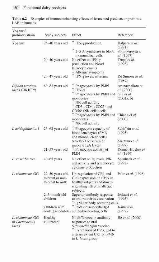
150 Functional dairy products
Table 6.2 Examples of immunoenhancing effects of fermented products or probioticLAB in humans
Yoghurt/probiotic strain Study subjects Effect Reference
Yoghurt 25–40 years old ↑ IFN-γ production Halpern et al.(1991)
↑ 2–5 A synthetase in blood Solis-Pereyra etmononuclear cells al. (1997)
20–40 years old No effect on IFN-γ Trapp et al.production and blood (1993)leukocyte counts↓ Allergic symptoms
20–47 years old ↑ IFN-γ levels in serum De Simone et al.(1989)
Bifidobacterium 60–83 years old ↑ Phagocytosis by PMN Arunachallam etlactis (DR10™) ↑ IFN-α al. (2000)
↑ Phagocytosis by PMN and Gill et al.monocytes (2001a, b)↑ NK cell activity↑ CD3+, CD4+, CD25+ andCD56+ (NK cells) cells↑ Phagocytosis by PMN and Chiang et al.monocytes (2000)↑ NK cell activity
L acidophilus La1 23–62 years old ↑ Phagocytic capacity of Schiffrin et al.blood leucocytes (PMN (1995)and mononuclear cells)No effect on serum or Marteau et al.mucosal IgA levels (1997)
21–57 years old ↑ Phagocytic activity of Donnet-Hughes etPMN al. (1999)
L. casei Shirota 40–65 years No effect on Ig levels, NK Spanhaak et al.cell activity and lymphocyte (1998)cytokine production
L. rhamnosus GG 22–50 years old, Up-regulation of CR1 and Pelto et al. (1998)tolerant or non- CR3 expression on PMN intolerant to milk healthy subjects and down-
regulating effect in allergicsubjects
2–5-month-old Superior antibody response Isolauri et al.children to oral rotavirus vaccination (1995)
↑ IgM antibody secreting cellsChildren with ↑ Rotavirus-specific IgA Kaila et al.acute gastoentritis antibody-secreting cells (1992)
L. rhamnosus GG Healthy No difference in antibody He et al. (2000)or Lactococcus volunteers responses to orallactis Salmonella typhi vaccine
↑ Expression of CR3, and toa lesser extent CR1 on PMNin L. lactis group
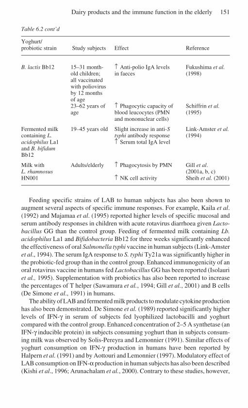
Dairy products and the immune function in the elderly 151
Table 6.2 cont’d
Yoghurt/probiotic strain Study subjects Effect Reference
B. lactis Bb12 15–31 month- ↑ Anti-polio IgA levels Fukushima et al.old children; in faeces (1998)all vaccinatedwith poliovirusby 12 monthsof age23–62 years of ↑ Phagocytic capacity of Schiffrin et al.age blood leucocytes (PMN (1995)
and mononuclear cells)
Fermented milk 19–45 years old Slight increase in anti-S Link-Amster et al.containing L. typhi antibody response (1994)acidophilus La1 ↑ Serum total IgA leveland B. bifidumBb12
Milk with Adults/elderly ↑ Phagocytosis by PMN Gill et al.L. rhamnosus (2001a, b, c)HN001 ↑ NK cell activity Sheih et al. (2001)
Feeding specific strains of LAB to human subjects has also been shown toaugment several aspects of specific immune responses. For example, Kaila et al.(1992) and Majamaa et al. (1995) reported higher levels of specific mucosal andserum antibody responses in children with acute rotavirus diarrhoea given Lacto-bacillus GG than the control group. Feeding of fermented milk containing Lb.acidophilus La1 and Bifidobacteria Bb12 for three weeks significantly enhancedthe effectiveness of oral Salmonella typhi vaccine in human subjects (Link-Amsteret al., 1994). The serum IgA response to S. typhi Ty21a was significantly higher inthe probiotic-fed group than in the control group. Enhanced immunogenicity of anoral rotavirus vaccine in humans fed Lactobacillus GG has been reported (Isolauriet al., 1995). Supplementation with probiotics has also been reported to increasethe percentages of T helper (Sawamura et al., 1994; Gill et al., 2001) and B cells(De Simone et al., 1991) in humans.
The ability of LAB and fermented milk products to modulate cytokine productionhas also been demonstrated. De Simone et al. (1989) reported significantly higherlevels of IFN-γ in serum of subjects fed lyophilized lactobacilli and yoghurtcompared with the control group. Enhanced concentration of 2–5 A synthetase (anIFN-γ inducible protein) in subjects consuming yoghurt than in subjects consum-ing milk was observed by Solis-Pereyra and Lemonnier (1991). Similar effects ofyoghurt consumption on IFN-γ production in humans have been reported byHalpern et al. (1991) and by Aottouri and Lemonnier (1997). Modulatory effect ofLAB consumption on IFN-α production in human subjects has also been described(Kishi et al., 1996; Arunachalam et al., 2000). Contrary to these studies, however,
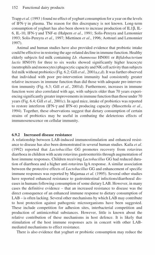
152 Functional dairy products
Trapp et al. (1993 ) found no effect of yoghurt consumption for a year on the levelsof IFN-γ in plasma. The reason for this discrepancy is not known. Long-termconsumption of yoghurt has also been shown to increase production of IL1β, IL-6, IL-10, IFN-γ and TNF-α (Halpern et al., 1991; Solis-Pereyra and Lemonnier1993; Solis-Pereyra et al., 1997; Miettinen et al., 1996; Aottouri and Lemonnier1997).
Animal and human studies have also provided evidence that probiotic intakecould be effective in restoring the age-related decline in immune function. Healthyelderly subjects fed milk containing Lb. rhamnosus HN001 or Bifidobacteriumlactis HN019) for three to six weeks showed significantly higher leucocyte(neutrophils and monocytes) phagocytic capacity and NK cell activity than subjectsfed milk without probiotics (Fig. 6.2; Gill et al., 2001a,c,d). It was further observedthat individual with poor pre-intervention immunity had consistently greaterrelative increases in immune function than did those with adequate pre-interven-tion immunity (Fig. 6.3; Gill et al., 2001d). Furthermore, increases in immunefunction were also correlated with age, with subjects older than 70 years experi-encing significantly greater improvements in immune function than those under 70years (Fig. 6.4; Gill et al., 2001c). In aged mice, intake of probiotics was reportedto restore interferon (IFN-γ and IFN-α) producing capacity (Muscettola et al.,1994). Together, these observations suggest that dietary consumption of certainstrains of probiotics may be useful in combating the deleterious effects ofimmunosenescence on cellular immunity.
6.9.2 Increased disease resistanceA relationship between LAB-induced immunostimulation and enhanced resist-ance to disease has also been demonstrated in several human studies. Kaila et al.(1992) reported that Lactobacillus GG promotes recovery from rotavirusdiarrhoea in children with acute rotavirus gastroenteritis through augmentation ofhost immune responses. Children receiving Lactobacillus GG had reduced dura-tion of diarrhoea and a higher anti-rotavirus IgA response. A similar associationbetween the protective effects of Lactobacillus GG and enhancement of specificimmune responses was reported by Majamaa et al. (1995). Several other studieshave reported enhanced resistance to gastrointestinal infections/diarrhoeal dis-eases in humans following consumption of some dietary LAB. However, in manycases the definitive evidence – that an increased resistance to disease was thedirect consequence of an enhanced immune response to dietary consumption ofLAB – is often lacking. Several other mechanisms by which LAB may contributeto host protection against pathogenic microorganisms have been suggested.These include competition for adhesion sites, interbacterial competition andproduction of antimicrobial substances. However, little is known about therelative contribution of these mechanisms in host defence. It is likely thatstimulation of the host immune responses acts in concert with other LAB-mediated mechanisms to effect resistance.
There is also evidence that yoghurt or probiotic consumption may reduce the
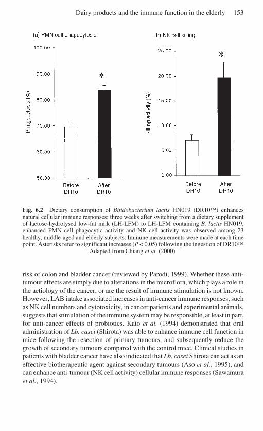
Dairy products and the immune function in the elderly 153
Fig. 6.2 Dietary consumption of Bifidobacterium lactis HN019 (DR10™) enhancesnatural cellular immune responses: three weeks after switching from a dietary supplementof lactose-hydrolysed low-fat milk (LH-LFM) to LH-LFM containing B. lactis HN019,enhanced PMN cell phagocytic activity and NK cell activity was observed among 23healthy, middle-aged and elderly subjects. Immune measurements were made at each timepoint. Asterisks refer to significant increases (P < 0.05) following the ingestion of DR10™
Adapted from Chiang et al. (2000).
risk of colon and bladder cancer (reviewed by Parodi, 1999). Whether these anti-tumour effects are simply due to alterations in the microflora, which plays a role inthe aetiology of the cancer, or are the result of immune stimulation is not known.However, LAB intake associated increases in anti-cancer immune responses, suchas NK cell numbers and cytotoxicity, in cancer patients and experimental animals,suggests that stimulation of the immune system may be responsible, at least in part,for anti-cancer effects of probiotics. Kato et al. (1994) demonstrated that oraladministration of Lb. casei (Shirota) was able to enhance immune cell function inmice following the resection of primary tumours, and subsequently reduce thegrowth of secondary tumours compared with the control mice. Clinical studies inpatients with bladder cancer have also indicated that Lb. casei Shirota can act as aneffective biotherapeutic agent against secondary tumours (Aso et al., 1995), andcan enhance anti-tumour (NK cell activity) cellular immune responses (Sawamuraet al., 1994).
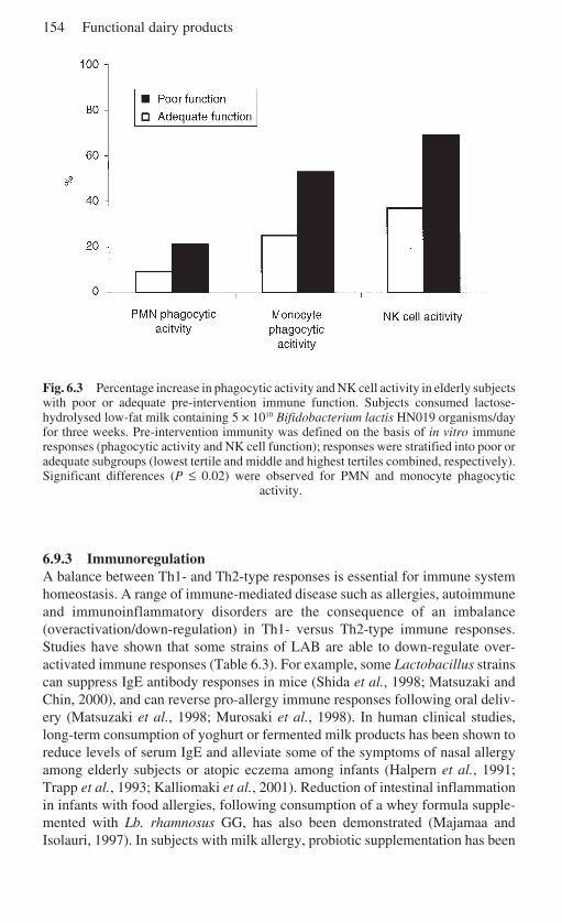
154 Functional dairy products
Fig. 6.3 Percentage increase in phagocytic activity and NK cell activity in elderly subjectswith poor or adequate pre-intervention immune function. Subjects consumed lactose-hydrolysed low-fat milk containing 5 × 10¹⁰ Bifidobacterium lactis HN019 organisms/dayfor three weeks. Pre-intervention immunity was defined on the basis of in vitro immuneresponses (phagocytic activity and NK cell function); responses were stratified into poor oradequate subgroups (lowest tertile and middle and highest tertiles combined, respectively).Significant differences (P ≤ 0.02) were observed for PMN and monocyte phagocytic
activity.
6.9.3 ImmunoregulationA balance between Th1- and Th2-type responses is essential for immune systemhomeostasis. A range of immune-mediated disease such as allergies, autoimmuneand immunoinflammatory disorders are the consequence of an imbalance(overactivation/down-regulation) in Th1- versus Th2-type immune responses.Studies have shown that some strains of LAB are able to down-regulate over-activated immune responses (Table 6.3). For example, some Lactobacillus strainscan suppress IgE antibody responses in mice (Shida et al., 1998; Matsuzaki andChin, 2000), and can reverse pro-allergy immune responses following oral deliv-ery (Matsuzaki et al., 1998; Murosaki et al., 1998). In human clinical studies,long-term consumption of yoghurt or fermented milk products has been shown toreduce levels of serum IgE and alleviate some of the symptoms of nasal allergyamong elderly subjects or atopic eczema among infants (Halpern et al., 1991;Trapp et al., 1993; Kalliomaki et al., 2001). Reduction of intestinal inflammationin infants with food allergies, following consumption of a whey formula supple-mented with Lb. rhamnosus GG, has also been demonstrated (Majamaa andIsolauri, 1997). In subjects with milk allergy, probiotic supplementation has been
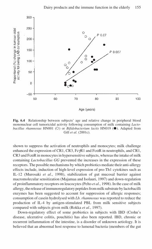
Dairy products and the immune function in the elderly 155
Fig. 6.4 Relationship between subjects’ age and relative change in peripheral bloodmononuclear cell tumoricidal activity following consumption of milk containing Lacto-bacillus rhamnosus HN001 (¤) or Bifidobacterium lactis HN019 (õ). Adapted from
Gill et al. (2001c).
shown to suppress the activation of neutrophils and monocytes; milk challengeenhanced the expression of CR1, CR3, FcγR1 and FcαR in neutrophils, and CR1,CR3 and FcαR in monocytes in hypersensitive subjects, whereas the intake of milkcontaining Lactobacillus GG prevented the increases in the expression of thesereceptors. The possible mechanisms by which probiotics mediate their anti-allergyeffects include, induction of high-level expression of pro-Th1 cytokines such asIL-12 (Murosaki et al., 1998), stabilization of gut mucosal barrier againstmacromolecular sensitization (Majamaa and Isolauri, 1997) and down-regulationof proinflammatory receptors on leucocytes (Pelto et al., 1998). In the case of milkallergy, the release of immunoregulatory peptides from milk substrate by lactobacillienzymes has been suggested to account for suppression of allergic responses;consumption of casein hydrolysed with Lb. rhamnosus was reported to reduce theproduction of IL-4 by antigen-stimulated PBL from milk sensitive subjectscompared with subjects given milk (Rokka et al., 1997).
Down-regulatory effect of some probiotics in subjects with IBD (Crohn’sdisease, ulcerative colitis, pouchitis) has also been reported. IBD, chronic orrecurrent inflammation of the intestine, is a disorder of unknown aetiology. It isbelieved that an abnormal host response to lumenal bacteria (members of the gut
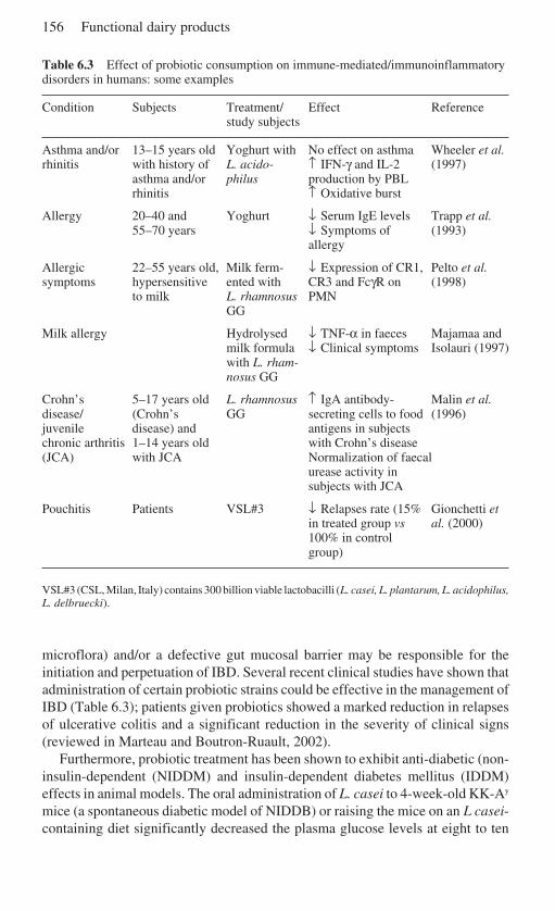
156 Functional dairy products
Table 6.3 Effect of probiotic consumption on immune-mediated/immunoinflammatorydisorders in humans: some examples
Condition Subjects Treatment/ Effect Referencestudy subjects
Asthma and/or 13–15 years old Yoghurt with No effect on asthma Wheeler et al.rhinitis with history of L. acido- ↑ IFN-γ and IL-2 (1997)
asthma and/or philus production by PBLrhinitis ↑ Oxidative burst
Allergy 20–40 and Yoghurt ↓ Serum IgE levels Trapp et al.55–70 years ↓ Symptoms of (1993)
allergy
Allergic 22–55 years old, Milk ferm- ↓ Expression of CR1, Pelto et al.symptoms hypersensitive ented with CR3 and FcγR on (1998)
to milk L. rhamnosus PMNGG
Milk allergy Hydrolysed ↓ TNF-α in faeces Majamaa andmilk formula ↓ Clinical symptoms Isolauri (1997)with L. rham-nosus GG
Crohn’s 5–17 years old L. rhamnosus ↑ IgA antibody- Malin et al.disease/ (Crohn’s GG secreting cells to food (1996)juvenile disease) and antigens in subjectschronic arthritis 1–14 years old with Crohn’s disease(JCA) with JCA Normalization of faecal
urease activity insubjects with JCA
Pouchitis Patients VSL#3 ↓ Relapses rate (15% Gionchetti etin treated group vs al. (2000)100% in controlgroup)
VSL#3 (CSL, Milan, Italy) contains 300 billion viable lactobacilli (L. casei, L. plantarum, L. acidophilus,L. delbruecki).
microflora) and/or a defective gut mucosal barrier may be responsible for theinitiation and perpetuation of IBD. Several recent clinical studies have shown thatadministration of certain probiotic strains could be effective in the management ofIBD (Table 6.3); patients given probiotics showed a marked reduction in relapsesof ulcerative colitis and a significant reduction in the severity of clinical signs(reviewed in Marteau and Boutron-Ruault, 2002).
Furthermore, probiotic treatment has been shown to exhibit anti-diabetic (non-insulin-dependent (NIDDM) and insulin-dependent diabetes mellitus (IDDM)effects in animal models. The oral administration of L. casei to 4-week-old KK-Ay
mice (a spontaneous diabetic model of NIDDB) or raising the mice on an L casei-containing diet significantly decreased the plasma glucose levels at eight to ten
Dairy products and the immune function in the elderly 157
weeks compared with the control group (Matsuzaki et al., 1997a). Analysis of hostimmune responses revealed that anti-diabetic effects of L. casei administrationwere associated with an increase in the proportions of CD4+ cells and a decrease inthe production of IFN-γ and IL-2. In non-obese diabetic (NOD) mice (an IDDMmodel), oral feeding of L. casei was found to inhibit the occurrence of diabetes andregulate host immune responses; the proportion of CD45R+ B cells in the spleenwas increased, and the proportion of CD8 cells and IFN-γ production weredecreased in mice fed L. casei compared with the control mice (Matsuzaki et al.,1997b).
The ability of lactobacilli to reduce subjective symptoms of arthritis amongrheumatoid patients (Nenonen et al.,1998) and the severity of collagen-inducedarthritis in mice (cited by Erickson and Hubbard, 2000 ) has also been reported.
6.10 Future trends
Ageing is associated with profound alterations in immune function. There iscompelling evidence to suggest that the increased susceptibility of the elderly toinfectious diseases, malignancies and autoimmune disorders is, at least in part, theresult of this immune dysfunction. Nutritional deficiencies, common in the elderly,are known to further impair immune responsiveness. Results of dietary interven-tion studies highlight the importance of nutrition in optimizing immune function inthe aged individuals. It has been demonstrated that supplementation with single ormultiple nutrients could restore or delay the age-related decline in immunefunction as well as increase resistance to infectious diseases. This offers uniqueopportunities to develop novel food products or supplements that are able tocorrect nutritional deficits as well as provide specific immune benefits. Dietaryapproaches are especially attractive as these are more practical and cost-effective,and could be implemented easily on a larger scale. In this context, the uniquenutritional and immunomodulatory properties of milk and milk products needcareful consideration. In addition to being a rich source of nutrients (such asproteins, growth factors, vitamins and minerals), bovine milk contains a host ofcomponents with potent immunomodulatory properties (immunostimulatory aswell as immunoregulatory). Several animal studies have shown that milk-derivedfactors are able to provide passive protection against a range of bacterial, viral andparasitic infections, enhance immune function and down-regulate over-activatedhost immune responses (inflammatory/autoimmune).
Consumption of fermented milk products containing specific strains of lacticacid bacteria has also been reported to enhance aspects of natural and acquiredimmune responses and regulate dysfunctional immune responses (allergies, chronicinflammatory diseases such as IBD) in human subjects. However, relatively littleis known about the effect of immunomodulatory milk or milk-derived products inthe elderly. Well-designed and controlled studies, involving healthy elderly,institutionalized elderly and the elderly who are afflicted by various immune-mediated and inflammatory diseases are required to evaluate the potential of
158 Functional dairy products
milk-based products for optimizing immune health in the elderly. These studiesneed to be conducted in conjunction with studies designed to understandmechanisms by which milk-derived products may influence the immune system.Without a mechanistic approach, the significance and relevance of this will becompromised. Identification of phenotypic markers of immune function is oflimited value unless these are of functional value. Thus the assays used to assessimmune status must be functionally relevant to clinical outcomes. Study designshould also reflect the real world. Also, there is a need for establishing dose–response relationships, for both the antigenic challenge and the nutrient/productunder investigation. The future will see the development of a range of milk andmilk-based products that are able to correct nutritional deficits as well as restore,prevent or slow the age-related decline in immune function and thus increaseresistance to diseases and responsiveness to vaccines. The potential of milk andmilk-based products to contribute to the improvement and maintenance of geriatrichealth is under-explored and untapped.
6.11 ReferencesALES-MARTINEZ J E, ALVAREJ-MON M, MERINO F, BONILLA F, MARTINEZ C, DURANTEZ C and
DE LA HERA A (1988). Decreased TcR-CD3-T cell numbers in healthy aged humans.Evidence that T cell defects are marked by a reciprocal increases of TcR-CD3-CD2+
natural killer cells. Eur J Immunol, 18, 1827–1830.AOTTOURI N and LEMONNIER D (1997), Production of interferon induced by Streptococcus
thermophilus: role of CD4+ and CD8+ lymphocytes. Nutr Biochem, 8, 25–31.ARUNACHALAM K, GILL H S and CHANDRA R K (2000), Enhancement of natural immune
function by dietary consumption of Bifidobacterium lactis (HN019). Eur J Clin Nutr, 54,1–5.
ASO Y, AKAZA H, KOTAKE T, TSUKAMOTO T, IMAI K and NAITO S (1995), Preventive effectof a Lactobacillus casei preparation on the recurrence of superficial bladder cancer in adouble-blind trial. The BLP Study Group. Eur Urol, 27, 104–109.
BOESMAN-FINKELSTEIN M, WALTON N E and FINKELSTEIN R E (1989), Bovine lactogenicimmunity against cholera toxin-related enterotoxins and Vibrio cholerae outer mem-branes. Infect Immunity, 57, 1227–1235.
BOGDEN J D, OLESKE J M, LAVENHAR M A, MUNVES E M, KEMP F W, BRUENING K S, HOLDINGK J, DENNY T N, GUARINO M A and HOLLAND B K (1990), Effects of one year ofsupplementation with zinc and other micronutrients on cellular immunity in the elderly.J Am Coll Nutr, 9, 214–225.
BOUNOUS G and GOLD P (1989), Whey protein composition, a method for producing it anduse of composition. New Zealand Patent 231865.
BOUNOUS G and GOLD P (1993), Treatment of HIV-sero-positive individuals – usingundenatured whey protein concentrate to increase T-helper cell concentration and bloodglutathione content. Patent Number 9320831 and 93–351356/44.
BOUNOUS G, STEVENSON M M and KONGSHAVN P A (1981), Influence of dietary lactalbuminhydrolysate on the immune system of mice and resistance to salmonellosis. J Infect Dis,144, 281–290.
BOUNOUS G and KONGSHAVN P A L (1985), Differential effect of dietary protein type on theB-cell and T-cell immune responses in mice. J Nutr, 115, 1403–1408.
BOUNOUS G and KONGSHAVN P A L (1989), Influence of protein type in nutritionally adequatediets on the development of immunity. In: Absorption and Utilisation of Amino Acids, Vol.II, M Freedman, Ed; Boca Raton, Florida, CRC Press, 219–233.
Dairy products and the immune function in the elderly 159
BOUNOUS G, LETOURNEAU L and KONGSHAVN P A L (1983), Influence of dietary protein typeon the immune system of mice. J Nutr, 113, 1415–1421.
BOUNOUS G, SHENOUDA N, KONGSHAVN P A and OSMOND D G (1985), Mechanism of alteredB-cell response induced by changes in dietary protein type in mice. J Nutr, 115, 1409–1417.
BOUNOU G, KONGSHAVN P A AND GOLD P (1988), The immunoenhancing property of dietarywhey protein concentrate. Clin Invest Med, 11, 271–278.
BOUNOUS G, BARUCHEL S, FALUTZ J AND GOLD P (1993), Whey proteins as a food supplementin HIV-seropositive individuals. Clin Invest Med, 16, 204–209.
BROSCHE T and PLATT D (1995), Nutritional factors and age-associated changes in cellularimmunity and phagocytosis: a review. Ageing Immunol Infect Dis, 6, 29–40.
BRUNSER O, ESPINOZA J, ARAYA M, SPENCER E, HILPERT H, LINK-AMSTER H and BRUSSOW H(1992) Field trial of an infant formula containing anti-rotavirus and anti-Escherichia colimilk antibodies from hyperimmunized cows. J Pediatr Gastroent Nutr, 15, 63–72.
BUTCHER S, CHAHEL H and LORD J M (2000), Ageing and the neutrophils: no appetite forkilling? Immunology, 1000, 411–416.
BUZINA-SUBOTICANEC K, BUZINA R, STAVLJENIC A, FARLEY T M, HALLER J, BERGMAN-MARKOVIC B and GORAJSCAN M (1998), Ageing, nutritional status and immune response.Int J Vit Nutr Res, 68, 133–141.
CAKMAN I, ROHWER J, SCHUTZ R M, KIRCHNER H and RINK L (1996), Dysregulation betweenTh1 and Th2 T cell subpopulations in the elderly. Mech Age Dev, 87, 197–209.
CASSWALL T H, SARKER S A, ALBERT M J, FUCHS G J, BERGSTROM M, BJORCK L andHAMMARSTROM L (1998), Treatment of Helicobacter pylori infection in infants in ruralBangladesh with oral immunoglobulins from hyperimmune bovine colostrum. AlimentPharmacol Ther, 12, 563–568.
CHANDRA R K (1985),Trace element regulation of immunity and infection. J Am Coll Nutr,4, 5–16.
CHANDRA R K (1989), Nutrition, regulation of immunity and risk of infection in old age.Immunology, 67, 141–147.
CHANDRA R K (1990), Nutrition is an important determinant of immunity in old age. In:Nutrition and Ageing, D M Prinsley and H H Standsteas, Eds; New York, Alan R Liss,321–334.
CHANDRA R K (1992), Effect of vitamin and trace-element supplementation on immuneresponses infection in elderly subjects. Lancet, 340, 1124–1127.
CHANDRA R K (1995), Nutrition and immunity in the elderly: Clinical significance. Nutr Rev,53, S80–83.
CHANDRA R K (1997a), Graying of the immune system: can nutrient supplements improveimmunity in the elderly? JAMA, 277, 1398–1399.
CHANDRA R K (1997b), Nutrition and the immune system: an introduction. Am J Clin Nutr,66, 460S–463S.
CHIANG B L, SHEIH Y H, WANG L H, LIAO C K and GILL H S (2000), Enhancing immunity bydietary consumption of a probiotic lactic acid bacterium (Bifidobacterium lactis HN019):optimization and definition of cellular immune responses. Eur J Clin Nutr, 54, 849–855.
CLARK J A AND PETERSON T C (1994), Cytokine production and ageing: overproduction ofIL-8 in elderly males in response to lipopolysaccharide. Mech. Ageing Dev, 77, 127–139.
CORBERAND J, NGYEN F, LAHARRAGUE P, FONTANILLES A M, GLEYZES B, GYRARD E andSENEGAS C (1981), Polymorphonuclear function and ageing in humans. J Am Geriatr Soc,29, 391–397.
CROSS M L and GILL H S (2001a), Immunomodulatory properties of milk. Br J Nutr, 84(Supplement No. 1), S81–S89.
DARDENNE M, PLEAU J M, NABARRA B, LEFRANCIER P., DERRIEN M, CHOAY M and BATCH J F
(1982), Contribution of Zn and other metals to the biologica activity of the serum thymicfactor, Proc Natl Acad Sci USA, 79, 5370–5373.
DE SIMONE C, BIANCHI SALVADORI B, JIRILLO E, BALDINELLI L, BITONTI F and VESELY R
160 Functional dairy products
(1989), Modulation of immune activities in humans and animals by dietary lactic acidbacteria. In: Yogurt Nutritional and Health Properties, R C Chandan, Ed; London, JohnLibbey Eutotext, 201–213.
DE SIMONE C, ROSATI E, MORETTI S, BIANCHI SALVADORI B, VESELY R and JIRILLO E (1991),Probiotics and stimulation of the immune response. Europ J Clin Nutr, 45 (Suppl.), 32–34.
DING A, HWANG S and SCHWAB R (1994), Effect of ageing on murine macrophages:diminished response to IFN-gamma for enhanced oxidative metabolism. J. Immunol, 153,2146–2152.
DONNET-HUGHES A, ROCHAT F, SERRANT P, AESCHLIMANN J M and SCHIFFRIN E J (1999),Modulation of nonspecific mechanisms of defense by lactic acid bacteria: effective dose.J Dairy Sci, 82, 863–869.
DORIA G and FRASCA D (1997), Genes, immunity, and senescence: looking for a link. ImunolRev, 160, 159–170.
EBINA T, SATO A, UMEZU K, ISHIDA N, OHYAMA S, OIZUMI A, AIKAWA K, KATAGIRI S,KATSUSHIMA N, IMAI A et al. (1985), Prevention of rotavirus infection by oral administra-tion of cow colostrum containing antihuman rotavirus antibody. Med Microbiol Immunol(Berl), 174, 177–185.
ERICKSON K L and HUBBARD N E (2000), Probiotic immunomodulation in health and disease.J. Nutr, 130, 403S–409S.
ERNST D N, WEIGLE W O, NOONAN D J, MCQUITTY D N and HOBBS M V (1990a), Differencesin the expression profile of CD45RB, Pgp-1 and 3G11 membrane antigens and in thepatterns of lymphokine secretion by splenic CD4+ T cells from young and aged mice. JImmunol, 145, 1295–1302.
ERNST D N, WEIGLE W O, NOONAN D J, MCQUITTY D N and HOBBS M V (1990b), Differencesin the subset composition of CD4+ T cell populations from young and old mice. AgeingImmunol Infect Dis, 2, 105–109.
ERNST D N, WEIGLE O and HOBBS M V (1995), Aging and lymphokine gene expression by Tcell subsets. Nutr Rev, 53(II), S18–S24.
ESPERAZA B, SANCHEZ M, RUIZ M, BARRANQUERO M, SABINO E and MERINO F (1996),Neutrophil function in elderly persons assessed by flow cytometry. Immunol Invest, 25,185–190.
FACCHINI A, MARIANI E, MARIANI A R, PAPA S, VITALE M and MANZOLI F A (1987), Increasednumber of circulating Leu 11+ (CD16) large granular lymphocytes and decreased NKactivity during ageing. Clin Exp Immunol, 68, 340–347.
FELL, J M, PAINTIN M, ARNAUD-BATTANDIER F, BEATTIE R M, HOLLIS A, KITCHING P, DONNET-HUGHES A, MACDONALD T T and WALKER-SMITH J A (2000), Mucosal healing and a fallin mucosal pro-inflammatory cytokine mRNA induced by a specific oral polymeric dietin paediatric Crohn’s disease. Aliment Pharmacol Ther, 14, 281–289.
FERGUSON F G, WIKBY A, MAXON P, OLSSON J and JOHNSSON B (1995), Immune parametersin a longitudinal study of a very old population of Swedish people: a comparison betweensurvivors and nonsurvivors. J Gerontol, 50, B378–B382.
FORD J T, WONG C W and COLDITZ I G (2001), Effects of dietary protein type on immuneresponses and levels of infection with Eimeria vermiformis in mice. Immunol Cell Biol,79, 23–27.
FORTES C, FORASTIERE F, AGABITI N, FANO V, PACIFICI R, VIRGILI F, PIRAS G, GUIDI L,BARTOLONI C, TRICERRI A, ZUCCARO P, EBRAHIM S and PERUCCI C A (1998), The effect ofzinc and vitamin A supplementation on immune response in an older population. J Am GerSoc, 46, 19–26.
FRANCESCHI C, MONTI D, SANSONI P and COSSARIZZA A (1995), The immunology ofindividuals: the lesson of centenarians. Immunol Today, 16, 12–16.
FREEDMAN D J, TACKET C O, DELEHANTY A, MANEVAL D R, NATARO J and CRABB J H (1998),Milk immunoblobulin with specific activity against purified colonization factor antigens
Dairy products and the immune function in the elderly 161
can protect against oral challenge with enterotoxigenic Escherichia coli. J Infect Dis, 177,662–667.
FUKUSHIMA Y, KAWATA Y, HARA H, TERADA A and MITSUOKA T (1998), Effect of a probioticformula on intestinal immunoglobulin A production in healthy children. Int J FoodMicrobiol, 42, 39–44.
FULOP T, KOMAROMI I, FORRIS G, WORUM I and LEOVY A (1986), Age-dependent variationsof intra-lysosomal release from human PMN leukocytes under various stimuli.Immunobiology, 171, 302–307.
FURUKAWA T, MEYDANI S N and BLUMBERG J B (1987), Reversal of age-associated declinein immune responsiveness by dietary glutathione supplementation in mice. Mech AgeingDevelop, 38, 107–117.
GARG M, LUO W, KAPLAN A M and BONDADA S (1996), Cellular basis of decreased immuneresponse to pneumococcal vaccine in aged mice. Infect Immunol, 64, 4456–4462.
GILL H S (1998), Stimulation of the immune system by lactic cultures. Int Dairy J, 8, 535–544.
GILL H S (2000), Dairy products and immune health. In: Functional Foods 2000, F Angusand C Miller, Eds, Leatherhead Publishing, Surrey, 268–284.
GILL H S and CROSS L (2000), Anti-cancer properties of milk. Brit J Nutr, 84, S161–S166.GILL H S and CROSS L (2002), Probiotics and immune function. In: Nutrition and Immune
Function, P Calder P, S Fields and H S Gill, Eds, London, CABI Publishing, 251–272.GILL H S and RUTHERFURD K J (1998), Immunomodulatory properties of bovine milk. Bull
Int Dairy Fed, 336, 31–35.GILL H S, DOULL F, RUTHERFURD K J and CROSS M L (2000), Immunoregulatory peptides in
milk. Br J Nutr, 84 (supplement 1), 111–117.GILL H S, CROSS M L, RUTHERFURD K J and GOPAL, P K (2001a), Dietary probiotic
supplementation to enhance cellular immunity in the elderly. Brit J Biomed Sci, 58, 94–96.GILL H S, DARRAGH A J and CROSS M L (2001b), Optimising immunity and gut function in the
elderly. J Nutr, Health Ageing, 5, 80–91.GILL H S, RUTHERFURD K J and CROSS M L (2001c), Dietary probiotic supplementation
enhances natural killer cell activity in the elderly: an investigation of age-relatedimmunological changes. J Clin Immunol, 21, 264–271.
GILL H S, RUTHERFURD K J, CROSS M L and GOPAL P K (2001d), Enhancement of immunityin the elderly by dietary supplementation with the probiotic Bifidobacterium lactisHN019. Am J Clin Nutr, 74, 833–839.
GILL H S, RUTHERFURD K J, FRAY L and BROOMFIELD A (2002a), Bovine whey proteins:immunomodulatory effects in mice. Unpublished.
GILL H S and colleagues (2002b), Dose response immunoenhancing effects of whey proteinconcentrate in follow-on milk. Unpublished.
GILL H S and colleagues (2002c), Effect of an immunostimulating whey protein concentratedelivered to mice in milk, based on a growing-up milk formula format. Unpublished.
GILL H S and colleagues (2002d), Immunostimulatory effects of bovine whey proteins ininfant milk formula. Unpublished.
GINALDI L, DE MARTINIS M, MODESTI M, LORETO M F and QUAGLINO D (1999), The immunesystem in the elderly. II. Specific cellular immunity. Immunol Res, 20, 109–115.
GINALDI L, DE MARTINIS M, MODESTI M, LORETO M F, CORSI M P and QUAGLINO D (2000),Immunophenotypical changes of T lymphocytes in the elderly. Gerontology, 46, 242–248.
GINALDI L, LORETO M F, CORSI M P, MODESTI M and DE MARTINUS M (2001), Immunosenescenceand infectious diseases. Microbes Infection, 3, 851–857.
GIONCHETTI P, RIZZELLO F, VENTURI A, BIRGIDI P, MATTEUZZI D, BAZZOCCHI G, POGGIOLI G,MIGLIOLI M and CAMPIERI M (2000), Oral bacteriotherapy as maintenance treatment inpatients with chronic pouchitis: a double-blind, placebo-controlled trial. Gastroenterol-ogy, 119, 305–309.
162 Functional dairy products
GOLD P, BOUNOUS G and KONGSHAVN P A L (1989), Lactalbumin as food supplement (effectof dietary whey protein (lactalbumin) on the immune response to sheep red blood cells,host resistance to bacterial infections, development of tumors and the process of ageing).European Patent 0339656.
GOODWIN J S (1992), Changes in lymphocyte sensitivity to prostaglandin E2, histamines,hydrocortisone, and X irradiation with age: studies in a healthy elderly population. Clin.Exp Immunol Immunopathol, 25, 243–251.
GOODWIN J S (1995), Decreased immunity and increased morbidity in the elderly. Nutr Rev,53(II), S41–45.
GREEN-JOHNSON J M, HAQ J A and SZEWCZUK M R (1991), Effects of aging on the production ofcytoplasmic interleuikn-4 and 5, and interferon-gamma, by mucosal and systemiclymphocytes after activation with phytohemagglutinin. Aging Immunol Infect Dis, 3, 43–47.
HALLER J, WEGGEMANS ER M, LAMMI-KEEFI C J and FERRY M (1996), Changes in the vitaminstatus of elderly Europeans: plasma vitamins A, E, B-6, B-12, folic acid and caroteniods.Eur J Clin Nutr, 50 (Suppl. 2), S32–S45.
HALPERN G M, VRUWINK K G, VAN DE WATER J, KEEN C L and GERSHWIN M E (1991), Influenceof long-term yoghurt consumption in young adults. Int J Immunotherapy, 7, 205–210.
HAYEK G M, MURA C, WU D, BEHRKA A A, HAN S N, PAULSON E, HWANG D and MEYDANI S N(1997), Enhanced expression of cyclooxygenase with age in murine macrophages. JImmunol, 159, 2445–2451.
HE F, TUOMOLA L A, ARVILOMMI H and SALMONEN S (2000), Modulation of humoral immuneresponses through intake of probiotics. FEMS Immunol Med Microbiol, 29, 47–52.
HEUSER M D and ADLER W H (1997), Immunological aspects of aging and malnutrition:consequences and intervention with nutritional immunomodulators. Clin Geriatr Med,13, 697–715.
HIGASHIMOTO Y, OHATA M, UETANI K et al. (1994), Influence of age on pulmonary alveolarmacrophage clonal growth. Japan J Geriatr, 31, 854–859.
HIGH K P (1999), Micronutrient supplementation and immune function in the elderly. ClinInfect Dis, 28, 717–722.
HOMES K L, SCHNIZLEIN C T, PERKINS E H and TEW J C (1984), The effect of age on nitrogenretention in lymphoid follicles and in collagenous tissues of mice. Mech Ageing Dev, 25,243–255.
INGLOT A. JANUSZ M and LISOWSKI J (1996), Colostrinine: a proline rich polypeptide fromovine colostrum is a modest cytokine inducer in human leukocytes. Arch Immune TherExp, 44, 215–224.
ISHIDA A, YOSHIKAI Y, MUROSAKI S, HIDAKA Y and NOMOTO K (1992a), Administration ofmilk from cows immunized with intestinal bacteria protects mice from radiation-inducedlethality. Biotherapy, 5, 215–225.
ISHIDA A, YOSHIKAI Y, MUROSAKI S, KUBO C, HIDAKA Y and NOMOTO K (1992b), Consump-tion of milk from cows immunized with intestinal bacteria influences age-related changesin immune competence in mice. J. Nutr, 122, 1875–1883.
ISOLAURI E, JOENSUS J, SUOMALAINEN H, LUOMALA M and VESIKARI, T (1995), Improvedimmunogenicity of oral D XRRV reabsorbant rotavirus vaccine by Lactobacillus caseiGG. Vaccine, 13, 310–312.
ISOLAURI E, SUTAS Y, KANKAANPAA P, ARVILOMMI H and SALMINEN S (2001), Probioticeffect on immunity. Am J Clin Nutr, 73, 444S–450S.
JANUSZ M, LISOWSKI J and FRANEK F (1974), Isolation and characterization of a proline-richpolypeptide from ovine colostrum. FEBS Lett, 49, 276–281.
JANUSZ M, ZIMECKI M and LISOWSKI J (1981), Chemical and physical characterization of aproline-rich peptide from sheep colostrum. Biochem J, 199, 9–15.
JANUSZ M, STAROSCIK K, ZIMECKI M, WIECZOREK Z and LISOWSKI J (1986), A proline-richpolypeptide PRP with immunoregulatory properties isolated from ovine colostrummurine thymocytes have on their surface receptor specific for PRP. Arch Immune TherExp, 34, 427–436.
Dairy products and the immune function in the elderly 163
JULIUS M H, JANSUZ M and LISOWSKI J (1988), A colostral protein that induces the growth anddifferntiation of resting B lymphocytes. J Immunol, 140, 1366–1371.
KAILA M, ISOLAURI E, SOPPI E, VIRTANEN E, LAINE S and ARVILOMMI H (1992), Enhancementof the circulating antibody secreting cell response in human diarrhea by a humanLactobacillus strain. Pediatr Res, 32, 141–144.
KALLIOMAKI M, SALMINEN S, ARVILOMMI H, KERO P, KOSKINEN P and ISOLAURI E (2001),Probiotics in primary prevention of atopic disease: a randomised placebo-controlled trial.Lancet, 357, 1076–1079.
KATO I, ENDO K YOKOKURA T (1994), Effects of oral administration of Lactobacillus caseion antitumor responses induced by tumor resection in mice. Int J Immunopharmacol, 16,29–36.
KEEN C L and GERSHWIN M E (1990), Zinc deficiency and immune function. Ann Rev Nutr,10, 415–431
KELLEY D S and BENDICH A (1996), Essential nutrients and immunologic functions. Am J ClinNutr, 63, 994S–996S.
KENNEDY R S, KONOK P, BOUNOUS G, BARUCHEL S and LEE T D G (1995), The use of wheyprotein concentrate in the treatment of patients with metastatic carcinoma: a phase I–IIclinical study. Anticancer Res, 15, 2643–2650.
KISHI A, UNO K, MATSUBARA Y, OKUDA C and KISHIDA T (1996), Effect of the oraladministration of Lactobacillus brevis subsp. coagulans on interferon-α producingcapacity in humans. J Am Coll Nutr, 15, 408–412.
KORHONEN H, MARNILA P and GILL H S (2000), Bovine milk antibodies for health. Brit J Nutr,84, SS135–S146.
KUBO M and CINADER B (1990), Polymorphism of age-related changes in interleukin (IL)production: differential changes of T-helper subpopulations, synthesizing IL-2, IL-3 andIL-4. Eur J Immunol, 24, 133–136.
KULKARNI A B and KARLSSON S (1993), Transforming growth factor β1-knockout mice. Amutation in one cytokine gene causes a dramatic inflammatory disease. Am J Pathol, 143,3–9.
KUNDU B, PURI A, SINGH G, SAHAI R, TRIPATHI L M and SRIVASTAVA V M L (2000),Immunomodulatory activities of hexapeptides related to proline rich peptide fromcolostrum. Bioorg Med Chem Lett, 10, 1181–1183.
LEMAOULT J, SZABO P and WEKSLER M E (1997), Effect of age on humoral immunity,selection of the B-cell repertoire and B-cell development. Immunol Rev, 160, 115–126.
LESOURD B (1990a), Protein malnutrition: major cause of malnutrition in the elderly. AgeNutr, 1, 132–138.
LESOURD B M (1990b), Le vieillissement immunologique: influence de la denutrition(Immunological ageing: influence of undernutrition). Ann Biol Clin (Paris), 48, 309–318.
LESOURD B M (1995), Protein undernutrition as the major cause of decreased immunefunction in the elderly: Clinical and functional implications. Nutr Rev, 53(II), S86–S92.
LESOURD B M (1997), Nutrition and immunity in the elderly: modification of immuneresponses with nutritional treatments. Am J Clin Nutr, 66, 478S–484S.
LESOURD B and MAZARI L (1999), Nutrition and immunity in the elderly. Proc Nutr Soc, 58,685–695.
LESOURD, B M and MEAUME S (1994), Cell mediated immunity changes in ageing: relativeimportance of cell subpopulation switches and of nutritional factors. Immunol Lett, 40,235–242.
LESOURD B M, FAURE-BECONE, M and THIOLLET (1990), Action immunostimulante d’unesupplementation orale complete chez des sujets ages dentris (Immunostimulatory actionof a complete oral supplementation in undernourished elderly patients). Age Nutr, 1, 41–51.
LESOURD B, MAZARI L and FERRY M (1998), The role of nutrition in immunity in the aged.Nutr Rev, 56, S113–S125.
164 Functional dairy products
LESZEK J, INGLOT A D, JANSUZ M, LISOWSKI J, KRUKOWSKA K and GEORGEAIDES J A (1999),Clostrinin®: A proline-rich polypeptide (PRP) complex isolated from ovine colostrum fortreatment of Alzheimer’s disease: a double-blind, placebo-controlled study. Arch ImmuneTher Exp, 47, 377–385.
LIGTHART G J, SCHUIT H R and HIJMANS W (1989), Natural killer cell function is notdiminished in the healthy aged and is proportional to the number of NK cells in theperipheral blood. Immunology, 68, 396–402.
LINK-AMSTER H, ROCHAT F, SAUDAN K Y, MIGNOT O and AESCHLIMANN J M (1994),Modulation of a specific humoral immune response and changes in intestinal floramediated through fermented milk intake. FEMS Immunol Med Microbiol, 10, 55–64.
LIPSCHITZ D A, UDUPA K D and BOXER L A (1988), The role of calcium in age-related declineof neutrophils function. Blood, 71, 659–665.
LODINOVA-ZADNIKOVA R, KORYCH B and BARTAKOVA Z (1987), Treatment of gastrointestinalinfections in infants by oral administration of colostral antibodies. Die Nahrung, 31, 465–467.
LOIMARANTA V, LAINE M, SODERLING E, VASARA E, ROKKA S, MARNILA P, KORHONEN H,TOSSAVAINEN O and TENOVUO J (1999), Effects of bovine immune- and non-immunewhey preparations on the composition and pH response of human dental plaque. EuropJ Oral Sci, 107, 244–250.
LOW P, RUTHERFURD K J, CROSS M L and GILL H S (2001), Enhancement of mucosal antibodyresponses by dietary whey protein concentrate. Food Agric Immunol, 13, 255–264.
MACHNICKI M, ZIMECKI M and ZAGULSKI T (1993), Lactoferrin regulates the release of tumornecrosis factor alpha and interleukin 6 in vitro. Int J Exp Pathol, 74, 433–439.
MAJAMAA H and ISOLAURI E (1997), Probiotics: a novel approach in the management of foodallergy. J Allergy Clin Immunol, 99, 179–185.
MAJAMAA H, ISOLAURI E, SAXELIN M and VESIKARI T (1995), Lactic acid bacteria in thetreatment of acute rotavirus gastroenteritis. J Pediatric Gastroent Nutr, 20, 333–338.
MALIN M, SOUMALAINEN H, SAXELIN M and ISOLAURI E (1996), Promotion of IgA in patientswith Crohn’s disease by oral bacterial therapy with Lactobacillus GG. Ann Nutr Metab,40, 137–145.
MARTEAU P and BOUTRON-RAULT M C (2002), Nutritional advantages of probiotics andprebiotics. Br J Nutr, 87, S153–S157.
MARTEAU P, VAERMAN J P, DEHENNIN J P, BORD S, BRASSART D and POCHART P (1997), Effectof intrajejunal perfusion and chronic ingestion of Lactobacillus johnsonni strain La1 onserum concentrations and jejunal secretions of immunoglobulins and serum proteins inhealthy humans. Gastroenterol Clin Biol, 21, 293–308.
MATSUZAKI T and CHIN J (2000), Modulating immune responses with probiotic bacteria.Immunol Cell Biol, 78, 67–73.
MATSUZAKI T, YAMAZAKI R, HASHIMOTO S and YOKOKURA T (1997a), Antidiabetic effect ofLactobacillus casei in a non-insulin-dependent diabetes mellitus (NIDDM) model usingKK-Ay mice. Endocrine J, 44, 357–365.
MATSUZAKI T, NAGARA Y, KADO S, UCHIDA K, KATO I, HASHIMOTO S and YOKOKURA T(1997b), Prevention of onset in an insulin-dependent diabetes mellitus model, NOD mice,by oral feeding Lactobacillus casei. APMIS, 105, 643–649.
MATSUZAKI T, YAMAZAKI R, HASHIMOTO S and YOKOKURA T (1998), The effect of oralfeeding of Lactobacillus casei strain Shirota on immunoglobulin E production in mice.J Dairy Sci, 81, 48–53.
MCLAUGHLIN B, O’MALLEY K and COTTER T G (1986), Age-related differences in granulocytechemotaxis and degranulation. Clin Sci, 70, 59–64.
METCHNIKOFF E (1907), The Prolongation of Life. London, Heinemann.MEYDANI S N and HA W-K (2000), Immunologic effects of yoghurt. Am J Nutr, 71, 861–872.MEYDANI S N, MEYDANI M, BLUMBERG J B, LEKA L S, SIBER G, LOSZEWSKI R, THOMPSON C,
PEDROSA M C, DIAMOND R D and STOLLAR B D (1997), Vitamin E supplementation and invivo immune response in healthy elderly subjects. JAMA, 277, 1380–1386.
Dairy products and the immune function in the elderly 165
MICHALE S M, MCGHEE J R, ARNOLD R R and MESTECKY J (1978), Effective immunity to dentalcaries: selective induction of secretory immunity by oral administration of Streptococcusmutans in rodents. Adv Exp Med Biol, 107, 261–269.
MIETTINEN M, VUOPIO-VARKILA J and VARKILA K (1996), Production of human tumournecrosis factor alpha, interleukin-6, and interleukin-10 is induced by lactic acid bacteria.Infect Immun, 64, 5403–5405.
MIKES Z, FERENICIK M, JAHNOVA E, EBRINGER L and CIZNAR I (1995), Hypocholesterolemicand immunostimulatory effects of orally applied Enterococcus faecium M-74 in man.Folia Microbiol, 40, 639–646.
MILLER R A (1996), The ageing immune system. Science, 273, 70–74.MIYAUCHI H, KAINO A, SHINODA I, FUKUWATARI Y and HAYASAWA H (1997),
Immunomodulatory effect of bovine lactoferrin pepsin hydrolysate on murine splenocytesand Peyer’s patch cells. J Dairy Sci, 80 (10):2330–2339.
MONNAI M, HORIMOTO Y and OTANI H (1998), Immunomodulatory effect of dietary bovinekappa-caseinoglycopeptide on serum antibody levels and proliferative responses oflymphocytes in mice. Milchwissenschaft, 53 (3):129–132.
MOSMANN T R and COFFMA R L (1989), Heterogeneity of cytokine secretion patterns andfunction of helper T cells. Adv Immunol, 46, 111–115.
MOULIAS R, DEVILLECHABROLLE A, LESOURD B, PROUST J, MARESCOT M R, DOUMERC S,FAVRE-BERRONE M, CONGY F and WANG A (1985), Respective roles of immune andnutritional factors in priming of the immune response of the elderly. Mech. Ageing Dev,31, 123–137.
MUROSAKI S, YAMOMOTO Y, ITO K, INOKUCHI T, KUSAKA H, IKEDAH and YOSHIKAI Y (1998),Heat-killed Lactobacillus plantarum L-137 suppresses naturally fed antigen-specific IgEproduction by stimulation of IL-12 production in mice. J Allergy Clin Immunol, 102, 57–64.
MUSCETTOLA M, MASSAI L, TANGANELLI C and GRASSO G (1994), Effects of lactobacilli oninterferon production in young and aged mice. Ann NY Acad Sci, 717, 226–232.
NENONEN M T, HELVE T A, RAUMA A L and HANNINEN O O (1998), Cooked, lactobacilli-rich,vegan food and rheumatoid arthritis. Br J Rheumatol, 37, 274–281.
NUTTER R L, GRIDLEY D S, KITTERING J D, ANDRES M L, APRECIO R M and SLATER J M (1983),Modification of a transplantable colon tumor and immune responses in mice fed differentsources of protein, fat and carbohydrate. Cancer Lett, 18, 49–62.
NUTTER R L, KITTERING J D, APRECIO R M, WEEKS D A and GRIDLEY D S (1990), Effects ofdietary fat and protein on DMH-induced tumor development and immune responses. NutrCancer, 13, 141–152.
OKHUYSEN P C, CHAPPELL C L, CRABB J, VALDER L M, DOUGLASS E T and DUPONT H L (1998),Prophylactic effect of bovine anti-Cryptosporidium parvum. Clin Infect Dis, 26. 1324–1329.
OONA M, RAGO T, MAAROOS H, MICKELSAAR M, LOIVUKENE K, SALMINEN S and KORHONENH (1997), Helicobacter pylori in children with abdominal complaints: has immune bovinecolostrum some influence on gastritis? Alpe Adria Microbiol J, 6, 49–57.
OPEKUN A R, EL-ZAIMAITY H M, OSATO M S, GILGER M A, MALATY H M, TERRY M, HEADOND R and GRAHAM D Y (1999), Novel therapies for Helicobacter pylori infection.Alimentary Pharmacol and Therapeut, 13, 35–42.
ORMROD D J and MILLER T E (1991), The anti-inflammatory activity of a low molecularweight component derived from the milk of hyperimmunised cows. Agents Actions, 32,160–166.
ORMROD D J and MILLER T E (1993), Milk from hyperimmunised dairy cows as a source ofa novel biologic response modifier. Agents Actions, 38, 146–149.
OTANI, H and YAMADA Y (1995), Effects of bovine kappa-casein and lactoferrin on severalexperimental models of allergic diseases. Milchwissenschaft, 50, 549–53.
OWENS W E and NICKERSON S C (1989), Evaluation of an anti-inflammatory factor derivedfrom hyperimmunized cows. Proc Soc Exp Biol Med, 190, 79–86.
166 Functional dairy products
PARKER N T and GOODRUM K J (1990), A comparison of casein lactalbumin and soy proteineffect on immune response to T-dependent antigen. Nutr Res, 10, 781–792.
PARODI P W (1999), The role of intestinal bacteria in the causation and prevention of cancer:modulation by diet and probiotics. Aust J Dairy Tech, 54, 103–121.
PAWELEC G and SOLANA R (1997), Immunosenescence. Immunol Today, 18: 514–516.PAWELEC G, ADIBZADEH M, POHLA H and SCHAUDT K (1995), Immunosenescence: ageing of
the immune system. Immunol Today, 16, 420–423.PELTO L, ISOLAURI E, LILIUS E M, NUUTILA J and SALMINEN S (1998), Probiotic bacteria down-
regulate the milk-induced inflammatory response in milk-hypersensitive subjects buthave an immunostimulatory effect in healthy subjects. Clin Exp Allergy Immunol, 28,1474–1479.
PENN N D, PURKINS L, KELLEHER J, HEATLEY R V, MASCIE-TAYLOR B H and BELFIELD P W(1991), The effect of dietary supplementation with vitamins A, C and E on cell-mediatedimmune function in elderly long-stay patients: a randomized controlled trial. Age Ageing,20, 169–174.
PENTTILA I A, ZHANG M F, BATES E, REGISTER G, READ L C and ZOLA H (2001), Immunemodulation in suckling rat pups by a growth factor extract derived from milk whey. J DairyRes, 68, 587–599.
POWERS D C and BELSHE R B (1993), Effect of ageing on cytotoxic T lymphocyte memoryas well as serum and local antibody responses elicited by inactivated influenza virusvaccine. J Infect Dis, 163, 584–592.
PRASAD A S, KAPLAN J, BECK F W, PENNY H S, SHAMSA F H, SALWEN W A, MARKS S C andMATHOG R H (1997), Trace elements in head and neck cancer patients: zinc status andimmunologic functions. Otolaryngol Head Neck Surg, 116, 624–629.
RASMUSSEN L B, KIENS B, PEDERSEN B K and RICHTER E A (1996), Effect of diet and plasmafatty acid composition on immune status in elderly men. Am J Clin Nutr, 59, 572–577.
REGISTER G O, MCINTOSH G J, LEE V W L and SMITHERS G W (1996), Whey proteins asnutritional and functional food ingredients. Food Australia, 48, 123–127.
RICH E, MINCEK M, ARMITAGE K et al. (1993), Accessory function and properties ofmonocytes from healthy elderly humans for T lymphocyte response to mitogens andantigens. Gerontology, 39, 93–108.
ROKKA T, SYVAOJA E L, TUOMINE J and KORHONEN H (1997), Release of bioactive peptidesby enzymatic proteolysis of Lactobacillus GG fermented UHT milk. Milchwissenschaft,52, 675–678.
RUMP J A, ARNDT R, ARNOLD A, BENDICK C, DICHTELMULLER H, FRANKE M, HELM E B, JAGER
H, KAMPMANN B, KOLB P et al. (1992), Treatment of diarrhoea in human immunodefi-ciency virus-infected patients with immunoglobulins from bovine colostrum. Clin Investig,70, 588–594.
SAWAMURA A., YAMAGUCHI Y, TOGE T, NAGATA N, IKEDA H, NAKANISHI K and ASAKURA A(1994), Enhancement of immuno-activities by oral administration of Lactobacillus caseiin colorectal cancer patients. Biotherapy, 8, 1567–1572.
SCHIFFRIN E J, ROCHAR F, LINK-AMSTER H, AESCHLIMANN J M and DONNET-HUGHES A (1995),Immunomodulation of human blood cells following the ingestion of lactic acid bacteria.J Dairy Sci, 78, 491–497.
SEKINE K, WATANABE E, NAKAMURA J, TAKASUKA N, KIM D J, ASAMOTO M, KRUTOVSKIKH
V, BAB-TORIYAMA H, OTA T, MOORE M, MASUDA M, SUGIMOTO H, NISHINO H, KAKIZOE Tand TSUDA H (1997), Inhibition of azoxymethane-initiated colon tumor by bovinelactoferrin administration in F344 rats. Jap J Cancer Res, 88, 523–526.
SHEIH Y H, CHIANG B L, WANG L H, LIAO C K and GILL H S (2001), Systemic immunity-enhancing effects in healthy subjects following dietary consumption of the lactic acidbacterium Lactobacillus rhamnosus HN001. J Am Coll Nutr, 20, 149–156.
SHIDA K, MAKINO K, MORISHITA A, TAKAMIZAWA K, HACHIMURA S, AMETANI A, SATO T,KUMAGAI Y, HABU S and KAMINOGAWA S (1998), Lactobacillus casei inhibits antigen-
Dairy products and the immune function in the elderly 167
induced IgE secretion through regulation of cytokine production in murine splenocytecultures. Int Arch Allergy Immunol, 115, 278–287.
SHIMIZU K, MATSUZAWA H, OKADA K, TAZUME S, DOSAKO S, KAWASAKI Y, HASHIMOTO K andKOGA Y (1996), Lactoferrin-mediated protection of the host from murine cytomegalovirusinfection by a T-cell-dependent augmentation of natural killer cell activity. Arch Virol,141, 1875–1889.
SOLIS-PEREYRA B and LEMONNIER D (1991), Induction of 2–5A synthetase activity andinterferon in humans by bacteria used in dairy products. Europ Cytokine Network, 2, 137–140.
SOLIS-PEREYRA B and LEMONNIER D (1993), Induction of human cytokines by bacteria usedin dairy foods. Nutr Res, 13, 1127–1140.
SOLIS-PEREYRA B, AATTOURI N and LEMONNIER D (1997), Role of food in the stimulation ofcytokine production. Am J Clin Nutr, 66, 521S–525S.
SPANHAAK S, HAVENAAR R and SCHAAFMA G (1998), The effect of consumption of milkfermented by Lactobacillus casei strain Shirota on the intestinal microflora and immuneparameters in humans. Eur J Clin Nutr, 52, 899–907.
STEPHAN W, DICHTELMULLER H and LISSNER R (1990), Antibodies from colostrum in oralimmunotherapy. J Clin Chem Clin Biochem, 28, 19–23.
STOECK M, RUEGG C, MIESCHER S, CARREL S, COX D, VON FLIEDNER V and ALKAN S (1989),Comparison of the immunosuppressive properties of milk growth factor and transforminggrowth factor β1 and β2. J Immunol, 143, 3258–3265.
TACKET C O, BINION S B, BOSTWICK E, LOSONSKY G, ROY M J and EDELMAN R (1992), Efficacyof bovine milk immunoglobulin concentrate in preventing illness after Shigella flexnerichallenge. Am J Trop Med Hyg, 47, 276–283.
TARPILA S, KORHONEN H and SALMINEN S (1994), Immune colostrums in the treatment ofHelicobacter pylori gastritis, In: Abstract book of 24th International Dairy Congress, 18–22 Sept. 1995, Melbourne, Australia, 293.
TRAPP C L, CHANG C C, HALPERN G M, KEEN C L and GERSHWIN M E (1993), The influenceof chronic yogurt consumption on populations of young and elderly adults. Int JImmunother, 9, 53–64.
TSUDA H, SEKINE K, FUJITA K and LIGO M (2002), Cancer prevention by lactoferrin andunderlying mechanisms – a review of experimental and clinical studies. Biochem CellBiol, 80, 131–136.
TYAN M L (1981), Marrow stem cells during development and ageing. In: Handbook ofImmunology and Ageing M M B Kay and T Makinodan, Eds; New York, CRC Press, 87–102.
UNITED NATIONS (1999), Population Ageing 1999. Population Division, United Nations.WANG W P, IIGO M, SATO J, SEKINE K, ADACHI I and TSUDA H (2000), Activation of intestinal
mucosal immunity in tumor-bearing mice by lactoferrin. Jpn J Cancer Res, 91, 1022–1027.
WATANABE A, OKADA K, SHIMIZU Y, WAKABAYASHI H, HIGUCHI K, NIIYA K, KUWABARA Y,YASUYAMA, T, ITO H, TSUKISHIRO T, KONDOH Y, EMI N and KOHRI H (2000), Nutritionaltherapy of chronic hepatitis by whey protein (non-heated). J Med, 31, 283–302.
WATSON D L, FRANCIS G L and BALLARD F J (1992), Factors in ruminant colostrum thatinfluence cell growth and murine IgE antibody responses. J Dairy Res, 59, 369–380.
WAYNE S J, RHYNE R L, GARRY P J and GOODWIN J S (1990), Cell-mediated immunity as apredictor of morbidity and mortality in the aged. J Gerontol Ser A Biol Sci Med Sci, 45,M45–M48.
WEKSLER M E (1995), Immune senescence: deficiency or dysregulation? Nutr Rev, 53(II):S3–S7.
WHEELER J G, SHEMA S, BOGLE M L, SHIRRELL A, BURKS A W, PITTLER A and HELM R M (1997),Immune and clinical impact of Lactobacillus acidophilus on asthma. Ann Allergy, AsthmaImmunol, 79, 229–233.
WICK G and GRUBECK-LOEBENSTEIN B (1997), Primary and secondary alterations of immune
168 Functional dairy products
reactivity in the elderly: impact of dietary factors and disease. Immunol Rev, 160: 171–184.
WOLD A E (2001), Immune effects of probiotics. Scand J Nutr, 45, 76–85.WONG C W and WATSON D L (1995), Immunomodulatory effects of dietary whey proteins in
mice. J Dairy Res, 62 (2):359–368.WHO (1999), Health and Development in the 20th Century. World Health Report, World
Health Organisation, Geneva, Switzerland.ZHAO H (1999), Effect of synthetic and bovine milk conjugated linoleic acid (CLA) on
immune function. MSc thesis, Massey University, Palmerston North, New Zealand.ZIMECKI M and KRUZEL M L (2000), Systemic or local co-administration of lactoferrin with
sensitizing dose of antigen enhances delayed type hypersensitivity in mice. Immunol Lett,74, 183–188.
ZIMECKI M, MIEDZYBRODZKI R and SZYMANIEC S (1998), Oral treatment of rats with bovinelactoferrin inhibits carageenan-induced inflammation: correlation with decreased cytokineproduction. Arch Immunol Therapiae Exp, 46, 361–365.
ZIMECKI M., JANUSZ M, STAROSCIK K, LISOWSKI J and WIECZOREK Z (1982), Effect of aproline-rich polypeptide on donor cells in graft-versus-host reaction. Immunology, 47,141–146.
ZIMECKI M, LISOWSKI J, HARABA T, WIECZOREK Z, JANUSZ M and STAROSCIK K (1984 a), Theeffect of a propine-rich polypeptide (PRP) on the humoral immune response I. Distincteffect of PRP on the T-cell properties of mouse glass-nonadherent (NAT) and glass-adherent (GAT) thymocytes in thymectomised mice. Arch Immune Ther Exp, 32,191–196.
ZIMECKI M, LISOWSKI J, HARABA T, WIECZOREK Z, JANUSZ M and STAROSCIK K (1984 b), Theeffect of a propine-rich polypeptide (PRP) on the humoral immune response II. PRPinduces differentiation of helper cells from glass-nonadherent thymocytes (NAT) andsuppressor cells from glass-adherent thymocytes (GAT). Arch Immune Ther Exp, 32,197–201.
ZIMECKI M, SPIEGEL K, WLASZCZYK A, KUBLER A and KRUZEL M L (1999), Lactoferrinincreases the output of neutrophil precursors and attenuates the spontaneous productionof TNF-alpha and IL-6 by peripheral blood cells. Arch Immunol Ther Exp (Warsz), 47,113–118.
7
The therapeutic use of probiotics ingastrointestinal inflammationF. Shanahan, University College Cork, Ireland
7.1 Introduction
If one considers the collective metabolic capacity of the intestinal microbiome, thepotential benefits of manipulating the microflora by naturally occurring food gradebacteria is limited only by one’s imagination. Although knowledge of the metabolicactivity of the intestinal flora is still relatively superficial, it is this aspect of theflora that probably offers the greatest potential for health benefit and therapeuticexploitation.
Modification of the gastrointestinal bacterial flora has become an attractivetherapeutic strategy for several inflammatory, infectious and neoplastic intestinaldisorders. Interest in this area has been generated by growing evidence for the roleof the commensal flora in the pathogenesis of Crohn’s disease, ulcerative colitisand colorectal cancer. In addition, the revelation of Helicobacter pylori and itspathogenic role in peptic ulcer disease has served as a sobering lesson on the roleof intestinal bacteria in chronic diseases.
Appropriate interpretation and handling of the intestinal microbial environmentby the host requires that the bacterial residents within the lumen be constrained,without excessive immune reactivity, while retaining the capacity for effectiveimmune responses to episodic challenge with pathogens. This entails exquisitelyprecise mucosal immune regulation and discriminatory accuracy in relation tocommensal and pathogenic bacterial signals from the lumen.1–8 Disruption of theseprocesses underlies chronic inflammatory bowel disease (IBD; Crohn’s diseaseand ulcerative colitis).
In this chapter the evidence for abnormal host–microbe interactions in IBD will
170 Functional dairy products
be reviewed and the therapeutic potential of manipulating the bacterial flora withfunctional dairy products, such as probiotics, will be summarised. First, to appreciatethe rationale for using functional dairy probiotic products, the composition of thehuman gut flora and its physiological and pathophysiological importance will beexamined.
7.2 Bacteria in the gut
Some extraordinary statistics have been cited to portray the human gut flora.Approximately 1–2 kg of bacteria reside within the adult gut, with bacterial cellsoutnumbering the total number of mammalian cells by a factor of ten. There areapparently more bacterial cells within the human body than the total number ofpeople who have ever lived on the planet! Furthermore, if one considers themetabolic activity of a single Escherichia coli, it can be appreciated that thecollective metabolic activity of the gut flora eclipses that of the liver and istantamount to a virtual or hidden organ. Indeed, the combined number of geneswithin the intestinal microbiome is up to a hundred times the size of the humangenome!
Notwithstanding their quantitative importance, comparatively little is known ofthe metabolic activity of the bacterial residents within the gut. Most of the floracannot be cultured by conventional methods and requires molecular strategies fordetailed study. In humans, the composition of the flora appears to be stable butindividual. Molecular bacterial fingerprinting techniques suggest that the indi-viduality of the flora may be subject to host genetic influences,9 whereasenvironmental influences such as sanitary or dietary variables appear to exert theirinfluence on early colonisation and probably also determine the induction ofbacterial enzymes and metabolic activity of the established flora.3,6,8
The quantitative and qualitative complexity of the resident commensal flora isgreatest at either end of the alimentary tract. In the oral cavity, there are about 200different bacterial species, whereas the stomach contains less than103 colonyforming units/ml (cfu/ml). Intestinal bacterial density increases distally with thegreatest gradient being across the ileocaecal valve where there are approxi-mately108 bacteria per gram of ileal content vs up to 1012 per gram of coloniccontent.3,5 The composition of the flora at the mucosal surface differs from thatwithin the lumen and faeces, with the ratios of anaerobes to aerobes being lower atmucosal surfaces.
7.3 Studying gut flora
Numerically, obligate anaerobes are the predominant components of thegastrointestinal flora and this poses methodological difficulties for conventionalmicrobiological approaches to the study of the flora. These include difficultieswith sampling, transport and strorage, reviewed elsewhere.10 Most of the indig-
The therapeutic use of probiotics in gastrointestinal inflammation 171
enous bacteria cannot be cultured by traditional methods. This has prompted a shiftfrom culture-dependent methods towards molecular-dependent study and a trendfavouring genotyping over traditional phenotyping.
While molecular approaches are unlikely to replace culture-based technology,they have made a major contribution to the knowledge of the unculturablebacteria.11,12 Thus, a profile of the composition of the flora can be obtained byextraction of bacterial nucleic acid from faecal or mucosal biopsy samples,amplification of 16SrDNA using universal primers spanning conserved andvariable regions and separation of the hypervariable DNA fragments by chemicalgradient or temperature gradient gel electrophoresis (DGGE and TGGE). Fulldenaturation of the DNA fragments is impeded by incorporating a GC-rich 5′ endto one of the primers (GC clamp). Variations in migration distance through thedenaturing gradients reflect the diversity of 16S ‘species’ in the sample. The moredominant the organism within the sample, the more intense the specific amplifiedproduct, making the technique semiquantitative. Identification of individualbacterial strains can also be achieved without a conventional culture step by cuttingamplified bands from the gel, further amplification, cloning and sequencing. Inaddition, the technique can be refined by incorporating species-specific primersinto the amplification reaction. Potential confounding factors with this technologyinclude variations in efficiency of extraction of nucleic acid from differentpopulations of bacterial cells, mutation artefacts and uncertainty in the fidelity ofthe amplification reactions.13
Molecular profiling of the flora has been facilitated by the increasing availabil-ity of sequence data, from which species-specific molecular probes can be designed.Such probes enable identification of bacteria using various techniques, such asfluorescent in situ hybridisation analysis (FISH), alone or in combination withflow cytometry (FISH-FLOW) and DNA microarray and DNA chip analyses.
In gastrointestinal inflammatory disorders such as Crohn’s disease and ulcera-tive colitis, immune reactivity against the flora can be used to identify the microbesinvolved in the pathogenesis of disease. Thus, anti-neutrophil cytoplasmic anti-bodies (pANCA) associated with ulcerative colitis have been used to identifycolonic bacteria expressing a pANCA-related epitope.14,15 Candidate microbeshave also been pursued by looking for unique bacterial nucleic acid sequencesassociated with particular locations or lesions using subtractive cloning andgenomic representational difference analysis. This technology has been reportedto have identified a novel bacterial gene linked with lesions of Crohn’s diseasecompared with adjacent non-lesional mucosa. The sequence was found to repres-ent a bacterial transcription factor from an apparent commensal organism,Pseudomonas fluorescens.16–18
7.4 Gut flora and intestinal function
Comparative studies of germ-free and conventionally colonised animals illustratewell the influence of the commensal flora on intestinal structure and function. Life
172 Functional dairy products
without a gut flora is associated with reductions in each of the following: mucosalcell turnover, digestive enzyme activity, cytokine production, lymphoid tissue,lamina propria cellularity, vascularity, muscle wall thickness and motility. Incontrast, enterochromaffin cell area and caloric intake required to sustain bodymass are increased. This implies that the flora produce regulatory signalsto condition development and function of epithelial and subepithelial structureswithin the gut. The identity of these signals is currently being pursued withmodern technology such as laser capture microdissection and gene array analy-sis.3,8
In the absence of bacterial signalling from the lumen, the mucosal-associatedlymphoid tissue (MALT) is rudimentary and associated with defective cell-mediated immunity.19 This can be accounted for, in part, by lack of microbialinduction of IL-12, but also defective education of the mucosal immune systemdue to lack of microbial stimulation early in development. In addition, themicrobial flora is critical for the fine tuning of T cell repertoires and T
H1/T
H2
cytokine profiles.20 Since dietary and other environmental variables influence thecomposition of the colonising flora, they also influence the mucosal immunesystem and may also account for apparent species variability in mucosalimmune responses. Thus, the normal default mucosal immune response toinnocuous luminal antigens is thought to be biased towards a T
H2/T
H3 profile of
cytokine production in mice,20 but in humans mucosal T-cell responses are TH1-biased.21
Several beneficial effects of the commensal flora within the gut may be deducedfrom comparisons of germ-free and re-colonised animals.22 Chief of these isprobably defence against infection by competition for nutrients and epithelialbinding sites and production of antimicrobial factors, such as lactic acid andbacteriocins effective against pathogens. Defence is also bolstered by the primingeffect of the flora on the mucosal immune response which is maintained in state of‘controlled physiological inflammation’ on ready alert. In addition to mucosaldefence, the flora produce short chain fatty acids – a major energy source forcolonic epithelia – from dietary fermentable carbohydrates. Other metabolicactivities that favourably affect the host include breakdown dietary carcinogensand synthesis of biotin, folate and K vitamins. The bacterial flora also influence thebioavailability of drugs. For example, clinicians treating patients with ulcerativecolitis for over half a century have exploited colonic bacterial azoreductases tometabolise the prodrug sulphasalazine, releasing the active aminosalicylate moi-ety where it can act locally.
For some individuals, under certain circumstances, components of the floramay become a risk factor for development of disease. For example, bacterialmetabolism of bile acids, dietary fat and preservatives such as nitrates has beenimplicated in the pathogenesis of colorectal and other cancers.23 Another risk isbacterial translocation into the systemic circulation, which may occur in the settingof overgrowth syndromes, immunodeficiency, or disruption of mucosal barrierfunction.3,24 Finally, as discussed in detail below, the flora represent an essentialingredient of the pathogenesis of Crohn’s disease and ulcerative colitis.
The therapeutic use of probiotics in gastrointestinal inflammation 173
7.5 Gut immune function
To contain the indigenous bacterial flora and to eliminate infections with patho-gens, the immune response must be capable of sampling and accurately interpretingthe local microenvironment.1 Immunological sampling of the intestinal lumenoccurs at three major sites:
• M cells overlying lymphoid follicles transport particulate and some microbialantigens to antigen-presenting cells (dendritic cells, B cells and macrophages)within the follicle;25
• surface enterocytes transport soluble antigens and may produce defensins andchemokines which recruit inflammatory cells to the site of any breach in themucosal barrier by invasive pathogens;26,27
• dendritic cells which serve an immunosensory function by extending dendritesinto the lumen between the enterocytes without disrupting tight junctions.28
Dendritic cells are antigen-presenting cells that are critical for the initiation ofimmune responses. They are capable of sensing diverse pathogens and exhibitpathogen-specific responses.29,30
The signals and host receptors underlying commensal/probiotic interactions withthe mucosal immune system are incompletely understood. Bacterial signals thathave been identified include formylated peptides such as f-met–leu–phe,31
lipopolysaccharide (LPS) and cell wall constituents such as peptidoglycans andbacterial nucleotides. The host discriminates pathogens from commensals bypattern recognition receptors, known as Toll-like receptors (TLRs), which areexpressed by epithelial, dendritic and other cell types.32 Epithelial expression ofTLRs has been reported to be differentially altered in ulcerative colitis and Crohn’sdisease,33,34 and it might be anticipated that one or more of the susceptibility genesfor IBD will be found to be associated with polymorphisms in the molecularrecognition of luminal bacteria by TLRs.
The host immune system uses multiple TLRs to detect several features of amicrobe simultaneously: TLR2 recognises lipoproteins and peptidoglycans andtriggers the host response to Gram-positive bacteria and yeast; TLR4 mediatesresponses to LPS primarily from Gram-negative bacteria; TLR1 and TLR6participate in activation of macrophages by Gram-positive bacteria; whereasTLR5 and TLR9 recognise flagellin and bacterial (CpG) DNA, respectively.32,35–37
Transduction of bacterial signals into host immune responses is not fullyunderstood, but nuclear factor κB (NF-κB) has been established as a key regulatorof epithelial responses to pathogens, such as invasive salmonella.38 In contrast, thecounter-regulatory factor to NF-κB seems to be exploited by some non-pathogeniccomponents of the flora which may attenuate proinflammatory responses bydelaying the degradation of the inhibitory IkB.40 Whether probiotic bifidobacteriaand lactobacilli use similar mechanisms to inhibit mucosal inflammation is notknown.
Colonisation of the gut with the bacterial flora activates various immune-associated genes, the functional significance of which for the bacteria is unclear,
174 Functional dairy products
but it is advantageous for host defence. However, unrestrained mucosal T-cellactivation poses a risk of tissue destruction and IBD.41 Termination of immuneresponses occurs by induction of apoptosis in activated immune cells, but inCrohn’s disease there is resistance of mucosal T cells to apoptosis and this leads toaccumulation and perpetuation of inflammation.42–44 Control of activated mucosalT cells also appears to be defective at the level of regulatory T cells in Crohn’sdisease.45–47 Regulatory T cells within the intestinal mucosa appear to be similar toregulatory T cells within most peripheral tissues that are responsible for preventingautoimmunity to tissue-specific self-antigens. Their regulatory activities are medi-ated by IL-10 and TGF-β and involve signalling through a negative regulator ofT-cell activation – the cytotoxic T lymphocyte-associated antigen (CTLA-4).48
The impact of the flora and probiotics on regulatory T cells is currently beingexplored.
7.6 Microbial subversion of intestinal immunosensoryfunction
As with any sampling system, sampling the intestinal microbial environment isvulnerable to episodic uptake of bacterial and viral pathogens. This occurs at thelevel of the M cells25 and at non-M cell enterocytes. For example, the humanimmunodeficiency virus (HIV) exploits galactosylceramide and the chemokinereceptor (CCR5) on the surface of small intestinal epithelial cells, as co-receptors,to cross the mucosal barrier and initiate a cascade of infection of mucosal Tcells.49,50 Bacterial pathogens also exploit mucosal immunosensory functionsusing various intriguing mechanisms. Enteropathogenic Escherichia coli deploy abifunctional protein, intimin, the ligand for which is inserted into the host enterocyteto facilitate adherence. Intimin also binds to β1-integrin on T cells and co-stimulates primed mucosal T cells, which leads to mucosal thickening, crypt cellhyperplasia and shedding. The net effect of this is increased surface area andepithelial renewal, thereby facilitating fresh colonisation and transmission.51 Patho-genic species of Yersinia can also modify the host immune response by blockingactivation of the transcription factor, NF-κB, and disrupts the signal transductionprocess within epithelial cells for tranducing microbial signals and alerting theimmune system to invasive pathogens.38,52 Finally, pathogens such asCryptosporidium parvum may prolong their survival within infected epithelia byinhibiting apoptosis of the infected cell.53,54
7.7 Bacterial translocationBacterial translocation is an important consideration for those recommending useof probiotic-containing dairy products. It may defined as the passage of viablebacteria from the gastrointestinal tract to extraintestinal sites, including the mes-enteric lymph node (MLN), liver, spleen and other endorgans.3,24 Low levels of
The therapeutic use of probiotics in gastrointestinal inflammation 175
spontaneous translocation of commensal bacteria are thought to occur continuouslyin healthy individuals. These migrating bacteria are eliminated en route and mayactually be beneficial to the host by stimulating protective immunity. Excessivebacterial translocation arises in three major situations: intestinal bacterialovergrowth syndromes, increased mucosal permeability with defective barrierfunction, and deficiencies of the host immune system. Animal studies haveindicated that resident bacteria translocate by an intraepithelial route and thencevia lymph to the mesenteric lymph node. In the setting of physical disruption to themucosal epithelium, direct passage to the portal circulation may also occur.Bacterial translocation has been confirmed in human surgical patients and appearsto be more common in the elderly, particularly in the presence of distal bowelobstruction, and in the setting of disorders requiring urgent abdominal surgery.55
Immunological containment of bacterial translocation involves both the innateand acquired immune systems, but the rapidity of innate responses appears to bemore important. While translocation seems to be increased in T-cell deficient miceand in IgA-deficient mice, systemic sepsis does not usually occur in theseconditions,24 nor does it occur in humans with IgA deficiency.56 However, systemicsepsis from translocating bacteria does occur when there is defective granulocyte-mediated microbicidal activity.56,57
The rate and efficiency with which commensal bacteria translocate from the gutare variable. Gram-negative, facultative anaerobes such as the Enterobacteriaceae,E. coli, Klebsiella pneumoniae and Proteus mirabilis translocate at a greater ratethan other indigenous bacteria, whereas obligate anaerobes and Gram-positivebacteria appear to be less efficient translocators. Thus, the bacterial species mostlikely to translocate are generally the same as that which commonly causesepticaemia in elderly hospitalised patients.55
7.8 Intestinal bacteria and IBD
Observational and experimental evidence in animals and humans has implicatedthe indigenous flora as an essential component of the pathogenesis of Crohn’sdisease and ulcerative colitis.58,59 The most compelling evidence is derived fromdifferent spontaneously occurring or genetically engineered (knock-out ortransgenic) animal models of IBD.60–62 Irrespective of the underlying geneticdefect/susceptibility, colonisation with commensal flora is required for expressionof the inflammatory disease. In some animal models, the bowel disease has beenadoptively transferred to immune-deficient recipients when reconstituted with Tcells from a diseased donor that were sensitised to the enteric bacterial flora.63
Furthermore, modification of the flora by administration of probiotics has delayedthe disease onset and attenuated the inflammatory process.64,65
In humans, several circumstantial lines of evidence implicate the indigenousflora in the pathogenesis of IBD:
• First, the inflammatory disease is greatest in areas of highest bacterial numbers.
176 Functional dairy products
• Second, diverting the faecal stream reduces the inflammatory activity andrelapse is inevitable with restoration of the faecal stream.
• Third, probiotic and some antibiotic regimens are therapeutically effective.• Fourth, immune reactivity against the bacterial flora is common in patients with
both colitis and Crohn’s disease and appears to reflect a loss of immunologicaltolerance to indigenous flora.66–69
• Fifth, patients with defective phagocytic microbicidal function, such as glyco-gen storage disease type 1b, chronic granulomatous disease andHermansky–Pudlak syndrome, develop Crohn’s-like lesions that respond toantibiotics or treatment of the underlying immune defect.70
• Sixth, experimental instillation of faecal material into normal-appearing loopsof bowel in patients with Crohn’s disease, suggests that bacterial productswithin the faecal stream are directly linked to the mechanism of disease.71,72
• Finally, it appears that patients with IBD do not handle resident bacterianormally, and have increased numbers of bacteria within the intestinal mucosacompared with non-inflamed and inflammatory disease controls.73–77
Whether the mucosal flora in IBD are also qualitatively abnormal is less clear.76
Reports on the composition of the flora have been conflicting and various speciesincluding including sulphate-reducing bacteria, non-H. pylori helicobacters andother species will require more detailed scrutiny.78
An important insight into the interaction between genes, bacteria and immunityhas been provided by the discovery of the NOD2 susceptibility gene for Crohn’sdisease, on chromosome 16.79,80 The gene product, NOD2 protein, serves as anintracytoplasmic receptor for bacterial products such as lipopolysaccharide (LPS).NOD2 is expressed in monocytes and appears to be linked to theimmunoinflammatory cascade by activating the transcription factor, NF-κB. Theprecise mechanism by which NOD2 mutations predispose to Crohn’s disease andwhether there is over- or under-activation of NF-κB has not been resolved but it isnoteworthy that NF-κB may also have an anti-inflammatory role in vivo in theresolution of inflammation.81
In patients with ulcerative colitis, the construction of an ileal pouch followingcolectomy represents a human ‘model’ showing the contribution of genes, bacteriaand immune mechanisms to the pathogenesis. The contribution of bacteria to thepathogenesis of pouchitis is shown by the efficacy of both antibiotic and probiotictherapy.82 Endogenous disease modifiers may also interact with the flora toinfluence disease activity in susceptible individuals. For example, stress appears toalter intestinal barrier function, favouring entry of luminal bacteria which mayactivate previously sensitised T cells and trigger relapse of inflammatory disease.83
The importance of the environmental contribution to the pathogenesis of IBD isillustrated by incomplete concordance rates in monozygotic twins for Crohn’sdisease (<50%) and ulcerative colitis (<10%).84 In addition, the striking increasesin incidence and prevalence of IBD worldwide have been too rapid to be accountedfor on the basis of changes in population susceptibility genes. While an environ-mental influence could suggest a transmissible agent as a cause of IBD, and this is
The therapeutic use of probiotics in gastrointestinal inflammation 177
difficult to exclude in the light of the once unexpected role of H. pylori as a causeof peptic ulceration, there may be a more subtle explanation. Thus, the environ-mental influence may relate to changes in immune perception of the microbialenvironment.
Evidence against a transmissible infection as a cause of IBD includes theabsence of horizontal or vertical transmission of either Crohn’s disease or ulcerativecolitis, the response of both conditions to suppression of the host immune response,and the fact that environmental conditions, such as poor sanitation, endemicparasitism and overcrowding, which should favour a transmissible agent, actuallyappear to protect against Crohn’s disease.85,86 In addition, evidence for specificinfections such as Mycobacterium paratuberculosis and measles virus in IBD hasbeen conflicting and difficult to reconcile with the efficacy of anti-TNF-α (tumournecrosis factor) therapies.87,88
An explanation for the increasing frequency of IBD as countries becomeindustrialised might be that the susceptibility genes for IBD in developed countriesare the same as those that protect against infection in an unsanitary environment.The survival advantage created by enhanced mucosal immune reactivity in adeveloping country may become a risk factor for immune-mediated disease aslifestyle and environmental conditions change.89 It is particularly noteworthy thatchanges in prevalence of IBD have occurred simultaneously with similar trends inother chronic inflammatory disorders such as allergies, asthma, multiple sclerosisand insulin-dependent diabetes mellitus. Immune-mediated tissue injury is afeature of all these conditions and it seems plausible that environmental factorsmay act at the level of immune regulation rather than as transmissible infections ofmultiple different target organs.
The immune system can be regarded as the sense of danger within themicroenvironment.1 As with the other senses, deprivation or inappropriate inputwill adversely affect immune education, learning and adaptation. At birth, mucosalimmune development is incomplete and continues throughout childhood.Immunological education, fine tuning of T-cell repertoires and cytokine balanceare determined by environmental contact with childhood infections and commensalflora within the gut.20 Lifestyle and environmental changes associated with indus-trialisation include improved sanitation and hygiene, decline in endemic parasitism,increased antibiotic usage, vaccinations, delayed exposure to childhood infections,smaller family size, life on concrete with reduced exposure to soil microbes inurban areas and reduced consumption of fermented food products. Collectively,these may influence immune education and perception and represent a risk factorfor IBD, in genetically susceptible individuals.
7.9 Modifying the gut flora: probiotics in practice
Commensal bacteria within the gut vary in their capacity to drive the inflammatoryprocess.58 Because lactobacilli, bifidobacteria and other bacterial species have noapparent proinflammatory activity, they have been explored for probiotic potential.
178 Functional dairy products
Probiotics are essentially commensal organisms, which when consumed in adequateamounts (usually as food-grade live microorganisms), confer a health effect on thehost. The emphasis on live microbes contrasts with prebiotics, which are non-digestable food ingredients, often of an oligo- or polysaccharide nature, thatbeneficially affect the health of the host by selectively stimulating the growth oractivity of certain indigenous bacterial species. Mixtures of probiotics and prebioticsare referred to as synbiotics.90 While probiotics may be ingested in various formats,consumption as a natural dairy product such as yoghurt has been the mosttraditional mode of delivery of probiotics to the gut.
Evidence supporting a role for probiotic and prebiotic strategies in IBD and non-IBD conditions such as atopy, infection and cancer have been reviewedelsewhere.58,90,91 The efficacy of probiotic feeding in animal models of IBD has beenshown by several investigators.64,65 In humans, a non-pathogenic strain of E. coliappears to have efficacy equivalent to that of mesalazine in ulcerative colitis.92,93 Themost compelling evidence for probiotics in IBD has been reported with a probioticcocktail of eight bacterial strains in the maintenance of remission of pouchitis.82
However, the optimum combination of probiotic organisms has not been definedand, indeed, the possibility that some combinations might be antagonistic ratherthan synergistic has not been determined. Another strategy that is being explored isrigorous definition of microbial, immunological and functional characteristics ofindividual probiotic strains so that mechanisms can be clarified and then appropriatecombinations designed if necessary.94,95 Furthermore, probiotic performance shouldbe defined in terms of the health benefit that is required, e.g. anti-inflammatory oranti-infective or both. Furthermore, in complex disorders such as IBD, there may besubset-specific indications requiring strain-specific prescriptions.58
7.10 Future trends
Potential problems with probiotic usage that require resolution have already beenalluded to, but there are additional confounding factors that need to be addressed.First, there is a lack of fidelity of in vitro assays for predicting probiotic perform-ance in vivo. Second, optimal probiotic dose and faecal recovery yield need to besettled; indeed, the target microbial niche within the gut may vary in differentclinical settings. Third, currently, there are poor standards of verification andregulation of probiotic product stability. Fourth, there is a remarkable lack ofrigorous probiotic strain–strain comparisons in different clinical settings. Finally,even though the mechanism of action of probiotics is unclear and probablyvariable, specific functional activity can be acquired by engineering non-patho-genic microorganisms to deliver anti-inflammatory cytokines or other biologicallyactive molecules such as vaccines to the intestinal mucosa. Proof of this principlehas been demonstrated with the food-grade Lactococcus lactis, engineered tosecrete IL-10 within the gut. This was shown to be effective in two animal modelsof IBD.96 However, before this approach can be applied to humans, individual andpublic health safety concerns must first be resolved.97
The therapeutic use of probiotics in gastrointestinal inflammation 179
7.11 Sources of further information and advice
The bibliography cited within the text and referenced below contains severalreviews that outline early work on functional foods, in general, and probiotics inparticular. The following two easily accessible sources are particularly notewor-thy:
1. Report of a Joint FAO/WHO Expert Consultation (2001) Health and nutri-tional properties of probiotics in food including powder milk and live lacticacid bacteria. http://www.fao.org/es/ESN/Probio/report.
2. Working group report. Guidelines for the evaluation of probiotics in food.Joint Food and Agriculture Organization of the United Nations and the WorldHealth Organisation. http://www.fao.org/es/ESN/Probio/probio.htm.
7.12 Acknowledgement
The author is supported in part by the Health Research Board (HRB) of Ireland, theHigher Education Authority (HEA) of Ireland, the European Union (PROGIDQLK-2000-00563).
7.13 References1. SHANAHAN F (2000), Mechanisms of immunologic sensation of intestinal contents. Am
J Physiol (Gastrointest Liver Physiol), 278, G191–G196.2. MACDONALD T T and PATTERSSON S (2000), Bacterial regulation of intestinal immune
responses. Inflammatory Bowel Diseases, 6, 116–122.3. BERG R D (1996), The indigenous gastrointestinal microflora. Trends Microbiol, 4, 430–
435.4. FRENCH N and PETTERSSON S (2000), Microbe-host interactions in the alimentary tract:
the gateway to understanding inflammatory bowel disease. Gut, 47, 162–163.5. BOCCI V (1992), The neglected organ: bacterial flora has a crucial immunostimulatory
role. Perspect Biol Med, 35, 251–260,.6. BENGMARK S (1998), Ecological control of the gastrointestinal tract. The role of
probiotic flora. Gut, 42, 2–7.7. GORDON J I, HOOPER L V, MCNEVIN S M, WONG M and BRY L (1997), Epithelial cell growth
and differentiation III. Promoting diversity in the intestine: conversations between themicroflora, epithelium, and diffuse GALT. Am J Physiol, 273 (Gastrointest LiverPhysiol), G565–G570.
8. HOOPER L V and GORDON J I (2001), Commensal host–bacterial relationships in the gut.Science, 292, 1115–1118.
9. VAN DE MERWE J P, STEGEMAN J H and HAZENBERG M P (1983). The resident faecal florais determined by genetic characteristics of the host. Implications for Crohn’s disease?Antonie van Leeuwenhoek, 49, 119–124.
10. BORRIELLO S P, HUDSON M and HILL M (1978), Investigation of the gastrointestinalbacteria flora. In: R D Russell, Ed, Clinics in Gastroenterology, Vol. 7, WB Saunders,Philadelphia, 329–349.
11. VAUGHAN E E, SCHUT F, HEILIG H G H J, ZOETENDAL E G, DE VOS A M and AKKERMANSA D L (2000), A molecular view of the intestinal ecosystem. Curr Issues Intest Microbiol,1, 1–12.
180 Functional dairy products
12. AKKERMANS A D L, ZOETENDAL E G, FAVIER C F, HEILIG H G H J, AKKERMANS-VAN VLIETW M and DE VOS W M (2000), Temperature and denaturing gradient gel electrophoresisanalysis of 16S rRNA from human faecal samples. Bioscience Microflora, 19, 93–98.
13. QIU S, WU L, HUANG L et al. (2001), Evaluation of PCR-generated chimeras, mutations,and heteroduplexes with 16S rRNA gene based cloning. Appl Environ Microbiol, 67,880–887.
14. DALWADI H, WEI B and BRAUN J (2000), Defining new pathogens and non-culturableinfectious agents. Curr Opin Gastroenterol, 16, 56–59.
15. COHAVY O, BRUCKNER D, GORDON L K, MISRA R, WEI B, EGGENA M E, TARGAN S R andBRAUN J (2000), Colonic bacteria express an ulcerative colitis pANCA-related proteinepitope. Infect Immun, 68, 1542–1548.
16. WEI B, DALWADI H, GORDON L K, LANDERS C, BRUCKNER D, TARGAN S R and BRAUN J(2001), Molecular cloning of a Bacteroides caccae TonB-linked outer membraneprotein identified by an inflammatory bowel disease marker antibody. Infect Immunol,69, 6044–6054.
17. DALWADI H, WEI B, KRONENBERG M, SUTTON C L and BRAUN J (2001), The Crohn’sdisease-associated bacterial protein I2 is a novel enteric T cell superantigen. Immunity,15, 149–158.
18. SUTTON C L, KIM J, YAMANE A, DALWADI H, WEI B, LANDERS C, TARGAN S R and BRAUN J(2000), Identification of a novel bacterial sequence associated with Crohn’s disease.Gastroenterology, 119, 23–31.
19. MACDONALD T T and CARTER P B (1979), Requirement for a bacterial flora before micegenerate cells capable of mediating the delayed hypersensitivity reaction to sheep redblood cells. J Immunol, 122, 2426–2429.
20. ROOK G A W and STANFORD J L (1998), Give us this day our daily germs. Immunol Today,19, 113–116.
21. MACDONALD T T (2001), Monteleone G. Interleukin-12 and Th1 immune responses inhuman Peyer’s patches. Trends Immunol, 22, 244–247.
22. MIDTVEDT T (1999), Microbial functional activities. In: L A Hanson and R H Yolken(Eds) Intestinal Microflora, Vol 42, Nestle Nutrition Workshop Series, Lippincott-Raven, Philadelphia, 79–96.
23. DUGAS B, MERCENIER A, LENOIR-WIJNKOOP I, ARNAUD C, DUGAS N and POSTAIRE E(1999), Immunity and probiotics. Immunol Today, 20, 387–390.
24. BERG R D (1999), Bacterial translocation from the gastrointestinal tract. Adv Exp MedBiol, 473, 11–30.
25. NEUTRA M R (1988), Role of M cells in transepithelial transport of antigens and pathogensto the mucosal immune system. Am J Physiol Gastrointestinal Liver Physiol, 274, G785–G791.
26. KAGNOFF M F and ECKMANN L (1997), Epithelial cells as sensors for microbial infection.J Clin Invest, 100, 6–10.
27. OUELLETTE A J (1999), Paneth cell antimicrobial peptides and the biology of the mucosalbarrier. Am J Physiol (Gastrointest Liver Physiol), 277, G257–G261.
28. RESCIGNO M, URBANO M, VALZASINA B, FRANCOLINI M, ROTTA G, BONASIO R, GRANUCCIF, KRAEHENBUHL J-P and RICCIARDI-CASTAGNOLI P (2001), Dendritic cells express tightjunction proteins and penetrate gut epithelial monolayers to sample bacteria. NatImmunology, 2, 361–367.
29. HUANG Q, LIU F, MAJEWSKI P et al. (2001), The plasticity of dendritic cell responses topathogens nd their components. Science, 294, 870–875.
30. IWASAKI A KELSALL B L (1999), Freshly isolated Peyer’s patch but not spleen, dendriticcells produce interleukin 10 and induce the differentiation of T helper type 2 cells. J ExpMed, 190, 229–239.
31. ANTON P, O’CONNELL J, O’CONNELL D et al. (1998), Mucosal binding sites for thebacterial chemotactic peptide, formyl-methionyl-leucyl-phenylalanine (FMLP). Gut,42, 374–379.
The therapeutic use of probiotics in gastrointestinal inflammation 181
32. AKIRA S, TAKEDA K and KAISHO T (2001), Toll-like receptors: critical proteins linkinginnate and acquired immunity. Nat Immunol, 2, 675–680.
33. CARIO E, ROSENBERG I M, BRANDWEIN S L et al. (2000), Lipopolysaccharide activatesdistinct signalling pathways in intestinal epithelial cell lines expressing Toll-likereceptors. J Immunol, 164, 966–972.
34. CARIO E and PODOLSKY D K (2000), Differential alteration in intestinal epithelial cellexpression of toll-like receptor 3 (TLR3) and TLR4 in inflammatory bowel disease.Infect Immunol, 68, 7010–7017.
35. KRIEG A M and WAGNER H (2000), Causing a commotion in the blood: immunotherapyprogresses from bacteria to bacterial DNA. Immunol Today, 21, 521–526.
36. HEMMI H, TAKEUCHI O, KAWAI T et al. (2000), A Toll-like receptor recognizes bacterialDNA. Nature, 408, 740–745.
37. BAUER S, KIRSCHNING C K, HACKER H et al. (2001), Human TLR9 confers responsivenessto bacterial DNA via species-specific CpG motif recognition. Proc Nat Acad Sci USA,98, 9237–9242.
38. ELEWAUT D, DIDONATO J A, KIM J M et al. (1999), NF-kB is a central regulator of theintestinal epithelial cell innate immune response induced by infection with enteroinvasivebacteria. J Immunol, 163, 1457–1466.
39. GERWITZ A T, RAO A S, SIMON P O et al. (2000), Salmonella typhimurium inducesepithelial IL-8 expression via Ca++-mediated activation of the NF-kB pathway. J ClinInvest, 105, 79–92.
40. NEISH A S, GEWIRTZ A T, ZENG H, YOUNG A N, HOBERT M E, KARMALI V, RAO A S andMADARA J L (2000), Prokaryotic regulation of epithelial responses by inhibition of IκB-α ubiquitination. Science, 289, 1560–1563.
41. SHANAHAN F (2002), Crohn’s disease. Lancet, 359, 62–69.42. BOIRIVANT M, MARINI M, DI FELICE G et al. (1999), Lamina propria T cells in Crohn’s
disease and other gastrointestinal inflammation show defective CD2 pathway-inducedapoptosis. Gastroenterology, 116, 557–565.
43. INA K, ITOH J, FUKUSHIMA K et al. (1999), Resistance of Crohn’s disease T cells tomultiple apoptotic signals is associated with a Bcl-2/Bax mucosal imbalance. J Immunol,163, 1081–1090.
44. ATREYA R, MUDTER J, FINOTTO S et al. (2000), Blockade of interleukin 6 trans signallingsuppresses T-cell resistance against apoptosis in chronic intestinal inflammation:evidence in Crohn’s disease and experimental colitis in vivo. Nat Med, 6, 583–588.
45. MALOY K J and POWRIE F (2001), Regulatory T cells in the control of immune pathology.Nat Immunol, 2, 816–822.
46. POWRIE F (1995), T cells in inflammatory bowel disease: protective and pathogenic roles.Immunity, 3, 171–174.
47. KRONENBERG M and CHEROUTRE H (2000), Do mucosal T cells prevent intestinalinflammation? Gastroenterology, 118, 974–977.
48. READ S, MALMSTRÖM V and POWRIE F (2000), Cytotoxic T lymphocyte-associatedantigen 4 plays an essential role in the function of CD25+CD4+ regulatory cells thatcontrol intestinal inflammation. J Exp Med, 192, 295–302.
49. BOMSEL M (1997), Transcytosis of infectious human immunodeficiency virus across atight human epithelial cell line barrier. Nat Med, 3, 42–47.
50. MENG G, WEI X, WU X et al. (2002), Primary intestinal epithelial cells selectively transferR5 HIV-1 to CCR5+ cells. Nat Med, 8, 150–156.
51. HIGGINS L M, FRANKEL G, CONNERTON I et al. (1999), Role of bacterial intimin in colonichyperplasia and inflammation. Science, 285, 588–591.
52. MEIJER L K, SCHESSER K, WOLF-WATZ H et al. (2000), The bacterial protein YopJabrogates multiple signal transduction pathways that converge on the transcriptionfactor CREB. Cell Microbiol, 2, 231–238.
53. MCCOLE D F, ECKMANN L, LAURENT F and KAGNOFF M F (2000), Intestinal epithelial cellapoptosis following Cryptosporidium parvum infection. Infect Immunol, 68, 1710–1713.
182 Functional dairy products
54. FAN T, LU H, HU H et al. (1998), Inhibition of apoptosis in chlamydia-infected cells:blockade of mitochondrial cytochrome c release and caspase activation. J Exp Med, 187,487–496.
55. O’BOYLE C J, MACFIE J, MITCHELL C J, JOHNSTONE D, SAGAR P M and SEDMAN P C (1988),Microbiology of bacterial translocation in humans. Gut, 42, 29–35.
56. SHANAHAN F and TARGAN S (1999), Immunologic diseases: gastrointestinal manifesta-tions of immunodeficiency, hypersensitivity, and graft versus host disease. In: T Yamada,D Alpers, C Owyang, D W Powell amd L Laine (Eds) Textbook of Gastroenterology, 3rdedn. JB Lippincott Company, Philadelphia, 2547–2563.
57. SHILOH M U, MACMICKING J D, NICHOLSON S et al. (1999), Phenotype of mice andmacrophages deficient in both phagocyte oxidase and inducible nitric oxide synthase.Immunity, 10, 29–38.
58. SHANAHAN F (2000), Probiotics and inflammatory bowel disease: is there a scientificrationale? Inflamm Bowel Dis, 6, 107–115.
59. SHANAHAN F (2001), Inflammatory bowel disease: immunodiagnostics,immunotherapeutics, and ecotherapeutics. Gastroenterology, 120, 622–635.
60. BLUMBERG R S, SAUBERMANN L J and STROBER W (1999), Animal models of mucosalinflammation and their relation to human inflammatory bowel disease. Curr OpinImmunol, 11, 648–656.
61. WIRTZ S and NEURATH M F (2000), Animal models of intestinal inflammation: newinsights into the molecular pathogenesis and immunotherapy of inflammatory boweldisease. Int J Colorectal Dis, 15, 144–160.
62. KOSIEWICZ M M, NAST C C, KRISHNAN A et al. (2001), Th1-type responses mediatespontaneous ileitis in a novel murine model of Crohn’s disease. J Clin Invest, 107, 695–702.
63. CONG Y, BRANDWEIN S L, MCCABE R P et al. (1998), CD4+ T cells reactive to entericbacterial antigens in spontaneously colitic C3H/HeJBir mice: increased T helper celltype 1 response and ability to transfer disease. J Exp Med, 187, 855–864.
64. MADSEN K L, DOYLE J S, JEWELL L D, TAVERNINI M M and FEDORAK R N (1999),Lactobacillus species prevents colitis in interleukin 10 gene-deficient mice. Gastroen-terology, 116, 1107–1114.
65. O’MAHONY L, FEENEY M, O’HALLORAN S et al. (2001), Probiotic impact on microbialflora inflammation and tumor development in IL-10 knock out mice. Aliment PharmacolTher, 15, 1219–1225.
66. DUCHMANN R, KAISER I, HERMANN E, MAYET W, EWE K and MEYER ZUM BÜSCHENFELDE
K-H (1995), Tolerance exists towards resident intestinal flora but is broken in activeinflammatory bowel disease (IBD). Clin Exp Immunol, 102, 448–455.
67. DUCHMANN R, SCHMITT E, KNOLLE P et al. (1996), Tolerance towards resident intestinalflora in mice is abrogated in experimental colitis and restored by treatment withinterleukin-10 or antibodies to interleukin-12. Eur J Immunol, 26, 934–938.
68. DUCHMANN R, MAY E, HEIKE M et al. (1999), T cell specificity and cross reactivity towardenterobacteria, Bacteroides, Bifidobacterium, and antigens from resident intestinal florain humans. Gut, 44, 812–818.
69. MACPHERSON A, KHOO U Y, FORGACS I et al. (1996), Mucosal antibodies in inflammatorybowel disease are directed against intestinal bacteria. Gut, 38, 365–375.
70. SHANAHAN F and BERNSTEIN C (1993), Odd forms of inflammatory bowel disease – whatcan they tell us? Gastroenterology, 104, 327–329.
71. HARPER P H, LEE E G G, KETTLEWELL M G W et al. (1985), Role of the faecal stream inthe maintenance of Crohn’s colitis. Gut, 26, 279–284.
72. D’HAENS G R, GEBOES K, PEETERS M et al. (1998), Early lesions of recurrent Crohn’sdisease caused by infusion of intestinal contents in excluded ileum. Gastroenterology,114, 262–267.
73. SCHULTSZ C, VAN DEN BERG F, TEN KATE F W et al. (1999), The intestinal mucus layer

The therapeutic use of probiotics in gastrointestinal inflammation 183
from patients with inflammatory bowel disease harbors high numbers of bacteriacompared with controls. Gastroenterology, 117, 1089–1097.
74. DARFEUILLE-MICHAUD A, NEUT C, BARNICH N et al. (1998), Presence of adherentEscherichia coli strains in ileal mucosa of patients with Crohn’s disease. Gastroenter-ology, 115, 1405–1413.
75. SWIDINSKI A, LADHOFF A, PERNTHALER A et al. (2002), Mucosal flora in inflammatorybowel disease. Gastroenterology, 122, 44–54.
76. BRAUN J (2002), Unsettling facts of life: bacterial commensalism, epithelial adherence,and inflammatory bowel disease. Gastroenterology, 122, 228–230.
77. GLASSER A-L, BOUDEAU J, BARNICH N et al. (2001), Adherent invasive Escherichia colistrains from patients with Crohn’s disease survive and replicate within macrophageswithout inducing host cell death. Infect Immunol, 69, 5529–5537.
78. FOX J G (2002), The non-H. pylori helicobacters: their expanding role in gastrointestinaland systemic diseases. Gut, 50, 273–283.
79. HUGOT J P, CHAMAILLARD M, ZOUALI H et al. (2001), Association of NOD2 leucine-richrepeat variants with susceptibility to Crohn’s disease. Nature, 411, 599–603.
80. OGURA Y, BONEN D K, INOHARA N et al. (2001), A frameshift mutation in NOD2associated with susceptibility to Crohn’s disease. Nature, 411, 603–606.
81. LAWRENCE T, GILROY D W, COLVILLE-NASH P R and WILLOUGHBY D A (2001), Possiblenew role for NF-kB in the resolution of inflammation. Nat Med, 7, 1291–1297.
82. GIONCHIETTI P, RIZZELLO F, VENTURI A et al. (2000), Oral bacteriotherapy as mainte-nance treatment in patients with chronic pouchitis: a double blind, placebo-controlledtrial. Gastroenterology, 119, 305–309.
83. QIU B S, VALLANCE B A, BLENNERHASSETT P A and COLLINS S M (1999), The role of CD4+
lymphocytes in the susceptibility of mice to stress-induced reactivation of experimentalcolitis. Nat Med, 5, 1178–1182.
84. TYSK C, LINDBERG E, JARNEROT G and FLODERUS-MYRHED B (1988), Ulcerative colitisand Crohn’s disease in an unselected population of monozygotic and dizygotic twins: astudy of heritability and the influence of smoking. Gut, 29, 990–996.
85. GENT A E, HELLIER M D, GRACE R H et al. (1994), Inflammatory bowel disease anddomestic hygiene in infancy. Lancet, 343, 766–767.
86. ELLIOTT D E, URBAN J F JR, ARGO C K and WEINSTOCK J V (2000), Does the failure toacquire helminthic parasites predispose to Crohn’s disease? FASEB J, 14, 1848–1855.
87. KEANE J, GERSHON F, WISE R P et al. (2001), Tuberculosis associated with infliximab, atumor necrosis factor α-neutralising agent. N Engl J Med, 345, 1098–1104.
88. ROBERTSON D J and SANDLER R S (2001), Measles virus and Crohn’s disease: a criticalappraisal of the current literature. Inflamm Bowel Dis, 7, 51–57.
89. TAYLOR K D, ROTTER J I and YANG H, Genetics of inflammatory bowel disease. In:Inflammatory Bowel Disease: From Bench to Bedside, 2nd edn, S R Targan, F Shanahanand L C Karp, (Eds), Kluwer Academic Publishers, Dordrecht (in press).
90. Report of a Joint FAO/WHO Expert Consultation (2001), Health and nutritional propertiesof probiotics in food including powder milk and live lactic acid bacteria. http://www.fao.org/es/ESN/Probio/report. Pdf file.
91. DUNNE C and SHANAHAN F (2002), Role of probiotics in the treatment of intestinalinfections and inflammation. Curr Opin Gastroenterol, 18, 40–45.
92. KRUIS W, SCHÜTZ E, FRIC P, FIXA B, JUDMAIERS F and STOLTE M (1997), Double-blindcomparison of an oral Escherichia coli preparation and mesalazine in maintainingremission of ulcerative colitis. Aliment Pharmacol Ther, 11, 853–858.
93. REMBACKEN B J, SNELLING A M, HAWKEY P M, CHALMERS D M and AXON A T R (1999),Non-pathogenic Escherichia coli versus mesalazine for the treatment of ulcerativecolitis: a randomised trial. Lancet, 354, 635–639.
94. COLLINS J K, MURPHY L, MORRISSEY D et al. (2002), A randomised controlled trial of aprobiotic Lactobacillus strain in healthy adults: assessment of its delivery, transit, and

184 Functional dairy products
influence on microbial flora and enteric immunity. Microbial EcolHealth and Dis, 14,81–89.
95. DUNNE C, MURPHY L, FLYNN S et al. (1999), Probiotics: from myth to reality. Demonstra-tion of functionality in animal models of disease and in human clinical trials. Antonie vonLeeuwenhook, 76, 279–292.
96. STEIDLER L, HANS W, SCHOTTE L et al. (2000), Treatment of murine colitis byLactococcus lactis secreting interleukin-10. Science, 289, 1352–1355.
97. SHANAHAN F (2000), Therapeutic manipulation of gut flora. Science, 289, 1311–1312.

Part II
Functional dairy ingredients
8
Caseinophosphopeptides (CPPs) asfunctional ingredientsR.J. FitzGerald, University of Limerick, Ireland, andH. Meisel, Institut für Chemie und Technologie der Milch, Germany
8.1 Introduction
Caseinophosphopeptides (CPPs), as the name suggests, are casein-derived peptidesthat have phosphorus bound via monoester linkages to seryl residues. The termappears to have been first introduced by Mellander in 1950 to describe a group ofphosphorylated casein peptides that enhanced bone calcification independently ofvitamin D in rachitic children. Given their highly negatively charged structuresarising from phosphorylation, CPPs have the ability to bind a range ofmacroelements such as calcium, magnesium and iron, and trace minerals such aszinc, barium, chromium, nickel, cobalt and selenium. It has long been thought thatthe enhanced dietary bioavailability of calcium from milk and dairy products maybe attributed to the presence of CPPs. This is due to the observation that in vitroCPPs possess the ability to bind and solubilise calcium at high pH, whichcorresponds to the alkaline conditions pertaining in the small intestine wheremaximal passive vitamin D-independent calcium adsorption takes place.
The functional food ingredient role of CPPs is mainly linked to their potentialability to enhance the bioavailability of dietary calcium. A number of foodingredient companies currently market CPP containing/enriched milk proteinhydrolysate products. A general challenge to the functional food ingredients sectoris to demonstrate the efficacy and safety of food products or components thereinthat are claimed to have beneficial health effects. The current challenge in relationto CPPs is to unequivocally demonstrate their efficacy in enhancing dietarymineral bioavailability in humans. This chapter will outline the structure of CPPs,their production, their potential contribution to calcium bioavailability, their safetyand the general ingredient roles of CPPs.
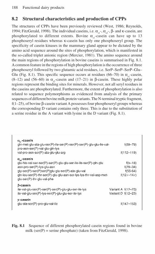
188 Functional dairy products
8.2 Structural characteristics and production of CPPs
The structures of CPPs have been previously reviewed (West, 1986; Reynolds,1994; FitzGerald, 1998). The individual caseins, i.e. α
s1-, α
s2-, β- and κ-casein, are
phosphorylated to different extents. Bovine αs1
-casein can have up to 13phosphoseryl residues whereas κ-casein has only one phosphoseryl group. Thespecificity of casein kinases in the mammary gland appear to be dictated by theamino acid sequence around the sites of phosphorylation, which is manifested inthe so-called triplet anionic region (Mercier, 1981). The amino sequence aroundthe main regions of phosphorylation in bovine caseins is summarised in Fig. 8.1.A common feature in the regions of high phosphorylation is the occurrence of threephosphoseryl followed by two glutamic acid residues, i.e. SerP–SerP–SerP–Glu–Glu (Fig. 8.1). This specific sequence occurs at residues (66–70) in α
s1-casein,
(8–12) and (56–60) in αs2
-casein and (17–21) in β-casein. These highly polarregions represent the binding sites for minerals. However, not all seryl residues inthe caseins are phosphorylated. Furthermore, the extent of phosphorylation is alsorelated to sequence polymorphisms as evidenced from analysis of the primarysequences of different bovine milk protein variants. The N-terminal tryptic fragment,f(1–25), of bovine β-casein variant A possesses four phosphoseryl groups whereasthe corresponding D variant contains only three. This is due to the substitution ofa serine residue in the A variant with lysine in the D variant (Fig. 8.1).
Fig. 8.1 Sequence of different phosphorylated casein regions found in bovinemilk (ser(P) = serine phosphate) (taken from FitzGerald, 1998).
Caseinophosphopeptides (CPPs) as functional ingredients 189
8.2.1 Production of CPPsThe basic protocol for the production of CPPs involves enzymatic digestion ofwhole casein or fractions enriched in specific individual caseins. A range ofproteinase activities from mammalian, bacterial and plant sources have beenemployed for casein hydrolysis (Manson and Annan, 1971; Brule et al., 1982,1989; Berrocal et al., 1989; Juillerat et al., 1989; Reynolds, 1992; Adamson andReynolds, 1995; McDonagh and FitzGerald, 1998). To date, crude and relativelypurified pancreatic proteinase preparations containing trypsin and chymotrypsin,and combinations of pancreatic activities (pancreatin) have been mainly described.However, proteinases from a range of bacterial, fungal and plant sources containing,for example, thermolysin, subtilisin, pronase, and papain activities have also beensuccessfully used in the hydrolysis of casein for CPP production.
Generally, hydrolysis is carried out at neutral to alkaline pH (pH 7.0–8.0).Adjustment of hydrolysates to pH 4.6 results in the appearance of insolublematerial that can be removed subsequently by centrifugation. The CPPs present inthe resulting clarified hydrolysate supernatants can be aggregated on inclusion ofa range of mineral salts around neutral pH. Calcium has preferentially been usedfor CPP aggregate formation; however, barium, iron, zinc, magnesium, copper,manganese and cobalt have all been mentioned in the literature as potentialaggregating agents (Reeves and Latour, 1958; Manson and Annan, 1971; Brule etal., 1982, 1989; Berrocal et al., 1989; Juillerat et al., 1989; Reynolds, 1992;Adamson and Reynolds, 1995; McDonagh and FitzGerald, 1998).
The aggregated CPPs can be precipitated with hydrophilic solvents such asethanol, methanol, propanol, butanol and acetone (Reeves and Latour, 1958;Adamson and Reynolds, 1995; McDonagh and FitzGerald, 1998) and have beencollected by centrifugation. Ultrafiltration through defined molecular mass cut-offmembranes has also been used at pilot-scale to separate aggregated CPPs fromnon-phosphorylated peptides (Brule et al., 1982, 1994; Reynolds, 1992). Subse-quently, diafiltration has been employed to remove excess mineral salt fromultrafiltered CPPs (Reynolds, 1993). Alternatively, CPPs have been isolated/enriched from calcium/ethanol precipitated aggregates using a range of ion-exchange procedures (Berrocal et al., 1989; Juillerat et al., 1989; Kunst, 1990;Koide et al., 1991; Lihme et al., 1994). Fractions containing isolated/enrichedCPPs are then subjected to spray- or freeze-drying. The yield of CPPs produced bythe above protocols is reported to range from 6 to 20% (Gerber and Jost, 1986;Juillerat et al., 1989; Kunst, 1990; McDonagh and FitzGerald, 1998; Ellegård etal., 1999).
Phosphopeptides are susceptible to dephosphorylation during exposure toalkaline and high heat treatments. These conditions result in β-elimination ofphosphoserine, resulting in the generation of dehydroalanine (Lorient, 1979).Lysinoalanine formation between dehydroalanine and peptide/protein bound lysinein turn may lead to reduced lysine bioavailability (Manson and Carolan, 1980;Hasegawa et al., 1981; De Koning and Van Rooijen, 1982). Therefore, theprocessing conditions during CPP production, especially for example, duringspray-drying, may have a significant impact on CPP quality. Heating sodium
190 Functional dairy products
caseinate and the pH 4.6 soluble portion of a tryptic hydrolysate of sodium caseinateat 170 ºC, a typical inlet temperature during spray-drying, resulted in significantlosses in peptide bound phosphorus in both samples (Meisel et al., 1991a).
8.3 CPPs and mineral (calcium) bioavailability
The functional food ingredient potential of CPPs is mainly related to the ability ofCPPs to bind and solubilise minerals such as calcium. CPPs prevent the precipita-tion of calcium in the presence of phosphate at alkaline pH in vitro (Berrocal et al.,1989; Sato et al., 1991; Gaucheron et al., 1995; Holt et al., 1996, 1998; Aoki et al.,1998; Holt, 2001). Holt (2001) has demonstrated that calcium and phosphate formsoluble nanoclusters in the presence of CPPs. Furthermore, CPPs inhibit theformation of hydroxyapatite and brushite crystals (Reynolds, 1993; Holt et al.,1996). The highly anionic phosphorylated regions in CPPs are responsible formineral binding. However, the amino acid sequence around this anionic hydrophilicdomain also seems to play a significant role in mineral binding (Reynolds, 1994;McDonagh and FitzGerald, 1998). Dephosphorylated peptides/proteins do notbind minerals such as calcium, zinc and iron as efficiently as their equivalentphosphorylated derivatives (Harzar and Kauer, 1982; Sato et al., 1983; Gerber andJost, 1986; Berrocal et al., 1989; Bouhallab et al., 1991; Meisel and Olieman,1998; Yeung et al., 2001).
The calcium-binding properties of CPPs have been studied using calciumselective electrodes (Berrocal et al., 1989; Meisel et al., 1991b), micro-ultrafiltra-tion methods in combination with atomic absorption (Perich et al., 1992),competitive assays with Chelex resin (Meisel et al., 1989) and capillary zoneelectrophoresis (Meisel and Olieman, 1998). The calcium binding constants forheterogeneous CPP preparations is reported to be in the order of 102–103 M–1
(Berrocal et al., 1989; Sato et al., 1991; Schlimme and Meisel, 1995). FractionedCPPs are reported to have significantly greater binding affinities for calcium, e.g.0.32 mM–1 for α
s1-casein (59–79)5P at pH 8.0 (Meisel and Olieman, 1998). Lee et
al. (1980) reported that one mole of CPP could bind approximately 40 moles ofcalcium. The calcium solubilising ability of CPP-enriched fractions derived fromcasein hydrolysates digested with bacterial, fungal, plant and animal origin rangedfrom 7.4 to 24.0 mg calcium per mg CPP (McDonagh and FitzGerald, 1998). Thecalcium solubilising ability of CPPs may be affected by genetic polymorphism.Han et al. (2000) reported that the calcium solubilising ability of CPPs derivedfrom Korean cattle containing the H variant of β-casein had approximately 23%increased calcium solubilising ability than CPPs derived from casein that did notcontain the H variant of β-casein.
The (1–25) phosphopeptide fragment of β-casein was reported to bind 4 moliron per mol peptide. Enzymatic dephosphorylation of the β-casein (1–25) renderedthe bound iron completely dialysable (Bouhallab et al., 1991). The presence ofCPP-bound iron was also shown to significantly inhibit enzymatic dephosphoryla-tion (Bouhallab et al., 1991; Yeung et al., 2001).
Caseinophosphopeptides (CPPs) as functional ingredients 191
All the calcium phosphate in milk exists in the form of nanometre-sizedclusters, or nanoclusters. Calcium phosphate nanoclusters are reported to consistof a core (radius = 2.4 nm) containing ~350 hydrated calcium phosphate ion pairs.This core is surrounded by a shell 1.6 nm thick comprising about 50 phosphopeptidechains (Holt et al., 1996, 1998). Soluble nanoclusters containing otherwise insolu-ble minerals and essential trace elements such as calcium, magnesium, zinc, iron,manganese and iodine can be formed in the presence of CPPs. By uniformly raisingthe pH in, for example, a calcium phosphate solution via the liberation of ammoniausing jack bean urease in the presence of urea, it was reported to be possible todevelop stable solutions containing up to 1 M calcium in the presence of β-casein(1–25)4P and β-casein (1–42)5P (Holt, 2001).
8.4 Human studies with CPPs
Milk and dairy products are abundant sources of bioavailable calcium with about70% of dietary calcium needs being supplied by milk and dairy products indeveloped Western society (IDF, 1997). The excellent bioavailability of dietarycalcium during the consumption of dairy products has been attributed to thepresence of CPPs in casein-containing diets (Gammelgård-Larsen, 1991; Kitts andYuan, 1992; Schaafsma, 1997). CPPs in vitro can maintain calcium soluble in thepresence of phosphate at alkaline pH (Reeves and Latour, 1958; Sato et al., 1991;Ono et al., 1994; Holt et al., 1996, 1998; Holt, 2001; Aoki et al., 1998). These arethe conditions that pertain in the small intestine where maximal passive vitamin D-independent calcium absorption takes place (Bronner, 1987). This ability of CPPto maintain calcium solubility in the conditions as found in the distal ileum istherefore proposed to be the basis for enhanced absorption of dietary calcium (Leeet al., 1980, 1983; Mykännen and Wasserman, 1980; West, 1986; Meisel andFrister, 1988; Li et al., 1989; Kitts and Yuan, 1992; Kitts et al., 1992).
If the above hypothesis is correct, it is clear that in order to act as mineral carriersor enhancers of mineral solubility, CPPs must display the ability to resist, or at leastto partially resist, enzymatic digestion during intestinal passage. Therefore, severalanimal and two human studies have reported to date on the production of CPPs invivo. Analysis of the intestinal fluid of minipigs fed a bovine casein-containingdiet, for example, demonstrated the presence of CPPs (Meisel and Frister, 1988).CPPs were detected in the stomach and duodenal contents of adult humans afteringestion of milk or yoghurt (Chabance et al., 1998). It has recently been demon-strated, for the first time, that phosphopeptides can survive the prolonged intestinalpassage to the distal small intestine (ileum) in humans following ingestion of dietscontaining milk or peptide preparations enriched in CPPs (Meisel et al., 2001,2002). Interestingly, these studies show better survival during intestinal passageby CPPs derived from consumption of milk as opposed to consumption of CPPsper se.
Numerous animal and a very limited number of human studies have beenperformed on the role of CPPs in mineral, mainly calcium, bioavailability (for
192 Functional dairy products
reviews see FitzGerald, 1998; Scholz-Ahrens and Schrezenmeir, 2000 andreferences therein). To date most studies have been performed on animals. Dietarystudies on CPPs with rats, chickens and piglets have yielded conflicting resultswhich have, in part, been attributed to differences in methodology used to quantifyextrinsic and intrinsic calcium absorption in addition to differences in the qualityof the CPP preparations under study (Kitts and Yuan, 1992; FitzGerald, 1998). Therat study of Bennett et al. (2000) reported that the true fractional absorption ofcalcium was in fact decreased on inclusion of high CPP levels (200–500 g CPP/kgmeal). These results suggested that high levels of dietary CPPs may chelate allavailable dietary calcium, thereby making it unavailable for subsequent absorption.
Co-ingestion of CPPs with calcium by postmenopausal women having lowbasal absorptive performance was reported to significantly enhance calciumabsorption (Heaney et al., 1994). The human studies of Hansen et al. (1997a)reported a 30% (approximate) increase in calcium and zinc absorption by adultsfrom a rice-based infant gruel supplemented with CPPs. This effect was notobserved in the presence of phytate-containing meals (Hansen et al., 1996, 1997b).Preliminary results from a more recent human study indicate that consumption ofCPPs in beverage format does not significantly increase the fractional absorptionof dietary calcium (Teucher et al., 2002). Further human studies with CPPs arerequired to fully elucidate the potential role of these peptides in modulatingmineral bioavailability. One of the difficulties in studying CPP effects on mineralbioavailability in animals or humans arises from the presence of complex inter-actions with other meal constituents (Rossander et al., 1992; Hansen et al., 1996).Furthermore, long-term stringent control during human feeding experiments isdifficult to achieve.
A recent study by Erba et al. (2001) using everted small intestine of rats clearlydemonstrated that the presence of CPPs can limit the inhibitory effect of phosphateon calcium availability and increase calcium transport across the distal ileum of rat.However, the extent of calcium absorption was dependent on the Ca:Pi ratio, i.e. inthe presence of CPPs, calcium transport was decreased by 40% at Ca:Pi = 1:1whereas 60% inhibition of calcium transport was observed at Ca:Pi = 1:2. Thesestudies clearly demonstrate that not only is the interaction of other meal constituentsimportant, but also that the concentration-dependent effects of other constituentson mineral bioavailability plays a role. Furthermore, the format in which CPPs areconsumed by humans appears to dictate the ability of CPPs to reach the distal ileum(Meisel et al., 2001, 2002).
8.5 Effect of CPPs on mineral uptake in specific cell systems
In vitro studies with the human intestinal cell line, HT-29, demonstrated in thepresence of extracellular calcium that both a commercial preparation containing amixture of CPPs and an individual CPP (β-casein (1–25) 4P) induced a transientrise in intracellular calcium, which did not influence ATP-induced release ofcalcium from intracellular stores. This effect was not observed in the absence of
Caseinophosphopeptides (CPPs) as functional ingredients 193
extracellular calcium. Pre-treatment of the cells with thapsinargin, which completelydepletes intracellular calcium stores, did not abolish the cell response to CPPs.Furthermore, repetitive stimulation of the cells with CPPs always resulted in atransient rise in intracellular calcium, indicating that the cells did not becomedesensitised to the addition of CPPs (Ferraretto et al., 2001). The authors concluded,since the data indicated that the process influenced by CPPs is non-saturating, thatit does not involve a membrane receptor or an endogenous ion channel system.They also suggested that CPPs may act on transmembrane calcium flux by eitherinserting themselves into the plasma membrane to form calcium selective channelsor as calcium carrier peptides, which could be rapidly internalised via endocytosisor other processes, resulting in the delivery of calcium to the cytosol.
The presence of CPPs in fertilisation medium was shown to increase the rate ofboar spermatozoa penetration of in vitro matured pig oocytes. Spermatozoa alsoretained the ability to penetrate oocytes for longer in the presence of CPPs.Calcium uptake by spermatozoa was increased on the inclusion of CPPs in thefertilisation medium (Mori et al., 1996).
CPPs influenced zinc absorption from aqueous phytate-containing solutions byhuman colon carcinoma (Caco–2) cells. In a control system, phytate reducedbinding and uptake of zinc by 79% in Caco-2 cells. Inclusion of low levels of CPPsincreased zinc binding and uptake by 94%. Higher levels of CPPs, however,inhibited zinc binding and uptake (Hansen et al., 1996). These results againemphasise the importance of CPP concentration on specific test parameters.
The presence of CPPs was reported to increase calcification in cultured rat boneembryonic explants. This may, in part, be linked to the observed increase insolubility of calcium in the culture medium (Gerber and Jost, 1986).
8.6 Cytomodulatory effects
Based on various cytochemical studies, there is increasing evidence of the possibleinvolvement of milk protein-derived peptides as specific signals that can triggerviability of cancer cells. Purified peptides, corresponding to bioactive sequencesof casein, revealed modulation of cell viability, i.e. proliferation and apoptosis, indifferent human cell culture models (Meisel and Günther, 1998; Hartmann et al.,2000). Accordingly, apoptosis of human leukaemia cells (HL-60) was induced bythe CPP β-CN(f1–25)4P. Since the dephosphorylated derivative β-CN(f1–25)0Pshowed comparable modulating activity, the effects are obviously not mediated byphosphoserine residues. Cheese contains phosphopeptides as natural constituents,and peptides from a lyophilised extract of Gouda cheese inhibited proliferation ofleukaemia cells, even at concentrations of 1 pmol/l (Meisel and Günther, 1998).The antiproliferative effect of Gouda extract was shown to be a result of peptide-induced apoptosis. It is interesting to note that cancer cell lines were more reactiveto peptide-induced apoptotic stimulation than non-malignant cells.
Effects on both cell viability and immune cell function (see Section 8.8.4 onimmunomodulation) may be a mechanism through which cytomodulatory CPPs
194 Functional dairy products
exert protective effects in cancer development. The most probable target site forcytomodulatory action seems to be the gastrointestinal tract. The antiproliferativeeffects of milk-protein derived peptides as well as other minor milk components incolon cancer cell lines suggests that they could have a role in the prevention ofcolon cancer by blocking hyperproliferation of the epithelium and by promotingapoptosis (MacDonald et al., 1994; Ganjam et al., 1997; Meisel et al., 1998).
Besides a possible positive action there may be adverse effects of CPPs thatwould impose restrictions to their application in functional foods for humannutrition. However, cytochemical experiments carried out on CPP preparationsusing a variety of human cell culture model systems did not point to a significantcytotoxic potential of CPPs towards human cells, provided that the orally ingesteddoses correspond to reasonable concentrations for use in functional foods. Thus,CPPs can be supposed not to provoke a cytotoxic response in cells of the respectivetypes in vivo (Hartmann et al., 2000; Hartmann and Meisel, 2002).
8.7 Safety assessment of CPPs
In order that CPPs can receive acceptance as functional food ingredients or asnutraceuticals it is essential to demonstrate their overall safety following humanconsumption. Information regarding the safety of consuming CPPs or fractionsenriched in CPPs is required by clinicians, legislators and the food industry in orderto prepare a case for the widespread utilisation of CPPs by consumers. Given thatCPPs have been consumed by humans for centuries as part of diets rich in dairyproducts, it would be expected that consumption of CPPs should pose little or norisk to the general population.
8.7.1 Antigenic and allergenic potential of CPPsStudies with synthetic peptides corresponding to phosphorylated regions of α
s1-
casein demonstrated that αs1
-casein (63–70)4P was more strongly recognised byanimal-derived polyclonal antisera than the corresponding dephosphorylated pep-tide (Perich et al., 1992). Studies with polyclonal anti-α
s1-casein antibodies also
indicated that the antigenicity of αs1
-casein was lost on dephosphorylation (Otaniet al., 1987). No significant modification in the antigenicity of animal antiseraraised against purified bovine α
s2- and β-casein was observed following enzymatic
dephosphorylation of these proteins or by deletion (through studies with deletionvariants) of an associated major phosphorylation site (Bernard et al., 2000). It wasconcluded from these findings that most of the polyclonal antibodies raised againstpure α
s2- and β-casein were not directed against the major phosphorylation sites on
these molecules and that immunoreactivity was not affected by phosphorylation ofthe serine residues. Furthermore, it was concluded that the immunoreactivity ofthese caseins appears to be unaffected by any potential conformational changeinduced by dephosphorylation.
Studies with antisera derived from human volunteers who display allergenic
Caseinophosphopeptides (CPPs) as functional ingredients 195
reactions to casein showed that dephosphorylation or deletion of phosphorylatedregions (as demonstrated using deletion variants) led to decreased recognition ofα
s2- and β-casein by the IgE in these sera. It was concluded that dephosphorylation
probably lowers the affinity of IgE for the caseins rather than lowering the specificIgE titre by comparison of the results of direct vs competitive inhibition enzyme-linked immunosorbent assay (ELISA). From supporting studies with isolatedphosphorylated and dephosphorylated β-casein (1–25), it was concluded that itwas very likely that at least part of the anti-casein IgE is directed against a majorsite of phosphorylation. It was also suggested that this finding may, in part, explainthe observed cross-reactivity between the individual caseins (Bernard et al., 2000).Therefore, the allergenic potential of CPPs, following their consumption by thesegment of the human population that displays allergenic reactions to caseins,should be considered in the exploitation of CPPs as functional food ingredients/nutraceuticals.
8.8 Potential ingredient applications of CPPsThe potential ingredient applications of CPPs have been previously reviewed(FitzGerald, 1998) and are summarised in Table 8.1.
Table 8.1 Potential applications of caseinophosphopeptides (CPPs) (reproduced fromFitzGerald, 1998. Copyright Elsevier Science 1998; reproduced with permission)
Application Rationale Product
Prevention of osteoporosis Increase absorption of Ca by Ca–CPP complexesincluding Ca–CPP as functionalfood ingredient for targetpopulations, allowing maximumdeposition of bone in early life
Recalcification of bones Increase serosal [Ca], aiding Ca–CPP complexesafter fracture bone mineralisation
Calcic addition during Increase serosal [Ca] Ca–CPP complexestreatment of rickets
Hypertension Increased serosal [Ca] may aid Ca–CPP complexesreduction of hypertension
Prevention of dental caries Inclusion of Ca–CPP as Ca–CPP complexes.ingredients in mouthwashes,toothpastes
Mg deficiency in pregnancy Aid Mg supplementation Mg-CPP complexes,and old age ingredients
Anaemia Aid Fe supplementation, Fe–CPP complexes.increase serosal [Fe]
Humanisation of bovine milk Increase phosphorus levels CPPs
Oligoelement Aid supplementation of Zn, Oligoelement–CPPsupplementation Cu, Cr, Ni, Co, Mn and Se complexes
196 Functional dairy products
8.8.1 Mineral supplementationThe application for which CPPs are probably best linked is in the area of mineralsupplementation. It is thought that CPPs may aid dietary mineral absorption byenhancing solubility. A whole range of minerals including calcium, magnesiumand iron are required to maintain health (Passmore et al., 1974; Fairweather-Tait,1988; Buttriss, 1990; Osborne et al., 1996). Inadequate uptake of these mineralsmay lead to conditions such as osteoporosis, dental caries, hypertension andanaemia. Various foods have been cited in the patent literature as potential carriersfor CPPs. These include confectionary products, breakfast cereals, juices, milk,dairy products and sports drinks (Reynolds, 1993; Han et al., 1996). However, asalready stated herein, there is a lack of extensive evidence linking consumption ofCPPs and enhanced mineral bioavailability in humans.
On the other hand, the recent patent by Holt (2001) signifies the potentialapplication of CPPs in the development of nanoclusters containing various mineralsand trace elements. Solubilisation of minerals and essential trace micronutrients innanocluster structures by the inclusion of CPPs allows an efficient means ofdelivering high doses of essential nutrients in a small volume. These complexeshave been shown to be stable following sterilisation and freeze-drying. Thepotential exploitation of CPP-containing nanoclusters has been cited for food,beverage, dental-care and pharmaceutical applications.
8.8.2 Anticaries effectsThe development of dental caries is associated with demineralisation of teeth as aresult of the production of organic acids by odontopathogenic dental plaque-forming bacteria during the fermentation of dietary sugars. The anticariogenicactivity of CPPs is related to their potential ability to localise calcium phosphate atthe tooth surface and as a consequence their ability to help prevent demineralisa-tion and aid in remineralisation of tooth enamel. The anticariogenic effects of CPPshave been shown in animal and human experiments (Reynolds, 1997, 1998, 1999;Rose, 2000a). The anticariogenic effect of CPPs may also be related to the abilityof CPPs to compete with dental plaque-forming bacteria such as Streptococcusmutans for calcium (Rose, 2000b). Claims have been made in the patent lieraturefor the incorporation of CPPs into dental hygiene products (Reynolds, 1993; Hanet al., 1996; Holt, 2001). The inclusion of CPP-amorphous calcium phosphatenanoclusters into sugar-free chewing gum has been shown to increase enamelremineralisation in a human in situ model (Shen et al., 2001).
8.8.3 Humanisation of bovine milkThe concept of supplementing bovine milk with CPPs in order to ‘humanise’bovine milk has been patented. This arises from the fact that bovine milk has anorganic phosphorus : total phosphorus ratio of 0.34, whereas human milk has aratio of 0.89 (Brule et al., 1989).
Caseinophosphopeptides (CPPs) as functional ingredients 197
8.8.4 Immunomodulation
Both peptides and proteins from milk may contribute to an overallimmunostimulatory response. Recent studies have focused on immunoenhancingproperties of caseinophosphopeptides. Hata et al. (1998, 1999) reported on theimmunostimulatory action of caseinophosphopeptides β-CN(f1–25)4P, β-CN(f1–28)4P, α
S1-CN(f59–79)5P and α
S2-CN(f1–32)4P which enhanced immunglobulin
G production in mouse spleen cell cultures. Moreover, the level of serum andintestinal antigen-specific IgA was higher in mice fed the caseinophosphopeptidepreparation than those fed the control diet (Otani et al. 2000). The oral ingestion ofimmunomodulatory caseinophosphopeptides is claimed to enhance mucosalimmunity (Otani et al., 2000). Hartmann et al. (2000) found that the treatment ofhuman lymphocytes (peripheral blood lymphocyte, PBL) with different CPPpreparations resulted in a significant increase in IgG production in vitro, evenexceeding the response mediated by known mitogens.
The molecular mechanism by which CPPs exert their immunomodulatoryeffects is not yet defined. It is known that lymphocytes and macrophages expressreceptors for many biologically active mediators. The immunostimulatory activityof CPPs was attributable to phosphoseryl residues (Hata et al. 1998, 1999).
8.9 Summary and future trends
The functional food ingredient/nutraceuticals potential of CPPs revolves aroundtheir ability to bind and solubilise minerals such as calcium. Preparative-scaleproduction procedures for the generation of CPPs are available. Peptide preparationsenriched in CPPs are available from multinational food ingredient suppliers.Furthermore, dental-care/hygiene products containing CPPs, targeted to have ananticariogenic effect, are currently available in the marketplace. A number ofpatents exist on the production and application of CPPs as functional foodingredients/nutraceuticals and as dental care/hygiene agents.
Despite numerous animal studies and a very limited number of human studies,unequivocal proof that consumption of CPPs enhances dietary calciumbioavailability is as yet unavailable. The difficulty in obtaining such informationarises from complex interactive effects between dietary minerals such as calciumand other dietary constituents such as phytate which strongly influencebioavailability. In this regard, it is interesting to note that the format in which CPPsare consumed, e.g. either in milk or as isolated CPPs, has been shown to stronglyinfluence the survivability of CPPs during gastrointestinal transit (Meisel et al.,2001, 2002). The effect of consuming minerals in nanocluster format on subsequentdietary mineral bioavailability is worthy of further study.
In vitro cell culture studies with isolated CPPs and peptide fractions enriched inCPPs demonstrate no adverse cytotoxicological effects associated with CPPs(Hartmann and Meisel, 2002; Hartmann et al., 2000). Interestingly, the fact thatCPPs have been shown in vitro to have an immunomodulatory effect may
198 Functional dairy products
represent a new biological function of ingested phosphorylated peptides (Hata etal., 1999; Hartmann and Meisel, 2002). The observation that IgE displays a higherbinding affinity for phosphorylated caseins than the correspondingdephosphorylated derivatives is relevant to consumers who display allergenicreactions to milk proteins (Bernard et al., 2000) and has significant consequencesfor the appropriate labelling of consumer foods and other products containingCPPs.
Finally, the continued study of CPP structure and function will undoubtedlylead to a greater understanding of the functional and metabolic role, and potentialapplications, of CPPs. This will contribute to the development and expansion ofCPPs as functional food ingredients in the prevention of diseases and asnutraceuticals in the treatment of particular disease states.
8.10 ReferencesADAMSON N J and REYNOLDS E C (1995), ‘Characterisation of tryptic casein phosphopeptides
prepared under industrially relevant conditions’, Biotechnol Bioeng, 45, 196–204.AOKI T, NAKANO T, IWASHITA T and others (1998), ‘Preparation and characterisation of
micellar calcium phosphate-casein phosphopeptide complex’, J Nutr Sci Vitaminol, 44,447–456.
BENNET T, DESMOND A, HARRINGTON M, MCDONAGH D, FITZGERALD R J, FLYNN andCASHMAN K D (2000), ‘The effect of high intakes of casein phosphopeptides on calciumabsorption in the rat’, Br J Nutr, 83, 673–680.
BERNARD H, MEISEL H, CREMMINON C and WAL J-M (2000), ‘Post-translational phosphoryla-tion affects the IgE binding capacity of caseins’, FEBs Lett, 467, 239–244.
BERROCAL R, CHANTON S, JUILLERAT M A, PAVILLARD B, SCHERZ J-C and JOST R (1989),‘Tryptic phosphopeptides from whole casein: II. Physicochemical properties related to thesolubilisation of calcium’, J Dy Res, 56, 335–341.
BOUHALLAB S, LÉONIL J and MAUBOIS J L (1991), ‘Complexation du fer par le phosphopeptide(91–25) de la caséine β; action de l’alcalase et de la phosphatase acide’, Lait, 71, 435–443.
BRONNER F (1987), ‘Intestinal calcium absorption: mechanisms and applications’, J Nutr,117, 1347–1352.
BRULE G, ROGER L, FAUQUANT J and PIOT M (1982), ‘Phosphopeptides from casein-basedmaterial’, US Patent No. 4,358,465.
BRULE G, ROGER L, FAUQUANT J and PIOT M (1989), ‘Casein phosphopeptide composition’,US Patent No. 4,816,398.
BRULE G, ROGER L, FAUQUANT J and PIOT M (1994), ‘Nutrient composition containing non-phosphorylated peptides from casein-based material’, US Patent No. 5,334,408.
BUTTRISS J (1990), ‘The role of calcium in a balanced diet’, J Soc Dy Technol, 43(1), 1–3.CHABANCE B, MARTEAU P, RAMBAUD J C, MIGLIORE-SAMOUR D, BOYNARD M, PERROTIN P,
JOLLÈS P and FIAT A M (1998), ‘Casein peptide release and passage to the blood in humansduring digestion of milk or yoghurt’, Biochimie, 80, 155–165.
DE KONING P J and VAN ROOIJEN P J (1982), ‘Aspects of formation of lysoalanine in milk andmilk products’, J Dy Res, 49, 725–736.
ELLEGÅRD K H, GAMMELGÅRD-LARSEN C, SØRENSEN E S and FEDESOV S (1999), ‘Process-scale chromatographic isolation, characterisation and identification of tryptic bioactivecasein phosphopeptides’, Int Dy J, 9(9), 639–652.
ERBA D, CIAPPELLANO S and TESTOLIN G (2001), ‘Effect of caseinophosphopeptides oninhibition of calcium intestinal absorption due to phosphate’, Nutr Res, 21, 649–656.
Caseinophosphopeptides (CPPs) as functional ingredients 199
FAIRWEATHER-TAIT S J (1988), ‘Zinc in human nutrition’, Nutr Res, 1, 23–27.FERRARETTO A, SIGNORILE A, RAVAGHI C, FIORILLI A and TETTAMANTI G (2001), ‘Casein-
phosphopeptides influence calcium uptake by cultured human intestinal HT-29 tumorcells’, J Nutr, 131, 1655–1661.
FITZGERALD R J (1998), ‘Potential uses of caseinophosphopeptides’, Int Dy J, 8, 451–457.GAMMELGÅRD-LARSEN C (1991), ‘Casein phosphopeptide, CPP – a promising new way to
enhance calcium utilisation and absorption’, Proceedings of Food Ingredients (Asia)Conference, Singapore, April, 1991, Maarssen Expoconsult, 53–58.
GANJAM L S, THORNTON W H, MARSHALL R T and MACDONALD R S (1997), ‘Antiproliferativeeffects of yoghurt fractions obtained by membrane dialysis on cultured mammalianintestinal cells’, J Dy Sci, 80, 2325–2329.
GAUCHERON F, LE GREAT Y, SINBANDHIT S, GUENOT P and BRULE G (1995), ‘Binding ofcalcium by β-casein in the presence of inorganic phosphate’, Sci des Aliments 15, 481–489.
GERBER H W and JOST R (1986), ‘Casein phosphopeptides: their effect on calcification of invitro cultured embryonic rat bone’, Cal Tissue Int, 38, 350–357.
HAN S K, SHIN Y C and PARK J H (1996), ‘Casein phosphopeptide, casein containing same anda process for the preparation thereof’, World Patent, WO 96/29340.
HAN S H, SHIN Y C and BYUN H D (2000), ‘Biochemical, molecular and physiologicalcharaterisation of a new beta-casein variant detected in Korean cattle’, Anim Genet, 31(1),49–51.
HANSEN M, SANDSTRÖM B and LONNERDAL B (1996), ‘The effect of casein phosphopeptideson zinc and calcium absorption from high phytate infant diets assessed in rat pups andCaco-2 cells’, Ped Res, 40(4), 547–552.
HANSEN M, SANDSTRÖM B, JENSEN M and SØRENSEN S S (1997a), ‘Casein phosphopeptidesimprove zinc and calcium absorption from rice-based but not from whole-grain infantcereals’, J Ped Gastroent Nutr, 24(1), 56–62.
HANSEN M, SANDSTRÖM B, JENSEN M and SØRENSEN S S (1997b), ‘Effect of caseinphosphopeptide on zinc and calcium absorption from bread meals’, J Tr Elem Med Biol,11(3), 143–149.
HARTMANN R and MEISEL H (2002), ‘Cytochemical assessment of phosphopeptides derivedfrom casein as potential ingredients for functional food’, Nahrung/Food (submitted).
HARTMANN R, GUNTHER S, MARTIN D, MEISEL H, PENTZIEN A-K, SCHLIMME E and SCHOLZ N(2000), ‘Cytochemical model systems for the detection and characterisation of potentiallybioactive milk components’, Kiel Milch Forsch, 52, 61–85.
HARZAR G and KAUER H (1982), ‘Binding of zinc to casein’, Am J Clin Nutr, 35, 981–987.HASEGAWA K, OKAMOTO N, OZAWA H, KITAJIMA S and TAKADO Y (1981), ‘Limits and sites
of lysoalanine formation in lysozyme, α-lactalbumin and αs1- and β-casein by alkalitreatment’, Agric Biol Chem, 45, 1645–1651.
HATA I, HIGASHIYAMA S and OTANI H (1998), ‘Identification of a phosphopeptide in bovineαS1-casein digests as a factor influencing proliferation and immunoglobulin production inlymphocyte cultures’, J Dy Res, 65, 569–578.
HATA I, UEDA J and OTANI H (1999), ‘Immunostimulatory action of a commercially availablecasein phosphopeptide preparation, CPP-III, in cell cultures’, Milchwissen, 54, 3–7.
HEANEY R P, SAITO Y and ORIMO H (1994), ‘Effect of caseinophosphopeptides on absorbabil-ity of co-ingested calcium in normal postmenopausal women’, J Bone Min Metab, 12(1),77–81.
HOLT C (2001), ‘Calcium phosphate nanoclusters and their application’, World Patent,WO0144106.
HOLT C, WAHLGREN N M and DRAKENBURG T (1996), ‘Ability of a β-casein phosphopeptideto modulate the precipitation of calcium phosphate by forming amorphous dicalciumphosphate nanoclusters’, Biochem J, 314, 1035–1039.
HOLT C, TIMMINS P A, ERRINGTON, N et al. (1998), ‘A core-shell model of calcium phosphate
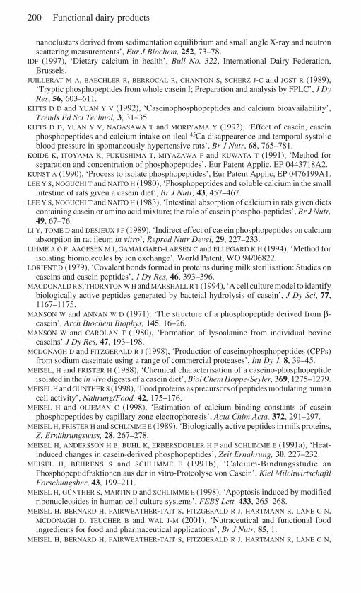
200 Functional dairy products
nanoclusters derived from sedimentation equilibrium and small angle X-ray and neutronscattering measurements’, Eur J Biochem, 252, 73–78.
IDF (1997), ‘Dietary calcium in health’, Bull No. 322, International Dairy Federation,Brussels.
JUILLERAT M A, BAECHLER R, BERROCAL R, CHANTON S, SCHERZ J-C and JOST R (1989),‘Tryptic phosphopeptides from whole casein I; Preparation and analysis by FPLC’, J DyRes, 56, 603–611.
KITTS D D and YUAN Y V (1992), ‘Caseinophosphopeptides and calcium bioavailability’,Trends Fd Sci Technol, 3, 31–35.
KITTS D D, YUAN Y V, NAGASAWA T and MORIYAMA Y (1992), ‘Effect of casein, caseinphosphopeptides and calcium intake on ileal 45Ca disappearence and temporal systolicblood pressure in spontaneously hypertensive rats’, Br J Nutr, 68, 765–781.
KOIDE K, ITOYAMA K, FUKUSHIMA T, MIYAZAWA F and KUWATA T (1991), ‘Method forseparation and concentration of phosphopeptides’, Eur Patent Applic, EP 0443718A2.
KUNST A (1990), ‘Process to isolate phosphopeptides’, Eur Patent Applic, EP 0476199A1.LEE Y S, NOGUCHI T and NAITO H (1980), ‘Phosphopeptides and soluble calcium in the small
intestine of rats given a casein diet’, Br J Nutr, 43, 457–467.LEE Y S, NOGUCHI T and NAITO H (1983), ‘Intestinal absorption of calcium in rats given diets
containing casein or amino acid mixture; the role of casein phospho-peptides’, Br J Nutr,49, 67–76.
LI Y, TOME D and DESJEUX J F (1989), ‘Indirect effect of casein phosphopeptides on calciumabsorption in rat ileum in vitro’, Reprod Nutr Devel, 29, 227–233.
LIHME A O F, AAGESEN M I, GAMALGARD-LARSEN C and ELLEGARD K H (1994), ‘Method forisolating biomolecules by ion exchange’, World Patent, WO 94/06822.
LORIENT D (1979), ‘Covalent bonds formed in proteins during milk sterilisation: Studies oncaseins and casein peptides’, J Dy Res, 46, 393–396.
MACDONALD R S, THORNTON W H and MARSHALL R T (1994), ‘A cell culture model to identifybiologically active peptides generated by bacteial hydrolysis of casein’, J Dy Sci, 77,1167–1175.
MANSON W and ANNAN W D (1971), ‘The structure of a phosphopeptide derived from β-casein’, Arch Biochem Biophys, 145, 16–26.
MANSON W and CAROLAN T (1980), ‘Formation of lysoalanine from individual bovinecaseins’ J Dy Res, 47, 193–198.
MCDONAGH D and FITZGERALD R J (1998), ‘Production of caseinophosphopeptides (CPPs)from sodium caseinate using a range of commercial proteases’, Int Dy J, 8, 39–45.
MEISEL, H and FRISTER H (1988), ‘Chemical characterisation of a caseino-phosphopeptideisolated in the in vivo digests of a casein diet’, Biol Chem Hoppe-Seyler, 369, 1275–1279.
MEISEL H and GÜNTHER S (1998), ‘Food proteins as precursors of peptides modulating humancell activity’, Nahrung/Food, 42, 175–176.
MEISEL H and OLIEMAN C (1998), ‘Estimation of calcium binding constants of caseinphosphopeptides by capillary zone electrophoresis’, Acta Chim Acta, 372, 291–297.
MEISEL H, FRISTER H and SCHLIMME E (1989), ‘Biologically active peptides in milk proteins,Z. Ernährungswiss, 28, 267–278.
MEISEL H, ANDERSSON H B, BUHL K, ERBERSDOBLER H F and SCHLIMME E (1991a), ‘Heat-induced changes in casein-derived phosphopeptides’, Zeit Ernahrung, 30, 227–232.
MEISEL H, BEHRENS S and SCHLIMME E (1991b), ‘Calcium-Bindungsstudie anPhosphopeptidfraktionen aus der in vitro-Proteolyse von Casein’, Kiel MilchwirtschaftlForschungsber, 43, 199–211.
MEISEL H, GÜNTHER S, MARTIN D and SCHLIMME E (1998), ‘Apoptosis induced by modifiedribonucleosides in human cell culture systems’, FEBS Lett, 433, 265–268.
MEISEL H, BERNARD H, FAIRWEATHER-TAIT S, FITZGERALD R J, HARTMANN R, LANE C N,MCDONAGH D, TEUCHER B and WAL J-M (2001), ‘Nutraceutical and functional foodingredients for food and pharmaceutical applications’, Br J Nutr, 85, 1.
MEISEL H, BERNARD H, FAIRWEATHER-TAIT S, FITZGERALD R J, HARTMANN R, LANE C N,
Caseinophosphopeptides (CPPs) as functional ingredients 201
MCDONAGH D, TEUCHER B and WAL J-M (2002), ‘Detection of caseinophosphopeptides(CPPs) in the distal ileum of human’, Br J Nutr, (in press).
MELLANDER O (1950), ‘The physiological importance of the casein phosphopeptide calciumsalts. 11. Peroral calcium dosage of infants’, Acta Soc Med Uppsala, 55, 247–255.
MERCIER J-C (1981), ‘Phosphorylation of caseins, present evidence for an amino acid tripletcode postranslationally recognised by specific kinases’ Biochimie, 63, 1–17.
MORI T, HIRAYAMA M, SUSUKI K, SHIMIZU H and NAGAI T (1996), ‘Effect of casein phosphopeptides and Ca+2 on penetration of boar spermatozoa into pig oocytes matured in vitro’,Biol Reprod, 55(2), 364–369.
MYKKÄNEN H M and WASSERMAN R H (1980), ‘Enhanced absorption of calcium by caseinphosphopeptides in rachitis and normal chicks, J Nutr, 119, 2141–2148.
ONO T, OHOTAWA T and TAKAGI Y (1994), ‘Complexes of casein phosphopeptide and calciumphosphate prepared from casein micelles by tryptic digestion’, Biosci, Biotech Biochem,58, 1376–1380.
OSBORNE C G, MCTYRE R B, DUDEK J, ROCHE K E, SCHEUPLEIN R, SILVERSTEIN B, WEINBERG
M S and SALKELD A A (1996), ‘Evidence for the relationship of calcium to blood pressure’,Nutr Rev, 54, 365–381.
OTANI H, HORI H and HOSONO A (1987), ‘Antigenic reactivity of dephosphorylated αs1-casein, phosphopeptide from β-casein and O-phospho-L-serine toward the antibody tonative αs1-casein’, Agric Biol Chem, 51, 2049–2054.
OTANI H, KIHARA Y and PARK M (2000), ‘The immunoenhancing property of a dietary caseinphosphopeptide preparation in mice’, Food Agric Immunol, 12, 165–173.
PASSMORE R, NICOL B M and RAO M N (1974), ‘Handbook on human nutritional requirement’,FAO Nutr Study No. 28, WHO Monograph Series No. 61.
PERICH J W, KELLY D P and REYNOLDS E C (1992), ‘Efficient solution-phase synthesis ofphosphopeptides related to casein and stratherin’, Int J Pep Prot Res, 40, 81–88.
REEVES R E and LATOUR N G (1958), ‘Calcium phosphate sequestering phosphopeptide fromcasein’, Science, 128, 472.
REYNOLDS E C (1992), ‘Production of phosphopeptides from casein’, World Patent Applic,WO 92/18526.
REYNOLDS E C (1993), ‘Phosphopeptides for the treatment of dental calculus’, World Patent,WO 93/03707.
REYNOLDS E C (1994), ‘Anticariogenic phosphopeptides’, In: Proc 24th Int Dy Congr,Melbourne, Australia, International Dairy Federation, Brussels, Record No. 7698–7796/10379.
REYNOLDS E C (1997), ‘Remineralisation of enamel subsurface lesions by caseinphosphopeptide-stabilised calcium phosphate solutions’ J Dent Res, 76(9), 1587–1595.
REYNOLDS E C (1998), ‘Anticariogenic complexes of amorphous calcium phosphatesstabilised by caseinophosphopeptides: a review’, SCD Sp Care Dent, 18, 6–16.
REYNOLDS E C (1999), ‘Anticariogenic casein phosphopeptides. Prot Pep Letts, 6, 295–303.ROSE R K (2000a), ‘Effect of anticariogenic casein phosphopeptide on calcium diffusion in
streptococcal model dental plaques’, Arch Oral Biol, 45(7), 569–575.ROSE R K (2000b), ‘Binding characteristics of Streptococcus mutans for calcium and casein
phosphopeptide’, Caries Res, 34(5), 427–431.ROSSANDER L, SANDBERG A S and SANDSRÖM B (1992), ‘The influence of dietary fibre on
mineral adsorption and utilisation’. In: T F Schweizer and C A Edwards, Eds, DietaryFibre – A Component of Food, London, Springer-Verlag, 197–216.
SATO R, NOGUCHI T and NAITO H (1983), ‘The necessity for the phosphate portion of caseinmolecules to enhance Ca absorption from the small intestine’, Agric Biol Chem, 47, 2415–2417.
SATO R, SHINDO M, GUNSHIN H, NOGUCHI T and NAITO H (1991), ‘Characterisation ofphosphopeptide derived from bovine β-casein: an inhibitor to intra-intestinal precipitationof calcium phosphate’, Biochim Biophys Acta, 1077, 413–415.

202 Functional dairy products
SCHAAFSMA G (1997), ‘Bioavailability of calcium’. In: Dietary Calcium and Health,Bulletin of the International Dairy Federation No. 322, 20–24.
SCHLIMME E and MEISEL H (1995), ‘Bioactive peptides derived from milk proteins.Structural, physiological and analytical aspects’, Die Nahrung, 39, 1–20.
SCHOLZ-AHRENS K and SCHREZENMEIR J (2000), ‘Effects of bioactive substances in milk onmineral and trace element metabolism with special reference to casein phosphopeptides’,Br J Nutr, 84(S1), 147–153.
SHEN P, CAI F, NOWICKI A, VINCENT J and REYNOLDS E C (2001), ‘Remineralisation of enamelsubsurface lesions by sugar-free chewing gum containing casein phosphopeptide-amor-phous calcium phosphate’, J Dent Res, 80(12), 2066–2070.
TEUCHER B, MAJSAK-NEWMAN G, DAINTY J R, MCDONAGH D and FAIRWEATHER-TAIT S J(2002), ‘The effect of caseinophosphopeptides on calcium absorption in adults’ (submit-ted for publication).
WEST D W (1986), ‘Structure and function of the phosphorylated residues of casein’, J Dy Res,53, 333–352.
YEUNG A C, GLAHN R P and MILLER D D (2001), ‘Dephosphorylation of sodium caseinate,ennzymatically hydrolysed casein and casein phosphopeptides by intestinal alkalinephosphatase: implications for iron availability’, J Nutr Biochem, 12, 292–299.
9
OligosaccharidesG. Boehm and B. Stahl, Numico Research Germany, Germany
9.1 Introduction
Since 1980, according to IUB-IUPAC terminology (IUB-IUPAC; JointCommission on Biochemical Nomenclature (JCBN), 1980), oligosaccharideshave been defined as carbohydrates with a degree of polymerisation up to 10.However, as there is no physiological reason for this definition, oligosaccharideshave also been defined as ranging from a degree of polymerisation of 2 up to 20 andmore (British Nutrition Foundation, 1990; Kunz et al., 2000). Recently the IUB-IUPAC Joint Commission on Biochemical Nomenclature stated that the borderlinebetween oligo- and polysaccharides cannot be drawn strictly. However, the termoligosaccharide is commonly used to refer to defined structures as opposed to apolymer of unspecified length (IUB-IUPAC, 1997).
Free oligosaccharides are natural constituents of all placental mammals’ milkand can also be found in bacteria, fungi, plants, etc. They derive from hydrolysisof dietary polymers during digestion. Technologically, they can be extracted fromnatural sources, and can be synthesised from monomers and/or smalloligosaccharides, or derived from hydrolysis of natural polymers. Human milkcontains considerable amounts of oligosaccharides (Thurl et al., 1996; Kunz et al.,2000), indicating that, at least during postnatal development, they play an import-ant physiological role. It is widely accepted that human milk oligosaccharidescontribute to the establishment of a particular intestinal flora (dominated bybifidobacteria), the postnatal stimulation of the immune system, to the defenceeffect of human milk against viral and bacterial infections, and the enhancement ofthe bioavailability of minerals. Although human milk is the specific diet during thepostnatal period, there is evidence that effects of oligosaccharides observed duringthe postnatal period can also be found in later life. Human milk oligosaccharides
204 Functional dairy products
can be seen as a natural example of a complex oligosaccharide mixture with severalbeneficial effects for humans.
9.2 Structural aspects of free oligosaccharides
9.2.1 Human milk oligosaccharides (HMOS)Human milk contains 0.7–1.2 g oligosaccharides/l, making the oligosaccharidefraction a major component of human milk (Thurl et al., 1996; Kunz et al., 2000).The monomers of human milk oligosaccharides are D-glucose (Glc), D-galactose(Gal), N-acetyglucosamine (GlcNAc), L-fucose (Fuc) and sialic acid (N-acetylneuraminic acid, Neu5Ac). The basic structure of the human milk oligosaccharideis given in Fig. 9.1. With few exceptions (Table 9.1, Nos. 79–87), all coremolecules carry lactose at their reducing end (Table 9.1 No. 1–78). Furthervariations occur due to activity of several fucosyltransferases and sialyltransferases,resulting in an attachment of Fuc and/or NeuAc at a different position in the coremolecule in different linkages (Fig. 9.1).
As a result, human milk contains a great variety of oligosaccharide structures.Table 9.1 summarises the known complex structures of human milk oligo-saccharides. In particular, the activity of the fucosyltransferases shows geneticallydetermined differences, resulting in differences in the pattern of oligosaccharidesamong individuals. In Caucasians, who are classified as secretors, the α1–2fucosyltransferase is dominant, resulting in more complex oligosaccharides, allpossessing Fucα1–2Galβ 1–3GlcNAc residues. Another fucosyltransferase isLewis gene dependent and attaches Fuc residues in α1–3/4 linkages to GlcNAcresidues in the core molecule. A further fucosyltransferase attaches fucose residuesexclusively in α1–3 linkages. Due to the activity of these fucosytransferases codedby secretor and/or Lewis genes, four different groups of oligosaccharide patternswere detected in individual human milk samples (Thurl et al., 1997). Duringlactation, the total concentration of oligosaccharides decreases. Compared to early
Fig. 9.1 Basic structure of human milk oligosaccharides. The arrows mark the possiblelocation of the glycosidic linkages of the respective fucose and sialic acid residues. Please
note that not all possible linkages might occur in a distinct oligosaccharide.
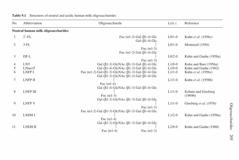
Oligosaccharides
205
Table 9.1 Structures of neutral and acidic human milk oligosaccharides
No. Abbreviation Oligosaccharide Lx/y-z Reference
Neutral human milk oligosaccharides
1 2´-FL Fuc (α1-2) Gal (β1-4) Glc L0/1-0 Kuhn et al. (1956c)Gal (β1-4) Glc
2 3-FL L0/1-0 Montreuil (1956)Fuc (α1-3)
Fuc (α1-2) Gal (β1-4) Glc3 DF-L L0/2-0 Kuhn and Gauhe (1958a)
Fuc (α1-3)4 LNT Gal (β1-3) GlcNAc (β1-3) Gal (β1-4) Glc L1/0-0 Kuhn and Baer (1956a)5 LNneoT Gal (β1-4) GlcNAc (β1-3) Gal (β1-4) Glc L1/0-0 Kuhn and Gauhe (1962)6 LNFP I Fuc (α1-2) Gal (β1-3) GlcNAc (β1-3) Gal (β1-4) Glc L1/1-0 Kuhn et al. (1956c)
Gal (β1-3) GlcNAc (β1-3) Gal (β1-4) Glc7 LNFP II L1/1-0 Kuhn et al. (1958b)
Fuc (α1-4)Gal (β1-4) GlcNAc (β1-3) Gal (β1-4) Glc
8 LNFP III L1/1-0 Kobata and GinsburgFuc (α1-3) (1969b)
Gal (β1-3) GlcNAc (β1-3) Gal (β1-4) Glc9 LNFP V L1/1-0 Ginsburg et al. (1976)
Fuc (α1-3)Fuc (α1-2) Gal (β1-3) GlcNAc (β1-3) Gal (β1-4) Glc
10 LNDH I L1/2-0 Kuhn and Gauhe (1958a) Fuc (α1-4)
Gal (β1-3) GlcNAc (β1-3) Gal (β1-4) Glc11 LNDH II L2/0-0 Kuhn and Gauhe (1960)
Fuc (α1-4) Fuc (α1-3)
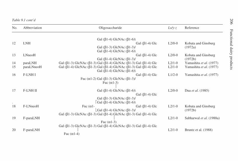
206Functional dairy products
Table 9.1 cont’d
No. Abbreviation Oligosaccharide Lx/y-z Reference
Gal (β1-4) GlcNAc (β1-6)\12 LNH Gal (β1-4) Glc L2/0-0 Kobata and Ginsburg
Gal (β1-3) GlcNAc (β1-3)/ (1972a)Gal (β1-4) GlcNAc (β1-6)\
13 LNneoH Gal (β1-4) Glc L2/0-0 Kobata and GinsburgGal (β1-4) GlcNAc (β1-3)/ (1972b)
14 paraLNH Gal (β1-3) GlcNAc (β1-3) Gal (β1-4) GlcNAc (β1-3) Gal (β1-4) Glc L2/1-0 Yamashita et al. (1977)15 paraLNneoH Gal (β1-4) GlcNAc (β1-3) Gal (β1-4) GlcNAc (β1-3) Gal (β1-4) Glc L2/1-0 Yamashita et al. (1977)
Gal (β1-4) GlcNAc (β1-6)\16 F-LNH I Gal (β1-4) Glc L1/2-0 Yamashita et al. (1977)
Fuc (α1-2) Gal (β1-3) GlcNAc (β1-3)/ Fuc (α1-3)
17 F-LNH II Gal (β1-4) GlcNAc (β1-6)\ L2/0-0 Dua et al. (1985)Gal (β1-4) Glc
Gal (β1-3) GlcNAc (β1-3)/. Gal (β1-4) GlcNAc (β1-6)\18 F-LNneoH Fuc (α1- Gal (β1-4) Glc L2/1-0 Kobata and Ginsburg
Gal (β1-4) GlcNAc (β1-3)/ (1972b)Gal (β1-3) GlcNAc (β1-3) Gal (β1-4) GlcNAc (β1-3) Gal (β1-4) Glc
19 F-paraLNH L2/1-0 Sabharwal et al. (1988a) Fuc (α1-3)
Gal (β1-3) GlcNAc (β1-3) Gal (β1-4) GlcNAc (β1-3) Gal (β1-4) Glc20 F-paraLNH L2/1-0 Bruntz et al. (1988)
Fuc (α1-4)
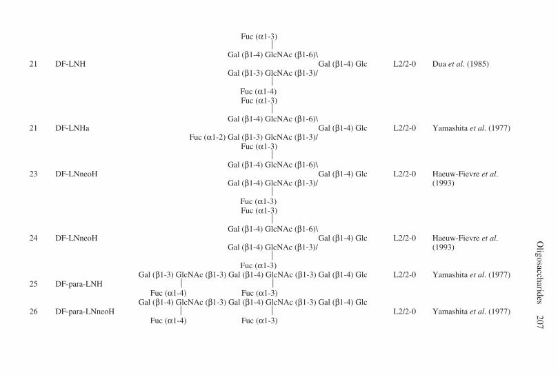
Oligosaccharides
207
Fuc (α1-3)
Gal (β1-4) GlcNAc (β1-6)\21 DF-LNH Gal (β1-4) Glc L2/2-0 Dua et al. (1985)
Gal (β1-3) GlcNAc (β1-3)/ Fuc (α1-4)
Fuc (α1-3)
Gal (β1-4) GlcNAc (β1-6)\21 DF-LNHa Gal (β1-4) Glc L2/2-0 Yamashita et al. (1977)
Fuc (α1-2) Gal (β1-3) GlcNAc (β1-3)/ Fuc (α1-3)
Gal (β1-4) GlcNAc (β1-6)\
23 DF-LNneoH Gal (β1-4) Glc L2/2-0 Haeuw-Fievre et al.Gal (β1-4) GlcNAc (β1-3)/ (1993)
Fuc (α1-3)
Fuc (α1-3)
Gal (β1-4) GlcNAc (β1-6)\24 DF-LNneoH Gal (β1-4) Glc L2/2-0 Haeuw-Fievre et al.
Gal (β1-4) GlcNAc (β1-3)/ (1993) Fuc (α1-3)
Gal (β1-3) GlcNAc (β1-3) Gal (β1-4) GlcNAc (β1-3) Gal (β1-4) Glc L2/2-0 Yamashita et al. (1977)25 DF-para-LNH
Fuc (α1-4) Fuc (α1-3)Gal (β1-4) GlcNAc (β1-3) Gal (β1-4) GlcNAc (β1-3) Gal (β1-4) Glc
26 DF-para-LNneoH L2/2-0 Yamashita et al. (1977) Fuc (α1-4) Fuc (α1-3)
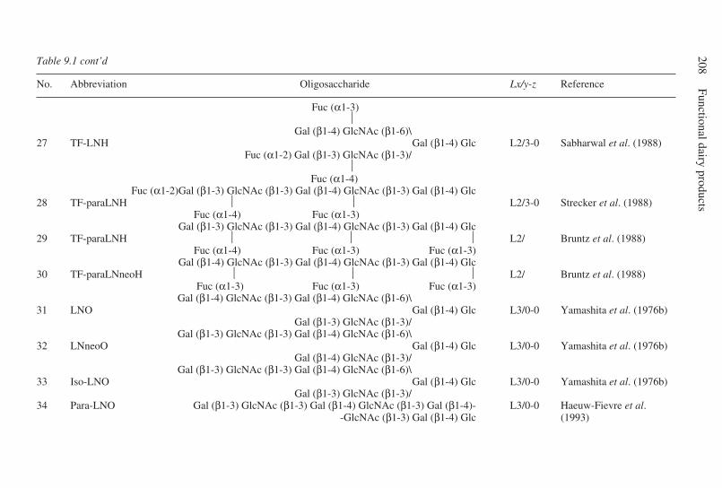
208Functional dairy products
Table 9.1 cont’d
No. Abbreviation Oligosaccharide Lx/y-z Reference
Fuc (α1-3)
Gal (β1-4) GlcNAc (β1-6)\27 TF-LNH Gal (β1-4) Glc L2/3-0 Sabharwal et al. (1988)
Fuc (α1-2) Gal (β1-3) GlcNAc (β1-3)/ Fuc (α1-4)
Fuc (α1-2)Gal (β1-3) GlcNAc (β1-3) Gal (β1-4) GlcNAc (β1-3) Gal (β1-4) Glc28 TF-paraLNH L2/3-0 Strecker et al. (1988)
Fuc (α1-4) Fuc (α1-3)Gal (β1-3) GlcNAc (β1-3) Gal (β1-4) GlcNAc (β1-3) Gal (β1-4) Glc
29 TF-paraLNH L2/ Bruntz et al. (1988) Fuc (α1-4) Fuc (α1-3) Fuc (α1-3)
Gal (β1-4) GlcNAc (β1-3) Gal (β1-4) GlcNAc (β1-3) Gal (β1-4) Glc30 TF-paraLNneoH L2/ Bruntz et al. (1988)
Fuc (α1-3) Fuc (α1-3) Fuc (α1-3)Gal (β1-4) GlcNAc (β1-3) Gal (β1-4) GlcNAc (β1-6)\
31 LNO Gal (β1-4) Glc L3/0-0 Yamashita et al. (1976b)Gal (β1-3) GlcNAc (β1-3)/
Gal (β1-3) GlcNAc (β1-3) Gal (β1-4) GlcNAc (β1-6)\32 LNneoO Gal (β1-4) Glc L3/0-0 Yamashita et al. (1976b)
Gal (β1-4) GlcNAc (β1-3)/Gal (β1-3) GlcNAc (β1-3) Gal (β1-4) GlcNAc (β1-6)\
33 Iso-LNO Gal (β1-4) Glc L3/0-0 Yamashita et al. (1976b)Gal (β1-3) GlcNAc (β1-3)/
34 Para-LNO Gal (β1-3) GlcNAc (β1-3) Gal (β1-4) GlcNAc (β1-3) Gal (β1-4)- L3/0-0 Haeuw-Fievre et al.-GlcNAc (β1-3) Gal (β1-4) Glc (1993)
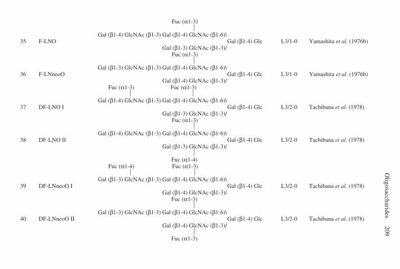
Oligosaccharides
209 Fuc (α1-3)
Gal (β1-4) GlcNAc (β1-3) Gal (β1-4) GlcNAc (β1-6)\
35 F-LNO Gal (β1-4) Glc L3/1-0 Yamashita et al. (1976b)Gal (β1-3) GlcNAc (β1-3)/
Fuc (α1-3)
Gal (β1-3) GlcNAc (β1-3) Gal (β1-4) GlcNAc (β1-6)\36 F-LNneoO Gal (β1-4) Glc L3/1-0 Yamashita et al. (1976b)
Gal (β1-4) GlcNAc (β1-3)/ Fuc (α1-3) Fuc (α1-3)
Gal (β1-4) GlcNAc (β1-3) Gal (β1-4) GlcNAc (β1-6)\37 DF-LNO I Gal (β1-4) Glc L3/2-0 Tachibana et al. (1978)
Gal (β1-3) GlcNAc (β1-3)/ Fuc (α1-3)
Gal (β1-4) GlcNAc (β1-3) Gal (β1-4) GlcNAc (β1-6)\
38 DF-LNO II Gal (β1-4) Glc L3/2-0 Tachibana et al. (1978)Gal (β1-3) GlcNAc (β1-3)/
Fuc (α1-4)
Fuc (α1-4) Fuc (α1-3)
Gal (β1-3) GlcNAc (β1-3) Gal (β1-4) GlcNAc (β1-6)\39 DF-LNneoO I Gal (β1-4) Glc L3/2-0 Tachibana et al. (1978)
Gal (β1-4) GlcNAc (β1-3)/ Fuc (α1-3)
Gal (β1-3) GlcNAc (β1-3) Gal (β1-4) GlcNAc (β1-6)\40 DF-LNneoO II Gal (β1-4) Glc L3/2-0 Tachibana et al. (1978)
Gal (β1-4) GlcNAc (β1-3)/ Fuc (α1-3)
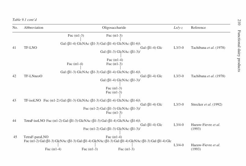
210Functional dairy products
Table 9.1 cont’d
No. Abbreviation Oligosaccharide Lx/y-z Reference
Fuc (α1-3) Fuc (α1-3)
Gal (β1-4) GlcNAc (β1-3) Gal (β1-4) GlcNAc (β1-6)\41 TF-LNO Gal (β1-4) Glc L3/3-0 Tachibana et al. (1978)
Gal (β1-3) GlcNAc (β1-3)/ Fuc (α1-4)
Fuc (α1-4) Fuc (α1-3)
Gal (β1-3) GlcNAc (β1-3) Gal (β1-4) GlcNAc (β1-6)\42 TF-LNneoO Gal (β1-4) Glc L3/3-0 Tachibana et al. (1978)
Gal (β1-4) GlcNAc (β1-3)/ Fuc (α1-3) Fuc (α1-3) 43 TF-isoLNO Fuc (α1-2) Gal (β1-3) GlcNAc (β1-3) Gal (β1-4) GlcNAc (β1-6)\
Gal (β1-4) Glc L3/3-0 Strecker et al. (1992)Fuc (α1-2) Gal (β1-3) GlcNAc (β1-3)/
Fuc (α1-3) 44 TetraF-isoLNO Fuc (α1-2) Gal (β1-3) GlcNAc (β1-3) Gal (β1-4) GlcNAc (β1-6)\
Gal (β1-4) Glc L3/4-0 Haeuw-Fievre et al.Fuc (α1-2) Gal (β1-3) GlcNAc (β1-3)/ (1993)
45 TetraF-paraLNO Fuc (α1-4)
Fuc (α1-2) Gal (β1-3) GlcNAc (β1-3) Gal (β1-4) GlcNAc (β1-3) Gal (β1-4) GlcNAc (β1-3) Gal (β1-4) GlcTetraF-paraLNO L3/4-0 Haeuw-Fievre et al.
Fuc (α1-4) Fuc (α1-3) Fuc (α1-3) (1993)
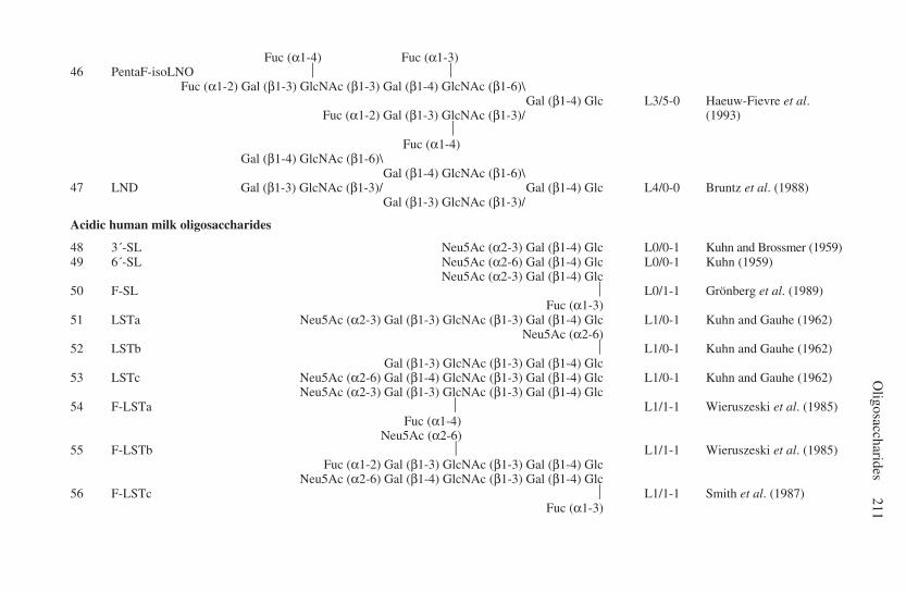
Oligosaccharides
211
Fuc (α1-4) Fuc (α1-3)46 PentaF-isoLNO
Fuc (α1-2) Gal (β1-3) GlcNAc (β1-3) Gal (β1-4) GlcNAc (β1-6)\PentaF-isoLNO Gal (β1-4) Glc L3/5-0 Haeuw-Fievre et al.
Fuc (α1-2) Gal (β1-3) GlcNAc (β1-3)/ (1993) Fuc (α1-4)
Gal (β1-4) GlcNAc (β1-6)\Gal (β1-4) GlcNAc (β1-6)\
47 LND Gal (β1-3) GlcNAc (β1-3)/ Gal (β1-4) Glc L4/0-0 Bruntz et al. (1988)Gal (β1-3) GlcNAc (β1-3)/
Acidic human milk oligosaccharides
48 3´-SL Neu5Ac (α2-3) Gal (β1-4) Glc L0/0-1 Kuhn and Brossmer (1959)49 6´-SL Neu5Ac (α2-6) Gal (β1-4) Glc L0/0-1 Kuhn (1959)
Neu5Ac (α2-3) Gal (β1-4) Glc50 F-SL L0/1-1 Grönberg et al. (1989)
Fuc (α1-3)51 LSTa Neu5Ac (α2-3) Gal (β1-3) GlcNAc (β1-3) Gal (β1-4) Glc L1/0-1 Kuhn and Gauhe (1962)
Neu5Ac (α2-6)52 LSTb L1/0-1 Kuhn and Gauhe (1962)
Gal (β1-3) GlcNAc (β1-3) Gal (β1-4) Glc53 LSTc Neu5Ac (α2-6) Gal (β1-4) GlcNAc (β1-3) Gal (β1-4) Glc L1/0-1 Kuhn and Gauhe (1962)
Neu5Ac (α2-3) Gal (β1-3) GlcNAc (β1-3) Gal (β1-4) Glc54 F-LSTa L1/1-1 Wieruszeski et al. (1985)
Fuc (α1-4) Neu5Ac (α2-6)
55 F-LSTb L1/1-1 Wieruszeski et al. (1985)Fuc (α1-2) Gal (β1-3) GlcNAc (β1-3) Gal (β1-4) Glc
Neu5Ac (α2-6) Gal (β1-4) GlcNAc (β1-3) Gal (β1-4) Glc56 F-LSTc L1/1-1 Smith et al. (1987)
Fuc (α1-3)
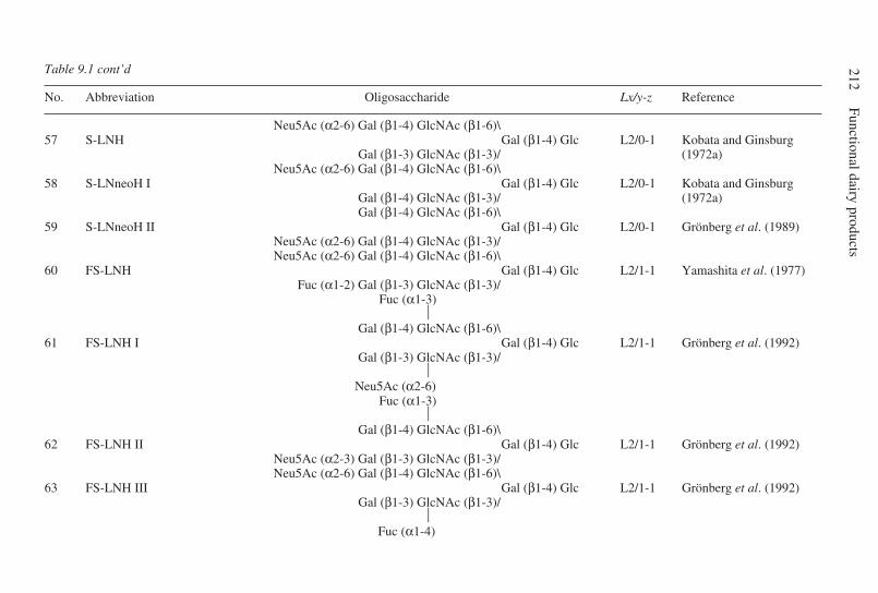
212Functional dairy products
Table 9.1 cont’d
No. Abbreviation Oligosaccharide Lx/y-z Reference
Neu5Ac (α2-6) Gal (β1-4) GlcNAc (β1-6)\57 S-LNH Gal (β1-4) Glc L2/0-1 Kobata and Ginsburg
Gal (β1-3) GlcNAc (β1-3)/ (1972a)Neu5Ac (α2-6) Gal (β1-4) GlcNAc (β1-6)\
58 S-LNneoH I Gal (β1-4) Glc L2/0-1 Kobata and GinsburgGal (β1-4) GlcNAc (β1-3)/ (1972a)Gal (β1-4) GlcNAc (β1-6)\
59 S-LNneoH II Gal (β1-4) Glc L2/0-1 Grönberg et al. (1989)Neu5Ac (α2-6) Gal (β1-4) GlcNAc (β1-3)/Neu5Ac (α2-6) Gal (β1-4) GlcNAc (β1-6)\
60 FS-LNH Gal (β1-4) Glc L2/1-1 Yamashita et al. (1977)Fuc (α1-2) Gal (β1-3) GlcNAc (β1-3)/
Fuc (α1-3)
Gal (β1-4) GlcNAc (β1-6)\61 FS-LNH I Gal (β1-4) Glc L2/1-1 Grönberg et al. (1992)
Gal (β1-3) GlcNAc (β1-3)/ Neu5Ac (α2-6)
Fuc (α1-3)
Gal (β1-4) GlcNAc (β1-6)\62 FS-LNH II Gal (β1-4) Glc L2/1-1 Grönberg et al. (1992)
Neu5Ac (α2-3) Gal (β1-3) GlcNAc (β1-3)/Neu5Ac (α2-6) Gal (β1-4) GlcNAc (β1-6)\
63 FS-LNH III Gal (β1-4) Glc L2/1-1 Grönberg et al. (1992)Gal (β1-3) GlcNAc (β1-3)/
Fuc (α1-4)
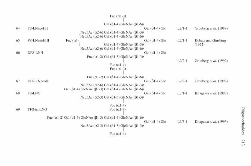
Oligosaccharides
213
Fuc (α1-3)
Gal (β1-4) GlcNAc (β1-6)\64 FS-LNneoH I Gal (β1-4) Glc L2/1-1 Grönberg et al. (1989)
Neu5Ac (α2-6) Gal (β1-4) GlcNAc (β1-3)/Neu5Ac (α2-6) Gal (β1-4) GlcNAc (β1-6)\
65 FS-LNneoH II Fuc (α1- Gal (β1-4) Glc L2/1-1 Kobata and Ginsburg Gal (β1-4) GlcNAc (β1-3)/ (1972)
Neu5Ac (α2-6) Gal (β1-4) GlcNAc (β1-6)\66 DFS-LNH Gal (β1-4) Glc
Fuc (α1-2) Gal (β1-3) GlcNAc (β1-3)/ L2/2-1 Grönberg et al. (1992) Fuc (α1-4)
Fuc (α1-3)
Fuc (α1-2) Gal (β1-4) GlcNAc (β1-6)\67 DFS-LNneoH Gal (β1-4) Glc L2/2-1 Grönberg et al. (1992)
Neu5Ac (α2-6) Gal (β1-4) GlcNAc (β1-3)/Gal (β1-4) GlcNAc (β1-3) Gal (β1-4) GlcNAc (β1-6)\
68 FS-LNO Gal (β1-4) Glc L3/1-1 Kitagawa et al. (1993)Neu5Ac (α2-3) Gal (β1-3) GlcNAc (β1-3)/
Fuc (α1-4)69 TFS-isoLNO Fuc (α1-3)
Fuc (α1-2) Gal (β1-3) GlcNAc (β1-3) Gal (β1-4) GlcNAc (β1-6)\TFS-isoLNO Gal (β1-4) Glc L3/3-1 Kitagawa et al. (1993)
Neu5Ac (α2-3) Gal (β1-3) GlcNAc (β1-3)/ Fuc (α1-4)
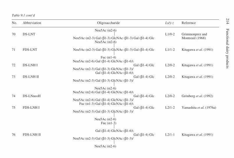
214Functional dairy products
Table 9.1 cont’d
No. Abbreviation Oligosaccharide Lx/y-z Reference
Neu5Ac (α2-6)70 DS-LNT L1/0-2 Grimmonprez and
Neu5Ac (α2-3) Gal (β1-3) GlcNAc (β1-3) Gal (β1-4) Glc Montreuil (1968) Neu5Ac (α2-6)
71 FDS-LNT Neu5Ac (α2-3) Gal (β1-3) GlcNAc (β1-3) Gal (β1-4) Glc L1/1-2 Kitagawa et al. (1991) Fuc (α1-4)
Neu5Ac (α2-6) Gal (β1-4) GlcNAc (β1-6)\72 DS-LNH I Gal (β1-4) Glc L2/0-2 Kitagawa et al. (1991)
Neu5Ac (α2-3) Gal (β1-3) GlcNAc (β1-3)/Gal (β1-4) GlcNAc (β1-6)\
73 DS-LNH II Gal (β1-4) Glc L2/0-2 Kitagawa et al. (1991)Neu5Ac (α2-3) Gal (β1-3) GlcNAc (β1-3)/
Neu5Ac (α2-6)
Neu5Ac (α2-6) Gal (β1-4) GlcNAc (β1-6)\74 DS-LNneoH Gal (β1-4) Glc L2/0-2 Grönberg et al. (1992)
Neu5Ac (α2-6) Gal (β1-4) GlcNAc (β1-3)/Fuc (α1-3) Gal (β1-4) GlcNAc (β1-6)\
75 FDS-LNH I Gal (β1-4) Glc L2/1-2 Yamashita et al. (1976a)Neu5Ac (α2-3) Gal (β1-3) GlcNAc (β1-3)/
Neu5Ac (α2-6)
Fuc (α1-3)
Gal (β1-4) GlcNAc (β1-6)\76 FDS-LNH II Gal (β1-4) Glc L2/1-1 Kitagawa et al. (1991)
Neu5Ac (α2-3) Gal (β1-3) GlcNAc (β1-3)/ Neu5Ac (α2-6)
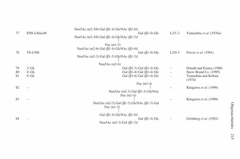
Oligosaccharides
215
Neu5Ac (α2-3/6) Gal (β1-4) GlcNAc (β1-6)\77 FDS-LNneoH Gal (β1-4) Glc L2/1-2 Yamashita et al. (1976a)
Neu5Ac (α2-3/6) Gal (β1-4) GlcNAc (β1-3)/ Fuc (α1-3)
Neu5Ac (α2-6) Gal (β1-4) GlcNAc (β1-6)\78 TS-LNH Gal (β1-4) Glc L2/0-3 Fievre et al. (1991)
Neu5Ac (α2-3) Gal (β1-3) GlcNAc (β1-3)/ Neu5Ac (α2-6)79 3'-GL Gal (β1-3) Gal (β1-4) Glc – Donald and Feeney (1988)80 4'-GL Gal (β1-4) Gal (β1-4) Glc – Snow Brand Co. (1995)81 6'-GL Gal (β1-6) Gal (β1-4) Glc – Yamashita and Kobata
(1974) Fuc (α1-4)
82 – – Kitagawa et al. (1990)Neu5Ac (α2-3) Gal (β1-3) GlcNAc
Fuc (α1-4)83 – – Kitagawa et al. (1990)
Neu5Ac (α2-3) Gal (β1-3) GlcNAc (β1-3) Gal Fuc (α1-3)
Gal (β1-4) GlcNAc (β1-6)\
84 – Gal (β1-4) Glc – Grönberg et al. (1992)Neu5Ac (α2-3) Gal (β1-3)/
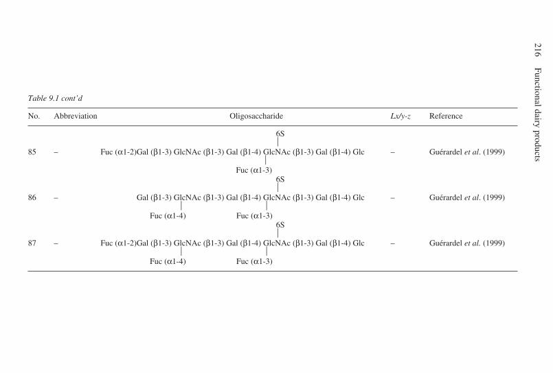
216Functional dairy products
Table 9.1 cont’d
No. Abbreviation Oligosaccharide Lx/y-z Reference
6S 85 – Fuc (α1-2)Gal (β1-3) GlcNAc (β1-3) Gal (β1-4) GlcNAc (β1-3) Gal (β1-4) Glc – Guérardel et al. (1999) Fuc (α1-3) 6S 86 – Gal (β1-3) GlcNAc (β1-3) Gal (β1-4) GlcNAc (β1-3) Gal (β1-4) Glc – Guérardel et al. (1999) Fuc (α1-4) Fuc (α1-3) 6S 87 – Fuc (α1-2)Gal (β1-3) GlcNAc (β1-3) Gal (β1-4) GlcNAc (β1-3) Gal (β1-4) Glc – Guérardel et al. (1999) Fuc (α1-4) Fuc (α1-3)
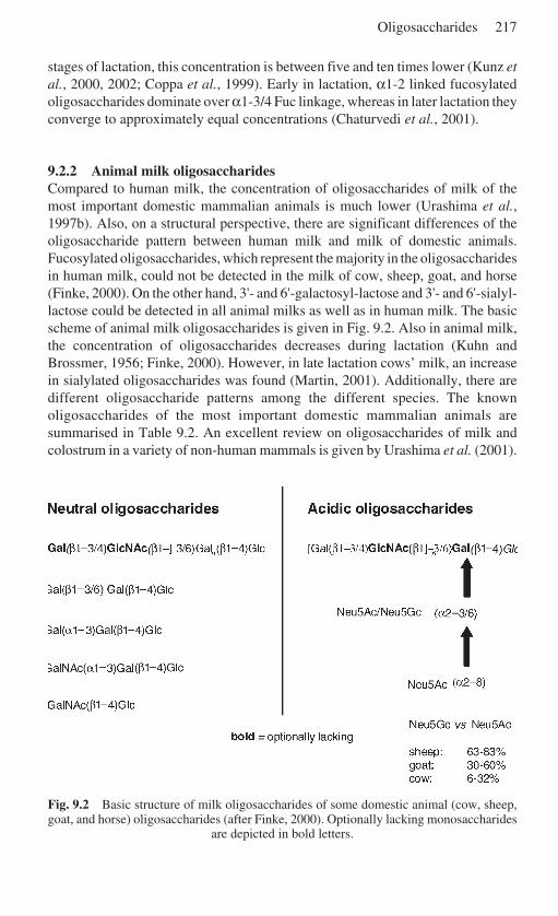
Oligosaccharides 217
stages of lactation, this concentration is between five and ten times lower (Kunz etal., 2000, 2002; Coppa et al., 1999). Early in lactation, α1-2 linked fucosylatedoligosaccharides dominate over α1-3/4 Fuc linkage, whereas in later lactation theyconverge to approximately equal concentrations (Chaturvedi et al., 2001).
9.2.2 Animal milk oligosaccharidesCompared to human milk, the concentration of oligosaccharides of milk of themost important domestic mammalian animals is much lower (Urashima et al.,1997b). Also, on a structural perspective, there are significant differences of theoligosaccharide pattern between human milk and milk of domestic animals.Fucosylated oligosaccharides, which represent the majority in the oligosaccharidesin human milk, could not be detected in the milk of cow, sheep, goat, and horse(Finke, 2000). On the other hand, 3'- and 6'-galactosyl-lactose and 3'- and 6'-sialyl-lactose could be detected in all animal milks as well as in human milk. The basicscheme of animal milk oligosaccharides is given in Fig. 9.2. Also in animal milk,the concentration of oligosaccharides decreases during lactation (Kuhn andBrossmer, 1956; Finke, 2000). However, in late lactation cows’ milk, an increasein sialylated oligosaccharides was found (Martin, 2001). Additionally, there aredifferent oligosaccharide patterns among the different species. The knownoligosaccharides of the most important domestic mammalian animals aresummarised in Table 9.2. An excellent review on oligosaccharides of milk andcolostrum in a variety of non-human mammals is given by Urashima et al. (2001).
Fig. 9.2 Basic structure of milk oligosaccharides of some domestic animal (cow, sheep,goat, and horse) oligosaccharides (after Finke, 2000). Optionally lacking monosaccharides
are depicted in bold letters.
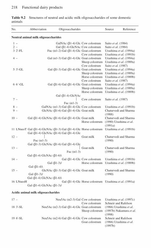
218 Functional dairy products
Table 9.2 Structures of neutral and acidic milk oligosaccharides of some domesticanimals
No. Abbreviation Oligosaccharides Source Reference
Neutral animal milk oligosaccharides
1 – GalNAc (β1-4) Glc Cow colostrum Saito et al. (1984)2 – Gal (β1-4) GlcNAc Cow colostrum Saito et al. (1984)3 2'-FL Fuc (α1-2) Gal (β1-4) Glc Goat colostrum Urashima et al. (1994a)
Cow colostrums Urashima et al. (1991b)4 – Gal (α1-3) Gal (β1-4) Glc Goat colostrum Urashima et al. (1994a)
Sheep colostrum Urashima et al. (1989a)Cow colostrum Saito et al. (1987)
5 3'-GL Gal (β1-3) Gal (β1-4) Glc Goat colostrum Urashima et al. (1994a)Sheep colostrum Urashima et al. (1989a)Horse colostrum Urashima et al. (1989b)Cow colostrum Saito et al. (1987)
6 6´-GL Gal (β1-6) Gal (β1-4) Glc Goat colostrum Urashima et al. (1994a)Sheep colostrum Urashima et al. (1989a)Horse colostrum Urashima et al. (1989b)
Gal (β1-4) GlcNAc7 – Cow colostrum Saito et al. (1987)
Fuc (α1-3)8 – GalNAc (α1-3) Gal (β1-4) Glc Cow colostrum Urashima et al. (1991b)9 – GlcNAc (β1-6) Gal (β1-4) Glc Goat milk Chaturvedi and Sharma
(1988)10 – Gal (β1-4) GlcNAc (β1-6) Gal (β1-4) Glc Goat milk Chaturvedi and Sharma
Horse colostrum (1990) Urashima et al.(1991a)
11 LNneoT Gal (β1-4) GlcNAc (β1-3) Gal (β1-4) Glc Horse colostrum Urashima et al. (1991b)Gal (β1-4) GlcNAc (β1-6) Gal (β1-4) Glc
12 – Goat milk Chaturvedi and Sharma Fuc (α1-3) (1990)
Gal (β1-3) GlcNAc (β1-6) Gal (β1-4) Glc13 – Goat milk Chaturvedi and Sharma
Fuc (α1-3) (1990)Gal (β1-4) GlcNAc (β1-6)\
14 – Gal (β1-4) Glc Cow colostrum Urashima et al. (1991b)Gal (β1-3)/ Horse colostrum Urashima et al. (1989b)
Gal (β1-4)\15 – GlcNAc (β1-3) Gal (β1-4) Glc Goat milk Chaturvedi and Sharma
Gal (β1-3)/ (1990)Gal (β1-4) GlcNAc (β1-6)\
16 LNneoH Gal (β1-4) Glc Horse colostrum Urashima et al. (1991a)Gal (β1-4) GlcNAc (β1-3)/
Acidic animal milk oligosaccharides
17 – Neu5Ac (α2-3) Gal Cow colostrum Urashima et al. (1997c)Cow colostrum Schneir and Rafelson
18 3'-SL Neu5Ac (α2-3) Gal (β1-4) Glc Goat colostrum (1966) Urashima et al.Sheep colostrum (1997b) Nakamura et al.
(1998)19 6'-SL Neu5Ac (α2-6) Gal (β1-4) Glc Cow colostrum Schneir and Rafelson
Goat colostrum (1966) Urashima et al.(1997b)
Oligosaccharides 219
Table 9.2 cont’d
No. Abbreviation Oligosaccharides Source Reference
Cow colostrum Kuhn and Gauhe (1965)20 – Neu5Gc (α2-3) Gal (β1-4) Glc Goat colostrum Urashima et al. (1997b)
Sheep colostrum Nakamura et al. (1998)Goat colostrum Veh et al. (1981)
21 – Neu5Gc (α2-6) Gal (β1-4) Glc Cow colostrum Urashima et al. (1997b)Sheep colostrum Nakamura et al. (1998)
22 – O-Acetyl-Neu5Ac (α2-3) Gal (β1-4) Glc Cow colostrum Urashima et al. (1997c)23 6'-SLn Neu5Ac (α2-6) Gal (β1-4) GlcNAc Cow colostrum Kuhn and Gauhe (1965)
Goat colostrum Urashima et al. (1997b)24 – Neu5Gc (α2-6) Gal (β1-4) GlcNAc Cow colostrum Kuhn and Gauhe (1965)25 6'-SLn-1-P Neu5Ac (α2-6) Gal (β1-4) GlcNAc-1-PO
4Cow colostrum Kuhn and Gauhe (1965)
26 6'-SLn-6-P Neu5Ac (α2-6) Gal (β1-4) GlcNAc-6-PO4
Cow colostrum Kuhn and Gauhe (1965)27 3´-SGL Neu5Ac (α2-3) Gal (β1-3) Gal (β1-4) Glc Cow colostrum Parkinnen and Finne
(1987)Gal (β1-3) Gal (β1-4) Glc
28 – Goat milk Viverge et al. (1997) Neu5Ac (α2-6)
Gal (β1-6) Gal (β1-4) Glc29 – Goat milk Viverge et al. (1997) Neu5Ac (α2-3)30 DS-L Neu5Ac (α2-8) Neu5Ac (α2-3) Gal (β1-4) Glc Cow colostrum Kuhn and Gauhe 1(965)
9.2.3 Oligosaccharides of non-milk originBecause the composition and structure of human milk oligosaccharides cannot bereproduced and until now large-scale preparations of animal milk oligosaccharideshave not been commercially available, oligosaccharides of more simple structuresthan human milk oligosaccharides have been used as components in severaldietary products to mimic the beneficial effects of human milk oligosaccharides(Gibson and Roberfroid, 1995; Roberfroid and Delzenne, 1998; Loo et al., 1999;Jenkins et al., 1999; Gibson, 1999). The most important prebiotic (see below, 9.4)oligosaccharides, larger than disaccharides, of non-milk origin, their structure andthe method of preparation are given in Table 9.3. The most intensive studies existfor fructans and, to a lesser extent, for galactooligosaccharides (Loo et al., 1999).Fructans are linear or branched fructose-polymers, which are either β2-1-linkedinulins or β2-6-linked levans. Because the inulin-type fructans can be easilyextracted from plant sources (e.g. asparagus, garlic, leek, onion, artichoke, chicoryroots, etc.) they have been widely used as an ingredient in dietary products. Thebasic structure of fructans is given in Table 9.3.
Galactooligosaccharides are synthesised from lactose via enzymatictransgalactosylation using ß-galactosidases mainly of bacterial origin (e.g. Bacilluscirculans) (Tanaka and Matsumoto, 1998). The commercial use of β-galactosidasein food processing is extensively reviewed elsewhere (Kinsella and Taylor, 1995).Galactooligosaccharides consist of a chain of galactose molecules usually with aglucose molecule at the reducing terminus, varying in chain length (degree ofpolymerisation range 3–8) and linkages (for basic structure, see Table 9.3).
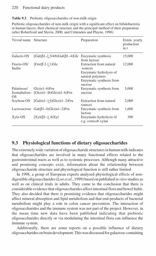
220 Functional dairy products
Table 9.3 Prebiotic oligosaccharides of non-milk origin
Prebiotic oligosaccharides of non-milk origin with a significant effect on bifidobacteriain human faeces, their chemical structure and the principal method of their preparation(after Roberfroid and Slavin, 2000, and Crittenden and Playne, 1996)
Trivial name Structure Preparation Estim. yearlyproductionin t
Galacto-OS [Gal(β1–]n3/4/6)Gal(β1–4)Glc Enzymatic synthesis 15,000
from lactoseFructo-OS/ [Fru(β 2-]
n1)Glc Extraction from natural 12,000
Inulin sourcesEnzymatic hydrolysis ofnatural polymersEnzymatic synthesis fromsucrose
Palatinose/ Glc(α1–6)Fru Enzymatic synthesis from 5,000Isomaltulose- [Glc(α1–]6)Glc(α1–6)Fru sucroseOSSoybean OS [Gal(α1–]
n6)Glc(α1–2)Fru Extraction from natural 2,000
sourcesLactosucrose Gal(β1–4)Glc(α1–2)Fru Enzymatic synthesis from 1,600
lactoseXylo-OS [Xyl(β1–]
n 4)Xyl Enzymatic hydrolysis of 300
e.g. corncob xylan
9.3 Physiological functions of dietary oligosaccharidesThe extremely wide variation of oligosaccharide structures in human milk indicatesthat oligosaccharides are involved in many functional effects related to thegastrointestinal tracts as well as to systemic processes. Although many attractiveand promising concepts exist, information about the relationship betweenoligosaccharide structure and physiological function is still rather limited.
In 1998, a group of European experts analysed physiological effects of non-digestible oligosaccharides (Loo et al., 1999) based on published in vitro studies aswell as on clinical trials in adults. They came to the conclusion that there isconsiderable evidence that oligosaccharides affect intestinal flora and bowel habit.They also decided that there is promising evidence that oligosaccharides mightaffect mineral absorption and lipid metabolism and that end-products of bacterialmetabolism might play a role in colon cancer prevention. The interaction ofoligosaccharides and the immune system was not part of the project. However, inthe mean time new data have been published indicating that prebioticoligosaccharides directly or via modulating the intestinal flora can influence theimmune system.
Additionally, there are some reports on a possible influence of dietaryoligosaccharides on brain development. This was discussed for galactose-containing
Oligosaccharides 221
molecules, with galactose as source for galactocerebroside, the predominantglycolipid in myelin and, thus especially important for the development of theinfant brain (Kunz et al., 1999).
Based on studies with newborn rats, it was discovered that oral administrationof sialic acid increases the cerebral and cerebellar sialic acid content (Carlson andHouse, 1986). However, contradicting results were obtained from studies withmice and rats receiving N-acetylneuraminic acid orally and intravenously (Nöhleand Schauer, 1981). Therefore, further research is needed to prove a possibleinfluence of dietary mono- and oligosaccharides, respectively, on braindevelopment.
9.4 Effect on intestinal flora: prebiotic roleIt has been known for more than one hundred years that bifidobacteria dominatethe faecal flora of breast-fed infants (Tissier, 1899). Around 50 years ago, humanmilk oligosaccharides were identified as one of the important bifidogenic factorsfound in human milk (György, et al.,1954; Kunz, et al., 2000). There is a wideconsensus that bifidobacteria, along with lactobacilli, can be classified as health-promoting bacteria, which is also the basis for their use as probiotics.Oligosaccharides seem to be an important factor in the defence of the newborninfant against infection (Kunz et al., 2000).
The basic assumption of the prebiotic concept is that these dietary ingredientsare non-digestible, reach the colon and can be utilised by the health-promotingcolonic bacteria (Gibson and Roberfroid, 1995). The human intestine lacksenzymes able to hydrolyse β-glycosidic linkages with the exception of lactose,which makes oligosaccharides with β-linkages non-digestible (Rivero-Urgell andSantamaría-Orleans, 2001; Engfer et al., 2000; Gnoth et al., 2000; Roberfroid andDelzenne, 1998). Thus, human milk oligosaccharides and most animal milkoligosaccharides, as well as the non-milk oligosaccharides, can be considered to beindigestible.
Another prerequisite for a prebiotic effect is that the dietary ingredient thatreaches the colon can be utilised by the intestinal flora to stimulate those bacteriathat are naturally part of the ecosystem in the colon. Most data concerning theutilisation of oligosaccharides by faecal bacteria come from in vitro studies and arerelated to non-milk oligosaccharides. Some common non-milk oligosaccharidesthat show significant bifidogenic effect in human faeces are summarised in Table9.3. Additionally, clinical trials in human infants and adults have demonstrated thestimulation of bifidobacteria and lactobacilli by several non-milk oligosaccharides,in particular galactooligosaccharides and fructans (Tables 9.4 and 9.5). Morerecently, it has been demonstrated in infants that a mixture ofgalactooligosaccharides and fructans is very effective in stimulating abifidobacteriae and changing the stool characteristics to become closer to thosefound in breast-fed infants (Moro et al., 2002; Boehm et al., 2002), indicating thatthese oligosaccharides of non-milk origin can, at least partially, mimic the effectsof human milk oligosaccharides.
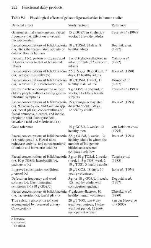
222 Functional dairy products
Table 9.4 Physiological effects of galactooligosaccharides in human studies
Detected effect Study protocol Reference
Gastrointestinal symptoms and faecal 15 g GOS/d in yoghurt, 3 Teuri et al. (1998)frequency (>). Effect on intestinal weeks, 12 healthy adultsmicroecosystem
Faecal concentrations of bifidobacteria 10 g TOS/d, 21 days, 8 Bouhnik et al.(>), alters the fermentative activity of healthy adults (1997)colonic flora in humans
Faecal pH (<), pattern of organic acid 1 or 2% glactosyllactose in Yahiro et al.in faeces closer to that of breast-fed infant formula, 27 newborn (1982)infants infants
Faecal concentrations of bifidobacteria 2.5 g, 5 g or 10 g GOS/d, 7 Ito et al. (1990)(>), lactobacilli slightly (>) days, 12 healthy adults
Faecal concentrations of bifidobacteria 10 g TOS/d, 1 week, 11 Dombo et al.(>), lactobacilli (>), bacteroides (<) healthy male adults (1997)
Seems to relieve constipation in most 9 g GOS/d in yoghurt, 2 Teuri et al. (1998)elderly people without causing gastro- weeks, 14 elderly femaleintestinal symptoms subjects
Faecal concentrations of bifidobacteria 15 g transgalactosylated Ito et al. (1993)(>), Bacteroidaceae and Candida spp. disaccharide/d, 6 days,(<), faecal pH (<), concentrations of 12 healthy adultsfaecal ammonia, p-cresol, and indole,propionic acid, isobutyric acid,isovaleric acid and valeric acid (<)
Good tolerance 15 g GOS/d, 3 weeks, 12 van Dokkum et al.healthy men (1995)
Faecal concentrations of bifidobacteria 2.5 g GOS/d, 3 weeks, 12 Ito et al. (1993)(>), pathogens (–). Faecal nitro- healthy adults in whom thereductase activity, and concentrations number of indigenousof indole and isovaleric acid (<) bifidobacteria were
comparatively low
Faecal concentrations of bifidobacteria 3 g or 10 g TOS/d, 2 weeks Tanaka et al.(>). 10 g TOS/d: lactobacilli (>), (week 1: 3 g TOS, week 2: (1983)pathogens (<) 10 g TOS), 5 healthy adults
Improved constipation condition, 10 g/d GOS, 18 days, 50 Ito et al. (1994)p-cresol (<) young volunteers
Defecation frequency and stool 5 g, or 10 g GOS/d, 1 week, Deguchi et al.softness (>). Gastrointestinal 128 healthy adults with (1997)symptoms (>) (10 g GOS/d) constipation tendency
Faecal concentrations of bifidobacteria 4' galactosyllactose, 10 Ohtsuka et al.(>), bacteroides (<), faecal pH (<) healthy human volunteers (1989)
True calcium absorption (>) (not 20 g/d TOS, two 9-day van der Heuvel etaccompanied by increased urinary treatment periods, 19-day al. (2000)Ca excretion) washout period, 12 post-
menopausal women
> increase.< decrease.– no effect.
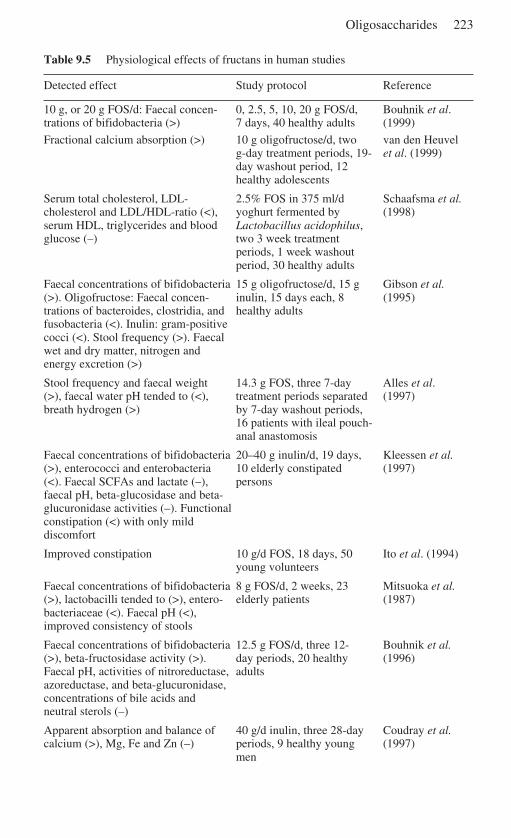
Oligosaccharides 223
Table 9.5 Physiological effects of fructans in human studies
Detected effect Study protocol Reference
10 g, or 20 g FOS/d: Faecal concen- 0, 2.5, 5, 10, 20 g FOS/d, Bouhnik et al.trations of bifidobacteria (>) 7 days, 40 healthy adults (1999)
Fractional calcium absorption (>) 10 g oligofructose/d, two van den Heuvelg-day treatment periods, 19- et al. (1999)day washout period, 12healthy adolescents
Serum total cholesterol, LDL- 2.5% FOS in 375 ml/d Schaafsma et al.cholesterol and LDL/HDL-ratio (<), yoghurt fermented by (1998)serum HDL, triglycerides and blood Lactobacillus acidophilus,glucose (–) two 3 week treatment
periods, 1 week washoutperiod, 30 healthy adults
Faecal concentrations of bifidobacteria 15 g oligofructose/d, 15 g Gibson et al.(>). Oligofructose: Faecal concen- inulin, 15 days each, 8 (1995)trations of bacteroides, clostridia, and healthy adultsfusobacteria (<). Inulin: gram-positivecocci (<). Stool frequency (>). Faecalwet and dry matter, nitrogen andenergy excretion (>)
Stool frequency and faecal weight 14.3 g FOS, three 7-day Alles et al.(>), faecal water pH tended to (<), treatment periods separated (1997)breath hydrogen (>) by 7-day washout periods,
16 patients with ileal pouch-anal anastomosis
Faecal concentrations of bifidobacteria 20–40 g inulin/d, 19 days, Kleessen et al.(>), enterococci and enterobacteria 10 elderly constipated (1997)(<). Faecal SCFAs and lactate (–), personsfaecal pH, beta-glucosidase and beta-glucuronidase activities (–). Functionalconstipation (<) with only milddiscomfort
Improved constipation 10 g/d FOS, 18 days, 50 Ito et al. (1994)young volunteers
Faecal concentrations of bifidobacteria 8 g FOS/d, 2 weeks, 23 Mitsuoka et al.(>), lactobacilli tended to (>), entero- elderly patients (1987)bacteriaceae (<). Faecal pH (<),improved consistency of stools
Faecal concentrations of bifidobacteria 12.5 g FOS/d, three 12- Bouhnik et al.(>), beta-fructosidase activity (>). day periods, 20 healthy (1996)Faecal pH, activities of nitroreductase, adultsazoreductase, and beta-glucuronidase,concentrations of bile acids andneutral sterols (–)
Apparent absorption and balance of 40 g/d inulin, three 28-day Coudray et al.calcium (>), Mg, Fe and Zn (–) periods, 9 healthy young (1997)
men
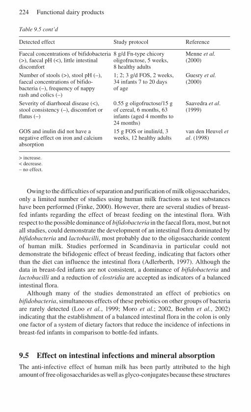
224 Functional dairy products
Table 9.5 cont’d
Detected effect Study protocol Reference
Faecal concentrations of bifidobacteria 8 g/d Fn-type chicory Menne et al.(>), faecal pH (<), little intestinal oligofructose, 5 weeks, (2000)discomfort 8 healthy adults
Number of stools (>), stool pH (–), 1; 2; 3 g/d FOS, 2 weeks, Guesry et al.faecal concentrations of bifido- 34 infants 7 to 20 days (2000)bacteria (–), frequency of nappy of agerash and colics (–)
Severity of diarrhoeal disease (<), 0.55 g oligofructose/15 g Saavedra et al.stool consistency (–), discomfort or of cereal, 6 months, 63 (1999)flatus (–) infants (aged 4 months to
24 months)
GOS and inulin did not have a 15 g FOS or inulin/d, 3 van den Heuvel etnegative effect on iron and calcium weeks, 12 healthy adults al. (1998)absorption
> increase.< decrease.– no effect.
Owing to the difficulties of separation and purification of milk oligosaccharides,only a limited number of studies using human milk fractions as test substanceshave been performed (Finke, 2000). However, there are several studies of breast-fed infants regarding the effect of breast feeding on the intestinal flora. Withrespect to the possible dominance of bifidobacteria in the faecal flora, most, but notall studies, could demonstrate the development of an intestinal flora dominated bybifidobacteria and lactobacilli, most probably due to the oligosaccharide contentof human milk. Studies performed in Scandinavia in particular could notdemonstrate the bifidogenic effect of breast feeding, indicating that factors otherthan the diet can influence the intestinal flora (Adlerberth, 1997). Although thedata in breast-fed infants are not consistent, a dominance of bifidobacteria andlactobacilli and a reduction of clostridia are accepted as indicators of a balancedintestinal flora.
Although many of the studies demonstrated an effect of prebiotics onbifidobacteria, simultaneous effects of these prebiotics on other groups of bacteriaare rarely detected (Loo et al., 1999; Moro et al.; 2002, Boehm et al., 2002)indicating that the establishment of a balanced intestinal flora in the colon is onlyone factor of a system of dietary factors that reduce the incidence of infections inbreast-fed infants in comparison to bottle-fed infants.
9.5 Effect on intestinal infections and mineral absorptionThe anti-infective effect of human milk has been partly attributed to the highamount of free oligosaccharides as well as glyco-conjugates because these structures
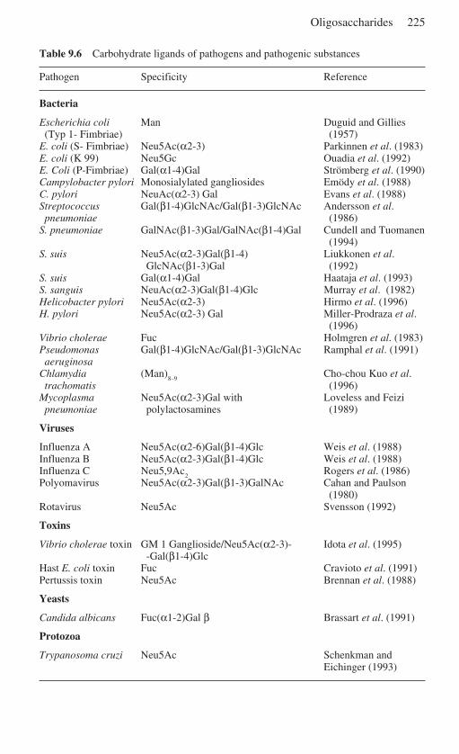
Oligosaccharides 225
Table 9.6 Carbohydrate ligands of pathogens and pathogenic substances
Pathogen Specificity Reference
Bacteria
Escherichia coli Man Duguid and Gillies(Typ 1- Fimbriae) (1957)
E. coli (S- Fimbriae) Neu5Ac(α2-3) Parkinnen et al. (1983)E. coli (K 99) Neu5Gc Ouadia et al. (1992)E. Coli (P-Fimbriae) Gal(α1-4)Gal Strömberg et al. (1990)Campylobacter pylori Monosialylated gangliosides Emödy et al. (1988)C. pylori NeuAc(α2-3) Gal Evans et al. (1988)Streptococcus Gal(β1-4)GlcNAc/Gal(β1-3)GlcNAc Andersson et al.pneumoniae (1986)
S. pneumoniae GalNAc(β1-3)Gal/GalNAc(β1-4)Gal Cundell and Tuomanen(1994)
S. suis Neu5Ac(α2-3)Gal(β1-4) Liukkonen et al.GlcNAc(β1-3)Gal (1992)
S. suis Gal(α1-4)Gal Haataja et al. (1993)S. sanguis NeuAc(α2-3)Gal(β1-4)Glc Murray et al. (1982)Helicobacter pylori Neu5Ac(α2-3) Hirmo et al. (1996)H. pylori Neu5Ac(α2-3) Gal Miller-Prodraza et al.
(1996)Vibrio cholerae Fuc Holmgren et al. (1983)Pseudomonas Gal(β1-4)GlcNAc/Gal(β1-3)GlcNAc Ramphal et al. (1991)aeruginosa
Chlamydia (Man)8–9
Cho-chou Kuo et al.trachomatis (1996)
Mycoplasma Neu5Ac(α2-3)Gal with Loveless and Feizipneumoniae polylactosamines (1989)
Viruses
Influenza A Neu5Ac(α2-6)Gal(β1-4)Glc Weis et al. (1988)Influenza B Neu5Ac(α2-3)Gal(β1-4)Glc Weis et al. (1988)Influenza C Neu5,9Ac
2Rogers et al. (1986)
Polyomavirus Neu5Ac(α2-3)Gal(β1-3)GalNAc Cahan and Paulson(1980)
Rotavirus Neu5Ac Svensson (1992)
Toxins
Vibrio cholerae toxin GM 1 Ganglioside/Neu5Ac(α2-3)- Idota et al. (1995)-Gal(β1-4)Glc
Hast E. coli toxin Fuc Cravioto et al. (1991)Pertussis toxin Neu5Ac Brennan et al. (1988)
Yeasts
Candida albicans Fuc(α1-2)Gal β Brassart et al. (1991)
Protozoa
Trypanosoma cruzi Neu5Ac Schenkman andEichinger (1993)
226 Functional dairy products
might prevent intestinal attachment of infectious agents by acting as receptoranalogues (Beachey, 1981; Coppa, 1990; Zopf and Roth, 1996; Newburg, 1999).Table 9.6 summarises the receptor analogues so far identified in human milk andthe respective infective agents. Similar observations have been made with cows’milk oligosaccharides, which reduced the adhesion of enterotoxic Escherichia colistrains of the calf (Mouricout et al., 1990).
There are also first in vitro results available that fructans interfere with theadhesion of Salmonella enterica sv. Typhimurium and non-pathogenic Escherichiacoli in the pig small intestine (Naughton et al., 2001)
Pectin, and in particular, pectin hydrolysate are also able to reduce the adhesionof bacteria to intestinal epithelial cells in vitro and in vivo (Guggenbichler et al.,1997). This effect of pectin hydrolysate can explain the therapeutic effect of carrotsoup, which has been used for 100 years by European paediatricians as treatmentagainst diarrhoea (Moro, 1908).
In summary, there is evidence that the prevention or reduction of adhesion ofinfectious agents to epithelial surfaces is an important mechanism of dietaryoligosaccharides in increasing the defence capacity against intestinal infections(Dai et al., 2000).
9.5.1 Effect on mineral absorptionThe animal studies as well as clinical trials in humans have been focused on theabsorption of calcium and magnesium, and in few experiments the effect of dietaryoligosaccharides on the metabolism of iron and iodine has been studied. In humaninfants, the calcium absorption from human milk is significantly higher than fromany formula (Carnielli et al., 1996). There are many factors in human milk thatmight influence calcium absorption. One of the important factors is the compositionof lipids which reduces the formation of calcium soaps in the colon (Carnielli et al.,1996). Thus, the difference in calcium absorption between human milk andformula cannot easily be attributed to the content of oligosaccharides.
There are several ways in which dietary oligosaccharides might influencemineral absorption. Most likely, the increase of bacterial synthesis of short chainfatty acids in the colon plays a key role, but also increasing the passive and activecalcium transport in the small intestine or increased transport of calcium from thesmall intestine to the colon may be involved in the stimulation of mineralabsorption by oligosaccharides (Scholz-Ahrens et al., 2001). In rat experimentswith dietary galactooligosaccharides (Chonan and Watanuki, 1996) measuringcomplete calcium absorption, including the colonic component, indicated that thedietary intake of galactooligosaccharides increased calcium absorption. In a studyof postmenopausal women, dietary galactooligosaccharides increased calciumabsorption significantly (van den Heuvel et al., 2000). Fructooligosaccharides arealso able to stimulate calcium absorption (Ohta et al., 1998; Roberfroid andDelzenne,1998; Frank, 1998). More recently, a preterm infant formula,supplemented with a mixture of galactooligosaccharides (90%) and inulin (10%),resulted in higher renal excretion of calcium compared to that found in infants fed
Oligosaccharides 227
a non-supplemented formula, indicating an increased calcium absorption (Lidestriet al., 2002).
There are also some reports that magnesium, iodine and iron metabolism can beimproved by dietary oligosaccharides (Roberfroid and Delzenne, 1998; Frank,1998). However, the data from the literature are not consistent, indicating that theexperimental design as well as the type of oligosaccharide and the mineral intakeinfluence the results.
9.6 Effect on the immune system and other physiologicaleffectsHuman milk protects the infant against infection, and oligosaccharides play animportant role among other factors (Xanthou, 1998). The development of abalanced intestinal microflora seems to be one of the main mechanisms for the hostdefence activity of oligosaccharides. This discussion has been greatly stimulatedby the observation of a Finnish group (Kalliomäki et al., 2001) that administrationof a probiotic strain Lactobacillus GG to a group of infants with a high risk ofdeveloping atopy reduced the frequency of atopic symptoms. Additionally, differ-ences in the intestinal microbiota composition between allergic and healthy infantshave been demonstrated (Ouwehand et al., 2001; Böttcher et al.; 2000). Observ-ations of the effect of prebiotic oligosaccharides on the immune system are rare. Ifthe internal microbiota is the acting factor in the postnatal immune modulation,then prebiotic oligosaccharides could have an effect on the immune system.Although the idea is attractive, no clinical studies demonstrating such a prebioticeffect on the postnatal developement in infants have been published. Very recently,it was reported that elderly persons receiving a prebiotic mixture (fructans ofdifferent molecular size) experienced significantly higher influenza A antibodytitres as a response to vaccination than patients receiving a placebo (Bunout et al.,2002). Only a few reports on the direct effect of human milk oligosaccharides onimmune functions have been reported so far. Human milk oligosaccharide structureslike LNFPIII and LNneoT (see Table 9.1) show an effect on IL10 production(Velupillai and Harn, 1994). It is further discussed that human milk is involved inthe generation of anti-inflammatory mediators that suppress Th-1-type andinflammatory responses (Terrazas et al., 2001). The interaction of human milkoligosaccharides with selectins in cell–cell-interactions of e.g. leucocytes andlymphocytes was described recently by Rudloff et al. (2002).
In summary, the possibility that prebiotic oligosaccharides could influence theimmune response of the host would make prebiotics dietary intervention inreduction of atopic diseases an attractive choice to probiotics, However, thecurrent data do not allow any conclusions to be drawn on the issue.
Other promising effects are the influence of prebiotic oligosaccharides on thelipid metabolism (for review see Roberfroid and Delzenne, 1998) and braindevelopment (Kunz et al., 1999; Carlson et al., 1986). The clinical relevance of theobserved effects is still under discussion and much more research is needed toprove these effects (Meyer et al., 2001).
228 Functional dairy products
9.7 Analytical methods
Depending on the type, size and structure as well as the (biological) source ofoligosaccharides, a variety of separation techniques and methods have to beapplied for the characterisation of the molecules of interest. Numerous publica-tions and reviews are available and only a selection of literature can be discussedin this chapter. The choice of a suitable analytical method depends strongly on thesize, composition and physicochemical properties of the carbohydrate of interest.For general information on oligosaccharides and glycoconjugates the reader isreferred to Varki et al. (1999) and Ernst et al. (2000). Food-derived carbohydratesare excellently described by Nakakuki (1994) as well as by Cho et al. (1999).General information on dietary fibers and strategies on the detection of dietaryfibers and prebiotic oligosaccharides has been provided by Southgate (1995) andSpiller (2001). For information on the complex group of heteropolysaccharides,especially those of lactic acid bacteriae from dairy products, the reader is referredto De Vuyst and Degeest (1999). For information on milk composition, the readeris referred to Jensen (1995). Milk oligosaccharides are described in detail byNewburg and Neubauer (1995) and Kunz et al. (2000). For general analyticaltechniques and methods in the field of glycobiology, the reader is referred to Ziadel Rassi (1995) and Lennarz and Hart (1994).
Briefly, the major techniques for the analyses of glycans (pure carbohydrates)and glycoconjugates (such as glycoproteins and glycolipids) are chromato-graphy, mass spectrometry (MS), spectroscopy, electrophoretic methods andseparation techniques like crystallisation and filtration. Chromatography plays amajor role in separation and analysis of oligosaccharides. Apart from ionexchange chromato-graphy, affinity chromatography (by lectins or antibodies)and gel permeation chromatography (GPC), high-performance liquid chromato-graphy (HPLC) is the most frequently used technique. As stationary phases,normal phase (e.g. amino phases), reversed phases (e.g. C-18) and graphitecarbon columns are used. Refractive index, UV and fluorescence radioactivityare used as detection principles, with evaporative light scattering detection as atechnique with great potential. High pH anion exchange chromatography withpulsed amperometric detection (HPAEC-PAD) is a powerful method widelyused for carbohydrate analysis (Lee, 1990, 1996). Many of the liquid chromato-graphy methods mentioned can be scaled up for separation of larger amounts ofsamples. Special continuous preparative chromatographical techniques, such assimulated moving bed chromatography (Finke et al., 2000) and annularchromatography (Finke et al., 2002) have to be mentioned here. Other chromato-graphic methods such as thin layer chromatography and paper chromatographyare less frequently used and not discussed here.
Filtration techniques (Sarney et al., 2000), centrifugation and precipitation canbe used for sample preparation, e.g. removal of salts, lipids, proteins andpolysaccharides, whereas crystallisation may be used for purification andpreparation of distinct oligosaccharide structures (first described by Polonowskiand Lespagnol, 1933). For methods such as capillary electrophoresis (Monnig and
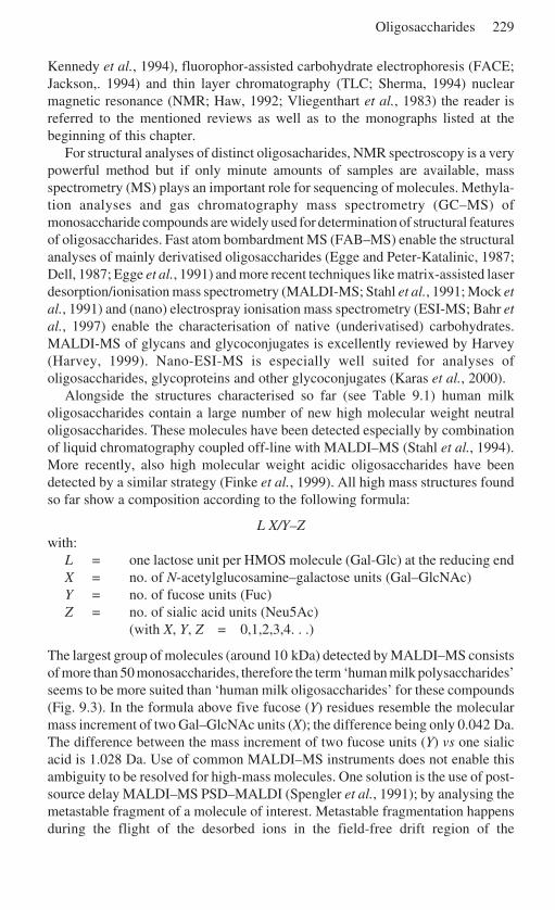
Oligosaccharides 229
Kennedy et al., 1994), fluorophor-assisted carbohydrate electrophoresis (FACE;Jackson,. 1994) and thin layer chromatography (TLC; Sherma, 1994) nuclearmagnetic resonance (NMR; Haw, 1992; Vliegenthart et al., 1983) the reader isreferred to the mentioned reviews as well as to the monographs listed at thebeginning of this chapter.
For structural analyses of distinct oligosacharides, NMR spectroscopy is a verypowerful method but if only minute amounts of samples are available, massspectrometry (MS) plays an important role for sequencing of molecules. Methyla-tion analyses and gas chromatography mass spectrometry (GC–MS) ofmonosaccharide compounds are widely used for determination of structural featuresof oligosaccharides. Fast atom bombardment MS (FAB–MS) enable the structuralanalyses of mainly derivatised oligosaccharides (Egge and Peter-Katalinic, 1987;Dell, 1987; Egge et al., 1991) and more recent techniques like matrix-assisted laserdesorption/ionisation mass spectrometry (MALDI-MS; Stahl et al., 1991; Mock etal., 1991) and (nano) electrospray ionisation mass spectrometry (ESI-MS; Bahr etal., 1997) enable the characterisation of native (underivatised) carbohydrates.MALDI-MS of glycans and glycoconjugates is excellently reviewed by Harvey(Harvey, 1999). Nano-ESI-MS is especially well suited for analyses ofoligosaccharides, glycoproteins and other glycoconjugates (Karas et al., 2000).
Alongside the structures characterised so far (see Table 9.1) human milkoligosaccharides contain a large number of new high molecular weight neutraloligosaccharides. These molecules have been detected especially by combinationof liquid chromatography coupled off-line with MALDI–MS (Stahl et al., 1994).More recently, also high molecular weight acidic oligosaccharides have beendetected by a similar strategy (Finke et al., 1999). All high mass structures foundso far show a composition according to the following formula:
L X/Y–Zwith:
L = one lactose unit per HMOS molecule (Gal-Glc) at the reducing endX = no. of N-acetylglucosamine–galactose units (Gal–GlcNAc)Y = no. of fucose units (Fuc)Z = no. of sialic acid units (Neu5Ac)
(with X, Y, Z = 0,1,2,3,4. . .)
The largest group of molecules (around 10 kDa) detected by MALDI–MS consistsof more than 50 monosaccharides, therefore the term ‘human milk polysaccharides’seems to be more suited than ‘human milk oligosaccharides’ for these compounds(Fig. 9.3). In the formula above five fucose (Y) residues resemble the molecularmass increment of two Gal–GlcNAc units (X); the difference being only 0.042 Da.The difference between the mass increment of two fucose units (Y) vs one sialicacid is 1.028 Da. Use of common MALDI–MS instruments does not enable thisambiguity to be resolved for high-mass molecules. One solution is the use of post-source delay MALDI–MS PSD–MALDI (Spengler et al., 1991); by analysing themetastable fragment of a molecule of interest. Metastable fragmentation happensduring the flight of the desorbed ions in the field-free drift region of the
230Functional dairy products
Fig. 9.3 Detection of high molecular weight human milk oligosaccharides by MALDI–MS after fractionation by size exclusion chromatography. Sumof 100 single spectra in positive ion mode; ions detected as cationided molecules (i.e. sodium- and potassium-adduct ions, respectively. Laserwavelength 337 nm, mass accuracy: 0.05%, UV-absorbing matrix: 5-chloro-2-mercaptobenzothiazole 10 g/l in water:tetrahydrofurane:ethanol =1:1:1(v/v/v). The bottom panel shows the section of the upper mass spectrum between the dashed lines. For the nomenclature of the oligosaccharides,
please refer to the text.
m/z [Th]
m/z [Th]
7400 7700 8000 8300 8600 89000
20
40
60
80
100
7982
7763
7836
7617
8129
8420
8495
8712
7690
8565
7471
7543
8275
8201
8054
7909
8860
[M+Na]+ [M+K]+
L14/17-0, L16/12-0, etc.
L15/15-0, L17/10-0, etc.
5500 6500 7500 8500 9500 10500
0
20
40
60
80
100
Int.
in
arb
. u
nit
s
1000070006000 8000 9000
Int.
in
arb
. u
nit
s
Oligosaccharides 231
MALDI–TOF (time of flight) instrument. This fragmentation can be differentiatedagainst prompt fragmentation during desorption or hydrolysis or a naturallyoccurring mixture of molecules by scanning the reflectron potential revealing a‘sequence’ of the oligosaccharide of interest.
As an alternative to PSD–MALDI, negative ion nano-ESI-ion trap MS of nativeunderivatised oligosaccharides is an even more powerful technique, since in theMSn-mode of fragmentation the real sequence from the reducing end of themolecules can be generated with additional information on position of the glycosidiclinkage of the released fragment (Pfenninger et al., 2002a, b). The use of MALDI–Fourier Transform Ion Cyclotron Resonance Mass Spectrometry (FTICR–MS)offers a direct detection of the distinct unambiguous composition of human milkoligosaccharides by high resolution measurements (Pfenninger et al., 2002c). Allthese techniques also can be applied to animal milk oligosaccharides. Theoligosaccharide pattern of domestic animal milks is much simpler and less concen-trated than that of human milk oligosaccharides (Table 9.2). The composition ofthese oligosaccharides does not follow the above-mentioned formula LX/Y–Z.Thus, an even larger variety of new structures follows from that, because for theseoligosaccharides isomeric molecules with identical masses also exist.
Because composition and structure especially of human milk have notcommercially been available until now, oligosaccharides of more simple structures,e.g. like the prebiotic oligosaccharides depicted in Table 9.3 have been used ascomponents to mimic the beneficial effects of human milk oligosaccharides. Forthe selection of prebiotic oligosaccharides for human consumption it is recom-mended that analytical techniques are used that are capable of resolving individualcomponents of usually complex mixtures beside the characterisation andquantitation via their monosaccharide components. Especially for the widely usedprebiotic oligosaccharides, the classical dietary fibre analyses have been insuffi-cient since these molecules show good solubility compared to typical fibres, theso-called non-starch polysaccharides. For the analyses of prebiotics such asfructans (oligofructoses, inulin) and galactooligosaccharides, AOAC methods(Association of Official Analytical Chemists) have been recently published.Fructans are extracted and hydrolysed by specific enzymes and the resultingmonosaccharides are analysed by HPAEC-PAD (Hoebregs et al., 1997), whereasgalacto-oligosaccharides are analysed by a different HPAEC-PAD method afterextraction of the oligosaccharides and hydrolysis by β-galactosidase (De Slegte etal., 2002).
As described above HPAEC is also suited for the detection of native underivatisedoligo-/polysaccharides. As an example a mixture of galactooligosaccharides (GOS)and long chain inulin (FOS) analysed by HPAEC is shown in Fig. 9.4. This mixtureaims to resemble the molecular weight profile of human milk oligosaccharides asthe weight of the size of prebiotic oligosaccharides determines, among others, thefermentation rate and thereby the location of fermentation in the gut. The GOS arein a tenfold excess over the FOS and the latter can be seen only after increasing theintensity scale of the chromatogram. Nevertheless, each individual oligosaccha-ride can be detected in one single liquid chromatography run.
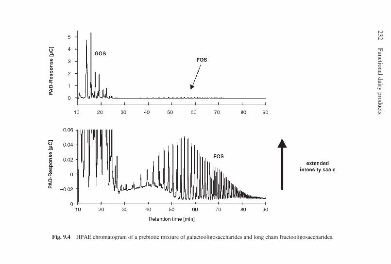
232Functional dairy products
Fig. 9.4 HPAE chromatogram of a prebiotic mixture of galactooligosaccharides and long chain fructooligosaccharides.
Oligosaccharides 233
9.8 Future trends
Although the understanding of the molecular mechanisms of dietaryoligosaccharides is weak, there are some health benefits that can be directlyattributed to these ingredients. These are stimulating attempts to investigate thestructural basis of these functions. In fact, there is some possibility of producingparticular ‘designed’ oligosaccharide molecules. Therefore, the development oftest systems to investigate the biological effects of oligosaccharides in vitro as wellas in vivo has a high priority. Because the possible health benefits of dietaryoligosaccharides are species dependent, animal studies have a limited importancein elucidating the beneficial effects of oligosaccharides to humans.
The intestinal microbiota play a key role as mediator of the physiological effectsof dietary oligosaccharides. During the last decade, enormous progress has beenmade in improving the analytical techniques to measure faecal flora (Millar et al.,1996). Unfortunately, the intestinal microbiota in humans can only be studiedusing faecal samples, which does not adequatably reflect the intestinal microbialecosystem. Therefore, non-invasive methods will be developed to investigate themetabolic activity of the intestinal microbiota using stable isotope techniques.
In summary, dietary oligosaccharides of different origins offer a variety offunctional effects which might have significant benefits to the general health ofhumans. Because oligosaccharides are safe, quite stable and can easily be processedin the dairy and food industries, there is a great potential for them to contribute asfunctional dietary ingredients in various functional products.
9.9 Acknowledgements
The authors thank Ms Beate Müller-Werner for her excellent HPAEC analyses andMr Marco Mank Dipl. Ing (FH) for his excellent MALDI–MS analyses.
9.10 ReferencesADLERBERTH I (1997), ‘The establishment of a normal intestinal microflora in the newborn
infant’ in L A Hanson and R B Yolken (Eds): Probiotics, Other Nutritional Factors, andIntestinal Flora, 42nd Nestlé Nutrition Workshop, New York, Raven Press, 63–78.
ALLES M S, KATAN M B, SALEMANS J M J I, LAERE VAN K M J, GERICHHAUSEN M J W,ROZENDAAL M J and NAGENGAST F M (1997), ‘Bacterial fermentation offructooligosaccharides and resistant starch in patients with an ileal pouch-anal anastomosis’,Am J Clin Nutr, 66, 1286–1292
ANDERSSON B, PORRAS O, HANSON L A, LAGERGARD T and SVANBORG-EDEN C (1986),‘Inhibition of attachment of Streptococcus pneumoniae and Haemophilus influenzae byhuman milk and receptor oligosaccharides’, J Infect Dis, 153, 232–237.
BABA M, SNOECK R, PAUWELS R and DECLERQ E (1988), ‘Sulfated polysaccharides are potentand selective inhibitors of various enveloped viruses, including herpes simplex virus,cytomegalovirus, vesicular stomatitis virus, and human immunodeficiency virus’,Antimicrob Agents Chemother, 32, 1742–1745.
BAHR U, PFENNINGER A, KARAS M and STAHL B (1997), ‘High sensitive analysis of neutral
234 Functional dairy products
underivatised oligosaccharides by nanoelectrospray mass spectrometry’, Anal Chem, 69,4530–4535.
BEACHEY E H (1981), ‘Bacterial adherence: adhesin–receptor interactions mediating theattachment of bacteria to mucosal surface’, J Infect Dis, 143, 325–345
BOEHM G, LIDESTRI M, CASETTA P, JELINEK J, NEGRETTI F, STAHL B and MARINI A (2002),Supplementation of a bovine milk formula with an oligosaccharide mixture increasescounts of faecal bifidobacteria in preterm infants’, Arch Dis Child, 86, F0–F4.
BÖTTCHER M F, NORDIN E K, SANDIN A, MIDTVEDT T and BJÖRKSTÉN B (2000), ‘Microflora-associated characteristics in faeces from allergic and non-allergic infants’, Clin ExpAllergy, 30, 1590–1596.
BOUHNIK Y, FLOURIE B, D‘AGAY-ABENSOUR L, POCHART P, GRAMET G, DURAND M andRAMBAUD J C (1997), ‘Administration of transgalacto-oligosaccharides increases faecalbifidobacteria and modifies colonic fermentation metabolism in healthy humans’, J Nutr,127, 444–448.
BOUHNIK Y, FLOURIE B, RIOTTOT M, BISETTI N, GAILING M F, GUIBERT A, BORNET F andRAMBAUD J C (1996), ‘Effects of fructo-oligosaccharides ingestion on faecal bifidobacteriaand selected metabolic indexes of colon carcinogenesis in healthy humans’, Nutr Cancer,26, 21–29
BOUHNIK Y, VAHEDI K, ACHOUR L, ATTAR A, SALFATI J, POCHART P, MARTEAU P, FLOURIE B,BORNET F and RAMBAUD J C (1999), ‘Short-chain fructo-oligosaccharide administrationdose-dependently increases fecal bifidobacteria in healthy humans’, J Nutr, 129, 113–116.
BRASSART D, WOLTZ A, GOLLIARD M and NEESER J R (1991), ‘In vitro inhibition of adhesionof Candida albicans clinical isolates to human buccal epithelial cells by Fucα1→2Galβ-bearing complex carbohydrates’, Infect Immun, 59, 1605–1613.
BRENNAN M J, DAVID J L, KENIMER J G and MANCLARK C R (1988), ‘Lectin-like binding ofpertussis toxin to a 165-kilodalton Chinese hamster ovary cell glycoprotein’, J Biol Chem,263, 4895–4899.
BRITISH NUTRITION FOUNDATION (1990), Complex Carbohydrates in Foods: Report of theBritish Nutrition´s Task Force, London, Chapman and Hall.
BRUNTZ R, DABROWSKI U, DABROWSKI J, EBERSHOLD A, PETER-KATALINIC J and EGGE K(1988),’ Fucose containing oligosaccharides from human milk from a donor of bloodgroup 0 lea nonsecretor’, Biol Chem Hoppe-Seyler, 369, 257–273.
BUNOUT D, HIRSCH S, DE LA MAZA M P and MUNOZ C (2002), ‘Effects of prebiotics on theimmune response to vaccination in the elderly’, J Clin Nutr, 75, 419.
CAHAN L D and PAULSON J C (1980), ‘Polyoma virus adsorbs to specific sialyloligo-saccharidereceptors on erythrocytes’, Virology, 103, 505–509.
CARLSON S E and HOUSE S G (1986),’ Oral and intraperitoneal administration of N-acetylneuraminic acid: effect on rat cerebral and cerebellar N-acetylneuraminic’, J Nutr.116, 881–886.
CARNIELLI V P, LUIJENDIJK I H T, GOUDOEVER J B, SULKERS E J, BOERLAGE A A, DEGENHARTH J and SAUER P J J (1996), ‘Structural position and amount of palmitic acid in infantformulas: effects on fat, fatty acid, and mineral balance’, J Pediatr Gastroenterol Nutr,23, 553–560.
CHATURVEDI P and SHARMA C B (1988), ‘Goat milk oligosaccharides: purification andcharacterization by HPLC and high-field 1H-NMR spectroscopy’, Biochim Biophys Acta,967, 115–121.
CHATURVEDI P and SHARMA C B (1990), ‘Purification, by high-performance liquid chroma-tography, and characterisation, by high-field 1H-n.m.r. spectroscopy, of twofucose-containing pentasaccharides of goat’s milk’, Carbohydr Res, 203, 91–101.
CHATURVEDI P, WARREN C D, ALTAYE M, MORROW A L, RUIZ-PALACIOS G, PICKERING L K andNEWBURG D S (2001), ‘Fucosylated human milk oligosaccharides vary between individu-als and over the course of lactation’, Glycobiology, 11, 365–372.
CHO S S, PROSKY L and DREHER M (Eds) (1999), Complex Carbohydrates in Foods, MarcelDekker.
Oligosaccharides 235
CHONAN O and WATANUKI M (1996), ‘The effect of 6'-galactooligosaccharides on bonemineralisation of rats adapted to different levels of dietary calcium’, Int J Vitam Nutr Res,66, 244–249.
COPPA G V, GABRIELLI O, GIORGI P, CATASSI C, MONTANARI M P, VARALDO P E and NICHOLSB L (1990), ‘Preliminary study of breastfeeding and bacterial adhesion to uroepithelialcells’, Lancet, 335, 569–571.
COPPA G V, PIERANI P, ZAMPINI L, CARLONI I, CARLUCCI A and GABRIELLI O (1999),‘Oligosaccharides in human milk during different phases of lactation’, Acta Paediatr, 430,89–94.
COUDRAY C, BELLANGER J, CASTIGLIA-DELAVAUD C, RÉMÉSY C, VERMOREL M andRAYSSIGUIER Y (1997), ‘Effect of soluble or partly soluble dietary supplementation onabsorption and balance of calcium,. magnesium, iron and zinc in healthy young men’, EurJ Clin Nutr, 51, 375–80.
CRAVIOTO A, TELLO A, VILLAFAN H, RUIZ J, DE VEDOVO S and NEESER J R (1991), ‘Inhibitionof localised adhesion of enteropathogenic Escherichia coli to HEP-2 cells by immu-noglobulin and oligosaccharide fractions of human colostrum and breast milk’, J InfectDis, 163, 1247–1255.
CRITTENDEN R G and PLAYNE M J (1996), ‘Production, properties and applications of food-grade oligosaccharides’, Trends in Food Science and Technology, 7, 353–361.
CUNDELL D R and TUOMANEN E I (1994), ‘Receptor specificity of adherence of Streptococcuspneumoniae to human type-II pneumocytes and vascular endothelial cells in vitro’,Microbiol Pathogenesis, 17, 361–374.
DAI D, NANTHKUMAR N N, NEWBURG D S and WALKER W A (2000), ‘Role of oligosac-charidesand glycoconjugates in intestinal host defense’, J Pediatr Gastroenterol Nutr, 30, 23–33.
DEGUCHI Y, MATSUMOTO K, ITO A and WATANUKI M (1997), ‘Effects of beta 1–4galactooligosaccharides administration on defecation of healthy volunteers with consti-pation tendency’, Jpn J Nutr, 55, 13–22
DE SLEGTE J, BAKX E, BASTIAANS H J A P, BENET S, BRUNT K, GRENIER-LUSTALOT M F,HEUTINK M A T, HISCHENHUBER C, KARPPINEN S, MENGERINK G W, MÜLLER-WERNER B,NIEMINEN A, NIJBOER F N, PIRO R, PRANDIN L, ROHRER J S, SAARINEN K, SANDERS P,SANGIORGI E, SCHOLSH A, SCHOTERMAN H C, SIPONEN S, STAHL B, STÖBER P, THOMAS DH and VISCOGLIOSI H (2002), ‘Determination of trans-galactooligosaccharides in selectedfood products by ion exchange chromatography: collaborative study’, J AOAC Intern,85(2), 417–423.
DE VUYST L and DEGEEST B (1999), ‘Heteropolysaccharides from lactic acid bacteria’, FEMSMicrobial Rev, 23, 153–177.
DELL A (1987), ‘Advances in carbohydrate chemistry and biochemistry’, FAB MassSpectrometry of Carbohydrates, 43, 36.
DOKKUM VAN W (1995), ‘Tolerance for galacto-oligosaccharides in humans’, Borculo DomoIngredients: Studies on Elix‘or.
DOMBO M, YAMAMOTO H and NAKAJIMA H (1997), ‘Production, health benefits andapplications of galacto-oligosaccharides’, New Technologies For Healthy Foods&Nutraceuticals, 143–156.
DONALD A S R and FEENEY J (1988), ‘Separation of human milk oligosaccharides by recyclingchromatography. First isolation of lacto-N-neo-difucohexaose II and 3´-galactosyllactosefrom this source’, Carbohydr Res, 178, 79–91.
DUA V K, GOSO K, DUBE V E and BUSH C A (1985), ‘Characterisation of lacto-N-hexaose andtwo fucosylated derivatives from human milk by high-performance liquid chromatogra-phy and proton NMR spectroscopy’, J Chromatogr, 328, 259–269.
DUGUID J P and GILLIES R R (1957), ‘Fimbriae and adhesive properties of dysentery bacilli’,J Pathol Bacteriol, 74, 397–411.
EGGE H and PETER-KATALINIC J (1987), ‘Fast atom bombardment mass spectrometry forstructural elucidation of glycoconjugates’, Mass Spectrom Rev, 6, 331–394.
EGGE H, PETER-KATALINIC J, KARAS M and STAHL B (1991), ‘The use of fast atom
236 Functional dairy products
bombardment and laser desorption mass spectrometry in the analysis of complexcarbohydrates’, Pure Appl Chem, 63, 491–498.
EL RASSI Z (Ed.) (1995), Carbohydrate Analyses: High Performance Liquid Chromatogra-phy and Capillary Electrophoresis, J. Chrom Library no. 58, Amsterdam, Elsevier.
EMÖDY L, CARLSSON A, LJUNGH A and WADSTRÖM T (1988), ‘Mannose-resistant haemagglu-tination by Campylobacter pylori’, Scand J Infect Dis, 20, 353–354.
ENGFER M B, STAHL B, FINKE B, SAWATZKI G and DANIEL H (2000), ‘Human milkoligosaccharides are resistant to enzymatic hydrolysis in the upper gastrointestinal tract’Am. J. Clin. Nutr, 71, 1589–1596.
ERNST B, HART G W and SINAY P (Eds) (2000), ‘Carbohydrates in Chemistry and Biology’Wiley, Weinheim.
EVANS D G, EVANS D J, MOULDS J J and GRAHAM D Y (1988), N-Acetylneuraminyllactose-binding fibrillar hemagglutination of Campylobacter pylori: a putative colonisation factorantigen’, Infect Immun, 56, 2896–2906.
FIÈVRE S, WIERUSZESKI J M, MICHALSKI J C, LEMOINE J, MONTREUIL J and STRECKER G (1991),‘Primary structure of a trisialylated oligosaccharide from human milk’, Biochem BiophysRes Commun, 177, 720–725.
FINKE B (2000), ’Isolierung und Charakterisierung von Oligosacchariden aus humanen undtierischen Milchen’, PhD thesis, Univ. Giessen, Shaker Verlag Aachen, Germany.
FINKE B, STAHL B, PFENNINGER F, KARAS M, DANIEL H and SAWATZKI G (1999), ‘Analysisof high molecular weight oligosaccharides from human milk by liquid chromatographyand MALDI-MS’, Anal Chem, 71, 3755–3762.
FINKE B, STAHL B, PRITSCHET M, FACIUS D, WOLFGANG J and BOEHM G (2002), ‘Preparativecontinuous annular chromatography (P-CAC) enable the large scale fractionation offructans’, J Agric Food Chem, 50, 4743–4748.
FRANK A (1998), ‘Prebiotics stimulate calcium absorption: a review’, Milchwissenschaft, 53,427–429.
GIBSON G R (1999), ‘Dietary modulation of the human gut microflora using the prebioticsoligofructose and inulin’, J Nutr, 129, 1438S–1441S.
GIBSON G R and ROBERFROID M B (1995), ‘Dietary modulation of the human colonicmicro-biota: introducing the concept of prebiotics’, J Nutr, 125, 1401–1412.
GIBSON G R, BEATTY E R, WANG X and CUMMINGS J H (1995), ‘Selective stimulation ofbifidobacteria in the human colon by oligofructose and inulin’, Gastroenterology, 108,975–982.
GINSBURG V, ZOPF D A, YAMASHITA K and KOBATA A (1976), ‘Oligosaccharides of humanmilk. Isolation of a new pentasaccharide, lacto-N-fucopentaose V’, Arch BiochemBiophys, 175, 565–568.
GNOTH M J., KUNZ C, KINNE-SAFFRAN E and RUDLOFF S (2000), ‘Human milk oligosaccharidesare minimally digested in vitro’, J Nutr, 130, 3014–3020.
GRIMMONPREZ L and MONTREUIL J (1968), ‘Etude physico-chemique de six nouveauxoligosaccharides isolés du lait de femme’, Bull Soc Chim Biol, 50, 843–855.
GRÖNBERG G, LIPNIUNAS P, LUNDGREN T, ERLANSSON K, LINDH F and NILSSON B (1989),‘Isolation of monosialylated oligosaccharides from human milk and structural analysis ofthree new compounds’, Carbohydr Res, 191, 261–278.
GRÖNBERG G, LIPNIUNAS P, LUNDGREN T, LINDH F and NILSSON B (1992), ‘Structural analysisof five new monosialylated oligosaccharides from human milk’, Arch Biochem Biophys,296, 597–610.
GUERARDEL Y, MORELLE W, PLANCKE Y, LEMOINE J and STRECKER G (1999), ‘Structuralanalysis of three sulfated oligosaccharides isolated from human milk’, Carbohydr Res,320, 230–238.
GUESRY P R, BODANSKI H, TOMSIT E and AESCHLIMANN J M (2000), ‘Effect of 3 doses offructo-oligosaccharides in infants’, J Pediatr Gastroenterol Nutr, 31, S252.
GUGGENBICHLER J P, DE BETTIGNIES-DUTZ A, MEISSNER P, SCHELLMOSER S and JURENITSCH
Oligosaccharides 237
J (1997), ‘Acidic oligosaccharides from natural sources block adherence of Escherichiacoli on uroepithelial cells’, Pharm Pharmacol Lett, 7, 35–38.
GYÖRGY P, NORRIS R F and ROSE C S (1954), ‘A variant of Lactobacillus bifidus requiring aspecial growth factor’, Arch Biochem Biophys, 48, 193–201.
HAATAJA S, TIKKANEN K, LIUKKONEN J, FRANCOIS-GERARD C and FINNE J (1993), ‘Charac-terisation of a novel bacterial adhesion specificity of Streptococcus suis recognising bloodgroup P receptor oligosaccharides’, J Biol Chem, 268, 4311–4317.
HAEUW-FIEVRE S, WIERUSZESKI J M, PLANCKE Y, MICHALSKI J C and MONTREUIL J (1993),‘Primary structure of human milk octa-, dodeca- and tridecasaccharides determined by acombination of 1H-NMR spectroscopy and fast-atom-bombardment mass spectrometry’,Eur J Biochem, 215, 361–371.
HARVEY D (1999), ‘Matrix assisted laser desorption/ionisation mass spectrometry ofcarbohydrates’, Mass Spectrom Rev, 18, 349–451.
HAW J F (1992), ‘Nuclear-magnetic-resonance spectroscopy’, Anal Chem, 64, 243R–254R.HEUVEL VAN DEN E G H M, MUYS T, DOKKUM VAN W and SCHAAFSMA G (1999), ‘Oligofructose
stimulates calcium absorption in adolescents’, Am J Clin Nutr, 69, 544–548.HEUVEL VAN DEN E G J M, SCHAAFSMA G, MUYS T and DOKKUM VAN W (1998), ‘Nondigestible
oligosaccharides do not interfere with calcium and nonheme-iron absorption in young,healthy men’, Am J Clin Nutr, 67, 445–451.
HEUVEL VAN DEN E G J M, SCHOTERMAN M H C and MUIJS T (2000), Transgalactooligo-saccharides stimulate calcium absorption in postmenopausal women’, J Nutr, 130,2938–2942.
HIRMO S, KELM S, SCHAUER R, NILSSON B and WADSTRÖM T (1996), ‘Adhesion ofHelicobacter pylori strains to α-2,3-linked sialic acids’, Glycoconjugate J, 13, 1005–1011.
HOEBREGS H, BALIS P, DEVRIES J, V EEKELEN J, FARNELL, P, GRAY K, GOEDHUYS B, HERMANSM, HEROFF J, VAN LEUWEN M, LI B W, MARTIN D, PIETERS M, QUEMENER B, ROOMANS H,SLAGHEK T, THIBAULT J F, VAN DER WAAL, W and DEWITT D (1997), ‘Fructans in foods andfood products, ion-exchange chromatographic method: collaborative study’, J AOACIntern, 80, 1029–1037.
HOLMGREN J, SVENNERHOLM A M and LINDBLAD M (1983), ‘Receptor-like glycocompoundsin human milk that inhibit classical and El Tor Vibrio cholerae cell adherence(hemagglutination)’, Infect Immun, 40, 563–569.
IDOTA T, KAWAKAMI H, MURAKAMI Y and SUGAWARA M (1995), ‘Inhibition of cholera toxinby human milk fractions and sialyllactose’, Biosc Biotech Biochem, 59, 417–419.
ITO M, DEGUCHI Y, MIYAMORI A, MATSUMOTO K, KIKUCHI H, MATSUMOTO K, KOBAYASHI Y,YAJIMA T and KAN T (1990), ‘Effects of administration of galactooligosaccharides on thehuman faecal microflora, stool weight and abdominal sensation’, Microb Ecol Health Dis,3, 285–292.
ITO M, DEGUCHI Y, MATSUMOTO K, KIMURA M, ONODERA N and YAJIMA T (1993), ‘Influenceof galactooligosaccharides on the human fecal microflora’, J Nutr Sci Vitaminol, 39, 635–640.
ITO M, KIMURA M, DEGUCHI Y, MIYAMORI-WATABE A, YAJIMA T and KAN T (1993), Effectsof transgalactosylated disaccharides on the human intestinal microflora and their metabo-lism’, J Nutr Sci Vitaminol, 39, 279–88.
IUB-IUPAC JOINT COMMISSION ON BIOCHEMICAL NOMENCLATURE (JCBN) (1980), ‘Abbrevi-ated terminology of oligosaccharide chains’, Recommendations, J Biol Chem, 257, 334.
IUPAC/IUBMB (1997), Joint Commission on Biochemical Nomenclature (JCBN) Nomencla-ture of Carbohydrates, Recommendations 1996 Carbohydr Res, 297(12), 9.
JACKSON P (1994), In: Methods Enzymol., W J Lennarz and G W Hart (Eds), Acad. Press,San Diego, 230, 250–264.
JENKINS D J A, KENDALL C W C and VUKSAN V (1999), ‘Inulin, oligofructose and intestinalfunction’, J Nutr, 129,1431S–1433S.
JENSEN R G (1995), Handbook of Milk Composition, San Diego, Academic Press.
238 Functional dairy products
KALLIOMÄKI M, SALMINEN S, ARVILOMMI H, KERO P, KOSKINEN P and ISOLAURI E (2001),‘Probiotics in primary prevention of atopic diseases: a randomised placebo-controlledtrial’, Lancet, 357, 1067–1079.
KARAS M, BAHR U and DULCKS T (2000), ‘Nano-electrospray ionisation mass spectrometry:addressing analytical problems beyond routine’, Fresenius J Anal Chem, 366, 669–676.
KINSELLA J E and TAYLOR S L (Eds) (1995), ‘Commercial β-galactosidase’ in FoodProcessing Advances in Food and Nutrition Research, San Diego, Academic Press.
KITAGAWA H, NAKADA H, NUMATA Y, KUROSAKA A, FUKUI S, FUNAKOSHI I, KAWASAKI T,SHIMADA I, INAGAKI F and YAMASHINA I (1990), ‘Occurrence of tetra- and pentasaccharideswith the sialyl-Lea structure in human milk’, J Biol Chem, 265, 4859–4862.
KITAGAWA H, NAKADA H, FUKUI S, FUNAKOSHI I, KAWASAKI T and YAMASHINA I (1991),‘Novel oligosaccharides with the sialyl-Lea structure in human milk’, Biochem, 30, 2869–2876.
KITAGAWA H, NAKADA H, FUKUI S, FUNAKOSHI I, KAWASAKI T, YAMASHINA I, TATE S I andINAGAKI F (1993), ‘Novel oligosaccharides with the sialyl-Lea structure in human milk’,J Biochem, 114, 504–508.
KLEESSEN B, SYKURA B, ZUNFT H J and BLAUT M (1997), ‘Effects of inulin and lactose onfaecal microflora, microbial activity, and bowel habit in elderly constipated persons’, AmJ Clin Nutr, 65, 1397–1402.
KOBATA A and GINSBURG V (1969), ‘Oligosaccharides of human milk. Isolation andcharacterisation of a new pentasaccharide, lacto-N-fucopentaose III’, J Biol Chem, 244,5496–5502.
KOBATA A and GINSBURG V (1972a), ‘Oligosaccharides of human milk. Isolation andcharacterisation of a new hexasaccharide, Lacto-N-hexaose’, J Biol Chem, 247, 1525–1529.
KOBATA A and GINSBURG V (1972b), ‘Oligosaccharides of human milk. Isolation andcharacterisation of a new hexasaccharide, Lacto-N-neohexaose’, Arch Biochem Biophys,150, 273–281.
KUHN R (1959), ‘Biochemie der Rezeptoren und Resistenzfaktoren. Von der Widerstands-fähigkeit der Lebewesen gegen Einwirkungen der Umwelt’, Naturwissenschaften, 46,43–50.
KUHN R and BAER H H (1956a), ’Die Konstitution der Lacto-N-tetraose’, Chem Ber, 89, 504–511.
KUHN R and BROSSMER R (1956), ’Über O-Acetyl-lactaminsäure-lactose aus Kuh-Colos-trum und ihre Spaltbarkeit durch Influenza-Virus’, Chem Ber, 89, 2013–2025.
KUHN R and BROSSMER R (1959), ‘Über das durch Viren der Influenza-Gruppe spalt-bareTrisaccharid der Milch’, Chem Ber, 92, 1667–1671.
KUHN R GAUHE A (1958); ‘Über die Lacto-difuco-tetraose der Frauenmilch’, Justus LiebigsAnn Chem, 611, 249–253.
KUHN R and GAUHE A (1960), ’Über ein kristallisiertes, Lea-aktives Hexasaccharid ausFrauenmilch’, Chem Ber, 93, 647–651.
KUHN R and GAUHE A (1962), ‘Die Konstitution der Lacto-N-neotetraose’, Chem Ber, 95,518–522.
KUHN R and GAUHE A (1965), ‘Bestimmung der Bindungsstellen von Sialinsäure-resten inOligosacchariden mit Hilfe von Perjodat’, Chem Ber, 98, 395–413.
KUHN R, BAER H H and GAUHE A (1956), ‘Kristallisation und Konstitutionsermittlung derLacto-N-Fucopentaose I’, Chem Ber, 89, 2514–2523.
KUHN R, BAER H H and GAUHE A (1958), ‘Die Konstitution der Lacto-N-fucopen-taose II’,Chem Ber, 91, 364.
KUNZ C, RUDLOFF S, SCHAD W and BRAUN D (1999), ‘Lactose-derived oligosaccharides inmilk of the elephants–comparison to human milk’, Brit J Nutr, 82, 391–399.
KUNZ C, RUDLOFF S, BAIER W, KLEIN N and STROBEL S (2000), ‘Oligosaccharides in humanmilk: structural, functional and metabolic aspects’, Ann Rev Nutr, 20, 699–722.
Oligosaccharides 239
KUNZ C, RUDLOFF S and MICHAELSEN K F (2002), ‘Concentration of human milkoligosaccharides during late lactation’, FASEB J, 15, A986.
KUO C, TAKAHASHI N, SWANSON A F, OZEKI Y and HAKOMORI S (1996), ‘An N-linked high-mannose type oligosaccharide, expressed at the outer membrane protein of Chlamydiatrachomatis, mediates attachment and infectivity of the microorganism to HeLa cells’, JClin Invest, 98, 2813–2818.
LEE Y C (1990), ‘High-performance anion-exchange chromatography for carbohydrateanalysis’, Anal Biochem, 189, 151–162.
LEE Y C (1996), ‘Carbohydrate analyses with high-performance anion-exchange chromatog-raphy’, J Chromatogr, 720, 137–149.
LENNARZ W J and HART G W (Eds) (1994), Methods in Enzymology: Techniques inGlycobiology, 230, San Diego, Academic Press.
LIDESTRI M, AGOSTI M, MARINI A and BOEHM G (2002), ‘Prebiotics might stimulate calciumabsorption in preterm infants’, Acta Paediatr (suppl.); in press.
LIO S, LING Y and TSAI C E (1994), ‘Biotechnically synthesised oligosaccharides and poly-dextrose reduce constipation and putrefactive metabolites in the human’, J Clin Nutr Soc,19, 221–232.
LIUKKONEN J, HAATAJA S, TIKKANEN K, KELM S and FINNE J (1992), ‘Identification of N-acetylneuraminyl α-2,3 poly-N-acetyllactosamine glycans as the receptors of sialic acidbinding Streptococcus suis strains’, J Biol Chem, 267, 21105–21111.
LOO J V, CUMMINGS J, DELZENNE N, ENGLYST H, FRANK A, HOPKINS M, KOK N, MACFARLANE
G, NEWTON D, QUINGLEY M, ROBERFROID M, VLIET T and HEUVEL E (1999), ‘Functionalfood properties of non-digestible oligosaccharides: a consensus report from the ENDOproject (DGXII AIRII-CT94-1095)’, B J Nutr, 81, 121–132.
LOVELESS R W and FEIZI G (1989), ‘Sialo-oligosaccharide receptor for Mycoplasmapneumoniae and related oligosaccharides of poly-N-acetyllactosamine series are polar-ised at the cilia and apical-microvillar domains of the ciliated cells in human bronchialepithelium’, Infect Immun, 57, 1285–1289.
MARTIN M J, MARTIN-SOSA S, GARCIA-PARDO L A and HUESO P (2001), ‘Distribution ofbovine milk sialoglycoconjugatesduring lactation’ J Diary Sci, 84, 995–1000.
MENNE E, GUGGENBUHL N and ROBERFROID M (2000), ‘Fn-type chicory inulin hydrolysatehas a prebiotic effect in humans’, J Nutr, 130, 1197–1199.
MEYER P D, TUNGLAND B C, CAUSEY J L and SLAVIN J L (2001), ‘In-vitro und in-vivo – Effektevon Inulin auf das Immunsystem’, Ernährungs-Umschau, 48, 13–16.
MILLAR M R, LINTON C J, CADE A, GLANCY D, HALL M and JALAL H (1996), ‘Application of16S rRNA gene PCR to study bowel flora of preterm infants with and without necrotisingenterocolitis’, J Clin Microbiol, 34, 2506–2510.
MILLER-PODRAZA H, MILH M A, BERGSTRÖM J and KARLSSON K A (1996), ‘Recognition ofglycoconjugates by Helicobacter pylori: an apparently high-affinity binding of humanpolyglycosylceramides, a second sialic acid-based specificity’, Glycoconjugate J, 13,453–460.
MITSUOKA T, HIDAKA H and EIDA T (1987), ‘Effect of fructo-oligosaccharides on intestinalmicroflora’, Die Nahrung, 31, 427–36.
MOCK K K, DAVEY M and COTTRELL J S (1991), ‘The analysis of underivatised oligosaccharidesby matrix-assisted laser desorption mass spectrometry’, Biochem Biophys Res Commun,177, 644–651.
MONNIG C A and KENNEDY R T (1994), ‘Capillary electrophoresis’, Anal Chem, 66, 280R–314R.
MONTREUIL J (1956), ‘Structure de deux triholosides isolés du lait de femme’, C R Acad Sci,242, 192–193.
MORO E (1908), ‘Karottensuppe bei Ernährungsstörungen der Säuglinge’, MünchenerMedizinische Wochenzeitschrift, 31, 1637–1640.
MORO G, MINOLI I, MOSCA M, FANARO S, JELINEK J, STAHL B and BOEHM G (2002), ‘Dosage-
240 Functional dairy products
related bifidogenic effects of galacto- and fructooligosaccharides in formula-fed terminfants’, J Pediatr Gastroenterol Nutr, 34, 291–295.
MOURICOUT M, PETIT J M, CARIAS J R and JULIEN R (1990),’Glycoprotein glycans that inhibitadhesion of Escherichia coli mediated by K99 fimbriae : treatment of experimentalcolibacillosis’, Infect Immun, 58, 98–106.
MURRAY P A, LEVINE M J, TABAK L A and REDDY M S (1982), ‘Specificity of salivary–bacterialinteractions: II. Evidence for a lectin on Streptococcus sanguis with specificity for aNeu5Acα2,3Galß1,3GalNAc sequence’, Biochem Biophys Res Comm, 106, 390–396.
NAKAKUKI T (Ed) (1994), ‘Oligosaccharides Production, Properties and Applications’,Switzerland, Gordon and Breach.
NAKAMURA T, URASHIMA T, NAKAGAWA M and SAITO T (1998), ‘Sialyllactose occurs as freelactones in ovine colostrum’, Biochim Biophys Acta, 1381, 286–292.
NAUGHTON P J, MIKKELSEN L L and JENSEN B B (2001), ‘Effects of non-digestibleoligosaccharides on Salmonella enterica serovar Typhimurium and non-pathogenicEscherichia coli in the pig small intestine in vitro’, Appl Environ Microbio, 67(8), 3391–3395.
NEWBURG D S (1999), ‘Human milk oligosaccharides that inhibit pathogens’ Curr MedChem, 6, 117–127.
NEWBURG D S and NEUBAUER S H (1995) ‘Carbohydrates in milk’. In: Handbook of MilkComposition, San Diego, Academic Press.
NÖHLE U and SCHAUER R (1981),’ Uptake metabolism and excretion of orally andintravenously administered, 14C- and 3H-labelled N-acetylneuraminic acid mixture in themouse and rat’, Hoppe Seylers Z Physiol Chem, 362, 1495–14506.
OHTA A, OHTSUKI M, HOSONO A, ADACHI T, HARA H and SAKATA T (1998), ’Dietaryfructooligosaccharides prevent osteopenia after gastrectomy in rats’, J Nutr, 128, 106–110.
OHTSUKA K, BENNO Y, ENDO K, UEDA H, OZAMU O, UCHIDA T and MITSUOKA T (1989),‘Effects of 4´galactosyllactose intake on human fecal microflora’, Bifidus, 2, 143–49.
OUADIA A, KARAMANOS Y and JULIEN R (1992), ‘Detection of the ganglioside N-glycolyl-neuraminyl-lactosyl-ceramide by biotinylated Escherichia coli K99 lectin’, GlycoconjugateJ, 9, 21–26.
OUWEHAND A C, ISOLAURI E, HE F, HASHIMOTO H, BENNO Y and SALMINEN S (2001),‘Differences in bifidobacterium flora composition in allergic and healthy infants’, JAllergy Clin Immunol, 108, 144–145.
PARKINNEN J and FINNE J (1987), ‘Isolation of sialyl oligosaccharides and sialyl oligo-saccharides phosphates from bovine colostrum and human urine’, Methods Enzymol, 138,289–300.
PARKINNEN J, FINNE J, ACHTMAN M, VÄISÄNEN V and KORHONEN T K (1983), ‘Escherichiacoli strains binding neuraminyl α2-3 galactosides’, Biochem Biophys Research Comm,111, 456–461.
PFENNINGER A, KARAS M, FINKE B and STAHL B (2002a), ‘Structural analysis of underivatisedhuman milk oligosaccharides in the negative ion mode by nano-electrospray MSn’, PartI Methodology’, J Am. Mass Spectrom, 2002, 13, 1331–1340.
PFENNINGER A, KARAS M, FINKE B and STAHL B (2002b), ‘Structural analysis of underivatisedhuman milk oligosaccharides in the negative ion mode by nano-electrospray MSn’, PartII Application to isomeric mixtures’, J Am. Mass Spectrom, 2002, 13, 1341–1348.
PFENNINGER A, CHAN S Y, KARAS M, FINKE B, STAHL B and COSTELLO C E (2002c), ‘Massspectral analysis of compositions for neutral, high molecular weight oligosaccharidesfrom human milk’, Glycobiology; sent for publication.
POLONOVSKI M and LESPAGNOL A (1933), ‘Nouvelles acquisitions sur les composesglucidiques du lait de femme’, Bull Soc Chim Biol, 15, 320–349.
RAMPHAL R, CARNOY C, FIEVRE S, MICHALSKI J C, HOUDRET N, LAMBLIN G, STRECKER G andROUSSEL P (1991), ‘Pseudomonas aeruginosa recognises carbohydrate chains containing
Oligosaccharides 241
type 1 (Galβ1-3GlcNAc) or type 2 (Galβ1-4GlcNAc) disaccharide units’, Infect Immun,59, 700–704.
RIVERO-URGELL, M and SANTAMARIA-ORLEANS A (2001), ‘Oligosaccharides: application ininfant food’, Early Human Development, 65 (Suppl), S43–52.
ROBERFROID M B and DELZENNE N M (1998), ‘Dietary fructans’, Ann Rev Nutr, 18, 117–143.ROBERFROID M and SLAVIN J (2000), ‘Nondigestible oligosaccharides’, Crit Rev Food Sci
Nutr, 40, 461–480.ROGERS G N, HERRLER G, PAULSON J C and KLENK H D (1986), ‘Influenza C virus uses 9-O-
acetyl-N-acetylneuraminic acid as high affinity receptor determinant for attach-ment tocells, J Biol Chem, 261, 5947–5951.
RUDLOFF S, STEFAN C, POHLENTZ G and KUNZ C (2002), ‘Detection of ligands for selectinsin the oligosaccharide fraction of human milk’, Eur. J Nutr, 41, 85–92.
SAAVEDRA J, TSCHERNIA, MOORE N, ABI-HANNA A, COLETTA F, EMENHISER C and YOLKEN
R (1999), ‘Gastro-intestinal function in infants consuming a weaning food supplementedwith oligofructose, a prebiotic’, J Pediatr Gastroenterol Nutr, 29, 95.
SABHARWAL H, NILSSON B, GRÖNBERG G, CHESTER M A, DAKOUR J, SJÖBLAD S andLUNDBLAD A (1988a), ‘Oligosaccharides from faeces of preterm infants fed on breastmilk’, Arch Biochem Biophys, 265, 390–406.
SABHARWAL H, NILSSON B, CHESTER M A, LINDH F, GRÖNBERG G, SJÖBLAD S and LUNDBLAD
A (1988b), ‘Oligosaccharides from faeces of a blood-group B, breast-fed infant’, CarbohydrRes, 178, 145–154.
SAITO T, ITOH T and ADACHI S (1984), ‘Presence of two neutral disaccharides containing N-acetylhexosamine in bovine colostrum as free forms’, Biochim Biophys Acta, 801,147–150.
SAITO T, ITOH T and ADACHI S (1987), ‘Chemical structural of three neutral trisaccharidesisolated in free forms from bovine colostrum’, Carbohydr Res, 165, 43–51.
SARNEY D B, HALE C, FRANKEL G and VULFSON E N (2000), ‘A novel approach to the recoveryof biological active oligosaccharides from milk using a combination of enzymatictreatment and nanofiltration’, Biotechnol Bioeng, 69, 461–467.
SCHAAFSMA G, MEULING W J A, DOKKUM W VAN and BOULEY C (1998), ‘Effects of a milkproduct, fermented by Lactobacillus acidophilus and with fructo-oligosaccharides added,on blood lipids in male volunteers’, Eur J Clin Nutr, 52, 436–40.
SCHENKMAN S and EICHINGER D (1993), ‘Trypanosoma cruzi trans sialidase and cellinvasion’, Parasitol Today, 9, 218–222.
SCHNEIR M L and RAFELSON M E (1966), ‘Isolation of two structural isomers of N-acetylneuraminyllactose from bovine colostrum’, Biochim Biophys Acta, 130, 1–11.
SCHOLZ-AHRENS K, SCHAAFSMA G, VAN DEN HEUVEL E G M H and SCHREZENMEIR J (2001),‘Effect of prebiotics on mineral metabolism’, Am J Clin Nutr, 73, 459S–464S.
SEGAL E (1996), ‘Inhibitors of Candida albicans adhesion to prevent candidiasis’ In:‘Toward anti-adhesion therapy for microbial diseases’, (I Kahane and I Ofek, Eds),Advances in Experimental Medicine and Biology, 408, New York, Plenum.
SHERMA J (1994), ‘Planar chromatography’, Anal Chem, 66, 67R–83R.SMITH D F, PRIETO P A, MCCRUMB D K and WANG W C (1987), ‘A novel sialylfucopentaose
in human milk’, J Biol Chem, 262, 12040–12047.SNOW BRAND MILK PRODUCTS CO., Technical Research Institute (1995), ‘A new oligosac-
charide; 4´-galactosyl-lactose in human milk’, Annual Meeting at Hokkaido University.SOUTHGATE D A T (1995), ‘Dietary fiber analysis RSC Food analysis monographs’ P S Belton
(Ed), The Royal Society of Chemistry, Cambridge.SPENGLER B, KIRSCH D and KAUFMANN R (1991), ‘Metastable decay of peptides and proteins
in matrix-assisted laser-desorption mass spectrometry’, Rapid Comm. Mass Spectrom, 5,198–202.
SPILLER G A (2001), CRC Handbook of Dietary Fiber in Human Nutrition, Boca Raton, CRCPress.
STAHL B, STEUP M, KARAS M and HILLENKAMP F (1991), ‘Analysis of neutral oligosaccharides
242 Functional dairy products
by matrix assisted laser desorption/ionisation mass spectrometry’ Anal Chem, 63, 1463–1466.
STAHL B, THURL S, ZENG J, KARAS M, HILLENKAMP F, STEUP M and SAWATZKI G (1994),‘Oligosaccharides from human milk as revealed by matrix-assisted laser desorption/ionisation mass spectrometry’, Anal Biochem, 223, 218–226.
STONE A L, MELTON D J and LEWIS M S (1998), ‘Structure function relations of heparinmimetic sulphated xylan oligosaccharied: inhibition of human immunodeficiency virus-1 infectivity in vitro’, Glycoconjugate J, 15, 697–712.
STRECKER G, FIÈVRE S, WIERUSZESKI J M, MICHALSKI J C and MONTREUIL J (1992), ‘Primarystructure of four human milk octa-, nona- and undeca-saccharides estab-lished by 1H- and13C-nuclear magnetic resonance spectroscopy’, Carbohydr Res, 226, 1–14.
STRECKER G, WIERUSZESKI J M, MICHALSKI J C and MONTREUIL J (1988), ‘Structure of a newnonasaccharide isolated from human milk: VI2Fuc, V4Fuc, III3Fuc-p-Lacto-N-hexaose’,Glycoconjugate J, 5, 385–396.
STRÖMBERG N, MARKLUND B I, LUND B, ILVER D, HAMERS A, GAASTRA W, KARLSSON K A andNORMARK S (1990), ‘Host-specifity of uropathogenic Escherichia coli depends ondifferences in binding specifity to Galα1-4Gal-containing isoreceptors’, EMBO J, 9,2001–2010.
SVENSSON L (1992), ‘Group-C rotavirus requires sialic acid for erythrocyte and cell receptorbinding’, J Virol, 66, 5582–5585.
TACHIBANA Y, YAMASHITA K and KOBATA A (1978), ‘Oligosaccharides of human milk:structural studies of di- and trifucosyl derivatives of Lacto-N-octaose and Lacto-N-neooctaose’, Arch Biochem Biophys, 188, 83–89.
TANAKA R and MATSUMOTO K (1998), ‘Recent progress on prebiotics in Japan, includinggalactooligosaccharides’, Bull IDF, 336, 21–27.
TANAKA R, TAKAYAMA H, MOROTOMI M, KUROSHIMA T, UEYAMA S, MATSUMOTO K, KURODA
and A MUTAI M (1983), ‘Effects of administration of TOS and bifidobacterium breve 4006on the human faecal flora’, Bifidobacteria Microflora, 2, 17–24.
TEURI U and KORPELA R (1998), ‘Galacto-oligosaccharides relieve constipation in elderlypeople’, Ann Nutr Metab, 42, 319–27.
TEURI U I, KORPELA R, SAXELIN M, MONTONEN L and SALMINEN S J (1998), ‘Increased faecalfrequency and gastrointestinal symptoms following ingestion of galacto-oligosaccharide-containing yogurt’, J Nutr Sci Vitaminol, 44, 465–71.
TERRAZAS L I, WALSH K L, PISKORSKA D, MCGUIRE E and HARN D A JR (2001), ‘Theschistosome oligosaccharide lacto-N-neotetraose expands Gr1(+) cells that secrete anti-inflammatory cytokines and inhibit proliferation of naive CD4(+) cells: a potentialmechanism for immune polarisation in helminth infections’, J Immunol, 1, 167(9):5294–5303.
THURL S, HENKER J, SIEGEL M, TOVAR K and SAWATZKI G (1997), ‘Detection of four humanmilk groups with respect to Lewis blood group dependent oligosaccharides. GlycoconjugateJ, 14, 795–799.
THURL S, MÜLLER-WERNER B and SAWATZKI G (1996), ‘Quantification of individualoligosaccharide compounds from human milk by gel chromatography’, Anal Biochem,235, 202–206.
TISSIER H C R (1899), ‘La vécation chromophile d’Escherich et la Bacterium coli’, Soc Biol,51, 943–945.
URASHIMA T, SAITO T, NAKAMURA T and MESSER M (2001), ‘Oligosaccharides of milk andcolostrum in non-human mammals’ Glycoconjugate J, 18, 357–371
URASHIMA T, MURATA S and NAKAMURA T (1997b), ‘Structural determination of mono-sialyltrisaccharides obtained from caprine colostrum’, Comp Biochem Physiol, 116, 431–435.
URASHIMA T, NAKAMURA T and SAITO T (1997c), ‘Biological significance of milkoligo-saccharides – homology and heterogeneity of milk oligosaccharides among mam-malian species’, Milk Science, 46, 211–220.
Oligosaccharides 243
URASHIMA T, BUBB W A, MESSER M, TSUJI Y and TANEDA Y (1994), ‘Studies of the neutraltrisaccharides of goat (Capra hircus) colostrum and of the one- and two-dimensional 1Hand 13C NMR spectra of 6'-N-acetylglucosaminyllactose’, Carbohydr Res, 262, 173–184.
URASHIMA T, SAITO T and KIMURA T (1991a), ‘Chemical structures of three neutral oligo-saccharides obtained from horse (Thoroughbred) colostrum’, Comp Biochem Physiol B,100, 177–183.
URASHIMA T, SAITO T, OHMISYA K and SHIMAZAKI K (1991b), ‘Structural determination ofthree neutral oligosaccharides in bovine (Holstein–Friesian) colostrum, including thenovel trisaccharide; GalNAcα1-3Galß1-4Glc’, Biochim Biophys Acta, 1073, 225–229.
URASHIMA T, SAITO T, NISHIMURA J and ARIGA H (1989), ‘New galactosyllactose containingα-glycosidid linkage isolated from ovine (Booroola dorset) colostrum’, Biochim BiophysActa, 992, 375–378.
URASHIMA T, SAKAMOTO T, ARIGA H and SAITO T (1989b), ‘Structure determination of threeneutral oligosaccharides obtained from horse colostrum’, Carbohydr Res, 194, 280–287.
VARKI A, CUMMINGS R, ESKO J, FREEZE H, HART G and MATH J (Eds) (1999), Essentials ofGlycobiology, New York, Cold Spring Harbor Laboratory Press.
VEH R W, MICHALSKI J C, CORFIELD A P, SANDER-WEWER M, GIES D and SCHAUER R (1981),‘New chromatographic system for the rapid analysis and preparation of colostrumsialyloligosaccharides’, J Chromatogr, 212, 313–322.
VELUPILLAI P and HARN D A (1994), ‘Oligosaccharide-specific induction of interleukin 10production by B220+ cells from schistosome-infected mice: a mechanism for regulationof CD4+ T-cell subsets’, Proc Natl Acad Sci, 91, 18–22.
VIVERGE D, GRIMMONPREZ L and SOLERE M (1997), ‘Chemical characterisation of sialyloligosaccharides isolated from goat (Capra hircus) milk’, Biochim Biophys Acta, 1336,157–164.
VLIEGENTHART J F G, DORLAND L and VAN HALBEEK H (1983), ‘High-resolution, 1H-nuclearmagnetic resonance spectroscopy as a tool in the structural analysis of carbohydratesrelated to glycoproteins’, Adv Carbohydr Chem Biochem, 41, 209–374.
WEIS W, BROWN J H, CUSACK S, PAULSON J C, SKEHEL J J and WILEY D C (1988), ‘Structureof the influenza virus haemagglutinin complexed with its receptor, sialic acid’, Nature,333, 426–431.
WIERUSZESKI J M, CHEKKOR A, BOUQUELOT S, MONTREUIL J, STRECKER G, PETER-KATALINICJ and EGGE H (1985), ‘Structure of two new oligosaccharides isolated from human milk:Sialylated lacto-N-fucopentaoses I and II’, Carbohydr Res, 137, 127–138.
XANTHOU M (1998), ‘Immune protection of human milk’, Biol Neonate, 74, 121–133.YAHIRO M, NISHIKAWA I, MURAKAMI Y, YOSHIDA H and AHIKO K (1982), ‘Studies on
application of galactosyl lactose for infant formula II. Changes of faecal characteristicson infants fed galactosyl lactose’, Reports of Research Laboratory, Snow Brand MilkProducts Co. Ltd., No. 78, 27–32.
YAMASHITA K, TACHIBANA Y and KOBATA A (1977), ‘Structural studies of two newoctasaccharides, difucosyl derivatives of para-lacto-N-neohexaose’, J Biol Chem, 252,5408–5411.
YAMASHITA K, TACHIBANA Y and KOBATA A (1976a), ‘Oligosaccharides of human milk.Isolation and characterisation of three new disialylfucosyl hexasaccharides’, Arch BiochemBiophys, 174, 582–591.
YAMASHITA K, TACHIBANA Y and KOBATA A (1976b), ‘Oligosaccharides of human milk.Isolation and characterisation of two new nonasaccharides, monofucosyllacto-N-octaoseand monofucosyllacto-N-neooctaose’, Biochemistry, 15, 3950–3955.
YAMASHITA K and KOBATA A (1974), ‘Oligosaccharides of human milk. Isolation andcharacterisation of a new trisaccharide, 6'-galactosyllactose’, Arch Biochem Biophys,161, 164–170.
ZOPF D and ROTH S (1996), ‘Oligosaccharide anti-infective agents’, Lancet, 347, 1017–1021.
10
Lactic acid bacteria (LAB) in functionaldairy productsR. Fondén, Arla Foods ICS, Sweden, M. Saarela, J. Mättö andT. Mattila-Sandholm, VTT Biotechnology, Finland
10.1 Introduction
When our ancestors developed the knowledge to use milk, they also learned howto use lactic acid bacteria (LAB) in dairy products even though they did not knowabout the existence of microorganisms. When milk was left at room temperature,it was spontaneously fermented and was used as such or after dilution with water.As an alternative it could be concentrated by drainage of whey, resulting in a newproduct, which could be stored for several months: the first cheese. Owing to thislong period of consumption, such products were found to be safe and healthy(Fleichman and Weigmann, 1932). By trial and error, the practice of inoculatingmilk with a small amount of previously fermented milk was developed. Heatingthe milk before inoculation increased the quality of the product and it becamepossible to develop a range of specific, traditional fermented milk products (Table10.1).
About two hundred years ago cheese and butter production became industrial-ised. Traditional products were standardised and slightly modified to adjust themto industrial production. Starters were grown in manufacturing plants, but, as thesize of the operations grew, the importance of standardised starters increased. Inthe late 19th century companies specialised in the starter production were founded.In the beginning the starters were still undefined, containing an unknown mixtureof different strains of the suitable species. Later on, starters were analysed anddefined, allowing them to be composed of specific strains with properties selectedto give a standard product. In general, defined starters are composed of somespecific strains with less species variety than undefined starters, and do not usuallycontain yeast.
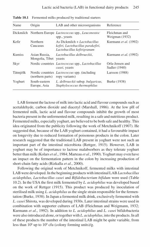
Lactic acid bacteria (LAB) in functional dairy products 245
Table 10.1 Fermented milks produced by traditional starters
Name Origin LAB and other microorganisms Reference
Dickmilch Northern Europe Lactococcus spp., Leuconostoc Fleichman andspp., yeasts Weigman (1932)
Kefir Northern As Dickmilch + Lactobacillus Kurmann et al. (1992)Caucasus kefiri, Lactobacillus parakefiri,
Lactobacillus kefirigranum
Kumiss Asian Russia, Lactobacillus delbrueckii, Kurmann et al. (1992)Mongolia, Tibet yeasts
Skyr Nordic countries Lactococcus spp., Lactobacillus Orla-Jensen andcasei, yeasts Sadler (1940)
Tätmjölk Nordic countries Lactococcus spp. (including Larsson (1988)(northern parts) ropy variants)
Yoghurt South-eastern L. delbrueckii subsp. bulgaricus, Burke (1938)Europe, Asia Staphylococcus thermophilus
LAB ferment the lactose of milk into lactic acid and flavour compounds such asacetaldehyde, carbon dioxide and diacetyl (Marshall, 1986). At the low pH offermented milk, lactic acid and flavour compounds inhibit the growth of mostbacteria present in the unfermented milk, resulting in a safe and nutritious product.Fermented milks, especially yoghurt, are believed to be both safe and healthy. Thisidea originated from the publicity following the work of Metchnikoff (1907). Hesuggested that, because of the LAB yoghurt contained, it had a favourable impacton longevity due to reduced formation of poisonous products in the colon. Laterresearch suggested that the traditional LAB present in yoghurt were not such animportant part of the intestinal microbiota (Rettger, 1915). However, LAB inyoghurt may be of importance to lactose malabsorbers as they tolerate yoghurtbetter than milk (Kolars et al., 1984; Marteau et al., 1990). Yoghurt may even havean impact on the fermentation pattern in the colon by increasing production ofshort-chain fatty acids (Rizkalla et al., 2000).
Following the original work of Metchnikoff, fermented milks with intestinalLAB were developed. In the beginning products with intestinal LAB Lactobacillusacidophilus, Lactobacillus casei and Bifidobacterium bifidum were used (Table10.2). In the USA the first milk fermented by L. acidophilus was developed basedon the work of Rettger (1915). This product was produced by inoculation ofsterilised milk using L. acidophilus as the single strain responsible for the fermen-tation (Burke, 1938). In Japan a fermented milk drink, exclusively fermented withL. casei Shirota, was developed during 1930s. Later intestinal strains were used incombination with supporter cultures of LAB (Fleichman and Weigmann, 1932;Kurmann et al., 1992). In addition to L. acidophilus and L. casei bifidobacteriawere also introduced alone, or together with L. acidophilus, into the products. In allof these products the number of the intestinal LAB might be quite variable, fromless than 106 up to 108 cfu (colony forming units)/g.
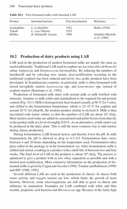
246 Functional dairy products
Table 10.2 First fermented milks with intestinal LAB
Product Intestinal bacteria First documented Reference
Acidophilus L. acidophilus 1922 Burke (1938)Yakult L. casei Shirota 1935Bifidus B. bifidum/B. longum 1968 Schuhler-Mayloth
et al. (1968)
10.2 Production of dairy products using LAB
LAB used in the production of modern fermented milks are mainly the same asused traditionally. Traditional LAB used in yoghurt are Lactobacillus delbrueckiisubsp. bulgaricus and Streptococcus thermophilus. By reducing the numbers oflactobacilli and by selecting new strains, post-acidification occurring in thetraditional yoghurts has been reduced and novel, less acidic products have beendeveloped. In Scandinavian countries, in particular, milk is often fermented withmixed mesophilic starters Lactococcus spp. and Leuconostoc spp. instead ofyoghurt starters (Kurmann et al., 1992).
Production of fermented milk starts with normal milk or milk fortified withminerals, vitamins or milk solids and standardised to the required fat and proteincontent (Fig. 10.1). Milk is homogenised, heat-treated (usually at 90 oC for 5 min),and chilled to the fermentation temperature, which is 32–43 oC for yoghurt andaround 20 oC for filmjölk, the modern product similar to dickmilch. Milk is theninoculated with starter culture so that the numbers of LAB are about 107 cfu/g.Most starters used today are added in concentrated and pellet-frozen form directlyto the product milk at a level of roughly 0.01%. As an alternative, a bulk starter canbe produced in the dairy plant. This is still the most common way to add startersduring cheese production.
During fermentation, LAB ferment lactose and thereby lower the pH. In milkfermentation the pH is allowed to drop to 4.3–4.5. Fermentation time variesbetween 4 and 20 hours depending on the temperature used. Fermentation takesplace either in the package or in the fermentation vat. After fermentation, milk ischilled and stored, resulting in a product with a shelf-life of between two and fourweeks. The final level of LAB in the products is about 109 cfu/g. The process isoptimised to give a product with as low whey separation as possible and with alimited post-acidification. More extensive information on the production of fer-mented milks is given by Cogan and Accolas (1996), Tamine and Robinson (1999)and IDF (1988, 1992).
Several different LAB are used in the production of cheese. In cheese bothwater activity and oxygen tension are low, which limits the growth of mostbacteria. However, some microorganisms are still able to grow in cheese andinfluence its maturation. Examples are LAB combined with white and bluemoulds, propionic acid bacteria and Micrococcus spp. Because of the fairly high
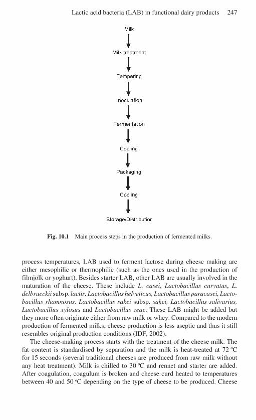
Lactic acid bacteria (LAB) in functional dairy products 247
Fig. 10.1 Main process steps in the production of fermented milks.
process temperatures, LAB used to ferment lactose during cheese making areeither mesophilic or thermophilic (such as the ones used in the production offilmjölk or yoghurt). Besides starter LAB, other LAB are usually involved in thematuration of the cheese. These include L. casei, Lactobacillus curvatus, L.delbrueckii subsp. lactis, Lactobacillus helveticus, Lactobacillus paracasei, Lacto-bacillus rhamnosus, Lactobacillus sakei subsp. sakei, Lactobacillus salivarius,Lactobacillus xylosus and Lactobacillus zeae. These LAB might be added butthey more often originate either from raw milk or whey. Compared to the modernproduction of fermented milks, cheese production is less aseptic and thus it stillresembles original production conditions (IDF, 2002).
The cheese-making process starts with the treatment of the cheese milk. Thefat content is standardised by separation and the milk is heat-treated at 72 ºCfor 15 seconds (several traditional cheeses are produced from raw milk withoutany heat treatment). Milk is chilled to 30 ºC and rennet and starter are added.After coagulation, coagulum is broken and cheese curd heated to temperaturesbetween 40 and 50 oC depending on the type of cheese to be produced. Cheese
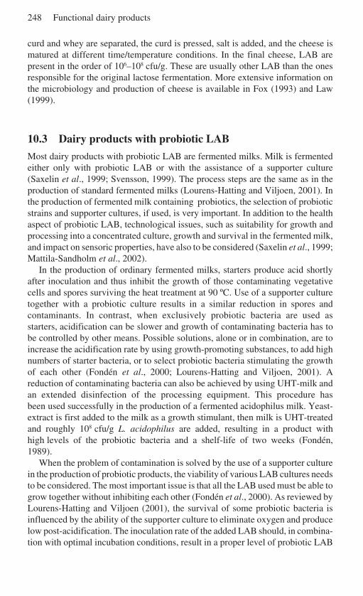
248 Functional dairy products
curd and whey are separated, the curd is pressed, salt is added, and the cheese ismatured at different time/temperature conditions. In the final cheese, LAB arepresent in the order of 106–108 cfu/g. These are usually other LAB than the onesresponsible for the original lactose fermentation. More extensive information onthe microbiology and production of cheese is available in Fox (1993) and Law(1999).
10.3 Dairy products with probiotic LAB
Most dairy products with probiotic LAB are fermented milks. Milk is fermentedeither only with probiotic LAB or with the assistance of a supporter culture(Saxelin et al., 1999; Svensson, 1999). The process steps are the same as in theproduction of standard fermented milks (Lourens-Hatting and Viljoen, 2001). Inthe production of fermented milk containing probiotics, the selection of probioticstrains and supporter cultures, if used, is very important. In addition to the healthaspect of probiotic LAB, technological issues, such as suitability for growth andprocessing into a concentrated culture, growth and survival in the fermented milk,and impact on sensoric properties, have also to be considered (Saxelin et al., 1999;Mattila-Sandholm et al., 2002).
In the production of ordinary fermented milks, starters produce acid shortlyafter inoculation and thus inhibit the growth of those contaminating vegetativecells and spores surviving the heat treatment at 90 ºC. Use of a supporter culturetogether with a probiotic culture results in a similar reduction in spores andcontaminants. In contrast, when exclusively probiotic bacteria are used asstarters, acidification can be slower and growth of contaminating bacteria has tobe controlled by other means. Possible solutions, alone or in combination, are toincrease the acidification rate by using growth-promoting substances, to add highnumbers of starter bacteria, or to select probiotic bacteria stimulating the growthof each other (Fondén et al., 2000; Lourens-Hatting and Viljoen, 2001). Areduction of contaminating bacteria can also be achieved by using UHT-milk andan extended disinfection of the processing equipment. This procedure hasbeen used successfully in the production of a fermented acidophilus milk. Yeast-extract is first added to the milk as a growth stimulant, then milk is UHT-treatedand roughly 108 cfu/g L. acidophilus are added, resulting in a product withhigh levels of the probiotic bacteria and a shelf-life of two weeks (Fondén,1989).
When the problem of contamination is solved by the use of a supporter culturein the production of probiotic products, the viability of various LAB cultures needsto be considered. The most important issue is that all the LAB used must be able togrow together without inhibiting each other (Fondén et al., 2000). As reviewed byLourens-Hatting and Viljoen (2001), the survival of some probiotic bacteria isinfluenced by the ability of the supporter culture to eliminate oxygen and producelow post-acidification. The inoculation rate of the added LAB should, in combina-tion with optimal incubation conditions, result in a proper level of probiotic LAB
Lactic acid bacteria (LAB) in functional dairy products 249
in the final product, which should be at least 106 cfu/g (Hawley et al., 1959;Gilliland, 1989). Probiotic LAB should preferably be added to milk beforefermentation since addition afterwards results in limited survival (Hull et al.,1984). A suitable final pH is ensured both by using strains with low post-acidification and by using proper cooling conditions. Taking into account theabove factors, it has been demonstrated that fermented milks with selectedprobiotic LAB can be stored for several weeks with a minimal loss of viability(Saxelin et al., 1999).
Today, most available dairy products with probiotic bacteria are fermentedmilks such as yoghurt. However, other milk products such as ordinary ‘sweet’milk, cheese, ice cream and milk powder can also be used as carriers of probioticLAB. One of the main advantages of using non-fermented milk products as carriersof probiotics is the absence of fermentation end-products. Organic acids andflavour compounds have a negative impact on the survival of probiotic LAB. In theproduction of sweet milk, probiotic bacteria are added after the heat treatment ofmilk. The numbers of added probiotic LAB and the shelf-life of the sweet milkproduct are limited by the acid fermentation that occurs during cold storage. Themaximum number of probiotic LAB that can be added to sweet milk depends onthe storage temperature and time as well as on the properties of the strain used.Sweet acidophilus milk is an example of a sweet milk product with added probioticL. acidophilus (Speck, 1975; Young and Nelson, 1978).
Ice cream is another suitable carrier for probiotic bacteria as the storage at a lowtemperature results in longer survival. Survival seems to depend on pH, sincesurvival is better in ice cream mixes of neutral pH than in ice cream similar tofrozen yoghurt. Products with at least 107 cfu/g and with a shelf life of up to eightmonths have been produced (Hekmat and McMahon, 1992; Hagen and Narvhus,1999; Davidson et al., 2000; Alampreese et al., 2002).
Milk powders with probiotic bacteria have a long shelf-life compared withproducts with high water activity. It has been demonstrated that most probioticbacteria can be produced as freeze-dried cultures with 1012 cfu/g with good storagestability even at +25 oC (Saxelin et al., 1999). These cultures can be mixed withmilk powder or similar ingredients to make dried food products with suitableconcentrations of probiotic bacteria. As an alternative, a concentrated probioticculture may be added into concentrated milk and spray-dried. However, numbersof viable probiotic bacteria are lower and the storage stability of spray-driedproducts is poorer than in freeze-dried products (Gardiner et al., 2000).
Cheese provides LAB conditions that assist both bacterial growth and survival.Many of the LAB found in cheese belong to the same species as probiotic bacteria,including L. casei, L. paracasei, L. rhamnosus and Lactobacillus plantarum(Lindberg et al., 1996; Gardiner et al., 1998). An L. paracasei strain, LactobacillusF19, originating from human GI microbiota and used as a probiotic bacterium, haseven been isolated from cheese produced from milk not inoculated with this strain(Björneholm and Fondén, 2002). The suitability of cheese as a carrier of probioticLAB has been shown both for mature cheeses and fresh cheeses (Gomes et al.,1995; Gardiner et al., 1998; Vinderola et al., 2000).
250 Functional dairy products
10.4 The health benefits of probiotic LAB
Owing to their perceived health benefits, lactobacilli and bifidobacteria have beenincreasingly included in yoghurts and fermented milks during the past two decades(Daly and Davis, 1998). A major development in the functional food sector hasemerged from the use of probiotic bacteria and prebiotic carbohydrates thatenhance health-promoting microbiota in the intestine. There is growing evidenceto support the concept that the maintenance of healthy gut microbiota may provideprotection against various gastrointestinal disorders including gastrointestinal(GI) infections, inflammatory bowel diseases and even cancer (Haenel and Bendig,1975; Mitsuoka, 1982; Salminen et al., 1998).
The human large intestine has a rich and dynamic microbiota consisting of atleast 400–500 bacterial species (Berg, 1996). Maintaining a balanced microbialecosystem is essential for the normal functions of the GI tract, especially inpreventing infections and stimulating the host’s immune response. Several factorsincluding stress, antibiotic treatments and other medications can alter the GI tractmicrobiota, predisposing the host to various diseases (Salminen et al., 1995;Schaafsma, 1995). Overprescription and misuse of antibiotics has led to a situationwhere increasing numbers of pathogens are becoming resistant to antibiotics(Austin et al., 1999; Robredo et al., 2000). The World Health Organization (WHO)has indicated that alternative disease control strategies, such as the use of probiotics– live microbial preparations with documented health benefits for consumers bymaintaining or improving their intestinal microbiota balance (Fuller, 1989) – maybe needed in the future in the prevention and treatment of certain infections(Bengmark, 1998).
To benefit the health of the consumer, a probiotic bacterium has to reach itstarget site (gut) alive. It should also have good technological properties so that itcan be manufactured and added into foods without losing viability and functional-ity, or creating unpleasant flavours or textures. It should survive passage throughthe upper GI tract and be able to function in the gut environment. Feeding trialswith different probiotic strains have shown that the probiotic strain usuallydisappears from the GI tract within a couple of weeks after the ingestion has beendiscontinued (Fukushima et al., 1998; Johansson et al., 1998; Donnet-Hughes etal., 1999; Alander et al., 1999, 2001). Obtaining a benefit from the probioticconsumption necessitates regular ingestion of probiotic products (daily dosageusually between 109–1011 cfu).
Most bacteria with probiotic properties belong to the genera Lactobacillus andBifidobacterium, which are common but non-dominant members of the indigenousmicrobiota of the human GI tract (Sghir et al., 2000; Walter et al., 2001). Theprobiotic potential of various Lactobacillus and Bifidobacterium strains has beendiscussed in numerous reviews and includes well-documented management ofintestinal disorders such as lactose intolerance, infant gastroenteritis and rotavirus-associated diarrhoea, antibiotic-associated intestinal symptoms (mainly diarrhoea),and food allergy in babies (Salminen et al., 1998; Isolauri et al., 1999, 2001;Marteau et al., 2001; Kaur et al., 2002). These disorders and diseases are associated
Lactic acid bacteria (LAB) in functional dairy products 251
Table 10.3 Probiotic health effects. Note that not all effects can be attributed to thesame probiotic strain
Established effects Potential effects
Alleviation of lactose intolerance symptoms Alleviation of IBD symptomsTreatment of viral (rotavirus) diarrhoea Alleviation of IBS symptomsTreatment of infant gastroenteritis Improvement of constipationTreatment of antibiotic associated diarrhoea Antimutagenic/anticarcinogenic activityAlleviation of atopic dermatitis symptoms Treatment of candidal and bacterialin children vaginitis
Positive effects on superficial bladder Cholesterol and blood pressure loweringcancer and cervical cancer Eradication of multidrug-resistant
Modulation of intestinal microbiota microbesImmune modulation Infection controlLowering biomarkers (harmful faecal Prevention of transmission of AIDS andenzymes) sexually transmitted diseases
with intestinal microbiota imbalance and increased gut permeability (Salminen etal., 1996a,b). In addition to these beneficial effects on disturbed intestinalmicrobiota, probiotics can modulate immune responses, lower biomarkers such asharmful faecal enzyme activities, and show positive effects against superficialbladder cancer and cervical cancer (McFarland, 2000). Other potential areas ofprobiotic nutritional management include alleviation of inflammatory boweldisease (IBD) and irritable bowel syndrome (IBS) symptoms, mucosal vaccinesand immunomodulation, infection control and eradication of multidrug-resistantmicrobes, treatment of candidal vaginitis, prevention of transmission of AIDS andsexually transmitted diseases, lowering cholesterol and blood, and antimutagenic/anticarcinogenic activity (Table 10.3) (Alvarez-Olmos and Oberhelman, 2001;Kopp-Hoolihan, 2001; Marteau et al., 2001; Kaur et al., 2002). The most widelystudied probiotic strains in human clinical trials include L. rhamnosus GG (by farthe most extensively studied strain), Bifidobacterium lactis Bb-12, L. casei Shirota,L. plantarum DSM9843 (299V), Lactobacillus reuteri (BioGaia Biologics) andLactobacillus johnsonii LJ-1 (Saarela et al., 2000).
When assessing the health-promoting effects of probiotics, it is important tokeep in mind that all probiotic strains are different. Even strains representing thesame species usually have different properties. Several attributes of probioticbacteria should be considered before clinical trials are performed. These include:
• origin (preferably human);• safety;• viability/activity in delivery vehicles (e.g. food);• resistance to acid and bile;• adherence to gut epithelial tissue;• ability to persist in the GI tract;• production of antimicrobial substances;• ability to stimulate host’s immune response;• ability to influence metabolic activities (Salminen et al., 1996b).
252 Functional dairy products
Earlier clinical studies on probiotic efficacy generally suffered from ambiguities inprobiotic product identification, stability and dosage, and from the small numberof test subjects and controls recruited. Considerable efforts have been made tocorrect these deficiencies, and evidence on the health-promoting effects of probioticsis currently accumulating from randomised and placebo-controlled double-blindstudies. Today, plenty of evidence exists on the positive effects of probiotics onhuman health. However, this has usually been demonstrated in diseased humanpopulations only (Salminen et al., 1998). Thus there is an urgent need for evidencefor probiotic health benefits in average (generally healthy) populations.
10.5 Enhancing the viability and stability of LAB
Traditional starters need to be viable and metabolically active during fermentationto be able to grow and produce the desired sensory compounds during processing,whereas their viability after fermentation is not necessary. In contrast, probioticsare by definition live bacteria and the product should contain a suitable level of thespecific probiotic strain(s) throughout storage. Although several studies haveshown that non-viable probiotics can have beneficial effects, such as immunemodulation and carcinogen binding (Ouwehand and Salminen, 1998), good viabil-ity is generally considered a prerequisite for optimal functionality (Saarela et al.,2000). The quality assurance of probiotic products is also usually based ontechniques detecting viable cells. Examination of existing products has revealeddeficiencies in cell numbers and labelling of some probiotic products on the market(Hamilton-Miller and Shah, 2001; Temmerman et al., 2002), which demonstratesthe necessity for optimising the parameters affecting the viability of probiotics.
Several factors such as strain characteristics, food matrix, temperature, pH andaccompanying microbes affect the viability of a probiotic strain (Fig. 10.2). Theability to tolerate environmental stresses varies greatly between different speciesand even between strains within a species (Schmidt and Zink, 2000). The stabilityof the probiotic strain in the product can be enhanced in several ways. Thepackaging materials used and the storage conditions of the final product areimportant for the quality of products. The parameters used in the fermentation anddownstream processing affect the stability of probiotics in the product. Theirviability can be enhanced by optimising the growth medium and the cryo-protectant, for example. Encapsulation techniques using resistant starch or alginateas a protective material have been developed to enhance the stability of probiotics(Shah, 2000; Sultana et al., 2000; Mattila-Sandholm et al., 2002). Encapsulationhas increased the viability of probiotics in novel product types such as frozenyoghurt. Combinations of prebiotics (fermentable non-digestible carbohydrates)and probiotics are called synbiotics (Roberfroid, 1998). Prebiotics may havefavourable impact on the stability of selected probiotic(s). However, as the conceptof synbiotic is relatively new, there are only few specific studies on the interactionbetween pro- and prebiotics.
The culture technique, based on reproduction of bacterial cells on agar plates,
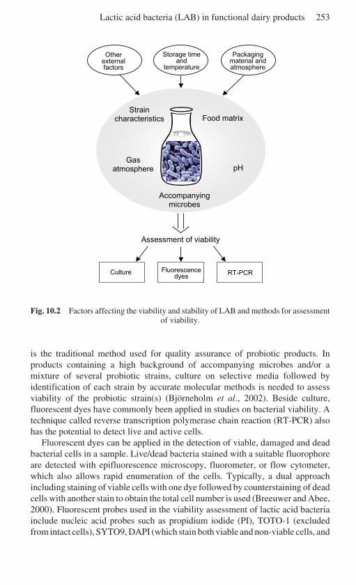
Lactic acid bacteria (LAB) in functional dairy products 253
Fig. 10.2 Factors affecting the viability and stability of LAB and methods for assessmentof viability.
is the traditional method used for quality assurance of probiotic products. Inproducts containing a high background of accompanying microbes and/or amixture of several probiotic strains, culture on selective media followed byidentification of each strain by accurate molecular methods is needed to assessviability of the probiotic strain(s) (Björneholm et al., 2002). Beside culture,fluorescent dyes have commonly been applied in studies on bacterial viability. Atechnique called reverse transcription polymerase chain reaction (RT-PCR) alsohas the potential to detect live and active cells.
Fluorescent dyes can be applied in the detection of viable, damaged and deadbacterial cells in a sample. Live/dead bacteria stained with a suitable fluorophoreare detected with epifluorescence microscopy, fluorometer, or flow cytometer,which also allows rapid enumeration of the cells. Typically, a dual approachincluding staining of viable cells with one dye followed by counterstaining of deadcells with another stain to obtain the total cell number is used (Breeuwer and Abee,2000). Fluorescent probes used in the viability assessment of lactic acid bacteriainclude nucleic acid probes such as propidium iodide (PI), TOTO-1 (excludedfrom intact cells), SYTO9, DAPI (which stain both viable and non-viable cells, and
Food matrix
Packagingmaterial andatmosphere
pH
Accompanyingmicrobes
Straincharacteristics
Gasatmosphere
Storage timeand
temperature
Otherexternalfactors
Culture Fluorescencedyes
RT-PCR
Assessment of viability
254 Functional dairy products
are used together with other probes), and physiological indicators such as 2'7'-bis-(2 carboxyethyl)-5(and-6) carboxyfluorescein (BCECF), carboxyfluoresceindiacetate (CFDA), N-(fluorescein thio-ureanyl)-glutamate (FTUG), and bisoxonol(BOX) (Molenaar et al., 1992; Karwoski et al., 1995; Glaasker et al., 1996;Ueckert et al., 1995, 1997, 1998; Auty et al., 2001; Bunthof et al., 2001; Bunthofand Abee, 2002). The time needed for analysis of the viability of cells is substan-tially shorter with fluorescent dyes (1–2 hours) than with culture (2–5 days)(Breeuwer and Abee, 2000). However, the commonly used fluorophores are non-specific and they cannot differentiate bacterial genera or species.
In RT-PCR the target of PCR amplification is messenger RNA (mRNA), whichis rapidly destroyed in dead bacterial cells by RNase activity. In RT-PCR themRNA is first transcribed into copy DNA (cDNA), which is then amplified byPCR. In lactic acid bacterial research RT-PCR has been used, for example, in nisingene functionality studies in Lactococcus lactis and in detecting inducible nitricoxidase synthase mRNA in the probiotic strain L. rhamnosus GG (Moschetti et al.,1996; Korhonen et al., 2001). Although RT-PCR is well suited for studying viableand active bacterial cells in laboratory conditions, there is a drawback in using it forviability studies in products. Under processing conditions where bacterial RNAremains fairly intact but bacterial RNase is destroyed or inactivated, a misleadingresult of the bacterial viability may be obtained, since under these circumstancesthe dead bacterial cells may amplify the numbers of cells identified in viabilitystudies of the final product.
10.6 Enhancing the functionality of LAB
Reviews covering the functionality of probiotic LAB have mostly been limited toselection of suitable strains and to the viability of the LAB in the product (Saarelaet al., 2000; Dunne et al., 2001; O’Sullivan, 2001). However, the composition ofthe probiotic product matrix and the status of the LAB during ingestion are alsoimportant factors contributing to the functionality of probiotic LAB. A goodexample of this is the fact that lactose malabsorbers tolerate milk better when it isincluded in a meal instead of consumed separately. Although most lactosemalabsorbers are able to tolerate several grams of lactose when consumed in milk,an increased intake of lactose results in flatulence, abdominal pain and sometimeseven diarrhoea (Hertzler et al., 1996; Suarez et al., 1995; Vesa et al., 1996).Fermented milk products including yoghurt and milk with L. acidophilus and L.casei are usually tolerated better than plain milk (Kolars et al., 1984). It has beensuggested that the activity of β-galactosidase in combination with a slower gastricemptying of the fermented milks results in increased tolerance (Marteau et al.,1990; Lin et al., 1991). Furthermore, by slowly increasing daily intake of lactose,the tolerated dose also increases. During the adaptation process the activity of theGI microbiota is changed as hydrogen production decreases and bacterial β-galactosidase activity increases (Johnson et al., 1993; Fondén, 2001). Enhancingthe usefulness of LAB-containing dairy products to lactose malabsorbers requires
Lactic acid bacteria (LAB) in functional dairy products 255
evaluation of not only the selection of a LAB strain producing β-galactosidase, butalso the impact of the product on gastric emptying rate and the influence of theprobiotic LAB on the intestinal fermentation of lactose during the adaptationprocess.
Most attention in product development is given to the alleviation of lactoseintolerance symptoms even if other non-absorbable, fermentable carbohydratescan also cause identical symptoms. Fructans, fructose, lactulose, sorbitol and evensome starches are examples of such carbohydrates (Rumessen et al., 1998;Lebenthal-Bendor et al., 2001). As in lactose intolerance, including LAB able toferment the specific carbohydrates could reduce the symptoms in products with aslow gastric emptying effect. The GI microbiota also plays a role here illustrated inindividuals with a disturbed microbiota (due to intake of antibiotics), showingincreased sensitivities to fructans (Orrhage et al., 2000).
Since probiotic LAB should be able to survive the conditions in the GI tract,probiotic strains are often selected based on their resistance to low pH and to a highconcentration of bile acids (Dunne et al., 2001). According to in vitro models, milkcomponents are in general protective against harmful conditions of the GI tract(Conway et al., 1987; Charteris et al., 1998). However, fermented milks canincrease the exposure of probiotic cells to a low pH and high concentrations ofundissociated organic acids. Other properties of the product, e.g. viscosity, whichinfluences gastric emptying time, are also important factors contributing to theexposure of probiotic cells to a low pH (Marteau et al., 1997). Low preformed acidand high buffering capacity may explain the better survival of probiotic LAB insweet milk and cheese compared to yoghurt (Pettersson et al., 1983; Gardiner etal., 1999). Similarly, exposure to bile acids depends on the fat content of theproduct. Low-fat dairy products will result in a lower excretion of bile acids(Cummings et al., 1978).
LAB are capable of displaying adaptive responses to stressful conditions,increasing their adaptability to intestinal conditions (Lemay et al., 2000). Acidtolerance in LAB can be triggered by allowing the final pH of the growth mediumto drop to 4.5 during fermentation (Lorca and Valdez, 2001). In addition, bile andacid resistance can be triggered by exposing LAB cells to sublethal temperaturesor NaCl concentrations. Utilising the stress response of probiotic LAB providesfurther possibilities of increasing their functionality (Kim et al., 2001). LAB canalso be protected from the harsh conditions in the upper part of the GI tract byphysical barriers such as enterocoating or microencapsulation. The high survivalof probiotic LAB in cheese may depend on a similar barrier effect, since during thedigestion process small pieces of cheese are formed, protecting LAB cells from theenvironment (Gardiner et al., 1999).
Adhesion to intestinal cells and mucus is one of the important functionalproperties of probiotic LAB. Adhesion is usually improved by milk components(Conway et al., 1987). This effect may be due to the presence of divalent ions inmilk, since calcium and magnesium have been shown to favour adhesion (Chauviereet al., 1992). In contrast, some free fatty acids (mainly polyunsaturated) and milkproteins reduce adhesion (Kankaanpää et al., 2001; Ouwehand et al., 2001). The

256 Functional dairy products
impact of dairy products on GI transit and adhesion of probiotic LAB is clearly aquite complex phenomenon, necessitating the performance of in vivo trials with thespecific LAB-containing product.
10.7 Future trends
Today’s consumers are increasingly attentive to the relationship between diet andwell-being. Not only are they aware of the positive effects of certain foods or foodcompounds on health, there is also an increased preoccupation about the incidenceof toxic food components, food-related disease and gastrointestinal disorders.From these trends, a clear need emerges to meet the current demand for safe andhealthy food of high quality, preferably produced by traditional methods and fromhigh-quality raw materials, and minimally treated with unnatural or technicalmeans in order to retain the original character and unique organoleptic and sensoryqualities of these foods. Functional starter cultures will lead to improvedorganoleptic, sensory and nutritional properties of the food on the one hand and theinhibition of food-borne pathogens on the other.
Emerging scientific developments will result in fermented foods with strongidentities and enhanced quality and safety, that are natural and healthy, and withincreased appeal to the consumer. Functional starter cultures are starter culturesthat possess at least one inherent functional property. This may be a preservative,organoleptic, technological, health-promoting or nutritional function. Probioticswill remain an important functional ingredient, and novel strains will be identifiedand foods will be developed to fulfil the needs of specific consumer groups. Newnon-digestible carbohydrates will be developed with controlled fermentationrates, and the use of prebiotics to modulate the microbiota composition, as well asspecific dietary fibres to enhance microbiota’s performance and gut function, willbe developed.
There is a clear trend towards the use of multiple, defined starter cultures toimprove control of industrial fermentation processes. Examples are found in thedairy industry, and promising results are in the pipeline for the processing ofcereals. Future possibilities of producing new ingredients for nutritionally optimisedfoods, which promote consumer health through the microbial reactions, are listedbelow:
• Selection and improvement of functional starter cultures.• Effect of food fermentation conditions on the microbes’ functionality.• Predictive modelling and simulation of microbes’ behaviour and functionality.• Targeted screening and selection of functional starter cultures, microbiological
analysis, phylogenetic positioning.• Ecology and technology studies of mixed starter cultures of LAB.• Effect of food properties on microbial functionality.• Mechanisms of action of probiotics in the GI tract and biomarkers for their
assessment.
Lactic acid bacteria (LAB) in functional dairy products 257
• Development of probiotics and bioactive compounds to prevent GI diseases, GIinfections and allergies.
• Ensurance of the stability of probiotics also in new types of food applications bydeveloping feasible technologies (e.g. process and material development formicroencapsulation).
• Genomics-based analysis of the functionality of food microbes.• Metabolomics- and physionomics-based analysis of the functionality of food
microbes.• Metabolic engineering and flux analysis for improved functionalities of novel
starter cultures.• Consumers’ acceptability of novel functional starter cultures.
10.8 Sources of further information and advice
Reviews published in the Supplements of British Journal of Nutrition (2000 Suppl2) and American Journal of Clinical Nutrition (2001, 73, 6) give an update ofknowledge available in the late 1990s. Newly published books edited by Tannock(2002) and Brandi, Biavati and Biasco (2002) give further information.
IDF, the International Dairy Federation, is the organisation of the internationaldairy world. References are made to several bulletins published by the organisa-tion (IDF 1988,1992, 2002). The activities of the organisation can be followed byreading the home page of IDF at www.fil-idf.org.
In order to follow the development of LAB and dairy science, the two mainjournals of special interest that should be followed are International Dairy Journaland Journal of Dairy Science. To follow the development on the market andquestions related to the interface between R&D and marketing the journal NewNutrition Business is highly recommended.
10.9 ReferencesALAMPREESE C, FOSCHINO R, ROSSI M, POMPEI C and SAVANI L (2002), ‘Survival of
Lactobacillus johnsonii La1 and influence of its addition in retail-manufactured ice creamproduced with different sugar and fat concentrations’, Int Dairy J, 12(2), 201–208.
ALANDER M, SATOKARI R, KORPELA R, SAXELIN M, VILPPONEN-SALMELA T, MATTILA-SANDHOLM T and VON WRIGHT A (1999), ‘Persistence of colonization of human colonicmucosa by a probiotic strain, Lactobacillus rhamnosus GG, after oral consumption’, ApplEnviron Microbiol, 65, 351–354.
ALANDER M, MÄTTÖ J, KNEIFEL W, JOHANSSON M, KÖGLER B, CRITTENDEN R, MATTILA-SANDHOLM T and SAARELA M (2001), ‘Effect of galacto-oligosaccharide supplementationon human faecal microflora and on survival and persistence of Bifidobacterium lactis Bb-12 in the gastrointestinal tract’, Int Dairy J, 11, 817–825.
ALVAREZ-OLMOS M I and OBERHELMAN R A (2001), ‘Probiotic agents and infectiousdiseases: a modern perspective on a traditional therapy’, Clin Infect Dis, 32, 1567–1576.
AUSTIN D J, KRISTINSSON K G and ANDERSON R M (1999), ‘The relationship between thevolume of antimicrobial consumption in human communities and the frequency ofresistance’, PNAS, 96, 1152–1156.
258 Functional dairy products
AUTY M A E, GARDINER G E, MCBREARTY S J, O’SULLIVAN E O, MULVIHILL D M, COLLINS JK, FITZGERALD G F, STANTON C and ROSS R P (2001), ‘Direct in situ assessment of bacteriain probiotic dairy products using viability staining in conjunction with confocal scanninglaser microscopy’, Appl Environ Microbiol, 67, 420–425.
BENGMARK S (1998), ‘Ecological control of the gastrointestinal tract. The role of probioticbacteria’, Gut, 42, 2–7.
BERG R D (1996), ‘The indigenous gastrointestinal microflora’, Trends Microbiol, 4, 430–435.BJÖRNEHOLM S and FONDÉN R (2002), ‘Lactobacillus F19 – closing the broken circle’ Microb
Ecol Health Dis, Suppl (3), 3.BJÖRNEHOLM S, EKLÖW A, SAARELA M and MÄTTÖ J (2002), ‘Enumeration and identification
of Lactobacillus paracasei subsp. paracasei F19’, Microbiol Ecol Health Dis, Suppl 3,7–13.
BRANDI G, BIAVATI B and BIASCO G (2002), Bifidobacteria: Microbiological Aspects andProbiotic Potentialities, Heidelberg, Springer-Verlag.
BREEUWER P and ABEE T (2000), ‘Assessment of viability of microorganisms employingfluorescence techniques’, Int J Food Microbiol, 55, 193–200.
BUNTHOF C J and ABEE T (2002), ‘Development of a flow cytometric method to analyzesubpopulations of bacteria in probiotic products and dairy starters’, Appl EnvironMicrobiol, 68, 2934–2942.
BUNTHOF C J, BLOEMEN K, BREEUWER P, ROMBOUTS F M and ABEE T (2001), ‘Flow cyto-metric assessment of viability of lactic acid bacteria’, Appl Environ Microbiol, 67,2326–2335.
BURKE A (1938), Practical Manufacture of Cultured Milks and Kindred Products, Milwaukee,Olsen Publishing Co.
CHARTERIS W P, KELLY P M, MORELLI L and COLLINS J K (1998), ‘Development andapplication of an in vitro methodology to determine the transit tolerance of potentiallyprobiotic Lactobacillus and Bifidobacterium species in the upper human gastrointestinaltract’, J Appl Microbiol, 84(5), 759–768.
CHAUVIERE G, COCONNIER M H, KERNEIS S, FOURNIAT J and SERVIN A L (1992), ‘Adhesionof human Lactobacillus acidophilus strain LB to human enterocyte-like Caco-2 cells’,J Gen Microbiol, 138(8),1689–1696.
COGAN T M ACCOLAS J P (1996), Dairy Starter Cultures, Weiheim, VCH Verlagsgesellschaft.CONWAY P L, GORBACH S L and GOLDIN B R (1987), ‘Survival of lactic acid bacteria in the
human stomach and adhesion to intestinal cells’, J Dairy Sci, 70(1), 1–12.CUMMINGS J H, WIGGINS H S, JENKINS D J, HOUSTON H, JIVRAJ T, DRASAR B S and HILL M J
(1978), ‘Influence of diets high and low in animal fat on bowel habit, gastrointestinaltransit time, fecal microflora, bile acid, and fat excretion’, J Clin Invest, 61(4), 953–963.
DALY C and DAVIS R (1998), ‘The biotechnology of lactic acid bacteria with emphasis onapplications in food safety and human health’, Agric. Food Sci Finland, 7, 251–265.
DAVIDSON R H, DUNCAN S E, HACKNEY C R, EIGEL W N and BOLING J W (2000),‘Probioticculture survival and implications in fermented frozen yoghurt characteristics’, J Dairy Sci,83(4), 666–673.
DONNET-HUGHES A, ROCHAT F, SERRANT P, AESCHLIMANN J M and SCHIFFRIN J E (1999),‘Modulation of nonspecific mechanisms of defence by lactic acid bacteria: effectivedose’, J Dairy Sci, 82, 863–869.
DUNNE C, O’MAHONY L, MURPHY L, THORNTON G, MORRISSEY D, O’HALLORAN S, FEENEY M,FLYNN S, FITZGERALD G, DALY C, KIELY B, O’SULLIVAN G C, SHANAHAN F and COLLINS
J K (2001), ‘In vitro selection criteria for probiotic bacteria of human origin: correlationwith in vivo findings’, Am J Clin Nutr, 73(2 Suppl), 386S–392S.
FLEICHMAN W and WEIGMANN H (1932), ‘Die Sauermilcharten’ in Lehrbuch derMilchwirtschaft, 7 Auflage, Berlin, Paul Parey, 369–386.
FONDÉN R (1989) ‘Development of new fermented milk products’, Food Techn Int Europe,1989(1), 158–160.
Lactic acid bacteria (LAB) in functional dairy products 259
FONDÉN R (2001), ‘Adaptation to lactose in lactose malabsorbers – importance of theintestinal microflora’, Scand J Nutr, 45(4), 174.
FONDÉN R, GRENOV B, RENIERO R, SAXELIN M and BIRKELAND S E (2000), ‘Industrial panelstatements: technological aspect’. In: M Alander and T. Mattila-Sandholm, FunctionalFoods for EU-health in 2000, Fourth Workshop, FAIR CT96-1028, PROBDEMO, VTTSymposium, Rovaniemi, Finland. VTT, Helsinki, Vol. 198, 43–50.
FOX P F (1993), Cheese: Chemistry, Physics and Microbiology, 2nd edn, London, Chapman& Hall.
FUKUSHIMA Y, KAWATA Y, HARA H, TERADA A and MITSUOKA T (1998), ‘Effect of a probioticformula on intestinal immunoglobulin A production in healthy children’, Int J FoodMicrobiol, 42, 39–44.
FULLER, R (1989), ‘Probiotics in man and animals’, J Appl Bacteriol, 66, 365–378.GARDINER G, ROSS R P, COLLINS J K, FITZGERALD G and STANTON C (1998), ‘Development
of a probiotic cheddar cheese containing human-derived Lactobacillus paracasei strains’,Appl Environ Microbiol, 64(6), 2192–2199.
GARDINER G, STANTON C, LYNCH P B, COLLINS J K, FITZGERALD G and ROSS R P (1999),‘Evaluation of cheddar cheese as a food carrier for delivery of a probiotic strain to thegastrointestinal tract’, J Dairy Sci, 82(7),1379–1387.
GARDINER G E, O’SULLIVAN E, KELLY J, AUTY M A E, FITZGERALD G F, COLLINS J K, ROSS RP and STANTON C (2000), ‘Comparitive survival rates of human-derived probioticLactobacillus paracasei and L. salivarius strains during heat treatment and spray drying’,Appl Envir Microbiol, 66(6), 2605–2612.
GILLILAND S E (1989), ‘Acidophilus milk products: a review of potential benefits to theconsumers’, J Dairy Sci, 72(10), 2483–2494.
GLAASKER E, KONINGS W N and POOLMAN B (1996), ‘The application of pH-sensitivefluorescent dyes in lactic acid bacteria reveals distinct extrusion systems for unmodifiedand conjugated dyes’, Mol Membr Biol, 13, 173–181.
GOMES A M P, MALCATA F A, KLAVER M and GRANDE H G (1995), ‘Incorporation and survivalof Bifidobacterium sp. strain Bo and Lactobacillus acidophilus strain Ki in a cheeseproduct’, Neth Milk Dairy J, 49(2), 71–95.
HAENEL H and BENDIG J (1975) ‘Intestinal flora in health and disease’, Progr Food Nutr Sci,1, 21–64.
HAGEN M and NARVHUS A (1999), ‘Production of ice cream containing probiotic bacteria’,Milchwissenschaft, 54(3), 265–268.
HAMILTON-MILLER J M T and SHAH S (2001), ‘Deficiencies in microbiological quality andlabelling of probiotic supplements’, Int J Food Microbiol, 72, 175–176.
HAWLEY H B, SHEPHERD P A and WHEATER D M (1959), ‘Factors affecting the implantationof lactobacilli in the intestine’, J Appl Bact, 22, 360.
HEKMAT S and MCMAHON D J (1992), ‘Survival of Lactobacillus acidophilus and Bifido-bacterium bifidum in ice cream for use as a probiotic food’, J Dairy Sci, 75(6), 1415–1422.
HERTZLER S R, HUYNH B C and SAVAIANO D A (1996), ‘How much lactose is low lactose?’,J Am Diet Assoc, 96(3), 243–246.
HULL R R, ROBERTS A V and MAYES J J (1984), ‘Survival of Lactobacillus acidophilus inyoghurt’, Austral J Dairy Technol, 39(4), 164–166.
IDF (1988), Fermented Milks – Science and Technology, IDF Bulletin, 227, Brussels, IDF.IDF (1992), New Technologies for Fermented Milks, IDF Bulletin, 277, Brussels, IDF.IDF (2002), Health benefits and safety evaluation of certain food components, IDF Bulletin,
377, Brussels, IDF.ISOLAURI E, SALMINEN S and MATTILA-SANDHOLM T (1999), ‘New functional foods in the
treatment of food allergy’, Ann Med, 31, 299–302.ISOLAURI E, SUTAS Y, KANKAANPÄÄ P, ARVILOMMI H and SALMINEN S (2001), ‘Probiotics:
effects on immunity’, Am J Clin Nutr, 73 (suppl), 444S–450S.JOHANSSON M-L, NOBAEK S, BERGGREN A, NYMAN M, BJÖRCK I, AHRNE S, JEPPSSON B and
MOLIN G (1998), ‘Survival of Lactobacillus plantarum DSM 9843 (299v), and effect on
260 Functional dairy products
the short-chain fatty acid content of faeces after ingestion of a rose-hip drink withfermented oats’, Int J Food Microbiol, 42, 29–38.
JOHNSON A O, SEMENYA J G, BUCHOWSKI M S, ENWONWU C O and SCRIMSHAW N S (1993),‘Adaptation of lactose maldigesters to continued milk intakes’, Am J Clin Nutr, 58(6),879–881.
KANKAANPÄÄ P E, SALMINEN S J, ISOLAURI E and LEE Y K (2001), ’The influence ofpolyunsaturated fatty acids on probiotic growth and adhesion’, FEMS Microbiol Lett,194(2), 149–53.
KARWOSKI M, VENELAMPI O, LINKO P and MATTILA-SANDHOLM T (1995), ‘A stainingprocedure for viability assessment of starter culture cells’, Food Microbiol, 12, 21–29.
KAUR I P, CHOPRA K and SAINI A (2002), ‘Probiotics: potential pharmaceutical applications’,Eur J Pharmac Sci, 15,1–9.
KIM W S, PERL L, PARK J H, TANDIANUS J E and DUNN M W (2001), ‘Assessment of stressresponse of the probiotic Lactobacillus acidophilus’, Curr Microbiol, 43(5),346–350.
KOLARS J C, LEVITT M D, AOUJI M and SAVIANO D A (1984), ‘Yogurt – an autodigesting sourceof lactose’, New Engl J Med, 310(1), 1–3.
KOPP-HOOLIHAN L (2001), ‘Prophylactic and therapeutic uses of probiotics: a review’, J AmDiet Assoc, 101, 229–238.
KORHONEN R, KORPELA R, SAXELIN M, MÄKI M, KANKAANRANTA H and MOILANEN E (2001),‘Induction of nitric oxide synthesis by probiotic Lactobacillus rhamnosus GG in J774macrophages and human T84 intestinal epithelial cells’, Inflammation, 25, 223–232.
KURMANN J, RAŠI J and KROGER, M (1992), Encyclopedia of Fermented Fresh Milk Products,New York, AVI.
LARSSON I (1988), Tätmjölk, tätgräs, surmjölk och skyr, Sthlm Studies Scand Philology,New Series 18, Stockholm, Almqvist and Wicksell (incl. English summary).
LAW B A (1999), Technology of Cheesemaking. Boca Raton, CRC Press.LEBENTHAL-BENDOR Y, THEUER R C, LEBENTHAL A, TABI I and LEBENTHAL E (2001),
‘Malabsorption of modified food starch (acetylated distarch phosphate) in normal infantsand in 8–24-month-old toddlers with non-specific diarrhea, as influenced by sorbitol andfructose’, Acta Paediatr, 90(12), 1368–1372.
LEMAY M-J, RODRIGUE N, CARIÉPY C and SAUCIER L (2000), ‘Adaptation of Lactobacillusalimentarius to environmental stresses’, Int J Food Microbiol, 55, 249–253.
LIN M Y, SAVAIANO D and HARLANDER S (1991), ‘Influence of nonfermented dairy productscontaining bacterial starter cultures on lactose maldigestion in humans’, J Dairy Sci,74(1), 87–95.
LINDBERG A, CHRISTIANSSON A, RUKKE E, EKLUND T and MOLIN G (1996), ‘Bacterial floraof Norwegian and Swedish semi-hard cheeses after ripening, with special references toLactobacillus’, Neth Milk Dairy J, 50(4), 563–572.
LORCA G L and VALDEZ G F (2001), ‘A low-pH-inducible, stationary-phase acid toleranceresponse in Lactobacillus acidophilus CRL 639’, Curr Microbiol, 42(1), 21–25.
LOURENS-HATTINGH A and VILJOEN B C (2001), ‘Review: yoghurt as probiotic carrier food’,Int Dairy J, 11(1), 1–17.
MARSHALL V (1986), ‘The microflora and production of fermented milks’. In: M R Adams,Micro-organisms in the Production of Food, Progress in Industrial Microbiol, Elsevier,Amsterdam, 23, 1–44.
MARTEAU P, FLOURIE B and POCHART P (1990), ‘Effect of the microbial lactase (EC 3.2.1.2.3)activity in yoghurt on the intestinal absorption of lactose: an in vivo study in lactase-deficient humans’, Br J Nutr, 64(1), 71–79.
MARTEAU P, MINEKUS M, HAVENAAR R and HUIS IN’T VELD J H (1997), ‘Survival of lactic acidbacteria in a dynamic model of the stomach and small intestine: validation and the effectsof bile’, J Dairy Sci, 80(6), 1031–1037.
MARTEAU P R, DE VRESE M, CELLIER C J and SCHREZENMEIR J (2001), ‘Protection fromgastrointestinal diseases with the use of probiotics’, Am J Clin Nutr, 73 (2 suppl), 430S–436S.
Lactic acid bacteria (LAB) in functional dairy products 261
MATTILA-SANDHOLM T, MYLLÄRINEN P, CRITTENDEN R, MOGENSEN G, FONDÉN R andSAARELA M (2002), ‘Technological challenges for future probiotic foods’, Int Dairy J,12(2), 173–182.
MCFARLAND L V (2000), ‘A review of the evidence of health claims for biotherapeuticagents’, Microbial Ecol Health Dis, 12(1), 65–76.
METCHNIKOFF E (1907), Essaises Optimiste, Paris, Maloine.MITSUOKA T (1982), ‘Recent trends in research on intestinal flora’, Bifidobacteria Microflora,
1, 3–24.MOLENAAR D, BOHUIS H, ABEE T, POOLMAN B and KONINGS W N (1992), ‘The efflux of a
fluorescent probe is catalyzed by an ATP-driven extrusion system in Lactococcus lactis’,J Bacteriol, 174, 3118–3124.
MOSCHETTI G, VILLANI F, BLAIOTTA G, BALDINELLI B and COPPOLA S (1996), ‘Presence ofnon-functional nisin genes in Lactococcus lactis subsp. lactis isolated from naturalstarters’, FEMS Microbiol Lett, 145(1), 27–32.
ORLA-JENSEN S and SADLER W (1940), ‘Bakteriologische untersuchungen über das isländischeSauermilchpräparat’, Zentralblatt f Bakt II Abt, 102, 260.
ORRHAGE K, SJOSTEDT S and NORD C E (2000), ’Effect of supplements with lactic acidbacteria and oligofructose on the intestinal microflora during administration of cefpodoximeproxetil’, J Antimicrob Chemother, 46(4), 603–612.
O’SULLIVAN D J (2001), ‘Screening of intestinal microflora for effective probiotic bacteria’,J Agric Food Chem, 49(4), 1751–1760.
OUWEHAND A C and SALMINEN S J (1998), ‘The health effects of cultured milk products withviable and non-viable bacteria’, Int Dairy J, 8, 749–758.
OUWEHAND A C, TUOMOLA E M, TÖLKKÖ S and SALMINEN S (2001), ‘Assessment of adhesionproperties of novel probiotic strains to human intestinal mucus’, Int J Food Microbiol,64(1–2), 19–26.
PETTERSSON L, GRAF W and SEWELIN U (1983), ‘Survival of Lactobacillus acidophilusNCDO 1748 in the human gastrointestinal tract. 2: Ability to pass the stomach andintestine in vivo’, in Nutrition and the Intestinal Microflora, XV Symposium of theSwedish Nutrition Foundation, Almqvist & Wicksell International, 123–125.
RETTGER L (1915), ‘The influence of milk feeding on mortality and growth and on thecharacteristics of the intestinal flora’, J Exp Med, 21, 365–388.
RIZKALLA S W, LUO J, KABIR M, CHEVALIER A, PACHER N and SLAMA G (2000), ‘Chronicconsumption of fresh but not heated yogurt improves breath-hydrogen status and short-chain fatty acid profiles: a controlled study in healthy men with or without lactosemaldigestion’, Am J Clin Nutr, 72(6), 1474–1479.
ROBERFROID M B (1998), ‘Prebiotics and synbiotics: concepts and nutritional properties’, BrJ Nutr, 80, S197–S202.
ROBREDO B, SINGH K V, BAQUERO R, MURRAY B E and TORRES C (2000), ‘Vancomycin-resistant enterococci isolated from animals and food’, Int J Food Microbiol, 54, 197–204.
RUMESSEN J and GUDMAND-HOYER E (1998), ‘Fructans of chicory: intestinal transport andfermentation of different chain lengths and relation to fructose and sorbitol malabsorp-tion’, Am J Clin Nutr, 68(2), 357–64.
SAARELA M, MOGENSEN G, FONDEN R, MÄTTÖ J and MATTILA-SANDHOLM T (2000),‘Probiotic bacteria: safety, functional and technological properties’, J Biotechnol, 84(3),197–215.
SALMINEN, S, ISOLAURI, E and ONNELA T (1995), ‘Gut microflora in health and disease’,Chemotherapy, 41, 5–15.
SALMINEN S, ISOLAURI E and SALMINEN E (1996a), ‘Probiotics and stabilisation of the gutmucosal barrier’, Asia Pacific J Clin Nutr, 5, 53–56.
SALMINEN S, VON WRIGHT A, LAINE M, VUOPIO-VARKILA J, KORHONEN T and MATTILA-SANDHOLM T (1996b), ‘Development of selection criteria for probiotic strains to assesstheir potential in functional foods: a Nordic and European approach’, Biosci Microbiol,15, 61–67.
262 Functional dairy products
SALMINEN S, OUWEHANDA A C and ISOLAURI E (1998), ‘Clinical applications of probioticbacteria’, Int Dairy J, 8, 563–572.
SAXELIN M, GRENOV B, SVENSSON U, FONDEN R, RENIERO R and MATTILA-SANDHOLM T
(1999), ‘The technology of probiotics’, Trends Food Sci Technol, 10(12), 387–392.SCHAAFSMA G (1995), ‘Application of lactic acid bacteria in novel foods from a nutritional
perspective´, in G Novel and J-F Le Querler, Lactic Acid Bacteria: Actes du ColloqueLACTIC 94. Caen, Presses Universitaires de Caen, p. 85–93.
SCHMIDT G and ZINK R (2000), ‘Basic features of the stress response in three species ofbifidobacteria: B. longum, B. adolescentis, and B. breve’, Int J Food Microbiol, 55(1–3),41–45.
SCHUHLER-MAYLOTH R, RUPPERT A and MULLER F (1968), ‘Die Mikroorganismen derBifidusgrupp (syn. Lactobacillus bifidus) 2. Mittelung: Die technologie der Bifiduskulturim milchverarbeitenden Betrieb’, Milchwissenschaft, 23(9), 554–558.
SGHIR A, GRAMET G, SUAU A, ROCHET V, POCHART P and DORE J (2000), ‘Quantificationof bacterial groups within human fecal flora by oligonucleotide probe hybridization’,Appl Environ Microbiol, 66, 2263–2266.
SHAH N P (2000), ‘Probiotic bacteria: selective enumeration and survival in dairy foods’, JDairy Sci, 83, 894–907.
SPECK M L (1975), ‘Market outlook for acidophilus food products’, Cultured DairyProducts J, 10(4), 8–10.
SUAREZ F L, SAVAIANO D A and LEVITT M D (1995), ‘A comparison of symptoms after theconsumption of milk or lactose-hydrolyzed milk by people with self-reported severelactose intolerance’, New Engl J Med, 333(1),1–4.
SULTANA K, GODWARD G, REYNOLDS N, ARUMUGASWAMY R, PEIRIS P and KAILASPATHY K(2000), ‘Encapsulation of probiotic bacteria with alginate-starch and evaluation ofsurvival in simulated gastrointestinal conditions and in yoghurt’, Int J Food Microbiol, 62,47–55.
SVENSSON U (1999), ‘Industrial perspectives’ in Tannock GW, Probiotics: A critical review,Wymondham, Horizon Scientific Press, 57–64.
TAMIME A Y and ROBINSON R K (1999), Yoghurt: Science and Technology, 2nd edn,Cambridge, Woodhead Publishing Ltd.
TANNOCK G W (2002), Probiotics and Prebiotics:Where Are We Going?, Wymondham,Caister Academic Press.
TEMMERMAN R, HUYS G, POT B and SWINGS J (2002), ‘Quality analysis and label correctnessof commercial probiotic products’, Innov Food Technol, 14, 72–73.
UECKERT J, BREEUWER P, ABEE T, STEPHENS P, NEBE VON CARON G and TER STEEG P F (1995),‘Flow cytometry applications in physiological study and detection of foodborne micro-organisms’, Int J Food Microbiol, 28, 317–326.
UECKERT J, NEBE VON CARON G, BOS A P TER STEEG P F (1997), ‘Flow cytometric analysisof Lactobacillus plantarum to monitor lag times, cell division and injury’, Lett ApplMicrobiol, 25, 295–299.
UECKERT J E, TER STEEG P F and COOTE P J (1998), ‘Synergistic antibacterial action of heatcombination with nisin and magainin II amide’, J Appl Microbiol, 85, 487–494.
WALTER J, HERTEL C, TANNOCK G W, LIS C M, MUNRO K and HAMMES W P (2001), ‘Detectionof Lactobacillus, Pediococcus, Leuconostoc, and Weissella species in human feces usinggroup-specific PCR primers and denaturing gradient gel electrophoresis’, Appl EnvironMicrobiol, 67, 2578–2585.
VESA T H, KORPELA R A and SAHI T (1996), ‘Tolerance to small amounts of lactose in lactosemaldigesters’, Am J Clin Nutr, 64(2),1 97–201.
VINDEROLA C G, PROSELLO W, GHIBERTO T D and REINHEIMER J A (2000), ‘Viability ofprobiotic (Bifidobacterium, Lactobacillus acidophilus and Lactobacillus casei) andnonprobiotic microflora in Argentinian Fresco cheese’, J Dairy Sci, 83(9), 1905–1911.
YOUNG C K and NELSON F E (1978), ‘Survival of Lactobacillus acidophilus in sweetacidophilus milk during refrigerated storage’, J Food Protec, 41(4), 248–250.
11
Conjugated linoleic acid (CLA) as afunctional ingredientS. Gnädig, Y. Xue, O. Berdeaux, J.M. Chardigny and J-L. Sebedio,Institut National de la Recherche Agronomique, France
11.1 IntroductionDuring the 1980s Michael Pariza and his colleagues found that a fraction isolatedfrom grilled minced beef could inhibit carcinogenesis (Pariza and Hargraves,1985). This fraction was shown to contain isomers of octadecadienoic acid havingtwo conjugated double bonds. These fatty acids have been referred to since asconjugated linoleic acid or CLA. CLA are usually found in dairy products and meatfrom ruminants (Sébédio et al., 1999). Out of the many positional and geometricalisomers of CLA the 9cis,11trans octadecadienoic acid (or 9c,11t-18:2) predominatesin these food products. Following the discovery of Pariza and co-workers, muchresearch was initiated. Particular attention was given to the development ofsuitable analytical techniques to determine the isomer mixtures present in foods asearlier studies were not able to provide any details about the isomer mixturesutilized. Methods to synthesize pure isomers, especially those present in commercialmixtures previously prepared, mainly the 9c,11t- and the 10t,12c-18:2 are nowavailable. Recent research now gives us better knowledge of the precise role of twoisomers so far studied. This review will describe the progress made in CLAresearch, especially in the field of analysis, synthesis and biological effects onanimals and humans. Further research needs will also be discussed.
11.2 Natural sources of CLAAs already mentioned, the major sources of naturally occurring CLA are foodscontaining ruminant fat while some products derived from plant oils also containsmall amounts of CLA. Milk and dairy products have been shown to contain the
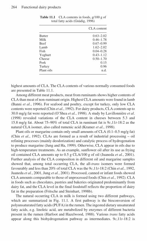
264 Functional dairy products
Table 11.1 CLA contents in foods, g/100 g oftotal fatty acids (Gnädig, 1996)
CLA content
Butter 0.63–2.02Milk 0.46–1.78Beef 0.67–0.99Lamb 1.62–2.02Fish 0.04–0.28Yoghurt 0.43–1.12Cheese 0.50–1.70Pork 0.15Turkey 0.96Plant oils n.d.
highest amounts of CLA. The CLA contents of various normally consumed foodsare presented in Table 11.1.
Among different meat products, meat from ruminants shows higher contents ofCLA than meat of non-ruminant origin. Highest CLA amounts were found in lamb(Banni et al., 1996). For seafood and poultry, except for turkey, only low CLAcontents were reported (Chin et al., 1992). For dairy products, CLA contents up to30.0 mg/g fat were reported (O’Shea et al., 1998). A study by Lavillonnière et al.(1998) revealed variations of the CLA content in cheeses between 5.3 and15.8 mg/g fat. About 75–90% of total CLA in ruminant fat is 9c,11t-18:2 as thenatural CLA isomer, also called rumenic acid (Kramer et al., 1998).
Plant oils or margarine contain only small amounts of CLA (0.1–0.5 mg/g fat)(Chin et al., 1992). CLAs are formed as a result of industrial processing – oilrefining processes (mainly deodorization) and catalytic process of hydrogenationto produce margarine (Jung and Ha, 1999). Otherwise, CLA appear in oils due tohigh-temperature treatments. As an example, sunflower oil after its use as fryingoil contained CLA amounts up to 0.5 g CLA/100 g of oil (Juaneda et al., 2001).Further analysis of the CLA composition in different oil and margarine samplesshowed that, among total occurring CLA, the all-trans isomers were formedfavourably and less than 20% of total CLA was the 9c,11t-18:2 (Chin et al., 1992,Juaneda et al., 2001, Jung et al., 2001). Processed, canned or infant foods showedCLA amounts comparable to those of unprocessed foods (Chin et al., 1992). CLAin foods such as chocolates, pastries and bakeries originated predominantly fromdairy fat, and the CLA level in the final foodstuff reflects the proportion of dairyfat in the preparation (Fritsche and Steinhart, 1998b).
The natural occurring CLA in milk is formed using two different pathways,which are summarized in Fig. 11.1. A first pathway is the bioconversion ofpolyunsaturated fatty acids (PUFA) in the rumen. The ingested dietary unsaturatedfatty acids, e.g. linoleic acid, are metabolized by enzymes of different bacteriapresent in the rumen (Harfoot and Hazelwood, 1988). Various trans fatty acidsappear along this biohydrogenation pathway as intermediates. 9c,11t-18:2 is
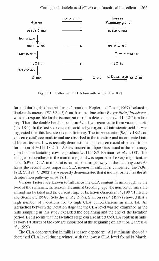
Conjugated linoleic acid (CLA) as a functional ingredient 265
Fig. 11.1 Pathways of CLA biosynthesis (9c,11t-18:2).
formed during this bacterial transformation. Kepler and Tove (1967) isolated alinoleate isomerase (EC 5.2.1.5) from the rumen bacterium Butyrivibrio fibrisolvens,which is responsible for the isomerization of linoleic acid into 9c,11t-18:2 in a firststep. Then, the double bond in position ∆9 is hydrogenated to form vaccenic acid(11t-18:1). In the last step vaccenic acid is hydrogenated into stearic acid. It wassuggested that this last step is rate limiting. The intermediates (9c,11t-18:2 andvaccenic acid) accumulate and are absorbed in the intestine and incorporated intodifferent tissues. It was recently demonstrated that vaccenic acid also leads to theformation of 9c,11t-18:2. It is ∆9 desaturated in adipose tissue and in the mammarygland of the lactating cow to produce 9c,11t-18:2 (Griinari et al., 2000). Theendogenous synthesis in the mammary gland was reported to be very important, asabout 60% of CLA in milk fat is formed via this pathway in the lactating cow. Asfar as the second most important CLA isomer in milk fat is concerned, the 7t,9c-18:2, Corl et al. (2002) have recently demonstrated that it is only formed via the ∆9desaturation pathway of 9t-18:1.
Various factors are known to influence the CLA content in milk, such as thefood of the ruminant, the season, the animal breeding type, the number of times theanimal has lactated and the current stage of lactation (Jahreis et al., 1997; Fritscheand Steinhart, 1998b; Sébédio et al., 1999). Stanton et al. (1997) showed that ahigh number of lactations led to high CLA concentrations in milk fat. Aninteraction between the lactation stage and the CLA level was not examined, as themilk sampling in this study excluded the beginning and the end of the lactationperiod. But it seems that the lactation stage can also affect the CLA content in milk,as body fat stores of the cow are mobilized at the beginning of lactation (Jahreis etal., 1999).
The CLA concentration in milk is season dependent. All ruminants showed adecreased CLA level during winter, with the lowest CLA level found in March,
266 Functional dairy products
and inversely an increase was observed during summer, which is positivelycorrelated to the grazing period. Indeed, pasture in spring time contains higheramounts of PUFAs, which caused a higher bacterial biohydrogenation in therumen (Jahreis et al., 1997). The relationship between pasture feeding and CLAlevel was also described by Dhiman et al. (1996). The CLA level increased from8.4 to 22.7 mg/g milk fat when the pasture level was increased in the diet. To showthe influence of different diets on the CLA content in milk, an experiment wascarried out to compare three different types of farm management applying threedifferent feeding methods. Cows were fed either with maize silage throughout theyear (indoor group), with grazing in summer and maize silage in winter (conven-tional group) or with grazing during summer and clover–alfalfa–grass silage inwinter (ecological group). The highest amounts of CLA in milk were measured inthe ecological group, whereas smallest CLA contents were found in the indoor-maize silage group (Jahreis et al., 1996). The application of ecological feedingconditions rich in pasture would result in the production of milk naturally high inCLA (Jiang et al., 1996).
Various experiments were carried out to increase the CLA content in milk, bymodulating the dietary regimen of the cows. Milk from cows offered a dietsupplemented with oils rich in PUFAs had up to fivefold higher CLA levels.Enrichment with sunflower, linseed or rapeseed oil also caused an increase in CLAlevels (Kelly et al., 1998). A positive correlation was found between the content oflinoleic acid in the oil and the increase of CLA in the milk. The same effects wereobtained by feeding with extruded oilseeds (Dhiman et al., 1999). Feeding fish oilswhich contained large amounts of PUFAs, mainly eicosapentaneoic acid anddocosahexaneoic acid, resulted in the same effects. CLA content increased signifi-cantly with the formation of biohydrogenation products in the rumen (Donovan etal., 2000; Jones et al., 2000). A recent study of Chouinard and coworkers (2001)investigated the effects of different dietary fat supplements on CLA content inmilk. The use of canola, soybean and linseed oil increased CLA content in the milkfat by three- to fivefold compared with a control diet. The influence of processingmethods to produce soybeans as dietary supplement in the cows’ diet was alsoexamined. It was found that the feeding of extruded and roasted soybeans resultedin two- to threefold higher contents of CLA in milk fat compared to a dietcontaining raw ground soybeans.
Direct feeding of CLA in the regimen and the abomasal infusion of CLA wastested as another possibility to enrich CLA in cows’ milk (Chouinard et al., 1999).It was found that direct CLA supplementation reduced milk fat yield, suggesting adecrease of de novo fatty acid synthesis (Loor and Herbein, 1998). Moreover, theamount of CLA that induced a substantial reduction of milk fat synthesis was foundto be low. As an example, infusion with 0.10 g/day of 10t,12c-18:2 (0.05% of diet)resulted in a 44% reduction in milk fat yield (Baumgard et al., 2001). It wasdemonstrated that only the 10t,12c-18:2 inhibited milk fat synthesis, whereas the9c,11t-18:2 had no effect. The application of this method to enrich CLA in milkseems not to be worth pursuing because of the disadvantageous reduction of milkfat resulting in the reduction of total CLA in milk.
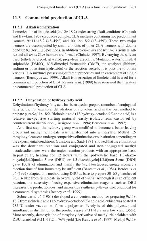
Conjugated linoleic acid (CLA) as a functional ingredient 267
11.3 Commercial production of CLA
11.3.1 Alkali isomerizationIsomerization of linoleic acid (9c,12c-18:2) under strong alkali conditions (Chipaultand Hawkins, 1959) produces complex CLA mixtures containing two predominantisomers. 9c,11t-18:2 (43–45%) and 10t,12c-18:2 (43–45%). These two majorisomers are accompanied by small amounts of other CLA isomers with doublebonds in 8,10 or 11,13 positions. In addition to cis–trans and trans–cis isomers, all-cis and all-trans CLA isomers are formed (Christie, 1997). By varying the solventused (ethylene glycol, glycerol, propylene glycol, tert-butanol, water, dimethylsulphoside (DMSO), N,N-dimethyl formamide (DMF), the catalysts (lithium,sodium or potassium hydroxide) or the reaction vessels, it is possible to obtainvarious CLA mixtures possessing different properties and an enrichment of singleisomers (Reaney et al., 1999). Alkali isomerization of linoleic acid is used for acommercial production of CLA. Reaney et al. (1999) have reviewed the literatureon commercial production of CLA.
11.3.2 Dehydration of hydroxy fatty acidDehydration of hydroxy fatty acid has been used to prepare a number of conjugatedfatty acids. For example, dehydration of ricinoleic acid is the best method toprepare pure 9c,11t-18:2. Ricinoleic acid (12-hydroxy-octadec-9Z-enoic acid) is arelative inexpensive starting material, easily isolated from castor oil bycountercurrent distribution (Tassignon et al., 1994; Berdeaux et al., 1997).
As a first step, the hydroxy group was modified to become a better leavinggroup and methyl ricinoleate was transformed into a mesylate. Methyl 12-mesyloxyoleate can undergo competitive elimination or substitution depending onthe experimental conditions. Gunstone and Said (1971) showed that the eliminationwas the dominant reaction and conjugated and non-conjugated methyloctadecadienoates were the major reaction products with an appropriate base.In particular, heating for 12 hours with the polycyclic base 1,8-diazo-bicyclo[5.4.0]undec-5-ene (DBU) or 1.5-diazobicyclo[4.3.0]non-5-ene (DBN)gave 100% of elimination and mainly the 9c,11t-octadecadienoate isomer; areaction time of four hours may be sufficient (Bascetta et al., 1984). Berdeaux etal. (1997) adapted this method using DBU as base to prepare 50–60 g batches of9c,11t-18:2 from ricinoleate in overall yield of >70%. Although it is an efficientreaction, the necessity of using expensive elimination reagents such as DBUincreases the production cost and makes this synthesis pathway uneconomical fora commercial synthesis (Reaney et al., 1999).
Schneider et al. (1964) developed a convenient method for preparing 9t,11t-18:2 from ricinelaic acid (12-hydroxy-octadec-9E-enoic acid) which was heated at235 °C under vacuum to form a polyester. Pyrolysis of this polyester andsimultaneous distillation of the products gave 9t,11t-18:2 in a low yield (35%).More recently, demesylation of mesyloxy derivative of methyl ricinelaidate withDBU furnished 9t,11t-18:2 in 76% yield (Lie Ken Jie et al., 1997). Methyl 9t,11t-
268 Functional dairy products
18:2 was also prepared from 9,12-dihydroxy-10t-octadecenoate usingchlorotrimethylsilane and sodium in acetonitrile in 72% yield (Lie Ken Jie andWong, 1992). Yurawecz et al. (1994) converted allylic hydroxy oleate to a mixtureof CLA isomers by an acid-catalyzed methylation using HCl/methanol or BF
3/
methanol. They obtained a mixture of 9c,11t-, 9t,11t-, 7t,9c- and 7t,9t-18:2isomers in a good yield (72–78%). This method was used to prepare mixture ofCLA isomers as analytical standard (Yurawecz et al., 1994).
11.3.3 Reduction of acetylenic bondsSantalbic acid (octadec-11E-en-9-ynoic acid) isolated from Santalum album seedoil or prepared from ricinoleic acid (Lie Ken Jie et al., 1996), is a useful precursorto synthesize 9c,11t-18:2. Zinc in aqueous n-propanol was highly selective in thereduction of the acetylenic bond to the corresponding Z-olefinic bond. Usingthis procedure, Lie Ken Jie et al. (1997) prepared 9c,11t-18:2 in the pure formfrom methyl santalbate in 82% yield. Following the same procedure, theyprepared pure 9c,11c-18:2 isomer from its Z isomer (octadec-11Z-en-9-ynoicacid). Lindlar catalyst and H
2 have also been used to reduce the acetylenic bond of
methyl santalbate into the cis-bond (Smith et al., 1991). Moreover, the Lindlarcatalyzed reduction of methyl santalbate has been used in presence of deuteriumgas to produce 9c,11t-18:2-9,10d
2 in yield of 65–70% (Adlof, 1999). Iron in 15%
n-propanol solution has also been used to reduce acetylenic bond of methylsantalbate and 9c,11t-18:2 was obtained in 85% yield (Morris et al., 1972). Morriset al. (1972) prepared some cis/cis CLA isomers (6c,8c-/7c,9c-/8c,10c-/9c,11c-18:2) by reduction of the corresponding synthesized conjugated methyloctadecadiynoate with iron in aqueous propanol in 56–77% yield.
11.3.4 Multiple step synthesesPure CLA isomers can also be produced by total stereoselective multiple stepsyntheses, in small quantities (up to 1 g), the isomeric purity can be higher than98%. Stereoselective synthesis guarantees the exact chemical structure of the finalmolecule and limits the formation of by-products due to several purificationprocedures performed during the synthesis. Moreover, these syntheses shouldallow the preparation of labeled molecules of CLA with deuterium, 13C or 14C.Recently Adlof (1997) described a multistep synthesis of deuterium-labeled9c,11t- and 9t,11t-18:2 with isotopic and chemical purities >95% for each geo-metrical isomers, in 12% overall yield. The two isomers were synthesized as amixture via a Wittig coupling reaction. The two isomers were readily separated bya combination of reverse-phase and silver resin chromatography.
Loreau et al. (2001) described the stereoselective multistep syntheses of 9c,11t,10t,12c and 10c,12c-[1-14C] conjugated linoleic acid isomers with high radio-chemical and isomeric purities (>98%) (Fig. 11.2). They described thestereo-controlled preparation of (Z,E)- or (Z,Z)-heptadecadienyl bromo precursorsof the CLA isomers by using the sequential substitution of (E)- or (Z)-1,2-dichloro-
Conjugated linoleic acid (CLA) as a functional ingredient 269
Fig. 11.2 (a) Synthesis of 10t,12c-conjugated linoleic acid. (b) Synthesis of 9c,11t-conjugated linoleic acid.
ethene. A first cross-coupling reaction between a terminal alkyne and 1,2-dichloro-ethene followed by a second coupling reaction with an alkylmagnesium bromidegave a conjugated enyne. Stereoselective reduction of triple bond and brominationafforded the expected bromo precursor. After formation of the heptadecadienyl-magnesium bromide, (9,11)- and (10,12)-CLA isomers were obtained with 14Ccarbon dioxide.
Stereoselective synthesis procedures for 9c,11c-, 9t,11t-, 10t,12c, 7t,9c- and9c,11t-18:2 using comparable pathways have been published by Lehmann (2001).All isomers were synthesized from commercial reagents via acetylenic-couplingreactions, and stereoselective hydrogenation of the first double bond in E or Zconfiguration. The second double bond was formed selectively in E or Zconfiguration by a Wittig reaction. All synthesized CLA isomers showed a goodisomeric purity without any further purification.
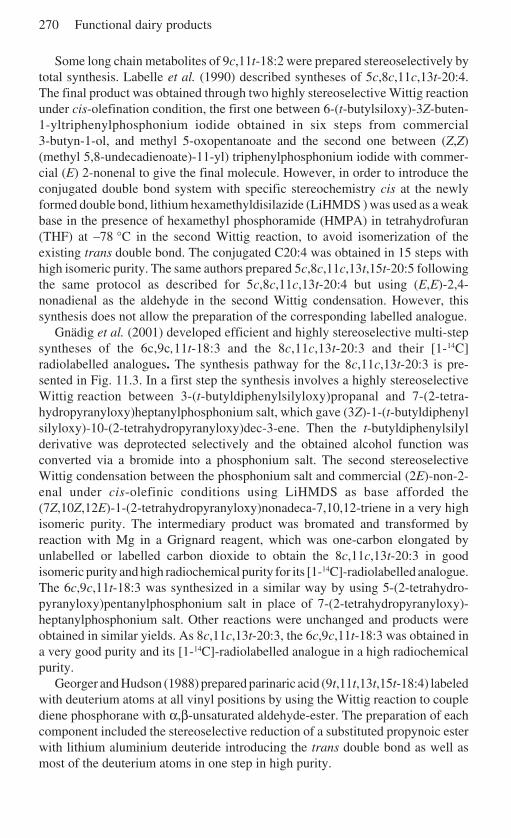
270 Functional dairy products
Some long chain metabolites of 9c,11t-18:2 were prepared stereoselectively bytotal synthesis. Labelle et al. (1990) described syntheses of 5c,8c,11c,13t-20:4.The final product was obtained through two highly stereoselective Wittig reactionunder cis-olefination condition, the first one between 6-(t-butylsiloxy)-3Z-buten-1-yltriphenylphosphonium iodide obtained in six steps from commercial3-butyn-1-ol, and methyl 5-oxopentanoate and the second one between (Z,Z)(methyl 5,8-undecadienoate)-11-yl) triphenylphosphonium iodide with commer-cial (E) 2-nonenal to give the final molecule. However, in order to introduce theconjugated double bond system with specific stereochemistry cis at the newlyformed double bond, lithium hexamethyldisilazide (LiHMDS ) was used as a weakbase in the presence of hexamethyl phosphoramide (HMPA) in tetrahydrofuran(THF) at –78 °C in the second Wittig reaction, to avoid isomerization of theexisting trans double bond. The conjugated C20:4 was obtained in 15 steps withhigh isomeric purity. The same authors prepared 5c,8c,11c,13t,15t-20:5 followingthe same protocol as described for 5c,8c,11c,13t-20:4 but using (E,E)-2,4-nonadienal as the aldehyde in the second Wittig condensation. However, thissynthesis does not allow the preparation of the corresponding labelled analogue.
Gnädig et al. (2001) developed efficient and highly stereoselective multi-stepsyntheses of the 6c,9c,11t-18:3 and the 8c,11c,13t-20:3 and their [1-14C]radiolabelled analogues. The synthesis pathway for the 8c,11c,13t-20:3 is pre-sented in Fig. 11.3. In a first step the synthesis involves a highly stereoselectiveWittig reaction between 3-(t-butyldiphenylsilyloxy)propanal and 7-(2-tetra-hydropyranyloxy)heptanylphosphonium salt, which gave (3Z)-1-(t-butyldiphenylsilyloxy)-10-(2-tetrahydropyranyloxy)dec-3-ene. Then the t-butyldiphenylsilylderivative was deprotected selectively and the obtained alcohol function wasconverted via a bromide into a phosphonium salt. The second stereoselectiveWittig condensation between the phosphonium salt and commercial (2E)-non-2-enal under cis-olefinic conditions using LiHMDS as base afforded the(7Z,10Z,12E)-1-(2-tetrahydropyranyloxy)nonadeca-7,10,12-triene in a very highisomeric purity. The intermediary product was bromated and transformed byreaction with Mg in a Grignard reagent, which was one-carbon elongated byunlabelled or labelled carbon dioxide to obtain the 8c,11c,13t-20:3 in goodisomeric purity and high radiochemical purity for its [1-14C]-radiolabelled analogue.The 6c,9c,11t-18:3 was synthesized in a similar way by using 5-(2-tetrahydro-pyranyloxy)pentanylphosphonium salt in place of 7-(2-tetrahydropyranyloxy)-heptanylphosphonium salt. Other reactions were unchanged and products wereobtained in similar yields. As 8c,11c,13t-20:3, the 6c,9c,11t-18:3 was obtained ina very good purity and its [1-14C]-radiolabelled analogue in a high radiochemicalpurity.
Georger and Hudson (1988) prepared parinaric acid (9t,11t,13t,15t-18:4) labeledwith deuterium atoms at all vinyl positions by using the Wittig reaction to couplediene phosphorane with α,β-unsaturated aldehyde-ester. The preparation of eachcomponent included the stereoselective reduction of a substituted propynoic esterwith lithium aluminium deuteride introducing the trans double bond as well asmost of the deuterium atoms in one step in high purity.
Conjugated linoleic acid (C
LA
) as a functional ingredient271Fig. 11.3 Synthesis of 8c,11c,13t-20:3.
272 Functional dairy products
11.3.5 Biochemical synthesesAt this time, the use of biosynthetic method utilizing specific enzymes isolatedfrom algal or bacterial sources to produce CLA isomers is limited to the prepara-tion of 9c,11t-18:2 isomer. First, 9c,11t-18:2 isomer was prepared from linoleicacid using the linoleate isomerase enzyme isolated from the rumen bacteriumButyrivibrio fibrisolvens (Kepler and Tove, 1967). Using the same linoleateisomerase enzyme from B. fibrisolvens, the same authors prepared also 14C- and2H-labeled 9c,11t-18:2 from linoleic acid (Kepler and Tove; 1967, Kepler et al.,1966). Recently, a microbial synthesis of 9c,11t-18:2 from linoleic acid using aculture of Lactobacillus sp. has been described (Reaney et al., 1999). A total CLAlevel of 7 mg/g cells was produced containing >96% 9c,11t-18:2 isomer.
11.4 Analytic methods
As described before, many positional and geometrical CLA isomers are present innatural and synthetically produced materials. Therefore, high-quality analyticalmethods are needed to determine the exact isomer composition of the complexCLA mixtures in synthetic CLA, foodstuff or biological matrices. Great care has tobe taken with the analysis of CLAs as they are unstable and very sensitive toisomerization (Shantha et al., 1993). Therefore, detected CLA levels could beincorrect because of the use of unsuitable analytical methods in the past (Hamilton,2001). It has been suggested that any data reported more than five years ago shouldbe critically regarded (Christie, cited in Hamilton, 2001).
To avoid any problems, the CLA-containing substance has to be transformedinto fatty acid methyl esters (FAME) using mild methylation methods. Acidcatalysts or high temperatures lead to isomerization, where cis–trans and trans–cisisomers are converted into trans–trans isomers, and to the formation of methoxyartefacts (Kramer et al., 1997; Park et al., 2001b). Also methoxy artefacts appearedusing the methylation with trimethylsilyl-diazomethane, an often recommendedweak methylation method (Christie et al., 2001). Usually sodium methoxide isused as a weak base.
CLA contents are currently determined using gas chromatography (GC) coupledto a flame ionization detector (FID). To obtain a good separation of the CLAisomers, high polar capillary columns 50 and 100 m in length are used. Althougha good separation of the CLA isomers can be reached, some of the positionalisomers, e.g. 7t,9c-18:2 and 9c,11t-18:2, co-elute and the samples have to beanalyzed using other techniques (Rickert and Steinhart, 2001). The utilization ofsilver ion (Ag+)-high-performance liquid chromatography (HPLC) improved theresolution of the CLA isomers, since two to six columns were coupled in series.The CLA isomers are separated into three groups depending on the configurationof the double bounds, in trans–trans, cis–trans/trans–cis and cis–cis respectively.Furthermore Ag+-HPLC led to a better separation of the CLA with less coelutioncompared to GC (Sehat et al., 1999).
Exact structural determination of single CLA isomers is possible using different
Conjugated linoleic acid (CLA) as a functional ingredient 273
spectroscopic methods. The analysis is often done using GC coupled with massspectrometry (MS). Therefore, the FAME are transformed into a 4-methyl-1,2,4-triazoline-3,5-dione (MTAD) adduct or a 4,4-dimethyloxazoline derivative(DMOX) (Dobson and Christie, 1996; Dobson, 1997). The use of these derivativesin GC–MS permits the localization of the double bond position in the fatty acidmolecule. To get information about the configuration of the double bond, GC–Fourier-transformed infra red spectroscopy (FTIR) is applied. The IR-spectra forconjugated trans–trans and cis–trans/trans–cis isomers exhibited absorption at990 cm-1 and characteristic bands at 988 and 949 cm-1, respectively. However,differentiation between a cis–trans and trans–cis diene is not possible throughFTIR (Mossoba et al., 1999). The positions and relative intensities of the =C–Hstretch bands are highly characteristic and could discriminate between cis–trans/trans–cis (3020 and 3002 cm-1), cis–cis (3037 and 3005 cm-1) and trans–trans(3017 cm-1) conjugated double bond systems (Fritsche et al., 1997).
11.5 The influence of processing on the CLA content of dairyproducts
The relationship between the CLA content and different parameters of foodprocessing in dairy products is a controversial area. Because of their chemicalstructure containing a conjugated double bond system, CLAs have been seen asmore sensitive to oxidation or to isomerization during heat treatment than linoleicacid. Research has been undertaken to determine the influence of heat treatments,such as grilling, cooking and frying, on CLA content, to test the stability in foodproducts and to avoid a CLA decrease by oxidative damage. Home-made foodpreparation had no influence on CLA content. Shantha et al. (1994) demonstratedthat frying, baking, broiling or microwaving did not change the CLA content ingrilled hamburger beef patties. These findings were supported by a recent study,which described that only intensive heating for 15 min, using temperatures higherthan 200 °C, led to isomerization of CLA in milk, whereas moderate heating hadno effect. Heating at 225 °C for 15 min decreased 9c,11t-18:2 from 1.7% to 1.1%of total fatty acids (Precht et al., 1999).
The influence of storage on the CLA content was also tested in butter, yoghurtand sour cream. No modification of the CLA content was detected (Shantha et al.,1995). Dairy products often undergo a microbial fermentation during processing.The use of different fermentation cultures, processing temperatures or ripeningperiods could affect the CLA level in the final foodstuff (Fritsche and Steinhart,1998a). These suggestions were confirmed by the findings of Jiang et al. (1998),who reported the ability of two strains of Propionibacterium spp. to produce CLAin culture by conversion of linoleic acid into 9c,11t-18:2 (Jiang et al., 1998). Morerecently, six further Lactobacillus spp. were identified as able to convert linoleicacid into CLA (T.Y. Lin et al., 1999a; Lin, 2000). The use of these strains could beone way of enriching yoghurts and cheese in CLA during fermentation.
As cheese processing involves bacterial fermentation, various studies have
274 Functional dairy products
been carried out on this subject. Only minor changes in the CLA contents ofdifferent cheddar cheese varieties were found (3.20 mg/g fat vs 3.55 mg/g fat) (H.Lin et al., 1999). Another study, published by a Swedish group (Jiang et al., 1997)tested various hard cheeses. They observed the same CLA amounts in raw materialand in the final product. Other processing parameters during cheese fabricationwere investigated to see if these were able to modify CLA content. Changes inmilling and pH values or the addition of butylhydroxyanisol (BHA) or amino acids(lysine, tyrosine) led to a small decrease of the CLA content in the final cheese(2.70 mg/g fat vs 2.19 mg/g fat by addition of tyrosine and reduced pH) (Lin et al.,1998).
Various studies have been undertaken to determine the CLA content in cheesespread using different processing conditions. Tests compared processing underatmospheric conditions and under nitrogen as protective gas. It was shown thatprocessing under atmospheric conditions at temperatures of 80–90 °C couldincrease the CLA content in the product. This effect was amplified by the additionof whey proteins as hydrogen donors or antioxidants as butylhydroxytoluene(BHT). Also the use of iron as Fe2+/Fe3+ ascorbate as additive seems to be useful toincrease the CLA level (Shantha et al., 1992, 1995).
In contrast, the addition of antioxidants (ascorbate or whey protein concentrates)acting as hydrogen donors to beef patties did not influence the CLA content of thesamples (Shantha and Decker, 1995). The controversial results concerning therelationship of antioxidants and hydrogen donors with the CLA content could berelated to the foodstuff itself and the presentation of the fatty acids in the foodmatrix. However, the changes in CLA content, reported during processing, aremuch less important than the seasonal differences in milk, so that the CLA contentin the processed food largely depends on the CLA content in the original milk.
11.6 Functional benefits of CLA: cancerIn the early 1980s Pariza and Hargraves (1985) reported that an extract of grilledground beef exhibited mutagenesis inhibitory activity in experiments using theAMES test. The active molecules were later identified as CLA (Ha et al., 1987).Since that time, researchers have paid more attention to CLA, and there is nowgrowing evidence of the multisite and multistage anticarcinogensis activity ofCLA. CLA could inhibit many chemical-induced cancers of the mammary, stomach,colon and skin, and a variety of tumour cell lines (including breast/mammarycancer, colorectal cancer, prostate cancer, lung adenocarcinoma, malignantmelonoma, neuroglioma) demonstrated inhibition by CLA. Moreover, CLA mayinhibit carnogenesis at each of its major stages, such as initiation, promotion (post-initiation), progression and metastasis (Pariza, 1997; Pariza et al., 1999).
Though the mechanisms through which CLA inhibits tumourigenesis are notcompletely established, they seem to fall into the following categories:
• an antioxidant mechanism involving lipid peroxidation production toxic tocancer cells

Conjugated linoleic acid (CLA) as a functional ingredient 275
• changing the fatty acid profile in tumour cells and interfering with eicosanoidmetabolism
• reducing the formation of carcinogen–DNA adducts• influencing the growth and development of certain types of mammary cells by
interfering with the estrogen response system, inducing apoptosis and regulatinggene expression such as c-myc, stearoyl-CoA desaturase, PPARα, (peroxisomeproliferator-activated receptor) cyclin A, B1, D1 cyclin-dependent kinaseinhibitors (CDKI) (P16 and P21) (Kritchevsky, 2000).
In the following sections, we try to summarize CLA multisite and multistageanticarcinogenesis and to explain all these possible mechanisms.
11.7 Multisite anticarcinogenesis
11.7.1 In vivo – mammary/breast cancer modelAlthough CLA demonstrates multisite anticarcinogenesis, research has concentratedon inhibition by CLA of mammary cancer because CLA has shown higherretention in adipose tissue (Sugano et al., 1997). The retention of conjugatedlinoleic acid in the mammary gland may be associated with tumour inhibition. Ipet al. (1997) examined the influence of dietary CLA on tumour development in therat after the use of dimethylbenz(a)anthracene (DMBA) as carcinogen adminis-trated in a single dose. One per cent CLA (from Nu-Chek Prep, Inc: 43.3% c9,t11-and t9,c11-CLA; 45.3% t10,c12; 1.9% c9,c11-CLA; 1.4% c10,c12-CLA; 2.6%t9,t11- and t10,t12-CLA; 4.4% linoleic acid) was given in the diet for 4 and 8weeks, and continuously after the exposure to the carcinogen. It was shown thatdietary CLA for 4 and 8 weeks did not prevent the occurrence of mammarycarcinoma. However, significant tumour inhibition was observed in rats fed witha CLA diet for the entire duration of the experiment. This demonstrated that thephysiological accumulation of CLA in the mammary gland contributed to cancerprotection in the tumour post-initiation stage. Analysis of fatty acids in mammarygland showed that more CLA was incorporated into mammary tissue neutral lipidsthan in mammary tissue phospholipids. Moreover, CLA levels in neutral lipidsdisappeared as CLA was removed from the diet, which resulted in the subsequentoccurrence of new tumours (Ip et al., 1997). However, five weeks of short-termCLA feeding, from weaning to the time of carcinogen administration (7,12-dimethylbenz(a)anthrane and methylnitrosourea) at 50 days of age, significantlyprotect against subsequent tumour occurrence, which is related to maturation ofthe mammary gland to the adult stage (Ip et al., 1994, 1995).
CLA at dietary concentrations that are close to the levels consumed by humansshowed a significant anticarcinogenic activity in experiments using the rat mammarycancer model. Diets containing 0.5, 1 or 1.5% CLA isomer mixture could reducethe total tumour numbers by 32, 56 and 60%, respectively (Ip et al., 1991). Afurther study showed that dietary CLA between 0.05 and 0.5% caused a dose-dependent inhibition of mammary tumourigenesis where the tumour yield was
276 Functional dairy products
reduced to 22, 36, 50 and 58% by feeding 0.05, 0.1, 0.25 and 0.5% CLA in the diet.No further beneficial effect was observed by increasing the CLA level in the dietto 1% (Ip et al., 1994). Moreover, the inhibition by CLA of mammary cancer isindependent of the level or type of fat in diet. To study the influence of the level ortype of fat in diet on CLA tumour inhibition, a fat blend (10, 13.3, 16.7 or 20% byweight in the diet) or 20% fat diet containing either corn oil or lard was used. Theresults showed that the inhibition by 1% CLA on tumour size was not influencedby the level or type of fat in the diet (Ip et al., 1996).
The main sources of CLA are dairy products and foods derived from ruminants.Therefore, they are in the form of triacylglycerols. It is necessary to carry out theanimal experiment using CLA as triacylglycerols. The working group of Ip was thefirst to compare the effects of CLA as free fatty acids and CLA as triacylglycerolon the development of mammary carcinogenesis using the rat methylnitrosourea(MNU) model. They reported that the anticancer activities of both molecules(triacylglycerols and free fatty acids) were identical (Ip et al., 1995). Subsequently,CLA-enriched butter fat (CLA as triacylglyerols in natural form) was used in ananimal experiment and it was shown that natural CLA possessed the biologicalaction in cancer inhibition (Ip et al., 1999a; O’Shea et al., 2000). In addition,Shirai’s group recently reported that conjugated fatty acids derived from saffloweroil (CFA-S) containing large amounts of CLA may retard the development of 2-amino-1-methyl-6-phenylimidazo(4,5-b) pyridine (PhIP)-induced mammarytumours (Kimoto et al., 2001; Futakuchi et al., 2002).
11.7.2 In vivo – other cancer modelsIn other animal models, CLA proved to be equally effective in tumour inhibition.The benzo(a)pyrene-induced mouse forestomach model was firstly used to studythe effect of CLA on carcinogenesis in animal. Ha et al. (1990) gave a syntheticCLA isomer mixture (c9,t11-, t10,c12-, t9, t11- and t10,t12-CLA) to female ICRmice treated with benzo(a)pyrene. Dietary CLA significantly reduced tumourincidence (up to 30%) as well as tumour multiplicity compared with the controlgroup. In mouse skin tumour promotion elicited by 12-O-tetradecanoyl phorbol-13-acetate (TPA), mice fed diets with 1.0–1.5% CLA developed significantlyfewer tumours than those fed a diet without CLA (Belury et al., 1996). Using a ratcolon cancer model and different carcinogens such as 2-amino-3-methylimidazo(4,5-f) quinoline (IQ), heterocyclic amine, 1,2-dimethylhydrasine, azoxymethane(AOM), researchers demonstrated that CLA significantly decreased the number ofcolonic aberrant crypt foci (ACF) and inhibited colon tumour incidence (Liew etal., 1995; Park et al., 2001a; Xu and Dashwood, 1999; Kohno et al., 2002).
11.7.3 In vitro modelTumour cell lines are easily available and the treatment is less complicated thanthat in animal model. A number of tumour cell lines were used to study the CLAanticarcinogenesis. Shultz et al. (1992) firstly confirmed that CLA isomer mixtures
Conjugated linoleic acid (CLA) as a functional ingredient 277
(43% c9,t11- and t9,c11-, 41.5% t10,c12-, 1.6% c9,c11-, 2.4% c10,c12-, 11.2%t9,t11- and t10,t12-) significantly reduced human (malignant melanoma M21-HPB, colorectal HT-29, breast MCF-7) cancer cells proliferation by 18–100%.Subsequently, much research attempted to explain CLA anticarcinogenesismechanism using different human cancer cells, mainly human mammary cancercell MCF-7. For example, the relation between CLA, linoleic acid, cyclooxygenaseand lipoxygenase inhibitor, estrogen reponse system, antioxidant enzyme defenseresponses and liperoxidation, cell signal transduction, arachidonic acid distribution,stearoyl-CoA desaturase activity were studied in MCF-7 breast cancer cell (O’Sheaet al., 1998, 1999; Cunningham et al., 1997; Durgam and Fernandes, 1997; Park etal., 2000; Choi et al., 2002). Besides breast cancer MCF-7 cells, human breast(MDA-MB-231 and MDA-MB-468) (Visonneau et al., 1997), gastric (SGC-7901) (Liu et al., 2002), colorectal (HT-29, MIP-101 and SW480), prostate (PC-3and DU-145) (Palombo et al., 2002), hepatoma (HepG2) (Igarashi and Miyazawa,2001), and three different lung adenocarcinoma (A-427, SK-LU-1, A549) cancercell lines were used in CLA research. However, CLA showed no influence onhuman glioblastoma cell line (A-172) growth (Schonberg and Krokan, 1995).
11.8 Multistage anticarcinogenesis
11.8.1 Initiation and promotion (post-initiation)For mammary tumour initiation research, the exposure of CLA started during theearly post-weaning and pubertal period before carcinogen administration, whichwas sufficient to reduce subsequent tumourigenesis induced by different carcino-gens such as MNU and DMBA; for mammary tumour promotion (post-initiation),CLA feeding was given after carcinogen administration, a continuous intake (20weeks) of CLA was required for inhibition of carcinogenesis, but 4 or 8 weeks ofCLA feeding did not show any effect on the tumour inhibition (Ip et al., 1994,1995, 1997). In mouse skin tumour promotion elicited by TPA, Belury et al. (1996)also confirmed the fact that CLA inhibited the tumour promotion.
In addition, DNA-adduct, as one of the biological makers of tumour initiation,was a hot topic for CLA research. Generally, CLA can inhibit different mutagen–DNA adduct formation in different organs. For example, Schut reported that CLAtreatment caused inhibition of adduct formation in the liver, lung, large intestineand kidney in CFD1 mouse given IQ. IQ–DNA adduct formation in the kidneys offemales was reduced by up to 95.2% (Schut and Zu, 1993). The F344 rat givenCLA also had lower IQ–DNA and PhIP–DNA adduct in various organs includingthe mammary gland and the colon (Schut et al., 1997). Recently, Futakuchi et al.(2002) confirmed that CFA-S (conjugated fatty acids derived from safflower oil;mainly CLA) could also suppress PhIP–DNA adduct formation in the epithelialcells of mammary gland. But Josyula et al. (1998) found that dietary CLA did notabsolutely decrease DNA adduct. They reported that CLA increased IQ–DNAadduct formation by 30.5% on day 8 in the colon of female Fisher 344 rats given
278 Functional dairy products
AIN-76 diet with 4% CLA and treated with IQ or PhIP after two weeks and had noeffect on adduct levels in liver or white blood cells (Josyula et al., 1998; Josyulaand Schut, 1998).
11.8.2 Progression and metastasisTo date, there have been few reports assessing the effects of CLA on the progressionor metastasis of established tumour. In the chemical-induced tumour model, theprogression or metastasis stage is difficult to confirm. Fortunately, the growth andspread of the transplantable tumours in animal provide an ideal way to assess theeffect of CLA on tumour progression and metastasis because of the high similarityto human cancer progression and metastasis in vivo. In one paper, CB-17 scid/scidmice (severe combined immunodeficient) were fed 1% CLA (42.3% c9,t11- and44.2% t10,c12-) diets starting two weeks before MDA-MB468 transplantationthroughout the whole of experiment. It was found that CLA not only inhibitedtumour growth, but also abrogated the spread of breast cancer cells to lung,peripheral blood and bone marrow (Visonneau et al., 1997). Hubbard et al. (2000)also reported that 0.1, 0.5 and 1.0% CLA diets significantly retarded the growth rateof transplantable mice mammary tumour cell line 4526 in BALB/cfC3H mouse in adose-dependent manner and decreased the numbers of pulmonary nodules. Thesame conclusion was drawn in the human prostatic cancer cell line DU-145 trans-plantable model in SCID mice (Cesano et al., 1998). However, Wong et al. (1997)reported that 0.1, 0.3 or 0.9% CLA diets had no obvious effect on the growth of theestablished, aggressive mammary tumour. Further research should be pursued.
11.9 Mechanisms of CLA anticarcinogenesis
11.9.1 Antioxidant mechanismTo explain its anticarcinogenic action, it was firstly suggested that CLAs may havean antioxidative activity because of their chemical structure containing a conju-gated double bond system. Ha et al. (1990) described that lowest concentrations ofCLA showed a high antioxidative action which was more potent than that of α-tocopherol. Leung’s research was also in agreement with the above conclusion(Leung and Liu, 2000). However, higher concentrations of CLA seem to showprooxidative activity, which suggests that there is a balance between antioxidationand prooxidation of CLA.
11.9.2 Cytotoxicity of lipid peroxidation production to cancer cellsTwo research groups directed by Shultz and O’Shea suggested that the cytotoxicityof lipid peroxidation induced by CLA plays an important role in anticarcinogenesis(O’Shea et al., 1999; Shultz et al., 1992). Shultz et al. (1992) treated MCF-7 breastcancer cells with 1.78-7.14 × 10⁻⁵ CLA, and found that treated MCF-7 cancer cells
Conjugated linoleic acid (CLA) as a functional ingredient 279
showed significantly growth inhibition in a dose- and time-dependent manner.Further studies confirmed that cytostatic and cytotoxic effects of CLA were morepronounced (8–81%) than LA at similar LA and CLA concentrations (Shultz et al.,1992). In O’Shea’s group, when MCF-7 and SW480 cells were treated with CLAamounts greater or equal than 15 ppm for eight days or more, the amount of thelipid peroxidation product was significantly increased and the cytotoxicity totumour cell induced by CLA is related to the extent of lipid peroxidation product(O’Shea et al., 1999). Moreover, it was found that bovine milk fat enriched withCLA could decrease the MCF-7 cell numbers up to 90% and with the increasingCLA concentration between 16.9 and 22.6 ppm in milk fat lipid peroxidationincreased 15-fold (O’Shea et al., 1999). But the cytotoxicity of lipid peroxidationinduced by CLA may be only one part of a CLA anticarcinogenesis mechanism.Schonberg and Krokan (1995) reported that in the lung adenocarcinoma cell lines,40 µM CLA significantly increased the lipid peroxidation level by twice as muchas 40 µM LA. However, 30 µM vitamin E could completely abolish the lipidperoxidation and the growth inhibition of CLA was only partially restored, whichindicates that a more complex mechanism may explain the anticarcinogenicactivities (Schonberg and Krokan, 1995).
11.9.3 Influencing growth and development of certain types of mammarycellsInterestingly, CLAs may influence the morphological status of the mammarygland, by which mammary cancer risk was reduced. It was shown that the feedingof 0.5 or 1% CLA to rats from weaning to about seven weeks of age in the normaldiet reduced the density of terminal end buds (TEB) and lobuloalveolar buds. TEBand lobuloalveolar buds are the target sites for the development of mammarycancer, so their decrease may reduce the cancer risk. Moreover, mammary cellsshowed lower proliferative activity in the sites of TEB and lobuloalveolar buds(Banni et al., 1999; Thompson et al., 1997).
The same team also tried to determine that a high CLA butter had similarbiological activities as the CLA isomer mixture. They reported that, in rats givenbutter fat CLA during the pubescent mammary gland development period, mammaryepithelial mass was reduced by 22%, the size of the TEB population was decreasedby 30%, the proliferation of TEB cells were suppressed by 30% and mammarytumour yield was inhibited by 50% (Ip et al., 1999). In general, it might be moreimportant for mammary tumour chemoprevention that CLA or triacylglycerolsCLA is given to the animal during the development of mammary gland.
11.9.4 Interfering in the estrogen response systemOne paper reported that CLA selectively inhibited proliferation of estrogenreceptor (ER) positive MCF-7 cells as compared with ER negative MDA-MB-231cells and a higher percentage of MCF-7 cells treated with CLA remained in the G0/G1 phase of cell cycle compared with control and those treated with linoleic acid
280 Functional dairy products
(Durgam and Fernandes, 1997). That CLA might inhibit MCF-7 cell growth byinterfering with the hormone-regulated mitogenic pathway needs to be furtherinvestigated.
11.9.5 Inducing apoptosisTumour chemoprevention can be carried out by two pathways: one is to inhibit theproliferation of tumour cells and the other is to induce the apoptosis of tumourcells. Ip et al. (1999) reported that CLA isomer mixture inhibited the growth ofmammary epithelial cell organoids (MEO) by a reduction in DNA synthesis and aninduction of normal mammary epithelial cells apoptosis in primary culture. Theysuggest that CLA-induced apopotosis may play an important role in reducing theepithelial carcinogen-suspectible target cell population. Moreover, they found thatCLA increased the chromatin condensation and induced DNA ladder in a ratmammary tumour cell line. This supports the speculation that CLA may inducetumour cell apoptosis. CLA inhibited the formation of premalignant lesions in therat mammary gland by increasing apoptosis and inducing the expression of bcl-2in the lesions, but showed no influence on the level of bak or bax in these lesions,and the apoptosis and expression of apoptosis-related protein in normal mammarygland alveole or terminal end buds (Ip et al., 2000). This may be an indication thatCLA are more sensitive to premalignancy. Recently, Park et al. (2001a) also madethe same conclusion in colonic mucosa of 1,2-dimethylhydrazine-treated rat.
11.9.6 Regulating the gene expressionWe have discussed that CLA could induce the expression of bcl-2 protein toincreasing the apoptosis occurrence. In addition, Durgam and Fernandes (1997)reported that CLA inhibited the expression of c-myc in MCF-7 cell. Using a humanhepatoblastoma cell line (HepG-2), it was shown that t10,c12-CLA did notinfluence SCD gene transcription, mRNA and protein levels, but could decreasethe SCD activity c9,t11-CLA showed no influence on the SCD gene expressionand activity (Choi et al., 2001). Subsequently, it was found that c9,t11- andt10,c12-CLA did not repress SCD mRNA in MDA-MB231 and MCF-7, but coulddecrease SCD protein in MDA-MB231 and SCD activity in MCF-7 (Choi et al.,2002). In addition, Belury and co-workers found that CLA are ligands andactivators of PPARα, suggesting that CLA might exert their protective role againstskin tumour promotion by activating PPARα (Moya-Camarena and Belury, 1999;Moya-Camarena et al., 1999a; Thuillier et al., 2000). Recently, Liu et al. (2002)reported that c9,t11-CLA could decrease the expression of cyclin A, B(1), D(1)and increase the expression of P16(ink4a) and P21(cip/waf1), cyclin-dependentkinase inhibitors (CDKI) in human gastric cancer cell line (SGC-7901).
11.9.7 Changing fatty acid profile and interfering eicosanoid metabolismCLAs are positional and structural isomers of linoleic acid and probably enjoy the
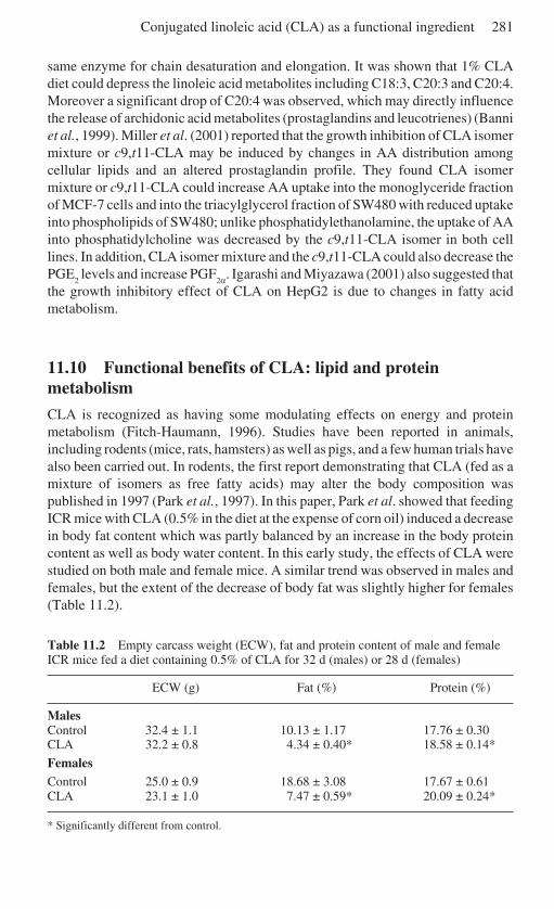
Conjugated linoleic acid (CLA) as a functional ingredient 281
same enzyme for chain desaturation and elongation. It was shown that 1% CLAdiet could depress the linoleic acid metabolites including C18:3, C20:3 and C20:4.Moreover a significant drop of C20:4 was observed, which may directly influencethe release of archidonic acid metabolites (prostaglandins and leucotrienes) (Banniet al., 1999). Miller et al. (2001) reported that the growth inhibition of CLA isomermixture or c9,t11-CLA may be induced by changes in AA distribution amongcellular lipids and an altered prostaglandin profile. They found CLA isomermixture or c9,t11-CLA could increase AA uptake into the monoglyceride fractionof MCF-7 cells and into the triacylglycerol fraction of SW480 with reduced uptakeinto phospholipids of SW480; unlike phosphatidylethanolamine, the uptake of AAinto phosphatidylcholine was decreased by the c9,t11-CLA isomer in both celllines. In addition, CLA isomer mixture and the c9,t11-CLA could also decrease thePGE
2 levels and increase PGF
2α. Igarashi and Miyazawa (2001) also suggested thatthe growth inhibitory effect of CLA on HepG2 is due to changes in fatty acidmetabolism.
11.10 Functional benefits of CLA: lipid and proteinmetabolism
CLA is recognized as having some modulating effects on energy and proteinmetabolism (Fitch-Haumann, 1996). Studies have been reported in animals,including rodents (mice, rats, hamsters) as well as pigs, and a few human trials havealso been carried out. In rodents, the first report demonstrating that CLA (fed as amixture of isomers as free fatty acids) may alter the body composition waspublished in 1997 (Park et al., 1997). In this paper, Park et al. showed that feedingICR mice with CLA (0.5% in the diet at the expense of corn oil) induced a decreasein body fat content which was partly balanced by an increase in the body proteincontent as well as body water content. In this early study, the effects of CLA werestudied on both male and female mice. A similar trend was observed in males andfemales, but the extent of the decrease of body fat was slightly higher for females(Table 11.2).
Table 11.2 Empty carcass weight (ECW), fat and protein content of male and femaleICR mice fed a diet containing 0.5% of CLA for 32 d (males) or 28 d (females)
ECW (g) Fat (%) Protein (%)
MalesControl 32.4 ± 1.1 10.13 ± 1.17 17.76 ± 0.30CLA 32.2 ± 0.8 4.34 ± 0.40* 18.58 ± 0.14*
FemalesControl 25.0 ± 0.9 18.68 ± 3.08 17.67 ± 0.61CLA 23.1 ± 1.0 7.47 ± 0.59* 20.09 ± 0.24*
* Significantly different from control.
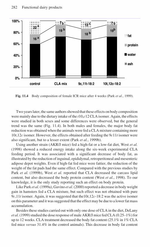
282 Functional dairy products
Fig. 11.4 Body composition of female ICR mice after 4 weeks (Park et al., 1999).
Two years later, the same authors showed that these effects on body compositionwere mainly due to the dietary intake of the t10,c12 CLA isomer. Again, the effectswere studied in both sexes and some differences were observed, but the generaltrend was the same (Fig. 11.4). In both males and females, the major body fatreduction was obtained when the animals were fed a CLA mixture containing more10t,12c isomer. However, the effects obtained after feeding the 9c11t isomer werealso significant, but to a lesser extent (Park et al., 1999b).
Using another strain (AKR/J mice) fed a high-fat or a low-fat diet, West et al.(1998) showed a reduced energy intake along the six-week experimental CLAfeeding period. It was associated with a significant decrease of body fat, asillustrated by the reduction of inguinal, epididymal, retroperitoneal and mesentericadipose depot weights. Even if high-fat fed mice were fattier, the reduction of theweight of the fat pads had the same effect. Compared with the previous studies byPark et al. (1999b), West et al. reported that CLA decreased the carcass lipidcontent, but also decreased the body protein content (West et al., 1998). To ourknowledge, it is the only study reporting such an effect on body protein.
Like Park et al. (1999a), Gavino et al. (2000) reported a decrease in body weightgain in hamsters fed a CLA mixture, but such effect was not obtained with pure9c,11t isomer. Again, it was suggested that the10t,12c-18:2 was the active isomeron this parameter and it was suggested that the effect may be due to a lower fat massaccumulation.
Besides these studies carried out with only one dose of CLA in the diet, DeLanyet al. (1999) studied the dose response of male AKR/J mice fed CLA (0.25–1%) forup to 12 weeks. CLA treatment decreased the body fat content (25.1% in 1% CLAfed mice versus 31.4% in the control animals). This decrease in body fat content
����������������������������������������������������������
��������������������������������������������������������
����������������������������������������������������������
��������������������������������������������������������
�����������������������������������������������������������������������������������������������������������������������������������������������������������������������������������������������������������
������������������������������������������������������������������������������������
��������������������������������������������������������������������������������������������������������������������
������������������������������������������������������������������������������������
16
1918
1922
13 77
0%
20%
40%
60%
80%
100%
control CLA mix 9c,11t-18:2 10t,12c-18:2
�������������� fat
protein
water��������������
ash
abcb
a
bb
b
Conjugated linoleic acid (CLA) as a functional ingredient 283
was observed when CLA was present at least at 0.50% in the diet. As reported byothers (see above), the body fat loss was partly balanced by a significant increaseof protein content and was not associated with any major effect on food intake. Inorder to study the time course of the body composition modifications, a kineticstudy was carried out (Park et al., 1999a). The authors suggested that CLA firstlyincreases the protein content and that the decrease in body fat occurs with a slightdelay.
Whereas most of the data have been obtained in mice, a few studies have beencarried out on other rodents such as rats. Stangl (2000) fed rats submitted to foodrestriction with 3% (by weight) of a CLA mixture at the expense of sunflower oil.CLA-fed animals gained 11% less weight than control animals and they presentedless body fat. The author concluded that CLA acts as a strong repartitioning agentin the Sprague–Dawley male rat by modulating the body composition. Recently,Sisk et al. (2001) showed that feeding CLA reduced the fat pad weight in lean ratsbut the opposite effect was obtained in obese Zucker rats. This difference inresponse was associated with an effect on fat cell size.
Besides the studies carried out in various rodents, pigs have also been studied,since the data on rodents suggested that feed conversion and carcass compositionwould be beneficially affected by CLA treatments. The first study on pigs waspublished in 1997 by Dugan et al. (1997) who reported that feeding CLA with 2%of a CLA mixture in the diet induced a reduction of deposition of subcutaneous fatassociated with an increased lean mass gain. The effect on feed conversion wasborderline significant (P = 0.06). Later, Ostrowska et al. (1999) studied the effectsof increasing CLA intake in female pigs (up to 10 g/kg of diet of a 55% pure CLAmixture). Whereas CLA did not alter weight gain and food intake, feed efficiencywas improved by CLA supplementation. Feeding CLA also decreased fat deposi-tion and improved the lean tissue deposition in finisher pigs. The decrease in fatdeposition was dose-dependent in the range of CLA supplementation studied.
More recently, Wiegand et al. (2001) showed that a commercial CLA mixture(details of isomers not available) significantly improved the feed efficiency from330 g/pkg food to 350 g/pkg food. Rib fat was also significantly decreased. Theseeffects were obtained with only 0.75% of CLA in the pig diet.
Taken altogether, these data in pigs strongly suggest that CLA can be consideredas a repartitioning agent which is also able to improve feed efficiency in finisherpigs. The effect on feed efficiency was not observed in growing pigs (Ramsay etal., 2001) except in the study reported by Dugan et al. (1997). Using a morecomplex CLA mixture, Bee (2000) studied the effects of CLA on pregnant andlactating sows. CLA did not modified the pig weight at birth or at weaning. Thefatty acid profile of milk and back fat of sows was modified, but the effect on thebody composition of the newborn has not been reported.
As overweight and obese subjects are more and more numerous in Westerncountries, CLA has been suggested to be a good candidate for modulating someimbalance occurring during these situations. However, compared with animalstudies, very few trials have been reported (and published as a full paper) inhumans. Blankson et al. (2000) showed that feeding CLA (mixture of isomers, up
284 Functional dairy products
to 6.8 g per day for days) induced a decrease in body fat mass associated with anincrease in lean mass. However, a dose-dependent relationship was difficult toestablish, as no significant effect was observed with an intermediate daily intake(5.1 g/d), which is not clearly understood. On the other hand, the study reported byZambell et al. (2000) did not show any effect on body composition on overweightwomen supplemented for 64 days with a CLA mixture. But Thom et al. (2001)reported that CLA given as a Tonalin® mixture induced a significant decrease inbody fat after three months of CLA intake.
This discrepancy has to be discussed considering the difference in the populationstudied as well as the CLA mixture used. In the first case, the volunteers were menand women with a body mass index (BMI) from 25 to 35, whereas the second studywas carried out only with women presenting a BMI between 22 and 23 (non-overweight subjects). Moreover, the CLA mixture used differed, which means thatthe intake of individual isomers differed. As the CLA isomer profile was alsodifferent, it is difficult to conclude, as some antagonism or synergy betweenisomers cannot be excluded when taking into account the present knowledge.
The effects of CLA on body composition have to be taken with care beforebuilding a conclusion. The effects seem to be species-dependent and the mechanismof action are not fully understood. It seems that CLA may alter fat deposition andalso modifies protein metabolism, but further studies are mandatory to have a goodknowledge of CLA action on body composition. However, growing animals seemto be more responsive than adult organisms.
11.11 The process of CLA metabolism
11.11.1 In animalsAs mentioned before, it has often been suggested that CLA may interact with thearachidonic acid metabolism perhaps by competing with linoleic acid. ConjugatedC18:3, C20:3 and C20:4 fatty acids, as corresponding metabolites, were identifiedin animals fed CLA. In a first study Sébédio et al. (1997) detected conjugatedC20:3 and C20:4 fatty acids in the liver and adipose tissue. Male Wistar rats werefed a fat-free diet for two weeks and afterwards force-fed with a CLA mixturecontaining mainly c9,t11-18:2 and t10,c12-18:2 for six days while maintaining thefat-free diet. Conjugated C20:3 and C20:4 were identified as respective long chainfatty acid metabolites of c9,t11-18:2 and t10,c12-18:2. A second experiment wascarried out by Banni et al. (1999) under normal nutritional conditions. FemaleSprague–Dawley rats were fed a standard diet enriched with CLA mixture for onemonth. They found conjugated C18:3 and C20:3 in liver and mammary tissue, butno conjugated C20:4 was detected. Recent data on this subject using a CLA-enriched butter fat, which contained the natural isomer composition (mainlyc9,t11-18:2), described the formation of conjugated C18:3, C20:3 and C20:4(Banni et al., 2001). The incorporation of the metabolites of CLA were foundprimarily in triacylglycerols. Furthermore, the level of conjugated C20:3 fatty acid
Conjugated linoleic acid (CLA) as a functional ingredient 285
Fig. 11.5 Hypothetical metabolic pathway of 9c,11t-18:2 and 10t,12c-18:2 compared tolinoleic acid.
in the liver was about four times higher than those of conjugated C18:3 and C20:4.Similar results were discussed in a feeding study on rats using pure CLA isomers(Sébédio et al., 2001). For c9,t11-18:2 more conjugated C20:3 than C18:3 werefound in the liver, whereas the inverse effect was reported for 10t,12c-18:2. Thesein vivo results were confirmed by an in vitro ∆6-desaturase study carried out on ratliver microsomes using radiolabeled CLA in isomeric mixture as substrate (Beluryand Kempa-Steczko, 1997). CLAs were converted into C18:3 fatty acids, suggestingthat the metabolites were the respective ∆6-desaturation products of CLA.
These various results reinforce the hypothesis that CLA could be metabolizedin vivo into long chain PUFA using the same pathway as linoleic acid. Thehypothetical metabolic pathway of CLA into conjugated C20:4 is shown in Fig.11.5.
11.11.2 In humansAs other fatty acids, CLA metabolism is shared between three major pathways,incorporation in tissue lipid classes (acylation), oxidation and desaturation/elongation. Again, few studies have been carried out on these aspects in humans.Cawood et al. (1983) showed that CLAs are incorporated in various human fluids,including bile and serum. Breast adipose tissue has also been reported to containsome CLA (Chajes et al., 2002), but extensive data on CLA incorporation inhuman tissues are not available.
We studied the content of CLA isomers and their metabolites in plasma samplescollected during two intervention studies. The first study was the TransLine study(Sébédio et al., 2000). Plasma lipids have been extracted according to Moilanenand Nikkari (1981) and further methylated. Reverse phase HPLC fractionationallowed us to analyse two fractions by GC: the first one contained CLA isomers,as well as other 18:2 fatty acids and 2:3 fatty acids, and the second one contained18:3 and 20:4 moieties. All the samples contained CLA isomers and also someconjugated 20:3 fatty acids, suggesting that humans, like rodents (see above), are
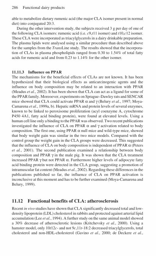
286 Functional dairy products
able to metabolize dietary rumenic acid (the major CLA isomer present in normaldiet) into conjugated 20:3.
During the other intervention study, the subjects received 3 g per day of one ofthe following CLA isomers: rumenic acid (i.e. c9,t11 isomer) and t10,c12 isomer.These CLA were incorporated as triacylglycerols in a dairy drinkable preparation.The plasma lipids were analysed using a similar procedure than described abovefor the samples from the TransLine study. The results showed that the incorpora-tion of CLAs in plasma phospholipids ranged from 0.30 to 1.54% of total fattyacids for rumenic acid and from 0.23 to 1.14% for the other isomer.
11.11.3 Influence on PPARThe mechanisms for the beneficial effects of CLAs are not known. It has beenhypothesized that their biological effects as anticarcinogenic agents and theinfluence on body composition may be related to an interaction with PPAR(Meadus et al., 2002). It has been shown that CLA can act as a ligand for some ofthe PPAR family. Moreoever, experiments on Sprague–Dawley rats and SENCARmice showed that CLA could activate PPAR α and γ (Belury et al., 1997; Moya-Camarena et al., 1999a, b). Hepatic mRNA and protein levels of several enzymes,known to be linked to peroxisome proliferation (acyl coenzyme A, cytochromeP450 4A1, fatty acid binding protein), were found at elevated levels. Using ahuman cell line only a binding to the PPAR was observed. Two recent publicationsinvestigated the influence of CLA on PPAR α and γ activation related to bodycomposition. The first one, using PPAR α null mice and wild-type mice, showedthat body weight gain was similar in the two mice models. Compared with thecontrol group the weight gain in the CLA groups were decreased. This suggestedthat the influence of CLA on body composition is independent of PPAR α (Peterset al., 2001). The second publication examined a relationship between bodycomposition and PPAR γ in the male pig. It was shown that the CLA treatmentincreased PPAR γ but not PPAR α. Furthermore higher levels of adipocyte fattyacid binding protein were detected in the CLA group, suggesting a promotion ofintramuscular fat content (Meadus et al., 2002). Regarding these differences in thepublications published so far, the influence of CLA on PPAR activation isinconclusive at this moment and has to be further examined (Moya-Camarena andBelury, 1999).
11.12 Functional benefits of CLA: atherosclerosis
Recent in vivo studies have shown that CLA significantly decreased total and low-density lipoprotein (LDL) cholesterol in rabbits and protected against arterial lipidaccumulation (Lee et al., 1994). A further study on the same animal model showeda 30% decrease of atherosclerotic lesions (Kritchevsky et al., 2000). Using ahamster model, only 10t12c- and not 9c,11t-18:2 decreased triacylglycerols, totalcholesterol and non-HDL-cholesterol (Gavino et al., 2000; de Deckere et al.,
Conjugated linoleic acid (CLA) as a functional ingredient 287
1999). In the rat model (adult male Sprague–Dawley rats) the feeding of high CLAdiets containing 3 and 5% of CLA mixture in the diet, reductions of LDL and HDLcholesterol were detected (Stangl, 2000). In contrast, Munday et al. (1999)reported an increase of aortic fatty streaks in C57BL/6 mice, but the serumtriacylglycerol level was reduced and the HDL to total cholesterol ratio wasincreased. A six-week study on female swine showed pro-atherosclerotic effectsafter feeding a CLA mixture. Very low-density lipoprotein (VLDL) and LDLcholesterol were increased by CLA. Moreover, the LDL to HDL cholesterol ratiowas significantly up-regulated (Stangl et al., 1999). The influence of CLA onatherosclerosis seems to be controversial and the influence of the animal model onthe observed effects should be further investigated.
Heart disease induced by atherosclerosis is identified as the first case of humanmortality. Therefore, the possible relationship between CLA and atherosclerosis inhumans has a large research interest. A human study was carried out to look ateffects of CLA treatment on plasma lipoproteins and tissue fatty acid composition.In a 63-day study healthy normolipidemic women were supplemented with CLA.Blood cholesterol and lipoprotein levels were not altered by the CLA treatment.9c,11t-18:2 was incorporated in plasma at only 4.23% of ingested CLA. Contraryto animal studies, short-term nutritional trials with CLA in humans did not changelipoprotein levels and seem to be ineffective in the prevention of atherosclerosis(Benito et al., 2001a).
CLAs were also described to induce an antithrombotic effect. The effects ofseveral CLA isomers on human platelet aggregation were examined in vitro (Truittet al., 1999). A CLA mixture was tested in comparison to linoleic acid usingplatelet aggregation agents as arachidonic acid, collagen and thrombin. CLA waseffective to inhibit platelet aggregation. To investigate the influence of CLA onplatelet cyclooxygenase and lipoxygenase activities, formation of [14C]-thromboxane B
2 (TXB
2) and [14C]-12hydroxyeicosatetranoic acid (HETE) was
measured. All tested CLA-isomers inhibited TXB2, while the HETE level was
unchanged. An in vivo experiment on platelet function was carried out in humans.However, the daily intake of 3.9 g of CLA for 63 days had no influence on in vitroplatelet aggregation. The authors concluded that short-term consumption of CLAdid not exhibit antithrombotic properties in humans (Benito et al., 2001b).
11.13 Functional benefits of CLA: immune function
Some studies indicate that CLA could protect against immune-induced cachexia(growth suppression or weight loss). Usually, an enhanced immunological functionleads to a decreased growth. Feeding CLA protected against these catabolic effectsof such an immune stimulation (Cook and Pariza, 1998). Miller et al. (1994)described how dietary CLA in mice prevent endotoxin-induced growth suppressioncompared to a control group. The authors hypothesized a relationship betweenCLA and possible changes in the interleukin-1 level. The capacity of CLA tochange interleukin production was later observed by Hayek et al. (1999).
288 Functional dairy products
CLA may also influence the allergic reaction pathways, by alteration of serumimmunoglobulins (Ig). IgA, IgG and IgM were increased, whereas IgE wasdecreased (Sugano et al., 1998). IgA, IgG and IgM are implicated in the defensemetabolism against virus or bacteria. As IgE is related to allergic reactions, itsdiminution is desired.
The influence of CLA on the immunological function may have implicationsfor human health. A study in humans examined possible changes in immune statusby testing the number of circulating white blood cells, granulocytes, monocytes,lymphocytes and lymphocyte proliferation. No changes in the investigated param-eters were observed (Kelley et al., 2000). In another experiment the specificantibody production after hepatitis B vaccination was tested depending on a CLAtreatment. The antibody formation after CLA-treatment (9c,11t-18:2/10t,12c-18:2; 50/50) was significantly higher (Mohede et al., 2001), suggesting that CLAcould be able to enhance immune function in humans.
11.14 Functional benefits of CLA: diabetes
The discussion concerning antidiabetic effects of CLA is controversial. Anexperiment using Zucker diabetic fatty rats showed a normalization of impairedglucose tolerance and improved hyperinsulinemia induced by CLA (Houseknechtet al., 1998). To get information about the active isomer, a second experiment wascarried out with the same animal model using CLA mixture and a 9c,11t-18:2enriched butter (90.5% 9c,11t-18:2 of total CLA). Only the CLA mixture, not9c,11t-18:2, reduced improved glucose tolerance. Furthermore, an improvedinsulin action in muscle was observed (Ryder et al., 2001).
A recent study showed inverse effects. C57BL/6J mice fed a CLA-enriched dietdeveloped a state resembling lipoatrophic diabetes, with a marked insulin resistance(hyperinsulinemia) (Tsuboyama-Kasaoka et al., 2000). The hyperinsulinemia wasreversible by continuous infusion with leptin, suggesting leptin may be antagonisticto the CLA-induced effects.
Only a few studies have been carried out on possible antidiabetic effects of CLAusing different animal models. One reason for the contrary results reported abovecould be a difference in the metabolic pathways of the two animal species.
Type II diabetes (diabetes induced by insulin resistance) represents one ofthe diseases of the affluent society and at the moment very little is known aboutthe effect of CLA on insulin resistance in humans. A nutritional study was carriedout on overweight middle-aged men with insulin resistance syndrome. Onegroup received a CLA mixture and the other one the 10t,12c-18:2 (Riserus andVessby, 2001). Compared with the control group, glucose metabolism remainedunchanged for the CLA mixture trial. The treatment with pure 10t,12c-18:2increased the insulin resistance and glycemia. It can be concluded that thetwo different CLA treatments showed diverging effects. The influence of CLA ontype II diabetes is not clear, therefore more results concerning this subject areneeded.
Conjugated linoleic acid (CLA) as a functional ingredient 289
11.15 Conclusion and future trends
The increase of CLA levels in food or the creation of functional foods containinghigh amounts of CLA may be beneficial for human health because of theirbiological activities discussed above. Current data exist predominantly fromexperiments on animals, where CLA showed high anticarcinogenic effects andproduced strong evidence for the influence on body composition in the growinganimal. Moreover CLA may have beneficial effects for cardiovascular diseases, asthey showed a lipid lowering potential. Currently, experiments on humans haveinvesttigated mainly the influence on body composition and the results obtainedremain controversial. This may be related to different subjects in the studies andthe use of CLA mixtures of variable isomer compositions. Furthermore, all humanstudies have been short-term experiments.
More experiments using isolated isomers to evaluate the effects of each CLAisomer, and long-term intervention studies, are needed. In addition, the toxicologicalrisks have to be evaluated, as little data are available on safety aspects of a CLAsupplementation.
11.16 ReferencesADLOF R (1997), Preparation of methyl cis-9,trans-11- and trans-9,trans-11-octadecadienoate-
17,17,18,18-d4, two of the isomers of conjugated linoleic acid, Chem Phys Lipids, 88,107–112.
ADLOF R (1999), The lindlar-catalyzed reduction of methyl santalbate: a facile preparationof methyl 9-cis,11-trans-octadecadienoate-9,10-d2, J Am Oil Chemists Soc, 76, 301–304.
BANNI S, CARTA G, CONTINI M S, ANGIONI E, DEIANA M, DESSI M A, MELIS M P and CORONGIUF P (1996), Characterization of conjugated diene fatty acids in milk, dairy products, andlamb tissues, J Nutr Biochem, 7, 150–155.
BANNI S, ANGIONI E, CASU V, MELIS M P, CARTA G, CORONGIU F P, THOMPSON H and IP C(1999), Decrease in linoleic acid metabolites as a potential mechanism in cancer riskreduction by conjugated linoleic acid, Carcinogenesis, 20, 1019–1024.
BANNI S, CARTA G, ANGIONI E, MURRU E, SCANU P, MELIS M P, BAUMAN D E, FISCHER S M andIP C (2001), Distribution of conjugated linoleic acid and metabolites in different lipidfractions in the rat liver, J Lipid Res, 42, 1056–1061.
BASCETTA E, GUNSTONE F D and SCRMGEOUR C M (1984), Synthesis, characterisation, andtransformation of a lipid cyclic peroxide, J Chem Soc Perkin Trans I, 2199–2205.
BAUMGARD L H, SANGSTER J K and BAUMAN D E (2001), Milk fat synthesis in dairy cows isprogressively reduced by increasing supplemental amounts of trans-10, cis-12 conjugatedlinoleic acid (CLA), J Nutr, 131, 1764–1769.
BEE G (2000), Dietary conjugated linoleic acids alter adipose tissue and milk lipids ofpregnant and lactating sows, J Nutr, 130, 2292–2298.
BELURY M A and KEMPA-STECZKO A (1997), Conjugated linoleic acid modulates hepatic lipidcomposition in mice, Lipids, 32, 199–204.
BELURY M A, NICKEL K P, BIRD C E and WU Y (1996), Dietary conjugated linoleic acidmodulation of phorbol ester skin tumor promotion, Nutr Cancer, 26, 149–157.
BELURY M A, MOYA-CAMARENA S Y, LIU K L and HEUVEL J P V (1997), Dietary conjugatedlinoleic acid induces peroxisome-specific enzyme accumulation and ornithine decar-boxylase activity in mouse liver, J Nutr Biochem, 8, 579–584.
BENITO P, NELSON G J, KELLEY D S, BARTOLINI G, SCHMIDT P C and SIMON V (2001a), The
290 Functional dairy products
effect of conjugated linoleic acid on plasma lipoproteins and tissue fatty acid compositionin humans, Lipids, 36, 229–236.
BENITO P, NELSON G J, KELLEY D S, BARTOLINI G, SCHMIDT P C and SIMON V (2001b), Theeffect of conjugated linoleic acid on platelet function, platelet fatty acid composition, andblood coagulation in humans, Lipids, 36, 221–227.
BERDEAUX O, CHRISTIE W W, GUNSTONE F D and SÉBÉDIO J L (1997), Large-scale synthesisof methyl cis-9,trans-11-octadecadienoate from methyl ricinoleate, J Am Oil Chem Soc,74, 1011–1015.
BLANKSON H, STAKKESTAD J A, FAGERTUN H, THOM E, WADSTEIN J and GUDMUNDSEN 0(2000), Conjugated linoleic acid reduces body fat mass in overweight and obese humans,J Nutr, 130, 2943–2948.
CAWOOD P, WICKENS D G, IVERSEN S A, BRAGANZA J M and DORMANDY T L (1983), The natureof diene conjugation in human serum, bile and duodenal juice, FEBS Lett, 162, 239–243.
CESANO A, VISONNEAU S, SCIMECA J A, KRITCHEVSKY D and SANTOLI D (1998), Oppositeeffects of linoleic acid and conjugated linoleic acid on human prostatic cancer in SCIDmice, Anticancer Res, 18, 1429–1434.
CHAJES V, LAVILLONNIERE F, FERRARI P, JOURDAN M L, PINAULT M, MAILLARD V, SEBEDIOJ L and BOUGNOUX P (2002), Conjugated linoleic acid content in breast adipose tissue isnot associated with the relative risk of breast cancer in a population of French patients,Cancer Epidemiol Biomarkers Prev, 11, 672–673.
CHIN S F, LIU W, STORKSON J M, HA Y L and PARIZA M W (1992), Dietary sources of conjugateddienoic isomers of linoleic acid, a newly recognized class of anticarcinogens, J FoodComp Anal, 5, 185–197.
CHIPAULT J R and HAWKINS J M (1959), The determination of conjugated cis–trans and trans–trans methyl octadecadienoates by infrared spectrometry, J Am Oil Chem Soc, 36,535–539.
CHOI Y, PARK Y, PARIZA M W and NTAMBI J M (2001), Regulation of stearoyl-CoA desaturaseactivity by the trans-10,cis-12 isomer of conjugated linoleic acid in HepG2 cells, BiochemBiophys Res Commun, 284, 689–693.
CHOI Y, PARK Y, STORKSON J M, PARIZA M W and NTAMBI J M (2002), Inhibition of stearoyl-CoA desaturase activity by the cis-9,trans-11 isomer and the trans-10,cis-12 isomer ofconjugated linoleic acid in MDA-MB-231 and MCF-7 human breast cancer cells,Biochem Biophys Res Commun, 294, 785–790.
CHOUINARD P Y, CORNEAU L, SAEBO A and BAUMAN D E (1999), Milk yield and compositionduring abomasal infusion of conjugated linoleic acids in dairy cows, J Dairy Sci, 82, 2737–2745.
CHOUINARD P Y, CORNEAU L, BUTLER W R, CHILLIARD Y, DRACKLEY J K and BAUMAN D E(2001), Effect of dietary lipid source on conjugated linoleic acid concentrations in milkfat, J Dairy Sci, 84, 680–690.
CHRISTIE W W (1997), Isomers in commercial samples of conjugated linoleic acid, J Am OilChem Soc, 74, 1231.
CHRISTIE W W (2001), in R J Hamilton, ‘CLA is focus for SCI seminar’, INFORM, 12, 629–633.
CHRISTIE W W, SÉBÉDIO J L and JUANEDA P (2001), A practical guide to the analysis ofconjugated linoleic acid, INFORM, 12, 147–152.
COOK M E and PARIZA M (1998), The role of conjugated linoleic acid (CLA) in health, IntDairy J, 8, 459–462.
CORL B, BAUMGARD L, GRIINARI J, DELMONTE P, MOREHOUSE K, YURAWECZ M andBAUMAN D (2002), trans-7,cis9 CLA is endogenously synthesized by D-9-desaturation inlactating dairy cows, AOCS annual meeting, Montreal.
CUNNINGHAM D C, HARRISON L Y and SCHULTZ T D (1997), Proliferative responses of normalhuman mammary and MCF-7 breast cancer cells to linoleic acid, conjugated linoleic acidand eicosanoid synthesis inhibitors in culture, Anticancer Research, 17, 197–204.
DE DECKERE E A M, VAN AMELSVOORT J M M, MCNEILL G P and JONES P (1999), Effects of
Conjugated linoleic acid (CLA) as a functional ingredient 291
conjugated linoleic acid (CLA) isomers on lipid levels and peroxisome proliferation in thehamster, Br J Nutr, 82, 309–317.
DELANY J P, BLOHM F, TRUETT A A, SCIMECA J A and WEST D B (1999), Conjugated linoleicacid rapidly reduces body fat content in mice without affecting energy intake, Am JPhysiol Reg Integrative Comp Physiol, 45, R1172–R1179.
DHIMAN T R, ANAND G R, SATTER L D and PARIZA M W (1996), Conjugated linoleic acidcontent of milk from cows fed different diets, J Dairy Sci, 79 (Suppl. 1), 35.
DHIMAN T R, HELMINK E D, MCMAHON D J, FIFE R L and PARIZA M W (1999), Conjugatedlinoleic acid content of milk and cheese from cows fed extruded oilseeds, J Dairy Sci, 82,412–419.
DOBSON G (1997), Identification of conjugated fatty acids by gas chromatography-massspectrometry of 4-methyl-1,2,4-triazoline-3,5-dione adducts, J Am Oil Chem Soc, 75,137–142.
DOBSON G and CHRISTIE W W (1996), Structural analysis of fatty acids by mass spectrometryof picolinyl esters and dimethyloxazoline derivatives, Trends in Analytical Chemistry, 15,130–137.
DONOVAN D C, SCHINGOETHE D J, BAER R J, RYALI J, HIPPEN A R and FRANKLIN S T (2000),Influence of dietary fish oil on conjugated linoleic acid and other fatty acids in milk fatfrom lactating dairy cows, J Dairy Sci, 83, 2620–2628.
DUGAN M E R, AALHUS, J L, SCHAEFER A L and KRAMER J K G (1997), The effect of conjugatedlinoleic acid on fat to lean repartitioning and feed conversion in pigs, Can J Anim Sci, 77,723–725.
DURGAM V R and FERNANDES G (1997), The growth inhibitory effect of conjugated linoleicacid on MCF-7 cells is related to estrogen response system, Cancer Lett, 116, 121–130.
FITCH-HAUMANN B (1996), Conjugated linoleic acid offers research promise, INFORM, 7,152–159.
FRITSCHE J and STEINHART H (1998a), Analysis, occurrence, and physiological properties oftrans fatty acids (TFA) with particular emphasis on conjugated linoleic acid isomers(CLA) – a review, Fett/Lipid, 100, 190–210.
FRITSCHE J and STEINHART H (1998b), Amounts of conjugated linoleic acid (CLA) in germanfoods and evaluation of daily intake, Z Lebensm Unters Forsch A, 206, 77–82.
FRITSCHE J, MOSSOBA M M, YURAWECZ M P, ROACH J A G, SEHAT N, KU Y and STEINHART H(1997), Conjugated linoleic acid (CLA) isomers in human adipose tissue, Z LebensmUnters Forsch A, 205, 415–418.
FUTAKUCHI M, CHENG J L, HIROSE M, KIMOTO N, CHO Y M, IWATA T, KASAI M, TOKUDOMES and SHIRAI T (2002), Inhibition of conjugated fatty acids derived from safflower orperilla oil of induction and development of mammary tumors in rats induced by 2-amino-1-methyl-6-phenylimidazo[4,5-b]pyridine (PhIP), Cancer Lett, 178, 131–139.
GAVINO V C, GAVINO G, LEBLANC M J and TUCHWEBER B (2000), An isomeric mixture ofconjugated linoleic acids but not pure cis-9,trans-11-octadecadienoic acid affects bodyweight gain and plasma lipids in hamsters, J Nutr, 130, 27–29.
GEORGER M M and HUDSON B S (1988), Synthesis of all-trans-parinaric acid-d8 specificallydeuteriated at all vinyl positions, J Org Chem, 53, 3148–3153.
GNÄDIG S (1996), Konjugierte Linolsäureisomere in Lebensmittlen, humanem Fettgewebeund humanem Blutplasma, Diplomarbeit, Fachbereich Chemie, Inst. für Biochemie undLebensmittelchemie, Abt. Lebensmittelchemie, Universität Hamburg, Hamburg.
GNÄDIG S, BERDEAUX O, LOREAU O, NOEL J P and SEBEDIO J L (2001), Synthesis of(6Z,9Z,11E)-octadecatrienoic and (8Z,11Z,13E)-eicosatrienoic acids and their [1-(14)C]-radiolabeled analogs, Chem Phys Lipids, 112, 121–135.
GRIINARI J M, CORI B A, LACY S H, CHOUINARD P Y, NURMELA K V V and BAUMAN D E (2000),Conjugated linoleic acid is synthesized endogenously in lactating dairy cows by Delta(9)-desaturase, J Nutr, 130, 2285–2291.
GUNSTONE F D and SAID A I (1971), Methyl 12-mesyloxyoleate as a source of cyclopropaneesters and of conjugated octadecadienoates, Chem Phys Lipids, 7, 121–134.
292 Functional dairy products
HA Y L , GRIMM N K and PARIZA M W (1987), Anticarcinogens from fried ground beef: heat-altered derivatives of linoleic acid, Carcinogenes, 8, 1881–1887.
HA, Y L, STORKSON J and PARIZA M W (1990), Inhibition of benzo(a)pyrene-induced mouseforestomach neoplasia by conjugated dienoic derivatives of linoleic acid, Cancer Res, 50,1097–1101.
HAMILTON R J (2001), CLA is focus for SCI seminar, INFORM, 12, 629–633.harfoot c g and hazelwood g p (1988), Lipid metabolism in the rumen, in P Hobson, The
Rumen Microbial Ecosystem, London, Elsevier Science Publishers, 285–322.HAYEK M G, HAN S N, WU D Y, WATKINS B A, MEYDANI M, DORSEY J L, SMITH D E and MEYDANI
S N (1999), Dietary conjugated linoleic acid influences the immune response of young andold C57BL/6NCrlBR mice, J Nutr, 129, 32–38.
HOUSEKNECHT K L, VANDENHEUVEL J P, MOYACAMARENA S Y, PORTOCARRERO C P, PECK LW, NICKEL K P and BELURY M A (1998), Dietary conjugated linoleic acid normalizesimpaired glucose tolerance in the Zucker diabetic fatty fa/fa rat, Biochem Biophys ResComm, 244, 678–682.
HUBBARD N E, LIM D, SUMMERS L and ERICKSON K L (2000), Reduction of murine mammarytumor metastasis by conjugated linoleic acid, Cancer Lett, 150, 93–100.
IGARASHI M and MIYAZAWA T (2001), The growth inhibitory effect of conjugated linoleicacid on a human hepatoma cell line, HepG2, is induced by a change in fatty acidmetabolism, but not the facilitation of lipid peroxidation in the cells, Biochim BiophysActa, 1530, 162–171.
IP C, CHIN S F, SCIMECA J A and PARIZA M W (1991), Mammary cancer prevention byconjugated dienoic derivative of linoleic acid, Cancer Res, 51, 6118–6124.
IP C, SINGH M, THOMPSON H J and SCIMECA J A (1994), Conjugated linoleic acid suppressesmammary carcinogenesis and proliferative activity of the mammary gland in the rat,Cancer Res, 54, 1212–1215.
IP C, SCIMECA J A and THOMPSON H (1995), Effect of timing and duration of dietaryconjugated linoleic acid on mammary cancer prevention, Nutr Cancer, 24, 241–247.
IP C, BRIGGS S P, HAEGELE A D, THOMPSON H J, STORKSON J and SCIMECA J A (1996), Theefficacy of conjugated linoleic acid in mammary cancer prevention is independent of thelevel or type of fat in the diet, Carcinogenesis, 17, 1045–1050.
IP C, JIANG C, THOMPSON H J and SCIMECA J A (1997), Retention of conjugated linoleic acidin the mammary gland is associated with tumor inhibition during the post-initiation phaseof carcinogenesis, Carcinogenesis, 18, 755–759.
IP C, BANNI S, ANGIONI E, CARTA G, MCGINLEY J, THOMPSON H, BARBANO D and BAUMAN D(1999), Conjugated linoleic acid-enriched butter fat alters mammary gland morphogen-esis and reduces cancer risk in rats, J Nutr, 129, 2135–2142.
IP C, IP M, LOFTUS T, SHOEMAKER S and SHEA-EATON W (2000), Induction of apoptosis byconjugated linoleic acid in cultured mammary tumor cells and premalignant lesions of therat mammary gland, Cancer Epidemiol Biomarkers Prevent, 9, 689–696.
JAHREIS G, FRITSCHE J and STEINHART H (1996), Monthly variation of milk composition withspecial regard to fatty acids depending on season and farm management systems –convential versus ecological, Fett/Lipid, 98, 356–359.
JAHREIS G, FRITSCHE J and STEINHART H (1997), Conjugated linoleic acid in milk fat – highvariation depending on production system, Nutr Res, 17, 1479–1484.
JAHREIS G, FRITSCHE J and KRAFT J (1999), Species-dependent, seasonal, and dietaryvariation of conjugated linoleic acid in milk. In: M P Yurawecz, M M Mossoba, J K GKramer, M W Pariza and G J Nelson (Eds), Advances in Conjugated Linoleic AcidResearch, Vol. 1, Champaign, IL, AOCS Press, 215–225.
JIANG J, BJOERCK L, FONDÉN R and EMANUELSON M (1996), Occurrence of conjugated cis-9,trans-11-octadecadienoic acid in bovine milk: effects of feed and dietary regimen, JDairy Sci, 79, 438–445.
JIANG J, BJORCK L and FONDEN R (1997), Conjugated linoleic acid in Swedish dairy productswith special reference to the manufacture of hard cheese, Int Dairy J, 7, 863–867.
Conjugated linoleic acid (CLA) as a functional ingredient 293
JIANG J, BJORCK L and FONDEN R (1998), Production of conjugated linoleic acid by dairystarter cultures, J Appl Microbiol, 85, 95–102.
JONES D F, WEISS W P and PALMQUIST D L (2000), Short communication: influence of dietarytallow and fish oil on milk fat composition, J Dairy Sci, 83, 2024–2026.
JOSYULA S and SCHUT H A (1998), Effects of dietary conjugated linoleic acid on DNA adductformation of PhIP and IQ after bolus administration to female F344 rats, Nutr Cancer, 32,139–145.
JOSYULA S, HE Y H, RUCH R J and SCHUT H A (1998(, Inhibition of DNA adduct formation ofPhIP in female F344 rats by dietary conjugated linoleic acid, Nutr Cancer, 32, 132–138.
JUANEDA P, CORDIER O, GREGOIRE S and SEBEDIO J L (2001), Conjugated linoleic acid (CLA)isomers in heat-treated vegetable oils, OCL, 8, 94–97.
JUNG M O, YOON S H and JUNG M Y (2001), Effects of temperature and agitation rate on theformation of conjugated linoleic acids in soybean oil during hydrogenation process, JAgric Food Chem, 49, 3010–3016.
JUNG M Y and HA Y L (1999), Conjugated linoleic acid isomers in partially hydrogenatedsoybean oil obtained during nonselective and selective hydrogenation processes, J AgricFood Chem, 47, 704–708.
KELLEY D S, TAYLOR P C, RUDOLPH I L, BENITO P, NELSON G J, MACKEY B E and ERICKSON
K L (2000), Dietary conjugated linoleic acid did not alter immune status in young healthywoman, Lipids, 35, 1065–1071.
KELLY M L, BERRY J R, DWYER D A, GRIINARI J M, CHOUINARD P Y, VANAMBURGH M E andBAUMAN D E (1998), Dietary fatty acid sources affect conjugated linoleic acid concentra-tions in milk from lactating dairy cows, J Nutr, 128, 881–885.
KEPLER C R and TOVE S B (1967), Biohydrogenation of unsaturated fatty acids, J Biol Chem,242, 5686–5692.
KEPLER C R, HIRONS K P, MCNEIL J J and TOVE S B (1966), Intermediates and products of thebiohydrogenation of linoleic acid by Butyrivibrio fibrisolvens, J Biol Chem, 241, 1350–1354.
KIMOTO N, HIROSE M, FUTAKUCHI M, IWATA T, KASAI M and SHIRAI T (2001), Site-dependentmodulating effects of conjugated fatty acids from safflower oil in a rat two-stagecarcinogenesis model in female Sprague–Dawley rats, Cancer Lett, 168, 15–21.
KOHNO H, SUZUKI R, NOGUCHI R, HOSOKAWA M, MIYASHITA K and TANAKA T (2002), Dietaryconjugated linoleic acid inhibits azoxymethane-induced colonic aberrant crypt foci inrats, Jpn J Cancer Res, 93, 133–142.
KRAMER J K C, FELLNER V, DUGAN M E R, SAUER F D, MOSSOBA M M and YURAWECZ M P(1997), Evaluating acid and base catalysts in the methylation of milk and rumen fatty acidswith special emphasis on conjugated dienes and total trans fatty acids, Lipids, 32, 1219–1228.
KRAMER J K C, PARODI P W, JENSEN R G, MOSSOBA M M, YURAWECZ M P and ADLOF R O (1998),Rumenic acid: a proposed common name for the major conjugated linoleic acid isomerfound in natural products, Lipids, 33, 835.
KRITCHEVSKY D (2000), Antimutagenic and some other effects of conjugated linoleic acid,Br J Nutr, 83, 459–465.
KRITCHEVSKY D, TEPPER S A, WRIGHT S, TSO P and CZARNECKI S K (2000), Influence ofconjugated linoleic acid (CLA) on establishment and progression of atherosclerosis inrabbits, J Am Coll Nutr, 19, 472S–477S.
LABELLE M, FALGUEYRET J P, RIENDEAU D and ROKACH J (1990), Synthesis of two analoguesof arachidonic acid and their reactions with 12-lipoxygenase, Tetrahedron, 46, 6301–6310.
LAVILLONNIÈRE F, MARTIN J C, BOUGNOUX P and SÉBÉDIO J L (1998), Analysis of conjugatedlinoleic acid isomers and content in French cheeses, J Am Oil Chem Soc, 75, 343–352.
LEE K N, KRITCHEVSKY D and PARIZA M W (1994), Conjugated linoleic acid and atheroscle-rosis in rabbits, Atherosclerosis, 108, 19–25.
LEHMANN L (2001), Identifizierung und stereoselektive Synthesen ungesättigter Signalstoffe,
294 Functional dairy products
Doktorarbeit, Fachbereich Chemie, Inst. für Organische Chemie, Universität Hamburg,Hamburg.
LEUNG Y H and LIU R H (2000), trans-10,cis-12-Conjugated linoleic acid isomer exhibitsstronger oxyradical scavenging capacity than cis-9,trans-11-conjugated linoleic acidisomer, J Agric Food Chem, 48, 5469–5475.
LIE KEN JIE M S F and WONG K P (1992), Dehydration reactions involving methyl 9,12-dihydroxy-10-octadecenoate, Chem Phys Lipids, 62, 177–183.
LIE KEN JIE M S F, KHYSAR PASHA M and AHMAD F (1996), Ultrasound-assisted synthesis ofsantalbic acid and a study of triacylglycerol species in Santalum album (Linn.) seed oil,Lipids, 31, 1083–1089.
LIE KEN JIE M S F, KHYSAR PASHA M and SHAHIN ALAM M (1997), Synthesis and nuclearmagnetic resonance properties of all geometrical isomers of conjugated linoleic acids,Lipids, 32, 1041–1044.
LIEW C, SCHUT H A J, CHIN S F, PARIZA M W and DASHWOOD R H (1995), Protection ofconjugated linoleic acid against 2-amino 3-methylimido[4,5-F] quinoline-induce coloncarcinogenesis in the F344 rat: a study of inhibitory mechanisms, Carcinogenesis, 16,3037–3044.
LIN H, BOYLSTON T D, LUEDECKE L O and SHULTZ T D (1998), Factors affecting the conjugatedlinoleic acid content of cheddar cheese, J Agric Food Chem, 46, 801–807.
LIN H, BOYLSTON T D, LUEDECKE L O and SHULTZ T D (1999b), Conjugated linoleic acidcontent of cheddar-type cheeses as affected by processing, J Food Sci, 64, 874–878.
LIN T Y (2000), Conjugated linoleic acid concentration as affected by lactic cultures andadditives, Food Chem, 69, 27–31.
LIN T Y, LIN C W and LEE C H (1999a), Conjugated linoleic acid concentration as affected bylactic cultures and added linoleic acid, Food Chem, 67, 1–5.
LIU J R, LI B X, CHEN B Q, HAN X H, XUE Y B, YANG Y M, ZHENG Y M and LIU R H (2002), Effectof cis-9, trans-11-conjugated linoleic acid on cell cycle of gastric adenocarcinoma cellline (SGC-7901), World J Gastroenterol, 8, 224–229.
LOOR J J and HERBEIN J H (1998), Exogenous conjugated linoleic acid isomers reduce bovinemilk fat concentration and yield by inhibiting de novo fatty acid synthesis, J Nutr, 128,2411–2419.
LOREAU O, MARET A, CHARDIGNY J M, SÉBÉDIO J L and NOEL J P (2001), Sequentialsubstitution of 1,2-dichloro-ethene: a convenient stereoselective route to (9Z,11E)-,(10E,12Z)- and (10Z,12Z)-[1-14C] conjugated linoleic acid isomers, Chem Phys Lipids,110, 57–67.
MEADUS W J, MACINNIS R and DUGAN M E (2002), Prolonged dietary treatment withconjugated linoleic acid stimulates porcine muscle peroxisome proliferator activatedreceptor gamma and glutamine-fructose aminotransferase gene expression in vivo, J MolEndocrinol, 28, 79–86.
MILLER A, STANTON C and DEVERY R (2001), Modulation of arachidonic acid distribution byconjugated linoleic acid isomers and linoleic acid in MCF-7 and SW480 cancer cells,Lipids, 36, 1161–1168.
MILLER C C, PARK Y M W P and COOK M E (1994), Feeding conjugated linoleic acid to animalspartially overcomes catabolic responses due to endotoxin injection, Biochem Biophys ResCommun, 198, 1107–1112.
MOHEDE I, ALBERS R, VAN DER WIELEN R, BRINK L and DOROVSKA-TARAN V (2001),Immuno-modulation: CLA stimulates antigen specific antibody production in humans,1st International Conference on Conjugated Linoleic Acid (CLA), Ålesund, Norway.
MOILANEN T and NIKKARI T (1981), The effect of storage on the fatty acid composition ofhuman serum, Clin Chim Acta, 114, 111–116.
MORRIS S G, MAGIDMAN P and HERB S F (1972), Hydrogenation of triple bonds to doublebonds in conjugated methyl octadecadiynoate and methyl santalbate, J Am Oil Chem Soc,49, 505–507.
MOSSOBA M M, KRAMER J K G, YURAWECZ M P, SEHAT N, ROACH J A G, EULITZ K, FRITSCHE
Conjugated linoleic acid (CLA) as a functional ingredient 295
J, DUGAN M E R and KU Y (1999), Impact of novel methodologies on the analysis ofconjugated linoleic acid (CLA). Implications of CLA feeding studies, Fett/Lipid, 101,235–243.
MOYA-CAMARENA S Y and BELURY M A (1999), Species differences in the metabolism andregulation of gene expression by conjugated linoleic acid, Nutr Rev, 57, 336–340.
MOYA-CAMARENA S Y, VANDEN HEUVEL J P and BELURY M A (1999a), Conjugated linoleicacid activates peroxisome proliferator-activated receptor α and β subtypes but does notinduce hepatic peroxisome proliferation in Sprague–Dawley rats, Biochim Biophys Acta,1436, 331–342.
MOYA-CAMARENA S Y, VANDEN HEUVEL J P, BLANCHARD S G, LEESNITZER L A and BELURYM A (1999b), Conjugated linoleic acid is a potent naturally occurring ligand and activatorof PPARα, J Lipid Res, 40, 1426–1433.
MUNDAY J S, THOMPSON K G and JAMES K A C (1999), Dietary conjugated linoleic acidspromote fatty streak formation in the C57BL/6 mouse atherosclerosis model, Br J Nutr,81, 251–255.
O’SHEA M, LAWLESS F, STANTON C and DEVERY R (1998), Conjugated linoleic acid in bovinemilk fat: a food-based approach to cancer chemoprevention, Trends Food Sci Technol, 9,192–196.
O’SHEA M, STANTON C and DEVERY R (1999), Antioxidant enzyme defence responses ofhuman MCF-7 and SW480 cancer cells to conjugated linoleic acid. Anticancer Res, 19,1953–1959.
O’SHEA M, DEVERY R, LAWLESS F, MURPHY J and STANTON C (2000), Milk fat conjugatedlinoleic acid inhibits growth of human mammary MCF-7 cancer cells, Anticancer Res, 20,3591–3602.
OSTROWSKA E, MURALITHARAN M, CROSS R F, BAUMAN D E and DUNSHEA F R (1999), Dietaryconjugated linoleic acids increase lean tissue and decrease fat deposition in growing pigs,J Nutr, 129, 2037–2042.
PALOMBO J D, GANGULY A, BISTRIAN B R and MENARD M P (2002), The antiproliferativeeffects of biologically active isomers of conjugated linoleic acid on human colorectal andprostatic cancer cells, Cancer Lett, 177, 163–172.
PARIZA M W (1997), Conjugated linoleic acid, a newly recognised nutrient, Chem Ind, 464–466.
PARIZA M W and HARGRAVES W A (1985), A beef-derived mutagenesis modulator inhibitsinitiation of mouse epidermal tumors by 7,12-dimethylbenz[a]anthracene, Carcinogen-esis, 6, 591–593.
PARIZA M, PARK Y and COOK M (1999), Conjugated linoleic acid and the control of cancerand obesity, Toxicol Sci, 52 (Suppl), 107–110.
PARK H S, RYU J H, HA Y L and PARK J H (2001a), Dietary conjugated linoleic acid (CLA)induces apoptosis of colonic mucosa in 1,2-dimethylhydrazine-treated rats: a possiblemechanism of the anticarcinogenic effect by CLA, Br J Nutr, 86, 549–555.
PARK Y, ALBRIGHT K J, LIU W, STORKSON J M, COOK M E and PARIZA M W (1997), Effect ofconjugated linoleic acid on body composition in mice, Lipids, 32, 853–858.
PARK Y, ALBRIGHT K J, STORKSON J M, LIU W, COOK M E and PARIZA M W (1999a), Changesin body composition in mice during feeding and withdrawal of conjugated linoleic acid,Lipids, 34, 243–248.
PARK Y, STORKSON J M, ALBRIGHT K J, LIU W and PARIZA M W (1999b), Evidence that thetrans-10,cis-12 isomer of conjugated linoleic acid induces body composition changes inmice, Lipids, 34, 235–241.
PARK Y, ALLEN K and SHULTZ T (2000), Modulation of MCF-7 breast cancer cell signaltransduction by linoleic acid and conjugated linoleic acid in culture, Anticancer Res, 20,669–676.
PARK Y, ALBRIGHT K J, CAI Z Y and PARIZA M W (2001b), Comparison of methylationprocedures for conjugated linoleic acid and artifact formation by commercial(trimethylsilyl)diazomethane, J. Agric. Food Chem., 49, 1158–1164.
296 Functional dairy products
PETERS J M, PARK Y, GONZALEZ F J and PARIZA M W (2001), Influence of conjugated linoleicacid on body composition and target gene expression in peroxisome proliferator-activatedreceptor alpha-null mice, Biochim Biophys Acta, 1533, 233–242.
PRECHT D, MOLKENTIN J and VAHLENDICK M (1999), Influence of the heating temperature onthe fat composition of milk fat with the emphasis on cis-/trans-isomerization, Nahrung/Food, 43, 25–33.
RAMSAY T G, EVOCK-CLOVER C M, STEELE N C and AZAIN M J (2001), Dietary conjugatedlinoleic acid alters fatty acid composition of pig skeletal muscle and fat, J Anim Sci, 79,2152–2161.
REANEY M J T, LIU Y-D and WESTCOTT N D (1999), Commercial production of conjugatedlinoleic acid. In: M P Yurawecz, M M Mossoba, J K G Kramer, M W Pariza and G J Nelson(Eds), Advances in Conjuged Linuleic Acid Research, Vol. 1, Champaign, IL, AOCSPress, 39–54.
RICKERT R and STEINHART H (2001), Bedeutung, Analytik sowie Vorkommen von konjugiertenLinolsäureisomeren (CLA) in Lebensmitteln, Ernährungs-Umschau, 48, 4–7.
RISERUS U and VESSBY B (2001), Diverging effects of CLA isomers on insulin resistance andlipid metabolsim in obese men with metabolic syndrome, 1st International Conference onConjugated Linoleic Acid (CLA), Ålesund, Norway.
YYDER J W, PORTOCARRERO C P, SONG X M, CUI L, YU M, COMBATSIARIS T, GALUSKA D,BAUMAN D E, BARBANO D M, CHARRON M J, ZIERATH J R and HOUSEKNECHT K L (2001),Isomer-specific antidiabetic properties of conjugated linoleic acid – improved glucosetolerance, skeletal muscle insulin action, and UCP-2 gene expression, Diabetes, 50, 1149–1157.
SCHNEIDER W J, GAST L E and TEETER H M (1964), A convenient laboratory method forpreparing trans,trans-9,11-octadecadienoic acid, J Am Oil Chem So, 41, 605–606.
SCHONBERG S and KROKAN H E (1995), The inhibitory effect of conjugated dienoicderivatives (CLA) of linoleic acid on the growth of human cell lines is in part due toincreased lipid peroxidation, Anticancer Res, 15, 1241–1246.
SCHUT H A and ZU H X (1993), Application of the 32P-postlabelling assay to the inhibitionof 2-amino-3-methylimidazo[4,5-f]quinoline (IQ)–DNA adduct formation by dietaryfatty acids, IARC Sci Publ, 181–188.
SCHUT H A, CUMMINGS D A, SMALE M H, JOSYULA S and FRIESEN M D (1997), DNA adductsof heterocyclic amines: formation, removal and inhibition by dietary components, MutatRes, 376, 185–194.
SEBEDIO J L, JUANEDA P, DOBSON G, RAMILISON I, MARTIN J C, CHARDIGNY J M and CHRISTIEW W (1997), Metabolites of conjugated isomers of linoleic acid (CLA) in the rat, BiochimBiophys Acta, 1345, 5–10.
SEBEDIO J L, GNÄDIG S and CHARDIGNY J M (1999), Recent advances in conjugated linoleicacid, Curr Opin Clin Nutr Metab Care, 2, 499–506.
SEBEDIO J L, VERMUNT S H F, CHARDIGNY J M, BEAUFRERE B, MENSINK R P, ARMSTRONG RA, CHRISTIE W W, NIEMELA J, HENON G and RIEMERSMA R A (2000), The effect of dietarytrans α-linoleic acid on plasma lipids and platelet fatty acid composition: the TransLinestudy, Eur J Clin Nutr, 54, 104–113.
SEBEDIO J L, ANGIONI E, CHARDIGNY J M, GREGOIRE S, JUANEDA P and BERDEAUX O (2001),The effect of conjugated linoleic acid isomers on fatty acid profiles of liver and adiposetissues and their conversion to isomers of 16:2 and 18:3 conjugated fatty acids in rats,Lipids, 36, 575–582.
SEHAT N, RICKERT R, MOSSOBA M M, KRAMER J K G, YURAWECZ M P, ROACH J A G, ADLOF
R O, MOREHOUSE K M, FRITSCHE J, EULITZ K D, STEINHART H and KU Y (1999), Improvedseparation of conjugated fatty acid methyl esters by silver ion high-performance liquidchromatography, Lipids, 34, 407–413.
SHANTHA N C, DECKER E A and USTUNOL Z (1992), Conjugated linoleic acid concentrationin processed cheese, J Am Oil Chem Soc, 69, 425–428.
SHANTHA N C, DECKER E A and HENNIG B (1993), Comparison of methylation methods for
Conjugated linoleic acid (CLA) as a functional ingredient 297
the quantitation of conjugated linoleic acid isomers, J Assoc Off Am Chem Int, 76, 644–649.
SHANTHA N C, CRUM A D and DECKER E A (1994), Evaluation of conjugated linoleic acidconcentrations in cooked beef, J Agric Food Chem, 42, 1757–1760.
SHANTHA N C, RAM L N, O’LEARY J, HICKS C L and DECKER E A (1995), Conjugated linoleicacid concentrations in dairy products as affected by processing and storage, J Food Res,60, 695–720.
SHANTHA D C and DECKER E A (1995), Conjugated linoleic acid concentrations in cooked beefcontaining antioxidants and hydrogen donors, J Food Lipids, 2, 57–64.
SHULTZ T D, CHEW B P, SEAMAN W R and LUEDECKE L O (1992), Inhibitory effect ofconjuagetd dienoic derivatives of linoleic acid and ß-carotene on the in vitro growth ofhuman cancer cells, Cancer Lett, 63, 125–133.
SISK M B, HAUSMAN D B, MARTIN R J and AZAIN M J (2001), Dietary conjugated linoleic acidreduces adiposity in lean but not obese Zucker rats, J Nutr, 131, 1668–1674.
SMITH G N, TAJ M and BRAGANZA J M (1991), On the identification of a conjugated dienecomponent of duodenal bile as 9Z,11E-octadecadienoic acid, Free Radic BiolMed, 10,13–21.
STANGL G I (2000), High dietary levels of conjugated linoleic acid mixture alter hepaticglycerophospholipid class profile and cholesterol-carrying serum lipoproteins of rats, JNutr Biochem, 11, 184–191.
STANGL G I, MÜLLER H and KIRCHGESSNER M (1999), Conjugated linoleic acid effects oncirculating hormones, metabolites and lipoproteins, and its proportion in fasting serumand erythrocyte membranes of swine, Eur J Nutr, 38, 271–277.
STANTON C, LAWLESS F, KJELLMER G, HARRINGTON D, DEVERY R, CONNOLLY J F and MURPHY
J (1997), Dietary influences on bovine milk cis-9,trans-11-conjugated linoleic acidcontent, J Food Sci, 62, 1083–1086.
SUGANO M, TSUJITA A, YAMASAKI K, IKEDA I and KRITCHEVSKY D (1997), Lymphaticrecovery, tissue distribution, and metabolic effects of conjugated linoleic acid in rats,J Nutr Biochem, 8, 38–43.
SUGANO M, TSUJITA A, YAMASAKI M, NOGUCHI M and YAMADA K (1998), Conjugated linoleicacid modulates tissue levels of chemical mediators and immunoglobulins in rats, Lipids,33, 521–527.
TASSIGNON P, DE WAARD P, DE RIJK T, TOURNOIS H, DE WIT D and DE BUYCK L (1994), Anefficient countercureent distribution method for large scale isolation of dimorphecolicacid methyl ester, Chem Phys Lipids, 71, 187–196.
THOM E, WADSTEIN J and GUDMUSEN O (2001), Conjugated linoleic acid reduces body fat inhealthy exercising humans, J Int Med Res, 29, 392–396.
THOMPSON H, ZHU Z J, BANNI S, DARCY K, LOFTUS T and IP C (1997), Morphological andbiochemical status of the mammary gland as influenced by conjugated linoleic acid:implication for a reduction in mammary cancer risk, Cancer Res, 57, 5067–5072.
THUILLIER P, ANCHIRAICO G J, NICKEL K P, MALDVE R E, GIMENEZ-CONTI I, MUGA S J, LIU KL, FISCHER S M and BELURY M A (2000), Activators of peroxisome proliferator-activatedreceptor-alpha partially inhibit mouse skin tumor promotion, Mol Carcinog, 29, 134–142.
TRUITT A, MCNEILL G and VANDERHOEK J Y (1999), Antiplatelet effects of conjugated linoleicacid isomers, Biochim Biophys Acta, 1438, 239–246.
TSUBOYAMA-KASAOKA N, TAKAHASHI M, TANEMURA K, KIM H J, TSUYOSHI T, OKUYAMA H,KASAI M, IKEMOTO S and EZAKI O (2000), Conjugated linoleic acid supplementationreduces adipose tissue by apoptosis and develops lipodystrophy in mice, Diabetes, 49,1534–1542.
VISONNEAU S, CESANO A, TEPPER S A, SCIMECA J A, SANTOLI D and KRITCHEVSKY D (1997),Conjugated linoleic acid suppresses the growth of human breast adenocarcinoma cells inSCID mice, Anticancer Res, 17, 969–973.
WEST D B, DELANY J P, CAMET P M, BLOHM F, TRUETT A A and SCIMECA J (1998), Effects of

298 Functional dairy products
conjugated linoleic acid on body fat and energy metabolism in the mouse, Am J Physiol,275, R667–R672.
WIEGAND B R, PARRISH F C, SWAN J E, LARSEN S T and BAAS T J (2001), Conjugated linoleicacid improves feed efficiency, decrease subcutaneous fat, and improves certain aspects ofmeat quality in stress-genotype pigs, J Anim Sci, 79, 2187–2195.
WONG M W, CHEW B P, WONG T S, HOSICK H L, BOYLSTON T D and SHULTZ T D (1997), Effectsof dietary conjugated linoleic acid on lymphocyte function and growth of mammarytumors in mice, Anticancer Res, 17, 987–994.
XU M and DASHWOOD R (1999), Chemoprevention studies of heterocyclic amine-inducedcolon carcinogenesis, Cancer Lett, 143, 179–183.
YURAWECZ M P, HOOD J K, ROACH J A G, MOSSOBA M, DANIELS D H, KU Y, PARIZA M and CHINS F (1994), Conversion of allylic hydroxy oleate to conjugated linoleic acid and methoxyoleate by acid catalysed methylation procedures, J Am Oil Chem Soc, 71, 1149–1155.
ZAMBELL K L, KEIM N L, VAN LOAN M D, GALE B, BENITO P, KELLEY D S and NELSON G J (2000),Conjugated linoleic acid supplementation in humans: effects on body composition andenergy expenditure, Lipids, 35, 777–782.

Part III
Product development
12
Enhancing the functionality of prebioticsand probioticsR. Rastall, The University of Reading, UK
12.1 Introduction
Most functional food products are designed to provide the consumer with somekind of health benefit above and beyond mere nutrition. In the case of probiotic andprebiotic functional foods, this involves the manipulation of the gut flora. Currently,however, in the EU a specific health claim cannot be made for a food product(Gibson et al., 2000). It could be argued, however, that modulation of the gut florausing probiotics and prebiotics is a functional aspect of the gut microecology.
An EU consensus document (the gastrointestinal tract group of the EU ConcertedAction on Functional Food Science in Europe, FUFOSE) discusses the establishedand possible effects of prebiotics (Salminen et al., 1998). The established effectsinclude non-digestibility and low energy value, a stool bulking effect and modula-tion of the gut flora, promoting bifidobacteria and repressing clostridia.
Postulated areas for development in the future might include prevention ofintestinal disorders (e.g. ulcerative colitis, irritable bowel syndrome) andgastrointestinal infections including diarrhoea, modulation of the immune response,prevention of colon carcinogenesis, reduction in serum triacylglycerols andcholesterol and the improvement of bioavailability of minerals such as calcium andmagnesium. This chapter discusses how prebiotics and probiotics might bedeveloped in order bring about some of these health benefits.
12.2 The functional enhancement of prebiotics
There are several recognised prebiotic oligosaccharides in use around the world(Playne and Crittenden, 1996; Crittenden and Playne, 1996). In Europe and the
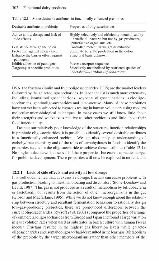
302 Functional dairy products
Table 12.1 Some desirable attributes in functionally enhanced prebiotics
Desirable attribute in prebiotic Properties of oligosaccharides
Active at low dosage and lack of Highly selectively and efficiently metabolised byside effects ‘beneficial’ bacteria but not by gas producers,
putrefactive organisms, etc.Persistence through the colon Controlled molecular weight distributionProtection against colon cancer Stimulate butyrate production in the colonEnhance the barrier effect against Structural basis unknown
pathogensInhibit adhesion of pathogens Possess receptor sequenceTargeting at specific probiotics Selectively metabolised by restricted species of
Lactobacillus and/or Bifidobacterium
USA, the fructans (inulin and fructooligosaccharides, FOS) are the market leadersfollowed by the galactooligosaccharides. In Japan the list is much more extensive,including isomaltooligosaccharides, soybean oligosaccharides, xylooligo-saccharides, gentiooligosaccharides and lactosucrose. Many of these prebioticshave not yet been subjected to rigorous testing in human volunteers using modernmolecular microbiological techniques. In many cases we still know little abouttheir strengths and weaknesses relative to other prebiotics and little about theirfood functionality.
Despite our relatively poor knowledge of the structure–function relationshipsin prebiotic oligosaccharides, it is possible to identify several desirable attributesin a functionally enhanced prebiotic. We can also apply an understanding ofcarbohydrate chemistry and of the roles of carbohydrates in foods to identify theproperties needed in the oligosaccharide to achieve these attributes (Table 12.1).No single molecule will possess all of these properties, but each is a practical targetfor prebiotic development. These properties will now be explored in more detail.
12.2.1 Lack of side effects and activity at low dosageIt is well documented that, at excessive dosage, fructans can cause problems withgas production, leading to intestinal bloating and discomfort (Stone-Dorshow andLevitt, 1987). This gas is not produced as a result of metabolism by bifidobacteriaor lactobacilli but results from the action of other microorganisms in the gut(Gibson and Macfarlane, 1995). While we do not know enough about the relation-ship between structure and resultant fermentation behaviour to rationally designlow-gas-producing prebiotics, there are pronounced differences between thecurrent oligosaccharides. Rycroft et al. (2001) compared the properties of a rangeof commercial oligosaccharides from Europe and Japan and found a large variationin gas evolution rates when used as substrates in batch culture with human faecalinocula. Fructans resulted in the highest gas liberation levels while galacto-oligosaccharides and isomaltooligosaccharides resulted in the least gas. Metabolismof the prebiotic by the target microorganisms rather than other members of the
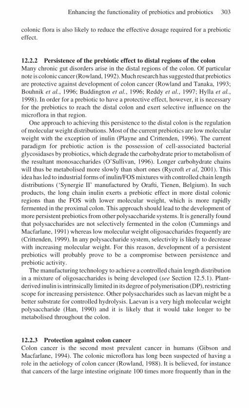
Enhancing the functionality of prebiotics and probiotics 303
colonic flora is also likely to reduce the effective dosage required for a prebioticeffect.
12.2.2 Persistence of the prebiotic effect to distal regions of the colonMany chronic gut disorders arise in the distal regions of the colon. Of particularnote is colonic cancer (Rowland, 1992). Much research has suggested that prebioticsare protective against development of colon cancer (Rowland and Tanaka, 1993;Bouhnik et al., 1996; Buddington et al., 1996; Reddy et al., 1997; Hylla et al.,1998). In order for a prebiotic to have a protective effect, however, it is necessaryfor the prebiotics to reach the distal colon and exert selective influence on themicroflora in that region.
One approach to achieving this persistence to the distal colon is the regulationof molecular weight distributions. Most of the current prebiotics are low molecularweight with the exception of inulin (Playne and Crittenden, 1996). The currentparadigm for prebiotic action is the possession of cell-associated bacterialglycosidases by probiotics, which degrade the carbohydrate prior to metabolism ofthe resultant monosaccharides (O’Sullivan, 1996). Longer carbohydrate chainswill thus be metabolised more slowly than short ones (Rycroft et al, 2001). Thisidea has led to industrial forms of inulin/FOS mixtures with controlled chain lengthdistributions (‘Synergie II’ manufactured by Orafti, Tienen, Belgium). In suchproducts, the long chain inulin exerts a prebiotic effect in more distal colonicregions than the FOS with lower molecular weight, which is more rapidlyfermented in the proximal colon. This approach should lead to the development ofmore persistent prebiotics from other polysaccharide systems. It is generally foundthat polysaccharides are not selectively fermented in the colon (Cummings andMacfarlane, 1991) whereas low molecular weight oligosaccharides frequently are(Crittenden, 1999). In any polysaccharide system, selectivity is likely to decreasewith increasing molecular weight. For this reason, development of a persistentprebiotics will probably prove to be a compromise between persistence andprebiotic activity.
The manufacturing technology to achieve a controlled chain length distributionin a mixture of oligosaccharides is being developed (see Section 12.5.1). Plant-derived inulin is intrinsically limited in its degree of polymerisation (DP), restrictingscope for increasing persistence. Other polysaccharides such as laevan might be abetter substrate for controlled hydrolysis. Laevan is a very high molecular weightpolysaccharide (Han, 1990) and it is likely that it would take longer to bemetabolised throughout the colon.
12.2.3 Protection against colon cancerColon cancer is the second most prevalent cancer in humans (Gibson andMacfarlane, 1994). The colonic microflora has long been suspected of having arole in the aetiology of colon cancer (Rowland, 1988). It is believed, for instancethat cancers of the large intestine originate 100 times more frequently than in the
304 Functional dairy products
small intestine (Morotomi et al., 1990). Many species of colonic bacteria producegenotoxic enzymes and produce carcinogens, and tumour promoters from a varietyof food components (Gibson and Macfarlane, 1994). It is believed that prebioticsmay help protect against cancer by either the production of butyrate or bymodulating the colonic flora away from protein and lipid metabolism towards themore benign carbohydrate metabolism.
Butyrate is believed to be protective against colon cancer as it is known tostimulate apoptosis in colonic cancer cell lines and it is the preferred fuel forhealthy colonocytes (Prasad, 1980; Kim et al., 1982). While it is probablydesirable to have high levels of butyrate generation in the distal colon, it must beborne in mind that lactobacilli and bifidobacteria do not produce butyrate. Theprincipal butyrate organisms that are believed to produce butyrate in the colon arethe clostridia and eubacteria (Cummings and Macfarlane, 1991). An obviousdevelopment target for prebiotics would be an oligosaccharide that stimulatedeubacteria but not clostridia. Some prebiotics have been found to result in thegeneration of butyrate (Olano-Martin et al., 2000; Rycroft et al., 2001), althoughthe microflora changes responsible are still uncertain as most such studies do notenumerate the changes in eubacteria.
An alternative approach would be to develop prebiotics that shift the metabolismof closridia and bacteroides towards saccharolysis rather than proteolysis orlipolysis. Clostridia are believed to convert dietary lipids such asphosphatidylcholine into carcinogens such as diacylglycerol (DAG, Morotomi etal., 1990). DAG is believed to be a protein kinase C activator, resulting instimulation of mucosal cell proliferation. We currently have an inadequate know-ledge of the effects of various prebiotics upon cancer biomarkers in the humancolon and more research is urgently needed in this area.
12.2.4 The barrier effect against gastrointestinal pathogensA very fertile area for the development of functionally enhanced prebiotics is theenhancement of the barrier effect of the normal gut microflora against pathogens.Dietary manipulation of this barrier to invading pathogens could make an importantcontribution to food safety. Bifidobacteria and lactobacilli produce a range of anti-microbial compounds from bacteriocins, active against related species, to morebroad spectrum agents active against a range of Gram-positive and Gram-negativepathogens such as salmonellae, campylobacters and Escherichia coli (Anand etal., 1985; Gibson and Wang, 1994; Araya-Kojima et al., 1995). This anti-pathogeneffect can be reproducibly seen in gut model systems and is likely to operate in thehuman gut.
In addition to the production of specific anti-microbial agents, fermentation ofprebiotics results in the production of short-chain fatty acids (SCFA; Gibson andRoberfroid, 1995). The resultant reduction in colonic pH inhibits growth andactivity of pathogens. There will also be increased competition with other organismsfor nutrients and for adhesion receptors on the gut wall (Araya-Kojima et al.,1995). The regulation of anti-microbial production by means of substrates in the

Enhancing the functionality of prebiotics and probiotics 305
Table 12.2 Receptor oligosaccharide sequences for selected gastrointestinal pathogensand toxins
Oligosaccharide sequence Pathogen/toxin
Galα4Gal E. coli (P-piliated), Vero cytotoxinGalβ4GlcNAcβ3Gal S. pneumoniaeGalNAcβ4Gal P. aeruginosa, H. influenzae, S. aureus,
K. pneumoniaeSialic acids E. coli (S-fimbriated)Galα3Galβ4GlcNAc C. difficile toxin AFucose V. choleraeGlcNAc E. coli, V. choleraeMannose E. coli, K. aerogenes, Salmonella spp.
(Type 1-fimbriated)
gut is imperfectly understood. In particular there is a surprising lack of informationabout the effects of different prebiotics on the expression levels and activityspectrum of anti-microbial agents produced by indigenous probiotics. This wouldseem to be an obvious target for prebiotic development.
12.2.5 Anti-adhesive activities against pathogens and toxinsAn interesting development to the prebiotic concept is the modulation of thecolonic flora using an anti-adhesive strategy. Many gastrointestinal pathogensrecognise oligosaccharide sequences carried on glycoproteins and glycolipids onthe gut wall as receptors (Table 12.2; Karlsson, 1989; Finlay and Falkow, 1989).There is currently much interest in the development of pharmaceutical agentsbased around this concept and there are several preparations in clinical trials. Thepotential for the incorporation of receptor oligosaccharides into foods, however,has not been fully explored, largely due to the high cost of oligosaccharides.Recent advances in the biotechnological manufacture (see section 12.5) of complexoligosaccharides, however, are likely to lead to significant cost reductions in thefuture.
12.3 Targeted prebiotics
A very exciting area for the future development of enhanced prebiotics is that oftargeted prebiotics. Prebiotics could conceivably be developed that will displayselectivity towards particular species of probiotic. Currently, prebiotics are de-fined as acting at the level of bifidobacteria and lactobacilli (Gibson and Roberfroid,1995). Virtually all of the available data on the prebiotic properties ofoligosaccharides describes microflora changes at the genus level. This is due to thechallenges of accurately identifying the colonic microflora using conventionalmicrobiological techniques. It would, however, be highly desirable to develop
306 Functional dairy products
prebiotics that are targeted at particular species of Bifidobacterium andLactobacillus. Such targeted prebiotics would have many applications.
12.3.1 Synbiotics with defined health benefitsCommercial probiotics are highly selected to have useful properties such asresistance to acid and bile and survival upon freeze-drying (Svensson, 1999; Leeet al., 1999). In addition, many of them have been developed for particular healthbenefits such as immune stimulation or anti-pathogen activity. It would be expectedthat synbiotic versions of these strains made with targeted prebiotics would displaybetter survival and colonisation in the gut (Bielecka et al., 2002).
12.3.2 Infant formulae and weaning foodsThe gut flora of breast-fed infants is generally dominated by bifidobacteria, whilethe gut flora of a formula-fed infant resembles that of an adult, with a wider rangeof microbial groups present (Cooperstock and Zedd, 1983; Benno et al., 1984). Itis believed that this bifidobacterial flora leads to health benefits for the infant,including a decreased susceptibility to microbial infections. It would be of interestto synthesise prebiotics with high selectivity towards those bifidobacteria that arepresent in the guts of breast-fed infants as the basis of novel infant foods.
12.3.3 Functional foods for healthy ageingAbove the age of 55–60 years, bifidobacterial counts in the colon decreasemarkedly compared with those of young adults (Mitsuoka, 1990; Kleessen et al.,1997). It is conceivable that this decrease in bifidobacterial numbers leads toreduced resistance to gastrointestinal infections. It is well known that elderlypeople tend to suffer more such infections. The development of functional foodsfor the elderly is the subject of the EU-funded Crownalife project, part of theProEUhealth cluster. The project will characterise the elderly gut flora acrossEurope, will run a synbiotic intervention trial and also develop prebiotics targetedat the bifidobacteria present in elderly people. The aim of the targeted prebioticdevelopment is increased resistance to gastrointestinal pathogens such as E. coli,Salmonella sp. and Campylobacter jejuni.
12.3.4 The development of targeted prebioticsTargeted prebiotics might be developed through one of two approaches. Screeninga wide range of oligosaccharide structures for their prebiotic properties will giveinformation on their selectivity towards particular species. In order to achieve thenecessary structural diversity, economical manufacturing technologies for complexoligosaccharides are required.
An alternative approach might be to use enzymes expressed by probioticbacteria as synthetic catalysts. Such enzymes will produce oligosaccharide mixtures,
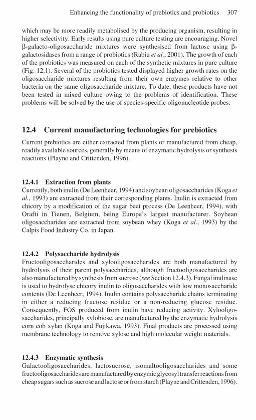
Enhancing the functionality of prebiotics and probiotics 307
which may be more readily metabolised by the producing organism, resulting inhigher selectivity. Early results using pure culture testing are encouraging. Novelβ-galacto-oligosaccharide mixtures were synthesised from lactose using β-galactosidases from a range of probiotics (Rabiu et al., 2001). The growth of eachof the probiotics was measured on each of the synthetic mixtures in pure culture(Fig. 12.1). Several of the probiotics tested displayed higher growth rates on theoligosaccharide mixtures resulting from their own enzymes relative to otherbacteria on the same oligosaccharide mixture. To date, these products have notbeen tested in mixed culture owing to the problems of identification. Theseproblems will be solved by the use of species-specific oligonucleotide probes.
12.4 Current manufacturing technologies for prebiotics
Current prebiotics are either extracted from plants or manufactured from cheap,readily available sources, generally by means of enzymatic hydrolysis or synthesisreactions (Playne and Crittenden, 1996).
12.4.1 Extraction from plantsCurrently, both inulin (De Leenheer, 1994) and soybean oligosaccharides (Koga etal., 1993) are extracted from their corresponding plants. Inulin is extracted fromchicory by a modification of the sugar beet process (De Leenheer, 1994), withOrafti in Tienen, Belgium, being Europe’s largest manufacturer. Soybeanoligosaccharides are extracted from soybean whey (Koga et al., 1993) by theCalpis Food Industry Co. in Japan.
12.4.2 Polysaccharide hydrolysisFructooligosaccharides and xylooligosaccharides are both manufactured byhydrolysis of their parent polysaccharides, although fructooligosaccharides arealso manufactured by synthesis from sucrose (see Section 12.4.3). Fungal inulinaseis used to hydrolyse chicory inulin to oligosaccharides with low monosaccharidecontents (De Leenheer, 1994). Inulin contains polysaccharide chains terminatingin either a reducing fructose residue or a non-reducing glucose residue.Consequently, FOS produced from inulin have reducing activity. Xylooligo-saccharides, principally xylobiose, are manufactured by the enzymatic hydrolysiscorn cob xylan (Koga and Fujikawa, 1993). Final products are processed usingmembrane technology to remove xylose and high molecular weight materials.
12.4.3 Enzymatic synthesisGalactooligosaccharides, lactosucrose, isomaltooligosaccharides and somefructooligosaccharides are manufactured by enzymic glycosyl transfer reactions fromcheap sugars such as sucrose and lactose or from starch (Playne and Crittenden, 1996).
308Functional dairy products
Fig. 12.1 Growth of selected probiotic strains of galactooligosaccharide mixtures generated by probiotic β-galactosidases. The control is a commercialgalactooligosaccharide mixture. In most cases, probiotics display higher growth rates on their homologous oligosaccharide mixture.
������
������
������
������
������
������
����������
����������
����������
����������
����������
����������
���������
����
����
����
����
�������
����������
����������
����������
����������
�����������
����
����
����
����
����
����
����
����
�������
����������
����������
����������
����������
����������
����������
����������
�����������
��������
����
����
����
����
����
����
����
����
����������
����������
����������
����������
����������
����������
����������
����������
������������
����
����
����
����
����
����
����
������
��������
��������
��������
��������
��������
��������
��������
������
����
����
����
����
����
����
����
����
����
����
����������
����������
����������
����������
����������
����������
����������
����������
����������
����������
��������
����
����
����
����
����
�������
����������
����������
����������
����������
����������
�����������
������
������
������
������
������
������
������
������
�������
��������
��������
��������
��������
��������
��������
��������
��������
���������
������
������
������
������
������
������
������
������
��������
����������
����������
����������
����������
����������
����������
����������
����������
������������
������
������
������
������
������
������
������
������
����������
����������
����������
����������
����������
����������
����������
������������
�������
����
����
����
����
����
����
����
����
����������
����������
����������
����������
����������
����������
����������
����������
���������
������
������
������
������
������
�������
��������
��������
��������
��������
��������
����������
����
����
����
����
����
����
����
����
����
����
�������
����������
����������
����������
����������
����������
����������
����������
����������
����������
����������
��������
����
����
����
����
����
����
����������
����������
����������
����������
����������
����������
�����������
����
����
����
����
����
����
����
����
�������
����������
����������
����������
����������
����������
����������
����������
����������
������
������
������
������
������
������
��������
��������
��������
��������
��������
��������
����
����
����
����
����
����
����
����
����������
����������
����������
����������
����������
����������
����������
����������
�����������
����
����
����
����
����
����
�������
����������
����������
����������
����������
����������
����������
��������
����
����
����
����
����
����
�������
����������
����������
����������
����������
����������
����������
�����������
����
����
����
����
����
����
�������
����������
����������
����������
����������
����������
�����������
������
����
����
����
����
����
����
����
����
����
����
����
����������
����������
����������
����������
����������
����������
����������
����������
����������
����������
����������
��������
����
����
����
����
����
����������
����������
����������
����������
����������
�����������
����
����
����
����
����
����
����
����
�������
����������
����������
����������
����������
����������
����������
����������
����������
���������
������
������
������
������
������
������
��������
��������
��������
��������
��������
��������
����������
������
������
������
������
������
������
������
������
����������
����������
����������
����������
����������
����������
����������
����������
�������
����
����
����
����
����
����
�������
����������
����������
����������
����������
����������
����������
��������
����
����
����
����
����
�������
����������
����������
����������
����������
����������
�����������
������
������
������
������
������
������
������
������
��������
����������
����������
����������
����������
����������
����������
����������
������������
���������
����
����
����
����
����
����
����
����
�������
����������
����������
����������
����������
����������
����������
����������
����������
�����������
����
����
����
����
����
����
����
����
����
����
����
����������
����������
����������
����������
����������
����������
����������
����������
����������
����������
����������
������
����
����
����
����
����
����������
����������
����������
����������
����������
���������
������
������
������
��������
����������
����������
����������
������������
����
����
����
����
����
�������
����������
����������
����������
����������
�����������
������
0
0.2
0.4
0.6
0.8
1
1.2
1.4
Gro
wth
rat
e (µ
h–1
)
Control B. lactis B. infantis B.pseudolongum
B. adolescentis B. angulatum L. plantarum
�������� L. acidophilus
L. plantarum����
B. pseudolongum
B. longum�������� B. infantis����
����
B. lactis
����
B. angulatum
B. adolescentis
Enhancing the functionality of prebiotics and probiotics 309
Fructooligosaccharides are made from sucrose by Meiji Seika in Japan and byBeghin-Meiji Industries in Europe (Playne and Crittenden, 1996). All of thesucrose-derived FOS terminate in a non-reducing glucose residue. Ion-exchangechromatography can be used to remove glucose and sucrose (Kono, 1993).Galactooligosaccharides are manufactured from lactose using β-galactosidase(Matsumoto, 1993). At high lactose concentration the enzyme formsgalactooligosaccharides (Schoterman and Timmermans, 2000). The products arecomplex mixtures of di- to pentasaccharides principally containing β1→3 andβ1→6 linkages. Galactooligosaccharides are made by Yakult Honsha and SnowBrand in Japan and by Borculo Domo Ingredients in Europe.
Lactosucrose is made using a similar approach, this time usingβ-fructofuranosidase to transfer fructose from sucrose to the reducing glucosylgroup on lactose (Kitahata and Fujita, 1993). Commercial production is centredin Japan by Hayashibara Shoji Inc. and Ensuiko Sugar Refining Co.Isomaltooligosaccharides (IMO) are manufactured by a more complex route(Yatake, 1993). Starch is first hydrolysed to maltooligosaccharides withα-amylase and pullulanase. The α1→4 linked maltooligosaccharides are thenconverted into α1→6 linked isomaltooligosaccharides by α-glucosidase. Thecommercial product manufactured by Showa Sangyu Co. in Japan consists of IMOof varying molecular weight together with panose.
12.5 Emerging manufacturing technologies for secondgeneration prebiotics
If the full potential of enhanced prebiotics is to be realised, new technologicalinnovations will be required. The challenge, as ever, for biotechnologists is toachieve the manufacturing technologies at economically viable costs. Two areas ofdevelopment are being explored in laboratories in Europe at the current time.
12.5.1 Controlled polysaccharide hydrolysisAs discussed above, polysaccharide hydrolysis is a commercial manufacturingapproach to prebiotics. Several of the functional enhancements discussed above,however, will demand a more controlled partial hydrolysis in order to achievecontrol over the molecular weight distribution of the products. Controlled partialhydrolysis has been investigated with dextran and pectins. Mountzouris et al.(1998, 2001) utilised endo-dextranase in an enzyme membrane reactor to makecontrolled IMO. By controlling factors such as residence time and the ratio ofenzyme to substrate, dextran could be converted into different IMO preparationswith average molecular weights varying from trisaccharide up to 12,000 Da. Thefractions displayed good prebiotic fermentation in an in vitro three-stage model ofthe human gut (Olano-Martin et al., 2000). Selective fermentation extended to thethird vessel of this system, which models the distal region of the gut (Macfarlaneet al., 1992, 1998).
310 Functional dairy products
Enzyme membrane reactors have also been used for the controlleddepolymerisation of pectins (Olano-Martin et al., 2001). In this case, low molecularweight products were produced. Such reactor systems show great promise but arein need of further research and development before they become a practical tool foroligosaccharide manufacture. This approach might, in future be extended to otherpolysaccharides. Bacterial extracellular polysaccharides (EPS), for example, dis-play huge structural variation and complexity and can often be manufacturedeconomically by fermentation processes (Sutherland, 1975; Morin, 1998). Theyfrequently contain rare monosaccharide residues in a wide range of linkageconfigurations. An interesting source of enzymes to degrade such polysaccharidescan be isolated from bacteriophage. These enzymes have been used in sequencingstudies of polysaccharides (Geyer et al., 1983; Stirm, 1994; Sutherland, 1999) butnot in a commercial manufacturing technology. This approach could extend ourcollection of oligosaccharide structures for screening as candidate probiotics withenhanced specificity at the species level.
12.5.2 Enzymatic synthesis of oligosaccharidesThe enzymatic synthesis of oligosaccharides can be achieved through severalroutes (Rastall and Bucke, 1992; Kren and Thiem, 1997; Crout and Vic, 1998).Many of these have not yet reached industrial implementation, although they aresubjects of active research programmes.
One class of enzymes with obvious potential for the industrial synthesis ofnovel prebiotics is the glycosyltransferases. These can be divided into the so-calledLeloir enzymes (Rastall and Bucke, 1992), which use sugar nucleotides as glycosyldonors, and the sucrases, which use sucrose. The sucrases can be used to generatenovel oligosaccharides (Robyt, 1995). Dextransucrase has been used in themanufacture of novel α-glucooligosaccharides that are selectively fermented bydesirable microorganisms on the skin (Dols et al., 1999) and the gut (Djouzi et al.,1995; Djouzi and Andrieux 1997). These oligosaccharides have been formulatedinto cosmetics but have not yet been fully evaluated as prebiotic food ingredients.
Glycosidase enzymes can also be used to synthesise novel oligosaccharidesunder appropriate conditions (Rastall and Bucke, 1992; Kren and Thiem, 1997;Crout and Vic, 1998). Vic et al. (1997), for example synthesised the trisaccharidereceptor for Clostridium difficile Toxin A using glycosidases in kineticallycontrolled syntheses. Suwasono and Rastall (1996) synthesised the receptoractive Manα1→2Man di- and trisaccharides using α1,2-mannosidase fromAspergillus phoenicis. These oligosaccharides are receptors for type-1 fimbriatedenteric microorganisms and may have potential for development as anti-adhesiveagents.
Glycosidases may also be used to manufacture novel oligosaccharides ascandidate prebiotics. Many glycosidases can recognise a range of sugars asacceptors, building up heterooligosaccharides. α-Mannosidase enzymes havebeen used to attach a mannose residue onto several disaccharide acceptors tomanufacture novel trisaccharides (Rastall et al., 1992; Smith et al., 1997; Suwasono
Enhancing the functionality of prebiotics and probiotics 311
and Rastall, 1998). The prebiotic potential of such oligosaccharides is now underinvestigation.
12.6 The functional enhancement of probiotics
Although it is not too difficult to conceive of desirable enhancements to probioticbacterial strains, the technology required to achieve these enhancements is com-plex. Genetic manipulation tools for probiotics have been developed (Kullen andKlaenhammer, 1999), but these have not yet been widely applied to the enhancementof probiotics destined for human food applications.
It is possible to define the characteristics that one would want in a probioticstrain (Shah, 2001) and this is covered in more depth elsewhere in this volume. Onesuch enhancement might be the metabolism of specific prebiotics.
12.6.1 Metabolism of specific prebioticsIt is possible to achieve stable, functional expression of heterologous enzymegenes in Lactobacillus species. This raises the possibility of generating probioticsthat can metabolise particular carbohydrates that most lactobacilli can not. Certainprebiotic oligosaccharides such as inulin, for instance, are not particularly wellfermented by many lactobacilli. There would be an obvious advantage in thedevelopment of synbiotics, which can metabolise unusual oligosaccharides.
To date, no such probiotics destined for human food applications have beendeveloped. Several genetically modified Lactobacillus species expressing glycanasegenes have been constructed as enhanced silage inocula. Most work has focused onLactobacillus plantarum strain Lp80, a common silage inoculum strain. Engineeredforms have been made with good expression levels of α-amylase (Schierlink, et al,1989; Fitzsimons et al., 1994) cellulases (Schierlink et al., 1989, 1990), xylanases(Schierlink et al., 1990), laevanases (Hols et al., 1994; Wanker et al., 1995) andchitinase (Brurberg et al., 1994). The laevanase gene has also been expressed in L.casei (Wanker et al., 1995) and the chitinase gene was expressed in L. lactis(Brurberg et al., 1994). The engineered lactobacilli displayed enhanced growthand lactic fermentation on complex biomass polysaccharides with no need foradded fermentable carbohydrates or enzymes. Of particular interest is the expres-sion of the laevanase gene from Bacilllus subtilis, as this enzyme enabled therecombinant strain to grow on laevan and on inulin (Wanker et al., 1995).
12.7 Conclusion and future trends
Prebiotic oligosaccharides are set to make a further impact on the area of functionalfoods. Current forms are efficient at stimulating bifidobacteria; however, the nextgeneration of prebiotics will entail multiple functionality. Biotechnologicalprocedures are increasingly being applied to the area and developments with novel
312 Functional dairy products
foods are proceeding quickly. It is clear that for the further development of thisfield reliable testing methods are needed. One example is through the applicationof molecular procedures to improve our knowledge of the gut microflora, itsinteractions with the human host and its role in maintenance of health. Advancesin these areas will ultimately provide us with very sophisticated dietary tools withwhich to manipulate this important ecosystem and improve public health.
12.8 ReferencesANAND S K, SRINIVASAN R A and RAO L K (1985), ‘Antibacterial activity associated with
Bifidobacterium bifidum – II’, Cult Dairy Prod J, 2, 21–23.ARAYA-KOJIMA A, YAESHIMA T, ISHIBASHI N, SHIMAMUR S and HAYASAWA H (1995),
‘Inhibitory effects of Bifidobacterium longum BB536 on harmful intestinal bacteria’,Bifidobacteria Microflora, 14, 59–66.
BENNO Y, SAWADA K and MITSUOKA T (1984), ‘The intestinal microflora of infants:composition of fecal flora in breast-fed and bottle-fed infants’, Micro Immun, 28, 975–986.
BIELECKA M, BIEDRZYCKA E and MAJKOWSKA A (2002), ‘Selection of probiotics andprebiotics for synbiotics and confirmation of their in vivo effectiveness’, Food Res Int, 35,125–131.
BOUHNIK Y, FLOURIE B, RIOTTOT M, BISETTI N, GAILING M F and GUIBERT A (1996), ‘Effectsof fructo-oligosaccharides ingestion on faecal bifidobacteria and selected metabolicindexes of colon carcinogenesis in healthy humans’, Nutr Cancer, 26, 21–29.
BRURBERG M B, HAANDRIKMAN A J, LEENHOUTS K J, VENEMA G and NES I F (1994),‘Expression of a chitinase gene from Serratia marcescens in Lactococcus lactis andLactobacillus plantarum’, Appl Micro Biotechnol, 42, 108–115.
BUDDINGTON R K, WILLIAMS C H, CHEN S-C and WITHERLY S A (1996), ‘Dietary supplemen-tation of neosugar alters the fecal flora and decreases activities of some reductive enzymesin human subjects’, Am J Clin Nutr, 63, 709–716.
COOPERSTOCK M S and ZEDD A J (1983), ‘Intestinal flora of infants’, in D J Hentges, HumanIntestinal Microflora in Health and Disease, London, Academic Press, 79–99.
CRITTENDEN R G (1999), ‘Prebiotics’, in G Tannock, Probiotics: A Critical Review,Wymondham, Horizon Scientific Press, 141–156.
CRITTENDEN R G and PLAYNE M J (1996), ‘Production, properties and applications of food-grade oligosaccharides’, Trends Food Sci Tech, 7, 353–361.
CROUT D H G and VIC G (1998), ‘Glycosidases and glycosyl transferases in glycoside andoligosaccharide synthesis’, Curr Opin Chem Biol, 2, 98–111.
CUMMINGS J H and MACFARLANE G T (1991), ‘The control and consequences of bacterialfermentation in the human colon’, J Appl Bact, 70, 443–459.
DE LEENHEER L (1994), ‘Production and use of inulin: industrial reality with a promisingfuture’, in H van Bekkum, H Roper and A G J Voragen, Carbohydrates as Organic RawMaterials III, Weinheim, VCH, 67–92.
DJOUZI Z and ANDRIEUX C (1997), ‘Compared effects of three oligosaccharides on metabolismof intestinal microflora in rats inoculated with a human faecal flora’, Br J Nutr, 78, 313–324.
DJOUZI Z, ANDRIEUX C, PELENC V, SOMARRIBA S, POPOT F, PAUL F, MONSAN P and SZYLIT O(1995), ‘Degradation and fermentation of α-gluco-oligosaccharides by bacterial strainsfrom human colon: in vitro and in vivo studies in gnotobiotic rats’, J Appl Bact, 79, 117–127.
DOLS M, MONCHOIS V, REMAUD-SIMEON M, WILLEMOT R-M and MONSAN P F (1999), ‘Theproduction of α1→2-terminated glucooligosaccharides’, in C Bucke, Carbohydrate
Enhancing the functionality of prebiotics and probiotics 313
Biotechnology Protocols, Methods in Biotechnology, Vol. 10, Totowa, NJ, HumanaPress, 129–139.
FINLAY B B and FALKOW S (1989), ‘Common themes in microbial pathogenicity’, MicrobiolRev, 53, 210–230.
FITZSIMONS A, HOLS P, JORE J, LEER R J, O’CONNELL M and DELCOUR J (1994), ‘Developmentof an amylolytic Lactobacillus plantarum silage strain expressing the Lactobacillusamylovorus alpha-amylase gene’, Appl Env Microbiol, 60, 3529–3535.
GEYER H, HIMMELSPACH K, KWIATKOWSKI B, SCHLECHT S and STIRM S (1993), ‘Degradationof bacterial surface carbohydrates by virus associated enzymes’, Pure Appl Chem, 55,637–653.
GIBSON G R and MACFARLANE G T (1994), ‘Intestinal bacteria and disease’, in S A W Gibson,Human Health – The Contribution of Microorganisms, London, Springer-Verlag, 53–62.
GIBSON G R and MACFARLANE G T (1995), Human Colonic Bacteria: Role in Nutrition,Physiology and Pathology, Boca Raton, CRC Press.
GIBSON G R and ROBERFROID M B (1995), ‘Dietary modulation of the human colonicmicrobiota: introducing the concept of prebiotics’, J Nutr, 125, 1401–1412.
GIBSON G R and WANG X (1994), ‘Regulatory effects of bifidobacteria on the growth of othercolonic bacteria’, J Appl Bact, 77, 412–420.
GIBSON G R, BERRY OTTAWAY P and RASTALL R A (2000), Prebiotics: New Developments inFunctional Foods, Oxford, Chandos Publishing.
HAN Y W (1990), ‘Microbial Levan’, in S L Neidleman and A I Laskin, Advances in AppliedMicrobiology, Vol. 35, London, Academic Press, 171–194.
HOLS P, FERAIN T, GARMYN D, BERNARD N and DELCOUR J (1994), ‘Use of homologousexpression-secretion signals and vector-free stable chromosomal integration in engineer-ing of Lactobacilllus plantarum for alpha-amylase and laevanase expression’, Appl EnvMicrobiol, 60, 1401–1413.
HYLLA S, GOSTNER A, DUSEL G, ANGER H, BARTRAM H-P, CHRISTL S U et al. (1998), ‘Effectsof resistant starch on the colon in healthy volunteers: possible implications for cancerprevention’, Am J Clin Nutr, 67, 136–142.
KARLSSON K-A (1989), ‘Animal glycosphingolipids as membrane attachment sites forbacteria’, Ann Rev Biochem, 58, 309–350.
KIM Y S, TSAO D, MORITA A and BELLA A (1982), ‘Effect of sodium butyrate and three humancolorectal adenocarcinoma cell lines in culture’, Falk Symposium, 31, 317–323.
KITAHATA S and FUJITA K (1993), ‘Xylsucrose, isomaltosucrose and lactosucrose’. In:Oligosaccharides. Production, Properties and Applications, Japanese Technology Review,Vol. 3, Switzerland, Gordon & Breach, 158–174.
KLEESSEN B, SYKURA B, ZUNFT H-J and BLAUT M (1997), ‘Effects of inulin and lactose onfecal microflora, microbial activity and bowel habit in elderly constipated persons’, AmJ Clin Nut, 65, 1397–1402.
KOGA K and FUJIKAWA S (1993), ‘Xylooligosaccharides’. In: Oligosaccharides. Production,Properties and Applications, Japanese Technology Review, Vol. 3, Switzerland, Gordon& Breach, 130–143.
KOGA Y, SHIBUTA T and O’BRIEN R (1993), ‘Soybean oligosaccharides’. In: Oligosaccharides.Production, Properties and Applications, Japanese Technology Review, Vol. 3,Switzerland, Gordon & Breach, 90–106.
KONO T (1993), ‘Fructooligosaccharides’. In: Oligosaccharides. Production, Properties andApplications, Japanese Technology Review, Vol. 3, Switzerland, Gordon & Breach, 50–78.
KREN V and THIEM J (1997), ‘Glycosylation employing bio-systems: from enzymes to wholecells’, Chem Soc Rev, 26, 463–473.
KULLEN M J and KLAENHAMMER T (1999), ‘Genetic modification of lactobacilli andbifidobacteria’, in G Tannock, Probiotics: A Critical Review, Wymondham, HorizonScientific Press, 65–83.
314 Functional dairy products
LEE Y-K, NOMOTO K, SALMINEN S and GORBACH S L (1999), Handbook of Probiotics, NewYork, Wiley.
MACFARLANE G T, GIBSON G R and CUMMINGS J H (1992), ‘Comparison of fermentationreactions in different regions of the colon’, J Appl Bact, 72, 57–64.
MACFARLANE G T, MACFARLANE S and GIBSON G R (1998), ‘Validation of a three-stagecompound continuous culture system for investigating the effect of retention time on theecology and metabolism of bacteria in the human colonic microbiota’, Micro Ecol, 35,180–187.
MATSUMOTO K (1993), ‘Galactooligosaccharides’. In: Oligosaccharides. Production, Prop-erties and Applications, Japanese Technology Review, Vol. 3, Switzerland, Gordon &Breach, 90–106.
MITSUOKA T (1990), ‘Bifidobacteria and their role in human health’, J Ind Microbiol, 6, 263–268.
MORIN A (1998), ‘Screening of polysaccharide producing organisms, factors influencing theproduction and recovery of microbial polysaccharides’, in S Dimitriu, Polysaccharides:Structural Diversity and Functional Versatility, New York, Marcel Dekker Inc.
MOROTOMI M, GUILLEM J G, LOGERFO P and WEINSTEN I B (1990), ‘Production of diacylglycerol,an activator of protein kinase C by human intestinal microflora’, Cancer Res, 50, 3595–3599.
MOUNTZOURIS K C, GILMOUR S G, GRANDISON A S and RASTALL R A (1998), ‘Modelling ofoligodextran production in an ultrafiltration stirred cell membrane reactor’, Enz MicrobialTechnol, 24, 75–85.
MOUNTZOURIS K C, GILMOUR S G and RASTALL R A (2001), ‘Continuous production ofoligodextrans via controlled hydrolysis of dextran in an enzyme membrane reactor’, JFood Sci, 67, 1767–1771.
OLANO-MARTIN E, MOUNTZOURIS K C, GIBSON G R and RASTALL R A (2001), ‘Continuousproduction of oligosaccharides from pectin in an enzyme membrane reactor’, J Food Sci,66, 966–971.
OLANO-MARTIN E, MOUNTZOURIS K C, GIBSON G R and RASTALL R A (2000), ‘In vitrofermentability of dextran, oligodextran and maltodextrin by human gut bacteria’, Brit JNutr, 83, 247–255.
O’SULLIVAN M G (1996), ‘Metabolism of bifidogenic factors – an overview’, Bull Int DairyFed, 313, 23–30.
PLAYNE M J and CRITTENDEN R (1996), ‘Commercially available oligosaccharides’, Bull IntDairy Fed, 313, 10–22.
PRASAD K N (1980), ‘Butyric acid: a small fatty acid with diverse biological functions’, LifeSci, 27, 1351–1358.
RABIU B A, JAY A J, GIBSON G R and RASTALL R A (2001), ‘Synthesis and fermentationproperties of novel galacto-oligosaccharides by beta-galactosidases from bifidobacteriumspecies’, Appl Env Microbiol, 67, 2526–2530.
RASTALL R A and BUCKE C (1992), ‘Enzymatic synthesis of oligosaccharides’, BiotechnolGen Eng Rev, 10, 253–281.
RASTALL R A, REES N H, WAIT R, ADLARD M W and BUCKE C (1992), ‘Alpha-mannosidase-catalysed synthesis of novel manno-, lyxo-, and heteromanno-oligosaccharides: acomparison of kinetically and thermodynamically mediated approaches’, Enz MicroTechnol, 14, 53–57.
REDDY B S, HAMID R and RAO C V (1997), ‘Effect of dietary oligofructose and inulin oncolonic preneoplastic aberrant crypt foci inhibition’, Carcinogenesis, 18, 1371–1374.
ROBYT J (1995), ‘Mechanisms in the glucansucrase synthesis of polysaccharides andoligosaccharides from sucrose’, Adv Carbohydr Chem, 51, 133–168.
ROWLAND I R (1988), Role of the Gut Flora in Toxicity and Cancer, London, Academic Press.ROWLAND I R (1992), ‘Metabolic interactions in the gut’, in R Fuller, Probiotics: the
Scientific Basis, Andover, Chapman and Hall, 29–53.ROWLAND I R and TANAKA R (1993), ‘The effects of transgalactosylated oligosaccharides on
Enhancing the functionality of prebiotics and probiotics 315
gut flora metabolism in rats associated with a human faecal microflora’, J Appl Bact, 74,667–674.
RYCROFT C E, JONES M R, GIBSON G R and RASTALL R A (2001), ‘A comparative in vitroevaluation of the fermentation properties of prebiotic oligosaccharides’, J Appl Microbiol,91, 878–887.
SALMINEN S, BOULEY C, BOUTRON-RUAULT M-C, CUMMINGS J H, FRANCK A, GIBSON G R,ISOLAURI E, MOREAU M-C, ROBERFROID M and ROWLAND I R (1998), ‘Functional foodscience and gastrointestinal physiology and function’, Br J Nutr, 80, S147–S171.
SCHIERLINK T, MAHILLON J, JOOS P, DHAESE P and MICHIELS F (1989), ‘Integration andexpression of alpha-amylase and endoglucanase genes in the Lactobacillus plantarumchromosome’, Appl Env Microbiol, 55, 2130–2137.
SCHIERLINK T, MEUTTER J D, ARNAUT G, JOSS H, CLAEYSSENS M and MICHIELS F (1990),‘Cloning and expression of cellulase and xylanase genes in Lactobacillus plantarum’,Appl Microbiol Biotechnol, 33, 534–541.
SCHOTERMAN H C and TIMMERMANS H J A R (2000), ‘Galacto-oligosaccharides’, inG R Gibson and F Angus, Prebiotics and Probiotics, LFRA Ingredients Handbook,Leatherhead, Leatherhead Food RA Publishing, 19–46.
SHAH N P (2001), ‘Functional foods from probiotics and prebiotics’, Food Tech, 51, 46–53.SMITH N K, GILMOUR S G and RASTALL R A (1997), ‘Statistical optimisation of enzymatic
synthesis of derivatives of trehalose and sucrose’, Enz Microbial Technol, 21, 349.STIRM S (1994), ‘Examination of the repeating units of bacterial exopolysaccharides’, in
J N BeMiller, D J Manners and R J Sturgeon, Methods in Carbohydrate Chemistry,Vol., X, New York, John Wiley.
STONE-DORSHOW T and LEVITT M D (1987), ‘Gaseous response to ingestion of a poorlyabsorbed fructo-oligosaccharide’, Am J Clin Nutr, 46, 61–65.
SUTHERLAND I (1975), Surface Carbohydrates of the Prokaryotic Cell, New York, AcademicPress.
SUTHERLAND I (1999), ‘Polysaccharases for microbial exopolysaccharides’, CarbohydrPolym, 38, 319–328.
SUWASONO S and RASTALL R A (1996), ‘A highly regioselective synthesis of mannobiose andmannotriose by reverse hydrolysis using specific 1,2-alpha-mannosidase from Aspergil-lus phoenicis’, Biotechnol Lett, 18, 851–856.
SUWASONO S and RASTALL R A (1998), ‘Enzymatic synthesis of manno- and heteromanno-oligosaccharides using α-mannosidases: a comparative study of linkage-specific andnon-linkage-specific enzymes”, J Chem Technol Biotechnol, 73, 37–42.
SVENSSON U (1999), “Industrial perspectives”, in G Tannock, Probiotics: A Critical Review,Wymondham, Horizon Scientific Press, 57–64.
VIC G, TRAN C H, SCIGELOVA M and CROUT D H G (1997), “Glycosidase-catalysed synthesisof oligosaccharides: a one step synthesis of lactosamine and of the linear B type 2trisaccharide α-D-Gal-(1→3)-β-D-Gal-(1→4)-β-D-GlcNAcSEt involved in the hyperacuterejection response in xenotransplantation from pigs to man and as the specific receptor fortoxin A from Clostridium difficile’, Chem Commun, 169–170.
WANKER E, LEER R J, POUWELS P H and SCHWAB H (1995), ‘Expression of Bacillus subtilislaevanase gene in Lactobacillus plantarum and Lactobacillus casei’, Appl MicrobiolBiotechnol, 43, 297–303.
YATAKE T (1993), ‘Anomalously linked oligosaccharides’. In: ‘Oligosaccharides. Production,Properties and Applications’, Japanese Technology Review, Vol. 3, Switzerland, Gordon& Breach, 79–89.
13
Safety evaluation of probioticsA.C. Ouwehand and S. Salminen, University of Turku, Finland
13.1 Introduction
Most probiotics belong to the genera Lactobacillus or Bifidobacterium. Thechapter will therefore mainly consider the safety issues associated with membersof these genera, although species of other genera are also used as probiotics andsimilar safety considerations apply to these. Many criteria are used in the selectionand use of probiotics. The two of greatest importance are efficacy and safety.Probiotic products have been shown to provide a variety of documented healthbenefits to all consumers (Salminen et al., 1998). The safety record of probioticsis also good, indicating no or negligible risks. However, in order to be able tocontinue to ensure the safety of probiotics and dairy starters, safety assessments arenecessary.
Lactobacilli and bifidobacteria are generally regarded as safe. This safety statusis based on a number of empirical observations:
• Lactobacilli, in particular dairy starters, have a long history of safe use.• Lactobacilli and bifidobacteria are extremely rarely associated with disease.• Bifidobacteria and to a lesser extent lactobacilli are major members of the
normal human intestinal microflora.
Considering the above, what are the safety concerns for probiotics?Most probiotics do not have a particularly long history of safe use. The longest
is probably exemplified by certain strains belonging to the Lactobacillus acido-philus group and Lactobacillus casei Shirota, which has been on the market forover 60 years. This is, however, short compared with the history of safe use ofmany dairy-associated lactic acid bacteria such as Lactobacillus delbrueckii subsp.bulgaricus, Streptococcus thermophilus and Lactoccus lactis.
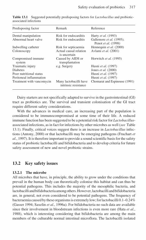
Safety evaluation of probiotics 317
Table 13.1 Suggested potentially predisposing factors for Lactobacillus and probiotic-associated infections
Predisposing factor Remark Reference
Dental manipulation Risk for endocarditis Harty et al. (1993)Abnormal heart valve Risk for endocarditis Gallemore et al. (1995);
Penot et al. (1998)Indwelling catheter Risk for septicaemia Hennequin et al. (2000)Colonoscopy Actual causal relation Avlami et al. (2001)
is uncertainCompromised immune Caused by AIDS or Horwitch et al. (1995)
system transplantationTraumatic injury e.g. Surgery Husni et al. (1997)Diabetes Jones et al. (2000)Poor nutritional status Husni et al. (1997)Peritoneal inflammation Husni et al. (1997)Treatment with vancomycin Many lactobacilli have Chomarat and Espinouse (1991)
intrinsic resistance
Dairy starters are not specifically adapted to survive in the gastrointestinal (GI)tract as probiotics are. The survival and transient colonisation of the GI tractrequire different safety considerations.
With the advances in medical care, an increasing part of the population isconsidered to be immunocompromised at some time of their life. A reducedimmune function has been suggested to be a potential risk factor for Lactobacillus-associated infections, as in fact for infections by other microbes as well (see Table13.1). Finally, critical voices suggest there is an increase in Lactobacillus infec-tions (Antony, 2000) or that lactobacilli may be emerging pathogens (Fruchart etal., 1997). It is therefore important to provide a sound scientific basis for the safetystatus of probiotic lactobacilli and bifidobacteria and to develop criteria for futuresafety assessment of new and novel probiotic strains.
13.2 Key safety issues
13.2.1 The microbeAll microbes that have, in principle, the ability to grow under the conditions thatprevail in the human body can theoretically colonise this habitat and can thus bepotential pathogens. This includes the majority of the mesophilic bacteria, andlactobacilli and bifidobacteria among others. However, lactobacilli and bifidobacteriaare, in general, not even considered to be potential pathogens. The frequency ofbacteraemia caused by these organisms is extremely low; for lactobacilli 0.1–0.24%(Gasser 1994; Saxelin et al., 1996a). For bifidobacteria no such data are availablesince their involvement in bloodstream infections is even more rare (Hata et al.,1988), which is interesting considering that bifidobacteria are among the mainmembers of the culturable normal intestinal microflora. The lactobacilli isolated
318 Functional dairy products
from bacteraemia cases have not been identified as dairy starters or probiotics(Saxelin et al., 1996a). Since lactobacilli and bifidobacteria have a natural associa-tion with human mucosal surfaces of the mouth, GI tract and genitourinary tract, thelactobacilli involved in the bacteraemia therefore probably originate from thepatient’s own microflora (Wang et al., 1996). However, two cases have beenreported where probiotic lactobacilli may have been involved in the aetiology ofdisease: a liver abscess caused by an L. rhamnosus indistinguishable from L.rhamnosus GG (Rautio et al., 1999) and a case of endocarditis possibly associatedwith a probiotic L. rhamnosus (Mackay et al., 1999). For the latter, however, theidentification is relatively uncertain, as only unconventional methods were used.
Despite these observations the general safety of lactobacilli and bifidobacteriain general and of probiotics in particular appears to be good considering the largeamounts of these bacteria consumed by people worldwide. This is, in fact, a farbetter safety record than even for water, with on average one reported fatalintoxication per year (Chen and Huang, 1995). The above two cases were not fatal;Lactobacillus infections are themselves usually not fatal and can be relativelyeasily treated once diagnosed. However, owing to their extremely low occurrence,recognition, identification and diagnosis by clinicians may sometimes be difficult.
13.2.2 The hostBecause the human body, from the microbe’s point of view, is a habitat rich innutrients, many defence mechanisms have evolved to resist colonisation bymicrobes. Only a relatively small number of microbes have adapted themselves toovercome these defence mechanisms, i.e. the ‘true’ pathogens. Under normalconditions the defence barriers function well and so non-pathogenic, and in factalso many pathogenic microorganisms, do not gain access into the human body.However, under adverse conditions such as disease and trauma, the defencebarriers break down and pathogens, and even generally non-pathogenic organismssuch as lactobacilli and bifidobacteria, may enter into the human body (Falkow,1997). A number of potentially predisposing factors for Lactobacillus-associatedinfections have been suggested (Table 13.1). Indeed, most Lactobacillus andBifidobacterium infections have occurred in subjects with severe underlyingdiseases (Gasser, 1994; Husni et al., 1997; Saxelin et al., 1996a).
Although a compromised immune system appears to be an obvious risk factor, itis important to note that probiotics can also significantly contribute to the protectionof, in particular, immunocompromised subjects (Bengmark and Jeppson 1995). Thishas also clearly been shown in animal experiments using lethally irradiated mice(Dong et al., 1987) and congenital immune-deficient mice (Wagner et al., 1997b).In both cases, probiotics provided significant protection for the host. However, ahigher mortality among offspring of some of the congenital immune-deficient micecolonised exclusively with Lactobacillus GG and Lactobacillus reuteri was observed(Wagner et al., 1997a). This clearly indicates that, in the case of Lactobacillus-associated disease, the health status of the host plays a significant role. The healthstatus is likely to be even more important than the properties of the lactobacilli.
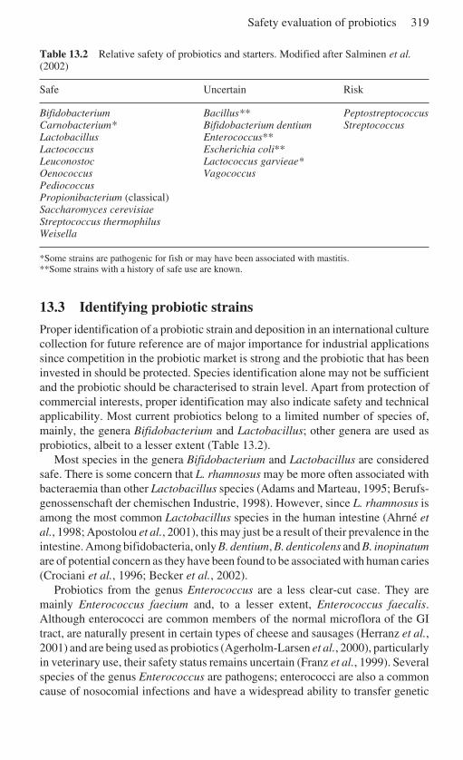
Safety evaluation of probiotics 319
Table 13.2 Relative safety of probiotics and starters. Modified after Salminen et al.(2002)
Safe Uncertain Risk
Bifidobacterium Bacillus** PeptostreptococcusCarnobacterium* Bifidobacterium dentium StreptococcusLactobacillus Enterococcus**Lactococcus Escherichia coli**Leuconostoc Lactococcus garvieae*Oenococcus VagococcusPediococcusPropionibacterium (classical)Saccharomyces cerevisiaeStreptococcus thermophilusWeisella
*Some strains are pathogenic for fish or may have been associated with mastitis.**Some strains with a history of safe use are known.
13.3 Identifying probiotic strains
Proper identification of a probiotic strain and deposition in an international culturecollection for future reference are of major importance for industrial applicationssince competition in the probiotic market is strong and the probiotic that has beeninvested in should be protected. Species identification alone may not be sufficientand the probiotic should be characterised to strain level. Apart from protection ofcommercial interests, proper identification may also indicate safety and technicalapplicability. Most current probiotics belong to a limited number of species of,mainly, the genera Bifidobacterium and Lactobacillus; other genera are used asprobiotics, albeit to a lesser extent (Table 13.2).
Most species in the genera Bifidobacterium and Lactobacillus are consideredsafe. There is some concern that L. rhamnosus may be more often associated withbacteraemia than other Lactobacillus species (Adams and Marteau, 1995; Berufs-genossenschaft der chemischen Industrie, 1998). However, since L. rhamnosus isamong the most common Lactobacillus species in the human intestine (Ahrné etal., 1998; Apostolou et al., 2001), this may just be a result of their prevalence in theintestine. Among bifidobacteria, only B. dentium, B. denticolens and B. inopinatumare of potential concern as they have been found to be associated with human caries(Crociani et al., 1996; Becker et al., 2002).
Probiotics from the genus Enterococcus are a less clear-cut case. They aremainly Enterococcus faecium and, to a lesser extent, Enterococcus faecalis.Although enterococci are common members of the normal microflora of the GItract, are naturally present in certain types of cheese and sausages (Herranz et al.,2001) and are being used as probiotics (Agerholm-Larsen et al., 2000), particularlyin veterinary use, their safety status remains uncertain (Franz et al., 1999). Severalspecies of the genus Enterococcus are pathogens; enterococci are also a commoncause of nosocomial infections and have a widespread ability to transfer genetic
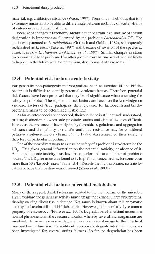
320 Functional dairy products
material, e.g. antibiotic resistance (Wade, 1997). From this it is obvious that it isextremely important to be able to differentiate between probiotic or starter strainsof enterococci and clinical strains.
Because of changes in taxonomy, identification to strain level and use of a straindesignation is important as illustrated by the probiotic Lactobacillus GG. Thestrain was patented as L. acidophilus (Gorbach and Goldin, 1989), subsequentlyreclassified as L. casei (Saxelin, 1997) and, because of revision of the species L.casei, it is now L. rhamnosus (Alander et al., 1997). Similar changes in straintaxonomy have been performed for other probiotic organisms as well and are likelyto happen in the future with the continuing development of taxonomy.
13.4 Potential risk factors: acute toxicity
For generally non-pathogenic microorganisms such as lactobacilli and bifido-bacteria it is difficult to identify potential virulence factors. Therefore, potentialrisk factors have been proposed that may be of significance when assessing thesafety of probiotics. These potential risk factors are based on the knowledge onvirulence factors of ‘true’ pathogens: their relevance for lactobacilli and bifido-bacteria remains to be determined (Table 13.3).
As far as enterococci are concerned, their virulence is still not well understood,making distinction between safe probiotic strains and clinical isolates difficult.However, the presence of haemolysin, hyaluronidase, gelatinase and aggregationsubstance and their ability to transfer antibiotic resistance may be consideredputative virulence factors (Franz et al., 1999). Assessment of their safety istherefore of particular importance.
One of the most direct ways to assess the safety of a probiotic is to determine theLD
50. This gives general information on the potential toxicity, or absence of it.
Acute and chronic toxicity tests have been performed for a number of probioticstrains. The LD
50 for mice was found to be high for all tested strains, for some even
more than 50 g/kg body mass (Table 13.4). Despite the high exposure, no translo-cation outside the intestine was observed (Zhou et al., 2000).
13.5 Potential risk factors: microbial metabolism
Many of the suggested risk factors are related to the metabolism of the microbe.Hyaluronidase and gelatinase activity may damage the extracellular matrix proteins,thereby causing direct tissue damage. Not much is known about this enzymaticactivity in lactobacilli and bifidobacteria. However, it is a relatively commonproperty of enterococci (Franz et al., 1999). Degradation of intestinal mucus is anormal phenomenon in the caecum and colon whereby several microorganisms areinvolved. However, excessive degradation may cause damage to the intestinalmucosal barrier function. The ability of probiotics to degrade intestinal mucus hasbeen investigated for several strains in vitro. So far, no degradation has been

Safety evaluation of probiotics 321
Table 13.3 Suggested potential risk factors of probiotic lactobacilli and bifidobacteria
Related to Property Remark
Microbial Hyaluronidase activity Of particular importance formetabolism Gelatinase activity Enterococcus sp.
DNAse activityMucus degradation Compromises mucosal barrierD-Lactic acid formation D-Lactic acidosisAminoacid decarboxylase activity Formation of biogenic aminesBile salt deconjugation Formation of primary bile saltsBile salt dehydroxylase activity Formation of secondary bile saltsPhosphatidylinositol-specific Translocation
phospholipase C
Microbial Capsule formation Resistance to phagocytosisproperties Transfer of genetic material Acquisition of virulence genes
Binding Adhesion to intestinal mucosa TranslocationAdhesion to extracellular Translocation
matrix proteinsBinding of essential nutrients or
therapeutic compounds
Blood Resistance to complement-mediated Bacteraemia killing
Resistance to phagocytosis BacteraemiaHaemolysis Anaemia, oedemaHaemagglutinationPlatelet aggregation Thrombosis, endocarditis
Immunology Cell wall composition ArthritisModulation of the immune response Inflammation, immune
suppression
Table 13.4 Acute toxicity of probiotic bacteria, combined from Momose et al. (1979),Donohue and Salminen (1996), Takahashi and Onoue (1999) and Zhou et al. (2000)
Probiotic strain LD50
(g/kg body mass)
Enterococcus faecium AD1050* >6.60Streptococcus equinus* >6.39Lactobacillus fermentum AD0002* >6.62Lactobacillus salivarius AD0001* >6.47Lactobacillus acidophilus HN017 >50Lactobacillus rhamnosus GG (ATCC 53103) >6.00Lactobacillus rhamnosus HN001 >50Lactobacillus helveticus >6.00Lactobacillus delbrueckii subsp. bulgaricus >6.00Lactobacillus casei Shirota >2.00Bifidobacterium longum 25Bifidobacterium longum BB-536 >50 (0.52 via intraperitoneal route)Bifidobacterium lactis HN019 >50
* Non-viable, heat-inactivated, preparations.
322 Functional dairy products
observed for any of the tested strains (Ruseler-van Embden et al., 1995; Zhou etal., 2001). Also in human studies, no change in excretion of mucus was observedupon consumption of probiotics (Ouwehand et al., 2002), suggesting that thetested probotics (L. reuteri, L. rhamnosus Lc705 and Propionibacteriumfreudenreichii JS) do not take part in the degradation of intestinal mucus.
D-Lactic acidosis is associated with short bowel syndrome and other forms ofmalabsorption. The reduced intestinal capacity for carbohydrate absorption andthe oral consumption of easily fermentable carbohydrates cause an increaseddelivery of carbohydrates in the colon. This, together with an over-growth ofendogenous lactobacilli, that by fermentation produce D-lactic acid, form the basisof D-lactic acidosis. Human tissue contains D-2-hydroxy acid dehydrogenasewhich converts D-lactate to pyruvate. This suggests that D-lactic acidosis onlyoccurs when absorption exceeds its metabolism. During active over-growth,lactobacilli may make up 40–60% of the total faecal flora (Bongaerts et al., 1997;Kaneko et al., 1997). Endogenous Lactobacillus species such as L. acidophilus,Lactobacillus fermentum and Lactobacillus delbrueckii subsp. lactis have beenimplicated in the aetiology (Bongaerts et al., 1997; Kaneko et al., 1997).
Although dietary lactobacilli have been suggested to be involved in D-lacticacidosis (Perlmutter et al., 1983), no attempt has been made to compare the D-lacticacid-producing stool isolates with the dietary isolates. Therefore, the causalityremains speculative. Nevertheless, the observations may indicate that consump-tion of D-lactate-producing microbes is not desirable in this patient group. Antibiotictreatment of these patients has been observed to be an additional potential factor inthe aetiology of D-lactic acidosis (Coronado et al., 1995). Interestingly, in one suchcase a L-lactic acid-producing probiotic (L. rhamnosus GG) was found to reversethe state, after correction of the diet and alternative antibiotic treatment had failed(Gavazzi et al., 2001).
Biogenic amines have been implicated in some rare cases of food poisoningfrom the consumption of fermented foods containing high amounts of thesesubstances. Non-starter lactic acid bacteria have been suggested to be mainlyresponsible for biogenic amine production in cheese (González de Liano et al.,1998). Formation of histamine through the decarboxylation of histidine appears tobe performed mainly by L. buchneri in cheese (Sumner et al., 1985) and Lactoba-cillus hilgardii in wine (Arena and Manca de Nadra, 2001), although alsoLactobacillus helveticus and Lc. lactis have been found to produce small quantitiesof histamine. Lactobacillus brevis and several Leuconostoc and Lactococcusspecies are tyrosine-decarboxylating, leading to the formation of tyramine(González de Liano et al., 1998). These findings suggest that microbes with no, ora low, ability to produce biogenic amines should be selected. Alternatively,organisms that do have the ability to produce biogenic amines should only be usedin products not containing precursors for the production of amines and in additionhave a low residual activity in the intestine.
Bile salts are the water-soluble end-products of the cholesterol metabolism ofthe liver and play an important role in digestion, in particular of lipids. Bile salts areactively absorbed from the terminal ileum and are subsequently resecreted in bile,
Safety evaluation of probiotics 323
forming an enterohepatic cycle. Because bile acids are part of the cholesterolmetabolism, they have often been targeted as a potential means of reducing serumcholestrol levels, with varying success (Rowland, 1999). The first step in microbialbile salt metabolism is deconjugation through hydrolysis of the glycine/taurinemoiety from the side chain of the steroid core. These deconjugated, primary, bileacids are less effective in solubilisation of dietary lipids. Too much and too earlydeconjugation, in the upper small intestine, may disturb lipid digestion and theuptake of fat-soluble vitamins. Primary bile acids can subsequently bedehydroxylated to yield secondary bile acids. The latter are most hydrophobic andtoxic to hepatocytes and the gastric and intestinal mucosa, and have been sug-gested to be cancer promoters and to be involved in the formation of gal stones (deBoever and Verstreate, 1999).
Many Bifidobacterium and Lactobacillus species are able to deconjugate bilesalts (Tanaka et al., 1999). However, little is known about whether they also have7α-dehydroxylase activity and can generate secondary bile acids. Because of thedelicate balance in the bile circulation, care should be taken not to excessivelyincrease the deconjugation of bile acids, especially in the upper small intestine.Considering the detrimental properties of secondary bile acids, no increase in 7α-dehydroxylase activity can be accepted anywhere in the intestine. Potentialprobiotics and starters should not exhibit this property.
Phosphatidylinositol-specific phospholipase C (PI-SPC) has been implicatedas an important virulence factor involved in translocation. Several lactic acidbacteria were observed to possess this activity. A PI-SPC positive L. rhamnosuswas found to translocate in Balb/C mice, while a PI-SPC negative L. rhamnosusdid not (Rodriguez et al., 2001). Considering the fact that only two strains weretested, it seems premature to consider this a risk factor. However, further researchis certainly warranted.
13.6 Potential risk factors: microbial properties and binding
13.6.1 Microbial propertiesSome suggested virulence factors are related to specific intrinsic properties oflactobacilli and bifidobacteria, e.g. the ability to produce a capsule or the ability totransfer genetic material.
The ability to form a capsule has been implicated with an increased resistanceto antibiotic treatment and phagocytosis (Sims, 1964). Certain fermented dairyproducts do indeed rely on the formation of capsules in order to obtain theircharacteristic texture (Roginski, 2002). However, the property of capsule forma-tion was found to be rare among L. rhamnosus isolates from clinical, food andfaecal origin (Baumgartner et al., 1998), suggesting that this potential virulencefactor is probably not a relevant safety concern.
With the exception of enterococci, the ability to transfer genetic material is lowin lactic acid bacteria. Also, the presence of antibiotic resistance plasmids andtransposable genetic elements is relatively rare among lactobacilli and bifidobacteria
324 Functional dairy products
(von Wright and Sibakov, 1998). However, it does occur and plasmids carryingresistance for erythromycin and related macrolides have been observed (Teuber etal., 1999). Thus, although the spread of antibiotic resistance through lactobacilli isfar less common than with enterococci, it cannot be ruled out. As far as intrinsicantibiotic (vancomycin) resistance is concerned, this is due to differences in thecomposition of the microbial cell wall and has not been found to be transferable(Tynkkynen et al., 1998). It should therefore not be considered a risk factor per se.However, since vancomycin is often used as a ‘last resort’ antibiotic in patientswith severe diseases, this may lead to over-growth of endogenous lactobacilli.This, in combination with the general poor health of the patient may increase therisk for Lactobacillus translocation. If this does occur, there are many otherantibiotics with which to successfully treat a Lactobacillus infection.
13.6.2 BindingAdhesion to the intestinal mucosa is one of the main selection criteria forprobiotics. This is thought to be important for prolonged transient colonisation,modulation of the immune system and competitive exclusion of pathogens(Ouwehand et al., 1999). However, adhesion to the intestinal mucosa is also thefirst step in pathogenesis (Finlay and Falkow, 1997). The question therefore ariseswhether this selection also leads to the selection of potentially pathogenic organ-isms. In a study comparing Lactobacillus bacteraemia isolates with currentprobiotics and dairy starters, the bacteraemia isolates were found to adheresignificantly better to intestinal mucus than the probiotic strains. When split tospecies level, bacteraemia L. rhamnosus isolates were found to adhere signifi-cantly better than faecal L. rhamnosus isolates. Although this would suggest thatclinical isolates indeed adhere better than faecal and probiotic strains, the variationbetween the isolates in each group was considerable, suggesting that other factorsmust have been involved in the translocation of the bacteraemia isolates as well(Apostolou et al., 2001). Considering these findings and the suggested importanceof adhesion for probiotic effects, this needs further attention with a larger numberof isolates. Strong adhesion has also been suggested to be a risk factor for plateletaggregation (see below) (Harty et al., 1994). However, in a comparative study withbacteraemia Lactobacillus isolates, no correlation could be observed. In fact, thestrains causing platelet aggregation exhibited a low level of adhesion, indicatingthat adhesion is not associated with platelet aggregation (Kirjavainen et al., 1999).
Extracellular matrix (ECM) proteins are typically exposed in wound tissue.Pathogenic microorganisms therefore often have affinity for these proteins, whichserve as receptors for invading microbes. Lactobacilli have been observed to bindto ECM proteins (Styriak et al., 2001). This binding has been observed to bebeneficial to the host: damaged gastric mucosa was found to heal quicker aftercolonisation with lactobacilli, probably through exclusion of potential pathogens(Elliott et al., 1998). This suggests that binding of lactobacilli to the ECM is morea benefit to the host than a risk.
Many lactic acid bacteria have been observed to be able to absorb environmental
Safety evaluation of probiotics 325
toxins; mycotoxins (Peltonen et al., 2000), heterocyclic amines (Sreekumar andHosono, 1998) and possibly even heavy metals. Reducing the bioavailability ofthese compounds is obviously a desirable trait. However, it is important that suchorganisms do not also bind to therapeutic compounds or essential nutrients asreported recently (Turbic et al., 2002).
13.7 Other potential risk factors
13.7.1 Survival of blood-related defence mechanismsBlood contains many different defence systems to eliminate invading microbes.Resistance to these mechanisms is important for successful infection by a patho-gen. The complement system in blood can opsonise bacteria and facilitate theirphagocytosis by leucocytes. The activated complement system can also form acomplex that kills bacteria. Baumgartner and co-workers (1998) observed that L.rhamnosus strains were, in general, resistant to serum and in some cases even grewin it. No significant difference was observed in resistance between clinical, faecalor dairy isolates. This indicates that serum resistance is likely to be an intrinsicproperty that may have little relevance for the safety. The serum resistance of otherspecies of Lactobacillus and Bifidobacterium, however, needs to be determined.
Upon phagocytosis, peripheral blood mononucleocytes produce a burst ofreactive oxygen species, which will kill and digest the phagocytosed particle. Theability to avoid the induction of such a respiratory burst may enhance the survivalof a translocated pathogen and hence the risk for infection. This has, to date, notbeen investigated for lactobacilli or bifidobacteria.
Haemolysis is a common virulence factor among pathogens. It serves mainly tomake iron available to the microbe and causes anaemia and oedema to the host.Lactobacilli have been observed to exhibit α-haemolysis towards sheep blood(Baumgartner et al., 1998; Horwitch et al., 1995). However, the relevance of thisis uncertain for human safety. In addition, haemolysis was found to be equallycommon among clinical, faecal and dairy L. rhamnosus isolates. Since it is not aparticular trait of the clinical isolates, it may not be a relevant safety concern.
Haemagglutination has been observed to be a rare phenomenon among lacto-bacilli (Baumgartner et al., 1998) and is therefore not likely to be a risk factor.Spontaneous aggregation of platelets leads to the formation of trombi, oedema andhas been implicated in infective endocarditis. Probiotics have not been observed tocause platelet aggregation and it was found to be limited among clinical Lactoba-cillus isolates as well (Table 13.5). The relevance for probiotic safety is thereforeuncertain.
13.7.2 Immunological effectsIn health, the immune system is in a delicate balance between inflammation andunresponsiveness. Disturbance of this balance may have dramatic consequences.Many components of the microbial cell and in particular whole probiotics have
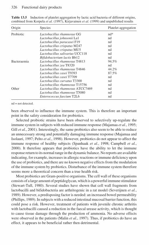
326 Functional dairy products
Table 13.5 Induction of platelet aggregation by lactic acid bacteria of different origins,combined from Korpela et al. (1997), Kirjavainen et al. (1999) and unpublished results
Origin Species Platelet aggregation
Probiotic Lactobacillus rhamnosus GG nd*Lactobacillus johnsonii La1 ndLactobacillus paracasei F19 ndLactobacillus crispatus M247 ndLactobacillus crispatus MU5 ndLactobacillus salivarius UCC118 ndBifidobacterium lactis Bb12 nd
Bacteraemia Lactobacillus rhamnosus T4813 94.5%Lactobacillus zea T8320 ndLactobacillus rhamnosus T4846 44.2%Lactobacillus casei T9393 87.5%Lactobacillus casei T7709 ndLactobacillus curvatus T3300 ndLactobacillus rhamnosus T15756 nd
Other Lactobacillus rhamnosus ATCC7469 ndLactobacillus rhamnosus T5080 ndEnterococcus faecium T2L6 nd
nd = not detected.
been observed to influence the immune system. This is therefore an importantpoint in the safety consideration for probiotics.
Selected probiotic strains have been observed to selectively up-regulate theimmune system in subjects with reduced immune response (Majamaa et al., 1995;Gill et al., 2001). Interestingly, the same probiotics also seem to be able to reducean unnecessary strong and potentially damaging immune response (Majamaa andIsolauri, 1997; Pelto et al., 1998). However, probiotics do not appear to affect theimmune response of healthy subjects (Spanhaak et al., 1998; Campbell et al.,2000). It therefore appears that probiotics have the ability to let the immuneresponse return to its normal range in the dynamic balance. No reports are availableindicating, for example, increases in allergic reactions or immune deficiency uponthe use of probiotics, and there are no known negative effects from the modulationof the immune system by probiotics. Disturbance of the immune system thereforeseems more a theoretical concern than a true health risk.
Most probiotics are Gram-positive organisms. The cell wall of these organismsconsists of a large amount of peptidoglycan, which is a powerful immune stimulator(Stewart-Tull, 1980). Several studies have shown that cell wall fragments fromlactobacilli and bifidobacteria are arthritogenic in a rat model (Severijnen et al.,1989). However, a predisposing factor is needed: an increased bowel permeability(Phillips, 1989). In subjects with a reduced intestinal mucosal barrier function, thiscould pose a risk. However, treatment of patients with juvenile chronic arthritiswith lactobacilli caused a reduction in the faecal urease activity, which is thoughtto cause tissue damage through the production of ammonia. No adverse effectswere observed in the patients (Malin et al., 1997). Thus, if probiotics do have aneffect, it appears to be beneficial rather then detrimental.
Safety evaluation of probiotics 327
13.8 Post-marketing surveillance
The manufacturer of a probiotic product has the final responsibility for its safety.Once a probiotic product has been brought onto the market, surveillance for anycases of disease related to the probiotic is important in order to be able to continueto ascertain the safety of the probiotic. Any Lactobacillus or Bifidobacteriumisolates associated with disease should be compared to the probiotic strain of thesame species (Saxelin et al., 1996b). This illustrates that accurate taxonomicidentification is essential. Especially in case reports on Lactobacillus- or Bifido-bacterium-associated infections, proper identification is important; unfortunatelythis is often not the case.
Proper storage and established availability of any clinical Lactobacillus orBifidobacterium isolates for future research are essential. This guarantees thesafety of the current probiotics on the market and these isolates may, at the sametime, provide information on the potential risk factors of these genera (Apostolouet al., 2001).
13.9 Safety issues for new generation probiotics
New probiotic microorganisms and novel probiotic products are likely to beintroduced to the market. These will require different safety assessment pro-cedures, which have been established, e.g. in the European Novel Foods Directiveand in the US Premarketing Approval Clearances.
13.9.1 Novel probiotic speciesMost of the current probiotics belong to the genera Lactobacillus and Bifido-bacterium. However, some probiotics belong to other genera (Table 13.2), whichin most cases do not have a history of safe use. For some of these organisms other,possibly more strict, safety assessments may be necessary. It is likely that a numberof novel probiotics will represent other genera of lactic acid bacteria, Lactococcus,Leuconostoc, etc., or they may be of other food-related genera, for example,Propionibacterium. However, most interesting will be probiotics from outside thistraditional field as e.g. Oxalobacter formigenes, which by metabolising oxalatemay reduce the risk for kidney stones (Sidhu et al., 2001). Such probiotics will setnew standards for the safety assessment.
13.9.2 Genetically modified probioticsGenetically modified organisms (GMO) have a low consumer acceptance, at leastin Europe. It is therefore unlikely that GMO versions of current probiotic productswill enter the market any time soon. However, in clinical applications GMOprobiotics may be more readily accepted and some potential applications in thisarea are under investigation. GMO probiotics could be used to deliver antigens for
328 Functional dairy products
vaccines. This would provide a safer method of vaccination than the use ofattenuated pathogens (Mercenier et al., 2000).
A GMO, Lactococcus lactis, has been observed to be able to produce IL-10 inthe mouse intestine (Steidler et al., 2000). This may provide new treatmentstrategies for inflammatory bowel disease, and similar applications may be usefulfor other diseases. The safety of such organisms that are able to produce these verypowerful bioactive substances is of major importance as excess production of thesesubstances in a healthy individual may be detrimental. However, in general thesafety assessment and regulations for the use of GMOs, including lactobacilli, isstrict and may provide sufficient protection. The details of regulations concerningGMOs fall outside the scope of this chapter.
13.9.3 Non-viable probioticsAlthough probiotics are generally defined as live microorganisms, there is someindication that non-viable probiotics may also have beneficial health effects(Ouwehand and Salminen 1998). Non-viable probiotics would have clear practicaland economic benefits: longer shelf-life, no need for cooled transportation andstorage, etc. They may also have a safety advantage. Although the risk for infectiondue to probiotic Lactobacillus or Bifidobacterium preparations is extremely small,non-viable probiotics would not have this risk at all. This is likely to providemainly a theoretical advantage, though non-viable probiotics could be consideredsafer when used in extremely high-risk immunosuppressed patients.
13.9.4 Novel applicationsThe main application for probiotics is their use in foods, aiming at affecting thecomposition or activity of the intestinal microflora or directly affecting thefunction of the intestine. However, the probiotic principle should be expected towork in any part of the body that has a normal microflora. With the exception of theurogenital tract, extra-intestinal applications of probiotics have received littleattention. Such probiotic preparations would clearly need different safety require-ments.
13.10 The safety of animal probiotics
Probiotics are also commonly used in veterinary applications (Nousiainen andSetälä, 1998). Despite the fact that the probiotics will be used for animals, morestrict safety assessment may be necessary than for humans. They should prove tobe safe for both animals and humans. In the case of probiotics for farm animals, theprobiotics may enter the food chain (Nikoskelainen et al., 2001). Because of theoften intimate relation between a pet and its owner, spread of the probiotic fromanimal to human is possible. Because enterococci are commonly used in probioticpreparations for animals, this may be reason for some concern.
Safety evaluation of probiotics 329
The safety requirements for animals are different from those for humans; Lacto-coccus garvieae has been found to be associated with mastitis in cows (Collins etal., 1983) and septicaemia in fish (Schmidtke and Carson 1999). Although Lc.garvieae has been found to be associated with disease in humans, this has so farbeen very limited and underlying diseases have been implicated (Fefer et al.,1998). Its true pathogenesis for humans remains therefore to be determined(Aguirre and Collins 1993). Another Lactococcus species that has been found to beinvolved in disease of fish is Lc. piscium (Williams et al., 1990). This species hasnot been found to be associated with disease in humans.
13.11 The current regulatory context
13.11.1 Dietary supplementsThe dietary supplement market for probiotics is large and growing in manycountries. A multitude of bacterial genera and species is used and include manydifferent Lactobacillus and Bifidobacterium species. In addition to these, many ofthese supplements also contain enterococci, yeasts and sometimes even Escher-ichia coli, Bacillus and Clostridium strains. The safety of these organisms has to beassessed separately.
13.11.2 Safety assessment in the United StatesIn the United States, the Food and Drug Administration (FDA) controls the safetyof foods and dietary supplements. Probiotics have been sold in the US as compo-nents of conventional foods or as dietary supplements. The safety of traditionallactic starter bacteria has been accepted. However, new and less traditional strainsof microbes have to be more carefully assessed prior to distribution to consumerswith potentially compromised health. In the US, Title 21 of the Code of FederalRegulations (21 CFR) and the FDA Office of Premarket Approval lists microor-ganisms that are approved food additives or which enjoy Generally Recognized asSafe (GRAS) status. ‘GRAS’ status is always considered only for a specified use.Thus, for instance, microbes themselves are not considered GRAS, but theirtraditional use in dairy foods is.
Based on the regulation, a substance that will be added to food is subject topremarket approval by the FDA unless its use is considered GRAS by qualifiedexperts. In addition, a substance that is used in accordance with a sanction grantedprior to 6 September 1958 is not subject to premarket approval. On 17 April 1997,the FDA issued a proposed rule (The GRAS proposal 62 FR 18938) that wouldestablish a notification procedure whereby any person (or interested company)may notify the FDA of a determination by that person that a particular use of asubstance is GRAS for specific use.
Currently, it can be understood that harmless lactic acid-producing bacteria,including L. acidophilus and other lactic acid bacteria (specifically S. thermophilus,
330 Functional dairy products
Lc. lactis subsp. cremoris, Lc. lactis subsp. lactis, L. delbrueckii subsp. bulgaricus,L. delbrueckii subsp. lactis, L. fermentum and three Leuconostoc species) can beused as additional ingredients in specified standardised foods such as cultured milk(including yoghurt and buttermilk), sour cream, cottage cheese and yoghurt,provided that the mandatory cultures of L. bulgaricus and S. thermophilus are alsoused in yoghurt. Many organisms used in foods are not included on the FDA list,but the absence of a species or a strain does not constitute a safety issue. In a morespecific manner, the FDA has also approved a GRAS petition by Nestlé USconcerning the use of Bifidobacterium lactis Bb12 and Streptococcus thermophilusTh4 in infant formula and foods for infants and children. Based on the publishedinformation by the FDA, other strains of Lactobacillus, Bifidobacterium andStreptococcus have been withdrawn from the petition. No additional lactic acidbacteria or currently used probiotics have been petitioned or approved at this time(April 2002).
Regarding the safety of enterococci, the picture is less clear-cut. Foods contain-ing enterococci are safely consumed on a regular basis. However, safety reportsseem to agree that enterococci pose a greater threat to health than other lactic acidbacteria. According to the US food regulations, such food substances that wereused prior to 1958 do not need approval procedures.
13.11.3 Safety assessment in the European UnionIn the European Union foods that were not available to a significant part of thepopulation in one of the member states before 1998 are considered novel foods;this includes also probiotics and starters. The purpose of the EC Novel FoodsRegulation (258/97) is to ensure the free movement of novel foods, while protect-ing the interests of consumers, especially with respect to safety, health andinformation. New probiotic or starter strains that are substantially equivalent toexisting conventional organisms concerning their composition, nutritional value,metabolism, intended use and level of undesirable substances contained in themare not regarded as novel foods. Otherwise, however, the strains will need to beassessed according to the novel food regulations and procedures.
13.11.4 Safety assessment in GermanyIn Germany, an occupational foundation of the chemical industry has establishedan expert group to assess the safety of microbiology and biotechnology. Thisexpert group has also assessed the safety of microbes and published a listcontaining the classification of bacteria used by different industries including foodand feed industries (Berufsgenossenschaft der chemischen Industrie, 1998). Theclassification divides bacteria into four risk groups. Almost all lactic acid bacteriaand bifidobacteria belong to risk group 1, indicating that there are no known risksin the handling of these bacteria by humans or animals. Some lactic acid bacteria,such as L. rhamnosus wild types and L. uli and L. catenaformis, belong to riskgroup 2, indicating that caution should be taken when handling these bacteria.
Safety evaluation of probiotics 331
Similar classification applies to most lactic acid bacteria and bifidobacteria; amongthe bifidobacteria, B. dentium is classified in risk group 2. In the Enterococcusgroup, a significant part of the species belong to risk group 2, associating thesebacteria with a low but existing risk of infection at the workplace. Also, many ofthe class 2 enterococci are scheduled for further reassessment. Basically, theGerman classification applies to occupational exposure, but the general safetyfocus may be applied in a wider perspective in some cases.
13.11.5 Safety assessment in JapanIn Japan probiotics and other functional foods fall under the Food for SpecifiedHealth Uses (FOSHU) regulation. FOSHU regulations do not specifically definethe safety aspects for probiotic microbes but for functional foods in general. Inorder for a product to be approved as FOSHU, companies need to go through anapplication process that typically takes about one year to complete. Applicationsare reviewed by local prefecture authorities and the Ministry of Health andWelfare. Applications must include scientific documentation demonstrating themedical or nutritional basis for a health claim, the basis for the recommended doseof the functional ingredient, information demonstrating the safety of the ingredi-ent, information on physical and chemical characteristics, relevant test methods,and a compositional analysis. This information is expected to be accompanied byscientific papers in the fields of medicine and/or nutrition that substantiate thehealth claim.
13.11.6 International Dairy Federation and dairy starter and probioticstrainsWithin the International Dairy Federation (IDF) an expert action team has beenpreparing a position document on the properties of starter cultures and probioticsused by the dairy industry. The IDF work is proceeding in collaboration with theEuropean Food and Feed Culture Association and the aim is to produce a list ofcultures with the safe history of use in dairy foods. Also, a document on the useprior to 1998, the introduction of the European Union Novel Foods Directive, isbeing prepared and the worldwide position document on this subject is underreview.
13.12 Conclusion and future trends
The current safety record of food starter cultures and probiotics appears to beexcellent. However, future development of probiotics, and also industrial starterculture and fermentation culture organisms, requires stringent guidelines for safetyassessment of such organisms. Therefore, constant surveillance of food microor-ganisms and their appearance in clinical situations is extremely important. Methodsfor both the premarket safety assessment and post-marketing surveillance of
332 Functional dairy products
human populations should be developed and refined to guarantee the safety offuture products in all populations including subjects with immune suppression.This will enable the future use of microbes and microbial fermentations for awidening area in food technology and in functional and clinical food areas.
13.13 Sources for further information and adviceADAMS M R and NOUT M J R (eds) (2001), Fermentation and Food Safety, Aspen Publishers,
Inc., Gaithersburg, 290 pp.ELMER G W, MCFARLAND L V and SURAWICZ C M (eds) (1999), Biotherapeutic Agents and
Infectious Diseases, Humana Press, Inc., Totowa, 316 pp.HANSON L Å and YOLKEN R H (eds) (1999), Probiotics, Other Nutritional Factors, and
Intestinal Microflora, Lippincott-Raven Publishers, Philadelphia, 306 pp.LEE Y-K, NOMOTO K, SALMINEN S and GORBACH S L (1999), Handbook of Probiotics, John
Wiley and Sons, Inc., New York, 211 pp.ROGINSKI H, FUQUAY J W and FOX P F (eds) (2002), Encyclopedia of Dairy Sciences,
Academic Press, New York.SALMINEN S, OUWEHAND A C and VON WRIGHT A (eds) (2003), Lactic Acid Bacteria:
Microbiology and Functional Aspects, Marcel Dekker, New York.
13.14 ReferencesADAMS M R and MARTEAU P (1995), ‘On the safety of lactic acid bacteria from food’, Int J
Food Microbiol, 27, 263–264.AGERHOLM-LARSEN L, RABEN A, HAULRIK N, HNASEN A S, MANDERS M and ASTRUP A (2000),
‘Effect of 8 week intake of probiotic milk products on risk factors for cardiovasculardiseases’, Eur J Clin Nutr, 54, 288–297.
AGUIRRE M and COLLINS M D (1993), ‘Lactic acid bacteria and human clinical infection’, JAppl Bact, 75, 95–107.
AHRNÉ S, NOBAEK S, JEPPSON B, ADLERBERTH I, WOLD A E and MOLIN G (1998), ‘The normalLactobacillus flora of healthy human rectal and oral mucosa’, J Appl Microbiol, 85, 88–94.
ALANDER M, KORPELA R, SAXELIN M, VILPPONEN-SALMELA T, MATTILA-SANDHOLM T andVON WRIGHT A (1997), ‘Recovery of Lactobacillus rhamnosus GG from human colonicbiopsies’, Lett Appl Microbiol, 24, 361–364.
ANTONY S J (2000), ‘Lactobacillemia: an emerging cause of infection in both theimmunocompromised and the immunocompetent host’, J Natl Med Assoc, 92, 83–86.
APOSTOLOU E, KIRJAVAINEN P V, SAXELIN M, RAUTELIN H, VALTONEN V, SALMINEN S J andOUWEHAND A C (2001), ‘Good adhesion properties of probiotics: a potential risk forbacteremia?’, FEMS Immunol Med Microbiol, 31, 35–39.
ARENA M E and MANCA DE NADRA M C (2001), ‘Biogenic amine production by Lactobacil-lus’, J Appl Microbiol, 90, 158–162.
AVLAMI A, KORDOSSIS T, VRIZIDIS N and SIPSAS N V (2001), ‘Lactobacillus rhamnosusendocarditis complicating colonoscopy’, J Infect, 42, 1–2.
BAUMGARTNER A, KUEFFER M, SIMMEN A and GRAND M (1998), ‘Relatedness of Lactobacil-lus rhamnosus strains isolated from clinical specimens and such from food-stuffs, humansand technology’, Lebensm Wiss Technol, 31, 489–494.
BECKER M R, PASTER B J, LEYS E J, MOESCHBERG M L, KENYON S G, GALVIN J L, BOCHES SK, DEWIRST F E and GRIFFEN A L (2002), ‘Molecular analysis of bacterial species associatedwith childhood caries’, J Clin Microbiol, 40, 1001–1009.
Safety evaluation of probiotics 333
BENGMARK S and JEPPSON B (1995), ‘Gastrointestinal surface protection and mucosareconditioning’, J Parent Ent Nutr, 19, 410–415.
BERUFSGENOSSENSCHAFT DER CHEMISCHEN INDUSTRIE (1998), Sichere Biotechnologie.Eingruppierung biologischer Agenzien: Bakterien. Merkblatt B 006. Heidelberg,Jedermannn Verlag Dr Otto Pfeffer oHG.
BONGAERTS G P A, TOLBOOM J J M, NABER A H J, SPERL W J K, SEVERIJNEN R S V M, BAKKEREN
J A J M and WILLEMS J L (1997), ‘Role of bacteria in the pathogenesis of short bowelsyndrome-associated D-lactic acidemia’, Microb Pathogen, 22, 285–293.
CAMPBELL C G, CHEW B P, LUEDECKE L O and SHULTZ T D (2000), ‘Yogurt consumption doesnot enhance immune function in healthy premenopausal women’, Nutr Canc, 37, 27–35.
CHEN X and HUANG G (1995), ‘Autopsy case report of a rare acute iatrogenic waterintoxication with a review of the literature’, Forensic Sci Int, 76, 27–34.
CHOMARAT M and ESPINOUSE D (1991), ‘Lactobacillus rhamnosus septicemia in patientswith prolonged aplasia receiving ceftazidime-vancomycin’, Eur J Clin Microbiol InfectDis, 10, 44.
COLLINS M D, FARROW J A E, PHILLIPS B A and KANDLER O (1983), ‘Streptococcus garvieaesp nov and Streptococcus plantarum sp nov’ J Gen Microbiol, 129, 3427–3431.
CORONADO B E, OPAL S M and YOBURN D C (1995), ‘Antibiotic induced D-lactic acidosis’,Ann Int Med, 122, 839–842.
CROCIANI F, BIAVATI B, ALESSANDRINI A, CHIARINI C and SCARDOVI V (1996), ‘Bifidobacteriuminopinatum sp. nov. and Bifidobacterium denticolens sp. nov., two new species isolatedfrom human dental caries’, Int J Syst Bacteriol, 46, 564–71.
DE BOEVER P and VERSTREATE W (1999), ‘Bile salt deconjugation by Lactobacillusplantarum 80 and its implication for bacterial toxicity’, J Appl Microbiol, 87, 345–352.
DONG M-Y, CHANG, T-W and GORBACH, SL (1987), ‘Effects of feeding Lactobacillus GG onlethal irradiation in mice’, Diagn Microbiol Infect Dis, 7, 1–7.
DONOHUE D C and SALMINEN S (1996), ‘Safety of probiotic bacteria’, Asia Pacific J Clin Nutr,4, 25–28.
ELLIOTT S N, BURET A, MCKNIGHT W, MILLER M J S and WALLACE J L (1998), ‘Bacteria rapidlycolonize and modulate healing of gastric ulcers in rats’, Am J Physiol, 275, G425–G432.
FALKOW S (1997), ‘What is a pathogen?’, ASM News, 63, 359–365.FEFER J J, RATZAN K R, SHARP S E and SAIZ E (1998), ‘Lactococcus garvieae endocarditis:
report of a case and review of the literature’, Diagn Microbiol Infect Dis, 32, 127–130.FINLAY B B and FALKOW S (1997), ‘Common themes in microbial pathogenicity revisited’,
Microbiol Mol Biol Rev, 61, 136–169.FRANZ C M A P, HOLZAPFEL W H and STILES M E (1999), ‘Enterococci at the crossroads of food
safety’, Int J Food Microbiol, 47, 1–24.FRUCHART C, SALAH A, GRAY C, MARTIN E, STAMATOULLAS A, BONMARCHAND G, LEMELAND
J F and TILLY H (1997), ‘Lactobacillus species as emerging pathogens in neutropenicpatients’, Eur J Clin Microbiol Infect Dis, 16, 681–684.
GALLEMORE G H, MOHON R T and FERGUSON D A (1995) ‘Lactobacillus fermentumendocarditis involving a native matral valve’, J Tenn Med Assoc, 88, 306–308.
GASSER F (1994), ‘Safety of lactic acid bacteria and their occurrence in human clinicalinfections’, Bull Inst Pasteur, 92, 45–67.
GAVAZZI C, STACCHIOTTI S, CAVALLETTI R and LODI R (2001), ‘Confusion after antibiotics’,Lancet, 357, 1410.
GILL H, RUTHERFORD K J, CROSS M L and GOPAL P K (2001), ‘Enhancement of immunity inthe elderly by dietary supplementation with the probiotic Bifidobacterium lactis HN019’,Am J Clin Nutr, 74, 833–839.
GONZÁLEZ DE LIANO D, CUESTA P and RODRÍGUEZ A (1998), ‘Biogenic amine production bywild lactococcal and leuconostoc strains’, Lett Appl Microbiol, 26, 270–274.
GORBACH S L and GOLDIN B R (1989) ‘Lactobacillus strains and methods of selection’, USPatent: no. 4,839,281.
HARTY D W S, PATRIAKIS M and KNOX K W (1993), ‘Identification of Lactobacillus strains
334 Functional dairy products
isolated from patients with infective endocarditis and comparison of their surface-associated properties with those of other strains of the same species’, Microb Ecol HealthDis, 6, 191–201.
HARTY D W S, OAKEY H J, PATRIKAKIS M, HUME E B H and KNOX K W (1994), ‘Pathogenicpotential of lactobacilli’, Int J Food Microbiol, 24, 179–189.
HATA D, YOSHIDA A, OHKUBO H, MOCHIZUKI Y, HOSOKI Y, TANAKA R and AZUMA R (1988),‘Meningitis caused by Bifidobacterium in an infant’, Ped Infect Dis J, 7, 669–671.
HENNEQUIN C, KAUFFMANN-LACROIX C, JOBERT A, VIARD T, RIQOUR C, JACQUEMIN J L andBERCHE P (2000), ‘Possible role of catheters in Saccharomyces boulardii fungemia’, EurJ Clin Microbiol Infect Dis, 19, 16–20.
HERRANZ C, CASAUS P, MUKHOPADHYAY S, MARTÍNEZ J M, RODRÍGUEZ J M, NES I F,HERNÁNDEZ P E and CINTAS L M (2001), ‘Enterococcus faecium P21: a strain occurringnaturally in dry-fermented sausages producing the class II bacteriocins enterocin A andenterocin B’, Food Microbiol, 18, 115–131.
HORWITCH C A, FURSETH H A, LARSON A M, JONES T L, OLLIFFE J F and SPACH D H (1995),‘Lactobacillemia in three patients with AIDS’, Clin Infect Dis, 21, 1460–1462.
HUSNI R N, GORDON S M, WASHINGTON J A and LONGWORTH D L (1997), ‘Lactobacillusbacteremia and endocarditis: review of 45 cases’, Clin Infect Dis, 25, 1048–1055.
JONES R, ELLBOGEN M and DUNLAY R (2000), ‘Lactobacillus allograft pyelonephritis andbacteremia’, Nephron, 86, 502.
KANEKO T, BANDO Y, KURIHARA H, SATOMI K, NONOYAMA K and MATSUURA N (1997), ‘Fecalmicroflora in a patient with short-bowel syndrome and identification of dominantlactobacilli’, J Clin Microbiol, 35, 3181–3185.
KIRJAVAINEN P V, TUOMOLA E M, CRITTENDEN R G, OUWEHAND A C, HARTY D W S, MORRIS
L F, RAUTELIN H, PLAYNE M J, DONOHUE D C and SALMINEN S J (1999), ‘In vitro adhesionand platelet aggregation properties of bacteremia-associated lactobacilli’, Infect Immun,67, 2653–2655.
KORPELA R, MOILAEN E, SAXELIN M and VAPAATALO H (1997), ‘Lactobacillus rhamnosusGG (ATCC 53103) and platelet aggregation in vitro’, Int J Food Microbiol, 37, 83–86.
MACKAY A D, TAYLOR M B, KIBBLER C C and HAMILTON-MILLER J M T (1999), ‘Lactobacillusendocarditis caused by a probiotic organism’, Clin Microbiol Infect, 5, 290–292.
MAJAMAA H and ISOLAURI E (1997), ‘Probiotics: A novel approach in the management offood allergy’, J Allergy Clin Immunol, 99, 179–185.
MAJAMAA H, ISOLAURI E, SAXELIN M and VESKARI T (1995), ‘Lactic acid bacteria in thetreatment on acute rotavirus gastroenteritis’, J Ped Gastroenterol Nutr, 20, 333–338.
MALIN M, VERRONEN P, KORHONEN H, SYVÄOJA E-L, SALMINEN S, MYKKÄNEN H, ARVILOMMI
H, EEROLA E and ISOLAURI E (1997), ‘Dietary therapy with Lactobacillus GG, bovinecolostrum or bovine immune colostrum in patients with juvenile chronic arthritis:evaluation of effect on gut defence mechanisms’, Inflammopharmacology, 5, 219–236.
MERCENIER A, MULLER-ALOUF H and GRANGETTE C (2000), ‘Lactic acid bacteria as livevaccines’, Curr Issues Mol Biol, 2, 17–25.
MOMOSE H, IGARASHI M, ERA T, FUKUDA Y, YAMADA M and OGASA K (1979), ‘Toxicologicalstudies on Bifidobacterium longum BB-536’, Ouyou Yakuri, 17, 881–887.
NIKOSKELAINEN S, SALMINEN S, BYLUND G and OUWEHAND A C (2001), ‘Characterization ofthe properties of human- and dairy-derived probiotics for prevention of infectious diseasesin fish’, Appl Environ Microbiol, 67, 2430–2435.
NOUSIAINEN J and SETÄLÄ J (1998),‘Lactic acid bacteria as animal probiotics’. In: S Salminenand A von Wright, Lactic Acid Bacteria: Microbiology and Functional Aspects, NewYork: Marcel Dekker, Inc, 437–473.
OUWEHAND A C, KIRJAVAINEN P V, GRÖNLUND M-M, ISOLAURI E and SALMINEN S J (1999),‘Adhesion of probiotic micro-organisms to intestinal mucus’, Int Dairy J, 9, 623–630.
OUWEHAND A C and SALMINEN S J (1998), ‘The health effects of viable and non-viablecultured milks’, Int Dairy J, 8, 749–758.
OUWEHAND A C, LAGSTRÖM H, SUOMALAINEN T and SALMINEN S (2002), ‘Effect of probiotics
Safety evaluation of probiotics 335
on constipation, fecal azoreductase activity and fecal mucin content in the elderly’, AnnNutr Metab, 46, 159–162.
PELTO L, ISOLAURI E, LILIUS E-M, NUUTILA J and SALMINEN S (1998), ‘Probiotic bacteria down-regulate the milk-induced inflammatory response in milk-hypersensitve subjects but havea immunostimulatory effect in healthy subjects’, Clin Exp Allergy, 28, 1474–1479.
PELTONEN K D, EL-NEZAMI H S, SALMINEN S J and AHOKAS J T (2000), ‘Binding of aflatoxinB1 by probiotic bacteria’, J Sci Food Agric, 80, 1942–1945.
PENOT J P, LAGRANGE P, DARODES N, PLOY M C, VIOLET T, VIROT P, MANSOUR L and BESAIDJ (1998), ‘Lactobacillus acidophilus endocarditis’, Presse Med, 27, 1009–1012.
PERLMUTTER D H, BOYLE J T, CAMPOS J M, EGLER J M and WATKINS J B (1983), ‘D-Lacticacidosis in children: an unusual metabolic complication of small bowel resection’, JPediatr, 102, 234–238.
PHILLIPS P E (1989), ‘How do bacteria cause chronic arthritis?’ J Reumathol, 16, 1017–1019.RAUTIO M, JOUSIMIES-SOMER H, KAUMA H, PIETARINEN I, SAXELIN M, TYNKKYNEN S and
KOSKELA M (1999), ‘Liver abscess due to a Lactobacillus rhamnosus strain indistinguish-able from L rhamnosus strain GG’, Clin Infect Dis, 28, 1159–1160.
RODRIGUEZ A V, BAIGORI M D, ALVAREZ S, CASTRO G R and OLIVER G (2001),‘Phosphatidylinositol-specific phospholipase C activity in Lactobacillus rhamnosus withcapacity to translocate’, FEMS Microbiol Lett, 204, 33–38.
ROGINSKI H (2002), ‘Fermented milks of northern Europe’ in Roginski H, Fuquay J W andFox P F Encyclopedia of Dairy Sciences, New York, Academic Press, in press.
ROWLAND I (1999), ‘Probiotics and benefits to human health – the evidence in favour’,Environ Microbiol, 1, 375–382.
RUSELER-VAN EMBDEN J G H, VAN LIESHOUT L M C and MARTEAU P (1995), ‘No degradationof intestinal mucus glycoproteins by Lactobacillus casei strain GG’, Microecol Ther, 25,304–309.
SALMINEN S, BOULEY C, BOUTRON-RUAULT M-C, CUMMINGS J H, FRANCK A, GIBSON G R,ISOLAURI E, MOREAU M-C, ROBERFROID M and ROWLAND I (1998), ‘Functional foodscience and gastrointestinal physiology and function’, Br J Nutr, 80, S147–S171.
SALMINEN S J, VON WRIGHT A J, OUWEHAND A C and HOLZAPFEL W H (2001), ‘Safetyassessment of probiotics and starters’, in Adams M R and Nout M J R, Fermentation andFood Safety, Gaithersburg: Aspen Publishers, Inc, 239–251.
SAXELIN M (1997), ‘Lactobacillus GG – a human probiotic strain with thorough clinicaldocumentation’, Food Rev Int, 13, 293–313.
SAXELIN M, CHUANG N-H, CHASSY B, RAUTELIN H, MÄKELÄ P H, SALMINEN S and GORBACHS L (1996a), ‘Lactobacilli and bacteremia in southern Finland, 1989–1992’, Clin INFECT
DIS, 22, 564–566.SAXELIN M, RAUTELIN H, SALMINEN S and MÄKELÄ P H (1996b), ‘Safety of commercial
products with viable Lactobacillus strains’, Infect Dis Clin Pract, 5, 331–335.SCHMIDTKE L M and CARSON J (1999), ‘Induction, characterisation and pathogenicity in
rainbow trout Oncorhynchus mykiss (Walbaum) of Lactococcus garvieae L-forms’, VetMicrobiol, 69, 287–300.
SEVERIJNEN A J, VAN KLEEF R, HAZENBERG M P and VAN DE MERWE J P (1989), ‘Cell wallfragments from major residents of the human intestinal flora induce chronic arthritis inrats’, J Reumathol, 16, 1061–1068.
SIDHU H, ALLISON M J, MAY CHOW J, CLARK A and PECK A B (2001), ‘Rapid reversal ofhyperoxaluria in a rat model after probiotic administration of Oxalobacter formigens’, JUrol, 166, 1487–1491.
SIMS W (1964), ‘A pathogenic Lactobacillus’, J Path Bact, 87, 99–105.SPANHAAK S, HAVENAAR R and SCHAAFSMA G (1998) ‘The effect of consumption of milk
fermented by Lactobacillus casei strain Shirota on the intestinal microflora and immuneparameters in humans’, Eur J Clin Nutr, 421, 65–72
SREEKUMAR O and HOSONO A (1998), ‘The heterocyclic amine binding receptors ofLactobacillus gasseri cells’, Mut Res, 421, 65–72.

336 Functional dairy products
STEIDLER L, HANS W, SCHOTTE L, NEIRYNCK S, OBERMEIER F, FALK W, FIERS W and REMAUTE (2000), ‘Treatment of murine colitis by Lactococcus lactis secreting interleukin-10’,Science, 289, 1352–1355.
STEWART-TULL D E S (1980), ‘The immunological activities of bacterial peptidoglycans’,Ann Rev Microbiol, 34, 311–340.
STYRIAK I, ZATKOVIC B and MARSALKOVA S (2001), ‘Binding of extracellular matrix proteinsby lactobacilli’, Folia Microbiol, 46, 83–85.
SUMNER S S, SPECKHARD M W, SOMERS E B and TAYLOR S L (1985), ‘Isolation of histamine-producing Lactobacillus buchneri from Swiss cheese implicated in a food poisoningoutbreak’, Appl Environ Microbiol, 50, 1094–1096.
TAKAHASHI M and ONOUE M (1999), ‘Safety of Lactobacillus casei strain Shirota’, in YakultCentral Institute for Microbiological Research Lactobacillus casei Strain Shirota –Intestinal Flora and Human Health, Tokyo: Yakult Honsha Co, Ltd, 263–281.
TANAKA H, DOESBURG K, IWASAKI T and MIERAU I (1999), ‘Screening of lactic acid bacteriafor bile salt hydrolase activity’, Int Dairy J, 82, 2530–2535.
TEUBER M, MEILE L and SCHWARZ F (1999), ‘Acquired antibiotic resistance in lactic acidbacteria from food’, Antonie van Leeuwenhoek, 76, 115–137.
TURBIC A, AHOKAS J T and HASKARD C A (2002), ‘Selective in vitro binding of dietarymutagens, individually or in combination, by lactic acid bacteria’, Food Add Contam, 19,144–152.
TYNKKYNEN S, SINGH K V and VARMANEN P (1998), ‘Vancomycin resistance factor ofLactobacillus rhamnosus GG in relation to enterococcal vancomycin resistance (van)genes’, Int J Food Microbiol, 41, 195–204.
WADE J J (1997), ‘Enterococcus faecium in hospitals’, Eur J Clin Microbiol Infect Dis, 16,113–119.
WAGNER R D, PIERSON C, WARNER T, DOHNALEK M, FARMER J, ROBERTS L, HILTY M andBALISH E (1997a), ‘Biotheraputic effects of probiotic bacteria on candidiasis inimmunodeficient mice’, Infect Immunol, 65, 4165–4172.
WAGNER R D, WARNER T, ROBERTS L, FARMER J and BALISH E (1997b), ‘Colonization ofcongenitally immunodeficient mice with probiotic bacteria’, Infect Immunol, 65, 3345–3351.
WANG X, ANDERSSON R, SOLTESZ V, LEVEAU P and IHSE I (1996), ‘Gut origin sepsis,macrophage function, and oxygen extraction associated with acute pancreatitis in the rat’,World J Surg, 20, 299–308.
WILLIAMS A M, FRYER J L and COLLINS M D (1990), ‘Lactococcus piscium sp nov: a newLactococcus species from salmonid fish’, FEMS Microbiol Lett, 68, 109–114.
VON WRIGHT A and SIBAKOV M (1998), ‘Genetic modification of lactic acid bacteria’. In:Salminen S and von Wright A, Lactic Acid Bacteria: Microbiology and FunctionalAspects, New York: Marcel Dekker, 161–210.
ZHOU J S, SHU Q, RUTHERFORD K J, PRASAD J, GOPAL P K and GILL H (2000), ‘Acute oraltoxicity and bacterial translocation studies on potentially probiotic strains of lactic acidbacteria’, Food Chem Toxicol, 38, 153–161.
ZHOU J S, GOPAL P K and GILL H S (2001), ‘Potential probiotic lactic acid bacteriaLactobacillus rhamnosus (HN001), Lactobacillus acidophilus (HN017) and Bifido-bacterium lactis (HN019) do not degrade gastric mucin in vitro’, Int J Food Microbiol,63, 81–90.
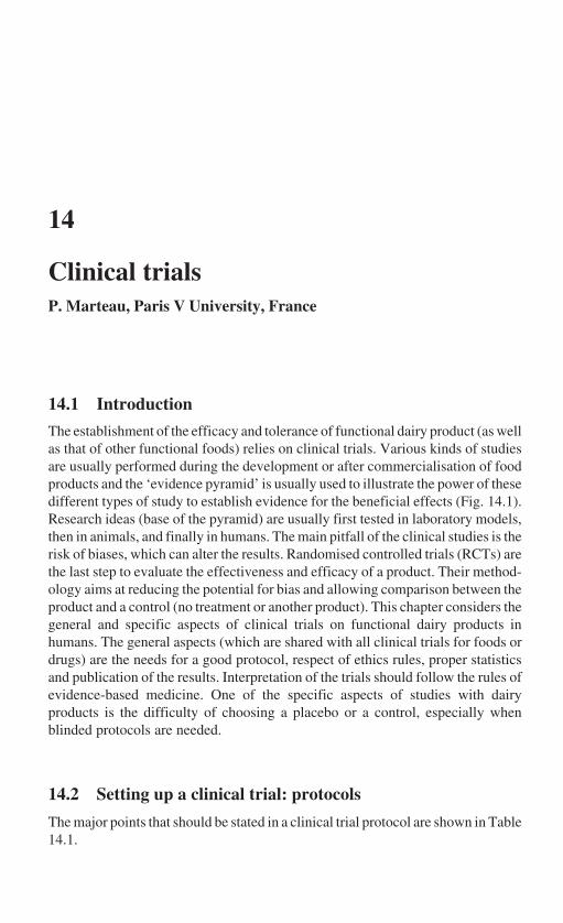
14
Clinical trialsP. Marteau, Paris V University, France
14.1 Introduction
The establishment of the efficacy and tolerance of functional dairy product (as wellas that of other functional foods) relies on clinical trials. Various kinds of studiesare usually performed during the development or after commercialisation of foodproducts and the ‘evidence pyramid’ is usually used to illustrate the power of thesedifferent types of study to establish evidence for the beneficial effects (Fig. 14.1).Research ideas (base of the pyramid) are usually first tested in laboratory models,then in animals, and finally in humans. The main pitfall of the clinical studies is therisk of biases, which can alter the results. Randomised controlled trials (RCTs) arethe last step to evaluate the effectiveness and efficacy of a product. Their method-ology aims at reducing the potential for bias and allowing comparison between theproduct and a control (no treatment or another product). This chapter considers thegeneral and specific aspects of clinical trials on functional dairy products inhumans. The general aspects (which are shared with all clinical trials for foods ordrugs) are the needs for a good protocol, respect of ethics rules, proper statisticsand publication of the results. Interpretation of the trials should follow the rules ofevidence-based medicine. One of the specific aspects of studies with dairyproducts is the difficulty of choosing a placebo or a control, especially whenblinded protocols are needed.
14.2 Setting up a clinical trial: protocols
The major points that should be stated in a clinical trial protocol are shown in Table14.1.
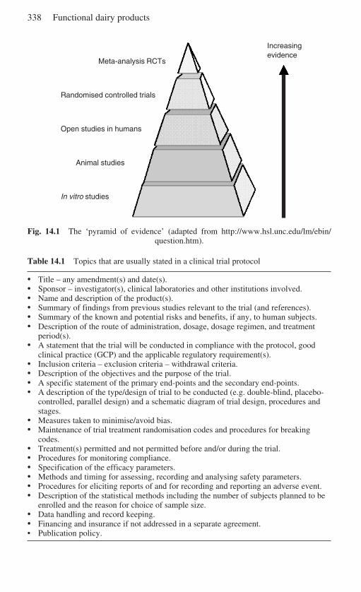
338 Functional dairy products
Fig. 14.1 The ‘pyramid of evidence’ (adapted from http://www.hsl.unc.edu/lm/ebin/question.htm).
Table 14.1 Topics that are usually stated in a clinical trial protocol
• Title – any amendment(s) and date(s).• Sponsor – investigator(s), clinical laboratories and other institutions involved.• Name and description of the product(s).• Summary of findings from previous studies relevant to the trial (and references).• Summary of the known and potential risks and benefits, if any, to human subjects.• Description of the route of administration, dosage, dosage regimen, and treatment
period(s).• A statement that the trial will be conducted in compliance with the protocol, good
clinical practice (GCP) and the applicable regulatory requirement(s).• Inclusion criteria – exclusion criteria – withdrawal criteria.• Description of the objectives and the purpose of the trial.• A specific statement of the primary end-points and the secondary end-points.• A description of the type/design of trial to be conducted (e.g. double-blind, placebo-
controlled, parallel design) and a schematic diagram of trial design, procedures andstages.
• Measures taken to minimise/avoid bias.• Maintenance of trial treatment randomisation codes and procedures for breaking
codes.• Treatment(s) permitted and not permitted before and/or during the trial.• Procedures for monitoring compliance.• Specification of the efficacy parameters.• Methods and timing for assessing, recording and analysing safety parameters.• Procedures for eliciting reports of and for recording and reporting an adverse event.• Description of the statistical methods including the number of subjects planned to be
enrolled and the reason for choice of sample size.• Data handling and record keeping.• Financing and insurance if not addressed in a separate agreement.• Publication policy.
Increasingevidence
Meta-analysis RCTs
Randomised controlled trials
Open studies in humans
Animal studies
In vitro studies
Clinical trials 339
14.2.1 How to ask a clinical question that can be answeredThe aim of a clinical trial is to answer a question. It is thus obvious that this questionhas to be formulated before the beginning of the trial, and that the design of the trialshould be adapted to answer the question. There are two major types of questionsconcerning efficacy of a product: is it more efficient than another product or is it asefficient as another product? The design of the trial, the number of subjects toinclude and the statistical tests depend on the type of question. Another point is thatthe evidence that supports the validity (truthfulness) of the results of a studydepends primarily on the studies methodology. The optimal methodology aims toavoid biases. These can be defined as the systematic tendency of any aspects of thedesign, conduct, analysis and interpretation of the results to make the estimate ofa product effect deviate from its true value. David Sackett, a pioneer in studymethodology, identified no less than 56 possible biases that may arise in clinicaltrials and stressed that more than half of them were related to the study design.1, 2
Consequently, one should always seek statistical help in planning a study, both onsample size and study design (in other words the statistician should be involvedwhen planning the study, not only when it is done, and that the results need to beanalysed). Study methodologies such as randomisation, blinding and accountingfor all patients help ensure that the study results are not biased.
14.2.2 Selection of the major end-point and of the control groupThe first critical step is to select the major end-point of the study. If the major end-point of a study is not clearly stated in an article (and when several end-points areused), the reader must always consider that positive results may be biased by theauthor opinion.3 The choice of the major end-point should be based on itsrelevance. Knowledge of the results of the major end-point in the control group(mean and standard deviation) is needed to calculate the number of subjects (seebelow).
The choice of the control group is also very important. Four types of controlscan be used: no treatment, placebo, different dose of the study product, or differentactive product. A placebo is a dummy product that must be as close as possible tothe test product in terms of colour, texture, taste, etc., but that does not contain theactive ingredient of the test product. Preparing such a placebo is often difficult withmilk products, as their flavour and texture are subtle. The composition of theplacebo should also be chosen in order to avoid nocebo effect. (The term noceboor ‘negative placebo’ is used for conditions that reduce the therapeutic effective-ness of a product, causing deteriorating symptoms or even new ones.) Forexample, a lactose-rich product is not a good placebo for a study trying to provethat a fermented dairy product will help shortening diarrhoea in subjects withgastroenteritis (because lactose can induce diarrhoea).
The use of a ‘no treatment’ or placebo control often raises problems of ethics,acceptability and feasibility, which have to be carefully considered before thestudy. One of the possibilities is to plan for ‘early escape’ from the protocol in caseof treatment failure or to use an add-on design. The term ‘add-on study’ is used for
340 Functional dairy products
a placebo-controlled trial of a product conducted in subjects also receivingstandard treatment. For example, Guslandi compared the efficacy of 3 g/dmesalazine to 2 g/d mesalazine plus 1 g of Saccharomyces boulardii in patientswith Crohn’s disease.4 With such a design, all the patients received at least 2 g ofthe classical treatment mesalazine.
14.2.3 Randomisation and blindingRandomisation and blinding are used to minimise bias due to the subject or theinvestigator opinion on the product.3,5 Double-blind randomised studies provide afar stronger evidence for efficacy than non-blinded and open studies.3 Indeed,randomisation is the best way to avoid a systematic difference between the groupsat the beginning of the study (‘baseline’). However, there is still a possibility thatthe two groups will differ by chance and this has to be tested when analysing theresults. Subjects and investigators may be consciously or unconsciously influ-enced to report favourable effects with the product if they can differentiate it fromthe control. Blinding is intended to minimise this bias. In single-blind studies, onlythe patients ignore which product they receive, while in double-blind studies, boththe patients and the investigators do not know which product is taken. Theorganisation of this ‘blindness’ needs external helps (usually by the statistician).The blindness code should not be broken before the end of the study except in thecase of a severe adverse event and this has to be monitored.
14.3 Statistical analysis
One of the major aims of the statistical analysis of the results of a clinical trial is toevaluate the chance (or risk) that they could be due to chance and not to the effectof the product. Everyone will intuitively understand that this risk is lower whenhigh numbers of subjects are studied, but how many subjects should there be?6,7
There are two types of statistical errors. The type I error (alpha error) is to reject thehypothesis tested (i.e. the existence of a difference of efficacy between twoproducts) when it is true. The type II error (beta error) is to reject the hypothesistested when a given alternative hypothesis is true. The alpha and beta errors arelinked and are also linked to the statistical power of a trial.
Most studies make the hypothesis that a product will be more efficient than theother; the power calculation can then be used to determine how many subjects areneeded to have a reasonable (usually 80% or 90%) chance of detecting a clinicallyimportant difference between the two products (Table 14.2). The power of a test isthe probability that the test will reject the hypothesis tested when a specificalternative hypothesis is true. To calculate the power of a given test, it is necessaryto specify alpha and to specify a specific alternative hypothesis.8
Several statistical tests can be used in equivalence studies. The absence ofdifference between two groups using common statistical tests designed to test fordifferences does not allow us to establish the absence of a difference. The three
Clinical trials 341
Table 14.2 Information needed to calculate the number of subjects for a clinical trial
1. What is the major study end-point (single result or score, percentage, mean of severalmeasurements, survival time…)?
2. What values would be expected in the control group? (including standard deviationfor measurements)? (Look at the literature*)
3. How large a difference between the tested product and the control would beconsidered clinically relevant?
4. Do you want to test for equivalence between the two groups or superiority of oneproduct? (Equivalence testing needs more subjects)
*If the information does not exist, the study will have to be considered as a pilot study.
double-blind RCT studies that compared the probiotic mutaflor (which contains 25× 109 viable E. coli strain Nissle 1917 per 100 mg) to mesalazine to reduce the riskof recurrence of ulcerative colitis illustrate that perfectly. In the first study, Kruiset al.9 included 120 patients in a 12-week double-blind, double-dummy study. Halfof them received 1.5 g/d of mesalazine, and the other half received 200 mg/d ofmutaflor. After 12 weeks, 11.3% of the subjects receiving mesalazine and 16% ofthose receiving the probiotic had relapsed. This difference was not significant.However, the statistical power of the study was low as the percentage of recurrenceafter 12 weeks was low in both groups. Although the authors were convinced thatthe two products were equivalent, their conclusion was not accepted by the medicalcommunity.10 A second RCT was thus performed that included 116 patientsfollowed for 1 year. Once again equivalence tests were not used and the power ofthe study was not calculated accordingly. Although this study suggested nosignificance difference between the two treatments, the finding could still not beaccepted by statisticians and by gastroenterologists.10 A third RCT has recentlybeen presented in an abstract form in which statistical tests were planned to assessequivalence between treatments and the numbers of subjects were calculatedaccordingly.12 Some 222 patients were treated either with mesalazine or withmutaflor for one year. The relapse rate was 36.4% in the probiotic vs. 33% in themesalazine group and equivalence was statistically proven. Non-inferiority trialsare designed to demonstrate that a product is not relevantly inferior to a standard.Equivalence trials are usually designed to demonstrate equivalence with respect toa rate and extent of drug absorption between a reference and a generic version(bioequivalence goal).
14.4 Ethical issues
Clinical trials should be conducted in accordance with ethical principles. Ethicalprinciples have been formulated in the Declaration of Helsinki.13 They are consist-ent with good clinical practice (GCP) and the applicable regulatory requirements.14
Every clinical trial must first be evaluated by an independent ethics committee. Thelegal status, composition, function, operations and regulatory requirements per-taining to independent ethics committees may differ among countries, but should
342 Functional dairy products
allow the committee to act in agreement with GCP as described in this guidance.14
The members of such a committee are both medical/scientific professionals andnon-medical/non-scientific persons; their responsibility is
to ensure the protection of the rights, safety, and well-being of humansubjects involved in the trial and to provide public assurance of thatprotection, by, among other things, reviewing and approving/providingfavourable opinion on the trial protocol, the suitability of theinvestigator(s), facilities, and the methods and material to be used inobtaining and documenting informed consent of the trial subjects.14
Table 14.3 Main points of GCP for the investigators and the sponsor (adapted fromreference 14)
Investigator• Investigator’s qualifications and agreements• Adequate resources• Medical care of the trial subjects• Communication with IRB/IEC• Compliance with protocol• Investigational product(s)• Randomisation procedures and unblinding• Informed consent of the trial subjects• Records and reports• Progress reports• Safety reporting• Premature termination or suspension of a trial• Final report(s) by investigator/institution
Sponsor• Quality assurance and quality control• Trial design• Information about the product(s)• Manufacturing, packaging, labelling and coding the product• Supplying and handling the product(s)• Trial management, data handling, record keeping• Monitoring committee• Investigator selection• Allocation of duties and functions• Compensation to subjects and investigators• Financing• Notification/submission to regulatory authority(ies)• Record access• Safety information• Adverse drug reaction reporting• Monitoring• Audit• Non-compliance• Premature termination or suspension of a trial• Clinical trial/study reports
Clinical trials 343
The independent ethics committee must obtain the following documents: trialprotocol(s)/amendment(s), written informed consent form(s) and consent formupdates that the investigator proposes for use in the trial, written information to beprovided to subjects, available safety information, information about paymentsand compensation available to subjects, the investigator’s current curriculum vitaeand/or other documentation of qualifications. The independent ethics committeereviews the proposed clinical trial within a reasonable time and documents itsviews (approval/favourable opinion; modifications required prior to the approval/favourable opinion; disapproval/negative opinion; or termination/suspension ofany prior approval/favourable opinion).
14.5 Managing a clinical trial
GCP is an international ethical and scientific quality standard for designing,conducting, recording and reporting trials involving the participation of humansubjects.14 Compliance with this standard provides public assurance that the rights,safety and well-being of trial subjects are protected, consistent with the principlesof guidelines such as the ICH GCP Guideline,14 which have been written to providea unified standard for the European Union, Japan and the United States to facilitatethe mutual acceptance of clinical data by the regulatory authorities in thesejurisdictions. It is not possible to describe here in detail all the rules that need to befollowed to respect GCP; however, a list of the headings of the main points for theinvestigators and the sponsor is shown in Table 14.3.
Monitoring is defined as the act of overseeing the progress of a clinical trial,and of ensuring that it is conducted, recorded and reported in accordance with theprotocol, standard operating procedures, GCP and the applicable regulatoryrequirement(s). Clinical trials have to follow the GCP rules and be submitted to theIndependent Ethics Committee Review. Authorities will base their decisions forlabelling products on the quality and relevance of the clinical studies that areanalysed by expert committees.
14.6 Assessing the validity of a clinical trial
The main points to analyse in order to answer the question of the validity of aclinical study are summarised in Table 14.4. The assignment of patients to eithergroup (product or control) must be done by a random allocation to help eliminateconscious or unconscious bias (see above). The importance of the blinding hasbeen stressed above. The study should begin and end with the same number ofpatients. Patients lost to the study must be accounted for or risk making theconclusions invalid. Indeed, patients may drop out because of the adverse effectsof the therapy being tested. If not accounted for, this can lead to conclusions thatmay be over-confident about the efficacy of the therapy. Good studies must havemore than 80% follow-up for their patients. The patients who were lost for follow-
344 Functional dairy products
Table 14.4 Main points to be considered in order to analyse the validity of the resultsof a clinical study (in order to avoid biased interpretation)
1. Was the assignment of patients to treatment randomised?2. Were all the patients who entered the trial properly accounted for at its conclusion?3. Was the follow-up complete?4. Were patients analysed in the groups to which they were (originally) randomised?5. Were patients, their clinicians, and study personnel ‘blind’ to the treatment?6. Were the groups similar at the start of the trial?7. Aside from the experimental intervention, were the groups treated equally?
up should be assigned to the ‘worst-case’ outcomes. Patients must be analysedwithin their assigned group: this is called ‘intention to treat’ analysis. Patients whoforget or refuse their treatment should also not be eliminated from the studyanalysis. Indeed, excluding non-compliant patients introduces a bias.
14.7 Sources of further information and advice
Information on clinical trial methodology, statistics, evidence-based medicine,GCP, ethics and regulatory process is available and updated on the Internet. Someof the main addresses, which I consulted to write this chapter, are indicated below:
Statistics http://www.animatedsoftware.com/statglos/statglos.htm
Good clinical practicehttp://www.fda.gov/cder/guidance/959fnl.pdfwww.fda.gov/oc/gcp/default.htm
Declaration of Helsinkihttp://ohsr.od.nih.gov/helsinki.php3
Evidence-based medicinecebm.jr2.ox.ac.ukwww.ebmny.org/
14.8 References1. SACKETT D L (1979), ‘Bias in analytic research’, J Chronic Dis, 32(1–2), 51–63.2. ALTMAN D G (1980), ‘Statistics and ethics in medical research: misuse of statistics is
unethical’, BMJ, 281, 1182–4.3. GUYATT G H, SACKETT D L and COOK D J (1993), ‘Users’ guides to the medical literature.
II. How to use an article about therapy or prevention. A. Are the results of the studyvalid?’, Evidence-Based Medicine Working Group, JAMA, 270, 2598–601.
4. GUSLANDI M, MEZZI G, SORGHI M and TESTONI P A (2000), ‘Saccharomyces boulardii inmaintenance treatment of Crohn’s disease’, Dig Dis Sci, 45, 1462–4.
5. ALTMAN D G and BLAND J M (1999), ‘Statistics notes. Treatment allocation in controlledtrials: why randomise?’, BMJ, 318, 1209–11.
6. ALTMAN D G (1980), ‘Statistics and ethics in medical research. III. How large a sample?’,BMJ, 281, 1336–8.

Clinical trials 345
7. http://members.aol.com/johnp71/javastat.html8. http://www.animatedsoftware.com/statglos/statglos.htm9. KRUIS W, SCHÜTZ E, FRIC P et al. (1997), ‘Double-blind comparison of an oral
Escherichia coli preparation and mesalazine in maintaining remission of ulcerativecolitis’, Alim Pharmacol Ther, 11, 853–8.
10. MARTEAU P R, DE VRESE M, CELLIER C J and SCHREZENMEIR J (2001), ‘Protection fromgastrointestinal diseases with the use of probiotics’, Am J Clin Nutr, 73(2 Suppl), 430S–436S.
11. REMBACKEN B J, SNELLING A M, HAWKEY P M et al. (1999), ‘Non-pathogenic Escherichiacoli versus mesalazine for the treatment of ulcerative colitis: a randomised trial’, Lancet,354, 635–9.
12. KRUIS W, FRIC P, STOLTE M and THE MUTAFLOR STUDY GROUP (2001), ‘Maintenance ofremission in ulcerative colitis is equally effective with Escherichia coli Nissle 1917 andwith standard mesalamine’, Gastroenterology, 120, A139.
13. World Medical Association Declaration of Helsinki Adopted by the 18th World MedicalAssembly Helsinki, Finland, June 1964 and amended by the 29th World MedicalAssembly Tokyo, Japan, October, 1975 35th World Medical Assembly Venice, Italy,October 1983 and the 41st World Medical Assembly Hong Kong, September 1989(http://ohsr.od.nih.gov/helsinki.php3).
14. FDA, (1997), ‘Good clinical practice’, Federal Register, 62, 25691–25709.
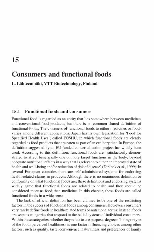
15
Consumers and functional foodsL. Lähteenmäki, VTT Biotechnology, Finland
15.1 Functional foods and consumers
Functional food is regarded as an entity that lies somewhere between medicinesand conventional food products, but there is no common shared definition offunctional foods. The closeness of functional foods to either medicines or foodsvaries among different applications. Japan has its own legislation for ‘Food forSpecified Health Uses’, called FOSHU, in which functional foods are clearlyregarded as food products that are eaten as part of an ordinary diet. In Europe, thedefinition suggested by an EU-funded concerted action project has widely beenused. According to this definition, functional foods are ‘satisfactorily demon-strated to affect beneficially one or more target functions in the body, beyondadequate nutritional effects in a way that is relevant to either an improved state ofhealth and well-being and/or reduction of risk of disease’ (Diplock et al., 1999). Inseveral European countries there are self-administered systems for endorsinghealth-related claims in products. Although there is no unanimous definition orconformity on what functional foods are, these definitions and endorsing systemswidely agree that functional foods are related to health and they should beconsidered more as food than medicine. In this chapter, these foods are calledfunctional foods in a wide sense.
The lack of official definition has been claimed to be one of the restrictingfactors in the success of functional foods among consumers. However, consumersvery rarely define foods in health-related terms or nutritional terms; instead, foodsare seen as categories that respond to the belief systems of individual consumers.Within these categories, whether they relate to use purpose, degree of liking or typeof the food, perceived healthiness is one factor influencing choices among otherfactors, such as quality, taste, convenience, naturalness and preferences of family
Consumers and functional foods 347
members. In a pan-European study, trying to eat healthily was mentioned by 32%of the respondents as one of the three most important reasons behind food choices(Lappalainen et al., 1998). Functional foods have a clear health connotation, andalthough they have distinct features from conventional products, consumersevaluate their perceived healthiness similarly to other food products. The first partof this chapter deals with the role of health in food choices in general, then thespecial characteristics of functional foods are considered from the consumer’spoint of view. After this, a few studies on consumer responses to functional foodsare examined in more detail. The last part concentrates on issues relevant to futureacceptance on functional foods.
15.2 The role of health in food choice
Health is one reason consumers mention when they are asked about factors thatinfluence their food choices. Correspondingly, when asked about health, whole-some and varied diet, together with regular exercise, were mentioned by sevenrespondents out of ten as the factors having the greatest impact on health inDenmark, Finland and the USA (Bech-Larsen et al., 2001). Health is mostly acredence characteristic in food products (Grunert et al., 2000). It has to beassumed, as in most cases, the possible health effects cannot be directly observedor experienced. Taste and other sensory qualities can be directly sensed and theygive an immediate feedback for the eater. The pleasure derived from eating apleasant tasting food is therefore a strong motivator for repeated choices. Physi-ological effects of foods influencing our well-being can, however, also havedirectly felt effects. Caffeine, which is the physiologically effective component incoffee, has a refreshing effect and research has shown that people learn to likecaffeine-containing drinks because they want to feel the refreshing effect or theytry to avoid possible caffeine deprivation (Yeomans et al., 2000).
Health can have an impact on food choices through several mechanisms(Fig. 15.1). Mostly healthiness is assessed through the nutritional quality of foodsand absence of any possibly harmful substances in foods. These characteristicscannot be directly observed from a food product; instead, consumers need infor-mation-based knowledge on the nutrient content and possible other health effects.The information has to be acquired and trusted by the consumer before it can havean impact on their behaviour. Our attitudes, which are either favourable or negativelearned pre-dispositions towards an object or behaviour, guide what informationwe notice and how readily we integrate new beliefs into our knowledge system.
Nutrition experts emphasise that the degree of healthy eating needs to be judgedon the level of diet, whereas no single food product can be categorised as health-promoting or not. Although this is true from a nutrition point of view, consumerstend to have ideas about the health-value of single products. Furthermore, theinformation about fat and vitamin content of foods tend to be generalised, so that,for example, all fruits are categorised as good sources of vitamin C and having lowfat content. Similarly, experience with certain types of foods creates expectations
348 Functional dairy products
Fig. 15.1 Perceived healthiness in food products is a combination of knowledge-basedbeliefs and experienced attributes. Nutrient and energy content need to be conveyed throughinformation, but sensory properties and perceived satiety may give feedback. The func-tional components very rarely have sensed physiological effects; instead they need to beverified through instrumental measures. With some effects the instrumental measure is not
readily available and consumers must rely solely on the information provided.
on the sensory characteristics, e.g. low-fat products are assumed to have a differenttaste and mouth feel from regular products. Perceived healthiness can also dependon any possible harmful substances food is known to have. Some mushrooms, forexample, have to be boiled in water before they are edible. In addition, for manyconsumers any artificial added components, such as food additives, are regarded asa potential risk for health. Functional components bring along promises of positivephysiological effects, the value of which depends on consumers’ health-relatedknowledge and viewpoints. Perceived healthiness depends on many factors and isbased on the interplay between beliefs and experiences.
What is considered to be healthy eating varies among countries and depends oncultural conventions. As mentioned earlier, in a pan-European study carried out inall EU member states health was mentioned as one of the three most importantreasons for food choices by 32% of the 14,331 respondents. The results variedamong the countries from 24% in Luxembourg up to 50% in Austria. The lack oftime and self-control were the most commonly mentioned barriers for healthyeating (Lappalainen et al., 1998). However, when the interviewees were asked‘what is healthy eating?’- the results varied between countries. About half of therespondents considered low fat products and less fat in the diet as part of healthyeating. More fruit and vegetables and balanced and varied diet were mentioned by42 and 41% of respondents, respectively (Margetts et al., 1997). Cultural differ-ence become apparent when frequencies of some responses are explored. Freshnessand naturalness of foods are mentioned on average by 28% of the respondents, but

Consumers and functional foods 349
among EU countries the range is from 6% in Denmark to 56% in Italy. Balanceddiet is mentioned by 41% of EU citizens with a range of 11–74%. The resultssuggest that people are aware of nutritional guidelines in each country and theseguidelines have an impact on the descriptions of healthy eating.
Health is clearly an essential criterion in food choices and it is largelydefined through choices that are in accordance with nutritional education ineach country. Roininen et al. (1999) have developed an attitude scale (GeneralHealth Interest) that measures an individual’s health orientation in order topredict their willingness to follow nutritional recommendations. When scaleswere applied in a cross-cultural study, the health orientation was rather high inthe three European countries studied (Finland, the Netherlands and the UK),with women showing clearly higher interest in health issues than men (Roininenet al., 2001). In another UK study, 70 out of 100 respondents agreed with thestatement that they are health conscious and 89 said that they tried to eat abalanced diet (Consumers’ Association, 2000). The general health interest maynot, however, correlate with consumers’ willingness to use functional foods, asthese food-related health behaviours have clear distinctions, as will be describedin the following section.
15.3 Nutritional guidelines and health claims
Traditional nutrition education has emphasised the role of a health-promotingdiet. Following nutritional guidelines will result in reduced risk to developobesity and other chronic lifestyle-related diseases, such as cardiovascular dis-eases, diabetes and cancer. The message on the benefits of a healthy diet may behard to convey to the consumer, as the end result is unsure and achieving thepossible reward takes several years or even decades. The advice based onrecommendations of dietary intakes of nutrients is usually given at a generallevel. Typically, guidelines suggest avoiding and favouring certain types offoods without giving any recommendations on any particular food products.Low-fat foods, low-salt foods or foods high in fibre are presented as categoriesrather than as single examples of products that should be consumed or notconsumed. This categorisation may be confusing to the consumers as low-fatfoods can be found in different food categories and their roles in the foodsystem differ from each other.
Functional foods offer a new kind of positive health message for the consumers.Instead of avoiding certain types of foods, these food products promise positivephysiological effects on bodily functions or even a reduction on risk level ofdiseases through eating a single product. By definition, the beneficial value of afunctional food comes from producing a positive effect in the body, althoughsometimes the outcome is the avoidance of a negative effect, such as stomach upsetor weakening of bones. As a reinforcement of desired behaviour, the positivereward is known to be more effective than the avoidance of punishment. Therefore,a health message in functional foods promising a positive health effect can be more
350 Functional dairy products
appealing to consumers than an avoidance message, which is the basis of manyhealth messages related to nutritional recommendations.
Functional messages may be easier for the consumer to understand since theycontain results that can be clearly defined. Most of the promised effects offunctional foods can be instrumentally measured, such as lowering the level ofcholesterol in blood, decreasing the blood pressure or increasing the density ofbone mass. However, some of the claimed effects are general and measuringtheir impact may be more difficult. In these cases the functional claims becomepurely credence characteristics that require consumers’ trust on the claim. Forexample, products containing probiotics are largely credence products as mosteffects related to gut functions are difficult to verify by simple physiologicalmeasures. Consumers’ own reported well-being cannot be used as an objectivemeasure of any physiological effects, as many psychological studies have shownthat the mere effect of paying attention to a phenomenon can always affectsubjective responses.
One of the worries brought forward by consumer organisations and healthprofessionals has been that health-related claims may confound the consumers andappear to offer easy options to promote health, so that consumers assume that ahealthy diet is no longer essential. Instead the health effect can be gained throughpurchasing the particular products. Therefore, for example, in the UK Joint HealthClaims Initiative (2002), one of the basic arguments has been that the health claimsshould only be combined with foods that have equal nutritional quality to those oftheir conventional counterparts. Quaker Oats tested how consumers understandhealth-related claims and found that those receiving an oatmeal or a fibre claim didnot believe more often that one does not need to pay attention to the rest of the dietthan those who received no claim in the package (Paul et al., 1999). There seemsto be no basis for believing that consumers would perceive the claims of functionalfoods as the salvation from having to follow a wholesome diet.
15.4 Consumers, claims and carrier products
Acceptance of any food product depends on the interaction between the consumerand the product (Fig. 15.2). Some people like one kind of flavour, whereas othersdislike it. Similarly, price is more important to some individuals, while others putmore emphasis on convenience. In so-called functional food products, the accept-ability depends on the same product characteristics as in any conventional product,and these characteristics have to be as good as in any conventional product.Consumers are not ready to negotiate these basic requirements, but the health-related claim attached to the product can bring additional value to the product.Although health-related claims should boost the appeal of the product, they alsobring an additional factor influencing acceptability. A claim carries a positivehealth-related message, but its weight depends on the individual consumer’smotivation. Furthermore, the reception of the claim depends on the characteristicsof the carrier product.
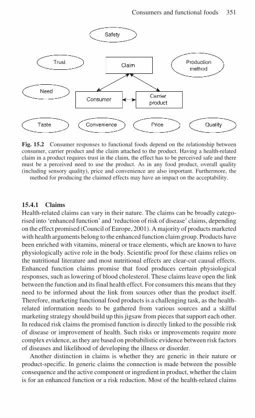
Consumers and functional foods 351
Fig. 15.2 Consumer responses to functional foods depend on the relationship betweenconsumer, carrier product and the claim attached to the product. Having a health-relatedclaim in a product requires trust in the claim, the effect has to be perceived safe and theremust be a perceived need to use the product. As in any food product, overall quality(including sensory quality), price and convenience are also important. Furthermore, the
method for producing the claimed effects may have an impact on the acceptability.
15.4.1 ClaimsHealth-related claims can vary in their nature. The claims can be broadly catego-rised into ‘enhanced function’ and ‘reduction of risk of disease’ claims, dependingon the effect promised (Council of Europe, 2001). A majority of products marketedwith health arguments belong to the enhanced function claim group. Products havebeen enriched with vitamins, mineral or trace elements, which are known to havephysiologically active role in the body. Scientific proof for these claims relies onthe nutritional literature and most nutritional effects are clear-cut causal effects.Enhanced function claims promise that food produces certain physiologicalresponses, such as lowering of blood cholesterol. These claims leave open the linkbetween the function and its final health effect. For consumers this means that theyneed to be informed about the link from sources other than the product itself.Therefore, marketing functional food products is a challenging task, as the health-related information needs to be gathered from various sources and a skilfulmarketing strategy should build up this jigsaw from pieces that support each other.In reduced risk claims the promised function is directly linked to the possible riskof disease or improvement of health. Such risks or improvements require morecomplex evidence, as they are based on probabilistic evidence between risk factorsof diseases and likelihood of developing the illness or disorder.
Another distinction in claims is whether they are generic in their nature orproduct-specific. In generic claims the connection is made between the possibleconsequence and the active component or ingredient in product, whether the claimis for an enhanced function or a risk reduction. Most of the health-related claims
352 Functional dairy products
allowed are generic in their nature and several countries have a list of acceptedclaims. For example, in Sweden the voluntary code ‘Health claims in the labellingand marketing of food products’ defined in 1990 eight generic claims, which linkcertain components with a physiological consequence (Asp and Trossing, 2001).These generic claims are based on the idea that a food product contains aningredient or component that is known to have a lowering effect of disease risk andtherefore the product is beneficial for health. The claim does not cover the productitself and can be used in all products that contain sufficient amount of thecomponent. These claims are similar to traditional nutritional messages, which canbe described as claims related to nutrient functions.
From the manufacturer’s point of view, product-related claims can be moreappealing as they link not only the compound or ingredient, but also the specificproduct with the promised benefit in the claim. The product-specific claim offersbetter protection against competitors to copy the products and means that the claimcan be used effectively in marketing. In the revised Swedish code, these claims arecalled product-specific physiological claims (Asp & Trossing, 2001) and in UKthey are called innovative claims (UK Joint Health Claims Initiative, 2002). Theclaims have to be based on adequate and sound scientific evidence, which meansthat the effect has to have been proven in independent clinical studies. The studiesshould give sufficient support for the evidence that products provide the promisedbenefits with portions that are eaten as a part of normal diet. However, translatingthe scientific probability-based information into consumer language is a tricky taskas the way of thinking behind scientific and everyday beliefs differs dramatically.
Science provides the optimum knowledge on the phenomenon at any moment.Scientific knowledge increases all the time and new information challenges andreplaces existing truths. Furthermore, the scientific evidence on health-relatedrisks is based on probabilities. Consumers, on the other hand, base their food-related beliefs in approximate and rather crude rules of reasoning. Rather thanusing probabilities, consumers tend to make clear-cut assumptions on linksbetween two statements or phenomena. If there is a reduced risk of a disease, forconsumers this may mean that the risk has been removed. The equation where theproduct is effective in five cases out of ten is hard to comprehend; for the consumerthe product should either be effective or not. An additional problem on probabilisticinformation is that the result cannot be promised to everyone. Although somethingis effective in most cases, the possibility of not personally gaining the benefitshould be made understandable to the consumers. Consumers tend to be wary oflinking food with reduction of disease, as this implies associating food that iscommonly considered as a source of pleasure with something unpleasant, even ifthe promised effect would be positive. This conforms with another typical featurein everyday thinking, which tends to associate any two things that appear at thesame time regardless whether they are causally connected or not. Food and health,in general, are both very sensitive topics and this adds to the challenge ofcommunicating possible health-related claims to consumers in a comprehensible,usable and non-alarming manner.
Consumers and functional foods 353
15.4.2 ConsumersConsumers need to be aware of the health benefits offered by functional foods, butknowledge in itself is not a sufficient condition for willingness to use theseproducts. The crucial factor in noticing and adopting messages related to the healtheffects of foods is the motivation of the respondent. The acceptability of new foodsdepends on the benefits these new foods provide for consumers (Frewer et al.,1997). Wrick (1995) divided the potential consumers of functional foods intothose who recognise themselves at risk for a disease and those who are healthconscious. The motivational expectations of these two groups vary: the risk groupwants measurable results, whereas the health-conscious group wants nutritionalinsurance and the ability to take care of oneself.
The most positive group towards functional foods have been women andmiddle-aged or elderly consumers (Poulsen, 1999; Bogue and Ryan, 2000). Agehas also an impact on what kind of effects are interesting. With age, the reductionof risk levels becomes more attractive, while young respondents are more inter-ested in increasing their energy levels. Therefore any generalisation of what age orsex is interested in functional foods is difficult to make, as the appeal depends onthe benefits promised by products. Women seem to be more responsive to productsthat are associated with breast cancer (Arvola et al., 2001) and men moreresponsive to products that lower the risk of prostate cancer (Hilliam, 2002).
In a cross-cultural study carried out in Denmark, Finland and the USA (Bech-Larsen et al., 2001), the most attractive influences of functional foods were heartor cardiovascular diseases mentioned by 54–59%, prevention of stomach cancermentioned by 34–48% depending on the country, and enhancing immune systemmentioned by 36–39% of the respondents. In an Irish study, the two first were thesame, but maintaining the health of teeth and bones rose as the third most appealinginfluence (Bogue and Ryan, 2000).
If a consumer feels a need to lower blood cholesterol level or reduce level ofstress, then the products promising these effects are appealing. Wansink (2001)argues that the missing link between known health benefits of soy and self-relevanthealth consequences may hinder the success of soy products. The nutrition-relatedmessages that were tailored in accordance with the receiver’s own food habit andconsumption patterns were more effective in adding their fibre consumption thanmessages that gave general advice on fibre-containing foods (Brinberg et al.,2000). The tailored messages appeal to a certain group of people, whereas moregeneral messages can be significant to a larger group, although their weight is notas strong. Producers of functional foods have to balance between the nicheproducts that are highly relevant to a small group of consumers and messages thataim to produce general well-being in most consumers.
15.4.3 Carrier product effectThe existing product image has an impact on how the claims are received. Theconsumer perception of a health-related claim depends on its suitability to theproduct in question. In Denmark consumers were more positive about functional
354 Functional dairy products
effects that were created through adding components that are naturally occurringingredients or components in that food product (Poulsen, 1999; Bech-Larsen,2001). Adding calcium into milk products may be more acceptable in consumers’minds than adding other minerals that do not originally belong to milk in anysignificant quantities. However, the health claim also needs to be in accordancewith the previous health image of the product (Lähteenmäki, 2000). In a Finnishstudy, the possible positive health effects of conjugated linoleic acid were discred-ited in butter, because in Finland butter has such a strong negative health image.Butter is considered as a high source of saturated fatty acids and therefore anypositive messages about butter and the possible positive health effects wereconsidered to be artificial and irrelevant to the total image of butter.
Functional foods are defined as products that are frequently consumed so thatthe possible effects they claim to have can also be attained. Food and meal systemsare culturally determined and the place of a carrier product in this system is welldefined. The health-related claim needs to fit into this food system in order to beaccepted. In some cases the functional food products have bypassed the foodsystem by offering an additional product that is eaten or drank separately in smallquantities. An example of this kind of product is the small bottles of probioticdrinks originally introduced by Yakult, which can be taken as a medicine orpreventive action against possible stress. The acceptability of these products canbe built in a different way as they do not require a place in the food system.
Functionality brings also a novel component to the food. Consumers are knownto be suspicious about new foods (Pliner and Hobden, 1992), but this dread ofnovelty is mostly for foods that have unfamiliar sensory characteristics such asethnic foods. The novelty in functional foods is, however, related to new compo-nents and the effects the foods have, although the food in itself looks very much thesame as any conventional food product. This may also cause suspicion in consum-ers as the functionality is not a property that can be experienced. Also, thetechnology required to produce these functional foods may raise doubts in con-sumers’ minds.
15.5 Consumer attitudes to functional foods
Very few studies have looked at consumer responses to so-called functional foods,although functional foods are a category of products that is seen to have a cleargrowth potential in the near future (Stanton et al., 2001). In Europe, gut health hasattracted greatest interest, with dairy products as the main category providingprobiotics and prebiotics to achieve this aim (Hilliam, 2002).
Consumers have become more aware of foods that have health benefits attachedto them (Bogue and Ryan, 2000; Bech-Larsen et al., 2001). Some studies haveshown rather strong reservations towards foods with health-related claims (Jonasand Beckmann, 1998; Consumers’ Association, 2000), but others have revealedpositive consumer attitudes towards claims (Bogue and Ryan, 2000; Bech-Larsenet al. 2001). In a UK study, consumers (n = 100) were asked opinions about three
Consumers and functional foods 355
example products with health-related claims (Consumers’ Association, 2000). Therespondents were best aware of cholesterol-lowering spread (72%) and probioticdrink (58%), but the orange juice with bone benefits was recognised only by a fewrespondents (7%). The functional foods were perceived to be processed everydayfoods, which are convenient to use. They were not considered to be medicines, butthe orange juice and probiotic drink were regarded as supplements, whereas two-thirds of respondents did not think the cholesterol-lowering spread was asupplement. Although the benefits of functional foods were noticed, the opinionwas that, with all example products, something else would have done the job aswell. The claims were presented in two forms: improving health or preventingillness. The improvement claims received bigger approval than prevention claims.Allowing respondents to have a closer examination of the product had an impacton opinion. The benefits of yoghurt and spread became more agreeable, whereasfewer respondents agreed with the preventive power of orange juice after studyingthe package. The product-dependent differences were large, so that 82% of theinterviewees agreed that cholesterol-lowering spread would improve specifichealth problems, whereas the agreement figure was 66% for the probiotic drinkand 49% for orange juice.
Responses to a different kind of health-related claims were studied in threecountries (Denmark, Finland, the USA; n = 1533) using the same method (Bech-Larsen et al., 2001). In addition to the no claim condition, two types of claims wereused, namely physiological enhancement and reduction of risk of disease claims,which were both applied to three types of products: juice, yoghurt and spread. Thecomponents producing the effects of these claims were omega-3 fatty acids oroligosaccharides. Adding the claim in the products increased the perceived whole-someness of the products, regardless of the type of the claim. The effect wasproduct-dependent, so that in juice and yoghurt the enriched products were ratedas less wholesome, whereas in spread the situation was the other way around. Inintentions to buy, the ‘no enrichment’ condition was favoured by the Danish andthe American respondents, while in Finland the effect was product dependent.Juice and yoghurt were more highly rated when they were not enriched, but inspread the enrichment increased the intention to buy. Claim as such was a positivefactor in all countries.
In Finland, an indirect approach was employed to study consumers’ responsesto functional foods. Instead of asking opinions about functional foods, consumerswere asked for their impressions of buyers of functional foods. Respondents ratedtheir impressions of a 40-year-old person who had either functional foods orconventional foods in his or her shopping list. The lists also contained filler items,either with positive or neutral health images, and respondents did not realise thatfunctional foods were the target foods under examination. This approach wasselected to avoid any moral and social issues that influence our health-relatedresponses. Users of functional foods were considered more innovative than usersof conventional foods and also more disciplined than those purchasing conven-tional foods with a neutral health image, but less disciplined than those choosingtraditional options with a high health image (Saher et al., 2002). These results
356 Functional dairy products
indicate that functional products present a new way of using food to improvehealth, which differs from the traditional way. Functional foods are regarded as aconvenient way to acquire required nutrients and other beneficial compounds(Poulsen, 1999), but one that may not be required if the diet is already more variedand balanced (Bogue and Ryan, 2000).
Functional foods with health-related claims tend to be more expensive thantheir traditional counterparts. One of the questions raised is whether this higherprice will be an obstacle for the success of functional foods. The foods are tailoredfor certain purposes and groups of consumers, and they are not meant as massproducts. Within such a new product category, consumers’ willingness to pay ishard to estimate. There seems to be a group of consumers who reported that theyare ready to pay a premium for functional foods (Poulsen, 1999; Bogue and Ryan,2000) or that the price had very little impact on intention to buy (Bech-Larsen,2001).
15.6 Future trends
The ability to provide benefits to consumers is a crucial factor in the future successof functional foods. As the benefits are not directly sensed in the body, thecredibility of health-related messages becomes the key factor. Functional dairyproducts have a immense potential to offer various health benefits for the con-sumer as described in other chapters of this book. Some of these may be easy forthe consumers to accept, as they enhance the existing beliefs of the beneficialeffects dairy products have, but some offer rather specialised high-technologysolutions for targeted populations.
Substantiating the health effects and ensuring their presence in the final productrequires scientifically sound research (Kwak & Jukes, 2001). The true challenge isto make this knowledge produced by science understandable and feasible for theconsumer. So-called everyday thinking, which we apply in solving everydayproblems, such as choices among food products, follows different rules fromscientific thinking. The scientific information about the health benefits is likely tobe probabilistic in its nature, which means that translating these messages for theconsumer is important. Consumers need to have confidence in the source andcontent of the information. Producers of functional foods have to be careful inensuring and maintaining this trust.
As approximation is one of the typical features in consumers’ way of handlinginformation, the health-related messages in products have to be simple and clearenough to be understood, but at the same time they need to be distinguishable fromother messages. As an example, scientists and manufacturers invest a lot of effortin establishing the functional properties of different probiotic strains and products,but consumers may find it hard to make the difference between these strains andtheir outcomes. The targeted functions require also targeted messages to consum-ers of varying ages and cultural backgrounds. The issue of communicating healthbenefits to consumers effectively remains the ultimate challenge in the future
Consumers and functional foods 357
success of functional foods. Only products that will be accepted and consumed canhave a role in improving human health.
15.7 Sources of further information and advice
Further information of functional dairy products and their benefits can be obtainedfrom manufacturers of the products.
More information about EU-funded research on consumer perceptions offunctional foods can be obtained from two websites: Flair-Flow project dissemi-nates information on all food-related research at the EU (www.flair-flow.com) andProEuHealth-cluster consists of eight projects concentrating on gut heath (http://proeuhealth.vtt.fi).
15.8 ReferencesARVOLA A, URALA N and LÄHTEENMÄKI L (2001), ‘Health claims as possible promoters of
product acceptability’, 4th Pangborn Sensory Science Symposium, Dijon, July 2001(abstract).
ASP N-G and TROSSING M (2001), ‘The Swedish code on health-related claims in action –extended to product-specific physiological claims’, Scand J Nutr, 45, 189.
BECH-LARSEN T, GRUNERT K G and POULSEN J B (2001), The Acceptance of Functional Foodsin Denmark, Finland and the United States. Working Paper no. 73, MAPP, Aarhus,Denmark.
BOGUE J and RYAN M (2000), Market-oriented New Product-development: Functional Foodsand the Irish Consumer. Agribusiness Discussion Paper no. 27, University College Cork.
BRINBERG D, AXELSON M L and PRICE S (2000), ‘Changing food knowledge, food choice, anddietary fiber consumption by using tailored messages, Appetite, 35, 35–43.
CONSUMERS’ ASSOCIATION (2000), Functional Food – Health or Hype? London, Consum-ers’ Association.
COUNCIL OF EUROPE (2001), Council of Europe’s Policy Statements Concerning Nutrition,Food Safety and Consumer Health, Guidelines Concerning Scientific Substantiation ofHealth-related Claims for Functional Food. Technical Document, 2 July 2001 (http://www.coe.int/soc-sp).
DIPLOCK A T, AGGETT P J, ASHWELL M, BORNET F, FERN EB and ROBERTFROID MB (1999),‘Scientific concepts of functional foods in Europe: Consensus Document’, Brit J Nutr,81(4), S1–S27.
FREWER L J, HOWARD C, HEDDERLEY D and SHEPHER R (1997) ‘Consumer attitudes towardsdiferent food-processing technologies used in cheese production – the influence ofconsumer benefit’, Food Qual Pref, 8, 271–280.
GRUNERT K G, BECH-LARSEN T and BREDAHL L (2000), ‘Three issues in consumer qualityperception and acceptance of dairy products’, Int Dairy J, 10, 575–584.
HILLIAM M (2002), ‘Functional food update’, The World of Food Ingredients, April/May,52–53.
JONAS M S and BECKMANN S C (1998), Functional Foods: Consumer Perceptions in Denmarkand England. Working Paper no. 55, MAPP, Aarhus, Denmark.
KWAK N S and JUKES D (2001), ‘Issues in the substantiation process of health claims’,Critical Rev Food Sci Nutr, 41, 465–479.
LÄHTEENMÄKI L (2000), ‘Consumers’ perspectives of functional foods’. In: Alander M and
358 Functional dairy products
Mattila-Sandholm T, Functional Foods for EU Health in 2000, VTT Symposium 198,Espoo, 19–21.
LAPPALAINEN R, KEARNEY J and GIBNEY M (1998), ‘A Pan EU survey of consumer attitudesto food, nutrition and health: an overview’, Food Qual Pref, 9, 467–478.
MARGETTS B M, MARTINEZ J A, SABA A, HOLM L and KEARNEY M (1997), ‘Definitions of“healthy” eating: a pan-EU survey of consumer attitudes to food, nutrition and health’, EurJ Clin Nutr,. 51 (Suppl. 2), S23–S29.
PAUL G L, INK S L and GEIGER C J (1999), ‘The Quaker Oats health claim: a case study’, JNutraceuticals, Funct and Med Foods, 1(4), 5–32.
PLINER P and HOBDEN K (1992), ‘Development of a scale to measure the trait of foodneophobia in humans’, Appetite, 19, 105–120.
POULSEN J B (1999), Danish Consumers’ Attitudes Towards Functional Foods. Workingpaper no. 62, MAPP, Aarhus, Denmark.
ROININEN K, LÄHTEENMÄKI L and TUORILA H (1999), ‘Quantification of consumer attitudesto health and hedonic characteristics of foods’, Appetite, 33, 71–88.
ROININEN K, TUORILA H, ZANDSTRA E H, DE GRAAF C, VEHKALAHTI K, STUBENITSKY K andMELA D J (2001), ‘Differences in health and taste attitudes and reported behavior amongFinnish, Dutch and British consumers: a cross-national validation of the health and tasteattitude scales (HTAS)’, Appetite, 37, 33–45.
SAHER M, ARVOLA A, LINDEMAN M and LÄHTEENMÄKI L (2002), ‘Impressions of functionalfood consumers’, submitted to Appetite.
STANTON C, GARDINER G, MEEHAN H, COLLINS K, FITZGERALD G, LYNCH P B and ROSS P(2001), ‘Market potential for probiotics’, Am J Clin Nutr, 73 (Suppl), 476S–483S.
UK JOINT HEALTH CLAIMS INITIATIVE (2002), http://www.jhci.co.uk.WANSINK B (2001). ‘When does nutritional knowledge relate to the acceptance of functional
food?’, Research brief, http://www.consumerpsychology.com/insights/researchingconsumers.htm, Campaign, University of Illinois.
WRICK K L (1995), ‘Consumer issues and expectations for functional foods’, Critical RevFood Sci, 35, 167–173.
YEOMANS M R, JACKSON A, LEE MD, STEER B, TINLEY E, DURLACH P and ROGERS P J (2000),‘Acquisition and extinction of flavour preferences conditioned by caffeine in humans’Appetite, 35, 131–141.
16
European research in probiotics andprebiotics: the PROEUHEALTH clusterT. Mattila-Sandholm, L. Lähteenmäki and M. Saarela, VTTBiotechnology, Finland
16.1 Introduction: research projects within thePROEUHEALTH cluster
Health is one of the main reasons behind food choices, but other factors are alsotaken into consideration by the consumer (Sloan, 2000). As pleasantness andsensory quality can be experienced directly, they are known to be the essentialfactors in repeated choices. Even with foods aimed for health, the taste has to begood to secure repeated choice. Probiotics modifying the gut microbiota havephysiological effects that cannot be experienced directly. The credibility ofhealth-related messages is therefore critical to guarantee that the product has areward value for the consumer (Bech-Larsen et al., 2001; Grunert et al., 2000).Consumers are selective in their attention so that messages that are relevant to usand congruent with our earlier beliefs are easily accepted. Therefore, food-relatedmessages have to correspond with our existing beliefs and motivation to use aproduct. Preventing a possible stomach upset has little relevance to a healthyperson, but can make a vast difference for someone suffering from inflammatorybowel disease (IBD) or someone who easily catches travellers’ diarrhoea(Salminen et al., 1995, 1998a, b, c).
Keeping this in mind, microbes with a positive health impact are, and willremain, an important functional ingredient for years to come. New strains will beidentified and foods will be developed to fulfil the needs of specific consumergroups. Increased understanding of interactions between gut microbiota, diet andthe host will open up possibilities of producing novel ingredients for nutritionally
360 Functional dairy products
optimised foods, which promote consumer health through microbial activities inthe gut (Salminen et al., 1998a, b, c; Naidu et al., 1999).
The acceptance of probiotic, prebiotic and synbiotic products in the future willdepend on the solid proof of the health benefits they promise at the moment.Therefore, research that provides the scientifically sound evidence to back up thehealth claims is needed. Making the knowledge produced by science comprehen-sible to the consumer is also a major challenge as consumers want clear-cutopinions about claims, whereas scientific thinking deals with probabilities anddegrees of uncertainty. Consumers’ trust in information depends on the source andcontent of a message. The critical point is that, with the right approach, producersof probiotic foods can gradually build and ensure consumer trust (Mattila-Sandholmand Saarela, 2000; Mattila-Sandholm et al., 2000; Alander et al., 2001; Crittendenet al., 2001).
A number of steps are essential in the development of efficacious probioticand prebiotic functional foods (Fig. 16.1). A prerequisite for mechanistic studiesof probiotic action is an understanding of the composition and activity of theintestinal microbiota as well as interactions with the host in both healthy anddiseased individuals. High-throughput molecular methods are required to exam-ine the intestinal microbiota and to track the location and activity of probioticstrains in the intestinal tract. An understanding of the mechanisms by whichprobiotics exert beneficial effects on human health allows selection of strainswith appropriate traits for hypothesis-driven clinical studies. The safety of newstrains must be demonstrated before they are used in human clinical trials. Animportant area of research is the development of technologies to maximise thestability of functional traits of probiotics during manufacture, formulation, stor-age and in the intestinal tract. Additionally, the efficacy of products may beenhanced by exploiting synergistic interactions between functional ingredientsas is potentially the case with synbiotics. Finally, an understanding of the mostappropriate methods to communicate the benefits of the functional foods toconsumers and the influence of health messages on consumer choice is essentialto ensure that products are appropriately applied and targeted to benefit specificpopulations (Mercenier and Mattila-Sandholm 2001; Mattila-Sandholm et al.,2002).
The Food, GI-tract Functionality and Human Health (PROEUHEALTH) Clus-ter brings together eight complementary, multicentre interdisciplinary researchprojects (Fig. 16.1). All have the common aim of improving the health and qualityof life of European consumers. The collaboration involves 64 different researchgroups from 16 different European countries and is coordinated by leadingscientists (Table 16.1). The research results from the cluster are disseminatedthrough annual workshops and through the activities of three different platforms:a science, an industry and a consumer platform (Fig. 16.2 and website: http://proeuhealth.vtt.fi). The cluster started in 2001 and will end in 2005.
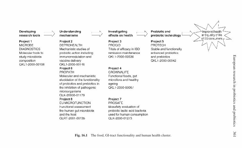
European research in probiotics and prebiotics
361Fig. 16.1 The food, GI-tract functionality and human health cluster.
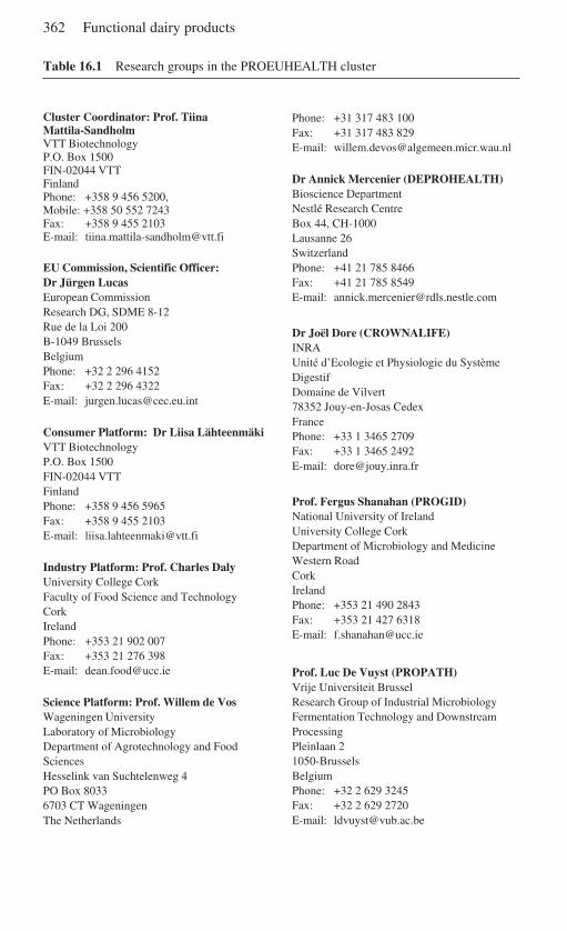
362 Functional dairy products
Table 16.1 Research groups in the PROEUHEALTH cluster
Cluster Coordinator: Prof. TiinaMattila-SandholmVTT BiotechnologyP.O. Box 1500FIN-02044 VTTFinlandPhone: +358 9 456 5200,Mobile: +358 50 552 7243Fax: +358 9 455 2103E-mail: [email protected]
EU Commission, Scientific Officer:Dr Jürgen LucasEuropean CommissionResearch DG, SDME 8-12Rue de la Loi 200B-1049 BrusselsBelgiumPhone: +32 2 296 4152Fax: +32 2 296 4322E-mail: [email protected]
Consumer Platform: Dr Liisa LähteenmäkiVTT BiotechnologyP.O. Box 1500FIN-02044 VTTFinlandPhone: +358 9 456 5965Fax: +358 9 455 2103E-mail: [email protected]
Industry Platform: Prof. Charles DalyUniversity College CorkFaculty of Food Science and TechnologyCorkIrelandPhone: +353 21 902 007Fax: +353 21 276 398E-mail: [email protected]
Science Platform: Prof. Willem de VosWageningen UniversityLaboratory of MicrobiologyDepartment of Agrotechnology and FoodSciencesHesselink van Suchtelenweg 4PO Box 80336703 CT WageningenThe Netherlands
Phone: +31 317 483 100Fax: +31 317 483 829E-mail: [email protected]
Dr Annick Mercenier (DEPROHEALTH)Bioscience DepartmentNestlé Research CentreBox 44, CH-1000Lausanne 26SwitzerlandPhone: +41 21 785 8466Fax: +41 21 785 8549E-mail: [email protected]
Dr Joël Dore (CROWNALIFE)INRAUnité d’Ecologie et Physiologie du SystèmeDigestifDomaine de Vilvert78352 Jouy-en-Josas CedexFrancePhone: +33 1 3465 2709Fax: +33 1 3465 2492E-mail: [email protected]
Prof. Fergus Shanahan (PROGID)National University of IrelandUniversity College CorkDepartment of Microbiology and MedicineWestern RoadCorkIrelandPhone: +353 21 490 2843Fax: +353 21 427 6318E-mail: [email protected]
Prof. Luc De Vuyst (PROPATH)Vrije Universiteit BrusselResearch Group of Industrial MicrobiologyFermentation Technology and DownstreamProcessingPleinlaan 21050-BrusselsBelgiumPhone: +32 2 629 3245Fax: +32 2 629 2720E-mail: [email protected]
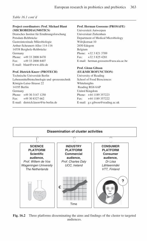
European research in probiotics and prebiotics 363
Fig. 16.2 Three platforms disseminating the aims and findings of the cluster to targetedaudiences.
Dissemination of cluster activities
SCIENCE INDUSTRY CONSUMERPLATFORM PLATFORM PLATFORMScientific Commercial Consumeraudience, audience, audience,
Prof. Willem de Vos Prof. Charles Daly Dr LiisaWageningen University UCC, Ireland Lähteenmäki
The Netherlands VTT, Finland
Time
?
Table 16.1 cont’d
Project coordinators: Prof. Michael Blaut(MICROBEDIAGN0STICS)Deutsches Institut für ErnährungsforschungPotsdam-RehbrückeGastrointestinale MikrobiologieArthur-Scheunert-Allee 114-11614558 Bergholz-RehbrückeGermanyPhone: +49 33 2008 8470Fax: +49 33 2008 8407E-mail: [email protected]
Prof. Dietrich Knorr (PROTECH)Technische Universität BerlinLebensmittelbiotechnologie und -prozesstechnikKönigin-Luise-Strasse 2214195 BerlinGermanyPhone: +49 30 3147 1250Fax: +49 30 8327 662E-mail: [email protected]
Prof. Herman Goossens (PROSAFE)Universiteit AntwerpenUniversitair ZiekenhuisDepartment of Medical MicrobiologyWilrijkstraat 102650 EdegemBelgiumPhone: +32 3 821 3789Fax: +32 3 825 4281E-mail: [email protected]
Prof. Glenn Gibson(EU&MICROFUNCTION)University of ReadingSchool of Food BiosciencesWhiteknights Reading RG6 6APUnited KingdomPhone: +44 1189 357223Fax: +44 1189 357222E-mail: [email protected]
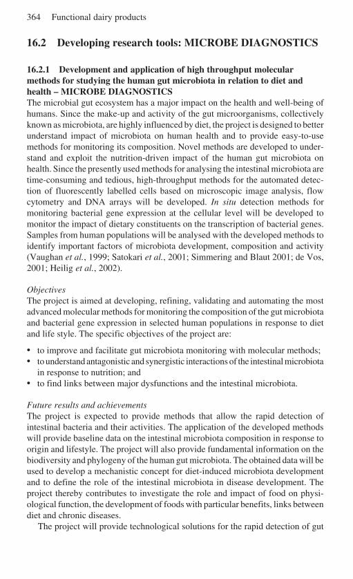
364 Functional dairy products
16.2 Developing research tools: MICROBE DIAGNOSTICS
16.2.1 Development and application of high throughput molecularmethods for studying the human gut microbiota in relation to diet andhealth – MICROBE DIAGNOSTICSThe microbial gut ecosystem has a major impact on the health and well-being ofhumans. Since the make-up and activity of the gut microorganisms, collectivelyknown as microbiota, are highly influenced by diet, the project is designed to betterunderstand impact of microbiota on human health and to provide easy-to-usemethods for monitoring its composition. Novel methods are developed to under-stand and exploit the nutrition-driven impact of the human gut microbiota onhealth. Since the presently used methods for analysing the intestinal microbiota aretime-consuming and tedious, high-throughput methods for the automated detec-tion of fluorescently labelled cells based on microscopic image analysis, flowcytometry and DNA arrays will be developed. In situ detection methods formonitoring bacterial gene expression at the cellular level will be developed tomonitor the impact of dietary constituents on the transcription of bacterial genes.Samples from human populations will be analysed with the developed methods toidentify important factors of microbiota development, composition and activity(Vaughan et al., 1999; Satokari et al., 2001; Simmering and Blaut 2001; de Vos,2001; Heilig et al., 2002).
ObjectivesThe project is aimed at developing, refining, validating and automating the mostadvanced molecular methods for monitoring the composition of the gut microbiotaand bacterial gene expression in selected human populations in response to dietand life style. The specific objectives of the project are:
• to improve and facilitate gut microbiota monitoring with molecular methods;• to understand antagonistic and synergistic interactions of the intestinal microbiota
in response to nutrition; and• to find links between major dysfunctions and the intestinal microbiota.
Future results and achievementsThe project is expected to provide methods that allow the rapid detection ofintestinal bacteria and their activities. The application of the developed methodswill provide baseline data on the intestinal microbiota composition in response toorigin and lifestyle. The project will also provide fundamental information on thebiodiversity and phylogeny of the human gut microbiota. The obtained data will beused to develop a mechanistic concept for diet-induced microbiota developmentand to define the role of the intestinal microbiota in disease development. Theproject thereby contributes to investigate the role and impact of food on physi-ological function, the development of foods with particular benefits, links betweendiet and chronic diseases.
The project will provide technological solutions for the rapid detection of gut
European research in probiotics and prebiotics 365
microorganisms with a high potential for a wide range of applications. Thescreening of gut microbiota composition will be applied in relevant biomedicalresearch areas. It will facilitate the diagnosis of a disturbed microbiota and thedevelopment of rational therapies based on knowledge of disease mechanism. Itwill support the development of functional foods aimed at improved gastrointestinalfunction and other health benefits.
16.3 Understanding mechanisms of actions:DEPROHEALTH, PROPATH and EU MICROFUNCTION
16.3.1 Probiotic strains with designed health properties –DEPROHEALTHLactic acid bacteria (LAB) are well known for their extensive use in the productionof fermented food products. The potential health benefits they may exert in humanswere intensively investigated during the last century. However, the mechanismsunderlying the health-promoting traits attributed to LAB, especially lactobacilli,remain mostly unknown and this has impaired the rational design of probioticscreening methods with accurate predictive value. This project aims at establishinga correlation between in vitro tests and mouse models mimicking important humanintestinal disorders such as IBD and Helicobacter pylori and rotavirus infections.As these diseases correspond to major public health problems, a second generationof probiotic strains with enhanced prophylactic or therapeutic properties will also bedesigned during the project. These designed strains and the isogenic parental oneswill be used to unravel mechanisms involved in the immunomodulation capacity ofspecific Lactobacillus strains (Steidler et al., 2000; Grangette et al. 2001).
ObjectivesThe general aim of this project is to acquire knowledge about the molecular factorsaffecting the immunomodulation and/or immunogenicity of selected probioticlactobacilli to allow for developing isolates with enhanced protective or therapeu-tic effect. The objectives of the proposal are to unravel mechanisms and identifykey components of the immunomodulation capacity of probiotic lactobacilli and todesign a so-called second generation of probiotic strains with enhanced propertiesagainst gastrointestinal disorders.
Two types of gastrointestinal diseases are targeted: inflammations such as IBDand infections such as those caused by H. pylori and rotavirus. For each of them,atherapeutic or prophylactic (i.e. vaccine) recombinant probiotic strains will beconstructed and tested in relevant animal models (i.e. mimicking the humandisease) to evaluate their capacity to induce or modulate the immune response inthe proper way. This data will be correlated to in vitro testing of theimmunomodulation capacity with an attempt to identify/develop screening methodsthat, in the future, will allow isolating efficient probiotic strains targeted to specificapplications. Recombinant DNA technology will also be used to assess the
366 Functional dairy products
importance of specific bacterial cell wall components and adhesion factors inimmunomodulation.
Future results and achievementsTwo types of modified probiotic strains will be constructed: mutant strainsaffected in their cell wall composition and adhesion proteins as well as recombinantstrains with enhanced therapeutic or protective properties, focusing ongastrointestinal diseases of inflammatory or infectious origin. The final goal wouldbe to prove that designed probiotic strains could be used as original therapeuticagents as, if successful, they would lead to novel anti-inflammatory treatments ororal vaccines against H. pylori and rotavirus. It is also expected that, by the end ofthe study, major progress will have been made towards the rational design ofsimplified probiotic screening methods with accurate predictive value.
16.3.2 Molecular analysis and mechanistic elucidation of the functionalityof probiotics and prebiotics in the inhibition of pathogenic microorganismsto combat gastrointestinal disorders and to improve human health –PROPATHRecently, much attention has been paid to the health-promoting properties oflactobacilli and bifidobacteria. Probiotics and prebiotics are the driving forces ofthe functional foods market. However, a major problem is that many of thesehealth-promoting properties are still questioned. For instance, the fundamentalbasis of the inhibition of Gram-negative pathogenic bacteria – such as theenterovirulent diarrhoeagenic Salmonellae, and H. pylori causing gastritis andgastric ulcer disease – by probiotic LAB has not been elucidated. This project willfocus on the identification of the responsible compounds. In addition, the mechan-ism of the inhibition of Gram-negative pathogens by probiotic lactobacilli andbifidobacteria will be studied using co-culture models (Coconnier et al., 1998,2000; Zamfir et al., 1999; Avonts and De Vuyst, 2001).
ObjectivesThe aim is to obtain a selection of probiotic lactobacilli and bifidobacteria thatdisplay a clear inhibition of diarrhoeagenic Gram-negative pathogenic bacteriaand H. pylori. Thereafter, the project focuses on identifying the metabolite(s)responsible for the inhibition and/or killing of Gram-negative pathogenic bacteriaas well as in having the conditions and kinetics of the production of antimicrobialsactive towards Gram-negative pathogens and to predict their in vivo action. One ofthe aims will be to establish co-culture models (simulated gut fermentations,human cell lines, animal models) and to study the interaction between inhibitorystrains of LAB and Gram-negative pathogens causing gastrointestinal disorders.Finally, the selected probiotic LAB will be tested in clinical studies.
Future results and achievementsThe results of the molecular analysis and mechanistic elucidation of the functionality
European research in probiotics and prebiotics 367
of probiotics and prebiotics in the inhibition of pathogenic microorganisms willresult in:
• a project culture collection of probiotic strains of lactobacilli and bifidobacteria,together with data on their characteristic and inhibitory spectrum;
• a molecular typing method for selected probiotic strains;• identified compounds responsible for the inhibition of Gram-negative patho-
genic bacteria;• conditions of antimicrobial production in the gut environment; and• co-culture models showing the inhibitory action by the probiotic strains.
16.3.3 Functional assessment of interactions between the human gutmicrobiota and the host – EU MICROFUNCTIONThe aim of this project is to identify the effects of dietary modulation on the humangastrointestinal microbiota. The influence of probiotics, prebiotics and synbioticswill be ascertained. The main objectives are to clarify effects on the normal gutmicrobiota and on the host gastrointestinal function, as well as to determinemechanisms involved in pro-, pre- and synbiotic functionality. These objectiveswill be achieved through exploitation of model systems and state-of-the-arttechnology (Bengmark, 1998; Salminen et al., 1998a, b, c; Collins and Gibson,1999; Suau et al., 1999, 2001; Steer et al., 2000).
ObjectivesThe principal objectives are to assess the efficacy and safety of probiotics andprebiotics, to determine effective doses/combinations, to identify mechanisms ofaction and to investigate impacts on host function. An important aspect will be thedevelopment of new synbiotics and their use in a human trial. The work aims toidentify the mechanisms of effect and to produce valuable information on theinfluence of dietary intervention on the activities of human gut microbiota. Inaddition, it provides essential means of validating probiotics and prebiotics, andwill give information on the optimal combinational approach.
There is currently an imperative requirement to identify the realistic healthoutcomes associated with probiotic and prebiotic intake and, importantly, giverigorous attention towards determining their mechanisms of effect. This projectwill aim to do so through investigating probiotic and prebiotic influence on hostfunctionality, including microbiological and physiological aspects.
Future results and achievementsThe aim is to identify mechanistic interrelationships through fundamental scien-tific approaches. The following milestones will be achieved:
• efficient prebiotics and required dosage;• active synbiotics;• effects of functional foods on bacterial translocation;• effects on host gene interactions;
368 Functional dairy products
• safety of functional foods determined;• effects on selected health indices in humans.
16.4 Investigating effects on health: PROGID,CROWNALIFE and PROSAFE
16.4.1 Probiotics and gastrointestinal disorders: controlled trials ofEuropean Union patients – PROGIDThis project will assess the efficacy of two previously selected probiotic micro-organisms, administered as dried fermented milk products, in alleviating theeffects of IBD – Crohn’s disease and ulcerative colitis. Specifically, two distinctlong-term (12-month), large-scale, multicentred, randomised, double-blind,placebo-controlled feeding trials will be performed within a subset of the EUpopulation suffering from these gastrointestinal disorders.
Background and objectivesIn recent years, the International Organisation for the Study of InflammatoryBowel Disease and others have published prominent articles highlighting thenecessity for standards in the design of trials evaluating therapies in gastrointestinaldisorders. In the assessment of probiotic products, however, most studies havebeen small, uncontrolled and poorly documented with imprecise definition of theend-points. In response, the European Commission has funded the PROGIDproject, which will evaluate two specific probiotic microorganisms in patients withulcerative colitis or Crohn’s disease from diverse geographical locations. Theparticipating centres in these studies will assess the efficacy of Bifidobacteriuminfantis UCC35624 and Lactobacillus salivarius UCC118 in one year, randomised,double-blind, placebo-controlled trials for maintenance of remission. Both of theselected probiotic strains have a history of safety and efficacy in healthy adults andrelapsed inflammatory bowel disease patients (Shanahan, 2000; Dunne, 2001;Dunne and Shanahan, 2002).
Future results and achievementsThe anticipated results from the PROGID project include:
• the provision of qualitative and quantitative evidence that the evaluated probioticsmay (or may not) have a role in maintaining remission of IBD;
• confirmation, or otherwise, of the involvement of specific members of thegastrointestinal microbiota as causative or contributory agents of IBD;
• generation of linear physiological and immunological data relevant to thedisease and remission states of IBD;
• creation of a greater awareness among the EU population of functional foodsand their potential benefits in maintaining healthy lifestyles; and
• the establishment of a repository of biological samples obtained from across theEU.
European research in probiotics and prebiotics 369
16.4.2 Functional food, gut microflora and healthy ageing –CROWNALIFEElderly people represent an increasing fraction of the European population. Theirhigher susceptibility to degenerative and infectious diseases leads to rising publichealth and social concerns. Appropriate preventive nutrition strategies can beapplied to restore and maintain a balanced intestinal microbiota exerting protectivefunctions against the above disorders. The project is based on the application ofselected biomarkers in human studies and we will identify the structural andfunctional specificity of the elderly’s intestinal microbiota across Europe. Usingthis baseline information, we will investigate functional food-based preventivenutrition strategies, aiming to beneficially affect the functional balance of theelderly’s intestinal microbiota. Expected outcomes include nutritional recommen-dations as well as new concepts and prototype functional foods, specificallyadapted for health benefits to the elderly population (Kruse et al., 1999; Liévin etal., 2000; Schwiertz et al., 2000; Sghir et al., 2000; Marteau et al., 2001).
ObjectivesThe overall objective is to improve the quality of life of the elderly throughout thethird age, with emphasis on the preservation of the period of independencerecognised as the crown of life. The focus is on preventive nutrition and theapplication of functional food to derive health benefits for the ever-increasingEuropean elderly population. Based on hypothesis-driven human studies, thespecific objectives of the project are:
• to assess structural and functional alterations of the intestinal microbiota withageing and across Europe; and
• to validate functional foods-based preventive nutrition strategies to restore andmaintain a healthy intestinal microbiota in the elderly.
Implementations include nutritional recommendations as well as new conceptsand prototype functional food specifically adapted for health benefits to the elderlypopulation.
Future results and achievementsThe results will establish six steps:
• the assessment of the gut microbiota diversity and composition in the Europeanelderly, and identification of its alterations with ageing (baseline human study);
• the assessment of modulation of the intestinal microbiota (intervention humanstudy with a functional food), and potential health benefits towards degenera-tive pathologies and infectious diseases;
• the improved health status of the ageing population via specific nutritionalrecommendations;
• the nutritional guidelines based on the complete assessment of a synbioticproduct;
• the design and provision of adapted food supplements directed towards intesti-nal microbial function; and
370 Functional dairy products
• the validation of processes and rationale for the design of a new generation offunctional foods to satisfy health benefits for the elderly, based on innovativetechnologies.
16.4.3 Biosafety evaluation of probiotic lactic acid bacteria used forhuman consumption – PROSAFESafety assessment is an essential phase in the development of any new pro- orprebiotic functional food. The safety record of probiotics is good, and lactobacilliand bifidobacteria have a long history of safe use. However, all probiotic strainsmust be evaluated for their safety before being used in human clinical studies andin functional food products. Conventional toxicology and safety evaluation aloneis of limited value in the safety evaluation of probiotic bacteria. Instead, amultidisciplinary approach is necessary, involving contributions from patholo-gists, geneticists, toxicologists, immunologists, gastroenterologists andmicrobiologists (Salminen and von Wright, 1998; Salminen et al., 1998a, b, c;Sanders and Huis in’t Veld, 1999; Crittenden et al., 2002).
ObjectivesThe safe use for human consumption of probiotic strains, selected LAB includinglactococci, lactobacilli, pediococci and bifidobacteria, and other food-associatedmicroorganisms such as enterococci, will be assured by proposing criteria, stand-ards, guidelines and regulations. Furthermore, procedures and standardisedmethodologies of pre-marketing biosafety testing and post-marketing surveillancewill be provided. The specific objectives of the project will include five stages:
• the taxonomic description of probiotics, LAB and other food-related micro-organisms;
• the detection of resistance and horizontal transfer of antibiotic resistance genes;• the careful analysis and definition of the nonpathogenic status of probiotic LAB;• the immunological adverse effects of the studied bacteria in the experimental
autoimmune encephalitis (EAE) mouse model; and• the survival, colonisation and genetic stability of probiotic strains in the human gut.
Future results and achievementsFuture results will finalise important goals relating to probiotic strains, selectedLAB and other food-associated microorganisms and will consist of:
• establishing a culture collection and database;• providing standardised methodologies to detect antibiotic resistance;• investigating the potential virulence properties and their association with
clinical disease and results obtained in rat endocarditis models;• studying potential immunological adverse effects;• analysing the genetic stability and colonisation of probiotic strains in the human
gastrointestinal tract; and• providing recommendations for biosafety evaluation of probiotic strains.
European research in probiotics and prebiotics 371
16.5 Probiotic and prebiotic technologies: PROTECH
16.5.1 Nutritional enhancement of probiotics and prebiotics: technologyaspects on microbial viability, stability, functionality, and on prebioticfunction – PROTECHMaintaining the functional properties of probiotics during manufacture, formula-tion and storage are essential steps in delivering health benefits to consumers fromthese products. The overall objective of this project is to address and overcomespecific scientific and technological hurdles that impact on the performance offunctional foods based on probiotic–prebiotic interactions. Such hurdles includethe lack of a strong knowledge on the primary factors responsible for probioticviability, stability and performance. Limited information is available on the impactof processing and storage and of food matrices or food constituents on probioticviability, stability and functionality. Furthermore, information about the interac-tions between probiotics and prebiotics in functional foods prior to consumption isalso limited (Saarela et al., 2000; Mattila-Sandholm et al., 2001).
ObjectivesThis project has three general objectives:
• to explore effects of processing on the stability and functionality of probioticsand on the performance of prebiotics;
• to apply selected processing techniques for prebiotic modification to identifyand optimise probiotic–prebiotic combinations; and
• to use the information generated as the basis for new process and product options.
As a result of achieving these objectives, selected prebiotics in combination withtailored manufacturing processes will not only contribute to probiotic functional-ity but also improve viability and stability of probiotic cultures within foodmatrices during processing and storage.
The project is divided into five specific objectives:
• Effects on probiotic viability: the aim is to consolidate quantitative data ofprocessing effects on the physiology and viability of probiotic organisms. Thiswill include effects during different fermentation processes, and freezing anddrying strategies and also during passage through the GI tract. Effects on cellcomposition including stress responses, cell membrane composition and leak-age, and cell viability will be measured. A viability model for probiotic cultureswill be developed and validated.
• Effects on probiotic stability: experiments will be performed to investigate theinfluence of media composition and processing conditions on probiotic stabil-ity. This includes cryotolerance, resistance to dehydration, storage and geneticstability. Stability of the important probiotic characteristics of strains will alsobe investigated including acid and bile tolerance and adhesion to intestinal cells.New formulation techniques including micro-encapsulation will be assessed inreal food matrices including dairy- and cereal-based foods.
372 Functional dairy products
• Probiotic–prebiotic interaction: the ultimate objective is to optimise probiotic–prebiotic interaction for maximum probiotic performance in culture, in foodsystems and in the GI tract. Special attention will be given to indigestible oligo-and polysaccharides with respect to fermentability and to the production ofshort chain fatty acids in animal models and in vitro.
• Prebiotic function: new prebiotics will be developed that are tailor-made forthe stabilisation and optimum performance of probiotics. These will be exam-ined for their physiological properties and their organoleptic andphysicochemical properties will be technologically evaluated in real foods.
• Probiotic functionality: the effect of growth conditions and stress environ-ments on the functionality of probiotic bacteria will be examined. Stressestested will include high acid, bile, temperature, oxygen and osmotic tension. Inaddition, high hydrostatic pressure and controlled permeabilisation technolo-gies will be evaluated for stress preconditioning of LAB, including bifidobacteria.Cross-protection against other stresses will be examined.
Future results and achievementsExpected achievements include the establishment of unique data sets that containthe identification of critical process parameters for probiotics and prebiotics andresults from systematic studies suggesting means to overcome existing process andproduct limitations. The compilation of protocols for probiotic performance,prebiotic function, and probiotic–prebiotic interactions will also be provided.Further, probiotic viability models and functionality biomarkers will be estab-lished. In addition, optimisation of probiotic viability, stability in culture and inreal food systems at a pilot scale, and the generation and modification of uniqueprebiotics, of probiotic interactions and of environmentally and processing in-duced functionality of probiotics will be attempted. Application of the expectedresults will lead to new process concepts for probiotics, for prebiotics and forprobiotic–prebiotic combinations.
16.6 Consumers and the perceived health benefits ofprobiotics
Science-based knowledge on how probiotic bacteria can promote our well-beinghas increased in recent years. The success of probiotic products will be determinedby consumers’ willingness to buy and eat them. The perceived benefits in thesenew products are key factors for the consumer acceptance. Some of the benefits arerelevant to a large group of people, but others are part of a well-defined targetgroup. For ordinary consumers, preventing travellers’ diarrhoea or helping withtemporary gut disorders is a clear advantage, but still the motivation to useprobiotics for patients suffering from irritable bowel syndrome (IBS) or IBD islikely to be higher if the probiotics can, at least partly, replace medication andreduce or even prevent symptoms (Poulsen, 1999; Roininen et al., 1999).
Functional foods, i.e. products promising specified effects on physiological
European research in probiotics and prebiotics 373
functions, represent a new kind of health message for the consumers. In traditionalnutritional messages the emphasis is on diet and on avoiding or favouring certaintypes of foods rather than giving recommendations on particular products. Probioticand other products containing the new kind of health message appear to betterrespond to the needs of different people than the idea of a nutritionally balanceddiet (Roininen et al., 1999; Sloan, 2000).
Although the most enthusiastic users of functional foods are believed to bemiddle-aged women who already follow healthy diets, in previous studies inFinland the general health interest, which measures people’s willingness tocomply with nutritional recommendations, has not been strongly linked with thewillingness to use functional foods. The well-defined benefits promised by probioticfoods contain a positive message to consumers. The idea of gaining a benefit byusing a product rather than avoiding a slow development of chronic diseases formsa different message. For Irish consumers the most interesting claims were relatedto heart diseases, cancer and bone health. Surprisingly, probiotics were not wellrecognised, even though yoghurt was among the most frequently used functionalfoods (Grunert et al., 2000).
Developing probiotic foods also introduces a novelty aspect to the product. Ingeneral, consumers tend to be suspicious towards novelty in foods because itmeans uncertainty and thus threat. Therefore, the benefits promised by these newfood products will be weighed against the perception of possible risks. Highlydeveloped technologies are often involved in manufacturing the new probioticfoods. In several European countries healthiness has been associated with natural-ness. The need for highly advanced technology in the production of probiotic foodsmay lower the perceived naturalness, which may create distrust towards functionalfoods among consumers. Consumers seem to find functions that enforce thenatural properties of a product more acceptable than those functions that areartificially added and in discordance with the earlier image of the product.Therefore, in addition to technological considerations, adding probiotics into foodproducts requires understanding of the existing beliefs consumers have about theseproducts (Lappalainen et al., 1998; Poulsen, 1999; Bogue and Ryan, 2000;Lähteenmäki and Arvola, 2001).
Consumer responses to functional foods and their health-related messagesrequire more research. Communicating gut health messages effectively to con-sumers of varying age and cultural backgrounds remains one of the key issues.Besides this, we need tremendous efforts towards communication and cooperationbetween medical doctors, nutritionists and pharmaceutics. As information is a vitalfactor, trust between all sides is required, so that messages will be given attention.The differences between functional properties of various strains are difficult toexplain to consumers and tools for doing this should be created. Some consumersmay find it hard to approve of food products working almost like medicines.Furthermore, food products need to have a place in the daily food system, whereasmedicines are taken when instructed, and using food as a replacement for medi-cines requires adjustments in eating habits as well. Probiotic products can improvehuman health only if they are eaten frequently. To ensure this, the above issues
374 Functional dairy products
Table 16.2 The cluster projects
• Which bacterium is which?• Second generation probiotics• New therapeutics with potential against IBD• A healthier retirement• Keeping probiotics alive and healthy so that they can keep you healthy• Probiotics can defend against bad bacteria• Ensuring the safety of probiotic bacteria• Why are probiotics effective?
must be addressed so that an appropriate set of products, which will be acceptedand consumed, can be developed, such as the PROEUHEALTH cluster projecttopics in consumer language (Table 16.2).
16.7 Conclusions and future trends
Probiotics, prebiotics and synbiotics aimed at improving intestinal health currentlyrepresent the largest segment of the functional foods market in Europe, Japan andAustralia. Evidence continues to emerge demonstrating that these ingredients havepotential to improve human health in specific intestinal disorders. The EuropeanCommission, through its Fifth Framework Programme, is presently investing asubstantial research effort in the intestinal microbiota, its interaction with its host,and methods to manipulate its composition and activity for the improvement ofhuman health. Eight multicentre interdisciplinary research projects currently covera variety of research topics required for the development of helpful probioticfoods, ranging from understanding probiotic mechanisms at a molecular level todeveloping technologies to ensure delivery of stable products and demonstratingsafety and efficacy in specific disorders. This concerted research effort promises toprovide an appreciable understanding of the human intestinal microbiota’s role inhealth and disease, and new approaches and products to tackle a variety ofintestinal afflictions.
The future research on food and microbes with a health impact will continue todevelop, aimed specifically at:
• exploring the mechanisms of action of microbes and their health effects in theGI tract, especially in healthy individuals;
• developing sophisticated diagnostic tools for the gut microbiota and biomarkersfor their assessing their functionality;
• examining the effects of food-derived bioactive compounds on GI diseases, GIinfections and allergies;
• developing new therapeutic and prophylactic treatments for different patientand population groups;
• realising molecular understanding of immune modulation by bacteria withhealth effects;
European research in probiotics and prebiotics 375
• elucidating the role of colon microbiota in the conversion of bioactive com-pounds;
• analysing the effects of the metabolites to the colon epithelium or the effectsafter absorption;
• ensuring the stability of microbes with health effects and their bioactivecompounds also in new types of food applications by developing feasibletechnologies; and finally
• providing the safety of the functional ingredients.
16.8 ReferencesALANDER M, MÄTTÖ J, KNEIFEL W, JOHANSSON M, CRITTENDEN R, MATTILA-SANDHOLM T
and SAARELA M (2001), ‘Effect of galacto-oligosaccharide supplementation on humanfaecal microflora and on survival and persistence of Bifidobacterium lactis Bb-12 in thegastrointestinal tract’, Int Dairy J, 11, 817–825.
AVONTS L and DE VUYST L (2001), ‘Antimicrobial potential of probiotic lactic acid bacteria’,Mededelingen van de Faculteit Landbouwkundige en Toegepaste BiologischeWetenschappen Universiteit Gent, 66/3b, 543–550.
BECH-LARSEN T, GRUNERT K G and POULSEN J B (2001), The Acceptance of Functional Foodsin Denmark, Finland and the United States, Working Paper no. 73, MAPP, Denmark.
BENGMARK S (1998), ‘Ecological control of the gastrointestinal tract. The role of probioticflora’, Gut, 42, 2–7.
BOGUE J and RYAN M (2000), Market-oriented New Product Development: Functional Foodsand the Irish Consumer, Agribusiness Discussion Paper no. 27, Department of FoodEconomics, University College Cork, Ireland.
COCONNIER M F, LIÉVIN V, HEMERY E, LABOISSE C L and SERVIN A L (1998), ‘Antagonisticactivity of the human Lactobacillus acidophilus strain LB against Helicobacter infectionin vitro and in vivo’, Appl Environ Microbiol, 64, 4573–4580.
COCONNIER M H, LIÉVIN V, LORROT M and SERVIN A L (2000), ‘Antagonistic activity ofLactobacillus acidophilus LB against intracellular Salmonella enterica SerovarTyphimurium infecting human enterocyte-like Caco-2/TC-7 cells’, Appl Environ Microbiol,66, 1152–1157.
COLLINS M D and GIBSON G R (1999), ‘Probiotics, prebiotics and synbiotics: dietaryapproaches for the modulation of microbial ecology’, Am J Clin Nutr, 69, 1052–1057.
CRITTENDEN R, LAITILA A, FORSSELL P, MÄTTÖ J, SAARELA M, MATTILA-SANDHOLM T andMYLLÄRINEN P (2001), ‘Adhesion of bifidobacteria to granular starch and implications inprobiotic technologies’, Appl Environm Microbiol, 67(8), 3469–3475.
CRITTENDEN R, SAARELA M, OUWEHAND A, SALMINEN S, VAUGHAN E, DE VOS W, VONWRIGHT A and MATTILA-SANDHOLM T (2002), ‘Lactobacillus paracasei F19: survival,ecology and safety in the human intestinal tract’, Microb Ecol Health Dis, 3, 22–26.
DE VOS W M (2001), ‘Advances in genomics for microbial food fermentations and safety’,Current Opin Biotechnol, 12(5), 493–498.
DUNNE C (2001), ‘Adaptation of bacteria to the intestinal niche – probiotics and gut disorder’,Inflam Bowel Dis, 7(2), 136–145.
DUNNE C and SHANAHAN F (2002), ‘The role of probiotics in the treatment of intestinalinfections and inflammation’, Curr Opin Gastroenterol, 18(1), 40–45.
GRANGETTE C, MÜLLER-ALOUF H, GOUDERCOURT D, GEOFFROY M-C, TURNEER M andMERCENIER A (2001), ‘Mucosal immune responses and protection against tetanus toxinafter intranasal immunization with recombinant Lactobacillus plantarum’, Infect Immun,69, 1547–1553.
376 Functional dairy products
GRUNERT K, BECH-LARSEN T and BREDAHL L (2000), ‘Three issues in consumer qualityperception and acceptance of dairy products’, Int Dairy J, 10, 575–584.
HEILIG H G, ZOETENDAL E G, VAUGHAN E E, MARTEAU P, AKKERMANS A D and DE VOS W M(2002), ‘Molecular diversity of Lactobacillus spp. and other lactic acid bacteria in thehuman intestine as determined by specific amplification of 16S ribosomal DNA’, ApplEnviron Microbiol, 68(1), 114–123.
KRUSE H P, KLEESSEN B and BLAUT M (1999), ‘Effects of inulin on faecal bifidobacteria inhuman subjects’, Brit J Nutr, 82, 375–382.
LÄHTEENMÄKI L and ARVOLA A (2001), ‘Food neophobia and variety seeking – consumerfear or demand for new food products’, in Frewer L J, Risvik E, and Schifferstein H, Food,People and Society. A European Perspective of Consumer Food Choices, Springer-Verlag, Berlin, 161–175.
LAPPALAINEN R, KEARNEY J and GIBNEY M A (1998), ‘Pan-European survey of consumers’attitudes to food, nutrition and health: an overview’, Food Qual Pref, 9, 467–478.
LIÉVIN V, PEIFFER I, HUDAULT S, ROCHAT F, BRASSART D, NEESER J-R and SERVIN A L (2000),‘Bifidobacterium strains from resident infant human gastrointestinal microflora exertantimicrobial activity’, Gut, 47, 646–652.
MARTEAU P, POCHART P, DORÉ J, MAILLET C, BERNALIER A and CORTHIER G (2001),‘Comparative study of the human cecal and fecal flora’, Appl Environ Microbiol, 67,4939–4942.
MATTILA-SANDHOLM T and SAARELA M (2000), ‘Probiotic functional foods’. In: Gibson GR and Williams C M F, Functional Foods – Concept to Product, Woodhead PublishingLimited, 287–313.
MATTILA-SANDHOLM T, BLUM S, COLLINS J K, CRITTENDEN R, DE VOS W, DUNNE C, FONDEN
R, GRENOV B, ISOLAURI E, KIELY B, MARTEU P, MORELLI L, OUWEHAND A, RENIERO R,SAARELA M, SALMINEN S, SAXELIN M, SCHIFFRIN E, SHANAHAN F, VAUGHAN E and VONWRIGHT A (2000), ‘Probiotics: towards demonstrating efficacy’, Trends Food Sci Technol,10, 393–399.
MATTILA-SANDHOLM T, MYLLÄRINEN P, CRITTENDEN R, MOGENSEN G, FONDEN R andSAARELA M (2001), ‘Technological challenges for future probiotic foods’, Int Dairy J, 12,1–10.
MATTILA-SANDHOLM T, BLAUT M, DALY C, DE VUYST L, DORÉ J, GIBSON G, GOOSSENS H,KNORR D, LUCAS J, LÄHTEENMÄKI L, MERCENIER A, SAARELA M, SHANAHAN F and DE VOS
W M (2002), ‘Food, GI-tract functionality and human health cluster: PROEUHEALTH’,Microb Ecol Health Dis, 14, 65–74.
MERCENIER A and MATTILA-SANDHOLM T (2001), ‘The food, GI-tract functionality andhuman health European research cluster, PROEUHEALTH’, Nutr Metab Cardiovas Dis,11(4), 1–5.
NAIDU A S, BIDLACK W R and CLEMENS R A (1999), ‘Probiotic spectra of lactic acid bacteria(LAB)’, Crit Rev Food Sci Nutrit, 38(1), 13–126.
POULSEN J B (1999), Danish Consumers’ Attitudes Towards Functional Foods, WorkingPaper no. 62, MAPP, Aarhus, Denmark.
ROININEN K, LÄHTEENMÄKI L and TUORILA H (1999), ‘Quantification of consumer attitudesto health and hedonic characteristics of foods’, Appetite, 33, 71–88.
SAARELA M, MOGENSEN G, FONDEN G, MÄTTÖ J and MATTILA-SANDHOLM T (2000),‘Probiotic bacteria: safety, functional and technological properties’, J Biotechnol, 84,197–215.
SALMINEN S and VON WRIGHT A (1998), ‘Current probiotics – safety assured?’, Microb EcolHealth Dis, 10, 68–77.
SALMINEN S, ISOLAURI E and ONNELA T (1995), ‘Gut flora in normal and disordered states’,Chemotherapy, 41, 5–15.
SALMINEN S, BOULEY C, BOUTRON-RUAULT M C, CUMMINGS J H, FRANCK A, GIBSON G R,ISOLAURI E, MOREAU M C, ROBERFROID M and ROWLAND I (1998a), ‘Functional foodscience and gastrointestinal physiology and function’, British J Nutr, 80 (Suppl), 147–
European research in probiotics and prebiotics 377
171.SALMINEN S, OUWEHAND A C and ISOLAURI E (1998b), ‘Clinical applications of probiotic
bacteria’, Int Dairy J, 8, 563–572.SALMINEN S, VON WRIGHT A, MORELLI L, MARTEAU P, BRASSART D, DE VOS W, FONDEN R,
SAXELIN M, COLLINS K, MOGENSEN G, BIRKELAND S E and MATTILA-SANDHOLM T (1998c),‘Demonstration of safety of probiotics – a review’, Int J Food Microbiol, 44(1–2), 93–106.
SANDERS M E and HUIS IN’T VELD J H J (1999), ‘Bringing a probiotic-containing functionalfood to the market: microbiological, product, regulatory and labeling issues’, Antonie vanLeeuwenhoek, 76, 293–315.
SATOKARI R M, VAUGHAN E E, AKKERMANS A D, SAARELA M and DE VOS W M (2001),‘Polymerase chain reaction and denaturing gradient gel electroforesis monitoring of fecalbifidobacterium populations in a prebiotic and probiotic feeding trial’, Syst Appl Microbiol,24(2), 227–231.
SCHWIERTZ A, LE BLAY G and BLAUT M (2000), ‘Quantification of different Eubacterium spp.in human fecal samples with species-specific 16S rRNA-targeted oligonucleotide probes’,Appl Environ Microbiol, 66, 375–382.
SGHIR G, GRAMET A, SUAU V, ROCHET P, POCHART P and DORÉ J (2000), ‘Quantification ofbacterial groups within human fecal flora by oligonucleotide probe hybridization’, ApplEnviron Microbiol, 66, 2263–2266.
SHANAHAN F (2000), ‘Probiotics in inflammatory bowel disease: is there a scientificrationale?’, Inflam Bowel Dis, 6, 107–1115.
SIMMERING R and BLAUT M (2001), ‘Pro- and prebiotics – the tasty guardian angels?’, ApplMicrobiol Biotechnol, 55, 19–28.
SLOAN E (2000), ‘The top ten functional food trends’, Food Technol, 54(4), 33–62.STEER T, CARPENTER H, TUOHY K and GIBSON G R (2000), ‘Perspectives on the role of the
human gut microbiota in health and methods of study’, Nutr Res Rev, 13, 229–254.STEIDLER L, HANS W, SCHOTTE L, NEIRYNCK S, OBERMEIER F, FALK W, FIERS W and REMAUT
E (2000), ‘Treatment of murine colitis by Lactococcus lactis secreting interleukin-10’,Science, 289, 1352–1355.
SUAU A, BONNET R, SUTREN M, GODON J J, GIBSON G R, COLLINS M D and DORÉ J (1999),‘Direct analysis of genes encoding 16S rRNA from complex communities reveals manynovel molecular species within the human gut’, Appl Environ Microbiol, 65, 4799–4807.
SUAU A, ROCHET V, SGHIR A, GRAMET G, BREWAEYS S, SUTREN M, RIGOTTIER-GOIS L andDORÉ J (2001), ‘Fusobacterium prausnitzii and related species represent a dominant groupwithin the human faecal flora’, Syst Appl Microbiol, 24, 139–145.
VAUGHAN E E, MOLLET B and DE VOS W M (1999), ‘Functionality of probiotics and intestinallactobacilli: light in the intestinal tract tunnel’, Curr Opin Biotechnol, 10(5), 505–510.
ZAMFIR M, CALLEWAERT R, CALINA CORNEA P, SAVU L, VATAFU I and DE VUYST L (1999),‘Purification and characterization of a bacteriocin produced by Lactobacillus acidophilusIBB 801’, J Appl Microbiol, 87, 923–931.
17
The market for functional dairyproducts: the case of the United StatesL. Hoolihan, Dairy Council of California, USA
17.1 Introduction
It could be argued that the functional foods movement began decades ago with thefortification of milk with vitamins A and D, salt with iodine, and bread with Bvitamins. These efforts and fortification of other ‘staple’ foods with specificnutrients were successful in eradicating diseases of the time such as rickets, goiterand pellagra. Similarly, the recent fortification of folic acid to cereals and grainswas intended to help reduce the incidence of certain kinds of birth defects to thespinal cord. In the mid-1990s, however, a very distinct mindshift occurred in thethinking behind fortification of the food supply. Whereas previously foods werefortified with specific nutrients for public health reasons, the new goal was tofortify foods with various nutritive components for achievement of optimal health,vitality, longevity and improved physical performance and endurance. Thismindshift marks the real introduction of the functional foods movement. Foodswere no longer consumed just for their inherent nutrients but for health benefitsbeyond the traditional nutrients they contained. Within a period of a few years, avast number and variety of functional foods were introduced to the market,including energy and sports bars, calcium-fortified orange juice, omega-3 enrichedeggs, yoghurts with probiotics, cereals, snack foods, fortified juices and otherbeverages.
Foods classified as functional are generally associated with an individual’sprimary health concerns, which in the US include cardiovascular disease, highblood pressure, stroke, high cholesterol and cancer. Other major health concernsinclude weight, diabetes, and overall nutrition and diet (IFIC, 2002). Which foodsthe consumer perceives as ‘functional’ will vary depending on their particular

The market for functional dairy products 379
health concerns and nutrition goals. Advertising messages and marketing cam-paigns will also influence how a consumer views a particular food product or typeof food. One survey found that the top ten foods that consumers identify asproviding health benefits beyond basic nutrition include broccoli, fish or fish oil,green leafy vegetables, oranges or orange juice, carrots, garlic, fiber, milk, oats/oatbran/oatmeal and tomatoes (IFIC, 2002).
17.2 Drivers of the functional foods market
Along with the general interest in achieving optimal health and well-being, thereare a number of factors fueling interest in the functional foods market. Escalatinghealth-care costs are causing people to look for means of preventing disease ratherthan treating it, and for alternatives to traditional medical care. Our aging popu-lation with the desire of living longer, healthier and more active lives prefers toachieve these goals through diet and lifestyle choices rather than through pharma-ceutical means. Scientific evidence over the past several years has strengthened thelink between diet and wellness, proving to even the most skeptical that diet playsan indisputable role in one’s health. The current age of information and technologyhas also played into this movement. With a wealth of health-related informationliterally at one’s fingertips through the Internet, consumers are educating them-selves and increasingly taking charge of their own health. This ‘self-health’movement has definitely contributed to the interest in functional foods. Finally,recent legislation (discussed in Section 17.4) has made it easier for food manufac-turers to include health-related information on their products, providing anopportunity to educate the consumer at point-of-purchase.
Surveys show that more North Americans than ever before are seeking informa-tion on diet and health and taking action to improve their eating habits. TheAmerican Dietetic Association has conducted surveys every two years for the pastdecade to track the evolution of people’s attitudes, knowledge and behaviorsaround diet and nutrition. In their most recent survey, 85% of American consumerssay that diet and nutrition are important to them personally, 75% say they carefullyselect foods in order to achieve balanced nutrition and a healthful diet, and 58% saythey actively seek information about nutrition and healthful eating (ADA, 2002).The strength of the perceived link between diet and nutrition has continued toincrease from their previous surveys.
The International Food Information Council (IFIC) also tracks consumerperceptions of functional foods. Their most recent survey of over 1000 adults inMassachusetts, USA, found that almost all consumers (94%) believe that certainfoods provide health benefits that go beyond basic nutrition and may reduce therisk of disease or other health concerns (IFIC, 2002). This level of interest has beenconsistently strong since their initial survey in 1998. In addition, a majority (68%)of consumers believe they have a ‘great amount’ of control over their own healthand 78% believe that food and nutrition play a ‘great role’ in maintaining orimproving overall health. Almost two-thirds (63%) of Americans say they are
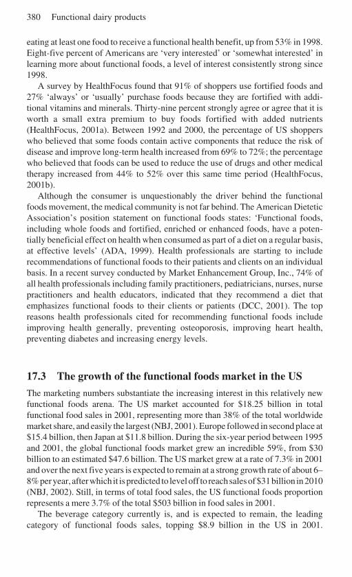
380 Functional dairy products
eating at least one food to receive a functional health benefit, up from 53% in 1998.Eight-five percent of Americans are ‘very interested’ or ‘somewhat interested’ inlearning more about functional foods, a level of interest consistently strong since1998.
A survey by HealthFocus found that 91% of shoppers use fortified foods and27% ‘always’ or ‘usually’ purchase foods because they are fortified with addi-tional vitamins and minerals. Thirty-nine percent strongly agree or agree that it isworth a small extra premium to buy foods fortified with added nutrients(HealthFocus, 2001a). Between 1992 and 2000, the percentage of US shopperswho believed that some foods contain active components that reduce the risk ofdisease and improve long-term health increased from 69% to 72%; the percentagewho believed that foods can be used to reduce the use of drugs and other medicaltherapy increased from 44% to 52% over this same time period (HealthFocus,2001b).
Although the consumer is unquestionably the driver behind the functionalfoods movement, the medical community is not far behind. The American DieteticAssociation’s position statement on functional foods states: ‘Functional foods,including whole foods and fortified, enriched or enhanced foods, have a poten-tially beneficial effect on health when consumed as part of a diet on a regular basis,at effective levels’ (ADA, 1999). Health professionals are starting to includerecommendations of functional foods to their patients and clients on an individualbasis. In a recent survey conducted by Market Enhancement Group, Inc., 74% ofall health professionals including family practitioners, pediatricians, nurses, nursepractitioners and health educators, indicated that they recommend a diet thatemphasizes functional foods to their clients or patients (DCC, 2001). The topreasons health professionals cited for recommending functional foods includeimproving health generally, preventing osteoporosis, improving heart health,preventing diabetes and increasing energy levels.
17.3 The growth of the functional foods market in the US
The marketing numbers substantiate the increasing interest in this relatively newfunctional foods arena. The US market accounted for $18.25 billion in totalfunctional food sales in 2001, representing more than 38% of the total worldwidemarket share, and easily the largest (NBJ, 2001). Europe followed in second place at$15.4 billion, then Japan at $11.8 billion. During the six-year period between 1995and 2001, the global functional foods market grew an incredible 59%, from $30billion to an estimated $47.6 billion. The US market grew at a rate of 7.3% in 2001and over the next five years is expected to remain at a strong growth rate of about 6–8% per year, after which it is predicted to level off to reach sales of $31 billion in 2010(NBJ, 2002). Still, in terms of total food sales, the US functional foods proportionrepresents a mere 3.7% of the total $503 billion in food sales in 2001.
The beverage category currently is, and is expected to remain, the leadingcategory of functional foods sales, topping $8.9 billion in the US in 2001.
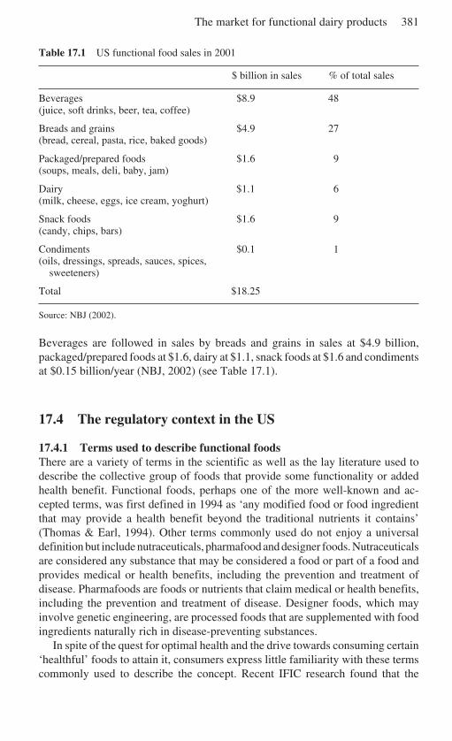
The market for functional dairy products 381
Table 17.1 US functional food sales in 2001
$ billion in sales % of total sales
Beverages $8.9 48(juice, soft drinks, beer, tea, coffee)
Breads and grains $4.9 27(bread, cereal, pasta, rice, baked goods)
Packaged/prepared foods $1.6 9(soups, meals, deli, baby, jam)
Dairy $1.1 6(milk, cheese, eggs, ice cream, yoghurt)
Snack foods $1.6 9(candy, chips, bars)
Condiments $0.1 1(oils, dressings, spreads, sauces, spices,
sweeteners)
Total $18.25
Source: NBJ (2002).
Beverages are followed in sales by breads and grains in sales at $4.9 billion,packaged/prepared foods at $1.6, dairy at $1.1, snack foods at $1.6 and condimentsat $0.15 billion/year (NBJ, 2002) (see Table 17.1).
17.4 The regulatory context in the US
17.4.1 Terms used to describe functional foodsThere are a variety of terms in the scientific as well as the lay literature used todescribe the collective group of foods that provide some functionality or addedhealth benefit. Functional foods, perhaps one of the more well-known and ac-cepted terms, was first defined in 1994 as ‘any modified food or food ingredientthat may provide a health benefit beyond the traditional nutrients it contains’(Thomas & Earl, 1994). Other terms commonly used do not enjoy a universaldefinition but include nutraceuticals, pharmafood and designer foods. Nutraceuticalsare considered any substance that may be considered a food or part of a food andprovides medical or health benefits, including the prevention and treatment ofdisease. Pharmafoods are foods or nutrients that claim medical or health benefits,including the prevention and treatment of disease. Designer foods, which mayinvolve genetic engineering, are processed foods that are supplemented with foodingredients naturally rich in disease-preventing substances.
In spite of the quest for optimal health and the drive towards consuming certain‘healthful’ foods to attain it, consumers express little familiarity with these termscommonly used to describe the concept. Recent IFIC research found that the
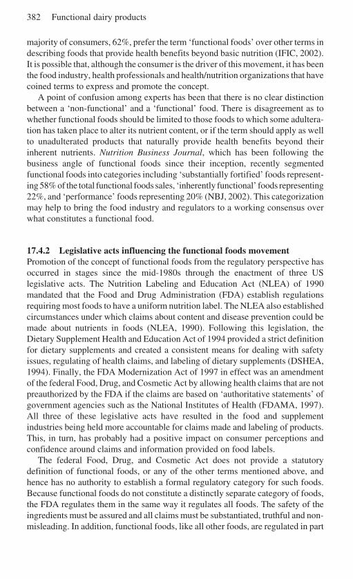
382 Functional dairy products
majority of consumers, 62%, prefer the term ‘functional foods’ over other terms indescribing foods that provide health benefits beyond basic nutrition (IFIC, 2002).It is possible that, although the consumer is the driver of this movement, it has beenthe food industry, health professionals and health/nutrition organizations that havecoined terms to express and promote the concept.
A point of confusion among experts has been that there is no clear distinctionbetween a ‘non-functional’ and a ‘functional’ food. There is disagreement as towhether functional foods should be limited to those foods to which some adultera-tion has taken place to alter its nutrient content, or if the term should apply as wellto unadulterated products that naturally provide health benefits beyond theirinherent nutrients. Nutrition Business Journal, which has been following thebusiness angle of functional foods since their inception, recently segmentedfunctional foods into categories including ‘substantially fortified’ foods represent-ing 58% of the total functional foods sales, ‘inherently functional’ foods representing22%, and ‘performance’ foods representing 20% (NBJ, 2002). This categorizationmay help to bring the food industry and regulators to a working consensus overwhat constitutes a functional food.
17.4.2 Legislative acts influencing the functional foods movementPromotion of the concept of functional foods from the regulatory perspective hasoccurred in stages since the mid-1980s through the enactment of three USlegislative acts. The Nutrition Labeling and Education Act (NLEA) of 1990mandated that the Food and Drug Administration (FDA) establish regulationsrequiring most foods to have a uniform nutrition label. The NLEA also establishedcircumstances under which claims about content and disease prevention could bemade about nutrients in foods (NLEA, 1990). Following this legislation, theDietary Supplement Health and Education Act of 1994 provided a strict definitionfor dietary supplements and created a consistent means for dealing with safetyissues, regulating of health claims, and labeling of dietary supplements (DSHEA,1994). Finally, the FDA Modernization Act of 1997 in effect was an amendmentof the federal Food, Drug, and Cosmetic Act by allowing health claims that are notpreauthorized by the FDA if the claims are based on ‘authoritative statements’ ofgovernment agencies such as the National Institutes of Health (FDAMA, 1997).All three of these legislative acts have resulted in the food and supplementindustries being held more accountable for claims made and labeling of products.This, in turn, has probably had a positive impact on consumer perceptions andconfidence around claims and information provided on food labels.
The federal Food, Drug, and Cosmetic Act does not provide a statutorydefinition of functional foods, or any of the other terms mentioned above, andhence has no authority to establish a formal regulatory category for such foods.Because functional foods do not constitute a distinctly separate category of foods,the FDA regulates them in the same way it regulates all foods. The safety of theingredients must be assured and all claims must be substantiated, truthful and non-misleading. In addition, functional foods, like all other foods, are regulated in part
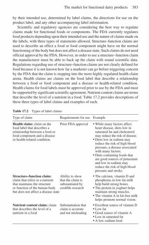
The market for functional dairy products 383
by their intended use, determined by label claims, the directions for use on theproduct label, and any other accompanying label information.
Scientific and regulatory agencies are considering the best way to regulateclaims made for functional foods or components. The FDA currently regulatesfood products depending upon their intended use and the nature of claims made onthe labels, with three types of statements allowed. Structure–function claims areused to describe an effect a food or food component might have on the normalfunctioning of the body but does not affect a disease state. Such claims do not needofficial approval by the FDA. However, in order to use a structure–function claimthe manufacturer must be able to back up the claim with sound scientific data.Regulations regarding use of structure–function claims are not clearly defined forfood because it is not known how far a marketer can go before triggering concernby the FDA that the claim is stepping into the more highly regulated health claimarena. Health claims are claims on the food label that describe a relationshipbetween a food or food component and a disease or health-related condition.Health claims for food labels must be approved prior to use by the FDA and mustbe supported by significant scientific agreement. Nutrient content claims are termsthat describe the level of a nutrient in a food. Table 17.2 provides descriptions ofthese three types of label claims and examples of each.
Table 17.2 Types of label claims
Type of claim Requirements for use Example
Health claim: claim on the Prior FDA approval • While many factors affectfood label that describes a heart disease, diets low inrelationship between a food or saturated fat and cholesterolfood component and a disease may reduce the risk of disease.or health-related condition. • Diets low in sodium may
reduce the risk of high bloodpressure, a disease associatedwith many factors.
• Diets containing foods thatare good sources of potassiumand low in sodium mayreduce the risk of high bloodpressure and stroke.
Structure–function claim: Ability to show • The calcium, vitamin D andclaim that refers to a nutrient that the claim is phosphorus in low-fat milkthat maintains the structure substantiated by help build strong bones.or function of the human body credible research • The protein in yoghurt helpsbut does not affect a disease state maintain strong muscles.
• The vitamin A in fat-free milkhelps promote normal vision.
Nutrient content claim: claim Substantiation that • Excellent source of vitamin Dthat describes the level of a claim is accurate • Low fatnutrient in a food and not misleading • Good source of vitamin A
• Low in saturated fat• A low sodium food
384 Functional dairy products
17.5 The potential for functional dairy foods in the US
One of the largest areas of functional foods in Europe and Japan has been in the dairymarket, accounting for about 65% of all functional food sales in Europe. This trendin the US has been considerably slower to catch on, with functional dairy productsaccounting for only 6.4% of the total functional foods sales in 2000 (NBJ, 2001).
However, there remains considerable potential for dairy products within thefunctional foods movement in the US. Over the past several years, consumption ofdairy products has been linked to improved bone health (Miller et al., 2000),reduced blood pressure (Bucher et al., 1996; Appel et al., 1997), an enhancedimmune system (Kelley et al., 2001; Erickson and Hubbard, 2000; Early et al.,2001), reduced risk of colon (Holt et al., 1998) and breast cancers (Hjartaker et al.,2001). Newer research is showing that the calcium from dairy products may evenplay a role in improved body weight maintenance (Zemel & Morgan, 2002).Specific components under investigation for these effects include calcium, conju-gated linoleic acid, probiotics, whey protein, lactoferrin, butyric acid andsphingolipids, among others.
Milk is in fact considered by some the ‘original functional food’, not just for thenutrients it inherently contains but also for its ability to deliver those nutrients tothe body. Lactoferrin present in milk enhances iron absorption by ensuring that theiron remains in a stable, non-toxic form. Research on the bioavailability of othernutrients from milk show that vitamin E is absorbed twice as well from milk as itis from fortified orange juice or from supplements (Hayes et al., 2001), and that thetriglyceride structure in milk enhances the absorption of both long chain fatty acidsand calcium (Kennedy et al., 1999). In addition, the ‘cluster’ of nutrients in milk,including calcium, vitamin D, protein, phosphorus, magnesium, vitamin A, vita-min B6 and trace elements such as zinc – all important for bone health – are presentin milk in levels appropriate to maximize calcium absorption (Anderson, 2000).
Other components not inherently concentrated in milk, but that are receivingattention as possible fortification opportunities, include beta-glucan for maintain-ing cardiovascular health, glucosamine for maintaining joint health, lactoferrin forhealth gastrointestinal microflora, lutein for healthy vision and general eye health,and magnesium for cardiovascular health, among others (FMSTI, 2001). Con-sumer reaction to, and acceptance of, the fortification of milk with these ingredientsis currently being investigated so that processors will know which to offer.
One of the most successful strategies for positioning functional foods, identi-fied in the 1998 IFIC survey, is to place functional foods in the most traditionalcontext possible. This makes milk, one of the most traditional foods available, wellpositioned in the functional foods movement. Milk not only contains a myriad ofimportant nutrients for growth, development and maintenance of optimal healthbut as discussed above is also being recognized as an ideal carrier for othernutrients that may not be highly concentrated in milk. This presents obviousopportunities for the industry to utilize milk as a fortification vehicle for nutrientsfor which consumption is marginal or inadequate, further enhancing the functionalfood status of dairy products.
The market for functional dairy products 385
17.5.1 Specific functional dairy foods in the marketRecognizing the potential for functional dairy products above and beyond tradi-tional dairy products, various producers have introduced a number of products intothe market. As examples of functional milks introduced in the US in July 2000,Parmalat USA unveiled three new functional milks, each claiming to expand thenatural benefits of milk. Milk-E containing additional vitamin E and biotin wastouted to promote healthy skin and faster metabolism. Lactose Free Plus wasintroduced as a lactose-free milk containing inulin, a source of dietary fiber, andLactobacillus acidophilus and Bifidobacterium, well-known probiotics with sub-stantial health benefits (See chapter 10). Finally, Skim Plus contains 34% morecalcium and 37% more protein than traditional milk. Since then Parmalat hasintroduced Plus Omega-3, a UHT milk containing 80 mg omega-3 per 100 ml withvitamins E, C and B6 as well as calcium.
Danone’s Actimel is another US example of a functional dairy product, cur-rently in its third year of test marketing in Colorado. Actimel is a yoghurt drinkfermented by Lactobacillus bulgaricus and Streptococcus thermophilus and con-taining added Lactobacillus casei immunitass. Stonyfield Farms is alsotest-marketing a drinkable yoghurt intended for adults, which is fortified with sixlive, active probiotic cultures. Suiza Foods manufactures Kidsmilk, described as‘containing 50% more of the 11 vitamins found in regular milk’. Campinaproduces a buttermilk and apple juice product with added vitamin D and calcium,as well as raftilose intended to increase calcium absorption.
There are considerable regional differences in the availability of these vari-ous products in the US, making broad-reaching marketing campaigns ineffectiveand inappropriate. As most of these products are either in the test-marketingstage or early in their life cycle, it remains to be seen how successful they willbe and to what extent the dairy industry will continue to develop and marketnew functional dairy products to meet the needs of specific segments of thepopulation.
17.5.2 Perceptions of functional dairy foodsAlthough fruits and vegetables initially received most of the spotlight in thefunctional foods arena for their healthful and disease-preventative attributes,dairy has become a strong player in the eyes of the health professional as well asthe consumer. A recent survey of health professionals in California indicatedthat 91% of those surveyed considered dairy products as a functional food.Yoghurt, milk and cottage cheese drew the most attention in the dairy functionalfoods category, followed by cheese, dry milk added to foods, and whey (DCC,2001). A similar survey in consumers found that 64% of consumers believe thatdairy foods can help decrease the risk of disease and/or optimize the health andwellness of one or more members of their family (DCC, 2002). Enhancingimmunity, improving the absorption and utilization of nutrients, and decreasingbreast cancer risk were some of the benefits of dairy consumption cited byconsumers.
386 Functional dairy products
17.6 Future trends
17.6.1 The future of functional diary productsDevelopment and marketing of functional dairy products is promising in a numberof areas. Fortifying dairy foods with iron is one area recently receiving attention.Because dairy foods represent a major food source for many cultures worldwideand they are inherently low in iron, dairy products may be a reasonable vehicle foriron fortification. Certain population subsegments such as children, women ofchild-bearing age and vegetarians whose iron consumption may be marginalwould, in particular, benefit from fortification of dairy foods with iron. Researchis currently underway to examine ways to get around the adverse effects added ironmay have on the lipids, caseins and whey proteins in dairy products. Complexingadded iron with lactoferrin may be one solution to fortify with iron without thecolor, flavor and quality issues generally associated with iron fortification.Remembering that texture, appearance and taste prevail over nutrient content andhealth benefits in purchasing decisions, successful fortification of a product mustconsider these factors. Convenience and pricing will also play a big role in theultimate success of a product.
Prebiotics – inulin and oligofructose – is another area receiving attention for theability to increase calcium absorption, improve bone mineral density, improvefunctioning of the gastrointestinal tract, and enhance immunity among otherpotential health benefits (Milner & Roberfroid, 1999). Stonyfield Farms’ family ofyoghurts currently contains inulin, along with their trademark six live activeprobiotic cultures. A variety of other food manufacturers are experimenting withthe fortification of prebiotics to their products as well.
Other substances that may play a role in future functional dairy products includewhey proteins for their immune-enhancing and cancer-fighting properties,sphingolipids for their ability to ward off some forms of cancer, conjugated linoleicacid for its body composition and cancer benefits, and bioactive peptides for theirimmune-enhancing effects. Research on these components is, at this point, prel-iminary, mostly taking place in laboratory and animal studies. Future research willundoubtedly focus on determining levels needed for consumers to reap thebenefits, identifying the most appropriate dairy product to use as a fortificationvehicle and determining stability and shelf-life of products incorporating thesecomponents, among other manufacturing concerns.
Thus dairy foods, long seen as ideal vehicles for fortification with addednutrients, are positioned strongly in the functional foods movement. Dairyfoods are already considered to be natural, healthy and wholesome foods.Sensory studies consistently show that the consumer enjoys their taste and othersensory qualities. They are also moderately priced and represent a ‘traditional’food category identified as having the most promise in successful marketingand sales of functional foods. It remains to be seen which nutrients and otheringredients will come out on top in terms of consumer perception and accept-ance and, perhaps just as important, how quickly the dairy industry chooses to
The market for functional dairy products 387
act in developing and marketing functional products incorporating those ingre-dients.
17.6.2 Future trends in the overall functional foods marketIn spite of the overall positive perceptions and attitudes towards functional foodsamongst the consumer and health professional, the continued growth of the marketwill depend upon a number of factors. How consumers learn about diet and healthconnections will be paramount, with many consumers skeptical when readingmarketing claims of food companies or on product labels. Consumer awarenesscampaigns such as promotion via health professionals or other credible authoritieswill be necessary in the successful marketing of functional food products. Theexpected health benefits of consuming a particular food and the levels andfrequency of consumption necessary to make a difference are areas that will needto be addressed in some form of consumer education. Knowledge about food–druginteractions is also needed, as functional foods may either block or increase theabsorption of drugs, either reducing their effectiveness or increasing the risk oftoxicity.
The pricing of functional food products will also impact the long-term successof this broad food category. Typically, functional foods attract a premium price,and even in cases in which a food might be proven beneficial for a certaincondition, its relatively high price may discourage people from purchasing theproduct. This will be particularly the case in the lower socio-economic group,which paradoxically is often the group with the greatest need for good nutrition.
A strong regulatory framework will also be essential in the long-term growthand success of the functional foods market. Over-aggressive marketing of foodswith added health benefits, misleading information on food labels and lack of astrong regulatory system for overseeing the production and marketing of thesefoods have led to a decline in consumer confidence. Finally, taste remains thenumber one reason for food purchases, emphasizing the need to the food industryto develop foods that, above all else, are palatable.
Citing the number of failed functional food products that have been pulled fromthe marketplace for sluggish sales over the past few years, US food manufacturersare hesitant to jump on the functional foods bandwagon that other countries havebeen riding. Rather than researching, developing and marketing brand new prod-ucts, the emphasis will continue to be on enhancing existing products by fortifyingwith high-profile nutrients such as calcium, iron, and antioxidant vitamins E, C andA. Traditional, familiar foods and food groups such as dairy will do well here as theconsumer already has considerable trust in these products. Given the growingbottled water market and the strong functional food beverage category,‘aquaceuticals’ – fortified bottled water – is also predicted to become mainstreamover the next few years. Probiotics for protection against infection and enhancedimmune system, fiber and potassium for blood pressure, and foods fortified withcalcium will likely be other emerging functional food categories.
The growth of the market will depend on these factors and others as the
388 Functional dairy products
consumer, food industry and regulators alike learn what works and what does not.There will undoubtedly be more product failures than successes in this market asmanufacturers juggle regulatory limitations with consumer demands and how todeliver credible information about their products. Regulatory agencies will need toput into place a framework that allows for new foods to be aggressively developedand marketed, yet which maintains safety standards and regulates informationprovided on product labels. In turn, the consumer will be overwhelmed with thenumber of food choices available as well as with the abundance of health- andnutrition-related information relevant to themselves and their families. Given theincreasingly strong interest in and link between health and nutrition, however, thefunctional foods arena will undoubtedly move forward; the question is not if andwhen, but how quickly and aggressively.
17.7 Sources of further information and adviceFunctional Foods for Health Program; Department of Food Science and Human Nutrition;
University of Illinois at Urbana-Champaign, http://www.ag.uiuc.edu/~ffh/ffh.htmlInstitute of Food Technologists, www.ift.orgInternational Food Information Council Foundation, http://www.ific.org/foodAmerican Dietetic Association, www.eatright.orgFood Science Program at Rutgers University, http://foodsci.rutgers.eduUSDA/ARS Food and Nutrition Information Center, http://www.nal.usda.gov/fnic
17.8 ReferencesADA (AMERICAN DIETETIC ASSOCIATION) (1999), ‘Functional foods – Position of ADA’, J
Am Diet Assoc, 99, 1278–1285.ADA (AMERICAN DIETETIC ASSOCIATION) (2002), ‘Tracking trends’, Dietetics in Practice,
2(1), 1–4, http://www.eatright.org/pr/2002/trends2002.html.ANDERSON J J B (2000), ‘Role of calcium and vitamin D in bone modeling and remodeling
in adolescent girls’, IFT Annual Meeting abstract no. 28–2.APPEL L J, MOORE T J, OBARZANEK E, VOLLMER W M, SVETKEY L P, SACKS F M, BRAY G A,
VOGT T M, CUTLER J A, WINDHAUSER M M, LIN P H and KARANJA N (1997), ‘A clinical trialof the effects of dietary patterns on blood pressure’, N Engl J Med, 336, 1117–24.
BUCHER H C, COOK R J, GUYATT G H, LANG, J D, COOK D J, HATALA R and HUNT D L (1996),‘Effects of dietary calcium supplementation on blood pressure: a meta-analysis ofrandomized controlled trials’, J Amer Med Assoc, 275, 1016–1022.
DCC (DAIRY COUNCIL OF CALIFORNIA) (2001), Functional Foods Study, June 2001; MarketEnhancement Group, Inc.
DCC (DAIRY COUNCIL OF CALIFORNIA) (2002), DCC Functional Foods Study, September2002; Market Enhancement Group, Inc.
DSHEA (DIETARY SUPPLEMENT HEALTH AND EDUCATION ACT) (1994), Public Law 103–417,section 13(a).
EARLY E M, HARDY H, FORDE T and KANE M (2001), ‘Bactericidal effect of a whey proteinconcentrate with anti-Helicobacter pylori activity’, J Appl Microbiol, 90(5), 741–748.
ERICKSON K L and HUBBARD N E (2000), ‘Probiotic immunomodulation in health anddisease’, J Nutr, 130, 403–409.
FDAMA (FEDERAL DRUG ADMINISTRATION MODERNIZATION ACT) (1997), Public law, 105–115.
The market for functional dairy products 389
FMSTI (FLUID MILK STRATEGIC THINKING INITIATIVE) (2001), ‘Fluid Milk’s Role in theFunctional Foods Movement: Milk’s Unique Nutrient Profile and Functional IngredientOpportunities’, www.milkplan.org.
HAYES KC, PRONCZUK A and PERLMAN D (2001), ‘Vitamin E in fortified cow milk uniquelyenriches human plasma lipoproteins’, Am J Clin Nutr, 74, 211–218.
HEALTHFOCUS (2001a), ‘Functional Foods and Nutraceuticals’, Consumer Trend Report.HEALTHFOCUS (2001b), ‘National Study of Public Attitudes and Actions Toward Shopping
and Eating’, Consumer Trend Report.HJARTAKER A, LAAKE P and LUND E (2001), ‘Childhood and adult milk consumption and risk
of premenopausal breast cancer in a cohort of 48,844 women – the Norwegian women andcancer study’, Int J Cancer, 93(6), 888–893.
HOLT PR, ATTILLASOY EO, GILMAN J, GUSS J, MOSS SF, NEWMARK H, FAN K, YANG K andLIPKIN M (1998), ‘Modulation of abnormal colonic epithelial cell proliferation anddifferentiation by low-fat dairy foods’, J Amer Med Assoc, 280, 1074–1079.
IFIC (INTERNATIONAL FOOD INFORMATION COUNCIL) (2002), ‘Functional foods attitudinalresearch: the consumer view on functional foods: yesterday and today’, Food Insight,May/June.
KELLEY D S, SIMON V A, TAYLOR P C, RUDOPH I L, BENITO P, NELSON G J, MACKEY B E andERICKSON K L (2001), ‘Dietary supplementation with conjugated linoleic acid increasedits concentration in human peripheral blood mononuclear cells, but did not alter theirfunction’, Lipids, 36, 669–674.
KENNEDY K, FEWTRELL M S, MORLEY R, ABBOTT R, QUINLAN P T, WELLS J C K, BINDELS J Gand LUCAS A (1999), ‘Double-blind, randomized trial of a synthetic triacylglycerol informula-fed infants: effects on stool biochemistry, stool characteristics, and bone miner-alization’, Am J Clin Nutr, 70, 920–927.
MILLER G D, JARVIS J K and MCBEAN L D (2000), Handbook of Dairy Foods and Nutrition.2nd ed. Boca Raton, London, New York, Washington, DC. CRC Press.
MILNER JA and ROBERFROID M (1999), ‘Nutritional and health benefits of inulin andoligofructose; proceedings of a conference held May 18–19, 1998’, J Nutrition, 129(7S),1395S–1502S.
NAS THOMAS P R and EARL R EDS (1994), Opportunities in the nutrition and food sciences;research challenges and the next generation of investigators. Institute of Medicine,National Academy of Sciences, Washington, DC, National Academy Press, 109.
NBJ (NUTRITION BUSINESS JOURNAL) (2001), ‘Functional foods V’, Nutr Bus J, 7(10), 1–8,www.nutritionbusiness.com.
NBJ (NUTRITION BUSINESS JOURNAL) (2002), ‘NBJ’s annual overview of the nutritionindustry VII’, Nutr Bus J, 7(5/6), 1–11, www.nutritionbusiness.com.
NLEA (NUTRITION LABELING and EDUCATION ACT) (1990), Code of Federal Regulations,101, 14(a)(1).
ZEMEL M B and MORGAN K (2002), ‘Regulation of adiposity and obesity risk by dietarycalcium: mechanisms and implications’, J Am Coll Nutr, 21, 146S–151S.
Index
aberrant crypt foci (ACF) 26ADA (American Dietetic Association)
379added-value products 5‘adenoma-carcinoma sequence’ 21AGA (American Gastroenterologists
Association) 20alcohol, and coronary heart disease 73allergic immune phenotype-oriented
approach, probiotics 124allergic sensitisation 111–12
and infant development 119–23allergies 147, 155angina pectoris 54–5, 58–9angiotensin converting enzyme (ACE)
inhibition 10–11animal probiotics, safety 328–9antibodies, in milk 145–8anticaries effects, caseinophosphopeptides
(CPPs) 196antioxidant nutrients, and coronary heart
disease 72atherogenic lipoprotein phenotype 67atheroscelerosis 58
and conjugated linoleic acid (CLA)285–7
bacteriaand assimilation of cholesterol 79in the gut 30, 170, 170–72and inflammatory bowel disease (IBD)
175–7bacterial translocation 174–5Bifidobacterium 6, 30, 86, 123, 221, 224,
250
safety 316bile 322–3biomarkers 42–3blood-related defence mechanisms 324bovine milk
humanisation 196and immunomodulation 139
British Nutrition Foundation 203Bulletin of the International Dairy
Federation 4buttermilk 2butyrate 304
calciumabsorption in human milk 226bioavailability and
caseinophosphopeptides (CPPs)190–91
and colorectal cancer (CRC) 23, 25–6intake and absorption 98–101
cancer 19–53breast cancer and CLA 275–6and conjugated linoleic acid (CLA)
274–81and diet 19–21see also colon cancer; colorectal cancer
carbohydrate intake, and coronary heartdisease (CHD) 71–2
cardiovascular health 10–12carrier product effect 353–4casein 1–2
and colorectal cancer (CRC) 26–8caseinophosphopeptides (CPPs) 13, 187–
202anticaries effects 196
Index 391
antigenic and allergenic potential 194–5cytomodulatory effects 193–4effect on mineral uptake 192–3human studies with 191–2humanisation of bovine milk 196immunomodulation 197and mineral (calcium) bioavailability
190–91mineral supplementation 196potential ingredient applications 195–7production 189–90safety assessment 194–5structure 188
cell-mediated immunity 134–5cellular immune response 141–3cheese
and probiotics 249production 246–8
cholesterolassimilation and bacteria 79products for controlling 11–12
clinical trials 337–45assessing validity 343–4ethical issues 341–3managing and monitoring 343probiotic bacteria 251–2protocols 337–40statistical analysis 340–41
Clostridia 224colon cancer, and prebiotics 303–4colon carcinogenesis 21–2colorectal cancer (CRC) 19–20
and calcium 23, 25–6and casein 26–8and conjugated linoleic acid (CLA)
28–9and dairy products 23, 24–5future trends 42–3
colostrum 145conjugated linoleic acid (CLA) 28–9,
263–98alkali isomerization 267analytic methods 272–3and atherosclerosis 285–7biochemical synthesis 272and cancer 28–9, 274–81commercial production 267–72dehydration of hydroxy fatty acid 267–
8and diabetes 288effect of processing on content of dairy
products 273–4and immune function 287–8in vivo cancer models 275–7
and lipid and protein metabolism 281–4mechanisms of anticarcinogenesis 278–
81multiple step synthesis 268–71natural sources 263–6process of metabolism 284–6reduction of acetylenic bonds 268
consumersattitudes to functional foods 354–6and functional foods 346–58, 380and perceived health benefits 372–4,
385Consumers’ Association 349, 355coronary heart disease (CHD) 54–93
and alcohol 73and antioxidant nutrients 72and carbohydrate intake 71–2classical lipid risk factors 64–5and diet 67–74effects of prebiotics 80–85effects of probiotics 74–9effects of synbiotics 85–6and lipoprotein metabolism 61–4newly recognised risk factors 65–7and non-starch polysaccharides 72pathology of development 57–8and polyunsaturated fatty acids
(PUFAs) 71risk, and dietary lipids 67–71risk factors 55–6, 57–61and sodium and potassium 73
Council of Europe 351Crohn’s disease 174, 176, 177CROWNALIFE research project 369–370cytokines 111–12, 120, 124, 125, 133,
135, 138, 147, 151–2
DCC 380, 385Declaration of Helsinki 341deconjunction reaction 79DEPROHEALTH research project 365–6diabetes 288diet
and cancer 19–21and coronary heart disease 73–4
E. coli 118, 174, 226elderly people 369–70, 379
functional food for 306immune function 132–68
enzyme-linked immunosorbent assay(ELISA) 195
EU MICROFUNCTION research project367–8

392 Index
European Union, safety assessment 330
FACE 229FDAMA (Federal Drug Administration
Modernization Act 1997) 382fenofibrate 56fermented milk products 2–5, 245, 246
health effects 4hypochloresterolaemic properties 74–5immunomodulatory effects 149–57and probiotic LAB 148, 248–9
food allergyclinical symptoms 112–13definition 109genetic predisposition 109, 111and gut microbiota composition 116–19and inflammation 121–3management of symptoms 114–16mechanisms and symptoms 109–13prevalence 113–14and probiotics 108–31reduction of risk 114
food choice, role of health 347–9Food and Drug Administration, USA 11,
329–30Food and Nutrition Board of the Institute
of Medicine 98Framingham Osteoporosis Study 103fructooligosaccharides (oligofructoses) 9,
302functional dairy products, definitions 5–6future trends in functional foods 356–8
galactooligosaccharide 9, 219, 221physiological effects in human studies
222–4gastrointestinal inflammation, therapeutic
use of probiotics 169–84genetically modified probiotics 327–8Germany, safety assessment 330–31glucose levels, effect of prebiotics 84–5good clinical practice (GCP) 341–2gut bacteria 250gut immune function 173–4gut microbiota composition, and food
allergy 116–19
health, role in food choice 347–9, 359–60health claims 350–52
and nutritional guidelines 349–50HealthFocus 380Heliobacter pylori 169, 177human milk
calcium absorption 226
effect of oligosaccharides on theimmune system 227
human milk oligosaccharides (HMOS)204–17, 230, 231
humanisation of bovine milk 196humoral immunity 135–6
and milk proteins 139–41hypertension, products for controlling 10–
11hypochloresterolaemic properties of
yoghurt 74–5
IARC 19, 20IDF (International Dairy Federation) 191,
246IFIC (International Food Information
Council) 378, 379immune function
and conjugated linoleic acid (CLA)287–8
in the elderly 132–68products for enhancing 13
immune response 42immune system 108, 133–4
effect of oligosaccharides 227immunological effects 325–6immunomodulation
and bovine milk 139caseinophosphopeptides (CPPs) 197
immunomodulatory effectsfermented milk products 149–57LAB 149–57
immunoregulation, and milk 145–8immunosenesence 134–7infant development, and allergic
sensitisation 119–23infant formulae and weaning foods 306inflammatory bowel disease (IBD) 169
and intestinal bacteria 175–7innate immunity 136–7
and milk protein 143–4insulin levels, effect of prebiotics 84–5International Food Information Council
(IFIC) 379intestinal flora, effect of oligosaccharides
221–4intestinal infection, effect of
oligosaccharides 224–6inulin 9, 80, 82, 83, 86, 302, 303
extraction 307IUB-IUPAC 203
Joint Commission on BiochemicalNomenclature (JCBN) 203
Index 393
lactic acid bacteria (LAB) 4–5, 244–62enhancing viability and stability 252–4immunomodulatory effects 149–57probiotic LAB 248–9production of dairy products using
246–8Lactobacillus 6, 7, 30, 250
deconjunction mechanism 79safety 316–17
Lactobacillus acidophilus 3, 35, 249Lactobacillus bulgaricus 78Lactobacillus GG 115, 123, 227Lactobacillus helveticus 11lactose 2lactose intolerance 254–5LDL chloresterol 60, 66–7, 77legislation 5, 8
EU Novel Foods 11US 382–3
lipid lowering mechanism, prebiotics 83–4
lipid metabolismeffect of prebiotics 80–83and oligosaccharides 227
lipid parameters, probiotic effect on 76–8lipid and protein metabolism, and
conjugated linoleic acid (CLA) 281–4lipoprotein metabolism, and coronary
heart disease (CHD) 61–4low density lipoprotein (LDL cholesterol)
55low-lactose and lactose free milk products
9–10
market for functional foods, US 378–89Masai warriors, fermented milk study 74melatonin 13–14MICROBE DIAGNOSTICS research
project 364–5microbes, and safety 317–18microbial binding 324–5microbial metabolism, as a risk factor
320–23microbial properties 323–4milk
antibodies in 145–8composition 1–2as a dietary source of calcium 100–101functional 385and immunoregulation 139, 145–8
milk proteins 139–44and innate immune responses 143–4
mineral absorption, effect ofoligosaccharides 226–7
mineral supplementation,caseinophosphopeptides (CPPs) 196
NBJ (Nutritional Business Journal) 380,381, 382, 384
new generation probiotics, safety 327–8NMR 229non-digestible oligosaccharides (NDOs)
30non-starch polysaccharides, and coronary
heart disease (CHD) 72non-viable probiotics 328novel probiotics 327NRC 28nutritional guidelines, and health claims
349–50
oligosaccharides 203–43, 302analytical methods 228–32in animal milk 217–19definition 203effect on the immune system 227effect on intestinal flora 221–4effect on intestinal infection 224–6effect on mineral absorption 226–7enzymatic synthesis 310–11human milk oligosaccharides (HMOS)
204–17and lipid metabolism 227non-milk origin 219, 220physiological functions 220–21prebiotic role 221–4
omega-3 fatty acids 12osteoporosis 13, 94–107
and dairy products 100–101epidemiology 94–7genetic markers of risk 101–2
Pasteur Institute, Paris 4pathogen associated molecular patterns
(PAMPs) 120pectin 226plant stanols and sterols 11, 12polyunsaturated fatty acids (PUFAs) 55,
58, 67and coronary heart disease 71
post-marketing surveillance 327PPAR, influence of CLA 286prebiotic role of oligosaccharides 221–4prebiotics 8–9, 30, 56, 386
activity at low dosage 302antigenotoxicity 36as a barrier against gastrointestinal
pathogens 304–5
394 Index
binding of carcogens 40–41and colon cancer 303–4and colorectal cancer (CRC) 30–40effect on glucose levels 84–5effect on insulin levels 84–5effect on lipid metabolism 80–83effects on bacterial enzyme activity
32–3effects on coronary heart disease
(CDH) 80–85enzymatic synthesis 307–9extraction from plants 307functional enhancement 301–5and the immune response 42lipid lowering mechanism 83–4manufacturing technologies 307–11mechanisms of anticarcinogenicity and
antigentoxicity 41–42poycsaccharide hydrolysis 307stimulation of protective enzymes 42targeted 305–7
pricing 387probiotic, definition 30probiotic bacteria 6
clinical trials 251probiotic LAB 248–9
enhancing functionality 254–6and fermented dairy products 148health benefits 250–52
probiotics 6–8, 56–7allergic immune phenotype-oriented
approach 124antigenotoxicity 36binding of carcinogens 41in cheese 249and colorectal cancer (CRC) 30–40effect on lipid parameters 76–8effects on bacterial enzyme activity
32–3effects on bacterial enzymes 41effects on coronary heart disease
(CHD) 74–9engineering desirable strains 125and food allergy 108–31, 114–16, 123–
5functional enhancement 311genetically modified 327–8and gut flora 177–8in ice cream 249identifying strains 319–20and the immune response 42mechanisms of action 78–9mechanisms of anticarcinogenicity and
antigentoxicity 41–42
microbiota-oriented approach 123–4in milk powder 249new generation, safety 327–8non-fermented milk 249non-viable 328risk factors 321safety of animal probiotics 328–9safety evaluation 316–36stimulation of protective enzymes 42therapeutic use in gastrointestinal
inflammation 169–84PROEUHEALTH Cluster 359–77PROGID research project 368PROPATH research project 366–7PROSAFE research project 370PROTECH research project 371–2PUFAs 264, 266
quark-based products 4
regulation 329–31, 387US 381–3
risk factorsblood-related defence mechanisms 324immunological effects 325–6microbial binding 324–5microbial metabolism 320–23microbial properties 323–4probiotics 321
safety of animal probiotics 328–9safety assessment 370
in the European Union 330in Germany 330–31in Japan 331in the United States 329–30
SCFAS 78, 84Seven Countries Study 67sleeping problems 13–14sodium and potassium, and coronary heart
disease (CHD) 73soy proteins 12sphingolipids 10, 29stanols 11, 12starter cultures 256–7sterols 11, 12streptococcus thermophilus 78synbiotics 8–9, 57, 306, 360
defined 31effect on coronary heart disease (CHD)
85–6
Th1/Th2 balance 119–21TLC 229

Index 395
Toll-like receptors (TLRs) 120, 173triacylglycerol (TAG) 55, 56, 61, 62
elevated levels 65–6and prebiotics 80–83
UK Joint Health Claims Initiative 350, 352United Nations 132United States Centers for Disease Control
113US
legislation 382–3market for functional foods 378–89safety assessment 329–30
vitamin D, and calcium absorption 98, 99vitamin K, and bone health 102–4VLDL (very low density lipoprotein) 56, 62
WCRF (World Cancer Research Fund)20, 21
whey 28WHO 132, 250
Yersinia 174yoghurt 8, 245
hypochloresterolaemic properties 74–9yoghurt drinks 385