function and structure in the diphenylamine-exposed...
TRANSCRIPT

Function and Structure in the
Diphenylamine-Exposed Kidney
KENNEmD. GARDNER,JR., SIDNEY SOLOMON,WILLIAM W. FrTZGERREL, and ANDREwP. EVANwith the technical assistance of PmscILA B. SEARL and THERESACHAVEZFrom the Departments of Medicine, Physiology, and Anatomy, University ofNew Mexico School of Medicine, Albuquerque, NewMexico 87131; and theDepartment of Medicine, University of Hawaii School of Medicine,Honolulu, Hawaii 96822
A B S T R A C T Standard micropuncture and microdis-section techniques were used to examine the function andstructure of nephrons in rats whose kidneys were madecystic by dietary exposure to diphenylamine. Hetero-geneity characterized the lesion, with dilation and frankcyst formation occurring in 5-30% of nephrons. Ele-vated intraluminal hydrostatic pressures, occurring inthe absence of increased glomerular filtration or de-creased net water reabsorption, were recorded in dilated,but not in nondilated nephrons. Structural studies dem-onstrated communication of dilated nephrons with cysts,concretions of debris within tubular lumens, evidence ofextrinsic pressure by cysts on adjacent tubules, and ap-parent luminal narrowing of some proximal tubules.These observations were used to explain prolonged loopof Henle transit times and occasional failure to detect[8H]inulin excretion after microperfusion into dilatedtubules. It was concluded that the elevated hydrostaticpressures in the dilated nephrons of diphenylamine-ex-posed kidneys were the consequence of variably severeand frequently incomplete tubular occlusion. These find-ings support the hypothesis that cyst formation is a con-sequence of partial obstruction and elevated intratubularpressure in this model and perhaps in other susceptiblemammalian kidneys.
INTRODUCTION
Numerous explanations have been offered to account forthe formation and expansion of cysts in the mammaliankidney (1, 2). They include a "blowing up," a "growingout," and a "pulling out" of renal tubular walls. Data
A portion of this work appeared in abstract form (J.Clin. Invest. 1972. 51: 34a).
Received for publication 23 January 1973 and in revisedform 3 November 1975.
offered in support of these hypotheses have been gatheredprimarily in careful studies of the morphology of humancystic kidneys. Commonly these organs have been ex-amined in the later stages of their involvement.
From such observations, it has not been possible todeduce the mechanism by which cysts form and expandin the mammalian kidney. Cyst formation does not ap-pear to be a simple case of continuing filtration into blindsacs or occluded tubules inasmuch as cysts can, and indiseases such as polycystic kidney disease usually do,form along nephrons which are not anatomically occluded(1, 3, 4). The absence of complete occlusion suggeststhat hydrostatic forces play an insignificant role in cystformation.
A recent experimental study supports this conclusion.Carone and associates examined structure and functionin rat kidneys made cystic by exposure to diphenyl thia-zole, a chemical which induces relatively homogeneousdilation of all nephrons (5). In the absence of detectabledifferences in glomerular function, intratubular pres-sures, water and solute reabsorption, and obstructionamong most tubules in these kidneys, Carone et al. con-cluded that a defect in tubular basement membrane, whichoperates in the presence of normal transtubular pres-sures, is responsible for cyst formation in drug-inducedrenal cystic disease.
Because of a question whether similar conditions existin other forms of mammalian renal cystic disease, weexamined structure and function in another model, inwhich cyst formation is induced in rats by chronic dietaryexposure to diphenylamine (DPA)1 (6). The lesionproduced by this agent differs significantly from that in
'Abbreviations used in this paper: DPA, diphenylamine;GFR, glomerular filtration rate; SNGFR, single nephronglomerular filtration rate; TF/P1,, tubular fluid to plasmainulin ratio.
The Journal of Clinical Investigation Volume 57 March 1976* 796-806796

the model of Carone et al. Changes in nephron structureare heterogeneous, not homogeneous. Cyst formation oc-curs only in a fraction of all nephrons present in a singlekidney. In this respect, the model more closely approxi-mates the lesion seen in human polycystic and medullarycystic kidney disease (3, 7). For the investigator theDPA model has a practical attribute. Cyst formationdepends on the impurity (8) and on the level and dura-tion (9) of exposure to the drug. Consequently, it is pos-sible to prepare and select rats for study not only whencyst formation is advanced, but also during evolution oftheir cystic renal disease when dilatation of scattered re-nal tubules is present but frank cyst formation is absentor rare.
Data obtained from the study of DPA-exposed kidneysindicate that increased intraluminal pressure exists insome nephrons in this model and implicates partial orintermittent obstruction as the cause. We are led to aconclusion that increased intraluminal pressure plays animportant role in cyst formation in these and perhaps inother susceptible mammalian kidneys.
METHODS
Male Sprague-Dawley rats weighing 75-100 g were fed 1%by weight DPA' in flavored laboratory chow for 5-20 mo.Experiments were carried out on the kidneys of these andcontrol rats of similar weights taken either from stock orfrom animals maintained on an identical diet in whichmethyl cellulose by weight had been substituted for DPA.
Experiments were performed after the rats had beenfasted for 18 h with free access to water. Animals wereanesthetized with Inactin (Promonta, Hamburg, W. Ger-many; 100 mg/kg body weight intraperitoneally), placed ona heated table to maintain body temperature between 360and 380C, as monitored rectally by thermacouple (Tele-Thermometer; Yellow Springs Instrument Co., YellowSprings, Ohio), and were subjected to tracheostomy andjugular venous catheterization. An infusion of Ringer'ssolution was begun at 4-6 ml/h and allowed to run for90 min to permit equilibration. Nephron micropuncture wascarried out by the general technique described by Baren-berg et al. (10). Micropipettes were prepared as describedby Vurek et al. (11) to 3-18 gm OD and were insertedunder visual guidance with micromanipulators (E. Leitz,Inc., Rockleigh, N. J.) and a dissecting microscope withfiber optic illumination (American Optical Corp., South-bridge, Mass.). When functional studies were completed,kidneys from some animals were taken at random andsubmitted for anatomical study.
Under these general conditions the following series ofobservations were made:
(a) Tubular diameters. In each experiment, the in vivodiameter of individual cortical nephrons was assessed visu-ally and recorded as "dilated" or "nondilated" (normal di-ameter). A representative number of nephrons was chosenat random and their luminal diameters were measureddirectly by using a filar micrometer eyepiece (Bausch &
'Obtained from Eastman Kodak Co., Rochester, N. Y.,and incorporated into pellet form by General Biochemicals,Inc., Chagrin Falls, Ohio.
Lomb, Inc., Rochester, N. Y.). Diameters were expressedin microns.
(b) Intratubular hydrostatic pressures. Two methodswere utilized to measure intraluminal pressures. The firstwas that of Gottschalk and Mylle, utilizing direct aqueousmanometry (12). 6-12 manometric readings were made ineach nephron by a second observer, who recorded on callfrom the experimentor at those instants when a 1% lissa-mine green-Ringer's solution neither entered nor left thetip of the micropipette. No data were recorded unless thetip was clearly visible in the tubular lumen. Readings werecorrected for the capillarity of the micropipette and aver-aged to arrive at a mean value for each nephron studied.
Pressures also were measured by utilizing the pressure-sensitive servo-nulling device described by Brenner et al.(13) to monitor pressure before and during the collectionof tubular fluid in studies of single nephron glomerular fil-tration rate (SNGFR; vide infra).
(c) SNGFR. SNGFR's were determined as described byBarenberg et al. (10). In these series of experiments ['H]-inulin (New England Nuclear, Boston, Mass.) was addedto the infusion to yield a concentration of 25 /ACi/ml. Sam-ples of ureteral urines were collected (bilaterally in mostexperiments) at 15-min intervals. Tail blood was sampledat the beginning, middle, and end of each experiment. Radio-activity in tubular fluid, urine, and serum was measuredwith a liquid scintillation counter (Packard Instrument Co.,Inc., Downers Grove, Ill.). Timed free-flow samples col-lected from individual nephrons were transferred to scin-tillation fluid for counting. SNGFR's were calculated bydividing total counts in each sample by the product of theplasma inulin concentration and the duration of the collec-tion period. Total kidney glomerular filtration rates (GFR)were calculated similarly using timed urine collections.
(d) Tubular fluid to plasma inulin ratios (TF/PIR,).Free flow samples of tubular fluid were collected as forSNGFR. Samples were extruded under oil onto a glass de-pression slide. Known volumes of tubular fluid (5 or morenanoliters) were recovered with precalibrated pipettes andwere transferred to scintillation vials for counting. Samplesof tail vein blood were obtained at 15-min intervals andcounted as above.
(e) Recovery of ['H]inulin after single nephron micro-injection. After insertion of the micropipette, consecutive10-min urine samples were collected from bilateral ureteralcatheters. By using a calibrated microperfusion pump (Wolf-gang Hampel, Berlin, W. Germany), known amounts ofRinger's solution containing 1%to lissamine green and ['H]-inulin in a concentration sufficient to yield 85 counts/nl-min(approximately three-times background) were perfused intoindividual nephrons over a 2-min period at 25/nl-min. Thispump was shown capable of delivering a constant amountof perfusate against a hydrostatic resistance of 60 cm waterapplied at the delivery end of a micropipette mounted in aspecially designed chamber. Consecutive 10-min urine col-lections were continued after each perfusion until radio-activity in urine from both kidneys returned to the pre-perfusion baseline.
['H]Inulin recovery was expressed as a percent of countsrecovered in ureteral urine from the left (perfused) andright (nonperfused) kidneys relative to the total amountof ['H] inulin injected. In four experiments, single micro-perfusions were performed in individual animals and urinecollections were continued for 70-90 min.
Oil blockade. In a separate series of five control ratsperfusions were performed in seven unobstructed tubulesand in six tubules in which a droplet of oil 6-8 diameters in
The Diphenylamine-Exposed Kidney 797
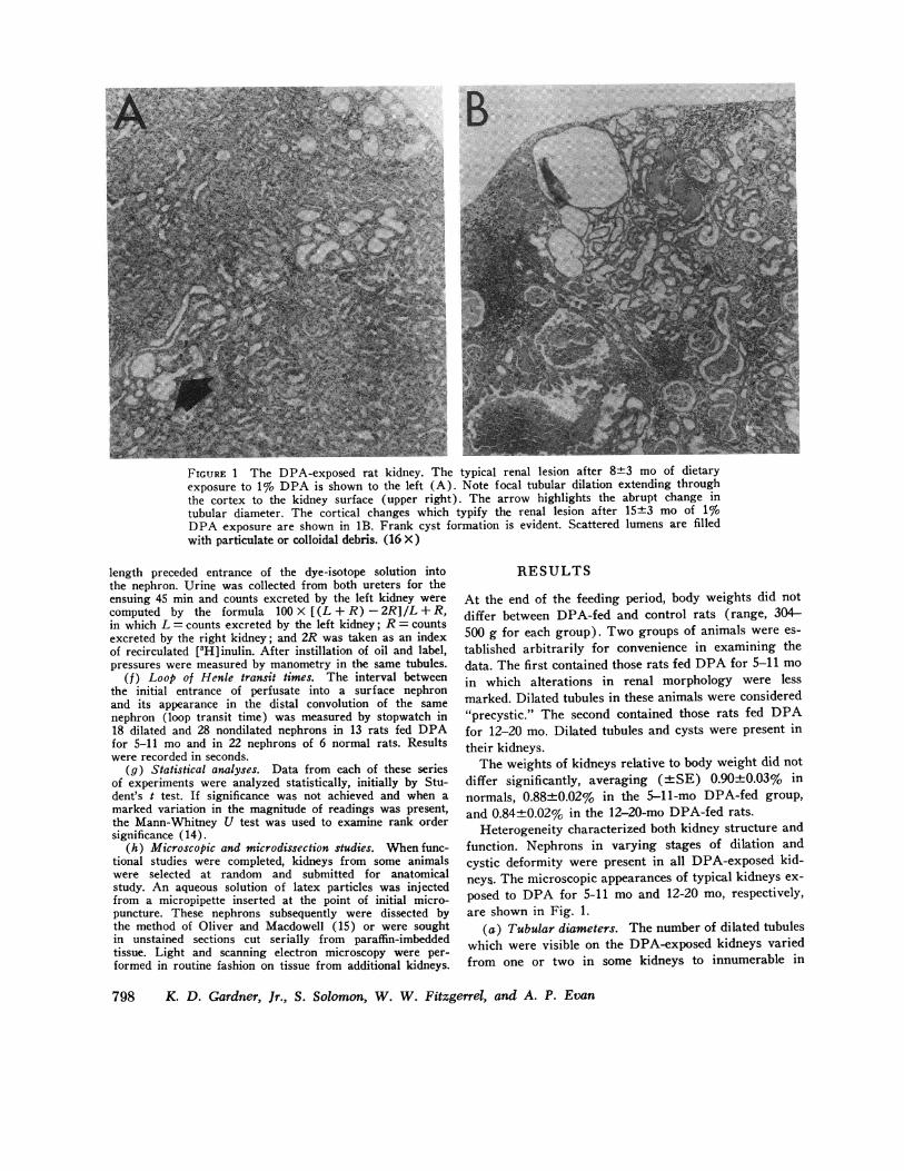
FIGURE 1 The DPA-exposed rat kidney. The typical renal lesion after 8+3 mo of dietaryexposure to 1% DPA is shown to the left (A). Note focal tubular dilation extending throughthe cortex to the kidney surface (upper right). The arrow highlights the abrupt change intubular diameter. The cortical changes which typify the renal lesion after 15+3 mo of 1%0DPA exposure are shown in 1B. Frank cyst formation is evident. Scattered lumens are filledwith particulate or colloidal debris. (16 X)
length preceded entrance of the dye-isotope solution intothe nephron. Urine was collected from both ureters for theensuing 45 min and counts excreted by the left kidney werecomputed by the formula 100 X [(L + R) - 2R]/L + R,in which L = counts excreted by the left kidney; R= countsexcreted by the right kidney; and 2R was taken as an indexof recirculated [3H]inulin. After instillation of oil and label,pressures were measured by manometry in the same tubules.
(f) Loop of Henle transit times. The interval betweenthe initial entrance of perfusate into a surface nephronand its appearance in the distal convolution of the samenephron (loop transit time) was measured by stopwatch in18 dilated and 28 nondilated nephrons in 13 rats fed DPAfor 5-11 mo and in 22 nephrons of 6 normal rats. Resultswere recorded in seconds.
(g) Statistical analyses. Data from each of these seriesof experiments were analyzed statistically, initially by Stu-dent's t test. If significance was not achieved and when amarked variation in the magnitude of readings was present,the Mann-Whitney U test was used to examine rank ordersignificance (14).
(h) Microscopic and microdissection studies. Whenfunc-tional studies were completed, kidneys from some animalswere selected at random and submitted for anatomicalstudy. An aqueous solution of latex particles was injectedfrom a micropipette inserted at the point of initial micro-puncture. These nephrons subsequently were dissected bythe method of Oliver and Macdowell (15) or were soughtin unstained sections cut serially from paraffin-imbeddedtissue. Light and scanning electron microscopy were per-formed in routine fashion on tissue from additional kidneys.
RESULTS
At the end of the feeding period, body weights did not
differ between DPA-fed and control rats (range, 304-500 g for each group). Two groups of animals were es-
tablished arbitrarily for convenience in examining thedata. The first contained those rats fed DPAfor 5-11 mo
in which alterations in renal morphology were lessmarked. Dilated tubules in these animals were considered"precystic." The second contained those rats fed DPAfor 12-20 mo. Dilated tubules and cysts were present intheir kidneys.
The weights of kidneys relative to body weight did not
differ significantly, averaging (-+SE) 0.90±0.03% innormals, 0.88±0.02% in the 5-11-mo DPA-fed group,and 0.84+0.02% in the 12-20-mo DPA-fed rats.
Heterogeneity characterized both kidney structure andfunction. Nephrons in varying stages of dilation andcystic deformity were present in all DPA-exposed kid-neys. The microscopic appearances of typical kidneys ex-
posed to DPA for 5-11 mo and 12-20 mo, respectively,are shown in Fig. 1.
(a) Tubular diameters. The number of dilated tubuleswhich were visible on the DPA-exposed kidneys variedfrom one or two in some kidneys to innumerable in
798 K. D. Gardner, Jr., S. Solomon, W. W. Fitzgerrel, and A. P. Evan

others. Longer exposure to DPA was associated with alarger number of visibly dilated tubules. Luminal diam-eters ranged between 34.2 and 110.8 iAm in 22 dilatednephrons and between 20.6 and 45.1 Am in 10 undilatednephrons in DPA-exposed kidneys. In six nephrons oftwo control rats, tubular diameter ranged between 25.9and 34.2 /Am. The differences in diameters between di-lated and nondilated nephrons were significant (P <0.001) by both methods of statistical analyses.
(b) Intraluminal hydrostatic pressures. Visibly di-lated surface proximal convoluted tubules had intralu-minal pressures that were significantly higher than thepressures recorded in nondilated nephrons in the samekidneys (Table I). This difference was documented byeach of the two techniques employed to measure pres-sure. Pressures measured by the servo-nulling transducertechnique tended to be higher, but the technique was ap-plied only in the 12-20-mo group. Significant differencesexisted among control, early, and late DPA-exposedkidneys in the rank order of their magnitude (U test)not in their arithmetical means (t test). There was oneexception: pressures measured by direct manometry av-eraged a significantly higher value in dilated nephronsof rats fed DPA for 5-11 mo. Among normal rats, thedifference between mean pressures measured by manom-etry and by transducer was not statistically significant.
(c) SNGFR. Whole kidney (left) GFR as a func-tion of total body weight did not differ significantlyamong the groups of kidneys studied (means in ml/min-kg body weight±SE): 3.88±0.42 in normal rats, 3.30±0.41 in 8 rats fed DPA for 5-11 mo, and 3.10±0.35 in13 rats fed DPA for 12-20 mo. Statistically significantdifferences were not found in SNGFR's (Table II). Insix rats fed DPA for 12-20 mo, intraluminal pressureswere monitored during the collection of samples of
TABLE IIntraluminal Pressures in Renal Tubules of DPA-Fed
and Normal (Control) Rats
No. No.of of
rats tubules Range Mean±SE
cm H20Water manometry
DPAdilated* 7 30 5.3-44.8 26.2 ±1.6tDPAnondilated* 7 24 4.2-31.5 19.3±0.8Control 2 1 1 3.9-24.8 20.6 ± 1.8
Servo-nulling transducerDPAdilated§ 6 1011 28.2-167.9 52.1 413.5¶DPAnondilated§ 5 1311 0.1-55.7 29.7 ±5.0Control 1 1 49 4.6-72.3 21.4 ± 1.9
* In rats fed DPA for 5-11 mo.Significantly different from other values in group; P < 0.05, t test.
§ In rats fed DPA for 12-20 mo.II Same nephrons studied for SNGFR(see Table II).¶ Significantly different from other values in group; P < 0.05, U test.
TABLE IIS.NGFR'S in Renal Tubules of DPA-Fed and
Normal (Control) Rats
No. No.of of
rats tubules Range Mean hSE*
ni/minDPA-fed 5-11 mo
Dilated 9 26 7.1-121.9 62.1+6.7Nondilated 9 30 6.4-131.6 56.6±42.7
DPA-fed 12-20 moDilated 5 13$ 22.9-91.4 50.6±+6.8Nondilated 6 10$ 13.3-99.6 48.4+7.8
Control 25 92 0.4-133.0 47.2±+2.9
* Differences not significant.t Same tubules in which pressures were monitored (Table I).
SNGFR. SNGFR's were found not to be related to ini-tial pressure, mean collection pressure, or to variation incollection pressures in either normal or DPA-exposedkidneys (Fig. 2).
(d) TF/PIA. TF/Pin s were measured in rats fedDPA and in normal animals. No significant differencewas found between the two groups of rats nor betweenthe two populations of nephrons in kidneys exposed toDPA for 5-11 mo. In the 12-20-mo group, however,TF/PIn's were significantly increased (Table III).
(e) Recovery of [H]inulin after single nephron mi-croinjection. The data from a typical experiment in thisseries are plotted in Fig. 3. Placement of the micropipette
PRESSURES
160
120.
Im/iti2
cm/-I2 0
801
S N G FRMean Colection
cmn H20
0
- ] 160
na mm/tersgamin
0
120
80
£0 0~~~~~~~~~~~~~~440 S if40-T00
0 * 0
ND D ND D ND DFIGURE 2 Initial and mean collection pressures (average,beginning, and ending pressures, plus pressure reading atfour intervals of 20, 40, 60, 80%o) during collection forSNGFRin 13 nondilated (ND) and 10 dilated (D) tubulesof six 12-20-mo DPA-fed rats. Cross bars represent meanvalues. Differences in mean collection pressures were shownby analyses of variance to be due to original differences inpressure.
The Diphenylamine-Exposed Kidney 799
:

TABLE I IITF/PI5 in Renal Tubules of DPA-Fed and
Normal (Control) Rats
No. No.of of
rats tubules Range Mean±SE
DPA-fed rats 5-11 moDilated 4 11 2.87-8.26 3.89±+0.75Nondilated 4 25 1.41-5.24 3.1440.25
DPA-fed rats 12-20 moDilated 4 20 2.89-11.40 6.06 ±0.59*Nondilated 4 18 3.14-8.40 4.47 ±0.57$
Control rats 3 23 1.49-6.72 2.91 ±0.22
* Significantly different from all but 12-20-mo nondilated nephrons;P <0.05.t Significantly different from control rats only; P < 0.05.
containing isotope-dye into a nephron characteristicallywas followed by a short burst of radioactivity in theurines in both kidneys. This was allowed to subside be-fore the perfusion pump was turned on. After injectionof normal and nondilated tubules, radioactivity usuallyappeared promptly in left ureteral urine and subsidedquickly. In contrast, placement of the micropipette in adilated tubule rarely was followed by a burst of radioac-tivity and perfusion of such a nephron resulted in therecovery of less isotope.
The results of all [5H]inulin recovery studies aresummarized in Table IV. In none of the three groupswas a significant amount of [3H]inulin recovered inurine from the right kidney. Detectable radioactivityemerged from the right side in 10 of 20 perfusions ofnephrons in DPA-exposed kidneys. No detectable in-crease in radioactivity was found in right ureteral urineafter perfusion of nephrons in normal control kidneys.
The range of values for inulin recoveries was ex-tremely wide after perfusion of dilated nephrons (0.4-93.3%). It contrasted sharply with the ranges recordedfor nondilated (72.0-121.2%) and normal (74.4-130.5%)nephrons. In four experiments in which collections wereextended for 70-90 min after microperfusion of one di-lated nephron in a single kidney, inulin recoveries av-eraged 1.4, 23.1, 2.9, and 30.8%, respectively, in urinefrom the perfused kidney. No detectable isotope appearedin the blood nor in right ureteral urine (with one ex-ception) of any of these animals.
Oil blockade. Dye-isotope excretion from unblockedtubules was prompt; green dye appeared promptly in theurine of all seven experiments. Excreted counts average83.4±2.9% total and ranged from 74.6 to 94.6% of radio-activity recovered in the urine over 45 min. Among thesetubules pressures average 24.7±2.1 and ranged from15.2 to 33.4 cm H20. In contrast among oil-blockadedtubules green urine never appeared subsequent to micro-
perfusion, excreted counts averaged - 6.5+1.5% andranged from - 1.2 to - 12.1% (i.e., 2R exceeded thesum of L + R), and pressures averaged 40.7±1.3 andranged between 35.8 and 44.0 cm H20. The differences incounts excreted (P < 0.001) and pressures (P < 0.001)were significant.
(f) Loop of Henle transit times. Transit time dataare summarized in Table V. The mean period of timerequired for a lissamine green solution to pass from in-jection site to the earliest visible loop of distal tubulewas prolonged 3-4 times in dilated nephrons. The endpoint often was difficult, sometimes impossible, to de-tect accurately. The difference in transit times betweennondilated and normal nephrons was far smaller but alsowas significant statistically.
Transit times and [5H]inulin recoveries were recordedin the same nephrons in 21 instances during this study(in nine dilated, eight nondilated, and four normal neph-rons). Longer transit times were associated with re-duced inulin recovery to a significant degree (correla-tion coefficient = 0.648; P < 0.01). Tubular diameterswere measured along with transit times on 14 occasions(in seven dilated and seven nondilated nephrons of fiverats fed DPA for 5-11 mo), and a direct and significant
PERIOD
FIGURE 3 Data from an ['H] inulin recovery experimentin an 11-mo DPA-exposed precystic kidney. Urinary radio-activity is indicated on the ordinate and consecutively num-bered 10-min collection periods on the abscissa. Illustratedis the sequence of steps-standardization of the pipette,placement in tubules, and perfusion-which was followed inthese experiments. ['H] Inulin recovery was derived fromthe counts recovered in ureteral urine during the two suc-cessive 10-min collection periods after perfusion and ex-pressed as a percentage of total counts injected. In thisanimal, perfusions 1, 2, 3, and 5 were into nondilated tubuleswhile 4 was into a dilated tubule. (['H] Inulin recovery was6%o of total counts injected after perfusion 4.) In thisexperiment, data from perfusions 3 and 5 were rejectedbecause of an error in perfusion speed and admixture oftwo left ureteral urine specimens, respectively.
800 K. D. Gardner, Jr., S. Solomon, W. W. Fitzgerrel, and A. P. Evan

B
FIGURE 4 (A) A representative latex-filled nephron dissected from a precystic kidney (in-direct illumination, approximately 125 X). (B) Tracing of nephron shown in A, indicatingglomerulus (G) and loci of tubular dilations (D). (C and D) Low-powered photomicrographof teased surface of a 14-mo DPA-exposed kidney showing appearance of single latex-fillednephron. Capsular surface is toward the top. Note mass of dilated cortical proximal convolutedtubule coils at surface; pars recta, with approximately normal diameter; and latex-filled cystdeep within renal substance.


TABLE IV[3H]Inulin Recovery from Left and Right Ureteral Urines
after Perfusion of Single Nephrons in Left Kidneyof Normal and 11 DPA-Fed Rats
Mean±SE% ['H]inulinRange of recovered
No. of observationnephrons periods Left Right
min %DPA-fed rats
Dilated 11 40-90 29.9 ±8.2 6.7 ±3.4*Nondilated 9 30-70 93.4±5. 7 1.4+5.7*
Control rats 6 20-70 100.859.8* 0.4±0.04
* Not significantly different from zero.P value of difference from dilated nephrons: <0.001.
correlation was found (correlation coefficient = 0.745;P<0.02).
(g) Microscopic and microdissection studies. In gen-eral terms, observations confirmed the changes in renalmorphology which have been described by others in theDPA-exposed kidney (6, 8, 9). Five observations weremade which were considered to have specific relevanceto the functional studies described above: Along proximaland collecting tubules dilation was shown to be seg-mental, not diffuse (Figs. 4A and 5E). Dilated tubulesat the surface occasionally communicated with cysts deepin the renal substance (Fig. 4B). Adjacent to intrarenalcysts, tubules often appeared compressed (Fig. 5A). Di-lated collecting tubules often contained debris whichappeared to be partially occluding their lumens (Fig.5B). And DPA-exposed kidneys possessed proximal con-voluted tubules with segments of apparent narrowing(Fig. 5C). In none of the nephrons or the microscopicsections studied was complete occlusion of a tubular lu-men identified.
DISCUSSIONDPA produces a renal lesion in rats which characteris-tically is heterogeneous in its severity and focal in itsdistribution (6). In the rats whose kidneys are reportedhere 5-30% of surface nephrons appeared visibly dilatedin vivo, an appraisal that was confirmed by measure-ments of tubular diameter. Usually, dilated nephronswere more prevalent at the poles than in the central por-
TABLE VVisual Proximal to Distal Tubule Transit Times in
13 DPA-Exposed and 6 Normal Kidneys
No. of Mean±SEnephrons transit time
5
DPA-exposed ratsDilated 18 98.148.0Nondilated 28 28.9 i±1.4*
Control rats 22 23.5 t 1.5t
* P value of difference from dilated nephrons: <0.001.$ P value of difference from nondilated nephrons: <0.05.
tion of the kidney. No ready explanation is available forthis observation.
Our results indicate that, under the conditions de-scribed, intraluminal pressures are elevated in the dilatedsurface nephrons of the DPA-exposed kidney. Two pos-sible explanations for this elevation are ruled out by themeasurements of SNGFR's and TF/Pin ratios. Wereelevated pressures the result of sustained "super-filtra-tion," one might have expected higher SNGFR's. Theywere not found; SNGFR's, including those measuredunder conditions of monitored pressure, were not sig-nificantly different among dilated versus nondilatednephrons in either early or late disease. Were elevatedpressures the result of a chronic reduction in net waterreabsorption, one would have expected lower TF/Pi.ratios in dilated nephrons. In actuality, equal to higherTF/Pi. ratios were found.
Persisting filtration into totally occluded nephrons alsocould account for elevated intraluminal pressures. Theevidence against this alternative is both morphologicaland functional: No completely occluded nephrons werefound by microdissection or microscopic examination,an experience identical to that of earlier workers (2-4,9). Lissamine green and [5H]inulin, placed into dilatedsegments of proximal nephrons, usually traversed theloop to appear only in ipsilateral urine. Complete oc-clusion, therefore, clearly does not exist in all dilatednephrons of the DPA-exposed kidney. Consequently itcannot be considered a prerequisite to elevated intralu-minal pressure and nephron dilation.
FIGURE 5 (A) Medium-power microscopy of area adjacent to large corticomedullary cyst(lumen upper right), showing compression of tubules (arrows) adjacent to a dilated nephron(1,100 X). (B) Scanning electron microscopic view of a cross-section of renal medulla fromDPA-exposed kidney showing dilated collecting tubule with a lumen partially occluded bydebris (arrow). (C and D) Renal cortex from DPA-fed (C) and normal (D) rats. Note"minitubule" in the DPA-exposed kidney. Its brush border identifies it as a proximal tubule.Tubules of similar diameter were not observed in kidneys from control rats. Latex particlesin such tubules indicated they communicated with dilated, micropunctured surface nephrons.(E) Scanning electron microscopic view of isolated collecting tubule from 14-mo DPA-exposedrat showing two foci of dilation adjacent to segments of normal diameter (400 X).
The Diphenylamine-Exposed Kidney 803

A fourth possibility to account for elevated pressure ispartial or intermittent occlusion. Microscopic sectionsand dissected nephrons were examined for sites of nar-rowing and partial obstruction, a more subjective anddifficult task than the identification of dilated tubules.Fig. 5 exemplifies the nature of the morphological evi-dence found: narrowed tubules, intraluminal casts anddebris, and compression of nephrons by adjacent cysts.Cysts, by compressing adjacent nephrons in vivo (Fig.5A), could cause their partial or intermittent occlusionwhile in vitro subsequent microdissection would revealwhat appeared to be perfectly patent tubules.
These considerations led us to conclude that elevatedpressures in the dilated surface nephrons of the DPA-exposed kidney most likely are the consequence of par-tial and/or intermittent downstream occlusion. This con-clusion was reinforced by the observation that pressureswere elevated and the excretion of the dye-isotope labelwas reduced or eliminated in normal tubules that wereintentionally blocked by droplets of oil.
Some of our results suggest that partial or intermit-tent occlusion may be relatively widespread and progres-sive in the DPA-exposed kidney. Although two differenttechniques were used to measure pressures (water ma-nometry and servo-nulling transducer), the highest pres-sures were encountered among dilated tubules in themost advanced stages of the lesion. TF/Pi. ratios wererelatively higher among the more severely dilated neph-rons in longer standing disease. Transit times were pro-longed significantly even among nondilated nephrons inthe earlier lesion. And the number of visibly dilated sur-face nephrons increased with the duration of DPA-exposure.
Three aspects of this study require further comment.The first deals with label "disappearance," a phenomenonobserved in a minority of the microperfusion experi-ments. The second concerns the finding that SNGFR'stended to remain unchanged in the face of increased in-traluminal pressure. The third concerns the quantitativesignificance of our data.
The appearance of labels in urine was not invariable.Lissamine green injection and ['H]inulin microperfusionoccasionally was followed by apparent "disappearance"of the markers. Failure of the microperfusion pump wasruled out when in vitro observations demonstrated itsability to deliver accurate volumes against as much as 60cm water pressure applied to the tip of its micropipette.Entrapment of the labels in medullary tissue by counter-current forces seemed implausible for several reasons:The efficiency of the countercurrent mechanism is im-paired in DPA-induced nephropathy (9). Dye stainingof medullary tissue was not observed after sectioning ofinjected kidneys. And [8H]inulin recirculation, an eventconsidered likely to accompany inulin escape from a mi-
croperfused nephron, did not occur regularly or evenfrequently, as evidenced by the absence of significant in-creases in the levels of blood and contralateral urineradioactivity in all but a rare instance of label disap-pearance.
Coupled with these considerations was the fact thatmorphological studies provided evidence in support of amore plausible, although speculative, explanation for la-bel "disappearance." Label could be trapped or delayedin nephrons compressed by adjacent cysts or in eddy cur-rents arising along dilated segments of tubules. Dilatedsurface nephrons were shown (Figs. 4 and 5) to com-municate with both compressed nephrons and frank cystsdeep within the renal substance. Some cysts measured inexcess of 2,500 .m in diameter. Assuming spherical con-figuration in vivo, they would have contained over 2,000nl of fluid, more than enough to dilute the 4,250 countsperfused in 50 nl to a level of radioactivity only 2-3counts above background. A change this small wouldhave gone undetected.
The observation that SNGFR's tended to remain un-changed despite higher pressures in dilated nephrons iscompatible with current theory and implies an intrinsicchange in glomerular function. Deen et al. (16) have in-dicated that glomerular filtration is determined by trans-glomerular capillary hydrostatic and osmotic gradientsand by the functional permeability of the glomerularmembrane. Were intratubular pressure to rise, therebynarrowing the hydrostatic gradient across the capillarywall, SNGFRcould remain unchanged only were thetransglomerular osmotic gradient to narrow or the func-tional permeability of the glomerular membrane to in-crease. We have no direct information as to which ofthese two alternatives occurs in the DPA-exposed kidneybut have observed obliteration of glomerular foot-proc-esses in the lesion (A. P. Evan, unpublished observa-tion). This morphological change suggests increasedglomerular permeability to protein. A glomerular proteinleak, by narrowing the transglomerular osmotic gradient,could offset the narrowed pressure gradient across theglomerulus and allow SNGFR's to be maintained.
The heterogeneity of nephron morphology and thewide ranges over which data from the DPA-exposed kid-neys often were spread have important implications inthe analyses and interpretation of our results. Save forobservations made under technically unsatisfactory cir-cumstances, no reading was discarded because it lay morethan two or three standard deviations away from themean of its group. Ignorance of the extent to whichheterogeneity of function as well as structure might ex-ist in these nephrons led us to a decision to include allobservations. This meant that occasionally a widely dis-crepant value was included, e.g., the precollection pres-sure of 167.9 cm H20 in a dilated, DPA-exposed nephron
804 K. D. Gardner, Jr., S. Solomon, W. W. Fitzgerrel, and A. P. Evan

(Fig. 2). Statistical significance was sought in differ-ences of rank order (the U test) as well as of groupmeans (the t test) to accommodate this variability. Un-der such circumstances and especially in light of thefrequently skewed distribution of data, arithmeticalmeans were given less quantitative significance thantraditionally is given them in studies of the homogeneous,normal kidney.
Although data are scant, there is reason to believethat increased intraluminal pressure plays an importantrole in cystic kidney disease. The case rests on the factsthat elevated pressures are found in cystic kidneys andthat elevated intratubular pressures lead to cystic de-formity.
In addition to the DPA-exposed kidney, elevated in-traluminal pressures also occur in the diphenyl thiazolemodel, albeit in "less than 5%" of nephrons (5). Vari-ably increased pressures have been recorded in vivoamong cysts in human polycystic kidney disease (17).Thus, to date elevated intraluminal pressures have beendocumented in each variety of cystic kidney diseasewhich has been examined.
The changes produced in nephron morphology by ure-teral obstruction, a procedure which elevates intralumi-nal pressure (12, 13) evidently by retarding drainagefrom nephrons with persisting glomerular filtration, havebeen studied by Strong, Fetterman, and Shimamura andtheir respective co-workers (18-20). Tubular dilation isnot diffuse but segmental, may involve not all but onlya portion of nephrons in the obstructed kidney, and ischaracterized as "cystic" in appearance. It occurs as anacquired lesion in a presumed previously normal kidney,a condition also met by the chemically induced models.But in contrast to these models, nephron dilation andcyst formation in the obstructed kidney occurs in the ab-sence of exposure to any exogenous cystogenic substance.
Currently, increased compliance of the tubular wall isan alternative hypothesis to account for cyst formation(5). The conclusion has been established by default. It
is based on a lack of evidence for obstruction in a ma-jority of nephrons in the diphenyl thiazole-exposed model(5) and demonstrations that metabolic factors can atleast accelerate nephron dilation and cyst formation (9).However, no direct studies of compliance in nephronsfrom cystic kidneys are available. There is no basis foran assumption that the cellular (degenerative) and peri-tubular (thickened basement membrane) changes whichcharacterize early DPA-induced nephropathy (2, 8, 9)increase rather than decrease or leave unchanged thecompliance of the tubular wall. And the studies of ure-teral obstruction already cited demonstrate that cystformation can occur without any postulated initial changein mural compliance, itself a postulated consequence ofthe action of a cystogenic chemical.
On the bases of published experience with ureteralobstruction and our demonstrations of increased intra-luminal pressure and delayed excretion of dye and iso-tope from dilated nephrons, we hypothesize that the pri-mary lesion in the DPA-exposed kidney is an obstruc-tive one, caused by tubular narrowing, intraluminal de-bris, and in the later stages by the compression of other-wise normal tubules from adjacent dilating nephrons.Weattribute cyst formation in this model to obstructionand a subsequent elevation of intraluminal pressures thatarise as the consequence of sustained glomerular filtra-tion and unaltered net water reabsorption in the involvednephrons.
It is tempting to extrapolate these findings and con-clusions to human renal cystic disease. While the DPAmodel and human cystic renal disease share severalpoints in common (heterogeneity in structural deformity[3, 4], apparent squeezing of adjacent otherwise patentand normal nephrons [3], variably elevated pressures intubules or cysts of the same kidney [17], and apparentpatency of dilated and cystic nephrons [3]), there is noreason to assume that the pathogenesis of cyst formationis identical in the model and in man. However, it seemslikely that data from the study of human material willcontinue to be relatively scarce, particularly since pre-transplant nephrectomy has become unfashionable. Care-ful study of animal models, at the moment, appears to bethe major approach available in order to achieve someunderstanding of the pathogenesis of cystic renal diseasein a susceptible mammalian kidney.
ACKNOWLEDGMENTS
The authors are grateful to Elizabeth Skipper, Ph.D., forher assistance with statistical analyses, and to PatriciaCarlton for preparation of histological sections.
This work was supported by Public Health Servicegrants HL 13137, HL 16775, and AMHL 17641.
REFERENCES
1. Osathanondh, V., and E. L. Potter. 1964. Pathogenesisof polycystic kidneys. Historical survey. Arch. Pathol.77: 459-465.
2. Darmady, E. M., J. Offer, and M. A. Woodhouse. 1970.Toxic metabolic defect in polycystic disease of thekidney. Evidence from microscope studies. Lancet. 1:547-550.
3. Lambert, P. P. 1947. Polycystic disease of the kidney.A review. Arch. Pathol. 44: 34-58.
4. Osathanondh, V., and E. L. Potter. 1964. Pathogenesisof polycystic kidneys. Survey of results of microdissec-tion. Arch. Pathol. 77: 510-512.
5. Carone, F. A., R. G. Rowland, S. G. Perlman, and C. E.Ganote. 1974. The pathogenesis of drug-induced renalcystic disease. Kidney Int. 5: 411-421.
6. Thomas, J. O., A. J. Cox, Jr., and F. DeEds. 1957.Kidney cysts produced by diphenylamine. Stanford Med.Bull. 15: 90-93.
The Diphenylamine-Exposed Kidney 805

7. Goldman, S. H., S. R. Walker, T. C. Merigan, Jr.,K. D. Gardner, Jr., and J. M. C. Bull. 1966. Heredi-tary occurrence of cystic disease of the renal medulla.N. Engl. J. Med. 274: 984-992.
8. Crocker, J. F. S., D. M. Brown, R. F. Borch, andR. L. Vernier. 1972. Renal cystic disease induced innewborn rats by diphenylamine derivatives. Am. J.Pathol. 66: 343-350.
9. Safouh, M., J. F. S. Crocker, and R. L. Vernier. 1970.Experimental cystic disease of the kidney. Sequential,functional and morphologic studies. Lab. Invest. 23:392-400.
10. Barenberg, R. L., S. Solomon, S. Papper, and R. An-derson. 1968. Clearance and micropuncture study ofrenal function in mercuric chloride treated rats. J. Lab.Clin. Med. 72: 473-484.
11. Vurek, G. G., C. M. Bennett, R. L. Jamison, and J. L.Troy. 1967. An air-driven micropipette sharpener. J.Appl. Physiol. 22: 191-192.
12. Gottschalk, C. W., and M. Mylle. 1956. Micropuncturestudy of pressures in proximal tubules and peritubularcapillaries of the rat kidney and their relation to ureteraland renal venous pressures. Am. J. Physiol. 185: 430-439.
13. Brenner, B. M., J. L. Troy, and T. M. Daugharty. 1972.Pressures in cortical structures of the rat kidney. Am.
J. Physiol. 222: 246-251.
14. Mann, H. B., and D. R. Whitney. 1947. On a test ofwhether one of two random variables is stochasticallylarger than the other. Ann. Math. Stat. 18: 50-60.
15. Oliver, J., and M. MacDowell. 1961. The structural andfunctional aspects of the handling of glucose by thenephrons and the kidney and their correlation by meansof structural-functional equivalents. J. Clin. Invest. 40:1093-1112.
16. Deen, W. M., C. R. Robertson, and B. M. Brenner.1972. A model of glomerular ultrafiltration in the rat.Am. J. Physiol. 223: 1178-1183.
17. Bjerle, P., B. Lindqvist, and G. Michaelson. 1971. Pres-sure measurements in renal cysts. Scand. J. Clin. Lab.Invest. 27: 135-138.
18. Strong, K. C. 1940. Plastic studies in abnormal renalarchitecture. V. The parenchymal alterations in 'experi-mental hydronephrosis. Arch. Pathol. 29: 77-119.
19. Fetterman, G. H., M. M. Ravitch, and F. E. Sherman.1974. Cystic changes in fetal kidneys following ureteralligation: studies by microdissection. Kidney Int. 5:111-121.
20. Shimamura, T., J. M. Kissane, and F. Gyorkey. 1966.Experimental hydronephrosis. Nephron dissection andelectron microscopy of the kidney following obstructionof the ureter and in recovery from obstruction. Lab.Invest. 15: 629-640.
806 K. D. Gardner, Jr., S. Solomon, W. W. Fitzgerrel, and A. P. Evan