fuller_mechanical ventilation ards in ed_chest 2015-2
TRANSCRIPT
ONLINE FIRST
This is an Online First, unedited version of this article. The final, edited
version will appear in a numbered issue of CHEST and may contain substantive changes. We encourage readers to check back for the final
article. Online First papers are indexed in PubMed and by search engines, but the information, including the final title and author list,
may be updated on final publication.
http://journal.publications.chestnet.org/
Online First articles are not copyedited prior to posting.
©American College of Chest Physicians. Reproduction of this article is prohibited without written permission from the
American College of Chest Physicians. See online for more details.
Page 1 of 36
Downloaded From: http://journal.publications.chestnet.org/ by a Washington University School of Medicine User on 03/06/2015
1
Title Page Word counts: Text: 2499 Abstract: 249 Title: Mechanical ventilation and acute respiratory distress syndrome in the emergency department: a multi-center, observational, prospective, cross-sectional study Short title/running head: Mechanical ventilation and ARDS in the ED Corresponding Author: Brian M. Fuller, MD, MSCI Departments of Emergency Medicine and Anesthesiology Division of Critical Care Washington University School of Medicine in St. Louis St. Louis, MO 63110 [email protected] Co-authors: Nicholas M. Mohr, MD Departments of Emergency Medicine and Anesthesiology Division of Critical Care Roy J. and Lucille A. Carver College of Medicine University of Iowa 200 Hawkins Drive, 1008 RCP Iowa City, IA 52242 [email protected] Christopher N. Miller, MD, MS Department of Emergency Medicine University of Cincinnati Medical Center 231 Albert Sabin Way PO Box 670769 Cincinnati, OH 45267-0769 [email protected] Andrew R. Deitchman, MD Department of Emergency Medicine Christiana Care Health System 4755 Ogletown Stanton Road Newark, DE 19718 [email protected] Brian J. Levine, MD Department of Emergency Medicine Christiana Care Health System 4755 Ogletown Stanton Road Newark, DE 19718 [email protected]
Page 2 of 36
Downloaded From: http://journal.publications.chestnet.org/ by a Washington University School of Medicine User on 03/06/2015

2
Nicole Castagno, MS University of Michigan Medical School 1137 Catherine St Ann Arbor, MI 48109-5608 [email protected] Elizabeth C. Hassebroek, MD Department of Critical Care Medicine Mayo Clinic Rochester, MN 55905 [email protected] Adam Dhedhi Saint Louis University School of Medicine St. Louis, MO 63103 [email protected] Nicholas Scott-Wittenborn Washington University in St. Louis St. Louis, MO 63130 [email protected] Edward Grace Middlebury College Middlebury, Vermont 05753 [email protected] Courtney Lehew Department of Emergency Medicine Washington University School of Medicine in St. Louis St. Louis, MO 63110 [email protected] Marin H. Kollef, MD Department of Medicine Division of Pulmonary and Critical Care Medicine Washington University School of Medicine in St. Louis [email protected] Conflicts of interest: All authors declare no conflicts of interest. Funding information: BMF was supported by the Emergency Medicine Grant-in-Aid from the Department of Emergency Medicine, Washington University School of Medicine in St. Louis, and the KL2 Career Development Award. NMM was supported by the Emergency Medicine Foundation Research Fellowship.NC was funded by the University of Cincinnati Department of Emergency Medicine. EG was supported by the ASPIRE Program at Washington University in St. Louis. MHK was supported by the Barnes Jewish Hospital Foundation. This publication was supported by the Washington University Institute of Clinical and Translational Sciences grants UL1 TR000448 and KL2 TR000450 from the National Center for Advancing Translational Sciences.
Page 3 of 36
Downloaded From: http://journal.publications.chestnet.org/ by a Washington University School of Medicine User on 03/06/2015

3
The content is solely the responsibility of the authors and does not necessarily represent the official views of the NIH or any of the other supporting bodies. Notation of prior publication/presentation: This work has not been published or presented in any form prior to this submission.
Page 4 of 36
Downloaded From: http://journal.publications.chestnet.org/ by a Washington University School of Medicine User on 03/06/2015

5
Abstract Background: There is little data regarding mechanical ventilation and acute respiratory distress syndrome (ARDS) in the emergency department (ED). This could be a vital arena for prevention and treatment. Methods: Multi-center, observational, prospective, cohort study aimed at analyzing ventilation practices in the ED. The primary outcome was the incidence of ARDS after admission. Multivariable logistic regression was used to determine predictors of ARDS. Results: We analyzed 219 mechanically ventilated patients to assess ED ventilation practices. Median tidal volume was 7.6 mL/kg predicted body weight [PBW] (IQR, 6.9 - 8.9), with a range of 4.3 - 12.2 mL/kg PBW. Lung-protective ventilation was used in 122 (55.7%) patients. The incidence of ARDS after admission from the ED was 14.7%, with a mean onset of 2.3 days. Progression to ARDS was associated with higher illness severity and intubation in the prehospital environment or transferring facility. Of the fifteen (6.8%) patients with ARDS in the ED, lung-protective ventilation was used in 7 (46.7%) patients. Patients that progressed to ARDS experienced greater duration in organ failure, ICU length of stay, and mortality. Conclusions: Lung-protective ventilation is infrequent in mechanically ventilated ED patients, regardless of ARDS status. Progression to ARDS is common after admission, occurs early, and worsens outcome. Patient- and treatment-related factors present in the ED are associated with ARDS. Given the limited treatment options for ARDS, and the early onset after admission from the ED, measures to prevent onset and mitigate severity should be instituted in the ED. Clinical Trial Registration: ClinicalTrials.gov (NCT01628523)
Page 6 of 36
Downloaded From: http://journal.publications.chestnet.org/ by a Washington University School of Medicine User on 03/06/2015

4
Abbreviations list aOR: adjusted odds ratio
APACHE: Acute Physiology and Chronic Health Evaluation ARDS: acute respiratory distress syndrome BMI: body mass index COPD: chronic obstructive pulmonary disease ED: emergency department
FiO2: fraction of inspired oxygen ICU: intensive care unit IQR: interquartile range
LIPS: lung injury prediction score LOS: length of stay
PaO2: partial pressure of oxygen PBW: predicted body weight PEEP: positive end-expiratory pressure SD: standard deviation SIMV: synchronized intermittent mandatory ventilation
SOFA: Sequential Organ Failure Assessment SpO2: pulse oximeter oxygenation STROBE: Strengthening the Reporting of Observational Studies in Epidemiology Statement VALI: ventilator-associated lung injury
Page 5 of 36
Downloaded From: http://journal.publications.chestnet.org/ by a Washington University School of Medicine User on 03/06/2015
6
INTRODUCTION
The frequency and severity of critically ill patients in the emergency department (ED) has
increased 1. The need for mechanical ventilation is one of the most common indications for
intensive care unit (ICU) admission, and has also increased in incidence 2,3. Initiation of
mechanical ventilation in the ED is common, and given the long ED lengths of stay for critically
ill patients, mechanical ventilation hours provided have also increased4-13. Despite these trends,
there remains relatively little data on ED-based mechanical ventilation practices 14.
Acute respiratory distress syndrome (ARDS) exacts a significant toll on mechanically
ventilated patients in terms of mortality, long-term survivor morbidity, and healthcare utilization
15,16. Compared to the ICU, ARDS data in the ED population is sparse. The ED prevalence of
ARDS and early factors that may promote its development and modify its severity are relatively
incomplete. Observational studies indicate an ARDS prevalence in mechanically ventilated ED
patients of approximately 9%14,17,18. Most of these data, however, are restricted to a narrow
cohort of patients (i.e. sepsis) and are single-center investigations.
In patients with ARDS, unequivocal data exists that harmful ventilator settings cause
ventilator-associated lung injury (VALI) and worsen outcome19-21. In patients without ARDS, but
at risk for the syndrome, there is mounting data to suggest that the mechanical ventilator
contributes to ARDS development22-31. Most relevant to the ED, the pathophysiology triggered
by VALI can occur within hours, and progression to ARDS in at-risk patients typically occurs
shortly after admission29,32-35. We hypothesize that modifiable patient characteristics and
treatment variables can influence clinical outcome during this most proximal time window. In the
future, the ED could therefore be a vital arena for the treatment and clinical investigation of
mechanically ventilated patients in order to: 1) further refine predictive variables of outcome; 2)
improve quality of mechanical ventilation delivered during early stages of respiratory failure; 3)
decrease the incidence of ARDS; and 4) decrease mortality and long term survivor morbidity.
Page 7 of 36
Downloaded From: http://journal.publications.chestnet.org/ by a Washington University School of Medicine User on 03/06/2015

7
The objectives of this study were: 1) further characterize ED mechanical ventilation
practices; 2) determine the incidence of ARDS after admission and the risk factors associated
with this outcome; 3) determine the prevalence of ARDS in the ED and assess ED compliance
with lung-protective ventilation; and 4) assess outcome differences between all patients with
ARDS compared to those without ARDS.
METHODS
This was a multicenter, prospective observational, cross-sectional study conducted at
four academic EDs. For each center, data was collected at four temporally distinct, one-month,
time periods (7/10/12 to 8/10/12; 9/1/12 to 10/2/12; 1/21/13 to 2/22/13; 7/2/13 to 8/3/13). The
study therefore spanned a total of thirteen months. This observational study is reported in
accordance with the Strengthening the Reporting of Observational Studies in Epidemiology
(STROBE) Statement: Guidelines for Reporting Observational Studies 36. The institutional
review boards approved the study at each site under waiver of informed consent (e-Appendix
1). The study was registered on ClinicalTrials.gov (NCT01628523).
Eligible patients were all mechanically ventilated ED patients age 18 years or older.
Exclusion criteria: 1) death in the ED; 2) ED length of stay (LOS) < 1 hour; 3) total mechanical
ventilation duration < 1 hour; and 4) elective extubation while in the ED. To ensure uniform data
collection and accuracy, all variables were defined a priori and recorded in a standardized
format during the data collection process.
The baseline patient characteristics included age, gender, race, weight, height, predicted
body weight (PBW), body mass index (BMI), ED LOS, patient comorbidities, home medications,
vital signs, hemodynamics, and laboratory values. Modified Acute Physiology and Chronic
Health Evaluation (APACHE) II, Sequential Organ Failure Assessment (SOFA), and lung injury
prediction (LIPS) scores were determined37-41. PBW in kilograms (kg) was calculated according
to the formula: males, 50 +2.3 [height (inches) – 60]; females, 45.5 +2.3 [height (inches) – 60]
Page 8 of 36
Downloaded From: http://journal.publications.chestnet.org/ by a Washington University School of Medicine User on 03/06/2015
8
42. Process of care variables in the ED [intravenous (IV) fluid, blood products, etc.] were
collected, as were all ventilator-related variables. All of these data were prospectively collected
by research assistants and principal investigators (PI) at each site.
Definitions. Definitions of comorbid conditions are provided in e-Appendix 2. Severe
sepsis and septic shock were defined as previously described 43,44. Lung-protective ventilation
was defined as the use of tidal volume of <8 mL/kg predicted body weight (PBW), as this was
the upper limit of tidal volume allowed by previous investigation of low tidal volume ventilation in
ARDS 19. We did not include a pressure limit to define lung-protective, as previous data suggest
monitoring of inspiratory plateau pressure is rare in intubated ED patients14.
Outcomes. All patients were analyzed for ED mechanical ventilation practices. The
primary outcome variable of interest was the development of ARDS after admission, and was
defined according to the Berlin definition 45. It was assessed by site PIs at least once daily
(based on frequency of chest radiographs and arterial blood gas measurements). Given the
focus of this investigation on ED-based factors associated with ARDS development, and data
suggesting that the majority of ARDS cases develops in the first five days after admission,
assessment of the primary outcome was restricted to day five after ICU admission (or death if
occurring prior to day 5)40. In patients without an arterial blood gas measurement, the
oxygenation criteria was determined using the pulse oximeter (SpO2):FiO2 ratio as described
previously 46. When more than one value was present, the worst value was selected.
A detailed description of our standard operating procedure for adjudicating ARDS status
is provided in e-Appendix 3. Secondary analyses of interest included clinical outcome
differences between all ARDS patients compared to those without ARDS. These outcomes were
assessed daily, until hospital discharge, by research assistants and site PIs.
Analysis. Descriptive statistics, including mean [standard deviation (SD)], median
[interquartile range (IQR)], and frequency distributions were used to assess the characteristics
of the patient cohort. Spearman’s correlation coefficient (rs) was used to assess the relationship
Page 9 of 36
Downloaded From: http://journal.publications.chestnet.org/ by a Washington University School of Medicine User on 03/06/2015

9
between ED and ICU tidal volume. To assess predictors of progression to ARDS, continuous
and categorical variables were compared using an unpaired t-test, Wilcoxon’s test, Chi-square
test, or Fisher’s exact test, as appropriate. Variables that were statistically significant in
univariate analyses at a p ≤ 0.10 level were candidates for inclusion in a bidirectional stepwise,
multivariable logistic regression analysis. The stepwise regression method selected variables for
inclusion or exclusion from the model in a sequential fashion based on the significance level of
0.10 for entry and 0.15 for removal. Statistical interactions and collinearity were assessed. The
model used variables that contributed information that was statistically independent of the other
variables in the model. The model’s goodness of fit was assessed with the Hosmer-Lemeshow
test. Adjusted odds ratios (aORs) and corresponding 95% CIs are reported for variables in the
multivariable model, adjusted for all variables in the model. To assess clinical outcomes based
on ARDS status, Chi-square and Kruskal-Wallis test were used to compare groups. The Kaplan-
Meier method was used to compare mortality difference. All tests were two-tailed, and a p value
<0.05 was considered statistically significant. A sample size calculation was not performed a
priori, as they primary analysis was descriptive and to further characterize mechanical
ventilation in the ED. Based on previous existing data, our sample size was recognized as likely
to be adequate for investigation of ED-based parameters associated with progression to
ARDS14,47. The analysis was conducted in consultation with a biostatistician.
RESULTS
Characteristics of study subjects
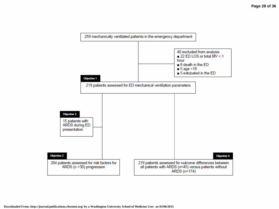
A total of 259 patients received mechanical ventilation in the ED during the study period
(Figure 1); the final study population totaled 219 patients. All patients were assessed for
mechanical ventilation practices and clinical outcomes. Fifteen patients (6.8%) had ARDS while
in the ED, and were excluded from the analysis of risk factors for ARDS progression after ED
Page 10 of 36
Downloaded From: http://journal.publications.chestnet.org/ by a Washington University School of Medicine User on 03/06/2015
10
admission. Table 1 shows the baseline characteristics of the study population. For the entire
cohort, the median ED LOS was 3.4 hours (IQR 2.2 - 5.4) with a range of 1.1 - 18.3 hours.
Ventilator characteristics
Ventilator variables are presented in Table 2. The preferred mode of ventilation across
centers was assist-control, volume-control ventilation (65.3%). Median tidal volume delivered
was 7.6 mL/kg PBW (IQR, 6.9 - 8.9), with a range of 4.3 - 12.2 mL/kg PBW. Figure 2 shows the
distribution of tidal volume in the ED. Lung-protective ventilation was used in 122 (55.7%)
patients and 25 (11.4%) patients were ventilated with a tidal volume > 10mL/kg PBW. Of the 97
patients ventilated with non lung-protective ventilation in the ED, 31/97 (32%) were changed to
protective settings upon ICU arrival. ED tidal volume was significantly correlated to ICU tidal
volume, rs= .60, p<0.001. In the subgroup of patients exposed to non lung-protective while in the
ED, ED tidal volume remained significantly correlated to ICU tidal volume, rs= .46, p< 0.001.
Inspiratory plateau pressure was recorded in 78 patients (35.6%). At least one ventilator
parameter was changed in 150 (68.5%) patients during their ED stay. The head of bed was
elevated in 79 (36.1%) patients while receiving mechanical ventilation in the ED.
Analysis of ARDS
The incidence of ARDS after admission from the ED was 14.7% (n= 30), with a mean
(±SD) onset of 2.3 days (±1.2) (Figure 3). There were no differences in the ED ventilator
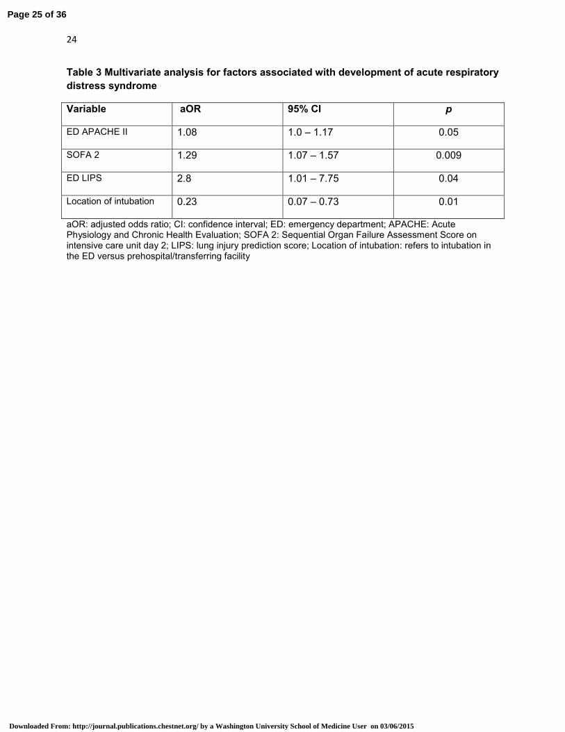
variables in these patients (Table 2). Multivariable logistic regression analysis demonstrated that
higher ED APACHE II and LIPS were associated with progression to ARDS, as was a higher
SOFA score (persistent organ failure) on ICU day 2. Intubation occurring prehospital or from a
transferring facility was associated with an increased risk of ARDS compared with ED intubation
(Table 3).
Page 11 of 36
Downloaded From: http://journal.publications.chestnet.org/ by a Washington University School of Medicine User on 03/06/2015

11
Fifteen (6.8%) patients had ARDS during their stay in the ED. Median tidal volume was
8.2mL/kg PBW (IQR, 7.2 - 9.0), compared to 7.6 mL/kg PBW (IQR, 6.9 - 8.9) in patients without
ARDS, p= 0.37. Lung-protective ventilation was used in 7 (46.7%) patients with ARDS.
Inspiratory plateau pressure was monitored in 6 ED patients with ARDS (40%). Exposure to
FiO2 of 1.0 in ED-ARDS patients was 99.0 minutes (IQR, 60 - 198) compared to 43.5 minutes
(IQR, 0 - 117.0) patients without ARDS in the ED.
Compared to patients without ARDS, patients that progressed to ARDS experienced a
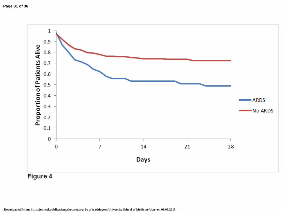
greater duration of organ failure, ICU LOS, and mortality (Table 4 and Figure 4).
DISCUSSION
Our first objective was to characterize further the use of mechanical ventilation in the ED
across a heterogeneous patient population in multiple centers. To summarize, based on our
analysis, mechanical ventilation in the ED is delivered using: 1) higher than recommended tidal
volumes and infrequent lung-protective ventilation regardless of ARDS status; 2) high FiO2 and
low PEEP; 3) infrequent monitoring of inspiratory plateau pressure; and 4) the supine, flat
position.
Previous work in severe sepsis and septic shock patients (database from 2005-2010)
showed a median tidal volume of 8.8mL/kg PBW (IQR, 7.8 to 10.0), ranging as high as
14.6mL/kg PBW14. The current investigation shows a decrease of about 1mL/kg PBW and
overall less variability in practice. However, a significant percentage of patients remain exposed
to high tidal volumes while in the ED. Based on three recent systematic reviews and meta-
analyses, non-lung protective ventilation seems associated with VALI and development of
ARDS 29,31,48.
Our results highlight the infrequency with which PEEP is titrated in the ED, in favor of
delivery of high levels of oxygen. Increasing evidence suggests excessive oxygen exposure has
adverse effects in various conditions, such as cardiac arrest, ARDS, chronic obstructive
Page 12 of 36
Downloaded From: http://journal.publications.chestnet.org/ by a Washington University School of Medicine User on 03/06/2015
12
pulmonary disease (COPD), and acute myocardial infarction 49-53. Providing an optimal
environment for lung protection, however, probably requires attention to not only tidal volume,
but also appropriate lung recruitment and oxygen exposure.
Only one-third of the study cohort had their head of bed elevated while undergoing
mechanical ventilation in the ED. Supine head position during the first 24 hours of mechanical
ventilation is an independent risk factor for pneumonia 54. This is an immediately modifiable
process of care variable that could reduce complications in mechanically ventilated patients
admitted from the ED.
Prior work showed an ARDS prevalence of 8.8% in mechanically ventilated ED patients
with severe sepsis and septic shock 14. This current investigation of a heterogeneous ED
population demonstrated an ARDS prevalence of 6.8%, and is similar to recent work examining
ARDS in mechanically ventilated ED patients 14,17,18. Combining data from these studies
provides some epidemiologic insight into an ED ARDS prevalence of approximately 8.4% in
intubated patients. Similar to previous work, adherence to lung-protective ventilation in ARDS
patients was low (46.7%). With a conservative estimate of 240,000 patients who receive ED
mechanical ventilation annually, the sheer number of patients exposed to potentially harmful
ventilation presents an opportunity to re-examine clinical practice and study these patients
further 6.
VALI and ARDS can evolve quickly 22,24,26,32,33,47,55,56. The ED represents a period of early
critical illness during which protective interventions can influence complications of critical illness
57. Our data cannot answer the question, however, if altering ED ventilator practices will
decrease ARDS or mitigate its severity, and our study did not show any association with
ventilator variables and incidence of ARDS 57,58. This may reflect that our study was
underpowered to detect a small difference that does exist, that there is a true lack of association
between ED ventilator management and downstream complications, or that the ED exposure is
too short to impact the outcome. However, our data does suggest that ED tidal volume settings
Page 13 of 36
Downloaded From: http://journal.publications.chestnet.org/ by a Washington University School of Medicine User on 03/06/2015
13
are influential on those delivered in the ICU. This remained true for patients exposed to non
lung-protective ventilation in the ED, and suggests that even suboptimal ventilator settings were
continued forth into early ICU care. Also, while tidal volume often exceeded 8mL/kg PBW, rarely
did it exceed levels shown in prior trials to be injurious in patients with established ARDS (i.e.
12mL/kg PBW), or at risk for the syndrome (i.e. 10mL/kg PBW, 11.4% of patients in this study)
19,25. So in a study of this size, deviations of this magnitude may not be enough to cause a
measurable clinical difference, both in terms of ARDS mortality and ARDS development. A lack
of association between ventilator variables and ARDS incidence may also reflect the fact that
ARDS etiology is quite heterogeneous; the most appropriate ED intervention may be a bundled,
quality-based approach to address ventilator and non-ventilator treatments 57.
The incidence of ARDS development in this heterogeneous cohort was 14.7%; a
previous investigation of severe sepsis and septic shock patients (perhaps the highest risk
cohort for ARDS) showed an ARDS incidence of 27.5% after ED admission 14. This provides
further evidence to consider ARDS prevention strategies a priority in emergency research and
quality initiatives. Clinical ARDS research has historically been confined to the ICU, but as
additional preventive therapies are proposed, ED-based trials will be critical to treat high risk
patients early in the course of disease. Results of our multivariable analysis coincide with prior
research, and suggest that these high risk patients are identified by higher illness severity
scores (APACHE II) and LIPS 40. Our results also suggest that two potential non-ventilator
related variables could be targets for futures ARDS prevention: reversal of early organ failure
and prehosptial intubation.
There are important limitations to this study. This was a cross-sectional study conducted
over a single time period (i.e. one month) at each center. ED mechanical ventilation practice
patterns and incidence of ARDS may vary in association with seasonal respiratory illnesses,
such as H1N1 influenza. However, there is a lack of data to support seasonal variation of
ARDS, and our study months were temporally distinct and varied seasonally across centers.
Page 14 of 36
Downloaded From: http://journal.publications.chestnet.org/ by a Washington University School of Medicine User on 03/06/2015

14
This temporal distribution offers some assurance our data represent a national longitudinal
sample 59.
This is a relatively small study and therefore prone to random error. However, our results
are consistent with prior evidence. This study was restricted to academic medical centers. It is
therefore possible that these data are not truly representative of ED-based mechanical
ventilation practices and ARDS prevalence in the community as a whole. The multi-center trial
design, consistency with the small amount of previously published data, and inclusion of all
mechanically ventilated patients do improve external validity of our results.
Adjudicating ARDS status can be difficult and will always have a subjective component.
This potentially exposes the study to ascertainment bias. Our adjudication protocol was
systematic, rigorous, and objective. Our event rate for ARDS was also consistent with previous
investigations. We are therefore confident we adjudicated the syndrome accurately for purposes
of this investigation.
Finally, the trained research assistants played no role in the clinical care of the patients,
and clinicians were unaware of our study hypotheses. The possibility that the presence of
bedside research assistants influenced clinical care and ventilator settings cannot be excluded
completely (i.e. Hawthorne-like effect). Our findings, particularly suboptimal adherence to best-
practice guidelines such as protective lung ventilation strategies and head-of-bed elevation
speaks against this possibility.
CONCLUSION
This multi-center study of ED patients with respiratory failure demonstrates a significant
opportunity to improve ED-based mechanical ventilation practices. This includes delivery of
lung-protective ventilation, monitoring of inspiratory plateau pressure, and head-of-bed
elevation. Across a heterogeneous intubated ED population, progression to ARDS is a common
occurrence, occurs early after ICU admission, and leads to significant negative clinical
Page 15 of 36
Downloaded From: http://journal.publications.chestnet.org/ by a Washington University School of Medicine User on 03/06/2015

15
consequences. Modifiable patient- and treatment-related variables exist which could prevent or
mitigate ARDS severity, and the ED and prehospital environments should be investigated
further.
ACKNOWLEGEMENTS
Guarantor statement: BMF takes responsibility for the content of the manuscript, including the data and analysis. Author contributions: BMF conceived and designed the study, obtained research funding,
supervised conduct of the trial, managed data, served as a referee in ARDS adjudication,
analyzed and interpreted data. NMM designed the study, supervised conduct of the trial,
managed data, analyzed and interpreted data, and served as a referee in ARDS adjudication.
CNM supervised conduct of the trial, managed data, analyzed and interpreted data, aided in
patient recruitment, and served as a referee in ARDS adjudication. ARD, BJL supervised
conduct of the trial, collected and managed data, analyzed and interpreted data, recruited
patients, and served as a referee in ARDS adjudication. NC, ECH, AD, NSW, EG, and CL
recruited patients, analyzed and interpreted data, collected and managed data. MHK aided in
conception and design of the study, analyzed and interpreted data, and aided in drafting of the
manuscript. All authors contributed to the drafting of the manuscript, revision of the manuscript,
and approval of the final version.
Disclosures: None, and all authors declare no conflicts of interest. Role of the sponsors: The funding sources played no role in the design, conduct, analysis, or
interpretation of these data, and played no role in the drafting, revision, or submission of the
manuscript.
Other contributions: The authors would like to acknowledge the following people: Karen Steger-
May, MA from the Division of Biostatistics for assistance with the statistical analysis of these
data; Lucas Allen Strakowski, John Collier, Bryan English, Brett Faine, Courtney Hancock, Willis
Page 16 of 36
Downloaded From: http://journal.publications.chestnet.org/ by a Washington University School of Medicine User on 03/06/2015

16
Hong, Frank Jareczek, Alycia Karsjens, Ashley Mills, Mackenzie Moore, Laura Nielsen, Angela
Ohrt, Randi Ryan, Dena Sult, Kelsey Winnike for assistance with data collection and entry.
BMF would like to acknowledge Brad Echols for a lifetime of friendship and support.
Page 17 of 36
Downloaded From: http://journal.publications.chestnet.org/ by a Washington University School of Medicine User on 03/06/2015

17
REFERENCES
1. Herring A, Ginde AA, Fahimi J, et al. Increasing critical care admissions from U.S. emergency
departments, 2001-2009*. Crit Care Med. 2013;41(5):1197-1204.
2. Esteban A, Anzueto A, Frutos F, et al. Characteristics and outcomes in adult patients receiving
mechanical ventilation: a 28-day international study. Jama. 2002;287(3):345-355.
3. Needham DM, Bronskill SE, Calinawan JR, Sibbald WJ, Pronovost PJ, Laupacis A. Projected
incidence of mechanical ventilation in Ontario to 2026: Preparing for the aging baby boomers*.
Critical care medicine. 2005;33(3):574-579.
4. Sagarin MJ, Barton ED, Chng Y-M, Walls RM. Airway management by US and Canadian
emergency medicine residents: a multicenter analysis of more than 6,000 endotracheal
intubation attempts. Annals of emergency medicine. 2005;46(4):328-336.
5. Wang HE, Shapiro NI, Angus DC, Yealy DM. National estimates of severe sepsis in United States
emergency departments. Critical care medicine. 2007;35(8):1928-1936.
6. Easter B, Fischer C, Fisher J. The use of mechanical ventilation in the ED. American Journal of
emergency medicine. 2012;30:1183-1188.
7. McCaig LF, Burt CW. National hospital ambulatory medical care survey: 1999 emergency
department summary. Department of Health and Human Services, Centers for Disease Control
and Prevention, National Center for Health Statistics; 2001.
8. Fromm Jr RE, Gibbs LR, McCallum WG, et al. Critical care in the emergency department: a time-
based study. Critical care medicine. 1993;21(7):970-976.
9. Lambe S, Washington DL, Fink A, et al. Trends in the use and capacity of California's emergency
departments, 1990-1999. Annals of emergency medicine. 2002;39(4):389-396.
10. McConnell KJ, Richards CF, Daya M, Bernell SL, Weathers CC, Lowe RA. Effect of increased ICU
capacity on emergency department length of stay and ambulance diversion. Annals of
emergency medicine. 2005;45(5):471.
11. Nelson M, Waldrop RD, Jones J, Randall Z. Critical care provided in an urban emergency
department. The American journal of emergency medicine. 1998;16(1):56-59.
12. Varon J, Fromm RE, Levine RL. Emergency department procedures and length of stay for
critically ill medical patients. Annals of emergency medicine. 1994;23(3):546-549.
13. Trzeciak S, Rivers E. Emergency department overcrowding in the United States: an emerging
threat to patient safety and public health. Emergency medicine journal. 2003;20(5):402-405.
14. Fuller B, Mohr NM, Dettmer M, Cullison K, Kennedy S, Bavolek R, Rathert N, McCammon, C.
Mechanical ventilation and acute lung injury in emergency department patients with severe
sepsis and septic shock: an observational study Acad Emerg Med. 2013;20(7):659-669.
15. Rubenfeld GD, Caldwell E, Peabody E, et al. Incidence and outcomes of acute lung injury. New
England Journal of Medicine. 2005;353(16):1685-1693.
16. Rubenfeld GD, Herridge MS. Epidemiology and Outcomes of Acute Lung Injury*. Chest.
2007;131(2):554-562.
17. Mikkelsen ME SC, Meyer NJ, Gaieski DF, Lyon S, Miltiades AN, Goyal M, Fuchs BD, Bellamy SL,
Christie JD. The epidemiology of acute respiratory distress syndrome in patients presenting to
the emergency department with severe sepsis. Shock. 2013;40(5):375-381.
18. Goyal M, Houseman D, Johnson NJ, Christie J, Mikkelsen ME, Gaieski DF. Prevalence of acute
lung injury among medical patients in the emergency department. Academic Emergency
Medicine. 2012;19(9):E1011-E1018.
19. TheAcuteRespiratoryDistressSyndromeNetwork. Ventilation with Lower Tidal Volumes as
Compared with Traditional Tidal Volumes for Acute Lung Injury and the Acute Respiratory
Distress Syndrome. New England Journal of Medicine. 2000;342:1301-1308.
Page 18 of 36
Downloaded From: http://journal.publications.chestnet.org/ by a Washington University School of Medicine User on 03/06/2015
18
20. Amato MBP, Barbas CSV, Medeiros DM, et al. Effect of a protective-ventilation strategy on
mortality in the acute respiratory distress syndrome. New England Journal of Medicine.
1998;338(6):347-354.
21. Villar J, Kacmarek RM, Pérez-Méndez L, Aguirre-Jaime A. A high positive end-expiratory
pressure, low tidal volume ventilatory strategy improves outcome in persistent acute respiratory
distress syndrome: A randomized, controlled trial*. Critical care medicine. 2006;34(5):1311-
1318.
22. Gajic O, Dara SI, Mendez JL, et al. Ventilator-associated lung injury in patients without acute lung
injury at the onset of mechanical ventilation. Crit Care Med. 2004;32(9):1817-1824.
23. Gajic O, Frutos-Vivar F, Esteban A, Hubmayr RD, Anzueto A. Ventilator settings as a risk factor
for acute respiratory distress syndrome in mechanically ventilated patients. Intensive care
medicine. 2005;31(7):922-926.
24. Jia X, Malhotra A, Saeed M, Mark RG, Talmor D. Risk Factors for ARDS in Patients Receiving
Mechanical Ventilation for> 48 h*. Chest. 2008;133(4):853-861.
25. Determann RM, Royakkers A, Wolthuis EK, et al. Ventilation with lower tidal volumes as
compared with conventional tidal volumes for patients without acute lung injury: a preventive
randomized controlled trial. Critical care. 2010;14(1):R1.
26. Mascia L, Zavala E, Bosma K, et al. High tidal volume is associated with the development of acute
lung injury after severe brain injury: An international observational study*. Critical care
medicine. 2007;35(8):1815.
27. Pasero D, Davi A, Guerriero F, Rana N, Merigo G, Mastromauro I, Viberti S, Mascia L, Rinaldi M,
Ranieri M. High tidal volume as an independent risk factor for acute lung injury after cardiac
surgery. Intensive care medicine. 2008;34(Supplement 1):0398.
28. Yilmaz M, Keegan MT, Iscimen R, et al. Toward the prevention of acute lung injury: Protocol-
guided limitation of large tidal volume ventilation and inappropriate transfusion*. Critical care
medicine. 2007;35(7):1660.
29. Fuller BM, Mohr NM, Drewry AM, Carpenter CR. Lower tidal volume at initiation of mechanical
ventilation may reduce progression to acute respiratory distress syndrome-a systematic review.
Critical Care. 2013;17(1):R11.
30. Futier E, Constantin J-M, Paugam-Burtz C, et al. A Trial of Intraoperative Low-Tidal-Volume
Ventilation in Abdominal Surgery. New England Journal of Medicine. 2013;369(5):428-437.
31. Neto AS, Cardoso SO, Manetta JA, et al. Association Between Use of Lung-Protective Ventilation
With Lower Tidal Volumes and Clinical Outcomes Among Patients Without Acute Respiratory
Distress SyndromeA Meta-analysisProtective Ventilation and Lower Tidal Volumes. JAMA: the
journal of the American Medical Association. 2012;308(16):1651-1659.
32. Muscedere J, Mullen J, Gan K, Slutsky A. Tidal ventilation at low airway pressures can augment
lung injury. American journal of respiratory and critical care medicine. 1994;149(5):1327-1334.
33. Tremblay L, Valenza F, Ribeiro SP, Li J, Slutsky AS. Injurious ventilatory strategies increase
cytokines and c-fos m-RNA expression in an isolated rat lung model. Journal of Clinical
Investigation. 1997;99(5):944.
34. Dreyfuss D, Soler P, Basset G, Saumon G. High inflation pressure pulmonary edema: respective
effects of high airway pressure, high tidal volume, and positive end-expiratory pressure.
American Review of Respiratory Disease. 1988;137(5):1159-1164.
35. Webb H. Experimental pulmonary edema due to intermittent positive pressure ventilation with
high inflation pressures. Protection by positive end-expiratory pressure. Am Rev Respir Dis.
1974;110(5):556-565.
36. von Elm E, Altman, DG, Egger, M, Pocock, SJ, Gotzsche, PC, Vandenbroucke, JP. The
Strengthening the Reporting of Observational Studies in Epidemiology (STROBE) Statement:
Page 19 of 36
Downloaded From: http://journal.publications.chestnet.org/ by a Washington University School of Medicine User on 03/06/2015
19
Guidelines for Reporting Observational Studies. Annals of internal medicine. 2007;147(8):573-
577.
37. Vincent JL, Angus DC, Artigas A, et al. Effects of drotrecogin alfa (activated) on organ dysfunction
in the PROWESS trial*. Critical care medicine. 2003;31(3):834.
38. Vincent JL, de Mendonca A, Cantraine F, et al. Use of the SOFA score to assess the incidence of
organ dysfunction/failure in intensive care units: results of a multicenter, prospective study.
Critical care medicine. 1998;26(11):1793.
39. Vincent JL, Moreno R, Takala J, et al. The SOFA (Sepsis-related Organ Failure Assessment) score
to describe organ dysfunction/failure. Intensive care medicine. 1996;22(7):707-710.
40. Gajic O, Dabbagh O, Park PK, et al. Early identification of patients at risk of acute lung injury:
evaluation of lung injury prediction score in a multicenter cohort study. American journal of
respiratory and critical care medicine. 2011;183(4):462-470.
41. Hou PC, Elie-Turenne M-C, Mitani A, et al. Towards prevention of acute lung injury: frequency
and outcomes of emergency department patients at-risk—a multicenter cohort study.
International journal of emergency medicine. 2012;5(1):22.
42. NHLBI ARDS Network Predicted body weight calculator. http://www.ardsnet.org/node/77460.
Available at.
43. Bone R, Balk RA, Cerra FB, Dellinger RP, Fein AM, Knaus WA, Schein RM, Sibbald WJ. Definitions
for sepsis and organ failure and guidelines for the use of innovative therapies in sepsis. The
ACCP/SCCM Consensus Conference Committee. American College of Chest Physicians/Society of
Critical Care Medicine. Chest. 1992;101(6):1644-1655.
44. Levy M, Fink MP, Marshall JC, Abraham E, Angus D, Cook D, Cohen J, Opal SM, Vincent JL,
Ramsay G. 2001 SCCM/ESICM/ACCP/ATS/SIS International Sepsis Definitions Conference.
Intensive care medicine. 2003;29(4):530-538.
45. The ARDS Definition Task Force. Acute respiratory distress syndrome: the Berlin definition.
JAMA: the journal of the American Medical Association. 2012;307(23):2526-2533.
46. Rice TW, Wheeler AP, Bernard GR, Hayden DL, Schoenfeld DA, Ware LB. Comparison of the
Spo2/Fio2 Ratio and the Pao2/Fio2 Ratio in Patients With Acute Lung Injury or ARDS*. Chest.
2007;132(2):410-417.
47. Iscimen R, Yilmaz M, Cartin-Ceba R, et al. Risk factors for the development of acute lung injury in
patients with septic shock: an observational cohort study. Critical Care. 2008;12(Suppl 2):P487.
48. Neto AS, Simonis FD, Barbas CS, et al. Association between tidal volume size, duration of
ventilation, and sedation needs in patients without acute respiratory distress syndrome: an
individual patient data meta-analysis. Intensive care medicine. 2014:1-8.
49. Austin MA, Wills KE, Blizzard L, Walters EH, Wood-Baker R. Effect of high flow oxygen on
mortality in chronic obstructive pulmonary disease patients in prehospital setting: randomised
controlled trial. BMJ. 2010;341.
50. Kilgannon JH, Jones AE, Parrillo JE, et al. Relationship between supranormal oxygen tension and
outcome after resuscitation from cardiac arrest. Circulation. 2011;123(23):2717-2722.
51. Kilgannon JH, Jones AE, Shapiro NI, et al. Association between arterial hyperoxia following
resuscitation from cardiac arrest and in-hospital mortality. Jama. 2010;303(21):2165-2171.
52. Rachmale S, Li G, Wilson G, Malinchoc M, Gajic O. Practice of excessive FIO2 and effect on
pulmonary outcomes in mechanically ventilated patients with acute lung injury. Respiratory
care. 2012;57(11):1887-1893.
53. Wijesinghe M, Perrin K, Ranchord A, Simmonds M, Weatherall M, Beasley R. Routine use of
oxygen in the treatment of myocardial infarction: systematic review. Heart. 2009;95(3):198-202.
54. Kollef MH. Ventilator-associated pneumonia: a multivariate analysis. Jama. 1993;270(16):1965-
1970.
Page 20 of 36
Downloaded From: http://journal.publications.chestnet.org/ by a Washington University School of Medicine User on 03/06/2015

20
55. Kahn JM, Caldwell EC, Deem S, Newell DW, Heckbert SR, Rubenfeld GD. Acute lung injury in
patients with subarachnoid hemorrhage: incidence, risk factors, and outcome. Critical care
medicine. 2006;34(1):196.
56. Pasero D, Davi, A., Guerriero, F., et al. High tidal volume as an independent risk factor for acute
lung injury after cardiac surgery. Intensive care medicine. 2008 2008;34(Supplement 1):0398.
57. Fuller B, Mohr NM, Hotchkiss RS, Kollef MH. Reducing the burden of acute respiratory distress
syndrome: the case for early intervention and the potential role of the emergency department.
Shock. 2014;41(5):378-387.
58. Spragg RG, Bernard GR, Checkley W, et al. Beyond mortality: future clinical research in acute
lung injury. American journal of respiratory and critical care medicine. May 15
2010;181(10):1121-1127.
59. Bersten AD, Edibam C, HUNT T, Moran J, GROUP TA, TRIALS NZICSC. Incidence and mortality of
acute lung injury and the acute respiratory distress syndrome in three Australian States.
American journal of respiratory and critical care medicine. 2002;165(4):443-448.
Page 21 of 36
Downloaded From: http://journal.publications.chestnet.org/ by a Washington University School of Medicine User on 03/06/2015
21
Table 1 Characteristics of mechanically ventilated emergency department patients Baseline characteristics, n=219
Age (yr) 55.6 (19.9)
Male, n (%) 128 (58.4)
Race, n (%) Caucasian African-American Other
152 (69.4) 59 (26.9)
8 (3.7)
Comorbidities, n (%) Diabetes Cirrhosis CHF CKD Dialysis Malignancy COPD Immunosuppression Alcohol abuse AIDS
47 (21.5)
13 (5.9) 20 (9.1) 17 (7.8) 10 (4.6)
26 (11.9) 42 (19.2)
19 (8.7) 11 (5.0) 6 (2.7)
Home medications, n (%) Corticosteroids Aspirin Statin H2 blocker/PPI
13 (5.9)
55 (25.1) 55 (25.1) 46 (21.0)
Height (in) 67.0 (4.3)
Weight (kg) 79.3 (23.1)
PBW (kg) 64.2 (11.5)
BMI 27.4 (7.4)
Temperature (Celsius) 36.3 (1.2)
HR 98.6 (29.1)
RR 20.2 (6.9)
SBP 130.8 (38.8)
DBP 75.1 (23.9)
SpO2 99.0 (96.0-100)
PaO2:FiO2 (n= 168) 236.5 (130.0-368.0)
Lactate (n= 168) 2.6 (1.6-4.8)
Creatinine 1.0 (0.76-1.5)
Hemoglobin 12.6 (3.4)
WBC 14.6 (8.0)
Platelet 232.0 (94.9)
INR 1.4 (1.3)
Total bilirubin 0.78 (1.2)
pH (n= 205) 7.3 (0.16)
Albumin (n= 151) 3.5 (0.82)
APACHE II* 15.7 (7.6)
SOFA* 3.0 (2.0-5.0)
LIPS 5.4 (2.9)
Location of intubation, n (%) ED Prehospital Transferring facility
146 (66.7) 45 (20.6) 28 (12.8)
Cause for initiation of mechanical ventilation, n (%) Medical Trauma
167 (76.3) 52 (23.7)
Page 22 of 36
Downloaded From: http://journal.publications.chestnet.org/ by a Washington University School of Medicine User on 03/06/2015
22
Sepsis, n (%) 42 (19.2)
Admit location, n (%) Medical ICU Surgical/trauma ICU Neurologic ICU Cardiac ICU Other
88 (40.2) 80 (36.5) 27 (12.3)
16 (7.3) 8 (3.7)
ED LOS (hours) 3.4 (2.2-5.4)
Process of Care Variables
Antibiotics, n (%) Appropriate antibiotic, n (%)**
77 (35.2) 58 (75.3)
Time to antibiotic administration (minutes) 96.0 (53.0-155.0)
Intravenous fluids in ED (liters) 1.0 (0-2.0)
Vasopressor infusion, n (%) Dose (mcg/min)
37 (16.9) 20 (10-30)
Blood product administration, n (%) 31 (14.2)
CHF: congestive heart failure; CKD: chronic kidney disease; COPD: chronic obstructive pulmonary disease; AIDS: acquired immunodeficiency syndrome; H2: histamine-2 receptor; PPI: proton pump inhibitor; PBW: predicted body weight; BMI: body mass index; HR: heart rate; RR: respiratory rate; SBP: systolic blood pressure; DBP: diastolic blood pressure; SpO2: pulse oximetry; PaO2: pulse pressure of arterial oxygen: FiO2: fraction of inspired oxygen; WBC: white blood cell; INR: international normalized ratio; APACHE: acute physiology and chronic health evaluation; SOFA: sequential organ failure assessment score; LIPS: lung injury prediction score; ED: emergency department; ICU: intensive care unit; LOS: length of stay Continuous variables are reported as mean (standard deviation) and median (interquartile range). *modified score, which excludes Glasgow Coma Scale **Refers to the 77 patients that received antibiotics while in the ED.
Page 23 of 36
Downloaded From: http://journal.publications.chestnet.org/ by a Washington University School of Medicine User on 03/06/2015
23
Table 2 Ventilator variables and care in the emergency department
Ventilator Settings
Entire Cohort
(n=219)
No ARDS in ED
(n= 204)
ARDS in ED (n= 15)
Developed ARDS after admission (n= 30)
Ventilator Mode, n (%) VC-AC VC-SIMV PC-AC Other
143 (65.3) 36 (16.4) 28 (12.8) 12 (5.5)
133 (65.2) 34 (16.7) 25 (12.3) 12 (5.9)
10 (66.7) 2 (13.3) 3 (20.0) 0 (0.0)
21 (70.0) 8 (26.7) 1 (3.3) 0 (0)
Tidal volume, mL 500 (450-520) 500 (450-520) 500 (475-500) 500 (500-550)
Tidal volume, mL/kg PBW
7.6 (6.9-8.9) 7.6 (6.9-8.9) 8.2 (7.2-9.0) 7.5 (6.7-9.1)
Lung protective ventilation, n (%)
122 (55.7) 115 (56.4) 7 (46.7) 20 (66.7)
PEEP 5.3 (1.3) 5.2 (1.1) 6.1 (2.9) 5.5 (1.5)
FiO2 88.0 (21.2) 87.8 (21.3) 90.0 (20.7) 92.0 (18.8)
Peak pressure, cm H2O (n=208)
24.0 (20.0-32.0) 24.0 (20.0-32.0) 28.0 (25.0-37.0) 29.0 (21.5-35.5)
Plateau pressure, cmH2O (n=78)
18.0 (16.0-23.0) 18.0 (16.0-22.0) 21.5 (20.0-24.5) 18.5 (17.3-20.8)
Ventilator parameters changed in ED, n (%)
150 (68.5) 139 (68.1) 11 (73.3) 21 (70.0)
Head of bed elevated in ED, n (%)
79 (36.1) 74 (36.2) 5 (33.3) 15 (50.0)
Exposure to FiO2 100%, (minutes)
27.5 (0-106.5) 43.5 (0-117.0) 99.0 (60.0-198.0) 36.0 (0-140.0)
1st ICU setting same
as last ED setting, n (%)
91 (41.6) 86 (42.2) 5 (33.3) 12 (40.0)
Exposure to same tidal volume for 24 hour, n (%)
61 (27.9) 58 (28.4) 3 (20.0) 12 (40.0)
There was no significant difference in any variable when comparing patients with ARDS in the ED versus those without ARDS in the ED. ED: emergency department; ARDS: acute respiratory distress syndrome; VC: volume controlled; AC: assist control; PC: pressure control; PBW: predicted body weight; PEEP: positive end expiratory pressure; FiO2: fraction of inspired oxygen; ICU: intensive care unit Continuous variables are reported as mean (standard deviation) and median (interquartile range).
Page 24 of 36
Downloaded From: http://journal.publications.chestnet.org/ by a Washington University School of Medicine User on 03/06/2015
24
Table 3 Multivariate analysis for factors associated with development of acute respiratory
distress syndrome
Variable aOR 95% CI p
ED APACHE II 1.08 1.0 – 1.17 0.05
SOFA 2 1.29 1.07 – 1.57 0.009
ED LIPS 2.8 1.01 – 7.75 0.04
Location of intubation 0.23 0.07 – 0.73 0.01
aOR: adjusted odds ratio; CI: confidence interval; ED: emergency department; APACHE: Acute Physiology and Chronic Health Evaluation; SOFA 2: Sequential Organ Failure Assessment Score on intensive care unit day 2; LIPS: lung injury prediction score; Location of intubation: refers to intubation in the ED versus prehospital/transferring facility
Page 25 of 36
Downloaded From: http://journal.publications.chestnet.org/ by a Washington University School of Medicine User on 03/06/2015
25
Table 4 Clinical outcomes comparing all patients that progressed to ARDS vs. no ARDS
progression
Outcome ARDS
(n=45)
No ARDS
(n=174)
p
∆ SOFA 2 0.3 (2.3) -0.8 (2.5) 0.01
Vasopressor duration, days 2.0 (3.9) 0.4 (1.4) <0.0001
Mechanical ventilation duration, days 6.8 (7.2) 3.6 (6.6) 0.0002
ICU LOS, days 8.4 (7.9) 4.8 (5.4) 0.002
HLOS, days 9.8 (8.1) 8.6 (9.5) 0.25
Mortality, n (%) 23 (51.1) 49 (28.2) 0.004
ARDS: acute respiratory distress syndrome; SOFA: Sequential Organ Failure Assessment Score; ICU: intensive care unit; LOS: length of stay; HLOS: hospital length of stay Continuous variables are reported as mean (standard deviation). ∆: refers to the change in SOFA score from ED baseline to 48 hours. A negative value reflects an
improvement in organ function.
Page 26 of 36
Downloaded From: http://journal.publications.chestnet.org/ by a Washington University School of Medicine User on 03/06/2015

26
FIGURE LEGENDS
Figure 1. Flow diagram depicting the patients analyzed to achieve each objective of the study. ED: emergency department; LOS: length of stay: MV: mechanical ventilation; ARDS: acute respiratory distress syndrome Figure 2. Delivered tidal volume in the ED. Of the 219 patients mechanically ventilated in the emergency department, 122 (55.7%) received lung-protective ventilation (<8mL/kg IBW), and 25 (11.4%) were ventilated with a tidal volume > 10mL/kg PBW. ED: emergency department; Vt: tidal volume; PBW: predicted body weight Figure 3. Hospital day 0 refers to the emergency department. Incidence of ARDS represents the development of new cases of ARDS on an individual hospital day (e.g. 7 new cases of ARDS development on hospital day 2). Prevalence of ARDS represents the total number of ARDS cases present on an individual hospital day, excluding those cases experiencing death. Figure 4. Probability of survival to hospital discharge in mechanically ventilated emergency department patients.
Page 27 of 36
Downloaded From: http://journal.publications.chestnet.org/ by a Washington University School of Medicine User on 03/06/2015
Page 28 of 36
Downloaded From: http://journal.publications.chestnet.org/ by a Washington University School of Medicine User on 03/06/2015

0
20
40
60
80
100
120
<6 6-8 8-10 10-12 >12
Pa
tie
nts
ve
nti
late
d in
ED
(n
)
Vt setting (mL/kg PBW)
Non-protective ventilation
Figure 2.
Page 29 of 36
Downloaded From: http://journal.publications.chestnet.org/ by a Washington University School of Medicine User on 03/06/2015
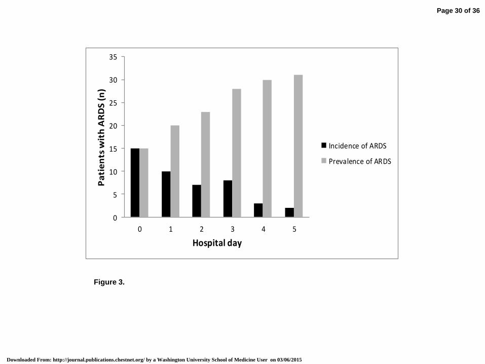
Figure 3.
0
5
10
15
20
25
30
35
0 1 2 3 4 5
Pa
tie
nts
wit
h A
RD
S (
n)
Hospital day
Incidence of ARDS
Prevalence of ARDS
Page 30 of 36
Downloaded From: http://journal.publications.chestnet.org/ by a Washington University School of Medicine User on 03/06/2015
Page 31 of 36
Downloaded From: http://journal.publications.chestnet.org/ by a Washington University School of Medicine User on 03/06/2015
Mechanical ventilation and acute respiratory distress syndrome in the emergency department: a multi-center, observational, prospective, cross-sectional study e-Appendix 1 The respective human research protection offices (HRPO) and institutional review boards (IRB) approved the study at each site under waiver of informed consent: Washington University in St. Louis St. Louis, Missouri, USA IRB Committee Name: Washington University Human Research Protection Office Project Approval Number: 201205165 The University of Iowa Iowa City, Iowa, USA IRB Committee Name: University of Iowa Human Subjects Office/Institutional Review Board Project Approval Number: 201206786 The University of Cincinnati Cincinnati, Ohio, USA UC Office of Research Integrity IRB Committee Name: University of Cincinnati Institutional Review Board Project Approval Number: 2013-2630 Christiana Care Health System Wilmington, Delaware, USA IRB Committee Name: Christiana Care Institutional Review Board Project Approval Number: 33001
Page 32 of 36
Downloaded From: http://journal.publications.chestnet.org/ by a Washington University School of Medicine User on 03/06/2015
e-Appendix 2 Definitions of clinical variables Diabetes Mellitus: Documentation of clinical history in patient’s medical record; if no documented history, assume 'no'. Cirrhosis: Biopsy proven cirrhosis or chart history suggestive of cirrhosis (ascites, coagulopathy, nodular liver on CT or ultrasound). Heart failure: Clinical diagnosis on current presentation or history in chart; no ejection fraction cutoff; includes systolic and diastolic dysfunction. Chronic kidney disease: Increased/abnormal creatinine or GFR <60 for ≥ 3 months; chart record of CKD in patient history. Dialysis/end stage renal disease: current use of peritoneal or hemodialysis as an outpatient. Malignancy: Active or history of; no requirement for history of or current radiation or chemotherapy COPD: Not fully reversible airflow limitation; FEV1 <80% + FEV1/FVC <70%; chart record of COPD in patient history. Immunosuppression: Therapy with immunosuppressants, chemotherapy, radiation, long term/recent high dose steroids, active leukemia, lymphoma, or acquired immunodeficiency syndrome (AIDS). Alcohol abuse: Known diagnosis of chronic alcoholism; previous admission for alcohol detoxification or withdrawal; daily consumption of >14 drinks/week or > 5 binges. AIDS: CD4 <200 and/or AIDS-indicator condition.
Page 33 of 36
Downloaded From: http://journal.publications.chestnet.org/ by a Washington University School of Medicine User on 03/06/2015
e-Appendix 3 Protocol for Adjudication of ARDS Diagnosis Study: Mechanical ventilation and acute respiratory distress syndrome in the emergency department: a multi-center, observational, prospective, cross-sectional study (NCT01628523) Objectives: 1) To further characterize the epidemiology and incidence of mechanical ventilation in critically ill emergency department (ED) patients; and 2) to determine the incidence of acute respiratory distress syndrome in the ED and progression after admission, examining risk factors present in the ED associated with this outcome. The term “acute lung injury (ALI)” is no longer used. Acute respiratory distress syndrome (ARDS) is now divided into subgroups:
1. Mild ARDS: 200mmHg < PaO2:FiO2 ≤ 300mmHg 2. Moderate ARDS: 100mmHg < PaO2:FiO2 ≤ 200mmHg 3. Severe ARDS: PaO2:FiO2 ≤ 100mmHg
For oxygenation criteria for ARDS, preferentially use the PaO2:FiO2 ratio. In patients without an arterial blood gas measurement in the ED, the pulse oximeter (SpO2:FiO2) ratio should be used as a substitute 1. Please note that, due to the shape of the oxyhemoglobin dissociation curve, large changes in the PaO2 can occur with little change in the SpO2 when the SpO2 is close to 100%. Therefore, when SpO2 is > 97%, this substitute should not be used to adjudicate ARDS status. It is recognized that this may limit the diagnosis of ARDS in the ED (Day 0). Therefore, when ARDS is diagnosed on Day 1, and the ED SpO2 was >97%, rendering ED ARDS status equivocal, presentations will be reviewed on a case-by-case basis to assess whether ARDS may have been present in the ED. This is relevant to accurately define the event rate of ARDS in the ED. Given the prospective design of this cohort study, reliable PEEP levels will be known, and the following table should be used for corresponding ratios 2.
SpO2:FiO2 ratio
PaO2:FiO2 Ratio PEEP < 8 PEEP 8-12 PEEP > 12
<300 <370 <387 <332
<200 <240 <259 <234
<100 <115 <130 <129
After admission to the ICU, the worst/lowest PaO2:FiO2 ratio should be used in the assessment of oxygenation status when multiple values are present on a given day. For an individual patient, the oxygenation criteria and x-ray criteria can exist within 12 hours of each other. For epidemiological purposes, we define “Day 0” as the ED stay. Subsequent days (Day 1-5) begin on the next calendar day. Chest X-Ray Interpretation for the Diagnosis of ARDS Defining ARDS status is challenging, despite a consensus definition of the syndrome. There is high interobserver variability in chest x-ray interpretation, which can confound and bias study
Page 34 of 36
Downloaded From: http://journal.publications.chestnet.org/ by a Washington University School of Medicine User on 03/06/2015

results when diagnosing study subjects as “ARDS vs. no ARDS”3. The recent Berlin definition of ARDS attempts to address this by stating that chest x-ray abnormalities consist of: “bilateral opacities consistent with pulmonary edema that are not fully explained by effusions, lobar/lung collapse, or nodules/masses on chest radiograph” 4. The purpose of this section is to decrease heterogeneity across centers in how the chest x-ray is interpreted during the adjudication process for ARDS.
1. Read “Supplementary Material” from Ferguson et al. for a set of illustrative chest x-rays which represent a spectrum of findings and clinical scenarios that are consistent, inconsistent, or equivocal for the diagnosis of ARDS 5. These should serve as training radiographs.
2. Evaluate each chest x-ray over the first 5 days of hospital admission, as data suggests that ARDS develops early in the course of ICU admission. It is also less likely that ARDS developing later after admission from the ED could reliably be attributed to factors present in the ED 6-9.
3. Categorize each x-ray as: consistent, inconsistent, or equivocal for the diagnosis of ARDS, taking into the clinical presentation of the patient.
4. Chest radiographs, as well as a brief clinical scenario of the patient (to allow judgment of evolution of disease), should then be de-identified and sent to the study principal investigator (PI).
a. To limit ascertainment bias, the study PI will be blinded to the ARDS adjudication status from the site PI.
5. After all images are reviewed, ARDS adjudication status will be unblinded and comparisons will be made. When agreement exists between investigators, then the patient will be deemed acceptable for ARDS adjudication status.
a. Please note that this includes not only ARDS adjudication status, but also timing of onset.
6. When disagreement exists, the images will be further reviewed independently by another site PI in a blinded fashion.
7. In cases of disagreement, consensus will be reached by emailed data set and/or conference call if further discussion is necessary.
Origin of Pulmonary Edema Note that the pulmonary artery occlusion pressure cut-off has been removed from the definitional criteria of ARDS. Patients may be considered as having ARDS if the origin of their respiratory failure is not fully explained by myocardial dysfunction or fluid overload If a patient has no identifiable risk factors for ARDS, before adjudicating ARDS status, some objective measure of the origin of edema (e.g. echocardiography) must have been obtained. Please see the “Supplementary Material” from Ferguson et al. for a series of clinical vignettes to aid in the assessment of left atrial hypertension/origin of pulmonary edema 5.
Page 35 of 36
Downloaded From: http://journal.publications.chestnet.org/ by a Washington University School of Medicine User on 03/06/2015
REFERENCES 1. Rice TW, Wheeler AP, Bernard GR, Hayden DL, Schoenfeld DA, Ware LB. Comparison of the
Spo2/Fio2 Ratio and the Pao2/Fio2 Ratio in Patients With Acute Lung Injury or ARDS*. Chest
2007;132:410-7.
2. Pandharipande PP, Shintani AK, Hagerman HE, et al. Derivation and validation of Spo2/Fio2 ratio
to impute for Pao2/Fio2 ratio in the respiratory component of the Sequential Organ Failure Assessment
score*. Critical care medicine 2009;37:1317-21.
3. MEADE MO, COOK RJ, GUYATT GH, et al. Interobserver variation in interpreting chest
radiographs for the diagnosis of acute respiratory distress syndrome. American journal of respiratory
and critical care medicine 2000;161:85-90.
4. GATTINONI L. Acute respiratory distress syndrome: the Berlin definition. Journal of the American
medical association 2012;307:2526-33.
5. Ferguson ND, Fan E, Camporota L, et al. The Berlin definition of ARDS: an expanded rationale,
justification, and supplementary material. Intensive care medicine 2012:1-10.
6. Gajic O, Dara SI, Mendez JL, et al. Ventilator-associated lung injury in patients without acute lung
injury at the onset of mechanical ventilation. CRITICAL CARE MEDICINE-BALTIMORE- 2004;32:1817-24.
7. Gajic O, Frutos-Vivar F, Esteban A, Hubmayr RD, Anzueto A. Ventilator settings as a risk factor
for acute respiratory distress syndrome in mechanically ventilated patients. Intensive care medicine
2005;31:922-6.
8. Iscimen R, Yilmaz M, Cartin-Ceba R, et al. Risk factors for the development of acute lung injury in
patients with septic shock: an observational cohort study. Critical Care 2008;12:P487.
9. Kahn JM, Caldwell EC, Deem S, Newell DW, Heckbert SR, Rubenfeld GD. Acute lung injury in
patients with subarachnoid hemorrhage: incidence, risk factors, and outcome. Critical care medicine
2006;34:196.
Page 36 of 36
Downloaded From: http://journal.publications.chestnet.org/ by a Washington University School of Medicine User on 03/06/2015