full paper synthesis and characterization of al doped and
TRANSCRIPT
*Corresponding author: S. Venkatramana Reddy, E-mail: [email protected]
Asian Journal of Nanoscience and Materials, 2018, 2(1), 111-119.
Synthesis And Characterization of Al Doped And (Co, Al) co-
doped ZnO Nanoparticles via Chemical co-precipitation Method
P. Swapna and S. Venkatramana Reddy*
Department of Physics, Sri Venkateswara University, Tirupati-517 502, A.P., India.
Received: 11 September 2018, Revised: 24 November 2018 and Accepted: 30 November 2018.
ABSTRACT: Pure, Al doped, and (Co, Al) co-doped ZnO nano-powders were synthesized through
chemical co-precipitation method at room temperature, using poly ethylene glycol (PEG) as
stabilizing agent. The synthesized samples are characterized by X-ray diffraction (XRD), scanning
electron microscopy (SEM) & energy-dispersive X-ray spectroscopy (EDS), transmission electron
microscopy (TEM), high resolution transmission electron microscopy (HRTEM), the selected area
electron diffraction (SAED) and vibrating sample magnetometer (VSM). XRD results revealed that all
the samples had a hexagonal wurtzite crystal structure with no secondary phases and this indicates the
absence of superfluous impurities. (Co, Al) co-doped samples reveals the lowest crystallite size
comparing to pure and Al doped ZnO nano-particles, doping of aluminum and cobalt, aluminum
could not disturb the Wurtzite structure of ZnO host lattice. SEM analysis demonstrated the
morphology of the Pure, Al doped, and (Co, Al) co-doped ZnO nano-particles, morphology of the
(Co, Al) co-doped samples shows high agglomeration compared to Pure and Al-doped ZnO nano-
particles. EDS spectrum shows the incorporation of dopant elements, it reveals the nonexistence of
impurities other than Zn, Al, and Co which is coincided by XRD results. TEM illustrations reveal the
exact size of the crystallites, which are approximately confirmed by the XRD data, TEM images
obviously shows that nano-particles are in heterogeneous spherical shape. HRTEM images of the Pure
and Al-doped ZnO nano-particles shows clear lattice fringes about 5 nm and (Co, Al) co-doped
images reveal lattice fringes are about 2 nm. VSM analysis of (Co, Al) co-doped samples reveals that
all the three (1, 2, 3 mol% of cobalt by keeping Al-5 mol% at constant) concentrations showed the
Ferromagnetic nature at room temperature. Among these 1 mol % of cobalt shows highest saturation
magnetization (Ms) and Retentivity (Mr) values, while 2 mol % of cobalt shows highest coercitivity
(Hc) value.
KEYWORDS: HRTEM, RTFM, Lattice fringes, Magnetic properties, VSM.
GRAPHICAL ABSTRACT:
1. Introduction
Zinc oxide (ZnO) is a semiconductor having
wurtzite crystal structure, large direct band
gap of 3.37 eV, and 60 meV exciton binding
energy possessing high Optical gain at room
temperature [1-5]. Zinc oxide could
extensively been used for various
applications such as field effect transistor,
FULL PAPER

112 Reddy et al.
Asian Journal of
Nanoscience and
Materials
solid state gas sensor, dye-sensitized solar
cell, optical devices [6-10]. Electrical
properties of zinc oxide are highly reliable
on its structure and composition, few
authors studied the consequences regarding
structural and electrical properties by
addition of transition metals into ZnO host
lattice synthesized by several methods [11-
15]. Transition metal doped ZnO nano-
particles are potential for a large kind of
applications owing to the charge and spin of
electrons, which are feasible for the origin
of novel Optical, magnetic and transport
properties. Several reports of these materials
which were synthesized through various
methods could demonstrate room
temperature ferromagnetism (RTFM) [16-
19]. Some authors could report RTFM in
Co-, Ni-, Mn-, Fe-, V- and Cu-doped ZnO
[20-25].
2. Experimental Section
2.1 Synthesis
For the synthesis of pristine, Al-
doped, and (Co, Al) co-doped Zinc Oxide
nano-particles, Zinc acetate de hydrate (Zn
(CH3COO)2 .2H2O), potassium hydroxide
(KOH) are taken as preliminary materials,
in addition aluminum nitrate nano-hydrate
(Al(NO3)3.9H2O) is taken for Al-doped ZnO
nano-particles and for (Co, Al) co- doped
ZnO nano-particles in addition to
preliminary materials aluminum nitrate
nano-hydrate (Al(NO3)3.9H2O) and cobalt
acetate tetra hydrate (Co(CH3COO)2 .4H2O)
are used. All the chemicals are analytical
grade and taken without any further
purification. To synthesize Pristine, Al-
doped, and (Co, Al) co-doped ZnO nano-
structures of 0.2 M solution, zinc acetate de
hydrate (Zn (CH3COO)2 .2H2O) is
dissolved in de-ionized water, potassium
hydroxide (KOH) solution is then added
drop wise, in constant stirring of 10 hrs to
form white precipitate. aluminum nitrate
nano-hydrate (Al(NO3)3.9H2O solution is
merged with the above solution, drop by
drop to synthesize Al-doped Zinc oxide
nano-structures, and for co-doped ZnO
nano-particles aluminium nitrate nano-
hydrate (Al(NO3)3.9H2O and cobalt acetate
solutions are mixed with the above (ZnO)
solution drop wise. By filtering the formed
precipitate and washed quite a few times by
means of de-ionized water, avoidable
chemical species formed during the process
of synthesis are detached. After that, all the
samples are dried at 70 °C for 9 hrs and
grind the samples finely through the help of
agate mortar. Eventually, all the samples are
annealed in the furnace at 500 °C for 1 h.
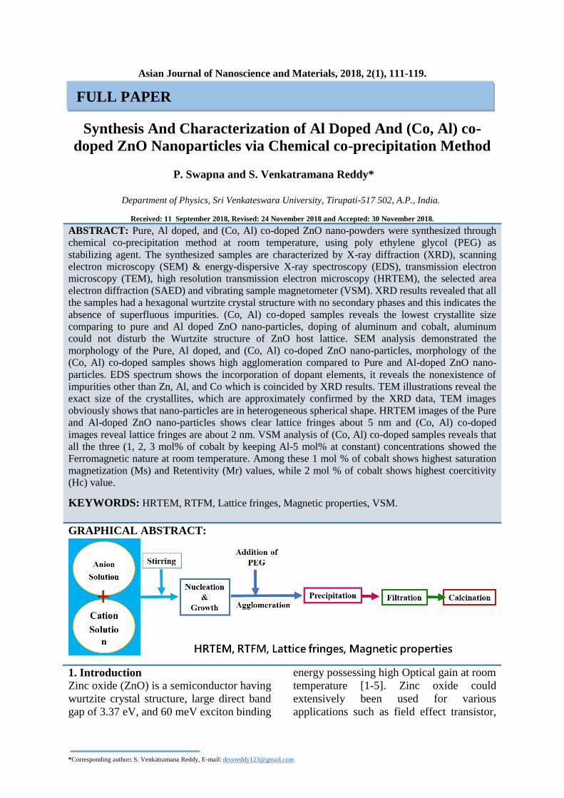
Graphical representation of synthesis is
demonstrated in figure 1 [26].
Fig. 1. Graphical representation of chemical co-precipitation method.
2.2 Characterizations
The synthesized samples were
subjected to the following characterizations.
The XRD pattern was recorded on Bruker
diffractometer within 2θ range of 20° to 80°
via CuKα as X-ray source (λ = 1.53906 Å).
The surface morphology and chemical
analysis of Pure, Al-doped, and (Co, Al) co-
doped Zinc Oxide nano-powders were
calculated by SEM /EDS (model CARL-

Reddy et al. 113
Asian Journal of
Nanoscience and
Materials
ZEISS EVOMA 15). The results obtained
from XRD were correlated by TEM (Model:
philips CM200) and high resolution TEM
(Model:Tecnai G2, F30). Magnetic
properties were studied using the instrument
Vibrating sample magnetometer (VSM).
3. Results and Discussions
3.1 Structural Properties
3.1.1 XRD Analysis The XRD peaks of the Pristine, Al-
doped, and (Co, Al) co-doped nano-
structures are shown in the figure 2. The
diffraction peaks are (100), (002), (101),
(102), (110), (103), (200), (112), (201) and
(202) showings hexagonal wurtzite
crystalline structure. The diffraction peaks
of the all samples associated to hexagonal
wurtzite crystal phase of the ZnO and the
positions of diffracted peaks are reliable by
the pattern (JCPDS CARD NO: 36-1451).
Nonexistence of secondary phases indicates
no impurity phase corresponds to aluminum
or cobalt within the detection range of the
instrument. The size of nano-particle is
deliberated by the Debye-scherrer formula
d=0.91λ/β cosθ, where‘d’ is the nano-
particle size, ‘λ’ is the wavelength of the X-
rays and ‘θ’ is the Bragg’s angle of
diffracted rays. The calculated crystallite
size of Pristine, Al-doped, and (Co, Al) co-
doped ZnO are in the range of 20, 21 and 19
nm respectively. From the calculations of
diffraction peaks, we observed that the
crystallite size decreases for co-doped
samples while increases for Al-doped ZnO
samples. The obtained results coincided
with the previous reports that are
incorporation of cobalt into ZnO host lattice
leads to decrease in the crystallite size [18,
27] and incorporation of Al into ZnO leads
to increase in the crystallite size [28].
Fig. 2. XRD illustration of (a) Pristine ZnO (b) Al (3mol %) doped ZnO (c) (Co, Al) co-doped( Al-3mol %, Co-3 mol %) ZnO nano-
particles.
3.2 Morphological and Compositional
Analysis
3.2.1 SEM and EDAX Analysis
Scanning electron microscopy
(SEM) was used to evaluate the morphology
of the Pristine, Al-doped, and (Co, Al) co-
doped ZnO nano-particles (Figure 3). The
SEM image of the Pristine ZnO nano-
particles shows a large agglomeration
whereas Al-doped and co-doped samples
show low agglomeration. All the images are
plainly representing the non-uniform
spherical and irregular shape of the nano-
particles. EDS spectrum (Figure 3) shows

114 Reddy et al.
Asian Journal of
Nanoscience and
Materials
the incorporation of dopant elements into
ZnO host lattice. It noticeably shows the
existence of impurities such as aluminum in
Al-doped ZnO Spectrum, Co, and Al in co-
doped spectrum, and also the spectrum
reveals the nonexistence of other impurities
in ZnO host lattice. The EDS spectrum of
Pristine ZnO reveals only zinc and oxygen
elements. Weight and atomic percent of
zinc, oxygen, cobalt and aluminum are
mentioned in Table 1.
Fig. 3. SEM images of (a) Pure ZnO (b) Al (3 mol%)
doped ZnO (c) (Co, Al) co-doped ZnO nano-particles (Here Al -3 mol% ,Co-3mol%).
Fig. 4. EDS spectrum of (a) Pure ZnO (b) Al doped
(3mol%) ZnO (c) (Co, Al) co-doped ZnO nano-particles (Here Al-3mol%, Co-3mol %).
Table 1. shows the weight and atomic percent of Zn, O, Co and Al Sample Zn O Co Al
Weight % Atomic % Weight % Atomic % Weight% Atomic % Weight % Atomic %
Pure ZnO 80.35 50.02 19.65 49.98 - - - -
Al-3mol% ZnO 63.17 31.22 30.02 60.03 - - 6.81 8.15
Al-3mol%, Co-
3mol% ZnO
36.34 13.55 47.73 72.72 5.37 3.58 10.56 10.15
3.2.2 TEM, HRTEM and SAED Analysis
Transmission electron microscopy
(TEM) was employed to assess the perfect
size of the Pristine, Al doped, and (Co, Al)
co-doped Zinc Oxide nano-particles. Figure
5 shows the TEM images of Pristine, Al

Reddy et al. 115
Asian Journal of
Nanoscience and
Materials
doped, and (Co, Al) co- doped ZnO nano-
particles. The crystallite sizes estimated
through TEM images and are confirmed by
the XRD data. Figure 6 shows the High
Resolution TEM (HRTEM) images of the
Pure, Al-doped, and (Co, Al) co-doped ZnO
nano-particles. HRTEM pictures of the Pure
ZnO and Al-doped ZnO shows the 5 nm
clear lattice fringes whereas co-doped
sample shows 2 nm clear lattice fringes.
Figure 7 shows the SAED pattern of Pure,
Al-doped, and (Co, Al) co-doped ZnO
nano-particles. In the HRTEM images
predictable d-spacing values of Pure, Al-
doped, and (Co, Al) co-doped ZnO nano-
particles between two closest lattice fringes
are observed as 0.29 nm, 0.28 nm, 0.28 nm
respectively and in consequence, it
corresponds to (101) diffracted planes of
hexagonal wurtzite crystal structure of ZnO
nano-particles. From fig. 7(a), 7(b), 7(c), the
SAED pattern of Pure, Al-doped, and (Co,
Al) co-doped ZnO nano-particles evidently
show the scattered diffraction spots in ring
pattern. It shows that pristine and doped
ZnO nano-particles are poly-crystalline
nano-crystallites. The predictable diffraction
spots and rings are indexed by the aid of
bulk ZnO data (JCPDS card NO: 36-1451).
4.0 Magnetic Properties
Room temperature magnetization
(M-H) curves of (Co, Al) co-doped ZnO
nano-particles are shown in the fig.8. Since
pure ZnO shows diamagnetic nature and is
not mentioned in the graph here, it is well-
known that Pristine bulk ZnO is
diamagnetic yet recent reports showed that
Pristine ZnO might show ferromagnetic
nature under certain film thickness [29],
also a few authors reported room
temperature ferromagnetism (RTFM) in
Pure ZnO [30, 31]. Some authors reported
that when the size of the particle is in
nanometers, ZnO itself also shows
ferromagnetic behavior [32, 33]. At room
temperature the magnetic nature in cobalt
doped ZnO is owing to the being of bound
magnetic polarons (BMPs), these could be
ascribed to the existence of defect related
states such as oxygen vacancies,
interstitials, etc. In the recent days, large
constructive (Both theoretical and
experimental) observations have been made
in this path which might be reported [34-
43]. According to previous report [44]
mostly cobalt doped ZnO shows
ferromagnetic nature with different
concentrations, coinciding that in the
present work also all the three
concentrations shows ferromagnetic nature
Fig. 5. TEM images of (a) Pure ZnO (b) Al doped (3
mol%) ZnO (c) (Co, Al) co-doped ZnO nano-particles (Here Al -3 mol%, Co-3 mol%).

116 Reddy et al.
Asian Journal of
Nanoscience and
Materials
Fig. 6. HRTEM images of (a) Pure ZnO (b) Al doped (3
mol%) ZnO (c) (Co, Al) co-doped ZnO nano-particles
(Here Al -3 mol%, Co-3 mol%).
Fig. 7. SAED pattern of (a) Pure ZnO (b) Al doped (3
mol%) ZnO (c) (Co, Al) co-doped ZnO nano-particles
(Here Al -3 mol%, Co-3 mol%).
at room temperature but owing to the
dissimilarity in the concentration of cobalt,
method of synthesis, annealing temperature
etc. there is variation in the order of
increasing and decreasing of Ms, Mr and Hc
values. A few others also reported RTFM in
cobalt doped ZnO nano-particles [45]. In
the present studies by increasing the cobalt
concentration, the saturation magnetization
(Ms), Retentivity (Mr) values decreases but

Reddy et al. 117
Asian Journal of
Nanoscience and
Materials
the coercitivity (Hc) value increases for 2
mol % concentration and decreases for 3
mol % concentration. Ms, Mr and Hc values
of all the concentrations are mentioned in
Table 2. Figure 9 illustrates the room
temperature M-H curves of expanded lower
field region of Figure 8.
Fig. 8. Room temperature M-H curves of (a) 1 mol% (b) 2
mol% (c) 3 mol% of (Co, Al) co-doped ZnO nano- particles (Here Al = 5.0 mol% is kept as constant).
Fig. 9. RT M-H curves of expanded lower field region of
(a) 1 mol% (b) 2 mol% (c) 3 mol% of (Co, Al) co-doped
ZnO nano-particles (Here Al = 5.0 mol% is kept as constant).
Table 2. shows the Coercivity (Hc), Retentivity (Mr) and saturation magnetization (Ms) values for (Co, Al) co-doped ZnO
nano-particles
Sample Magnetization (Ms) (emu/g) Coercitivity (Hc)
(Kilo Guass)
Retentivity (Mr)
(emu/g)
Co-1, Al-5 mol % 0.146 0.388 0.179
Co-2, Al-5 mol % 0.045 0.403 0.003
Co-3, Al-5 mol % 0.024 0.311 0.001
5. Conclusions
Pristine, Al-doped ZnO, and (Co,
Al) co-doped ZnO nano-structures were
synthesized efficiently using the chemical
co-precipitation method through PEG as
stabilizing agent at room temperature. The
synthesized samples were characterized
using XRD, SEM/EDS, TEM, HRTEM,
SAED pattern, and VSM techniques and
deliberate different kind of properties such
as structural, morphological, compositional
and magnetic. TEM and EDS analysis is
consistent through XRD data. XRD data
shows that all the samples had a hexagonal
wurtzite crystal structure without secondary
phases connecting to Al or Co, TEM
pictures illustrate the exact size of the
crystallite which is approximately coincide
with XRD data. VSM measurements reveals
the ferromagnetic nature of the (Co, Al) co-
doped ZnO samples.
Acknowledgements Authors are thankful to the
University Grants Commission (UGC),
New Delhi, India, for providing financial
assistance through RFSMS program, and
also thankful to IIT Bombay (SAIF) for
providing TEM characterization
References:
[1] Huang MH, Mao S, Feick H, Yan H,
Wu Y, Kind H, Weber E, Russo R , Yang P
(2001) Science., 292:1897- 1899.
[2] Wong E, Searson P (1999) Appl. Phys.
Lett.,74: 2939-2941.

118 Reddy et al.
Asian Journal of
Nanoscience and
Materials
[3] Choopun S, Vispute R, Noch W,
Balsamo A, Sharma R.P, Venkatesan T,
Iliadis A, Look D.C (1999) Appl.
Phys. Lett., 75: 3947-3949.
[4] Swapna P and Venkatramana Reddy S
(2018) IOP Conf. Series: Materials Science
and Engineering., 310: 012011.
[5] Swapna P , Venkatramana Reddy S
(2017) Mechanics, Materials Science &
Engineering, 9: ISSN 2412-5954 .
[6] Wang X.D, Zhou J, Song J.H , Liu J,
Xu N, Wang Z.L (2006) Nano Lett., 6:
2768-2772.
[7] Yang P.D, Yan H.Q, Mao S, Russo R,
Johnson J, Saykally R, Morris N, Pham J,
He R, Choi H.J (2002) Adv. Funct. Mater.
12: 323-331.
[8] Law M, Greene L.E, Johnson J.C,
Saykally R, Yang P.D (2005) Nat. Mater.
4: 455-459.
[9] Navale S.C, Gosavi S.W, Mulla I.S
(2008) Talanta. 75:1315-1319.
[10] Zhang J, Wang S, Wang Y, Xu M, Xia
H, Zhang S, Huang W, Guo X, Wu S (2009)
Sens. Actuators B. 139: 411-417.
[11] Sedky A, Abu-Abdeen M, Almulhem
A.A (2007) Physica B 388: 266-273.
[12] Yuan D, Wang G.S, Xiang Y, Chen Y,
Gao X.Q, Lin G (2009) J. Alloys Compd.
478: 489-492.
[13] Wang X.C, Mi W.B, Dong S, Chen
X.M, Yang B.H (2009)J. Alloys Compd.
478: 507-512.
[14] Mamta Sharma R.M, Mehra (2008)
Appl. Surf. Sci. 255: 2527-2532.
[15] Ilican S, Caglar Y, Caglar M,
Yakuphanoglu F (2008) Appl.Surf. Sci.
255: 2353-2359.
[16] Sharma P, Gupta A, Rao K. V, Owens
F. J, Sharma R, Ahuja R, Osorio J. M
,Johansson B (2003) Nature Matter. 2: 673-
677.
[17] Zhang H.-W, W ei Z.-R, Li Z.-Q.
Dong G.-Y (2007) Materials Letters. 61:
3605-3607.
[18] Hays J, Reddy K. M, Graces N. Y,
Engelhard M. H, Shutthanandan V, Luo M,
Xu C, Giles N.C, Wang C, Thevuthasan S
Punnoose A (2007) J.Phys.: Condens.
Matter, 19: 266203.
[19] Srinivas K, Rao S. M, Reddy P. V
(2011) Journal of Nanoparticle Research.
13: 817-837.
[20] Tahir N, Hussain S. T, Usman M,
Hasanain S. K, Mumtaz A (2009) Applied
Surface Science. 255: 8506- 8510.
[21] Costa-Krämer J. L, Briones F,
Fernández J. F, Caballero A. C, Villegas M,
Díaz M, García M. A, Hernando A (2005)
Nanotechnology. 16: 214-218.
[22] Karmakar D, Mandal S. K, Kadam R.
M, Paulose P. L, Rajarajan A. K, Nath T. K,
Das A. K, Dasgupta I Das G. P (2007)
Physical Review B, 75: 144404.
[23] Lakshmi Y. K, Srinivas K, Sreedhar B,
Raja M. M, Vithal M, Reddy P. V (2009)
Materials Chemistry and Physics, 113: 749-
755.
[24] Lu J. J, Lin T. C, Tsai S. Y, Mo T. S,
Gan K. J (2011) Journal of Magnetism and
Magnetic Materials. 323: 829-832.
[25] Buchholz D. B, Chang R. P. H, Song
J.-Y, Ketterson J. B (2005) Applied Physics
Letters. 87: 082504.
[26] Swapna P, Venkatramana Reddy S
(2018) Adv.Sci.Lett. 24: 5636-5639.
[27]Sankara Reddy B, Venkatramana
Reddy S, Koteeswara Reddy N (2013) J
Mater Sci: Mater Electron.24: 5204–5210
[28] Vanaja A, Ramaraju G.V, Srinivasa
Rao K(2016) Indian Journal of Science and
Technology 9 ISSN:0974-5645 [29]
Kapilashrami M, Xu J, Rao K.V, Belova L
(2010) Processing and Application of
Ceramics., 4:225-229.
[30] Hu J, Zhang Z, Zho Z, Qin H, Jiang M
(2008) Appl. Phys. Lett. 93:192503.
[31]Kapilashrami M, Xu J, Ström V, Rao
K.V, Belova L (2009) Appl. Phys. Lett. 95:
033104.
[32] Sundaresan A, Bhargavi R, Rangarajan
N, Siddesh U, Rao C.N. R (2006)
Phys.Rev. B 74: 161306 .

Reddy et al. 119
Asian Journal of
Nanoscience and
Materials
[33]Garcia M. A, Merino J. M, Pinel E. F,
Quesada A, Venta J. D, Gonzalez R, Castro
G, Crespo P, Liopis J, GonzalezCalbet J. M,
Hernando A. (2007) Nano Lett. 7: 1489-
1494.
[34] Kaminski A, Das Sarma S (2003) Phy.
Rev. B. 68: 235210.
[35] Coey J. M. D, Venkatesan M,
Fitzgerald C. B (2005) Nature. Mater.
4:173-179.
[36] Liao L, Yan B, Hao Y. F, Xing G. Z,
Liu J. P, Zhao B. C, Shen Z. X, Wu T,
Wang L, Thong J. T. L, Li C. M,
Huang W, Yu1 T (2009) Appl. Phys.
Lett. 94: 113106.
[37] Tian Y, Li Y, He M, Putra I. A, Peng
H, Yao B, Cheong S.A, Wu T (2011) Appl.
Phy. Lett. 98: 162503.
[38] Ogale S. B (2010) Adv. Mater., 22:
3125-3155.
[39] Li Y, Deng R, Yao B, Xing G, Wang
D, Wu T (2010) Appl. Phys. Lett., 97 :102-
506.
[40] Zhang B. Y, Yao B, Li Y. F, Liu A. M,
Zhang Z. Z, Li B. H, Xing G. Z, Wu T, Qin
X. B, Zhao D. X, ShanC.X, Shen D. Z
(2011) Appl. Phys. Lett., 99:182503.
[41] Xing G. Z, Lu Y. H, Tian Y. F, Yi J. B,
Lim C. C, Li Y. F, Li G. P, Wang D.D, Yao
B, Ding J, Feng Y. P, Wu1T (2011) AIP.
Adv., 1: 022152.
[42] Xing G. Z, Wang D. D, Cheng C.-J, He
M, Li S, Wu T (2013) Appl. Phys. Lett.,
103: 022402.
[43] Ochsenbein S. T, Feng Y, Whitaker K.
M, Badaeva E, Liu W. K, Li X, Gamelin D.
R, (2009) Nature Nanotechnology., 4: 681-
687.
[44]Djaja N. F, Montja D. A, Saleh R
(2013) Advances in Materials Physics and
Chemistry, 3: 33-41.
[45]Pal B, Giri P.K (2011)International
Journal of Nanoscience.,10: 1-5.
How to cite this manuscript: P. Swapna and S. Venkatramana Reddy*. Synthesis And
Characterization of Al Doped And (Co, Al) co-doped ZnO Nanoparticles via Chemical
co-precipitation Method. Asian Journal of Nanoscience and Materials, 2018, 2(1) , 111-
119.