fsafasdasdfaskkkkkkkkkkkkkkkkkkkkkkkkkkkkkkkkkkkkkkkkkkkkk ...€¦ · ligand binding assay...
TRANSCRIPT

fsafasdasdfaskkkkkkkkkkkkkkkkkkkkkkkkkkkkkkkkkkkkkkkkkkkkkkkkkkkkkkkkkkkkkkkkkkkkkkkkkkkkkkkkkkkkkkkkkkkkkkkkkkkkkkkkkkkkkkkkkkkkkkkkkkkkkkkkkkkkkkkkkkkkkkkkkkkkkkkkkkkkkkkkkkkkkkkkkkkkkkkkkkkkkkkkkkkkkkkkkkkkkkkkkkkkkkkkkkkkkkkkkkkkkkkkkkkkkkkkkkkkkkkkkkkkkkkkkkkkkkkkkkkkkkkkkkkkkkkkkkkkkkkkkkkkkkkkkkkkkkkkkkkkkkkkkkkkkkkkkkkkkkkkkkkkkkkkkkkkkkkkkkkkkkkkkkkkkk
Ligand Binding Assay Bioanalytical Focus Group Newsletter December 2013
In this Issue:
Message from the Chair, by Lakshmi Amaravadi
Words of Wisdom, by Ron Bowsher
LBABFG Action Program Committee Updates
Technology and Innovation, 21st Century Lab, by Ago Ahene Flow Cytometry, by Murli Krishna, Cherie Green, Virginia Litwin
Technical Corner
LCMS/MS for Protein Bioanalysis, by Hao Jiang and Jianing Zeng ADC Protein Chemistry, by Vangipuram S Rangan and Shrikant
Deshpande
Student Section, by Hardik Mody
Literature Highlights
Important Dates National Biotech Conference
Food for Thought, by Heather Myler and Lora Hamuro
Acknowledgements
Year End Photos
Chair Lakshmi Amaravadi Chair Elect Lindsay King Past Chair Phillip Oldfield Steering Committee Chair Boris Gorovits Steering Committee Ago Ahene Aleks Davis Carol Davis Shalini Gupta Marian Kelley Murli Krishna Joe Marini Heather Myler Marie Rock Martin Ullmann Yuanxin Xu Communications Lead Heather Myler Communications Committee Shannon Chilewski Shobha Purushothama Lauri Neyer Hardik Mody Mamta Bhushan Chris Beaver, BIOTEC liaison AAPS Leadership Maria Nadeau
LBAFG Quarterly Newsletter December 2013| Page | 2
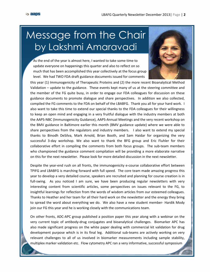
Message from the Chair by Lakshmi Amaravadi
As the end of the year is almost here, I wanted to take some time to update everyone on happenings this quarter and also to reflect on so much that has been accomplished this year collectively at the focus group level. We had TWO FDA draft guidance documents issued for comments
this year (1) Immunogenicity of Therapeutic Proteins and (2) the more recent Bioanalytical Method Validation – update to the guidance. These events kept many of us at the steering committee and the member of the FG quite busy, in order to engage our FDA colleagues for discussion on these guidance documents to promote dialogue and share perspectives. In addition we also collected, compiled the FG comments to the FDA on behalf of the LBABFG. Thank you all for your hard work. I also want to take this time to extend our special thanks to the FDA colleagues for their willingness to keep an open mind and engaging in a very fruitful dialogue with the industry members at both the AAPS-NBC (Immunogenicity Guidance), AAPS-Annual Meetings and the very recent workshop on the BMV guidance in Baltimore earlier this month (BMV guidance update) where we were able to share perspectives from the regulators and industry members. I also want to extend my special thanks to Binodh DeSilva, Mark Arnold, Brian Booth, and Sam Haidar for organizing the very successful 3-day workshop. We also want to thank the BFG group and Eric Fluhler for their collaborative effort in compiling the comments from both focus groups. The sub-team members who championed the guidance comment compilation will be providing a more elaborate narrative on this for the next newsletter. Please look for more detailed discussion in the next newsletter.
Despite the year-end rush on all fronts, the immunogenicity e-course collaborative effort between TPIFG and LBABFG is marching forward with full speed. The core team made amazing progress this year to develop a very detailed course, speakers are recruited and planning for course creation is in full-swing. As you noticed I am sure, we have been producing regular newsletters with very interesting content from scientific articles, some perspectives on issues relevant to the FG, to insightful learnings for reflection from the words of wisdom articles from our esteemed colleagues. Thanks to Heather and her team for all their hard work on the newsletter and the energy they bring to spread the word about everything we do. We also have a new student member- Hardik Mody join our FG this year and he is working closely with the communications team.
On other fronts, ADC-APC group published a position paper this year along with a webinar on the very current topic of antibody-drug conjugates and bioanalytical challenges. Biomarker APC has also made significant progress on the white paper dealing with commercial kit validation for drug development purpose which is in its final leg. Additional sub-teams are actively working on very relevant challenges to all of us involved in biomarker measurements including sample stability, multiplex marker validation etc. Flow cytometry APC ran a very informative, successful symposium
LBAFG Quarterly Newsletter December 2013| Page | 3
Message from the Chair cont. session this year at the NBC meeting on the topic of PD measurement by receptor occupancy. Manuscript preparation is underway for this topic and the group is also working addressing flow cytometry specimen stability issues as applicable to drug development. Immunogenicity and cell based-assay APCs made significant progress this year on the very important topic of relevance to the FG community: choice of cell-based vs non-cell based neutralizing antibody assays, strategy and decision tree development. This topic was the subject of discussion at the open forum held in Nov. at the 2013 AAPS – annual meeting. Look for more updates to come on this topic in the coming months. Enzymatic Nab assay sub-team and newly formed Target Interference sub-team under the immunogenicity APC are making significant headway. Expect more to come from these teams in 2014.
Emerging Technologies APC (ET-APC) with its’ various sub-teams has made amazing progress addressing topics of interest to the FG community, while taking the angle of evaluating latest technologies available to address the scientific challenges of relevance to the FG. Given the synergies and closeness of topics we anticipate the 21st century lab APC and the ET-APC to work under one umbrella in 2014. Biosimilars APC conducted two great roundtable discussions to present the progress on PK and Immunogenicity Assays for Biosimilars, at the NBC and AM meetings respectively this year. This APC has also made such an amazing progress to address these key topics and we anticipate publication of the first recommendations paper addressing PK assays Q1-2014.
As you are very aware we are all witnessing the development of complex therapeutics with newer analytical challenges related to PK, immunogenicity and biomarker assays. In parallel newer, more sensitive and capable technologies are also becoming available to address these complexities. To adapt to these challenges our FG has also grown with various sub-teams over the past few years with several active projects. Together with this growth comes the challenge of ensuring synergies, eliminating redundancies across the sub-teams and keeping a clear and unique focus at each project/sub-team level. With this in mind, over the last few months, the steering committee of the FG has been busy with crafting a sustainable structure for the long term, with keeping in mind the adjacencies of various sub-teams. To this end, we envision the new structure to have the broader teams of (1) PK (2) Immunogenicity (3) Biomarker (4) 21st Century Technologies along with (5) Communications, Education and Distance Learning teams with various sub-teams underneath these teams supporting the goals of the broader teams and the overall mission of the LBABFG. More details on this to come in the next newsletter. On that note I would like express my sincere thanks to all the hard working members of the FG leadership, for the active collaboration and participation from all levels of the FG members, and the leadership of the Biotech Section for their support. What a tremendous and rewarding year 2013 has been!!! Relax and Enjoy the Holiday season with your family and friends. Get plenty of rest to recover from a busy 2013 and get ready for an exciting 2014 coming our way. Happy Holidays everyone.
Sincerely - Lakshmi Amaravadi (Chair- LBABFG, AAPS)

LBAFG Quarterly Newsletter December 2013| Page | 4
diligent is in the use of terminology. To lessen miscommunication and misunderstanding, we need to avoid using multiple terms to describe the same concepts. This is particularly true because BMV of LBAs is now a global endeavor. Thus, I believe that clear and concise communication is mandatory. An important part of good communication is using a standardized lexicon of terms. Thus I believe the more we can limit interchangeability of terms, there will be less chance for confusion and more optimal communication will result. In this regard, I spent some time reflecting on the various phases of LBA development and validation and have created the following diagram to describe the BMV process from method conceptualization to delivery of regulatory compliant results. The information in the figure below was gleaned from numerous publications, guidance documents, and personal interactions with colleagues over the past couple of decades and is sufficiently generic to be applicable to most all LBAs. The figure is intended to stimulate thought and discussion, as opposed to being purely a didactic description for de novo LBA method development and validation.
Phase 1 (Assay Feasibility) constitutes Early Phase Method Development. It is during this stage of development that synthetic (peptide) chemistry is performed and critical reagents are produced or identified and thoroughly characterized. Equally important is defining the assay design (e.g., noncompetitive or competitive) and the technology platform. In most cases, the work in early phase development is buffer-based and culminates in a functional assay in buffer which I refer to as a Prototype Assay.
Phase 2 (Assay Optimization) constitutes Late Phase Method Development and is initiated by the introduction of the matrix-of-choice in to the prototype assay. The goal here is to systematically optimize the LBA for its intended purpose. Important too is planning for the assay’s life cycle and scale up of critical reagents to support near-term bioanalysis of the analyte of interest. Upon completion of this critical phase, the deliverable is an Optimized Assay, as the overarching goal is to develop a valid LBA, as opposed to simply validating a developed LBA.
Words of Wisdom AN APPEAL FOR STANDARDIZED NOMENCLATURE TO DESCRIBE THE DISTINCT
PHASES FOR BMV OF LIGAND BINDING ASSAYS
By Ron Bowsher, B2S Consulting / B2S Labs LLC / AIT Bioscience
Attendees at the recently held Crystal City BMV conference in Baltimore who also attended the March 2000 Crystal City conference, the first one that addressed BMV of biotherapeutics, had to be struck by how different these meetings were in terms of both their content and interactions. It is indeed gratifying how much we have progressed with respect to the application of LBA technology for regulated bioanalysis. While work remains as this methodology matures and the diversity and complexity of biotherapeutics continue to evolve, greater understanding and harmonization in practices are apparent in the application of LBAs. As work progresses in this area, one aspect of LBAs where we need to remain

LBAFG Quarterly Newsletter December 2013| Page | 5
Words of Wisdom cont. Phase 3 (Assay Validation) is synonymous with Pre-Study Validation and involves the systematic verification of the optimized method’s analytical performance characteristics. Historically speaking, this phase represents the point of demarcation for GLP and is usually preceded by the preparation of a validation plan and spiked validation samples. The goal here is to evaluate the method under conditions that will resemble the analysis of test samples to document that it will provide reliable results under the conditions of intended use. Pre-study validation culminates in acceptance (or rejection) of a LBA method with the deliverable being a Validated Assay.
Phase 4 (Sample Analysis) involves the analysis of study test samples by the validated LBA and is commonly referred to as In-study Validation. This important phase involves acceptance (or rejection) of a LBA batch (or run) and often involves application of the commonly-used 4-6-20 rule for LBAs. The ultimate deliverable here is Regulatory Compliant Data from an accepted LBA batch.
In summary by applying a common set of terms to describe the generic phases for development and validation of LBA methods, we can facilitate communication and understanding and ultimately aid this bioanalytical science in being applied in a more efficient manner. The above flow diagram is one attempt at standardizing the overall process and terminology for BMV of LBAs.
Early Phase Development
• Prep Critical Reagents (Chemistry & Abs)• Select assay design & technology platform• Initial assay development in buffer• Yields “buffer-based” PROTOTYPE ASSAY
Late Phase Development
• Involves introduction of test sample matrix• Method optimization for intended purpose• Scale up of critical reagents• Yields OPTIMIZED ASSAY
Pre-StudyValidation
• Systematic verification of assay performance• Culminates in method acceptance• Yields VALIDATED ASSAY
In-Study Validation
• Involves analysis of test samples• Culminates in batch (run) acceptance• Yields REGULATORY COMPLIANT DATA
PHASE 1
ASSAY FEASIBILITY
PHASE 212
ASSAY OPTIMIZATION
PHASE 3
AASSAY VALIDATION
PHASE 4A
SAMPLE ANALYSIS
LBAFG Quarterly Newsletter December 2013| Page | 6
(APC) was formed as a result to assess the state of the art and to evaluate how best to goad the industry on to take advantage of these advances. Due to the complexity and the varied nature of the task the APC was subdivided in four groups, namely: automation, platform, reagent and electronic solution. The deliberations of these groups led to the publication of a themed issue on the topic in the AAPS Journal (2012). In it, the APC laid down reasons and justifications to persuade or seek alignment among players in the industry for a change. To realize this goal in part, the APC advocated for streamlining of platform interfaces, electronic solution, and automation. In addition, the APC provided recommendations for characterization and supply of critical reagents.
The response to date has been very encouraging. In one area, progress has been made by vendor(s) offering (or developing) fully automated off–the-shelf ELISA system(s) incorporating many of the attributes of the 21stcentury bioanalytical laboratory initiative. The prototype of a fully automated bench top ELISA system, the ELISA Nimbus showcased by Hamilton Company at the 2013 NBC holds much promise. In another area, efficiencies in automation have been achieved through integration of a LIMS system with an automated liquid handler and a microfluidics equipment without extensive customization by scientists from Amgen, Tecan US, and Gyros. Partnership relationships were forged to produce better products for ligand binding assays as in the collaboration between Pfizer scientists and Telos (software vendor) to use artificial intelligence software to operate automation systems without resulting to detailed programming acumen. The collaboration between Radix BioSolutions to share the risk on the development of phosphoprotein reagents on the Luminex platform is another example of partnership at work.
21st Century Lab APC Recent Advances in the 21st Century Bioanalytical Laboratory Efficiencies
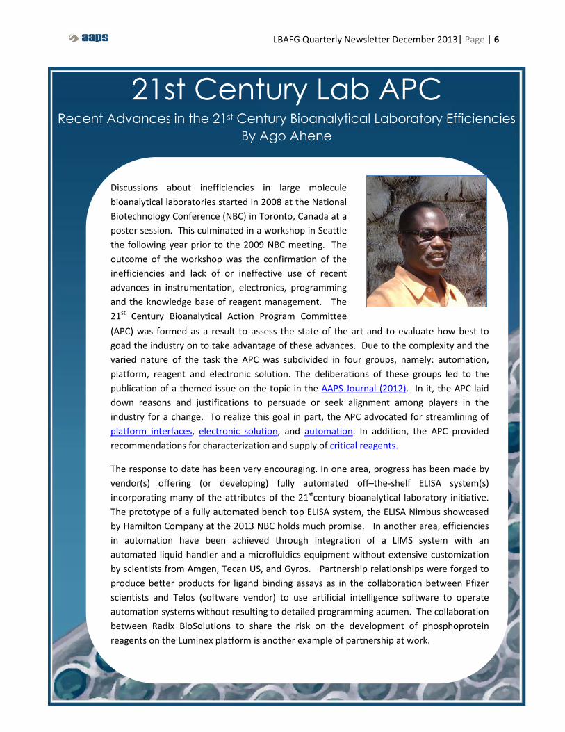
By Ago Ahene
Discussions about inefficiencies in large molecule bioanalytical laboratories started in 2008 at the National Biotechnology Conference (NBC) in Toronto, Canada at a poster session. This culminated in a workshop in Seattle the following year prior to the 2009 NBC meeting. The outcome of the workshop was the confirmation of the inefficiencies and lack of or ineffective use of recent advances in instrumentation, electronics, programming and the knowledge base of reagent management. The 21st Century Bioanalytical Action Program Committee

LBAFG Quarterly Newsletter December 2013| Page | 7
21st Century Lab APC cont.
In conformance with the critical reagents white paper, better characterization and life-cycle management of reagents were advocated. To that end, critical reagent roles have been emphasized in the industry of late, bioanalytical analysts are integrating characterization and re-test recommendations into standard operation procedures and best practices. In addition, there have been continued publications on reagent characterization and management (e.g. Global Bioanalysis Consortium L4-Reagent and Stability publication).
Almost a year since the publication of the 21st Century papers laboratory efficiency papers, considerable progress has been made. The desired end results of these improvements are to reduce cost, speed up bioanalysis, cut manual operations to a minimum, provide adequately characterized reagents and facilitate the conduct of bioanalysis in the coming years.

LBAFG Quarterly Newsletter December 2013| Page | 8
Flow Cytometry APC By Murli Krishna, Cherie Green, Virginia Litwin
Flow Cytometry is a powerful, decision-enabling analytical tool increasingly being utilized in all stages of drug development. Because of the high degree of flexibility and complexity of this platform, it’s essential to understand all aspects of the instrumentation, reagents, and biological systems under investigation in order to generate high quality data.
The Flow Cytometry action program committee (APC) was formed by John Ferbas and Virginia Litwin in 2006. The mission of the Flow Cytometry APC is to promote discussion regarding the proper application of flow cytometry in drug development with an emphasis on establishing best practices regarding assay and instrument validation. A key driver in forming this group was the lack of official industry guidance regarding the application and validation of flow cytometric methods in drug development. The Flow Cytometry APC has contributed to several recent publications in this area and this year had the privilege of presenting two of the papers at the 2013 FDA workshop. This past year, they gained significant momentum as described below.
Members
This group is currently comprised of a dynamic mix of 13 dedicated scientists from across the industry, representing small and large biotechnology and pharmaceutical companies as well as contract research organizations. Murli Krishna is the LBABFG sponsor and Virginia Litwin and Cherie Green currently serve as co-chairs for the Flow Cytometry APC. Find them on LinkedIn/Groups/Flow Cytometry Action.

LBAFG Quarterly Newsletter December 2013| Page | 9
Active Members Murli Krishna AAPS LBABFG sponsor (Bristol-Myers Squibb)
Virginia Litwin co-chair (Covance Central Laboratory Services, Inc.) Cherie Green co-chair (Amgen)
Kathy Howell (Genentech) Nick Jones (LabCorp Clinical Trials)
Tom McCloskey (ICON Central Laboratories) Maxime Moulard (BioCytex)
Peter J. O’Brien (Pfizer) Jennifer Stewart (Flow Contract Site Laboratory) Christopher Wiwi (Celgene Cellular Therapeutics)
Wendy White (MedImmune, LLC) Yuanxin Xu (Sanofi)
Tim Wyant (Millennium Pharmaceuticals) Past Members
John Ferbas Lynette Brown
Sophie Corneau Dianna Wu
Manjula Reddy Denise O’Hara
Zhyian (Eric) Lianz John Sloan
Flow Cytometry APC Cont.
Conferences Since 2006, the Flow Cytometry APC has supported programming at the numerous AAPS annual and National Biotechnology meetings, bringing key opinion leaders from across the field to present on relevant topics such as; fit-for-purpose validation, receptor occupancy and cell signaling. The Flow APC sponsored AAPS programming has included: Hot Topics Session (Annual Meeting 2006), Roundtable Session (NBC Meeting 2007), Symposium session (Annual Meeting 2011), Symposium Session (Annual Meeting 2012), and Roundtable Session (NBC 2013). At NBC 2013, the group sponsored a symposium “Design and application of receptor occupancy assays used to measure pharmacodynamic response to treatment with biologic therapies”. John Ferbas (Amgen), Meina Liang (Medimmune), Marna Williams (Genentech), and David Wunderlund (Pfizer) were the speakers. This session sparked productive discussions which are currently being summarized in a post conference report. In 2013, the group expanded their reach by collaborating with other professional societies International Society for Analytical Cytology (ISAC), International Clinical Cytometry Society (ICCS), and Bioanalysis Forum (EBF)) in both programming and publications. In May, the Flow APC continued to support advancing the quality and standardization of flow cytometry by hosting two workshops at the ISAC conference. The first focused on exploring key regulated environments (GLP/CLIA/GMP), “Navigating the labyrinth of regulated flow cytometry in drug development”. Members of the Flow APC, Christopher Wiwi and Cherie Green, were among the speakers. The second focused on “Pharmacodynamic Measurements for Biologics by Receptor Occupancy Flow Cytometry”. In addition, Virginia Litwin
LBAFG Quarterly Newsletter December 2013| Page | 10
Flow Cytometry APC Cont.
presented "Translating Discovery into the Regulatory Environment” and “Breaking Bad: Better Data through Instrument Standardization” at ISCC 2013 meeting and “Analytical Method Validation: Perspectives from the Flow Cytometry Action Programming Committee of the AAPS” at EBF 2013. The ultimate moment for the Flow Cytometry APC in 2013 was presenting at the FDA-sponsored public workshop on Clinical Flow Cytometry in Hematologic Malignancies, in Silver Spring, MD (http://www.fda.gov/MedicalDevices/NewsEvents/WorkshopsConferences/ucm334772.htm). Virginia Litwin and Cherie Green delivered a presentation entitled, “The Role of Biomarkers in Clinical Trials and the Fit-for-Purpose Method Validation Approach”. The invitation to present resulted from the teams’ long standing interactions with Gerald Marti, MD, PhD, Lab Chief at CDER/FDA and was a follow up to the Fit-For-Purpose Symposium at the 2011 AAPS Annual Meeting. The purpose of this public workshop was to seek input from relevant stakeholders on the role of clinical flow cytometry in hematologic malignancies, in order to develop a specific regulatory policy for in vitro diagnostic devices. This was a significant accomplishment for the group to be engaged in the dialogue with regulatory agencies to establish validation guidelines for flow cytometric methods used to support FDA submissions. Webinars In 2011, the group presented two AAPS sponsored webinars “Introduction to Flow Cytometry” and “Flow Cytometry Validation for Drug Development: Instruments and Methods”. These introductory webinars are a great resource for those new to the field and are available for viewing http://www.aaps.org/Meetings_and_Professional_Development/eLearning_Repository Publications In 2011, the Flow APC published recommendations for flow cytometry assay and instrument validation for drug development (O’hara et al and Green et al. 2011) in a special issue of Journal of Immunological Methods dedicated to flow cytometry biomarkers. The Flow APC has continued its publishing efforts well into 2013 as evidenced by acceptance of 2 key papers: (1) Wood, B., et al. Validation of cell-based fluorescence assays: Practice guidelines from the ICSH and ICCS – part V – assay performance criteria. Cytometry Part B: Clinical Cytometry. 85:315, 2013. This publication is significant as it was part of a special Issue of Clinical Cytometry generated by a working group representing the International Council for Standardization of Hematology (ICSH), and the International Clinical Cytometry Society (ICCS). The working group has plans to submit the recommendations to the FDA for consideration as official guidance documents for IVD submissions. This publication is the first important step of that process. The validation recommendations closely parallel those published in 2011 by the Flow Cytometry APC. (2) Litwin, V., et al. New Considerations for the Validation of Flow Cytometry Lab Developed Tests. Clinical Laboratory News, December, 2013.
LBAFG Quarterly Newsletter December 2013| Page | 11
2014 Goals Next year, the Flow APC will continue to support AAPS programming with what is sure to be lively roundtable on complex assay validation. The roundtable session "Validation of Cell-based Assays for Implementation In Regulatory Environments Suitable for Drug Development" was accepted for the NBC in May 2014 (San Diego, CA). The group will also continue publication efforts. Early in 2014, the team will submit their third recommendation paper; this one focusing on an approach to stability assessment for flow cytometric methods. In addition, they plan to prepare a consensus guideline for receptor occupancy assays based on the programming from 2013 and sponsor a special Cytometry issue dedicated to this topic. Publication Highlights: Green, C., Brown, L., Stewart, J., Litwin, V., and McCloskey. T. Recommendations for the Validation of Flow Cytometric Testing During Drug Development: I Instruments. Journal of Immunological Methods, 363:104-119, 2011. O’Hara, D., Xu, Y. Lianz, E., Reddy, M., Wu, D., and Litwin, V. Recommendations for the Validation of Flow Cytometric Testing During Drug Development: II Assays. Journal of Immunological Methods, 363:120-134, 2011. Hill, C., Wu, D., Ferbas, J., Litwin, V., and Reddy, M. Regulatory Compliance and Method Validation. Flow Cytometry in Drug Discovery and Development, Wiley-Blackwell, John Wiley & Sons, Inc., 2011. Xu, Y., and Richards, S.M. Pharmacokinetics by Flow Cytometry -Recommendations for Development and Validation of Flow Cytometric Method for Pharmacokinetic Studies. Flow Cytometry in Drug Discovery and Development, Wiley-Blackwell, John Wiley & Sons, Inc., 2011. Wood, B., Jevremovic, D., Béné, MC., Yan, M,. Jacobs, P., Litwin, V. Validation of cell-based fluorescence assays: Practice guidelines from the ICSH and ICCS – part V – assay performance criteria. Cytometry Part B: Clinical Cytometry. 85:315, 2013. Litwin, V., Salkowitz-Bokal, J., Steele, P. New Considerations for the Validation of Flow Cytometry Lab Developed Tests. Clinical Laboratory News, December, 2013.
Publications in progress:
Lynette Brown, et al. Recommendations for the Evaluation of Specimen Stability for Flow Cytometric Testing During Drug Development (in progress, target submission Dec 2013)
Flow Cytometry APC Cont.
LBAFG Quarterly Newsletter December 2013| Page | 12
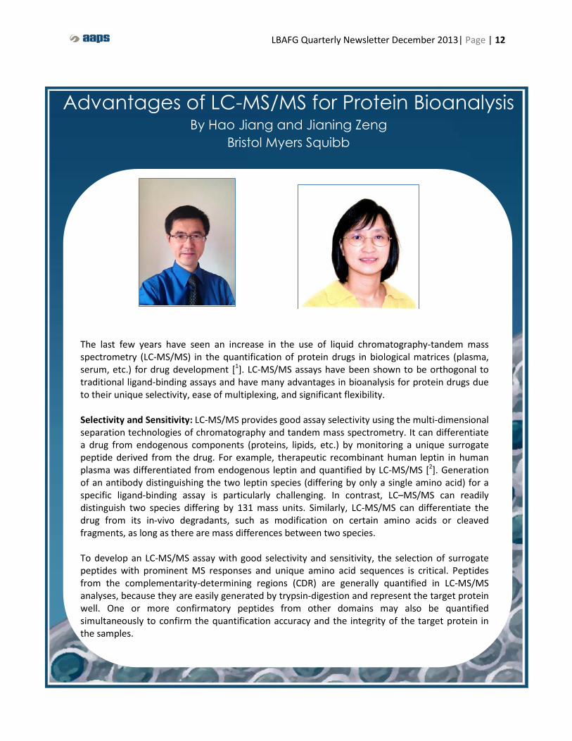
Advantages of LC-MS/MS for Protein Bioanalysis By Hao Jiang and Jianing Zeng
Bristol Myers Squibb
The last few years have seen an increase in the use of liquid chromatography-tandem mass spectrometry (LC-MS/MS) in the quantification of protein drugs in biological matrices (plasma, serum, etc.) for drug development [1]. LC-MS/MS assays have been shown to be orthogonal to traditional ligand-binding assays and have many advantages in bioanalysis for protein drugs due to their unique selectivity, ease of multiplexing, and significant flexibility. Selectivity and Sensitivity: LC-MS/MS provides good assay selectivity using the multi-dimensional separation technologies of chromatography and tandem mass spectrometry. It can differentiate a drug from endogenous components (proteins, lipids, etc.) by monitoring a unique surrogate peptide derived from the drug. For example, therapeutic recombinant human leptin in human plasma was differentiated from endogenous leptin and quantified by LC-MS/MS [2]. Generation of an antibody distinguishing the two leptin species (differing by only a single amino acid) for a specific ligand-binding assay is particularly challenging. In contrast, LC–MS/MS can readily distinguish two species differing by 131 mass units. Similarly, LC-MS/MS can differentiate the drug from its in-vivo degradants, such as modification on certain amino acids or cleaved fragments, as long as there are mass differences between two species. To develop an LC-MS/MS assay with good selectivity and sensitivity, the selection of surrogate peptides with prominent MS responses and unique amino acid sequences is critical. Peptides from the complementarity-determining regions (CDR) are generally quantified in LC-MS/MS analyses, because they are easily generated by trypsin-digestion and represent the target protein well. One or more confirmatory peptides from other domains may also be quantified simultaneously to confirm the quantification accuracy and the integrity of the target protein in the samples.

LBAFG Quarterly Newsletter December 2013| Page | 13
LC-MS/MS has been reported to provide a quantification limit in the range of 10-100 nM therapeutic proteins in serum, due to suppressed mass spectrometry responses of the surrogate peptides by endogenous components. A good sample cleanup will improve the assay sensitivity by several orders of magnitude to ~0.3 nM or below which will meet general requirements for clinical studies. The sample cleanup process can be simple or complex depending on the required detection limit. Protein precipitation with water miscible organic solvents (acetonitrile, methanol, or 2-isopropanol) and small amounts of acid (formic acid or acetic acid) is the simplest but yields a “dirty” extract that still contains a large portion of the endogenous components. The immuno-capture technique for purifying target proteins before digestion or surrogate peptides after digestion is a better approach to improve assay selectivity and sensitivity (~0.5 pM) [3]. However, this approach is more reagent-dependent and sometimes needs target-specific antibodies for immuno-precipitation.
Multiplexing: Another advantage of LC-MS/MS assays is the ability to simultaneously quantify multiple peptides using the multiple reaction monitoring (MRM) mode, because MRM allows very fast mass scanning (~5 millisecond duty cycle), and ~30 peptides can be measured at one time. These peptides can be surrogate peptides derived from different domains of a same protein, or from different proteins. Recently, we have developed and validated an LC-MS/MS assay for the simultaneous quantification of two co-administered antibody drugs in cynomolgus monkey serum [4]. Four peptides, a surrogate peptide for each antibody and two confirmatory peptides from different antibody domains, were simultaneously quantified by LC-MS/MS with good intra- and inter-assay precision (10.0% and 8.1% CV), accuracy (± 5.4% bias), and reproducibility. The data generated from the LC-MS/MS assay and the ligand-binding assay were in good agreement. In addition, the drug (free or total), drug degradants, the target, and associated biomarkers in serum can be simultaneously quantified by LC-MS/MS if unique surrogate peptides are determined for each analyte and a suitable sample extraction method is developed. Anti-drug antibodies in serum can also be quantified by LC-MS/MS after they are separated from endogenous components [5]. Flexibility: The quantification ranges of LC-MS/MS assays are generally wide and can be easily adjusted within several orders of magnitudes by changing sample aliquot volumes, LC injection volumes, or MS parameters. This advantage was shown in an animal toxicology study [4] for which the samples needed to be diluted 1000 time to fit the calibration curve of the LBA, but LC-MS/MS could analyze samples without extensive dilutions by using a lower LC injection volume and a serum aliquot volume. LC-MS/MS assays for target proteins quantitation in biological matrices generally do not require special reagents which are generally commercially available and of consistent quality. When specific immuno-capture is needed to separate target proteins for a better LC-MS/MS sensitivity, the immuno-capture
LC-MS/MS for Protein Bioanalysis cont.

LBAFG Quarterly Newsletter December 2013| Page | 14
reagents become critical because the assay only measures what is captured and thus needs to be characterized. After these critical reagents are commercialized and widely available and convenient, minimal efforts are still needed to evaluate lot changes and potentially stability [6]. In addition, an LC-MS/MS assay developed for one animal species can readily be validated in another species. Once optimized, the procedures can be easily applied to develop an assay for another protein drug of the same class. “Universal” surrogate peptides [7] sometimes can be directly used for quantification of another protein drug if both proteins have common peptides for quantification. This flexibility saves significant amounts of time and effort in method development and validation for regulated bioanalysis.
In summary, these advantages of LC-MS/MS, and ongoing technological and methodological improvements in LC-MS/MS quantification of proteins are likely to make this approach more feasible in the near future for a wider range of low abundance proteins, including therapeutics, biomarkers, and ADAs. Application of the LC-MS/MS technique in bioanalysis not only delivers high quality bioanalytical data with good accuracy, precision, and reproducibility, but also answers some challenging questions during drug development.
References 1. van den Broek, I.; Niessen, W. M.; van Dongen, W. D. Bioanalytical LC-MS/MS of protein-based
biopharmaceuticals. J. Chromatogr B. 2013, 929, 161 – 179.
2. Wang, Y.; Heilig, J. S. Differentiation and quantification of endogenous and recombinant-methionyl human leptin in clinical plasma samples by immunocapture/mass spectrometry. J. Pharm. Biomed. Anal. 2012, 70, 440 – 446.
3. Neubert, H.; Muirhead, D.; Kabir, M.; Grace, C.; Cleton, A.; Arends, R. Sequential Protein and Peptide Immunoaffinity Capture for Mass Spectrometry-Based Quantification of Total Human β Nerve Growth Factor. Anal. Chem. 2013, 85, 1719 − 1726.
4. Jiang, H.; Zeng, J.; Titsch, C.; Voronin, K.; Akinsanya, B.; Luo, L.; Shen, H.; Desai, D. D.; Allentoff, A.; Aubry, A. F.; DeSilva, B. S.; Arnold, M. E. Fully Validated LC-MS/MS Assay for the Simultaneous Quantitation of Coaministered Therapeutic Antibodies in Cynomolgus Monkey Serum. Anal. Chem. 2013, 85, 9859 – 9867.
5. Kelley, M.; Ahene, A. B.; Gorovits, B.; Kamerud, J.; King, L. E.; McIntosh, T.; Yang, J. Theoretical considerations and practical approaches to address the effect of anti-drug antibody (ADA) on quantification of biotherapeutics in circulation. AAPS J. 2013, 15, 646 – 658.
6. O'Hara, D. M.; Theobald, V.; Egan, A. C.; Usansky, J.; Krishna, M.; TerWee, J.; Maia, M.; Spriggs, F. P.; Kenney, J.; Safavi, A.; Keefe, J. Ligand Binding Assays in the 21st Century Laboratory: Recommendations for Characterization and Supply of Critical Reagents. AAPS J. 2012, 14, 316 – 328.
7. Furlong, M. T.; Ouyang, Z.; Wu, S.; Tamura, J.; Olah, T.; Tymiak, A.; Jemal, M. A universal surrogate peptide to enable LC-MS/MS bioanalysis of a diversity of human monoclonal antibody and human Fc-fusion protein drug candidates in pre-clinical animal studies. Biomed. Chromatogr. 2012, 26, 1024 – 1032
LC-MS/MS for Protein Bioanalysis cont.

LBAFG Quarterly Newsletter December 2013| Page | 15
Antibody Drug Conjugates: Bioanalytical
Challenges By Vangipuram S Rangan and Shrikant Deshpande
Bristol Myers- Squibb
Antibody drug conjugates (ADCs) are an emerging class of therapeutic biologics that have brought considerable excitement in the biopharmaceutical industry due to their potential to treat cancer patients without the severe side-effects of chemotherapy. ADCs utilize the tumor targeting potential of monoclonal antibodies to deliver highly potent cytotoxic drugs predominantly to the tumor by carefully selecting the target antigens that are overexpressed in tumor tissues compared to that of normal tissues. The main components of an ADC are an antibody, a cytotoxic drug, a linker and a reactive handle (group) that attaches the drug-linker to the antibody. The proposed mechanism of action of ADCs involves the internalization of ADCs upon binding to their target antigen, trafficking of ADCs to low pH endosome or lysosomal vesicles and release of the cytotoxic drug either by degradation of antibody or the linker cleavage by lysosomal proteases. The released cytotoxic drug then binds to its molecular targets to induce tumor cell death. If the released cytotoxic drug is cell permeable, it could also diffuse out of tumor cells and exhibit bystander cell killing effects on cancer and normal cells. Various classes of cytotoxic drugs are being evaluated as ADCs that include DNA alkylators, DNA strand breakers, tubulyn inhibitors and tubulyn stabilizers, just to name a few. With the recent US FDA approval of Adecetris (brentuximab vedotin) to treat Hodgkin’s lymphoma and Kadcyla TM (trastuzumab emtansine) to treat advanced Her2 positive breast cancers, the validity of ADC technology to treat cancer is well demonstrated. Currently, there are more than 20 ADCs at various stages of clinical trials for variety of cancers pointing to the vast potential of this technology to address the long standing need for successful treatment of cancer patients.
LBAFG Quarterly Newsletter December 2013| Page | 16
ADC cont.
ADCs are typically heterogeneous mixtures not only because of the presence of three vastly different components such as antibody, cytotoxic drug, and linker but also due to methods employed to conjugate the cytotoxic drugs to antibodies. Drug to antibody ratio (DAR) of an ADC provides information about the average number drugs attached per antibody molecule. Most often the drugs are attached either by reactions at lysine-side chain amines or at sulfhydry groups of cysteine residues after the controlled reduction of interchain disulfide bonds of antibodies. Due to the random nature of the conjugation process, final ADC material contains vast number of species with varying numbers drugs attached per antibody molecule (DAR) as well as drugs attached at various conjugation sites. To overcome the issues associated with the random conjugation, new technologies that employ site-specific conjugation of cytotoxic drugs to an antibody are being developed such as ThioMab and novel technologies from Ambrx, Sutro, etc. However, majority of ADCs that are currently in clinical trials utilize the random conjugation process involving either the lysine residues or sulfhydryl groups of interchain cysteine residues of antibodies. Due to the complex nature of ADCs, bioanalytical strategies that are unique to ADCs need to be employed along with the well established methods for therapeutic antibodies and small molecule drugs. Main focus of this article is to identify critical bioanalytical assays required for understanding the PK of ADCs and the challenges to be addressed while developing these assays.
ADCs are similar to its antibody backbone in terms of target specific binding, Fc related function and PK properties such as slow clearance, long half-life and low volume distribution but differ from antibodies in terms of presence of cytotoxic drugs. ADCs are also more heterogeneous than antibodies due to its manufacturing process as well as biological and chemical processing of ADCs in circulation following in vivo administration. Therefore, when developing the bioanalytical strategies for ADCs one has to carefully develop assays to not only monitor the fate of antibody and the cytotoxic component but also design methods to identify various ADC species found in the systemic circulation. Such a strategy is critical to provide information about ADC Exposure-Response relationship for efficacy and safety during preclinical and clinical development (Gorovits B et al, Bioanalysis, 5, 997-1006, 2013 and Kaur S et al, Bioanalysis, 5, 201-226, 2013).
Unlike antibodies or small molecule drugs, it is difficult to predict which species or intermediates may provide information about the safety and efficacy of ADCs. Therefore multiple assays are required to follow the pharmacokinetics (PK) of ADCs to account for multiple analytes that are found in plasma samples after in vivo administration of ADCs. Bioanalytical assays for ADCs typically focus on measuring the levels of three major analytes from plasma PK samples. They are: 1) measuring the total antibody concentration that accounts for both the conjugated and unconjugated antibody species, 2) measuring the conjugated antibody concentration that accounts for both the intact antibody and cytotoxic components of ADC and 3) measuring the unconjugated cytotoxic drug that accounts for the complete loss of cytotoxic drug from antibody as well as linker cleaved active drug. It is important to recognize that a variety of unique reagents are needed to develop bioanalytical assays for ADCs. For ELISA assays either the target antigen or anti-idiotypic antibodies are required to capture the ADCs from the plasma matrix.
LBAFG Quarterly Newsletter December 2013| Page | 17
An antibody that recognizes the cytotoxic drug specifically is required to measure the cytotoxic drug component of ADC and it is usually a challenging task. To support LC-MS based assays, immobilized anti-idiotypic antibody beads or immobilized anti-cytotoxic drug beads are required to capture the ADC species from plasma matrix for further processing. Synthesis of cytotoxic drug standards as well as potential drug metabolite standards are required to assist the LC-MS based MRM methods. Measuring the total antibody concentration The main focus of this assay is to measure the levels of unconjugated as well as all the conjugated antibody species and it provides information about the in vivo stability and integrity of the antibody. This assay also informs whether the clearance rate of ADC is different from that of the antibody. Ligand binding assay formats such as ELISA are typically used to measure the total antibody concentration either by using target antigen or anti-idiotypic antibodies to capture ADCs from plasma matrix. Anti-human IgGs can also be used to capture ADCs from plasma samples during preclinical development. Although the assay protocols and parameters are similar to those described for therapeutic antibodies, additional parameters are required for ADCs. Since ADCs are highly heterogeneous mixture, it has been shown that measuring the total antibody concentration for ADCs with different DAR is strongly influenced by ELISA assay formats since in a given assay format ADCs with different DAR values might not be uniformly detected and quantified (Stephan JP, et al, Bioconjug.Chem. 19, 1673-1683, 2008). Therefore, all assay formats should be evaluated for the effect of ADCs with different DAR values on assay specificity, accuracy and reproducibility and compared with assay performance obtained with the unconjugated antibody. However, when dealing with ADCs generated by random conjugation process involving lysine residues, it is not possible to obtain ADCs with individual DAR values unlike ADCs generated by targeting cysteine residues. In those instances, the assay formats should be evaluated by generating ADCs with different average DAR values. For total antibody assay, same lot of ADC that was used in preclinical study or ADC drug substance used in clinical study is typically used as reference material to generate the standard curve instead of the unconjugated antibody. Measuring the conjugated antibody concentration This assay focuses on measuring the levels of both intact antibody and cytotoxic drug component of ADC and in general considered to provide relevant description of PK of the ADC. Changes in the circulating levels of conjugated antibodies are mainly attributed to three main biological and chemical events occurring with ADCs in circulation. First, it could be due to elimination of certain intact ADC species from circulation and such a scenario has been reported for ADC species with high DAR values. Second, factors like stability of the linker in circulation, the conjugation chemistry used to generate ADCs and the location of conjugation site could contribute significantly to in vivo deconjugation events. For example, hydrazine-based linkers are found to be more susceptible for low pH dependent cleavage than the peptide linkers. Similarly some ADCs generated by disulfide or cysteine chemistries are reported to undergo chemical deconjugation process due to transfer of the cytotoxic drug from antibody to other thiol containing serum components like glutathione and albumin (Alley SC et al, Bioconjug.Chem. 19, 759-765, 2008). Factors like the location of the
ADC Cont.
LBAFG Quarterly Newsletter December 2013| Page | 18
conjugation site as well as the solvent accessibility of the linker are also reported to influence the rate of thiol exchange deconjugation process. Finally, cytotoxic drug component of conjugated antibody could undergo modification without any deconjugation due to enzymatic or chemical events in circulation. Such modifications could render the cytotoxic drug inactive and thus affecting the potency of ADCs.
Conjugated antibody levels are typically measured using ELISA assays where ADCs are captured by various reagents like target antigen, anti-idiotypic antibodies or anti-cytotoxic drug antibodies. Due to the dynamic nature of the ADC heterogeneity in circulation, it is critical to develop ELISA assays not only to identify and quantify the most prevalent ADC species but also the most relevant ADC species that provide critical information to establish ADC Exposure-Response relationship for efficacy and safety. Since the ADC material used to generate the standard curve is not identical to the ADC species found in circulation, it is required to develop ELISA reagents and formats that accurately measure the decrease in DAR over time by using that standard curve. If the cytotoxic drug component of ADC is susceptible for modification in circulation, the ability of the anti-drug antibody to recognize the modified version of the drug needs to evaluated. Demonstration of the potency and safety of the ADC species with modified drug is also required to establish the overall ADC Exposure-Response relationship. Availability of anti-drug antibodies that specifically recognize either the intact cytotoxic drug or the modified version of the drug greatly facilitates the accurate measurement of both the ADC species and that information could be used to demonstrate the Exposure-Response relationship of pharmacologically relevant ADC species.
Although the conjugated antibody ELISA is capable of distinguishing the naked antibodies (DAR=0) and the antibodies carrying the drug, it is unable to distinguish the ADC species with different DARs. LC-MS based orthogonal methods are therefore proposed to demonstrate presence of ADC species with different DAR over time in plasma PK samples (Xu K et al, Bioanalysis, 5, 1057-1071, 2013). It involves the affinity capture of ADCs from plasma matrix using either the anti-idiotypic antibodies or anti-drug antibodies that were immobilized on sepharose beads. After deglycosylation, the ADCs are released from the affinity matrix and mass of the intact antibody measured by LC-MS to identify and quantify the relative abundance of ADC species with different DAR. However, it is technically challenging assay to validate and hence can be used only during preclinical development for information purposes only.
ADC Cont.

LBAFG Quarterly Newsletter December 2013| Page | 19
Literature Highlights 3Q2013
ADC Cont.
The ADC levels in plasma or serum samples can also be quantified by measuring the total cytotoxic drug conjugated to the antibody and measurement of this analyte is usually considered as an alternative way to assess the conjugate exposure. This approach utilizes the capture of ADC species from plasma or serum matrix using either the anti-idiotypic antibodies or anti-drug antibodies that were immobilized on sepharose beads. The linker is cleaved enzymatically or chemically from the captured ADCs and the released drug is identified and quantified by LC-MS/MS methods. By adopting the orthogonal approaches to measure the levels of conjugated antibody by ELISA and the levels of antibody-conjugated drug by LC-MS/MS methods, it is possible to assess the ADC exposure accurately during preclinical development. Measuring the unconjugated drug Typically LC-MS based MRM assays are developed to measure the level of linker cleaved unconjugated drugs in plasma samples (Kaur S et al, Bioanalysis, 5, 201-226, 2013) and it provides the measure of in vivo linker stability. Since the released cytotoxic drugs in circulation are susceptible for further metabolism, LC-MS/MS assays should also be developed to identify and quantitate all the potential drug degradation products. Therefore, the choice of reference material for unconjugated drug assay may depend on the type of modification that drug undergoes upon its release to circulation. Since relatively low levels of unconjugated drugs are generally expected in circulation, technically challenging high sensitivity assays are required. In summary, bioanalytical methods required for PK/PD assessment of ADCs are still evolving. They are dependent on the antibody, the class of cytotoxic drug, cleavable or noncleavable linker and the conjugation technology. In general, due to the heterogeneous nature of ADCs bioanalytical strategies should be developed by taking into account all the factors that contribute to ADC heterogeneity. For successful clinical development of ADCs, critical bioanalytical assays to monitor the most relevant ADC species should be developed at preclinical development stage.
LBAFG Quarterly Newsletter December 2013| Page | 20
present in the intronic or intergenic regions in the genome. They may be present in those regions in the genome which are distant from the other regulatory genes or in the form of a cluster or as a long polycistronic primary transcript [1]. They are usually transcribed by the RNA polymerase II (Pol II) as a long primary transcript which is known as pri-miRNA. Pri-miRNA contains a polyA (polyadenylated) tail at its 3’end while a 5’methyl capping of methyl gaunosine. Pri-miRNAs are usually processed in the nucleus by the RNase II endonucleaseIII Drosha in co-operation with the DiGeorge syndrome critical region gene 8 (DGCR8) / Pasha proteins to form a 60-110 nt long hairpin structure termed as precursor of miRNA (pre-miRNA) which possesses a characteristic 3’overhangs. Pre-miRNAs are usually exported out of the nucleus into the cytoplasm with the help of Exportin-5 present on the cell membrane. In the cytoplasm, pre-miRNAs undergo cleavage by the RNase III Dicer-I together with transactivation response RNA binding proteins (TRBP) and protein kinase RNA activator proteins (PACT) resulting in the formation of a double stranded miRNA/miRNA* duplex. This duplex is unwounded with the help of helicase resulting in the formation of 20-22 nt long mature miRNA and the other complementary passenger strand is usually degraded [1; 2]. Mature miRNAs regulate gene expression through their association with the argonaute proteins of the RNA inducing silencing complex (RISC) which in turn represses the complementary mRNA targets. The mature strand is responsible for its mRNA target recognition and for their incorporation into the RISC. They may bind to the coding regions or 3’UTR or 5’UTR of their target mRNA [3]. Although, bioinformatical analysis have shown that miRNAs usually targets 3’UTR of its target mRNA which have been verified experimentally to a large extent. Hence, the specificity of a particular miRNA for its target gene is defined by comparing the sequences at the 5’ miRNA (2 to 8 positions which is also known as the seed sequence) with the 3’ UTR of the target mRNA. Depending on the degree of complementarity in the sequences between the mature miRNA and its target mRNA, they are involved in the mRNA degradation or translational repression. In case of a perfect match between the sequences of miRNA and target mRNA, miRNA binds to the Open Reading Frame (ORF) region / coding region of the target mRNA and are involved in the mRNA cleavage or degradation. On the other hand, if there is an imperfect match between the sequences of the miRNA and target mRNA, miRNAs can bind to the untranslated regions of its target gene and may lead to translational repression [4].
Student Corner: MicoRNA Therapuetics in Cancer By Hardik Mody University of Georgia
Introduction: MicroRNAs (miRNAs) belong to the class of non protein-coding RNAs and are usually 20-22 nucleotides (nt) in length. They are important regulatory nucleic acids involved in the regulation of various genes which in turn are involved in different cellular processes including differentiation and proliferation, survival and death [1]. They regulate the gene expression through posttranscriptional mRNA cleavage or translational repression of their target mRNA. MiRNAs are generally
LBAFG Quarterly Newsletter December 2013| Page | 21
MicroRNA Cont.
Implication of miRNAs in cancer: Aberrant expression of a single miRNA may disrupt homeostatis and normalcy and may be lead to various diseases. Many studies have shown the implication of miRNAs in diseases like cancer. On one side, certain miRNAs may act as tumor suppressors and negatively regulate their target oncogenes, whereas on the other side, some may act as oncogenes negatively regulating tumor suppressors. In cancer, some of these tumor suppressor miRNAs may be downregulated while some of the oncogenic miRNAs may be upregulated. Thus, cancer may result from or may result to aberrant expression of multiple miRNAs. Thereby, restoring such aberrantly expressed miRNAs to their normal levels (reducing expression of oncogenic miRNAs and increasing expression of tumor suppressor miRNAs) may be one of the therapeutic strategies in combating lethal diseases like cancer [5]. This review summarizes certain examples of miRNAs acting as tumor suppressors or oncogenes, how their aberrant expression is implicated in cancer, therapeutic strategies in restoring their expression to normalcy and challenges behind these therapeutic strategies. Developmental approaches for miRNA therapeutics: The aberrant miRNA expression levels in cancer can be manipulated directly with the help of antisense oligonucleotides or plasmid constructs. They may inhibit or lower the expression of oncogenic miRNAs which are usually overexpressed in cancer or they may increase or upregulate the expression of tumor suppressor miRNAs which are usually downregulated in cancer. The miRNA expression levels may also be modulated with the help of certain small molecule inhibitors which may aid in lowering their expression. The following section summarizes these strategies utilizing miRNAs as either drugs or drug targets. MiRNA replacement therapy: MiRNA as tumor suppressors: As mentioned earlier, certain diseased conditions like cancer may result from or may result into the reduction of expression or loss of a particular miRNA or a cluster of miRNAs. Most of the miRNAs are thought to be tumor suppressors negatively regulating expression of certain oncogenes and are downregulated in cancer. One of the earlier discovered miRNA, let-7, is a tumor suppressor gene downregulated in a variety of cancer types including ovarian, breast, lung, colon, stomach along with others and have also been implicated in cancer stem cells [6; 7; 8; 9; 10]. Let-7 targets various oncogenes like Ras, Myc and HMGA-2 (high-mobility group AT-hook 2) at the mRNA and protein level. For example, reduced expression of the let-7 in lung tumor compared with the normal tissue was found to be in correlation with the increase in the RAS protein levels [9]. Similarly, one study reported low expression levels of let-7 in ovarian cancer samples with high HMGA2 mRNA [11]. Also, certain miRNAs are found to be located in genomic regions which are usually altered or deleted in various cancer types. For instance, miR-15 and miR-16 were shown to be silenced or reduced in chronic cytic leukemia (CLL) and in prostate cancer. Both these miRNAs are located in the chromosome 13q14 which is usually absent in CLL [12]. These genes usually
LBAFG Quarterly Newsletter December 2013| Page | 22
MicroRNA Cont.
act as tumor suppressors by regulating apoptosis by targeting BCL2, an antiapoptotic factor, and thereby their downregulation in CLL may result in aberrant apoptotic signaling, evading cancer cells from undergoing cell death and increasing their survival [13]. Thus, restoring the levels of tumor-suppressor miRNAs which are lost or reduced in cancer may be one of the therapeutic strategies or may help in increasing the chemosensitivity and the overall therapeutic effectiveness of the conventional chemotherapy. MiRNA mimics: MiRNA levels can be restored by the external administration of their precursor or mature forms. One strategy utilized in the restoration of miRNAs includes the introduction of miRNA mimics, which are chemically modified synthetic oligonucleotides identical or similar to particular miRNAs. They are usually double stranded RNA molecules with one strand being the guide strand and the other is the passenger strand [5]. The guide strand is similar or identical to the miRNA of interest, while the other strand is the passenger strand which can be designed with some chemical modifications to enhance their stability, permeability into the cells and targeting. These modifications may include 2’-O-methyl, 2’-O-methoxyethyl or 2’-O-fluoro oligonucleotides with a phosphorothioate backbone [5; 14]. Additionally, the features of the pre-miRNA duplexes including 3’overhangs and 5’phosphate groups which aid in efficient recognition and loading of the guide strand into the RISC are considered while designing these synthetic nucleic acids. The 3’overhangs may be evaded from nuclease driven degradation with the incorporation of inverted dT, nonnucleotide or benzene – pyridine modifications at the 3’end of the miRNA duplexes [14]. Many studies have shown inhibition in proliferation and increase in cancer cell death after the introduction of such synthetic oligonucleotides mimicking tumor-suppressor miRNAs. Examples may include introduction of a synthetic mimic of miR-15a which aided in enhanced cell death in prostate cancer cells [15]. Similarly, delivery of exogenous miR-16, which is otherwise downregulated in cancer, resulted in reduced growth and proliferation in prostate cancer cells. Moreover, systemic administration of miR-16 and atelocollagen complex via tail vein injection inhibited the growth of the prostate cancer cells in a mouse model [16]. Another example may include systemic delivery of a let-7 mimic with a lipid complex resulted in inhibition of tumor growth in a K-ras mutant mouse model. On the one hand, intratumoral injection of let-7 oligonucleotide in non-small cell lung carcinoma reduced the tumor burden, inhibited tumor progression and induced necrosis by modulating its targets N-Ras and cyclin dependent kinase 6 (CDK6). On the other hand, depleting let-7 levels aided tumor formation, growth and progression indicating their therapeutic potential [17; 18; 19]. Mir-34, which is regulated by the tumor suppressor p53, is another gene shown to be downregulated in a variety of cancer types including pancreatic cancer and gastric cancer along with others [20; 21]. It acts as a tumor suppressor by negatively regulating its targets including Bcl2, Notch and HMGA2. Moreover, miR-34 has shown to negatively regulate CD44 in prostate cancer cells and thereby arrest cancer cell growth and metastasis and inhibit the cancer stem cells as well [22]. MiR-34 lipid complex mimic led to significant reduction in tumor growth in non-small cell lung cancer mouse model when delivered systemically or intratumorally [23]. Similar findings were also obtained in murine mouse models of pancreatic and prostate cancer, resulting in an
LBAFG Quarterly Newsletter December 2013| Page | 23
MicroRNA Cont.
extension of survival of such immunocompromised tumor bearing mice. One study also utilized lipid nanoparticles as delivery carriers for miR-34 mimic in subcutaneous and orthotopic pancreatic cancer mouse models. Tail vein injection of miR-34 mimic resulted in inhibiting tumor growth and activating apoptosis in such nude mice models [24]. Knockdown of oncomiRs: MiRNAs as oncogenes: OncomiRs As mentioned earlier, miRNAs are implicated in the initiation and progression of cancer. Certain miRNAs may be dysregulated in cancer and may function as oncogenes. They may be involved in the negative regulation of certain tumor suppressor genes and may be upregulated or overexpressed in cancer. For instance, miR-155 is overexpressed in certain cancers like lung adenocarcinoma as well as lymphoproliferative disorders like leukemia and lymphoma. Mir-155 directly acts on the Src homology 2 domain-containing inositol 5-phosphate 1 (SHIP1), a key player in the process of proliferation through negative regulation of the AKT pathway, and thereby act as an oncomiR [25; 26]. Similarly, miR-21 is upregulated in various solid tumors like breast cancer. One of the studies reported that overexpression of only miR-21 was enough to initiate tumor formation and that the tumor volume and survival were primarily dependent on the expression levels of miR-21 [27]. Inhibiting or downregulating these oncomiRs may be one of the therapeutic strategies to combat cancer or may also be utilized as an adjuvant to the conventional cancer chemotherapy. Various approaches have been tested so far to inhibit miRNAs in vitro and in vivo. OncomiR inhibition using antisense oligonucleotides: Antisense oligonucleotides, also termed as antimiRs, are short, single stranded RNA or DNA molecules which are designed to bind to nucleic acids and competitively block Watson-Crick base pairing at any of the important step in the biogenesis or regulatory mechanism of miRNAs. They work at any of the three steps including miRNA processing, loading of miRNA into the RISC complex or miRNA-mRNA duplex formation. These oligonucleotides are usually complementary to the mature miRNA or to the guiding strand of the miRNA:miRNA* duplex, and thereby forms base pairing with them and function as miRNA inhibition by blocking the ability of miRNAs to bind to the target mRNAs with subsequent repression [5; 14]. Such oligonucleotides have been tested to block several oncogenic miRNAs both in vivo and in vitro resulting in the inhibition of tumor proliferation, progression, invasion and metastasis, thereby indicating their therapeutic potential. For example, miR-17-92 cluster have been reported to be oncogenic and upregulated in a variety of cancers including lung cancer and lymphomas. Inhibition of miRNAs belonging to this cluster using antisense oligonucleotide technology aided in inducing apoptosis in lung cancer cells as well as prostate cancer cells. MiR-17-92 cluster have shown to be involved in translational repression of E2F family proteins and hence inhibiting them may aid in blocking this translational repression of E2F which may mediate induction of apoptosis in cancer cells [28]. Another study reported cell death and metastasis arrest aided with antisense ologinucleotides targeting miR-182. Challenges & Future directions: Ever since the discovery of miRNAs in 1993, large number of studies have been published especially in the last decade uncovering their posttranscriptional regulatory roles in various
LBAFG Quarterly Newsletter December 2013| Page | 24
MicroRNA Cont.
fundamental cellular processes and signaling pathways. The understanding related to the implications of miRNAs in diseases like cancer and how they can be utilized as therapeutic targets or as therapeutics have also progressed during this phase with the aid of modern molecular biological tools which is expected to be translated into the clinics in the coming years. However, since they are in their developmental phase, major challenges remain in the utilization of miRNAs as therapeutics. The principal issue that needs to be addressed is an in-depth investigation of the role of miRNAs and their regulatory mechanisms in the initiation and progression of cancer. Although, many studies have been published demonstrating certain miRNAs as oncogenes while some others as tumor-suppressors, a better understanding is required pertaining to the complexity of multiple miRNAs with multiple roles in tumorigenesis. Another major challenge is the lack of specificity or the multi functional role of a particular miRNA. Since a particular miRNA may be involved in the regulation of hundreds of genes which in turn may be involved in the regulation of various cellular processes and signaling pathways, modulation of a single miRNA may have an effect on all of its target genes and thereby on various processes and pathways which may lead to undesired effects or even toxic effects. Hence, future research should be focused on investigating the potential off-target effects and potential therapeutic strategies to prevent them. In addition, pharmacokinetic challenges exist for such RNA based therapeutics including their cellular uptake which may be critical in governing sustained manipulation of miRNAs in vivo which in turn may determine the dose required for intended therapeutic effects. Also, systemic administration of such therapeutics may expose them to enzymatic degradation in the bodily fluids including blood via the nucleases. Another important aspect to be considered is the duration of miRNA modulation required for a particular therapeutic effect and physiological consequences of long term miRNA manipulation. One strategy to address the above mentioned challenges is chemical modifications of antisense oligonucleotides targeting specific miRNAs which may enhance their cellular uptake and protect them from nucleases. Such chemical modifications, as discussed earlier, may also increase their specificity towards target miRNAs. However, some modifications like cholesterol conjugations have been linked with modified biological activity and may increase their toxicity. Another strategy to overcome the potential off-target effects induced by miRNA therapeutics is through their systemic delivery to the desired site or to a specific tissue of interest. This may be achieved with the help of targeted drug delivery systems like liposomes and nanoparticles. They may not only improve targeted delivery of antisense oligonucleotides, but also provide protection from nucleases, improve their cellular uptake through their surface modifications and improve the overall pharmacokinetic and pharmacodynamic characteristics with minimal toxicity. Further, short and long term safety and toxicity studies should be carried out in animal models to determine therapeutic index and dosing schedules. Inspite of all these challenges, the therapeutic potential of miRNAs cannot be underestimated. Further research should be carried out at the present rate aiming to address the above mentioned challenges which may catalyze the transition of miRNA therapeutics from bench to bedside.

LBAFG Quarterly Newsletter December 2013| Page | 25
MicroRNA Cont.
References:
[1]D.P. Bartel, MicroRNAs: genomics, biogenesis, mechanism, and function. Cell 116 (2004) 281-297. [2]H. Siomi, M.C. Siomi, Posttranscriptional regulation of microRNA biogenesis in animals. Molecular cell 38 (2010) 323-332. [3]M.I. Almeida, R.M. Reis, G.A. Calin, MicroRNA history: discovery, recent applications, and next frontiers. Mutation research 717 (2011) 1-8. [4]D.P. Bartel, MicroRNAs: target recognition and regulatory functions. Cell 136 (2009) 215-233. [5]R. Garzon, G. Marcucci, C.M. Croce, Targeting microRNAs in cancer: rationale, strategies and challenges. Nature reviews. Drug discovery 9
(2010) 775-789. [6]N. Yang, S. Kaur, S. Volinia, J. Greshock, H. Lassus, K. Hasegawa, S. Liang, A. Leminen, S. Deng, L. Smith, C.N. Johnstone, X.M. Chen, C.G. Liu, Q.
Huang, D. Katsaros, G.A. Calin, B.L. Weber, R. Butzow, C.M. Croce, G. Coukos, L. Zhang, MicroRNA microarray identifies Let-7i as a novel biomarker and therapeutic target in human epithelial ovarian cancer. Cancer research 68 (2008) 10307-10314.
[7]A.J. Schetter, S.Y. Leung, J.J. Sohn, K.A. Zanetti, E.D. Bowman, N. Yanaihara, S.T. Yuen, T.L. Chan, D.L. Kwong, G.K. Au, C.G. Liu, G.A. Calin, C.M. Croce, C.C. Harris, MicroRNA expression profiles associated with prognosis and therapeutic outcome in colon adenocarcinoma. JAMA : the journal of the American Medical Association 299 (2008) 425-436.
[8]F. Petrocca, R. Visone, M.R. Onelli, M.H. Shah, M.S. Nicoloso, I. de Martino, D. Iliopoulos, E. Pilozzi, C.G. Liu, M. Negrini, L. Cavazzini, S. Volinia, H. Alder, L.P. Ruco, G. Baldassarre, C.M. Croce, A. Vecchione, E2F1-regulated microRNAs impair TGFbeta-dependent cell-cycle arrest and apoptosis in gastric cancer. Cancer cell 13 (2008) 272-286.
[9]S.M. Johnson, H. Grosshans, J. Shingara, M. Byrom, R. Jarvis, A. Cheng, E. Labourier, K.L. Reinert, D. Brown, F.J. Slack, RAS is regulated by the let-7 microRNA family. Cell 120 (2005) 635-647.
[10]M.V. Iorio, M. Ferracin, C.G. Liu, A. Veronese, R. Spizzo, S. Sabbioni, E. Magri, M. Pedriali, M. Fabbri, M. Campiglio, S. Menard, J.P. Palazzo, A. Rosenberg, P. Musiani, S. Volinia, I. Nenci, G.A. Calin, P. Querzoli, M. Negrini, C.M. Croce, MicroRNA gene expression deregulation in human breast cancer. Cancer research 65 (2005) 7065-7070.
[11]J.A. Broderick, P.D. Zamore, MicroRNA therapeutics. Gene therapy 18 (2011) 1104-1110. [12]G.A. Calin, C.D. Dumitru, M. Shimizu, R. Bichi, S. Zupo, E. Noch, H. Aldler, S. Rattan, M. Keating, K. Rai, L. Rassenti, T. Kipps, M. Negrini, F.
Bullrich, C.M. Croce, Frequent deletions and down-regulation of micro- RNA genes miR15 and miR16 at 13q14 in chronic lymphocytic leukemia. Proceedings of the National Academy of Sciences of the United States of America 99 (2002) 15524-15529.
[13]A. Cimmino, G.A. Calin, M. Fabbri, M.V. Iorio, M. Ferracin, M. Shimizu, S.E. Wojcik, R.I. Aqeilan, S. Zupo, M. Dono, L. Rassenti, H. Alder, S. Volinia, C.G. Liu, T.J. Kipps, M. Negrini, C.M. Croce, miR-15 and miR-16 induce apoptosis by targeting BCL2. Proceedings of the National Academy of Sciences of the United States of America 102 (2005) 13944-13949.
[14]S.B. Thorsen, S. Obad, N.F. Jensen, J. Stenvang, S. Kauppinen, The therapeutic potential of microRNAs in cancer. Cancer J 18 (2012) 275-284. [15]D. Bonci, V. Coppola, M. Musumeci, A. Addario, R. Giuffrida, L. Memeo, L. D'Urso, A. Pagliuca, M. Biffoni, C. Labbaye, M. Bartucci, G. Muto,
C. Peschle, R. De Maria, The miR-15a-miR-16-1 cluster controls prostate cancer by targeting multiple oncogenic activities. Nature medicine 14 (2008) 1271-1277.
[16]F. Takeshita, L. Patrawala, M. Osaki, R.U. Takahashi, Y. Yamamoto, N. Kosaka, M. Kawamata, K. Kelnar, A.G. Bader, D. Brown, T. Ochiya, Systemic delivery of synthetic microRNA-16 inhibits the growth of metastatic prostate tumors via downregulation of multiple cell-cycle genes. Molecular therapy : the journal of the American Society of Gene Therapy 18 (2010) 181-187.
[17]P. Trang, P.P. Medina, J.F. Wiggins, L. Ruffino, K. Kelnar, M. Omotola, R. Homer, D. Brown, A.G. Bader, J.B. Weidhaas, F.J. Slack, Regression of murine lung tumors by the let-7 microRNA. Oncogene 29 (2010) 1580-1587.
[18]M.S. Kumar, S.J. Erkeland, R.E. Pester, C.Y. Chen, M.S. Ebert, P.A. Sharp, T. Jacks, Suppression of non-small cell lung tumor development by the let-7 microRNA family. Proceedings of the National Academy of Sciences of the United States of America 105 (2008) 3903-3908.
[19]C.D. Johnson, A. Esquela-Kerscher, G. Stefani, M. Byrom, K. Kelnar, D. Ovcharenko, M. Wilson, X. Wang, J. Shelton, J. Shingara, L. Chin, D. Brown, F.J. Slack, The let-7 microRNA represses cell proliferation pathways in human cells. Cancer research 67 (2007) 7713-7722.
[20]H. Hermeking, The miR-34 family in cancer and apoptosis. Cell death and differentiation 17 (2010) 193-199. [21]L. He, X. He, L.P. Lim, E. de Stanchina, Z. Xuan, Y. Liang, W. Xue, L. Zender, J. Magnus, D. Ridzon, A.L. Jackson, P.S. Linsley, C. Chen, S.W.
Lowe, M.A. Cleary, G.J. Hannon, A microRNA component of the p53 tumour suppressor network. Nature 447 (2007) 1130-1134. [22]C. Liu, K. Kelnar, B. Liu, X. Chen, T. Calhoun-Davis, H. Li, L. Patrawala, H. Yan, C. Jeter, S. Honorio, J.F. Wiggins, A.G. Bader, R. Fagin, D. Brown,
D.G. Tang, The microRNA miR-34a inhibits prostate cancer stem cells and metastasis by directly repressing CD44. Nature medicine 17 (2011) 211-215.
[23]P. Trang, J.F. Wiggins, C.L. Daige, C. Cho, M. Omotola, D. Brown, J.B. Weidhaas, A.G. Bader, F.J. Slack, Systemic delivery of tumor suppressor microRNA mimics using a neutral lipid emulsion inhibits lung tumors in mice. Molecular therapy : the journal of the American Society of Gene Therapy 19 (2011) 1116-1122.
[24]D. Pramanik, N.R. Campbell, C. Karikari, R. Chivukula, O.A. Kent, J.T. Mendell, A. Maitra, Restitution of tumor suppressor microRNAs using a systemic nanovector inhibits pancreatic cancer growth in mice. Molecular cancer therapeutics 10 (2011) 1470-1480.
[25]R.M. O'Connell, A.A. Chaudhuri, D.S. Rao, D. Baltimore, Inositol phosphatase SHIP1 is a primary target of miR-155. Proceedings of the National Academy of Sciences of the United States of America 106 (2009) 7113-7118.
[26]S. Costinean, S.K. Sandhu, I.M. Pedersen, E. Tili, R. Trotta, D. Perrotti, D. Ciarlariello, P. Neviani, J. Harb, L.R. Kauffman, A. Shidham, C.M. Croce, Src homology 2 domain-containing inositol-5-phosphatase and CCAAT enhancer-binding protein beta are targeted by miR-155 in B cells of Emicro-MiR-155 transgenic mice. Blood 114 (2009) 1374-1382.
[27]P.P. Medina, M. Nolde, F.J. Slack, OncomiR addiction in an in vivo model of microRNA-21-induced pre-B-cell lymphoma. Nature 467 (2010) 86-90.
[28]Y. Sylvestre, V. De Guire, E. Querido, U.K. Mukhopadhyay, V. Bourdeau, F. Major, G. Ferbeyre, P. Chartrand, An E2F/miR-20a autoregulatory feedback loop. The Journal of biological chemistry 282 (2007) 2135-2143.

LBAFG Quarterly Newsletter December 2013| Page | 26
1) Jiang H., Zeng J., Titsch C., Voronin K., Akinsanya B., Luo L., Shen H., Desai D.D., Allentoff A., Aubry A.F., Desilva B.S., Arnold M.E. Fully Validated LC-MS/MS Assay for the Simultaneous Quantitation of Co-Administered Therapeutic Antibodies in Cynomolgus Monkey Serum. Anal Chem. 2013; 85(20):9859-67.
2) Jawa V., Cousens L.P., Awwad M., Wakshull E., Kropshofer H., De Groot A.S. T-cell Dependent Immunogenicity of Proetin Therapuetics: Preclinical Assessment and Mitigation. Clinical Immunology. 2013;149(3):534-55.
3) Stevenson L., Kelley M., Gorovits B., Kingsley C., Myler H., Osterlund K., Muruganandam A., Minamide Y., Dominguez M. Large Molecule Specific Assay Operation: Recommendation for Nest Practice and Harmonization from the Global Bioanalysis Consortium Harmonization Team. 2013. AAPS J. DOI: 10.1208/s12248-013-9542-y
the team does not recommend routine testing of lipemic and hemolyzed samples in pre-study Instead these assessments should be considered when the characteristics of the therapeutic molecule, its target, the disease indication or the assay format provide a scientific rationale for doing so. The team also did not advocate routine parallelism assessments, but noted that factors important when considering the need for a parallelism investigation include: propensity of the drug to aggregate, drug stability in vivo, presence of anti-drug antibodies, changes in abundance of endogenous binding partners and the specificity of assay reagents toward complexes formed.
Lauren Stevenson
The L2 Global Harmonization Team on large molecule specific assay operation focused on the following topics: setting up a balanced validation design, specificity testing, selectivity testing, dilutional linearity, hook effect, parallelism, and testing of robustness and ruggedness. validation selectivity assessments. In general, the team’s recommendations are well aligned with previously published white papers and regulatory guidance, although some recommendations do differ. Specifically,
Highlights from the Literature
Article Summary by Lauren Stevenson, Biogen Idec
LBAFG Quarterly Newsletter December 2013| Page | 27
Article Summary by Michael Moxness, Amgen
Significant effort by the AAPS LBABFG over the past 10 years has produced a series of recommendations in the form of white papers for developing and validating biotherapeutic immunogenicity assays. This manuscript applied those recommendations to explore various approaches for the establishment of confirmatory assays cut points. A specificity assay using excess drug to deplete assay signal was used as a test model. Design of experiment and statistical analysis were applied for cut point derivation. The interference from serum containing drug and high levels of anti-drug antibodies (ADA) were parameters of particular interest. The appropriate level of drug to add was optimized for assay range and drug tolerance. The recommendation to use naïve donor serum without addition of anti-drug antibody to establish a depletion cut point was verified. Use of a surrogate antibody to represent a heterogeneous immune response led to cut points that were inappropriate for clinical samples. Statistically derived confirmatory cut points using naïve donor samples eliminates arbitrary choices and helps classify samples that are near the screening assay cut point.
4) Driscoll RO, Zhou L, Moxness M, Mytych D,Chirmule N, Jawa V. Statistical and Bioanalytical Considerations for Establishing a Depletion Criterion for Specificity Testing During Immunogenicity Assessment of a Biotherapeutic. AAPS J. 2013; 15(4): 1160-7.
Rocio Driscoll
Vibha Jawa
Highlights from the Literature

LBAFG Quarterly Newsletter December 2013| Page | 28
Important Dates and BIOTEC Section FG Web Links
Therapeutic Protein Immunogenicity Focus Group https://www.aaps.org/Therapeutic_Protein_Immunogenicity/
Protein Aggregation and Biological Consequences Focus Group
https://www.aaps.org/Protein_Aggregation_and_Biological_Consequences/
Biotherapeutics ADME Focus Group
https://www.aaps.org/Biotherapeutics_ADME/
Ligand Binding Assay Bioanalytical Focus Group
https://www.aaps.org/Ligand_Binding_Assay_Bioanalytical/
Submission Deadline: February 12, 2014
Check out the AAPS website for all events!
http://www.aaps.org/calendar.aspx?type=all
NBC Submission Deadline: February 14, 2014
LBAFG Quarterly Newsletter December 2013| Page | 29
Food for Thought: Why we do what we do: a
human and scientific perspective on muscular dystrophy
By Heather Myler and Lora Hamuro
I was 14 when my uncle Lonnie was diagnosed with muscular dystrophy. He was a strong man, a father to two teenage boys, a husband to a loving wife, a farmer, and a man diagnosed with a fatal disease with limited treatment options. At age 14, I had trouble processing the diagnosis and was not prepared to witness the evolution of his physical state: first unable to ride a horse, then unable to hug his children, and finally, unable to utter a single word. The disease progressed over 2-3 years with increasingly deteriorating motor function, eventually resulting in paralysis and heart and lung failure. Higher brain function, such as thinking and experiencing emotion are not impacted leaving the stricken individual fully capable of comprehending the situation but unable to act or even communicate. Muscular dystrophy is a progressively, debilitating disease that weakens the muscle and musculo-skeletal system, resulting in paralysis and ultimately death. There are several genetic forms of the disease and the most severe forms often affect young boys (e.g, Duchenne’s and Becker’s Muscular Dystrophy). At the molecular level, disruptions of the dystrophin protein or altered interactions of the dystrophin protein with cytoskeletal elements are believed to form the basis of the genetic forms of the disease. Although there are several current promising therapies for muscular dystrophy in development: 1) genetic correction of the dystrophin transcipt through “exon skipping” and 2) stem cell replacement therapies, there are currently no disease modifying treatment options available classifying muscular dystrophy as an orphan disease (reviewed (1)). I remember the first time I heard the phrase “orphan disease”. I was just starting out in my career at a start-up company and we were focused on developing drugs against a target in a fairly common auto-immune disorder. This terminology was confusing to me at first...isn’t every disease an orphan? After all no-one wants any disease. I later came to realize that a common strategy for early stage, start-up companies is to pursue drug targets that are considered “orphaned” or abandoned by the pharmaceutical industry. Some pursuits for altruistic reasons, but others to have a niche and increased likelihood of success through accelerated FDA approval
LBAFG Quarterly Newsletter December 2013| Page | 30
and extended marketing exclusivity. Here’s one from Medterms.com (2). If you “google” (now a verb) the word “orphan disease” you strategy for early stage, start-up companies is to pursue drug targets that are considered “orphaned” or abandoned by the pharmaceutical industry. Some pursuits for altruistic reasons, but others to have a niche and increased likelihood of success through accelerated FDA approval and extended marketing exclusivity. If you “google” (now a verb) the word “orphan disease” you get various hits. Here’s one from Medterms.com (2). “Orphan disease: A disease that has not been adopted by the pharmaceutical industry because it provides little financial incentive for the private sector to make and market new medications to treat or prevent it. An orphan disease may be a rare disease (according to US criteria, a disease that affects fewer than 200,000 people) or a common disease that has been ignored (such as tuberculosis, cholera, typhoid, and malaria) because it is far more prevalent in developing countries than in the developed world.” Somewhat of a depressing definition, but I’m happy to report that Medterm’s definition no longer applies. To the contrary, large pharmaceutical companies are now actively pursuing “orphan diseases”. There was a recent article this year in the May issue of Chemical Engineering News (C&EN) that highlighted some of these companies and presented some of the rational as to why it makes sense to pursue these often rare, orphan diseases (3). Besides the obvious of finding treatments (and potentially cures) for desperate patients and pursuing unmet medical needs, orphan diseases often provide a window into a broader understanding of more complex, common diseases. One notable example the article described was a rare lysosomal storage disease known as Sanfilippo syndrome. Animal models of this storage disease have tau signatures in the brain that suggest this disease is a tauopathy, opening a potential mechanistic link to other tauopathy diseases, including Alzheimer’s (4). Orphan diseases can also a have strong genetic link and in the age of genomic sequencing and genetically tailored therapies, can provide a direct mechanistic test of the impact of a single genetic mutation on the disease itself. My personal experience with orphan diseases is fortunately not as personal as Heather’s, described at the beginning of this article. I’ve been proud to work for a pharmaceutical company, supporting internal teams on indications that have strong unmet medical need. When I’m sitting in meetings to discuss clinical trial designs and specific patient endpoints, I’m reminded of the patients themselves and what it must be like to know there is no cure and at best some disease-modifying therapies that may enable a better quality of life. There is a sense of urgency that is felt on the teams and a determination to push through obstacles to be able to test a potential therapeutic mechanism in patients. I’ve come full circle in some sense in my career...still working on orphan diseases but no longer for a start-up company, now full steam ahead with a large pharmaceutical company. 1. Rodino-Klapac, L. R., Mendell, J. R., and Sahenk, Z. (2013) Update on the treatment of
Duchenne muscular dystrophy, Curr Neurol Neurosci Rep 13, 332. 2. http://www.medterms.com/script/main/art.asp?articlekey=11418. 3. Jarvis, L. M. (2013) Orphans Find a Home, In Chemical and Engineering News, pp 12-15, ACS. 4. Ohmi, K., Kudo, L. C., Ryazantsev, S., Zhao, H. Z., Karsten, S. L., and Neufeld, E. F. (2009)
Sanfilippo syndrome type B, a lysosomal storage disease, is also a tauopathy, Proc Natl Acad Sci U S A 106, 8332-8337.
Food for Thought Cont.

LBAFG Quarterly Newsletter December 2013| Page | 31
Lakshmi Amaravadi Andrew Vick Kenneth Emancipator Johanna R Mora Stephanie Fraser Fabio Garofolo
Jim Clark Atul Saluja Trinh Vo Tracy Ooi Leo Kirkovsky Yan Ni
Marie Rock Adrienne Clements-Egan Joe Marini Shannon Rebarchak Boris Gorovits Tim Wyant
Alyssa Morimoto Naveen Dakappagari Tong-Yuan Yang Heather Myler William Nowatzke Binodh DeSilva
Yuanxin Xu Marian Kelley Lindsay King Philip Oldfield Gary Skiles Aniket Badkar Hardik Mody Shobha Purushothama Paul Rhyne Steve Piccoli Lora Hamuro Ago Ahene Ron Bowsher Vangipuram Rangan Shrikant Deshpande
Lauren Stevenson Murli Krishna Jianing Zeng Hao Jiang Michael Moxness Virginia Litwin
Cherie Green
A very special thank you to everyone who submitted articles for the 2013 Newsletters!! Without your
contributions the newsletter would not be possible!

LBAFG Quarterly Newsletter December 2013| Page | 32
Yoshiyuki Minamide
Philip Oldfield
Suk Kwok
Stacey May
Boris Gorovits
Meredith Weston
Marie Rock
Amy Manney
Shannon Chilewski

LBAFG Quarterly Newsletter December 2013| Page | 33
Mamta Bhushan
Marian Kelley
Maria Nadeau
Shobha Purushhothama
Karolina Osterlund
Heather Myler
Fabio Garofolo
Mario Dominguez
Arumugam Muruganandam
Wuxi Bioanalytical Team