fruit rot of chilli : its diversity, characterization ... · fruit rot of chilli : its diversity,...
TRANSCRIPT

i
FRUIT ROT OF CHILLI : ITS DIVERSITY, CHARACTERIZATION, EPIDEMIOLOGY AND INTEGRATED MANAGEMENT
SANTOSHREDDY MACHENAHALLI
DEPARTMENT OF PLANT PATHOLOGY COLLEGE OF AGRICULTURE, DHARWAD
UNIVERSITY OF AGRICULTURAL SCIENCES, DHARWAD – 580 005
JUNE, 2014
FRUIT ROT OF CHILLI : ITS DIVERSITY, CHARACTERIZATION, EPIDEMIOLOGY AND INTEGRATED MANAGEMENT
ii
Thesis submitted to the University of Agricultural Sciences, Dharwad
In partial fulfillment of the requirements for the
Degree of
Doctor of Philosophy
in
Plant Pathology
By
SANTOSHREDDY MACHENAHALLI
DEPARTMENT OF PLANT PATHOLOGY COLLEGE OF AGRICULTURE, DHARWAD
UNIVERSITY OF AGRICULTURAL SCIENCES, DHARWAD – 580 005
JUNE, 2014
DEPARTMENT OF PLANT PATHOLOGY COLLEGE OF AGRICULTURE, DHARWAD
UNIVERSITY OF AGRICULTURAL SCIENCES, DHARWAD

iii
CERTIFICATE
This is to certify that the thesis entitled "FRUIT ROT OF CHILLI : ITS DIVERSITY,
CHARACTERIZATION, EPIDEMIOLOGY AND INTEGRATED MANAGEMENT"
submitted by Mr. SANTOSHREDDY MACHENAHALLI, for the degree of DOCTOR OF
PHILOSOPHY in PLANT PATHOLOGY, to the University of Agricultural Sciences,
Dharwad is a record of research work done by him during the period of his study in this
university under my guidance and the thesis has not previously formed the basis for the
award of any degree, diploma, associateship, fellowship or other similar titles.
DHARWAD
JUNE, 2014 (V. B. NARGUND) CHAIRMAN Approved by: Chairman:
Members: 1.
2.
3.
4.
(A. S. BYADGI)
(YASHODA HEGDE)
(R.S. GIRADDI)
(V. B. NARGUND)
(RAMKRISHNA V. HEGDE)

iv
Acknowledgement
“A smile happens in a flash, but its memory can last a lifetime.”
It is matter of pleasure to glance back and recall the path one traverses during the
days of hard work and pre-perseverance. It is still great at this juncture to recall all the
faces spirits in the form of Teachers, Friends, Near and Dear ones. But it is often difficult
to put one’s feelings into words and is the most difficult job to accomplish and to express
all my feelings and sense of gratitude in words.
I feel extremely honored for the opportunity to work under the versatile guidance
of Dr. V. B. NARGUND, Professor and PG coordinator, Department of Plant Pathology,
College of Agriculture Dharwad and Chairman of my advisory committee. It is my proud
privilege to record a deep sense of heartfelt gratitude for the invaluable guidance,
constant inspiration and help, kind and constructive criticism, unfailing interest and
meticulous planning right from suggesting the problem till the completion of this
manuscript.
I am highly indebted to the members of my advisory committee, Dr. A. S. BYADGI,
Professor and Head Department of Plant Pathology, A.C. Dharwad, Dr. YASHODA
HEGDE, Professor of Plant Pathology, College of Agriculture, Dharwad, and Dr. R. S.
GIRADDI, Dean (Agri) A.C. Bheemarayanagudi, UAS, Raichur, Dr. Ramkrishna, V.
HEGDE., Associate Professor, Department of Horticulture, for their valuable suggestions
which made this work to obtain its full form.
I wish to express my earnest and profound sense of gratitude to my special thanks
to Dr. S. Lingaraju, Professor and Head, Institute of Organic Farming, University of
Agricultural Sciences, Dharwad. Dr. K. Basavangouda, Professor and Head, Department
of Agricultural Entomology, University of Agricultural Sciences, Dharwad, Dr. V. I. Benagi,
Dean (Agri.) Hanumanamatti, UAS, Dharwad,
Dr. R. K. Mesta Professor and Head Department of Plant Pathology, UHS, Bagalkot, Dr.
O. Sridevi Professor and Head, Department of Genetics and Plant Breeding, A.C.
Dharwad for their constant encouragement, support and help rendered throughout the
investigation. I am also thankful to the all teachers of Department of plant Pathology, Dr.
Virupaksha Prabhu, Dr. M. S Patil and Shamrao Jahagirdar.
I sincerely acknowledge with pleasure to Dr. M. S. L. Rao senior scientist (Plant
Pathology) Hebballi farm UAS, Dharwad and Dr. G.M. Hegde. Associate professor (Plant
v
Pathology) College of Forestry their fruitful and constant support, valuable suggestions
and sensible criticism during the period of venture of mine.
The credit of my rise in academic career goes entirely to parents who have
initiated me to the beautiful world of learning and were there to help me out when I need
them the most. I owe to lot my mother Smt. Premavathi, father Shri. Basavareddy, Sisters
Shruti, Vaishnavi, Jyothi, Rashmi and brother Dharmendra for their support, constant
encouragement and unshakable confidence in me.
It is an immense pleasure to express my sincere gratitude and heartfelt thank to
Raviprakash Saini, Roopesh Kamat, Rajesh Kamat, Satish, G. M. for their moral support
in my life and also a constant encouragement throughout my life.
I owe heartfelt sense of gratitude to Ragavendra, S. D., Suresh Patil,
Ravichandran S., Raghu, S., Ranganathswamy, M., Swamy, K. M., Gurupad, B., and
Abdul Kareem, who has given sound and fruitful advice, helping me in my research work
and without them I would have not finished thesis work.
On personal note, I am very glad to mention sincere mental support, words of
encouragement, boundless love, interest and selfless scarifies of my beloved friends
Arvind, Manjunath, K., Pradip Manyam, Hulugappa, Anil, Anand, Madhu, Kavya, Ramya,
Sangeetha, Rathnamma, Sukrutha, Sharada, Veena, Chidanand, Druva, Lingaraj, Nazia,
Ganesh, Ananth, Sunil, and seniors Jayalkshmi, Chaithra, Raju, Hemachandra, Madhu.
S. Giri. Non teaching staff of Department of Plant Pathology Nadaf, Gangadhar, Yallappa,
Manju Patil, Sangmesh, Hajarath, Smt. Shoba, Smt. Shanthamma, Siddappa, who
helped in field and lab, without which I will not had much of ease in completing Ph.D.
degree programme.
I share my sincere thanks and inspirations to all those seen and unseen hands and
minds.
I am thankful to Mr. Kalmesh, Mr. Kumbar and Mr. Arjun, of Arjun Computers for
their co-operation in the preparation of this manuscript.
DHARWAD JUNE, 2014 (SANTOSHREDDY MACHENAHALLI)
vi
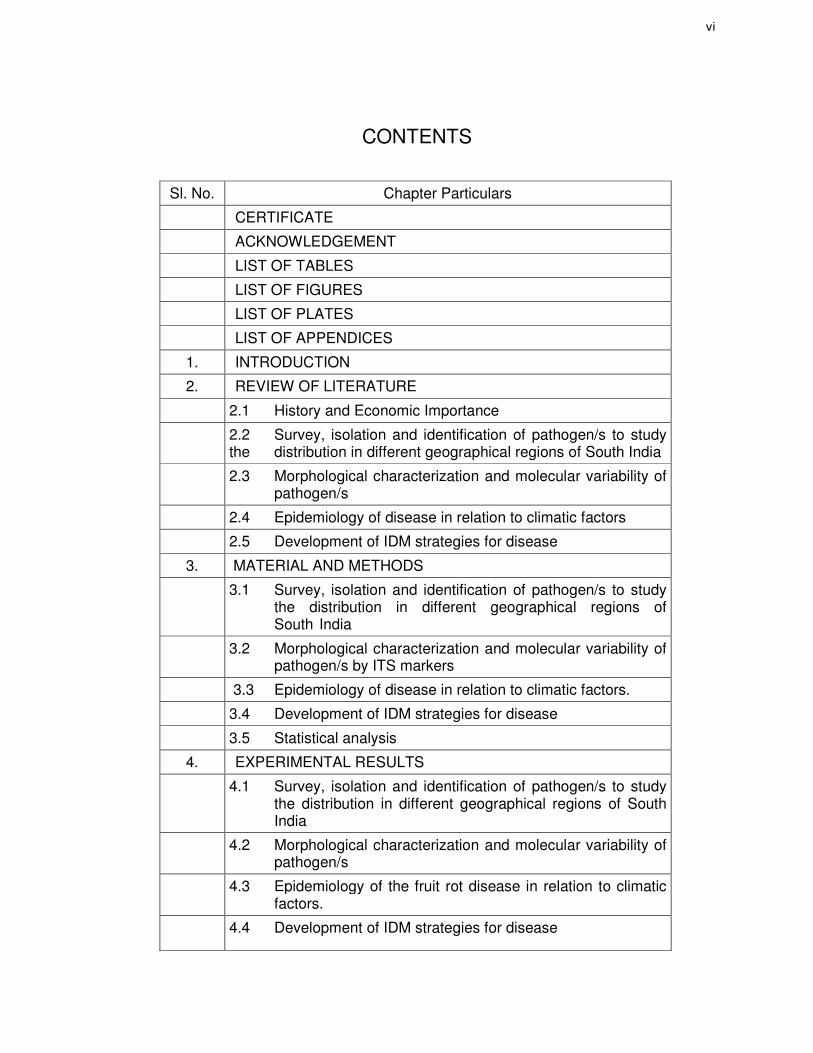
CONTENTS
Sl. No. Chapter Particulars
CERTIFICATE
ACKNOWLEDGEMENT
LIST OF TABLES
LIST OF FIGURES
LIST OF PLATES
LIST OF APPENDICES
1. INTRODUCTION
2. REVIEW OF LITERATURE
2.1 History and Economic Importance
2.2 Survey, isolation and identification of pathogen/s to study the distribution in different geographical regions of South India
2.3 Morphological characterization and molecular variability of pathogen/s
2.4 Epidemiology of disease in relation to climatic factors
2.5 Development of IDM strategies for disease
3. MATERIAL AND METHODS
3.1 Survey, isolation and identification of pathogen/s to study the distribution in different geographical regions of South India
3.2 Morphological characterization and molecular variability of pathogen/s by ITS markers
3.3 Epidemiology of disease in relation to climatic factors.
3.4 Development of IDM strategies for disease
3.5 Statistical analysis
4. EXPERIMENTAL RESULTS
4.1 Survey, isolation and identification of pathogen/s to study the distribution in different geographical regions of South India
4.2 Morphological characterization and molecular variability of pathogen/s
4.3 Epidemiology of the fruit rot disease in relation to climatic factors.
4.4 Development of IDM strategies for disease

vii
5. DISCUSSION
5.1 Survey, isolation and identification of pathogen/s to study the distribution in different geographical regions of South India
5.2 Morphological characterization and molecular variability of pathogen/s
5.3 Epidemiology of the fruit rot disease in relation to climatic factors.
5.4 Development of IDM strategies for disease.
6. SUMMARY AND CONCLUSIONS
REFERENCES
APPENDICES

viii
LIST OF TABLES
Table No.
Title
1a Detailed survey on chilli fruit rot and dieback disease during
2012-13 in South India.
1b Mean percent disease incidence and severity of fruit rot and
die-back disease of chilli during 2012-13 in different taluks
of Karnataka, Andhra Pradesh and Maharashtra
2a Detailed survey on chilli fruit rot and dieback disease during
2013-14 in South India
2b Mean percent disease incidence and severity of fruit rot and die-back disease of chilli during 2013-14 in different taluks of Karnataka, Andhra Pradesh.
3 District wise mean percent disease incidence and severity of fruit rot and die-back disease of chilli during 2012-13 and 2013-14 of Karnataka, Andhra Pradesh and Maharashtra
4a Mean fruit rot and die-back incidence and severity under rainfed and irrigated condition in the survey during 2012-13 and 2013-14
4b Mean fruit rot and die-back incidence and severity in red soil and black soil during the survey during 2012-13 and 2013-14
4c Mean fruit rot and die-back incidence and severity in sole crop and inter cropping system during the survey during 2012-13 and 2013-14
5 Prevalence of fruit rot and die-back disease in different chilli genotypes in the survey during 2012-13 and 2013-14
6 Predominance of pathogen/s observed in the survey during 2012-13 and 2013-14
7 Frequency of pathogen/s isolated from chilli fruit rot disease
8 Morphological and cultural diversity of different isolates of
C. capsici
9 Morphological and cultural diversity of different isolates of C. gloeosporioides
10 Morphological and cultural diversity of different isolates of C. acutatum
11 Morphological and cultural diversity of different isolates of Alternaria alternata

ix
12 Morphological and cultural diversity of different isolates of Fusarium oxysporum
13 Morphological and cultural diversity of different isolates Fusarium sporotrichioides
14 Comparison and identity of chilli fruit rot causing pathogens isolates with gene bank in NCBI BLAST program
15a Survival ability of pathogen/s in infected plant debris under three different conditions during 2012-13
15b Survival ability of pathogen/s in infected plant debris under three different conditions during 2013-14
16 Hosts reaction to Colletotrichum capsici inoculation
17 Reaction on different parts of chilli by cross inoculation of Colletotrichum capsici
18 Interaction effect of chilli fruit rot pathogens
19 Effect of environmental factors in relation to spore load of Colletotrichum spp. and disease progression during kharif 2012 at MARS, Dharwad
20 Effect of environmental factors in relation to spore load of Colletotrichum spp. and disease progression during kharif 2013 at MARS, Dharwad
21a Correlation coefficient (r) of spore load of Colletotrichum spp. with weather parameters during kharif 2012 and 2013
21b Correlation coefficient (r) of fruit rot incidence with spore load and weather parameters during kharif 2012 and 2013
22a Multiple regression analysis between weather parameters
on the spore load of Colletotrichum spp. during kharif
2012and 2013
22b Observed and predicted spore load of Colletotrichum spp. during kharif 2012and 2013
23a Multiple regression analysis between fruit rot incidence with spore load and weather parameters during kharif 2012 and 2013
23b Observed and predicted fruit rot incidence with spore load and weather parameters during kharif 2012 and 2013
24a Effect of date of planting on development chilli fruit rot and die-back disease during kharif 2012-13
24b Effect of date of planting on development chilli fruit rot and die-back disease during kharif 2013-14

x
25a Grouping of chilli genotypes based on resistance against fruit rot disease under field condition during kharif 2012-13
25b Grouping of chilli genotypes based on resistance against fruit rot disease under field condition during kharif 2013-14
25c Grouping of resistant and moderately resistant chilli
genotypes against fruit rot disease under field condition
during kharif 2012-13 and 2013-14.
26a Reaction of chilli genotypes against Colletotrichum spp. under in vitro condition
26b Reaction of chilli genotypes against A. alternata under in vitro condition
26c Reaction of chilli genotypes against Fusarium spp. under in vitro condition
26d Reaction of chilli genotypes against Colletortrichum spp. A. alternata, Fusarium spp. under in vitro condition
27 Chilli seed mycoflora under stereo binocular microscope
28 Effect of fungicide seed treatment on per cent seedling infection and vigour index in chilli
29 Effect of bio fungicide seed treatment on per cent seedling infection and vigour index in chilli
30 Chemical management of fruit rot and dieback disease during kharif 2012-13
31 Chemical management of fruit rot and dieback disease during kharif 2013-14
32 Pooled analysis of chemical management of fruit rot and dieback disease during 2012-13 and 2013-14
33a Management modules for chilli fruit rot disease during kharif 2012-13
33b Economics of disease management modules against chilli fruit rot and dieback disease during 2012-13.
34a Management modules for chilli fruit rot disease during kharif 2013-14
34b Economics of disease management modules against chilli fruit rot and dieback disease during kharif 2013-14.
35 Pooled analysis management modules for chilli fruit rot disease during kharif 2012-13 and 2013-14

xi
LIST OF FIGURES
Figure No.
Title
1 Chilli dieback disease rating scale
2 State wise mean percent disease incidence and severity of fruit rot and die-back disease of chilli during 2012-13 and 2013-14
3 District wise mean percent disease incidence and severity of fruit rot and die-back disease of chilli during 2012-13 and 2013-14 of Karnataka
4 District wise mean percent disease incidence and severity of fruit rot and die-back disease of chilli during 2012-13 and 2013-14 of Andhra Pradesh and Maharashtra
5 Fruit rot and die-back incidence and severity under rainfed and irrigated condition in the survey during 2012-13 and 2013-14
6 Fruit rot and die-back incidence and severity in red soil and black soil during the survey during 2012-13 and 2013-14
7 Fruit rot and die-back incidence and severity in sole crop and inter cropping system during the survey during 2012-13 and 2013-14
8 Prevalence of fruit rot and die-back disease in different chilli genotypes in the survey during 2012-13 and 2013-14
9 Frequency of pathogen/s isolated chilli fruit rot disease
10 PCR-RFLP diversity profile of C. capsici by HaeIII
11 PCR-RFLP diversity profile of C. gloeosporioides by Hae III
12 PCR-RFLP diversity profile of C.acutatum by Hae III
13 PCR-RFLP diversity profile of A. alternata by TaqI
14 PCR-RFLP diversity profile of Fusarium spp. Taq I
15 Observed and predicted spore load of Colletotrichum spp.
16 Observed and predicted fruit rot incidence with spore load and weather parameters
17 Frequency of chilli seed mycoflora
18 Effect of fungicide seed treatment on per cent seedling infection of chilli
19 Effect of bio fungicide seed treatment on per cent seedling infection of chilli
20 Chemical management of fruit rot and dieback disease.
21 Management modules for chilli fruit rot disease

xii
LIST OF PLATES
Plate No.
Title
1 Importance of chilli
2 Disease scale of chilli fruit rot (0-9)
3 Chilli fruit rot incidence in Karnataka, Andhra Pradesh and Maharashtra
4 Observations during survey
5 Symptoms caused by chilli fruit rot pathogens
6 Symptoms caused by Colletotrichum spp. on chilli fruits
7 Symptoms caused by A. alternata, Fusarium spp. on chilli fruits and mixed infection
8 Proving pathogenecity of Colletotrichum capsici, C. gloeosporioides, C. acutatum,
9 Cultural and morphological diversity of different isolates of Colletotrichum capsici, C. gloeosporioides, C. acutatum on PDA
10 Cultural and morphological diversity of different isolates of A. alternata, F. oxysporum, F. sporotrichioides
11 Fruiting body and spore morphology of Colletotrichum spp. (400X)
12 Spore morphology of A. alternata, F. oxysporum, F. sporotrichioides
13 Amplification of ITS-1 and ITS-4 region of representative Colletotrichum spp., Alternaria sp. and Fusarium spp.
14 Specific amplification of Colletotrichum capsici by C. cap primer
15 Specific amplification of Colletotrichum gloeosporioides at CgInt region
16 Specific amplification of Colletotrichum acutatum at CcInt region
17 Specific amplification of Alternaria alternata by AAF and AAR primer
18 Specific amplification of Fusarium spp. by Tef. Fu primer
19 PCR-RFLP diversity pattern of C. capsici by HaeIII
20 PCR-RFLP diversity pattern of C. gloeosporioides by Hae III
21 PCR-RFLP diversity pattern of C.acutatum by Hae III
22 PCR-RFLP diversity pattern of A. alternata by TaqI
23 PCR-RFLP diversity profile of Fusarium spp. Taq I
xiii
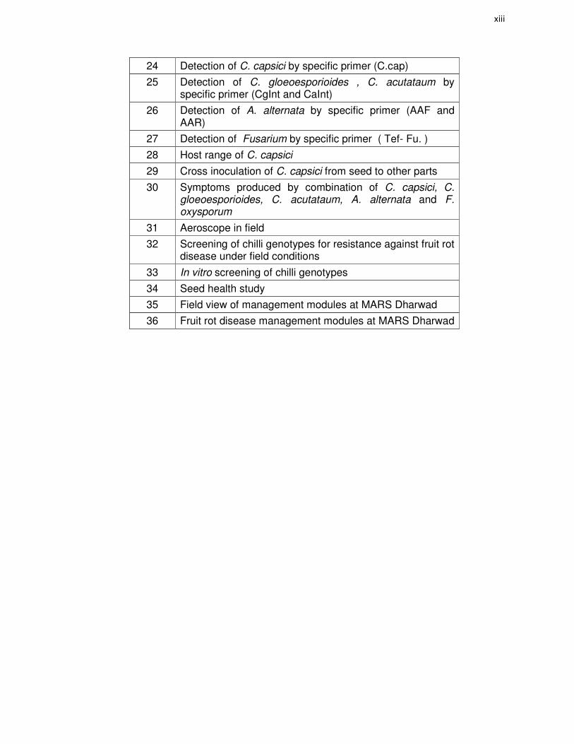
24 Detection of C. capsici by specific primer (C.cap)
25 Detection of C. gloeoesporioides , C. acutataum by specific primer (CgInt and CaInt)
26 Detection of A. alternata by specific primer (AAF and AAR)
27 Detection of Fusarium by specific primer ( Tef- Fu. )
28 Host range of C. capsici
29 Cross inoculation of C. capsici from seed to other parts
30 Symptoms produced by combination of C. capsici, C. gloeoesporioides, C. acutataum, A. alternata and F. oxysporum
31 Aeroscope in field
32 Screening of chilli genotypes for resistance against fruit rot disease under field conditions
33 In vitro screening of chilli genotypes
34 Seed health study
35 Field view of management modules at MARS Dharwad
36 Fruit rot disease management modules at MARS Dharwad

xiv
LIST OF APPENDICES
Appendix
No. Title
I Physical and chemical properties of the experimental site at
MARS Dharwad
II Price of fungicides biofungicides which used in during
investigation 2012-13 and 2013-14.

1. INTRODUCTION
Chilli (Capsicum annuum L.) despite of its fiery hotness, is one of the very
popular spice and vegetable crop grown worldwide. It is also known for its medicinal
and health benefiting properties. The fruit of Capsicum has a variety of names, such as
‘chilli’, ‘chilli pepper’ or ‘pepper’ depending on place and type of fruits. The chilli is
actually a fruit pod from the plant belonging to the family of solanaceae. Several
cultivars of chilli are grown all around the world. The chilli plant is native to Central
American region where it was used as the chief spice ingredients in Mexican cuisine
for centuries. It was introduced to the rest of the world by Spanish and Portuguese
explorers during 16th and 17th centuries and now grown widely in many parts of the
world as an important commercial crop. India is the largest producer of chilli, grown
over an area of 0.79 m. ha with an annual production of 0.13 m. tons with the
productivity of 1.6 m tons /ha (Anon., 2014).
Fruits are variable in size, shape, color, and pungency. The hotness of chilli is
measured in “Scoville heat units” (SHU). Chilli contains an impressive list of plant
derived chemical compounds that are known to have disease preventing and health
promoting properties. It contains health benefiting alkaloid capsaicin, which gives
strong spicy pungent character. It has anti-bacterial, anti-carcinogenic, analgesic and
anti-diabetic properties. Chilli is also good in other antioxidants like vitamin A,
flavonoids like ß-carotene, α-carotene, lutein, zea-xanthin, and cryptoxanthin. It is also
good in vitamin C and B-complex group of vitamins such as niacin, pyridoxine (vitamin
B-6), riboflavin and thiamin (vitamin B-1). Chilli is also source of minerals like
potassium, manganese, iron, and magnesium (Bosland and Votava, 2003) (Plate 1).
Chilli is suffering from several economically important diseases like damping off,
die back, fruit rot, leaf spots, leaf curl, wilt etc. which are posing a serious threat to the
successful large-scale cultivation. The fruit rot disease caused by fungi Colletotrichum
spp. (C. capsici, C. gloeosporioides and C. acutatum), also Alternaria alternata and
Fusarium spp. is major yield limiting factor. It has been observed to occur in three
phases viz. (i) seedling blight or damping off stage, prevalent in the nursery, (ii) die
back stage which is initiated at different stages of growth and (iii) fruit rot stage in which

2
3
the ripe fruits are infected. The last phase causes extensive damage to the fruits since
the lesions on the fruits considerably reduce the market value of the produce. The
infected seeds, plant debris and fruits act as primary source of inoculum (Siddique et
al., 1977).
The disease is more severe in India because of its complex nature. Symptoms
vary in different stages of crop. In the present situation of climate change, there is a
need to investigate the disease in depth, as epidemics vary in different regions giving
scope for understanding the extent of variability in pathogen population. Hence,
collection of isolates from different regions and their characterization by morphological
studies is quite relevant. Further, their variation at the molecular level using ITS rDNA,
a more reliable technique can give a logical conclusion regarding the genetic variability
existing among isolates. Hence, it is proposed to sequence ITS rDNA region.
Epidemiological study helps to know about survival ability of pathogen/s and
favorable environmental conditions for disease spread. It also helps in forecasting
system to delink the infection chain at appropriate time in order to manage the disease
effectively. It is essential to manage the disease in an integrated manner in which
fungicides, botanicals and bio-agents, resistant genotype/s, optimum date of planting
play an integral part and becoming more relevant in the present day disease
management scenario. Therefore evaluations of bio-intensive, adoptive and chemical
modules are of utmost concern to identify best module for management of disease with
maximum cost benefit ratio which will help the farming community to a greater extent.
The present studies were therefore directed to throw some light on different
aspects of the disease and pathogens which have a bearing on the facts discussed in
the preceding paragraphs. Hence, the investigation was taken up to unravel the
complexes involved in the fruit rot complex disease of chilli and issues are addressed
through following objectives.

4
Objectives of investigation
• Survey, isolation and identification of pathogen/s to study the distribution in
different geographical regions of South India.
• Morphological characterization and molecular variability of major pathogen/s.
• Epidemiology of disease in relation to climatic factors.
• Development of IDM strategies for disease.

2. REVIEW OF LITERATURE
Fruit rot complex disease of chilli is caused by Colletotrichum capsici,
C. gleosporioides, C. acutatum, Alternaria alternata, Fusarium oxysporum and
F. sporotrichoides which are carried along with seed to cause deterioration of seed in
storage, pre and post emergence damping off and later dieback leading to heavy loss.
Large numbers of reports are available in literature regarding this disease which has
been reviewed in the chapter. The review pertains to survey, isolation, and
identification of pathogens, epidemiology, morphological, molecular variability, seed
health management and integrated management of chilli fruit rot.
2.1 History and Economic Importance
2.1.1 Fruit rot
Chilli fruit rot disease is one of the most economically important diseases was
reported for the first time in India by Sydow from Coimbatore of Madras presidency in
1913. Colletotrichum species including C. acutatum (Simmonds), C. capsici (Syd.)
Butler and Bisby, C. gloeosporioides (Penz.) Penz. and Sacc. and C. coccodes
(Wallr.) S. Hughes (Simmonds, 1965; Johnston and Jones, 1997; Kim et al., 1999;
Nirenberg et al., 2002; Voorrips et al., 2004; Sharma et al., 2005; Pakdeevaraporn et
al,. 2005; Than et al., 2008b) reduced marketable yield from 10% to 80% of the crop
production m(Poonpolgul and Kumphai, 2007). Fruit rot is mainly a problem on mature
fruits, causing severe losses due to both pre and post harvest fruit decay (Hadden and
Black, 1989; Bosland and Votava, 2003).
Many post-harvest diseases of fruits exhibit the phenomenon of quiescence in
which symptoms do not develop until the fruit ripens. Fruit rot causes extensive pre
and post harvest damage to chilli fruits causing anthracnose lesions. Even small
anthracnose lesions on chilli fruits reduce their marketable value (Manandhar et al.,
1995).
Colletotrichum species are the most important pathogens that cause latent
infection (Jeffries et al., 1990). Appressoria are known to form adhesive disks that
adhere to plant surfaces and remain latent until physiological changes occur in fruits
6
(Bailey and Jeger, 1992). Appressoria that formed on immature fruits may
remain quiescent until ontogenic changes occur in the fruits (Prusky and Plumbley,
1992).
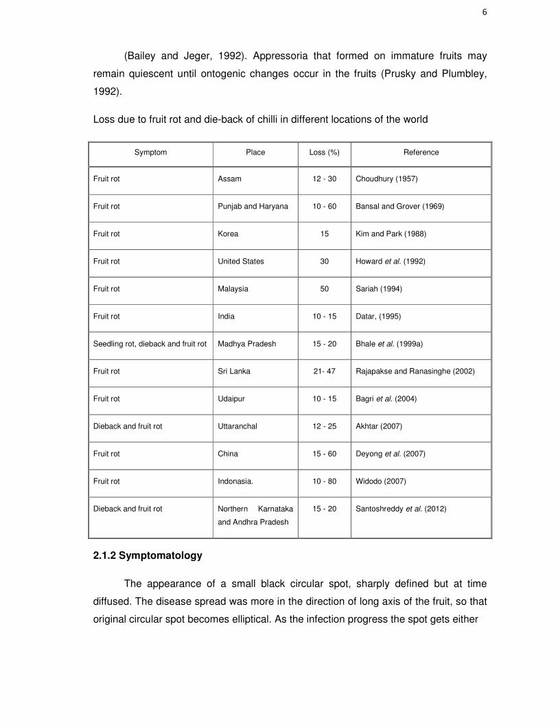
Loss due to fruit rot and die-back of chilli in different locations of the world
Symptom Place Loss (%) Reference
Fruit rot Assam 12 - 30 Choudhury (1957)
Fruit rot Punjab and Haryana 10 - 60 Bansal and Grover (1969)
Fruit rot Korea 15 Kim and Park (1988)
Fruit rot United States 30 Howard et al. (1992)
Fruit rot Malaysia 50 Sariah (1994)
Fruit rot India 10 - 15 Datar, (1995)
Seedling rot, dieback and fruit rot Madhya Pradesh 15 - 20 Bhale et al. (1999a)
Fruit rot Sri Lanka 21- 47 Rajapakse and Ranasinghe (2002)
Fruit rot Udaipur 10 - 15 Bagri et al. (2004)
Dieback and fruit rot Uttaranchal 12 - 25 Akhtar (2007)
Fruit rot China 15 - 60 Deyong et al. (2007)
Fruit rot Indonasia. 10 - 80 Widodo (2007)
Dieback and fruit rot Northern Karnataka
and Andhra Pradesh
15 - 20 Santoshreddy et al. (2012)
2.1.2 Symptomatology
The appearance of a small black circular spot, sharply defined but at time
diffused. The disease spread was more in the direction of long axis of the fruit, so that
original circular spot becomes elliptical. As the infection progress the spot gets either

7
diffused black, greenish black or dirty gray in colour, markedly delimited by a thick
sharp black outline enclosing a dark lighten black or straw coloured area. Two or more
spots coalesced to form bigger spots. Diseased fruits lost their normal red color and
turned straw colored or in some cases white. When diseased fruit was cut open the
lower surface of the skin was found to be covered with minute, black, spherical
elevations. In advanced cases, seeds were covered by a mat of mycelium
(Choudhury, 1957).
Siddique et al. (1977) and Isaac, (1992) observed the disease symptoms in
three phases viz., i) seedling blight or damping off, prevalent in the nursery, ii) leaf
spotting and dieback which is initiated at different stages of the growth. Die back
infection starts from the growing point of secondary branches gradually advance
downwards and invades the entire branch and iii) fruit spotting and rotting in which
mostly the ripened fruits were infected.
Symptoms of Alternaria rot on chilli begin as water soaked, gray lesions on fruit
further they become darken and become covered with spores, internal necrosis and
mycelial growth occured on the seeds, placenta and pericarp ( Halfon et al., 1983 ;
Wall and Biles, 1993).
Symptoms appear mostly on the ripened fruits but the highest infection occurs
on stored fruits. The dry rot symptom comprising of brown circular to irregular lesions
which were increased and coalesced damaging the fruit partly or completely however
soft rot showing water soaked lesions appeared on the fruits and then turned to
brownish, soft rot set in and the fruit was completely rotted (Datar, 1995).
Khodke and Gahukar (1995) described the disease symptoms of fruit rot of
chilli (Colletotrichum gloeosporioides) as depressed sunken, discoloured, circular to
irregular spots of varying sizes. Oh et al. (1998) observed that initial anthracnose
symptoms were detected on some green fruits at two days after inoculation resulting
in typical sunken necrosis within five days after inoculation.
Lesions of Alternaria alternata on chilli fruit were darker in color and covered by
moldy growth of fungus with heavy sporulation (Shivakumara, 2006).

8
Typical fruit symptoms are circular or angular, depressed sunken lesions, with
concentric rings of acervuli that are often wet and produce pink to orange conidial
masses. Under severe disease pressure, lesions may coalesce. Conidial masses may
also occur scattered or in concentric rings on the lesions (Shivakumara, 2006; Than et
al., 2008a; Akhtar, et al., 2009).
2.1.3 Causal agent
Generally, anthracnose symptom of fruit rot disease is caused by
Colletotrichum species which belongs to the Kingdom Fungi; Phylum Ascomycota,
Class Sordariomycetes; Order Phyllachorales; and Family Phyllachoraceae. The
anamorphs are Glomerella species. Anthracnose of chilli was first reported from New
Jersey, USA, by Halsted (1890) in 1890 who described the causal agents as
Gloeopsorium piperatum and Colletotrichum nigrum. These taxa were then considered
as synonyms of C. gloeosporioides by Von Arx (1957).
The fungus, C. capsici was reported for the first time in India by Sydow on chilli
from Coimbatore of Madras presidency in 1913. Since then it has been reported and
described from several parts of the world (Butler, 1918; Dastur, 1921; Seaver et al.
1932; Marchionatto, 1935; Ling and Lin, 1944; Bansal and Grover, 1969; Thind and
Jhooty, 1985; Mridha and Siddique, 1989; Hegde and Kulkarni, 2001a; Meenugupta
and Garg, 2002). Von Arx (1957) noted C. capsici as a synonym of C. dematium
(Pres. Ex. Fr.) Groove.
Oh et al. (1998) reported that C. gloeosporioides (Glomerella cingulata) is a
common pathogenic fungus in many plants. When the isolate of Glomerella cingulata
was inoculated on both green and red fruits, conidial germination, appressoria and
infection hyphae were observed on both fruits within 24 hours after inoculation.
Than et al. (2008b) revealed that in the Colletotrichum patho-system, different
species can be associated with anthracnose of chilli have been reported from different
countries and regions of world. Although these species have been the subject of
numerous investigations, there remain many gaps in the knowledge of the disease
process and understanding of the complex relationships between the species
involved.
9
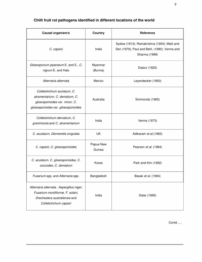
Chilli fruit rot pathogens identified in different locations of the world
Causal organism/s Country Reference
C. capsici India
Sydow (1913); Ramakrishna (1954); Maiti and
Sen (1979); Paul and Behl, (1990); Verma and
Sharma (1999)
Gloeosporium piperatum E. and E., C.
nigrum E. and Hals
Myanmar
(Burma) Dastur (1920)
Alternaria alternata Mexico Leyendecker (1950)
Colletotrichum acutatum, C.
atramentarium, C. dematium, C.
gloeosporioides var. minor, C.
gloeosporioides var. gloeosporioides
Australia Simmonds (1965)
Colletotrichum dematium, C.
graminicola and C. atramentarium India Verma (1973)
C. acutatum, Glomerella cingulata UK Adikaram et al.(1983)
C. capsici, C. gloeosporioides Papua New
Guinea Pearson et al. (1984)
C. acutatum, C. gloeosporioides, C.
coccodes, C. dematium Korea Park and Kim (1992)
Fusarium spp. and Alternaria spp. Bangladesh Basak et al. (1994)
Alternaria alternata , Aspergillus niger,
Fusarium moniliforme, F. solani,
Drecheslera australiensis and
Colletotrichum capsici
India Datar (1995)
Contd…..

10
Causal organism/s State Reference
C. acutatum, C. capsici, C.
Gloeosporioides Taiwan Manandhar et al. (1995)
C. coccodes New Zealand Johnston and Jones (1997)
C. capsici C. gloeosporioides Sri Lanka Rajapakse (1998)
C. acutatum USA Roberts et al. (2001)
C. acutatum, C. capsici, C.
gloeosporioides Indonesia Voorrips et al.(2004)
Colletotrichum dematium, Glomerella
cingulata, C. gloeosproides,
C.coccodes, C. acutatum
Korea Byung-Soo (2007)
C. acutatum, C. capsici, C.
gloeosporioides, C. Nigrum Vietnam Don et al. (2007)
C.capsici C. gloeosporioides C.
acutatum India
Ramachandran et al. (2007) ; Lydia and
Zachariah ( 2012),
Colletotrichum acutatum, C.boninense
C. gloeosporioides and C.capsici
Taiwan.
Sheu et al. (2007)
C. capsici, C. acutatum, C.
gleosporioides Thailand Taylor et al. (2007)
C. acutatum, C. capsici, C.
gloeosporioides Thailand Than et al. (2008b)
C.capsici C. gloeosporioides C.
acutatum Malaysia Yun et al. (2009)
From Indian regions
C. capsici Coimbatore Sydow (1913)
Colletotrichum, Choanephora,
Alternaria, Phomopsis, Pythium ,
Rhizopus, Fusarium spp.
Punjab Thind and Jhooty (1985)
Alternaria alternata, Curvularia lunata,
C. pallescens Andhra Pradesh Prabhavathy and Reddy (1995)
C. capsici, Alternaria and Fusarium spp. Karnataka Mesta (1996)

11
Causal organism/s State Reference
Alternaria alternata Madhya
Pradesh Bhale et al. (1999a)
C. capsici Karnataka Hegde and Kulkarni (2001a); Ekbote (2002a);
Rajput (2011);
C.capsici C. gloeosporioides, Alternaria
alternate, Rhizopus, Fusarium spp.,
Aspergillus, and Curvularia
Karnataka Shivakumara (2006)
C. dematium, C. gloeosporioides, C.
graminicola and C. atramentarium
Arunachal
Pradesh Selvakumar (2007)
C. capsici, C. acutatum, C.
gleosporioides Karnataka Chowdappa (2010)
C. capsici, C. acutatum, C.
gleosporioides and Alternaria alternata Tamil Nadu Madhvan et al. (2010)
C. gleosporioides Nagaland Ngullie et al. (2010)
Alternaria alternata Udaipur Bagri et al. (2011)
Fusarium oxysporum Udaipur Bagri et al. (2012)
C. capsici, C. acutatum, C.
gleosporioides, Alternaria alternata and
Fusarium spp.
Karnataka and
Andhra Pradesh Santoshreddy et al. (2012)
C.capsici , Fusarium moniliforme. F.
pallidoroseum, F. oxysporum, Alternaria
alternata and Aspergillus flavus
Jammu and
Kashmir Parey et al. (2013)

12
2.2 Survey, isolation and identification of pathogen/s to study the
distribution in different geographical regions of South India
2.2.1 Survey
Thind and Jhooty (1985) reported that Colletotrichum capsici as a predominant
fungus in causing fruit rot of chilli. The incidence varied between 66-84%. Mathew et
al. (1995) made a survey during 1989-91 in Vellanikkara, Trichur, Kerala and reported
that dieback and fruit rot were serious problems during rainy season.
Ekbote (2002a) conducted a survey during 1998-99 and 1999-2000 kharif in
Haveri district. Among five taluks surveyed, the fruit rot of chilli incidence was more in
Savanur taluka (42%) followed by Siggaon taluka (41%).
Shivakumara (2006) conducted a roving survey in North Eastern districts of
Karnataka viz., Raichur, Gulbarga and Bellary during 2005 and 2006 which revealed
that mean per cent disease incidence ranged from 13.96 to 20.55. Maximum per cent
disease index (11.56 PDI) was recorded in Gulbarga district followed by Raichur
(11.17 PDI) district.
Akhtar et al. (2008) reported that chilli growing areas in Tarai belt of Uttaranchal
(Sitarganj, Pantnagar and Bilaspur) showed anthracnose symptoms, particularly
dieback and fruit rot which caused heavy loss in transit and storage.
Rajput (2011) conducted roving survey in Dharwad, Gadag and Haveri districts
of northern Karnataka and revealed that disease severity ranged from 24.13 to 46.35
per cent. The highest severity of fruit rot was noticed in Nalvadi and Sanshi villages of
Dharwad district.
2.2.2 Isolation and identification of pathogen/s
Ramakrishna (1954) reported the pathogenicity of two isolates of C. capsici
obtained from chilli fruits. Kenchaiah (1975) proved the pathogenicity of the two
isolates using both ripe and unripe fruits of C. annum and C. fruitescens and were
pathogenic to their respective hosts and also cross inoculable. Singh et al. (1977)

13
isolated C. capsici from a severely infected chilli fruit and confirmed its pathogenicity
experimentally.
Thind and Jhooty (1985) reported that, the chilli fruits were affected by
Colletotrichum, Choanephora, Alternaria, Phomopsis, Pythium, Rhizopus, Fusarium
spp. of fungi. Raut et al. (1990) isolated eleven fungi from apparently healthy, green,
semiripe and ripe capsicum fruits. Alternaria tenuis, Cladosporium oxysporum,
Colletotrichum dematium, Curvularia lunata, Drechslera tetramera, Exserohilum
rostratum and Machrophomina phaseolina were the major ones. The sclerotial stage
of Macrophomina phaseolina was isolated from the pedicel, pericarp, placenta and
seeds of infected fruits. Pathogenicity was confirmed in green, semiripe and ripe chilli
fruits. Datar (1995) reported that six fungi, viz., Alternaria alternata , Aspergillus niger,
Fusarium moniliforme, F. solani, Drecheslera australiensis and Colletotrichum capsici
were associated with chilli fruit rot.
Major fruit rot causing pathogens on capsicum fruits Alternaria capsici-annui
and A. tenuis, Cercospora capsici, Colletotrichum capsici, C. gloeosporioides,
Fusarium spp. and Periconia byssoides from farmer’s fields of Bangladesh. Among
them Fusarium rot caused greater reduction in dry weight of fruits and it was
maximum at the ripened stage (Basak et al., 1994).
Prabhavathy and Reddy (1995) isolated fungi causing black rot disease on chilli
fruits viz., Alternaria alternata, Curvularia lunatus, C. pallescens, Pythyium butleri,
Botryodiplodia theobromae, Phomopsis equiseti, Rhizopus stolonifer, Fusarium
semitectum and Choanephora cucurbitarum for the first time in Andhra Pradesh.
Incidence and severity of Alternaria alternata on chilli fruits causing fruit rot is also a
problem, its pathogenicity was confirmed on chilli fruit (Khodke and Gahukar, 1993).
Phoma sorghina was reported from Capsicum annuum (Khare et al., 1995).
Khodke and Gahukar (1995) isolated C. gloeosporioides (Glomerella cingulata)
from ripe chilli fruits (Capsicum annuum) in pure culture and its pathogenicity was
confirmed. This was considered to be the first record of this species occurring on chilli
in Maharashtra, India. Amusa and Alabi (1996) isolated C. gloeosporioides from
infected pepper.

14
Paul and Behl (1990) reported that, chilli suffers considerable losses due to fruit
rot/dieback/anthracnose caused by C. capsici in tropical and subtropical areas.
Khaleeque and Khan (1991) proved pathogenicity of C. capsici in chilli fruits. Thind
and Jhooty (1990) conducted the pathogenecity test on chilli by detached fruit method
and proved pathogenecity of C. capsici.
Bhale et al. (1999a) revealed that, A. alternata was found responsible for
severe seed rot, seedling decay, tender twig tip drying and fruit rot. Wall and Biles
(1993) reported that, fruit rot of New Mexican type of chilli is caused by A. alternata.
Verma and Sharma (1999) reported that, fruit rot of chilli caused by C. capsici
was an important disease in field, transit, transport and storage.
Shivakumara (2006) found maximum frequency (25.0%) of Colletotrichum
followed by Alternaria (21.0%) and others, viz., Aspergillus, Fusarium, Curvularia,
Rhizopus and mixed infections in chilli fruit rot samples collected from northern
Karnataka.
Suthinraj and John (2009) isolated pathogen from infected chilli fruits collected
from the Chidambaram. The fungus was purified and identified as C. capsici.
Byung-Soo, (2007) reported that C. dematium, Glomerella cingulata,
C. gloeosproides, C.coccodes, C. acutatum causes fruit rot. Among them
C. gloeosprioides and C. acutatum were dominant in Korea.
Singh et al. (2007) collected plant part showing anthracnose symptoms,
particularly dieback and fruit rot which were subjected to microscopic examination
which showed C. capsici spores. Further purified cultures of C. capsici used for in vitro
pathogenicity on detached fruits of chilli cultivar Kandhari which revealed that sunken
anthracnose symptoms on fruits.
Ramachandran et al. (2007) conducted survey and collected 92 isolates from
different chilli growing areas of India. Among them 53 were identified as C.capsici 38
as C. gloeosporioides and one was found to be C. acutatum.

15
Than et al. (2008b) reported that several species of Colletotrichum viz.,
C. capsici (Butler Bisby), C. gloeosporioides (Penz.), C. acutatum (Simmonds),
C. atramentarium (Berk and Broome), C. dematium (Pers.) and C. coccodes (Wallr.),
Glomerella cingulata (Stoneman) along with A. alternata (Keissler) were causal agents
of chilli fruit rot worldwide .
Roat et al. (2009) collected the infected chilli fruits and isolated the C.capsici.
Fresh chilli fruit samples were surface sterilized and anthracnose symptoms were
observed after seven days of incubation.
The isolates were used in the inoculation of chilli seedlings and fruits by the
detached leaf assay procedure. (i) Conidial suspension (1 x 10⁶conidia per ml) of
twelve day old PDA grown cultures was sprayed on one-month-old chilli plants. (ii)
The inoculated plants were covered with plastic bags for two days to maintain
humidity. (iii) The plants were assayed for disease seven days after inoculation and
continued to be so for up to 20 days; (iv) The presence of the pathogen was further
confirmed by incubating the leaves in moist chambers for 5-7 days at 22 ± 1ºC and
observed for the development of fungal growth.( Chandra Nayak et al., 2009)
Madhvan et al. (2010) reported that out of the 16 chilli fruit samples collected
from Tamil Nadu, C. capsici was the most commonly isolated (69%) followed by C.
gloeosporioides (19%). Alternaria alternata was isolated from Poothapadi of Salem
district and Gibberella sp. Othakadai of Madurai district.
2.3 Morphological characterization and molecular variability of
pathogen/s
2.3.1 Morphological characterization
Hegde, (1998) reported that colony of C. capsici on potato dextrose agar was
dark grey to blackish with a smooth margin. Whitish aerial mycelial growth and
concentric rings of growth were visible along with light rose colored spore masses.
Conidia were single celled curved with smooth margin and hyaline with central oil
globule.
16
Among the five C. capsici isolates, isolate II had the largest conidia (22.9 x 394
µm), with an average of 53.8 setae on each acervuli and 4.2-6.4 septa and showed
highest growth rate on PDA and PDB at pH 6.5. Thermal death point of its conidia was
47o C and isolate was most virulent, as determined by fruit rot incidence and infectivity
(Jeyalakshmi and Seetharaman, 1999 ; Rohana et al., 2005).
Wharton and Uribeonodo (2004) reported that C. acutatum conidia were single
septate and fusiform.
Shivakumara (2006) revealed that morphologically C. gloeosporioides was
distinct from C. capsici showing fast, fluffy, dull white mycelium and within the isolates
also found distinct characteristics with respect to growth, colour of mycelia and
sporulation. A. alternata isolates showed ash colour to dark coloured mycelium with
medium to fast growth and moderate to excellent sporulation.
Venkataravanappa and Nargund (2007) revealed that conidia size of six
isolates of C. gloeosporioides varied between 10.9-20.6 µm in length and 4.39 – 6.65
µm in width.
Five isolates of C. capsici from Uttaranchal showed variation in colony color
(white grey to blackish grey), growth (smooth to wavy margin) and sickle shaped
conidia with size varied from 25.27 to 25.15 µm length, 3.17 to 3.66 µm width (Akhtar
and Singh 2007).
Than et al. (2008a) reported that mycelium of C. gloeosporioides varied from
light orange colony colour with delicate and thin to pale grey to black zonated colonies
with abundant orange conidial masses near the centre. Conidia were cylindrical in
shape and measured about 13.5 X 4.5 µm. C. acutatum white to olive grey colour
colony with very thick cottony mycelium to orange-coloured colony, conidia were
fusiform in shape and measured about 13 – 14 X 3.5 µm.
Zivkovic et al. (2010) reported that conidia of C. acutatum were elliptic-fusiform
in shape and conidia size varied between 8-16 x 2.5 – 4 µm.
Sangdee et al. (2011) recorded variation of six isolates of C. capsici in Thailand
based on colony color, growth, sporulation, spore size and shape.

17
Four isolates of Alterneria alternata variation was recorded based on colony
color, growth, sporulation and spore size (Amit Kumar et al., 2012).
Biju et al. (2012) revealed that C. gloeosporioides obtained from different
location were analyzed for diversity employing macro and micro morphological
features. The isolates were further grouped based on colour of the colony into five
groups Viz., grey, white, grayish white, grayish olive and pale pink. Further, they also
reported that sporogenic microsclerotia produced by C. gloeosporioides act as a
potential source of inoculum for anthracnose of black pepper.
Chandramani (2012) reported that cultures of C. gloeosporioides on PDA
produced white to grayish of dark orange on upper surface, while the lower side of
colonies were of white, yellowish orange to black. Conidia were straight, one celled,
hyaline, oblong, or cylindrical, slightly curved with translate base and rounded apex
and measured 11.57 – 15.50 X 3.38 – 7.52 µm.
Christopher et al. (2013) collected twenty isolates of C. capsici from different
growing regions of Tamil Nadu and characterized morphologically based on colony
color, growth, sporulation and spore size.
Masoodi et al. (2013) revealed that twenty isolates of C. capsici from different
growing regions of Kashmir valley which showed variation in culture color from white
to grey, growth between 32.0-67.5mm with cottony to fluffy and regular to irregular
margin, conidial size varied between 2.23 – 33.6 µm.
2.3.2 Molecular variability of pathogen/s
Morphology and pathogenecity are not enough to distinguish between
Colletotrichum spp. diversity. Studies include internal transcribed spacer (ITS) regions
(Freeman et al., 2001; Moriwaki et al., 2002; Sanders and Korsten, 2003; Lee et al.,
2007) as well as restriction fragment length polymorphism (RFLP) (Balardin et al.,
1999; Martin and Garcia-Figueres, 1999; Saha et al., 2002 ; Weir et al., 2012; Patil
and Nargund 2013)
Shivakumara (2006) studied strainal variations among the species of C. capsici
and C. gloeosporioides by RAPD profile.

18
ITS-RFLP technique is an efficient method for rapid diagnosis of Colletotrichum
species from pepper. A total of 412 Taiwan isolates collected from pepper production
areas were analyzed through ITS-RFLP fingerprinting. Among them, 245 C. acutatum,
34 C. boninense, 52 C. capsici and 69 C. gleosporioides were identified. Other
Colletotrichum isolates (3%) were not distinguishable, which inferred to the various
inter- and intra-species variations in Colletotrichum members (Sheu et al., 2007).
Patil and Nargund (2013) collected 68 fungal pathogen isolates causing onion
twister disease from onion growing regions of Karnataka and identified by PCR-based
molecular method using specific primers as 19 C. gloeosporioides, 24 C. acutatum
and 25 F. oxysporum isolates. Further these isolates were analyzed through RFLP
fingerprinting by digestion with HaeIII which resulted in a characteristic pattern of three
fragments and showed variability between the species as four clusters of C.
gloeosporioides, six clusters of C. acutatum and five clusters of F. oxysporum.
Fungal isolates from chilli (Capsicum spp.) fruits in Thailand that showed typical
anthracnose symptoms were identified as Colletotrichum acutatum, C. capsici and C.
gloeosporioides. Phylogenetic analyses from DNA sequence data of ITS rDNA and β-
tubulin (tub2) gene regions revealed three major clusters representing these three
species. (Than et al., 2008a)
Chowdappa and Chethana (2012) reported that 28 isolates of C.
gloeosporioides from Sikkim causing anthracnose of orchids species specific PCR
using primer CgINT and ITS4 amplified at 450bp.
Imjit et al. (2013) reported that PCR- based molecular method provided
improved early detection and diagnosis system of fruit rot disease of chilli.
Pongpisutta et al. (2013) reported that effects of environmental manipulation on
the morphological stability makes identification between Colletotrichum species
difficult. ITS RFLP and ITS sequence analysis are considered to be essential tools to
solve the problems of species differentiation and specific identification of the chilli
anthracnose causal agents.

19
Sharma and Shenoy (2013) reported that 52 isolates of C. gloeosporioides of
chilli fruit rot in southern India which showed affinity with C. siamense and
C. fructicola within C. gloeosporioides species complex based on ITS / 5.8S rRNA and
glyceraldehyde-3-phosphate dehydrogenase (gapdh) genes.
Santoshreddy et al. (2013) detected C. capsici C. gloeosporioides, C.
acutatum, A. alternata and Fusarium spp. from seeds, fruits and die-back infected
twigs of chilli plants by PCR-based molecular method using specific primers.
2.4 Epidemiology of disease in relation to climatic factors.
2.4.1 Survival ability of pathogens
C. capsici causing fruit rot of chilli survived in the field in plant debris atleast for
six months (Choudhary, 1957; Rai and Chohan, 1966). C. piperratum and C.capsici
were found to be capable of surviving in both internal and external tissues of dry
pepper seeds for nine months (Smith and Crosson, 1958).
Ahmed (1982) found that, Colletotrichum capsici causing fruit rot of chilli
survived up to eight months in both seed and culture.
Manandhar et al. (1995) reported that, Colletotrichum species were generally
able to survive in or on seeds and one of the ways that anthracnose was introduced to
the chilli field is through infected transplants.
Sanathkumar, (1999) observed that, the C.capsici could survive upto 225 days
on infected seeds stored under room conditions where as on pedicel and fruit rind it
survived for 195 days.
Kulkarni and Benagi, (2012) reported that, Colletotrichum truncatum survivived
360 days in greengram debris under 40 - 50C, 90 days under field condition and in
seed up to 12 months.
2.4.2 Host range
Sundararaman, (1927) found that C. capsici infected soybean pods, tomato
fruits, brinjal fruits by artificial inoculation. Pring et al. (1995) revealed that C. capsici

20
can overwinter on alternative hosts such as solanaceous vegetables and legume
crops, plant debris and rotten fruits in the field. Hegde, (1998) revealed that C. capsici
which causes fruit rot of chilli also infected tomato, potato, and brinjal.
Pandey, (2006) reported that C. capsici from chilli caused fruit rot, seed and
seedling mortality in tomato based on pathogenecity test by pin prick method and also
by molecular characterization with species specific primer (CcINT) which amplified at
450 bp from both chilli and tomato.
Mehetre and Joshi et al. (2010) revealed that Colletotrichum capsici, isolated
from Yam anthracnose could infect chilli, turmeric and mango.
2.4.3 Aerobiology
Thakur and Khare (1991) reported that, maximum trapping of spores
(C. lindemuthianum and C. dematium) in greengram was recorded when there was
moderate temperature between 26 to 29˚C, relative humidity between 91 to 96 per
cent, rainfall from 0 to 21.6 mm and wind velocity from 6 to 10 km per ha. Highest
spore trap coincided with these conditions prevalent on July 30.
The maximum growth and sporulation of C. capsici observed at 25˚-30˚C
(Ekbote, 1994; Angadi, 1999).
Roberts et al. (2001) reported that fruit rot of chilli infection occurs during warm,
wet weather. Temperatures around 27°C and high humidity (80%) were optimum.
The aerobiological studies on effect of weather factors on the development of
spore load of C.truncatum indicated that more conidial counts were observed during
last week of July and first week of August, which coincided with the critical stages of
infection in green gram (Kulkarni, 2009).
Patil et al. (2013a) revealed that cumulative rainfall contributed maximum (R2 =
0.97) to development of onion twister disease severity caused by C. gleosporioides,
C. acutatum and F. oxysporum.

21
2.5 Development of IDM strategies for disease
2.5.1 Evaluation of genotypes
Mesta (1996) studied the reaction of 217 chilli genotypes to Colletotrichum
capsici in Dharwad and the results revealed that none of the genotypes were found to
be immune to fruit rot either in field or in laboratory. Out of 217 chilli genotypes 11
genotypes were found to be resistant and 24 were moderately resistant in both field
and laboratory conditions. Further he observed that, among different seed treating
chemicals captan and thiram at 0.3 per cent were effective in controlling seedling
infection.
Basak (1997) screened ten chilli cultivars against three major fruit rot fungi,
Colletotrichum capsici, C. gloeosporioides and Fusarium semitectum. None of the
cultivars were found to be immune however, except few remaining were rated as
moderately resistant. Cultivars C-011 and C-045 were susceptible to C.
gloeosporioides, C. capsici, C-123 to C. capsici and Chittagong local and Bogra local
were susceptible to F. semitectum and highly susceptible to Colletotrichum spp.
Of fifty chilli cultivars/lines tested under artificial inoculation, five (Pusa Sada
Bahar, 91-2, DC-9, DC-27 and Achar) were free from infection whereas six (86-5,
Aparna, Kalyanpur Red, Sabour Anil, BG-1, Lorai and Perennial) were resistant and
four (Pant C-1, DC-18, Suryamani and PS-1) were moderately resistant to C. capsici
(Singh et al., 1997).
Bhale et al. (1999b) reported that pinprick method was found to be most
effective method for the pathogenicity of Colletotrichum dematium, on semiripe and
fully ripe detached fruits in five and six days after inoculation respectively
Hegde and Anahosur (2001b) evaluated fifty two chilli genotypes against fruit
rot fungus, C. capsici under natural condition. LCA-301, LCA-324, K-1 and Byadagi
Kaddi were found resistant. Whereas, KDSC-210-10 and S-32 were found highly
susceptible. Resistant genotypes contained higher capsaicin, ascorbic acid and lower
amount of total sugars than susceptible ones.
Fifty one chilli cultivars were screened for field resistance to fruit rot fungus
C. capsici. Of the cultivars tested, none were immune, one was resistant, three were

22
moderately resistant, five were moderately susceptible, seven were susceptible and
nine were highly susceptible to the disease (Ekbote et al., 2002a).
Naik and Rawal (2002) used pinprick method of inoculation under laboratory
condition to identify the resistant source against aflatoxin fungus and anthracnose
pathogen.
The observation on incidence of fruit rot was recorded at each picking. None of
the cultivars was found to be immune to the disease. However, cultuivars namely
Jwala, Phule Suryamukhi, Arka Lohit, KAC-86-25, Agni Rekha, AKC-BC-89-11,
Parbhani tall, X-235, AKC-BC-89-8, G-4 and Surkta were found to be moderately
resistant and seven genotypes were susceptible to disease (Patil et al., 2002).
Angadi et al. (2003) screened 37 genotypes against anthracnose disease of
chilli using pinprick method of inaculation in which six of the genotypes viz., KDSC-
110-10, CO-1, PC-1, LCA-301, IHR-3023 and H-232 were resistant, seventeen were
susceptible and four were highly susceptible.
Das et al. (2004) tested chilli genotypes against leaf spot and anthracnose
(dieback and ripe fruit rot) disease under field condition. Among them, none of the
entries were free from both the disease. However, cultivar KS3 (31.52%), CA 219
(34.07%), KS1 (35.41%) were tolerant to dieback disease. In case of ripe fruit rot,
lowest rotting was observed in KS1 (33.41%) and KS3 (40.45%).
Malathi (2004) evaluated twelve hybrids with seven parents for yield and
anthracnose resistance in three different seasons during 2002-03. The disease
resistance in the parent S1 showed its superiority followed by S 2, Arka Lohit and CC-
4. Among the hybrids, the least PDI of 3.02 per cent was recorded in the S hybrid S 1
x Ujwala followed by CC-4 x S 2 (3.27 %) and PDI of the top two best hybrids, Ujwala
x 1 and S 1 x CC-4 were 9.06 and 11.60 per cent respectively.
Shivakumara (2006) screened the sixty one chilli genotypes against C.
gloeosporioides and A. alternata under in vitro condition, among them P-14 showed
resistant reaction for both pathogens. The popular cultivars Byadagi Dabbi and
Byadagi Kaddi showed highly susceptible reaction to both pathogens.

23
Pugalendhi (2010) reported that, the genotypes Arka Lohit, Pepper Hot, CA 97,
KDC 1, CC 4, CA 95, CA 115 and CA 59 were found to be moderately resistant to
anthracnose and identified a high yielding moderately resistant to fruit rot disease chilli
hybrid CCH1(Sln 1 x CA 97).
Susheela (2012) reported that, spray inoculation method in field is ideal
screening method for anthracnose disease in chilli compared to fruit puncture method
in laboratory.
2.5.2 Seed health management
2.5.2.1 Seed mycoflora
Basak (1994) identified seventeen fungi (11 genera) from chilli seeds which
included A. solani, C. gloeosporioides, Phomopsis capsici, Bipolaris spp. and
Verticillium sp. The different species of Fusarium were isolated by blotter method in
chilli seed samples and also observed the seedling mortality after 30 days (Liang,
1990).
Basak et al. (1996b) screened different grades of infected seeds obtained from
different types of fruit rot diseases of chilli. The highest percentage of diseased seeds
was recorded in seeds infected by Fusarium spp. followed by C. capsici,
C. gloeosporioides, Alternaria spp. and Cercospora capsici. The highest percentage of
seed borne Fusarium infection was found in the seed coat (75%) (Basak et al.,
1996a). A positive correlation between fruit infection and seed infection was
established (Mridha and Siddique, 1989).
Among the sixteen fungi, C. dematium and A. alternata associated with chilli
seeds were found responsible for severe seed rot and seedling decay. Standard
blotter method was better than agar plate method for their detection. Pre and post
emergence mortality was also recorded due to fungi (Bhale et al., 1999a;
Hemannavar, 2008).
Solanke et al. (2001) reported the presence of C. capsici, F. moniliformae,
A. alternata from chilli seed samples.

24
Shivakumara, (2006) recorded that 97.73 per cent seed mycoflora on seeds
collected from fruit rot infected chilli, 83.66 per cent and 38.24 per cent seed mycoflora
on partially and apparently healthy seeds respectively after 120 hr of incubation.
Hemannavar et al. (2009) conducted seed health test of different fruit rot
affected chilli seed samples collected from different parts of northern Karnataka
revealed the dominance of C. capsici (71.24%) followed by Cercospora sp. (14.37%)
and Alternaria sp. (3.28%). Other saprophytic fungi included species of Penicillium
and Aspergillus.
Chauhan et al. (2010b) reported that toxic metabolites of C. capsici and
C. gloeosporioides reduced seed germination, seedling vigour and seedling mortality.
Jayalakshmi et al. (2013) found that active metabolites released by
C. gloeosporioides were toxic to seed germination of sorghum and growth of tomato
seedlings.
Pandey et al. (2012) reported that 16.5 to 28.4% of chilli seeds were infected by
A. alternata which cause seed rot, seedling decay, leaf spot, fruit rot and tender tip
drying at different stages of crop growth
2.5.2.2 Seed treatment
Hegde and Kulkarni, (2001b) recorded the observations on control of damping
off of chilli by treating the seeds with bioagents and chemicals. Among them the least
per cent mortality (10.13) was observed in captan treated seeds, which was on par
with Pseudomonas fluorescens (11.12) and thiram (12.34).
Shivakumara, (2006) evaluated six fungicides as seed treatment, among them
prochloraz at 0.1 per cent concentration recorded highest seed germination per cent
and also lowest seed mycoflora from the infected seed samples.
Srinivas et al. (2006) reported that P. fluorescens was more effective followed
by T. harzianum in management seed and seedling rot of chilli caused by C. capsici

25
Hemannavar, (2008) revealed that, seed treatment with carboxin + thiram at
0.2 per cent concentration along with P. fluorescens at 0.6 per cent concentration
followed by hexaconazole foliar spray showed least per cent disease index, maximum
yield and maximum benefit cost ratio.
Deshmukh et al. (2012) revealed that seed inoculation with Trichoderma
harzianum recorded least damping off incidence (18.90%) followed by T.viride
(19.30%) and Pseudomonas fluorescence (19.70%) compared to untreated control
(54.70%) in chilli.
Santoshreddy et al. (2013) reported that seed treatment of carboxin 37.5 +
thiram 37.5 WS (2g/kg) and consortium of T. harzianum and P. fluorescens managed
seed and seedling health of chilli under nursery condition.
2.5.3 Integrated disease management
Sharma et al. (2004) evaluated three chilli anthracnose management modules
biological, chemical and IPM. Among them IPM module was found superior to
biological and chemical modules with lower incidence for die-back and fruit rot
compared to control with higher yield.
Lydia and Zachariah (2012) evaluated three chilli anthracnose management
modules biological, chemical and IDM. Among them IDM module is superior to
biological and chemical modules with lower incidence and greater disease control with
higher yield.
Patil and Nargund (2013) evaluated five management modules for onion twister
disease among them combination of adoptive and nutrient module found superior to
alone chemical, biological, nutrient modules with least disease severity and high yield
and C:B ratio.

26
Evaluation of fungicides, bioagents, against fruit rot causing pathogens of chilli
are summerised in the following table
In vitro Colletotrichum capsici, C. gleosporioides and C. acutatum
Fungicide Concentratio
n (%) Remarks Reference
Blitox 0.1, 0.2, 0.3 Complete inhibition of
mycelial growth Naik and Hiremath (1986)
Captofal,
mancozeb, copper
oxy chloride and
carbendazim
0.1, 0.2 and
0.3
Inhibited the conidial
germination
Patel and Joshi, (2002); Abhishek and
Verma (2007); Patel (2009); Watve et al.
(2009); Vinod et al. (2009)
Chlorothalonil 0.2, 0.3 Effective Bernard and Schrader (1984); Patil et al.
(2013b)
Carbendazim +
mancozeb 0.2
Highest per cent
inhibition of mycelial
growth (89.23%)
Prashanth et al. (2008) ;
Patel (2009) ; Jayalakshmi (2010) ;
Praveena et al. (2011)
Difenconazole,
Propiconazole 0.1
90.78% inhibition of
mycelial growth
Patel and Joshi, (2002); Prashanth et al.
(2008); Gud and Raut (2008) ; Patel
(2009); Watve et al. (2009); Patil et al.
(2013b); Nargund et al. (2013a)
Hexaconazole 0.10, 0.15,
0.20 and 0.40
Completely inhibited the
mycelial growth
Patel and Joshi, (2002); Patil et al.
(2013b)
Propicinazole and
iprobenfos 0.1 0.15
Very effective in
inhibiting the mycelial
growth of the fungus
Nargund et al. (2013a) ; Patil et al.
(2013b)
Propineb 0.2 87.78% inhibition of
mycelial growth Prashanth et al. (2008)
Thiophanate
methyl 0.1, 0.2 Highly effective
Patel and Joshi, (2002); Abhishek and
Verma (2007)

27
Tricyclazole 0.1 Very effective
Venkataravanappa and Nargund (2002);
Patel and Joshi, (2002); Patel (2009); Patil
et al. (2013b)
In vivo /Field
Captafol seed
treatment with per
cent blitox foliar
spray
0.3 Effectively managed
dieback of chilli Arunkmar and Vyas (2003)
Copper hydroxide 0.25 Effectively managed
fruit rot of chilli Ekbote (2002b) ; Nargund et al. (2012)
Copper oxy
chloride 0.1
Pre-flowering followed
by monthly application
from fruit set onwards
for effective in control of
mango anthracnose.
Lonsdale (1992), Sanders and Korsten,
(2003)
Carbendazim 0.1 Effectively managed
fruit rot of chilli Hegde and Anahosur (2001a)
Carbendazim +
Mancozeb and
propiconazole
0.3,
0.1
Effectively managed
anthracnose of
pomegranate
Hegde et al. (2002b) ; Nargund et al.
(2012) ; Biju et al. (2011)
Difenconazole 25
EC Prochloraz 45
EC
0.1
Effective in reducing the
anthracnose of
pomegranate
Jamadar and Patil (2007); Benagi et al.
(2009); Nargund et al. (2012)
Hexaconazole 0.1 Effectively managed
fruit rot of chilli
Hegde and Anahosur (2001a) ; Hegde et
al. (2002b); Yenjerappa et al. (2002);
Hemannavar (2008)
Kitazin 0.15 Effectively managed
fruit rot of chilli Nagaraja et al. (2004)

28
Bio agents
Bio agents Pathogens/ disease Remarks Reference
Aspergillus niger C.gloeosporioides
Effective in
inhibiting the
growth
Patel and Joshi, (2002); Santha
Kumari (2002)
Bacillus subtilis
(isolate Tp-Tu 311),
Pseudomonas
fluorescens (isolate
Tn-S 221) and
Pichia ohmeri
(isolate Y 24-8)
C. gloeosporioides Inhibited the
mycelial growth Chuang and Ann (1997)
Combination of T.
viride, T. harzianum
and Gliocladium
virens
C. gloeosporioides, C.
capsici
Effective
antagonistic
Gud and Raut (2008); Jadav et al.
(2008); Vinod et al. (2009) ;
Chauhan et al. (2010a) ; Rahaman
et al. (2011)
RB50
(Rhizobacterial
strain)
C. gloeosporioides
Effective
antagonistic
inhibition
Mallesh et al. (2009)
T. harzianum A. alternata Effective Gohel and Solanky (2011)
Soil solarization and
T. harzianum @ 5.0
g/kg with farmyard
manure @ 0.2kg/m2
Damping – off of chilli,
tomato and brinjal Effective Akhtar et al.(2012)
Trichoderma sp. C. gloeosporioides Inhibited the
mycelial growth Bhuvaneswari and Rao (2001)
T. viride and plant
growth promoting
rhizobacteria
C. gloeosporioides
Effective
antagonistic Babu et al. (2008)
T. viride, T.
harazianum,
Pseudomonas
fluorescence,
Bacillus subtillis
C. gloeosporioides
C.capsici
Effective
antagonistic
Patel and Joshi, (2002); Hegde et
al. (2002a); Raheja and Thakore
(2002); Santha Kumari (2002);
Prashanth et al. (2008) Hemannavar
(2008) ; Watve et al. (2009) ; Ngullie
et al. (2010) ; Jayalakshmi et al.
(2012) ; Patil et al. (2013b)
T. viride, T. virens
and Bacillus subtillis
C. graminicola, C.
gloeosporioides, C. capsici
Effective
antagonistic
Akhtar and Dwivedi (2006)
Basha et al., (2010)

29
Bio agents Pathogens/ disease Remarks Reference
Bacillus subtilis Colletotrichum capsici
Peroxidase,
Polyphenol
oxidase,
Phenylalanine
ammonia lyase
and total phenols
Ramanujam et al. (2011)
Bacillus
thermophillus
Pomegranate leaf spot
and purple blotch of
onion
Restrict the fungal
growth and
invasion in host
plant
Mandhare and Suryawanshi (2003)
Fruit dip of Bacillus
licheniformis
(isolate B 250 and
B 251)
Anthracnose
Gave good control
of anthracnose
and stem end rot
Korsten et al. (1993)
Propiconazole(0.05
%) + P. fluorescens
(0.2%)
Colletotrichum capsici
Effectively inhibits
the production of
cellulase and
pectinase
enzymes
Chandramani et al. (2013)
Pseudomonas
fluorescens
Effectively manage the
chilli dieback and fruit rot Beneficial yield Ekbote (2005)
Pseudomonas
fluorescens
amended with chitin
Effectively manage the
chilli dieback and fruit rot Higher yield Sarvanan (2012)
P. fluorescens and
T. viride seedling
dip and three spray
Effectively manage the
chilli dieback and fruit rot Higher yield Keshgond et al. (2013)
Trichoderma
harzianum
Alternaria fruit rot of chilli Effective Begum et al. (2010)

30
Fusarium spp. In vitro
Fungicide Concentration (%) Remarks Reference
Hexaconozole,
carbendazim,
mancozeb
0.1 and 0.3 Significantly
inhibited the
mycelial growth
Musmade et al. (2009),Taskeen et
al. (2011)
Propiconazole 0.1 Effective in
inhibiting mycelial
the growth
Patil et al. (2013b)
Mancozeb 63% +
carbendazim 12%
trifloxystrobin 25%
+ tebuconazole
59%
0.2 and 0.3 Effective in
inhibiting mycelial
the growth
Anupama et al. (2012)
Bio agents
Bio agents Pathogens Remarks Reference
T.harzianum and
Pseudomonas sp. Pf
12
F. oxysporum.f. sp.
Cepae
Inhibited the
growth
Malathi and Mohan (2011)
T.harzianum, T.
virens, T.koningii and
T. viride
F. oxysporum.f. sp.
Cepae
Inhibited the
growth
Mishra et al. (2010) Anupama et al.
(2012), Patil et al. (2013b)

3. MATERIAL AND METHODS
The research activities were carried out during 2012 and 2013 in the Department
of Plant Pathology, Main Agriculture Research Station, University of Agricultural
Sciences, Dharwad. Laboratory experiments were carried out in the Department of
Plant Pathology, College of Agriculture, University of Agricultural Sciences, Dharwad,
Karnataka. Dharwad is situated in northern transitional zone (Zone 8) of Karnataka state
at 15015' N latitude, 7507' N longitude and at an altitude of 774.0 m above mean sea
level. The physical and chemical properties of soil of experimental field were mentioned
in Apendix I. The mean maximum and minimum temperatures, relative humidity of
morning and evening along with rainfall data have been collected from Main Agricultural
Research Station (MARS), University of Agricultural Sciences, Dharwad for 2012 and
2013 (Table19 and 20). The details of the materials used and the methodology adopted
during the course of investigations are presented in this chapter.
3.1 Survey, isolation and identification of pathogen/s to study the distribution
in different geographical regions of South India
3.1.1 Survey
An intensive roving survey was carried out during 2012-13 and 2013-14 (kharif
and rabi/ summer) in major chilli growing states of South India viz., Andhra Pradesh,
Maharashtra, Karnataka to know the incidence and severity of fruit rot disease.
During 2012-13, totally 56 villages belonging to 15 taluks of 7 districts of
Karnataka state, four taluks belonging to Kurnool district of Andhra Pradesh and three
villages of Solapur district of Maharashtra state were surveyed. In each field, 10 plants
were selected in zig zag manner starting from one end of field and noted the disease
incidence and graded according to the scale and calculated disease severity. Similarly
in 2013-14, totally 57 villages belonging to 15 taluks of seven districts of Karnataka, 20
villages belonging to six taluks of two districts of Andhra Pradesh were surveyed.
Fruit rot severity was recorded by referring the following 0-9 scale given by
Mayee and Datar (1986) (Plate 2).

Plate 2 Disease scale of chilli fruit rot (0-9)

33
Grade Per cent fruit area
infection Reaction
0 0 Immune
1 1-10 Resistant
3 11 – 25 Moderately resistant
5 26 – 50 Moderately susceptible
7 51 – 75 Susceptible
9 > 75 Highly susceptible
Die-back severity was recorded by referring the following 0-9 scale (Fig.1) based
on per cent branches infected in each plant as given below
Grade Branches infected per plant (%)
0 0
1 1-10
3 11 – 25
5 26 – 50
7 51 - 75
9 > 75
Per cent disease incidence of fruit rot was calculated by
Per cent disease incidence = Number of fruits infected
x 100 Total number of fruits examined
Per cent Disease Index was calculated to estimate the disease severity of fruit rot
disease as per the formula given by Wheeler (1969).
Fruit rot PDI = Sum of numerical disease rating
x 100 Total no. of samples x Maximum of disease rating scale
Per cent disease incidence of die-back was calculated by
Per cent disease incidence =
Number of plants infected
x 100 Total number of plants examined


35
Die-back severity was estimated as per the formula
Die-back PDI = Sum of numerical disease rating
x 100 Total no. of plants x Maximum of disease rating scale
3.1.2.1 Isolation and identification of pathogen/s
The pathogen/s were isolated from chilli plant samples collected during survey
which showed the typical fruit rot and dieback symptom. The infected parts like fruit,
twigs and fruit pedicel were cut into small bits and surface sterilized with one per cent
sodium hypochlorite solution for two to three minutes and three times repeatedly
washed in sterilized distilled water. Then the infected bits were transferred on to Petri
dishes (1-2 bits per Petri dish) containing Potato Dextrose Agar (PDA) with the help of
a sterile forceps and incubated at 25± 1°C for seven days. Further based on culture
morphology and conidia pathogens were identified.
3.1.2.2 The frequencies of associated organisms in the infected chilli fruits
The frequencies of fungi and their dominance were also recorded by keeping the
infected chilli fruits in a moist chamber for four days. Then the spores from the infected
tissue were observed under microscope (400X). Based on morphology of spores fungi
were identified. Frequencies of fungi were worked out for each sample which was
collected from different geographical regions.
3.1.2.3 Single spore isolation
Ten ml of clear filtered two per cent water agar solution was poured into sterile
petriplates and allowed to solidify. The dilute spore suspensions were prepared in sterile
distilled water from ten days old culture. Two ml of spore suspension was spread
uniformly on water agar plates. The excess suspension was aseptically drained off.
After four hours of incubation at 25o ±1oC, the plates were examined to locate the
germinated conidia. Single isolated germinated conidium was marked with ink on the
lower glass surface of the Petriplate, which was cut and transferred to PDA slants in
such a way that the conidium bearing surface was in contact with PDA surface and

36
incubated at 25o ± 1oC. The pure culture obtained was preserved in refrigerator and
subcultured once in a month.
3.1.2.4 Maintenance of the cultures
The fungus was subcultured on PDA slants and allowed to grow at 28o ±1oC for
12 days. Such slants were preserved in refrigerator at 5o C and maintained.
Subculturing was done once in a month. Such cultures were used throughout the study.
3.1.2.5 Pathogenicity test
Pinprick method of inoculation
For inoculation, pinprick method on chilli fruit developed by Naik and Rawal
(2002) was followed. Ten days old cultures were used for artificial inoculation. Chilli
fruits harvested at red ripened stage were surface sterilized with one per cent sodium
hypochlorite solution and then washed in two changes of sterile water. Thereafter, the
fruits were pricked with pin bundles specially designed for pricking. The pinpricked fruits
were then dipped in spore suspension having 1 X 10⁶ spores /ml for one minute. This
procedure is followed for all Colletotrichum spp., Fusarium spp. and A. alternata.
Further these fruits were kept for incubation on a perforated tray under humid chamber.
The humid chamber was prepared by keeping water in the tray, which was placed
below the perforated tray kept with inoculated fruits. Three wet cotton pieces were
placed on the tray. The tray was covered with polythene sheet to maintain the relative
humidity of over 90 per cent and then incubated at 25o ±1oC for eight days.
After the development of symptom on the chilli fruits, reisolation of the fungus
was made from the affected portion of the fruit as per the method described earlier and
Koch’s postulates were proved.
3.2 Morphological characterization and molecular variability of
pathogen/s by ITS markers
3.2.1 Morphological characterization
The pathogens were isolated from chilli plant samples collected during survey
which showed the typical fruit rot and dieback symptom and also from Aurangabad and

37
Coimbatore as explained in 3.1.2.1. Further purification and sub culturing were done on
PDA slants and Petridishes. These isolates were characterized based on diagnostic
characters like colony color, spore shape and size and asexual fruiting body after ten
days of incubation.
3.2.2 Molecular variability of pathogen/s
3.2.2.1 Isolation and purification of genomic DNA
Isolation of total DNA from Colletotrichum spp., Fusarium sp. and Alternaria sp.
was done as given below
• Fungal mat (2-3g) grown on potato dextrose broth was taken and homogenized
using pestle and mortar by using liquid nitrogen.
• To the above solution 1 ml of lysis buffer was added and incubated for 60
minutes in hot water broth.
• The suspension in pestle and mortar was extracted with equal volume of phenol:
chloroform: isoamyl alcohol (1:1 W/V) in centrifugation tube, centrifuged at
10,000 rpm for 20 minutes at 4oC.
• Supernatant was taken in fresh centrifuge tube and 2.5µl RNase and 2.5µl
protienase-K were added and incubated at room temperature for 30 minutes.
• Cool isopropanol of about 1/3rd volume (300-400 µl) was added and centrifuged
@ 10, 000 rpm for 15 minutes at 4oC.
• Wash buffer 500µl was added and centrifuged at 10, 000rpm for 5 min at 4oC.
• Pellet was washed with 70 per cent ethanol, air dried and resuspended in 500µl
of T10E1 buffer.
• DNA obtained was further quantified by agarose gel electrophoresis.
3.2.2.2 Standardization of template DNA, primer and dNTPs concentration
Two concentrations of the template DNA (40 ng, 80 ng) and primer (2 pmol, 5
pmol and 10 pmol) were checked for good amplification and 40 ng of template and 5

38
pmol of primer concentration were selected for further standardization of dNTPs
concentration. Three concentrations of dNTPs (100 150 and 200 l) were used. Based
on the preliminary results, 150 l of dNTPs concentration was selected.
3.2.2.3 Polymerase chain reaction
The primers for amplification were custom synthesized at Bangalore Genie Pvt.
Ltd. Bangalore and supplied as lyophilized products of desalted oligos. Primer
sequences used are given below.
Organism Sequence
Universal fungal ITS ITS-1 - f 5’-TCCGTAGGTGAACCTGCG-3’
ITS-4 - r 5’-TCCTCCGCTTATTGATATGC-3’
C. capsici C.cap-f 5’-GTAGGCGTCCCCTAAAAAGG-3’
C.cap-r 5’-CCCAATGCGAGACGAAATG-3’
C. gloeosporioides CgInt -f 5-´GGCCTCCCGCCTCCGGGCGG-3’
ITS-4 - r 5´- TCCTCCGCTTATTGATATGC-3’
C. acutatum CaInt-2 - f 5´- GGGGAAGCCTCTCGCGG -3’
ITS-4 - r 5-´TCCTCCGCTTATTGATATGC-3’
Alternaria alternata AAF2 5’-TGCAATCAGCGTCAGTAACAAAT-3’
AAR3 5’-ATGGATGCTAGACCTTTGCTGAT-3’
Fusarium spp. Tef-Fu3f 5’-GGTATCGACAAGCGAACCAT-3’
Tef-Fu3r 5’-TAGTAGCGGGGAGTCTCGAA-3’
3.3.2.4 PCR reaction mixture
Reaction mixture Quantity ( l)
Template DNA (40 ng)
Primer (5pm)
dNTP’s (2 mM)
Taq buffer A (10X)
Taq DNA polymerase (3U/µl)
Sterile water
1
1
2
2
0.3
13.7
Total 20

39
3.2.2.5 PCR condition for ITS region, Colletotrichum capsici C. gloeosporioides C. acutatum, Alternaeria alternata and Fusarium spp.
amplification
Universal ITS C. capsici C.
gloeosporioides C. acutatum A.alternata Fusarium spp.
Step Temp
(°C)
Durati
on
(min)
Temp
(°C)
Durati
on
(min)
Temp
(°C)
Duratio
n (min)
Temp
(°C)
Durati
on
(min)
Temp
(°C)
Durati
on
(min)
Temp
(°C)
Durati
on
(min)
Initial denaturation
94 5 95 3 95 5 95 5 94 2 95 5
Denaturation 94 1 95 1 94 1 94 1 94 0.5 58 1
Annealing 54 1 52 0.5 62 1 62 1 55 0.5 94 1
Extension 72 2 72 1 7 1 72 1 72 0.5 72 2
Final extension 72 10 72 10 72 7 72 7 72 5 72 8
Hold 4 20 4 20 4 20 4 20 4 20 4 20
No. of cycles
Denaturation
Annealing
Extension
35 30 30 30 30 30

40
3.2.2.6 Separation of amplified products by agarose gel electrophoresis
Agarose of 1.2g was added to a conical flask containing 100 ml of 1 x TAE
buffer. The agarose was melted by heating the solution in an electric oven and the
solution was stirred to ensure even mixing and complete dissolution of agarose.
The solution was then cooled to about 40-45oC. Two to three drops of ethidium
bromide (0.5 µg ml-1) was added. The solution was mixed and poured into the gel
casting platform after inserting the comb in the trough. While pouring sufficient care
was taken for not allowing the air bubbles to trap in the gel. The gel was allowed to
solidify and the comb was removed after placing the solidified gel into the
electrophoretic apparatus containing sufficient buffer (1xTAE) so as to cover the
wells completely. The amplified products (20µl) to be analysed were carefully
loaded into the sample wells, after adding bromophenol blue with the help of
micropipette. Electrophoresis was carried out at 75 volts, until the tracking dye
migrated to the end of the gel. Ethidium bromide stained DNA bands were viewed
under UV transilluminator and photographed for documentation.
3.2.2.7 Sequencing of ITS region
The ITS region was sequenced based on morphological variability in three
Colletotrichum isolates, two Fusarium isolates and one Alternaria isolates
belonging to five different geographical regions to confirm organism at nucleotide
sequence level.
3.2.2.8 Sequencing and in silico analysis
The PCR product was sequenced using forward and reverse primers at
Chromos Biotech Ltd., Bangalore. Homology search was done using BLAST
algorithm available at http://www.ncbi.nlm.nih.gov.
3.2.2.9 Specific amplification of Colletotrichum capsici, C.gloeosporioides,
C. acutatum, Alternaeria alternata and Fusarium spp.
DNA of 83 isolates were further subjected for specific amplification with
species specific primers (mentioned in 3.3.2.3) of Colletotrichum capsici, C.
gloeosporioides, C. acutatum, A. alternata and genera specific primers of Fusarium
spp.

41
3.2.2.10 Analysis of the genetic variability among the Colletotrichum capsici
C. gloeosporioides, C. acutatum, Alternaeria alternata and Fusarium
spp. by using PCR-RFLP
Amplified PCR product was digested with four restriction enzymes namely
EcoR I, Taq I, Hae III and Hind III. Enzyme treatments were made by taking a 4 µl
aliquot of the PCR reaction and incubating it with 0.5µl of the 1U respective
enzyme for 4 hour at 37°C using the 1µl digestion buffer specified by the
manufacturer. The restriction fragments generated were analyzed by
electrophoresis in 3 per cent agarose gels in TAE buffer as explained in 3.3.2.6.
3.2.2.11 Quick detection of fruit rot fungal pathogens from host tissue.
For quick detection, infected parts (seed, fruit, pedicel, die-back affected
stem) were macerated into fine powder in liquid nitrogen using a mortar and pestle.
Genomic DNA is extracted by CTAB method as explained in 3.3.2.1. Further
species specific primers of Colletotrichum capsici, C. gloeosporioides, C.
acutatum, Alternaeria alternata and Fusarium genera specific primers were used to
perform PCR amplification in Eppendorf master cycler as described earlier. The
amplification was quantified by electrophoresis in 1.2 per cnet agarose gels in TAE
buffer as explained in 3.3.2.6
3.3 Epidemiology of disease in relation to climatic factors.
3.3.1 Survival ability of Colletotrichum spp. A. alternata and F. oxysporum in
plant debris
The infected fruit and twig samples collected from field during survey (2012-
13) were used for the study. The infected samples (100g) were kept under three
different conditions. a) On soil surface of pot under natural condition b) in soil at 10
cm depth and c) laboratory condition in brown paper bag. The infected samples
kept in different environmental conditions were subjected for isolation at 15 days
interval on PDA using standard tissue isolation and incubated at 25±1oC for seven
days. Similar procedure was repeatedly followed at 15 days interval until the
pathogens (Colletotrichum spp., A. alternata and Fusarium oxysporum) could not

42
be recovered from the infected parts (fruits, pedicel, and twig) preserved under
different environmental conditions.
3.3.2 Host range of C. capsici
Host range studies were made in order to find out the capacity of the C.
capsici to infect any host other than chilli, solanaceous vegetables like brinjal,
tomato and legume crops viz., moth bean, black gram, green gram, cowpea,
soybean, chickpea and pea. Five seeds of these plants were sown in earthern pots
of 30cm diameter (2kg soil), one month old plants were inoculated with spore
suspension of C. capsici, by spray inoculation technique. Plants were kept in moist
chamber for 24 h before and after inoculation. Chilli plants inoculated with spore
suspension served as control. Symptoms were recorded at every 24 h after
inoculation up to 10 days.
3.3.3 Cross inoculation and pathogen interaction
To know the cross inoculation nature of C. capsici which causes seed rot,
seedling rot, die-back and fruit rot, different disease affected parts (seed, twigs,
pedicel, fruits) were collected and subjected for isolation on PDA using standard
tissue isolation and incubated at 25±1oC for seven days. Pathogen from infected
seed, twig and pedicel were inoculated to fruits and vice-versa, development of
symptoms were recorded after ten days of incubation in glasshouse condition.
Interaction of the Colletotrichum spp. alone also with F. oxysporum and A.
alternata in combination were studied. Byadgi Dabbi chilli fruits were collected and
surface sterilized by sodium hypochlorite solution (1%). Further pin prick method
was used for injury and inoculation of spore suspension (1X10⁶/ml) of pathogens.
These fruits were incubated in moist chamber for five days, development of
symptoms were recorded.
3.3.4 Aerobiology
Aerobiological studies were carried out to trap the spores of Colletotrichum
spp. present in the air current during kharif 2012 and 2013. For this, aeroscope
exposure of stationary slide was done by mounting it on a wind vane and placed
inside chilli cv. Byadgi Dabbi field at MARS, Dharwad. A slide of 150 sq. cm

43
smeared with a thin layer of vaseline was used for trapping spores, by keeping
smeared slide in the slot inside the box. The slide was removed every day at 08.30
hr. Average number of conidia per microscopic field was recorded under low power
(100X) taking count of ten microscopic fields on a slide after staining with cotton
blue. Incidence of fruit rot disease on chilli crop in the aeroscope installed field was
also recorded. The weather data viz., maximum and minimum temperature,
morning and evening relative humidity and rainfall received during the period of
aerobiological studies were recorded from MARS Dharwad (Appendix I and II). The
information obtained from these observations were studied in relation to weather
factors viz., minimum and maximum temperature, rainfall and relative humidity
(morning and evening) prevailed during the crop period by following standard
statistical methods. The multiple regression equation was developed for estimation
of spore load and fruit rot incidence by taking weather parameters as input
variables.
3.3.6 Effect of date of planting on fruit rot and die-back disease incidence
and severity
An experiment was conducted to see the effect of various dates of planting
on incidence and severity of chilli fruit rot and die-back disease. Seedlings of one
month old were transplanted in the main field. The disease severity was recorded
by using 0-9 scale at the time of harvesting. Details of experiment given below.
Location MARS Dharwad MARS Dharwad
Season Kharif 2012-13 Kharif 2013-14
Dates of planting
June 15th, July 1st, July 15th , August 1st
June 15th, July 1st, July 15th
Genotypes Byadgi Kaddi, Byadgi Dabbi, Sankeshwar and Guntur
Byadgi Kaddi, Byadgi Dabbi and Sankeshwar
Plot size 3.0 X 3.0 m 5.0 X 5.0 m
No. of lines 5 10
Replications
3 3
Design RCBD RCBD
44
3.4 Development of IDM strategies for disease
3.4.1.1 Field evaluation of chilli genotypes
Chilli genotypes obtained from Agricultural Research Station, Devihosur and
Genetics and Plant Breeding Department, UAS, Dharwad were screened under
field condition at MARS Dharwad during kharif 2012 and kharif 2013. All 343 chilli
genotypes were sown in three meter lines of two rows. Five plants were selected in
each line and percent disease incidence, percent disease severity were recorded.
The reaction of fruit infection was graded on 0-9 scale as described earlier.
3.4.2.2 In vitro Evaluation of genotypes
Chilli genotypes which found resistant and moderately resistant under field
conditions during kharif 2013-14 were screened under artificial condition in
laboratory using pinprick method (Susheela, 2012) of inoculation for identifying the
resistant source against fruit rot disease. Fruit of uniform size of both green and
red color of 38 genotypes were selected, surface sterilized with sodium
hypochlorite (1%) and washed thoroughly with sterile distilled water. The sterilized
pins were dipped in testing fungal spore suspension (1 x 10⁶ spores/ml) for two
min thereafter, fruits pricked with these pins to depth of 2-3 mm. Inoculated fruits
were incubated in moist chamber at 25±20C for a week. The reaction of fruit
infection was graded by using 0-9 scale.
3.4.3 Seed health management
3.4.3.1 Standard Blotter Method
The standard Blotter Method developed by Doyer in 1938 which was later
included in the International Seed Testing Association Rules of 1999 was followed.
Four hundred seeds of each variety were tested by employing standard blotter
method in 3 replications. Three pieces of blotting paper of 90 mm size were
moistened with sterile distilled water and placed in 90 mm sterilized Petriplates
after draining excess water. Untreated seeds were placed at the rate of 25 seeds
per Petriplate at equal distance in each Petriplate. The plates were incubated at
room temperature (25±2°C) under alternate cycles of 12 hours light and darkness.

45
After eight days of incubation the seeds were examined under stereoscopic-
binocular microscope for the associated fungi and they were identified based on
“habit characters” (Anon., 2005).
3.4.3.2 Seed treatment of chemical fungicides
The effect of three systemic, two non systemic and six combiproduct
fungicides at 2g/kg of seed were evaluated in nursery. The details of experiments
are given here
Year: kharif 2013 Location: MARS, Dharwad
Variety: Byadagi Dabbi Design: RCBD
No. of treatments : 11 Replications: 3
Type of nursery : Raised bed Bed size: 2.0 X 1.0 m
Height : 10.0 cm Line Spacing: 4.0 cm
Soil type: Black clay Observations: Incidence of seedling rot and Vigour index
Vigour index was calculated by the formula, given by Abdul Baki and
Anderson (1973).
Vigour index = Seed germination (%) x Seedling length (shoot length + root length
(cm)).
Common Name Chemical name Trade
Name
a.i.
(%
Formulation
Captan
N-trichloromethyl mercapta 4-
cyclolexene-1-2-dicarboximide-Ntrichlormethyl
thiotetrahydropthalamide
Captaf 75 WP
Carbendazim Methyl 2 Benzimidazole carbomate Bavistin 50 WP
Pyraclostrobin Methyl-N-(2-[1-(4chlorphenyl)-
1Hpyrazol3yl]oxymethylpheny)-(N-methoxy)carbamat
Headline 20 WG

46
Tebuconazole 1- (4-chlorophenol)-4.4diamethyle-
3- (1, 2, 4-triazole-1-yl-methyle-pemtene-3-ol
Raxil 2 DS
Mancozeb Manganese ethylene bis
dithiocarbonate plus zinc
Indofil-M45
75 WP
Carboxin 37.5% +
Thiram 37.5%
3-(3-5-dichlorophenyl)-N-(1-methyl ethyl)-
2-4-dioxo-1-lemadazolidine carboximide + tetramethyl thiuram
disulphide
Vitavax power
75 WS
Carbendazim 25% +
Mancozeb 50%
Methyl 2 Benzimidazole carbomate 25 + Manganese ethylene bis
dithiocarbonate plus zinc50
Sprint 75 WS
Carbendazim 25% +
Iprodione 25%
E(3-5-dichlorophenyl)-N-isopropyl, 2-
(methoxy carbomyl)-benzimidazole
Quintal 50 WP
Hexaconazole 4% + Zineb
68%
RS-2- (2, 4-D)-1- (1H-1, 2, 4 Trizole-1-yl)
hezan 2-ol 4 + Zineb 68
Avtar 72 WP
Metalaxyl 4% + Mancozeb 64%
Methyl N-(methoxyacetyl)-N-(2, 6-xylyl)-DL-alaninate; methyl 2-(((2, 6-dimethylphenyl)amino)propinate. +
Manganese ethylene bis
dithiocarbonate plus zinc
Ridomil-Gold
68 WP
Tricyclazole 18% +
Mancozeb 62%
5-methyl-1, 2, 4-triazole (3, 4b)
Benzothiazole 18 + Manganese ethylene bis
dithiocarbonate plus zinc 62
Merger 80 WP
3.4.3.3 Seed treatment of bio-fungicides
The effect of four bio-fungicides collected from Institute of Organic Farming,
UAS, Dharwad, viz., Trichoderma harzianum, Pseudomonas fluorescens, Bacillus
subtilis and Verticillium lecanii @ 10g/kg and their combination (5.0 + 5.0 g)were
evaluated in nursery. The details of experiments are given here

47
Year: kharif 2013 Location: MARS, Dharwad
Variety: Byadagi Dabbi Design: RCBD
No. of treatments : 9 Replications: 3
Type of nursery : Raised bed Bed size: 2.0 X 1.0 m
Height : 10.0 cm Line Spacing: 4.0 cm
Soil type: Black clay Observations: Incidence of seedling rot and Vigour index
Treatment details
Treatments Rate per kg seed
Trichoderma harzianum 10g
Pseudomonas fluorescens 10g
Bacillus subtilis 10g
T. harzianum + P. fluorescens 5g+5g
T. harzianum + B. subtilis 5g+5g
P. fluorescens + B. subtilis 5g+5g
T. harzianum + Verticillium lecanii 5g+5g
P. fluorescens + Verticillium lecanii 5g+5g
B. subtilis + Verticilium lecanii 5g+5g
3.4.4 Field efficacy of chemicals for the management of fruit rot and die-back
disease of chilli during kharif 2012 and 2013
The efficacies of nine fungicides were tested against the management of
fruit rot disease of chilli under natural endemic field condition. The details of
experiments are given here

48
Year: kharif 2012 and 2013 Location: MARS, Dharwad
Variety: Byadagi Dabbi Design: RCBD
No. of treatments : 09 Replications: 3
Plot size: 3.0 X 3.0 m (5 lines) Spacing: 60.0 X 60.0 cm
Soil type: Black clay Observations: Incidence and severity of die-back disease, fruit rot, and fruit yield
Common Name
Chemical name TradeName a.i.
(%) Formulation Concentration%
Difenconazole
Cis, trans-3-chloro-4 (4-methyl-2- (1H-1, 2, 4- Traizole-1-y1, methyl)-1, 3-dioxolan-2-y1) phenyl 4 chlorophenyl ether
Score 25 EC 0.1
Propiconazole 1-[2- (2, 4-dichlorophenyl) pentyl]-1H-1, 2, 4-Triazole
Tilt 25 EC 0.1
Pyraclostrobin Methyl-N-(2-[1-(4chlorphenyl)-1Hpyrazol3yl]oxymethylpheny)-
(N-methoxy)carbamat Headline 20 WG 0.1
Tebuconazole
1-(4-chlorophenol)-4.4diamethyle-3- (1, 2, 4-
triazole-1-yl-methyle-pemtene-3-ol
Folicur 25.9 EC 0.1
Carbendazim 12%+
Mancozeb 63%
Methyl 2 Benzimidazole carbomate 12 + Manganese ethylene bisdithiocarbonate
plus zinc 63
Saaf 76 WP 0.25
Hexaconazole 4% + Zineb
68%
RS-2- (2, 4-D)-1- (1H-1, 2, 4 Trizole-1-yl) hezan 2-ol 4 +
Zineb 68 Avatar 72 WP 0.25
Tricyclazole 18% +
Mancozeb62%
5-methyl-1, 2, 4-triazole (3, 4b)
Benzothiazole 18 + Manganese ethylene
bisdithiocarbonate plus zinc 62
Merger 80 WP 0.25
Pyraclostrobin 5% + Metiram
55%
Methyl-N-(2-[1-(4chlorphenyl)-1Hpyrazol3yl]oxymethylpheny)-
(N-methoxy)carbamat + zinc ammoniate ethylene bis (dithiocarbamate) poly
[ethylenebis(thiuram disulfide)]
Cabriotop 60 WG 0.25
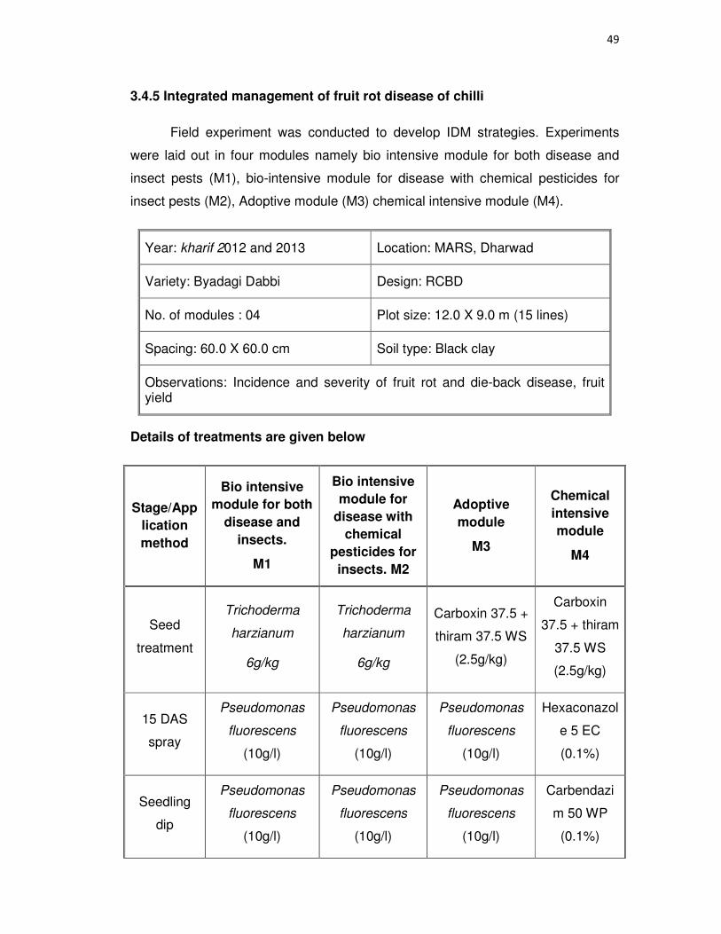
49
3.4.5 Integrated management of fruit rot disease of chilli
Field experiment was conducted to develop IDM strategies. Experiments
were laid out in four modules namely bio intensive module for both disease and
insect pests (M1), bio-intensive module for disease with chemical pesticides for
insect pests (M2), Adoptive module (M3) chemical intensive module (M4).
Year: kharif 2012 and 2013 Location: MARS, Dharwad
Variety: Byadagi Dabbi Design: RCBD
No. of modules : 04 Plot size: 12.0 X 9.0 m (15 lines)
Spacing: 60.0 X 60.0 cm Soil type: Black clay
Observations: Incidence and severity of fruit rot and die-back disease, fruit yield
Details of treatments are given below
Stage/App
lication
method
Bio intensive
module for both
disease and
insects.
M1
Bio intensive
module for
disease with
chemical
pesticides for
insects. M2
Adoptive
module
M3
Chemical
intensive
module
M4
Seed
treatment
Trichoderma
harzianum
6g/kg
Trichoderma
harzianum
6g/kg
Carboxin 37.5 +
thiram 37.5 WS
(2.5g/kg)
Carboxin
37.5 + thiram
37.5 WS
(2.5g/kg)
15 DAS
spray
Pseudomonas
fluorescens
(10g/l)
Pseudomonas
fluorescens
(10g/l)
Pseudomonas
fluorescens
(10g/l)
Hexaconazol
e 5 EC
(0.1%)
Seedling
dip
Pseudomonas
fluorescens
(10g/l)
Pseudomonas
fluorescens
(10g/l)
Pseudomonas
fluorescens
(10g/l)
Carbendazi
m 50 WP
(0.1%)

50
94 DAT spray
Neem oil (10ml/l) Neem oil (10ml/l)
Neem oil (10ml/l)
Carbendazim 12% +
Mancozeb 63% WP (0.25%)
108 DAT spray
Bacillus subtilis (10g/l)
Bacillus subtilis (10g/l)
Hexaconazole 5 EC (0.1%)
Difenconazole 25 EC (0.1%)
122 DAT spray
Neem oil (10ml/l) Neem oil (10ml/l)
Propiconazole 25 EC (0.1%)
Pyraclostrobin 20% WG
(0.1%)
130 DAT, spray
Pseudomonas fluorescens
(10g/l)
Pseudomonas fluorescens
(10g/l)
Carbendazim 12% +
Mancozeb 63% WP (0.25%)
Mancozeb 75WP (2.5%)
Insect pest management
15 DAS spray
Verticillium lecanii (4g/l)
Imidachloprid 17.80 SL, (0.5ml/l) +
Fenazaquin 10%EC (2.0
ml/l)
Imidachloprid 17.80 SL, (0.5ml/l) +
Fenazaquin 10%EC (2.0
ml/l)
Imidachloprid 17.80 SL, (0.5ml/l) +
Fenazaquin 10%EC (2.0
ml/l)
30 DAT spray
Verticillium lecanii (4g/l)
Imidachloprid 17.80 SL, (0.5ml/l) +
Fenazaquin 10%EC (2.0
ml/l)
Imidachloprid 17.80 SL, (0.5ml/l) +
Fenazaquin 10%EC (2.0
ml/l)
Imidachloprid 17.80 SL, (0.5ml/l) +
Fenazaquin 10%EC (2.0
ml/l)
60 DAT spray
Neem oil (10ml/l) Neem oil (10ml/l)
Neem oil (10ml/l)
Imidachloprid 17.80 SL,
(0.5ml/l)
90 DAT spray
Verticillium lecanii (4g/l)
Imidachloprid 17.80 SL, (0.5ml/l) +
Spiromesifen 22.9%
(0.75ml/l)
Imidachloprid 17.80 SL, (0.5ml/l) +
Spiromesifen 22.9%
(0.75ml/l)
Imidachloprid 17.80 SL, (0.5ml/l)+
Spiromesifen 22.9%
(0.75ml/l)
125DAT, spray
Nomuraea rileyi (4g/l)
Indoxacarb 14.5 SC (0.5ml/l)
Indoxacarb 14.5 SC (0.5ml/l)
Indoxacarb14.5 SC (0.5ml/l)

51
3.5 Statistical analysis
Statistical analysis was carried out as per the procedures given by Panse
and Sukhatme (1985). Actual data in percentage were converted to angular
transformed values, before analysis according to the table given by Walter (1997).
Fischer’s method of analysis of variance was used for analysis and interpretation
of the data as outlined by Gomez and Gomez (1984). The level of significance
used in ‘F’ and ‘T’ tests was p=0.05. Critical differences were calculated wherever
‘F’ test was significant. Other statistical analysis viz., calculation of correlation
coefficients, regression equations etc. were done using MS-excel.
4. EXPERIMENTAL RESULTS
The experiments were conducted on various aspects of chilli fruit rot and die-back
disease with reference to prevalence and distribution of disease, pathogen diversity,
severity of the disease in various geographical regions of South India, isolation,
identification, morphological and molecular variability of pathogens and their quick
detection by molecular method to know the complexity of causal organisms. Role of
weather parameters in disease development and survival ability of pathogens, host range
and cross inoculations were studied. Further, seed health management and integrated
disease management strategies comprising screening of genotypes for resistance,
evaluation of chemicals under field conditions and various disease management modules
were studied for identification of the best management module with maximum C:B ratio
which helps the farming community to a greater extent. These experiments were
conducted in the laboratory as well as in the field during 2012 and 2013 at the Department
of Plant Pathology, College of Agriculture and MARS, UAS, Dharwad. The results thus
obtained are presented in different sections under this chapter.
4.1 Survey, isolation and identification of pathogen/s to study the distribution in
different geographical regions of South India
4.1.1 Survey
A roving survey was carried out to know the incidence and severity of fruit rot
disease during 2012 and 2013 in major chilli growing states of South India viz., Andhra
Pradesh (Guntur, Kurnool), Maharashtra (Solapur) and Karnataka (Bagalkot, Belgavi,
Bellary, Dharwad, Gadag, Haveri, Koppal, , Raichur). The observations are presented in
Table 1a, 1b, 2a, 2b and 3 (Plate 3).
Year 2012-13
Among three states the highest fruit rot incidence (19.97%) with 13.77 PDI was
observed in Maharashtra followed by Karnataka (18.13%, 11.78 PDI) where as the highest
die-back incidence (10.55%) with 43.90 PDI was observed in Andhra Pradesh followed by
Karnataka (8.59%, 37.36 PDI) (Table 3).

53
Plate 3 Chilli fruit rot incidence and severity in Karnataka, Andhra
Pradesh and Maharashtra

54
Karnataka
Data pertaining to survey conducted during 2012-13 are presented in Table 1a and
1b. Among the 56 villages the highest fruit rot incidence of 27.52 per cent with 19.60 PDI
was recorded in Hirekotnekal village of Manvi taluk, Raichur district followed by Koluru
(26.52%, 18.60 PDI) village of Bellary district. The highest die-back incidence of 13.18 per
cent with 48.91 PDI was observed in Hulkoti village of Gadag taluk. Among the 15 taluks
highest fruit rot incidence of 22.87 per cent with 16.07 PDI was observed in Bellary taluk
followed by Manvi (21.70%, 13.95 PDI). The lowest fruit rot incidence of 11.36 per cent
and 7.38 PDI was observed in Kustagi taluk. The highest die-back incidence of 10.83 per
cent with 43.79 PDI was observed in Hubli taluk. In Gangavati taluk die-back disease was
absent followed by 5.77 per cent with 28.50 PDI was observed in Koppal taluk. Among the
seven districts of Karnataka the highest fruit rot incidence of 21.71 per cent with 14.99
PDI was observed in Bellary followed by Raichur ( 20.24%, 12.55 PDI), where as the
lowest fruit rot incidence of 12.83 per cent with 8.09 PDI was observed in Koppal. The
highest die-back incidence of 10.65 per cent with 43.19 PDI was observed in Gadag,
where as the lowest die-back incidence of 4.29 per cent with 30.50 PDI was observed in
Koppal.
Andhra Pradesh
Among the 12 villages the highest fruit rot incidence of 24.82 per cent with 19.20
PDI was recorded in Madire village of Adoni taluk followed by Alur (21.48%, 16.75 PDI)
village of Adoni taluk. The highest die-back incidence of 13.21 per cent with 49.41 PDI
was observed in Dharmapuram village of Mantralaya taluk. Among the four taluks of
Kurnool district the highest fruit rot incidence of 20.20 per cent with 15.74 PDI was
observed in Adoni taluk followed by Mantralaya (19.60%, 11.85 PDI), where as lowest fruit
rot incidence of 13.23 per cent and 10.42 PDI was observed in Emmiganur taluk. The
highest die-back incidence of 11.20 per cent with 45.09 PDI was observed in Mantralaya
taluk followed by Adoni (10.68%, 43.99 PDI), where as lowest die-back incidence of 10.00
per cent with 40.11 PDI was observed in Emmiganur taluk (Table 1a, 1b Plate.4).

55
Table 1a: Detailed survey on chilli fruit rot and dieback disease during 2012-13 in South India
State District Taluka Village/ place Area (ha)
Stage of crop
Geno type
Inter crop / sole crop
R/I Soil type
Fruit rot Die back Pathogens noticed
Incidence (%)
PDI Incide
nce (%)
PDI Cc Cg Ca F A
Karn
ata
ka
Bagalkot Bagalkot
Kagalgombe 2.0 Fruiting Byadgi Dabbi Sole crop
R Black 19.18 13.42 11.48 46.38
- + - - -
Lokapur 2.5 Fruiting Byadgi kaddi Sole crop
I Black 22.75 15.64 9.27 34.28
+ - - + -
Kaladagi 2.0 Fruiting Byadgi Dabbi Sole crop
I Red 18.48 11.73 10.27 37.82
+ - - + -
Mean 20.13 13.59 10.34 39.49 Range 19-23 11-16 9-12 34-47
Mean 20.13 13.59 10.34 39.49
Bellary
Bellary
Koluru 4.0 Fruiting Byadgi Kaddi Sole crop
I Red 26.52 18.60 8.42 39.84
+ - - - +
Raravi 6.0 Fruiting Guntur Sole crop
I Black 18.36 13.40 6.74 34.46
+ - + + -
Sindigeri 4.0 Fruiting Byadgi Dabbi Sole crop
R Red 23.72 16.21 7.89 36.92
+ + - - +
22.87 16.07 7.68 37.07
Kerur 3.0 Fruiting Byadgi Dabbi Sole crop
R Red 16.49 11.25 8.94 52.48 + - - + +
Siraguppa
Siraguppa 4.0 Fruiting Guntur Sole crop
I Red 18.62 14.79 10.46 41.92 - + - + -
Shanvaspur 4.0 Fruiting Byadgi Dabbi Sole crop
I Red 24.71 16.93 10.00 52.16 + + - - -
Sirigeri cross 2.0 Fruiting Byadgi Dabbi Sole crop
I Red 22.41 12.64 12.94 39.92 + - - + -
Mean 20.55 13.90 10.58 44.67
Range 16-25 11-19 7-13 34-53
Mean 21.71 14.99 9.13 40.87
Dharwad Dharwad
Amminabhavi 1.5 Fruiting Byadgi Dabbi Onion R Black 18.96 10.97 9.46 38.92 + - - - -
Byahatti 2.0 Fruiting Byadgi Dabbi Onion R Black 15.48 9.34 8.72 34.83 - + - - +
Govankoppa 1.5 Fruiting Sitara Sole crop
R Black 12.73 9.21 9.14 35.82 + - + - -
Kumbapur farm
2.0 Fruiting Byadgi Dabbi Sole crop
I Red 19.54 12.46 8.76 33.94 + - - + +
Somapur 1.0 Fruiting Byadgi Dabbi Onion R Black 21.73 13.81 10.16 31.24 + - - - +
Cc – Colletotrichum capsici Cg – C. gloeosorioides, Ca – C. acutatum, A – Alternaria sp., F – Fusarium spp. R- rainfed, I – Irrigated, + Present - Absent

56
State District Taluka Village/ place Area (ha)
Stage of crop
Geno type
Inter crop / sole crop
R/I Soil type
Fruit rot Die back Pathogens noticed
Incidence (%)
PDI Incide
nce (%)
PDI Cc Cg Ca F A
Dharwad
Shivalli 1.5 Fruiting
Byadgi Dabbi
Onion R Black 24.14 12.07 9.43 40.78 + - - + -
UAS Campus 2.0 Fruiting Byadgi Dabbi
Sole crop
R Black 22.73 11.73 8.93 41.05 + + + + +
Mean 19.33 11.37 9.23 36.65
Hubli
Hubli 2.0 Fruiting Byadgi Dabbi
Sole crop
R Black 14.25 10.71 12.83 48.29 + - - - +
Hebsur 2.5 Fruiting Byadgi Dabbi
Onion R Black 12.34 9.27 9.13 38.92 + - - + -
Shirguppi 2.0 Fruiting Byadgi Dabbi
Sole crop
R Black 16.27 11.35 10.52 44.16 + + - - -
Mean 14.29 10.44 10.83 43.79
Karn
ata
ka
Kundgol
Gudigeri 2.0 Fruiting Byadgi Dabbi
Sole crop
R Black 15.14 10.76 10.18 41.7 + - - + -
Kundagol 5.0 Fruiting Byadgi Dabbi
Sole crop
R Black 17.19 11.41 9.27 40.31 + - - - +
Sharewad 4.0 Fruiting Byadgi Dabbi
Sole crop
R Black 15.52 10.16 12.13 42.91 + - - + -
Savnsi 2.0 Fruiting Byadgi Kaddi
Sole crop
R Black 12.31 8.94 9.43 36.81 + - - - -
Mean 15.04 10.32 10.25 40.01
Navalgund
Alagavadi 3.0 Fruiting and
flowering Byadgi Dabbi
Onion R Black 14.13 11.74 9.14 35.27 + - - + -
Byalal 2.5 Fruiting and
flowering Byadgi Dabbi
Onion R Black 16.10 12.15 8.92 34.93 + - - + -
Gummagola 2.0 Fruiting and
flowering Byadgi Dabbi
Onion R Black 11.83 9.82 9.47 36.72 + - - - +
Mean 14.02 11.24 9.18 35.64 Range 12-20 9-14 8-13 31-49
Mean 15.67 10.84 9.87 39.02
Gadag Gadag
Binkadakatti 3.0 Fruiting and
flowering Gadag Local
Sole crop
R Black 12.76 9.24 9.13 40.24 - + - - +
Badrapur 2.0 Fruiting and
flowering Byadgi Dabbi
Cotton , onion
R Black 16.81 12.16 10.47 43.28 + - - + -
Dumdur 1.5 Fruiting and
flowering Byadgi Dabbi
Cotton, onion
R Black 20.47 14.38 9.81 40.31 + - - + -
Cc – Colletotrichum capsici Cg – C. gloeosorioides, Ca – C. acutatum, A – Alternaria sp., F – Fusarium spp. R- rainfed, I – Irrigated, + Present - Absent

57
State District Taluka Village/ place Area (ha)
Stage of crop
Geno type
Inter crop / sole crop
R/I Soil type
Fruit rot Die back Pathogens noticed
Incidence (%)
PDI Incide
nce (%)
PDI Cc Cg Ca F A
Hulkoti 2.0
Fruiting and
flowering
Byadgi Dabbi
Cotton , onion
R Black 18.32 10.13 13.18 48.91 + - - - +
Mean 17.09 11.48 10.65 43.19
Range 12-21 9-15 9-14 40-49
Mean 17.09 11.48 10.65 43.19
Haveri Haveri
Byadgi 2.0 Fruiting
and flowering
Byadgi Dabbi
Sole crop I Black 20.19 12.20 7.43 32.31 + + - - +
Devihosur 3.0 Fruiting
and flowering
Byadgi Dabbi
Sole crop R Black 16.31 9.12 8.24 34.31 + - + - +
Dummihal 1.5 Fruiting
and flowering
Byadgi Kaddi
Sole crop R Black 14.28 8.41 9.21 39.21 + - - + -
Mean 16.92 9.91 8.29 35.28
Karn
ata
ka
Ranebenn
ur
Guttal 2.0 Fruiting
and flowering
Dyavanur Dabbi
Sole crop R Black 18.41 11.02 8.11 32.51 + + - - +
Kadaramandalgi
2.5 Fruiting
and flowering
Byadgi Dabbi
Sole crop I Black 26.18 14.17 9.02 34.10 + - - - -
Lingadahalli 2.0 Fruiting
and flowering
Byadgi Dabbi
Sole crop R Black 18.21 10.43 0.00 0.00 + - + - -
Ranebennur 1.0 Fruiting
and flowering
Byadgi Dabbi
Sole crop R Black 23.40 12.10 8.14 34.32 + - - + -
Mean 21.55 11.93 6.32 33.64 Range 18-27 10-15 0-9 0-35
Mean 19.23 10.92 7.30 34.46
Koppal Gangavati
Budugumpa 2.0 Fruiting
and flowering
Local Sole crop I
Red 14.25 9.01 0.00 0.00 + + - - -
Kanakapur 1.5 Fruiting
and flowering
Byadgi Kaddi
Sole crop I
Red 9.71 4.83 0.00 0.00 - + - + -
Mean 11.98 6.92 0 0
Cc – Colletotrichum capsici Cg – C. gloeosorioides, Ca – C. acutatum, A – Alternaria sp., F – Fusarium spp. R- rainfed, I – Irrigated, + Present - Absent

58
State District Taluka Village/ place Area (ha)
Stage of crop
Geno type
Inter crop / sole crop
R/I Soil type
Fruit rot Die back Pathogens noticed
Incidence (%)
PDI Incide
nce (%)
PDI Cc Cg Ca F A
Koppal
Basapur 2.0 Fruiting
and flowering
Guntur Sole crop
I Red 13.16 8.42 0.00 0.00 + - - - -
Beluru 1.5 Fruiting
and flowering
Gadag local Sole crop
I Red 19.20 10.28 9.46 43.48 + + - - +
Bommanal 2.0 Fruiting
and flowering
Byadgi Kaddi
Sole crop
I Red 12.30 9.20 5.20 27.4 + - - + -
Dambarahalli 2.0 Fruiting
and flowering
Guntur Sole crop
I Red 16.82 11.64 9.36 40.24 + - - - -
Talakall 1.5 Fruiting
and flowering
Byadgi Dabbi
Sole crop
R Black 14.20 10.24 4.82 31.4 + + - - +
Mean 15.14 9.96 5.77 28.50
Karn
ata
ka
Kustagi
Chikkatebinahal 2.0 Fruiting
and flowering
Local Sole crop I Red 12.30 7.92 8.94 38.00 + - - + -
Kustagi 2.5 Fruiting
and flowering
Byadgi Kaddi
Sole crop I Red 10.42 6.84 4.20 26.10 + - - - +
Mean 11.36 7.38 6.57 32.05 Range 10-20 4-12 0-10 0-44
Mean 12.83 8.09 6.17 30.50
Raichur
Manvi
Herekotneekal 4.0 Fruiting
and flowering
Guntur Sole crop I Black 27.52 19.60 9.42 39.84 + - - + +
Kapgal 3.5 Fruiting
and flowering
Byadgi kaddi
Sole crop I Black 16.04 10.32 9.66 38.33 + + - - +
Neer manvi 3.0 Fruiting
and flowering
Guntur Sole crop I Red 21.55 11.93 6.32 17.73 + - - - +
Mean 21.70 13.95 8.47 31.97
Raichur Belgammanadod
di 4.0
Fruiting and
flowering
Byadgi Dabbi
Sole crop I Black 22.87 11.07 10.68 47.07 + - + - +
Cc – Colletotrichum capsici Cg – C. gloeosorioides, Ca – C. acutatum, A – Alternaria sp., F – Fusarium spp. R- rainfed, I – Irrigated, + Present - Absent

59
State Distric
t Taluka Village/ place
Area
(ha)
Stage of crop
Geno
type
Inter crop / sole crop
R/I Soil type
Fruit rot Die back Pathogens noticed
Incidence (%)
PDI Incide
nce (%)
PDI Cc Cg Ca F A
Mandapura 40 Fruiting and
flowering Byadgi Dabbi
Sole crop
I Black 15.28 8.54 9.21 39.21 + + - + +
Nelhal 5.0 Fruiting and
flowering Guntur
Sole crop
I Black 23.40 12.10 8.14 34.32 + - - + +
Panchamukhi 4.0 Fruiting and
flowering Byadgi Kaddi
Sole crop
R Black 14.02 11.24 9.18 35.64 + + - + -
Raichur 5.0 Fruiting and
flowering Byadgi Dabbi
Sole crop
I Black 19.73 12.01 10.14 42.10 + - - + +
Raladoddi 4.0 Fruiting and
flowering Guntur
Sole crop
I Red 21.55 11.93 6.32 17.73 - + - + -
Mean 18.79 11.16 8.59 36.01
Range
14-27 10-19 6-11 17-47
Mean 20.24 12.55 8.53 33.99
An
dh
ra P
rad
esh
Kurnool
Adoni
Alur 2.0 Fruiting and
flowering Byadgi Kaddi
Sole crop
I Black 21.48 16.75 11.20 47.35 - + - + -
Madire 3.0 Fruiting and
flowering Brahma
theja Sole crop
I Black 24.82 19.20 10.51 38.24 + - - + -
Marakattu 2.0 Fruiting and
flowering Guntur
Sole crop
I Black 20.31 14.26 - - + - + - +
Mean 20.20 15.74 10.68 43.99
Emmiganur
Bodikanda 2.0 Fruiting and
flowering Theja
Sole crop
I Black 12.50 9.71 8.17 38.16 + - - + -
Emmiganur 2.5 Fruiting and
flowering Guntur
Sole crop
I Red 16.89 12.40 12.00 44.28 + + - - +
Hanumapuram
3.0 Fruiting and
flowering C-341
Sole crop
I Black 10.72 9.10 - - - + - - +
Kotekallu 3.0 Fruiting and
flowering Guntur
Sole crop
I Red 12.80 10.45 9.82 37.90 + - - + -
Mean 13.23 10.42 10.00 40.11
Cc – Colletotrichum capsici Cg – C. gloeosorioides, Ca – C. acutatum, A – Alternaria sp., F – Fusarium spp. R- rainfed, I – Irrigated, + Present - Absent

60
State District Taluka Village/ place Area (ha)
Stage of crop
Geno type
Inter crop / sole crop
R/I Soil type
Fruit rot Die back Pathogens noticed
Incidence (%)
PDI Incide
nce (%)
PDI Cc Cg Ca F A
Mantralaya
Dharmapuram 2.5 Fruiting and
flowering Guntur
Sole crop
I Red 21.41 13.20 13.21 49.41 + - - + -
Halaharavi 2.0 Fruiting and
flowering Theja
Sole crop
R Black 18.31 11.45 9.67 42.46 + - - - -
Kalludevarakunta
2.0 Fruiting and flowering
Guntur Sole crop
I Red 20.26 12.00 10.72 43.41 - + - - +
Manthralaya 2.0 Fruiting and
flowering Guntur
Sole crop
I Red 18.40 10.73 - - + - + + -
Mean 19.60 11.85 11.20 45.09
Nandavaram
Mugathi 2.0 Fruiting and
flowering Guntur
Sole crop
I Red 14.20 12.73 10.32 46.40 + - - - -
Range 9-25 9-20 8-13 37-50
Mean 16.81 12.69 10.55 43.90
Mah
ara
sh
tra
Solapur Pandarapu
r
Mangalaveda 2.0 Fruiting and
flowering Local
Sole Crop
I Black 26.82 20.45 8.60 36.25 + + - + -
Pandarapur 2.5 Fruiting and
flowering Capsicum
Sole Crop
I Red 14.20 9.48 - - + - - - -
Sangola 2.0 Fruiting and
flowering Local
Sole Crop
I Red 18.91 11.40 6.80 33.90 + - - + +
Mean 19.97 13.77 7.70 35.07
Range 14-27 9-21 6-9 33-37
Mean 19.97 13.77 7.70 35.07
Cc – Colletotrichum capsici Cg – C. gloeosorioides, Ca – C. acutatum, A – Alternaria sp., F – Fusarium spp. R- rainfed, I – Irrigated, + Present - Absent
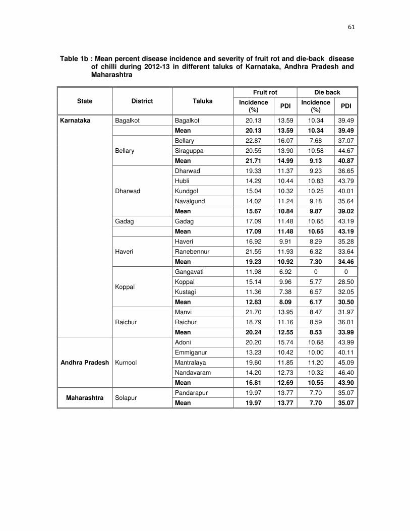
61
Table 1b : Mean percent disease incidence and severity of fruit rot and die-back disease of chilli during 2012-13 in different taluks of Karnataka, Andhra Pradesh and Maharashtra
State District Taluka
Fruit rot Die back
Incidence (%)
PDI Incidence
(%) PDI
Karnataka Bagalkot Bagalkot 20.13 13.59 10.34 39.49
Mean 20.13 13.59 10.34 39.49
Bellary
Bellary 22.87 16.07 7.68 37.07
Siraguppa 20.55 13.90 10.58 44.67
Mean 21.71 14.99 9.13 40.87
Dharwad
Dharwad 19.33 11.37 9.23 36.65
Hubli 14.29 10.44 10.83 43.79
Kundgol 15.04 10.32 10.25 40.01
Navalgund 14.02 11.24 9.18 35.64
Mean 15.67 10.84 9.87 39.02
Gadag Gadag 17.09 11.48 10.65 43.19
Mean 17.09 11.48 10.65 43.19
Haveri
Haveri 16.92 9.91 8.29 35.28
Ranebennur 21.55 11.93 6.32 33.64
Mean 19.23 10.92 7.30 34.46
Koppal
Gangavati 11.98 6.92 0 0
Koppal 15.14 9.96 5.77 28.50
Kustagi 11.36 7.38 6.57 32.05
Mean 12.83 8.09 6.17 30.50
Raichur
Manvi 21.70 13.95 8.47 31.97
Raichur 18.79 11.16 8.59 36.01
Mean 20.24 12.55 8.53 33.99
Andhra Pradesh Kurnool
Adoni 20.20 15.74 10.68 43.99
Emmiganur 13.23 10.42 10.00 40.11
Mantralaya 19.60 11.85 11.20 45.09
Nandavaram 14.20 12.73 10.32 46.40
Mean 16.81 12.69 10.55 43.90
Maharashtra Solapur Pandarapur 19.97 13.77 7.70 35.07
Mean 19.97 13.77 7.70 35.07

62
Maharashtra
Among three villages of Pandarpur taluk of Solapur district the highest fruit rot
incidence of 26.82 per cent with 20.45 PDI was observed in Mangalveda followed by
Sangola (18.91%, 11.40 PDI) and also the highest die-back incidence of 8.60 per cent
with 36.25 PDI was observed in Mangalveda followed by Sangola (6.80%, 33.90 PDI).
Year 2013-14
The highest fruit rot incidence of 20.33 per cent with 13.31 PDI was observed in
Andhra Pradesh followed by Karnataka (18.12%, 11.25 PDI), where as the highest die-
back incidence of 11.44 per cent with 41.67 PDI was observed in Andhra Pradesh
followed by Karnataka (9.54%, 38.92 PDI) (Table 3).
Karnataka
Data pertaining to survey conducted during 2013-14 are presented in Table 2a
and 2b. Among the 57 villages the highest fruit rot incidence of 27.13 per cent with
16.21 PDI was recorded in Dumdur village of Gadag taluk, Gadag district followed by
Sirigeri cross (26.73%, 16.79 PDI) village of Siraguppa taluk Bellary district, where as
the lowest fruit rot incidence of 10.20 per cent and 8.40 PDI was observed in Salahalli
village of Soundatti taluk of Belgaum district. The highest die-back incidence of 12.94
per cent with 44.32 PDI was observed in Sirigeri cross of Siraguppa taluk Bellary
district. The lowest die-back incidence 1.00 per cent with 10.24 PDI was observed in
Kanakapur village of Gangavati taluk Koppal district where as die-back disease was
absent in Basapur village of Koppal district. Among the 15 taluks highest fruit rot
incidence of 24.10 per cent with 15.21 PDI was observed in Siraguppa taluk of Bellary
district followed by Gadag (22.24%, 13.62 PDI). The lowest fruit rot incidence of 11.83
per cent and 7.34 PDI was observed in Gangavati taluk. The lowest die-back incidence
of 1.50 per cent with 11.27 PDI was observed in Gangavati taluk of Koppal district.
Among the seven districts of Karnataka the highest fruit rot incidence of 22.65 per cent
with 14.51 PDI was observed in Bellary followed by Gadag ( 22.24%, 13.62 PDI), where
as the lowest fruit rot incidence of 14.02 per cent with 9.13 PDI was observed in Koppal.
The highest die-back incidence of 11.70 per cent with 44.64 PDI was observed in

63
Table 2a: Detailed survey on chilli fruit rot and dieback disease during 2013-14 in South India
State District Taluka Village/place
Area (ha)
Stage of crop Geno type
Inter Crop /
Sole Crop R/I
Soil type
Fruit rot Die back Pathogens noticed
Incidence (%)
PDI Incide
nce (%)
PDI Cc Cg Ca F A
Karn
ata
ka
Bellary
Bellary
Koluru 4.0 Flowering and
Fruiting Byadgi Kaddi Sole Crop I Red 22.14 14.94 6.73 35.24
+ - - - +
Raravi 6.0 Flowering and
Fruiting Guntur Sole Crop I Black 20.00 12.21 8.42 39.82
+ - + + -
Sindigeri 4.0 Flowering and
Fruiting Byadgi Dabbi Sole Crop R Red 21.47 14.32 8.21 40.96 +
+ - - +
Mean 21.20 13.82 7.79 38.67
Siraguppa
Kerur 3.0 Flowering and
Fruiting Byadgi Dabbi Sole Crop R Red 21.37 13.76 8.94 47.41
+ - - + +
Siraguppa 4.0 Fruiting Guntur Sole Crop I Red 24.00 14.91 10.46 38.32 - + - + -
Shanvaspur 4.0 Flowering and
Fruiting Byadgi Dabbi Sole Crop I Red 24.32 15.40 10.00 50.64
+ + - - -
Sirigeri cross 2.0 Flowering and
Fruiting Byadgi Dabbi Sole Crop I Red 26.73 16.79 12.94 44.32
+ - - + -
Mean 24.10 15.21 10.58 45.17
Range 20-27 12-17 6-11 35-48 Mean 22.65 14.51 9.18 41.92
Belgavi Soundatti
Inamhongal 2.0 Fruiting and
flowering Byadgi Dabbi Onion R Black 18.19 11.20 10.19 42.70 - + - - -
Panchagav 2.0 Fruiting and
flowering Byadgi Dabbi
Onion, coriander
R Black 15.30 10.00 9.20 41.31 + - - + -
Salahalli 1.5 Fruiting and
flowering Byadgi Dabbi Onion R Black 10.20 8.40 12.12 40.91
+ - - + -
Soundatti 2.0 Fruiting and
flowering Byadgi Dabbi Onion R Black 14.62 9.25 9.40 35.81
+ - - - +
Mean 14.58 9.71 10.23 40.18
Range 10-19 8-11 9-12 35-42
Mean 14.58 9.71 10.23 40.18
Dharwad Dharwad
Amminabhavi 1.5 Fruiting Byadgi Dabbi Onion R Black 20.18 11.86 10.32 40.28 + - - - -
Byahatti 2.0 Fruiting Byadgi Dabbi Onion R Black 16.39 9.86 10.86 40.12 - + - - +
Govankoppa 1.5 Fruiting Byadgi Kaddi Sole Crop R Black 14.12 9.64 9.26 38.19 + - + - -
Kumbapur farm 2.0 Fruiting Byadgi Dabbi Sole Crop I Red 16.24 11.83 9.14 36.85 + - - + +
Somapur 1.0 Fruiting Byadgi Dabbi Onion R Black 20.74 10.91 9.20 38.94 + - - - +
Shivalli 1.5 Fruiting Byadgi Dabbi Onion R Black 22.92 11.43 8.49 34.17 + - - + -
UAS Campus 2.0 Fruiting Byadgi Dabbi Sole Crop R Black 26.84 14.35 9.12 63.84 + + + + +
Mean 19.63 11.41 9.48 38.34
Cc – Colletotrichum capsici Cg – C. gloeosorioides, Ca – C. acutatum, A – Alternaria sp., F – Fusarium spp. R- rainfed, I – Irrigated, + Present - Absent

64
State District Taluka Village/ place
Area (ha)
Stage of crop
Genotype Inter crop / Sole crop
R/I Soil type
Fruit rot Die back Pathogens noticed
Incidence (%)
PDI Incide
nce (%)
PDI Cc Cg Ca F A
Karn
ata
ka
Hubli
Hubli 2.0 Fruiting Byadgi Dabbi Sole Crop R Black 18.27 12.14 12.83 48.29 + - - - +
Hebsur 2.5 Fruiting Byadgi Dabbi Onion R Black 13.84 10.13 9.13 38.92 + - - + -
Shirguppi 2.0 Fruiting Byadgi Dabbi Sole Crop R Black 18.27 12.10 10.52 44.16 + + - - -
Mean 16.79 11.46 10.83 43.79
Kundgol
Gudigeri 2.0 Fruiting Byadgi Dabbi Sole Crop R Black 16.42 11.12 10.18 41.70 + - - + -
Kundagol 5.0 Fruiting Byadgi Dabbi Sole Crop R Black 20.83 12.76 9.27 40.31 + - - - +
Sharewad 4.0 Fruiting Byadgi Dabbi Sole Crop R Black 17.32 11.20 12.13 42.91 + - - + -
Savnsi 2.0 Fruiting Byadgi Kaddi Sole Crop R Black 14.91 10.28 9.43 36.81 + - - - -
Mean 17.37 11.34 10.25 40.43
Navalgund
Alagavadi 3.0 Fruiting and
flowering Byadgi Dabbi Onion R Black 16.27 12.37 11.25 39.19
+ - - + -
Byalal 2.5 Fruiting and
flowering Byadgi Dabbi Onion R Black 13.27 10.21 9.64 37.12
+ - - + -
Gummagola 2.0 Fruiting and
flowering Byadgi Dabbi Onion R Black 14.28 11.14 10.82 41.36
+ - - - +
Mean 14.61 11.24 10.57 39.22
Range 13-21 9-15 8-13 36-64
Mean 17.10 11.36 10.28 40.45
Gadag Gadag
Binkadakatti 3.0 Fruiting and
flowering Gadag Local Sole Crop R Black 18.91 11.86 11.43 42.16
- + - - +
Badrapur 2.0 Fruiting and
flowering Byadgi Dabbi Cotton , Onion R Black 22.17 14.28 12.73 46.21
+ - - + -
Dumdur 1.5 Fruiting and
flowering Byadgi Dabbi Cotton, Onion R Black 27.13 16.21 10.45 43.27
+ - - + -
Hulkoti 2.0 Fruiting and
flowering Byadgi Dabbi Cotton , Onion R Black 20.74 12.14 12.18 46.92
+ - - - +
Mean Mean 22.24 13.62 11.70 44.64
Range 18-28 11-15 10-13 42-47
Mean 22.24 13.62 11.70 44.64
Haveri
Haveri
Byadgi 2.0 Fruiting and
flowering Byadgi Dabbi Sole Crop I Black 22.18 13.41 9.40 42.24
+ + - - +
Devihosur 3.0 Fruiting and
flowering Byadgi Dabbi Sole Crop R Black 18.24 10.84 10.04 44.60
+ - + - +
Dummihal 1.5 Fruiting and
flowering Byadgi Kaddi Sole Crop R Black 19.24 12.01 9.82 42.91
+ - - + -
Mean Mean 19.88 12.08 9.75 43.25
Ranebennur
Guttal 2.0 Fruiting and
flowering Dyavanur Dabbi Sole Crop R Black 20.61 10.14 9.42 41.50
+ + - - +
Kadaramandalgi
2.5 Fruiting and
flowering Byadgi Dabbi Sole Crop I Black 22.40 11.84 10.13 46.31
+ - - - -
Cc – Colletotrichum capsici Cg – C. gloeosorioides, Ca – C. acutatum, A – Alternaria sp., F – Fusarium spp. R- rainfed, I – Irrigated, + Present - Absent
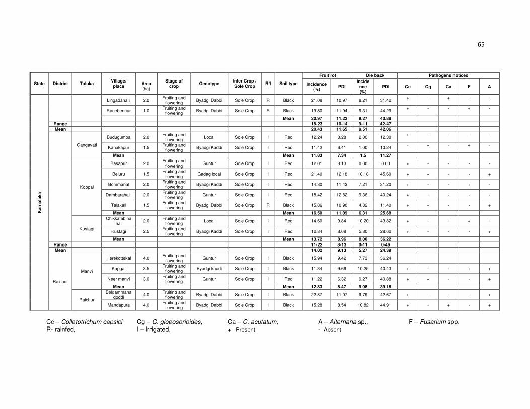
65
State District Taluka Village/ place
Area (ha)
Stage of crop
Genotype Inter Crop / Sole Crop
R/I Soil type
Fruit rot Die back Pathogens noticed
Incidence (%)
PDI Incide
nce (%)
PDI Cc Cg Ca F A
Karn
ata
ka
Lingadahalli 2.0 Fruiting and
flowering Byadgi Dabbi Sole Crop R Black 21.08 10.97 8.21 31.42
+ - + - -
Ranebennur 1.0 Fruiting and
flowering Byadgi Dabbi Sole Crop R Black 19.80 11.94 9.31 44.29
+ - - + -
Mean 20.97 11.22 9.27 40.88
Range 18-23 10-14 9-11 42-47
Mean 20.43 11.65 9.51 42.06
Gangavati
Budugumpa 2.0 Fruiting and
flowering Local Sole Crop I Red 12.24 8.28 2.00 12.30
+ + - - -
Kanakapur 1.5 Fruiting and
flowering Byadgi Kaddi Sole Crop I Red 11.42 6.41 1.00 10.24
- + - + -
Mean Mean 11.83 7.34 1.5 11.27
Koppal
Basapur 2.0 Fruiting and
flowering Guntur Sole Crop I Red 12.01 8.13 0.00 0.00 + - - - -
Beluru 1.5 Fruiting and
flowering Gadag local Sole Crop I Red 21.40 12.18 10.18 45.60 + + - - +
Bommanal 2.0 Fruiting and
flowering Byadgi Kaddi Sole Crop I Red 14.80 11.42 7.21 31.20 + - - + -
Dambarahalli 2.0 Fruiting and
flowering Guntur Sole Crop I Red 18.42 12.82 9.36 40.24 + - - - -
Talakall 1.5 Fruiting and
flowering Byadgi Dabbi Sole Crop R Black 15.86 10.90 4.82 11.40 + + - - +
Mean Mean 16.50 11.09 6.31 25.68
Kustagi
Chikkatebinahal
2.0 Fruiting and
flowering Local Sole Crop I Red 14.60 9.84 10.20 43.82 + - - + -
Kustagi 2.5 Fruiting and
flowering Byadgi Kaddi Sole Crop I Red 12.84 8.08 5.80 28.62 + - - - +
Mean Mean 13.72 8.96 8.00 36.22
Range 11-22 8-13 0-11 0-46
Mean 14.02 9.13 5.27 24.39
Raichur
Manvi
Herekottekal 4.0 Fruiting and
flowering Guntur Sole Crop I Black 15.94 9.42 7.73 36.24
Kapgal 3.5 Fruiting and
flowering Byadgi kaddi Sole Crop I Black 11.34 9.66 10.25 40.43 + - - + +
Neer manvi 3.0 Fruiting and
flowering Guntur Sole Crop I Red 11.22 6.32 9.27 40.88 + + - - +
Mean Mean 12.83 8.47 9.08 39.18
Raichur
Belgammanadoddi
4.0 Fruiting and
flowering Byadgi Dabbi Sole Crop I Black 22.87 11.07 9.79 42.67 + - - - +
Mandapura 4.0 Fruiting and
flowering Byadgi Dabbi Sole Crop I Black 15.28 8.54 10.82 44.91 + - + - +
Cc – Colletotrichum capsici Cg – C. gloeosorioides, Ca – C. acutatum, A – Alternaria sp., F – Fusarium spp. R- rainfed, I – Irrigated, + Present - Absent

66
State District Taluka Village/place
Area (ha)
Stage of crop Geno type
Inter Crop / Sole Crop
R/I Soil type
Fruit rot Die back Pathogens noticed
Incidence (%)
PDI Incide
nce (%)
PDI Cc Cg Ca F A
Karn
ata
ka
Nelhal 5 Fruiting and
flowering Guntur Sole Crop I Black 23.40 12.10 9.31 40.29 + + - + +
Panchamukhi 4 Fruiting and
flowering Byadgi Kaddi Sole Crop R Black 14.02 11.24 10.57 39.22 + - - + +
Raichur 5 Fruiting and
flowering Byadgi Dabbi Sole Crop I Black 19.73 12.01 9.84 39.41 + + - + -
Raladoddi 4 Fruiting and
flowering Guntur Sole Crop I Red 21.55 11.93 9.27 40.88 + - - + +
Mean Mean 18.79 11.16 9.96 40.94
Range 11-24 6-13 7-11 36-45
Mean 15.81 9.82 9.52 40.06
An
dra
Pra
des
h
Guntur
Sattanpalli
Kattavaripalem 5.0 Fruiting and
flowering LCA-960 Sole Crop I Black 19.47 10.26 9.10 38.92 - + - + -
Medikonduru 4.0 Fruiting and
flowering LCA-960 Sole Crop I Black 24.29 12.40 10.25 41.34 + - - - -
Paaldagu 3.0 Fruiting and
flowering Theja Sole Crop I Black 18.48 11.74 10.00 40.00 +
+ - - +
Pedakurpadu 2.5 Fruiting and
flowering G-888 Sole Crop I Black 20.50 12.45 8.26
36.20
+ - -
+ +
Perecherla 4.0 Fruiting and
flowering Eldom-5 Sole Crop I Black 27.00 15.13 13.50 34.25 +
- + - -
Mean Mean 21.94 12.39 10.22 40.14
Krosuru
Ananthapuram 4.0 Fruiting and
flowering Theja Sole Crop I Black 19.60 12.36 12.80 39.50 - + - + +
Bayyavaram 5.0 Fruiting and
flowering G-888 Sole Crop I Black 20.00 12.00 10.80 38.48 -
+ - - +
Peasapada 4.0 Fruiting and
flowering Theja Sole Crop I Black 25.61 16.34 14.80 40.45 +
- - - -
Mean Mean 21.73 13.56 12.8 39.47
Range 18-27 10-17 8-15 34-42
Mean 21.84 12.98 11.51 39.80
Kurnool Emmiganur
Bodikanda 2.0 Fruiting and
flowering Theja Sole Crop I Black 14.71 11.20 10.82 41.43 + - - + -
Emmiganur 2.5 Fruiting and
flowering Guntur Sole Crop I Red 18.62 14.17 10.41 46.81 + - - + -
Hanumapuram 3.0 Fruiting and
flowering Guntur Sole Crop I Black 13.60 10.32 - - + + - - +
Kotekallu 3.0 Fruiting and
flowering Guntur Sole Crop I Red 15.67 12.48 10.43 44.81 - + - - +
Mean 15.65 12.04 10.55 44.35
Cc – Colletotrichum capsici Cg – C. gloeosorioides, Ca – C. acutatum, A – Alternaria sp., F – Fusarium spp. R- rainfed, I – Irrigated, + Present - Absent
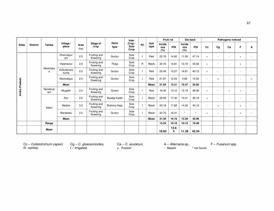
67
State District Taluka Village /
place
Area
(ha)
Stage of crop
Geno
type
Inter Crop / Sole Crop
R/I Soil type
Fruit rot Die back Pathogens noticed
Incidence (%)
PDI Incide
nce (%)
PDI Cc Cg Ca F A
An
dra
Pra
desh
Mantralaya
Dharmapuram
2.5 Fruiting and
flowering Guntur
Sole Crop
I Red 22.76 14.92 11.50 47.14 + - - + -
Halaharavi 2.0 Fruiting and
flowering Theja
Sole Crop
R Black 20.70 12.81 10.72 45.63 + - - + -
Kalludevarakunta
2.0 Fruiting and
flowering Guntur
Sole Crop
I Red 22.49 13.27 14.81 40.12 + - - - -
Mantralaya 2.0 Fruiting and
flowering Guntur
Sole Crop
I Red 21.81 12.25 4.86 10.52 - + - - +
Mean Mean 21.94 13.31 10.47 35.85
Nandavaram
Mugathi 2.0 Fruiting and
flowering Guntur
Sole Crop
I Red 16.40 13.10 12.19 48.90 + - + + -
Adoni
Alur 2.0 Fruiting and
flowering Byadgi Kaddi
Sole Crop
I Black 22.92 17.40 10.41 46.18 + - - - -
Madire 3.0 Fruiting and
flowering Brahma theja
Sole Crop
I Black 23.18 17.82 14.26 40.10 - + - + -
Marakattu 2.0 Fruiting and
flowering Guntur
Sole Crop
I Black 22.70 16.31 * * + - - + -
Mean Mean 21.30 16.16 12.29 45.06
Range 13-24 10-18 10-15 10-49
Mean 18.82
13.65 11.38 43.54
Cc – Colletotrichum capsici Cg – C. gloeosorioides, Ca – C. acutatum, A – Alternaria sp., F – Fusarium spp. R- rainfed, I – Irrigated, + Present - Absent * not found

68
Table 2b: Mean percent disease incidence and severity of fruit rot and die-back disease of chilli during 2013-14 in different taluks of Karnataka, Andhra Pradesh.
State District Taluka
Fruit rot Die back
Incidence (%)
PDI Incidence
(%) PDI
Karnataka
Bellary
Bellary 21.20 13.82 7.79 38.67
Siraguppa 24.10 15.21 10.58 45.17
Mean 22.65 14.51 9.18 41.92
Belgavi Soundatti 14.58 9.71 10.23 40.18
14.58 9.71 10.23 40.18
Dharwad
Dharwad 19.63 11.41 9.48 38.34
Hubli 16.79 11.46 10.83 43.79
Kundgol 17.37 11.34 10.25 40.43
Navalgund 14.61 11.24 10.57 39.22
Mean 17.10 11.36 10.28 40.45
Gadag Gadag 22.24 13.62 11.70 44.64
Mean 22.24 13.62 11.70 44.64
Haveri
Haveri 19.88 12.08 9.75 43.25
Ranebennur 20.97 11.22 9.27 40.88
Mean 20.43 11.65 9.51 42.06
Koppal
Gangavati 11.83 7.34 1.5 11.27
Koppal 16.50 11.09 6.31 25.68
Kustagi 13.72 8.96 8.00 36.22
Mean 14.02 9.13 5.27 24.39
Raichur
Manvi 12.83 8.47 9.08 39.18
Raichur 18.79 11.16 9.96 40.94
Mean 15.81 9.82 9.52 40.06
Andhra Pradesh
Guntur
Sattanpalli 21.94 12.39 10.22 40.14
Krosuru 21.73 13.56 12.80 39.47
Mean 21.84 12.98 11.51 39.80
Kurnool
Adoni 21.30 16.16 12.29 45.06
Emmiganur 15.65 12.04 10.55 44.35
Mantralaya 21.94 13.31 10.47 35.85
Nandavaram 16.40 13.10 12.19 48.90
Mean 18.82 13.65 11.38 43.54

69
Gadag, where as the lowest die-back incidence of 5.27 per cent with 24.39 PDI was
observed in Koppal.
Andhra Pradesh
Among the 20 villages the highest fruit rot incidence of 27.00 per cent with 15.13
PDI was recorded in Perecherla village of Sattanapalli taluk followed by Peasapadu
(25.61%, 16.34 PDI) village of Krosuru taluk. Among six taluks the highest fruit rot
incidence of 21.94 per cent with 13.31 PDI was observed in Mantralaya where as the
highest die-back incidence of 12.80 per cent with 39.47 PDI was recorded in Krosuru
taluk of Guntur district. The lowest fruit rot incidence of 15.65 per cent with 12.04 PDI
was observed in Emmiganur taluk where as lowest die-back incidence of 10.22 per cent
with 40.14 PDI was observed in Sattanapalli taluk of Guntur district. Among the two
districts the highest fruit rot incidence of 21.84 per cent with 12.98 PDI was observed in
Guntur, where as the highest die-back incidence of 11.51 per cent with 39.80 PDI was
recorded in Guntur (Table 2a, 2b).
4.1.2 Distribution of fruit rot and die-back incidence and severity during 2012-13
and 2013-14
4.1.2.1 Rainfed and irrigated conditions
During 2012-13 survey (Table 4a) totally 355 fields were visited. Among them
145 fields were rainfed which recorded 16.96 per cent of fruit rot incidence with 11.10
PDI where as die-back incidence was 9.20 per cent with 36.16 PDI. In 210 irrigated
fields recorded 18.69 per cent of fruit rot incidence with 12.25 PDI, die-back incidence of
7.54 per cent with 35.44 PDI was recorded. In 2013-14 totally 385 fields were visited,
among them 165 fields were rainfed which recorded 18.37 per cent of fruit rot incidence
with 11.58 PDI where as 10.01 per cent of die-back incidence with 40.03 PDI was
observed. In 220 irrigated fields 19.23 per cent of fruit rot incidence with 12.22 PDI,
where as 9.03 per cent of die-back incidence with 34.11 PDI was observed.
4.1.2.2 Red and black soil
During 2012-13 survey (Table 4b) totally 355 fields were visited. Among them
130 fields were under red soil which recorded 17.97 per cent of fruit rot incidence with

70
Table 3: District wise mean percent disease incidence and severity of fruit rot and die-back disease of chilli during 2012-13 and 2013-14 of Karnataka, Andhra Pradesh and Maharashtra
State District Fruit rot Die-back
Incidence (%) PDI Incidence (%) PDI 2012-13 2013-14 Mean 2012-13 2013-14 Mean 2012-13 2013-14 Mean 2012-13 2013-14 Mean
Karnataka
Bagalkot 20.13 - 20.13 13.59 - 13.59 10.34 - 10.34 39.49 - 39.49
Belgavi - 14.58 14.58 - 9.71 9.71 - 9.18 9.18 - 40.18 40.18
Bellary 21.71 22.65 22.18 14.99 14.51 14.75 9.13 10.23 9.68 40.87 41.92 41.40
Dharwad 15.67 17.10 16.39 10.84 10.28 10.56 9.87 11.36 10.62 39.02 40.45 39.74
Gadag 17.09 22.24 19.67 11.48 13.62 12.55 10.65 11.70 11.18 43.19 44.64 43.92
Haveri 19.23 20.43 19.83 10.92 11.65 11.29 7.30 9.51 8.41 34.46 42.06 38.26
Koppal 12.83 14.02 13.43 8.09 9.13 8.61 4.29 5.27 4.78 30.50 24.39 27.45
Raichur 20.24 15.81 18.03 12.55 9.82 11.19 8.53 9.52 9.03 33.99 40.06 37.03
Mean 18.13 18.12 18.12 11.78 11.25 11.51 8.59 9.54 9.06 37.36 38.92 38.14
Andhra Pradesh
Guntur - 21.84 21.84 - 12.98 12.98 - 11.51 11.51 - 39.80 39.80
Kurnool 16.81 18.82 17.81 12.69 13.65 13.17 10.55 11.38 10.96 43.90 43.54 43.72
Mean 16.81 20.33 19.82 12.69 13.31 13.07 10.55 11.44 11.23 43.90 41.67 42.79
Maharashtra
Solapur 19.97 - 19.97 13.77 - 13.77 7.70 - 7.70 35.07 - 35.07
Mean 19.97 - 19.97 13.77 - 13.77 7.70 - 7.70 35.07 - 35.07
- Not visited

71
11.88 PDI where as die-back incidence was 8.76 per cent with 37.30 PDI. Remaining
225 fields were under black soil which recorded 18.10 per cent fruit rot incidence with
11.90 PDI. Die-back incidence of 9.22 per cent with 37.85 PDI was observed. In 2013-
14 totally 385 fields were visited among them 150 fields were under red soil which
recorded 18.10 per cent of fruit rot incidence with 11.90 PDI, where as 9.22 per cent of
die-back incidence with 37.85 PDI was observed. Remaining 235 fields were under
black soil recorded 19.02 per cent fruit rot incidence with 11.86 PDI, where as 10.26 per
cent of die-back incidence with 40.69 PDI was observed.
4.1.2.3 Sole and inter crop
During 2012-13 survey (Table 4c) totally 355 fields were visited. Among them
252 fields were sole crop which recorded 18.05 per cent of fruit rot incidence with 11.81
PDI where as 8.55 per cent of die-back incidence with 35.80 PDI was observed. In 103
inter crop fields recorded 17.30 per cent fruit rot incidence with 11.44 PDI, die-back
incidence 9.81 per cent with 38.56 PDI. In 2013-14 totally 385 fields were visited among
them 284 fields were sole crop which recorded 19.12 per cent of fruit rot incidence with
12.09 PDI where as 9.54 per cent of die-back incidence with 38.91 PDI was observed.
Remaining 101 inter crop fields recorded 17.75 per cent fruit rot incidence with 11.29
PDI, where as 10.40 per cent die-back incidence with 40.48 PDI was observed.
4.1.2.4 In different chilli genotypes
The prevalence of fruit rot and die-back disease in different genotypes was also
recorded during the survey (Table 5). The high fruit rot incidence (21.88 %) was
recorded in LCA -960 with 11.33 PDI followed by Byadgi Dabbi (19.51 per cent, 11.80
PDI), where as less fruit rot incidence (10.72 %) was observed in C-341 with 9.10 PDI
followed by Byadgi Kaddi genotype (15.87 %, 11.09 PDI). The high die-back incidence
(10.84 %) was recorded in Theja with 42.91 PDI followed by Gadag local (10.05%,
42.87 PDI), where as less die-back incidence (7.81 %) was observed in Byadgi Kaddi
with 29.20 PDI.

72
Table 4a: Mean fruit rot and die-back incidence and severity under rainfed and irrigated condition in the survey during 2012-13 and 2013-14
Year
Rain fed Irrigated
Total fields Fruit rot
incidence (%)
Fruit rot
PDI
Die-back incidence (%)
Die-back
PDI
Total fields
Fruit rot incidence
(%)
Fruit rot
PDI
Die-back incidence
(%)
Die-back
PDI
2012-13 145 16.96 11.10 9.20 36.16 210 18.69 12.25 7.54 35.44
2013-14 165 18.37 11.58 10.01 40.03 220 19.23 12.22 9.03 34.11
Mean 155 17.66 11.34 9.60 38.09 215 18.96 12.23 8.28 34.77
Table 4b: Mean fruit rot and die-back incidence and severity in red soil and black soil during the survey during 2012-13 and 2013-14
Year
Red soil Black soil
Total fields Fruit rot
incidence (%)
Fruit rot
PDI
Die-back incidence
(%)
Die-back
PDI
Total fields
Fruit rot incidence
(%)
Fruit rot
PDI
Die-back incidence
(%)
Die-back
PDI
2012-13 130 17.97 11.78 8.76 37.30 225 18.10 11.90 9.22 37.85
2013-14 150 18.10 11.90 9.22 37.85 235 19.02 11.86 10.26 40.69
Mean 140 18.03 11.84 8.99 37.57 230 18.56 11.88 9.74 39.27

73
Table 4c: Mean fruit rot and die-back incidence and severity in sole crop and inter cropping system during the survey during 2012-13 and 2013-14
Year
Sole crop Inter crop
Total fields Fruit rot
incidence (%)
Fruit rot
PDI
Die-back incidence
(%)
Die-back
PDI
Total fields
Fruit rot incidence
(%)
Fruit rot
PDI
Die-back incidence
(%)
Die-back
PDI
2012-13 252 18.05 11.81 8.55 35.80 103 17.30 11.44 9.81 38.56
2013-14 284 19.12 12.09 9.54 38.91 101 17.75 11.29 10.40 40.48
Mean 256 18.58 11.95 9.04 37.35 102 17.52 11.36 10.10 39.52

74
Table 5 : Prevalence of fruit rot and die-back disease in different chilli genotypes in the survey during 2012-13 and 2013-14
Genotype
Fruit rot Dieback
Incidence (%) PDI Incidence (%) PDI
2012 2013 Mean 2012 2013 Mean 2012 2013 Mean 2012 2013 Mean
Byadgi Dabbi 18.41 20.61 19.51 11.71 11.89 11.80 9.39 10.01 9.70 36.88 40.67 38.78
Byadgi Kaddi 15.98 15.77 15.88 11.08 11.10 11.09 7.58 8.04 7.81 27.50 30.90 29.20
Dyavanur dabbi
18.55 19.28 18.92 11.02 10.14 10.58 8.11 9.42 8.77 32.51 41.5 37.01
Guntur 18.63 18.79 18.71 12.50 12.17 12.33 8.76 9.14 8.95 32.54 35.36 33.95
Theja 18.54 20.38 19.46 13.45 13.71 13.58 9.45 12.23 10.84 39.62 46.19 42.91
Gadag Local 15.98 20.15 18.06 9.76 12.02 10.89 9.29 10.80 10.05 41.86 43.88 42.87
C-341 10.72 - 10.72 9.1 - 9.1 - - - - - -
LCA-960 - 21.88 21.88 - 11.33 11.33 - 9.67 9.67 - 40.13 40.13
G-888 20.25 - 20.25 - 12.22 12.22 - 9.53 9.53 - 42.34 42.34
- Not recorded

75
Plate 4 Observations during survey

76
4.1.2.5 Symptoms observed during investigation
During survey different symptoms of the disease were noticed on seeds,
seedlings, twigs, fruit and pedicel (Plate 5).
Seeds: Discolored seeds with presence of mycelial bit were commonly observed in
infected fruits. Such seeds were tested by standard blotter paper method which
revealed growth of fungi on seeds. Further, it also showed seed and seedling rot.
Acervulii of Colletotrichum were observed on plumule and radicle (Plate 5 a).
Seedling: When infected seeds were sown in sterile soil, they showed softening, rotting
and decaying of tissues at collar region leading to damping off symptoms (Plate 5 b, c).
Twigs: Necrosis of tender twigs from the tip to backwards with brown to grayish white
straw colored lesions developed. Further entire branch withered with defoliation. Black
dots of fruiting bodies (acervulii) of pathogen were observed (Plate 5 d).
Fruit: Circular to elliptical sunken spots with salmon color and acervuli with conidia
observed on Colletotrichum infected both red and green colored fruits (Plate 6 a, b, c, d,
e). Black color spore masses with sunken lesions on Alternaria infected fruit (Plate 7 a
and b). Drying of fruit from tip with white mycelial growth accompanied pink color spore
mass observed on Fusarium infected fruits (Plate 7 c, d, e, f, g).
Pedicel: Brown color discoloration with black acervuli observed (Plate 5 e, f).
4.1.2.6 Predominance of pathogen/s causing fruit rot disease
Predominance of various pathogens observed during survey is furnished in Table
6. The results revealed that C. capsici alone was 9.22 per cent and
C. gloeosporioides 1.42 per cent where as C. acutatum, A. alternata and Fusarium spp.
were not found alone. In combination of C. capsici and Fusarium spp. found 28.37 per
cent where as C. capsici with A. alternata was 11.35 per cent.
4.1.1.7 Frequency of pathogen/s
In order to determine the frequency of fungal pathogen/s associated with chilli
samples showing typical fruit rot and dieback symptoms were kept in a moist chamber

77
Table 6: Predominance of pathogen/s observed in the survey during 2012-13 and 2013-14
Pathogens Per cent Predominance of pathogen/s
2012 2013 Mean
Cc 7.46 10.81 9.22 Cg 1.49 1.35 1.42 Ca 0.00 0.00 0.00 F 0.00 0.00 0.00 A 0.00 0.00 0.00
Cc + Cg 4.48 4.05 4.26
Cc+ Ca 2.99 4.05 3.55 Cc+ F 28.36 28.38 28.37 Cc+ A 10.45 12.16 11.35 Cg+ Ca 0.00 0.00 0.00 Cg+ F 5.97 5.41 5.67 Cg+ A 5.97 5.41 5.67 Ca+ F 0.00 0.00 0.00
Ca+ A 0.00 0.00 0.00 F+ A 0.00 0.00 0.00 Cc+ Cg+ Ca 0.00 0.00 0.00 Cc + F + A 8.96 5.41 7.09 Cc+ Cg+ F 2.99 1.35 2.13 Cc+ Ca +A 2.99 4.05 3.55 Cg +Ca +F 0.00 0.00 0.00
Cc + Cg +A 10.45 10.81 10.64 Cc +Ca +F 2.99 2.70 2.84 Cg +F +A 1.49 1.35 1.42 Ca +F +A 0.00 0.00 0.00 Ca+Cg+A 0.00 0.00 0.00 Cc+Cg +Ca+F 0.00 0.00 0.00 Cc+Cg+Ca+A 0.00 0.00 0.00
Ca+Cg+F+A 0.00 0.00 0.00 Cc+Ca+F+A 0.00 0.00 0.00 Cc+Cg+F+A 1.49 1.35 1.42 Cc+Ca+Cg+F 0.00 0.00 0.00 Cc+Cg+Ca+F+A 1.49 1.35 1.42 Cc – Colletotrichum capsici Cg – C. gloeosorioides, Ca – C. acutatum, A – Alternaria sp., F – Fusarium spp.

78
Plate 5 Symptoms caused by chilli fruit rot pathogens
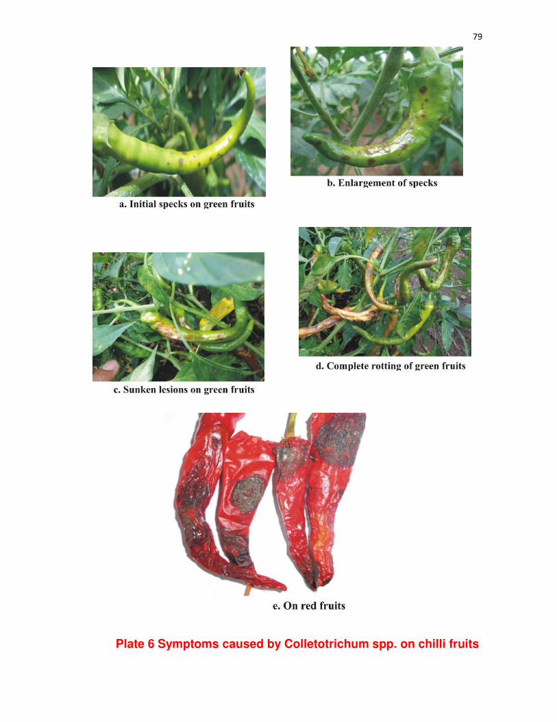
79
Plate 6 Symptoms caused by Colletotrichum spp. on chilli fruits
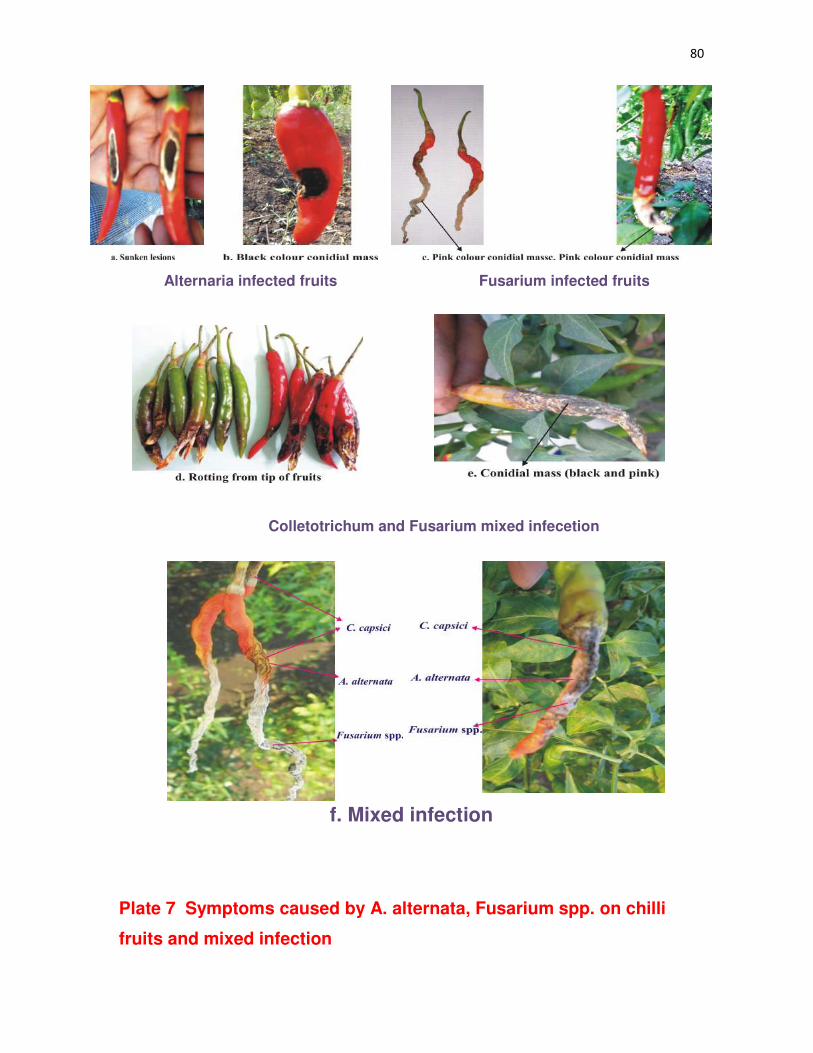
80
Alternaria infected fruits Fusarium infected fruits
Colletotrichum and Fusarium mixed infecetion
f. Mixed infection
Plate 7 Symptoms caused by A. alternata, Fusarium spp. on chilli
fruits and mixed infection

81
as described in 3.1.2.2. The observations (Table 7) showed that C. capsici recorded
highest frequency of 38.56 per cent followed by A. alternata (18.98 %), where as C.
acutatum occurred in lowest frequency of 5.04 per cent.
4.1.1.8 Pathogenecity test
C.capsici C. gloeosporioides and C. acutatum
All three species of Colletotrichum caused a depressed sunken, discolored,
circular to irregular spots of 5 to 10 mm sizes after two days of inoculation (DAI) on fruit
by pin prick method. The spots became black to grey in color after ten DAI. On these
discolored lesions salmon colored spore mass was found fifteen DAI. When the
diseased fruit was cut open inner surface of the skin was found to be covered with black
somatic masses of pathogen. Seeds were discolored and distinguished from healthy
seeds (Plate 8).
A. alternata
Sunken discolored, circular spots of 5 to 10 mm size was observed after two DAI
on fruit by pin prick method. Lesions became darker and covered by black moldy growth
of fungus was observed at ten DAI. On these lesions heavy sporulation of black color
was found on fifteen DAI.
F. oxysporum and F. sporotrichioides
Water soaked sunken lesions of 5 to 10 mm size was seen after two DAI on fruit
and covered by whitish pink moldy growth of fungus was observed on ten DAI. On these
lesions sporulation was found as pink color mass on fifteen DAI.
4.2 Morphological characterization and molecular variability of pathogen/s
4.2.1 Morphological characterization
Pathogens were isolated by plating infected parts on PDA. The isolates were
purified by establishing single spore culture as explained in 3.1.2.3. Morphological and
cultural characters of the C. capsici, C. gloeosporioides, C. acutatum, A. alternata and

82
Table 7: Frequency of pathogen/s isolated from chilli fruit rot disease
Pathogens Frequency (%) Mean
frequency (%) 2012-13 2013-14
Colletotrichum capsici 37.25 39.87 38.56
C. gloeosporioides 15.68 14.11 14.89
C. accutatum 4.57 5.52 5.04
Fusarium oxysporum 10.17 12.07 11.12
F. sporotrichioides 13.23 9.28 11.25
Alternaria alternata 18.95 19.01 18.98

83
Plate 8 Proving pathogenecity of C. capsici, C. gloeosporioides, C. acutatum, A. alternata, F. sporotrichioides and F.oxysporum

84
F. oxysporum, F. sporotrichioides were recorded on PDA and explained in Table 8, 9,
10, 11, 12 and 13 (Plate 9 and 10) .
C. capsici
Among the 25 isolates of C. capsici, growth characters of colony varied from
white to grey color, flat to raised fluffy and smooth regular to coarse irregular margin on
PDA. Acervuli size varied between 130.0 to 162.6 µm with setae length 145.4 to 179.1
µm. Medium to good sporulation (20 – 60 spores/ microscopic field under 100X) with
falcate shaped spores of size between 13.5 - 21.2 X 3.2 - 4.8 µm with single oil globule
at center was observed under 400X magnification (Table 8, Plate 9 a, Plate 11 a and b).
C. gloeosporioides
Among the 20 isolates of C. gloeosporioides, growth characters of colony on
PDA varied from white to grey color, where as in three isolates Cg-9, Cg-10 and Cg-12
whitish saffron color was observed with flat to raised fluffy and smooth regular to coarse
irregular margin. Sectoring was observed in three isolates (Cg-4, Cg-12 and Cg-20).
Medium to excellent sporulation (20 – 80 spores/ microscopic field under 100X) of
cylindrical shaped spores with oil globules and size between 12.5 - 16.7 X 1.6 - 2.8 µm
was observed under 400X magnification (Table 9, Plate 9 b Plate 11 c and d).
C. acutatum
Among the 12 isolates of C. acutatum, growth characters of colony on PDA
varied from whitish pink to pink color, where as in one isolate (Ca-1) brownish pink color
observed with flat to fluffy and smooth regular to coarse irregular margin. Sectoring was
observed in two isolates Ca-8 and Ca-12. Medium to good sporulation (20 – 60 spores/
microscopic field under 100X) of fusiform shaped spores and size between 11.4 - 14.1
X 1.1 - 2.3 µm was observed under 400X magnification (Table 10 Plate 9 c Plate 11c).
A. alternata
Among the 10 isolates of A. alternata, growth characters of colony on PDA varied from
ash to dark ash color with medium fluffy to raised fluffy and smooth regular to
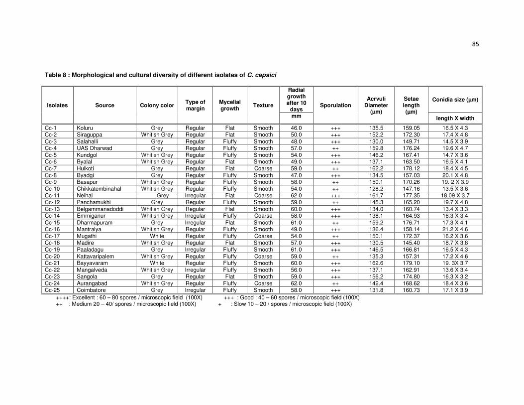
85
Table 8 : Morphological and cultural diversity of different isolates of C. capsici
Isolates Source Colony color Type of margin
Mycelial growth
Texture
Radial growth after 10
days Sporulation
Acrvuli Diameter
(µm)
Setae length (µm)
Conidia size (µm)
mm length X width
Cc-1 Koluru Grey Regular Flat Smooth 46.0 +++ 135.5 159.05 16.5 X 4.3 Cc-2 Siraguppa Whitish Grey Regular Flat Smooth 50.0 +++ 152.2 172.30 17.4 X 4.8
Cc-3 Salahalli Grey Regular Fluffy Smooth 48.0 +++ 130.0 149.71 14.5 X 3.9 Cc-4 UAS Dharwad Grey Regular Fluffy Smooth 57.0 ++ 159.8 176.24 19.6 X 4.7
Cc-5 Kundgol Whitish Grey Regular Fluffy Smooth 54.0 +++ 146.2 167.41 14.7 X 3.6 Cc-6 Byalal Whitish Grey Regular Flat Smooth 49.0 +++ 137.1 163.50 16.5 X 4.1
Cc-7 Hulkoti Grey Regular Flat Coarse 59.0 ++ 162.2 178.12 18.4 X 4.5
Cc-8 Byadgi Grey Regular Fluffy Smooth 47.0 +++ 134.5 157.03 20.1 X 4.8 Cc-9 Basapur Whitish Grey Regular Fluffy Smooth 58.0 ++ 150.1 170.26 19. 2 X 3.9
Cc-10 Chikkatembinahal Whitish Grey Regular Fluffy Smooth 54.0 ++ 128.2 147.16 13.5 X 3.6 Cc-11 Nelhal Grey Irregular Flat Coarse 62.0 +++ 161.7 177.35 18.09 X 3.7
Cc-12 Panchamukhi Grey Regular Fluffy Smooth 59.0 ++ 145.3 165.20 19.7 X 4.8
Cc-13 Belgammanadoddi Whitish Grey Regular Flat Smooth 60.0 +++ 134.0 160.74 13.4 X 3.3 Cc-14 Emmiganur Whitish Grey Irregular Fluffy Coarse 58.0 +++ 138.1 164.93 16.3 X 3.4
Cc-15 Dharmapuram Grey Irregular Flat Smooth 61.0 ++ 159.2 176.71 17.3 X 4.1 Cc-16 Mantralya Whitish Grey Regular Fluffy Smooth 49.0 +++ 136.4 158.14 21.2 X 4.6
Cc-17 Mugathi White Regular Fluffy Coarse 54.0 ++ 150.1 172.37 16.2 X 3.6 Cc-18 Madire Whitish Grey Regular Flat Smooth 57.0 +++ 130.5 145.40 18.7 X 3.8
Cc-19 Paaladagu Grey Irregular Fluffy Smooth 61.0 +++ 146.5 166.81 16.5 X 4.3
Cc-20 Kattavaripalem Whitish Grey Regular Fluffy Coarse 59.0 ++ 135.3 157.31 17.2 X 4.6 Cc-21 Bayyavaram White Regular Fluffy Smooth 60.0 +++ 162.6 179.10 19. 3X 3.7
Cc-22 Mangalveda Whitish Grey Irregular Fluffy Smooth 56.0 +++ 137.1 162.91 13.6 X 3.4 Cc-23 Sangola Grey Regular Flat Smooth 59.0 +++ 156.2 174.80 16.3 X 3.2
Cc-24 Aurangabad Whitish Grey Regular Fluffy Coarse 62.0 ++ 142.4 168.62 18.4 X 3.6
Cc-25 Coimbatore Grey Irregular Fluffy Smooth 58.0 +++ 131.8 160.73 17.1 X 3.9
++++: Excellent : 60 – 80 spores / microscopic field (100X) +++ : Good : 40 – 60 spores / microscopic field (100X) ++ : Medium 20 – 40/ spores / microscopic field (100X) + : Slow 10 – 20 / spores / microscopic field (100X)

86
Table 9 : Morphological and cultural diversity of different isolates of C .gloeosporioides
Isolates Source Colony color Type of margin
Mycelial growth
Texture
Radial growth after
10 days mm
Conidia Size (µm)
Sporulation length X width
Cg-1 Kagalgombe Grey Regular Flat Smooth 67.0 14.5 X 2.3 +++
Cg-2 Shanvaspura Greyish Irregular Fluffy Coarse 72.0 15.4 X 2.8 ++
Cg-3 Inamhongal Grey Irregular Flat Coarse 70.0 12.5 X 1.9 ++++
Cg-4 Byahatti Greyish Irregular Fluffy sectoring Coarse 78.0 13.6 X 2.7 ++
Cg-5 UAS Dharwad Grey Regular Fluffy Coarse 69.0 12.7 X 2.1 ++++
Cg-6 Shirguppi Grey Irregular Fluffy Coarse 71.0 14.5 X 2.3 +++
Cg-7 Binkadkatti Greyish Irregular Flat Smooth 74.0 15.4 X 2.6 +++
Cg-8 Guttal Greyish Regular Flat Smooth 66.0 16.1 X 2.8 +++
Cg-9 Kanakapur Whitish to Saffron Regular Flat Smooth 80.0 15. 2 X 2.4 +++
Cg-10 Beluru Whitish to Saffron Regular Flat Coarse 76.0 13.5 X 2.6 ++
Cg-11 Mandapur Grey Irregular Flat Coarse 81.0 16.09 X 1.7 ++++
Cg-12 Raladoddi Whitish to Saffron Irregular Flat sectoring Smooth 78.0 14.7 X 2.1 ++
Cg-13 Hanumapuram Grey Irregular fluffy Coarse 76.0 13.4 X 2.1 +++
Cg-14 Alur Whitish Regular Flat Coarse 81.0 14.3 X 2.2 +++
Cg-15 Kalludevarakunta Grey Irregular Fluffy Coarse 80.0 15.3 X 2.4 ++
Cg-16 Mangalaveda Greyish Irregular Fluffy Smooth 74.0 16.2 X 2.6 ++
Cg-17 Medikonduru Greyish Regular Fluffy Smooth 79.0 14.2 X 1.6 +++
Cg-18 Percherela Greyish Regular Fluffy Coarse 74.0 16.7 X 1.8 ++
Cg-19 Ananthapuram Greyish Regular Fluffy Coarse 80.0 14.5 X 2.3 +++
Cg-20 Coimbatore Grey Regular Flat Sectoring Coarse 88.0 15.2 X 2.6 +++
++++: Excellent : 60 – 80 spores / microscopic field (100X) +++ : Good : 40 – 60 spores / microscopic field (100X) ++ : Medium 20 – 40/ spores / microscopic field (100X) + : Slow 10 – 20 / spores / microscopic field (100X)
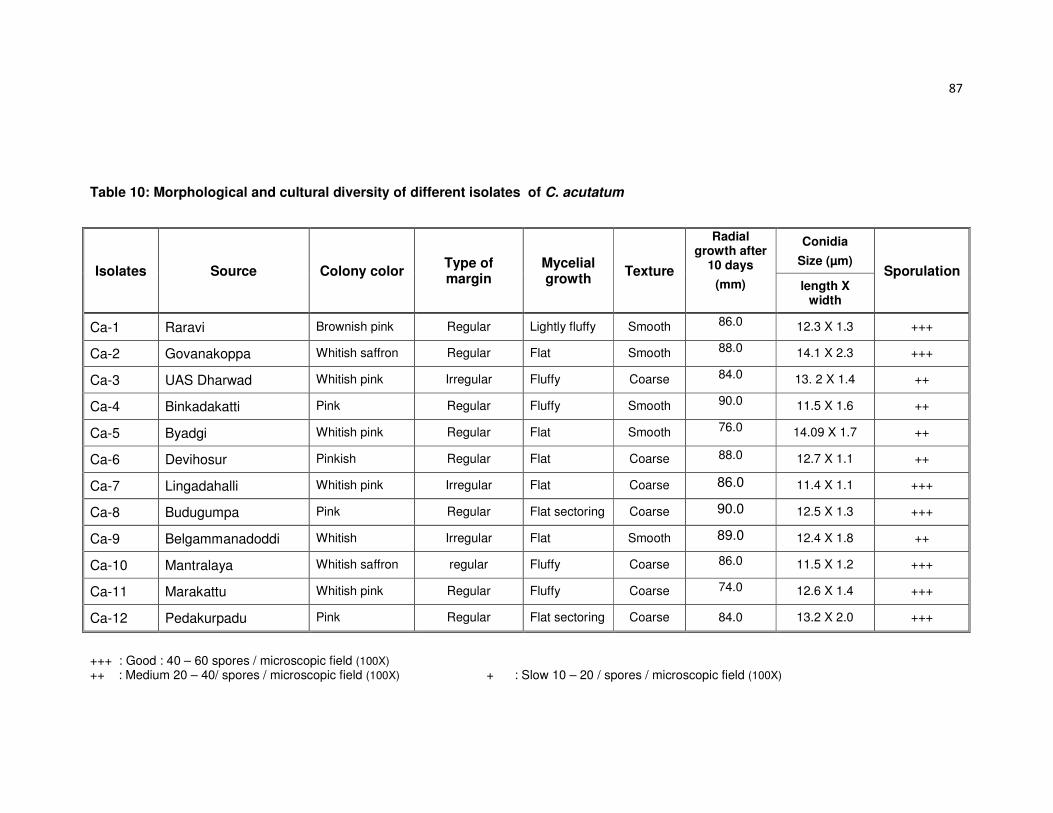
87
Table 10: Morphological and cultural diversity of different isolates of C. acutatum
Isolates Source Colony color Type of margin
Mycelial growth
Texture
Radial growth after
10 days
(mm)
Conidia
Size (µm) Sporulation
length X width
Ca-1 Raravi Brownish pink Regular Lightly fluffy Smooth 86.0 12.3 X 1.3 +++
Ca-2 Govanakoppa Whitish saffron Regular Flat Smooth 88.0 14.1 X 2.3 +++
Ca-3 UAS Dharwad Whitish pink Irregular Fluffy Coarse 84.0 13. 2 X 1.4 ++
Ca-4 Binkadakatti Pink Regular Fluffy Smooth 90.0 11.5 X 1.6 ++
Ca-5 Byadgi Whitish pink Regular Flat Smooth 76.0 14.09 X 1.7 ++
Ca-6 Devihosur Pinkish Regular Flat Coarse 88.0 12.7 X 1.1 ++
Ca-7 Lingadahalli Whitish pink Irregular Flat Coarse 86.0 11.4 X 1.1 +++
Ca-8 Budugumpa Pink Regular Flat sectoring Coarse 90.0 12.5 X 1.3 +++
Ca-9 Belgammanadoddi Whitish Irregular Flat Smooth 89.0 12.4 X 1.8 ++
Ca-10 Mantralaya Whitish saffron regular Fluffy Coarse 86.0 11.5 X 1.2 +++
Ca-11 Marakattu Whitish pink Regular Fluffy Coarse 74.0 12.6 X 1.4 +++
Ca-12 Pedakurpadu Pink Regular Flat sectoring Coarse 84.0 13.2 X 2.0 +++
+++ : Good : 40 – 60 spores / microscopic field (100X) ++ : Medium 20 – 40/ spores / microscopic field (100X) + : Slow 10 – 20 / spores / microscopic field (100X)
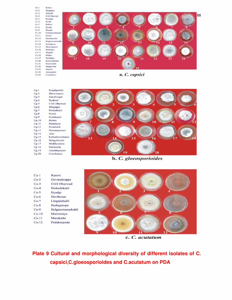
88
Plate 9 Cultural and morphological diversity of different isolates of C.
capsici,C.gloeosporioides and C.acutatum on PDA

89
Plate 10 Cultural and morphological diversity of different isolates of
A. alternata, F.oxysporum and F.Sporotrichioides on PDA

90
coarse irregular margin. Medium to excellent sporulation (20 – 70 spores/ microscopic
field under 100X) of muriform spores with beak with 3 – 5 horizontal septa 1 – 2 vertical
septa, with spore size varied between 14.1 - 21.8 X 6.1 - 8.4 µm was observed under
under 400X magnification (Table 11 Plate 10 a, Plate 12 a ).
F. oxysporum
Among the nine isolates of F. oxysporum growth characters of colony on PDA
varied from white to whitish purple color with flat, smooth and regular margin except Fo-
7 which is irregular. Medium to excellent sporulation (20 – 70 spores/ microscopic field
under 100X) of falcate macro conidia with 3 – 5 horizontal septa and size varied
between 22.01 - 32.10 X 3.2- 5.2µm was observed under 400X magnification (Table12
Plate 10 b, Plate 12 b).
F. sporotrichioides
Among the seven isolates of F. sporotrichioides, growth characters of colony on
PDA varied from light brown to white rose color, fluffy, smooth to coarse and regular to
irregular margin. Good to excellent sporulation (20 – 70 spores/ microscopic field under
100X) of falcate macro conidia with 3 – 5 horizontal septa and size varied between 32.1
- 42.0 X 3.4-5.2µm, micro conidia were globose of size varied between 6.5-7.0 X 2.5 –
3.0 µm were observed under 400X magnification (Table 13 Plate 10c, Plate 12 c).
4.2.2 Molecular characterization and variability of pathogen/s
4.2.2.1 Isolation of Genomic DNA
Genomic DNA of the fungi were isolated as described in material and methods.
The DNA obtained was observed by running on 1.0 per cent agarose gel
electrophoresis. The DNA obtained was about 7000-8000 bp of whole genomic DNA of
Colletotrichum spp. Fusarium spp. and Alternaria sp. with nanodrop equipment
concentration of 91µg/µl of DNA.
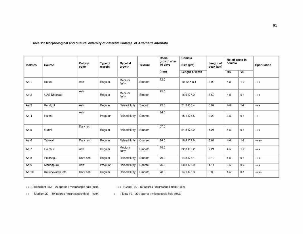
91
Table 11: Morphological and cultural diversity of different isolates of Alternaria alternata
Isolates Source Colony color
Type of margin
Mycelial growth
Texture
Radial growth after 10 days
(mm)
Conidia
Size (µm) Length of beak (µm)
No. of septa in conidia
Sporulation
Length X width HS VS
Aa-1 Koluru Ash Regular Medium fluffy
Smooth 72.0
19.12 X 8.1 3.90 4-5 1-2 +++
Aa-2 UAS Dharwad Ash
Regular
Medium fluffy
Smooth 75.0
16.9 X 7.2 3.60 4-5 0-1 +++
Aa-3 Kundgol Ash Regular Raised fluffy Smooth 79.0 21.3 X 8.4 6.82 4-6 1-2 +++
Aa-4 Hulkoti
Ash
Irregular Raised fluffy Coarse
84.0
15.1 X 6.5 3.20 3-5 0-1 ++
Aa-5 Guttal Dark ash
Regular Raised fluffy Smooth
67.0 21.8 X 8.2 4.21 4-5 0-1 +++
Aa-6 Talakall Dark ash Regular Raised fluffy Coarse 74.0 18.4 X 7.9 3.61 4-6 1-2 ++++
Aa-7 Raichur Ash Regular Medium fluffy
Smooth 75.0
22.3 X 9.2 7.21 4-5 1-2 +++
Aa-8 Paldaagu Dark ash Regular Raised fluffy Smooth 79.0 14.8 X 6.1 3.10 4-5 0-1 ++++
Aa-9 Mandapura Ash Irregular Raised fluffy Coarse 76.0 20.8 X 7.9 4.11 3-5 0-2 +++
Aa-10 Kalludevarakunta Dark ash Regular Raised fluffy Smooth 78.0 14.1 X 6.3 3.00 4-5 0-1 ++++
++++: Excellent : 50 – 70 spores / microscopic field (100X) +++ : Good : 30 – 50 spores / microscopic field (100X)
++ : Medium 20 – 30/ spores / microscopic field (100X) + : Slow 10 – 20 / spores / microscopic field (100X)

92
Table 12: Morphological and cultural diversity of different isolates of Fusarium oxysporum
Isolates Source Colony color Type of margin
Radial growth after 10 days
(mm)
Macroconidia
Size (µm) Sporulation
Fo-1 Raravi White Regular 68.0 22.01 X 3.2 ++++
Fo-2 UAS Dharwad
Whitish purple Regular 70.0 23.12 X 4.1 ++++
Fo-3 Sharewad White Regular 69.0 26.2 X 5.1 ++++
Fo-4 Byalal White Regular 73.0 32.10 X 4.0 +++
Fo-5 Ranebennur Whitish purple Regular 72.0 24.12 X 3.6 ++++
Fo-6 Nelhal White Regular 70.0 30.21 X 5.0 ++++
Fo-7 Alur White Irregular 68.0 27.30 X 3.7 ++++
Fo-8 Emmiganur White Regular 72.0 29.21 X 4.5 ++
Fo-9 Mangalaveda
White Regular 71.0 26.32 X 5.2 ++++
++++: Excellent : 50 – 70 spores / microscopic field (100X) +++ : Good : 30 – 50 spores / microscopic field (100X) ++ : Medium 20 – 30/ spores / microscopic field (100X) + : Slow 10 – 20 / spores / microscopic field (100X)

93
Table 13: Morphological and cultural diversity of different isolates Fusarium sporotrichioides
Isolates Source Colony color Type of margin
Texture
Radial growth after
10 days
(mm)
Macroconidia
Size (µm) Sporulation
Fs-1 Lokapur Light brown Irregular Coarse 89.0 33.2 X 4.6 ++++
Fs-2 Kumbapur farm
White rose Regular Smooth 82.0 36.0 X 4.2 +++
Fs-3 Badrapur Light brown Irregular Coarse 90.0 32.1 X 3.4 ++++
Fs-4 Bommanal Light brown Regular Smooth 89.0 41.3 X 5.2 +++
Fs-5 Raladoddi White rose Irregular Coarse 88.0 34.6 X 3.6 ++++
Fs-6 Mantralaya White rose Regular Smooth 90.0 42.0 X 4.8 ++++
Fs -7 Perecherla Light brown Irregular Coarse 90.0 40.2 X 4.3 ++++
++++: Excellent : 50 – 70 spores / microscopic field (100X) +++ : Good : 30 – 50 spores / microscopic field (100X) ++ : Medium 20 – 30/ spores / microscopic field (100X) + : Slow 10 – 20 / spores / microscopic field (100X)

94
Plate 11: Fruiting body and spore morphology of Colletotrichum spp. (400X)

95
Plate 12: Spore morphology of A. alternata, F. oxysporum and
F.sporotrichioides (400X)

96
4.2.2.2 Amplification of ITS-1 and ITS-4 region
The full length ITS rDNA region was amplified with ITS-1 (5’-TCCGTAGGTGAACCTGCGG-3')
and ITS-4 (5’-TCCTCCGCTTATTGATATGC-3’) primers for isolates of Colletotrichum spp., Alternaria
sp., Fusarium spp. and DNA amplicon was observed at the region 560 bp with a concentration of
around 437 µg/ µl.. The amplified products were checked on 1.4 per cent agarose gel electrophoresis
(Plate 13)
4.2.2.3 DNA sequencing
The DNA sequences were obtained for ITS rDNA. The sequences of these isolates are given
below.
Colletotrichum spp. isolates
1. CACCCTTTGTGACATACCTTAACTGTTGCTTCGGCGGGTAGGCGTCCCCTAAAAAGGACGTCTCCCGGCCCTCTCCCGTCCGCGGGTGGGGCGCCCGCCGGAGGATAACCAAACTCTGATTTAACGACGTTTCTTCTGAGTGACACAAGCAAATAATCAAAACTTTTAACAACGGATCTCTTGGTTCTGGCATCGATGAAGAACGCAGCGAAATGCGATAAGTAATGTGAATTGCAGAATTCAGTGAATCATCGAATCTTTGAACGCACATTGCGCCCGCCAGCATTCTGGCGGGCATGCGTCATTTCAACCCTCAAGCTCTGCTTGGTGTTGGGGCTCTACGGTTGACGTAGGCCCTTAAAGGTAGTGGCGGACCCTCTCGGAGCCTCCTTTGCGTAGTAACATTTCGTTGTTCGAGCGTCATTTCAACCCTCAAGCTCTGCTTGGTGTTGGGGCTCTACGGTTGACGTAGGCCCTTAAAGGTAGTGGCGGACCCTCTCGGAGCCTCCTTTGCGTAGTAACATTTCGTCTCGCATTGGGATTCGGAGGGACTCTAGCCGTAAAACCCCCAATTTTACTAAGGTTGACCTCGGATCAGGTAGGAATACCCGCTGAACTTA AGCATATCATAGCCGG
2.
GTGCACTGAGATGCGCTCTACACCCTTTGTGACATACCTATAACTGTTGCTTCGGCGGGTAGGGTCTCCGCGACCCTCCCGGCCTCCCGCCTCCGGGCGGGTCGGCGCCCGCCGGAGGATAACCAAACTCTGATTTAACGACGTTTCTTCTGAGTGGTACAAGCAAATAATCAAAACTTTTAACAACGGATCTCTTGGTTCTGGCATCGATGAAGAACGCAGCGAAATGCGATAAGTAATGTGAATTGCAGAATTCAGTGAATCATCGAATCTTTGAACGCACATTGCGCCCGCCAGCATTCTGGCGGGCATGCCTGTTCGAGCGTCATTTCAACCCTCAAGCTCTGCTTGGTGTTGGGGCCCTACAGGACCCTCTCGGAGCCTCCTTTGCGTAGTAACTTTACGTCTCGCAGGTGGGATCCGGAGGGACTCTTGCCGTAAAACCCCCAATTTTCCAAAGGTTGACCTCGGATCAGGTAGGAATACCCGCTCTGAGTTTTACCCCCAAACCCTTTGTGAAAATACCTATAACTGTTGCTTCGCGGGAAGGTCTCCCGAC
3.
GGCACCACCAGGGGTGGANNCTGCGGCTTAATTTGACTCAACACGGGGAAACTCACCAGGTCCAGACACAATGAGGATTGACAGATTGAGAGCTCTTTCTTGATTTTGTGGGTGGTGGTGCATGGCCGTTCTTAGTTGGTGGAGTGATTTGTCTGCTTAATTGCGATAACGAACGAGACTTAACCTGCTAAATAGCCCGTATTGCTTTGGCAGTACGCCGGCTTCTTAGAGGGACTATGGCTCAAGCCGATGGAAGTTT

97
GAGGCAATAACAGGTCTGTGATGCCCTTAGATGTTCTGGGCCGCACGCGCGTTACACTGACGGAGCCAGCGAGTTCTCCCTTGGCCGGAAGGCCCGGGTTGCAGAATTCAGTGAATCATCGAATCTTTGAACGCACATTGCGCTCGCCAGCATTCTGGCGAGCATGCCTGTTCGAGCGTCATTTCAACCCTCAAGCACCGCTTGGTTTTGGGGCCCCACCTAACGTCTCGCACTGGGATCCGGAGGGACTCTTGCCGTAAAACCCCCAAATTTTTTACAGGTTGACCTCGGATCAGGTAGGAATACCCG CTGAACTTAA
Alternaria sp. isolate
TTCTTTTTCCTGCATAACCGAGATCCCGCTGGCGCCGCGCTGCTTCGGTTGATGCTCCATTAGTGTGATTCTTATCTGTTGATTTGGTGGTTCGGTGGCGGGCTGGCCGCTCCGGCTGGAAAGAGCCCGCTTTTGTAATTGCACTCGTGCTGTGAGTGTTGTCTGAAGAAATGTGTAATTAATAAAACTTTCATGAATCGAGCTCTTGGCTCTGGCATCGATGAATGCCACAACGAAAGGGAATTGCTAATGTCAATTAATCAATTTATCTTTTCACCCAATCTGTGCCCTTTGGTTGTCCCCCCGGCATGTCCGAGGGACCGGCCTGTGTACCCTCCAGCTTTGCTTGCAGTTGGACGTCGTGCCTGGAGCTTTGCTGGACACTCGCCTTAAAGTAATTGGCAGCCGGCCTACTGGTTTCGGAGCGCACCACAAGTCGCACTCTCTATCACCAAAGGTCTAGCATCCATTAAGCCTTTTTTCAACTTTTGACCTCGGATCAGGTAGGGATACCCGCTGAACTTAAGCATATCAATAAGCGGAGGAA
Fusarium spp. isolates
1.
GAGTTTTCCGGGGGGGGAAAGAGTTCACTCCCAACCCCTGTGAACATACCACTTGTTGCCTCGGCGGATCAGCCCGCTCCCGGTAAAACGGGACGGCCCGCCAGAGGACCCCTAAACTCTGTTTCTATATGTAACTTCTGAGTAAAACCATAAATAAATCAAAACTTTCAACAACGGATCTCTTGGTTCTGGCATCGATGAAGAACGCAGCAAAATGCGATAAGTAATGTGAATTGCAGAATTCAGTGAATCATCGAATCTTTGAACGCACATTGCGCCCGCCAGTATTCTGGCGGGCATGCCTGTTCGAGCGTCATTTCAACCCTCAAGCACAGCTTGGTGTTGGGACTCGCGTTAATTCGCGTTCCTCAAATTGATTGGCGGTCACGTCGAGCTTCCATAGCGTAGTAGTAAAACCCTCGTTACTGGTAATCGTCGCGGCCACGCCGTTAAACCCCAACTTCTGAATGTTGACCTCGGATCAGGTAGGAATACCCGCTGAACTTAAGCATATCAATAAGCGGAGGAAA
2.
GGGGGGTCTTTCGGCATTGCACTCAGGCCCCTGTGACATACCTATACGTTGCCTCGGCGGATCAGCCCGCGCACAAAAACGGGACGGCCCGCCCGAGGACCCCTAAACTCTGTCTCTTAAAGAAACTTCTGAGTAAAACAAACAAATAAATCAAAACTTTCAACACCGGATCTCTTGGTTCTGGTTTTTCTCAAAAAACCCCCCCAAATTTTATAAGTAATGTGAGTTGCAGAATTCCCACCCACCCCCCCCAATCTCTCAACCCCC
DNA sequences of selected six isolates were compared using bioinformatics tool
like NCBI (National Centre for Bioinformatics) BLAST (Basic Local Alignment Search Tool)
programme. Basedon sequence comparison, the identification of Colletotrichum spp. (3 isolates)
Alternaria sp. (1 isolate) and Fusarium spp. (2 isolates) isolates were confirmed as C. capsici,
C.gloeosporioides, C. acutatum, A. alternata and F. oxysporum, F. sporotrichioides respectively. The
list of isolates, accession number, per cent homology and name identified are given in (Table 14).

98
Plate 13 Amplification of ITS1 and ITS4 region of representative Colletotrichum
spp., Alternaria sp.and Fusarium spp.

99
Table 14: Comparison and identity of chilli fruit rot causing pathogens isolates with gene bank in NCBI BLAST program
Sl. No
Accession number
NCBI BLAST Hit results Max. score
Total scor
e
Query coverag
e (%)
E-value (%)
Max. Ident.
(%)
1 KF147902.1
Colletotrichum capsici isolate CCM 18S ribosomal RNA gene, partial sequence; internal transcribed spacer 1, 5.8S ribosomal RNA gene, and internal transcribed spacer 2, complete sequence; and 28S ribosomal RNA gene, partial sequence
793 793 100 0 100
2 KC895535.1
Colletotrichum gloeosporioides isolate CJBB21-25 18S ribosomal RNA
gene, partial sequence; internal transcribed spacer 1, 5.8S ribosomal RNA gene, and internal transcribed spacer 2, complete sequence; and 28S ribosomal RNA gene, partial sequence
854 854 86 0.0 99
3 AJ749694.1 Colletotrichum acutatum ITS1, 5.8S rRNA gene and ITS2, isolate PT227 665 665 100 0.0 94
4 EU87848.1 Alternaria alternata isolate G2 A1-32 internal transcribed spacer 1, partial sequence; 5.8S ribosomal RNA gene, complete sequence; and internal transcribed spacer 2, partial sequence
48.1 128 100 2e-07 100
5 KC351189.1 Fusarium sporotrichioides 18S ribosomal RNA gene, partial sequence 436 436 99 3e-119 97
6 FR750924.1 Fusarium sporotrichioides genomic DNA containing 18S rRNA gene, ITS1, 5.8S rRNA gene, ITS2, 28S rRNA gene, culture collection MTCC:7375
172 172 83 7e-40 87
7 JN232187.1
Fusarium oxysporum isolate 847 18S ribosomal RNA gene, partial sequence; internal transcribed spacer 1, 5.8S ribosomal RNA gene, and internal transcribed spacer 2, complete sequence; and 28S ribosomal RNA gene, partial sequenc
278 278 83 2e-71 94
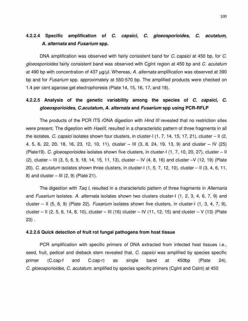
100
4.2.2.4 Specific amplification of C. capsici, C. gloeosporioides, C. acutatum,
A. alternata and Fusarium spp.
DNA amplification was observed with fairly consistent band for C. capsici at 450 bp, for C.
gloeosporioides fairly consistent band was observed with CgInt region at 450 bp and C. acutatum
at 490 bp with concentration of 437 µg/ µl. Whereas, A. alternata amplification was observed at 390
bp and for Fusarium spp. approximately at 550-570 bp. The amplified products were checked on
1.4 per cent agarose gel electrophoresis (Plate 14, 15, 16, 17, and 18).
4.2.2.5 Analysis of the genetic variability among the species of C. capsici, C.
gloeosporioides, C.acutatum, A. alternata and Fusarium spp using PCR-RFLP
The products of the PCR ITS rDNA digestion with Hind III revealed that no restriction sites
were present. The digestion with HaeIII, resulted in a characteristic pattern of three fragments in all
the isolates. C. capsici isolates shown four clusters, in cluster-I (1, 7, 14, 15, 17, 21), cluster – II (2,
4, 5, 6, 22, 20, 18, 16, 23, 12, 10, 11), cluster – III (3, 8, 24, 19, 13, 9) and cluster – IV (25)
(Plate19). C. gloeosporioides isolates shown five clusters, in cluster-I (1, 7, 10, 20, 27), cluster – II
(2), cluster – III (3, 5, 6, 9, 18, 14, 15, 11, 13), cluster – IV (4, 8, 16) and cluster –V (12, 19) (Plate
20). C. acutatum isolates shown three clusters, in cluster-I (1, 5, 7, 12, 10), cluster – II (3, 4, 6, 11,
8) and cluster – III (2, 9) (Plate 21).
The digestion with Taq I, resulted in a characteristic pattern of three fragments in Alternaria
and Fusarium isolates. A. alternata isolates shown two clusters cluster-I (1, 2, 3, 4, 6, 7, 9) and
cluster – II (5, 8, 9) (Plate 22). Fusarium isolates shown five clusters, in cluster-I (1, 3, 4, 7, 9),
cluster – II (2, 5, 6, 14, 8, 10), cluster – III (16) cluster – IV (11, 12, 15) and cluster – V (13) (Plate
23) .
4.2.2.6 Quick detection of fruit rot fungal pathogens from host tissue
PCR amplification with specific primers of DNA extracted from infected host tissues i.e.,
seed, fruit, pedicel and dieback stem revealed that, C. capsici was amplified by species specific
primer (C.cap-f and C.cap-r) as single band at 450bp (Plate 24).
C. gloeosporioides, C. acutatum, amplified by species specific primers (CgInt and CaInt) at 450

101
Plate 14: Specific amplification of Colletotrichum capsici by C.cap primer

102
Plate 15 Specific amplification of Colletotrichum gloeosporioides at CgInt region

103
Plate 16 Specific amplification of Colletotrichum acutatum at CaInt region
Plate 17 Specific amplification of Alternaria alternata by AAF and AAR primer

104
Plate 18 Specific amplification of Fusarium spp. by Tef.Fu primer

105
Plate 19: PCR-RFLP pattern of C. capsici by HaeIII enzyme

106
Plate 20: PCR-RFLP pattern of C. gloeosporioides by HaeIII enzyme

107
Plate 21: PCR-RFLP pattern of C. acutatum by HaeIII enzyme
Plate 22 PCR-RFLP pattern of A. alternata by TaqI enzyme

108
Plate 23 PCR-RFLP pattern of Fusarium spp. by TaqI enzyme

109
Plate 24 Detection of C. capsici by specific primer (C.cap)
Plate 25 Detection of C. gloeoesporioides , C. acutataum by specific
primer (CgInt and CaInt)

110
Plate 26 Detection of A. alternata by specific primer (AAF and AAR)
Plate 27 Detection of Fusarium by specific primer ( Tef- Fu. )

111
and 490bp respectively (Plate 25) , A. alternata amplified by species specific primer (AAf and Aar)
at 390bp (Plate 26) and Fusarium spp. were amplified at 550 – 570bp (Plate 27). From seed, fruit,
die-back infected twigs, and fruit pedicel samples C. capsici, C. gloeosporioides, C. acutatum, A.
alternata and Fusarium spp. were detected.
4.3 Epidemiology of the fruit rot disease in relation to climatic factors
4.3.1 Survival ability of Colletotrichum spp. A. alternata and F. oxysporum in plant debris
The present study was undertaken to know the survival period of Colletotrichum spp. A.
alternata and F. oxysporum in host tissues as explained in materials and methods. The results
obtained on recovery of fungus by its growth are presented in Table 15a and 15b.
The plant debris kept for survival ability in 2012-13 revealed that Colletotrichum spp.
remained viable up to 165 days under laboratory conditions, 120 days on soil surface and 60 days
under 10cm depth of soil condition. A. alternata remained viable up to 120 days under laboratory
conditions, 90 days on soil surface and 45 days under 10cm depth of soil condition. F. oxysporum
remained viable up to 150 days under laboratory conditions, 135 days on soil surface and 60 days
under 10cm depth of soil condition. In 2013-14 Colletotrichum spp. remained viable up to 150 days
under laboratory conditions, 105 days on soil surface and 60 days under 10cm depth of soil
condition. A. alternata remained viable up to 105 days under laboratory conditions, 90 days on soil
surface and 45 days under 10cm depth of soil condition. F. oxysporum remained viable up to 150
days under laboratory conditions, 120 days on soil surface and 60 days under 10cm depth of soil
condition. The results indicated that the pathogen/s survived well in the infected host tissue for five
to six months. The survival ability of these three pathogens was more in lab condition (150-
165days) and which was reduced (90-125 days) on soil surface and still reduction in survival was
recorded in 10cm depth of soil condition (45-60 days).
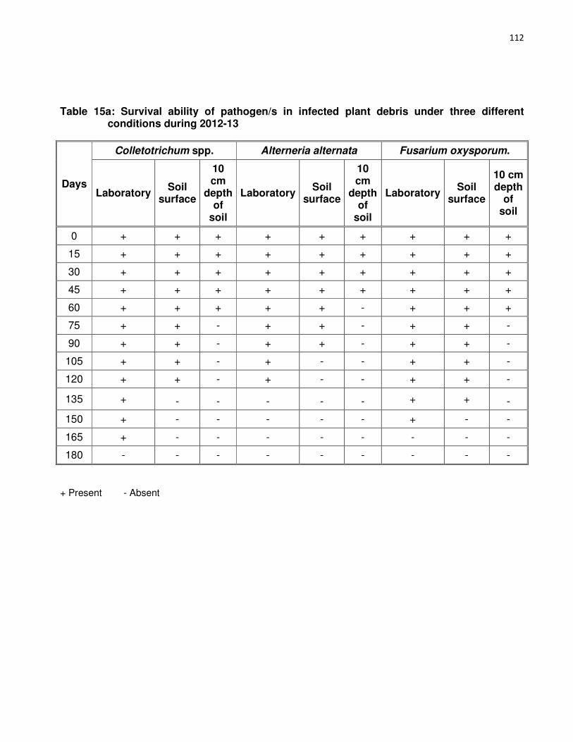
112
Table 15a: Survival ability of pathogen/s in infected plant debris under three different conditions during 2012-13
Days
Colletotrichum spp. Alterneria alternata Fusarium oxysporum.
Laboratory Soil
surface
10 cm
depth of
soil
Laboratory Soil
surface
10 cm
depth of
soil
Laboratory Soil
surface
10 cm depth
of soil
0 + + + + + + + + +
15 + + + + + + + + +
30 + + + + + + + + +
45 + + + + + + + + +
60 + + + + + - + + +
75 + + - + + - + + -
90 + + - + + - + + -
105 + + - + - - + + -
120 + + - + - - + + -
135 + - - - - - + + -
150 + - - - - - + - -
165 + - - - - - - - -
180 - - - - - - - - -
+ Present - Absent
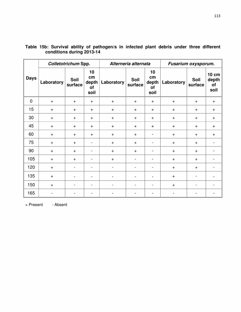
113
Table 15b: Survival ability of pathogen/s in infected plant debris under three different conditions during 2013-14
Days
Colletotrichum Spp. Alterneria alternata Fusarium oxysporum.
Laboratory Soil
surface
10 cm
depth of
soil
Laboratory Soil
surface
10 cm
depth of
soil
Laboratory Soil
surface
10 cm depth
of soil
0 + + + + + + + + +
15 + + + + + + + + +
30 + + + + + + + + +
45 + + + + + + + + +
60 + + + + + - + + +
75 + + - + + - + + -
90 + + - + + - + + -
105 + + - + - - + + -
120 + - - - - - + + -
135 + - - - - - + - -
150 + - - - - - + - -
165 - - - - - - - - -
+ Present - Absent

114
4.3.2 Host range of C. capsici
To know the host range of C. capsici two solanaceous vegetables and seven legume hosts
were tested. The results revealed that C. capsici can infect to all nine hosts. On solanaceous
vegetables tomato and brinjal, chlorotic lesions were produced on 4th day after inoculation. These
lesions turned to brown color leading to necrosis after seven days of inoculation. In legume crops
like cowpea, green gram and black gram brown color horse shoe type lesions were observed after
eight days after inoculation. An earliest visible chlorotic symptom was observed in pea after two
days of inoculation, while maximum duration (six days) taken to produce the initial symptom in
moth bean. Details are furnished in Table 16 and Plate 28.
4.3.3.1 Cross inoculation of C. capsici
Cross inoculation study of C. capsici among twig, pedicel and fruit of chilli revealed that C.
capsici can cause seed rot, die-back of twigs, pedicel discoloration and fruit rot from all tested
above ground parts of chilli plant by cross inoculation (Table 17 and Plate 29).
4.3.3.2 Pathogen interaction
Studies on host-pathogen interaction aimed at studying various symptoms produced by
pathogens in combinations are presented in (Table 18 and Plate 30).
C. capsici + A. alternata
Symptoms appear on two days after inoculation as water soaked lesion of 1.0-1.5cm
diameter. After five days of inoculation concentric rings with black color mold growth was observed
on 2-3 cm diameter lesion, eight days after inoculation black color spore mass wer produced on
these lesions (Plate 30a).
C. capsici + F. oxysporum
Symptoms appear on two days after inoculation as water soaked lesion of 0.5-1.0 cm
diameter, after four days of inoculation pinkish white color mycelial growth was observed on 1.5 –
2.0 cm diameter lesion. Eight days after inoculation pink color spore mass were produced on these
lesions (Plate 30b)..

115
Table 16. Hosts reaction to Colletotrichum capsici inoculation
Sl.
No. Host Reaction
Days after inoculation
Symptoms observed
1 Tomato +
4 Initiation of Chlorotic lesion
6 Chlorotic lesion
8 Chlorotic lesion turning to brown color necrotic lesion
2 Brinjal
+
4 Chlorotic lesion from margin of leaf
6 Lesion turning to brown color
8 Necrosis of leaf
3 Chick pea
+
4 Chlorotic lesions from margin of leaves
6 Chlorotic lesions to brown color with drying from margin
8 Necrosis of leaves
4 Moth bean
+
6 Chlorotic lesions from tip of leaves
8 Chlorotic lesions turning to brown color
10 Necrosis of leaf from tip of leaf
5 Green gram
+
4 Chlorotic lesions on lower surface of leaf
6 Chlorotic lesions on upper surface of leaf
8 Chlorotic lesions turning to reddish brown color horse shoe type symptoms on upper surface
6 Cowpea +
4 Chlorotic lesions on lower surface of leaf
6 Chlorotic lesions on upper surface of leaf
8 Chlorotic lesions turning to reddish brown color horse shoe type symptoms on upper surface
Contd…

116
7 Black gram
+
4 Chlorotic lesions on lower surface of leaf
6 Chlorotic lesions on upper surface of leaf
8 Chlorotic lesions turning to reddish brown color horse shoe type symptoms on upper surface
8 Soybean +
3 Chlorotic lesions on leaves
5 Reddish brown color lesion
7 Necrotic lesion
9 Pea +
2 Chlorotic lesions on leaves
4 Chlorotic lesions to brown color with necrosis
6 Necrotic lesion

117

118
Plate 28 Host range of C. capsici
Table 17. Reaction on different parts of chilli by cross inoculation of Colletotrichum capsici
Isolated from Inoculation to
Twig Fruit Pedicel
Seed + + +
Twig + + +
Fruit + + +
Pedicel + + +

119
Plate 29 Cross inoculation of C. capsici from seed to other parts

120
Table 18: Interaction effect of chilli fruit rot pathogens
Pathogen
First appearance
of symptoms (DAI)
Symptom observed
(DAI) Symptom
C. capsici + A. alternata
2
2 Water soaked lesion of 1.0-1.5 cm diameter
5 Concentric rings with black color mold growth was observed on 2.0-3.0 cm diameter lesion
8 Black color spore mass produced on these lesions
C. capsici + F. oxysporum
2
2 Water soaked lesion of 0.5-1.0 cm diameter
4 Pinkish white color mycelial growth was observed on 1.5 - 2 .0 cm diameter lesion
8 Pink color spore mass produced on these lesions.
C. capsici + C. gloeosporioides + C. acutatum.
1
1 Water soaked lesion of 0.5 -1.0 cm diameter.
7 Sunken lesion with salmon color spore mass produced on 1.5 - 2 .0 cm diameter lesion
C. capsici + A.alternata + F.oxysporum
1
1 Water soaked lesion of 0.5 – 1.0 cm diameter
4 Greyish mycelial growth observed on 2.0 – 2.5 cm diameter
6 Concentric rings with grayish salmon colored spore mass were produced on lesion

121
C. capsici + C. gloeosporioides + C. acutatum
Symptoms appear on one day after inoculation as water soaked lesion of 0.5 -
1.0 cm diameter. After seven days of inoculation sunken lesion with salmon color spore
mass produced on 1.5 – 2.0 cm diameter lesion (Plate 30c).
C. capsici + A.alternata + F.oxysporum
Symptoms appear on one day after inoculation as water soaked lesion of 0.5 –
1.0 cm diameter, after four days of inoculation grayish mycelial growth observed on 2.0
– 2.5 cm diameter, six days after inoculation concentric rings with grayish salmon color
spore mass were produced on these lesions (Plate 30d).
4.3.3.1 Effect of weather parameters on spore load of Colletotrichum spp.
An attempt was made to study the effect of various weather parameters on
progression of spore load of major chilli fruit rot pathogen Colletotrichum spp. and fruit
rot incidence. Weather factors viz., temperature (max. and min.), relative humidity
(Morning and Evening) and cumulative rainfall were noted from observatory during
kharif 2012-13 (Table 19, 20). The weekly averages of various weather parameters,
spore load and fruit rot incidence were recorded for the two cropping seasons. The
correlation and multiple regression analysis was worked out and presented in Table
21a, 21b, 21c and 23a and 23b, Plate 31.
The number of spores trapped weekly varied as the disease severity progressed
in the field. Air sampling carried out during 2012 indicated that, the first appearance of
spores in the atmosphere was recorded after 41 days after transplanting. Maximum
spore load (10.67) recorded during 43rd standard week with highest fruit rot incidence
15.33 per cent. In 2013 the first appearance of spores in the atmosphere was recorded
after 34 days after transplanting. The spore load gradually increased, maximum spore
load (13.74) recorded during 39th standard week with highest fruit rot incidence 19.27
per cent.

122
Plate 30 Symptoms produced by combination of C. capsici, C.
gloeosporioides C. acutatum, A. alte

123
Plate 31 Aeroscope in field

124
Table 19. Effect of environmental factors in relation to spore load of Colletotrichum spp. and disease progression during kharif 2012 at MARS, Dharwad
Std. week Stage of
crop (DAT)
Average weekly
spore load of
Colletotrichum
Weekly incidence %
Temperature (°C) Relative humidity (%) Cumulative
rainfall (mm) Maximum Minimum Morning Evening
25 - 0 0 28.7 20.7 92 66 15.6 26 6-12 0 0 27.6 20.7 92 79 28.8 27 13-19 0 0 26.6 20.7 94 75 58.0 28 20-26 0 0 28.8 20.8 92 66 66.0 29 27-33 0 0 26.6 20.9 93 79 114.4 30 34-40 0 0 27.0 21.0 93 74 139.2 31 41-47 2.20 3.33 26.7 20.4 93 76 163.4 32 48-54 3.61 4.67 26.6 20.8 95 80 203.6 33 55-61 4.00 7.67 27.5 20.2 93 69 218.0 34 62-68 4.20 9.33 28.3 20.6 93 68 226.2 35 69-75 6.80 10.67 25.8 20.2 95 84 238.6 36 76-82 6.00 11.67 26.4 20.7 94 85 267.0 37 83-89 8.74 13.00 27.8 20.1 93 75 268.8 38 90-96 7.28 11.71 28.8 18.6 85 59 270.0 39 97-102 10.32 17.67 30.7 19.3 88 55 319.8 40 103 -109 7.00 5.67 27.3 20.5 93 73 363.0 41 110-116 5.82 8.33 30.5 18.4 77 48 400.6 42 117-123 4.82 10.32 31.2 17.1 69 40 400.6 43 124-130 10.67 15.33 29.9 18.0 83 47 409.0 44 131-137 10.00 13.10 27.6 17.8 82 58 443.5

125
Table 20: Effect of environmental factors in relation to spore load of Colletotrichum spp. and disease progression during kharif 2013 at MARS, Dharwad
Std. week Stage of
crop (DAT)
Average weekly spore load
Weekly fruit rot
incidence
Temperature (°C) Relative humidity (%) Cumulative
rainfall (mm)
Maximum Minimum Morning Evening
25 - 0 0 27.7 20.7 93 71 - 26 6-12 0 0 27.3 20.6 94 76 33.4 27 13-19 0 0 26.7 20.6 95 74 49.6 28 20-26 0 0 25.5 20.3 95 81 77.4 29 27-33 0 0 25.7 20.7 95 84 114.2
30 34-40 3.92 2.82 23.9 20.1 95 88 199.8 31 41-47 5.85 5.00 25.0 20.1 95 85 259.4 32 48-54 7.14 5.67 27.2 20.0 94 78 277.8 33 55-61 9.71 9.00 26.3 20.5 95 79 289.8 34 62-68 10.28 11.00 26.3 19.5 92 79 301.4 35 69-75 10.71 12.13 29.4 19.3 91 63 309.4
36 76-82 11.42 13.17 28.6 20.0 94 73 318.8 37 83-89 11.85 15.43 28.0 21.0 95 73 415.8 38 90-96 12.28 14.00 27.2 20.4 95 75 431.2 39 97-102 13.74 19.27 26.5 19.7 94 77 437.6 40 103 -109 8.1 6.31 27.5 20.0 93 73 447.8 41 110-116 6.28 8.19 29.2 19.4 93 59 447.8 42 117-123 5.71 9.96 30.3 19.4 90 50 465.4
43 124-130 9.32 11.53 27.8 20.0 92 73 513.0 44 131-137 10.41 12.83 31.03 17.7 87 50 513.0

126
The spore load of Colletotrichum spp. during kharif 2012 was positively and
highly significantly correlated with cumulative rainfall (r = 0.86), positively correlated
with maximum temperature (r = 0.34) and negatively correlatedwith relative humidity of
morning and evening (r = -0.37 and r = -0.41) and minimum temperature r = -0.63.
In 2013 also highly significant positive co-relation of spore load was recorded
with cumulative rainfall (r = 0.81), positively correlatedwith maximum temperature (r =
0.33) and negative correlation with morning (r = -0.25) and evening (r = -0.22) relative
humidity and minimum temperature (r = -0.38).
In the pooled data of two consecutive years (2012 and 2013), cumulative rainfall
recorded highly significant positive correlation (r = 0.84) and positively correlatedwith
maximum temperature (r = 0.34). Whereas, other weather parameters viz., relative
humidity of evening and morning were negatively correlated (Table 21a).
Hence, it could be inferred that among weather parameters selected for
correlation on the spore load of Colletotrichum spp. cumulative rainfall and maximum
temperature showed significantly positive correlation.
The coefficient of determinative value (R2) was found to be 90 and 83 per cent
in 2012 and 2013 respectively. There was variation in the spore load progression which
was accounted by the linear functions of the independent variables such as maximum
and minimum temperature, morning and evening relative humidity, rainfall and number
of rainy days and dependent variable was spore load trapped by aeroscope (Table 22a
and 21b). The regression equations are as below.
Y1 = -71.13 + 2.97X1-5.56X2+0.78X3+0.42X4+0.02X5 (for kharif 2012)
Y2 = -87.31+2.68X1-4.76X2+0.92X3+0.36X4+0.02X5 (for kharif 2013)
YP =- 79.22+2.83 X1 – 5.16 X2 + 0.85X3 + 0.39 X4 + 0.02X5 (for pooled data)
It is clear that from the table 21b that during 2012 predicted spore load was less
compared to actual observed value. The difference between observed and predicted
values showed a range of -0.11 to + 1.30. Similarly during 2013 the difference between
observed and predicted values showed a range of -0.22 to + 2.48.

127
Table 21a. Correlation coefficient (r) of spore load of Colletotrichum spp. with weather parameters during kharif 2012 and 2013
Weather parameters
r values
2012 2013 Pooled
Maximum temperature (°C) 0.34 0.33 0.34
Minimum temperature (°C) -0.63 -0.38 -0.51
Relative humidity (Morning) (%) -0.37 -0.25 -0.31
Relative humidity (evening) (%) -0.41 -0.22 -0.32
Cumulative Rainfall (mm) 0.86 0.81 0.84
Table 21b. Correlation coefficient (r) of fruit rot incidence with spore load and weather parameters during kharif 2012 and 2013
Weather parameters r values
2012 2013 Pooled
Spore load 0.92 0.95 0.94
Maximum temperature (°C) 0.38 0.43 0.41
Minimum temperature (°C) -0.63 -0.41 -0.52
Relative humidity (Morning) (%) -0.39 -0.32 -0.36
Relative humidity (evening) (%) -0.41 -0.35 -0.38
Cumulative Rainfall (mm) 0.77 0.82 0.80

128
Pooled analysis of two years on observed and predicted spore load of
Colletotrichum spp. revealed that, the difference in range varied from -4.28 to +1.19
4.3.3.2 Effect of Colletotrichum spp. spore load and weather parameters on fruit
rot disease
To know the effect of Colletotrichum spp. spore load and various weather
parameters on fruit rot incidence an epidemiological study was attempted by
monitoring the independent variables such as maximum and minimum temperature,
relative humidity (morning and evening), rainfall and cumulative rainfall and dependent
variable fruit rot incidence during kharif 2012and 2013. The weekly averages of
weather parameters and fruit rot incidence were recorded for the two cropping
seasons. The correlation and multiple regression analysis of independent variables
were worked out (Table 20b).
The chilli fruit rot incidence during kharif 2012 was positively and highly
significantly correlated with spore load (r = 0.92) followed by cumulative rainfall (r =
0.77), positively correlatedwith maximum temperature (r = 0.38) and negatively
correlatedwith relative humidity of morning and evening (r = -0.39 and r = -0.41) and
minimum temperature r = -0.63).
In 2013 also highly significant positive correlation was recorded with spore load
(r = 0.95) followed by cumulative rainfall (r = 0.82), positively correlatedwith maximum
temperature (r = 0.43) and negative correlation with morning (r = -0.32) and evening (r
= -0.35) relative humidity and minimum temperature (r = -0.41).
In the pooled data of two consecutive years (2012 and 2013), spore load
recorded highly significant positive correlation (r = 0.94) followed by cumulative rainfall
(r = 0.80), and maximum temperature (r = 0.41). Whereas, other weather parameters
viz., relative humidity of evening and morning were negatively correlated.
Hence, it could be inferred that spore load of Colletotrichum spp. and among
weather parameters cumulative rainfall and maximum temperature showed significantly
positive correlation on fruit rot incidence in chilli.

129
Table 22a: Multiple regression analysis between weather parameters on the spore load of Colletotrichum spp. during kharif 2012and 2013
Parameter X1 (Max. Temp.) X2 (Min. Temp.) X3 (Morn. RH) X4 (Even. RH) X5 (Cumulative Rainfall)
2012 2013 Pooled 2012 2013 Pooled 2012 2013 Pooled 2012 2013 Pooled 2012 2013 Pooled
β -value (RC) 2.97 2.68 2.83 -5.56 -4.76 -5.16 0.78 0.92 0.85 0.42 0.36 0.39 0.02 0.02 0.02
SE of β (r) 0.89 0.98 0.94 1.46 1.61 1.54 0.21 0.23 0.22 0.15 0.16 0.16 0.005 0.006 0.01
t value of β 3.32 2.73 3.03 -3.79 -2.95 -3.37 3.73 3.96 3.85 2.74 2.14 2.44 4.69 4.40 4.55
2012 2013 Pooled
Intercept (α) -71.13 -87.31 -79.22
R2 value 0.90 0.83 0.87
2012 Y1 = -71.13 + 2.97X1-5.56X2+0.78X3+0.42X4+0.02X5
2013 Y2= -87.31+2.68X1-4.76X2+0.92X3+0.36X4+0.02X5
Pooled YP = -79.22+2.83 X1 – 5.16 X2 + 0.85X3 + 0.39 X4
+ 0.02X5

130
Table 22b: Observed and predicted spore load of Colletotrichum spp. during kharif 2012and 2013
Standard weeks
2012 2013 Pooled
Observed Predicted Difference Observed Predicted Difference Observed Predicted Difference
30 0 -1.30 1.30 3.92 4.14 -0.22 1.96 1.42 0.54
31 2.20 2.47 -0.27 5.85 7.20 -1.35 4.03 4.84 -0.81
32 3.61 4.00 -0.39 7.14 10.50 -3.36 5.38 7.25 -1.88
33 4.00 4.11 -0.11 9.71 7.23 2.48 6.86 5.67 1.19
34 4.20 4.01 0.19 10.28 9.46 0.82 7.24 6.74 0.51
35 6.80 7.34 -0.54 10.71 12.20 -1.49 8.76 9.77 -1.02
36 6.00 6.55 -0.55 11.42 13.27 -1.85 8.71 9.91 -1.20
37 8.74 9.10 -0.36 11.85 9.77 2.08 10.30 9.44 0.86
38 7.28 7.47 -0.19 12.28 11.51 0.77 9.78 9.49 0.29
39 10.32 10.88 -0.56 13.74 12.89 0.85 12.03 11.89 0.15
40 7.00 6.43 0.57 8.10 11.99 -3.89 7.55 9.21 -1.66
41 5.82 5.38 0.44 6.28 14.36 -8.08 6.05 9.87 -3.82
42 4.82 5.09 -0.27 5.71 11.66 -5.95 5.27 8.38 -3.11
43 10.67 10.25 0.42 9.32 13.17 -3.85 10.00 11.71 -1.72
44 10.00 9.06 0.94 10.41 19.90 -9.49 10.21 14.48 -4.28

131
The coefficient of determinative value (R2) was found to be 88 and 85 per cent
in 2012 and 2013 respectively. There was variation in the fruit rot disease incidence
which was accounted by the linear functions of the independent variables such as
spore load, maximum and minimum temperature, morning and evening relative
humidity, rainfall and number of rainy days and dependent variable was fruit rot
disease incidence (Table 22a and 22b). The regression equations are as below.
Y1 = 75.79+0.41X1+3.69X2-6.40X3+0.73X4+0.52X5+0.01X6 (for kharif 2012)
Y2= -106.48+0.35X1+4.07X2-5.95X3+0.81X4+0.57X5+0.02X6 (for kharif 2013)
YP = -91.14+0.38X1+3.88X2-6.18X3+0.77X4+0.55X5+0.02X6 (for pooled)
It is clear that from the table 22b that during 2012 predicted fruit rot incidence
was high compared to actual observed value. The difference between observed and
predicted values showed a range of -0.43 to + 5.47. Similarly during 2013 the
difference between observed and predicted values showed a range of -0.76 to + 1.88.
Pooled analysis of two years on observed and predicted fruit rot incidence
Colletotrichum spp. revealed that, the difference in range varied from -0.66 to + 2.48
4.3.5 Effect of date of planting on fruit rot and die-back disease incidence and severity
A field trial was carried out to assess the effect of planting time on fruit rot and
die-back disease development during 2012-13 kharif with four different dates, starting
from 15th June to 1st August at fortnightly intervals with four genotypes Byadgi Kaddi,
Byadgi Dabbi, Sankeshwar and Guntur at the Main Agricultural Research Station,
Dharwad. Disease incidence and severity of both fruit rot and die-back were recorded
(Table 24a). The results revealed that the highest fruit rot incidence 27.15 per cent with
29.24 PDI was observed in July 15th planting, where as in die-back incidence no
signicant difference was found among four dates of planting and four varieties. Among
four varieties Byadgi Dabbi recorded highest fruit rot incidence (28.09 %) and severity
(36.08 PDI).
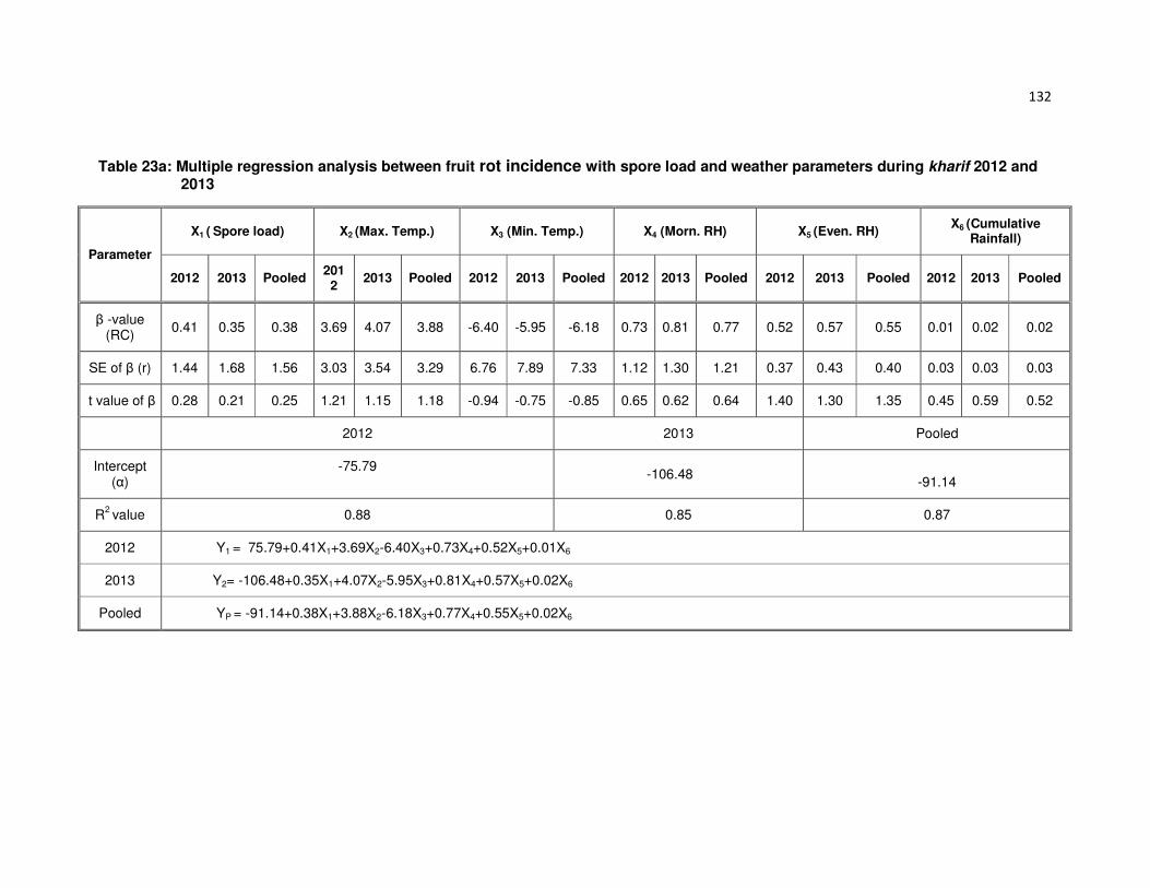
132
Table 23a: Multiple regression analysis between fruit rot incidence with spore load and weather parameters during kharif 2012 and 2013
Parameter
X1 ( Spore load) X2 (Max. Temp.) X3 (Min. Temp.) X4 (Morn. RH) X5 (Even. RH) X6 (Cumulative
Rainfall)
2012 2013 Pooled 2012
2013 Pooled 2012 2013 Pooled 2012 2013 Pooled 2012 2013 Pooled 2012 2013 Pooled
β -value (RC)
0.41 0.35 0.38 3.69 4.07 3.88 -6.40 -5.95 -6.18 0.73 0.81 0.77 0.52 0.57 0.55 0.01 0.02 0.02
SE of β (r) 1.44 1.68 1.56 3.03 3.54 3.29 6.76 7.89 7.33 1.12 1.30 1.21 0.37 0.43 0.40 0.03 0.03 0.03
t value of β 0.28 0.21 0.25 1.21 1.15 1.18 -0.94 -0.75 -0.85 0.65 0.62 0.64 1.40 1.30 1.35 0.45 0.59 0.52
2012 2013 Pooled
Intercept (α)
-75.79 -106.48
-91.14
R2 value 0.88 0.85 0.87
2012 Y1 = 75.79+0.41X1+3.69X2-6.40X3+0.73X4+0.52X5+0.01X6
2013 Y2= -106.48+0.35X1+4.07X2-5.95X3+0.81X4+0.57X5+0.02X6
Pooled YP = -91.14+0.38X1+3.88X2-6.18X3+0.77X4+0.55X5+0.02X6

133
Table 23b: Observed and predicted fruit rot incidence with spore load and weather parameters during kharif 2012 and 2013
Standard weeks
2012 2013 Pooled
Observed Predicted Difference Observed Predicted Difference Observed Predicted Difference
30 0 -2.80 2.80 2.82 3.68 -0.86 1.41 0.44 0.97
31 3.33 2.12 1.21 5.00 8.31 -3.31 4.17 5.22 -1.05
32 4.67 3.71 0.96 5.67 13.88 -8.21 5.17 8.80 -3.63
33 7.67 3.99 3.68 9.00 9.76 -0.76 8.34 6.88 1.46
34 9.33 4.03 5.30 11.00 13.71 -2.71 10.17 8.87 1.30
35 10.67 8.34 2.33 12.13 17.90 -5.77 11.40 13.12 -1.72
36 11.67 7.10 4.57 13.17 19.05 -5.88 12.42 13.08 -0.66
37 13.00 11.31 1.69 15.43 13.55 1.88 14.22 12.43 1.79
38 11.71 12.84 -1.13 14.00 15.47 -1.47 14.67 12.67 2.00
39 17.67 14.24 3.43 19.27 17.75 1.52 18.47 16.00 2.48
40 5.67 6.10 -0.43 6.31 15.18 -8.87 5.99 10.64 -4.65
41 8.33 6.56 1.77 8.19 17.05 -8.86 8.26 11.81 -3.55
42 10.32 7.05 3.27 9.96 14.12 -4.16 10.14 10.59 -0.45
43 15.33 9.86 5.47 11.53 17.32 -5.79 11.62 15.08 -3.46
44 13.10 10.69 2.41 12.83 27.37 -14.54 12.97 19.03 -6.07
Table 24a : Effect of date of planting on development chilli fruit rot and die-back disease during kharif 2012-13
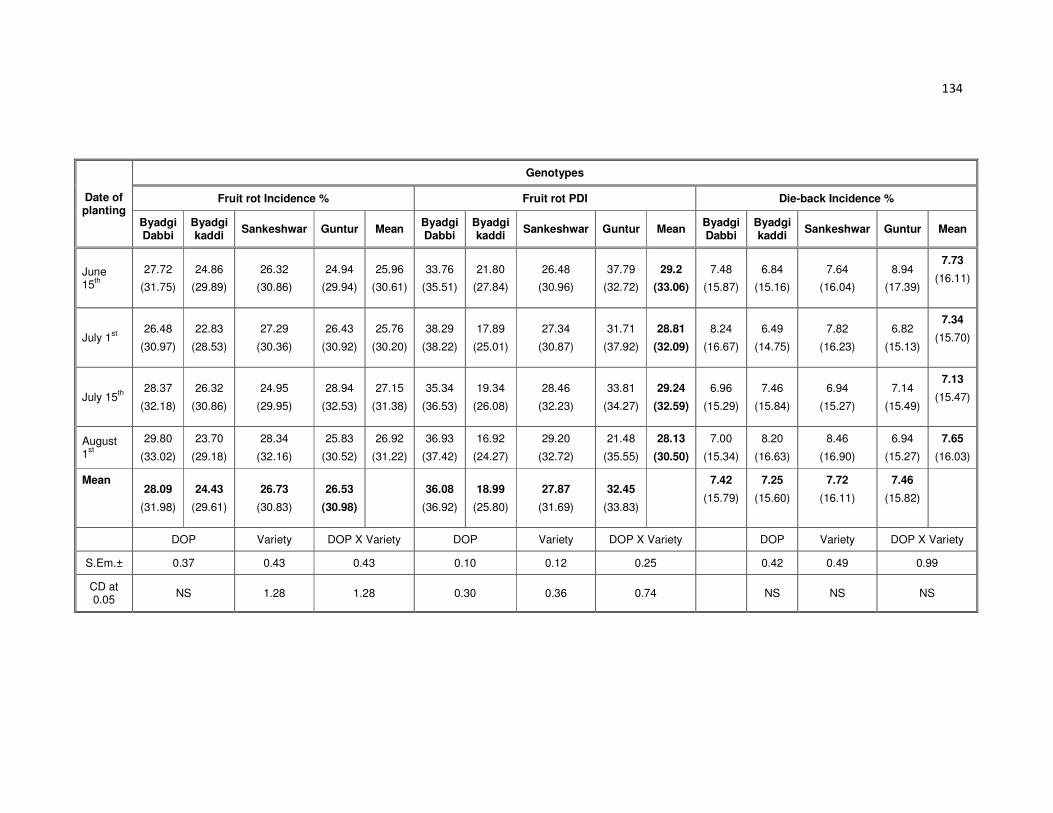
134
Date of planting
Genotypes
Fruit rot Incidence % Fruit rot PDI Die-back Incidence %
Byadgi Dabbi
Byadgi kaddi
Sankeshwar Guntur Mean Byadgi Dabbi
Byadgi kaddi
Sankeshwar Guntur Mean Byadgi Dabbi
Byadgi kaddi
Sankeshwar Guntur Mean
June 15
th
27.72
(31.75)
24.86
(29.89)
26.32
(30.86)
24.94
(29.94)
25.96
(30.61)
33.76
(35.51)
21.80
(27.84)
26.48
(30.96)
37.79
(32.72)
29.2
(33.06)
7.48
(15.87)
6.84
(15.16)
7.64
(16.04)
8.94
(17.39)
7.73
(16.11)
July 1st
26.48
(30.97)
22.83
(28.53)
27.29
(30.36)
26.43
(30.92)
25.76
(30.20)
38.29
(38.22)
17.89
(25.01)
27.34
(30.87)
31.71
(37.92)
28.81
(32.09)
8.24
(16.67)
6.49
(14.75)
7.82
(16.23)
6.82
(15.13)
7.34
(15.70)
July 15th
28.37
(32.18)
26.32
(30.86)
24.95
(29.95)
28.94
(32.53)
27.15
(31.38)
35.34
(36.53)
19.34
(26.08)
28.46
(32.23)
33.81
(34.27)
29.24
(32.59)
6.96
(15.29)
7.46
(15.84)
6.94
(15.27)
7.14
(15.49)
7.13
(15.47)
August 1
st
29.80
(33.02)
23.70
(29.18)
28.34
(32.16)
25.83
(30.52)
26.92
(31.22)
36.93
(37.42)
16.92
(24.27)
29.20
(32.72)
21.48
(35.55)
28.13
(30.50)
7.00
(15.34)
8.20
(16.63)
8.46
(16.90)
6.94
(15.27)
7.65
(16.03)
Mean 28.09
(31.98)
24.43
(29.61)
26.73
(30.83)
26.53
(30.98)
36.08
(36.92)
18.99
(25.80)
27.87
(31.69)
32.45
(33.83)
7.42
(15.79)
7.25
(15.60)
7.72
(16.11)
7.46
(15.82)
DOP Variety DOP X Variety DOP Variety DOP X Variety DOP Variety DOP X Variety
S.Em.± 0.37 0.43 0.43 0.10 0.12 0.25 0.42 0.49 0.99
CD at 0.05
NS 1.28 1.28 0.30 0.36 0.74 NS NS NS
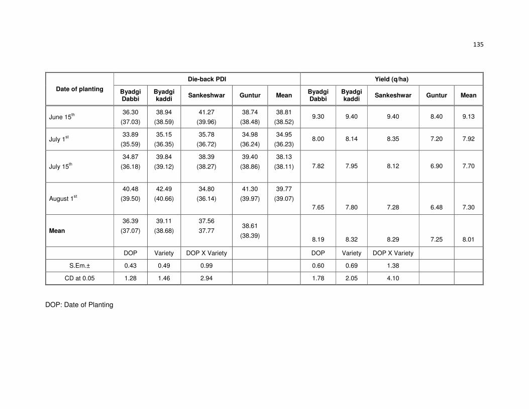
135
Date of planting
Die-back PDI Yield (q/ha)
Byadgi Dabbi
Byadgi kaddi
Sankeshwar Guntur Mean Byadgi Dabbi
Byadgi kaddi
Sankeshwar Guntur Mean
June 15th
36.30
(37.03)
38.94
(38.59)
41.27
(39.96)
38.74
(38.48)
38.81
(38.52) 9.30 9.40 9.40 8.40 9.13
July 1st
33.89
(35.59)
35.15
(36.35)
35.78
(36.72)
34.98
(36.24)
34.95
(36.23) 8.00 8.14 8.35 7.20 7.92
July 15th
34.87
(36.18)
39.84
(39.12)
38.39
(38.27)
39.40
(38.86)
38.13
(38.11)
7.82 7.95 8.12 6.90 7.70
August 1st
40.48
(39.50)
42.49
(40.66)
34.80
(36.14)
41.30
(39.97)
39.77
(39.07)
7.65 7.80 7.28 6.48 7.30
Mean
36.39
(37.07)
39.11
(38.68)
37.56
37.77
38.61
(38.39)
8.19 8.32 8.29 7.25 8.01
DOP Variety DOP X Variety DOP Variety DOP X Variety
S.Em.± 0.43 0.49 0.99 0.60 0.69 1.38
CD at 0.05 1.28 1.46 2.94 1.78 2.05 4.10
DOP: Date of Planting

136
Table 24b: Effect of date of planting on development chilli fruit rot and die-back disease during kharif 2013-14
Date of planting
Fruit rot Incidence % Fruit rot PDI Die-back Incidence % Die-back PDI Yield (q/ha)
BD BK SK Mean BD BK SK Mean BD BK SK Mean BD Q SK Mean BD BK SK Mean
June 15th 25.52 22.66 24.12 24.10 31.56 19.6 24.28 25.15 7.30 5.55 6.88 6.58 35.06 37.7 40.03 37.60 7.21 7.53 7.89 7.54
July 1st 24.28 20.63 25.09 23.33 36.09 15.69 25.14 25.64 6.54 5.90 6.70 6.38 32.65 33.91 34.54 33.70 7.04 7.89 7.74 7.56
July 15th 26.17 24.12 22.75 24.35 33.14 17.14 26.26 25.51 6.02 6.52 6.00 6.18 33.63 38.6 37.15 36.46 7.79 7.34 6.67 7.27
Mean 25.32 22.47 23.99 23.93 33.60 17.48 25.23 25.43 6.62 5.99 6.53 6.38 33.78 36.74 37.24 35.92 7.35 7.59 7.43 7.46
BD - Byadagi Dabbi BK – Byadagi Kaddi SK – Sankeshwar

137
In 2013-14 kharif with three different dates of planting was carried out at the
Main Agricultural Research Station, Dharwad, with three different dates, starting from
15th June to 15th July at fortnightly intervals with three genotypes Byadgi Kaddi, Byadgi
Dabbi and Sankeshwar. Disease incidence and severity of both fruit rot and die-back
was recorded and presented in Table 24b. The highest fruit rot incidence 24.35 per
cent with 25.51 PDI was observed in July 15th planting, where as highest die-back
incidence 6.58 per cent with 37.60 PDI was observed in June 15th planting. The highest
yield (7.56q/ha) was recorded in July 1st planting.
4.4 Development of IDM strategies for disease
4.4.1 Field evaluation of chilli genotypes
A total of 343 chilli genotypes were screened in natural endemic condition in
MARS Dharwad during kharif 2012 and kharif 2013. These genotypes were grouped as
per the scale and data is presented in the Table 25a and 25b, Plate 32.
Among 343 genotypes screened under natural endemic field condition during
kharif season of 2012, none of them were found immune, while 15 genotypes showed
resistant reaction, 39 genotypes were found moderately resistant with grade scale of 3.
However, 199 genotypes were moderately susceptible, 86 genotypes were found
susceptible and the remaining 4 genotypes were highly susceptible with infection of
>75 per cent.
During kharif 2013 out of 343 genotypes screened, none of them were found
immune, while eight genotypes were found resistant, 30 genotypes were moderately
resistant. However, 168 genotypes were moderately susceptible, 131 genotype were
susceptible and six genotypes were highly susceptible with infection of >75 per cent.
For both years of investigation kharif 2012-13 and 2013-14 eight genotypes
were found resistant where as 21 genotypes were recorded as moderately resistant.
4.4.1.1 In vitro screening of genotypes
To identify the resistant sources to fruit rot severity, a total of 38 chilli genotypes
were screened against Colletotrichum spp. (C.capsici, C. gloeosporioides and C.

138
Table 25a: Grouping of chilli genotypes based on resistance against fruit rot disease under field condition during kharif 2012-13
Grade PDI Reaction Genotype No. of
genotypes
0 0 Immune 0
1 1-10 Resistant Capsicum baccatum, DC – 1001, DC – 1002, DCA-101, DH-10-1, DH-12-1, VN-2, Tiwan-17, Nooji-2, IC-119559, Jin’s Joy, Susan’s Joy, Moor/Perenni, Gift from Maldov, VN2 X B. Dabbi,
15
3 11-25 Moderately resistant
Byadgi Kaddi, DC – 1006, DC – 1007, DCA-102-2, DCA-106-1, DCA-107-1, DCA-109-2, DCA-118, DCA-118-1, KDSC-210-10-4, LCA-312, SNK, SIC-11-179, SIC-10-166-1, Pant C-1-1, Pant C-1-2, Pant C-1-3, Cochin, Cochin-2 UFM-2-1, DH-2, DH-12, DH-11, LCA-206, LCA-206-1, VN-2-1, SKAU- SC-618-2, SKAU- SC-231, DCA-241, Tiwan-8, Tiwan-5, Tiwan-4, Tiwan-15, Tiwan-19, IC-119756, Punjab Guchedar, CCA-3288, CCA-3468, CCA-3336
39
5 26-50 Moderately susceptible
C. chinense, DC – 1003, DC – 1008, DCA-101-1, DCA-102, DCA-102-
3, DCA-103-1, DCA-106, DCA-106-2, DCA-107, DCA-108, DCA-108-1,
DCA-108-2, DCA-108-3, DCA-109-1, DCA-110, DCA-110-1, DCA-112,
DCA-112-1, DCA-113, DCA-114-1, DCA-115-1, DCA115-2, DCA-115-3,
DCA-116, DCA-120, DCA-121, DCA-122-2, KDSC-210-10-1, DCA-123-
2, DCA-124-1, DCA-125-1, DCA-126, DCA-127-1, DCA-129, DCA-131,
DCA-132, DCA-132-1, DCA-133, DCA-135-1 KDSC-210-10, DCA-123-
1KDSC-21010-2, KDSC-210-10-3, KDSC-510-10-1, KDSC-510-10-2,
CO-3, Arka Lohith, PMR-5, PMR-5-1, LCA-305, LCA-206, LCA-283,
LCA-283-1, LCA-312-1, LCA-312-2, LCA-324, Hissar Shakthi,Hissar
Shakthi-1, Hissar Shakthi-2, G-3-1, LCA-310 (2-3 pods/node), LCA-310-
1, LCA-310-2, Hissar Vijay, LCA-304-1, DCA- 155, DH-9-6-6, GPC-82-1,
AR-75, GPC-82-1-1, S-32, SIC-10-166, Serano, Cochin-1, Button
,,UFM-2, Buttom-1, Button-2, DH-5, DH-5-1, DH-5-2, DH-10, DH-1,
Mosac-27, 1701 F4, GPM-60, GPM-50, GPM-50-1, PBC-460, PMR-21,
Pant C-2 , Paprika-2, CO-3, Phule-5, JJK-2000-114, JJK-2000-114-1, K-
1, JCA-283, JCA-283-1, CO-1, AR-75, D3-46, S-32, Pant C-1-1, LCA-
304, LCA-304-2 SKAU- SC-105-4, SKAU- SC-305, SKAU- SC-1003-2,
SKAU- SC-23-1, SKAU- SC-101-1, SKAU- SC-502-1, SKAU- SC-885,
SKAU- SC-97, SKAU- SC-3, SKAU- SC-107, SKAU-Agnirekha-Sel-1,
SKAU-Wonder hot- Sel -1, SKAU-PS-1 (Payorika-Sel-1), SKAU-PS-4
(Payorika-Sel-4), SKAU-PS-2, SKAU-PS-3, SKAU- SC-105, SKAU-SC-
115, S-32-1, DCA-237, DCA-238, DCA-239, DCA-240, Tiwan-1, Tiwan-
2, Tiwan-3, Tiwan-7,Tiwan-9, Tiwan-10, Tiwan-11, Tiwan-12, Tiwan-13,
Tiwan-14, Tiwan-16, Tiwan-18, China-1, China-2, China-3, Sankeshwar-
1, Ornamental, Nooji-1 , Lokal Capcicum-red, Tomato Chilli (Warangal
Sapota), Kadrolli Local, Chikballapur Local, Phule Jyothi, Phule Sai, X-
235, IC-92109, IC-119243, IC-119264, IC-119267, IC-119546, IC-
119578, IC-119581, IC-119594, IC-119614, IC-119736, IC-208595, IC-
20416, IC-20901, Horti Local, S-2530 (PAV), Mr. Lee No. 3, MSH-1,
PBC-308, PBC-396, PBC-788,, CCA-1349 , CCA-3106, CCA-3331, Mr.
Lee No. 3, MSH-1, PBC-308, PBC-396, PBC-788, CCA-1349 , CCA-
3106, CCA-3331, CCA-3743, CCA-260, CCA-4-0, CCA-2340-2, CCA-
321, CCA-323, CCA-984-A, CCA-982-A, CCA-1410-A , CCA-42-A,
CCA-48-A, US- 170, US- 344, US- 341, US- 611, US- 720, US- 1055
199

139
Grade PDI Reaction Genotype No. of
genotypes
7 51-75 Susceptible Sankeshwar, DC – 1004, DC – 1005, DC - 1009 , DCA-101-2,
DCA-101-3, DCA-103, DCA-104, DCA-104-1, DCA-105, DCA-
109, DCA-113-1, DCA-114, DCA-115, DCA-117, DCA-119,
DCA-122, DCA-123, DCA-124, DCA-125, DCA-127, DCA-128,
DCA-130, DCA-130-1, DCA-134, DCA-134-1, DCA-135, DCA-
135-2, KDSC-510-10, GPC-69, GPC-69-1, GPC-80, CO-1,
Arka asir, LCA-206-1, LCA-324-1, LCA-324-2, Hissar Shakthi-
3, G-3, LCA-304, DH-9-6-6-1, G-4, Yellow, Jwala-UR, Phule-5,
Phule-5-1, Pant C-1, UNFM-1, Button-1, JJK -2000-114,
Button, DH-8, GPM-56, Paprika-1, Co-3-1, Hissar Vijay, PBC-
460, LCA-310, LCA-304-1, SKAU- SC-578-1, SKAU- SC-101,
DCA-236, SKAU- SC-814-2, SKAU- SC-1003, DCC – 201,
B.Dabbi(Kusugal), IC-119561, IC-119592, IC-119598, IC-
119608, IC-119622, IC-119696, IC-119701, 96 TH 1101, PBC-
142 , PBC-535, CCA-3619, CCA-4-1, CCA-750, CCA-3636,
CCA-191-A, CCA-215-A, US- 113, US- 918
86
9 > 75 Highly susceptible
Byadgi Dabbi, GPM-64, Pant C-1, Punjab Surkha 4
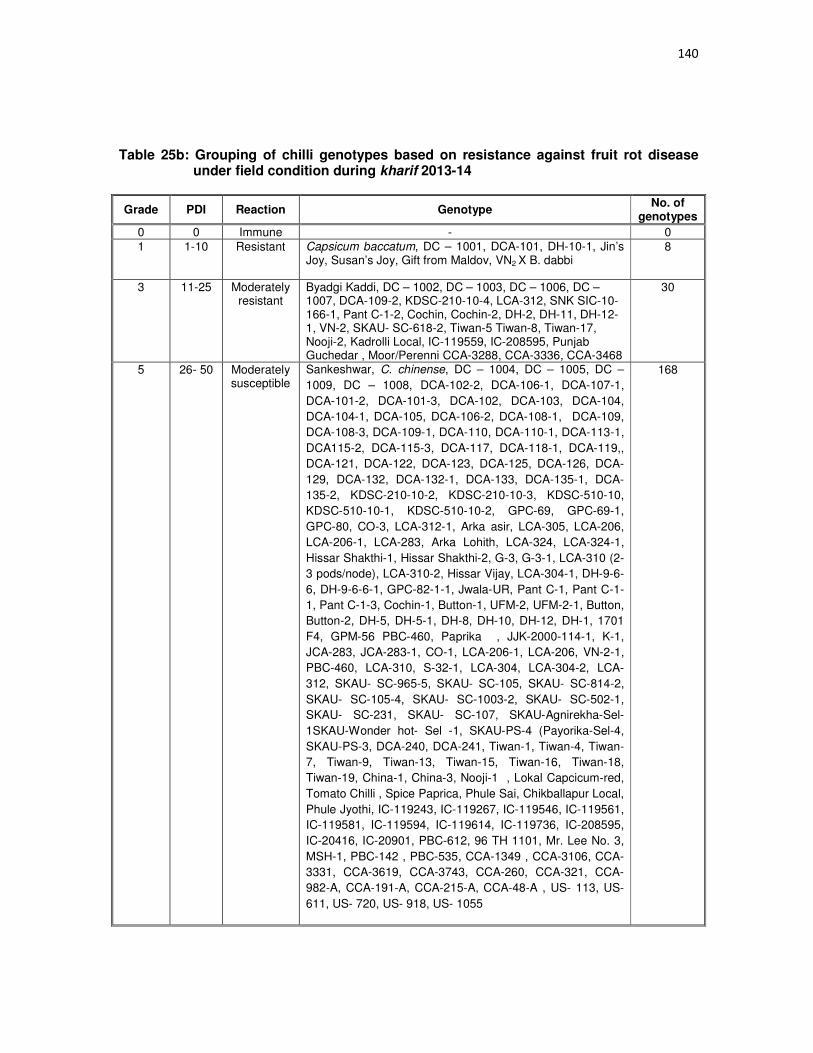
140
Table 25b: Grouping of chilli genotypes based on resistance against fruit rot disease under field condition during kharif 2013-14
Grade PDI Reaction Genotype No. of
genotypes
0 0 Immune - 0
1 1-10 Resistant Capsicum baccatum, DC – 1001, DCA-101, DH-10-1, Jin’s Joy, Susan’s Joy, Gift from Maldov, VN2 X B. dabbi
8
3 11-25 Moderately resistant
Byadgi Kaddi, DC – 1002, DC – 1003, DC – 1006, DC – 1007, DCA-109-2, KDSC-210-10-4, LCA-312, SNK SIC-10-166-1, Pant C-1-2, Cochin, Cochin-2, DH-2, DH-11, DH-12-1, VN-2, SKAU- SC-618-2, Tiwan-5 Tiwan-8, Tiwan-17, Nooji-2, Kadrolli Local, IC-119559, IC-208595, Punjab Guchedar , Moor/Perenni CCA-3288, CCA-3336, CCA-3468
30
5 26- 50 Moderately susceptible
Sankeshwar, C. chinense, DC – 1004, DC – 1005, DC –
1009, DC – 1008, DCA-102-2, DCA-106-1, DCA-107-1,
DCA-101-2, DCA-101-3, DCA-102, DCA-103, DCA-104,
DCA-104-1, DCA-105, DCA-106-2, DCA-108-1, DCA-109,
DCA-108-3, DCA-109-1, DCA-110, DCA-110-1, DCA-113-1,
DCA115-2, DCA-115-3, DCA-117, DCA-118-1, DCA-119,,
DCA-121, DCA-122, DCA-123, DCA-125, DCA-126, DCA-
129, DCA-132, DCA-132-1, DCA-133, DCA-135-1, DCA-
135-2, KDSC-210-10-2, KDSC-210-10-3, KDSC-510-10,
KDSC-510-10-1, KDSC-510-10-2, GPC-69, GPC-69-1,
GPC-80, CO-3, LCA-312-1, Arka asir, LCA-305, LCA-206,
LCA-206-1, LCA-283, Arka Lohith, LCA-324, LCA-324-1,
Hissar Shakthi-1, Hissar Shakthi-2, G-3, G-3-1, LCA-310 (2-
3 pods/node), LCA-310-2, Hissar Vijay, LCA-304-1, DH-9-6-
6, DH-9-6-6-1, GPC-82-1-1, Jwala-UR, Pant C-1, Pant C-1-
1, Pant C-1-3, Cochin-1, Button-1, UFM-2, UFM-2-1, Button,
Button-2, DH-5, DH-5-1, DH-8, DH-10, DH-12, DH-1, 1701
F4, GPM-56 PBC-460, Paprika , JJK-2000-114-1, K-1,
JCA-283, JCA-283-1, CO-1, LCA-206-1, LCA-206, VN-2-1,
PBC-460, LCA-310, S-32-1, LCA-304, LCA-304-2, LCA-
312, SKAU- SC-965-5, SKAU- SC-105, SKAU- SC-814-2,
SKAU- SC-105-4, SKAU- SC-1003-2, SKAU- SC-502-1,
SKAU- SC-231, SKAU- SC-107, SKAU-Agnirekha-Sel-
1SKAU-Wonder hot- Sel -1, SKAU-PS-4 (Payorika-Sel-4,
SKAU-PS-3, DCA-240, DCA-241, Tiwan-1, Tiwan-4, Tiwan-
7, Tiwan-9, Tiwan-13, Tiwan-15, Tiwan-16, Tiwan-18,
Tiwan-19, China-1, China-3, Nooji-1 , Lokal Capcicum-red,
Tomato Chilli , Spice Paprica, Phule Sai, Chikballapur Local,
Phule Jyothi, IC-119243, IC-119267, IC-119546, IC-119561,
IC-119581, IC-119594, IC-119614, IC-119736, IC-208595,
IC-20416, IC-20901, PBC-612, 96 TH 1101, Mr. Lee No. 3,
MSH-1, PBC-142 , PBC-535, CCA-1349 , CCA-3106, CCA-
3331, CCA-3619, CCA-3743, CCA-260, CCA-321, CCA-
982-A, CCA-191-A, CCA-215-A, CCA-48-A , US- 113, US-
611, US- 720, US- 918, US- 1055
168

141
Grade PDI Reaction Genotype No. of
genotypes
7 51 - 75 Susceptible DCA-101-1, DCA-102-3, DCA-103-1, DCA-106, DCA-
107, DCA-108-2, DCA-112, DCA-112-1, DCA-113, DCA-
114, DCA-114-1, DCA-115, DCA-115-1, DCA-116, DCA-
118, DCA-120, DCA-122-2, DCA-123-1, DCA-123-2,
DCA-124, DCA-124-1, DCA-125-1, DCA-127, DCA-127-
1, DCA-128, DCA-130, DCA-130-1, DCA-131, DCA-134,
DCA-134-1, DCA-135, KDSC-210-10, KDSC-210-10-1,
CO-1, PMR-5, PMR-5-1, LCA-283-1, LCA-312-2, LCA-
324-2, Hissar Shakthi, Hissar Shakthi-3, LCA-310-1,
LCA-304, DCA- 155, GPC-82-1, AR-75, S-32, Yellow,
SIC-11-179, SIC-10-166, Serano, Phule-5 UNFM-1,
Button , JJK -2000-114, Buttom-1, DH-5-2, Mosac-27,
GPM-60, GPM-50, GPM-50-1, PMR-21, Pant C-2 ,
Paprika-1, Paprika-2, Co-3-1, Phule-5, Hissar Vijay, JJK-
2000-114, AR-75, D3-46, S-32, Pant C-1, Pant C-1-1,
LCA-304-1, SKAU-SC-304-1, SKAU- SC-578-1, SKAU-
SC-1003, DCC – 201, SKAU- SC-305, SKAU- SC-23-1,
SKAU- SC-101, SKAU- SC-101-1, SKAU- SC-885,
SKAU- SC-97, SKAU- SC-3, SKAU-PS-1 (Payorika-Sel-
1), DCA-237, DCA-238, DCA-239, Tiwan-2, Tiwan-3,
Tiwan-10, Tiwan-11, Tiwan-12, Tiwan-14, Tiwan-17,,
B.Dabbi(Kusugal), China-2, Sankeshwar-1, Ornamental,
X-235, IC-92109, IC-119264, IC-119578, IC-119592, IC-
119598, IC-119608, IC-119622, IC-119696, IC-119701,
Horti Local, S-2530 (PAV), Punjab Surkha, PBC-308,
PBC-396, PBC-788, CCA-4-0, CCA-4-1, CCA-2340-2,
CCA-750, CCA-323, CCA-3636, CCA-984-A, CCA-1410-
A, CCA-42-A, US- 170, US- 341, US- 344
130
9 > 75 Highly susceptible
Byadgi Dabbi, DCA-108, G-4, Phule-5-1, GPM-64, SKAU-PS-2, DCA-236
7

142
Table 25c: Grouping of resistant and moderately resistant chilli genotypes against fruit rot disease under field condition during kharif 2012-13 and 2013-14
Grade PDI Reaction Genotype No. of
genotypes
1 1-10 Resistant Capsicum baccatum, DC – 1001, DCA-101, DH-10-1,
Jin’s Joy, Susan’s Joy, Gift from Maldov, VN2 X B.
dabbi
8
3 11-25 Moderately
resistant
Byadgi Kaddi, DC – 1006, DC – 1007, DCA-109-2,
KDSC-210-10-4, LCA-312, SNK, SIC-10-166-1,
Pant C-1-2, Cochin, Cochin-2, DH-2, DH-11, SKAU-
SC-618-2, Tiwan-5 Tiwan-8, IC-119756, Punjab
Guchedar , CCA-3288, CCA-3336, CCA-3468
21

143
Plate 32a Screening of chilli genotypes for resistance against fruit rot
disease under field condition

144
Table 26a: Reaction of chilli genotypes against Colletotrichum spp. under in vitro
condition
Grade PDI Reaction Genotype No. of
genotypes
0 0 Immune 0
1 1-10 Resistant 0
3 11 – 25 Moderately resistant Capsicum baccatum, DC – 1001, DC – 1002, DC – 1003 ,DC – 1006, DC – 1007, DCA-101, Jin’s Joy, Susan’s Joy, Gift from Maldov, VN2 X Byadgi Dabbi, DH-10-1, Punjab Guchedar, LCA-312, Tiwan-17, Nooji-2, Moor/Perenni
17
5 26 – 50 Moderately susceptible
Byadgi Kaddi , DCA-109-2, KDSC-210-10-4, SNK, SIC-10-166-1, Pant C-1-2, Cochin Cochin-2, DH-2, DH-11
10
7 51 - 75 Susceptible IC-119756, Tiwan-8, DCA-236, DH-12-1 VN-2, SKAU- SC-618-2
6
9 > 75 Highly susceptible Byadgi Dabbi, Kadrolli Local, IC-119559, CCA-3288, CCA-3336, CCA-3468
6

145
Table 26b: Reaction of chilli genotypes against A. alternata under in vitro condition
Grade PDI Reaction Genotype No. of
genotypes
0 0 Immune 0
1 1-10 Resistant 0
3 11 – 25 Moderately resistant Capsicum baccatum, DC – 1001, DC – 1002, DC – 1003 ,DC – 1006, DC – 1007, DCA-101, Jin’s Joy, Susan’s Joy, Gift from Maldov, VN2 X Byadgi Dabbi, DH-10-1 Punjab Guchedar, DCA-109-2, DH-11, LCA-312, Tiwan-17, Nooji-2, Moor/Perenni
19
5 26 – 50 Moderately susceptible
Byadgi Kaddi , DCA-109-2, KDSC-210-10-4, SNK, SIC-10-166-1, Pant C-1-2, Cochin, Cochin-2, DH-2
9
7 51 - 75 Susceptible IC-119756, Tiwan-8, DCA-236, DH-12-1 VN-2, SKAU- SC-618-2
6
9 > 75 Highly susceptible Byadgi Dabbi, Kadrolli Local, IC-119559, CCA-3288, CCA-3336, CCA-3468
6

146
Table 26c: Reaction of chilli genotypes against Fusarium spp. under in vitro condition
Grade PDI Reaction Genotype No. of
genotypes
0 0 Immune 0 0
1 1-10 Resistant 0 0
3 11 – 25 Moderately resistant Capsicum baccatum, DC – 1001, DC – 1002, DC – 1003 ,DC – 1006, DC – 1007, DCA-101, Jin’s Joy, Susan’s Joy, Gift from Maldov, VN2 X Byadgi Dabbi, DH-10-1 Punjab Guchedar, DCA-109-2, DH-11, LCA-312, Tiwan-17, Nooji-2, Moor/Perenni, DH-2
20
5 26 – 50 Moderately susceptible
Byadgi Kaddi , KDSC-210-10-4, SNK, Pant C-1-2, Cochin, Cochin-2, DH-2 7
7 51 - 75 Susceptible SIC-10-166-1, IC-119756, Tiwan-8, DCA-236, VN-2, SKAU- SC-618-2
6
9 > 75 Highly susceptible Byadgi Dabbi, Kadrolli Local, IC-119559, DCA-236, CCA-3336, CCA-3468
6
Table 26d: Reaction of chilli genotypes against Colletortrichum spp. A. alternata, Fusarium spp. under in vitro condition
Grade PDI Reaction Genotype No. of
genotypes
0 0 Immune 0
1 1-10 Resistant 0
3 11 – 25 Moderately resistant Capsicum baccatum, DC – 1001, DC – 1002, DC – 1003 ,DC – 1006, DC – 1007, DCA-101, Jin’s Joy, Susan’s Joy, Gift from Maldov, VN2 X Byadgi Dabbi, DH-10-1 Punjab Guchedar, LCA-312, Tiwan-17, Nooji-2, Moor/Perenni
17
5 26 – 50 Moderately susceptible
Byadgi Kaddi , KDSC-210-10-4, SNK, SIC-10-166-1, Pant C-1-2, Cochin, Cochin-2, DH-2 7
7 51 - 75 Susceptible IC-119756, Tiwan-8, DCA-236, DH-12-1 VN-2, SKAU- SC-618-2 6
9 > 75 Highly susceptible Byadgi Dabbi, Kadrolli Local, IC-119559, CCA-3288, CCA-3336, CCA-3468
6

147
Plate 33: In vitro screening of chilli genotypes

148
acutatum) A.alternata, F. oxysporum, F. sporotrichioides in laboratory condition by pin
prick method and the reactions of all these genotypes are furnished in the Table 26a,
26b and 26c (Plate 33).
Results indicated that the genotypes have diverse degree of reaction against
Colletotrichum spp. (C.capsici, C. gloeosporioides and C. acutatum) A.alternata, F.
oxysporum, F. sporotrichioides. Among 38 genotypes, none of them showed immune
and resistant reaction. However, moderately resistant reaction was observed in
seventeen genotypes, ten genotypes showed moderately susceptible reaction against
C.capsici, C. gloeosporioides, C. acutatum. 19 genotypes showed moderately
resistant, eight showed moderately susceptible reaction against A.alternata and 20
genotypes showed moderately resistant, seven showed moderately susceptible
reaction against F. oxysporum and F. sporotrichioides.
4.4.2 Seed health management
4.4.2.1 Standard Blotter Method
The seeds which were showing fungal colonies were observed under
stereoscopic-binocular microscope, morphology of mycelium, asexual fruiting body and
conidia revealed the presence of Colletotrichum capsici (72.85%), C. gloeosporioides
(9.26%) and C. acutatum (4.76%), Alternaria alternata (5.20%) and Fusarium
sporotrichioides (3.45%) and F. oxysporum (4.30%) (Table 27, Plate 34).
4.4.2.2 Seed treatment with chemical fungicides
Evaluation of seed treatment fungicides (Table28) revealed that among three
systemic fungicides evaluated at 0.2% concentration, pyraclostrobin 20WG showed
least infection (11.63%) with highest vigour index (861.17). Between two non systemic
fungicides captan 75WP showed least infection (12.28%) with highest vigour index
(754.64). Among six combi product fungicides, carboxin 37.5% + thiram 37.5% WS
showed least infection (7.25%) with highest vigour index (932.02) followed by
metalaxyl 4%+mancozeb64% (10.09%, 871.70). Highest infection (43.26%) with least
vigour index (351.77) was observed in untreated control.

149
Table 27: Chilli seed mycoflora under stereo binocular microscope
Fungi Frequency (%)
Colletotrichum capsici 72.85
C. gloeosporioides 9.26
C. acutatum 4.76
Alternaria alternata 5.20
Fusarium sporotrichioides 3.45
F. oxysporum 4.30

150
Plate 34 Seed health study
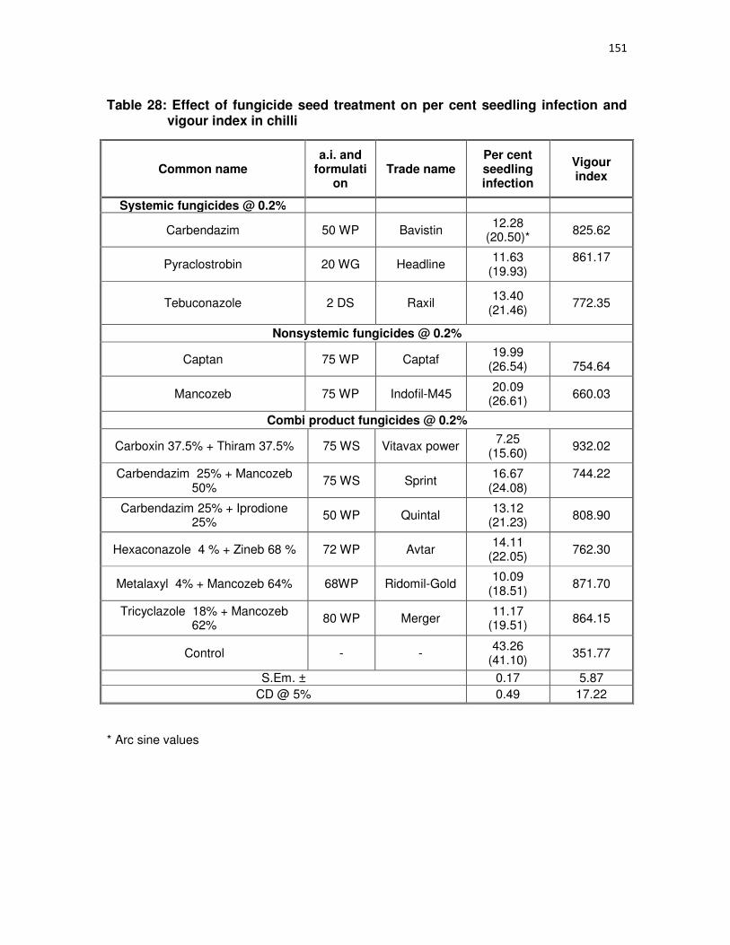
151
Table 28: Effect of fungicide seed treatment on per cent seedling infection and vigour index in chilli
Common name a.i. and
formulation
Trade name Per cent seedling infection
Vigour index
Systemic fungicides @ 0.2%
Carbendazim 50 WP Bavistin 12.28
(20.50)* 825.62
Pyraclostrobin 20 WG Headline 11.63
(19.93) 861.17
Tebuconazole 2 DS Raxil 13.40
(21.46)
772.35
Nonsystemic fungicides @ 0.2%
Captan 75 WP Captaf 19.99
(26.54)
754.64
Mancozeb 75 WP Indofil-M45 20.09
(26.61) 660.03
Combi product fungicides @ 0.2%
Carboxin 37.5% + Thiram 37.5% 75 WS Vitavax power 7.25
(15.60) 932.02
Carbendazim 25% + Mancozeb 50%
75 WS Sprint 16.67
(24.08) 744.22
Carbendazim 25% + Iprodione 25%
50 WP Quintal 13.12
(21.23) 808.90
Hexaconazole 4 % + Zineb 68 % 72 WP Avtar 14.11
(22.05) 762.30
Metalaxyl 4% + Mancozeb 64% 68WP Ridomil-Gold 10.09
(18.51) 871.70
Tricyclazole 18% + Mancozeb 62%
80 WP Merger 11.17
(19.51) 864.15
Control - - 43.26
(41.10) 351.77
S.Em. ± 0.17 5.87
CD @ 5% 0.49 17.22
* Arc sine values

152
4.4.2.2 Seed treatment with bio-fungicides
Results of four bio fungicides (Table29) alone (@ 10.0 g/kg) and in combination
(5.0 +5.0 g/kg) seed treatment revealed that T. harzianum 5.0g + P. fluorescens 5.0g
showed least infection (14.89%) with highest vigour index (930.74) followed by
P. fluorescens (10.0 g/kg) 14.94% infection with 915.27 vigour index. Highest seedling
infection (42.99%) with least vigour index (361.87) was observed in untreated control.
4.4.3 Field efficacy of chemicals for the management of fruit rot and die-back disease
of chilli during kharif 2012 and 2013
A field experiment was conducted during kharif 2012 and 2013 at MARS Farm,
Dharwad for management of fruit rot and die-back disease of chilli using four systemic
and four combi-product fungicides along with untreated control. Insect management
was done as per package of practice. Data are furnished in Table 30 to 32.
Kharif 2012
Evaluation of fungicides revealed that among four systemic fungicides evaluated
at 0.1% concentration difenconazole 25EC showed least incidence of die back (1.33
%) with 12.31 PDI which is on par with pyraclostrobin 20WG (2.0 per cent, 16.49PDI)
and propiconazole 25EC (2.66 per cent, 19.89 PDI). Least fruit rot incidence (8.31 %)
was observed in difenconazole 25EC followed by pyraclostrobin 20WG (12.26 %),
whereas least fruit rot severity (7.33 PDI) observed in pyraclostrobin 20WG (Table 30).
Among four combi product fungicides tested pyraclostrobin 5% + metiram 55%
WG showed the least incidence (1.66 %) of die back with least severity (13.83 PDI)
which is on par with tricyclazole 18% + mancozeb 62% WP (1.68 per cent, 14.31 PDI).
Least fruit rot incidence (10.49 %) was observed in tricyclazole 18% + mancozeb 62%
WP, whereas least fruit rot severity (9.00 PDI) observed in pyraclostrobin 5% + metiram
55% WG. Highest yield was observed in difenconazole 25EC (9.15 q/ha) with cost
benfit ratio 2.66 which is on par with tricyclazole 18% + mancozeb 62% WP (8.75 q/ha,
C:B 2.44) and pyraclostrobin 5% + metiram 55% (8.65 q/ha C:B 2.48).

153
Table 29: Effect of bio fungicide seed treatment on per cent seedling infection and vigour index in chilli
Treatments
(10g/kg) Rate per kg seed
Per cent seedling infection
Vigour index
Trichoderma harzianum 10g 16.66
(24.07)* 804.90
Pseudomonas fluorescens
10g 14.94
(22.72) 915.27
Bacillus subtilis 10g 17.44
(24.67) 804.29
T. harzianum + P. fluorescens
5g+5g 14.89
(22.68) 930.74
T. harzianum + B. subtilis
5g+5g 16.16
(23.69) 833.14
P. fluorescens + B. subtilis
5g+5g 15.39
(23.08) 900.99
T. harzianum + Verticillium lecanii
5g+5g 16.35
(23.84) 831.53
P. fluorescens + Verticillium lecanii
5g+5g 15.58
(23.23)
836.00
B. subtilis + Verticilium lecanii
5g+5g 16.64
(24.06) 815.12
Control - 42.99
(41.10) 361.87
S.Em. ± 0.20 4.94
CD @ 5% 0.59 14.68
* Arc sine values
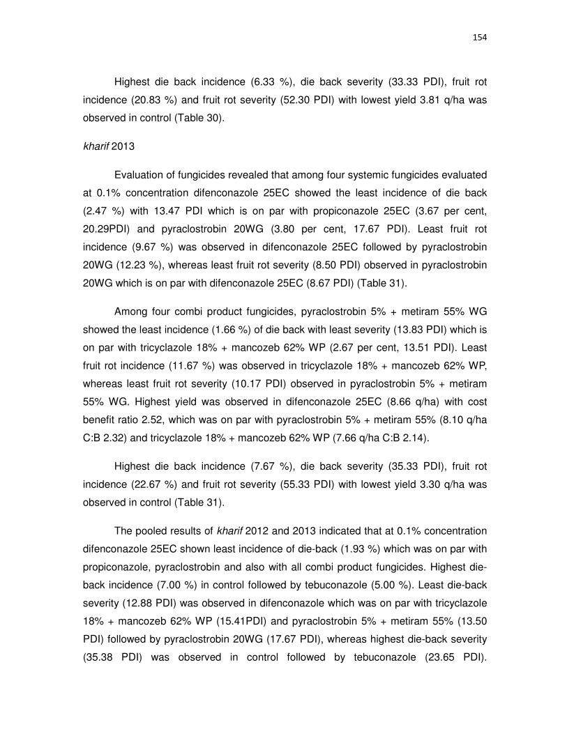
154
Highest die back incidence (6.33 %), die back severity (33.33 PDI), fruit rot
incidence (20.83 %) and fruit rot severity (52.30 PDI) with lowest yield 3.81 q/ha was
observed in control (Table 30).
kharif 2013
Evaluation of fungicides revealed that among four systemic fungicides evaluated
at 0.1% concentration difenconazole 25EC showed the least incidence of die back
(2.47 %) with 13.47 PDI which is on par with propiconazole 25EC (3.67 per cent,
20.29PDI) and pyraclostrobin 20WG (3.80 per cent, 17.67 PDI). Least fruit rot
incidence (9.67 %) was observed in difenconazole 25EC followed by pyraclostrobin
20WG (12.23 %), whereas least fruit rot severity (8.50 PDI) observed in pyraclostrobin
20WG which is on par with difenconazole 25EC (8.67 PDI) (Table 31).
Among four combi product fungicides, pyraclostrobin 5% + metiram 55% WG
showed the least incidence (1.66 %) of die back with least severity (13.83 PDI) which is
on par with tricyclazole 18% + mancozeb 62% WP (2.67 per cent, 13.51 PDI). Least
fruit rot incidence (11.67 %) was observed in tricyclazole 18% + mancozeb 62% WP,
whereas least fruit rot severity (10.17 PDI) observed in pyraclostrobin 5% + metiram
55% WG. Highest yield was observed in difenconazole 25EC (8.66 q/ha) with cost
benefit ratio 2.52, which was on par with pyraclostrobin 5% + metiram 55% (8.10 q/ha
C:B 2.32) and tricyclazole 18% + mancozeb 62% WP (7.66 q/ha C:B 2.14).
Highest die back incidence (7.67 %), die back severity (35.33 PDI), fruit rot
incidence (22.67 %) and fruit rot severity (55.33 PDI) with lowest yield 3.30 q/ha was
observed in control (Table 31).
The pooled results of kharif 2012 and 2013 indicated that at 0.1% concentration
difenconazole 25EC shown least incidence of die-back (1.93 %) which was on par with
propiconazole, pyraclostrobin and also with all combi product fungicides. Highest die-
back incidence (7.00 %) in control followed by tebuconazole (5.00 %). Least die-back
severity (12.88 PDI) was observed in difenconazole which was on par with tricyclazole
18% + mancozeb 62% WP (15.41PDI) and pyraclostrobin 5% + metiram 55% (13.50
PDI) followed by pyraclostrobin 20WG (17.67 PDI), whereas highest die-back severity
(35.38 PDI) was observed in control followed by tebuconazole (23.65 PDI).

155
Table 30: Chemical management of fruit rot and dieback disease during kharif 2012-13
Treatment Trade Name
a.i. Formulati
on
Concentration %
Dieback Incidence
(%)
Dieback Severity
(%)
Fruit rot Incidence
(%)
Fruit rot Severity (%)
Dry Fruit Yield q/ha
C: B
Difenconazole Score 25 EC 0.1 1.33
(6.53)* 12.31
(20.31) 8.31
(16.72)
7.67 (16.02)
9.15 2.66
Propiconazole Tilt 25 EC 0.1 2.66
(9.08) 19.89
(26.47) 13.12
(21.20)
11.00 (19.32)
7.65 2.21
Pyraclostrobin Headline 20 WG 0.1 2.0
(7.94) 16.49
(23.93) 12.26
(20.48)
7.33 (15.48)
8.30 2.31
Tebuconazole Folicur 25.9 EC 0.1 4.66
(12.35) 22.90
(28.55) 15.31
(23.03)
12.00 (20.22)
7.05 2.02
Hexaconazole 4% + Zineb 68% Avatar 72 WP 0.25 2.33
(8.74) 18.99
(25.81) 12.56
(20.74)
9.33 (17.63)
8.05 2.27
Tricyclazole 18% + Mancozeb62% Merger 80 WP 0.25 1.68
(7.33) 14.31
(22.22) 10.49
(18.89)
10.00 (18.37)
8.75 2.44
Carbendazim 12%+ Mancozeb 63%
Saaf 75 WP 0.25 3.33
(10.49) 21.41
(27.49) 13.43
(19.71)
12.33 (20.49)
7.9 2.26
Pyraclostrobin 5% + Metiram 55% Cabriotop 60 WG 0.25 1.66
(7.15) 13.83
(21.80) 11.39
(21.49)
9.00 (17.30)
8.65 2.48
Control 6.33
(16.77) 33.33
(35.25) 20.83
(27.14)
52.30
(46.30) 3.81
1.27
S.Em.± 1.63 1.71 0.84 1.50 0.72
CD @0.05 3.39 3.63 1.80 3.20 2.2
* Arc sine values

156
Table 31: Chemical management of fruit rot and dieback disease during kharif 2013-14
Treatment Trade Name
a.i. Formulati
on
Concentration
%
Dieback Incidenc
e (%)
Dieback Severity
(%)
Fruit rot Incidence
(%)
Fruit rot Severity
(%)
Dry Fruit Yield q/ha
C: B
Difenconazole Score 25 EC 0.1 2.47
(9.03)* 13.47
(21.52) 9.67
(18.11) 8.67
(17.11) 8.66
2.52
Propiconazole Tilt 25 EC 0.1 3.67
(11.04) 20.29
(26.76) 14.28
(22.19) 12.45
(20.65) 6.33
1.83
Pyraclostrobin Headline 20 WG 0.1 3.80
(11.24) 17.67
(24.84) 12.23
(20.46) 8.50
(16.94) 7.16
2.00
Tebuconazole Folicur 25.9 EC 0.1 5.33
(13.35) 23.67
(29.10) 16.67
(24.09) 13.83
(21.82) 6.83
1.96
Hexaconazole 4% + Zineb 68% Avatar 72 WP 0.25 3.87
(11.34) 19.33
(26.07) 14.41
(22.30) 10.28
(18.69) 7.16
2.02
Tricyclazole 18% + Mancozeb62%
Merger 80 WP 0.25 3.93
(11.43) 15.44
(23.13) 11.67
(19.96) 12.35
(20.57) 7.66
2.14
Carbendazim 12%+ Mancozeb 63%
Saaf 75 WP 0.25 4.77
(12.61) 22.40
(28.24) 14.33
(22.24) 13.90
(21.88) 7.12
2.04
Pyraclostrobin 5% + Metiram 55%
Cabriotop 60 WG 0.25 2.67
(9.39) 13.51
(21.56) 12.00
(20.26) 10.17
(18.59) 8.10
2.32
Control 7.67
(16.07) 35.33
(36.46) 22.67
(28.42) 55.33
(48.04) 3.30 1.10
S.Em.± 0.99 0.68 1.13 1.23 0.93
CD @0.05 2.96 2.03
3.38
3.70
2.78
* Arc sine values

157
Table 32: Pooled analysis of chemical management of fruit rot and dieback disease during 2012-13 and 2013-14
Treatment Trade Name
a.i. Formulat
ion
Concentration
%
Dieback Incidenc
e (%)
Dieback Severity
(%)
Fruit rot Incidence
(%)
Fruit rot Severity
(%)
Dry Fruit Yield q/ha
C: B
Difenconazole Score 25 EC 0.1 1.93
(7.99)* 12.88
(20.98) 8.99
(17.44) 8.18
(16.62) 8.86 2.59
Propiconazole Tilt 25 EC 0.1 3.13
(10.18) 20.23
(26.68) 13.73
(21.74) 11.77
(20.05) 6.95 2.02
Pyraclostrobin Headline 20 WG 0.1 2.93
(9.86) 17.67
(24.79) 12.25
(20.48) 7.91
(16.32) 7.74 2.16
Tebuconazole Folicur 25.9 EC 0.1 5.00
(12.92) 23.65
(29.06) 15.96
(23.540 12.95
(21.08) 6.93 1.99
Hexaconazole 4% + Zineb 68% Avatar 72 WP 0.25 3.13
(10.19) 19.33
(26.05) 13.42
(21.48) 9.82
(18.26) 7.60 2.15
Tricyclazole 18% + Mancozeb62% Merger 80 WP 0.25 2.80
(9.63) 15.41
(23.10) 11.08
(19.44) 11.18
(19.53) 8.21 2.29
Carbendazim 12%+ Mancozeb 63% Saaf 75 WP 0.25 4.03
(11.58) 22.42
(28.16) 13.00
(21.13) 13.13
(21.24) 7.53 2.15
Pyraclostrobin 5% + Metiram 55% Cabriotop 60 WG 0.25 2.23
(8.59)
13.50 (21.53)
11.70 (19.99)
9.55 (17.99)
8.33 2.40
Control 7.00
(15.34) 35.38
(36.48) 21.79
(27.82) 53.83
(47.18) 3.58
S.Em.± 1.33 1.02 0.85 0.77 0.90
CD @0.05 3.92 3.07 3.07 2.30
2.69
* Arc sine values

158
In difenconazole 25EC least fruit rot incidence (8.99 %) was observed which
was on par with tricyclazole 18% + mancozeb 62% WP (11.08 %) and pyraclostrobin
5% + metiram 55% (11.70 %) and pyraclostrobin 20WG (12.25 %), whereas highest
fruit rot incidence (21.79 %) was observed in control. Least fruit rot severity (7.91 PDI)
was observed in pyraclostrobin 20WG which was on par with difenconazole 25EC
(8.18 PDI) pyraclostrobin 5% + metiram 55% (9.55PDI), whereas highest fruit rot
severity (53.83 PDI) was observed in control.
Highest yield was observed in difenconazole 25EC (8.86 q/ha) with cost benefit
ratio 2.59 which was on par with pyraclostrobin 5% + metiram 55% (8.33 q/ha, C:B
2.40) and tricyclazole 18% + mancozeb 62% WP (8.21 q/ha C:B 2.29), whereas lowest
yield (3.58 q/ha) was observed in control.
4.4.4 Integrated management of fruit rot disease of chilli
Field experiment was conducted during kharif 2012 and 2013 at Dharwad with
four modules namely bio intensive module for both disease and insect pests (M1), bio-
intensive module for disease with chemical pesticides for insect pests (M2), Adoptive
module (M3), chemical intensive module (M4) to develop best disease management
module for chilli fruit rot disease, details of treatments were explained in material
methods 3.4.5. Data are presented in Table 33a, 33b, 34a, 34b and 35 Plate 35and 36.
Kharif 2012
The results of kharif 2012 indicated that least seedling rot infection (7.31 %) was
recorded in chemical module which was on par with adoptive module (7.33 %).
At 93 DAT (days after transplanting) M3 (Adoptive module) recorded the least
fruit rot incidence (1.60 %) which is on par with M4 (Chemical intensive module)
(1.72%), M2 (Bio intensive module for disease) (2.04 %) and M1 (Biointensive module
for both disease and insects) (2.31 %). The least fruit rot severity (1.20 PDI) was
recorded in M3 (Adoptive module) which is also on par with M4 (Chemical intensive
module), M2 (Bio intensive module for disease) and M1 (Biointensive module for both
disease and insects).

159
Plate 35: Field view of management modules at MARS Dharwad

160
Table 33a: Management modules for chilli fruit rot disease during kharif 2012-13
Module Fruit rot incidence (%) DAT Fruit rot severity (%) DAT Yield
q/ha 93 100 107 114 121 128 135 Mean 93 100 107 114 121 128 135 Mean
Biointensive module for
both disease and
insects.
( M1)
2.31
(8.73)*
9.26
(17.71)
13.03
(21.14)
13.32
(21.3)
9.40
(17.84)
10.07
(18.48)
12.67
(20.81)
9.93
(17.96)
1.80
(7.54)
6.72
(14.98)
7.45
(15.79)
7.93
(16.33)
6.28
(14.49)
6.12
(14.29)
6.93
(15.13)
6.14
(14.08) 5.53
Bio intensive
module for disease
( M2)
2.04
(8.10)
8.30
(16.74)
12.65
(20.79)
12.83
(20.97)
8.59
(16.97)
9.57
(17.96)
10.22
(18.6)
9.24
(17.28)
1.60
(7.17)
6.39
(14.55)
6.51
(14.77)
6.83
(15.08)
5.77
(13.80)
5.98
(14.11)
7.15
(15.46)
5.71
(13.56) 6.10
Adoptive module
( M3)
1.60
(7.21)
4.84
(12.62)
5.05
(12.82)
7.38
(15.73)
3.52
(10.79)
4.21
(11.68)
4.58
(12.29)
4.45
(11.96)
1.20
(6.21)
2.64
(9.260
2.76
(9.54)
3.24
(10.35)
2.83
(9.61)
3.25
(10.31)
3.69
(10.95)
2.80
(9.53) 9.02
Chemical intensive module
( M4)
1.72
(7.46)
6.06
(14.22)
8.36
(16.77)
10.04
(18.45)
5.24
(13.11)
5.50
(13.54)
6.42
(14.48)
6.19
(14.07)
1.40
(6.69)
3.07
(10.01)
3.24
(10.32)
3.57
(10.82)
3.26
(10.36)
3.13
(10.10)
4.23
(11.81)
3.18
(10.21) 8.80
S.Em. + 0.73 0.52 0.40 0.67 0.65 1.13 0.39 2.12 0.96 0.87 0.65 0.60 0.84 0.85 1.16 2.75 0.61
CD @ 0.05 1.55 1.60 1.23 2.06 2.00 3.48 1.20 3.70 2.04 1.85 1.38 1.28 1.79 1.81 2.47 3.47 1.82
* Arc sine values DAT: Days after transplanting

161
Table 33b: Economics of disease management modules against chilli fruit rot and dieback disease during 2012-13.
Module
Seedling rot
Incidence (%)
Fruit rot Incidence
(%)
Fruit rot Severity
(%)
Dieback incidence
Dieback severity
Yield q/ha
Treatment cost (Rs/ha)
Treatment cost + cost of
cultivation (Rs/ha)
Net income* ( Rs/ha)
C: B ratio
Biointensive module for both disease and insects.
( M1)
16.16
(23.68)*
9.93
(17.96)
6.14
(14.08)
2.00
(8.13)
19.0
(25.82) 5.53 3028 33028 55300 1.67
Bio intensive module for disease
( M2)
16.12
(23.62)
9.24
(17.28)
5.71
(13.56)
2.00
(8.13)
18.60
(25.54) 6.10 6559 36559 61000 1.66
Adoptive module
( M3)
7.33
(15.67)
4.45
(11.96)
2.80
(9.53)
1.20
(6.21)
10.40
(18.79) 9.02 6514 36514 90200 2.47
Chemical intensive module
( M4)
7.31
(15.67)
6.19
(14.07)
3.18
(10.21)
1.40
(6.69)
12.80
(20.95) 8.80 7503 37503 88000 2.34
S.Em. + 0.77 2.12 2.75 0.79 0.63 0.61
CD @ 0.05 1.65 3.70 3.47 1.69 1.35 1.82
Rs. 10000/q., fixed cost: Rs. 30,000/ha.
* Arc sine values

162
Table 34a: Management modules for chilli fruit rot disease during kharif 2013-14
Module
Fruit rot incidence (%) DAT Fruit rot severity (%) DAT Yield q/ha
93 100 107 114 121 128 135 Mean 93 100 107 114 121 128 135 Mean
Biointensive module for
both disease and
insects.
( M1)
2.37
(8.83)*
9.42
(17.85)
9.47
(17.92)
9.65
(17.84)
8.07
(16.41)
9.29
(17.68)
9.36
(17.80)
8.09
(16.25)
1.60
(7.17)
5.90
(14.01)
7.08
(15.38)
7.48
(15.85)
5.80
(13.88)
6.12
(14.25)
7.00
(15.30)
5.83
(13.72) 5.20
Bio intensive
module for disease
( M2)
1.98
(7.99)
9.00
(17.45)
8.92
(17.28)
9.23
(17.61)
7.91
(16.33)
8.48
(16.85)
8.87
(17.27)
7.41
(15.50)
1.40
(6.69)
5.81
(13.84)
6.02
(14.15)
6.36
(14.56)
5.46
(13.44)
5.66
(13.69)
6.86
(15.12)
5.38
(13.16) 5.90
Adoptive module
( M3)
1.48
(6.90)
5.05
(12.88)
5.41
(12.90)
5.57
(13.46)
5.43
(13.35)
4.12
11.70
4.40
(11.97)
4.49
(12.05)
1.00
(5.73)
2.28
(8.50)
2.60
(9.25)
2.68
(9.34)
3.00
(9.86)
3.06
(9.98)
3.40
(10.41)
2.57
(9.11) 8.80
Chemical intensive module
( M4)
1.60
(7.20)
5.87
(14.00)
6.85
(14.54)
7.67
(15.95)
6.57
(14.71)
6.13
14.33
6.39
(14.45)
5.86 (13.76)
1.20
(6.21)
2.90
(9.76)
2.76
(9.37)
2.90
(9.72)
2.80
(9.54)
3.08
(10.03)
3.90
(11.32)
2.79 (9.51)
8.60
S.Em. + 0.64 1.82 1.45 1.08 1.32 1.35 0.80 0.77 0.78 0.92 0.88 0.61 1.42 1.09 1.10 2.28 0.65
CD @ 0.05 1.37 3.86 3.09 2.29 2.81 2.88 1.71 1.34 1.67 1.97 1.87 1.30 3.03 2.32 2.35 3.98 1.94
* Arc sine values DAT: Days after transplanting

163
At 100 DAT M3 (Adoptive module) recorded the least fruit rot incidence (4.84 %)
which is on par with M4 (Chemical intensive module) (6.06 %) followed by M2 (Bio
intensive module for disease) (8.30 per cent. The least fruit rot severity (2.64 PDI)
recorded in M3(Adoptive module) which is also on par with M4 (Chemical intensive
module) (3.07 PDI).
At 135 DAT M3 (Adoptive module) recorded the least fruit rot incidence (4.58 %)
followed by M4 (Chemical intensive module) (6.42 %). The least fruit rot severity (3.69
PDI) recorded in M3 (Adoptive module) which is on par with M4 (Chemical intensive
module) (4.23 PDI).
The least die-back incidence (1.20 %) was recorded in M3 (Adoptive module)
which was on par with M4 (Chemical intensive module) (1.40 %). The least die-back
severity (10.40 PDI) was recorded in M3 (Adoptive module) followed by M4 (Chemical
intensive module) (12.80 PDI).
The yield was significantly superior in M3 (Adoptive module) (9.02 q/ha) with
2.47 cost benefit ratio, which was on par with M4 (Chemical intensive module) (8.80
q/ha, 2.34 C:B ratio), whereas the least yield (5.53 q/ha, 1.67 C:B ratio) was recorded
in M1 (Bio intensive module for both disease and insects).
Kharif 2013
The results of kharif 2013 indicated that least seedling rot infection (6.78 %) was
recorded in adoptive module which was on par with chemical module (6.93 %)
At 93 DAT M3 (Adoptive module) recorded the least fruit rot incidence (1.48 %)
which was on par with M4 (Chemical intensive module) (1.60 %) and M2 (Bio intensive
module for disease)(1.98 %). The least fruit rot severity (1.00 PDI) was recorded in M3
(Adoptive module) which was on par with M4 (Chemical intensive module), M2 (Bio
intensive module for disease) and M3 (Biointensive module for both disease and
insects).
At 100 DAT M3 (Adoptive module) recorded the least fruit rot incidence (5.05 %)
which was on par with M4 (Chemical intensive module) (5.87 %) followed by M2 (Bio
intensive module for disease) (9.00 %). The least fruit rot severity (2.28 PDI) recorded

164
Table 34b: Economics of disease management modules against chilli fruit rot and dieback disease during kharif 2013-14.
Module
Seedling rot
Incidence (%)
Fruit rot
Incidence (%)
Fruit rot
Severity (%)
Dieback
incidence
Dieback
severity
Yield
q/ha
Treatment cost (Rs/ha)
Treatment cost + cost
of cultivation
(Rs/ha)
Net income*
( Rs/ha)
C: B ratio
Biointensive module for both disease and insects.
( M1)
16.38
(23.85)*
8.09
(16.25)
5.83
(13.72)
2.20
(8.38)
18.00
(25.06) 5.20 3028 33028 52000 1.57
Bio intensive module for disease
( M2)
16.00
(23.51)
7.41
(15.50)
5.38
(13.16)
2.10
(8.33)
17.60
(24.77) 5.90 6559 36559 59000 1.61
Adoptive module
( M3)
6.78
(15.07)
4.49
(12.05)
2.57
(9.11)
1.20
(6.21)
8.20
(16.51) 8.80 6514 36514 88000 2.41
Chemical intensive module
( M4)
6.93
(15.24)
5.86
(13.76)
2.79
(9.51)
1.00
(5.43)
9.40
(17.82) 8.60 7503 37503 86000 2.29
S.Em. + 1.11 0.77 2.28 1.19 0.98 0.65
CD @ 0.05 2.36 1.34 3.98 2.53 2.87 1.94
Rs. 10000/q., fixed cost: Rs. 30,000/ha.
* Arc sine values
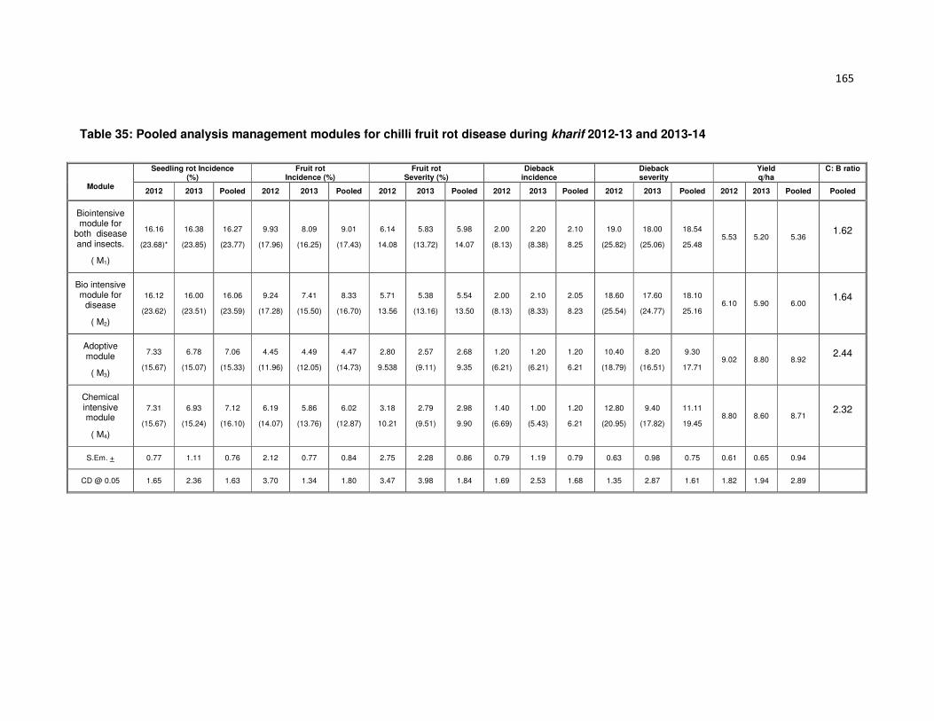
165
Table 35: Pooled analysis management modules for chilli fruit rot disease during kharif 2012-13 and 2013-14
Module
Seedling rot Incidence (%)
Fruit rot Incidence (%)
Fruit rot Severity (%)
Dieback incidence
Dieback severity
Yield q/ha
C: B ratio
2012 2013 Pooled 2012 2013 Pooled 2012 2013 Pooled 2012 2013 Pooled 2012 2013 Pooled 2012 2013 Pooled Pooled
Biointensive module for
both disease and insects.
( M1)
16.16
(23.68)*
16.38
(23.85)
16.27
(23.77)
9.93
(17.96)
8.09
(16.25)
9.01
(17.43)
6.14
14.08
5.83
(13.72)
5.98
14.07
2.00
(8.13)
2.20
(8.38)
2.10
8.25
19.0
(25.82)
18.00
(25.06)
18.54
25.48 5.53 5.20 5.36
1.62
Bio intensive module for
disease
( M2)
16.12
(23.62)
16.00
(23.51)
16.06
(23.59)
9.24
(17.28)
7.41
(15.50)
8.33
(16.70)
5.71
13.56
5.38
(13.16)
5.54
13.50
2.00
(8.13)
2.10
(8.33)
2.05
8.23
18.60
(25.54)
17.60
(24.77)
18.10
25.16 6.10 5.90 6.00
1.64
Adoptive module
( M3)
7.33
(15.67)
6.78
(15.07)
7.06
(15.33)
4.45
(11.96)
4.49
(12.05)
4.47
(14.73)
2.80
9.538
2.57
(9.11)
2.68
9.35
1.20
(6.21)
1.20
(6.21)
1.20
6.21
10.40
(18.79)
8.20
(16.51)
9.30
17.71 9.02 8.80 8.92
2.44
Chemical intensive module
( M4)
7.31
(15.67)
6.93
(15.24)
7.12
(16.10)
6.19
(14.07)
5.86
(13.76)
6.02
(12.87)
3.18
10.21
2.79
(9.51)
2.98
9.90
1.40
(6.69)
1.00
(5.43)
1.20
6.21
12.80
(20.95)
9.40
(17.82)
11.11
19.45 8.80 8.60 8.71
2.32
S.Em. + 0.77 1.11 0.76 2.12 0.77 0.84 2.75 2.28 0.86 0.79 1.19 0.79 0.63 0.98 0.75 0.61 0.65 0.94
CD @ 0.05 1.65 2.36 1.63 3.70 1.34 1.80 3.47 3.98 1.84 1.69 2.53 1.68 1.35 2.87 1.61 1.82 1.94 2.89

166
in M3 (Adoptive module) which was on par with M4 (Chemical intensive module) (2.90
PDI).
At 135 DAT M3 (Adoptive module) recorded the least fruit rot incidence (4.40 %)
which was on par with M4 (Chemical intensive module) (6.39 %) followed by M2 (Bio
intensive module for disease) (8.87 %). The least fruit rot severity (3.40 PDI) recorded in
M3(Adoptive module) which was on par with M4 (Chemical intensive module) (3.90 PDI)
followed by M2 (Bio intensive module for disease) (6.86 PDI).
The least die-back incidence (1.00 %) was recorded in M4 (Chemical intensive
module) which was on par with M3 (Adoptive module) (1.20 %) followed by M2 (Bio
intensive module for disease) (2.10 %). The least die-back severity (8.20 PDI) was
recorded in M3 (Adoptive module) which was on par with M4 (Chemical intensive module)
(9.40 PDI).
The yield was significantly superior in M3 (Adoptive module) (8.80 q/ha) with 2.41
cost benefit ratio, which was on par with M4 (Chemical intensive module) (8.60 q/ha, 2.29
C:B ratio) followed by M2 (Bio intensive module for disease) (5.90 q/ha, 1.61 C:B ratio).
The pooled results of kharif 2012 and 2013 indicated that least seedling rot
incidence (7.06 %) was recorded in M3 (Adoptive module) which was on par with M4
(Chemical intensive module) (7.12 %) followed by M2 (Bio intensive module for disease)
(16.06 %).
In M3 (Adoptive module) least fruit rot incidence (4.47 %) was recorded which was
on par with M4 (Chemical intensive module) (6.02 %) followed by M2 (Bio intensive
module for disease) (8.33 %). The least fruit rot severity (2.68 PDI) was recorded in M3
(Adoptive module) which was on par with M4 (Chemical module) (2.98 PDI) followed by
M2 (Bio intensive module for disease) (5.54 PDI).
The least die-back incidence (1.21 %) was recorded in both M3 (Adoptive module)
and M4 (Chemical intensive module) followed by M2 (Bio intensive module for disease)
(2.05 %). The least die-back severity (9.30 PDI) was recorded in M3 (Adoptive module)
which was on par with M4 (Chemical module) (11.11 PDI) followed by M2 (Bio intensive
module for disease) (18.10 PDI).

167
Plate 36: Fruit rot disease management modules at MARS Dharwad

168
The yield was significantly superior in M3 (Adoptive module) (8.92 q/ha) with 1.25
cost benefit ratio, which was on par with M4 (Chemical intensive module) (8.71 q/ha, 2.32
C:B ratio) followed by M2 (Bio intensive module for disease) (6.0 q/ha, 1.64 C:B ratio),
whereas least yield (5.36 q/ha, 1.62 C:B ratio) was recorded in M1 (Bio intensive module
for disease and insects.).

169
5. DISCUSSION
Chilli (Capsicum annuum L.) is a very remunerative, widely grown indispensable
spice crop in the world and is cultivated in almost all the states of India. Chilli is
suffering from several economically important diseases like damping off, die back, fruit
rot, leaf spots, leaf curl, wilt etc. which pose a serious threat to the successful large-
scale cultivation. The fruit rot disease caused by fungi Colletotrichum (namely C.
capsici, C. gloeosporioides and C. acutatum), A. alternata. and Fusarium spp. is a
major yield limiting factor.
The disease is more severe in India because of its complex nature and
symptoms vary in different stages of crop. In the present situation of climate change
there are lot of variations and fluctuations in environmental conditions which may lead
into variability in pathogen population and disease epidemics in different regions.
Therefore to throw light on many questions, experiments were conducted on various
aspects of chilli fruit rot and die-back disease with reference to prevalence and
distribution of disease, pathogen diversity, severity of the disease in various
geographical regions of South India, isolation, identification, morphological, and
molecular variability of pathogens and their quick detection by molecular method to
know the complexity of causal organisms.
Role of weather parameters on disease incidence and survival ability of
pathogens, host range and cross inoculations were studied. Further, seed health
management and integrated disease management strategies comprising screening of
genotypes for resistance, evaluation of chemicals under field conditions and various
disease management modules were studied for identification of the best management
module with maximum C:B ratio which helps the farming community to a greater
extent. The experiments were conducted in the laboratory as well as in the field during
2012 and 2013 the results obtained on these aspects are discussed here under.
5.1 Survey, isolation and identification of pathogen/s to study the distribution in
different geographical regions of South India
Survey on the incidence and severity of disease helps to gather information on
the prevalence, severity and distribution of disease, pathogen diversity in particular

170
agro-climatic zone. It reveals the magnitude of the problem on hand and serves as a
precursor for evolving the management strategies.
Survey and surveillance form the basis for any successful plant protection that
depends on early detection of disease followed by timely adoption of management
measures. In the present investigation roving survey was undertaken for two years in
major chilli growing areas of Karnataka, Maharashtra and Andhra Pradesh to assess
the incidence and severity of fruit rot. During the survey it was generally observed that
disease incidence on red colored fruits was more than other parts of plant in most of
the areas surveyed.
The survey also supplements the information about intensity and existence of
biotypes in particular geographical locations. Hence, an attempt was made to assess
the incidence and severity of fruit rot disease of chilli. The severity of fruit rot of chilli
expressed as disease incidence and per cent disease index. The survey also revealed
that the severity of fruit rot and dieback disease from location to location and also
varietal performance differed from place to place, obviously due to various factors like
temperature, relative humidity, pattern of rainfall and even it could also be attributed to
existence of variability of pathogen/s, cropping pattern and genotype grown.
From the pooled results of two years survey (2012-13 and 2013-14) it was
observed that among the three states the highest fruit rot incidence (19.97%) with
13.77 PDI was observed in Maharashtra followed by Andhra Pradesh (19.82%, 13.07
PDI) whereas the highest die-back incidence (11.23%) with 42.79 PDI was observed in
Andhra Pradesh followed by Karnataka (9.06%, 38.14 PDI) (Fig.2).
Among eight districts of Karnataka, the highest fruit rot incidence (22.18%) with
14.75 PDI was observed in Bellary, whereas the highest die-back incidence (11.18%)
with 43.92 PDI was observed in Gadag (Fig.3 and Fig. 4). In Guntur district of Andhra
Pradesh the highest fruit rot incidence (21.84%) with 12.98 PDI was recorded.
Shivakumara (2006) reported that maximum fruit rot severity (11.56 PDI) was recorded
in Gulbarga district followed by Raichur (11.17 PDI) district. Rajput (2011) reported that
highest severity of fruit rot was noticed in Nalvadi and Sanshi villages of Dharwad
district.
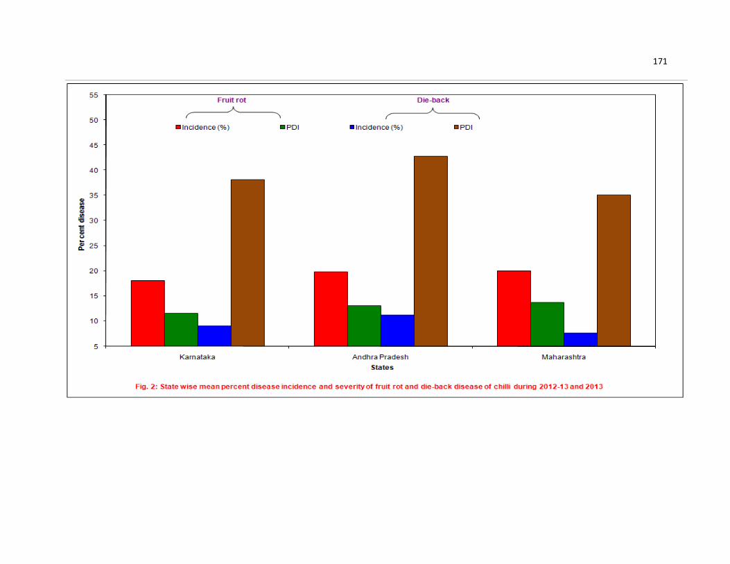
171

172

173

174
In the present study maximum fruit rot incidence and severity observed in
Maharashtra and Andhra Pradesh is mainly due to irrigated condition with temperature
of 25-30°C which is ideal for disease development. The same trend of temperature and
irrigation is also followed in Bellary district. In these regions, chilli is grown in large area
and also continuously year after year which leads to increase in inoculum of pathogens
in seeds, plant debris and in soil. Use of untreated seeds which were produced locally
is also one of the main reason for increase in disease incidence and severity of fruit rot.
In the present survey it is observed that in both rainfed (17.66%, 11.34PDI) and
irrigated (18.96%, 12.23 PDI) conditions disease was equally distributed in severe form
(Fig.5). Soil type (red and black) has no distinct differentiation for fruit rot disease. If the
congenial weather condition and sufficient amount of inoculum is available then it leads
to disease development in all these situations on commonly cultivated susceptible
genotype like Byadgi Dabbi (Fig. 6). It is observed that in both sole crop (18.58%,
11.95 PDI) and inter crop (17.52%, 11.36 PDI) disease was equally distributed in
severe form, indicating that inter crop has never helped in reducing the disease (Fig.
7).
With reference to genotypes observed during survey, fruit rot and die-back
disease (15.87 to 21.88%, 9.77 to 10.84% respectively) was equally severe in all
genotypes. It is indicating the susceptible nature of commonly cultivated genotypes
viz., Byadgi Dabbi, Byadgi Kaddi and Guntur (Shivakumara, 2006) (Fig. 8).
Predominance of various pathogens was observed during survey. Frequency of
occurrence of single pathogen was always less compared to combination of
pathogens. The combination of C. capsici and Fusarium spp. was 28.37 per cent, C.
capsici with A. alternata 11.35 per cent, combination of C. capsici, C. gloeosporioides
and A. alternata was 10.64 per cent. C. capsici as a predominant fungus causing fruit
rot of chilli was reported by Thind and Jhooty (1985). Ramachandran et al. (2007)
conducted survey and collected 92 isolates from different chilli growing areas in Andhra
Pradesh and Karnataka. Among them 53 were identified as C.capsici, 38 as C.
gloeosporioides and one was C. acutatum. Chilli fruit samples collected from Tamil
Nadu, revealed that C. capsici was the most commonly isolated (69%) followed by C.
gloeosporioides (19%) and A. alternata (Madhvan et al., 2010). Association of

175

176

177

178

179
Fusarium spp. in the present study is supported by the work of Parey et al. (2013) who
reported different Fusarium spp. were involved in fruit rot in Jammu and Kashmir.
Mild to severe form of fruit rot and die-back disease was observed irrespective
of varieties during the field survey under taken. During 2012-13 and 2013 -14 such
variation in disease incidence in different locations is quite often attributed to
environmental conditions. However, the variations may also be attributed to the
presence of variability in pathogenic fungi. The mean frequency of pathogens isolated
from samples collected during survey recorded as the highest frequency (38.56%) was
observed in C. capsici followed by A. alternata (18.98%) and C. gloeosporioides
(14.89%) (Fig. 9). Shivakumara (2006) isolated Colletotrichum spp. (25%) followed by
A. alternata (21%) from fruit infected chilli from northern Karnataka.
The present results are well supported by the reports of Hegde and Kulkarni
(2001a), Bagri et al. (2004) and Das et al. (2004) wherein they reported the maximum
prevalence of Colletotrichum capsici. In fruit rot disease of chilli, multiple infections
were quite common. Many workers have also reported the prevalence of more than
two fungal pathogens in fruit rot disease of chilli (Prabhavathy and Reddy, 1995; Basak
et al., 1996b; Bagri et al., 2004).
5.2 Morphological characterization and molecular variability of pathogen/s
5.2.1 Morphological characterization
Accurate identification of Colletotrichum species along with the knowledge of
populations responsible for epidemics are essential for developing and implementing
effective disease management strategies (Freeman et al., 1998). Traditionally,
identification and characterization of Colletotrichum species have been based on
morphological characters, such as size and shape of conidia and appressoria;
existence of setae; the teleomorph state and cultural characters such as colony colour,
growth rate and texture (Von Arx, 1957; Smith and Black, 1990). In the present
investigation 25 isolates of C. capsici , 20 of C. gloeosporioides , 12 of C. acutatum, 10
of A. alternata, 9 of F. oxysporum and 7 of F. sporotrichioides were selected for
morphological variations and results are discussed here.
180

181
C. capsici
The colony varied from white to grey color, flat to raised fluffy and smooth
regular to coarse irregular margin. Acervuli size varied between 130.01 to 162.60 µm
with setae length 145.40 to 179.10 µm, medium to good sporulation, falcate shaped
spores with size between 13.5 - 21.2 X 3.2 - 4.8 µm. These findings are in agreement
with the findings of earlier workers (Sydow, 1913; Butler, 1918). Whitish aerial mycelial
growth and concentric rings of growth were visible along with light rose colored spore
masses. Conidia were single celled curved with smooth margin and hyaline (Hegde,
1998; Jeyalakshmi and Seetharaman, 1999; Rohana et al. 2005; Shivakumara, 2006;
Rajput , 2011; Sangdee et al., 2011; Christopher et al., 2013).
Akhtar and Singh (2007) collected five isolates of C. capsici from Uttaranchal
which showed variation in culture color (white grey to blackish grey), colony
morphology (smooth to wavy margin growth) on PDA media, conidial size (varied
between 25.27 to 26.15 µm length, 3.17 to 3.66 µm width). These observations were
similar to present investigation but differed slightly with conidial size. Variation in colony
and spore morphology is supported by the work of (Masoodi et al., 2013). Parey et al.
(2013) observed C. capsici isolates varied in mycelial color fairly white to light grey,
circular to irregular margin with fluffy to flat growth, conidial size varied between 21.4 to
25.3 µm length, 3.7 to 4.3 µm width.
C. gloeosporioides
The colony varied from white to grey color, where as in three isolates Cg-9, Cg-
10 and Cg-12 whitish saffron color was observed. Flat to raised fluffy and smooth
regular to coarse irregular margin and sectoring was observed in three isolates, Cg-4,
Cg-12 and Cg-20. Medium to excellent sporulation of cylindrical shaped spores with oil
globules with size between 12.5 - 16.7 X 1.6 - 2.8 µm was recorded. These findings are
in agreement with the findings of earlier workers (Simmondes, 1965; Mordue, 1971;
Irwin and Camerson, 1978; Holiday, 1980; Jeffries et al., 1990; Ekbote., 1994;
Sudhakar 2000; Prasannakumar 2001; Venkataravanappa and Nargund 2002 and
Prashanth, 2007 Than et al., 2008a; Jayalakshmi, 2010; Biju et al., 2012;
Chandramani, 2012; Patil and Nargund, 2013). The present results are well supported
by the earlier reports, wherein they reported the colony of C. gloeosporioides varied

182
from light orange to pale grey colour, conidia were cylindrical in shape and measured
about 13.5 X 4.5 µm.
C. gloeosporioides isolated from chilli was distinct from C. capsici showing fast,
fluffy, dull white mycelium and within the isolates also found distinct characteristics with
respect to growth, colour of mycelia and sporulation (Shivakumara, 2006).
Venkataravanappa and Nargund (2007) revealed that conidia size of six isolates of C.
gloeosporioides from mango anthracnose samples, varied between 10.9-20.6 µm in
length and 4.39 – 6.65 µm in width.
C. acutatum
The colony was varied from whitish pink to brownish pink color with flat to fluffy
and smooth regular to coarse irregular margin. Medium to good sporulation of fusiform
shaped spores with size varied between 11.4 - 14.1 X 1.1 - 2.3 µm was recorded.
These findings are in agreement with the findings of earlier workers (Simmonds, 1965;
Wharton and Uribeonodo, 2004; Than et al., 2008a; Zivkovic et al., 2010; Patil and
Nargund, 2013), wherein they reported C. acutatum conidia were single septate,
fusiform in shape and conidia size varies between 8 -16 X 2.5 – 4.0 µm.
A. alternata
The colony varied from ash to dark ash color, medium fluffy to raised fluffy and
smooth regular to coarse irregular margin. Medium to excellent sporulation of muriform
spores with beak with 3 – 5 horizontal septa 1 – 2 vertical septa, spore size varied
between 14.1 - 21.8 X 6.1 - 8.4 µm was observed.
A. alternata isolates from chilli showed ash colour to black coloured mycelium
with medium to fast growth and moderate to excellent sporulation, spore size varied
between 18.1 - 30.1 X 5.8 – 6.7 µm (Shivakumara, 2006; Amitkumar et al., 2012; Parey
et al., 2013)
F. oxysporum
The colony was varied from white to whitish purple color, flat, smooth and
regular margin. Medium to excellent sporulation of falcate macro conidia with 3 – 5
horizontal septa and size varied between 22.01 - 32.10 X 3.2- 5.2µm.

183
The fungus produced microconidia, which were oval-ellipsoid, cylindrical,
straight to curved, variable and found abundantly. Macro conidia, sparse and thin
walled, having 3-5 septate, fusoid and subfulcate measures 26 - 42 X 2.8 – 4.5 µm.
Culture of fungus on potato dextrose agar was whitish to peach purple coloured with
woolly, aerial mycelium (Booth, 1971; Joffe, 1986; Bagri et al., 2012; Patil and
Nargund, 2013).
F. sporotrichioides
The colony varied from light brown to white rose colour, fluffy, smooth to coarse
and regular to irregular margin. Good to excellent sporulation of falcate macro conidia
with 3 – 5 horizontal septa and size varies between 32.1 - 42.0 X 3.4 -5.2 µm, globose
microconidia were observed. These observations were also found similar to Booth
(1971) and Joffe (1986) wherein they reported that mycelium varied from white rose to
light brown, red colour, aerial growth, microconidia were globose to lemon shape,
macroconidia were curved, falcate elongated with 3 – 5 septate measures 22-45 X 3.6
-5.4 µm.
5.2.2 Molecular characterization and variability of pathogen/s
Morphological characterization alone, are not always adequate for species
identification due to overlap in morphological characters and phenotypic variation
among species under different environmental conditions. Conidial shape has been
applied as a reliable means of discriminating certain species; for example, conidial
shape has differentiated Colletotrichum species pathogenic to strawberry (Denoyes
and Baudry, 1995). However, in other cases, identification can be complicated because
of overlapping ranges of conidial morphology and variation in colony characteristics
(Adaskaveg and Hartin, 1997). Correct taxonomic identification is important in disease
management such as choosing appropriate fungicides (Whitelaw-Weckert et al., 2007).
To overcome the inadequacies of traditional morphology based identification
schemes, DNA sequence analyses have been used to characterize and analyze the
taxonomic complexity of fungal pathogens. Cannon et al. (2000) stated that data
derived from nucleic acid analyses should provide the most reliable framework to build
a classification of Colletotrichum, as DNA characters were not directly influenced by

184
environmental factors. Assessment of molecular variability by using microsatellite
markers were used in differentiation species of Fusarium (Prasad et al., 2004). Genetic
differentiation of C. gloeosporioides isolates from cultivated strawberry plants and non-
cultivated hosts was done by using boot strap analysis and constructed a dendrogram
using RAPD markers (Xiao et al., 2004). Though RAPD differentiated C. capsici and C.
gloeosporioides and also the strainal variations among these species but in recent
years RAPD technique has its own limitation. Most fungal phylogenetic studies utilized
sequences from the ribosomal gene cluster, since they were present in large numbers
as tandem repeats and evolved as a single unit. In particular, sequence analysis of the
internal transcribed spacer (ITS) regions which lie between the 18S and 5.8S genes
and the 5.8S and 28S genes, has proved useful in studying phylogenetic relationships
of Colletotrichum species because of their comparative variability (Sreenivasaprasad et
al., 1994; Moriwaki et al., 2002; Photita et al., 2005).
A combined application of morphological characters, molecular diagnostic tools
and pathogenicity helps in identification and to study variability among species. To
understand existence of variation among the isolates of pathogens, PCR based
technique i.e., ITS was used in the present investigation. Ramachandran et al. (2007)
identified 92 isolates from different chilli growing areas of India, among them 53 were
C.capsici 38 as C. gloeosporioides and one was found to be C. acutatum based on ITS
sequence analysis. Phylogenetic analyses from DNA sequence data of ITS rDNA
region revealed three major clusters representing these three species of Colletotrichum
spp. in chilli (Than et al., 2008b).
The genera Colletotrichum spp. Alternaria spp. and Fusarium spp. pose a major
challenge for systematics because of the phylogenetic relationships of many of their
members still are unclear. DNA sequences of selected six isolates were compared
using bioinformatics tool like NCBI BLAST programme. Based on sequence
comparison these isolates were identified as C.capsici, C. gloeosporioides, C.
acutatum, A. alternata, F. sporotrichioides and F. oxysporum and were markedly
similar to the reference strain of NCBI BLAST Genbank. The ITS region of onion
twisting disease causing fungal pathogen isolates were amplified by using the NCBI
BLAST program, they confirmed these isolates as C.gloeosporioides, C.acutatum and
F. oxysporum (Patil and Nargund, 2013).

185
Pongpisutta et al. (2013) reported that effects of environmental manipulation on
the morphological stability makes identification between Colletotrichum species
difficult. ITS RFLP and ITS sequence analysis are considered to be essential tools to
solve the problems of species differentiation and specific identification of the chilli
anthracnose causal agents. After ITS rDNA region identification of pathogens C.
capsici, C.gloeosporioides, C.acutatum , Alternaria alternata, F. sporotrichioides and F.
oxysporum further studies were carried out for specific DNA amplification which was
observed with fairly consistent band for C. capsici (25 isolates) at 450 bp, for C.
gloeosporioides (20 isolates) fairly consistent band was observed with CgInt region at
450 bp and C. acutatum (12 isolates) at 490 bp with concentration of 437 µg/µl.
Whereas, Alternaria alternata amplification was observed at 390 bp and for Fusarium
spp approximately at 550-570 bp. Fungal isolates from chilli fruits in Thailand that
showed typical anthracnose symptoms were identified as C. acutatum, C. capsici and
C. gloeosporioides by ITS sequence study (Than et al., 2008a). Morphology and
pathogenecity are not enough to distinguish between Colletotrichum spp. diversity.
Studies include internal transcribed spacer (ITS) regions (Freeman et al., 2001;
Moriwaki et al., 2002; Sanders and Korsten, 2003; Lee et al., 2007) as well as
restriction fragment length polymorphism (RFLP) (Balardin et al., 1999; Martin and
Garcia-Figueres 1999; Saha et al., 2002 ; Weir et al., 2012).
The products of the PCR ITS rDNA digestion with Hind III revealed that no
restriction sites were present. The digestion with HaeIII, resulted in a characteristic
pattern of three fragments in all the isolates. C. capsici isolates showed four clusters
(Fig. 10), in cluster-I (six isolates), cluster – II (12 isolates), cluster – III (six isolates),
cluster – IV (one isolate). C. gloeosporioides isolates showed five clusters (Fig. 11), in
cluster-I (five isolates), cluster – II (one isolate), cluster – III (nine isolates), cluster – IV
(three isolates) and cluster –V (two isolates). C. acutatum isolates showed three
clusters (Fig. 12), in cluster-I (five isolates), cluster – II (five isolates) and cluster – III
(two isolates). The digestion with Taq I, resulted in a characteristic pattern of three
fragments in Alternaria and Fusarium isolates. A. alternata isolates showed two
clusters (Fig. 13) cluster-I (seven isolates), cluster – II (three isolates). Fusarium
isolates shown five clusters (Fig. 14), in cluster-I (five isolates), cluster – II (six
isolates), cluster – III (one isolate) cluster – IV (three isolates) and cluster – V (one

0
LEGEND
Isolates Source
1 Koluru
2 Siraguppa
3 Salahalli
4 UAS Dharwad
5 Kundgol
6 Byalal
7 Hulkoti
8 Byadgi
9 Basapur
10 Chikkatembinahal
11 Nelhal
12 Panchamukhi
13 Belgammanadoddi
14 Emmiganur
15 Dharmapuram
16 Mantralya
17 Mugathi
18 Madire
19 Paaladagu
20 Kattavaripalem
21 Bayyavaram
22 Mangalveda
23 Sangola
24 Aurangabad
25 Coimbatore

186
Fig. 10: PCR-RFLP diversity profile of C. capsici by HaeIII
Coefficient
0.50 0.63 0.75 0.88 1.00
1
7
14
15
17
21
2
4
5
6
22
20
18
16
23
12
10
11
3
8
24
19
13
9
25
Cluster 1
Cluster 2
Cluster 3
Cluster 4
Similarity coefficient
`

0
LEGEND
Isolates Source
1 Kagalgombe
2 Shanvaspura
3 Inamhongal
4 Byahatti
5 UAS Dharwad
6 Shirguppi
7 Binkadkatti
8 Guttal
9 Kanakapur
10 Beluru
11 Mandapur
12 Raladoddi
13 Hanumapuram
14 Alur
15 Kalludevarakunta
16 Mangalaveda
17 Medikonduru
18 Percherela
19 Ananthapuram
20 Coimbatore

187
Coefficient
0.51 0.63 0.75 0.88 1.00
1
7
10
20
17
2
3
5
6
9
18
14
15
11
13
4
8
16
12
19
Cluster 1
Cluster 3
Cluster 4
Cluster 5
Fig. 11 : PCR-RFLP diversity profile of C. gloeosporioides by Hae III
Cluster 2
Similarity coefficient

0
LEGEND
Isolates Source
1 Raravi
2 Govanakoppa
3 UAS Dharwad
4 Binkadakatti
5 Byadgi
6 Devihosur
7 Lingadahalli
8 Budugumpa
9 Belgammanadoddi
10 Mantralaya
11 Marakattu
12 Pedakurpadu

188
Coefficient
0.50 0.63 0.75 0.88 1.00
1
5
7
12
10
3
4
6
11
8
2
9
Fig. 12 : PCR-RFLP diversity profile of C.acutatum by Hae III
Cluster 1
Cluster 2
Cluster 3
Similarity coefficient

0
LEGEND
Isolates Source
1 Koluru
2 UAS Dharwad
3 Kundgol
4 Hulkoti
5 Guttal
6 Talakall
7 Raichur
8 Paldaagu
9 Mandapura
10 Kalludevarakunta

189
Coefficient
0.67 0.75 0.83 0.92 1.00
1
2
3
4
7
9
6
5
8
10
Cluster 1
Cluster 2
Fig. 13: PCR-RFLP diversity profile of A. alternata by TaqI
Similarity coefficient

0
LEGEND
Isolates Source
1 Raravi
2 UAS Dharwad
3 Sharewad
4 Byalal
5 Ranebennur
6 Nelhal
7 Alur
8 Emmiganur
9 Mangalaveda
10 Lokapur
11 Kumbapur farm
12 Badrapur
13 Bommanal
14 Raladoddi
15 Mantralaya
16 Perecherla

190
Coefficient
0.43 0.57 0.72 0.86 1.00
1
3
4
7
9
2
5
6
14
8
10
16
11
12
15
13
Cluster 1
Cluster 2
Cluster 4
Cluster 5
Cluster 3
Fig. 14: PCR-RFLP diversity profile of Fusarium spp. Taq I

191
isolate). ITS-RFLP technique is an efficient method for rapid diagnosis of
Colletotrichum species from chilli. Similar type of observations were made by Sheu et
al. (2007) where in they collected a total of 412 Taiwan isolates from chilli production
areas analyzed through ITS-RFLP fingerprinting. Among them, 245 C. acutatum, 34 C.
boninense, 52 C. capsici and 69 C. gleosporioides were identified. Other
Colletotrichum isolates (3%) were not distinguishable, which inferred to the various
inter- and intra-species variations in Colletotrichum members.
Imjit et al. (2013) reported that PCR- based molecular method provided
improved early detection and diagnosis system of fruit rot disease of chilli. In the
present study detection of pathogens from different parts of plant like seeds, fruits,
infected twig/stem by PCR-based method was done by using specific primers. specific
amplification with specific primers of DNA extracted directly from infected host tissues
i.e., seed, fruit, pedicel and dieback stem revealed that C. capsici was amplified by
species specific primer (C.cap-F and C.cap-R) as single band at 450bp. C.
gloeosporioides, C. acutatum, amplified by species specific primers (CgInt and CaInt)
at 450 and 490bp respectively, Alternaria alternata amplified by species specific primer
at 390bp and Fusarium spp. were amplified at 550 – 570bp. Quick detection and
diagnosis will help in plant quarantine laboratories to obtain fast and accurate test
results. Present results are well supported by the reports of Zivkovic et al. (2010)
Chowdappa and Chethana (2012) and Pongpisutta et al. (2013).
5.3 Epidemiology of the fruit rot disease in relation to climatic factors
5.3.1 Survival ability of C. capsici, C. gloeosporioides, A. alternata and
F. oxysporum in plant debris
During the absence of an active host plant, pathogen/s may survive themselves
to maintain continuity of the disease cycle and to provide primary inoculum for infection
in the next season. Dormant mycelium plays an important role in the perpetuation,
dissemination and inciting of disease from one season to another. The study was
initiated to know the survival ability of the pathogen at different ecological conditions
viz., laboratory conditions, soil surface and 10 cm depth of soil.
192
The present study revealed that Colletotrichum spp. remained viable up to 165
days under laboratory conditions, 120 days on soil surface and 60 days under 10cm
depth of soil condition. A. alternata remained viable up to 120 days under laboratory
conditions, 90 days on soil surface and 45 days under 10cm depth of soil condition. F.
oxysporum remained viable up to 150 days under laboratory conditions, 135 days on
soil surface and 60 days under 10cm depth of soil condition.
The present results were slightly different from earlier reports in which Ahmed
(1982) found that, C. capsici causing fruit rot of chilli survived up to eight months in
both seed and culture. C. capsici could survive up to 225 days on infected seeds of
chilli stored under room conditions where as on pedicel and fruit rind it survived for 195
days (Sanathakumar, 1999). F. oxysporum remained viable up to 120 days under
laboratory, 255 days on pot soil surface and 270 days under refrigerator condition,
whereas Colletotrichum spp. remained viable up to 165 days under laboratory, 120,
210 days in pot soil surface and refrigerator condition respectively. These pathogens
survived in the infected debris and it served as primary source of infection during the
favorable conditions in next season (Patil and Nargund, 2013)
5.3.2 Host range of chilli fruit rot pathogens
C. capsici can overwinter on alternative hosts such as solanaceous vegetables
and legume crops, plant debris and rotten fruits in the field (Pring et al., 1995). In the
present study two solanaceous vegetables and seven legume hosts were tested. On
solanaceous vegetables, tomato and brinjal chlorotic lesions were produced on 4th day
after inoculation. These lesions turned to brown color leading to necrosis after seven
days of inoculation. In legume crops like cowpea, green gram and black gram brown
color horse shoe type lesions were observed after eight days after inoculation. C.
capsici which causes fruit rot of chilli also infects soybean, tomato, potato, and brinjal
(Sundararaman, 1927; Hegde, 1998). Pandey (2006) showed on the basis of
morphological, pathological and molecular characterization that C. capsici from chilli
causes fruit rot, seed and seedling mortality in tomato.
193
5.3.3.1 Cross inoculation of C. capsici
Colletotrichum species were generally able to survive in or on seeds and one of
the ways that anthracnose was introduced to the chilli field is through infected
transplants. C. capsici infection of chilli was shown to have two pathways: invasion
through the seed coat and invasion through the openings of the testa (Jewsakun,
1978). C. capsici caused seed rot and root rot of seedlings (Bhale et al., 1999a;
Shivakumara, 2006; Hemannavar, 2008; Chauhan et al., 2010a) and also it causes
die-back of chilli (Hegde, 1998; Shivakumara 2006; Akhtar et al., 2008; Rajput, 2011).
Hence, to know whether the same pathogen will cause all these symptoms, in the
present investigation cross inoculation of C. capsici between seed, twig, fruit pedicel
and fruit was carried out, the results revealed that C. capsici can cause seed rot, die-
back of twigs, pedicel discoloration and fruit rot from all tested parts of chilli plant.
5.3.3.2 Pathogen interaction
Fruit rot of chilli is caused by more than one pathogen i.e., different species of
Colletotrichum, (C. capsici, C. gloeosporioides, C. acutatum), A. alternata and
Fusarium spp. (Simmonds, 1965; Thind and Jhooty 1985; Datar, 1995; Mesta, 1996;
Shivakumara, 2006; Santoshreddy et al., 2012; Parey et al., 2013). Hence to know
about the symptoms caused by these pathogens in combination, an experiment was
conducted in this aspect. It revealed that symptoms caused by combination of C.
capsici, C. gloeosporioides, C. acutatum appear on one day after inoculation as water
soaked lesion of 0.5 -1.0 cm diameter. After seven days of inoculation sunken lesion
with salmon color spore mass produced on 1.5 – 2.0 cm diameter lesion. Symptoms
caused by combination of C. capsici, A.alternata and F. oxysporum appear on one day
after inoculation as water soaked lesion of 0.5 – 1.0 cm diameter, after four days of
inoculation grayish mycelial growth observed on 2.0 – 2.5 cm diameter, six days after
inoculation concentric rings with grayish salmon color spore mass produced on these
lesions.
5.3.4.1 Effect of weather parameters on spore load of Colletotrichum spp.
Environmental factors play a major role in the development of disease
epidemics. The relationships among rainfall intensity, duration and crop geometry and

194
the dispersal of inoculum possibly lead to different levels of disease severity. The
effects of temperature often interact with other factors, such as leaf surface wetness,
humidity, light or competitive microbiota. The duration of the surface wetness, however,
appears to have the most direct influence on the germination, infection and growth of
the pathogen on the host. In the two consecutive years of investigation, the simple
correlation and multiple regression analysis was worked out to understand relationship
between weather parameters and spore load of Colletotrichum spp.
In the pooled data of two consecutive years (2012 and 2013), cumulative rainfall
recorded highly significant positive correlation (r = 0.84) with maximum temperature (r
= 0.34). Whereas, other weather parameters viz., relative humidity of evening and
morning were negatively correlated.
It indicates that among weather parameters selected for correlation on the spore
load of Colletotrichum spp. cumulative rainfall and maximum temperature showed
significantly positive correlation.
The coefficient of determinative value (R2) was found to be 90 and 83 per
cent in 2012 and 2013 respectively. The difference between observed and predicted
spore load of Colletotrichum spp. varied from -4.28 to +1.19 (Fig. 15)
There was variation in the spore load progression which was accounted by the
linear functions of the independent variables such as maximum and minimum
temperature, morning and evening relative humidity, rainfall and number of rainy days
and dependent variable was spore load trapped by aeroscope. Influence of weather
factors on the development of spore load of C. truncatum indicated that more conidial
counts were observed during last week of July and first week of August, which
coincided with the critical stages of infection in green gram anthracnose disease
(Kulkurni, 2009).
5.3.4.2 Effect of Colletotrichum spp. spore load and weather parameters on fruit
rot disease:-In the two consecutive years of investigation, the simple correlation
and multiple regression analysis was worked out to understand relationship between
weather parameters, Colletotrichum spp. spore load and fruit rot incidence.
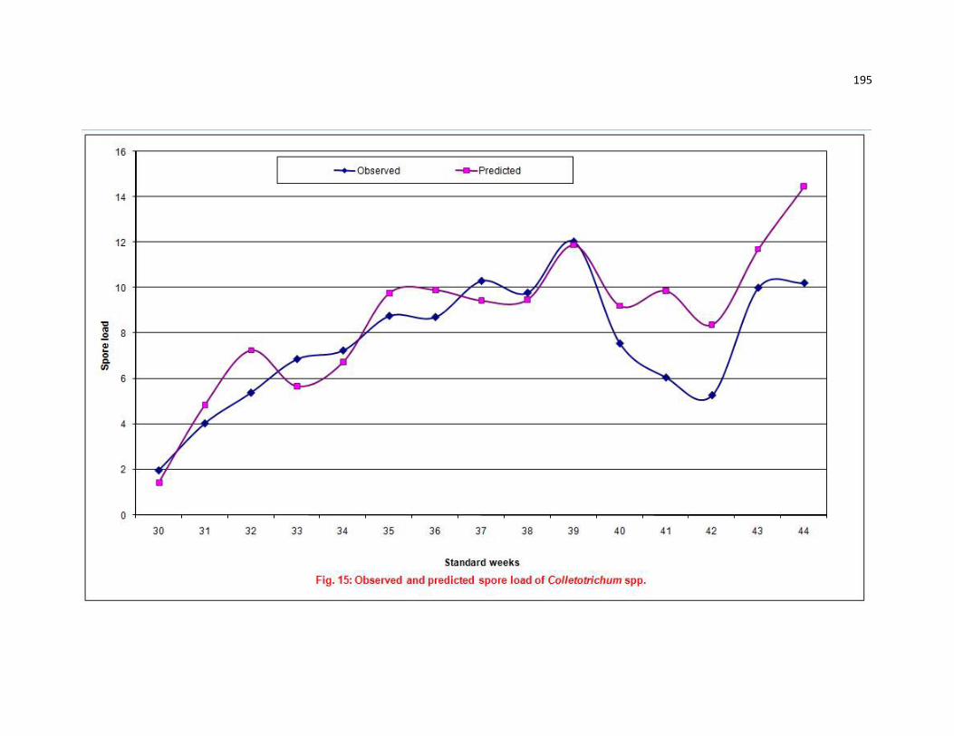
195

196
In the pooled data of two consecutive years (2012 and 2013), spore load
recorded highly significant positive correlation (r = 0.94) followed by cumulative rainfall
(r = 0.80), and maximum temperature (r = 0.41). It indicates that spore load of
Colletotrichum spp. and among weather parameters cumulative rainfall and maximum
temperature showed significantly positive correlation on fruit rot incidence in chilli.
The coefficient of determinative value (R2) was found to be 88 and 85 per cent in
2012 and 2013 respectively. The difference between observed and predicted fruit rot
incidence Colletotrichum spp. varied from -0.66 to + 2.48 (Fig. 16)
Cumulative rainfall contributed maximum (R2 = 0.97) to development of onion
twister disease severity caused by C. gleosporioides, C. acutatum and F. oxysporum
(Patil et al., 2013a).
5.3.5 Effect of date of planting on fruit rot and die-back disease incidence and
severity
A considerable variation on incidence of twister disease of onion caused by C.
gleosporioides, C. acutatum and F. oxysporum with respect to date of planting, in
which planting at 15th July was found ideal for obtaining maximum severity of disease
(Patil et al., 2013a). In present investigation a field trial was carried out to assess the
effect of planting time on fruit rot and die-back disease development which revealed
that highest fruit rot incidence was observed in July 15th planting, where as highest die-
back incidence was observed in June 15th planting. Byadgi Dabbi recorded highest fruit
rot incidence and severity where as therer was no significant difference in die back
disease incidence in date of planting and varieties.
5.4 Development of IDM strategies for disease
No single specific management program could eliminate chilli fruit rot complex.
Effective management of such complex diseases usually involves the integrated
management strategies to reduce the use of fungicides, to enforce eco friendly low
cost and effective management by combination of cultural, biological, chemical
management with intrinsic resistance strategies. Hence, in the present investigation
emphasis was given on identification of resistance genotypes, evaluation of seed

197

198
treatment chemical and bio fungicides to eliminate primary inoculum , chemical
management and also evaluated four management modules which include bio-
intensive, chemical and adoptive by combination of both biological and chemical
management which are discussed in following paragraph.
5.4.1 Evaluation of chilli genotypes
Among disease management strategies the use of resistant cultivars is the
cheapest, easiest, safest and most effective means of controlling the disease. This is
not only to eliminate losses from the disease but also decrease the cost of chemical
and mechanical control, as well as reduce contamination of the environment from the
use of toxic chemicals. This is an important choice in all crop improvement programme.
Besides, the resistant cultivars conserve natural resources and reduce the cost, time
and energy when compared to other methods of disease management. Increase in use
of fungicides to control the fruit rot has led to consciousness of their persistence and
development of new strains of pathogen. To avoid this situation, identifying the
resistant cultivars against chilli fruit rot is most significant one.
Earlier reports indicated varied degree of reaction of genotypes against fruit rot
fungus, C. capsici under natural endemic condition. LCA-301, LCA-324, K-1 and
Byadagi Kaddi were found resistant. Whereas KDSC-210-10 and S-32 were found
highly susceptible (Hegde and Anahosur 2001b). The genotypes Arka Lohit, Pepper
Hot, CA 97, KDC 1, CC 4, CA 95, CA 115 and CA 59 were found to be moderately
resistant to anthracnose and a high yielding moderately resistant to fruit rot disease
chilli hybrid CCH1(Sln 1 x CA 97) was identified (Pugalendhi et al., 2010).
Among 343 genotypes screened under natural endemic field condition during
kharif season of 2012 and 2013, none were immune, eight found resistant, 21
genotypes were moderatly resistant.
Under In vitro screening by pin prick method none of the genotype showed
immune and resistant reaction. However, moderately resistant reaction was observed
in seventeen genotypes, ten genotypes showed moderately susceptible reaction
against C.capsici, C. gloeosporioides, C. acutatum. Ninteen genotypes showed
moderately resistant, eight showed moderately susceptible reaction against A.alternata

199
and 20 genotypes showed moderately resistant, seven showed moderately susceptible
reaction against F. oxysporum and F. sporotrichioides. These results are well
supported by Parey et al., (2013), wherein they revealed that evaluation of genotypes
against C. capsici none was resistant, however, DC-4, Arka Lohit, LCA-235, LCA-333,
LCA-301 exhibited moderately resistant reaction under both field and pot culture
conditions and minimum lesion size was also observed under in vitro by pin prick
method in these genotypes. In particular, some lines of C. baccatum showed
resistance to the pathogen, and pathogen inoculation resulted in limited lesions on the
chilli fruits (Yoon, 2003). However, to date, no resistance has been found in Capsicum
annuum, which is the only species grown worldwide (Park, 2007).
5.4.2 Seed health management
5.4.2.1 Standard Blotter Method
The seeds showing fungal colonies under stereo-binocular microscope revealed
the presence of C. capsici (72.85%), C. gloeosporioides (9.26%) and C. acutatum
(4.76%), A. alternata (5.20%) and F. sporotrichioides (3.45%) and F. oxysporum
(4.30%) (Fig. 17). The present studies are in conformity with Solanke et al. (2001) who
reported the presence of C. capsici, F. moniliformae, A. alternata from chilli seed
samples. C. capsici, species of Alternaria and Fusarium were observed in fruit rot
affected chilli seed samples of northern Karnataka (Shivakumara, 2006; Hemannavar
et al., 2009). C. dematium and A. alternata were associated with chilli seeds and found
responsible for severe seed rot and seedling rot was reported by Bhale et al., (1999a).
5.4.2.2 Evaluation of chemical fungicides
Simple seed treatment is known to reduce the seed-borne infections and
subsequently protect seed from deterioration. In absence of resistant cultivars the use
of fungicides has become inevitable method in the management of seed borne fruit rot
fungi in chilli. Evaluation of seed treatment fungicides revealed that among three
systemic fungicides evaluated at 0.2% concentration pyraclostrobin 20WG had shown
least infection (11.63%) with highest vigour index (861.17). Between two non systemic
fungicides captan 75WP showed least infection (12.28%) with highest vigour index
(754.64). Similar results were reported by Arunkumar and Vyas (2003). Among six

200

201
combi product fungicides, carboxin 37.5% + thiram 37.5% WS showed least infection
(7.25%) with highest vigour index (932.02) followed by metalaxyl 4%+mancozeb64%
(10.09%, 871.70) (Fig. 18). Among fungicides, carboxin + thiram (Vitavax power) at
0.2% was most effective seed treatment fungicide for chilli seed health management in
nursery conditions. Similar results were reported by Hemannavar (2008) under
laboratory conditions. In combi product carboxin 37.5% + thiram 37.5% WS, carboxin
inhibits the mitochondrial electron transport of fungi which act on target site succinate
dehydrogenase enzyme. The other compound thiram increases the efficacy of carboxin
by reacting with protein SH group and also it acts on multi sites and effectively acts
against seed and soil borne fungal pathogens.
5.4.2.3 Evaluation of bio fungicides
Seed treatment with biofungicides is the oldest practice in plant protection which
provide economical and relatively nonpolluting and now, this is an attractive delivery
system for either fungal or bacterial bioprotectants compared to other field application
systems. The uses and expectations of seed treatment are greater today due to the
impact of environmental regulations that have either banned or restricted use of
organic mercurial fungicides because of their residual toxicity. Bioprotectants applied to
seeds may not only protect seeds but also may colonize and protect roots and increase
plant growth. Trichoderma spp. and Pseudomonas fluorescens have been considered
as good model of biological control because of their ubiquitous nature, easy to isolate,
rapid growth on many substrates, affect wild range of plant pathogens, acts as
mycoparasite, competes well for food and site, have enzyme system capable of
attacking many plant pathogens and easy in application.
Results of four bio fungicides alone (@ 10.0 g/kg) and in combination (5.0 +5.0
g/kg) seed treatment revealed that T. harzianum 5.0g + P. fluorescens 5.0g showed
least infection (14.89%) with highest vigour index (930.74) followed by P. fluorescens
(10.0 g/kg) 14.94% infection with 915.27 vigour index. The highest seedling infection
(42.99%) with least vigour index (361.87) was observed in untreated control. Suthinraj
and John (2009) reported that seed treatment with P. fluorescens (5g/kg) and T.
harzianum (10g/kg) reduces 25% and 24.10% incidence of C. capsici and increased
seedling vigour of chilli by 13.70% and 12.10% respectively (Fig. 19). P. fluorescens

202

203

204
showed higher antagonistic activity against C. capsici under in vitro conditions and also
less seedling rot was obtained in P. fluorescens treated seeds compared to T.
harzianum (Hegde et al., 2001b, Srinivas et al., 2006; Azad et al., 2013). Seed
treatment with Trichoderma harzianum recorded least damping off incidence followed
by T.viride and Pseudomonas fluorescence compared to untreated control in chilli
(Deshmukh et al., 2012). Choudhary et al., (2013) reported that seed treatment with
carbendazim, thiram and T. viride effectively managed seedling rot caused by C.
capsici.
5.4.3 Field efficacy of chemicals for the management of fruit rot and die-back disease
of chilli during kharif 2012 and 2013
Chemicals are the most common and practical method to control anthracnose
diseases. However, fungicide tolerance often arises quickly, if a single compound is
relied upon too heavily. The disease can be managed under normal weather conditions
with a reasonable spray program. However, there are numerous reports of negative
effects of using chemicals on farmers’ income and health, and toxic contamination to
the environment, particularly in developing countries (Voorrips et al., 2004). A field
experiment was conducted during kharif 2012 and 2013 at MARS Farm, Dharwad for
management of fruit rot and die-back disease of chilli using four systemic and four
combi-product fungicides along with a control where only insect management was
done as per package of practices. The pooled results indicated that at 0.1%
concentration difenconazole 25EC and pyraclostrobin 20WG had shown least
incidence of die-back (1.93 and 2.93% respectively) and fruit rot (8.99% and 12.25%
respectively). Among combi-products tricyclazole 18% + mancozeb 62% WP and
pyraclostrobin 5% + metiram 55% had shown least incidence of die-back (2.80% and
2.23% respectively) and fruit rot (11.08% and 11.70% respectively) (Fig. 20). Similar
observations were reported by Alexander and Waldenmaier, 2002; Lewis and Miller,
2003, wherein they reported the strobilurin fungicides azoxystrobin (Quadris),
trifloxystrobin (Flint), and pyraclostrobin (Cabrio) have recently been labeled for the
effective management of chilli fruit rot. Difenconazole, propiconazole and tricyclazole at
0.1% effectively manages the fruit rot diseases caused by Colletotrichum spp. in fruits
and vegetables (Hegde et al., 2002b; Jamadar and Patil 2007; Benagi et al., 2009; Biju
et al., 2011; Nargund et al., 2011; Nargund et al., 2013b). These triazoles are sterol

205
inhibiting fungicides affect cytochrome P-450 enzymes inhibitors of sterol C-14
demethylation by this they act against most of the Ascomycota group fungal
pathogens, whereas strobulirins act through inhibition of respiration by binding to the
Qo center of the cytochrome b. These strobulirins are very broad and balanced
spectrum of activity on the foliage and have very favorable toxicological profile rapidly
dissipating from soil and surface water which are unlikely to cause hazard to non target
organisms and they have both protective and curative effect.
Use of combi product fungicides avoids the development of resistance of fungi
to systemic fungicides because these systemic fungicides interfere with only one or
sometimes two functions in physiology of fungus which it easily overcomes by either a
single mutation or by selection of resistant individuals in a population. Wherein non-
systemic protectant fungicides affect too many functions in fungus physiology and to
develop resistance the fungus will have to make too many gene changes. Hence the
combination of both systemic and non-systemic fungicides provides better
management of plant fungal disease for long duration
Highest yield was observed in difenconazole 25EC (8.86 q/ha) with cost benefit
ratio 2.59 which was on par with pyraclostrobin 5% + metiram 55% (8.33 q/ha, C:B
2.40) and tricyclazole 18% + mancozeb 62% WP (8.21 q/ha C:B 2.29), whereas lowest
yield (3.58 q/ha) was observed in control.
5.4.4 Integrated management of fruit rot disease of chilli
Field experiment was conducted during kharif 2012 and 2013 at Dharwad with
four modules namely bio intensive module for both disease and insect pests (M1), bio-
intensive module for disease with chemical pesticides for insect pests (M2), adoptive
module (M3), chemical intensive module (M4) to develop best disease management
module for chilli fruit rot disease.
This study found that adoptive module is a cost-effective and eco-friendly
approach in the chilli fruit rot management. The result of two years experimentation
revealed that adoptive module showed least seedling infection (7.06 %), die-back
incidence(1.21 %) and severity(9.30 PDI), fruit rot incidence (4.47 %), severity (2.68
PDI) and recorded high yield 8.92 q/ha with 2.44 cost benefit ratio which was also on

206

207

208
par with chemical intensive module with 8.71 q/ha 2.32 C:B ratio (Fig. 21). The present
results were also supported by (Sharma et al., 2004; Lydia and Zachariah, 2012; Patil
and Nargund, 2013) where in they reported among management modules, biological,
chemical and IDM. IDM module was superior to biological and chemical modules. As
no single specific management practice could eliminate chilli fruit rot complex here
adoptive module involves the integrated management strategies at right time
application reduces the use of fungicides, and it enforces eco friendly low cost and
effective management of chilli fruit rot.
Future line of work
• An extensive and continuous survey in intensive manner to see the diversity of
pathogens is needed.
• Identification of susceptible stage for die-back disease.
• Apart from ITS region, sequence analysis of protein coding genes such as
partial β-tubulin gene is needed to resolve phylogenetic relationships among the
Colletotrichum species.
• Marker Assisted Selection for identification and development of multiple disease
resistance genotypes is needed.
• IDM module (adoptive) needs to be demonstrated in farmer’s fields for
popularization of cost effective and eco-friendly management systems to
farming community.

6. SUMMARY AND CONCLUSIONS
Chilli (Capsicum annuum L.) is one of the very popular spice/ vegetable crop
grown worldwide known for its medicinal and health benefiting properties. This crop is
suffering from several economically important diseases. Fruit rot disease caused by
fungi Colletotrichum spp. (namely C. capsici, C. gloeosporioides and C. acutatum), A.
alternata. and Fusarium spp. is major yield limiting factor in all chilli growing areas. In
view of the destructive nature of disease the present investigation was executed.
The investigations included various aspects of chilli fruit rot and die-back
disease with reference to prevalence and distribution of disease, pathogen diversity,
severity of the disease in various geographical regions of South India, isolation,
identification, morphological and molecular variability of pathogens and their quick
detection by molecular method. Epidemiology and integrated disease management
strategies comprising screening for resistance, evaluation of chemicals, various
disease management modules to find out the best management module with
maximum C:B ratio which helps the farming community to a greater extent. The
findings of the investigations are summarized in this chapter.
Survey carried out during 2012-13 and 2013-14 revealed that among the three
states the highest fruit rot incidence (19.97%) with 13.77 PDI was observed in
Maharashtra followed by Andhra Pradesh (19.82%, 13.07 PDI) where as the highest
die-back incidence (11.23%) with 42.79 PDI was observed in Andhra Pradesh
followed by Karnataka (9.06%, 38.14 PDI). Among eight districts of Karnataka the
highest fruit rot incidence (22.18%) with 14.75 PDI was observed in Bellary, where as
the highest die-back incidence (11.18%) with 43.92 PDI was observed in Gadag. In
Guntur district of Andhra Pradesh the highest fruit rot incidence (21.84%) with 12.98
PDI was recorded.
With respect to rainfed and irrigated conditions during survey, it revealed that,
both rainfed and irrigated conditions disease was equally distributed in severe form.
In both soil types (black and red) there was no distinct differentiation for fruit rot
disease. In both sole crop and inter crop disease was uniformly distributed in severe

210
form. With reference to genotypes observed during survey, fruit rot and die-back
disease was equally severe in all genotypes.
During investigation, different symptoms of the disease were noticed viz., seed
and seedling rot, acervulii of Colletotrichum were observed on plumule and radical,
softening, rotting and decaying of tissues at collar region of seedlings were observed.
Necrosis of tender twigs from the tip to backwards, brown to grayish white straw
colored lesions developed. Circular to elliptical sunken spots with salmon color and
acervuli with conidia observed on Colletotrichum infected both red and green colored
fruits. Black color spore masses with sunken lesions on Alternaria infected fruit was
observed. Rotting of fruit from tip and pink color spore mass was observed on
Fusarium infected fruits. Brown color discoloration and acervuli with conidia were
observed on pedicel.
Predominance of various pathogens was observed during survey. Frequency
of occurrence of single pathogen was always less compared to combination of
pathogens. In combination of C. capsici and Fusarium spp. were found in 28.37 per
cent where as C. capsici with A. alternata was 11.35 per cent. Among the mean
frequency of fruit rot pathogens observed the C. capsici recorded highest frequency
of 38.56 per cent followed by A. alternata (18.98 %), where as C. acutatum occurred
in lowest frequency of 5.04 per cent.
The colony of C. capsici varied from white to grey color, flat to raised fluffy and
smooth regular to coarse irregular margin. Acervuli size varied between 130.0 to
162.6 µm with setae length 145.4 to 179.1 µm. Medium to good sporulation with
falcate shaped spores were observed.
The colony of C. gloeosporioides varied from white to grey color to whitish
saffron color with flat to raised fluffy and smooth regular to coarse irregular margin
was observed. Medium to excellent sporulation of cylindrical shaped spores with oil
globules were observed under microscope.
The colony of C. acutatum varied from whitish pink to brownish pink color, flat
to fluffy and smooth regular to coarse irregular margin. Medium to good sporulation of
fusiform shaped spores were observed under microscope.

211
The colony of A. alternata, varied from ash to dark ash color, medium fluffy to
raised fluffy and smooth regular to coarse irregular margin. Medium to excellent
sporulation of muriform spores were observed.
The colony of F. oxysporum varied from white to whitish purple color, flat,
smooth and regular margin. Medium to excellent sporulation of falcate macro conidia
were observed under microscope.
The colony of F. sporotrichioides varied from light brown to white rose color,
fluffy, smooth to coarse and regular to irregular margin. Good to excellent sporulation
of falcate macro conidia with 3 – 5 horizontal septa were observed under microscope.
The isolates were identified as Colletotrichum spp., Alternaria sp. and
Fusarium spp. by morphological characters. Later, the identity of the pathogen was
confirmed by sequencing the amplified ITS rDNA region, and analysing in NCBI
BLAST program and identified as C. capsici, C. gloeosporioides, C. acutatum, A.
alternata, F. sporotrichioides and F. oxysporum.
Specific DNA amplification was observed with fairly consistent band for
C. capsici at 450 bp by C. cap primer, for C. gloeosporioides fairly consistent band
was observed with CgInt region at 450 bp and C. acutatum was amplified with CaInt
region at 490 bp with concentration of 437 µg/µl. Whereas, A. alternata amplification
was observed at 390 bp by AAR and AAF primer, for Fusarium spp. approximately at
550-570 bp by Tef- Fu primer.
The products of the PCR ITS rDNA digestion with Hind III revealed that no
restriction sites were present. The digestion with HaeIII, resulted in a characteristic
pattern of three fragments in all the isolates. C. capsici isolates were grouped into
four clusters, C. gloeosporioides isolates were grouped into five clusters, C. acutatum
isolates were grouped into three clusters. The digestion with Taq I, resulted in a
characteristic pattern of three fragments in Alternaria and Fusarium isolates. A.
alternata isolates were grouped into two clusters, Fusarium isolates grouped into five
clusters.
PCR-based detection method using specific primers is best early detection and
diagnosis system of fruit rot disease of chilli.

212
The survival ability of Colletotrichum spp., A. alternata and F. oxysporum was
more in lab condition (150-165 days) and reduced to 90-125 days on soil surface and
still reduction in survival was recorded in 10cm depth of soil condition (45-60 days).
C. capsici showed symptoms on both tested solanaceous vegetables (tomato
and brinjal) and also all seven tested legume crops (cowpea, green gram, black gram,
mothbean soybean chickpea and pea).
C. capsici can cause seed rot, die-back of twigs, pedicel discoloration and fruit
rot from all tested above ground parts of chilli plant by cross inoculation.
Combinations of more than one pathogen cause high fruit rot severity and fast
disease development was confirmed by pathogen interaction study.
The weather factors had direct and significant influence on the spore load of
Colletotrichum spp. in which cumulative rainfall recorded highly significant positive
correlation.
The weather factors and Colletotrichum spp. spore load had significant
influence on the fruit rot incidence spore load recorded highly significant positive
correlation
(r = 0.94) followed by cumulative rainfall (r = 0.80), and maximum temperature
(r = 0.41).
The highest fruit rot incidence and severity was observed in July 15th planting
and in Byadgi Dabbi variety, where as die-back disease incidence and severity were
distributed uniformly irrespective of genotypes in all dates of planting.
Among 343 genotypes screened under natural endemic field condition during
kharif season of 2012 and 2013 none were immune, eight genotypes found resistant
where as 21 genotypes found moderate resistant.
38 genotypes were screened under laboratory condition by pin prick method
results indicated that none of them showed immune and resistant reaction. However,
moderately resistant reaction was observed in 17 genotypes.
213
The frequency of seed mycoflora observed under stereo-binocular microscope,
revealed the presence of C. capsici (72.85%), C. gloeosporioides (9.26%) and
C. acutatum (4.76%), A. alternata (5.20%) and F. sporotrichioides (3.45%) and F.
oxysporum (4.30%).
Seed treatment of carboxin 37.5% + thiram 37.5% (vitavax power 75WS) at
2g/kg seed and combinations of T. harzianum 5.0g + P. fluorescens 5.0g were most
effective in management of fruit rot causing seed borne fungal pathogens.
Among four systemic fungicides difenconazole 25EC and pyraclostrobin 20WG
at 0.1% concentration and among five combiproduct fungicides, tricyclazole 18% +
mancozeb 62% WP and pyraclostrobin 5% + metiram 55% found effective against
fruit rot and dieback disease. Highest yield was observed in difenconazole 25EC
(8.86 q/ha) with cost benefit ratio 2.59 which was on par with pyraclostrobin 5% +
metiram 55% (8.33 q/ha, C:B 2.40) and tricyclazole 18% + mancozeb 62% WP (8.21
q/ha C:B 2.29).
Integrated management of chilli fruit rot disease during kharif 2012 and 2013
indicated that adoptive module showed least seedling infection (7.06 %), die-back
incidence (1.21 %) and severity (9.30 PDI), fruit rot incidence (4.47 %), severity (2.68
PDI) and recorded high yield 8.92 q/ha with 2.44 cost benefit ratio which was also on
par with chemical intensive module with 8.71 q/ha 2.32 CB ratio.
REFERENCES
Abdul Baki, A. A. and Anderson, J. P., 1973, Vigour determination in soybean seeds by
multiple criteria. Crop. Sci., 13: 630-633
Abhishek, S. and Verma, K. S., 2007, In vitro cross pathogenicity and management of
Colletotrichum gloeosporioides causing anthracnose of mango. Ann. Pl. Prot.
Sci., 15(1): 186-188.
Adaskaveg, J. E. and Hartin, R. J., 1997, Characterization of Colletotrichum acutatum
isolates causing anthracnose of almond and peach in California. Phytopathol.
87(9): 979-987.
Adikaram, N. K. B., Brown, A. and Swinburne, T. R., 1983, Observations on infection of
Capsicum annuum fruit by Glomerella cingulata and Colletotrichum capsici.
Trans. Brit. Mycol. Soc., 80: 395-401
Ahmed, S. S., 1982, Studies on seed borne aspects of anthracnose of chillies caused
by Colletotrichum capsici (Sydow) Butler and Bisby. M. Sc. (Agri.) Thesis,
Univ. Agric. Sci., Bangalore, (India).
Akhtar, J., 2007, Effect of fungicides on growth and sensitivity of the isolates of
Colletotrichum capsici causing fruit rot of chilli. J. Pl. Dis. Sci, 2(1):
59 – 62
Akhtar, J. and Dwivedi, R. R., 2006, In vitro screening of bio-agents against
Colletotrichum graminicola causing anthracnose of sorghum. Ann. Pl. Prot.
Sci. 14 (2): 505.
Akhtar, J., Khalid, A. and Chaube, H. S., 2009, Occurrence and Management of Die-
back in Chillies: an overview. In: Plant Disease Management for Sustainable
Agriculture. Ed. Ahamad, S., Daya Publishing House, Delhi, pp.121-135.
Akhtar, J. and Singh, M.K., 2007, Studies on the variability in Colletotrichum capsici
causing chilli anthracnose. Indian Phytopath., 60 (1) : 63-67.
215
Akhtar, J., Singh, M. K. and Choube, H. S., 2008, Effect of nutrition on formation of
acervuli, setae and sporulation of the isolates of Colletotrichum capsici.
Pantnagar J. Res. 6(1): 110-113.
Akhtar, J., Kumudrani, T., Lal, H. C., Atulkumar, Singh, V. K. and Khan, Z., 2012,
Impact of soil solarization on some solanaceous vegetables nursery in
plateau region of Jharkhand, India. Vegetos. 25 (2) : 109-114.
Alexander, S. A. and Waldenmaier, C. M., 2002, Management of anthracnose in bell
pepper- Fungicide and nematicide tests. New fungicide and nematicide data
committee of the Am. Phytopath. Soc. 58: 49.
Amit Kumar, H. N., Pankaj Sharma, G. and Rakesh Shah, 2012, Investigations on
variability and eco-friendly management of Alternaria alternata causing
tomato blight. Vegetos. 25 (1) : 52-58.
Amusa, N. A. and Alabi, A. A., 1996, Host range of Colletotrichum gloeosporioides from
Gliricidia sepium and its implication in crop production. Crop Res.,11: 359-
363.
Angadi, H. D., 1999, Studies on anthracnose of chilli (Capsicum annum) and its
management. M. Sc. (Agri.) Thesis, Univ. Agric. Sci., Dharwad , (India).
Angadi, H.D., Naik, M.K., Patil, M.G. and Patil, R.G., 2003, Evaluation of chilli
genotypes against anthracnose disease. Veg. Sci. 30(2): 164-165.
Anonymous, 2005, International seed testing association. Proc. Int. Seed Test. Assoc.
Int. Rul. Seed Test. Seed Sci. Tech. 15: 1-9.
Anonymous, 2014, Indian horticulture database-2013. Nation. Hort. Board, Min. Agric. ,
Govt. India. P.23
Anupama, Patil., Benagi, V.I. and Nargund, V.B., 2012, Management of basal rot of
onion caused by Fusarium oxysporum f.sp. cepae. In: Nation. Symp. Blend.
Conven. Mod. Pl. Pathol. Sust. Agric. IIHR Bangaluru, pp. 196-197

216
Arunkumar and Vyas, N.L., 2003, Management of die-back in chilli through seed and
spray treatments. Hum. Imp. Des. Env. pp.348-352.
Azad, C. S., Srivastava, J. N. and Gireesh Chand., 2013, Evaluation of bio-agents for
controlling fruit rot/ Anthracnose of banana caused by Colletotrichum
gloeosporioides in-vitro condition. The Bios. 8(4): 1221-1224.
Babu, L. J., Gour, H. N. and Pankaj, S., 2008, Efficacy of phytoextracts and bioagents
against Colletotrichum gloeosporioides causing banana fruit rot. J. Mycol. Pl.
Pathol. 38(3): 635-637.
Bagri, R. K., Choudhary, S.L. and Rai, P.K., 2004, Management of fruit rot of chilli with
different plant products. Indian Phytopath. 57(1): 107-109.
Bagri, R. K., Yadav, R. K., Pankaj Sharma. and Choudhary, S. C. L., 2011,
Performance of partially purified plant product preparations against fruit rot of
chilli caused by Alternaria alternata. Ind. J. Pl. Prot. 39(2):141-145.
Bagri, R. K., Yadav, R.K., Dheeraj Singh and Choudhary, S. C. L., 2012, Efficacy of
partially purified plant product preparations against fruit rot of chilli caused by
Fusarium oxysporum. Pl. Dis. Res., 27 (1): 12-16
Bailey, J. A. and Jeger, M. J., 1992. Colletotrichum: Biology, Pathology and Control.
Commonwealth Mycological Institute, Wallingford, p.388
Balardin R. S., Smith, J. J. and Kelly, J. D., 1999, Ribosomal DNA polymorphism in
Colletotrichum lindemuthianum. Mycol. Res., 103(7): 841-848.
Bansal, R. D. and Grover, R.K., 1969, Reaction of chilli (Capsicum frutescens) varities
to Colletotrichum capsici. J. Res. Punjab Agric. Univ., 6:345-348.
Basak, A. B., 1994, A comparative study on dry weight loss due to major fruit rot
diseases of chilli. Chittagong Univ. Stud. Sci., 18(1): 129-131.
Basak, A. B., 1997, Reaction of some chilli germplasms to major fruit rotting fungal
pathogens. Chittagong Univ. Stud. Sci., 21(1): 123-125.

217
Basak, A. B., Fakir, G.A. and Mridha, M. A. U., 1994, Studies on the prevalence of six
major fruit rot diseases of chilli at different stages of fruit development in
Chittagong district. Chittagong Univ. Stud. Sci., 18(1): 125-128.
Basak, A. B., Fakir, G. A. and Mridha, M. A. U., 1996a, Relation of seed borne infection
to different infection grades in fruit rot diseases of chilli. Seed Res. 24(1): 69-
70.
Basak, A. B., Fakir, G. A. and Mridha, M.A.U., 1996b, Studies on the location of
Fusarium spp. in the infected chilli seeds. Bangladesh J. Bot. 25 (2):
227-229.
Basha, H., Hemannavar, V., Ramanujam, B., Rageshwaran, R. and Sriram, S., 2010,
Screening of chilli microflora and other biocontrol agents for their antagonistic
effects on Colletotrichum spp. infecting chllies. J. Pl. Prot. Sci. 2 (1): 38 - 44.
Begum, M. F., Rahman, M. A. and Alam, M. F., 2010, Biological control of Alternaria
fruit rot of chilli by Trichoderma species under field conditions. Mycobiol. 38
(2): 113 – 117.
Benagi, V. I., Mesta, R. K., and Nargund, V. B., 2009, Major diseases of chilli and their
integrated management. Paper presented In: Nation. Work. Curren. Trend.
Future Prosp. Prod. Expo. Spice Crops Spec. Ref. Chillies, UAS, Dharwad.
27 - 28 Feburaury (India) pp. 101-106
Bernard, E. L. and Schrader, R. A., 1984, Anthracnose of acacia in Florida. Occurrence
and fungicidal control. Proc. Florida. State Hort. Sci. 97 : 244-247.
Bhale, M. S., Bhale, U. and Khare, M. N., 1999b, A method of testing virulence in chilli
anthracnose pathogen. J. Mycol. Pl. Pathol. 29(2): 253-254.
Bhale, U., Bhale, M.S. and Khare, M.N., 1999a, Efficacy of methods in detection of
Alternaria alternata on chilli seeds. J. Mycol. Pl. Pathol. 29(1):125-126.
Bhuvaneswari, V. and Rao, M. S., 2001, Evaluation of Trichoderma viride antagonistic
to post-harvest pathogens on mango. Indian Phytopath. 54(4) : 493-494.
218
Biju, C. N., Praveena, R., Ankegowa, S.J., Darshana, C. N. and Jashmi, K. C., 2011,
Evaluation of fungicides for the management of anthracnose disease of black
pepper in the nursery. Paper presented In: Nation. Symp. Spices Aromatic
Crops (SYMSAC VI). December 8-10. UAS, Dharwad, p. 245.
Biju, C.J., Praveena, R., Ankegowda, S. J. and Jashmi, K.C., 2012, In planta survival of
C. gloeosporioides the incitant of black pepper anthracnose. Paper presented
In: Nation. Symp. Presp. Pl. Healt. Mangm. December. 14-16. AAU, Gujarat.
pp. 54-55.
Booth, C., 1971, The genus Fusarium. Common wealth Mycological Institute, Kew.
Surrey, England, p.132.
Bosland, P. W. and Votava, E. J., 2003. Peppers: vegetable and spice capsicums. CAB
International, England, p.233.
Butler, E. J., 1918, Fungi and diseases in plants. Thacker, Spink and Co., Culcutta, VI,
p. 547.
Byung-Soo, K., 2007, Country report of anthracnose research in Korea. Paper
presented In: Int. Symp. Chilli. Anth., Hoam Facul. Hous. Seoul Nat. Univ.
Seoul Korea, September 17-19, p. 23.
Cannon, P. F., Bridge, P. D., Monte, E., 2000, Linking the Past, Present, and Future of
Colletotrichum Systematics. In: Colletotrichum: Host specificity, Pathology,
and Host-pathogen Interaction. Ed. Prusky, D., Freeman, S., Dickman, M.,
APS Press, St. Paul, Minnesota, p.1 20
Chandra Nayaka, S., Udaya, A. C., Shankar Niranjana, S. R., Prakash, H. S and
Mortensen, C. N., 2009, Anthracnose disease of chilli pepper. Tech. Bull.
Danish Seed Health Center. Univ. Copenhagen., Denmark, pp. 1-13.
Chandramani, R., Shweta, S., Nakkaran S. and Chandrasekhar, G., 2012,
Morphogenetic characterization of Colletotrichum spp. complex associated
with anthracnose disease of chilli. Paper presented In: Nation. Symp. Spice.
Aromatic Crop. (SYMSAC VI). UAS, Dharwad, December 8-10. p. 79.

219
Chandramani, R., Manjunath Hubballi., Prabhuling., R. B., Daliyamol, Akkeeran , S. and
Chandrasekar G., 2013, Inhibition of hydrolytic enzymes of Colletotrichum
capsici by propiconazole and PGPR strains. Bioinfolet. 10
(1 A) : 82 – 84.
Chauhan, Y. B., Patel, R. L., Chaudhari, S. M. and Patel , K. K., 2010a, Efficacy of
bioagents and Phytoextracts against Collectotrichum capsici. Paper
presented In: Nation. Symp. Persp. Pl. Health Mangm. December. 14-16.
AAU, Gujarat. p. 58.
Chauhan, Y.B., Patel, R.L., Chaudhari, S.M. and Chaudhari, R. F., 2010b, Production of
toxic metabolites by isolates of Collectotrichum. spp. and their effect on chilli
seedilings. Paper presented In: Nation. Symp. Persp. Pl. Health Mangm.
December. 14-16. AAU, Gujarat. pp. 142-143.
Choudhury, S., 1957, Studies on the development and control of fruit rot of chillies.
Indian Phytopath., 10: 55-62.
Choudhary, C. S., Jain, S. C., Ritesh Kumar and Jaipal Singh Choudhary., 2013,
Efficacy of different fungicides, biocides and botanical extract seed treatment
for controlling seed-borne Colletotrichum sp. in chilli (Capsicum annuum L.).
The Bios. 8(1): 123-126.
Chowdappa, P., 2010, Anthracnose of chilli. ORP on leaf spot diseases series. 2,
Ind.Inst.Hort.Res., Banglore (India).
Chowdappa, P. and Chethana, C. S., 2012, A polyphasic approach for characterization
of Colletotrichum gloeosporioides (Penz.) Penz, and Sacc. Associated with
anthracnose orchids. Paper presented In: Nation. Symp. Blending Conven.
Mod. Pl. Pathol. Sust. Agric., IIHR, Bangalauru, December, p. 89.
Christopher, D. J., Suthinraj, T. and Sudharajakumar, R., 2013, Morphological,
pathological and molecular variability of Colletotrichum capsici causing
anthracnose of chilli. Ann. Pl. Prot. Sci., 21 (2) : 309-314.
220
Chuang, T. Y. and Ann, P. J., 1997, Biological control of mango diseases. Pl. Prot. Bull.,
39(3) : 227
Das, S., Somnathsil and Jahangir, K., 2004, Disease reaction and its progress on
different chilli cultivars (Capsicum annuum L.) against anthracnose and leaf
curl virus under field condition. J. Mycol. Res. 42(1): 49-52.
Dastur, J. F., 1920, Glomerella cingulata (Stoneman) spald and its conidial form,
Gloesporium piperatum E. and E. and Colletotrichum nigrum E. and Hals. on
chillies and Carica papaya. Appl. Biol. 6(4):245-268.
Dastur, J. F., 1921, Dieback of chillies (Capsicum spp.) in Bihar. Memoirs. Depart.
Agric. in India. Botan. Series. 35: 409-413.
Datar, V. V., 1995, Pathogenecity and effect of temperature on six fungi causing fruit rot
of chilli. Indian J. Mycol. Pl. Pathol. 25 (3):195-197.
Denoyes, B. and Baudry, A., 1995, Species identification an pathogenicity study of
French Colletotrichum strains isolated from strawberry using morphological
and cultural characteristics. Phytopath. 85(1):53-57.
Deshmukh, R. R., Apet, K.T., Kamble, H. N. and Utpal, D., 2012, Effect of different
inoculants on germination and biometric character of chilli (Var. Parbhani
Tejas), Int. J. Pl. Prot. 5(2) : 252 – 255.
Deyong, Z., Chunhui, Z., and Yong L., 2007, Chilli anthracnose research in china: an
overview. Paper presented In: Int. Symp. Chilli Anth. Hoam Facul. Hous.
Seoul Nat. Univ., Seoul, Korea, September 17-19 p. 27.

221
Don, L. D., Van, T. T., Phuong, V. T. T. and Kieu, P. T. M., 2007, Colletotrichum spp.
attacking on chilli pepper growing in Vietnam country report. Paper presented
In: Int. Symp. Chilli Anth., Hoam Facul. Hous. Seoul Nat. Univ., Seoul,
September 17-19. p. 24.
Ekbote, S. D., 1994, Studies on anthracnose of mango (Mangifera indica L.) caused by
Colletotrichum gloeosporoioides (Penz) Penz. and Sacc. M. Sc. (Agri.)
Thesis, Univ. Agric. Sci., Dharwad (India).
Ekbote, S. D., 2002a, Survey of chilli diseases in Haveri district. Karnataka J. Agric. Sci.
15 (4):726-728.
Ekbote, S. D., 2002b, Bioefficacy of copper hydroxide (coxid) against anthracnose of
chilli. Karnataka. J. Agric. Sci. 15 (4):729-730.
Ekbote, S. D., 2005, Effect of Pseudomonas fluorescens on anthracnose of chilli caused
by Colletotrichum capsici. Karnataka J. Agric. Sci. 18 (1):162-165.
Ekbote, S.D., Jagadeesha, R.C. and Patil, M.S., 2002, Reaction of chilli germplasm to
fruit rot disease. Karnataka J. Agril. Sci. 15(4): 717-718.
Freeman, S., Katan, T., and Shabi, E., 1998, Characterization of Colletotrichum species
responsible for anthracnose diseases of various fruits. Pl. Dis. 82(6):
596-605.
Freeman, S., Minz, D., Maymon, M. and Zveibil, A., 2001, Genetic diversity within
Colletotrichum acutatum sensu Simmonds. Phytopath. 91(6):586-592.
Gohel, N. M. and Solanky K. U., 2011, Bio-control of Alternaria alternata (Fr.) Keissler
causing leaf spot and fruit rot of chilli. J. Pl. Dis. Sci. 6(2):
200 – 201
Gomez, K. A. and Gomez, A. A., 1984, Statistical Procedures for Agricultural Research.
Second Edition. A Willey Inter-Science Publication, New York, USA. p. 693.
Gud, M. A. and Raut, S. P., 2008, Control of mango anthracnose and stem end rot fungi
by fungicides and bioagents. J. Maharashtra Agric. Uni., 33(1) : 120-122.

222
Hadden, J.F. and Black, L.L., 1989. Anthracnose of pepper caused by Colletotrichum
spp. Proc. Int. Symp. Integrat. Mangm. Pract. Tomato Pepper Prod. Tropic.
Asian Veg. Res. Develop. Centre., Taiwan, pp. 189-199
Halfon, M. A. and Rylski, I., 1983, Internal mold caused in sweet pepper by Alternaria
alternata : Fungal ingress. Phytopathol., 73: 67-70
Halsted, B. D., 1890, A new anthracnose of pepper. Bull. Torrey Botan. Club. 18:
14-15.
Hegde, G. M., 1998, Studies on fruit rot of chilli (Capsicum annuum L.) caused by
Colletotrichum capsici (Syd.) Butler and Bisby. M. Sc. (Agri.) Thesis, Univ.
Agric. Sci., Dharwad (India).
Hegde, G. M. and Anahosur, K. H., 2001a, Biochemical basis of resistance to fruit rot
(Colletotrichum capsici) in chilli genotypes. Karnataka J. Agric. Sci., 14(3):
686-690.
Hegde, G. M. and Anahosur, K. H., 2001b, Evaluation of fungi toxicants against fruit rot
of chilli and their effect on biochemical constituents. Karnataka J. Agric.
Sci.14(3): 836-838.
Hegde, G.M., Anahosur, K.H. and Kulkarni, S., 2002a, Biological control of
Colletotrichum capsici causing fruit rot of chilli. Pl. Pathol. Newslett. 20:
4-5.
Hegde, G.M. and Kulkarni, S., 2001a, Vulnerable infection stage of chilli fruit by
Colletotrichum capsici (Sydow) Butler and Bisby. Karnataka J. Agric.
Sci.14(1): 162-163.
Hegde, G.M. and Kulkarni, S., 2001b, Seed treatment to control damping off of chilli
caused by Colletotrichum capsici (Sydow) Butler and Bisby. Karnataka J.
Agric. Sci. 14 (3): 829-830.
Hegde, G.M., Mesta, R.K. and Anahosur, K.H., 2002b, Efficacy of triazole compounds
against fruit rot pathogen (Colletotrichum capsici) of chilli. Pl. Pathol.
Newslett. 20: 3-4.
223
Hemannavar, V., 2008, Studies on seed borne aspects of anthracnose of chilli and its
management. M. Sc. (Agri.) Thesis, Univ.Agric. Sci. Dharwad (India).
Hemannavar, V., Rao, M. S. L., Hegde, Y. and Mohankumar, H. D., 2009, Status of
seed borne incidence of anthracnose of chilli in northern Karnataka and
evaluation of seed health testing methods for the detection of Colletotrichum
capsici. Karnataka J. Agric. Sci. 22(4): 807-809.
Holiday, P., 1980, Fungus Disease of Tropical Crops. Cambridge University Press,
Cambridge.
Howard, C. M., Maas, J. L., Chandler, C. K. and Albregts, E. E., 1992, Anthracnose of
strawberry caused by the Colletotrichum complex in Florida. Pl. Dis. 76: 976-
981.
http://www.ncbi.nlm.nih.gov.
Imjit, N., Rattanakreetakul, C. and Pongpisutta, R., 2013, Polymerase chain reaction
based detection of chilli anthracnose disease. Acta. Hort. 973: 199-206
Irwin, J.A.G. and Cameron, D.F., 1978, Two diseases in Stylosanthes sp. caused by
Colletotrichum gloeosporioides in Australia and pathogenic specialization
within one of the causal organisms. Australian J. Agric. Res. 29: 305-317.
Isaac, S., 1992, Fungal Plant Interaction. Chapman and Hall Press, London, p. 115.
Jadav, S. K., Diwakar, M. P., Sawant, U. K. and Kadam, J. J., 2008, Management of
leaf spot disease of Kokum incited by Colletotrichum gloeosporioides. J. Pl.
Dis. Sci. 3(2) : 193-196.
Jamadar, M. M. and Patil, D. R., 2007, Bio-efficacy of new formulations against leaf/fruit
spot of pomegranate. Karnataka J. Agric. Sci., 20(4): 865-866.
Jayalakshmi, K., 2010, Studies on anthracnose of pomegranate caused by
Colletotrichum gloeosporioides (Penz.) Penz. and Sacc. M. Sc. (Agri.)
Thesis, Univ. Agric. Sci., Dharwad, Karnataka (India).

224
Jayalakshmi, K., Nargund, V. B., and Raju, J., 2013, Toxin studies of Colletotrichum
gloeosporioides causal agent of pomegranate. J. Mycopathol. Res. 51(2):
347-350.
Jayalakshmi, K., Nargund, V. B., Raju, J. and Benagi, V.I., 2012, In vitro management
of anthracnose of pomegranate incited by Colletotrichum gloeosporioides
(Penz.) Penz. and Sacc. Int. J. Pl. Prot. 5(2) 304 – 307.
Jeyalakshmi, C. and Seetharaman, K., 1999, Studies on the variability of the isolates of
Colletotrichum capsici (Syd.) Butler and Bisby causing fruit rot. Crop Res. 17
(1): 94-99.
Jeffries, P., Dodd, J. C., Jegerand, M. J. and Plumbley, R. A., 1990, The biology and
control of Colletotrichum species on tropical fruit crops. Pl. Pathol. 39(3):
343-366.
Jewsakun, S., 1978, Serology, seed transmission on anthracnose disease of pepper
and effect of foliar fungicides to the causal pathogens. M.S. Thesis, Kasetsart
Univ.
Joffe, A. Z., 1986, Fusarium species: Their biology and toxicology, A Wiley- Interscience
Publication, pp. 406-408.
Johnston, P. R. and Jones, D., 1997, Relationships among Colletotrichum isolates from
fruit-rots assessed using rDNA sequences. Mycologia., 89(3):420-430.
Kenchaiah, 1975, Studies on chilli anthracnose caused by Colletotrichum capsici
(Sydow.) Butler and Bisby in Karnataka. M.Sc. (Agri.) Thesis, Univ. Agric.
Sci., Bangalore, (India).
Keshgond, R. S., Naik, M. K and Amaresh, Y. S., 2013, Management of chilli fruit rot
using indigenous strains of bio agents. Paper presented In: Nation. Symp.
Pathogenom. Diagn. Mangm. Pl. Dis. CTCRI., Thiruvananthapuram, October
24-25, Kerala, p. 145.
Khaleeque, M. I. and Khan, S.M., 1991, Fungi associated with fruit rot and die back
disease of chillies in Faisalabad. Pakistan J. Phytopath. 3:50-52.

225
Khare, D., Atri, D. C. and Shrivastava, C. J., 1995, A new fruit rot disease of chilli.
Indian Phytopath. 48(1): 111.
Khodke, S. W. and Gahukar, K. B., 1993, Fruit rot disease of chilli caused by Alternaria
alternata Keissal. in Maharastra. P.K.V. Res. J., 17(2): 206-207.
Khodke, S.W. and Gahukar, K.B., 1995, Changes in biochemical constituents of chilli
fruits infected with Colletotrichum gloeosporioides Penz. J. Maharashtra
Agric. Univ., 20(1): 142-143.
Kim, C. H. and Park, K. S., 1988, A predictive model of disease progression of red
pepper anthracnose. Korean, J. Pathol.4(4): 325 – 331.
Kim, K. D., Oh, B. J. and Yang, J., 1999, Differential interactions of a Colletotrichum
gloeosporioides isolate with green and red pepper fruits. Phytoparasitica.
27:1-10.
Korsten, L., De Villiers, E. E. and Lonsdale, J. H., 1993, Biological control of mango
post harvest disease in the packhouse. South African Mango Growers
Association Year Book, 13 : 117-121.
Kulkarni, S. A., 2009, Epidemiology and Integrated management of anthracnose of
greengram. Ph.D. Thesis, Univ. Agric. Sci., Dharwad (India).
Kulkarni, S. A. and Benagi, V. I., 2012, Survival of Colletotrichum truncatum in seeds
and crop debris, of greengram (Vigna radiata (L.) Wilczek) Int. J. Pl. Prot. 5
(2): 312 -314.
Lee, D.H., Kim, D.H., Jeon, Y. A., Uhm, J.Y. and Hong, S. B., 2007, Molecular and
cultural characterization of Colletotrichum spp. causing bitter rot of apples in
Korea. Pl. Pathol. J. 23(2): 37-44.
Lewis, I. M. L. and Miller, S.A., 2003, Evaluation of fungicides and a biocontrol agents
for the control of anthracnose on green pepper fruit. Am. Phytopath. Soc., 58,
p. 62.
226
Leyendecker, P. J. 1950, Frost aids mold growth in sun-dried chilli New Mexico State
Univ. Aric. Expt. Stn. Bull, p. 1045.
Liang, L.Z., 1990, Seed borne Fusarium of chilli and their pathogenic significance. Acta
Phytopath. 20(2): 117-121.
Ling, L. and Lin, K.R., 1944, On the occurrence of Colletotrichum capsici in China.
Indian J. Agric. Sci. 14: 162-167.
Lonsdale, J. H., 1992, Pre harvest fungicidal sprays for the control of post harvest
diseases of mango. Year Book South African Mango Growers Association.
12 : 28-31.
Lydia, C. and Zachariah, S., 2012, Evaluation of biological, chemical, and IDM modules
for the management of anthracnose and die-back of chilli. J. Mycol. Pl.
Pathol. 42(4): 435-438.
Madhavan, S., Paranidharan, V. and Velazhahan, R., 2010, RAPD and virulence
analyses of Colletotrichum capsici isolates from chilli (Capsicum annuum). J.
Pl. Prot. 117(6): 253-257.
Maiti, S. and Sen, C., 1979, Fungal diseases of betel vine. Proc. Nation. Acad. Sci.
United States of America. 25: 150-157
Malathi, G. 2004, Genotype and seasonal interaction of F1 hybrids and their parents in
chilli (Capsicum annuum L.) for anthracnose resistance and high yield. Ph.D.,
(Hort.) Thesis, Tamil Nadu Agric. Univ. Coimbatore (India).
Malathi, S. and Mohan, S., 2011, Evaluation of biocontrol agents and organic
amendments against onion basal rot caused by Fusarium oxysporum f. sp.
cepae. Madras Agric. J. 98 (12) : 382-385.
Mallesh, S. B., Lingaraju, S., Benagi, V. I., Basamma, K., Kumari and Vinaya, H., 2009,
Native plant growth promoting rhizobacteria for the suppression of pathogens
of pomegranate. Paper presented In: Int. Symp. on Pomegranate and Minor
Meditteranean Fruit Crop. Univ. Agric. Sci., Dharwad, p. 142.

227
Manandhar, J.B., Hartman, G.L. and Wang, T.C., 1995, Anthracnose development on
pepper fruits inoculated with Colletotrichum gloeosporioides. Pl. Dis.79:380-
383.
Mandhare, V. K. and Suryawanshi, A. V., 2003, Antagonistic effect of Bacillus
thermopillus on some pathogens. J. Maharashtra Agric. Univ. 28(3):
274-277.
Marchionatto, J.B., 1935, Notes on plant pathology, Revista Facultad de Agronomia
Univ. Nation. Plata. 19: 407-426.
Martín, M.P. and García-Figueres, F., 1999, Colletotrichum acutatum and
Colletotrichum gloeosporioides cause anthracnose on olives. Eur. J. Pl.
Pathol. 105 (8): 733-741.
Masoodi, L., Anwar, A., Ahemed, S. and Sofi, T.A., 2013, Cutural, morphological and
pathogenic variability in Colletotrichum capsici causing die-back and fruit rot
of chilli. Asian. J. Pl. Pathol. 7(1): 29-41.
Mathew, S. K., Wahab, M. A. and Devi, S. N., 1995, Seasonal occurrence of chilli
(Capsicum annuum L.) disease in Kerala, India. J. Spic. Arom. Crop. 4:
86-87.
Mayee, C. D. and Datar, V. V., 1986, Phytopathometry. Tech. Bull. Marathawad Agric.
Univ., Parbhani (India).
Mehetre, P. B. and Joshi, D. M., 2010, Investihations on Collectotrichum capsici, (Syd.)
Butler. and Bisby) of Yam (Dioscorea alatal.) under South Gujrat conditions.
Paper presented In: Nation. Symp. Persp. Pl. Health Mangm. AAU. Gujarat,
pp. 89-90.
Meenugupta and Garg, R.C., 2002, Epidemiological studies on ripe fruit rot of chilli
caused by Colletotrichum capsici (Syd.) Butler & Bisby. Indian J. Hill
Farming. 15(1): 126.

228
Mesta, R. K., 1996, Studies on fruit rot of chilli (Capsicum annum L.) caused by
Colletotrichum capsici (Sydow) Butler and Bisby. M. Sc. (Agri.) Thesis, Univ.
Agric. Sci., Dharwad (India).
Mishra, A. K., Gupta, V. K., Gaur, R. K., Jain, P. K. and Saroj, S., 2010, Current status
of Fusarium wilt disease of guava (Psidium guajava L.) in India. Biotech., 9(2)
: 176-195.
Mordue, M., 1971, Discription of pathogenic fungi and bacteria, C.M.I. Sheet No.315,
Commonwealth Mycological Institute, Surrey, Kew, England. p.2.
Moriwaki, J., Tsukiboshi, T. and Sato, T., 2002, Grouping of Colletotrichum species in
Japan based on rDNA sequences. J. Gen. Plant Pathol. 68(4): 307-320.
Mridha, M. A. U. and Siddique, A. B. M., 1989, Fruit rot disease of chilli in relation to
seed infection. Seed Res. 17(2): 174-177.
Musmade, N., Tini, P. and Kuldeep, T., 2009, Biological and chemical management of
tomato wilt caused by Fusarium oxysporum f. sp. lycopersici. J. Soils and
Crops. 19 (1): 118-121.
Naik, M. K. and Hiremath, P. C., 1986, Unrecorded pathogen on betelvine causing
anthracnose. Curr. Sci., 55 : 625.
Naik, M.K. and Rawal, R.D., 2002, Disease resistance in horticultural crops. In:
Resource Management in Plant Protection, Ed. Sarath Babu., Pl. Prot.
Assoc. of India, Hyderabad, pp. 64-84.
Nagaraja, A., Naik, K.S. and Palakshappa, M.G., 2004, Chemical control of ripe fruit rot
of chilli, Agric. Sci. Digest. 24(1):57 – 58.
Nargund, V. B., Benagi, V.I., Byadgi, A.S., Yashoda Hegde., Hegde, R. V. and
Santoshreddy Machenahalli, 2013b, Management of fruit rot of chilli – a
major problem in quality and production. Paper presented In: Int. Symp.
Hum. Healt. Effect. Fruit. Veg., UAS., Dharwad, January 7-11. p 155.

229
Nargund, V. B., Gurudath, H., Nayak, G. V., Benagi, V. I., Suresh, P., Dharmatti, P. R.
and Ravichandran, S., 2013a, Management of twister disease in sweet onion
- a strategy for livelihood improvement and welfare of mankind. Paper
presented In: Int. Symp. Hum. Healt. Effect. Fruit. Veg., UAS., Dharwad,
January 7-11. p. 205.
Nargund, V. B., Jayalakshmi, K., Benagi, V.I., Byadgi, A.S. and Patil, R.V., 2012, Status
and management of anthracnose of pomegranate in Karnataka state of India.
Proc. II- Int. Symp. Pomegranate, Madrid and Orihuela (Spain)
pp. 117-120.
Nargund, V. B., Shivakumar, A.P., Byadgi, A.S. and Akhtar, J., 2011, Integrated
Disease Management of Chilli. In: Plant Disease management in horticultural
Crops. Ed. Ahamad, S., Anwar, A., and Sharma, P.K., Daya Paublishing
House, New Delhi , pp. 225-242.
Ngullie, M., Daiho, L. and Upadhyay, N., 2010, Biological management of fruit rot in the
worlds hottest chilli (Capsicum chinense Jacq.). J. Pl. Prot. Res. 50(3): 269-
273.
Nirenberg, H. I., Feiler, U. and Hagedorn, G., 2002, Description of Colletotrichum lupini
in modern terms. Mycologia. 94(2):307-320.
Oh, B. J., Kim, K. and Youngsoon, K., 1998, A microscopic characterization of the
infection of green and red pepper fruits by an isolate of Colletotrichum
gloeosporioides. J. Phytopath.146: 301-303.
Pakdeevaraporn, P., Wasee, S., Taylor, P.W.J. and Mongkolporn, O., 2005, Inheritance
of resistance to anthracnose caused by Colletotrichum capsici in Capsicum.
Pl. Breed. 124(2):206-208.
Pandey, K. K., 2006, Colletotrichum capsici: a new fruit rot pathogen of tomato in Uttar
Pradesh. J. Mycol. Pl. Pathol. 36 (1) : 104-105.

230
Pandey, M., Srivastava, M and Mishra, R. P., 2012, Establishment of seed-borne nature
of Alternaria alternata causing leaf spot and fruit rot of chilli. Arch. Phytopath.
45(7): 869–872.
Panse, V. G. and Sukhatme, P. V., 1985, Statistical methods for Agricultural Workers,
ICAR Publications, New Delhi, India, p. 359.
Parey, A. M., Razdan, V. K., and Sofi, T. A., 2013, Comparative study of different fungi
associated with fruit rot of chilli and screening of chilli germplasm against
Colletotrichum capsici. Int. J. Agric. Crop Sci. 5 (7) : 723-730
Park, H. G., 2007, Problems of anthracnose in pepper and prospects for its
management. Paper presented In: Int. Symp. Chilli. Anth., Hoam Facul.
Hous. Seoul. Nation. Univ. Korea, September 17-19. p. 19.
Park, K. S. and Kim, C. H., 1992, Identification, distribution, and etiological
characteristics of anthracnose fungi of red pepper in Korea. Korean J. Pl.
Pathol. 8:61-69.
Patel, D. S., 2009, Chemical management of fruit spot of pomegranate caused by
Colletotrichum gloeosporioides Penz. and Sacc. Indian Phytopath. 62(2) :
252-253.
Patel, R. V. and Joshi, K. R., 2002, Efficacy of different fungicides against
Colletotrichum gloeosporioides Penz. and Sacc. causing leaf spot of
turmeric. J. Mycol. Pl. Pathol. 32 : 413-414.
Patil, S. and Nargund, V.B., 2013, Onion twister disease: etiology, characterization and
integrated management. Paper presented In: Nation. Symp. Pathogenom.
Diagn. Mangm. Pl. Dis., CTCRI, Thiruvanthapuram, October 24-25. p.
94-95.
Patil, S., Nargund, V. B., Benagi, V. I. and Jahagirdhar, S., 2013a, Influence of weather
factors on development of twister disease of onion. Paper presented In:
Nation. Symp. Pathogenom. Diagn. Mangm. Pl. Dis., CTCRI.,
Thiruvanthapuram, October 24-25. p. 52.

231
Patil, S., Nargund, V. B., Santoshreddy, M. and Ravichandran, 2013b, In vitro
evaluation of fungicides, botanicals and bioagents against pathogens of
twister disease of onion. . Paper presented In: Nation. Symp. Pathogenom.
Diagn. Mangm. Pl. Dis., CTCRI., Thiruvanthapuram October 24-25, pp. 153-
154.
Patil, M. J., Ukey, S. P. and Ghoderao, B. N., 2002, Performance of chilli against fruit rot
in natural condition. Anns. of Pl. Physiol. 16(2): 202-203
Paul, Y. S. and Behl, M. K., 1990, Some studies on bell pepper anthracnose caused by
Colletotrichum capsici and its control. Seed Res. 1:656-659.
Pearson, M. N., Bull, P. B. and Speke, H., 1984, Anthracnose of Capsicum in Papua,
New Guinea; varietal reaction and associated fungi. Trop. Pest. Mangm. 30:
230-233.
Photita, W., Taylor, P.W.J., Ford, R., Lumyong, P., McKenzie, H.C. and Hyde, K.D.,
2005, Morphological and molecular characterization of Colletotrichum
species from herbaceous plants in Thailand. Fung. Div. 18:117-133.
Pongpisutta, R., Winyarat, W. and Rattanakreetakul, C., 2013, RFLP identification of
Colletotrichum species isolated from chilli in Thailand. Acta. Hort. 973: 181-
186
Poonpolgul, S. and Kumphai, S., 2007, Chilli pepper anthracnose in Thailand. country
report. Paper presented In: Int. Symp. Chilli Anth., Hoam Facul. Hous. Seoul
Nat. Univ., Seoul, Korea, September 17-19. p. 23.
Prabhavathy, K. G. and Reddy, S. R., 1995, Post harvest fungal diseases of chilli
(Capsicum annuum) from Andhra Pradesh. Indian Phytopath. 48(4): 492.

232
Prasad, R. D., Sharma, T. R., Jana, T. K., Prameela Devi, T., Singh, N. K. and Koundal,
K. R., 2004, Molecular analysis of genetic variability in Fusarium species
using microsatellite markers. Indian Phytopath. 57(3): 272-279.
Prashanth, A., 2007, Investigation on anthracnose (Colletotrichum gloeosporioides
(Penz.) Penz. and Sacc.) of pomegranate (Punica granatum L.). M. Sc.
(Agri.) Thesis, Uni. Agric. Sci., Dharwad (India).
Prashanth, A., Arun, R. S., Naik, M. K., Patil, M. B. and Rajesh, S. P., 2008, Evaluation
of fungicides, bioagents, and botanicals against pomegranate anthracnose.
Indian J. Pl. Prot. 36(2) : 283-287.
Praveena R., Biju, C. N., Darshana, C. N. and Jashmi, K. C., 2011, Sensitivity of
fungicides against Colletotrichum gloeosporioides, the incitant of anthracnose
disease of black pepper, Paper presented In: Nation. Symp. Spices Aromatic
Crops (SYMSAC VI). UAS, Dharwad, December 8-10.
p. 256.
Prasanna Kumar, M. K., 2001, Management of post harvest diseases of mango. M.Sc.
(Agri.) Thesis, Uni. Agric. Sci., Dharwad (India).
Pring, R. J., Nash, C., Zakaria, M. and Bailey, J. A., 1995, Infection process and host
range of Colletotrichum capsici. Physiol. Mol. Pl. Pathol. 46(2): 137-152.
Prusky, D. and Plumbley, R.A., 1992, Quiescent Infections off Colletotrichum in Tropical
and Subtropical Fruits. In: Colletotrichum: Biology, Pathology, and Control.
Ed. Bailey, J.A., Jeger, M.J., CAB International Wallingford, pp. 289-307.
Pugalendhi, L., Veeraragavaathatham, D., Sathiyamurthy, V.A. and Natarajan, S., 2010,
High yielding and moderately resistant to fruit rot disease chilli hybrid- CCH-
1(TNAU chilli hybrid CO1). Elect. J. Pl. Breed. 1(4):1049-1059.
Raheja, S. and Thakore, B. B. L., 2002, Effect of physical factors, plant extracts and
bioagents on Colletotrichum gloeosporioides Penz, the causal organism of
anthracnose of Yam. J. Mycol. Pl. Pathol. 32: 293-294.

233
Rahman, A. M., Rahman, M.M., Azad, A.K. and Alam, N.F., 2011, Inhibitory effect of
different plant extracts and antifungal metabolites of Trichoderma strains on
the conidial germination and germ tube growth of Colletotrichum capsici
causing chilli anthracnose. Int. J. Agro. Agri. Res. 1(1): 20 – 28
Rai, I. S. and Chohan, J. S., 1966, Studies on variation and perpetuation of
Colletotrichum capsici (Syd.) Butler and Bisby causing fruit rot of chillies in
Punjab. J. Res., Punjab Agric. Univ. 3: 32-36.
Rajapakse, R. G. A. S., 1998, Observations on the anthracnose of chilli pepper
(Capsicum annum) caused by Colletotrichum species in Sri Lanka. Ph. D.
Thesis, Univ. of London, UK.
Rajapakse, R. G. A. S. and Ranasinghe, A. D. A. R., 2002, Development of variety
screening method for anthracnose disease of chilli (Capsicum annum L.)
under field conditions, Trop. Agri. Res. Extn. 5 (1): 7 - 11
Rajput, B. R., 2011, Organic management of anthracnose of chilli caused by
Colletotrichum capsici (Syd.) Butler and Bisby. M.Sc. (Agri.) Thesis, Univ.
Agric. Sci., Dharwad (India).
Ramachandran, N., Madhavi Reddy, K. and Rathnamma, K., 2007, Current status of
chilli anthracnose in India. Paper presented In: Int. Symp. Chilli Anth., Hoam
Facul. Hous. Seoul Nat. Univ., Seoul, Korea, September 17-19. p.26.
Ramanujam B., Vinay Hemannavar, Honur Basha., Rageshwaram, R. and Sriram, S.,
2011, Induction of defense – related enzyme and phenols in chilli seedlings
by Bacillus subtilis against anthracnose pathogen Colletotrichum capsici.
Paper presented In: Nation. Symp. Biol. Infect., Immune. Dis. Cont. Patho.
Pl.Interac. Univ. of Hyderabad, p. 76.
Ramkrishna, T.S., 1954, Leaf spot disease of turmeric (Curcuma longa Linn.) caused by
Colletotrichum capsici (Syd.) Butler and Bisby. Indian Phytopath. 7:
111-117.

234
Raut, J. G., Deshmukh, R. N. and Gahukar, K. B., 1990, Fruit rot of chilli caused by
Macrophomina phaseolina. P.K.V. Res. J., 14(1): 86.
Roat, B. L., Jeewa Ram and Choudhary, S. L., 2009, Fruit rot of chilli incited by
Colletotrichum capsici and management through plant products. Ann. Pl.
Protec. Sci. 17 (2): 398 - 401.
Roberts, P.D., Pernezny, K. and Kucharek, T.A., 2001, Anthracnose caused by
Colletotrichum sp.on pepper. J. Univ.. Flor. Inst. Food. Agric. Sci.,
http://edis.ifas.ufl.edu/PP104 (Accessed on 18/9/2013).
Rohana, W., Aggarwal, R. and Agarwal, D.K., 2005, Morphological and molecular
characterization of five Colletotrichum species from India. Indian Phytopath.
58(4): 448-453.
Saha, T., Kumar, A., Ravindran, M., Jacob, C.K., Roy, B. and Nazeer, M.A., 2002,
Identification of Colletotrichum acutatum from rubber using random amplified
polymorphic DNAs and ribosomal DNA polymorphisms. Mycol. Res. 106(2):
215-221.
Sanathkumar, V. B., 1999, Studies on anthracnose of chilli caused by Colletotrichum
capsici (Sydow) Butler and Bisby with special reference to epidemiology and
management. M. Sc. (Agri.) Thesis, Univ. Agric. Sci., Bangalore (India).
Sanders, G. M. and Korsten, L., 2003, Comparison of cross inoculation potential of
South African avocado and mango isolates of Colletotrichum
gloeosporioides. Microbiol. Res. 158(2): 143-150.
Sangdee , A., Sachan, S. and Khankhum, S., 2011, Morphological, pathological and
molecular variability of Colletotrichum capsici causing anthracnose of chilli in
North-east of Thailand. African. J. Micro. Res. 5(25):4368-4372
Santhakumari, P., 2002, Biocontrol of anthracnose of black pepper. J. Mycol. Pl. Pathol.
32 : 358.
Santoshreddy, M., Nargund, V. B., Hegde, R. V. and Yashoda Hegde, 2012, Chilli fruit
rot complex in northern Karnataka and neighboring areas of Andhra Pradesh.

235
Paper presented In: Nation. Symp. Heading Toward. Mol. Horiz. Pl. Pathol.
Host Resist. Patho. Dyna., Diagn. Mangm., SBI, Coimbatore, December 24 –
25. p. 85.
Santoshreddy, M., Nargund, V. B., Hegde, R. V., Yashoda R. Hegde., Byadgi, A. S.,
and Giraddi, R. S., 2013, Evaluation of seed treatment chemicals and
bioagents on chilli seedling health. Paper presented In: Nation. Symp.
Pathogenom. Diagn. Mangm. Pl. Dis., CTCRI., Thiruvunanthapuram, October
24 -25. p. 149.
Sariah, M., 1994, Incidence of Colletotrichum spp. on chilli in Malaysia and
pathogenecity of Colletotrichum gloeosporioides crop pathogen biology and
control. Biotrop. Spec. Publ. 54: 103 - 120
Sarvanan, T., Subramanian, V., and Ragavan, T., 2012, Efficiency of Pseudomonas
fluorescens with chitin against anthracnose disease of chilli. Paper presented
In: Nation. Symp. Pl. Prot. Hortic. Crops Emerg. Challeng. Sust. Pest
Managmt., IIHR, Bangalore, April 25-28. p. 114.
Seaver, F. J., Chardon, C. E. and Toro, R. A., 1932, Suppliment to mycology ex.
Scientific survey of Puerto Rico and Virgin Islands. New York Acad. Sci. 8:
209.
Selvakumar, R., 2007, Variability among Colletotrichum capsici causing chilli
anthracnose in North Eastern India. Paper presented In: Int. Symp. Chilli
anthracnose, Hoam Facul. Hous. Seoul Nat. Univ., Seoul, Korea, September
17-19. p. 39.
Sharma, G. and Shenoy, B. D., 2013, Colletotrichum fructicola and C. siamense are
involved in chilli anthracnose in India. Arch. Phytopath. Pl. Prot. 47 (10) :
1179–1194.
Sharma, P., Kulshrestha, G., Gopal, M. and Kadu, L.N., 2004, Integrated management
of chilli die back and anthracnose in Delhi region. Indian Phytopath. 57 (4) :
427-434.

236
Sharma, P.N., Kaur, M., Sharma, O.P., Sharma, P. and Pathania, A., 2005.
Morphological, pathological and molecular variability in Colletotrichum
capsici, the cause of fruit rot of chillies in the subtropical region of north-
western India. J. Phytopath. 153 (4) : 232 - 237.
Sheu, Z., Chenand, J. and Wang, T., 2007, Application of ITS-RFLP Analysis for
Identifying Colletotrichum species associated with pepper anthracnose in
Taiwan. Paper presented In: Int. Symp. Chilli Anth., Hoam Facul. Hous.
Seoul Nat. Univ., Seoul, Korea, September 17-19. p.32.
Shivakumara, A. P., 2006, Management of post harvest diseases of chilli. M.Sc. (Agri.).
Thesis, Univ. Agric. Sci., Dharwad (India).
Siddque, M. R., Singh, D. and Guar, A., 1977, Prevalence of chilli anthracnose fungus
on seeds and its effective control. Seed Res. 5:67-72
Simmonds, J. H., 1965, A study of the species of Colletotrichum causing ripe fruit rots in
Queensland. Queensland J. Agric. Anim. Sci. 22:437-459.
Singh, R. S., Singh, P. N. and Singh, D. R., 1977, Note on fruit rot disease of chilli.
Indian J. Agric. Res. 11: 188-190.
Singh, M. K., Jameel Akhtar, Atul kumar and Abdul Khalid., 2007, Sensitivity of different
isolates of Colletotrichum capsici to Trichoderma spp. J. Eco-friend. Agric.
2(1):54-55.
Singh, S. N., Yadav, B. P., Sinha, S. K. and Ojha, K.L., 1997, Reaction of chilli
genotypes against dieback disease caused by Colletotrichum capsici. J.
Appl. Biol. 7(12): 62-64.
Smith, B. J. and Black, L. L., 1990, Morphological, cultural, and pathogenic variation
among Colletotrichum species isolated from strawberry. Pl. Dis. 74(1):
69-76.
Smith, R. W. and Crosson, D. F., 1958, The taxonomy, etiology and control of
Colletotrichum piperatum and C. capsici. Pl. Dis. Rep. 42: 1099 – 1103

237
Solanke, R. B., Deosarkar, D. B. and Jawale, L. N., 2001, Seed borne fungi of chilli and
response of Fusarium moniliformae to various seed dressers. J. Maharashtra
Agric. Univ. 26(2): 187-188.
Sreenivasaprasad, S., Mills, P. and Brown, A., 1994, Nucleotide sequence of the rDNA
spacer 1 enables identification of isolates of Colletotrichum as C. acutatum.
Mycol. Res. 98:186-188.
Srinivas, C., Niranjana, S.R., Praveen Kumar, L., Chandra Nayaka, S. and Shetty, H.S.,
2006, Effect of fungicides and bioagents against Colletotrichum capsici on
seed quality of Chilli. Indian Phytopath. 59 (1): 62-67.
Sudhakar, 2000, Biology and management of Stylosanthes anthracnose caused
Colletotrichum gloeosporioides (Penz). Penz. and Sacc. M.Sc. (Agri.) Thesis,
Uni. Agric. Sci., Dharwad (India), pp. 34-35.
Sundararaman, S., 1927, Some Vermicularis of economic importance in South India.
Year book, Madras. Agric. Dep. pp. 10-12.
Susheela, K., 2012, Evaluation of screening methods for anthracnose disease in chilli.
Pest Mangmt. Horti. Ecosys. 18(2): 188-193.
Suthinraj, T. and John, C.D., 2009, Effect of biocontrol agents and fungicides on
Colletotrichum capsici causing fruit rot of chilli. Ann. Pl. Prot. Sci. 17(1):143-
145.
Sydow, H., 1913, Vermicularis capsici. Ann. Mycol. 11:329.
Taskeen, U. N., Wani, A. H, Mohd, Y. B., Pala, S. A. and Mir, R. A., 2011, In vitro
inhibitory effect of fungicides and botanicals on mycelial growth and spore
germination of Fusarium oxysporum. J. Biopest. 4(1): 53 – 56.
Taylor, P. W. J., Mongkolporn, O., Than, P. P., Montri, P., Ranathunge, N., Kanchana-
udonkarn, C., Ford, R., Pongsupasamit, S., and Hyde, K.D., 2007,
Pathotypes of Colletotrichum spp. infecting chilli peppers and mechanisms of
resistance. Proc. Int. Symp. Chilli Anth., Hoam Facul. Hous. Seoul Nat. Univ.,
Seoul, Korea, September 17-19. p. 29.

238
Thakur, M. P. and Khare, M. N., 1991, Epidemiology and aerobiology of mungbean
anthracnose. Indian J. Mycol. Pl. Pathol. 21(3): 233-240.
Than, P. P., Jeewon, R., Hyde, K. D., Ponsupasamit, S., Monkolporn and Taylor, P. W.
J., 2008a, Characterization and pathogenecity of Colletotrichum species
associated with anthracnose of chilli (Capsicum spp.) in Thailand. Pl. Pathol.
57(3): 262- 572.
Than, P. P., Prihastuti, H., Phoulivong, S., Taylor, W.J. P. and Hyde, K.D., 2008b, Chilli
anthracnose disease caused by Colletotrichum species. J. Zhejiang Univ.
Sci. B., 9(10):764-778
Thind, T. S. and Jhooty, J. S., 1985, Fungal diseases of chilli fruits, Ind. J. Mycol. Pl.
Pathol. 15(3):305-307.
Thind, T. S. and Jhooty, J. S., 1990, Studies on variability in two Colletotrichum species
causing anthracnose and fruit rot of chillies in Punjab. Indian Phytopath.
43:53-58.
Venkataravanappa, V. and Nargund, V. B., 2002, Evaluation of different fungicides
against Colletotrichum gloeosporioides causal organism of anthracnose of
mango. Paper presented In. Nation. Symp. on Pl. Di . Scenar. South. India.
UAS, Bangalore, December, 19-21. p. 56.
Venkataravanappa, V. and Nargund, V.B., 2007, Morphological and pathological
variation in mango isolates of Colletotrichum gloeosporioides Penz. (Penz.)
Sacc. Environ. Ecol. 25(2):479-481
Verma, M.C. 1973, Comparative studies on virulence of isolates of four species of
Colletotrichum parasitic on chillies. Indian Phytopath. 24: 28-331.
Verma, L.R. and Sharma, R. C., 1999, Disease of horticultural crops-vegetables
ornamentals and mushrooms, Indus Pub. Co., New Delhi, p. 1460.
Vinod, T., Benagi, V. I., Hegde, Y. R., Kamanna, B. C. and Ramachandra Naik, K.,
2009, In vitro evaluation of botanicals, bioagents and fungicides against
239
anthracnose of papaya caused by Colletotrichum gloeosporioides. Karnataka
J. Agric. Sci. 22(4): 803-806.
Von Arx, J. A.., 1957, Die Arten der Gattung Colletotrichum. Cda. Phytopathol. Zei. 29:
413-468.
Voorrips, R. E., Finkers, R., Sanjaya, L. and Groenwold, R., 2004, QTL mapping of
anthracnose (Colletotrichum spp.) resistance in a cross between Capsicum
annuum and C.chinense. Theor. Appl. Genet. 109(6): 1275-1282.
Watve, Y. G., Diwakar, M. P., Sawant, U. K. and Kadam, J. J., 2009, Studies on effect
of different fungicides on Colletotrichum gloeosporioides Penz. causing leaf
spot of jatropha. J. Pl. Dis. Sci. 4(1) : 95-98.
Wall, M.M., and Biles, C. L., 1993, Alternaria fruit rot of ripening chilli pepper. Phytopath.
83:324-328.
Walter, P. F., 1997, Experimental design theory and application. 3rd edition, New York.
Weir, B. S., Johnston, P. R. and Damm, U., 2012, The Colletotrichum gloeosporioides
complex. Stud. In Mycol. 73: 115-180.
Wharton, P. S. and Uribeondo, J. D., 2004, The biology of Colletotrichum acutatum.
Anal. Jard. Botan. Mad. 61(1) : 3-22.
Wheeler, B. E. J., 1969, An introduction to plant diseases, John Wiley and sons limited,
London,p 301.
Whitelaw-Weckert, M. A., Curtin, S. J., Huang, Steel, R. C. C., Blanchard, C. L. and
Roffey, P. E., 2007, Phylogenetic relationships and pathogenicity of
Colletotrichum acutatum isolates from grape in subtropical Australia. Pl.
Path., 56(3):448-463.
Widodo, 2007, Status of chilli anthracnose in Indonasia. Proc.Int.symp. on Chilli,
anthracnose, Hoam Facul. Hous. Seoul Nat. Univ.., Seoul, Korea, September
17-19, p. 28.

240
Xiao, C.L., Mackenzie, S.J. and Legend, D.E., 2004, Genetic and pathogenic analysis of
Colletotrichum gloeosporioides isolates from strawberry and non-cultivated
hosts. Phytopath. 94: 446-453.
Yenjerappa, S.T., Kulkarni, S., Ravikumar, M.R. and Jawadagi, R.S., 2002, Efficacy of
different fungicides and bio-fungicides against fruit rot in chilli. Pl. Pathol.
Newslett. 20: 32-33.
Yoon, J. B., 2003, Identification of genetic resource, interspecific hybridization and
inheritance analysis for breeding pepper (Capsicum annuum) resistant to
anthracnose. Ph. D. Thesis, Seoul Nation. Univ., Korea.
Yun, H. K., Ahmad, A.H., Muid, S. and Seelan, J.S.S., 2009, First report of
Colletotrichum spp. causing diseases on Capsicum spp. in Sabah, Borneo,
Malaysia, J. Threat. Taxa. 1(8): 419 -424.
Zivkovic, S., Stojanovic1, S., Ivanovic1, Z., Trkulja1, N., Dolovac1, N., Aleksic1., G and
Balaz, J., 2010, Morphological and molecular identification of Colletotrichum
acutatum from tomato fruit. Pestic. Phytomed. (Belgrade). 25(3) : 231–239.

Appendix I: Physical and chemical properties of the experimental site at MARS
Dharwad
A. physical properties Values
Particle size (%)
a. Coarse sand 6.01
b. Fine sand 13.21
c. Silt 27.83
Textural class Clayey
Soil moisture constants
Field capacity
0-15 cm 32.03
15-30 cm 34.18
30-45 m 38.23
Permanent wilting point (%)
0-15 cm 13.63
15-30 cm 15.78
30-45 m 16.81
Bulk density (g/cc)
0-15 cm 1.23
15-30 cm 1.26
30-45 cm 1.30
B. Chemical properties
1. Organic carbon (%) 0.73
2. pH 7.50
3. Available nitrogen ( kg ha-1) 223.8
4. Available P2O5 (kg ha-1) 31.60
5. Available K2O, ( kg ha-1) 332.3

242
Appendix II: Price of fungicides biofungicides which used in during investigation 2012-13 and 2013-14
Products Trade
Name
a.i.
(%) Formulation
Price
(Rs.) Quantity
Carbendazim Bavistin 50 WP 220 500 g
Difenconazole Score 25 EC 2720 1000 ml
Hexaconazloe Contaf 5 EC 567 1000 ml
Propiconazole Tilt 25 EC 1328 1000 ml
Pyraclostrobin Headline 20 WG 1675 500 g
Tebuconazole Folicur 25.9 EC 1700 1000 ml
Mancozeb IndofilM45 75 WP 44 100 g
Carboxin 37.5 + Thiram 37.5 Vitavaxpower 75 WS 356 250 g
Carbendazim 12%+ Mancozeb 63% Saaf 75 WP 780 1000 g
Hexaconazole 4% + Zineb 68% Avatar 72 WP 390 500 g
Tricyclazole 18% + Mancozeb62% Merger 80 WP 480 500 g
Pyraclostrobin 5% + Metiram 55% Cabriotop 60 WG 441 500 g
Imidachloprid Confidar 17.80 SL, 1279 500 ml
Fenazaquin Magister 10 EC 1007 500 ml
Spiromesifen Oberon 22.9 W/W SC 2042 500 ml
Indoxacarb Avuant 14.5 SC 1540 500 ml
Neem oil 277 1000 ml
Trichoderma harzianum 60 500 g
Pseudomonas fluorescens 60 500 g
Bacillus subtilis 60 500 g
Verticillium lecanii 60 500 g
Nomuraea rileyi 60 500 g
Fixed cost
(Land cost, agronomical operations and labour)
30,000/ha.

FRUIT ROT OF CHILLI: ITS DIVERSITY, CHARACTERIZATION, EPIDEMIOLOGY AND
INTEGRATED MANAGEMENT
SANTOSHREDDY 2014 Dr. V. B. NARGUND MACHENAHALLI Major Advisor
ABSTRACT
Fruit rot of chilli caused by Colletotrichum spp. (C. capsici, C.
gloeosporioides and C. acutatum), Alternaria alternata and Fusarium spp. is
major yield limiting factor in all chilli growing areas. Highest fruit rot incidence
(19.97%) with 13.77 PDI was observed in Maharashtra. The maximum die-
back incidence (11.23%) with 42.79 PDI was observed in Andhra Pradesh
followed by Karnataka (9.06%, 38.14 PDI). Predominance of C. capsici and
Fusarium spp. in combination was to the extent of 28.37 per cent. C. capsici
recorded highest frequency of 38.56 per cent followed by A. alternata
(18.98%). Molecular identification of fungal pathogens by amplification of ITS
rDNA region was done, sequenced and confirmed. By using specific primers
amplification of C. capsici at 450 bp, C. gloeosporioides at 450 bp and C.
acutatum at 490 bp, A. alternata at 390 bp and Fusarium spp. at 550-570 bp
was obtained. PCR-RFLP with HaeIII resulted in four clusters each in C.
capsici and C. gloeosporioides, five clusters in C. acutatum isolates. Digestion
with TaqI in A. alternata isolates resulted in two clusters, Fusarium isolates
into five clusters. PCR-based method was the best for early detection and
quick diagnosis.
Seed treatment with carboxin + thiram at 2g/kg and combinations of
Trichoderma harzianum 5g + Pseudomonas fluorescens 5g was most
effective. Spraying of difenconazole and pyraclostrobin, tricyclazole +
mancozeb and pyraclostrobin + metiram were found effective against fruit rot
and dieback disease under field conditions. Integrated management study
revealed that adoptive module including seed treatment with carboxin + thiram
at 2g/kg, seedling dip in P. fluorescens (10g/l), spray with neem oil (10ml/l),
hexaconazole, propicoanzole (0.1%) and carbendazim + mancozeb (0.2%)
showed least seedling infection, die-back, fruit rot incidence and severity with
high dry chilli yield (8.92 q/ha) and C:B ratio (2.44).