frontiers in life science (volume ii)
TRANSCRIPT


Frontiers in Life Science (Volume II) (ISBN: 978-81-953600-8-6)
Editors
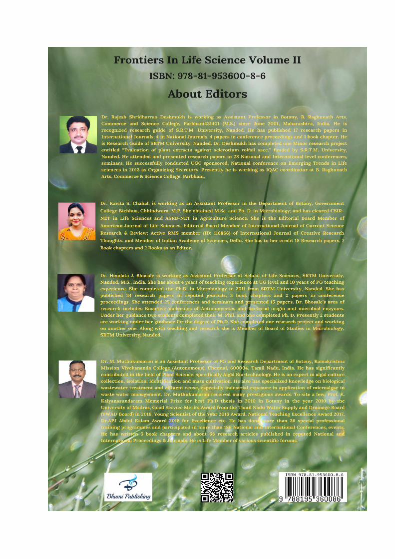
Dr. Rajesh S. Deshmukh
Department of Botany,
B. Raghunath Arts, Commerce
and Science College,
Parbhani – 431 401 M.S., India
Dr. Hemlata J. Bhosale
School of Life Sciences,
Swami Ramanand Teerth
Marathwada University (SRTM),
Nanded – 431 606, M.S., India
Dr. Kavita S. Chahal
Department of Botany,
Government College Bichhua
Chhindwara – 480 111
M.P., India
Dr. M. Muthukumaran
PG and Research Department of Botany,
Ramakrishna Mission Vivekananda College
(Autonomous),
Chennai – 600 004, Tamil Nadu, India
2021
First Edition: 2021
ISBN: 978-81-953600-8-6
Copyright reserved by the publishers
Publication, Distribution and Promotion Rights reserved by Bhumi Publishing, Nigave Khalasa, Kolhapur
Despite every effort, there may still be chances for some errors and omissions to have crept in
inadvertently.
No part of this publication may be reproduced in any form or by any means, electronically, mechanically,
by photocopying, recording or otherwise, without the prior permission of the publishers.
The views and results expressed in various articles are those of the authors and not of editors or
publisher of the book.
Published by:
Bhumi Publishing,
Nigave Khalasa, Kolhapur 416207, Maharashtra, India
Website: www.bhumipublishing.com
E-mail: [email protected]
Book Available online at:
https://www.bhumipublishing.com/books/
PREFACE
Life Sciences have always been a fundamental area of science. The exponential
increase in the quantity of scientific information and the rate, at which new discoveries
are made, require very elaborate, interdisciplinary and up-to-date information and their
understanding. Enhanced understanding of biological phenomenon incorporated with
interdisciplinary approaches has resulted in major breakthrough products for betterment
of society. To keep the view in mind we are delighted to publish our book entitled
"Frontiers in Life Science Volume II". This book is the compilation of esteemed articles of
acknowledged experts in the fields of basic and applied life science.
This book is published in the hopes of sharing the new research and findings in the
field of life science subjects. Life science can help us unlock the mysteries of our universe,
but beyond that, conquering it can be personally satisfying. We developed this digital book
with the goal of helping people achieve that feeling of accomplishment.
The articles in the book have been contributed by eminent scientists, academicians.
Our special thanks and appreciation goes to experts and research workers whose
contributions have enriched this book. We thank our publisher Bhumi Publishing, India for
taking pains in bringing out the book.
Finally, we will always remain a debtor to all our well-wishers for their blessings,
without which this book would not have come into existence.
- Editorial Team
Frontiers in Life Science Volume II
ISBN: 978-81-953600-8-6

CONTENTS
Sr. No. Chapter and Author(s) Page No.
1 Habitat, Threats and Factors Affecting House Sparrow
(Passer Domesticus) Survival In Barmer District,
Rajasthan, India
Nadim Chishty, Narayan Lal Choudhary and Puneet Sharma
1 – 13
2 The Effect of Endosulfan on Oxygen consumption of
Fresh water Female Crab Barytelphusa guerini
Rajesh B. Desai
14 – 20
3 Oyster Mushroom: Cultivation, Bioactive Significance and
Commercial Status
Mukundraj G. Rathod, Rohini B. Gadade,
Gayatri M. Thakur and Anupama P. Pathak
21 – 30
4 Comparative Evaluation of Nutrients of Raw and Cooked
Samples of Rheum palamatum- A Potent Medicinal Plant
Jamuna M and Jyothsna Karanth
31 – 42
5 Plants Based Ethno-Medicine In The Lambani
Communities In The Vicinity of Vijayapura District
of Karnataka
Ramachandra Naik M., Vinayaka K. S. and Joy Hoskeri
43 – 58
6 In Vitro Screening of Antifungal Bacteria Against
Phytopathogenic Fungi of Pumpkin (Cucurbita maxima)
Pooja H. V. and Girish K
59 – 71
7 Automated Drip Irrigation System for Sustainable
Crop Production
Sumit Sow and Shivani Ranjan
72 – 83
8 Studies on Some Protozoan Parasites of Indian Goat
Capra indica
Khan Rumana Shahin Amanullah
84 – 87

9 Chemical Analysis of Summer Honeys Collected From
Apis dorsata Hives of Chimur Tahsil of Chandrapur
District of Maharashtra State (India)
Laxmikant N. Borkar and Devendra M. Mate
88 – 91
10 Study on Socio-Economic Status of Fish Farmers In
Chikkamagaluru District, Karnataka-India
Prapthi U. B. and Annapurneshwari H
92 – 102
11 Acupuncture Therapy: A Fascinating Approach for
Palliate Health
Megha Jain and Vinay Jain
103 – 110
12 Mucormycosis (Black Fungus): Introduction,
Pathophysiology and Its Impact on People of
Indian Subcontinent
Rohit Batra, Pankaj Sharma and Vinay Jain
111 – 118
13 Palynology in Forensic Science
Megha Sapkota, Sandeep Sangle and Arun Ghuge
119 – 128
14 Basic Biology of Inflammatory Bowel Disease
Manoj Patidar
129 – 133
15 Impact of Climate Change on Biodiversity in India:
Review
Ruhiya Sultana
134 – 142
16 Review on Role of Phosphate Solubiliing Bacteria in
Sustaninable Agriculture
Hemlata Janardhan Bhosale and Shraddha Bhagwatrao Kakde
143 – 162

Frontiers in Life Science (Volume II)
ISBN: 978-81-953600-8-6
1
HABITAT, THREATS AND FACTORS AFFECTING HOUSE SPARROW
(PASSER DOMESTICUS) SURVIVAL IN BARMER DISTRICT,
RAJASTHAN, INDIA
Nadim Chishty1, Narayan Lal Choudhary1 and Puneet Sharma2
1Wildlife, Limnology and Toxicology Research Laboratory,
Department of Zoology, Government Meera Girl’s College
(Mohanlal Sukhadia University) Udaipur, Rajasthan, India-313001
2Department of Life Sciences, Aryan College Ajmer, Rajasthan, India-305001
Corresponding authors E-mail: [email protected],
[email protected], [email protected]
Abstract:
The house sparrow is present everywhere and inhabited in almost all types of
environment like rural area, urban areas, forests, woodland and cultivated land. Presence and
population of these birds has been declined drastically in urban and as well as rural areas due to
lack of appropriate nesting sites, roosting and feeding habitat. Predation by domestic cats, feral
dogs and snakes, excessive use of insecticides and pesticides, food scarcity, nestling mortality,
environmental pollution, electromagnetic radiations and avian diseases are responsible for
dramatically declining house sparrow population at global level. Cutting of large and old trees
reduced availability of cavity and hole in stems has also reduced nesting habitat of house
sparrow in urban areas. Immense road trafficking and industrial areas has increased the risk of
collision of birds with fast moving vehicles which has also contributed to increased mortality rate
in house sparrow's particularly in urban area. Initiatives related to sparrow saving action plan
(SSAP) at different levels like town, village, colonies and city can be useful for conservation of
house sparrow population and habitat, similarly organization of awareness program among
public and community level may also help in saving the current population of sparrows and
further increase in sparrow population in near future while providing suitable nesting sites and
feeding opportunities to them.
Keywords: House sparrow, habitat, decline, threat, nesting, population, conservation.

Bhumi Publishing, India
2
Introduction:
Birds are an essential and important part of our ecosystem and food webs. House
sparrows presence is an important sign of good quality habitat. These birds are very susceptible
to change in the environmental conditions. They act as a bio indicator species for healthy urban
ecosystems and indirectly represent status of human health in its surroundings. They perform
vital functional role in food chain and food web of terrestrial ecosystem.
House sparrow is closely related and inhabited near human dominated landscape,
cultivated areas, forest and urban areas and provides large and different kinds of value in
ecosystem like recreational value, economical and aesthetic value (Ghosh et al., 2010). Presence
of house sparrow near human dominated habitat can be seen in various places such as gardens,
rural areas, colonies, suburban areas, parks, agricultural land, feedlots and granaries, which are
well studied at global level by different researchers (Louther and Cink, 1992; Monika, 2005;
Sharma, 2009; Balaji, 2014; Bavia et al., 2014 and Kamath et al., 2014;). At global level house
sparrow population decline was recorded to be highest in London (60%), Glasgow (99%) and
Hamburg (77%) and is categorized as threatened under red list species (Crick et al., 2002;
Prowse, 2002 and Smith, 2005). Natural predator like hawk, eagle, black kite and owl hunts and
feed upon house sparrows. Domestic cats are significant predator and are also responsible for
dramatic decline of house sparrow population worldwide (Churcher and Lawton, 1987). House
sparrow populations were also adversely affected by epidemic avian diseases (Menegaux, 1919-
1921 and Stenhouse1928) considering the fact that such type‘s diseases and declination in
population are limited in particular or specific areas.
Increasing anthropogenic activities have been modified and converted the natural habitats
into urban areas which had accelerated the habitat alteration rate and vanishing of natural
vegetation (Marzluff et al., 2001; 2008). Urbanization exposes animals to potentially critical
survival factors adversely such as human generated disturbances, pollutants, artificial lightening,
and scarcity of food which are also responsible for biodiversity decline (Shochat et al., 2006).
The House sparrow is primarily a seed eater bird found in almost all habitats and mostly seed
availability occurs in cultivated area of rural landscape, but they also show wide variety of food
preferences and choices like grains, seeds, insects, spider, nectar, caterpillars, human made food
like wheat and bajara breads and digestible materials from cow dung (Yahanghi et al., 2010).
Food shortage can also affect individual fitness and survival rate of house sparrow species which
results into breeding failure due to poor development and malnutrition (Newton, 1998).
Extension of industrialization, civilization and human settlement leads to lack of foraging site,
feeding habitat and nesting places in cities which has largely contributed in declination of house

Frontiers in Life Science (Volume II)
ISBN: 978-81-953600-8-6
3
sparrow population (Cram et al., 1995; Rao, 2000; Summer & Smith, 2003; Robinson et al.,
2005; Pineda et al., 2013). In the present chapter we discuss about habitat, behavioural biology,
threats and conservation issues related to house sparrow's in Barmer district of Rajasthan, India
from January, 2016 to December, 2020.
Materials and Methods:
The present study was carried out in different microhabitat of Barmer district of
Rajasthan, India from January, 2016 to December, 2020. The geographic location of Barmer
district is located between 24.58ˈ to 26, 32ˈN latitudes and 70, 05ˈ to 72, 52ˈE longitudes. The
average temperature is high and show large fluctuations in various seasons and as well as day
and night. In summer season temperature reaches up to 46°C to 51°C and in winter it drops to 0°
C and annual precipitations is very low approximately about 277mm. Scheduled visual
observations were done with the help of binoculars (Nikon 8x40)and accordingly photographs
were clicked with the help of Nikon P900 and P1000 cameras. Data were collected from various
habitats of house sparrows like- cultivated land, urban area, rural area, grasslands and forest area.
Observation hours were set in early morning hours from 6.00am to 9.00 am and in evening hours
from 4.00pm to 7.00pm when house sparrows were comparatively more active as compared to
rest of the hours of the day.
Result and Discussion:
Distribution and population
House sparrow's (Passer domesticus) are widely distributed in all types of habitat
throughout Indian subcontinent and other parts of the world (Blair, 1999). In context of Indian
subcontinent they are generally found in all states of India and as an introduced population in
Andaman Island (Ali and Ripley, 1987). During survey maximum house sparrow populations
were recorded in rural area as compared to urban landscape out of different study areas.
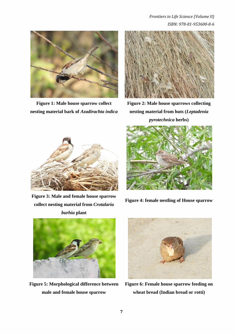
Cultivated lands of rural areas provide suitable habitat, nesting material (Figure 1, 2, 3) and more
availability of food material as compared to urban areas.
Morphology and systematic position
House sparrow belongs to the Passerine family and is a small, stock song bird with
chunky bill, short legs, length approximately 14-16 cm, weight 26-32 gm and having 19-25 cm
open wing span. House sparrow are monogamous pairing for long time during breeding season,
sexual dimorphism is distinct and well developed and male and female can be easily identified

Bhumi Publishing, India
4
and differentiated by necked eye (Anderson, 2006)(Figure 4,5). While males have a grey crown,
a chestnut back and neck, wings having black tip, white checks and grey rump and tail, the
females are relatively uniform pale and light grey coloured with dark spikelet wings. Male is
heavier and larger in size as compared to the female house sparrow's (The House Sparrow-
Passer domestics, 2007).
Taxonomic position
Kingdom- Animalia
Phylum- Chordata
Class- Aves
Order- Passeriformes
Family- Passeridae
Genus- Passer
Species- domesticus
Habitat and feeding behaviour
The house sparrows are unique wild birds and closely associated with nearby human
settlements like houses, agricultural land and both rural areas as well as urban areas (Crick et al.,
2002). House sparrow usually feed upon Oats, Wheat (Figure 6), Bajara or Bajari (Figure 7),
Corn, Sorghum and other major sources of seeds and on other seasonal and perennial herbs such
as grasses (Graminae), rushes (Juncidae) and goose foot (Chenopodium).They also feed upon
fruits and beery like Pillu (Salvodora persica) (Figure 8), Ber (Ziziphus mauritiana) and other
edible fruits and seeds. House sparrow prefers more insects in diet during breeding season which
plays a potential role in early development of nestling and enhance breeding success rate in these
birds (Figure 9). House sparrow commonly feed upon honey bees, wasps, ant, componotus,
aphids (Aphidoiea), spiders (Arachnida), beetles (Coleoptera), weevils (Curculionidae),
grasshopper (Orthoptera) and caterpillars of butterflies and moths (Lepidoptera) (Wilson et al.,
1999). In summer season house sparrow eat insect and feed them to their nestling. House
sparrow catches insects in air by sudden jumping and flying on them during more active feeding
hours of early morning and evening time and also feeding with other birds (Figure 10).
Nesting and breeding behavior
House sparrow build their nest on various nesting sites for example high on walls of
house, roof spaces, trees and hole of trees, other safe and undisturbed locations of cemented
house, cottage in both urban and rural habitat (Figure 11,12). Mostly they construct their nests in
roof spaces of hut, cattle shades, wooden fencing, mud houses and tree cavities in rural areas
(Figure 13, 14). Sometime unexpected nests were also observed at electrical transformers and

Frontiers in Life Science (Volume II)
ISBN: 978-81-953600-8-6
5
electric meter boxes, safe and undisturbed terminal of various pipes and in between bricks and
stones (Figure 15, 16). House sparrows are monogamous and pairs in breeding season and in
small colonies while feeding in flock and sometimes individually (Figure 17). They breed in the
months of September to October in central India, March to June in Northern India and
throughout the year in southern India. House sparrows generally laid four to five eggs and
minimum clutch size is two eggs. Eggs are bluish white or greenish white with grey or brown
spots. The incubation period of house sparrow ranges from 10 to 12 days.
Threats and conservation problems
House sparrow population rapidly declined in urban area and rural area due to lack of
suitable breeding or nesting sites, food scarcity, invasion of exotic species plant and animal
species, establishment of cemented houses and buildings and various petrochemical and other
industries. At present house sparrow population and abundance had dramatically declined in
metropolitan cities like Bangalore, Mumbai, Hyderabad and New Delhi (Rajasheker and
Venkatesha, 2008). Another reason for their population declination includes excessive use of
hazardous and noxious compounds such as insecticides and pesticides used in cultivation of
crops and gardens. These chemicals are responsible for breeding failure and mortality of
sparrows and other birds. House sparrow population also declined due to competition with other
species for food, roosting and nesting places and excessive predation by domestic cats and
sometime feral dogs in urban areas (Figure 18, 19). Many studies had been conducted on status
and trends of house sparrow population declination in India (Summers and Smith, 2003). Also
sparrow and other bird population had declined due to environmental stress badly affected by
various types of pollutions (Baker et al., 2005 and Shaw, 2009). Reduction in arthropod diversity
might also be responsible for population declination of insectivorous birds. Because insects
perform crucial role as a food material during developmental stages of breeding season (Peach et
al., 2008). In urban area sparrow populations also declined due to scarcity of food material and
inter specific and intra specific competitions with other birds for nesting and roosting places.
House sparrow generally compete for food with red vented bulbul, yellow vented bulbul, feral
pigeons, babblers, robins and other frugivorous and insectivorous birds.
Factor responsible for nestling and adult mortality
Nestling and adult house sparrows died due to attacking of predators such as crow,
shikra, black kite, hawks, eagles and owls. Sometime nestling and adult died and injured due to
collision with plastic threads and anthropogenic materials in urban areas. House sparrow
population also declined due to low rate of hatchling and nestling success in urban and rural
areas. Inter specific and intra specific competition for nesting places is responsible for breeding

Bhumi Publishing, India
6
failure among birds. Several toxic compounds are now continuously increasing in natural
environment like pesticides, insecticides, heavy metal contamination in food chain, industrial and
sewerage waste, combustion and emission of fuels or gases are creating unfavorable habitat for
biotic life including human beings. Some of these toxic chemical and pollutants adversely
influence and are responsible for population decline of birds at global level including house
sparrows (Newton, 1998). Multiple factors are responsible for the cause of the decline of house
sparrow population in urban, semi urban, industrial area and rural areas. Contaminated water and
chemically treated grains caused poisoning and mass mortality of house sparrows and other
granivore birds. The stress from growing human activities such as expansion of civilization,
construction of cemented houses and buildings, establishment of various industries, habitat
fragments and alteration, deforestation, excessive use of insecticides and pesticides, mobile
tower radiations and spreading of invasion species, over abundance and congested spaces
affected house sparrow population and survival rate adversely.
Effects of urbanization and environmental factors
Expansion of urbanization becomes limiting factor and causes negative impact on growth
of nestling, body size and feather quality of house sparrow (Meillere et al., 2017) and hence poor
growth and development. Electromagnetic radiations also affected house sparrow fitness and
survival among urban habitats in a declining state. Modern designs of house and building
infrastructure do not leave any space or cervices even outside the building which has reduced the
nesting sites of house sparrows and other small birds (Figure 20). Environmental and climatic
conditions like seasonal factors such as annual precipitation, temperature, humidity, wind
velocity, time of dawn and dusk has also influenced the population density of house sparrows.
High wind velocity also responsible for breeding failure due destruction of nests and fallen of
eggs on ground (Figure 21).
Conservation recommendations for saving house sparrows:
Artificial and wooden boxes for nests are useful in conserving house sparrow population
by providing suitable habitat for nesting and breeding success particularly in urban areas.
Enhancement and establishment of huts with the help of tree branches and grass material are also
helpful for enhancing sparrow population. Similarly mud houses also hold large number of nests
of house sparrows. Encourage low level of pesticide and insecticide use in cultivated land,
gardens and parks also reduces bird‘s mortality rate. Public awareness and conservation program
organized in schools, colleges and villages are also helpful for better conservation measures for
house sparrows in study area. Further scientific and genetic studies at population level and
regional level are required in determining factors and cause of rapid declination of this bird
species Passer domesticus in current scenario.
Frontiers in Life Science (Volume II)
ISBN: 978-81-953600-8-6
7
Figure 1: Male house sparrow collect
nesting material bark of Azadirachta indica
Figure 2: Male house sparrows collecting
nesting material from huts (Leptadenia
pyrotechnica herbs)
Figure 3: Male and female house sparrow
collect nesting material from Crotalaria
burhia plant
Figure 4: female nestling of House sparrow
Figure 5: Morphological difference between
male and female house sparrow
Figure 6: Female house sparrow feeding on
wheat bread (Indian bread or rotti)

Bhumi Publishing, India
8
Figure 7: Male house sparrow feed upon
grains of Pennisetum glaucumin rural area
Figure 8: Female house sparrow feeding
fruit of Salvodora persica
Figure 9: Male house sparrow capture
caterpillar
Figure 10: House sparrow communally
forage with seven sister babblers
Figure 11: House sparrow nesting on
Prosopis cineraria
Figure 12: House sparrow nesting in urban
area at cemented teen shade

Frontiers in Life Science (Volume II)
ISBN: 978-81-953600-8-6
9
Figure 13: House sparrow nesting in huts of
rural area
Figure 14: House sparrow construct nest
inside stem of Bambusa vulgaris
Figure 15: House sparrow nesting inside
electric transformer
Figure 16: House sparrow nesting inside
electric meter box
Figure 17: Female house sparrow forage
and feeding near cattle shade
Figure 18: Domestic cat movement around
sparrows' habitat

Bhumi Publishing, India
10
Figure 19: Domestic cat movement in houses
of urban areas
Figure 20: Lack of safe nesting site house
sparrows egg fallen on ground and broken
Figure 21: Due to high wind velocity sparrow nest scattered and eggs fallen on ground
Conclusion:
The house sparrow is gregarious at all season and live in flock or groups and feed upon
grains, seeds, insects, fruits, berry, insects, kitchen food and flower buds in rural and urban
habitat. Abundant population of domestic cats and feral dogs has also reduced the house sparrow
population including adults and nestling. Lack of suitable nesting sites for house sparrows and
other small birds with other factors may be responsible to shift this bird species towards
extinction in near future. House sparrow population has majorly declined due to extension of
civilization areas at the cost of degradation of natural environment and lack of nesting places at
all the levels. Organization of house sparrow conservation and saving programs among
community, villages, farmers, might be helpful for saving this beautiful neighbor of humans, the
house sparrows from extinction.

Frontiers in Life Science (Volume II)
ISBN: 978-81-953600-8-6
11
References:
Blair, R.B., (1999). Birds and butterflies along an urban gradient: Surrogate taxa for assessing
biodiversity. Ecological Applications.9:164-170.
Ali, S., and Ripley, S.D., (1987). Handbook of the birds of India and Pakistan, Compact edition,
Oxford University Press, New Delhi.
Anderson, (2006). Biology of the Ubiquitous House Sparrow: From Genes to Populations,
Oxford University Press. p. 560.
Baker, P.J., Bentley, A.J., Ansell, R.J., Harris, S., (2005). Impact of predation by domestic cats
Felix catus in an urban area, Mammal Review, 35, 2005, pp. 302– 312.
Bavia, M. S., Maria J. R., Felix, K., Ponnivalavan (2014). Study on Disappearance of House
Sparrow Using Induced Fuzzy Cognitive Maps (IFCMs). Inte. J. Comp. Algo. 03:871-875.
Cramp, S., Simmons, K,E,L., Brooks, D.C., Collar, N.J., Dunn, E., R. Gillmor, R.,Hollom,
P.A.D., Hudson, R., Nicholson, E.M., Ogilvie, M.A., Olney, P.J.S., C.S. Roselaar,
C.S.,Voous, K.H., Wallace, D.I.M.,Wattel, J., &Wilson, M.G., (1983). Handbook of the
Birds of Europe, the Middle East and North Africa. The Birds of the Western Palearctic.
Oxford University Press, Oxford, UK.
Crick, H.Q., Robinson, R.A., Appleton, G.F., Clark, N.A., Rickard, A.D., (2002). Investigation
into the causes of the decline of starlings and House sparrows in Great Britain, Published
by the Department for Environment, Food and Rural Affairs (DEFRA) and British Trust
for Ornithology, London. 290.
Ghosh, S., Kihyun, K., Bhattacharya, R., (2010). A Survey on House Sparrow. Environmental
Science, Kalyani University. pp. 147 – 152.
Kamath, V., Mathew A. O., Lewlyn, I., Rodrigues, R., (2014). Indian Sparrows on the brink of
extinction: population dynamics combined with ecological changes. International Journal
of Renew Energy and Environment and Engineering.02 (01): 17-22.
Louther, P. E., and Cink, C. L., (1992). House Sparrow in The Birds of North America (A.
Poole, P. Stettenheim, and F. Gill, eds.). Philadelphia Academy of Sciences, Philadelphia,
81-89.
Marzluff, J. M., Bowman, R.,& Donnelly, R., (2001). A historical perspective on urban bird
research: trends, terms, and approaches. In: In: J. M. Marzluff, R. Bowman & R. Donnelly
(Eds.), Avian ecology and conservation in an urbanizing world (pp. 1–17). Kluwer
Academic Publishers, Boston, Massachusetts, USA.

Bhumi Publishing, India
12
Marzluff, J. M., Shulenberger, E., Endlicher, W., Alberti, M., Bradley, G., Ryan, C.,
ZumBrunnen, C.,& Simon, U., (2008). Urban ecology: An international perspective on the
interaction between humans and nature. Springer, New York, USA.
Meillere, A., Dupoue, A., Lourdais, O., Angelier, F., (2017). Traffic noise decreases nestlings‘
metabolic rates in an urban exploiter. Journal of Avian Biology. 48(1):905909.
Menegaux, A., (1919-1921).Êenquetesur la disparition du Moineau dans la Midi. Rev. Franc.
d‘Orn. 11: 65-71, 129- 131; 12: 32-36, 52-55, 77-78; 13: 41-42, 127-128.
Monika, G., (2005). Preliminary survey of House Sparrow ( Passer domesticus) in three areas of
Haridwar, Uttrakhand. M.Sc. thesis, GurukulKangri University, Haridwar, India: 1-27.
Newton, I., (1998). Population Limitation in Birds. Academic Press Limited, USA, p.147.
Peach, W.J., Vincent, K., Fowler, J.A., Grice, P.V., (2008). Reproductive success of Passer
domesticus along an urban gradient, Animal Conservation, 11, 2008, pp. 493–503.
Pineda, J., Herrera, A., Antonio, M. T., &Aguirre, J., (2013). Urban models and their effects on
immune system of house sparrow Passer domesticus populations in Central Spain.
EOU2013UK - 9th Conference of the European Ornithologists‘ Union.
Prowse, A., (2002). The urban decline of House P. Rajasekarapandian, Assistant in Library for
the Sparrow, British Birds., 95: 143-146.
Rajashekar, S., and Venkatesha, M.G., (2008). Occurrence of house sparrow, Passer domesticus
indicus in and around Bangalore. Current Science. 94(4):446-449.
Rao, R., (2000). A Field Study of The House Sparrow (Passer domesticus)
http://www.indianwildlifeclub.com/ResearchPapers/Field-Studyof-House Sparrow.aspx
Robinson, R.A., Siriwardena, G.M., &Crick, H.Q.P. (2005). Size and trends of the House
Sparrow Passer domesticus population in Great Britain. Ibis 147: 552–562;
http://doi.org/10.1111/j.1474919x.2005.00427.x
Sharma, K., (2009). Where are all the sparrow gone? Retrined June, from
http://indiwo.in.com/india/feather/harmony-in-life-life/where-are-all-the-sparrow-oBalaji,
S. (2014): Artificial nest box for house sparrow: An apt method to save the dwindling
species in an urban environment. International Journal of Biodiversity Conservation
6(3):194-198. Ne/40681/0.
Shaw, L.M., (2009). Investigating the role of socioeconomic status in determining urban habitat
quality for the House Sparrow, Passer domesticus, PhD Thesis, University of Exeter.
Shochat, E., Warren, P. S., Faeth, S.H., McIntyre, N. E.,& Hope, D., (2006). From patterns to
emerging processes in mechanistic urban ecology. Trends in Ecology and Evolution, 21:
186–191.

Frontiers in Life Science (Volume II)
ISBN: 978-81-953600-8-6
13
Smith, J.D.S., (2005). Changes of House Sparrow population in Britain, International Studies on
Sparrows, 30: 23-37.
Stenhouse, J. H., (1928). Remarkable decrease of the house sparrow in Fair Isle and Shetland.
Scot. Nat., 162.
Summers and Smith, J.D., (2007). Is unleaded petrol a factor in urban House Sparrow decline?
British Birds 100(9): 558–559.
The house sparrow Passer domesticus (2007). NSW Department of Primary Industries,
Australia, <http://www.dpi.nsw.gov.au/agriculture/pests-weeds/vertebratepests/pest-
animals-in-nsw/sparrows>.
Wilson, J., Morris, A., Arroyo, B., Clark, S., Bradbury, R., (1999). Review of the abundance and
diversity of invertebrate and plant foods of granivorous birds in northern Europe in relation
to agricultural change. Agriculture, Ecosystems and Environment.75:13-30.
Yahaghi, A., Behrouzi-Rad, B., Amininasab, S., Askari, R., (2011). Determination of number
and biometry of House sparrow Passer domesticus eggs in public parks of Shushtar in
South of Iran (Spring 2010). World Journal of Science and Technology. 1(5):56-61.

Bhumi Publishing, India
14
THE EFFECT OF ENDOSULFAN ON OXYGEN CONSUMPTION OF FRESH
WATER FEMALE CRAB BARYTELPHUSA GUERINI
Rajesh B. Desai
Department of Zoology,
Mahatma Gandhi Mahavidyalaya, Ahmedpur,
Dist. Latur, Maharashtra, INDIA 413 515
Corresponding authors E-mail: [email protected]
Abstract:
Respiration is a process during which the organisms obtain oxygen from external medium
and use it for the purpose of energy release during oxidative metabolism. As such the process of
respiration in animals is studied by determining the oxygen consumption. Existence of living
organisms depends on the ability of their cells to incorporate a number of simple compounds and
transform them into more complex molecules required for cellular structure and function. The
energy required for these synthetic reactions must be obtained from the same substrates by
suitable oxidation reactions coupled to the generation of high energy phosphate esters. The entire
process ultimately depends on the availability of molecular oxygen in cellular environment.
Oxygen is made available to the tissues and in turn to cells by respiration. The present study
deals with oxygen consumption, when experimental animal fresh water female crab
Barytelphusa guerini is exposed in the pesticide like Endosulfan. It shows variable changes in
respiratory physiology which discussed by statistically and graphically.
Keywords: Endosulfan, Oxygen consumption, Female Crab, Barytelphusa guerini.
Introduction:
The chlorinated hydrocarbons are basically organic compounds that have been
chlorinated with several atoms of chlorine per molecule. These insecticides have very low
solubility in water but are readily soluble in fats. These compounds are chemically stable and
show considerable persistence upon introduction into physical environment. Since these
compounds are stable and persistent, they are referred to as ―hard pesticides‖ (Abbot et al.,
1966). Some examples of these pesticides are DDT, Lindane, Heptachlor, Mirex, Chlordane,

Frontiers in Life Science (Volume II)
ISBN: 978-81-953600-8-6
15
Aldrin, Methoxychlor, Dieldrin, endosulfan, Toxaphene etc. These compounds exhibit biological
magnification in the food chain (Macek, 1969).
Organochlorine pesticide action was said to be neurotoxin. They were found to create
energy crisis by inhibiting Mg2+ ATP ase and aerobic segmental enzymes of the brain (Philip,
1984). The effects of various organochlorine compounds on different animals (like rat, fish,
chicken and mice) and concluded that these compounds disrupt ATP dependent active transport.
ATP activity is perhaps disrupted by the uncoupling of oxidative phosphorylation, have studied
(Rangaswamy, 1984).
It has been reported that respiratory distress is one of the common symptoms manifested
during organochlorine insecticide toxicity (Sathyaprasad, 1983). It has reported that the rate of
decrease in oxygen consumption of fish increase with increase in the concentration of pesticides
in the Biosystem.
BHC produced a significant increase in haemolymph sugar, protein; ICDH, SDH and
LDH in the crab, Ozitelphusa senex senex (Basha et al., 1984) have observed inhibition of
oxidative metabolism in Tilapia mossambica during sublithal exposure to Lindane.
Glycogenolysis and Gluconeogenesis were found to be elevated in the carp, Cyprinus carpio,
exposed to Aldrin, Dieldrin and Endrin (Gluth and Hanke, 1985).
Material and Method:
Oxygen consumption was determined by Wrinkler method (Welsh and Smith, 1953). The
freshwater female crab Brytelphusa gueriniwas used for experimentation. The animals were
collected form their natural habitat and maintained in the laboratory before experimentation. The
proper selection of animals were made for present investigation and procedure was followed for
the exposure of animal with pesticide pollutant i.e. Endosulfan.
The apparatus used in this experiment was similar as described by Saroja. The apparatus
mainly consisted of a reservoir (R) and respiratory chamber (RC). A 500ml wide mouthed bottle
was used as respiratory chamber. The size of the bottle was such that it was not too big for the
enclosed crab to give considerable difference in oxygen content between initial and final sample.
The chamber was coated with black paint to avoid the activity due to light.
Before starting the experiment crab was left in running tap water for about 10 minute to
facilitate them to reach a state of normality from a state of excitement; if any. After this
equilibration period, one crab was kept in respiratory chamber without causing any damage to
the animal and initial sample was collected immediately as described above. Then the crab was
Bhumi Publishing, India
16
allowed to respire for one hour. Immediately after one hour final sample was collected. The
amount of dissolved oxygen in this sample was determined by the standard Winkler‘s method, as
given by.The total oxygen consumption and rate of oxygen consumption was calculated by
considering the wet weight of animals. The values for total oxygen consumption are expressed as
ml. (c.c.) of O2 animals / hr and for the rate of O2 consumption are expressed as ml. (c.c.) of 02 /
gm / hr wet wt. of the animal.
Result and Observation:
Effect of Endosulfan on Total Oxygen Consumption and Rate of Oxygen Consumption in
Freshwater Female Crab Barytelphusa guerini
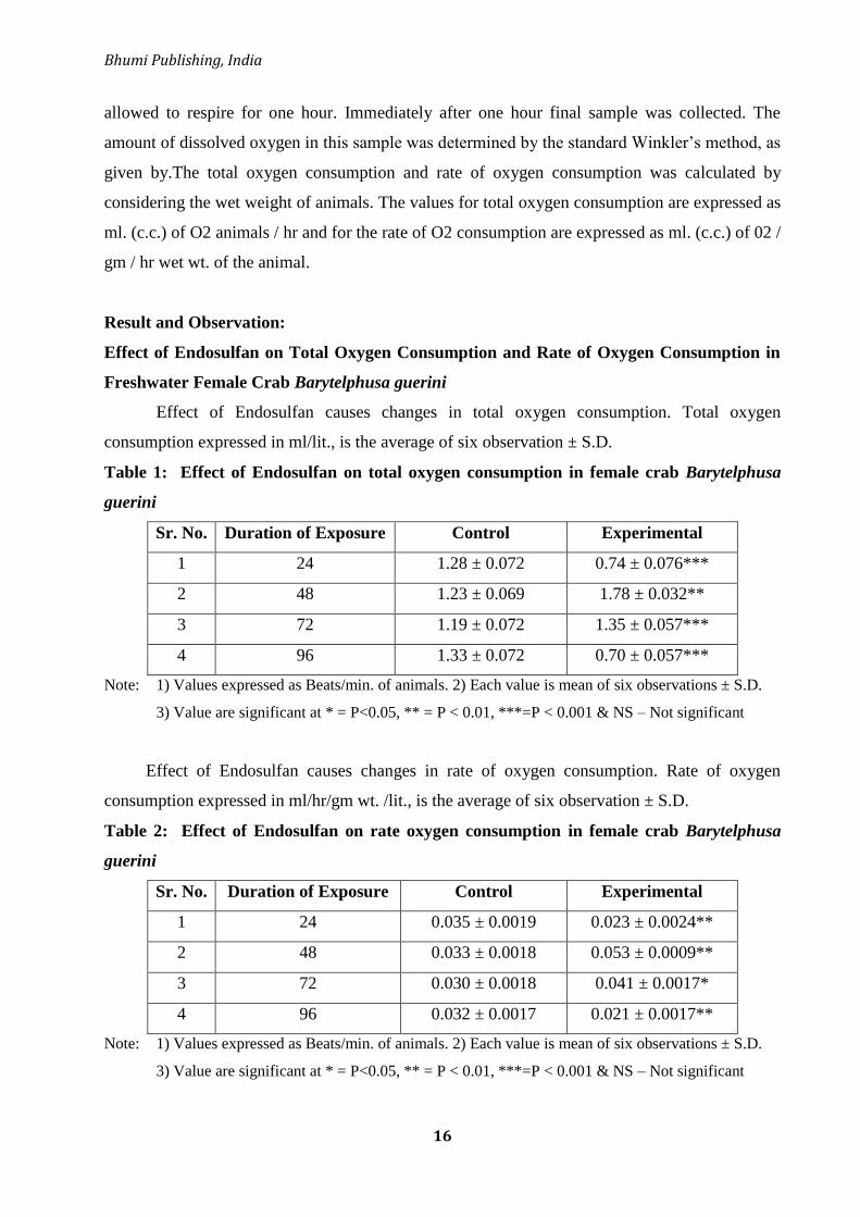
Effect of Endosulfan causes changes in total oxygen consumption. Total oxygen
consumption expressed in ml/lit., is the average of six observation ± S.D.
Table 1: Effect of Endosulfan on total oxygen consumption in female crab Barytelphusa
guerini
Sr. No. Duration of Exposure Control Experimental
1 24 1.28 ± 0.072 0.74 ± 0.076***
2 48 1.23 ± 0.069 1.78 ± 0.032**
3 72 1.19 ± 0.072 1.35 ± 0.057***
4 96 1.33 ± 0.072 0.70 ± 0.057***
Note: 1) Values expressed as Beats/min. of animals. 2) Each value is mean of six observations ± S.D.
3) Value are significant at * = P<0.05, ** = P < 0.01, ***=P < 0.001 & NS – Not significant
Effect of Endosulfan causes changes in rate of oxygen consumption. Rate of oxygen
consumption expressed in ml/hr/gm wt. /lit., is the average of six observation ± S.D.
Table 2: Effect of Endosulfan on rate oxygen consumption in female crab Barytelphusa
guerini
Sr. No. Duration of Exposure Control Experimental
1 24 0.035 ± 0.0019 0.023 ± 0.0024**
2 48 0.033 ± 0.0018 0.053 ± 0.0009**
3 72 0.030 ± 0.0018 0.041 ± 0.0017*
4 96 0.032 ± 0.0017 0.021 ± 0.0017**
Note: 1) Values expressed as Beats/min. of animals. 2) Each value is mean of six observations ± S.D.
3) Value are significant at * = P<0.05, ** = P < 0.01, ***=P < 0.001 & NS – Not significant

Frontiers in Life Science (Volume II)
ISBN: 978-81-953600-8-6
17
Figure 1: Effect of Endosulfan on Total Oxygen consumption in Barytelphusaguerini
Figure 2: Effect of Endosulfan on Rate of Oxygen consumption in Barytelphusa guerini
Result:
The freshwater female crab Barytelphusa guerini showed variation in the Rate of oxygen
consumption and Total oxygen consumption when exposed in endosulfan.
In the present investigation it was showed that the oxygen consumption in the animal
exposed to Endosulfanwas decreased upto 96 hours. The Endosulfan exposed animals
ENDOSUFAN
Mean Total of O2 Consumption
1.3323
0.749
1.1923
1.239
1.2857
1.3557
1.7857
0.7023
0.6
0.7
0.8
0.9
1
1.1
1.2
1.3
1.4
1.5
1.6
1.7
1.8
1.9
24 hrs 48 hrs 72 hrs 96 hrs
TIME
To
tal
of
O2
Co
ns
um
pti
on
ml.
/lt
Control Experimental
ENDOSUNAN
Mean Rate of O2 Consumption
0.0234
0.0536
0.041
0.0212
0.0325
0.03
0.0356
0.0334
0.020
0.025
0.030
0.035
0.040
0.045
0.050
0.055
0.060
24 hrs 48 hrs 72 hrs 96 hrs
TIME
Ra
te o
f O
2 C
on
su
mp
tio
n m
l./h
r/g
m w
t./l
it
Control Experimental
Bhumi Publishing, India
18
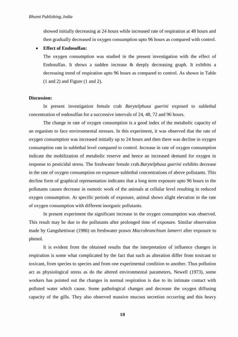
showed initially decreasing at 24 hours while increased rate of respiration at 48 hours and
then gradually decreased in oxygen consumption upto 96 hours as compared with control.
Effect of Endosulfan:
The oxygen consumption was studied in the present investigation with the effect of
Endosulfan. It shows a sudden increase & deeply decreasing graph. It exhibits a
decreasing trend of respiration upto 96 hours as compared to control. As shown in Table
(1 and 2) and Figure (1 and 2).
Discussion:
In present investigation female crab Barytelphusa guerini exposed to sublethal
concentration of endosulfan for a successive intervals of 24, 48, 72 and 96 hours.
The change in rate of oxygen consumption is a good index of the metabolic capacity of
an organism to face environmental stresses. In this experiment, it was observed that the rate of
oxygen consumption was increased initially up to 24 hours and then there was decline in oxygen
consumption rate in sublethal level compared to control. Increase in rate of oxygen consumption
indicate the mobilization of metabolic reserve and hence an increased demand for oxygen in
response to pesticidal stress. The freshwater female crab.Barytelphusa guerini exhibits decrease
in the rate of oxygen consumption on exposure sublethal concentrations of above pollutants. This
decline form of graphical representation indicates that a long term exposure upto 96 hours in the
pollutants causes decrease in osmotic work of the animals at cellular level resulting in reduced
oxygen consumption. At specific periods of exposure, animal shows slight elevation in the rate
of oxygen consumption with different inorganic pollutants.
In present experiment the significant increase in the oxygen consumption was observed.
This result may be due to the pollutants after prolonged time of exposure. Similar observation
made by Gangshettiwar (1986) on freshwater prawn Macrobranchium lamerri after exposure to
phenol.
It is evident from the obtained results that the interpretation of influence changes in
respiration is some what complicated by the fact that such as alteration differ from toxicant to
toxicant, from species to species and from one experimental condition to another. Thus pollution
act as physiological stress as do the altered environmental parameters, Newell (1973), some
workers has pointed out the changes in normal respiration is due to its intimate contact with
polluted water which cause. Some pathological changes and decrease the oxygen diffusing
capacity of the gills. They also observed massive mucous secretion occurring and this heavy

Frontiers in Life Science (Volume II)
ISBN: 978-81-953600-8-6
19
mucous secretion and coagulation may cause breathing distress to the animal due to precipitation
of mucous over the gills.
Several authors have reported decrease in animal oxygen consumption of different
freshwater species exposed to different insecticides. Reddy and Gomathy (1977) reported
decrease in animal oxygen consumption of Mystus vittatus treated with sublethal concentration
of endosulfan. Subhadra Devi (1985) observed a significant decrease in animal oxygen
consumption of Oziotelphusa senex senex exposed to sublethal and lethal concentrations of
endosulfan.
Conclusion:
All organisms require oxygen for their metabolic activates. Generally metabolism of an
organism is measured by estimating oxygen consumption. The rate of oxygen
consumption of an intact animal and its tissues reflects the respective metabolic rates and
hence energy output. It also provides information on the ability of the organism to extract
oxygen from the environment.
Acknowledgement:
The author is thankful to research supervisor Dr. N. E. Ambore for guidance during the
research work and Dr. V. B. Garade, Head, Department of Zoology and Fishery Science, DSM,
College Parbhani for providing laboratory facility to carry out the present work.
References:
Abbot, D.C., Harrison, R.B., Tatton, J.O.G. and Thomson, J. 1966. Organochlorine pesticides in
the atmosphere. Nature 211: 259-261.
Basha, S.M., K.S. Prasada Rao, K.R.S. Sambasiva Rao and K.V. Ramana Rao. 1984. Respiratory
potentials of the fish, T. mossambica under malathion, carbaryl and lindanein toxicantion.
Bull. Environ. Contam. Toxicol. 32(5): 570-574.
Gangshettiwar, V.B. 1986. Effect of phenol poisoning on the physiology of the prawn.
Macrobanchium lamerri, Ph.D. Thesis, Marathwada University, Aurangabad (M.S.), India.
Gluth, G. and W. Hanke. 1985. A comparision of physiological changes in carp, Cyprinus carpio
induced by several pollutants at sublethal concentration. Ecotoxicol. Environ. Safety. 9:
179-188.

Bhumi Publishing, India
20
Macek, K.J.C., Hutchinson and Cope, C.B. 1969. Effects of temperature on the susceptibility of
blue gills and rainbow trout to selected pesticides. Bull. Environ. Contam. Toxicol. 4: 174.
Newell, R.C. 1973. Factors affecting the respiration of invertebrates. Amer. Zool. 13: 513-528.
Philip, H. 1984. Effect of BHC on some aspects of metabolism in the Indian field mouse,
Musbooduga (gray), Ph.D. Thesis, S.V. University, Tirupati, India.
Rangaswami, C.P. 1984. Impact of endosulfan toxicity on some physiological properties of the
blood & aspects of energy metabolism of a freshwater fish, Tilapia mossambica, Ph.D.
Thesis, S.V. University, Tirupati, India.
Reddy, T.G.K. and S. Gomathy. 1977. Toxicity and respiratory effects of the pesticide, thiodan
on catfish, Mystus vittatus. Ind. J. Environ. Hlth. 19: 360-363.
Sathyaprasad, K. 1983. Studies on the toxic impact of the lindane on tissue metabolic profiles in
a freshwater fish, Tilapia mosambica with emphasis on crab, hydrate metabolism. Ph.D.
Thesis, S.V. University, Tirupati, India.
Subhadra Devi, G. 1985. Impact of endosulfan toxicity on whole animal oxygen consumption
and biochemical composition of the haemolymph of a freshwater field crab, O. senex
senex, M.Phil. Dissertation, S.V. University, Tirupati, India.
Welsh, J.H. and Smith, R.I. 1953. In: Laboratory exercises in invertebrate physiology,
Bergmeyer Publishing Co., Minneapolis.
Frontiers in Life Science (Volume II)
ISBN: 978-81-953600-8-6
21
OYSTER MUSHROOM: CULTIVATION, BIOACTIVE SIGNIFICANCE
AND COMMERCIAL STATUS
Mukundraj G. Rathod*1, Rohini B. Gadade1,
Gayatri M. Thakur1 and Anupama P. Pathak2
1Department of Biotechnology and Bioinformatics (U.G. & P.G.),
Yeshwant College of Information Technology
(Affiliated to S.R.T.M. University, Nanded), Parbhani 431401, Maharashtra, India.
2 Director, School of Life Sciences (DST-FIST phase-I & UGC-SAP DRS-II sponsored school),
Swami Ramanand Teerth Marathwada University, Nanded 431606.
*Corresponding authors E-mail: [email protected]
Abstract:
Mushrooms have been included in the category of tasty and nutritional food, by different
societies worldwide. Mushroom is the choice of many gourmet persons due to its delicious taste
and health benefits. Mushrooms seem to be an important cultural heritage as they have been used
from the time immemorial. The knowledge of its edible species and medicinal properties has
been transmitted from one generation to another. Their diversity, habitats, and properties have
been reported in folk literature. Thus there is a relationship between mushrooms and man from
the ancient time. Supply of a sufficient protein content as body-building material is necessary in
a healthy diet which can be fulfilled by the inclusion of mushrooms in diet since they are
nutritive with good quantity of vital proteins, vitamins and minerals. Hence mushrooms have
been recommended for obese persons and diabetes patients because of low caloric value.
Mushrooms exhibit health-promoting properties viz. antimicrobial, antioxidant and
immunomodulating activities. These afore-mentioned properties have recognized in some
countries, especially in China and now China is the top leader in the production of cultivated
mushrooms. Lentinula is the first major genus, contributing in the world‘s cultivated mushrooms,
whereas Pleurotus is a close second, with its five or six cultivated species. Research interest in
mushroom is increasing day by day as many bioactive compounds and chemical constituents
have been reported from mushrooms. In this review chapter, we have focused on cultivation,
bioactive significance and commercial status of oyster and some other mushrooms.

Bhumi Publishing, India
22
Introduction:
Mushroom cultivation is the gaining prominence worldwide because of a natural source
of dietary food protein. Mushroom cultivation is unique in the sense that it is the most efficient
and economically valuable technology for conservation of ligno-cellulosic material into a great
quality of protein containing food. Mushroom cultivation is a side business of many farmers
(Maher, 1991). Of the many species, oyster mushroom is the most suitable for cultivation by
utilizing various agro wastes without composting. Oyster mushroom is generally known as the
wood fungus and in India commonly known as Dhingri. Oyster mushroom is scientifically
known as Pleurotus sp. Oyster mushroom was first described scientifically in 1975 by a Dutch
naturist Nikolaus Joseph Freiherr von Jacquin (1721-1817) and named Agaricus ostreatus. Later
on, these species of oyster mushrooms were included in the genus Pleurotus by a German
mycologist Paul Kummer in 1871 (Stamets 1993). This species has been classified as kingdom-
fungi, phylum-basidiomycota, class-agaricomycetes, order-agaricales, family-pleurotaceae and
genus-Pleurotus. There are about thirty eight species described under genus Pleurotus from
different part of the world, out of which 25 species are under cultivation. Some commercially
important oyster mushrooms predominantly include Pleurotus ostreatus, Pleurotus eryngii,
Pleurotus flabellatus, Pleurotus sajor-caju, Pleurotus eous, Pleurotus florida, and Pleurotus
cornicorpae. Pleurotus sajor-caju is commonly called grey oyster. Pleurotus florida is popularly
called white oyster. Pleurotus eous is generally known as pink oyster (Smith et al., 1993; Raman
et al., 2020). The external appearance of oyster mushrooms have been characterized by the
presence of a spatula shaped cap called pileus. This is fleshy part. Fruiting body of oyster
mushroom has a stalk, which may be short, long, lateral or central. This stalk is called stipe. An
interesting and attractive feature of oyster mushroom is presence of long ridges and furrows
underneath the pileus which is called gills or lamellae. The gills bear the germinating-spores
which help in reproduction of oyster mushroom. These spores are characterized as smooth and
cylindrical which can germinate on any type of mycological media within incubation period of
48 to 96 h. The mycelium color of Pleurotus florida, an oyster species, is purely white (Chitra et
al., 2018). Evolutionary connection among species in the genus Pleurotus is still not clear and
many taxonomic issues remain controversial. The genus Pleurotus is highly diverse since its
many species have been characterized and identified. They show a general life cycle of
basidiomycetes (Stamets and Chilton, 1983). Taxonomy and cultivation methods of Pleurotus
species have been described by Oloke and Adebayo (2015).

Frontiers in Life Science (Volume II)
ISBN: 978-81-953600-8-6
23
Cultivation and growth parameters:
Flank (1917), cultivated the oyster, Pleurotus ostreatus, simply on experimental basis on
tree stumps and wood logs in Germany. Then, cultivation of diverse varieties of oyster
mushroom was initiated in India. Mushrooms can be cultivated under different culture
techniques viz. log culture, mound (bed) culture, column culture, bag culture, rack culture and
tray culture (Stamets and Chilton, 1983). The procedure for oyster mushroom cultivation can be
divided into following four steps viz. preparation of spawns, substrate preparation, spawning of
substrate and crop management. Spawns can be purchased from agro companies or can be
prepared from pre-existing pure culture of mushroom. Large volumes of rice and wheat straw are
produced as agricultural by-products that can be used as substrates for mushroom cultivation.
Substrates need to be sterilized by chemical method or moist heat sterilization technique prior to
use. Oyster mushroom can grow at moderate temperature ranging from 20 to 30 oC and humidity
60-80% up to 6 to 8 months of monsoon and winter in a year. It can also be cultivated in summer
months by providing the extra humidity required for its growth. In an average, about 600 kg
fresh oyster mushrooms can be yielded by using 100 kg of wheat and paddy straw in 2 months
(Uddin et al., 2011).
Bioactive significance:
The health-promoting properties of mushrooms have long been recognized in some
countries, especially in China. Application of modern analytical techniques has identified various
mushroom-derived compounds, polysaccharides and tri-terpenoids for example, which exhibit a
wide range of medicinal properties including immuno-enhancing, anti-tumor, antiviral and
hypocholesterolemic activities (Wasser 2010). It has been proved that mushroom nutriceuticals
help to boost immunity. Mushrooms produce many bioactive proteins and peptides, primarily
including lectins, fungal immunomodulatory proteins, ribosome inactivating proteins,
antimicrobial/antifungal proteins, ribonucleases and laccases (Xu et al., 2011). Metabolic
diversity of mushrooms is integral to bioremediation and biocontrol functions (Petre, 2016).
Oyster mushrooms are a good source of vitamin C and B complex. Niacin content is very high in
oyster mushroom. Crude protein content of oyster mushroom is lower than animal meats but
higher than milk which is an animal product. Pleurotus species have been recognized as edible
mushroom with dual functions to humans; both as food and medicine. It contains mineral salts
which are helpful to human body (Adebayo and Oloke, 2017). Mushroom is a good source of
folic acid which can fulfill its daily requirement in our body. Inclusion of mushrooms in the diet
Bhumi Publishing, India
24
of people suffering by hypertension, obesity and diabetes has given promising results. Moreover,
dietary supplement of mushroom has given satisfactory results in patients suffering by acidity
and constipation problems. Pleurotin is an aromatic compound found in Pleurotus griseus which
exhibited antibiotic properties (Naraian et al., 2016). Pleurotus eryngii is known as king oyster
mushroom (KOM) and popular for its rigid structure, delicious taste, savory flavor and nutrient
content. Oyster mushroom waste (OMW) is a by-product which can be used as an additive in
poultry nutrition (Hassan et al., 2020). Oyster mushrooms are used in traditional Chinese
medicines to stimulate both innate and adaptive immunity. Most studies in solid culture aim at
fruit bodies production. The submerged culture of the genus Pleurotus has also been studied by
several authors with the most varied objectives including the production of liquid inoculums,
extra-cellular enzymes, flavoring agents, β-glucosidases, antimicrobials, vitamins, and
extracellular polysaccharides (EPS). Traditionally, extracts from Pleurotus species have been
reported to be used in treating some ailments (Martin, 1992; Garzllo et al., 1994; Adebayo and
Oloke, 2017; Bellettini et al., 2019).
Antitumor activity:
Mushrooms produced bioactive compound1,6-branched1,3β-glucans which have been
reported to inhibit tumor growth by activating immune compatible cells and cytokine production
(Hetland et al., 2011, Roupas et al., 2012). Water soluble extract from Pleurotus ostreatus
showed significant effects against prostate cancer PC-3 cells. Hot water extracts of Pleurotus
ostreatus also suppressed proliferation of MCF-7 human breast cancer cells (Martin and Brophy,
2010).
Antimicrobial activity:
Bioactive substances of several Pleurotus sp. have showed antibacterial, antifungal,
antiviral, and antimicrobial activities. Many studies have demonstrated the antimicrobial efficacy
of extracts obtained from oyster mushrooms. Secondary metabolites, protein-polysaccharide
compounds, some phenols and phytochemicals, free fatty acids and their derivatives may have
the role in producing antimicrobial effects (Bala et al., 2012). These constituents of mushroom
make them suitable candidates in drug discoveries.
Antioxidant activity:
Antioxidant compounds help to prevent from oxidative damage of cells and tissues.
Stress on the body due to aging, obesity, and detrimental lifestyle choices is another serious

Frontiers in Life Science (Volume II)
ISBN: 978-81-953600-8-6
25
health issue, which often takes the form of oxidative damage to tissues. The requirements for
natural sources of antioxidant foods have been identified in edible mushrooms (Khan et al.,
2010). A whole range of edible mushrooms (wild and cultivated) were reported to possess
antioxidant activity and broadly recited as Agaricus bisporus, Agaricus brasiliensis, Agrocybe
aegerita, Auricularia auricular, Auricularia cornea, Auricularia polytricha, Auricularia
mesenterica, Auricularia fuscosuccinea, Agrocybe cylindracea, Amanita rubescens, Agaricus
arvensis, Armillariella mellea, Agaricus silvicola, Agaricus silvaticus, Agaricus
romagnesii, Antrodia camphorate, Boletus edulis, Boletus badius, Cantharellus
lutescens, Cantharellus clavatus, Cantharellus cibarius, Cordyceps sinensis, Calvatia
gigantea, Cerrena unicolor, Coprinus comatus, Dictophora indusiata, Flammulina
velutipes (white), Flammulina velutipes (yellow), Inonotus obliquus, Ganoderma
lucidum, Ganoderma tsugae, Grifola frondosa, Ganoderma applanatum, Geastrum
arenarius, Geastrum saccatum, Ganoderma atrum, Hericium erinaceus, Hericium
coralloides, Hydnum repandum, Hygrophorus agathosmus, Hypsizigus marmoreus, Hypholoma
fasciculare, Helvella crispa, Lepista nuda, Lentinus edodes, Lactarius sanguifluus, Lentinus
squarrosulus, Lactarius deliciosus, Lentius sajor-caju, Leucopaxillus giganteus, Lactarius
piperatus, Laetiporus sulphureus, Lycoperdon molle, Lycoperdon perlatum, Lactarius piperatus,
Morchella esculenta, Morchella conica, Macrolepiota procera, Morchella
angusticeps, Macrolepiota procera, Pleurotus ostreatus, Pleurotus eryngii, Pleurotus
citrinopileatus, Pleurotus djamor, Pleurotus sajor-caju, Pleurotus cystidiosus, Pleurotus
australis, Pleurotus tuber-regium, Phellinus linteus, Phellinus rimosus, Phellinus
merrillii, Polyporus squamosus, Picoa juniperi, Pleurotus florida, Pleurotus
pulmonarius, Paecilomyces japonica, Piptoporus betulinus, Russula brevipes, Russula
cyanoxantha, Russula delica, Ramaria botrytis, Russula vinosa, Sparassis crispa, Suillus
bellini, Suillus luteus, Suillus granulatus, Sarcodon imbricatus, Schizophyllum commune,
Sparassis crispa, Suillus bellini, Suillus luteus, Suillus granulatus, Sarcodon
imbricatus, Schizophyllum commune, Tricholoma acerbum, Tricholoma equestre, Tricholoma
giganteum, Tricholomopsis rutilans, Termitomyces microcarpus, Termitomyces
schimperi, Termitomyces mummiformis, Termitomyces tylerance, Termitomyces
heimii, Termitomyces albuminosus, Termitomyces robustus, Terfezia claveryi, Tremella
fuciformis, Trametes (Coriolus) versicolor, Trametes orientalis, Verpa conica and Volvariella
volvacea (Kozarski et al., 2015).
Bhumi Publishing, India
26
Mushroom immunomodulators:
Immunomodulators are also known as biological response modifiers,
immunoaugmentors, or immunorestoratives. In any healthy organism, the immune system
produces a wide range of immunomodulators to maintain homeostasis within the body. Lectins,
terpenoids, proteins, and polysaccharides are some classes of immunomodulators, classified on
the basis of their chemical nature; whereas in clinical practice they are classified into
immunosuppressants, immunostimulants, and immunoadjuvants. Top ten mushrooms with
immunomodulatory activity are Agaricus subrufescens, Cordyceps sinensis, Ganoderma
lucidum, Grifola frondosa, Hericium erinaceus, Inonotus obliquus, Lentinula edodes, Pleurotus
ostreatus, Poria cocos and Trametes versicolor (Enshasy and Hatti-Kaul 2013).
Nutritional value:
Many researchers have assessed nutritional value of dietary mushrooms and found to be
satisfactory to keep us healthy.
(a) Vitamins composition
Edible mushrooms have been reported to be a good source for several vitamins. Vitamin
B3 (nicotinic acid), B5 (pantothenic acid), B2 (riboflavin), B1 (thiamine), B6 (pyridoxine), B7
(biotin) and B9 (folic acid) are found abundantly in Pleurotus citrinopileatus. Maximum amount
of vitamins were reported from Pleurotus ostreatus as vitamin E (7.23 mg/g), vitamin A (0.363
mg/g) and vitamin C (0.363 mg/g) (Adebayo and Oloke, 2017).
(b) Fatty acid composition
Fatty acids are structurally characterized as straight-chain-mono-unsaturated and
polyunsaturated with branched chain building blocks of dietary fats and oils. The essential fatty
acids viz. linoleic and linolenic acids are two long chain fatty acids that are fundamental to
human diets. Total fat or lipid content production varied from one species of Pleurotus to the
other. Linoleic acid (19.1%) was found in Pleurotus ostreatus. Palmitic acid (18.4%) was found
in Pleurotus pulmonarius. Linoleic acid (13.5%) was found in Pleurotus sajor-caju. Several
other fatty acids produced by these organisms are pentadecanoic acid, stearic acid, oleic acid,
methyl hexadecanoate, ethyl hexadecanoate, methyl 8,11-octadecadienoate and ethyl linoleate
(Kaur et al., 2014; Sande et al., 2019).
(c) Protein, carbohydrate, vitamin and mineral composition
Lee-Hoone et al. (2020) reported nutrient content in oyster mushroom as protein ( 3.31g),
ash (1.01 g), carbohydrates (6.09 g), dietary fibre (2.3 g), calcium (3 mg), copper (0.24 mg), iron
(1.33 mg), magnesium (18 mg), manganese (0.11 mg), phosphorus (120 mg), potassium (420

Frontiers in Life Science (Volume II)
ISBN: 978-81-953600-8-6
27
mg), selenium (2.6 µg), sodium (18 mg), zink (0.77 mg), thiamin (0.125 mg), riboflavin (0.35
mg), niacin (4.96 mg), and pantothenic acid (1.29 mg) per 100 g edible portion (Ho et al., 2020,
Raman et al., 2020).
The nutritional attributes of oyster mushroom stated above hold tremendous promise in
complementing the human diet. Consumption of these mushroom products could be nutritionally
and medicinally beneficial to human.
Commercial and economical scenario:
Mushrooms impact on human welfare in many ways. In the world, more than 60 species
are now cultivated on a commercial scale, and this figure is increasing every year as more
species are domesticated. One important facet of mushroom biotechnology is focused on
mushroom products obtained by fermentation or extraction from fruiting bodies, fungal
mycelium, or spent culture liquor. It is this sector of the mushroom industry, currently estimated
to be worth in excess of 20 billion US dollars annually (Grimm and Wosten, 2018).
Lentinula is the first major genus, contributing in the world‘s cultivated mushrooms,
whereas Pleurotus is a close second, with its five or six cultivated species. China is the main
producer of edible mushrooms contributing 85% of the total world production. In India, the
domestic demand of oyster mushroom is very low. Large export demand of mushroom can be
fulfilled only if a linkage is developed between manufacturer, cooperatives and exporters. About
25 countries of Asia, Europe and America cultivate oyster mushroom at large scale. At present
India is producing approximately 10,000 tons of oyster mushroom by the year. It is popularly
grown in the states of Orissa, Karnataka, Maharashtra, Andhra Pradesh, Madhya Pradesh, and
West Bengal and in the North-Eastern States of Meghalaya, Tripura Manipur, Mizoram and
Assam (Royse et al., 2017).
Conclusions:
Several species of oyster mushroom are grown at a large and small scale in many
countries. Oyster mushrooms have notable place in nutraceutical science, which are rich
nutritionally, with medicinal values, especially as antioxidant, antimicrobial, anticancer,
immunomodulators. Furthermore, high content of proteins, amino acids, vitamins, minerals and
low content of fat and sugar in oyster mushroom make them suitable to include in diet. The
current nutrient deficiency and health problems all over the world may be brought under control

Bhumi Publishing, India
28
by regular consumption of Pleurotus. The consumption of edible mushrooms will help to
maintain longevity and life quality.
Acknowledgement:
The first author is grateful for the financial support by Swami Ramanand Teerth
Marathwada University, Nanded for the sanctioned research project under RGSTC ‗Assistance
for S & T Application‘ scheme.
References:
Adebayo E.A. and Oloke, J. K. (2017). Oyster mushroom (Pleurotus species); a natural
functional food. The Journal of Microbiology, Biotechnology and Food Sciences, 7(3),
254.
Bala, N., Aitken, E. A., Cusack, A., and Steadman, K. J. (2012). Antimicrobial potential of
Australian macrofungi extracts against foodborne and other pathogens. Phytotherapy
Research, 26(3), 465-469.
Bellettini, M. B., Fiorda, F. A., Maieves, H. A., Teixeira, G. L., Ávila, S., Hornung, P. S., ... and
Ribani, R. H. (2019). Factors affecting mushroom Pleurotus spp. Saudi Journal of
Biological Sciences, 26(4), 633-646.
Chitra, K., Sathyaparvathavarthini, B., Mahalakshmi, S., Kamali, R., Sharavanan, P. T.,
Balisasikumar, C., and Dhanalakshmi, K. (2018). Effect of Abiotic Factors on Oyster
Mushroom Production (Pleurotus Species). International Journal of Current Microbiology
and Applied Sciences, 7(07), 1032-1036.
El Enshasy, H. A., and Hatti-Kaul, R. (2013). Mushroom immunomodulators: unique molecules
with unlimited applications. Trends in biotechnology, 31(12), 668-677.
Garzlllo, A. M. V., Di Paolo, S., Ruzzi, M., and Buonocore, V. (1994). Hydrolytic properties of
extracellular cellulases from Pleurotus ostreatus. Applied microbiology and
biotechnology,42(2), 476-481.
Grimm, D., and Wösten, H. A. (2018). Mushroom cultivation in the circular economy. Applied
microbiology and biotechnology, 102(18), 7795-7803.
Hassan, R. A., Shafi, M. E., Attia, K. M., and Assar, M. H. (2020). Influence of oyster
mushroom waste on growth performance, immunity and intestinal morphology compared
with antibiotics in broiler chickens. Frontiers in Veterinary Science, 7, 333.
Hetland, G., Johnson, E., Lyberg, T., and Kvalheim, G. (2011). The mushroom Agaricus blazei
Murill elicits medicinal effects on tumor, infection, allergy, and inflammation through its

Frontiers in Life Science (Volume II)
ISBN: 978-81-953600-8-6
29
modulation of innate immunity and amelioration of Th1/Th2 imbalance and
inflammation. Advances in pharmacological sciences, 2011.
Ho, L. H., Zulkifli, N. A., and Tan, T. C. (2020). Edible mushroom: nutritional properties,
potential nutraceutical values, and its utilisation in food product development. An
introduction to mushroom. Ed. Ajit Kumar Passari. DOI: 10.5772/intechopen.91827.
Khan, M. A., Tania, M., Zhang, D. Z., and Chen, H. C. (2010). Antioxidant enzymes and
cancer. Chinese Journal of Cancer Research, 22(2), 87-92.
Kaur, N., Chugh, V., and Gupta, A. K. (2014). Essential fatty acids as functional components of
foods-a review. Journal of food science and technology, 51(10), 2289-2303.
Kozarski, M., Klaus, A., Jakovljevic, D., Todorovic, N., Vunduk, J., Petrović, P., ... and Van
Griensven, L. (2015). Antioxidants of edible mushrooms. Molecules, 20(10), 19489-
19525.
Martin, A. M. (1992). Study of the growth and biomass composition of the edible mushroom
Pleurotus ostreatus. InDevelopments in Food Science (Vol. 29, pp. 239-248). Elsevier.
Maher, M.J. 1991. Mushroom Science XIII: Science and Cultivation of Edible Fungi. vol. I and
II. A.A. Balkema, Rotterdam, Netherlands.
Martin, K. R., and Brophy, S. K. (2010). Commonly consumed and specialty dietary mushrooms
reduce cellular proliferation in MCF-7 human breast cancer cells. Experimental biology
and medicine, 235(11), 1306-1314.
Naraian, R., Kumari, S., and Ram, S. (2016). Pleurotus as an exclusive eco-friendly modular
biotool. The handbook of microbial bioresources, 140-158.
Oloke, J. K., and Adebayo, E. A. (2015). Effectiveness of immunotherapies from oyster
mushroom (Pleurotus species) in the management of immunocompromised
patients. International Journal of Immunology, 3(2), 8.
Petre M. (2016) Mushroom biotechnology developments and applications. Academic press,
Amsterdam.
Raman, J., Jang, K. Y., Oh, Y. L., Oh, M., Im, J. H., Lakshmanan, H., and Sabaratnam, V.
(2020). Cultivation and Nutritional Value of Prominent Pleurotus Spp.: An
Overview. Mycobiology, 1-14
Roupas, P., Keogh, J., Noakes, M., Margetts, C., and Taylor, P. (2012). The role of edible
mushrooms in health: Evaluation of the evidence. Journal of Functional Foods, 4(4), 687-
709.

Bhumi Publishing, India
30
Royse, D. J., Baars, J., and Tan, Q. (2017). Current overview of mushroom production in the
world. Edible and medicinal mushrooms: technology and applications, 5-13.
Stamets P. and Chilton J.S. (1983) The mushroom cultivator: a practical guide to growing
mushrooms at home. Agariikon Press, Olympia, Washington.
Sande, D., de Oliveira, G. P., e Moura, M. A. F., de Almeida Martins, B., Lima, M. T. N. S., and
Takahashi, J. A. (2019). Edible mushrooms as a ubiquitous source of essential fatty
acids. Food Research International, 125, 108524.
Smith, B.L. Sun, and O.K. Miller, 1993. Intersterility groups in thePleurotus ostreatus complex
from the continental United States and adjacent Canada. Canadian Journal of Botany 71:113-
128.
Stamets P. Growing gourmet and medicinal mushrooms. 3. New York: Crown Publishing Group;
1993.
Uddin, M. N., Yesmin, S., Khan, M. A., Tania, M., Moonmoon, M., and Ahmed, S. (2011).
Production of oyster mushrooms in different seasonal conditions of Bangladesh. Journal of
scientific Research, 3(1), 161-161.
Wasser, S. P. (2010). Medicinal mushroom science: history, current status, future trends, and
unsolved problems. International Journal of Medicinal Mushrooms, 12(1).
Xu, X., Yan, H., Chen, J., and Zhang, X. (2011). Bioactive proteins from
mushrooms. Biotechnology advances, 29(6), 667-674.

Frontiers in Life Science (Volume II)
ISBN: 978-81-953600-8-6
31
COMPARATIVE EVALUATION OF NUTRIENTS OF RAW AND COOKED
SAMPLES OF RHEUM PALAMATUM- A POTENT MEDICINAL PLANT
Jamuna M1 and Jyothsna Karanth2
1Postgraduate Department of Chemistry,
Maharani’s Science College for Women,
JLB Road, Mysore - 570 005, Karnataka
2Department of Biochemistry,
Government College for Women [Autonomous],
Mandya - 571401, Karnataka
Corresponding authors E-mail: [email protected],2 [email protected]
Abstract:
Like all leafy vegetables, rhubarb is good source of precious nutrients like vitamins,
minerals and health benefiting anti-oxidants. Rhubarb is commonly known as Chinese medicinal
plant. Vitamin K plays very significant role in Alzheimer`s disease. Copper and iron content in
Rhubarb which are helpful in blood circulation and is used in cancer prevention. Rhubarb
contains only 21 calories, and low in fat and cholesterol hence it is used in cardiovascular
diseases. Rhubarb extract significantly inhibits ADA (Adenosine Deaminase) enzyme activity
both in cancerous and non-cancerous gastric and colon tissue. In the present investigation the
extract collected from young stem raw as well as cooked was analyzed for nutrient composition
such as inorganic phosphorus, copper, vitamin –C, calcium, magnesium and zinc. Inorganic
phosphorus and copper were determined by spectrophotometry and was found to be 82% and
85.3% loss on cooking respectively, while copper content has not been detected by others.
Vitamin– C was determined by redox titration and was found to be 99.1% loss, calcium,
magnesium and zinc were determined by complexometric titration and was found to be 25%,
33.3% and 66.25% loss on cooking respectively. Hence it is better to use raw rhubarb in salad
form than in cooked form.
Keywords: Rhubarb, Nutrients, Titration, Minerals, Cooked vegetable.
Introduction:
Rhubarb (Rheum palmatum) is a perennial vegetable that grows in most of the United
States. Rhubarb is a species of plant in the family polygonaceae (Cai, 2004). It produces large

Bhumi Publishing, India
32
leaves that are triangular with long fleshy edible stalks and small flowers grouped in large white
to red inflorescence. Rhubarb is thick and branching with a brown exterior and a yellow interior.
The medicinally used rhubarb part is root /rhizome. Garden rhubarb typically grows to about
three feet and has reddish to purple stems. It contains similar active ingredients and has
medicinal value (Shou-zhong, 1997). Rhubarb is most often used to make jellies, jam, cakes,
muffins and other desserts. It can also be used in savory dishes and is good as a sauce to serve
with meats and fish. One characteristic consistent with all rhubarb is the toxicity of the leaves
and roots. Rhubarb is a hardy as a weed (Locock et al., 1997). It is a very beautiful garden plant,
with huge extravagant, lush green leaves and brown or red stalks. But the leaves contain high
amount of oxalic acid, a toxic and potentially deadly poisons (Bradley, 1992). Rhubarb is an
ancient plant as well. Chinese rhubarb has been traced back to 270 BC (Li et al., 2018; Tsai et
al., 2013). The deeper the red, the more flavorful the stalks are likely to be medium size, stalks
are generally tenderer than large ones, which may be stringy (Castleman, 1991). The freshly
harvested stalks can be kept in the refrigerator, unwashed and wrapped tightly in plastic for up to
three weeks. Rhubarb requires the addition of sugar to combat its extreme tartness (Lee et al.,
2017).
Rhubarb is one of the lowest caloric vegetable on the market, and as such, it is often
recommended for people who are struggling to lose weight, but still want to remain healthy as
100 g of rhubarb contains only 21 calories. Rhubarb is extremely low in fat and cholesterol, the
vegetable poses no threat to cardiovascular health (Cao et al., 2017) and it can actually increase
the levels of good cholesterol due to the presence of dietary fiber, which is known to scrape
excess cholesterol from the walls of blood vessels and arteries (Chen and Wang, 2017). The
trace amounts of copper and iron found in rhubarb are enough to stimulate the production of new
red blood cells, increasing the total RBC count in the body and increasing oxygenation of
essential areas of the body thereby improving their function and boosting the overall metabolism
of the body. Our digestive system plays a huge part in digestive system healthy and regulated
(Chen et al., 2009). The high amount of dietary fiber found in rhubarb can help guaranteed a
healthy digestive system by bulking up stool and making sure that bowel movements are smooth
and regular (Commission, 2015). Rhubarb has traditionally been used as a cure for constipation,
but it was only recently discovered why it had such a powerful effect. By easing constipation and
other digestive issues, rhubarb prevent a wide range of more serious gastrointestinal disorders,
including bloating, cramping, and even colorectal cancer (Lin et al., 2009). The most prominent
vitamin in rhubarb is actually vitamin k and plays a significant role in brain and neuronal health
(Lu et al., 2010; Chiou et al., 2012), it can prevent the oxidation of brain cells and stimulates

Frontiers in Life Science (Volume II)
ISBN: 978-81-953600-8-6
33
cognitive activity, thereby helping to delay or even prevent the onset of Alzheimer‘s disease (Liu
et al., 2010; Liu et al., 2013; Kwon et al.,. 2014; Yoon et al.,. 2016). Along with its role in
protecting the brain from neural degeneration, vitamin k also promotes osteotrophic activity
meaning that it stimulates bone growth and repair. Combined with the rich amount of calcium
and other minerals found in rhubarb, the vegetable as a whole is a major player in bone
protection.
The antioxidants in rhubarb ensure that free radicals don‘t cause heart disease and a wide
range of other dangerous health condition (Chu et al., 2018). Modern pharmacological studies
indicated that these anthraquinones possess a wide spectrum of pharmacological properties, such
as anti-inflammatory (Ma et al., 2014), antioxidant, antitumor, and antivirus (Zhang et al., 2014).
Antioxidants have been widely studied in recent years due to their ability to neutralize free
radicals throughout the body. Rhubarb is a good source of beta carotene and other polyphenolic
compound like lutein and zeaxanthin which act in similar way to vitamin A, protecting the skin
and eyes from the effects of free radicals (Xun Li et al., 2019). A decent amount of antioxidants
in our diet can help delay premature aging, cataracts, macular degeneration and wrinkles.
Rhubarb anthraquinones which play a neuroprotective (Dong et al., 2016) role in CIS are mainly
emodin, chrysophanol, and rhein or different rhubarb anthraquinones act together (Shu-Chun
Hsu and Jing-Gung Chung, 2012; Sun and Liu, 2015). The main effective ingredients of rhubarb
are anthraquinone derivatives including emodin, aloe-emodin, chrysophanol, rhein (Zhou et al.,
2015), physcion and danthron (Huang et al., 2007). In addition to this, aqueous extract from
rhubarb plant inhibits Adenosine deaminase activity in cancerous and non cancerous human
gastric and colon tissues (Zhi-Kun et al., 2012; Li and Fan, 2010; Durak et al., 2015).
Investigation of possible effects of rhubarb extract on adenosine (ADA) deaminase activity
(Xiang et al., 2020) in cancerous & non-cancerous human gastric & colon tissue’s has been
made to obtain information about possible mechanism of anticancer action of rhubarb on
cancerous and non-cancerous human gastric and colon tissues, tissues removed from patients by
surgical operation were used in the studies. As it has many health benefits and medicinal value
the present investigation has been undertaken to explore the health benefits of raw as well as
cooked rhubarb sample. There are mainly two varieties red petioles (leaf stalks) includes sub-
varieties such as Canada Red (long, thick stalks, extra sweet), Cherry Red (rich red inside and
out), Crimson Red (tall, plump petioles), Mac Donald (tender skin; brilliant red), Valentine
(petioles 22 by 1-1/2 inches, good flavor), Ruby and Green Petioles (leaf stalks) with sub variety
Bhumi Publishing, India
34
Victoria (shaded with red). In this study Mac Donald variety was used as it was available in the
local market.
Materials and Method:
Chemicals:
General chemicals and solvents used were of analytical grade obtained from RANKEM.
They are potassium dihydrogen phosphate and ammonium molybdate, copper sulphate, FAS,
thiocyanate, 1, 10-phenanthrolein, acetic acid, sodium acetate, Ce (IV) sulphate, Zinc Sulphate,
EDTA, ammonium chloride etc.
Collection of the extract from Rhubarb:
One Kilogram of fresh green Rheum palmatum (Rhubarb) Mac Donald variety was
obtained from the super market. The leaves were cut off and 0.5 kg of young stem of raw
rhubarb was grinded from which 250 mL of the extract was obtained through filtration and
centrifugation. Similarly 250 mL of extract was obtained from 0.5 kg by steam cooked for 10
mins. The collected extract was stored in freezer and then the frozen extract was used for further
analysis.
Determination of inorganic phosphorus by spectrophotometry:
Phosphorus is one of the major nutrients required for better growth. It participates in the
synthesis of important organic compound such as phospholipids, ATP, Phosphoproteins, nucleic
acids and other. Here the phosphorus can be determined by spectrophotometer, which is based on
Beer – Lambert`s law. It forms the complex in the form of inorganic phosphorus molybdate by
reacting with ammonium vanadate and ammonium molybdate (Mendham et al., 2006). The
absorbance of the complex was measured at 460 nm. Where the absorbed color is blue and the
transmitted color is bright yellow. Into a series of 25 mL volumetric flask 0.5, 0.1, 1.5, 2.0, 2.5
and 3.0 mL standard phosphate solution was added through micro burette. To each of the flask
(including test solution) 2 mL of ammonium vanadate solution, 2 mL of 2M HNO3 solution was
added to produce bright yellow color of phosphovanadate molybdate complex. These solutions
were allowed to stand for about 15 minutes. Each of these solutions was diluted up to the mark
and shaken well for uniform concentration with distilled water. The absorbance of these
solutions was measured at 480 nm using UV-Visible single beam spectrophotometer. A
calibration graph of absorbance versus the concentration of standards was plotted and the
concentration of inorganic phosphorus in the form of phosphate in the sample extract was
calculated.

Frontiers in Life Science (Volume II)
ISBN: 978-81-953600-8-6
35
Determination of Copper by Spectrophotometry:
Intensity of light decreases exponentially when the concentration increases arithmetically.
Solution of CuSO4 salt gives blue color with ammonia and intensity of blue color varies with the
concentration of Cu2+
ion. When monochromatic light is focused on a homogeneous medium, it
is partly reflected, partly absorbed and partly transmitted. Using spectrophotometer absorbance
was measured for different concentration of Cu2+
ion in standard solutions and sample extract of
raw and cooked rhubarb. A graph of optical density versus concentration gives straight line
passing through origin from which concentration of copper in the extract samples can be
determined. About 5 mL of 0.1 M solution of copper sulfate was pipetted into 100 mL
volumetric flask and made up to the mark with distilled water. Add 1.25, 2.5, 3.75, 5.0, 6.25 and
7.5 mL of the solution was run down into 6 different 25 mL standard flask. About 5mL of liquor
ammonia was added to each flask to get cuprous ammonium sulfate solution and made up to the
mark with distilled water. Similar procedure was carried out with the unknown amount of extract
sample. The absorbance was measured at 480 nm. A calibration graph was constructed by
plotting the absorbance versus concentration of copper sulfate from which concentration of the
copper in the extract samples was calculated.
Determination of Vitamin – C by redox titration:
Vitamin – C can be estimated by titrating against cerium (IV) using ferroin as an
indicator. Being a strong oxidizing agent Ce (IV) oxidizes vitamin – C in acid medium to its
dehydro form. Ten mL of 0.01 M FAS was pipetted into a clean 100 mL volumetric flask, 2 mL
2M H2SO4 was added and titrated against Ce (IV) solution taken in the burette using ferroin as
an indicator. Titration carried out till the color changes from orange red to pale green. Titration
was repeated to get concordant value. Titer values were recorded and concentration of Ce (IV)
solution was calculated. Two mL of the latex extract was pipetted into 10 mL volumetric flask, 2
mL H2SO4 was added and titrated against standard Ce (IV) solution, taken in the burette till the
color changes from red to pale green by using ferroin as an indicator. Titration was repeated to
get concordant value and amount of vitamin – C present in latex extract was calculated.
Determination of Calcium and Magnesium by Complexometric titration:
Ethylene diamine tetra acetic acid is a hexadentate ligand. It has six potential sites for
binding with metal ion. In strongly basic solution (pH 10) all the HCOOH groups are
deprotonated and forms 1:1 complex with a variety of multivalent metal ions like Mg2+
, Ba2+
and
Ca2+
etc. The indicator used was EBT (Eriochrome Black-T). In the pH range 7-11 the indicator
is blue in color. In the pH range below 5.5 the indicator is red in color. To the metal indicator

Bhumi Publishing, India
36
complex which is wine red in color, if EDTA is added will displace the indicator from the metal
indicator complex and forms metal EDTA complex, just after the end point. On the contrary
Patton reeder‘s indicator is used to determine the calcium alone in the presence of Magnesium in
the raw and cooked sample. About 5 mL of the extract sample both raw and cooked were
pipetted into a clean conical flask. Diluted with 10 ml of distilled water and 3 ml of ammonia-
ammonium chloride buffer was added to maintain pH 10, followed by 3 drops of Eriochrome
Black T indicator. Solution turns wine red colour. The solution was titrated with the standard
EDTA solution until the colour changes to blue. Similarly, for calcium alone, Patton Reeder
indicator used, pH maintained at 11 by 8 M KOH. The amount of Mg and Ca present in the
extract sample calculated using the relation - 1 mL of 1 M EDTA = 24 mg of Mg and 1 mL of 1
M EDTA = 40 mg of Ca.
Determination of Zinc by Complexometric titration:
About 5 mL of the extract sample was taken and pipette out into a clean conical flask.
Diluted with 10 mL of distilled water and 3 drops of xylenol orange indicator was added. A
spatula of powdered hexamine added with agitation to the resulting yellow solution until it
acquires an intense red color. The solution was titrated with the standard EDTA solution until the
color changes sharply from red to yellow. The amount of zinc present in the extract sample
calculated using the relation 1mL of 1M EDTA = 65.38 mg of zinc.
Results and Discussion:
Rhubarb commonly known as Chinese medicinal plant is traditionally used against tooth
ache in china. Vitamin-K promotes Osteotrophic activity meaning that it stimulates bone growth
and the repair. Combined with rich amount of calcium and other minerals found in rhubarb, the
vegetable as a whole is a major player in ―Bone protection. Rhubarb is a good source of ―Beta
Carotene‖ and other polyphenol compounds like Lutein and Zia xanthine which acts in a similar
way to Vitamin-A, protecting the skin and eyes from the effects of free radicals. These phenolic
compounds have been connected to preventing oral and the lung cancer. The most important
vitamin present in rhubarb is Vitamin –K, plays a very significant role in brain and neuronal
health. It prevents the oxidation of brain cells and stimulates cognitive activity (Komatsu et al.,
2006), thereby helping to delay or even prevent the onset of Alzheimer‘s disease. Rhubarb is
extremely low in fat and cholesterol, the vegetables poses no threats to cardiovascular health.
In the present study the 1kg of rhubarb young stem was collected from market, from that
0.5 kg of rhubarb young raw stem was grinded and extract was collected. Another 0.5 kg of
rhubarb stem was boiled in pressure cooker for about 10 mins, and cooled and grinded, the

Frontiers in Life Science (Volume II)
ISBN: 978-81-953600-8-6
37
extract was collected. Both extracts were analyzed for phosphorous, copper, vitamin –C, zinc,
calcium and magnesium in raw as well as cooked sample. Inorganic Phosphorus was determined
by spectrophotometry and it values were shown in the table 1. It was found that raw rhubarb
contain 8.8 mg/100 g rhubarb and cooked contain 1.6 mg/100 g rhubarb, whereas 82% of
inorganic phosphorus has been reduced in cooked extract of rhubarb.
Copper was analysed by spectrophotometry by using copper sulphate and liquor
ammonia as a reagent. Its graphical representation is shown in the fig-1. From this analysis it was
found that raw rhubarb contains copper 0.68 mg/100 g of rhubarb and cooked rhubarb contains
0.1 mg/100 g rhubarb and it was found to be 85.3% loss, where as its copper content has not
been detected by others.
Table 1: Determination of Inorganic phosphorus using spectrophotometry
Sr.
No.
Volume of
phosphate
solution in mL
Concentration
of phosphate in
ppm
Reagent
2M
HNO3
Reagent
Ammonium
vanadate solution
Absorbance
at 480 nm
1 0 0
2 mL 2 mL
0
2 0.5 20 0.04
3 1.0 40 0.078
4 1.5 60 0.12
5 2.0 80 0.17
6 2.5 100 0.23
7 Raw sample - 0.228
8 Cooked Sample - 0.204
The vitamin -C content was determined by redox titration. It was found that 100 g of raw
rhubarb contain a 10.8 mg, whereas 100 g cooked rhubarb contain 0.01 mg of vitamin – C it was
found the while cooking the amount of vitamin – C has drastically reduced, about 99 % loss has
been reported indicating highly thermo labile.
The calcium content was determined by complexometric titration. Raw rhubarb of 100 g
contains a 121 mg of calcium and cooked rhubarb contains a 91 mg whereas 25% of calcium has
been reduced while cooking the young stem of rhubarb.
The magnesium was also determined by complexometric titration. Raw rhubarb contains
a 12.6 mg of magnesium and cooked rhubarb contains a 8.4 mg per 100 g of rhubarb indicating
33% of magnesium has been reduced drastically on cooking.
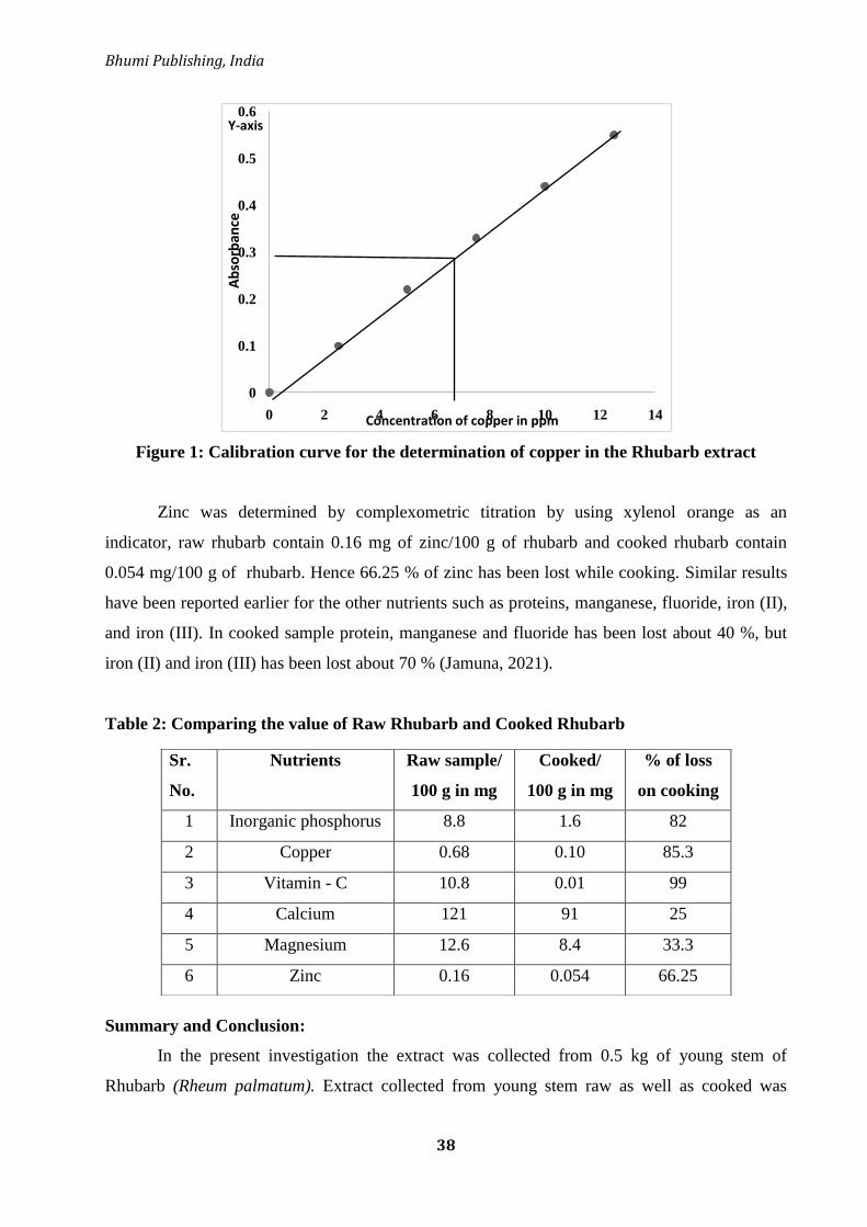
Bhumi Publishing, India
38
Figure 1: Calibration curve for the determination of copper in the Rhubarb extract
Zinc was determined by complexometric titration by using xylenol orange as an
indicator, raw rhubarb contain 0.16 mg of zinc/100 g of rhubarb and cooked rhubarb contain
0.054 mg/100 g of rhubarb. Hence 66.25 % of zinc has been lost while cooking. Similar results
have been reported earlier for the other nutrients such as proteins, manganese, fluoride, iron (II),
and iron (III). In cooked sample protein, manganese and fluoride has been lost about 40 %, but
iron (II) and iron (III) has been lost about 70 % (Jamuna, 2021).
Table 2: Comparing the value of Raw Rhubarb and Cooked Rhubarb
Summary and Conclusion:
In the present investigation the extract was collected from 0.5 kg of young stem of
Rhubarb (Rheum palmatum). Extract collected from young stem raw as well as cooked was
0
0.1
0.2
0.3
0.4
0.5
0.6
0 2 4 6 8 10 12 14
Ab
sorb
ance
Concentration of copper in ppm
Y-axis
Sr.
No.
Nutrients Raw sample/
100 g in mg
Cooked/
100 g in mg
% of loss
on cooking
1 Inorganic phosphorus 8.8 1.6 82
2 Copper 0.68 0.10 85.3
3 Vitamin - C 10.8 0.01 99
4 Calcium 121 91 25
5 Magnesium 12.6 8.4 33.3
6 Zinc 0.16 0.054 66.25

Frontiers in Life Science (Volume II)
ISBN: 978-81-953600-8-6
39
analyzed for nutrients such as inorganic phosphorus, copper, vitamin –C, calcium, magnesium
and zinc. Except for calcium and magnesium which were lost below 35 %, all the other nutrients
analysed were lost above 60 % indicating they are thermo labile. The above analysis indicates
that cooked sample has lost some % of nutrients. Hence raw sample can be used in the form of
salads, since some of the nutrients are thermo labile.
Rhubarb contains only 21 calories in 100 g of rhubarb. It is rich in precious nutrients like
calcium and magnesium. It is also a very rich source of many vital phyto-nutrients, vitamins,
minerals and health benefiting anti-oxidants. Hence the above investigation was carried in order
to explore the importance of nutritional composition of rhubarb and also to exhibit that there is
some percentage of nutrient loss in cooked sample, so it is better to use raw rhubarb in salad
form than cooked rhubarb.
List of abbreviations:
ADA – Adenosine Deaminases Activity
ATP – Adenosine Tri Phosphate
CIS – Central Ischemic Stroke
FAS – Ferrous Ammonium Sulphate
EDTA – Ethylene Diamine Tetra Acetate
EBT – Eriochrome Black T
RBC – Red Blood Cells
Chemicals Molecular Formulae:
KOH – Potassium hydroxide
HNO3 – Nitric acid
HCl – Hydrochloric acid
H2SO4 – Sulphuric acid
Ce (IV) – Cerium IV sulphate
CuSO4 – Copper sulphate
ZnSO4.7H2O – Zincsulphate heptahydrate
Acknowledgment
The authors are grateful to the Principal, Maharani‘s Science College for Women,
Mysuru-05, for providing necessary facilities and encouragement during the course of this study.

Bhumi Publishing, India
40
References:
Cai Y., Sun M., Xing J., and Corke H. 2004. Antioxidant phenolic constituents in roots of
Rheum officinale and rubia cordifolia: structure-radical scavenging activity relationships.
Journal of Agricultural and Food Chemistry. 52;26, 7884–7890.
Shou-zhong Y. 1997. The Divine Farmers Materia Medica: (A Translation of the Shen Nong Ben
Cao Jing), Blue Poppy Press, Boulder, Colorad.
Locock R. 1997. Herbal medicine: Essiac. Canadian Pharmaceutical Journal. 130: 18-19.
Bradley PR. 1992. British herbal compendium: a handbook of scientific information on widely
used plant drugs / published by the British Herbal Medicine Association and produced by
its Scientific Committee. Bournemouth, Dorset: The Association.
Li J., Zhu J., Hu H. et al. 2018. Internationalization of traditional/complementary medicine
products: market entry as medicine. Chinese Medicine, 13;1, 50.
Tsai KH, Hsien HH, Chen LM, Ting WJ, Yang YS, Kuo CH, Tsai CH, Tsai FJ, Tsai HJ, Hang
CY. 2013. Rhubarb inhibits hepatocellular carcinoma cell metastasis via GSK-3-β
activation to enhance protein degradation and attenuate nuclear translocation of β-
catenin. Food Chem. 138 (1): 278–285.
Castleman M. 1991. The Healing Herbs: The Ultimate guide to the curative powers of nature's
medicine. Emmaus, PA: Rodale Press, 305-307.
Lee MR, Hutcheon J, Dukan E, Milne I. 2017. Rhubarb (Rheum species): the role of Edinburgh
in its cultivation and development. R Coll Phys Edinburgh. 47 (1): 102–109.
Cao YJ, Pu ZJ, Tang YP, Shen J, Chen YY, Kang A, Zhou GS, Duan JA. 2017. Advances in bio-
active constituents, pharmacology and clinical applications of rhubarb. Chin Med. 12 (1):
36–47.
Chen D, Wang L. 2009. Mechanisms of therapeutic effects of rhubarb on gut origin
sepsis. Chinese Journal of Traumatology. ; 12 (6):365–369.
Chen DC, Ma LQ, Liu SZ. 2009. Effects of rhubarb on intestinal flora and bacterial translocation
in rats with sepsis. Chin Crit Care Med. 21 (1):17–20.
C. P. Commission. 2015. Pharmacopoeia of the Peoples Republic of China, Chemical Industry
Press, China.
Lin C.-C., Chen J.-T., Yang J.-S. et al. 2009. Danthron inhibits the migration and invasion of
human brain glioblastoma multiforme cells through the inhibition of mRNA expression of
focal adhesion kinase, rho kinases-1 and metalloproteinase-9. Oncology Reports. 22; 5,
1033–1037.
Lu H.-F., Wang H.-L., Chuang Y.-Y. et al. 2010. Danthron induced apoptosis through
mitochondria- and caspase-3-dependent pathways in human brain glioblastoma multiforms
GBM 8401 cells. Neurochemical Research, 35; 3, 390–398.

Frontiers in Life Science (Volume II)
ISBN: 978-81-953600-8-6
41
Lu H.-F., Lai T.-Y., Hsia T.-C. et al. 2010. Danthron induces DNA damage and inhibits DNA
repair gene expressions in GBM 8401 human brain glioblastoma multiforms
cells. Neurochemical Research, 35;7, 1105–1110,
Chiou S.-M., Chiu C.-H., Yang S.-T. et al. 2012. Danthron triggers ROS and mitochondria-
mediated apoptotic death in C6 rat glioma cells through caspase cascades, apoptosis-
inducing factor and endonuclease G multiple signaling. Neurochemical Research, 37; 8,
1790–1800.
Yoon H., An H., Ko M. et al. 2016. Upregulation of human ST8Sia VI (α2,8-Sialyltransferase)
gene expression by physcion in SK-N-BE(2)-C human neuroblastoma cells. International
Journal of Molecular Sciences, 17; 8, 1246.
Kwon Y. S., Koh J. Y., Song D. K. et al. 2004. Danthron inhibits the neurotoxicity induced by
various compounds causing oxidative damages including beta-amyloid (25-35) in primary
cortical cultures. Biological & Pharmaceutical Bulletin, 27; 5, 723–726.
Liu T., Jin H., Sun Q.-R., Xu J.-H., and Hu H.-T. 2010. Neuroprotective effects of emodin in rat
cortical neurons against β-amyloid-induced neurotoxicity. Brain Research, 1347, 149–160.
Liu J., Hu G., Xu R. et al. 2013. Rhein lysinate decreases the generation of β-amyloid in the
brain tissues of Alzheimer's disease model mice by inhibiting inflammatory response and
oxidative stress,‖ Journal of Asian Natural Products Research, vol. 15, no. 7, pp. 756–763.
Benjamin E. J., Virani S. S., Callaway C. W. et al. 2018 Heart disease and stroke statistics-2018
update: a report from the american heart association,‖ Circulation, 137;12, 467–492.
Chu X., Zhou S., Sun R. et al. 2018. Chrysophanol relieves cognition deficits and neuronal loss
through inhibition of inflammation in diabetic mice. Neurochemical Research, 43; 4, 972–
983.
Ma L., Zhao L., Hu H. et al. 2014. Interaction of five anthraquinones from rhubarb with human
organic anion transporter 1 (SLC22A6) and 3 (SLC22A8) and drug-drug interaction in
rats. Journal of Ethnopharmacology, 153; 3, 864–871.
Zhang N., Zhang X., Liu X. et al. 2014. Chrysophanol inhibits NALP3 inflammasome activation
and ameliorates cerebral ischemia/reperfusion in mice. Mediators of Inflammation, Article
ID 370530, 12 pages.
Xun Li,
Shifeng Chu, Yinjiao Liu, and Naihong Chen. 2019. Neuroprotective Effects of
Anthraquinones from Rhubarb in Central Nervous System Diseases. Evidence-based
Complementary and Alternative Medicine. 12:1-12, DOI:10.1155/2019/3790728.
Dong X., Fu J., Yin X. et al. 2016. Emodin: a review of its pharmacology, toxicity and
pharmacokinetics. Phytotherapy Research, 30; 8, 1207–1218.
Shu-Chun Hsu and Jing-Gung Chung. 2012. Anticancer potential of emodin. Biomedicine
(Taipei). 2 (3): 108–116.

Bhumi Publishing, India
42
Sun Y.-P. and Liu J.-P. 2015. Blockade of emodin on amyloid-β 25-35-induced neurotoxicity in
A β PP/PS1 mice and PC12 cells through activation of the class III phosphatidylinositol 3-
kinase/beclin-1/B-cell lymphoma 2 pathway. Planta Medica, 81; 2, 108–115.
Zhou Y.-X., Xia W., Yue W., Peng C., Rahman K., and Zhang H. 2015. Rhein: a review of
pharmacological activities. Evidence-Based Complementary and Alternative Medicine,
Article ID 578107, 10 pages.
Huang Q., Lu G., Shen H., Chung M. C. M., and Choon N. O. 2007. Anti-cancer properties of
anthraquinones from rhubarb. Medicinal Research Reviews, 27, 5, 609–630.
Zhi-Kun S., Hong-Qi Y., Zhi-Quan W., Jing P., Zhen H., and Sheng-Di C. 2012. Erythropoietin
prevents PC12 cells from beta-amyloid-induced apoptosis via PI3K/Akt
pathway. Translational Neurodegeneration, 1; 1, 7.
Li X. and Fan Z. 2010. The epidermal growth factor receptor antibody cetuximab induces
autophagy in cancer cells by downregulating HIF-1α and Bcl-2 and activating the Beclin
1/hVps34 complex. Cancer Research, 70; 14, 5942–5952.
Durak, Z., Buber, S., Devrim, E., Kocaoglu, H., & Durak, I. 2015. Aquoeus Extract from
Rhubarb Plant Inhibits Adenosine Deaminase Activity in Cancerous and Non Cancerous
Human Gastric and Colon Tissues. Journal of Progressive Research in Chemistry, 1;1, 9-
13.
Mendham RC, Denny JD, Barner M, Thomson B, Sivasankar. Vogel‘s Quantitative Chemical
Analysis, 6th
edition, 336-341.
Xiang H., Zuo J., Guo F., and Dong D. 2020. What we already know about rhubarb: A
comprehensive review, Chinese Medicine. 15; 1, DOI:10.1186/s13020-020-00370-6.
Komatsu K. , Nagayama Y., Tanaka K., Ling Y., Cai S., Omote T., Ragab Meselhy M. 2006
Comparative study of chemical constituents of rhubarb from different origins. Chem
Pharm Bull (Tokyo). 54;11, 1491-1499. doi: 10.1248/cpb.54.1491.
Jamuna M. 2021. Biochemical composition of the Rheum palmatum (Rhubarb) in raw and
cooked sample: A comparative study. Ciencia and Engenharia/ Science and Engineering
Journal ISSN: 0103-944X | EISSN: 1983-4071 DOI:
https://doicatalog.org/19.4104/c&e.v68i6.1614, 152-170.
Frontiers in Life Science (Volume II)
ISBN: 978-81-953600-8-6
43
PLANTS BASED ETHNO-MEDICINE IN THE LAMBANI COMMUNITIES IN
THE VICINITY OF VIJAYAPURA DISTRICT OF KARNATAKA
Ramachandra Naik M.1, Vinayaka K.S.2* and Joy Hoskeri3
1Department of Botany,
S B Arts and KCP Science College, BLDE’S, Vijayapura, Karnataka
2Department of Botany,
Venkataramana Swamy College, Bantwal - 574211, D.K., Karnataka
3Department of Bioinformatics,
Karnataka State Akkamahadevi Women’s University, Vijayapura, Karnataka, India
*Corresponding authors E-mail: [email protected]
Abstract:
Ethno botany is the study of plants and its applications on the inhabitants of an area and
deals with folklare concepts of classification by habitat and usage or some other parameters.
Ethnobotanical surveys are important steps in the identification, selection and development of
therapy from medicinal plants which further serve as a base for development of new compounds
with active principles for phytochemical, pharmacological and clinical research. Now days, the
whole world is relying on plant-based health remedies for safe and effective cure of dangerous
human ailments. In this present content, we have focused on the ethno botanical knowledge and
culture of the Lambani ethnic group from Vijayapura district of Karnataka. Lambani community
people basically stay in remote area and these communities still rely only on their traditional
medicinal practices for their health care needs. We selected a total of 23 villages with Lambani
community for the study in Vijayapura district. Ethnomedicinal information on medicinal plants
was recorded through interviews, discussions and field observation of traditional healers and
practitioners with knowledgeable people of selected study area using a specialized questionnaire.
In this study, 25 folk herbal practitioners aged between 25 - 75 years were consulted to gather the
information for analysis on the study area. Most of the respondents (70%) received only primary
and high school education. While, a few (10%) received pre-university education; 5% were
degree holders and 10% were illiterate individuals. Majority of the traditional healers acquired
the therapeutic knowledge from their ancestors. In the present study, we have documented
around 42 medicinal plant species from 28 families were used for the treatment of human and
livestock ailments. Plants belonging to Lamiaceaea, Fabaceae, Menispermaceae, Amaranthaceae,
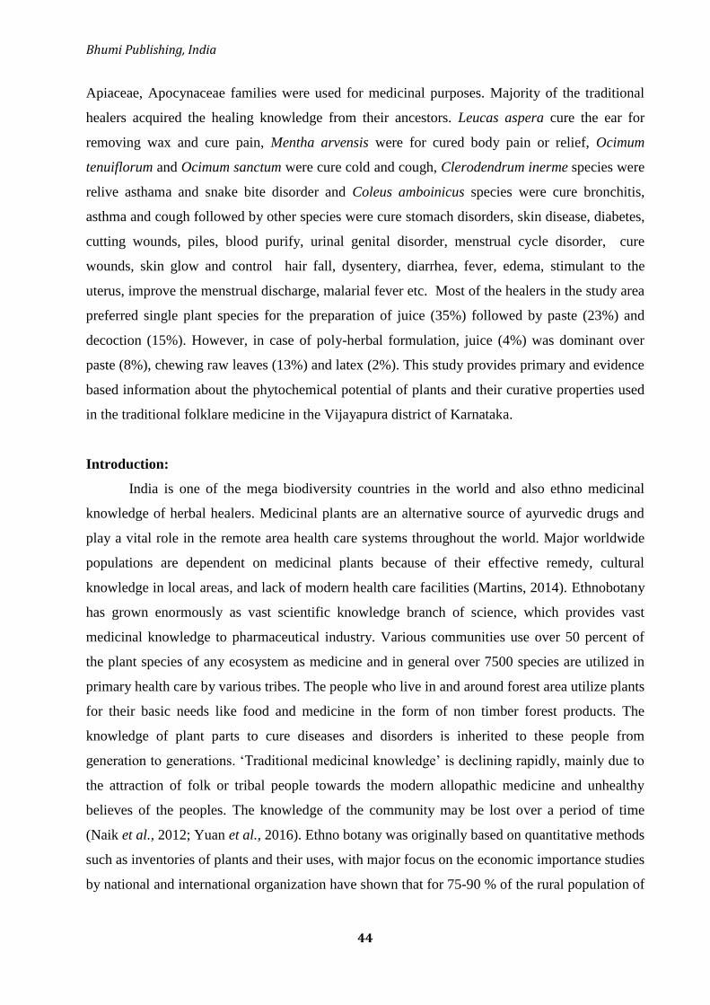
Bhumi Publishing, India
44
Apiaceae, Apocynaceae families were used for medicinal purposes. Majority of the traditional
healers acquired the healing knowledge from their ancestors. Leucas aspera cure the ear for
removing wax and cure pain, Mentha arvensis were for cured body pain or relief, Ocimum
tenuiflorum and Ocimum sanctum were cure cold and cough, Clerodendrum inerme species were
relive asthama and snake bite disorder and Coleus amboinicus species were cure bronchitis,
asthma and cough followed by other species were cure stomach disorders, skin disease, diabetes,
cutting wounds, piles, blood purify, urinal genital disorder, menstrual cycle disorder, cure
wounds, skin glow and control hair fall, dysentery, diarrhea, fever, edema, stimulant to the
uterus, improve the menstrual discharge, malarial fever etc. Most of the healers in the study area
preferred single plant species for the preparation of juice (35%) followed by paste (23%) and
decoction (15%). However, in case of poly-herbal formulation, juice (4%) was dominant over
paste (8%), chewing raw leaves (13%) and latex (2%). This study provides primary and evidence
based information about the phytochemical potential of plants and their curative properties used
in the traditional folklare medicine in the Vijayapura district of Karnataka.
Introduction:
India is one of the mega biodiversity countries in the world and also ethno medicinal
knowledge of herbal healers. Medicinal plants are an alternative source of ayurvedic drugs and
play a vital role in the remote area health care systems throughout the world. Major worldwide
populations are dependent on medicinal plants because of their effective remedy, cultural
knowledge in local areas, and lack of modern health care facilities (Martins, 2014). Ethnobotany
has grown enormously as vast scientific knowledge branch of science, which provides vast
medicinal knowledge to pharmaceutical industry. Various communities use over 50 percent of
the plant species of any ecosystem as medicine and in general over 7500 species are utilized in
primary health care by various tribes. The people who live in and around forest area utilize plants
for their basic needs like food and medicine in the form of non timber forest products. The
knowledge of plant parts to cure diseases and disorders is inherited to these people from
generation to generations. ‗Traditional medicinal knowledge‘ is declining rapidly, mainly due to
the attraction of folk or tribal people towards the modern allopathic medicine and unhealthy
believes of the peoples. The knowledge of the community may be lost over a period of time
(Naik et al., 2012; Yuan et al., 2016). Ethno botany was originally based on quantitative methods
such as inventories of plants and their uses, with major focus on the economic importance studies
by national and international organization have shown that for 75-90 % of the rural population of
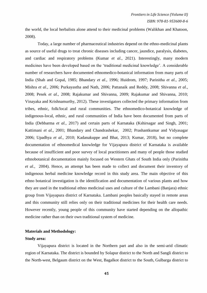
Frontiers in Life Science (Volume II)
ISBN: 978-81-953600-8-6
45
the world, the local herbalists alone attend to their medicinal problems (Walikhan and Khatoon,
2008).
Today, a large number of pharmaceutical industries depend on the ethno-medicinal plants
as source of useful drugs to treat chronic diseases including cancer, jaundice, paralysis, diabetes,
and cardiac and respiratory problems (Kumar et al., 2021). Interestingly, many modern
medicines have been developed based on the ‗traditional medicinal knowledge‘. A considerable
number of researchers have documented ethnomedico-botanical information from many parts of
India (Shah and Gopal, 1985; Bhandary et al., 1996; Huidrom, 1997; Parinitha et al., 2005;
Mishra et al., 2006; Purkayastha and Nath, 2006; Pattanaik and Reddy, 2008; Shivanna et al.,
2008; Pesek et al., 2008; Rajakumar and Shivanna, 2009; Rajakumar and Shivanna, 2010;
Vinayaka and Krishnamurthy, 2012). These investigators collected the primary information from
tribes, ethnic, folk/local and rural communities. The ethnomedico-botanical knowledge of
indigenous-local, ethnic, and rural communities of India have been documented from parts of
India (Debbarma et al., 2017) and certain parts of Karnataka (Kshirsagar and Singh, 2001;
Kattimani et al., 2001; Bhandary and Chandrashekar, 2002; Prashantkumar and Vidyasagar
2006; Upadhya et al., 2010; Kadanakuppe and Bhat, 2013; Kumar, 2018), but no complete
documentation of ethnomedical knowledge for Vijayapura district of Karnataka is available
because of insufficient and poor survey of local practitioners and many of people those studied
ethnobotanical documentation mainly focused on Western Ghats of South India only (Parinitha
et al., 2004). Hence, an attempt has been made to collect and document their inventory of
indigenous herbal medicine knowledge record in this study area. The main objective of this
ethno botanical investigation is the identification and documentation of various plants and how
they are used in the traditional ethno medicinal uses and culture of the Lambani (Banjara) ethnic
group from Vijayapura district of Karnataka. Lambani peoples basically stayed in remote areas
and this community still relies only on their traditional medicines for their health care needs.
However recently, young people of this community have started depending on the allopathic
medicine rather than on their own traditional system of medicine.
Materials and Methodology:
Study area:
Vijayapura district is located in the Northern part and also in the semi-arid climatic
region of Karnataka. The district is bounded by Solapur district to the North and Sangli district to
the North-west, Belgaum district on the West, Bagalkot district to the South, Gulbarga district to

Bhumi Publishing, India
46
the East and Koppal district to the South-east. Vijayapura district is spread over an area of
10,498 sq km and lies between 16° 49' 39.1620'' N latitude and between 75° 43' 31.1772'' E
longitude at a mean elevation of 592.23 m (Fig 1). Vijayapura formally called as Bijapur has a
very interesting history with many dynasties ruling the district for many centuries. The
monuments in Vijayapura speak about the history that the city faced from different rulers. The
history of Vijayapura goes back to Palaeolithic age. In summer session especially in April and
May it is too hot temperature ranges from 400C to 42
0C and the average annual rainfall for the
whole district is 552.8 mm in rainy days. The total livestock population of the district is 326, 360
according to 2011 census. The study area is known for the major portion of the district consists
of this kind of soil which has a great moisture-holding capacity. Second one is "red soil‟
(masari/maddibhoomi), which is generally poor, good for irrigation and horticulture. Krishna
River, which is the most important river of the district, it flows about 125 miles in the district. A
dam is built across the river at Almatti and Bhima river flows in northern part of district for
about 20 miles. It overflows in the rainy season and spreads over a wider area, which is thereby
rendered extremely fertile land. In central part of district Doni river flows some area of irrigated
land total five rivers flows in this district. Agriculture plays a major role in the economy of this
district and which is good for the crops like jawar, wheat, pulses, sunflower, groundnut,
sugarcane, and cotton and fruit crops like grapes, lemon and pomegranate the main crops
provide sources for irrigation. Natural vegetation occurred in this study area comprising of semi-
arid forest shaving a variety of medicinal plants. In the present study, ethno-medicinal surveys
were carried out in the taluka places including Vijayapura, Basavana Bagewadi, Sindagi, Indi,
Muddebihal, Nidagundi, Talikote, Babaleshwar, Tikota, Chadchan, Devara Hipparagi, Kolhar
surrounding interviewed to Lambani peoples in Vijayapura district. These communities have
their own language and food habits, dress code, historical culture and socio-religious myths.
Figure 1: Map showing study area Vijayapura and its taluka places

Frontiers in Life Science (Volume II)
ISBN: 978-81-953600-8-6
47
About the Lambani communities of Vijayapura district:
In the present study, we have focused on local herbalists, healers; farmers and village
headman of the Lambani communities found in different ―thandas‖ were interviewed for
responses regarding traditional knowledge and their uses. In Karnataka, the Lambani community
is also refered as Banjara, Lambadi, Lamani, Gor/Gour Banjara, Gour Maati, Naik and Sugali.
These community women wear colorful and beautiful costumes like phetiya (as Ghagra) and
Kancholi (as top) and have tattoos on their hands. These inhabitants of this community live in
remote places of villages and they rely mainly on surrounding bioresources for their food, shelter
and medical care etc. The study area belonged to different Jath/ gothras of lambani communities
such are list below.
The gothras of Rathod/Bhukya and their 27 padaas or variants, Pawar and their 12 padas
or variants, Chavan/Chauhan and their 6 padas or variants, Vadithya/Jadhav and their 52 padas
or variants , Banoth and their 15 padas or variants. Villages of most gothras are majorly situated
in the remote areas; usually people of certain gothras are experts of ethno medicinal treatement
of various diseases. The knowledge about ethno-medicinal plants is specialized by the Rathod
and their padaas Meghavath, Rajaavath and Ramaavath and Gothras of Chuhaan and their Padas
Korra and Mood and Gothras of Pawar and their Vislaavath were experts to ethnobotanical
‗nativaidyas‘ or country medicines in study area.
Data collection:
Frequent field surveys were conducted at 23 randomly selected villages in Vijayapura
district during the period from April 2019 to March 2021. Ethnomedicinal information on
medicinal plants was recorded through interviews discussion and field observation of traditional
healers and practitioners with knowledgeable people of selected study area using a specialized
questionnaire. Among them 25 folk herbal practitioners aged between 25 and 75 years were
consulted to gather information in the study area, which included 20 were male and 05 female
respondents under the age group herbal healers have 15-25 years of experience in the treatment
of different diseases. An ex-post facto research design was adopted to assess the ethno-
medicines used in the different places of Vijayapura district in Karnataka. The responses
included details about the botanical and common name of the plant prescribed for medicine
purpose, part of the plant used, mode of preparation (decoction, paste, powder or juice), dosage
and duration were confirmed through discussions with respondents who practiced the use of the
documented plants for medicinal purposes.

Bhumi Publishing, India
48
Figure 2: Pictorial view of traditional healers with mode of medicine preparation
Result and Discussion:
The present study areas of some villages possess interesting characteristics related to life
in these remote areas were dependency on agricultural field plants and local wild plants for
making various food products and therapeutic herbal medicine. In rural areas many plant parts
are used for medicinal purposes although they may have other potential utility values. Such
ethnic medicine purpose their potential health benefits and their health risks associated with such
remedies. Some of these knowledge have been documented and codified or studied scientifically.
Most of the ethno medicine practitioners were avoids the transfer of their traditional herbal
knowledge to others. Because they believed that the medicinal practices by a layman on
remedies of the plant species would not lead to recovery or cure the disease to the other people.
The present study documented the ethno medico botanical uses of 46 plants belonging to
28 families (Table-1) that have been gathered as primary information and documented from local
herbal practitioners in the present study. Majority of the plant species belongs to the family
Lamiaceae (13%), other families such as Papilionaceae, Caesalpiniaceae, Menispermaceae,
Amaranthaceae, Apiaceae, Apocynaceae were represents by 7%, families like Asteraceae and
Myrtaceae are represented 4% and remaining all families were present in 2% respectively. Total
42 plant species were projected for their use as medicinal plants, among them 26 plants are
commonly collected from wild habitats such as scrubby forest area, road side, gomalas and river
canal border only and remaining 16 plant species were cultivated and collected from agricultural
land and house premises.

Frontiers in Life Science (Volume II)
ISBN: 978-81-953600-8-6
49
Table 1: Showing List of plants used by the Lambani community for ethnomedicinal purposes with their formulations
Sl.
No.
Botanical Name Family name Local name Ailment Mode of uses
01 Abrus precatorius L. Fabaceae Gulaganji fevers, coughs and
colds
A cup of tea is made from the leaves used every morning
02 Acacia ferruginea L. Mimosaceae Banni mara itching,
leucoderma, ulcers
Fruit juice is taking orally every day up to one week
03 Achyranthes aspera L. Amaranthaceae Uttarani menstrual cycle
disorder
Root juice with 250 ml water taken orally twice a day for 5 days
04 Aloe vera (L.) Asphodelaceae Lolesera Skin glow and
control hair fall
Aloe veragell is directly apply on face and hair
05 Amaranthus caudatus L.
Amaranthaceae Rajagiri soppu Enrich of protein
and energy
whole plant are used as edible and are frequently used as a
source of food in India
06 Amaranthus spinosus L. Amaranthaceae Mulluarivae
sappu
urinal genital
disorder
Leaves along with masala ingredients, salt making food eating
with rooti
07 Argemone Mexicana L. Papaveraceae Dhatthoora Pain killer and
cough
Leaves paste applied on effected part of body and the seeds
decoction drunk to relieve cough
08 Azadirachta indica
A. Juss.
Meliaceae Bevu Wounds and
cleaning tooth
Leaves paste is applied on effected parts and twig is used for
making tooth brash
09 Azima tetracantha L. Salvadoraceae Uppi mullu poison vomiting Crushed leaves with Neem leaves paste mixing with water taken
orally to poison dunked person
10 Basella alba L. Basellaceae Basalae Dysentery Leaf juice with half spoon salt mix with boil water is taken orally
for 2 days
Bhumi Publishing, India
50
11 Caesalpinia bunducella L. Caesalpiniaceae. Gajagadgida Stimulant to the
uterus, improve the
menstrual
discharge, malarial
fever. relieving
intestinal worms
The fruit nut and root bark extract has been used twice every day
up to 15 days
12 Calotropis procera
(Aiton) W.T.Aiton
Apocynaceae Beli yakka Cutting wounds Plant latex is applied on affected parts
13 Cassia auriculata Linn. Fabaceae Tavarkegida Diabetes Chewing Bark every day along with bettle leaves
14 Cassia fistula Linn Fabaceae Kakkegida skin
diseases
Flashy fruit paste is applied on face every morning
15 Centella asiatica
(L.) Urban
Apiaceaea Ondelaga Stomach disorders Leaves paste taken orally for 5 days
16 Clerodendrum inerme (L.) Verbinaceae Kaadu mallige asthama and snake
bite
Leaves and flower extracts with 250 ml water is taken orally for
weakly two times and root paste is applied on effected parts for
cure snake bite
17 Cocculus hirsutus
(L.) Diels
Menispermaceae Gowri huvu Preparation of chair Stem is used in making basket chairs
18 Coleus amboinicus Lour. Lamiaceae Doddapatre Cough Plant extract is taken orally for 6 days
19 Coleus amboinicus Lour. Lamiaceae Dodda patre Cure bronchitis,
asthma,
Leaves extract drinking orally twice a day upto 15 days
20 Coriandrum sativum L. Apiaceaea Kotambari sappu Increase iron
content in blood
Seeds are used for preparation of decoction taking orally twice a
day
21 Cynodon dactylon (L.) Pers. Poaceae Cynodon blood bleeding Root decoction 3-4 drops is taken orally in nose for 2 days

Frontiers in Life Science (Volume II)
ISBN: 978-81-953600-8-6
51
22 Datura metel L. Solanaceae Daturi headaches Dry leaves inhalation trice a day up to 12 days
23 Eucalyptus globulus Labill.
Myrtaceae Neelagiri Fragrance and
insecticide property
Plant oil is used for fragrance and oil is used to control
insecticide property on dead body
24 Ficus religiosa L. Moraceae Arali mara vomiting, & cough Bark extract is taken orally for 6 days
25 Lawsonia inermis L. Lythraceae Gorenty Blood purify Leaves decoction is taken orally twice a day for 10 days
26 Limonia acidissima L. Rutaceae Belada mara Increase blood cell fruit paste with cow milk is drunk daily two times for 20 days
27 Mentha arvensis L. Lamiaceae Pudina Body pain Plant leaves juice is taken orally for 2 days and leaves paste
applied on external affected parts
28 Michelia champaca L. Magnoliaceae Sampige gida Oral cancer &
gonorrhea disease
Flowers paste are used orally to animal
29 Nelumbo nucifera
Gaertn.
Nelumbonaceae Tavare huvu skin burn leaves paste is applied on external affected parts till cure skin
burning
30 Nerium indicum L. Apocynaceae Kanagilae piles Root juice with one hand full lawsonia intermis, one onion
grinding with water taken orally for 15 days
31 Ocimum sanctum L. Lamiaceae Tulasi Cold and cough Raw leaves chewing every day up to 12 days
32 Ocimum tenuiflorum L. Lamiaceae Tulasi Cold & cough Leaves juice is taken orally for 5 days
33 Oroxylum indicum (L.)
Vent.
Bignoniaceae Aralu Diarrhoea, Fever,
Oedema
Flowers and fruit were eaten with rice.
34 Peucedanum graveolens (L.)
Hiern
Apiaceaea Sabbasige sappu wounds Plant paste with turmeric powder is applied on affected area
35 Piper beetle L. Piperaceae Veelyadaele tooth ache Leaves along with 3 clove, 2 pieces of garlic paste applied
externally on effected tooth till cure
Bhumi Publishing, India
52
36 Santalum album Linn. Santalaceae Gandada mara Insecticide property Sandal wood oil is used on dead body for control germs and
insect
37 Sauropus androgynus
L.(Merr.)
Phyllanthaceae Chakramuni Weight loss and
improvement of
macronutrients
The leaves extracts with 250 ml water taken daily twice times
38 Syzygium cumini (Linn.)
Skeels
Myrtaceae Nerale Cure dysentery Bark extract decoction is used twice a day upto one week
39 Tagetes erecta L. Asteraceae Chendi huvu blood purify Flower juice drunken for one weak
40 Tinospora cordifolia
(Thunb.) Miers
Menispermaceae Amrutha balli diabetes Leaves juice is taken orally with water for daily morning
41 Tinospora corifolia (Thunb.)
Miers
Menispermaceae Amruth balli Cureing chicken
gunya disease
Stem boiling with water up 10 minute then using plant decoction
every morning
42 Tridax procumbens L. Asteraceae Gejje tikke Skin disease Leaf paste is applied on sun burning skin up to 30 days
43 Trigonella foenum-graecum
L.
Fabaceae Mentte sappu Increase immunity Along with food, Trigonella leaves are chewing raw is used for
increasing body immunity
44 Vigna sinensis (L.) Endl. Fabaceae Halasandi For strength and
energy
Leaves is used as cooking food for gaining multi vitamin and
protein
45 Vinca rosea L. Apocynaceae Sada mallige diabetes Stem decoction is taken orally twice a day
46 Ziziphus mauritiana Lam. Rhamnaceae Badara Cure Piles, Fruit juice drinking every day up to 30 days

Frontiers in Life Science (Volume II)
ISBN: 978-81-953600-8-6
53
In some cases, ethnic people suggest single plants species are uses specific ailments and
some cases to added different ethno botanical ingredients were crushing, powdering and boiling
plant specimen to make a various ailments, some of the medicinal plants leaves and fruits
directly recommended for chewing in raw to cure various diseases such as, the family Lamiaceae
some species like Leucas aspera were used to cure the ear for removing wax and cure pain,
Mentha arvensis were used used to cure body pain or relief, Ocimum tenuiflorum and Ocimum
sanctum species were used cure cold and cough, Clerodendrum inerme species were used relive
asthama and snake bite disorder and Coleus amboinicus species were used to cure bronchitis,
asthma and cough. Similarly other plant species remedies were included by the practitioners for
their use in cure of various diseases like stomach disorders, skin disease, diabetes, cutting
wounds, piles, blood purify, urinal genital disorder, menstrual cycle disorder, enrich of protein
and energy, cure wounds, increase iron content in blood, skin glow and control hair fall,
dysentery, diarrhea, fever, edema, stimulant to the uterus, improve the menstrual discharge,
malarial fever, relieving intestinal worms, increase immunity, for strength and energy, fevers,
skin diseases, cough, body pain, preparation of chair, blood purify, curing chicken pox disease,
Insecticide property, fragrance and insecticide property, wounds and cleaning tooth, cure skin
burn, tooth ache, blood bleeding control, pain killer, weight loss and improvement of
macronutrients, Increase blood cell, cure piles, poison vomiting, cure headaches, cure vomiting
(Table-1).
Figure 3: Pi chart showing Plant parts used for drug formulation
Whole plant12%
Roots9%
Stems12%
Barks9%
leaves29%
Flower9%
fruits and seed20%
Plant parts used

Bhumi Publishing, India
54
Most of the healers in the study area preferred single plant species for the preparation of
juice (35%) followed by paste (23%) and decoction (15%). However, in case of poly-herbal
formulation, juice (4%) was dominant over paste (8%), chewing raw leaves (13%) and latex
(2%) were used (Fig.3). The percentages of different plant parts used for the preparation of
medicine (Fig.4) Whole plants (11%) were found to be the most frequently used plant parts in
the preparation of medicine followed by roots (8%), stems (11%), bark (9%), Leaves (29%),
flowers (9%) and fruits and seeds (20%) and background literature on ethnobotanical studies
confirmed that leaves are the major portion of the plant used in the treatment of diseases. Ethno
botanical survey is of crucial importance in finding some miraculous medicines for curing
various human and veterinary diseases. Hence ethno botanical survey is of crucial importance in
finding some miraculous medicines for curing nearly 32 human diseases. The list of folk
medicinal plants from the northern region of Karnataka and their utilization will provide primary
data for further studies aimed at conservation, cultivation, traditional medicine and economic
welfare of rural and tribal population of the region.
Figure 4: Pi chart showing Preferred Herbal formulation in the study area
The preparation of herbal medicine is varies from one place to other place and differs to
usage of ethno medicine in allied disease. For example in study area Vijayapura district ethnic
people uses Tridax procumbens species used to cure skin disease, the same species used in
Shimoga district tribal peoples to cure toothache (Shivanna and Rajkumar, 2010) similarly Vinca
rosea, Tinospora cordifolia and Cassia auriculata species were used to cure diabetes, the
Tinospora cordifolia plant is used as reduce sugar level in blood (Mishra et al., 2019).,
Calotropis procera species are used cure cut wounds, same plants in Hosanagara taluk is used to
Juice 35%
Paste23%
Decoction 15%
Polyherbal Juice4%
Polyherbal Paste
8%
Chewing raw leaves 13% Latex
2%
Herbal formulation
Frontiers in Life Science (Volume II)
ISBN: 978-81-953600-8-6
55
cure Epilepsy (Shivanna and Rajkumar, 2011), Nerium indicum were used to cure piles
(Chetwani et al., 2017). Nerium indicum also act as antibacterial potential, Amaranthus spinosus
for curing urinal genital disorder, in this plant studied (Rahman et al., 2013) by earlier workers
for the cure Asthama and Achyranthes aspera species roots extract can form abortion. This same
plant is used in study area for curing menstrual cycle disorder, Coriandrum sativum to enrich
iron content in blood in Indonesia this plants used to cure gastrointestinal disorders (Jadid et al.,
2020), Aloe vera and Cassia fistula to enhance skin glow and cure skin disease. Mishra et al.,
(2019) discussed same plants used body balancing level of glucose content. Coleus amboinicus is
used to cure throat infection (Somashekar et al., 2010) but in the present study it was
documented as used to cough. Azadirachta indica for curing wounds and cleaning tooth,
Syzygium cumini for cure dysentery, Piper betle is used as cure toothache, in Indonesia same
plants used to cure urogenital and gynecological problems (Jadid et al., 2020). The present study,
a list of folk medicinal plants from the northern region of Karnataka and their utilization will
provide primary data for further studies aimed at conservation, cultivation; need to explore
traditional medicine and economic welfare of rural and tribal communities of the region. Further
survey and research focusing the on the medicinal plants documented in this study might give
information regarding the hunt for bioactive compounds and the discovery of new drugs to fight
diseases in an effective manner.
Conclusion:
This ethno botanical work is first recorded in study areas of Lambani communities in
different region of Vijayapura district of Karnataka. A total 42 plant species belong to 28
families were used cured 32 human diseases. The present study focused on ethno medicinal uses
and their potential health benefits in rural areas. Investigators noticed that younger generation
they do not believe in their ancestral traditional medicine and have developed dependency on
allopathic medicine for faster relief of disease. Some pharmaceutical companies‘ approaches to
ethnic people for documentation on utilization of a number of medicinal plants species used to
treat aliments. Hence the younger researchers focus on to collect this information and develop a
database of medicinal plants for future research and potential development of new drugs.
Acknowledgements:
The authors are thankful to the Ethnic people in Vijayapura district for their valuable help
in documentation of indigenous ethno medicinal knowledge.
Bhumi Publishing, India
56
References:
Bhandary M.J, Chandrashekar K.R, Kaveriappa K.M (1996) Ethnobotany of Gowlis of Utttara
Kannada district, Karnataka. Journal of Economic and Taxonomical Botany, 12:244-249.
Bhandary M.J, Chandrashekar K.R. (2002) Glimpses of ethnic herbal medicine of costal
Karnataka. Ethnobotany, 14:1–12.
Chetwani Kavita, Agnihotri R.K. and Purti Chaturvedi (2017) Aqueous, Acetone and Ethanolic
extract of Nerium indicum L. as potential antibacterial agent against Pseudomonosa
aeruginosa, International Journal of Applied Environmental Sciences, 12(9): 1721-1732.
Debbarma Maria, Nazir A. Pala, Munesh Kumar, Rainer W. Bussmann (2017) Traditional
Knowledge of Medicinal Plants in Tribes of Tripura In Northeast, India, Afr. J. Tradit.
Complement. Altern. Med. 2017, 14(4): 156–168.
Economic survey of Karnataka, 2014. http://planning.kar.nic.in/node/128.html.
Ghosh S, Suryawanshi S.A (2021) Effect of Vinca rosea extracts in treatment of alloxan diabetes
in male albino rats, Indian Journal of Experimental Biology, 39(8): 748-59.
Government of Karnataka. Status of Scheduled Tribes of Karnataka. In: Karnataka Human
Development report. http://planning.kar.nic.in/sites/planning.kar.nic.in/files/khdr2005/
Huidrom B.K.S (1997) Studies on medico-botany of Meiti community in Manipur State, India.
Advances in Plant Sciences, 10:13-18.
Jadid Nurul, Erwin Kurniawan, Chusnul Eka Safitri Himayani, Andriyani (2020) An
Ethnobotanical study of medicinal plants used by the Tengger tribe in Ngadisari village,
Indonesia. PLOS ONE, 15(7):1-16.
Kadanakuppe S, Bhat P.K. (2013) Oral health status and treatment needs of Iruligas at
Ramanagara District, Karnataka, India. West Indian Med. Jrl, 62:73–80.
Kattimani K.N, Munikrishnappa P.M, Hussain S.A, Reddy P.N (2001) Use plants as medicine
under semi-arid tropical climate of Raichur district of north Karnataka. J Med Arom Pl
Sci., 22-23:607–11.
Kshirsagar R.D, Singh N.P (2001) Some less known ethnomedicinal uses from Mysore and
Coorg districts, Karnataka State, India. J Ethnopharmacol., 75:231-238.
Kumar S. (2008) Genetic Profile of Jenu Kuruba, Betta Kuruba and Soliga Tribes of Southern
Karnataka and their phylogenetic relationships. Anthropologist, 10:11–20.
Mishra D., Rajesh, Chakravarthy M, Gowtam M.P. (2006) Traditional medicinal practices
among the various tribal groups of Kankar district of Bastar division. Indian forester, 132:
850-855.
Frontiers in Life Science (Volume II)
ISBN: 978-81-953600-8-6
57
Mishra jiban, Gyanranjan mahalik, Sagarika parida (2019) Ethnobotanical study of traditional
medicinal plants used in the management of diabetes in the urban areas of khurda, odisha,
india. Asian J Pharm Clin Res., 12 (9): 73-78.
Mishra Jiban, Gyanranjan Mahalik, Sagarika Parida (2019) Ethnobotanical Study Of Traditional
Medicinal Plants Used In The Management of Diabetes In The Urban Areas of Khurda,
Odisha, India. Asian Journal of Pharmaceutical and Clinical Research, 12: 9.
Naik M Ramachandra, Vaishnavi Venugopalan, Preethi Kumaravelayutham and Y.L.
Krishnamurthy (2012) Ethnoveterinary uses of medicinal plants among the Lambani
community in Chitradurga district, Karnataka, India. Asian Pacific Journal of Tropical
Biomedicine,2:S470-S476.
Parinitha M, Harish G.U, Vivek N.C, Mahesh T and Shivanna M.B (2004) Ethno-botanical
wealth of Bhadra wild life sanctuary in Karnataka. Indian J Tradit Knowl., 3:37–50.
Parinitha M, Srinivasa B.H and Shivanna M.B (2005) Medicinal plants wealth of local
communities in some villages in Shimoga district of Karnataka, India. Journal of
Ethnopharmacology, 98:307-312.
Pattanaik C. and Reddy C.S (2008) Medicinal plant wealth of local communities in Kuldiha
wildlife sanctuary, Orissa, India. Journal of Herbs Spices & Medicinal Plants, 14 (3): 175-
184
Pesek T.J, Helton L.R, Reminick R, Kannan D and Nair M. (2008) Healing traditions of
Southern India and the conservation of culture and biodiversity: A preliminary study.
Ethnobotany Research & Applications, 6: 471-479.
Prashantkumar P and Vidyasagar G.M. (2006) Documentation of traditional knowledge on
medicinal plants of Bidar district, Karnataka. Indian J Tradit Knowl,. 5: 295–299.
Purkayastha J, Nath S.C (2006) Biological activities of ethnomedicinal claims of some plant
species of Assam. Indian Journal of Traditional knowledge, 5:229-236.
Rahman A.H.M., Mahbubur, Nasrin Sultana, A.K.M. Rafiul Islam and A.T.M.N. Zaman (2013)
Study of Medical Ethno-botany at the Village Genda under Savar Upazilla of District
Dhaka, Bangladesh, Journal of Medicinal Plants Studies, 1(5): 72-86.
Rajakumar N and Shivanna M.B. (2009) Ethno-medicinal application of plants in the eastern
region of Shimoga district, Karnataka, India. Journal of Ethnopharmacology, 126:64–73.
Rajakumar N and Shivanna M.B (2010) Traditional herbal medicinal knowledge in Sagara taluk
of Shimoga District, Karnataka, India. Indian Journal of Natural Products & Resources 1
(1): 102-108.

Bhumi Publishing, India
58
Shah and Gopal, (1986) Some folklore medicines of Vasavas, Gujarat, India. Acta Botanica,
14:48-53.
Shivanna M.B and N. Rajkumar (2011) Traditional Medico-Botanical Knowledge of Local
Communities in Hosanagara Taluk of Shimoga District in Karnataka, India, Journal of
Herbs, Spices & Medicinal Plants, 17:291–317.
Shivanna M.B, Mangala K.R Parinitha M. (2008) Ethno-medicinal knowledge of Lambani
community in Chikmagalur district of Karnataka, India. Journal of Medicinal and Aromatic
plant sciences, 30:105-108.
Siddalinga M.S.M and Vidyasagar G.M (2013) Medicinal plants used in the treatment of
gastrointestinal disorders in Bellary district, Karnataka, India. Indian Journal of Traditional
Knowledge, 12(2): 321-325.
Somashekar Achar G and Rajkumar N and Shivanna M.B (2010) Ethno-Medico-Botanical
Knowledge of Khare-vokkaliga Community in Uttara Kannada District of Karnataka,
India. Journal of Complementary and Integrative Medicine, 7(1): 1-18.
Upadhya V, Mesta D, Hegde H.V, Bhat S, Kholkute S.D. (2010) Ethnomedicinal plants of
belgaum Region, Karnataka. J Econ Taxon Bot., 33:300–8.
Vinayaka K.S. and Krishnamurthy Y.L. (2012) Ethno-lichenological studies of Shimoga and
Mysore districts, Karnataka, India. Advances in Plant Sciences, 25 (1): 265-267.
Walikhan and Khatoon (2008) Ethnobotanical studies on some useful herbs of Haramosh and
Bugrote Valleys in Gilgil, Northern Areas of Pakistan. Pak. J. Bot., 40(1): 43-58.

Frontiers in Life Science (Volume II)
ISBN: 978-81-953600-8-6
59
IN VITRO SCREENING OF ANTIFUNGAL BACTERIA AGAINST
PHYTOPATHOGENIC FUNGI OF PUMPKIN (CUCURBITA MAXIMA)
Pooja H. V. and Girish K*
Postgraduate Department of Microbiology,
Maharani’s Science College for Women,
JLB Road, Mysuru – 570 005, INDIA
*Corresponding authors E-mail: [email protected]
Abstract:
Pumpkin is a nutritious vegetable cultivated and consumed worldwide for its nutritional as
well as medicinal values. It is also used in the production of many commercial products. Like all
other vegetable crops pumpkin is also susceptible for many phytopathogens especially fungi.
Though these plant pathogenic fungi can be managed by the application of synthetic fungicides,
their extensive usage leads to many adverse effects. Thus presently interest is towards the usage
of biocontrol strategies for the management of phytopathogens. Owing to this in the present
study nine MTCC bacterial cultures reported to have antifungal activity were screened against
three pathogenic fungi infecting pumpkin such as Alternaria sp., Drechslera sp., and Fusarium
sp., isolated from infected pumpkin plant explants. The screening was done by dual culture
method. Two bacterial isolates namely Enterobacter aerogenes (MTCC 8558) and Pseudomonas
aeruginosa (MTCC 7904) exhibited comparatively better antifungal activity against all the fungi
tested such as Alternaria sp., Drechslera sp., and Fusarium sp. Therefore these two bacterial
strains could be further studied for the development of biopesticide formulations that could be
used for effective and eco-friendly management of these fungi.
Keywords: Pumpkin, fungal diseases, phytopathogenic fungi, antifungal bacteria, dual culture,
biocontrol
Introduction:
Cucurbita maxima (pumpkin) is a popular type of hard squash that belongs to the family
Cucurbitaceae and the genus Cucurbita. In India pumpkin is commonly known as ‗Kashiphal‘ or
‗kaddu‘ and in kannada it is called as ―Kumbala kayi‖ (Ahamed et al., 2019). Pumpkin plants are
hardy creepers or soil surface runners, but able to climb where there are supports. Pumpkins are
cultivated for their ripe fruit with the seeds in the central cavity and the yellow or orange flesh
being eaten (Ravani and Joshi, 2014).

Bhumi Publishing, India
60
Pumpkin is one of the important vegetable crop because of the nutritional and medicinal
values. Pumpkin is a good source of vitamin C (ascorbic acid), minerals, carotenes (α and β),
various dietary fibers and phenolic mixtures. Seeds of pumpkin are also rich in protein, low in
phytic acids and trypsin inhibitor and high in sulfur-containing amino acids. In addition to high
levels of Zn, P, K, Se, Mn and Cu the seeds of the pumpkin also contain Mg and Fe and has high
amount of fatty acids content like palmitic, stearic, oleic and linoleic acids. The oil of pumpkin
seeds improves the nutrition of human diets having high unsaturation and tocopherol content
(Ahmad and Khan, 2019). Pumpkin contains an important antioxidant, beta-carotene, which is
converted to vitamin A in the body (Ravani and Joshi, 2014).
Due to the medicinal properties of pumpkin the whole part of the vegetable can be used for
human consumption. Pumpkin also finds its application in the production of commercial
products such as jam, jelly, marmalades, puree, sauces, chutney, pickle, halwa, curry, soups,
stews, cookies and weaning mix, pies and beverages. Worldwide production of pumpkin,
squashes, and gourds is 24.62 million metric tons from an area of 5,10,0000 hectares and in
India, the total production is 49,00,000 tons from an area of 45,000 hectares (Ahamed et al.,
2019).
As with all crops, pumpkins are also subject to abiotic and biotic diseases that can limit
crop quality and yield. Abiotic diseases are caused by environmental conditions such as soil
nutrient or pH imbalances, soil moisture extremes and chemical injuries (herbicides and other
toxic chemicals). Biotic diseases are caused by plant pathogens that include fungi, bacteria,
viruses and nematodes. Plant pathogens can directly attack the fruit, rendering it unmarketable.
In addition, biotic diseases indirectly reduce yields by killing plants prior to harvest or by
causing defoliation, which can reduce fruit size and quality as well as exposing fruit to sun scald
(Damicone and Brandenberger, 2019).
Fungi are the major pathogens causing diseases in pumpkin. Some of the important fungal
disease of pumpkin are leaf blight caused by Alternaria cucumerina; leaf spot caused by
Alternaria alternata, Cercospora citrullina, and Drechslera rostrata; crown and foot rot caused
by Fusarium solani; Gummy stem blight incited by Didymella bryoniae; wilt caused by
Verticillium dahlia, etc. (Anonymous, 2021).
Fungal diseases of pumpkin are spreading at an alarming rate and needs to be managed
properly at the earliest to minimize the yield loss as well as loss of quality of the fruit caused by
these diseases. The use of fungicides is the regular approach for the management of
phytopathogenic fungi but this is not eco-friendly, as its usage can cause many problems like
adverse effects on beneficial soil microorganisms (Srinivasulu and Ortiz, 2017) and many plant

Frontiers in Life Science (Volume II)
ISBN: 978-81-953600-8-6
61
pathogens may develop resistance to synthetic fungicides with continuous exposure (Possiede et
al. 2009). Extensive usage of synthetic fungicides can also result in residual toxicity and
pollution and are shown to have carcinogenic and teratogenic attributes on humans and animals
(Kalemba and Kunicka, 2003). Therefore an alternative method for the effective management of
these phytopathogens is required. Hence to develop an eco-friendly biocontrol management
strategy against the phytopathogenic fungi of pumpkin, the present study was undertaken with
the objectives of isolation of plant pathogenic fungi from diseased pumpkin plant explants and
screening of MTCC bacterial cultures for antifungal activity by dual culture against these
isolated phytopathogenic fungi.
Materials and Methods:
Isolation of phytopathogenic fungi from infected pumpkin plant explants:
The technique used for isolation of pathogens was tissue isolation procedure as described
by Dhingra and Sinclair (1995). The infected pumpkin leaves and fruit samples were collected
from the field and transported to the lab in clean polythene covers. The collected infected leaves
and fruit samples were first washed in running tap water to remove dirt and other foreign
materials. Affected portion of the pumpkin leaves and fruits were cut into small bits (4-5 mm).
These bits were surface sterilized with sodium hypochlorite and then washed five times with
sterile distilled water. Finally the disinfected bits were transferred with the help of a sterilized
inoculating needle to the Petri plates (4 bits per plate) containing sterilized solidified potato
dextrose agar (PDA, Himedia) amended with 100 ppm of chloramphenicol (20 ml per plate). All
these operations were carried out inside aseptic inoculation chamber. The inoculated plates were
incubated for 7 days at room temperature (RT) with 12 h photoperiod and observed for the
growth of pathogens from the plated plant tissues. The incubation was continued for 15 days to
allow sporulation. The spores were observed microscopically. Identification of the isolated
pathogens was done with the help of standard manuals based on cultural characteristics and
sporulation.
All the isolates were sub-cultured on to fresh PDA media plates by disc inoculation
method and the inoculated plates were incubated at RT for 7 days. The plates were preserved in
a refrigerator at 5°C until used and renewed once in 30 days.
Screening of bacteria for antifungal activity by dual culture:
Nine bacterial cultures obtained from MTCC (Microbial Type Culture Collection,
IMTECH, Chandigarh, India) such as Bacillus amyloliquefaciens (MTCC 10439), Bacillus

Bhumi Publishing, India
62
cereus (MTCC 9017), Bacillus subtilis (MTCC 8142), Enterobacter aerogenes (MTCC 8558),
Erwinia sp. (MTCC 2760), Pseudomonas aeruginosa (MTCC 7904), Pseudomonas fluorescens
(MTCC 9768), Pseudomonas marginalis (MTCC 2758), and Pseudomonas monteilii (MTCC
9796), were screened for their antifungal activity. These nine bacterial cultures were sub-cultured
on nutrient agar (NA) slants.
Screening of antifungal activity of bacteria was done by dual culture method (Girish and
Bhavya, 2018). 5.0 mm discs, obtained from 7-day-old pure cultures, of the isolated
phytopathogenic fungi Alternaria sp., Drechslera sp., and Fusarium sp., were inoculated at the
center of the solidified PDA medium taken in a 90 mm Petri plates, separately. Each bacterial
culture was then streaked above and below the fungal disc with sterilized inoculation loop at a
distance of 2.0 cm, separately. The inoculated Petri plates were then incubated at RT for 10 days.
After incubation the plates were observed for the presence of zone of inhibition or reduction in
the mycelial growth (in comparison to control plates) indicating the antifungal activity of
bacteria.
Results:
Isolation of phytopathogenic fungi from infected pumpkin plant explants:
Phytopathogenic fungi infecting pumpkin such as Alternaria sp., Drechslera sp., and
Fusarium sp., were isolated from diseased pumpkin plant explants such as leaves and fruits
(Figure 1). They were identified based on cultural characteristics and sporulation (Figures 2 - 4).
Figure 1: Fungal growth from infected pumpkin plant explants (fruits and leaves) on PDA
plates after 7 days of incubation

Frontiers in Life Science (Volume II)
ISBN: 978-81-953600-8-6
63
Figure 2: (A) Pure culture of Alternaria sp.,
on PDA plate (10-days-old)
(B) Conidia of Alternaria sp.
Figure 3: (A) Pure culture of Drechslera sp.,
on PDA plate (10-days-old) (B) Conidia of Drechslera sp.
Figure 4: (A) Pure culture of Fusarium sp.,
on PDA plate (10-days-old) (B) Macroconidia of Fusarium sp.
Screening of bacteria for antifungal activity against Alternaria sp., by dual culture method:
Among the nine different bacterial cultures screened, bacteria such as Enterobacter
aerogenes (MTCC 8558), Erwinia sp. (MTCC 2760), and Pseudomonas aeruginosa (MTCC
7904), exhibited comparatively more effective antifungal activity against the fungi Alternaria
sp., showing the zone of inhibition and reduced mycelial growth in comparison to control after
10 days of incubation by dual culture method.

Bhumi Publishing, India
64
Pseudomonas marginalis (MTCC 2758), Pseudomonas monteilii (MTCC 9796),
Pseudomonas fluorescens (MTCC 9768), and Bacillus subtilis (MTCC 8142) suppressed the
growth of Alternaria sp., whereas Bacillus amyloliquefaciens (MTCC 10439) and Bacillus
cereus (MTCC 9017) did not show any antifungal activity (Figures 5 and 6).
Screening of bacteria for antifungal activity against Drechslera sp., by dual culture method:
All the nine different bacterial cultures screened, exhibited effective antifungal activity
against the fungi Drechslera sp., showing the zone of inhibition and reduced mycelial growth in
comparison to control after 10 days of incubation by dual culture method.
Comparatively Pseudomonas marginalis (MTCC 2758), Pseudomonas monteilii (MTCC
9796) and Pseudomonas fluorescens (MTCC 9768) showed less antifungal activity against
Drechslera sp. (Figures 7 and 8).
Screening of bacteria for antifungal activity against Fusarium sp., by dual culture method:
Out of the nine different bacterial cultures screened, bacteria such as Enterobacter
aerogenes (MTCC 8558), Pseudomonas aeruginosa (MTCC 7904) and Pseudomonas marginalis
(MTCC 2758), exhibited comparatively better antifungal activity against Fusarium sp., showing
the zone of inhibition and reduced mycelial growth in comparison to control after 10 days of
incubation by dual culture method.
Figure 5: Antifungal activity of bacteria against Alternaria sp.
Control Bacillus cereus Bacillus
amyloliquefaciens
Enterobacter aerogenes Erwinia sp. Bacillus subtilis

Frontiers in Life Science (Volume II)
ISBN: 978-81-953600-8-6
65
Figure 6: Antifungal activity of bacteria against Alternaria sp.
Figure 7: Antifungal activity of bacteria against Drechslera sp.
Control Pseudomonas
aeruginosa Pseudomonas
marginalis
Enterobacter aerogenes Erwinia sp. Bacillus subtilis
Control Bacillus cereus Bacillus
amyloliquefaciens
Pseudomonas Pseudomonas
monteilii fluorescens

Bhumi Publishing, India
66
Figure 8: Antifungal activity of bacteria against Drechslera sp.
Figure 9: Antifungal activity of bacteria against Fusarium sp.
Control Pseudomonas Pseudomonas
aeruginosa marginalis
Pseudomonas Pseudomonas
monteilii fluorescens
Enterobacter aerogenes Erwinia sp. Bacillus subtilis
Control Bacillus cereus Bacillus
amyloliquefaciens

Frontiers in Life Science (Volume II)
ISBN: 978-81-953600-8-6
67
Figure 10: Antifungal activity of bacteria against Fusarium sp.
Bacillus cereus (MTCC 9017), Erwinia sp. (MTCC 2760), and Pseudomonas fluorescens
(MTCC 9768) suppressed the growth of Fusarium sp., while Bacillus amyloliquefaciens (MTCC
10439), Bacillus subtilis (MTCC 8142) and Pseudomonas monteilii (MTCC 9796) did not show
any antifungal activity (Figures 9 and 10).
Enterobacter aerogenes (MTCC 8558) and Pseudomonas aeruginosa (MTCC 7904)
exhibited effective antifungal activity against all the fungi tested such as Alternaria sp.,
Drechslera sp., and Fusarium sp., and could be further studied for the development of eco-
friendly management strategy against these fungi.
Discussion:
The major reason for carrying out the analysis of antifungal activity of bacteria
extensively and intensively in recent years is the concern on human health. Chemical drugs,
synthetic fungicides and preservatives are considered to be responsible for many carcinogenic
and teratogenic attributes, as well as residual toxicity (Moreira et al., 2005). The chemical
additives and synthetic products are viewed in a suspicious manner by consumers and thereby
the demand for the natural products has been intensified (Kalemba and Kunicka, 2003). Most of
Control Pseudomonas Pseudomonas
aeruginosa marginalis
Pseudomonas Pseudomonas
monteilii fluorescens

Bhumi Publishing, India
68
the natural products are socially acceptable as they protect the people, livestock and food from
diseases, pests and spoilage without any harm to environment and health.
The use of many synthetic fungicides has been cautioned due to their polluting effects,
non-biodegradability and residual toxicities. Most of these fungicides have become a popular
target of conservationists and are treated to be one of the most vital man-made pollutants
(Khoshoo, 1980). Further many plant pathogens can develop resistance to synthetic fungicides
with continuous exposure (Brent, 1995). This has led to finding eco-friendly alternative
approaches for management of plant diseases (Girish et al., 2009).
Softer biological measures for the control of plant diseases are gaining popularity in
recent years. Biocontrol agents are considered as better alternatives with different mechanisms of
action than chemical pesticides. Search is on for the development of plant disease control agents,
which are non-toxic, biodegradable and ecofriendly. Bacterial cultures show antifungal activity
against a wide range of phytopathogenic fungi. A lot of researchers have documented the
antifungal activity of bacterial cultures against different fungal species (Shoda, 2000).
The present study evaluated the effect of nine bacterial cultures obtained from MTCC on
phytopathogenic fungi of important commercial crop pumpkin such as Alternaria sp.,
Drechslera sp., and Fusarium sp. Although Erwinia sp. (MTCC 2760), Pseudomonas marginalis
(MTCC 2758), Pseudomonas fluorescens (MTCC 9768) inhibited the growth of fungi,
Enterobacter aerogenes (MTCC 8558) and Pseudomonas aeruginosa (MTCC 7904) proved to
be more potent in inhibiting the mycelial growth of tested fungi. Comparatively bacteria were
more potent against Drechslera sp., while least potent against Fusarium sp. Screening was done
by dual culture method. This method has been regularly employed by researchers for initial
screening of antifungal activity of bacteria (Girish et al., 2009; Mahmoud et al., 2016; Girish and
Bhavya, 2018). The results of present study indicated that bacterial cultures tested possess
significant antifungal activity and could be exploited as an ideal treatment for pumpkin plant
disease management.
Similar results of antifungal activity by Enterobacter spp., and Pseudomonas aeruginosa
have been reported by many workers. Enterobacter agglomerans IC1270 exhibited significant
antifungal activity against Rhizoctonia solani causing root rot in cotton (Chernin et al., 1995).
Enterobacter asburiae completely inhibited the growth of Aspergillus flavus and also the
production of aflatoxin by it (Gong et al., 2019). P. aeruginosa PGPR2 was reported to be very
effective against charcoal rot disease causing Macrophomina phaseolina and other
phytopathogenic fungi such as Alternaria sp., Aspergillus sp., Fusarium sp., and Rhizoctonia sp.
Frontiers in Life Science (Volume II)
ISBN: 978-81-953600-8-6
69
(Hameed et al., 2013). P. aeruginosa showed significant antifungal activity against Fusarium
oxysporum causing root rot when screened using dual culture assay (Mahmoud et al., 2016).
Mechanisms of biocontrol of plant pathogens by bacteria in vitro are the result of mainly
antibiosis (Kohl et al., 2019). The antifungal activity observed in the present studies may also be
attributed mainly to antibiosis. Enterobacter agglomerans produced pyrrolnitrin having broad
spectrum antagonistic activity against fungi (Chernin et al., 1996). Pseudomonas aeruginosa was
reported to produce pyocyanin active against F. oxysporum (Mahmoud et al., 2016). Bacteria can
also cause degradation and digestion of cell wall components resulting in hyphal perforation, loss
of structural integrity of the mycelium, along with degradation of conidia (Harish Kumar et al.,
2017). This is a preliminary investigation to know the potential of these bacterial cultures against
phytopathogenic fungi. Further study is warranted to know the actual mechanism of antifungal
activity against the phytopathogenic fungi.
However, in the field these bacteria may also exhibit, in addition, other modes of
antagonism reported such as predation or parasitism, induced resistance of the host plant, and
direct competition for space and limited resources (Kohl et al., 2019), and understanding of this
requires further research.
Conclusion:
The results of the present study indicates that out of the nine MTCC bacterial cultures
studied, two bacteria namely Enterobacter aerogenes (MTCC 8558) and Pseudomonas
aeruginosa (MTCC 7904) possess significant antifungal activity against all the three tested plant
pathogenic fungi and could be exploited as biopesticides for the effective and eco-friendly
management of pumpkin phytopathogens such as Alternaria sp., Drechslera sp., and Fusarium
sp. However this is a preliminary investigation and requires further studies to have the precise
knowledge of active fractions and their activity in green house and field conditions, so that
effective biopesticide formulations of these two bacterial strains could be developed.
References:
Ahamed, K.U., Akhter, B., Islam, M.R., and Humauan, M.R. (2019): An assessment of
morphology and yield characteristics of pumpkin (Cucurbita moschata) genotypes in
Northern Bangladesh. Tropical Agricultural Research and Extension 14(1): 7-11.
doi: http://doi.org/10.4038/tare.v14i1.4834

Bhumi Publishing, India
70
Ahmad, G., and Khan, A.A. (2019): Pumpkin: horticultural importance and its roles in various
forms; a Review. International Journal of Horticulture and Agriculture 32: 246-248.
Anonymous (2021). Pumpkin: diseases and pests. Available at:
https://plantvillage.psu.edu/topics/pumpkin/infos/diseases_and_pests (Accessed on
18.06.2021).
Brent, K.J. (1995). Fungicide resistance in crop pathogens: How can it be managed? In: FRAC
Monograph No. l. Global Crop Protection Federation, Brussels. 12: 48-50.
Chernin, L., Brandis, A., Ismailov, Z., and Chet, I. (1996). Pyrrolnitrin production by an
Enterobacter agglomerans strain with a broad spectrum of antagonistic activity towards
fungal and bacterial phytopathogens. Current Microbiology 32: 208–212.
Chernin, L.S., Ismailov, Z.F., Haran, S., and Chet, I. (1995). Chitinolytic Enterobacter
agglomerans antagonistic to fungal plant pathogens. Applied and Environmental
Microbiology 61: 1720–1726.
Damicone, J., and Brandenberger, L. (2019). Pumpkin and squash diseases. Oklahoma
Cooperative Extension Fact Sheets. Available at: https://extension.okstate.edu/fact-
sheets/pumpkin-and-squash-diseases.html
Dhingra, O.D., and Sinclair, J.B. (1995). Basic plant pathology methods, 2nd
ed. CRC Press,
Boca Raton, Florida, USA.
Girish, K., and Bhavya, B.M. (2018). Antifungal activity of Lantana camara L., rhizosphere
bacteria. EurAsian Journal of BioSciences 12: 245-251.
Girish, K., Shankara Bhat, S., and Raveesha, K.A. (2009). In vitro evaluation of antagonistic
microorganisms for the control of die back of neem causal agent Phomopsis azadirachtae.
Journal of Plant Protection Research 49: 362-368.
Gong, A.D., Dong, F.Y., Hu, M.J., Kong, X.W., Wei, F.F., Gong, S.J., Zhang, Y.M.,
Zhang, J.B., Wu, A.B., and Liao, Y.C. (2019). Antifungal activity of volatile emitted
from Enterobacter asburiae Vt-7 against Aspergillus flavus and aflatoxins in peanuts
during storage. Food Control 106, 106718, https://doi.org/10.1016/j.foodcont.2019.106718
Hameed, R.H., Abbas, F.M., and Hameed, I.H. 2018. Bioactive chemical analysis of
Enterobacter aerogenes and test of its anti-fungal and anti-bacterial activity and
determination. Indian Journal of Public Health Research and Development 9(5): 438-444.
DOI: 10.5958/0976-5506.2018.00484.9
Harish Kumar, Dubey, R.C., and Maheshwari, D.K. (2017) Seed-coating fenugreek with
Burkholderia rhizobacteria enhances yield in field trials and can combat Fusarium wilt.
Rhizosphere 3: 92-99. https://doi.org/10.1016/j.rhisph.2017.01.004

Frontiers in Life Science (Volume II)
ISBN: 978-81-953600-8-6
71
Kalemba, D., and Kunicka, A. (2003). Antibacterial and antifungal properties of essential oils.
Current Medicinal Chemistry 10: 813-829.
Khoshoo, T.N. (1986). Environmental priorities in India and sustainable development. In:
Monograph of 73rd Session of Indian Science Congress. Indian Science Congress
Association, New Delhi. pp 450-451.
Köhl, J., Kolnaar, R., and Ravensberg, W.J. (2019). Mode of action of microbial biological
control agents against plant diseases: relevance beyond efficacy. Frontiers in Plant Science
10: 845. https://doi.org/10.3389/fpls.2019.00845
Mahmoud, S.Y., Ziedan, E.H., Farrag, E.S., Kalafalla, R.S., and Ali, A.M. (2016). Antifungal
activity of pyocyanin produced by Pseudomonas aeruginosa against Fusarium
oxysporum Schlech a root-rot phytopathogenic fungi. International Journal of
PharmTech Research 9(8): 43-50.
Moreira, M.R., Ponce, A.G., Del Valle, C.E., and Roura, S.I. (2005). Inhibitory parameters of
essential oils to reduce a food-borne pathogen. LWT - Food Science and Technology 38(5):
565-570.
Possiede, Y.M., Gabardo, J., Kava-Cordeiro, V., Galli-Terasawa, L.V., Azevedo, J.L., and
Glienke, C. (2009). Fungicide resistance and genetic variability in plant pathogenic strains
of Guignardia citricarpa. Brazilian Journal of Microbiology 40: 308-313.
https://doi.org/10.1590/S1517-838220090002000018
Ravani, A., and Joshi, D.C. (2014). Processing for value addition of underutilized fruit crops.
Trends in Post Harvest Technology 2(2): 15-21.
Shoda, M. (2000). Bacterial control of plant diseases. Journal of Bioscience and Bioengineering
89: 515-521.
Srinivasulu, M., and Ortiz, D.R. (2017). Effect of pesticides on bacterial and fungal populations
in Ecuadorian tomato cultivated soils. Environmental Processes 4: 93–105.
https://doi.org/10.1007/s40710-017-0212-4

Bhumi Publishing, India
72
AUTOMATED DRIP IRRIGATION SYSTEM FOR
SUSTAINABLE CROP PRODUCTION
Sumit Sow* and Shivani Ranjan
Department of Agronomy,
Bihar Agricultural University,
Sabour, Bhagalpur, India
*Corresponding authors E-mail: [email protected]
Abstract:
Nowadays agriculture uses 85% of water for irrigation purposes only and this percentage
may increase due to increased population growth and food demands. The most significant
environmental factor affecting growth, production and quality of crops is water stress. The
modern and updated irrigation techniques introduce automated irrigation using
differentequipment to provide water and nutrients to plants as soon as they need it. Automation
is a sort of innovation, where the manual interventions are substituted by an automated system to
carry out all the operations. It is relatively new but gaining popularity day-by-day. Various types
of automated irrigation systems are available such as time-based system, volume based system
open loop system, closed loop system, real time feedback system, computer-based irrigation
control systems, sensor controlled micro irrigation etc. The use of automation in drip irrigation
system can help to overcome the problem of water wastage especially in areas where water is
scarce. The automatic drip irrigation scheduling can be a better substitute for manual drip
irrigation operation and it also enhances the water use efficiency and quality of the produce. As
the farm holdings are not large enough in India, appropriate low cost automatic irrigation is
suitable to farmers.
Keywords: Automated drip irrigation, Water stress, Water use efficiency
Introduction:
Agriculture is the prime backbone of Indian Economy, but in India most of the irrigation
systems are still operated manually. In the changing climate scenario, the water resource has
become very scarce and also being unscientifically used in the farming fields.The available
traditional methods of irrigation are drip irrigation, ditch irrigation, sprinkler system. Nowadays
automated and semi-automated technologies been installed for irrigating the field which has

Frontiers in Life Science (Volume II)
ISBN: 978-81-953600-8-6
73
replaced the traditional agricultural mechanism (Phuntsho et al., 2011). Automation is the
godsend of farming and a sort of innovation, in which the manual interventions are replaced by
an automated system to execute all the operations (Anjaly and Hakkim, 2017). In developed
countries a large area has been covered under automated micro irrigation system (Edordu and
Sacks, 2006). It is relatively new but getting popularity day-by-day. Now, in India predisposition
towards automization of drip or micro irrigation is attaining momentum due to:
• Automation gets rid of manual operation to open or close valves, particularly in intensive
irrigation process.
• Possibility to change recurrence of irrigation and fertigation process and furthermore to
optimize these processes.
• Adoption of advanced cropping systems and new technologies, especially intensive
cultivation which are complex and difficult to operate manually.
• Utilization of water from various sources and increased water use efficiency.
• System can be operated at night; thus the day time can be used for other agricultural activities.
• Pump starts and stops precisely when required, thus optimizing energy requirements.
Micro-irrigation:
The term micro-irrigation relates to irrigation technologies utlizing water emitters with
tiny apertures whichconvey water at low flow rates, less than 200 L h-1
. In the last decade, the
utilization of micro-sprinklers has been stretched out to the irrigation of vegetables and field
crops and in mobile center pivots and linear-move laterals. Micro emitters are commonly made
from rigid plastic materials and are much smaller and less expensive than conventional
sprinklers. Spoke-type static deflectors emit a number of streams that spray out from the emitter.
These deflectors are less sensitive to windy conditions and the emitter is reliable because there
are no moving parts. In the vibrating deflector type, the water is discharged from a circular
orifice and strikes a deflector that scatters the water around. This type of emitter is simple and
reliable. Micro-irrigation with foggers is used frequently in greenhouses for increasing the
relative humidity and decreasing the temperature of the ambient air (Abdul et al., 2016). They
are operated intermittently in pulses by an automatic controller. Bridge type micro-sprinklers
offerenhanced support to the rotating spinner, but the vertical part of the bridge creates a dry area
behind the vertical support.
Drip irrigation:
Drip irrigation is used for the most precise water application related to crop water
requirement and root system development. Drip irrigation uses a lower pressure than sprinkler
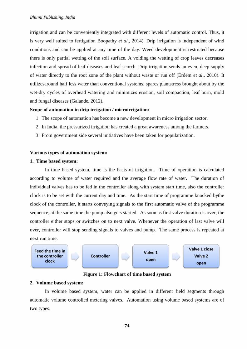
Bhumi Publishing, India
74
irrigation and can be conveniently integrated with different levels of automatic control. Thus, it
is very well suited to fertigation Boopathy et al., 2014). Drip irrigation is independent of wind
conditions and can be applied at any time of the day. Weed development is restricted because
there is only partial wetting of the soil surface. A voiding the wetting of crop leaves decreases
infection and spread of leaf diseases and leaf scorch. Drip irrigation sends an even, deep supply
of water directly to the root zone of the plant without waste or run off (Erdem et al., 2010). It
utilizesaround half less water than conventional systems, spares plantstress brought about by the
wet-dry cycles of overhead watering and minimizes erosion, soil compaction, leaf burn, mold
and fungal diseases (Galande, 2012).
Scope of automation in drip irrigation / microirrigation:
1 The scope of automation has become a new development in micro irrigation sector.
2 In India, the pressurized irrigation has created a great awareness among the farmers.
3 From government side several initiatives have been taken for popularization.
Various types of automation system:
1. Time based system:
In time based system, time is the basis of irrigation. Time of operation is calculated
according to volume of water required and the average flow rate of water. The duration of
individual valves has to be fed in the controller along with system start time, also the controller
clock is to be set with the current day and time. As the start time of programme knocked bythe
clock of the controller, it starts conveying signals to the first automatic valve of the programme
sequence, at the same time the pump also gets started. As soon as first valve duration is over, the
controller either stops or switches on to next valve. Whenever the operation of last valve will
over, controller will stop sending signals to valves and pump. The same process is repeated at
next run time.
Figure 1: Flowchart of time based system
2. Volume based system:
In volume based system, water can be applied in different field segments through
automatic volume controlled metering valves. Automation using volume based systems are of
two types.
Feed the time in the controller
clockController
Valve 1
open
Valve 1 close
Valve 2
open

Frontiers in Life Science (Volume II)
ISBN: 978-81-953600-8-6
75
In first type of system, automatic metering valve with pulse output furnishes one pulse
after completion of one dial in the automatic metering valve. Consequently, it can count the
volume of water passed through by counting the number of pulses received by the controller.
After providing required volume of water through first valve, it closes down and controller
switches on the next valve subsequently.
In second type of system, there is no requirement of any controller. Automatic metering
valves are placed near the field of each segment. In series all automatic metering valves are
interconnected with the help of control tube. Components like T-connector, shuttle valve and a 3-
way relay are also installed along the circuitfor automatic closing and opening of the metering
valves with the help of water pressure signal. In the time of sequential operation only one
automatic metering valve remains open. After closing the first valve, the next valve in the series
opens. Shut down of the irrigation pump can be made automatic after closure of the last valve in
series by connecting the spare end of the last valve T-connector to a microswitch with control
tube. Micro-switch is connected to the pump motor starter‘s magnetic coil. When the last
automatic metering valve closes, it transmits pressure signal to the micro-switch by means of
pressure which in turn activates a pressure switch and terminates the motor starter circuit
resulting in automatic shutdown of irrigation pump.
The major advantage of volume based irrigation system over time-based system is
assurance for delivering the preset amount of water irrespective of continuous availability of
electricity, but time based system is comparatively cheaper and hence gaining more popularity
than the volume based system.
3. Open loop systems
In an open loop system, the operator decides the amountan time of application of water.
This information is programmed into the controller and the water is applied according to the
desired schedule. Open loop control systems uses either the irrigation duration or a specified
applied volume for control purposes. Open loop control systems are cheaper and readily
available. The drawback of open loop systems is that they do not respond automatically to
changing conditions in the environment. In addition, they may require frequent resetting in order
to achieve high irrigation efficiency.
4. Closed loop systems
This system requires feedback from one or more sensors. The operator develops a general
control strategy. After defining general strategy, the control system takes over and makes
decision regarding time and amount of irrigation. Irrigation decisions are made and actions are
Bhumi Publishing, India
76
carried out based on data from sensors. In this type of system, the feedback and control of the
system work continuously. Closed loop controllers require data acquisition of environmental
parameters such as soil moisture, temperature, radiation, wind-speed as well as system
parameters like pressure, flow, etc.
5. Real time feedback system
Real time feedback is based on actual dynamic demand of the plant itself, plant root zone
effectively reflecting all environmental factors acting upon the plant. It operates within
controlled parameters and the plant itself determines the required amount of irrigation. Various
sensors such astensiometers, relative humidity sensors, rain sensors, temperature sensors etc.
control the irrigation scheduling. These sensors provide feedback to the controller to control its
operation.
6. Computer-based irrigation control systems
A computer-based control system consists of a hardware and software that works as a
supervisor for managing irrigation and other related practices such as fertigation and
maintenance. Generally, the computer-based control systems that are used for managing micro
irrigation systems can be divided into two categories:
A. Interactive systems, B. Fully automatic systems.
Closed control loops are used in these systems which include:
Monitoring the state variables that are pressure, flow within the system.
Comparison of state variables with their desirable state.
Decision regarding actions are necessary to change the state of the system.
These functions requires a combination of hardware and software that must be
implemented for each specific application.
7. Interactive Systems:
Interactive systems requires a microcomputer, either a standard personal computer (PC)
or a specially designed unit. The information is transferred into a central unit either directly from
sensors in the pipeline or from intermediate units which collect the data from a number of
sensors and then process and store them temporarily for further transfer to the central computer.
These systems have features that allow the operator to send commands back to different control
units of the irrigation system. The field devices such as valves, regulators, pumpsare fitted with
electrically operated servo-devices which allow actuation of the pumps, closing and opening of
valves, and adjusting pilot valves of flow regulators. This type of system enables the operator to
control the flow from the central computer by controlling flow parameters such as pressure and

Frontiers in Life Science (Volume II)
ISBN: 978-81-953600-8-6
77
flow rate, according to specific needs at the given time, and to receive immediate feedback on
the response of the system.
8. Automatic Systems:
In fully automated systems the human factor is not required and it is replaced by a
computer specifically programmed in order to react appropriately to any changes in the
parameters monitored by sensors. The automatic functions are activated by feedback from field
units and corrections in the flow parameters by control of devices in the irrigation system until
the desired performance level is achieved. Automatic systems can also perform auxiliary
functions like stopping irrigation in case of rain, injecting acid to control pH, sounding alarms,
etc. Most of the control systems include protection in emergencies such as loss of the handled
liquid due to pipe burst.
Intelligent IoT Based Irrigation System:
The automated irrigation system consists of two units are control unit and sensing unit.
The control unit consists of microcontroller which controls the execution of operation and the
sensing unit consists of different sensors such as DHT11 sensor, ARDUINO board and soil
moisture sensor. The DHT11 sensor is used to measure the temperature and humidity of a root
zone of plants in dry and wet conditions. For remote monitoring, GPRS based systems are
employed on wireless sensor networks for monitoring the temperature and humidity data
continuously or periodically (Hui et al., 2008). A threshold can be set such that the
microcontroller unit sends an SMS to the mobile phone of an owner. Threshold must be chosen
in the range of 40 to 30 degrees centigrade, because the most of the values fall on those ranges
during normal and wet conditions (Rajendranath and Hency, 2015). The microcontroller sends
the alert SMS and also send the particular exceed value of a temperature or humidity or soil
moisture value to the owner in consecutive SMS. This kind of system proved that the use of
water can be diminished and hence water will not be wasted and reduces the human resources
(Shekhar et al., 2017). The overall system can be powered up using solar cells to maintain the
system in low cost (Rajendranath and Hency, 2015). This type of system can be extended by
using WSN nodes for transmit data and also using data base systems to store the data at the field.
In Future, an Intelligent IoT based Automated Irrigation system can be extended not just for
irrigating the field with water but also for deciding on spraying appropriate chemicals for proper
growth of crop.

Bhumi Publishing, India
78
Development of automation in drip irrigation system
Agarwal et al. (2014) developed a soil moisture sensor based on the fact that water is not
pure water which is non-conductor, but it is impure which is slightly conductor. Water sensor is
nothing but a series of very close printed circuit boards (PCB) tracks. In normal mode these
tracks do not conduct, but when some water falls on these tracks start conducting slightly, some
positive voltage is available at the base of transistor So negative positive negative (NPN)
transistor is on and NPN transistor provide a negative voltage as a pulse to the microcontroller.
The output voltage of a sensor is amplified by an operational amplifier, and is provided to the
base of transistor. The moisture sensitivity adjusting the gain of an operational amplifier by
variable resistor (VR).
Chandrasekhar and Chakravarthi (2013) developed an automatic drip irrigation system
using low cost sensors and simple circuitry. Irrigation system uses valves to start and stop
irrigation. These valves are automated by using controllers and solenoids. The humidity sensors
are constructed using aluminium sheets and housed in easily available materials. The aim is to
use the easily available material to construct sensors involving low cost. Five relays are
controlled by the microcontroller through the high current driver IC, ULN2003. Four relays help
in controlling four solenoid valves in order to control the flow of water to four different parts of
the field. One relay is used to shut-off the main motor which is used to pump the water to the
field.
Figure 2: Block diagram implementation of an automated irrigation system
Ingale and Kasat (2012) prepared a circuit which is cheap and reliable to develop an
automated irrigation system. The system provides with several benefits and can operate with less
manpower. The system supplies water only when the humidity in the soil goes below the
Humidity Sensor Temperature Sensor
Microcontroller
LCD Interface
Soil Moisture Sensor
Mobile Phone

Frontiers in Life Science (Volume II)
ISBN: 978-81-953600-8-6
79
reference. Due to the direct transfer of water to the roots, water is not wasted and it also helps in
maintaining the moisture to soil ratio at the root zone constant to some extent. Thus the system is
efficient and compatible to thechanging environment. Also the system saves the water and
improves the growth of plants.
Hameed and Agarwala (2009) proposed a novel approach to determine the efficient water
requirement of agricultural fields for farming in a most scientific and cost effective manner and
to design a ultra-low cost moisture sensor using computer and thereby to manage the water
resources more appropriately in agricultural farms for the plants irrigation and its results reveals
that the capacitance response characteristics make it possible to maintain soil moisture at the
desired level as per the crop requirement and its withstanding capacity from its field capacity
level. Due to plant uptake, evapotranspiration, atmospheric temperature, the soil moisture
continues to vary and most of the crops can give a better yield at up to 50% decrease in soil
moisture content from its field capacity. So, in this experiment an effort has been made to use a
sensor to estimate the PWP and FC and a comparative study is made using
tensiometerconcurrently and its being concluded that it may improve a new insight to irrigation
automation and will be a boon to unskilled farmers to optimize their crop yield as well as saving
water, electricity apart from reducing greenhouse gases emission from agricultural fields which
takes place up on full saturation.
Cepuder and Nolz (2007) stated that water is a limited resource, mainly in intensively
used agricultural areas in Austria, where groundwater is used as drinking water as well as for
irrigation purposes. In order to guarantee a sustainable use of irrigation water, soil water
measurement devices can be used to optimise irrigation, which means that controlling the soil
water content in the entire root system may prevent water stress due to water deficiency on the
one hand, and over wetting on the other hand. Furthermore, losses of nutrients due to leaching
can be avoided. Sensors in various depths measure the plant water uptake in the root zone under
standard irrigation practices on different sites and soils, respectively.
Use of automated irrigation system for different crops
Anand and his co-workers investigated the effect of automation irrigation system for
maize in polybags, they collected data on plants of equal height and diameter continuously for 8
days. From Table 1, we can conclude that there is a considerable faster growth was observed
with automation irrigation (34.70 cm) compared with without automation (34.20 cm) in maize.

Bhumi Publishing, India
80
Table 1: Effect of automated irrigation system on maize (Anand et al., 2018)
Day With Automation Without Automation
Plant Height (cm) Diameter (cm) Plant Height (cm) Diameter (cm)
1 34.00 1.75 34.00 1.75
2 34.20 1.76 34.00 1.75
3 34.20 1.76 34.00 1.75
4 34.50 1.77 34.10 1.76
5 34.50 1.78 34.10 1.76
6 34.50 1.78 34.10 1.76
7 34.60 1.78 34.10 1.76
8 34.70 1.79 34.20 1.77
In this automated irrigation system wheat crop was grown under two different water
regimes, 80% and 40% by using humidity sensors. Fig. 3 and 4 show that there was no
significant difference between plant height in automated and manual irrigation but there was a
significant difference in total dry weights both at 80% and 40% of field capacity.
Figure 3: Effect of automated and manual irrigation on plant height (Boutraa et al., 2011)
Figure 4: Effect of automated and manual irrigation on total dry weight (Boutraa et al., 2011)

Frontiers in Life Science (Volume II)
ISBN: 978-81-953600-8-6
81
As a result of this two-year field studied for wheat crop under arid region, it was
concluded that the IIS method under sprinkler irrigation offered significant advantage for
seasons, its easiness application and more water saving (Table 2). The ICS treatment resulting in
greater yield than IIS could be attributed to variation of amount of water added to the two
treatments. Therefore, the IIS irrigation method would be recommended due to its easiness
application and more water saving. The IIS technique conserved irrigation water by 26% more
than that provided by control system ICS.
Table 2: Comparison of yield components and efficiencies using sprinkler irrigation
systems for both intelligent irrigation (IIS) and control (ICS) (Ghobari et al., 2013)
Lailhacar et al. (2008) noticed that the modern technologies could able to improve
irrigation efficiency of turfgrass, encouraging water conservation and decreasing environmental
impacts. The objectives of thier research was to compute irrigation water use and to assess turf
quality differences between (1) Time-based scheduling with and without a rain sensor (RS); (2)
A time-based schedule compared to a soil moisture sensor (SMS) based irrigation system; and
(3) Different commercially available SMS systems. SMS based treatments comprised of
irrigating one, two, or seven days a week, each with four different commercial SMS brands.
Time-based treatments with or without RS and a non-irrigated treatment were also executed.
Remarkable differences in turfgrass quality among treatments were not detected due to the
sustained wet weather conditions during the testing periods. The treatment with the rain sensor
resulted in 34% less water applied than without the rain sensor (2-WORS) treatment. Most of
SMS brands recorded irrigation water savings as compared to 2-WORS, ranging from 69 to 92%
for three of four SMSs tested, depending on the irrigation frequency. Therefore, SMS systems
represent a promising technology because of the water savings that they can achieve during
application.

Bhumi Publishing, India
82
Conclusion:
The automation of the irrigation process is important for three main reasons: scarcity of
water, timely irrigation and maximum crop profit. Automated irrigation system permits
cultivation in places with water scarcity thereby improving sustainability as well as maintains
optimum soil moisture levels required for crop. This reduces the human Automated errors in
maintain soil moisture and nutrients at optimum levels, there by increases yield with less cost of
production as cost on workforce will be cut down. Automated system components require
minimum maintenance. Reductions in water use range as high as 70% compared to farmer
practices with no negative impact on crop yields. Automatic irrigation systems presently
available are costly and are not adopted by most of the Indian farmers. To apply simple
electronic circuit principles in irrigation an attempt has been made to develop low cost automated
drip irrigation based on soil moisture.
References:
Abdul, H.V.M., Abhilash, J.E., Ajay, G.A.J. and Mufeedha, K. (2016). Fertigation: A novel and
efficient means for fertilizer. International Journal of Current Research, 8(8), 35757-35759.
Agarwal, S., Pandey, S., Kumar, S. and Chaudhery, K. (2014). Automatic irrigation system.
International Journal of Advance Research and Innovation, 1, 92-94.
Anand, B.A., Reddy, V.H.K. and Raj, S.B.A. (2018). Automation irrigation system for
floriculture, vegetable and cereal crops. International Journal of Current Microbiology and
Applied Sciences, 7(9), 859-866.
Anjaly, C.S. and Hakkim, V.M.A.(2017).Automated and non-automated fertigation systems
inside the polyhouse- a comparative evaluation. International Journal of Current
Microbiology and Applied Science, 6(5), 2328-2335.
Boopathy, S., Kumar, N.R., Premkumar, P. and Govindaraju, P. (2014). Implementation of
automatic fertigation system by measuring the plant parameters. International Journal of
Engineering Research and Technology, 3(10), 583-586.
Boutraa, T., Akhkha, A.,Alshuaibi, A. and Atta, R. (2011). Evaluation of the effectiveness of an
automated irrigation system using wheat crops. Agriculture and Biology Journal of North
America, 2(1), 80-88.
Cepuder, P. and Nolz, R. (2007). Irrigation management by means of soil moisture sensor
technologies. Journal of Water and Land Development, 11, 79-90.
Chandrasekhar, T. and Chakravarthi, J.S. (2013). Automatic drip irrigation system. International
Journal of Communications Networking System, 2(1), 153-157.
Edordu, C. and Sacks, L. (2006). Self Organizing Wireless Sensor Networks as a Land
Management Tool in Developing Countries: A Preliminary Survey. Proceedings of the

Frontiers in Life Science (Volume II)
ISBN: 978-81-953600-8-6
83
2006 London Communications Symposium, September 2006, Communications
Engineering Doctorate Centre, London, UK.
Erdem, Y., Arin, L., Erdem, T., Polat, S., Deveci, M., Okursoy, H. and Gültas, H. T. (2010).
Crop water stress index forassessing irrigation scheduling of drip irrigated broccoli
(Brassica oleracea L. var. Italic), Agriculture. Water Management, 98(1), 148-156.
Galande, S. (2012). Agricultured automatic drip irrigation system. IOSR Journal of Agriculture
and Veterinary Science, 1, 24-27.
Ghobari, H.M.Al., Mohammad, F.S. and Marazky, M.S.A.El. (2013). Effect of intelligent
irrigation on water use efficiency of wheat crop in arid region.The Journal of Animal &
Plant Sciences, 23(6), 1691-1699.
Hagin, J. and Lowengart, A. (1996). Fertigation for minimizing environmental pollution by
fertilizers. Fertilizer Research, 43, 5-7.
Hameed, S.K.S.S. and Agarwala, A.K. (2009). Computer aided design of soil moisture sensor
and its measurements for agricultural water resources management. International Journal
on Design and Manufacturing technologies, 3, 38-44.
Ingale, H. and Kasat, N.N. (2012). Automated solar based agriculture pumping. International
Journal of Advanced Research in Computer Science and Software Engineering, 2(11), 407-
410.
Lailhacar, C.B., Dukes, P.E.M.D. and Miller, G.L. (2008). Sensor based automation of irrigation
on bermudagrass during wet weather conditions. Journal of Irrigation and Drainage
Engineering (ASCE), 134(2), 120-128.
Liu, Hui., Wang, Mao-hua., Wang, Yue-xuan., Ma. Dao-kun. and Li, Hai-xia (2008).
Development of Farmland Soil Moisture and Temperature Monitoring system based on
Wireless sensor networks. Journal of Jilin University, Engineering and Technology
Edition.
Phuntsho, S., Shon, H.K., Hong, S., Lee, S. and Vigneswaran, S. (2011). A novel low energy
fertilizer driven forward osmosis desalination for direct fertigation: evaluating the
performance of fertilizer draw solutions. Journal of Membrane Science, 375, 172-181.
Rajendranath, U.N.V.P. and Berlin, H.V. (2015). Implementation of an automated irrigation
system: Smart irrigation system. Proceedings of IEEE Sponsored 2nd
International
Conference on Innovations in Information, Embedded and Communication systems
(ICIIECS) 2015.
Shekhar, Y., Dagur, E., Mishra, S., Tom, R.J., Veeramanikandan, M. and Sankaranarayanan, S.S.
(2017). Intelligent IoT Based Automated Irrigation System. International Journal of
Applied Engineering Research. 12(18), 7306-7320.

Bhumi Publishing, India
84
STUDIES ON SOME PROTOZOAN PARASITES OF
INDIAN GOAT CAPRA INDICA
Khan Rumana Shahin Amanullah
J. A. T Arts, Science and Commerce College (For Women),
Malegaon, Maharashtra, India
Corresponding authors E-mail: [email protected]
Abstract:
Goats and sheep are the important source of animal protein. They are primarily useful for
meat and wool production. They also produce a considerable amount of manure, which is of
special importance in those areas where cattle are of lesser importance. Parasites infections of
goats are major factor responsible for economic losses through reduction in productivity and
increased mortality. Goats harbor a variety of gastrointestinal parasites that affect the growth as
well as production of animal.
Keyword: Protozaon parasites, Capra indica.
Introduction:
Most of the protozoans are highly pathogenic to their vertebrate hosts and hence are of
concern to veterinary and human medicine. In subkingdom protozoa coccidia is included in
phylum Apicomplexa. The phylum Apicomplexa was established to include protozoans that
possess a certain combination of structures collectively known as the apical complex which is
distinguishable by electron microscopy.Coccidia are microscopic, spore-forming, obligate, and
intracellular parasites, they must live and reproduce within an animal cell, belonging to the class
Sporozoea according to revisedclassification.
Coccidian parasites infect the intestinal tracts of animals, and are largest group of
protozoa. Eimeria is a genus of apicomplexan parasites that includes various species responsible
for the disease coccidiosis. These species cause pathological damage and mortality in poultry,
cattle, sheep, goat, pig, rabbit and other animals. The genus Eimeria (Schneider, 1975) with
more than 1400 species described to date, is the largest genus, and may be the most specious
genus of all animal genera. Coccidia have a complex life cycle and other unusual characteristics
which have led to investigations by increasing number ofbiologists.

Frontiers in Life Science (Volume II)
ISBN: 978-81-953600-8-6
85
Coccidiosis:
In India studies on Coccidiosis have been relatively scanty. Mandal (1975) in review on
Fifty years progress in the taxonomy of coccidian (Protozoa: Sporozoa) from India, estimated
that about 200 species of coccida spread over seventeen genera were described till then this
information is thus very negligible. Considering the very large size of our country and the
endless variety of animal fauna that exists here is very mearge. The necessity for basic
taxonomic studies is also reflected in the statement of Levine (1973) that Eimeriahad been
described only from 1.2% of the world‘s chordates and 5.7% of the world‘s mammals. He also
estimated that if all chordates were examined perhaps atleast 34000 species of Eimeria might be
found and that 3,500 of them would be from mammals. In India the studies on the coccidia of
invertebrates have been carried out mostly in west Bengal and Andhra Pradesh.
The few contributions on the coccidia of fishes, Amphibia and Reptiles are also from
west Bengal. While a contributions on the coccidia of birds have come from Bengal and Uttar
Pradesh. The mammalian coccidia study was done from the veterinary laboratories at Bombay,
Calcutta, Mathura, and Izatnagar. During the last ten years more and more workers have been
attracted to this area and many useful contributions have been made from the different parts of
the Northern India. Comprehensive studies on the incidence of coccidia have been made by
workers from the protozoology laboratory of the Dr. Babasaheb Ambedkar Marathwada
University, about 40 years ago. During this period six major contributions have been made on
the coccidian fauna of the Marathwada region.
The first contribution covered the systematics of avian coccidia. The second deals with
the prevalence and systematics of the coccidia in goats of Parbhani district, besides a
taxonomic survey of Coccidia of rats and Squirrels.
The third contribution made an extensive analysis of the prevalence and systematics of
the sheep and goats of this region. In this series fourth attempt covers a comparative study of
the Coccidia of Goats and Sheep in the Marathwada region, beside a taxonomical account of the
coccidia of rabbits in this area. Fifth research in this series deals with the prevalence of
incidence and morphological study of the coccidia of buffaloes and goats. Sixth deals with
study of coccidiosis in chicken in Aurangabad District. Seventh in this series is comparative
study of species composition of coccidia in sheep and goats in Beed district (M.S). The present
author worked on studies on some endoparasites of Capra indica (Indian goats).
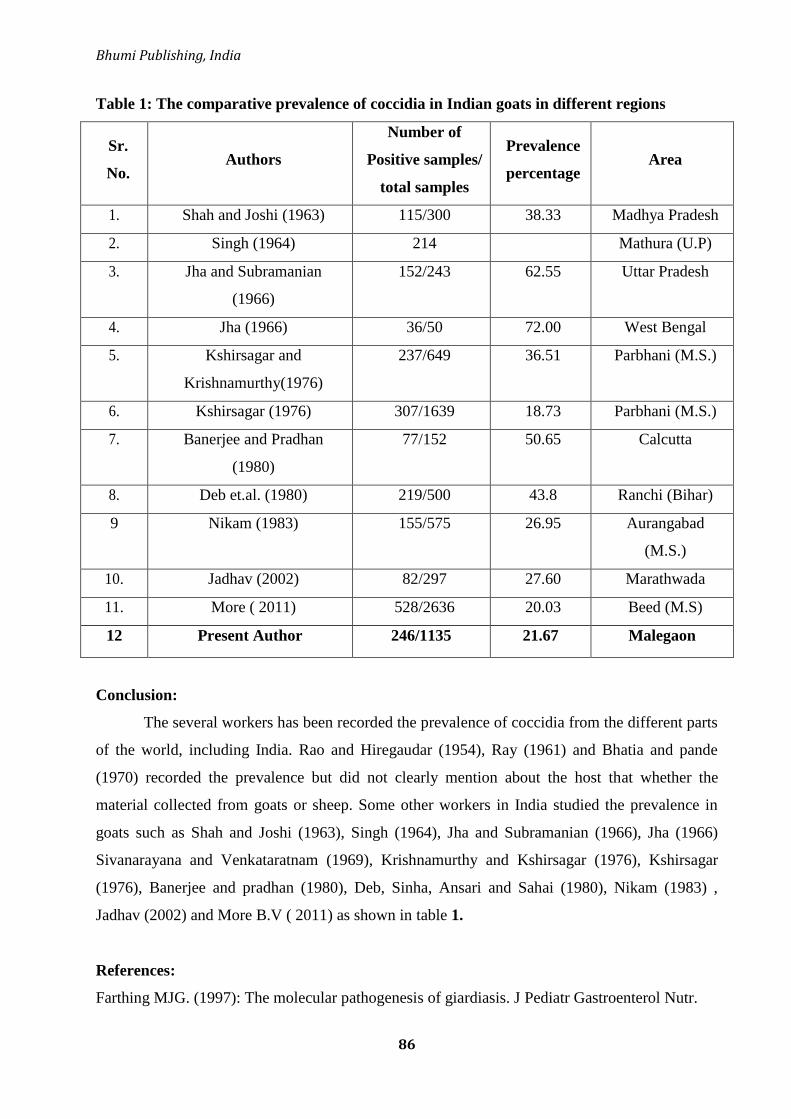
Bhumi Publishing, India
86
Table 1: The comparative prevalence of coccidia in Indian goats in different regions
Sr.
No. Authors
Number of
Positive samples/
total samples
Prevalence
percentage Area
1. Shah and Joshi (1963) 115/300 38.33 Madhya Pradesh
2. Singh (1964) 214 Mathura (U.P)
3. Jha and Subramanian
(1966)
152/243 62.55 Uttar Pradesh
4. Jha (1966) 36/50 72.00 West Bengal
5. Kshirsagar and
Krishnamurthy(1976)
237/649 36.51 Parbhani (M.S.)
6. Kshirsagar (1976) 307/1639 18.73 Parbhani (M.S.)
7. Banerjee and Pradhan
(1980)
77/152 50.65 Calcutta
8. Deb et.al. (1980) 219/500 43.8 Ranchi (Bihar)
9 Nikam (1983) 155/575 26.95 Aurangabad
(M.S.)
10. Jadhav (2002) 82/297 27.60 Marathwada
11. More ( 2011) 528/2636 20.03 Beed (M.S)
12 Present Author 246/1135 21.67 Malegaon
Conclusion:
The several workers has been recorded the prevalence of coccidia from the different parts
of the world, including India. Rao and Hiregaudar (1954), Ray (1961) and Bhatia and pande
(1970) recorded the prevalence but did not clearly mention about the host that whether the
material collected from goats or sheep. Some other workers in India studied the prevalence in
goats such as Shah and Joshi (1963), Singh (1964), Jha and Subramanian (1966), Jha (1966)
Sivanarayana and Venkataratnam (1969), Krishnamurthy and Kshirsagar (1976), Kshirsagar
(1976), Banerjee and pradhan (1980), Deb, Sinha, Ansari and Sahai (1980), Nikam (1983) ,
Jadhav (2002) and More B.V ( 2011) as shown in table 1.
References:
Farthing MJG. (1997): The molecular pathogenesis of giardiasis. J Pediatr Gastroenterol Nutr.
Frontiers in Life Science (Volume II)
ISBN: 978-81-953600-8-6
87
Levine (1961): Protozoan parasites of domestic animals and of man. Burgess Publishing
Company. Minnesota. 412
Levine, N. D. (1961): Protozoan parasites of domestic animals and of man IEd. Burgess Publ.
co. Minneapolis, Minnesota.
Levine, N. D. (1973): Protozoan parasites of domestic animals and man II Ed.Burgess Publishing
Company. Minneapolis, Minnesota.
Nikam (1983): Studies on the protozoan parasites of some mammals. Ph.D. Thesis, Marathwada
University Library Aurangabad.
Paul S, Sharma DK, Boral R, Mishra AK, Nayakwadi S, Banerjee PS, Pawaiya RS. (2005):
Cryptosporidiosis in goats: a review.
Pollott G and Wilson RT (2009): Sheep and goats for diverse products and profits. FAO
diversification booklet.
Robertson LJ. (2009): Giardia and Cryptosporidium infections in sheep and goats: a review of
the potential for transmission to humans via environmental contamination. Epidemiol
Infect.
Soulsby, E. J. L. (1978). Helminths, arthropods and protozoa of domesticated animals. The
English Language Book Society and Bailliere, Tindall and cassell Ltd. pp. 824.
Thompson RCA, Palmer CS, O‘handley R. (2008): The public health and clinical significance
of Giardia and Crypt sporidium in domestic animals.
Bhumi Publishing, India
88
CHEMICAL ANALYSIS OF SUMMER HONEYS COLLECTED FROM APIS
DORSATA HIVES OF CHIMUR TAHSIL OF CHANDRAPUR DISTRICT
OF MAHARASHTRA STATE (INDIA)
Laxmikant N. Borkar1* and Devendra M. Mate2
1Department of Botany,
S. S. Jaiswal Arts, Comm. and Science College,
Arjuni (Mor), Gondia, India.
2Department of Botany,
Nutan Adarsh Arts, Commerce and M. H. Wegad Science College,
Umrer, Dist – Nagpur, India
*Corresponding authors E-mail: [email protected]
Abstract:
The present investigation was undertaken to determine the chemical analysis of 5 summer
honey samples (CHN-CHI-ALL, CHN-CHI-TAL-N, CHN-CHI-JAM, CHN-CHI-TUK, CHN-
CHI-BOR) collected from forest area of Chimur Tahsil of Chandrapur District of Maharashtra
State (India). These samples were analysed for several parameters such as moisture, total
reducing sugar, Levulose or Fructose, Dextrose or Glucose, L/D ratio, Sucrose, Acidity. This
type of chemical analysis favours the utilization of the honey for good quality in this area.
Keywords: Chemical Analysis, Summer Honey, Chimur Tahsil.
Introduction:
Honey is a natural product rich in carbohydrates, extracted from nectar by bees. Honey
has been used as nutritious food and traditional medicine by all civilizations. The quality of
honey depends on many physiological factors such as climate and soil. Sugar, protein, moisture,
vitamins, minerals, enzymes, polyphenols, and flavonoids (Al-Manary et al., 2002) Due to this
unique and complex nature, honey has been shown to be useful for treating burns, wounds, and
skin ulcers Antioxidants, external eye disease (Balasubramanyam, 2011). In addition, honey is a
very valuable ingredient in spices, beverages, sauces and candies. In fact, many studies on the
physical, chemical and melisopalinological parameters of honey have been published around the
world. (Adenken et al., 2010; Anklam, 1998; Cherian et al., 2011; Borkar Laxmikant and Mate
Devendra, 2014; Downey et al., 2005; Ramnath nad Shivaramm, 2012, Terrab et al., 2002;
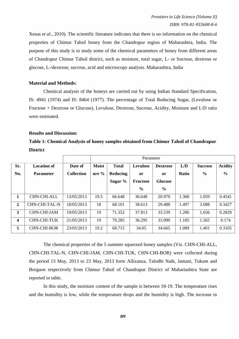
Frontiers in Life Science (Volume II)
ISBN: 978-81-953600-8-6
89
Xesus et al., 2010). The scientific literature indicates that there is no information on the chemical
properties of Chimur Tahsil honey from the Chandrapur region of Maharashtra, India. The
purpose of this study is to study some of the chemical parameters of honey from different areas
of Chandrapur Chimur Tahsil district, such as moisture, total sugar, L- or fructose, dextrose or
glucose, L-/dextrose, sucrose, acid and microscopy analysis. Maharashtra, India
Material and Methods:
Chemical analysis of the honeys are carried out by using Indian Standard Specification,
IS: 4941 (1974) and IS: 8464 (1977). The percentage of Total Reducing Sugar, (Levulose or
Fructose + Dextrose or Glucose), Levulose, Dextrose, Sucrose, Acidity, Moisture and L/D ratio
were estimated.
Results and Discussion:
Table 1: Chemical Analysis of honey samples obtained from Chimur Tahsil of Chandrapur
District
Parameter
Sr.
No.
Location of
Parameter
Date of
Collection
Moist
ure %
Total
Reducing
Sugar %
Levulose
or
Fructose
%
Dextrose
or
Glucose
%
L/D
Ratio
Sucrose
%
Acidity
%
1 CHN-CHI-ALL 13/05/2013 19.5 66.648 36.648 20.970 1.368 1.059 0.4541
2 CHN-CHI-TAL-N 18/05/2013 18 68.101 38.613 29.488 1.497 3.088 0.3427
3 CHN-CHI-JAM 19/05/2013 19 71.352 37.813 33.539 1.286 1.656 0.2829
4 CHN-CHI-TUK 21/05/2013 19 70.285 36.295 33.990 1.185 1.565 0.174
5 CHN-CHI-BOR 23/05/2013 19.2 68.715 34.05 34.665 1.089 1.401 0.3105
The chemical properties of the 5 summer squeezed honey samples (Viz. CHN-CHI-ALL,
CHN-CHI-TAL-N, CHN-CHI-JAM, CHN-CHI-TUK, CHN-CHI-BOR) were collected during
the period 13 May, 2013 to 23 May, 2013 form Allizanza, Talodhi Naik, Jamani, Tukum and
Borgaon respectively from Chimur Tahsil of Chandrapur District of Mahartashtra State are
reported in table.
In this study, the moisture content of the sample is between 18-19. The temperature rises
and the humidity is low, while the temperature drops and the humidity is high. The increase in
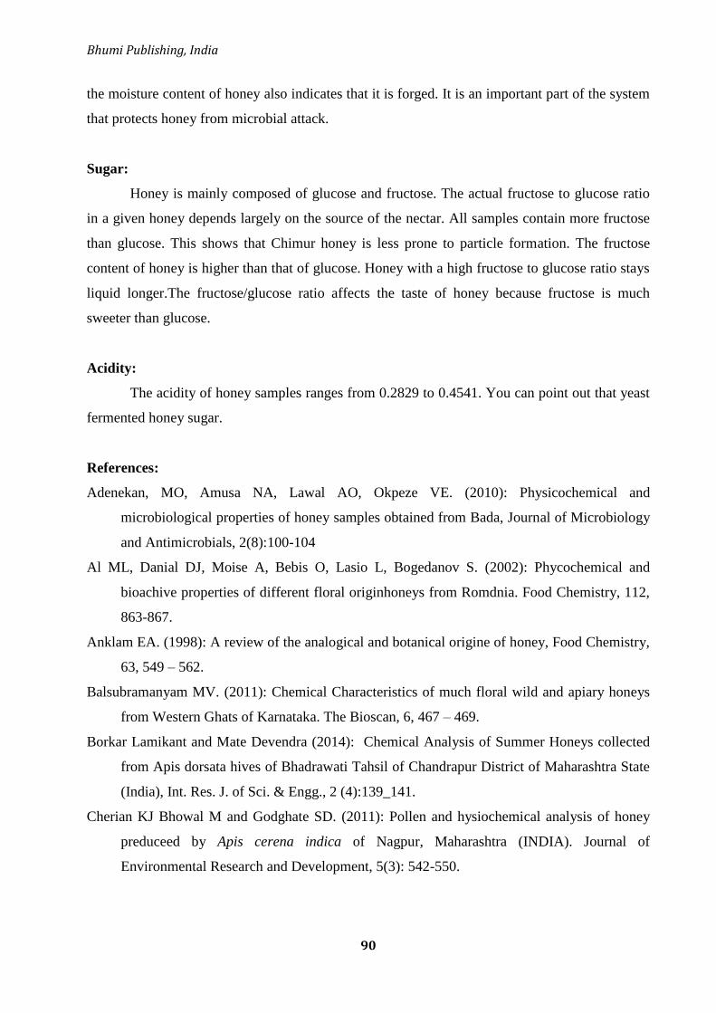
Bhumi Publishing, India
90
the moisture content of honey also indicates that it is forged. It is an important part of the system
that protects honey from microbial attack.
Sugar:
Honey is mainly composed of glucose and fructose. The actual fructose to glucose ratio
in a given honey depends largely on the source of the nectar. All samples contain more fructose
than glucose. This shows that Chimur honey is less prone to particle formation. The fructose
content of honey is higher than that of glucose. Honey with a high fructose to glucose ratio stays
liquid longer.The fructose/glucose ratio affects the taste of honey because fructose is much
sweeter than glucose.
Acidity:
The acidity of honey samples ranges from 0.2829 to 0.4541. You can point out that yeast
fermented honey sugar.
References:
Adenekan, MO, Amusa NA, Lawal AO, Okpeze VE. (2010): Physicochemical and
microbiological properties of honey samples obtained from Bada, Journal of Microbiology
and Antimicrobials, 2(8):100-104
Al ML, Danial DJ, Moise A, Bebis O, Lasio L, Bogedanov S. (2002): Phycochemical and
bioachive properties of different floral originhoneys from Romdnia. Food Chemistry, 112,
863-867.
Anklam EA. (1998): A review of the analogical and botanical origine of honey, Food Chemistry,
63, 549 – 562.
Balsubramanyam MV. (2011): Chemical Characteristics of much floral wild and apiary honeys
from Western Ghats of Karnataka. The Bioscan, 6, 467 – 469.
Borkar Lamikant and Mate Devendra (2014): Chemical Analysis of Summer Honeys collected
from Apis dorsata hives of Bhadrawati Tahsil of Chandrapur District of Maharashtra State
(India), Int. Res. J. of Sci. & Engg., 2 (4):139_141.
Cherian KJ Bhowal M and Godghate SD. (2011): Pollen and hysiochemical analysis of honey
preduceed by Apis cerena indica of Nagpur, Maharashtra (INDIA). Journal of
Environmental Research and Development, 5(3): 542-550.
Frontiers in Life Science (Volume II)
ISBN: 978-81-953600-8-6
91
Downey GJ, Hussey K, Kelly JD, Walshe TF and Martin PG. (2005): Preliminary contribution to
the characteristics of artisanal honey produced on the island of Ireland by palynological
and physico – chemical data. Food Chemistry, 91, 347-354.
IS: 4941-1974 Indian Standard Specification for extracted honey (First Revision), Indian
Standards Institution, 1974, New Delhi : 1-16.
IS: 8464-1977 Indian Standard Specification for Squeezed honey, Indian Standards Institution,
1977, New Delhi : 1-8.
Ramnath Subharani and Venkataramegouda Sivaramm (2012): Physicochemical and pollen
analysis of Western ghats honey of Karnataka south, India. I.J. S.N., 3(4):831- 835
Terrab AJ, Diez MJ and Heredia FJ. (2002): Characterization of Moroccan unifloral honeys by
theis physicochemical characteristics. Food Chemistry, 79, 373 – 379.
Xesus FJ, Jose P, Maria LE, Antonio I and Jose PA. (2010): Palynological and physicochemical
data charctrazation of honeys produced in the Entre – Douro e Munho region of Portugal,
International Journal of food Science and Technology, 45,: 1255-1262.

Bhumi Publishing, India
92
STUDY ON SOCIO-ECONOMIC STATUS OF FISH FARMERS
IN CHIKKAMAGALURU DISTRICT, KARNATAKA-INDIA
Prapthi U. B. and Annapurneshwari H*
Department of Zoology,
I D S G Government College,
Chikkamaguluru 577102, Karnataka, INDIA
*Corresponding authors E-mail: [email protected]
Abstract:
Fish farming practices also hold promises for many small farmers and potential
significant benefit for strengthening the socio-economic status of the people those who are
directly or indirectly connected with exploitation, production and processing of fish. The present
study is an attempt to understand the socio-economic condition of fish farmers in the
Chikkamagaluru district, Karnataka. A total of 10 fish farmers from each selected 10 villages of
the Chikkmagaluru district, were interviewed to collect the information with a pre-tested
structural questionnaire. Socio-economicstatus namely, family size, age, gender, marital Status,
Education, Type of occupation, Experience, Income, Utilization of supplementary fish feed and
Involvement in fish culture were collected and calculated on percentage basis. It was observed
that the highest number of fish farmer‘s age were above 20 up to 60 years (75%). It was found
that 35% of farmers were belonging to the medium sized family. It was found that 35% of
farmers were belonging to the medium sized family. Thus it can be concluded that fish farming
is a profitable business that can help the fish farmers to uplift their livelihood as well as
economic status.
Keywords: Socio –economic status, fish farming, family size, marital status.
Introduction:
India is the second largest producer of fish next to China. In India, aquaculture constitutes
about 5% of the global fish production and 3% of the global fish trade. India is one of the major
fish producing countries in the world hiring over seven million people in fishing and associated
industries and contributing 60 crores annual1y to national income and mainly generating
employment for rural people (Gadage, 2005). Fisheries sector also supports canneries and
processing establishments, gears and equipment manufacturers, boat yards, refrigeration and ice

Frontiers in Life Science (Volume II)
ISBN: 978-81-953600-8-6
93
making plants and transport services in addition to those working in State fisheries Department,
Fisheries Corporation and other Government based fisheries institutions (Bias, 2018). Fisheries
development in India has made considerable progress over the successive Five Year Plan
periods.
Fish protein is easily digestible and has proved growth promoting value for human
consumption. The fish protein comprises all the ten essential amino acids, minerals, vitamins and
fat. With increase in human population, shortage of human dietary protein occurs, thus this
shortage can be fulfilled by fish protein, mainly in developing countries, where protein shortage
is serious. But, this goal may be difficult due to the high cost of good quality fish meals which
costs about 40-60% of total operating costs in intensive aquaculture enterprises.In present-day
economic, environmental and social styles are pushing farmer to look for viable options to
accomplish the nutritional requirement for the growing population.
A fisheries sector have significant role in the national economy through improved food
supply, employment and income, and contributes 1.5% to total GDP and 4.3% to the GDP from
agriculture. Mainly, Karnataka has rich and varied inland water resources in the forms of ponds,
tanks, reservoirs, rivers and lakes which offer great potential for freshwater aquaculture
development. The state ranks tenth in the country in terms of inland fish productionAnd
contribute about 3.3% to the Indian inland fish production. Both fish and seed production in the
state have been rising gradually in the recent year.
Fish farming practices also hold promises for many small farmers and potential significant
benefit for strengthening the socio-economic status of the people those who are directly or
indirectly connected with exploitation, production and processing of fish.
Study of socio-economic status of fish farmers is important because of the fact that on the
one hand it has influence on the farming practice implemented by the farmer and on the other
hand it is the outcome of the farming practices and performance. In the earlier year, fish
practice was totally dependent on the captured fishes but now culture fish practice has also
started. Study on the economics and technical bottlenecks of fish culture on the regional basis is
highly necessary. The present study is an attempt to understand the socio-economic condition of
fish farmers in the Chikkamagaluru district, Karnataka, India. This understanding is important
to trigger the policy measures in the right direction.
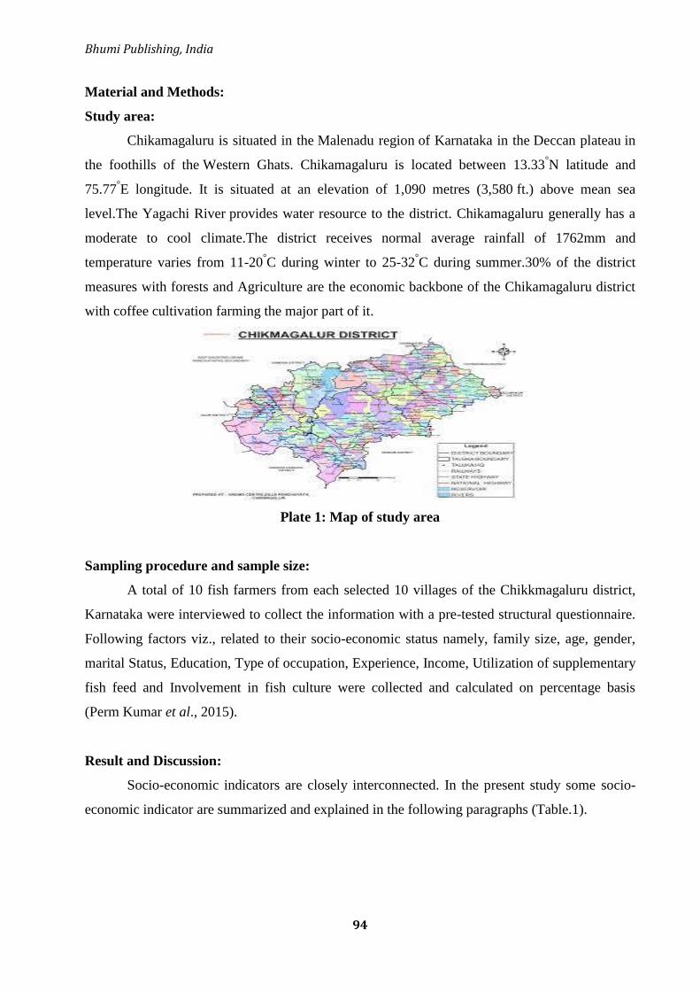
Bhumi Publishing, India
94
Material and Methods:
Study area:
Chikamagaluru is situated in the Malenadu region of Karnataka in the Deccan plateau in
the foothills of the Western Ghats. Chikamagaluru is located between 13.33°N latitude and
75.77°E longitude. It is situated at an elevation of 1,090 metres (3,580 ft.) above mean sea
level.The Yagachi River provides water resource to the district. Chikamagaluru generally has a
moderate to cool climate.The district receives normal average rainfall of 1762mm and
temperature varies from 11-20°C during winter to 25-32
°C during summer.30% of the district
measures with forests and Agriculture are the economic backbone of the Chikamagaluru district
with coffee cultivation farming the major part of it.
Plate 1: Map of study area
Sampling procedure and sample size:
A total of 10 fish farmers from each selected 10 villages of the Chikkmagaluru district,
Karnataka were interviewed to collect the information with a pre-tested structural questionnaire.
Following factors viz., related to their socio-economic status namely, family size, age, gender,
marital Status, Education, Type of occupation, Experience, Income, Utilization of supplementary
fish feed and Involvement in fish culture were collected and calculated on percentage basis
(Perm Kumar et al., 2015).
Result and Discussion:
Socio-economic indicators are closely interconnected. In the present study some socio-
economic indicator are summarized and explained in the following paragraphs (Table.1).

Frontiers in Life Science (Volume II)
ISBN: 978-81-953600-8-6
95
Table 1: Socioeconomic profile of fish farmers in Chikkamagaluru district, Karnataka
Sr. No. Variable Percentage
1. Family size:
1. Small size (<4 members)
2. Medium size (4-6members)
3. Large size (>6members)
-
62.5
37.5
2. Age:
1. Below 20
2. Above 20 [up to 60]
3. Above 60
-
75
25
3. Gender:
1. Male
2. Female
100
-
4. Marital status: 1. Single
2. Married
25
75
5. Education: 1. Primary (1th -5th)
2. Secondary ( 5th
-10th
)
3. Higher Secondary (10th-
PU)
4. Above
12.5
37.5
38
12
6. Type occupation: (Fish rearing)
1. Part time
2. Full time
100
-
7. Experience in fish rearing :
1. Less than 5years
2. 5-10 years
3. More than 10 years
37
25
37.5
8. Income category:
1. High income ( >36000 )
2. Middle income ( 12-36000)
3. Low income ( >10000 )
25
75
-
9. Type of fish culture:
1. Monoculture
2. Integrated
-
100
10. Utilization of supplementary fish feeds :
1. User
2. Non-User
60
40
11. Involvement in fish culture:
1. Regular
2. Occasional
3. No involvement
62.5
37.5
-

Bhumi Publishing, India
96
Family size:
Family size is significance socio-economic indicator as it reflects the income, food
consumption and socio-economic status of the families (Hussain et al., 2009). The present study
was categorized in three family size groups viz., small family (<4 members), medium family (4-
6 members) and large family (>6 members). Majority (62%) of farmers belong to medium
family (Sen and Roy, 2015). (Table 1) which is followed by small family and large family
(38%) (Fig.1)
Figure 1: Family size
Age structure:
Understanding of age structure of fish farmers play a major role in calculating potential
productive human resources.In the present study, the average age of fish farmers was found to be
46years ranging from a minimum of 20years to a maximum of 65 years. Majority of fish farmers
(75%) are middle age group between 20 to 60 years which is followed by old age group (above
60) registering 25% of the total farmers (Fig.2).
Figure 2: Age structure
Small0%
Medium
62%
Large38%
0%Small
Medium
Large
Below 200%
Above 20
75%
Above 60
25% Below 20
Above 20
Above 60

Frontiers in Life Science (Volume II)
ISBN: 978-81-953600-8-6
97
Gender:
In spite of the efforts of fisheries agencies to encourage for fish farming and farming
communities, gender inequity is seen.The present study represents that there were no women
involved in fish farming in the selected area. Only men involved in fisheries activity i.e. 100% of
the total data collected (Fig.3).
Figure 3: Gender data
Marital Status:
Fishery activities are energy demanding jobs, so the farmers should be physically
strong.In the present study, majority (75%) of the farmers interviewed in this study were married
and the remaining 25% were unmarried (Fig.4).
Figure 4: Marital status
Education:
Education and literacy are considered to be good indicators of development in a
society.Fishing activity requires knowledge, skill developments, production technology and
marketing practices (Sen and Roy, 2015). In the present study, maximum number of individuals
were literates. Some of them have studied up to primary levels (around 12.5%) (Table 1). Hardly
a few individuals have studied up to secondary (5th
– 10th
) (37%).The maximum number of fish
male100%
female
0%0%0% male
female
Single25%
Married75%
0% Single
Married

Bhumi Publishing, India
98
farmers associated with the categories studied above 10th
(38%)and 12% of them were degree
holders. However, this study shows that the majority of farmers have secondary and higher
secondary education. This study represents that the young generation are stepping towards
higher education despite fish culture activities and other agricultural activities(Fig.5).
Figure 5: Education
Occupation:
The standard of living and earning of fish farmers depends on their occupation (Goswami,
et al. 2002).Main stream occupation respondents in the present study was agriculture and
associated activities. In the present study, data shows 100% of the farmers were involved in both
fish farming as well as agriculture practice. In the study area, the majorities of the fish farmers
were involved in agricultural farming as a principal occupation (38%), followed by business
including small trading and shop keeping (26%). Only 6% of the pond fish farmers were
involved in fish farming as their principal occupation. 36% of the respondents stated that, they
were involved infish farming as their secondary occupation to secure their livelihood (Fig.6). In
the study, only primary occupation was insufficient to provide adequate means of livelihood.
Figure 6: Occupation
Experience:
Experience is most important indicator in the fisheries sector. It helps in improvement of
traditional knowledge and techniques to getting good output.In the present study, 37.5% of
Primary12%
Secondary38%
Higher38%
Above12%
Education
Primary
Secondary
Higher
Above
Part Time100%
Full time0%0%0% Part Time
Full time
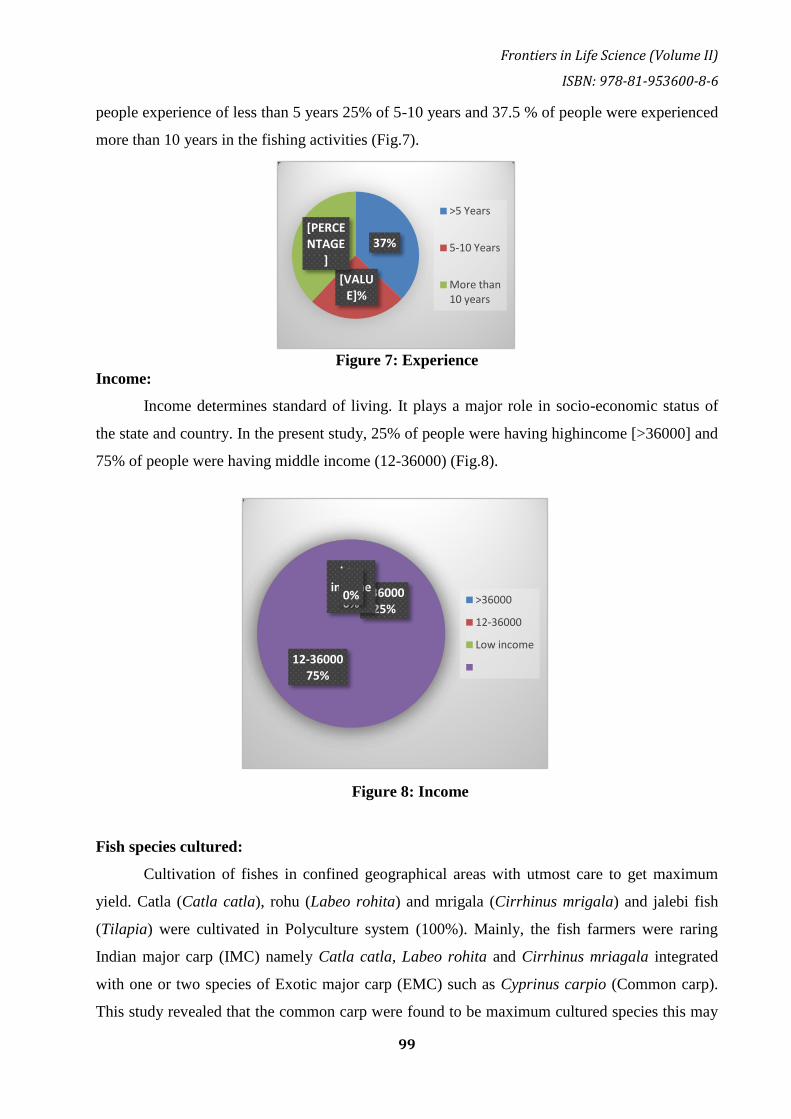
Frontiers in Life Science (Volume II)
ISBN: 978-81-953600-8-6
99
people experience of less than 5 years 25% of 5-10 years and 37.5 % of people were experienced
more than 10 years in the fishing activities (Fig.7).
Figure 7: Experience
Income:
Income determines standard of living. It plays a major role in socio-economic status of
the state and country. In the present study, 25% of people were having highincome [>36000] and
75% of people were having middle income (12-36000) (Fig.8).
Figure 8: Income
Fish species cultured:
Cultivation of fishes in confined geographical areas with utmost care to get maximum
yield. Catla (Catla catla), rohu (Labeo rohita) and mrigala (Cirrhinus mrigala) and jalebi fish
(Tilapia) were cultivated in Polyculture system (100%). Mainly, the fish farmers were raring
Indian major carp (IMC) namely Catla catla, Labeo rohita and Cirrhinus mriagala integrated
with one or two species of Exotic major carp (EMC) such as Cyprinus carpio (Common carp).
This study revealed that the common carp were found to be maximum cultured species this may
37%
[VALUE]%
[PERCENTAGE
]
>5 Years
5-10 Years
More than 10 years
>3600025%
12-3600075%
Low income
0%0% >36000
12-36000
Low income

Bhumi Publishing, India
100
be due to consumer preference, faster growth rate, and low mortality, disease resistant and easy
to culture with low input.
Figure 9: Type of fish culture
Utilization fish feed:
The fish farmers were using different types of organic andInorganic fertilizers toenrich
the natural planktons. However, It was found that 60% of the farmers applied supplementary
and homemade feed prepared with rice-bran and ground nut oil cake and about 40% of the
farmers were entirely dependent on natural food (Fig.10).
Figure 10: Utilization
Involvement in fish culture:
The involvement of fish farmer in fisheries activity was divided into three different
groups such as regular, occasionally, and no involvement. In the present studymost of them were
regularly involved in Fish rearing activity (62.5%) and 37.5% fish farmers were occasionally
involved fishing activity (Fig.11).
Monoculture0%
Integrated
100%
Monoculture
Integrated
1st Qtr60%
Non user40%
0%0% 1st Qtr
Non user
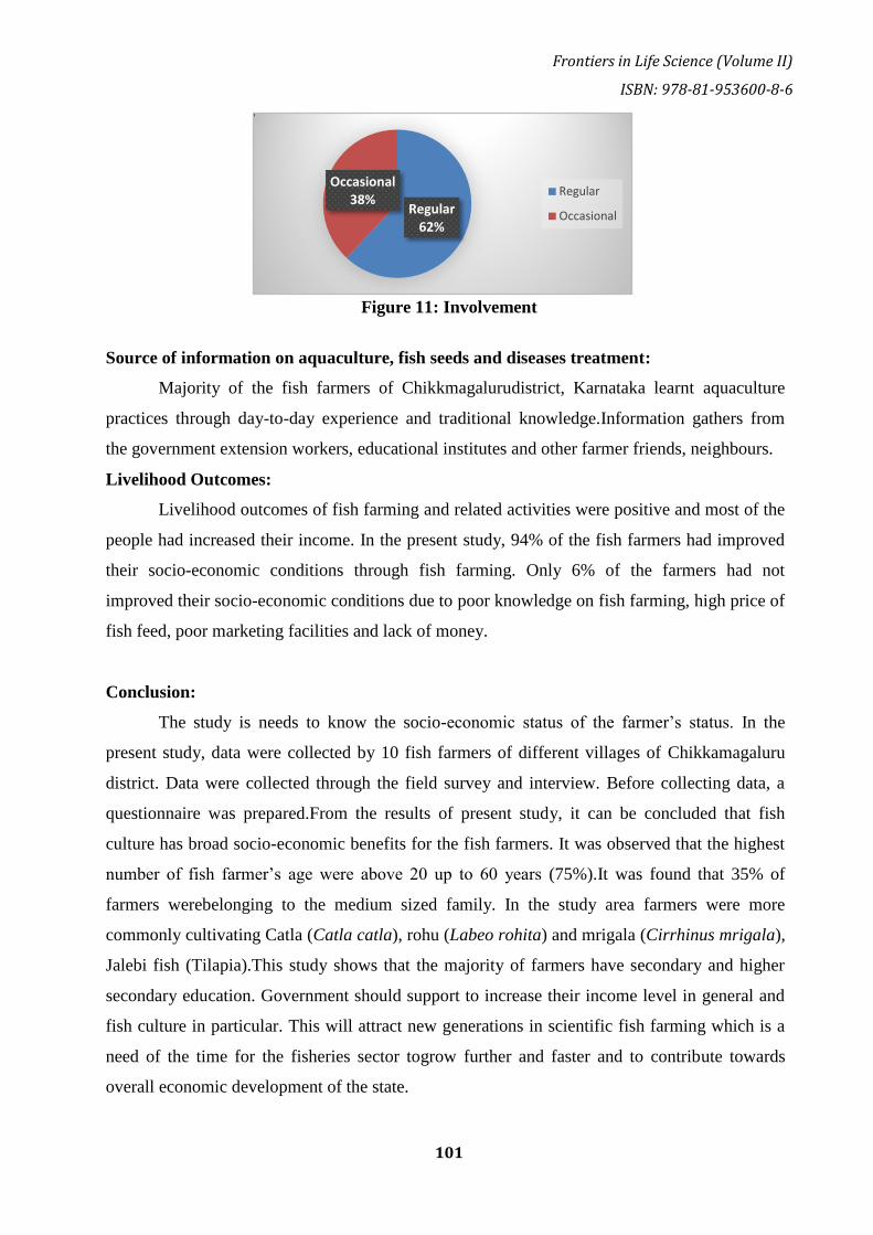
Frontiers in Life Science (Volume II)
ISBN: 978-81-953600-8-6
101
Figure 11: Involvement
Source of information on aquaculture, fish seeds and diseases treatment:
Majority of the fish farmers of Chikkmagalurudistrict, Karnataka learnt aquaculture
practices through day-to-day experience and traditional knowledge.Information gathers from
the government extension workers, educational institutes and other farmer friends, neighbours.
Livelihood Outcomes:
Livelihood outcomes of fish farming and related activities were positive and most of the
people had increased their income. In the present study, 94% of the fish farmers had improved
their socio-economic conditions through fish farming. Only 6% of the farmers had not
improved their socio-economic conditions due to poor knowledge on fish farming, high price of
fish feed, poor marketing facilities and lack of money.
Conclusion:
The study is needs to know the socio-economic status of the farmer‘s status. In the
present study, data were collected by 10 fish farmers of different villages of Chikkamagaluru
district. Data were collected through the field survey and interview. Before collecting data, a
questionnaire was prepared.From the results of present study, it can be concluded that fish
culture has broad socio-economic benefits for the fish farmers. It was observed that the highest
number of fish farmer‘s age were above 20 up to 60 years (75%).It was found that 35% of
farmers werebelonging to the medium sized family. In the study area farmers were more
commonly cultivating Catla (Catla catla), rohu (Labeo rohita) and mrigala (Cirrhinus mrigala),
Jalebi fish (Tilapia).This study shows that the majority of farmers have secondary and higher
secondary education. Government should support to increase their income level in general and
fish culture in particular. This will attract new generations in scientific fish farming which is a
need of the time for the fisheries sector togrow further and faster and to contribute towards
overall economic development of the state.
Regular62%
Occasional38%
Regular
Occasional
Bhumi Publishing, India
102
References:
Bias, B. 2018. Fish scenario in India with emphasis on Indian major carps. International Journal
of Avian & Wildlife Biology. 3(6):409‒411
Gadage, R. S. 2005. Production and marketing of fish and fish preparations in India. Indian
Journal of Agricultural marketing. 19:61
Hussain, M. I., Siwar, C., Mokhtar, M. B., Dey, M. M., and Jaafar, A. H. 2009. Socio-economic
condition of fishermen in seasonal floodplain beels in Rajshahi District, Bangladesh.
Research Journal of Social Sciences. 4 : 74- 81
Prem Kumar, Sanjay Khar, Rakesh Sharma, PunitChoudhary, K.V. Himabindu, Sushma Sharma,
S.K. Sharma and S. Jagmohan. 2015. Identifying Socio-Economic Features of Fish
Farmers. Agro-Economist- an Inernational Journal. Vol. 2, No. 1, 29-34.
Sen, A and Roy, M. 2015. Socio-Economic status of fish farmers in Tripura, India. International
Journal of current research.Vol. 7, Issue, 06, pp.17090-17096.

Frontiers in Life Science (Volume II)
ISBN: 978-81-953600-8-6
103
ACUPUNCTURE THERAPY: A FASCINATING APPROACH
FOR PALLIATE HEALTH
Megha Jain1* and Vinay Jain2
1Oriental College of Pharmacy, Oriental University, Indore
2Shri Ram College of Pharmacy, Banmore, M.P., India
*Corresponding authors E-mail: [email protected]
Introduction:
As the name suggests, Acupuncture Therapy is an alternative therapeutic treatment in this
the specific unique techniques or healing force is used to treat illness. It works on the belief of
vitalism. This chapter mainly emphasizes on the history of acupuncture therapy, basic principles
of acupuncture therapy, treatment methods used in this therapy and its applications.
Acupuncture therapy is an fundamentally portion of an primitive Chinese framework of
pharmaceutical that has been utilized for more than 2500 years a long time to treat infections and
relieve pain (Grigory et al., 2005).
―De qi‖ is the equivalent word of conventional needle therapy; this can e utilized to
depict the association between needle therapy and the body's energy pathways. The ―de‖
component of the word compound implies ―obtain,‖ and the ―qi‖ component is ―vital energy‖
(Kaptchuk, 2000). Customarily, other terms have been utilized for the same marvel counting
―qizhi‖, which indicates‖ arrival‖ of qi. Needle therapy is the incitement of particular acupoints
along the skin of the body including different strategies such as the application of needling,
moxibustion, electric needle therapy (electro-acupuncture), laser needle therapy (photo-
acupuncture), miniaturized scale framework needle therapy such as ear (auricular), confront,
hand and scalp needle therapy, and acupressure.
Acupuncture is based on following principle:
Yin-Yang Theory: Yin-Yang‘s theory is a concept based on a philosophy that is two
opposite aspects of nature that are interrelated with each other. Yin- Yang theory
developed in the Chinese they, side that the entire space is regulate by two opposing forces.
All matters which are present in nature can classify by Yin and Yang. The manner of Yin
philosophy fuscous on the feminine, latent and passive principle; while the Yang describe

Bhumi Publishing, India
104
the masculine, active and positive. Principle of the two competing universal powers by
which creative energy is split and the fusion of which in physical matter produces the
unimaginable universe.
Sr. No. Nature Yin Yang
1 Colour Dark Light
2 Temperature Cold Warm
3 Time Night Day
4 Humidity Wet Dry
5 Texture Soft Hard
6 Gender Female Male
7 Weather Winter Summer
8 Position Down Up
9 Movement Still Move
10 Action Passive Active

Frontiers in Life Science (Volume II)
ISBN: 978-81-953600-8-6
105
Four examinations observe according to the traditional Chinese medicine system:
Five Phases Theory: Five phase‘s theory/Five Elements Theory is another basic theory
in TCM. By comparing their structures, properties and behavior, the ancient Chinese divided
material objects or phenomena into five groups, based on observations. The theory of the Five
Phases posits wood, fire, earth, metal and water as the fundamental elements of the material
world and these elements are continually moving and evolving in relation to each other.
Sr.
No.
Traditional Chinese
Medicine four Examination
Yin Yang
1 Inspection Dim Bright
2 Listening and Smelling Low and weak voice,
silent and reticent
High and strong voice,
talkative and agitated
3 Inquiry Cold , not thirst,
diarrhea
Hot, thirst, constipation
4 Palpation Deep and slow, thread,
forceless pulse
Floating, full, slippery,
forceful pulse

Bhumi Publishing, India
106
Theory of Qi, Blood, Fluid and Humor: Qi, Blood, Fluid, and Humor theory in TCM is
used to explain the human body's growth, transportation, distribution, physiological
function, pathological change, and reciprocal relationships of Qi, Blood, Fluids, and
Humors. In TCM, Qi refers to the purified nutrient that flows in the body and to its
functional activities. Blood is a kind of nutritionally rich red liquid that circulates inside the
blood vessels, which has the functions of nourishing and hydrating the entire body. Fluid
and humor refer to all types of normal body fluid, excluding blood, also known as body
fluids. They are the essential materials that form the human body and that regulate the
activities of life. Though the three have different properties, shapes and functions, they are
physiologically dependent on each other and pathologically influence each other.
Visceral Manifestation Theory: The internal organs and associated terminologies in
TCM differ greatly from those of Western medicine. e The organs are divided into 'Zang'
or 'viscera' (i.e., Lung, heart, liver, spleen, and kidney), where Essence and Qi are
developed and stored, and 'Fu' or 'bowel' (i.e., gallbladder, stomach, large intestine, small
intestine, urinary bladder and triple energizers) where food is provided,
Meridian, Collateral and Acupoint: Meridian and collateral systems serve as conduits
facilitating QI and blood circulation, bowel contact, viscera, extremities, superficial organs
and tissues, creating the body an organic whole. It consists principally of the twelve
meridians/channels, the eight extra meridians and the fifteen collateral vessels.
Sr.
No.
Human body Five Phases
Wood Fire Earth Metal Water
1 Zang Organ Liver Heart Spleen Lung Kidney
2 Fu Organ Gallbladd
er
Small
intestine
Stomach Large
intestine
Urinary
bladder
3 Orifice Eye Tongue Mouth Nose Ear
4 Tissue Tendon Vessel Muscle Skin &
hair
Bone
5 Secretion Tear Sweat Serous saliva Nasal
discharge
Mucous
saliva
6 Manifestation Nail Face Lip Body hair Hair
7 Emotion Anger Joy Pensiveness Grief Fear
8 Voice Shout Laugh Sing Cry Groan
9 Action Gripping Restlessness Vomiting Coughing Shivering

Frontiers in Life Science (Volume II)
ISBN: 978-81-953600-8-6
107
Acupoints are subdivided into three categories:
Acupoints are present along each meridian for application of acupuncture and
moxibustion.
1. Acupoints of 14 meridians: total of 361 acupoints which make up most of all points
present on human body.
2. Extra point (Nonmeridian): Acupoints which are useful in treatment plan as they have
been discovered during practice. They have exact positions but aren't Fourteen meridians
situated on.
3. Ouch point (Ashi): The location of which in some Diseases defined in some diseases by
tenderness, responsiveness, or other pathogenic responses. We do not have any clear
locations or name which is "where there is a painful spot, there is an acupuncture point".
(Virginia and Wong, 2008-2014).
Diagnosis in Acupuncture Therapy:
In TCM, disease is generally perceived as a disharmony (or imbalance) in the roles or
interactions of yin, yang, qi, xuA, zA ng fc, meridians etc. and/or the human body's relationship
with the environment. Practitioners may analyze items like the color and form of the tongue, the
relative intensity of the pulse points, the odor of the breath, the performance of the breath to
evaluate the pattern is at hand (Maciocia, 1995; 2005; Shen, 1996; Qiu et al., 1997).
Applications of Acupuncture Therapy:
1. Osteoarthritis: acupuncture to provide clinically significant relief from knee osteoarthritis
pain and a larger improvement in function (Selfe et al., 2008). Childbirth: In childbirth,
acupuncture analgesia is useful for relieving labour pain and can significantly reduce the
duration of labor (Sheng and Fan, 2010).
2. Infections: It has been confirmed that acupuncture is beneficial in the treatment of acute
bacillary dysentery Acupuncture may be helpful in the treatment of pertussis (whooping
cough), cough relief and heal (Marwick, 1997).
3. Respiratory disorders: Acupuncture is often used in treating respiratory disorders.
Allergic rhinitis is among the major indications. In controlled studies, it has been shown
that acupuncture is more effective than antihistamine in bronchial asthma acupuncture is
effective (Lau and Jones, 2008).

Bhumi Publishing, India
108
4. Digestive disorders: Epigastric pain is a common symptom in diseases of the stomach,
including peptic ulcer, acute and chronic gastritis, and gastric spasm. Acupuncture
provides satisfactory relief of epigastric pain (Klein, 1988).
5. Pain: Acupuncture seems to have it can be prescribed for good analgesic and
antispasmodic activity on the biliary tract. Acupuncture may serve as a complementary or
alternative therapeutic measure (Helms, 1995).
Frontiers in Life Science (Volume II)
ISBN: 978-81-953600-8-6
109
6. Urinary disorder: Acupuncture shows satisfactory result in treatment of urinary stone.
Urinary retention (Philp et al., 1988). Treatment of defective ejaculation (no ejaculation
during intercourse) (Emmons and Otto, 2005).
7. Cardiovascular disorders: Acupuncture is effective in the management of primary
hypotension and early hypertension (Wood et al., 2003).
8. Psychiatric disorders and mental disturbances: Taking acupuncture in psychological
disorders is rising (Boyd et al., 2001).
9. Skin diseases: Acupuncture worked considerably better than vitamins C and E, Using as
therapy for acne vulgaris (Lewas, 1992).
10. Gynaecological and obstetric disorders: Acupuncture was deemed successful in treating
anovular infertility in females. Primary dysmenorrhoea prevents miscarriage, morning
sicknessstimulates Milk production during conception which may be used to combat
emotional lactation deficits or stress (Zhu and Zhu, 1995).
References:
Boyd, D., Evans, C. and Drennan, V. (2001). Using reflexology: a feasibility study. Mental
Health Nursing, 21(6):14–17.
Emmons, S.L. and Otto, L. (2005) Acupuncture for overactive bladder: a randomized control
trial. Obstetrics and Gynecology, 106 (1): 138–143.
Grigory,V., Chernyak, and Daniel I.S. (2005). Preoperative Acupuncture and related techniques.
Anesthesiology, 102(5): 1031–1078.
Helms, J. (1995). Acupuncture Energetics: A Clinical Approach for Physians.Berkely: Medical
Acupuncture Publishers, 198-201.
Kaptchuk, T.J. (2002). Acupuncture: theory, efficacy, and practice. Ann Intern Med, 136: 74–
383.
Klein, K.B. (1988). Controlled treatment trials in the irritable bowl syndrome: A critique.
Gastroenterology, 95: 232-241.
Lau, K.S. and Jones AY. (2008). A single session of Acu-TENS increases FEV1 and reduces
dyspnoea in patients with chronic obstructive pulmonary disease: a randomised, placebo
controlled trial. Australian Journal of Physiotherapy, 54: 179-84.
Lewas, P.I. (1992). Acupunctlre treatmenr of dermatological disorders. Journal of the Australian
Medical Acupuncture society, 10 (1): 3-8.
Maciocia, G. (1995). Tongue Diagnosis in Chinese Medicine. Eastland Press.

Bhumi Publishing, India
110
Maciocia, G. (2005). The Foundations of Chinese Medicine. Churchill Livingstone.
Marwick, C. (1997). Acceptance of some acupuncture applications. JAMA, 278: 1725-7.
Philp, T., Shah, P.J.R. and Worth, P.H.L. (1988). Acupuncture in the treatment of bladder
instability. British Journal of Urology, 61(6):490–493.
Qiu L. (1997). Acupuncture treatment of severe leg pain in 60 cases of thromboangitis
obliterans. Chinese Acupuncture and Moxibustion, 17(11): 677–678.
Selfe, T.K. and Taylor, A.G. (2008). Acupuncture and osteoarthritis of the knee: a review of
randomized, controlled trials. Family & Community Health, 31 (3): 247–54.
Shen, S.J. (1996). Immediate analgesic effect of acupuncture at binao (LI14) for pain due to
subconjunctival injection. Chinese Acupuncture and Moxibustion, 16(2): 71–72.
Sheng, X.P. and Fan, T.Y. (2010). Comparative observation on hip osteoarthritis treated with
electroacupuncture and medication [Article in Chinese]. Zhongguo Zhen Jiu, 30: 982-4.
Virginia, C.N. Wong. Alternative therapy - TCM therapies. International encyclopedia of
rehabilitation. Copyright © 2008-2014 by the Center for International Rehabilitation
Research Information and Exchange.
Wood, M., Stewart, R., Merry, H., Johnstone, D. and Cox, J. (2003).Use of complementary and
alternative medical therapies in patients with cardiovascular disease. Am Heart J, 145:
806–812.
Zhu, Y. and Zhu, X. (1995). Treatment of primary dysmenorrhoea by acupuncture: A report of
30 cases. International Journal of Clinical Acupuncture, 6 (4): 473-474.

Frontiers in Life Science (Volume II)
ISBN: 978-81-953600-8-6
111
MUCORMYCOSIS (BLACK FUNGUS): INTRODUCTION, PATHOPHYSIOLOGY
AND ITS IMPACT ON PEOPLE OF INDIAN SUBCONTINENT
Rohit Batra*1, Pankaj Sharma2 and Vinay Jain3
1Department of Pharmacology
2Department of Pharmaceutics
3Department of Pharmacognosy,
ShriRam College of Pharmacy, Banmore, Near Gwalior, M.P
*Corresponding authors E-mail: [email protected]
Abstract:
Mucormycosis or black fungus infection is a rare and serious opportunistic infection
which is caused by molds of family Mucormycetes. Black fungus infection is non-communicable
infection that occurs mostly in immunocompromised patients, patients with high iron load (and
using deferoxamine) and in patients with uncontrolled diabetes mellitus.This disease primarily
affects nasal sinuses, orbits, brain, lungs and stomach. If untreated hematogenous dissemination
occurs and patient dies within 2 weeks.
Treatment includes treatment of underlying cause (diabetes mellitus, metabolic acidosis
and reduction in dose of immunosuppressant medication), IV therapy with liposomal
amphotericin B and surgery of necrotic tissue.
Indian subcontinent is contributing 71% to the global pool of black fungus infection. This
disease is prominently affecting patients who have uncontrolled diabetes mellitus, CoVID-19
recovered patients who were given unjustified dose of steroids, and those who have abused zinc
metal (as tablets) during home isolation. So far Nepal and Bangladesh have also reported 1 case
each, Russia also has unreported load of black fungus infection. Is another pandemic knocking
the door of world in the form of black fungus infection? Is the world ready?
Keywords: Mucormycosis, black fungus infection, immunosuppressant, amphotericin-B,
uncontrolled diabetes mellitus, pandemic.
Black Fungus Infection: an introduction
Mucormycosis or black fungus disease is a rare (but sometimes serious) fungal infection
caused by Mucor, Saksenaea, Absidia and Apophysomyces (all are members of family of molds
called Mucormycetes)
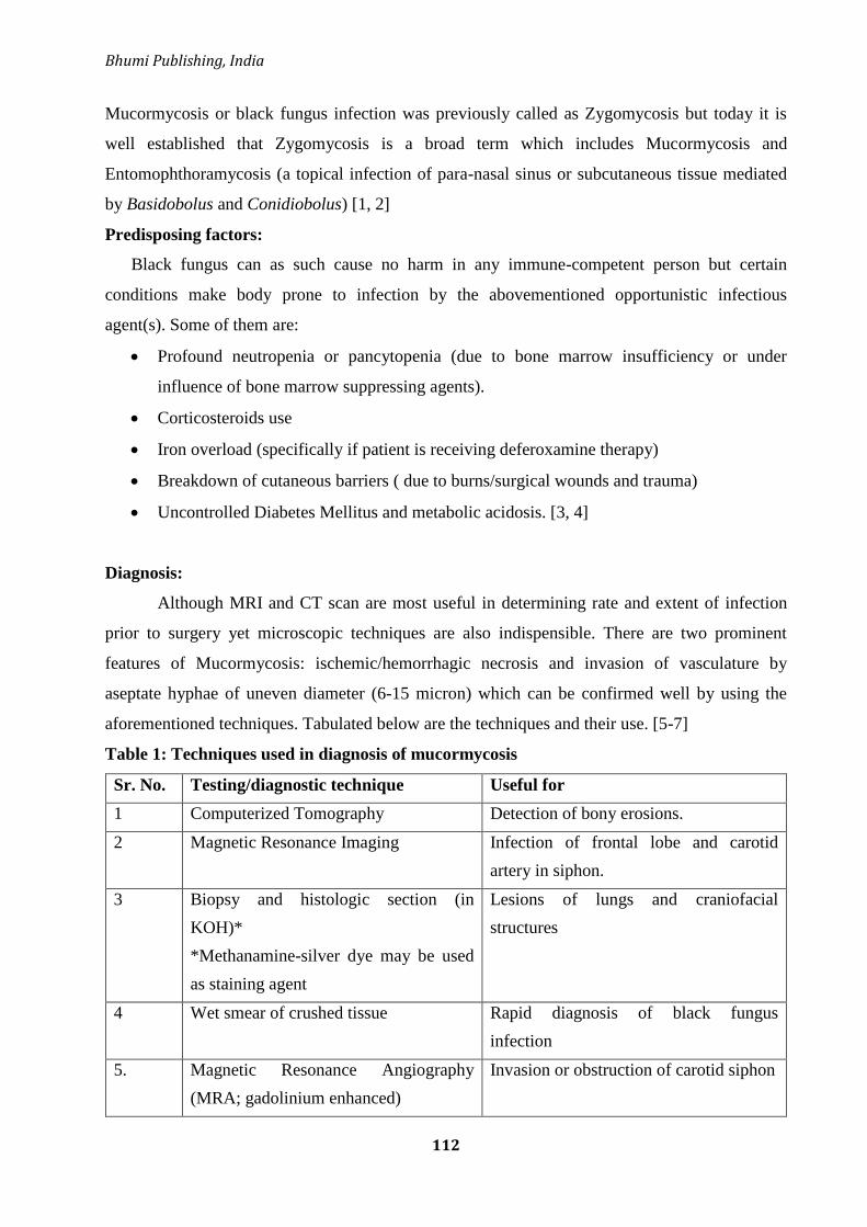
Bhumi Publishing, India
112
Mucormycosis or black fungus infection was previously called as Zygomycosis but today it is
well established that Zygomycosis is a broad term which includes Mucormycosis and
Entomophthoramycosis (a topical infection of para-nasal sinus or subcutaneous tissue mediated
by Basidobolus and Conidiobolus) [1, 2]
Predisposing factors:
Black fungus can as such cause no harm in any immune-competent person but certain
conditions make body prone to infection by the abovementioned opportunistic infectious
agent(s). Some of them are:
Profound neutropenia or pancytopenia (due to bone marrow insufficiency or under
influence of bone marrow suppressing agents).
Corticosteroids use
Iron overload (specifically if patient is receiving deferoxamine therapy)
Breakdown of cutaneous barriers ( due to burns/surgical wounds and trauma)
Uncontrolled Diabetes Mellitus and metabolic acidosis. [3, 4]
Diagnosis:
Although MRI and CT scan are most useful in determining rate and extent of infection
prior to surgery yet microscopic techniques are also indispensible. There are two prominent
features of Mucormycosis: ischemic/hemorrhagic necrosis and invasion of vasculature by
aseptate hyphae of uneven diameter (6-15 micron) which can be confirmed well by using the
aforementioned techniques. Tabulated below are the techniques and their use. [5-7]
Table 1: Techniques used in diagnosis of mucormycosis
Sr. No. Testing/diagnostic technique Useful for
1 Computerized Tomography Detection of bony erosions.
2 Magnetic Resonance Imaging Infection of frontal lobe and carotid
artery in siphon.
3 Biopsy and histologic section (in
KOH)*
*Methanamine-silver dye may be used
as staining agent
Lesions of lungs and craniofacial
structures
4 Wet smear of crushed tissue Rapid diagnosis of black fungus
infection
5. Magnetic Resonance Angiography
(MRA; gadolinium enhanced)
Invasion or obstruction of carotid siphon

Frontiers in Life Science (Volume II)
ISBN: 978-81-953600-8-6
113
Pathology:
It is well established that black fungus is an opportunistic infection. It happens only when
immune system of a person is compromised (idiopathic/iatrogenic/infectious/metabolic
disorder). Black fungus infection affects three primary locations: nasal sinus (which may lead to
orbital and craniofacial infection), lungs and stomach. In some cases, skin is also affected but
such cases are highly rare. If untreated, hematogenous dissemination may occur and chances of
survival decreases (life cannot be expected beyond 2 week). [8-9]
Tabulated below are the location and attribution for black fungus infection.
Table 2: location of infection and its probable causes in mucormycosis
Sr. No. Location of infection Reason
1 Mucormycosis in para nasal sinuses and
nose (which may lead to orbital and
craniofacial infection).
Poorly controlled diabetes mellitus
2 Mucormycosis of lungs (accompanied with
nose)
Occurs in patient who has
undergone organ transplant or who
has received long term
deferoxamine therapy or in patient
who has hematologic malignancy.
3 Gastrointestinal Mucormycosis Uremia, severe malnutrition,
diarrheal disease
Clinical Manifestation:
Clinical manifestation depends on the site which is infected by black fungus.
Mucormycosis affecting nasal and para-nasal sinus:
This infection projects a different clinical picture. It is often characterized by dull pain in
sinuses, nasal congestion, and bloody nasal discharge. Patient may have low grade pyrexia.
Delineated area of necrosis (respecting midline) occurs in hard palate. Facial skin near to para-
nasal sinus becomes red, purple and black.
This infection may then rise to upper levels and may involve eyes and brain. When eyes
are affected; clinical features includes double vision, obtundation. Along with this unilateral
chemosis, unilateral proptosis and reduction of unilateral generalized ocular motion is also seen.
During orbital invasion, blood may clot in sinus (Cavernous Sinus Thrombosis).
Bhumi Publishing, India
114
Once brain starts getting infected, clouding of sensorium may be attributed to diabetic acidosis.
Coma occurs due to invasion offrontal lobe by pathogen.[10]
Pulmonary Mucormycosis:
The prominent feature of pulmonary mucormycosis is severe pyrexia and pneumonia.
Cavities are formed in infected area. Along with this hemoptysis can occur from cavities so
formed. [11]
Gastrointestinal Mucormycosis:
This kind of infection is expressed by an individual as multiple gastric ulcers which are
prone to perforation. [12]
Cutaneous Mucormycosis:
This kind of infection is highly rare and occurs mainly in burn eschars, at the site of
minor trauma in immune-compromised patient, in low weight neonates and below occlusive
dressings. [13]
Treatment of Mucormycosis:
There are three strategies for treatment of black fungus infection. First one is regulation
of underlying cause which could be diabetes mellitus (by using appropriate hypoglycemic
agents) or by controlling bone marrow suppression (which requires dose reduction of
immunosuppressant or other causative agent) Second one includes extensive debriment of
craniofacial lesions and Orbital exenteration (if required). Third strategy includes treatment with
IV amphotericin B (Liposomal formulation, dose: 5mg/kg for 10-12 weeks). There is very high
mortality rate in patients receiving deferoxamine and those with pulmonary, gastrointestinal and
disseminated mucormycosis. [14-16]
Impact of mucormycosis on people of Indian subcontinent:
CoVID-19 pandemic undoubtedly left entire globe in destitute. With more and more
number of cases being reported, it has claimed 3,718,849 lives till now (as on 04-JUN-2021).
[17] But does this end here? Answer is no. Another challenge which has started claiming lives of
people (especially in India) is black fungus infection. Indian subcontinent has started to suffer so
much because of this infection that some reports have started identifying black fungus disease as
Pandemic within the pandemic. [18]
As on 06th
JUN-2021, India reported 8848 infection cases and 219 deaths due to black
fungus disease [19]. Same has now been declared as epidemic in Tamil Nadu, Gujarat, Odisha,

Frontiers in Life Science (Volume II)
ISBN: 978-81-953600-8-6
115
Bihar, Uttarakhand, Rajasthan, Telangana and Chandigarh. Such a high mortality in this
infection is attributed to the fact that most of the Indians either used drugs irrationally (self-
medication or overdose) especially of zinc along with excessive use of stream. Not only this,
according to one report, irrational and unjustified use of steroids in patients by doctors is also
one of the factors why black fungus infection is happening in persons with CoVID-19
history.[20, 21]Black fungus infection has also started to affect neighbor countries. Nepal also
reported its first clinical case of black fungus disease on 04th
JUN-2021. [22]
According to a report, each patient requires approximately 50-60 doses of amphotericin-
B. Profiteers did not leave this opportunity as well. Injections of liposomal Amphotericin-B were
hoarded and black marketed. States of India started facing shortage of liposomal amphotericin-B
injections. Government took cognizance of this problem and just like remdesivir; amphotericin-B
was also supposed to be supplied centrally and by state government directly to the hospitals. Its
export was also restricted. Use of oral posaconazole medications was also suggested if IV
therapy of black fungus infection is not possible.
Some countries like Bangladesh and Egypt have responded well to some Indian state‘s
(like Rajasthan) global Expression of Interest (EOI) request for amphotericin-B [23-26]
Having mentioned all these things a valid question now arises here; is another pandemic also
knocking the door of this globe in the form of black fungus disease? Is the world ready?
Conclusion:
Although no person to person transfer has been established for black fungus infection yet
we need to exercise utmost caution. People must maintain a high level of hygiene and must avoid
residing in damp places.
People must try to.control their sugar intake as uncontrolled diabetes mellitus has highest
attribution in the cases of mucormycosis. There is a need that government must also ramp up
production of amphotericin B since many have died in India due to lack of therapeutic agent on
account of either delay in supply or acute shortage
References:
1. Kasper, Braunwald, Fauci et al. (2005) Harrison‘s principle of internal medicine, McGraw
Hill Companies Inc. 16th
edition, Vol. 1, Page No. 1190-1191
Bhumi Publishing, India
116
2. Mucormycosis: What to Know. Available online at
https://www.webmd.com/lung/mucormycosis-black-fungus-infection. (accessed 04-JUN-
2021)
3. Greenwood D., Slack R. Barer et al. (2007), Medical Microbiology, A guide to infections:
pathogenesis, immunity, laboratory diagnosis and control, Churchill Livingstone, Elsevier,
17th
edition Page 617-618.
4. Black Fungus Update: Who is at High Risk? What You Should do if You Experience
Symptoms? Things to Know. Available online at: https://www.india.com/health/black-
fungus-update-who-is-at-high-risk-what-you-should-do-if-you-experience-symptoms-
things-to-know-4679450/. (accessed 04-JUN-2021)
5. Slonimsky, G., Slonimsky, E., Yakirevitch, A., et al. (2018). The significance of Computed
Tomography in invasive paranasal mucormycosis. Rhinology, 56(1), 54–58.
https://doi.org/10.4193/Rhin17.153
6. How to identify black fungus cases, what to do next? AIIMS issues new guidelines.
Available online at: https://www.indiatoday.in/india/story/how-to-identify-black-fungus-
cases-what-to-do-aiims-guidelines-1804627-2021-05-20. (accessed 05-JUN-2021)
7. Guarner, J., & Brandt, M. E. (2011). Histopathologic diagnosis of fungal infections in the
21st century. Clinical microbiology reviews, 24(2), 247–280.
https://doi.org/10.1128/CMR.00053-10
8. Kumar V, Abbas A, Fausto N et al (2010), Robbins and Contran: Pathologic basis of
disease, Saunders, Elsevier, 8th
edition,Page No. 385
9. Colledge N, Walker B. and Ralston S. (2010), Davidson‘s principles and practice of
Medicine, Churchill living-stone, Elsevier, 21st edition, Page No. 379.
10. Black Fungus Infection (Mucormycosis). Available online at:
https://www.medicoverhospitals.in/blog/black-fungus. (accessed 05-JUN-2021)
11. Fernandez, J. F., Maselli, D. J., Simpson, T., et al. (2013). Pulmonary mucormycosis: what
is the best strategy for therapy? Respiratory care, 58(5), e60–e63.
https://doi.org/10.4187/respcare.02106
12. Spellberg B. (2012). Gastrointestinal mucormycosis: an evolving disease. Gastroenterology
& hepatology, 8(2), 140–142.
13. Castrejón-Pérez, A. D., Welsh, E. C., Miranda, I., Ocampo-Candiani, J., et al. (2017).
Cutaneous mucormycosis. Anais brasileiros de dermatologia, 92(3), 304–311.
https://doi.org/10.1590/abd1806-4841.20176614

Frontiers in Life Science (Volume II)
ISBN: 978-81-953600-8-6
117
14. Treatment for Mucormycosis. Available online at:
https://www.cdc.gov/fungal/diseases/mucormycosis/treatment.html. (accessed 05-JUN-
2021)
15. Mucormycosis (zygomycosis). Available online at:
https://www.uptodate.com/contents/mucormycosis-zygomycosis. (accessed 05-JUN-2021)
16. Spellberg, B., & Ibrahim, A. S. (2010). Recent advances in the treatment of mucormycosis.
Current infectious disease reports, 12(6), 423–429. https://doi.org/10.1007/s11908-010-
0129-9
17. Coronavirus Death Toll. Available online
at:https://www.worldometers.info/coronavirus/coronavirus-death-toll/. (accessed 04-JUN-
2021)
18. Pandemic within pandemic: Black fungus cases much higher in second Covid wave.
Available online at:
https://economictimes.indiatimes.com/industry/healthcare/biotech/healthcare/mucormycosi
s-much-higher-in-2nd-wave/articleshow/82781096.cms?from=mdr . (accessed 04-JUN-
2021)
19. India has 8,848 cases of black fungus, over 23,000 vials of medicine allotted to states, says
Centre. Available online at: https://scroll.in/latest/995504/india-has-8848-cases-of-black-
fungus-over-23000-vials-of-medicine-allotted-to-states-says-centre. (accessed 06-JUN-
2021)
20. Uncontrolled diabetes, improper steroid use key in emergence of black fungus: PGIMER
study. Available online at: https://www.hindustantimes.com/cities/chandigarh-
news/uncontrolled-diabetes-improper-steroid-use-key-in-emergence-of-black-fungus-
pgimer-study-101622919866327.html. (accessed 06-JUN-2021)
21. Probe link between Black Fungus, use of zinc in treatment of Covid patients: Experts.
Available online at: https://www.indiatoday.in/coronavirus-outbreak/story/black-fungus-
mucormycosis-cases-epidemic-zinc-treatment-covid-experts-1805824-2021-05-
22(accessed 06-JUN-2021)
22. Nepal Reports First Death Due To Black Fungus. Available online at:
https://www.ndtv.com/world-news/nepal-reports-first-black-fungus-or-mucormycosis-
death-amid-surge-in-covid-cases-2456320. (accessed 06-JUN-2021)

Bhumi Publishing, India
118
23. India Faces Amphotericin B Shortage as ‗Black Fungus‘ Adds to COVID Woes. Available
online at: https://science.thewire.in/health/india-faces-amphotericin-b-shortage-as-black-
fungus-adds-to-covid-woes/. (accessed 06-JUN-2021)
24. Inside black fungus wards in Rajasthan: Amid injection shortage, increasing patients, many
end up losing vision. Available online athttps://indianexpress.com/article/india/in-black-
fungus-wards-in-rajasthan-many-end-up-losing-vision-amid-injection-shortage-rising-
patient-numbers-7345983/. (accessed 06-JUN-2021)
25. Black Fungus: Centre restricts export of Amphotericin-B injections. Available online at:
https://www.businesstoday.in/current/economy-politics/black-fungus-centre-restricts-
export-of-amphotericin-b-injections/story/440548.html. (accessed 06-JUN-2021)
26. Amphotericin B not the only cure available for black fungus: ICMR. Available online at:
https://www.businesstoday.in/current/economy-politics/amphotericin-b-not-the-only-cure-
available-for-black-fungus-icmr-informs-delhi-hc/story/440843.html. (accessed 07-JUN-
2021).

Frontiers in Life Science (Volume II)
ISBN: 978-81-953600-8-6
119
PALYNOLOGY IN FORENSIC SCIENCE
Megha Sapkota, Sandeep Sangle and Arun Ghuge*
Department of Forensic Biology,
Government Institute of Forensic Science,
Nipat Niranjan Nagar, Caves Road, Aurangabad 431004, India
*Corresponding authors E-mail: [email protected]/[email protected]
Introduction:
Forensic science is a word derived from ‗forensis‘ having latin origin. Forensic science
deals with associating people, places and things involved in criminal acts. It is science which
aids in criminal and civil cases[1]. Forensic science is divided into sub-branches like Forensic
Biology, Forensic Chemistry, Forensic Physics, Cyber forensics etc.
Figure 1: Branches of forensic science
Forensic biology is the branch of forensic science that deals with application of biological
evidences to catch the criminals. There is other sub-discipline which comes under the Forensic
biology like Serology, limnology, Forensic entomology, Forensic anthropology, Forensic
Palynology etc.
Forensic expert deals withdifferent forms of evidences that assist investigators to solve
the crimes.Most often encountered evidencesare in the form of trace evidences. Trace evidence is

Bhumi Publishing, India
120
the microscopic evidence left behind by two objects or people coming in contact. Trace evidence
is ruled by Locard's principle of exchange which states that ―Every contact leaves a trace‖[2].
Different trace evidences involve Hair samples, fibers or fabrics, grass, soil particles, skin
remnants, etc.
One such crucial evidence which can provide an important link in investigation is pollen
grains.Pollen grains are the coarse powder containing micro-gametophyte of the seed, plants that
produce the male gametes (sperm cell)[3].Forensic Palynology, a subdivision of forensic biology
that mainly deals with the application of pollen and spores in criminal investigations. Experts in
the field are called palynologists.
Pollen grains vary in size from 5µm-20µm. Pollen is in distinct shapes for instance,
circular, triangular, elliptical etc. Some may have variation even in the basic shape. When
observed with naked eyes it seems like the dust of yellow powder but even the colour varies
from white, crème to yellow from species to species. Pollen grains have hard coats i.e., exine
made up of ―sporopollenin‖ that protect the gametophyte during the process of their movements
from stamen to pistil of flowering plants or from male cone to female coniferous plants. The
inner layer of pollen is called intine, made up of cellulose and pectin degrades faster as
fossilization takes place[4].
Figure 2: Schematic representation of Androecium, Gynoecium and Pollen grain

Frontiers in Life Science (Volume II)
ISBN: 978-81-953600-8-6
121
Pollination takes place commonly by these modes:
Figure 3: Different Modes of Pollination [4]
Palynology has an extremely important role to play in palaeontology, archaeology, and
forensic science. Pollen grains are used as a tool to reconstruct the past environmental activities.
Exine possessed by pollen grain can withstand environmental degradation for thousands of years.
Palynologists can learn about the flora that existed in the past. Studying the Pollen grains,
Archaeologists can tell how humans in the past interacted with their environment [5].
Being microscopic, found on nearly every surface and objects, suspects will be carrying
them unknowingly on their clothing, hairs, footwear‘s, etc. Pollen grains are unique to each
species of plant and geographic location[5]. Pollen grains are extremely resistant to
environmental degradation and can remain unaffected for years[6]. Thus, pollen is considered as
important trace evidence and can help to link a suspect to a crime scene.
Table 1: Entire Process from filing of report to Conviction of Accused[7][8]
Forgery Assaults Robbery Rapes
Genocide Terrorism Arson Hit and run
crimes
Withdrawal of artifacts
from historic or
archaeological sites
poaching of animals
including fish
pollution of the
environment
Homicide

Bhumi Publishing, India
122
Sample collection:
Pollen can solve the cases such as
Forensic Significance:
Pollen is a form of trace evidence that is present in small but measurable amounts. The
forensic significance of pollen analysis is support to prove or disprove a linkage between people
and objects with places or with other people.
Pollen grains can tell us about the geographical area from where it belongs.
Pollen grains can help us to trace the geographical location which is most often the scene of
offence or sometimes the origin of victim, suspect or any object whose presence is unsure at that
place.

Frontiers in Life Science (Volume II)
ISBN: 978-81-953600-8-6
123
Figure 4: Process of Evidence collection and Analysis[9]
In 1979, a case in which the body of a young girl was found in the maize field in
Rochester, New York [10]. All her belongings were missing which would have helped to
identify. Also her fingerprints were not in the record as she was too young for her fingerprint
data to be in record. It was about 3 decades from then since her identity was not established even
after a lot of investigative efforts. But then in 2006, Pollen analysis was done ofher clothes. The
pollen extracted from her pockets suggested the region which was about 2000 miles away to the
west from where her body was recovered as they belonged to an Australian pine which was not
common in the local area. Also, it was said by investigating palynologist that it was
impracticable that this pollen has travelled thousands of miles to the site of offence.During
Bhumi Publishing, India
124
investigation in 1979, Officers assumed that the girl belonged to the local area and had restricted
their investigation.The case remained unsolved as investigators were unable to prove the identity
of the victim as there was no missing complaint lodged in any station house.
This case suggests that if the evidence is preserved properly without any contamination, it can be
used for analysis even after years of crime and still give significant lead in the case.
It can determine the geographic source of fruit, drugs, and imported goods.
Pollen grains assist to determine the source of transported goods. It helps to understand
the locality of particular fruit or drug or any goods that investigators come across. There have
been several cases which got lead through pollen analysis. One such case took place in 1990[11].
Aerial surveillance helped police to locate a large field of marijuana grown illegally. A day after
the police noticed a well-dressed person coming out of the bushes a few kilometersapart from the
place. Upon Searching officers acquired a small soil testing kit and few uprooted plants. On
questioning the man told that he was there to get some new plants for planting in his yard. After
analysing the soil in the testing kit and from plants uprooted, police found that it belonged to the
same place where illegal planting of marijuana was done. When taken into custody, the man
confessed being the owner of those plants.
Other cases in which there was drug trafficking and illegal poaching and its
transportation from one place to another were also given insight through pollen analysis[12].
Determine the travel history of items.
Location of travelling of person can be determined by analysis of the suspects belonging.
Pollen grains found on the belongings can be used to understand the probable location that he
would have travelled.
In one caseman robbed an entire store and tried to escape. But the police officers chased
him. He managed to abandon his motorbike and ran into the woods. The Next day one man came
to the police station to lodge a complaint that his motorbike was missing. Police officials were in
suspicion that he is the same guy that robbed the store and escaped yesterday as his personality
was matching the previous day man. Upon enquiry he denied to be that person. When his
belongings were analysed, it was found that his boots contained the mud which was present on
the same hilly woods and also got the same pollen flora.This confirmed that the person was gone
in the woods and later the suspect confessed his crime[13].
Confirms victim’s locality.
So often it becomes difficult to identify the victim as there is no solid clue for their
identification. In such cases Pollen analysis gives at least some lead.
Recent case in whicha corpse was found with hardly any belongings. Her hands and feet
were cut so that no identification can be done from fingerprints or footprints. It became difficult

Frontiers in Life Science (Volume II)
ISBN: 978-81-953600-8-6
125
to trace her location from where she belonged. There were no blood traces present which
confirmed that it was a secondary scene of crime.
On hard try later a palaeontologist came into the process. Upon analysing local pollen
flora and the Pollen grains from victims hairs and nasal hair it was found that there was
significant difference in the variety of both samples. When the pollen grains from the victim's
body were matched with the Available data for pollen, it matched with the area which was about
150 miles away from the place where the body was recovered.Later on investigation was done in
that area which gave lead in the case. The case is still under trial[11].
Associate a suspect to the scene of a crime and discovery site.
Pollens being unique and different for each place can provide a link in the investigation.
Suspects can be identified or ruled out from suspicion by examining traces that can give hints as
to whether they took part in the crime[14].
A case came to an end after about 28 years in 2012. In 1985 Christopher Laverackfrom
Humberside, UK went missing from his Uncle Melvyn Read‘splace. Two days later the body
was found folded in the carpet which was weighted along with brick. Childs Uncle was the prime
suspect in the case. On investigation, Police learned about the paedophilic behaviour of Uncle
Read. Although there were not many evidences against Melvyn Read to prove him guilty for the
murder of Christopher Laverack.In 2002 he was arrested for number of sex offences against
children. In 2008 Melvyn Read died in custody due to cancer. Laterpalaeontologist was involved
in the case, Pollen analysis was done from the clothes worn by child on the day of his murder.
This pollen was the same when compared with the soil of Uncle Read‘s garden. Also the Brick
found near a body supposed to be from Read‘s Garden. These helped to conclude Uncle Read as
murderer of the Christopher Laverack in 2012[11].
Relate an item left at the crime scene or discovery scene to a suspect.
Any material left behind by the criminal can be useful to track down his locality. This can
be well understood from the case in which a Van was stolen. It was used for a bank robbery in
Oahu, Hawaii. Robbers abandoned this car a few miles away from the crime scene. This car was
found by the investigators. Upon pollen analysis of air filters, Palynologist found pollen grains
not only from the region of bank but also from the mountainous region of Koolaumountains.
These mountains lie at the central part of Oahu Island. Investigators took this as supportive
evidence for geolocation information. Upon enquiry in this region of Koolau mountain, a clerk of
one store recognised the Van and also told investigators that he saw this van a few miles away
from the store near a cabin on his way back home. Investigators caught Robbers who were
hiding in the same cabin and were waiting for the chance to escape from the island[15].

Bhumi Publishing, India
126
The van abandoned provided information as to the possible location of the robbers.
Helps narrow down the suspect size.
When there are many suspects involved in an investigation in a particular crime, It
becomes a tedious job for the investigator to handle the case and come to a conclusion. In such
cases Pollen analysis helps investigators to a greater extent to confirm the presence of a
particular suspect.
A casein which a body of the farmer hanged was found in the outhouse. At first glance it
appeared to be suicide. But there was not solid reason found for his suicide as he was not
depressed or worried neither any suicide note was found. On Enquiry, Police found five suspects
who can be directly benefitted from victim‘s death. However, each suspect has his own reason to
deny the charges against them. On pollen analysis of the rope used to hang the victim it was
found that the pollen belonged to the farm where mostly vegetables are grown. The outhouse
where the dead body was found was a maize field. One suspect from five had a small vegetable
farm, from which he used to earn a living. Although this evidence was not sufficient for the
conviction of the accused, it helped to narrow down the number of suspects to one. On thorough
investigation Police officers were able to collect enough evidences against suspect to be held
responsible for farmer‘s murder[16].
It can be helpful to find the season of crime.
As some plants are seasonal, pollens produced by them are also dominant in the same
season. For example, in India spring is the dominant season for pollen as most of the plants and
trees bloom during spring. Careful analysis and thorough understanding of the Pollen
Populations and their seasonal variation in particular areas can prove very important in the
Investigation.
In the year 1994 Magdeburg, Germany, 32 skeletal remains of young males were found
during excavation. Examination of the body revealed that they all were headshot and then buried.
But the question arises, who was responsible for the same? Few traces of clothes and personal
belongings couldn't give conclusive evidence about who was the mass murderer. There were two
assumptions made. Either they were partisans who were killed by Nazi Gestapo in late winter or
early spring of 1945 or possibly they were Russian soldiers killed by Soviet secret police who
killed soldiers because they refused to kill local German citizens during riots against Soviet
secret police in late summer of 1953. When the nasal cavity of a few skulls were rinsed and
examined, the Palynologist found pollen grains from trees which belonged to summer
pollinators. This pollen was possibly inhaled before death. Thus this confirmed that the pollen
grains found in the nasal cavity were dominant in June and July month and thus they were the
soldiers who were shot by soviet secret police[17].
Frontiers in Life Science (Volume II)
ISBN: 978-81-953600-8-6
127
Summary:
Forensic palynology is one of the vital branches in forensic science and it is in its
infancy. This field can help to apprehend the criminals within a time frame and it will be helpful
in reducing the burden of forensic science professionals as well as the judicial system.
However, since last decade the use of palynological evidence has increased and the value
of these evidences is recognised at large. There are many cases solved till date in which
palynological evidence has provided a foremost breakthrough in the case[18]. Few landmark
cases such as Baby Doe case[12], Cocaine smuggling case in New York[19], etc.
Though there are some countries in which the use of palynology is less as evidence[20].
Some major research needs to be done in this area like the Pollen database, which should be
authentic and officially hosted by investigating agencies thus can be withheld in the court of law.
References:
[1] E. Katz, Forensic Science – Chemistry , Physics , Biology , and Engineering –
Introduction, (2016) 1–4.
[2] M. Kumari, M.S. Sankhla, M. Nandan, K. Sharma, Role of Forensic Palynology in Crime
Investigation, (2017).
[3] S. Raja, E.S. Waghchoure, R. Mahmood, G. Sarwar, F. Iftikhar, M.S. Munawar,
Comparative study on improvement in Pollen Collection Technology, Halteres. 2 (2010)
1–6. http://www.antdiversityindia.com/halteresentomology_research_journal.
[4] S. Agashe, E. Caulton, Forensic Palynology, 2009. https://doi.org/10.1201/b10256-21.
[5] R.P. Wodehouse, Pollen Grains in the Identification and Classification of Plants 1. The
Ambrosiaceae, Bull. Torrey Bot. Club. 55 (1928) 181. https://doi.org/10.2307/2480471.
[6] S.S. Alotaibi, S.M. Sayed, M. Alosaimi, R. Alharthi, A. Banjar, Pollen Molecular
Biology : applications in the forensic palynology and Future prospects : A review Saudi
Journal of Biological Sciences Pollen molecular biology : Applications in the forensic
palynology and future prospects : A review, Saudi J. Biol. Sci. (2020).
https://doi.org/10.1016/j.sjbs.2020.02.019.
[7] G. Margiotta, G. Bacaro, E. Carnevali, S. Severini, M. Bacci, M. Gabbrielli, Journal of
Forensic and Legal Medicine Forensic botany as a useful tool in the crime scene : Report
of a case, J. Forensic Leg. Med. 34 (2015) 24–28.
https://doi.org/10.1016/j.jflm.2015.05.003.
[8] C.-L. Wu, C.-H. Yang, T.-C. Huang, S.-H. Chen, Forensic Pollen Evidence from Clothes
by the Tape Adhesive Method *, Taiwania. 51 (2006) 123–130.
Bhumi Publishing, India
128
[9] a. Traverse, Palynological laboratory techniques, Paleopalynology. (2007) 616–667.
https://doi.org/10.1007/978-1-4020-5610-9_16.
[10] D.C. Mildenhall, P.E.J. Wiltshire, V.M. Bryant, Forensic palynology: Why do it and how
it works, Forensic Sci. Int. 163 (2006) 163–172.
https://doi.org/10.1016/j.forsciint.2006.07.012.
[11] V.M. Bryant, J.G. Jones, D.C. Mildenhall, Forensic palynology in the United States of
America, Palynology. 14 (1990) 193–208.
https://doi.org/10.1080/01916122.1990.9989380.
[12] A.R. Laurence, V.M. Bryant, Forensic palynology and the search for geolocation: Factors
for analysis and the Baby Doe case, Forensic Sci. Int. 302 (2019) 109903.
https://doi.org/10.1016/j.forsciint.2019.109903.
[13] H. Halbritter, S. Ulrich, F. Grímsson, M. Weber, R. Zetter, M. Hesse, R. Buchner, M.
Svojtka, A. Frosch-Radivo, Illustrated Pollen Terminology, Illus. Pollen Terminol. (2018)
3–21. https://doi.org/10.1007/978-3-319-71365-6.
[14] P. Nguyen, M. Weber, Can pollen match shoes to a previously visited indoor location?,
Grana. 55 (2016) 164–172. https://doi.org/10.1080/00173134.2015.1096955.
[15] A. Services, T. Centre, RESEARCH ARTICLE * Paramjeet Cheema USE OF POLLEN
FINGERPRINTS AS A TOOL IN FORENSIC SCIENCE, (2014).
[16] V.M. Bryant, D.C. Mildenhall, Forensic palynology: a new way to catch crooks, New
Dev. Palynomorph Sampling, Extr. Anal. 33 (1998) 145–155.
http://anthropologyworldnews.tamu.edu/faculty/bryant/publications/Bryant-Mildenhall-
1998-New-Way-to-Catch-Crooks.pdf.
[17] R. Szibor, C. Schubert, R. Schoning, D. Krause, U. Wendt, Pollen analysis reveals murder
season [4], Nature. 395 (1998) 449–450. https://doi.org/10.1038/26646.
[18] B.G. Rawlins, D. Ph, S.J. Kemp, B. Sc, E.H. Hodgkinson, D. Ph, J.B. Riding, D. Sc, C.H.
Vane, D. Ph, C. Poulton, M. Sc, K. Freeborough, B. Sc, Potential and Pitfalls in
Establishing the Provenance of Earth-Related Samples in Forensic Investigations, 51
(2006) 832–845. https://doi.org/10.1111/j.1556-4029.2006.00152.x.
[19] W.O. Library, Pollen and Spore Evidence, (2014).
[20] D.A. Korejwo, J.B. Webb, D.A. Willard, T.P. Sheehan, Korejwo et al ANO Pollen
Analysis-An Underutilized Discipline in the U.S. Forensic, (n.d.).

Frontiers in Life Science (Volume II)
ISBN: 978-81-953600-8-6
129
BASIC BIOLOGY OF INFLAMMATORY BOWEL DISEASE
Manoj Patidar
Department of Zoology,
Govt. College Manawar,
Dhar, Madhya Pradesh, India
Corresponding authors E-mail: [email protected]
Abstract:
Autoimmune diseases are the conditions where the body‘s immune system act against the
body‘s tissues. Inflammatory bowel disease highly prevalent in developed countries like the
United States of America is an example of autoimmune disease. Inflammation of the small and
large intestine is the major symptom of inflammatory Bowel Disease which is classified as
Ulcerative colitis and Crohn‘s disease. Various studies link the association of microflora with the
pathogenesis of IBD. The ability of such microbes to hide from the host immune system is
required. And the breaching of such a hiding mechanism might lead to IBD. The unbalancing
between the beneficiary and harmful microbes is another reason for the same. This article is an
attempt to discuss the possible therapeutic strategies to overcome IBD and also the need of
including probiotics as a suitable approach.
Keywords: Inflammation, Auto-immunity, Probiotics, Therapeutics, Micro-flora
Introduction:
The immune system detects and eliminates any foreign disease-causing agent from our
body and protects us from infections. But what happened when due to some mistakes it
recognize self-body parts as foreign? It starts attacking its healthy cells, tissues, and organs. This
situation is known as autoimmune disease, which leads to life-threatening conditions. Around 80
such autoimmune diseases are known for example type 1 diabetes, multiple sclerosis, systemic
lupus erythematosus, rheumatoid arthritis, celiac disease, Graves‘ disease, inflammatory bowel
disease (IBD), psoriasis. Various types of interaction between genetic and environmental factors
might be the reason for this. Apart from this, Gender, race, and ethnicity behaviors are also
associated with the emergence of autoimmune disease. Inflammatory bowel disease (IBD) is the
inflammation of the small and large intestine and is classified as Ulcerative colitis (UC; affects
the colon) and Crohn‘s disease (CD; affects the whole intestinal wall but mainly to the ileum)
(Dalal et al., 2014). The major affected nation due to IBD is the United States where 1-1.6

Bhumi Publishing, India
130
million people are having IBD (most prevalent to the younger population of 15-30 yrs) (IBD).
The major symptoms include Abdominal cramping, fever, fatigue, weight loss, sweats, diarrhea,
constipation, growth retardation, and abnormal bowel movement. The disease is not limited to
the digestive tract inflammation, but it can give rise to arthritis, thromboembolism,
cardiovascular-, pulmonary- & neurological diseases. It has a long history of family-run and can
affect the whole family started from one member.
Microbial flora and IBD:
As we know that 10-100 fold more microbes are present in our gut as compared to our
mammalian cells. 1,800 genera with 15,000 - 36,000 bacterial species are found in the intestine
of one healthy person (Peterson et al., 2008). The entry of these microbes into the gut is
controlled by the epithelial layers (Goto et al., 2012). The balance between the microbial flora
and immune system is required to prohibit the disease conditions. But the unbalancing leads to
the inflammation associated with the IBD as the name suggests. The molecular patterns like
PAMPs (pathogen-associated molecular patterns) for example the toll-like receptors (TLRs)
present on the surface of microbes are recognized by the immune cells at the beginning of the
immune response. Some metabolites from microbes are also shown to induce inflammatory
responses. It was also observed that the penetration of the bowel wall by microbes provoked the
immune responses (Becker et al. 2015) as in the case of Ulcerative colitis (UC) and Crohn‘s
disease (CD) (CAMPIERI et al., 2001; Sheil et al., 2007; Verna et al., 2010). The correlation
of bacterial load with inflammation rate in IBD is well accepted. As higher the load, the higher is
the inflammation. The Experimental mice inoculated with the bacteria got IBD is the proof of it
(Becker et al., 2015). The use of antibiotics leads to amelioration of IBD suggests the
involvement of microflora in IBD. The identification of >160 SNPs related to hosting responses
against the microflora is also proving the role of the same in IBD pathogenesis (Dalal et al.,
2014).
It was shown that decrease in the ratio of beneficial bacteria: harmful bacteria results in
the occurrence of Crohn's disease (http://www.sciencemag.org/news/2014/03/crohns-disease-
marked-dramatic-changes-gut-bacteria). In both UC and CD, a load of beneficiary bacteria like
Firmicutes and Bacteroidetes decreased. Not only the bacteria but the involvement of fungal
flora in the pathogenesis of IBD especially UC was demonstrated. Change in the concentration
of Salmonella, Mycobacteria, Shigella, Yersinia, Clostridium difficile, and Bacteroides vulgates
are linked with IBD progression (Steidler, 2001). E. coli also has a significant role in IBD as its
load was found to be decreased in UC patients and increased in CD patients (Mylonaki et al.,
2005).

Frontiers in Life Science (Volume II)
ISBN: 978-81-953600-8-6
131
It was assumed that the change in Gut microbiota activates the immune responses. The
lack of any type of intestinal inflammation due to immune activation in germ-free (GF) rodents
vis-a-vis low density of T cells supports this narration (Smith et al., 2007). Enterobacteriaceae,
Bacteroides, and Bifidobacterium can elicit the T-cell response in the gut of individuals
(Duchmann et al., 1999). The T cell responses are not just elicited by the pathogens, but also by
the microbes which are breaking the homeostasis (Larmonier, Shehab, Ghishan et al., 2015).
Apart from hiding from the effector T cells, these microbes use another strategy to protect
themselves in the form of initiation of regulatory T cell responses (Larmonier et al., 2015).
These findings suggest the involvement of microflora in the pathogenesis of IBD.
Possible treatments for IBD:
There is no perfect treatment is available for IBD, instead, symptomatic approaches are
being used to control the inflammation. Aminosalicylates like Mesalazine, Sulfasalazine,
Olsalazine, Balsalazide, Prednisone, and Corticosteroids like Budesonide, Dexamethasone,
Hydrocortisone, Methylprednisolone, and Prednisolone are known to regulate the inflammatory
responses (Sales-Campos et al., 2015; Williams et al., 2011). Aminosalicylates activate the
PPARγ, Inhibit the chemotaxis of the macrophage and also block the NF-κB, STAT & AP-1
signaling. Corticosteroids are the down-regulator of NF-κB singling. They also interfere with the
production of cytokines, immune cell recruitment at the site of infection and also inhibit the
expression of adhesion molecules. Metronidazole and Ciprofloxacin antibiotics are also useful to
control the bacterial load. These antibiotics also suppress the immune system in the intestine
(Underhill et al., 2008). Azathioprine and 6-mercaptopurine suppress the immune systems by
blocking the proliferation of lymphocytes and inducing apoptosis of activated T cells (Sales-
Campos et al., 2015). Anti-TNF like Infliximab, Adalimumab, Certolizumab pegol, Golimumab,
and Anti-α4-integrin like Natalizumab, Vedolizumab is also useful to prevent the inflammatory
responses by blocking the activity of TNF-α and migration of immune cells. But these types of
treatment of costly and many side effects are also making the process challenging. Diarrhea,
nausea, vomiting, headache, abdominal pain, hepatic abnormalities, arthralgia, myalgia,
Osteoporosis, metabolic syndrome, cardiovascular disease, infections, osteonecrosis, cataract,
numbness, tingling, muscle pain, weakness, and tendon rupture are the side effects observed in
the patients going under the above treatments. Down-regulation of immune system might be lead
to other infections, skin cancer, liver disorders, or autoimmunity.
The possible adverse effects of the said therapeutics prompted the investigators to explore
the medicinal potentials of probiotics. Lactobacillus species, Bifidobacterium species, E. Coli
Nissle, and yeast Saccharomyces boulardii are known to inhibit the harmful bacteria from

Bhumi Publishing, India
132
intestine linings, blocking inflammation, and facilitate the normal flora (Bibiloni et al., 2005;
Fedorak et al., 2004).
Conclusion and future perspective:
The development of suitable therapeutics for IBD is the demand of time and for this, it is
mandatory to understand the association of microflora with IBD pathogenesis. Therapeutics
including antibiotics, drugs, and immune modulators are useful for the symptomatic or timely
management of the diseases. The majority of these agents are active through the down-regulation
of immune systems and this might be lead to other health risks. As we know that the weakening
of the immune system is linked to many infections and co-infections by different pathogens can
add to this risk. Therefore, the discovery of safer natural treatments is desirable. And, hence the
exploration of the therapeutic potential of probiotics against IBD is under investigation.
Acknowledgment:
The author acknowledges the Department of Higher Education, Govt. of Madhya
Pradesh, and Principal, IQAC head, Govt. College Manawar.
References:
Becker, C., Neurath, M. F., & Wirtz, S. (2015). The Intestinal Microbiota in Inflammatory
Bowel Disease. ILAR J, 56(2), 192-204. doi: 10.1093/ilar/ilv030
Bibiloni, R., Fedorak, R. N., Tannock, G. W., Madsen, K. L., Gionchetti, P., Campieri, M.,
Sartor, R. B. (2005). VSL#3 probiotic-mixture induces remission in patients with active
ulcerative colitis. Am J Gastroenterol, 100(7), 1539-1546.
Campieri, Massimo, and Gionchetti, Paolo. (2001). Bacteria as the cause of ulcerative colitis.
Gut, 48(1), 132-135. doi: 10.1136/gut.48.1.132
Dalal, Sushila R., & Chang, Eugene B. (2014). The microbial basis of inflammatory bowel
diseases. J Clin Invest, 124(10), 4190-4196. doi: 10.1172/jci72330
Duchmann, R., May, E., Heike, M., Knolle, P., Neurath, M., & Meyer zum Buschenfelde, K. H.
(1999). T cell specificity and cross reactivity towards enterobacteria, bacteroides,
bifidobacterium, and antigens from resident intestinal flora in humans. Gut, 44(6), 812-
818.
Fedorak, R. N., & Madsen, K. L. (2004). Probiotics and the management of inflammatory bowel
disease. Inflamm Bowel Dis, 10(3), 286-299.
Goto, Y., & Kiyono, H. (2012). Epithelial barrier: an interface for the cross-communication
between gut flora and immune system. Immunol Rev, 245(1), 147-163. doi:
10.1111/j.1600-065X.2011.01078.x

Frontiers in Life Science (Volume II)
ISBN: 978-81-953600-8-6
133
http://www.sciencemag.org/news/2014/03/crohns-disease-marked-dramatic-changes-gut-
bacteria.
IBD, Epidemiology of the. https://www.cdc.gov/ibd/ibd-epidemiology.htm.
Larmonier, C. B., Shehab, K. W., Ghishan, F. K., & Kiela, P. R. (2015). T Lymphocyte
Dynamics in Inflammatory Bowel Diseases: Role of the Microbiome. Biomed Res Int,
2015, 504638. doi: 10.1155/2015/504638
Mylonaki, M., Rayment, N. B., Rampton, D. S., Hudspith, B. N., & Brostoff, J. (2005).
Molecular characterization of rectal mucosa-associated bacterial flora in inflammatory
bowel disease. Inflamm Bowel Dis, 11(5), 481-487.
Peterson, Daniel A., Frank, Daniel N., Pace, Norman R., & Gordon, Jeffrey I. (2008).
Metagenomic Approaches for Defining the Pathogenesis of Inflammatory Bowel Diseases.
Cell host & microbe, 3(6), 417-427. doi: 10.1016/j.chom.2008.05.001
Sales-Campos, H., Basso, P. J., Alves, V. B. F., Fonseca, M. T. C., Bonfá, G., Nardini, V., &
Cardoso, C. R. B. (2015). Classical and recent advances in the treatment of inflammatory
bowel diseases. Brazilian Journal of Medical and Biological Research, 48(2), 96-107. doi:
10.1590/1414-431x20143774
Sheil, Barbara, Shanahan, Fergus, & O'Mahony, Liam. (2007). Probiotic Effects on
Inflammatory Bowel Disease. The Journal of Nutrition, 137(3), 819S-824S.
Smith, K., McCoy, K. D., & Macpherson, A. J. (2007). Use of axenic animals in studying the
adaptation of mammals to their commensal intestinal microbiota. Semin Immunol, 19(2),
59-69. doi: 10.1016/j.smim.2006.10.002
Steidler, Lothar. (2001). Microbiological and immunological strategies for treatment of
inflammatory bowel disease. Microbes and Infection, 3, 10.
Underhill, David, & Braun, Jonathan. (2008). Current Understanding of Fungal Microflora in
Inflammatory Bowel Disease Pathogenesis. Inflammatory bowel diseases, 14(8), 1147-
1153. doi: 10.1002/ibd.20402
Verna, Elizabeth C., & Lucak, Susan. (2010). Use of probiotics in gastrointestinal disorders:
what to recommend? Therapeutic Advances in Gastroenterology, 3(5), 307-319. doi:
10.1177/1756283x10373814
Williams, Chadwick, Panaccione, Remo, Ghosh, Subrata, & Rioux, Kevin. (2011). Optimizing
clinical use of mesalazine (5-aminosalicylic acid) in inflammatory bowel disease.
Therapeutic Advances in Gastroenterology, 4(4), 237-248. doi:
10.1177/1756283x11405250

Bhumi Publishing, India
134
IMPACT OF CLIMATE CHANGE ON BIODIVERSITY IN INDIA: REVIEW
Ruhiya Sultana
PG Department of Botany,
Sarojini Naidu Vanita Maha vidyalya,
Hyderabad, Telangana
*Corresponding authors E-mail: [email protected]
Abstract:
A changing climate has a constant impact on biodiversity. Conditions vary at different
rates around the globe, causing biological relationships to reorganize. Climate change and, as a
result, biodiversity loss pose a threat to humanity's survival. The anthropological system is under
peril from the loss of biodiversity that has been occurring over the planet. India's biodiversity is
diverse, and in recent decades, it has been threatened by climate change, which has been
hastened by human activities from different sources of pollution. Climate change has emerged as
the decade's most pressing environmental issue. Much attention is being paid to lowering carbon
emissions and greenhouse gas emissions from the industrial, energy, and transportation sectors
by reducing fuel usage and increasing the use of renewable/green energy sources. Climate
change is becoming one of the most serious threats to biodiversity, putting more strain on genetic
resources, species, and populations. The review shows that climate change is a severe
environmental threat that jeopardizes efforts to achieve long-term development. Climate change
mitigation strategies include biodiversity protection and sustainable development. Despite the
fact that adequate efforts have been made around the world to address environmental concerns,
the negative effects of climate change are still growing, and the global rate of biodiversity loss is
ongoing.
Keywords: Environmental threat, Biodiversity, Climate change, Renewable sources.
Introduction:
Significant and long-term changes in a region's climate are referred to as climate change.
These changes can take decades or millions of years to occur. Climate change has an impact on
entire ecosystems, as well as the plants and animals that dwell within them. All living animals
have had to adapt, shift, or die out as the environment has changed over Earth's history.
Ecosystems and species can evolve together when these changes occur gradually. A gradual

Frontiers in Life Science (Volume II)
ISBN: 978-81-953600-8-6
135
transition also allows organisms to adapt to changing conditions. However, when change occurs
rapidly, as it does now, the ability of species to adapt or relocate—assuming a suitable area
exists—is a major worry. The global drive for modernization has resulted in excessive expansion
in industrialization, urbanization, and transportation, resulting in the loss of the natural balance
through climate change. Greenhouse effect, global warming, ozone depletion, and epidemics are
some of the key impacts of climate change, all of which have direct or indirect effects on nature's
biological resources and life-sustaining systems. Climate change is causing a great deal of worry
around the world. This problem is being caused by over use of nature, which is affecting the
weather pattern. Global warming is caused by rising levels of greenhouse gases and
deforestation, which alters weather patterns, wind patterns, and higher atmospheric circulation
(Pandey, 2007). Humans have modified ecosystems faster and widely in the last 50 years than at
any other time in human history. ―If current warming rates are maintained, Himalayan glaciers
could decay at very rapid rates, shrinking from the current 500,000 square kilometres to 100,000
square kilometres by the 2030s,‖ according to a recent summary of the conclusions of Working
Group II of the Intergovernmental Panel on Climate Change. India is likewise not immune to the
effects of climate change. Shrinkage of glaciers, decreased water flow in perennial rivers,
prolonged rain failure during the monsoon season, heavy and un-occasional rain in coastal areas,
shorter winters and longer summer seasons are some of the most significant examples of climate
change described under Indian perspective (Thomas, 2007).
Biodiversity of India:
India is known around the world for its great past and diverse flora and fauna. India is
home to over 45,000 plant species and 65,000 animal species, according to estimates. There are
15,000 species of flowering plants, with several hundred (5000-7500) being endemic. More than
50,000 insect species, 4,000 mollusk species, 6,500 other vertebrates, 2,546 fish species, 197
amphibians, 408 reptiles, 1224 birds, and 350 mammalian species are found in various settings.
As a result of its rich flora and fauna, the country has been added to the list of mega biodiversity
hotspots around the world.( : (Myers et al., 2000). The Western Ghats and the Eastern
Himalayas are India's mega biodiversity hotspots.
Climate change and its impact on Biodiversity:
Ecosystems that are healthy and have a diverse range of species are essential for life on
our planet. Several species' habitats are being impacted by climate change, forcing them to adapt

Bhumi Publishing, India
136
or relocate to locations with better conditions. Small variations in average temperatures can have
a big impact on ecosystems.
Figure 1: Link between climate change and its impacts on biodiversity and ecosystem
services, and the impact of biodiversity loss on climate change
Climate change has been highlighted as a significant threat to biodiversity, which are
expected to have negative consequences for biodiversity Climate variability in terms of
precipitation and temperature are two crucial characteristics that are predicted to have a direct
and major impact on India's biodiversity. According to Thuiller (2007), every 10°C increase in
temperature shifts the occurrence zone of certain specialist species by 160 metres vertically and
160 kilometres horizontally. Although the effects of climate change on India's natural resources
by region is still being researched. According to Sukumar (1995), indigenous mammals such as
the Nilgiri Thar are in risk of extinction. The impact of climate change on biodiversity is
expected to increase in magnitude and prevalence as CO2 levels and temperatures continue to
rise, and extreme conditions, such as heat and storms, increase in frequency and intensity,
according to a study by the Intergovernmental Panel on Climate Change (IPCC), a United
Nations Scientific Consortium (IPCC, 2007). Biodiversity is crucial in mitigating and responding
to the effects of climate change. Forests and peat areas, for example, trap carbon in their flora
and soil, providing climate-regulating functions around the world. Globally, greenhouse gas

Frontiers in Life Science (Volume II)
ISBN: 978-81-953600-8-6
137
emissions have been observed to constitute a hazard to biodiversity. The IPCC predicted in 2007
that global surface temperatures will rise by 1.8 to 40 degrees Celsius by the end of the century,
and that a rise of 1.5 to 2.50 degrees Celsius would endanger the extinction of 20 to 30 percent of
the world's plant and animal species. Climate change, according to Bates et al., (2008), may
have a negative influence on over 5,000 plant species due to the lack of suitable habitats.
According to Gitay et al. (2002), the global mean surface temperature has risen by 0.6°C in the
last 100 years. According to the IPCC and Anon (2007), global average temperature will rise by
0.1- 0.30 degrees Celsius a decade. The rise in CO2 concentration from 280 parts per million
(ppm) in 1750 to 379 ppm in 2005 to around 395 ppm at present, according to IPCC (2007),
indicates that humans are playing a major role in raising global atmospheric CO2 levels.
According to the IPCC assessments, human activities have had a significant impact on the
worldwide water cycle via influencing the global carbon cycle (NASA, 2010).
Climate has a crucial impact in people's lives and livelihoods in socioeconomic
development. During the period 1901–2012, the climate warmed by 0.890°C (0.69–1.080°C),
owing primarily to anthropogenic activities. According to IMD (2012), increasing temperature
trends on the order of 0.60°C have been seen over the last 112 years. According to Goswami et
al. (2006), there has been an increase in heavy rainfall events and a decrease in low and medium
rainfall events across India. Extreme weather events such as hurricanes, as well as climate
change, will have an impact on coastal development, water supply, energy, agriculture, and
health, to name a few. Climate change causes abiotic environments to shift, affecting biological
systems and processes. The rate, amount, and nature of climate change, as well as ecological
sensitivity and adaptation capability to environmental change, all influence biological responses.
It has also been stated that a combination of these factors has an impact on biodiversity at all
levels. According to Campbell (2009), climate change may hasten the destruction of wetlands
and freshwater ecosystems such as lakes, marshes, and rivers. Water quality may degrade as a
result of rising temperatures, which will have significant consequences for aquatic life, including
the extinction of some species. The character, quantity, and rate of changes experienced by a
species or system (exposure), the degree to which they are influenced by or sensitive to those
changes (sensitivity), and the ability to accept impacts with little disturbance determine
biodiversity's susceptibility to climate change. Due to uncertainty in climate change estimates for
the coming decades and limitations in our knowledge of biological and ecological responses to
these changes, each of these components is difficult to quantify (Glick et al., 2011).
Bhumi Publishing, India
138
Anthropogenic stressors such as land use change, non-native invasive species,
exploitation, pollution, and illness all have an impact on biodiversity. Other stresses are presently
or will be the principal drivers of biodiversity loss in many cases. Overall, climate change's
effects are expected to become more prominent and dominating in the future decades, interacting
synergistically with other stressors to increase biodiversity's vulnerability. Despite the fact that
the overall impact on biodiversity is projected to be detrimental. As a result, an urgent goal is to
conduct a full census and catalogue of the country's biological richness before it is lost forever.
This will allow for the creation of effective biodiversity management plans as well as the
determination of whether biodiversity changes are caused by environmental deterioration caused
by other factors or by climate change. The authors used a contemporary information system to
consolidate information from many corners of the literature, while also inserting their own
opinions. Climate change is having and will continue to have widespread and varied
consequences on all components of biodiversity, according to the results of this review. The
author attempted to highlight the existing and future implications of climate change on
biodiversity, as well as significant vulnerabilities, dangers, and potential risk-reduction
techniques. Many species are shifting their geographical ranges, distributions, and phenologies at
faster rates than previously assumed as a result of climate change; however, these rates are not
uniform among species. According to mounting evidence, range shifts and new climates will
result in new community assemblages, species connections, and interactions that have never
existed before. In reaction to climate change, differences in how organisms adapt decide which
species or populations will prosper (winners) and which will decrease and possibly become
extinct (losers). Developing climate change adaptation methods and limiting biodiversity loss in
the future decades would require identifying highly vulnerable species and understanding why
they are fragile. Effective management and conservation decisions require consideration of
uncertain future estimates as well as historic conditions when species shift in location and time
as a result of climate change. Climate change is influencing biodiversity in a variety of ways, and
the effects are predicted to worsen in the coming century; hence, an assessment of biodiversity
risks and exposures is required to assist decision-making and where and how to best deploy
scarce resources.
Management of climate change:
Climate change is a worldwide issue, and India may have a dual role to play in
addressing it as a responsible member of the international community and in its own self-interest.
Mitigation and adaptation are two aspects of the reaction to global warming. Controlling the

Frontiers in Life Science (Volume II)
ISBN: 978-81-953600-8-6
139
mechanisms that cause climate change in the region is one of the mitigation techniques. Reduced
greenhouse gas emissions can help to mitigate the effects of climate change such as warming,
glacier melting, river flooding, and habitat loss. In the region, aforestation should be carried out.
Producers and consumers may be enticed to invest considerably in products, technology, and
processes that release fewer greenhouse gases as a result of policy tools. Global greenhouse gas
emissions will continue to rise in the coming decades and beyond without new mitigation
initiatives. To achieve a stabilization of greenhouse gas concentrations in the atmosphere, rapid
global investments and deployment of mitigation technology, as well as research into new energy
sources, will be required. Floods, extreme weather events, and coastal erosion are some of the
effects of climate change that can be mitigated through improved physical infrastructure.
Changes in temperature and water availability can be mitigated by switching to different crops,
seeds, or agricultural practices. Adaptation efforts can be aided by education, training, and rural
extension services. Improved weather and flood predictions, as well as improved
communications, can help with evacuation, relief, and recovery. As a developing country, India's
primary focus must be on adaptability. Tourism is another key activity in the region, as there are
numerous tourist attractions. As a result, habitat disturbance may occur as a result of mass heat,
dhabas, restaurants, transportation, and sports activities. Climate change necessitates adaptive
forest design and management practices.
Climate change considerations must be factored into long-term forest planning and policy
development. The Forest Departments' traditional Working Plan approach to forest management,
which is insufficient even in the absence of climate changes, may need to be upgraded and made
dynamic to account for climate impacts. India has a wide range of tropical and subtropical forest
habitats that are susceptible to a variety of socioeconomic stresses. The complex forest
ecosystems will be put under even more stress as a result of climate change.
To make a meaningful assessment of the effects of climate change on forest ecosystems
and biodiversity, modeling that incorporates socioeconomic and land-use change pressures as
well as expected climate change parameters is required. Furthermore, models, as well as input
data on vegetation parameters, soil and water characteristics, climate variables, and
socioeconomic aspects, constrain climate impact and vulnerability assessment studies. Enhanced
crop and grazing land management (e.g., improved agronomic methods, nutrient utilization, and
tillage and residue management), restoration of organic soils drained for crop production, and
restoration of degraded lands are some of the other choices. Mitigation strategies are needed to
minimize global greenhouse gas emissions with the goal of stabilizing atmospheric

Bhumi Publishing, India
140
concentrations at a level where a sustainable dynamic equilibrium between climate, ecosystems,
and human society may be maintained. However, because of the inertia of both the climate
system and our energy infrastructure, greenhouse gases that have accumulated in the atmosphere
since the preindustrial era will continue to have an impact on global climate for a long time.
Adaptive solutions are required to improve the coping skills of valuable ecosystems, vulnerable
communities, and exposed infrastructures, given the existing exposure of many communities and
assets to weather extremes. Changes in consumption patterns can aid in the fight against climate
change. Certain fossil fuel alternatives should be discovered and invented as mitigation methods
to reduce greenhouse gas emissions. Governments can use a number of policy mechanisms to
incentivize mitigation activity, including legislation, taxation, tradable permit schemes,
subsidies, and voluntary agreements.
Conclusion:
Climate change has emerged as the decade's most important environmental issue. Reduce
carbon and greenhouse gas emissions from energy, industrial, and transportation sources by
reducing the quality and quantity of fuel used, implementing improved and advanced
technologies, increasing public awareness of the environment in which they live, and raising
awareness of the importance of biodiversity and climate change. Promoting suitable and
effective coordination among biodiversity and climate change initiatives in India through the
incorporation of eco-friendly environmental policies and the integration of biodiversity and
climate change into national plans and programmes. Developing policy and guidelines for
biodiversity and climate change in order to reduce local communities' vulnerability to climate
change impacts and increase their flexibility in dealing with the effects of climate change. Since
it is vital to know the demands because they are dependent on a specific ecosystem, public
participation is required to integrate ecosystem conservation and rural development. Determine
which sectors of the country are particularly vulnerable to climate change, such as water
resources, agriculture, health, coastal areas, and forests. Encourage research into the
development of methodologies for tracing changes in glaciers, river flows, and biodiversity as a
result of climate change. Examine the need for national and local adaptation to future climate
change impacts, as well as the scope for incorporating the findings of such assessments into
relevant programmes, such as watershed management, coastal zone planning and regulation,
agricultural technologies and practices, forestry management, and health programmes. Coastal
management plans, as well as infrastructure planning and construction standards, should
explicitly reflect the susceptibility of coastal areas and its biodiversity to climate change and sea
Frontiers in Life Science (Volume II)
ISBN: 978-81-953600-8-6
141
level rise. Identify the most significant information gaps that impede the national ability to
develop and implement climate change adaptation policies for species, ecological systems, and
functions. Improve the country's climate modeling capability to acquire a better understanding of
the effects of climate change on biodiversity at the national and local levels. Decision-makers are
increasingly realizing that biodiversity is not an afterthought in human affairs, but rather the
essential core of our existence.
References:
Asija, Mahendera Jeet jt. Biodiversity: Principles and Conservation.Agrobis.
CBD (2009). The Convention on Biological Diversity: Year in Review 2009 edited, researched
and compiled by Johan Hedlund, CBD Secretariat
Dwivedi, Padmanabh(2007), Biodiversity and Biotechnology, Jodhpur: Scientific pub.
Ghosh(2007)Biodiversity Conservation, APH Pub. Corp, New Delhi
Goswami, B.N., Venugopal, V., Sengupta, D., Madhusoodanan, M.S. and Xavier, P. K.
Increasing trend of Extreme Rain Events over India in a Warming Environment. Science
IPCC (2007). Special Report on The Regional Impacts of Climate Change: An Assessment of
climate change. Inter-governmental Panel on climate change (IPCC)
Krishnan(2008), Biodiversity Conservation and Management, Swasthik pub.New Delhi.
Kumar(2006), Biodiversity Principles and Conservation, Agrobis, Jodhpur.
Maiti, P.K, Textbook of Biodiversity.PHI learning.
Millennium Ecosystem Assessment (2005) Ecosystems and human well-being: global
assessment reports. Island Press, Washington, DC
MoEF (2000). Annual Report 1999-2000, New Delhi: Ministry of Environment and Forests,
Government of India.
MoEFCC (2014). India‘s fifth national report to the convention on biological diversity 2014,
Ministry of Environment, Forests and Climate Change, New Delhi, India
NASA (2010). Global warming/ www.earthobservatory. nasa.gov.
Pandey, (2006): Biodiversity Environment, APH pub. Corp, New Delhi.
Pandey, Sadhana (2007), Textbook of Biodiversity.APH Pub.corp, New delhi.
Parmesan, C. (2006). Ecological and evolutionary responses to recent climate change. Ecology
Evolution.
Prabodh k. Maiti, Paulami Maiti.( Second Edition) Biodiversity perception, Peril and
Preservation..
Bhumi Publishing, India
142
Prithipal Singh( 2007) Biodiversity, Conservation and Systematics , scientific pub, New Delhi.
Rallapalli(2009), Biodiversity- Monitoring, Management, Conservation and Enhancement. APH
Pub.corp, New delhi.
Ram Reddy, S. biodiversity traditional knowledge and intellectual property rights, scientific pub.
New Delhi
Ravindranath, N.H. and Sukumar, R. (1998). Climatic change and tropical forests in India.
Climatic Change.
Roy, Biodiversity and Biotechnology(2006), New Delhi: Central book agency, Calcutta.
Schultz(1994) Biodiversiry and Ecology Function, Springer, New Delhi.
Singh(2006), Biodiversity Planning for Sustained Plants, Deep and Deep, New Delhi.
Trivedi, P.C,(2007): Biodiversity for Sustainable Development, Aavishkan Pub, Jaipur.
https://www.sciencedirect.com/science/article/pii/S0048969720312948
https://www.nature.com/articles/nature11688
https://link.springer.com/article/10.1007%2Fs10531-019-01699-7
https://journals.ansfoundation.org/index.php/jans/article/view/48
https://www.tandfonline.com/doi/full/10.1080/20964129.2018.1530054
Frontiers in Life Science (Volume II)
ISBN: 978-81-953600-8-6
143
REVIEW ON ROLE OF PHOSPHATE SOLUBILIING BACTERIA
IN SUSTANINABLE AGRICULTURE
Hemlata Janardhan Bhosale and Shraddha Bhagwatrao Kakde
School of Life Sciences,
Swami Ramanand Teerth Marathwada University,
Nanded, M.S., India
*Corresponding authors E-mail: [email protected]
Abstract:
Compared to other major nutrient, phosphorus is least mobile and available for plant in
most soil. Although P is abundant in soils, in both organic and inorganic forms, its availability is
restricted because it is most often in insoluble form.The average soil phosphorus content is
approximately 0.05%(w/w) but due to low solubility and its fixation in soil, 0.1% of the total
phosphorus is available to plant.An adequate supply of phosphorus in the early stages of plant
development is essential for laying down the primordia of plant reproductive parts.it helps in
seed formation and early maturation of crops like cereals and legumes.Poor availability or lack
of phosphorus (P)clearly reduces plant size and growth.Phosphorus is about 0.2 - 0.8% of the
plant dry weight.To satisfy crop nutritional requirements, P is usually added to soil as chemical P
fertilizer, however synthesis of chemical P fertilizer is extremely energy intensive processes, and
has long term effect on the environment in terms of eutrophication, soil fertility depletion, carbon
footprint.furthermore, plants can use only a small amount of this P since 75–90% of added P is
precipitated by metal–cation complexes, and dissolves quickly in soil.Such environmental
concerns have concerns motivate the search for sustainable way of P nutrition of crops.In this
regard phosphate-solubilizing microorganisms (PSM) have been seen as best eco-friendly and
increase phosphate uptake by the plant and crop yield.This review is focused on the role of ‗P‘
and ‗PSB‘ in plant nutrition and sustainable agriculture.
Introduction:
As the worlds human population grows, so will the demands on agriculture for future
food supplies is a major challenge facing the agrarian community. In order to meet this
challenge, a great deal of effort focusing on the entire soil biological system and agro ecosystem

Bhumi Publishing, India
144
as a whole is needed to understand better the complex processes and interaction governing the
stability of agricultural land.At the present time,food is not (usually) in short supply (conversely,
lack of time delivery of food where needed) to some extent due to high-input farming,resulting in
this green revolution.The green revolution has been one of the most extremely successful human
activities resulting in global food security and, consequently, transformed some of the
developing countries,like India, from being food-shortages to having a food surplus.However,
consistent and worrying human population growth is at risk again worlds food security.There is
an urgent need for second green revolution to increase food production by almost 50% in the 20
years to keep up with population pressures (Vasil, 1998; Leisinger, 1999).
Chemical fertilizers, e.g., formed water-soluble phosphatic (WSP) fertilizers
(superphosphates) have played aimportant role in the green revolution and it is generally
recommended to eliminate phosphorus deficiencies. Most developing countries, however, import
these fertilizers, which are often in limited supplies and resources represent a major expense for
poor farmers. In addition, the intensity of agricultural production in these countries requires not
only to increase crop yields but also to improve the phosphatic condition of soil to prevent
further degradation of soil, furthermore, the use of chemical fertilizers has reached the theoretical
maximum use and there will be no further increase in crop production (Ahmed, 1995). However,
the traditional farming is becoming clearer day by day, practice production base, healthy plants
can not sustain soil system, for long time;so, to the increase crop production,agronomists rely
heavily on chemical fertilizers.
Phosphorus (P), a major nutrient, is required for various metabolic processes such as
energy transfer, signal transduction, macro-molecular biosynthesis photosynthesis and
respiration by plants (Fernández et al., 2007). Phosphorus is present in the soils both in organic
and inorganic forms. Of these, P is an important reservoir is, with organic matter found in humus
and other organic matter, including decaying plant, animal, and microbial tissues, with
approximately 20-80% of the total soil phosphate being stable (Richardson et al.,1994).
Phosphorus in labiled organic compounds can be gradually mineralized as inorganic P or it can
be immobilized as part of the soil organic matter (Mckenzie et al., 1986). The process of
mineralization or immobilization is carried out by microorganisms and has a profound effect on
soil moisture and temperature.Soil inorganic phosphorus, however, is controlled mainly by
concentration of solution ph and cation and in most soils maximum phosphorus availability is
found between ph 5.5 to 7.In this ph range, as a result, the most efficient use of P occurs in
neutral and calcareous soils and P is determined by the hydrous oxides of Fe, Al, and Mn while
between pH 6 to 8.5, P is fixed by silicate minerals and Ca. Thus, the majority of P is unavailable
Frontiers in Life Science (Volume II)
ISBN: 978-81-953600-8-6
145
for uptake by plants due to its rapid rate of fixation/complex formation with other elements of
soils (Goldstein et al., 1986 ).In the united states, for example, an average of 29% P is added to
manure and compost (Sharpley et al., 2006). Therefore , P fertilizer are applied to the soil to
meet the P demand of the growing plant.Phosphate fertilizers are expensive because the
efficiency of added phosphate fertilizers is about 10% lower (Werft Van Der et al.,1996). This
has led to the search for environmentally friendly and economically feasible alternative strategies
to improve crop yield in low or phosphate deficient soil.In this context, organisms with
phosphate solubilizing activity are called phosphate solubilizing microorganisms (PSM), PSM
can provide viable alternatives to chemical phosphate fertilizers. Of the different PSMs
inhabiting rhizosphere, PS bacteria [PSB] are consider as promising bio fertilizers because they
can supply plant with P from sources otherwise poorly available.Although PSBs are commonly
found in soils, their formation and performance are severely affected by environmental factors,
especially under stressful conditions.However, the beneficial effect of the inoculation with PSB
(Poonguzhali et al., 2008, Chen et al.,2008 ) , alone or using other rhizosphericmicrobes have
been reported ( Zaid et al., 2006; Vikram et al., 2008).
Importance of phosphorous in plant growth:
Phosphorus is considered to be one of the most essential macro elements required for
plant growth and development. Phosphorus nutrition is related to many of the main function of
plant including root development, stalk and stalk strengthening, flower and seed preparation,
crop maturity and quality in production, determination of nitrogen in legumes and strengthening
of seedlings from disease.Phosphorus is an part of many essential plants as a catalyst in the
conversion of numerous key biochemical reactions in compositional compounds and plants.
Phosphorus is especially renowned for its role in converting solar energy into useful plant
compounds.Phosphorus is an important component of DNA, the genetic ―memory unit‖ of all
living things. Also it is component of RNA, the compound that reads the DNA genetic code to
build proteins and other compounds essential for plant structure, seed yielding and genetic
transfer. The structures of both DNA and RNA are linkedtogether by phosphorus
bond.Phosphorus is an important component of ATP, the ―energy unit‖ of plant .ATP forms
during photosynthesis ‗has phosphorus in its structure, and processes from the beginning of
seedling growth through to the formation of grain and maturity.Phosphorus can be found
naturally in soil solution in a variety of forms.They can be broadly classified as insoluble in
inorganic phosphorus and insoluble organic phosphorus.However, due to low solubility and

Bhumi Publishing, India
146
fixation in soil, only a small fraction of phosphorus is available in the soil solution (1 ppm or
0.1%), which is readily available to plants.
Phosphate solubilizing bacteria:
Different types of microorganisms have the ability to dissolve phosphate.These include
bacteria, fungi, actinomycetes, and algae and are collectively called as Phosphate solubilizing
microorganisms.Bacteria are more effective in Phosphate solubilization than fungi.Phosphate
solubilizing bacteria belong to the PGPR. PSB are ubiquitous and vary in size and population in
different soil.Their population in the soil depending on the chemical and physical properties,
organic matter and P content of the soil. PSB found in majority of soil.Their population was
generally low in arid and semi-arid region, possibly due to low organic matter levels and high
temperatures. The PSB population was higher in soils under mild and moist climates than in dry
ones. The larger population of PSB is present in agricultural and rangeland soils.Among the
whole soil microbial population, phosphate soluble bacteria are 1 to 50%, while phosphorus
soluble fungi have only 0.1 to 0.5 % phosphate soluble capacity.Evidence of natural phosphate
formation dissolving microorganisms date back to 1903. Phosphate soluble bacteria have the
ability to dissolve phosphate and can convert inorganic phosphate compound to make them
soluble in soil and make them available for plant absorption. These bacteria in the presence of
labile carbon serve as a sink for P by rapidly immobilizing it even in low P soils.
Subsequently,PSB become a source of P to plant upon its release from their cells.Dissolution of
insoluble phosphate in rhizospheres is the most common method of increasing the availability of
nutrients to the bacteria that cause plant growth. The PSB may release several organic acids
including oxalic, citric, butyric, malonic, lactic, succinic, malic, gluconic, acetic, glyconic,
fumaric, adipic, and 2-ketogluconic acid (Rose, 1957). Salstorm in 1903 first demonstrated the
microbial solubilization of inorganic phosphates by incubating TCP (Tricalcium phosphate) with
bacteria from milk and soil infusions as mentioned by Deepshika et al. (2014) and Gerretson in
1948 demonstrated the microbial activity in the rhizosphere could dissolve the sparingly soluble
inorganic P and increase plant growth, mentioned the same (Deepshik et al., 2014).
Different bacterial genera and within genera different bacterial species have been
reported to have ability to solubilizing P. Strains from bacterial genera Bacillus megaterium,
Pseudomonas, Bacillus, and Enterobacter (Whitelaw, 2000), Rhizobium, B. polymyxa, B.
sircalmous, B. circulans, B. subtilis, Pseudomonas striata, and Enterobacter are the most
powerful P solubilizers (Subbarao, 1988).
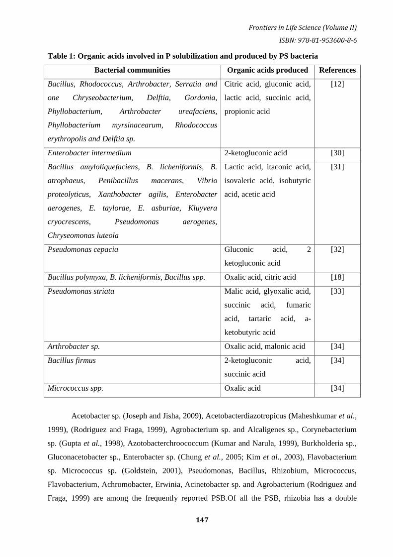
Frontiers in Life Science (Volume II)
ISBN: 978-81-953600-8-6
147
Table 1: Organic acids involved in P solubilization and produced by PS bacteria
Bacterial communities Organic acids produced References
Bacillus, Rhodococcus, Arthrobacter, Serratia and
one Chryseobacterium, Delftia, Gordonia,
Phyllobacterium, Arthrobacter ureafaciens,
Phyllobacterium myrsinacearum, Rhodococcus
erythropolis and Delftia sp.
Citric acid, gluconic acid,
lactic acid, succinic acid,
propionic acid
[12]
Enterobacter intermedium 2-ketogluconic acid [30]
Bacillus amyloliquefaciens, B. licheniformis, B.
atrophaeus, Penibacillus macerans, Vibrio
proteolyticus, Xanthobacter agilis, Enterobacter
aerogenes, E. taylorae, E. asburiae, Kluyvera
cryocrescens, Pseudomonas aerogenes,
Chryseomonas luteola
Lactic acid, itaconic acid,
isovaleric acid, isobutyric
acid, acetic acid
[31]
Pseudomonas cepacia Gluconic acid, 2
ketogluconic acid
[32]
Bacillus polymyxa, B. licheniformis, Bacillus spp. Oxalic acid, citric acid [18]
Pseudomonas striata Malic acid, glyoxalic acid,
succinic acid, fumaric
acid, tartaric acid, a-
ketobutyric acid
[33]
Arthrobacter sp. Oxalic acid, malonic acid [34]
Bacillus firmus 2-ketogluconic acid,
succinic acid
[34]
Micrococcus spp. Oxalic acid [34]
Acetobacter sp. (Joseph and Jisha, 2009), Acetobacterdiazotropicus (Maheshkumar et al.,
1999), (Rodriguez and Fraga, 1999), Agrobacterium sp. and Alcaligenes sp., Corynebacterium
sp. (Gupta et al., 1998), Azotobacterchroococcum (Kumar and Narula, 1999), Burkholderia sp.,
Gluconacetobacter sp., Enterobacter sp. (Chung et al., 2005; Kim et al., 2003), Flavobacterium
sp. Micrococcus sp. (Goldstein, 2001), Pseudomonas, Bacillus, Rhizobium, Micrococcus,
Flavobacterium, Achromobacter, Erwinia, Acinetobacter sp. and Agrobacterium (Rodriguez and
Fraga, 1999) are among the frequently reported PSB.Of all the PSB, rhizobia has a double

Bhumi Publishing, India
148
advantage; they provide N in addition to phosphate dissolution and can improve the growth of
legumes with other PGPR or mycorrhizaal fungi.
These PSB are ubiquitous but can vary in mineral phosphate solubilization capacity from
soil to soil or from one production system to another production system and greatly affected by
the nutritional status of soil and environmental factors. However, the metal, temperature, and
concentration of C and N sources greatly affect the PS potential of these bacteria in vitro
condition (Zaidi et al., 1999). These organisms are usually isolated from rhizosphere and non-
rhizosphere soil, rhizoplanes, phyllospheres, RP deposit area soils, and even in stressed soil by
serial plate dilution method or enrichment culture technique. Since 1948, when Gerretson
suggested that the phosphate which is present in soil, microbe could hardly soluble available
form and play an important role in providing phosphate to plant, a number of methods and
medium, such as (Pikovskaya et al., 1948), bromophenol blue dye method (Gupta et al., 1994)
and National Botanical Research Institute P (NBRIP) medium (Nautiyal et al.,1999) have been
proposed. Bacterial strains with phosphate solubilizing function are found due to the formation
of clear halo (a sign of solubilization) around the colonies (fig 2). Due to inconsistency and
variations in PS activity, the bacterial cultures are frequently sub-culture to test the persistence of
PS potential. Once selected efficient PSB, the release of P by PS bacteria is quantitatively
assayed and the potential P solubilizers are then mass produced and tested under pot/field
environments with different samples.
Mechanism of Phosphate solubilization by PSB
While reviewing phosphate chemistry in soil, sims and pierzensky (2005)identified the
major processes in the phosphate cycle that affect the soil solution P concentration as
1.dissolution-precipitation (mineral equilibria), 2. Sorption-desorption (interaction between P in
solution and soil solid surface, and 3. Mineralization–immobilization (biologically mediated
conversions of P between inorganic and organic form). The main phosphate merger processes
employed by soil microorganisms include: (1) release of complexing or mineral dissolving
compounds e.g. organic acid anions, siderophores, protons, hydroxyl ions, CO2, (2) liberation of
extracellular enzymes (biochemical P mineralization) and (3) the release of P during substrate
degradation (biological P mineralization) (McGill and Cole 1981). Therefore, microorganisms
play an important role in the three major components of the soil phosphate cycle (i.e. dissolution-
precipitation,sorption-desorption, and mineralization-immobilization,).In addition to these
microorganisms,the presence of labile C act as a sick for phosphate, making it rapidly
competitive even in low phosphate soil; so phosphate solubilizing microbes become a source of
phosphate for plants after they are excreted from their cells.Release of P immobilized by PSB
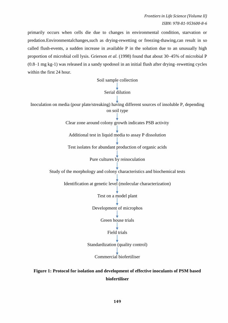
Frontiers in Life Science (Volume II)
ISBN: 978-81-953600-8-6
149
primarily occurs when cells die due to changes in environmental condition, starvation or
predation.Environmentalchanges,such as drying-rewetting or freezing-thawing,can result in so
called flush-events, a sudden increase in available P in the solution due to an unusually high
proportion of microbial cell lysis. Grierson et al. (1998) found that about 30–45% of microbial P
(0.8–1 mg kg-1) was released in a sandy spodosol in an initial flush after drying–rewetting cycles
within the first 24 hour.
Soil sample collection
Serial dilution
Inoculation on media (pour plate/streaking) having different sources of insoluble P, depending
on soil type
Clear zone around colony growth indicates PSB activity
Additional test in liquid media to assay P dissolution
Test isolates for abundant production of organic acids
Pure cultures by reinoculation
Study of the morphology and colony characteristics and biochemical tests
Identification at genetic level (molecular characterization)
Test on a model plant
Development of microphos
Green house trials
Field trials
Standardization (quality control)
Commercial biofertiliser
Figure 1: Protocol for isolation and development of effective inoculants of PSM based
biofertiliser

Bhumi Publishing, India
150
Figure 2: Solubilization of tri-calcium phosphate on Pikovskaya plates by species of
(A) Pseudomonas, (B) Serratia, (C) Bacillus
Figure 3: Mechanism of soil P solubilization/mineralization and immobilization by PSB
Inorganic P solubilizing:
P-solubilizing Bacteriasolubilise inorganic P mainly by organic acid production, either
by: (i) lowering the pH, or (ii) by enhancing chelation of the cations bound to P (iii) by

Frontiers in Life Science (Volume II)
ISBN: 978-81-953600-8-6
151
competing with P for adsorption sites on the soil (iv) by forming soluble complexes with metal
ions associated with insoluble P (Ca, Al, Fe) and thus P is released. The lowering in pH of the
medium suggests the release of organic acids by the P-solubilizing microorganisms (Whitelaw
2000; Maliha et al., 2004) via the direct oxidation pathway that occurs on the outer face of the
cytoplasmic membrane (Zaidi et al., 2009). These acid are products of microbial metabolism,
mainly by oxidative respiration or by fermentation of organic carbon sources, or such organic
acid can either dissolve mineral phosphate directly as result of anion exchange of P by acid anion
or can chelate Fe, Al and Ca ion associated with P (Omar, 1998).Monovalent anion phosphate
H2PO4 is soluble form of inorganic phosphate that usually occurs at low PH. However as the ph
of the soil environment increases the divalent and trivalent form of Pi (HPO4 -2 and HPO4 -3
respectively) occur. Thus, PSB strain is synthesis and discharge of organic acid into the
surrounding environment acidify the cell and their surrounding environment which causes the P
ions to be released from P minerals by H+ substitution for cation that binds phosphate.Major
acids secreted by PSB dissolution of insoluble P contains oxalic acid, citricacid (Kim et al.,
1997), gluconic acid (Di-Simine et al., 1998; Bar-Yosef et al., 1999), tartaric acid, aspartic acid
(Venkateswarlu et al., 1984). Evidence from an abiotic study using, P solubilize by HCL and
gluconic acid indicated that chelation of Al3+ by gluconic acid could be a factor in the
solubilization of colloidal Al phosphate (Whitelaw et al., 1999). Organic acid produced by PSB
can detect by high efficiency liquid chromatography and enzymatic methods. (Parks et al., 1990;
Whitelaw 2000) However, acidification does not appear to be the only mechanism for dissolving,
as in some cases the ability to lower the Ph does not match the ability to dissolve mineral
phosphate (SubbaRao, 1982). No organic acid were found in culture filters and therefore, the
authors concluded that acidification was probably not the major mechanism of dissolution as ph
never came below 5. The action of phosphate dissolution was attributed to both chelation and
reduction processes. However, organic acid has been suggested as the main mechanism of P
solubilization, solubilization of P by inorganic acid (HCL) has been reported, although HCL
could dissolve at low ph acid from hydroxyapatite than citric acid or oxalic acid at the same ph.
(Kim et al., 1997). Bacteria of the species Nitrosomonas and Thiobacillus can also dissolve
phosphate compounds by producing nitric and sulphuric acid (Azam and Memon
1996).According to sink theory, PSB remove and assimilate phosphate from the liquid and
therefore, activate the indirect decomposition of calcium phosphate compounds by continuously
removing P from the liquid culture medium.For example,the P content in the biomass of
Pseudomonas sp. And P. Aurantiogriseum was similar to that seen in non-P-solubilizing

Bhumi Publishing, India
152
microorganisms (Illmer et al., 1995) this can be explained by the fact the content of phosphate in
the biomass of organisms is constantly related to the organic decomposition of P containing
organic substrates (Dighton and Boddy, 1989). The other mechanism is the production of H2S,
which reacts with ferric phosphate to form ferrous sulphate released in association with P.
(Swaby and Sperber, 1958). Parks et al., (1990) proposed the H+ excretion originating from NH4
+ assimilation is the alternative mechanisms of P solubilization.An HPLC analysis of the culture
solution of Pseudomonas sp., in contrast to the expectation, did not detect any organic acid while
solubilization occurred (Illmer and Schinner, 1995). They also reported that the most probable
reason for solubilization without acid production is the release of protons accompanying
respiration or NH4 + assimilation. Krishnaraj et al. (1998) has proposed a model explaining the
importance of protons outside the cell responsible for P solubilization.Here the direct role of
organic or inorganic acid is denied. For some microbes, NH4+driven proton release appears to be
the only mechanism to promote phosphate solubilization.According to a study by Pseudomonas
fluorescence,the nature of the C supply (e.g. NH4+ vs. NO3-) had the greatest effect on proton
release (Park et al., 2009). Acidification of the rhizosphere of cactus seedlings (giant cardon,
Pachycereuspringlei) after inoculation with the plant growth promoting bacterium Azospirillum
brasilense, in the presence or absence of ammonium and nitrate,was studied and assumed to be
so the effect of inoculation with this PGPB on plant growth, combining nitrogen with nutrients
may affect one or more things due to metabolic pathways of plants that increase proton flow
from the roots and released organic acid,leading to rhizosphere acidification (Carrillo et al.,
2002). This suggests that for different species, different mechanisms are responsible for proton
release, depending only on the presence of NH4+. Goldstein (1995) suggested that extracellular
oxidation via direct oxidation pathway may play an important role in soils in which calcium
phosphates provide a significant pool of unavailable mineral phosphorus.
Organic P solubilization:
Organic P solubilizationis also known as mineralization of organic phosphorus. Soil organic
mineralization plays an essential role in the phosphorus cycling of phosphorus cycling of
phosphate farming systems.Organic phosphate can be 4-90% of the total phosphate soil. Such P
can be released from organic compounds in soil by enzyme:
Non-specific acid phosphatases (NSAPs) which dephosphorylate phosphor-ester or
phosphoanhydride bonds of organic matter.In a variety of the phosphatase enzyme class
released by PSB, phosphomonosterase (often simply called phosphatases) are most
abundant and best studied (Nannipieri et al., 2011). Based on their ph optima,these
enzymes are divided into acid and alkaline phosphomonoesterases and can be produed

Frontiers in Life Science (Volume II)
ISBN: 978-81-953600-8-6
153
both by PSB depending on external conditions (Kim et al., 1998; Jorquera et al., 2008).
Usually, acid phosphatases predominate in acid soils, so alkaline phosphatases are more
abundant in neutral and alkaline soils (Eivazi and Tabatabai 1977; Juma and Tabatabai
1977, 1998; Renella et al., 2006). Although plant roots produce acid phosphatases but
rarely produce large amounts of alkaline phosphatases, this suggests that this is a
potential niche for PSB (Juma and Tabatabai 1998; Criquet et al., 2004). It is also
difficult to distinguish between root and PSB produced phosphatases (Richardson et al.,
2009a, b) but some evidence suggests that phosphatases of microbial origin are more
abundant in organic phosphate compounds than those derived from plant roots (Tarafdar
et al., 2001).
Phytases, which in particular cause release of P from phytate degradation.
Basicallyphytate is the main source of inositol and the main stored form of P in plant
seeds and pollen and is a major component of 0rganic phosphate in the soil. (Richardson,
1994)
Phosphonatases and C–P Lyases, the phosphonates degrading enzymes that perform C–P
cleavage in organophosphonates (Rodriguez et al., 2006).
Role of siderophores in P solubilization:
Siderophores are complex agents that have a high iron attraction and form almost all
microorganisms in response to iron deficiency.In iron limiting condition, siderophore act as
solubilizing agent for iron from minerals or organic compounds. There are approximately 500
known siderophore, most of which are used by all microbs and plants some of them being
exclusively used by the microbial species and strains that produce them (Crowley 2007). Given
the predominance of mineral dissolution on ligand exchange by organic acid anion as a
phosphate solubilizing mechanism, the potential role of siderophores in increasing phosphate
availability needs to be clarified.
Role of EPS in P solubilization:
The role of polysaccharides in the microbial mediation of phosphorus was recently
evaluated by Yi et al. (2008). Microbial exopolysaccharides (EPS) are polymers consisting
primarily of carbohydrates in which certain bacteria and fungi are excreted on the outside of their
cell walls. Their structure and composition are very different; they can be homo or
heteropolysaccharides and can also contain a lot of organic and inorganic substances (Sutherland
2001). Four bacterial strains of, Arthrobacter sp. (ArHy-505), Enterobacter sp. (EnHy-401),
Enterobacter sp. (EnHy-402), Azotobacter sp. (AzHy-510) had the ability to dissolve TCP (Tri
Bhumi Publishing, India
154
calcium phosphate), (Yi et al. 2008) and was used to evaluate the role of exopolysaccharide
(EPS) in the solubilization of P. These phosphate solubilizing bacteria produced significant
amount of EPS and showed a strong potential for phosphate solubilization.
Phosphate solubilizing bacteria in sustainable agriculture:
Energy crisis, environmental hazards, and depleting soil fertility etc. are the factor which
is responsible for increasing the risk to sustainable agriculture. Therefore the use of bio fertilizers
in agriculture has proven to be environmentally friendly, productive and accessible (Sheraz et al.,
2010). PSB are a major contributor to plant nutrition in agriculture. The use of PSM is currently
considered as a concrete strategy to improve the productivity of the soil under crop production.
PSB have been used as bio fertilizers since the 1950 (Krasilinikov, 1957). PGPR and PSB
collectively can reduce the use of phosphate fertilizer by 50% without significantly reducing the
yield of crop (Jilani et al., 2007). Seed inoculation with PSB is a promising technique that
eliminates phosphate deficiency (Qureshi et al., 2012). inoculation with PSB such as
Pseudomonas, Bacillus, Rhizobium, Micrococcus, Flavobacterium, Achromobacter, Erwinia,
and Agrobacterium have been reported to increase the curtainPS ensuring high crop yields
(Rodriguez and Fraga, 1999). Bacillus and Paenibacillus have been reported to be potential
PGPRs in sustainable agriculture and are major sources of broad spectrum peptides antibiotic
that are active against various microbial and nematode pathogens (Venkadasamy et al., 2010).
Ankit et al. referred to Bacillus as the PGPR in crop ecosystem (Ankit et al., 2011).Diriba et al.,
phosphate in the presence of two phosphate sources, hydroxyapatite and tricalcium phosphate,
the solubility of different strain of rhizobacteria associated with Coffea arabica L. is evaluated.
Total 395 is isolated tested, more than 72% Pseudomonas erwinia and P. chlororaphis were
mainly able to strongly dissolve phosphate sources, and the analysis of HPCL showed several
organic acid, with 2-ketogluconic acid dominating (Diriba Muleta et al., 2013). Isolates Bacillus
spp. (67%) and Burcholderia spp. (58.5%) were shown in the corn rhizosheresolubilisation of
aluminium, phosphate (AlPO4), and tricalcium phosphate (Ca3 (PO4)2) (Oliveira et al., 2009).
Studies on Pseudomonas and Trichodermam have shown positive IAA (indole-3-acetic acid)
production, and antagonistic action against Fusarium oxysporum and Rhizoctonia solani to
increase growth and disease consumption (Jay et al., 2014). Experiment on the effect of
Pseudomonas letiola on mobilization of P and growth of Ligol (young apple trees) grown in a
pot, significantly increased the total shoot length and solubilised insoluble P compounds (Ewa et
al., 2013). Rhodococcus sp. EC35, Pseudomonas sp. EAV and Arthrobacter nicotinovorans
EAPAA when inoculated with Zea mays and grown in P deficient soils amended with tricalcium
phosphate enhanced the plant growth (Sofia and Paula, 2014). PSB can be considered as broad

Frontiers in Life Science (Volume II)
ISBN: 978-81-953600-8-6
155
spectrum biofertilizer. PSB like Bacillus megaterium, Bacillus circulans, Bacillus subtilis and
Pseudomonas straita are effective biofertilizers. Burkholderia cepacia in the rhizosphere of
maize showed tricalcium phosphate solubility and promoted the growth of both healthy and
Helminthosporium maydis infected maize plant, indicating that the isolate was a good candidate
to be applied as a biofertilizer (Ke Zhao et al., 2014). Pseudomonas sp., Serratia marcescens and
Bacillus cereus were known to reduce the bacterial wilt caused by Ralstonia solanacearum in
tomato proved to be effective biocontrol agents (Henok and Kerstin, 2013). In a pot experiment
Pseudomonas striata when combined with endophytic fungi Piriformospora indica increased the
dry plant biomass in chick peasand increased the population of P. striata in the rhizosphere
(Kamlesh et al., 2010). Pseudomonas chlororaphis, Bacillus cereus, and Pseudomonas
fluorescens inoculated with walnut seedlings improved height, shoot and root dry weight, and
increasing P and nitrogen (N) uptake of the seedlings (Xuan et al., 2011). Evaluation of the
effect of PSB, with differentiation of, Bacillus M-13 and without varying amounts of phosphorus
on growth and production of sunflower in agricultural condition gives the highest yield of sun
flower seeds (Zehra, 2010). Bacillus sp. enhanced the growth and yield of cotton (Qureshi et al.,
2012). In addition to the above phosphate solubilising microorganisms facilitate the growth and
development of plant in the presence of various stresses. Pseudomonas sp. enhanced plant
growth in salt stress (Shimaila et al., 2014) and Pseudomonas putida (Pandey et al., 2006),
Pseudomonas corrugate (Trivedi and Sa, 2008), Mycobacterium sp. (Egamberdiyeva and
Hoflich (2003) are showed to be cold tolerant. Pseudomonas sp. (Sandhya et al., 2010),
Arthrobacter sp., Bacillus sp. (Banerjee et al., 2010) showed tolerant towards drought stress and
Bacillus and Hallo Bacillus towards salinity stress (Dhanushkodi et al., 2013).
Conclusion:
In agricultural practices, chemical phosphate fertilizers are applied to eliminate phosphate
deficiency. However, excessive and harmful use of these fertilizers poses a serious threat to
microbial diversity, soil microbial community structure, soil fertility and consequently crop
productivity in various agricultural systems around the world. Phosphate and Phosphatic
fertilizers are expensive in most soil around the world, so the focus is on the use of soil
microorganisms rich in phosphate solubilization capacity, which cannot be utilized to deter
phosphate from sources not available in the soil. The use of such phosphate solubilizing
microorganisms along with bacteria opens up new horizons in terms of better plant production
and protects agro-ecosytem from the dangers of agrochemicals. Protecting the soil environment
Bhumi Publishing, India
156
by using PSB can be a big win for crops grown in decomposing soils. Furthermore the molecular
engineering of PSB has provided new insights for the cultivation of crop in P deficient soil.
References:
Ahmad, F., Ahmad, I., Khan, M. S.: Screening of free-living rhizospheric bacteria for their
multiple plant growth promoting activities. Microbiol Res 163, 173–181 (2008).
Asea, P. E. A., Kucey, R. M. N., Stewart, J. W. B.: Inorganic phosphate solubilization by two
Penicillium species in solution culture and soil. Soil BiolBiochem 20, 459–464 (1988).
Azcon-Aguilar, C., Diaz-Rodriguez, R., Barea, J. M.: Effect of soil microorganisms on spore
germination and growth on the vesicular arbuscularmycorrhizal fungus Glomus moseae.
Trans Br MycolSoc 86, 337–340 (1986).
Banik, S., Dey, B. K.: Available phosphate content of an alluvial soil as influenced by
inoculation of some isolated phosphate-solubilizing microorganisms. Plant Soil 69, 353–
364 (1982).
Bar-Yosef, B., Rogers, R. D., Wolfram, J. H., Richman, E.: Pseudomonas cepacia mediated rock
phosphate solubilization in kaolinite and montmorillonite suspensions. Soil SciSoc Am J
63, 1703–1708 (1999).
Canbolat, M. Y., Bilen, S., Cakmakci, R., Sahin, F., Aydin, A.: Effect of plant growth-promoting
bacteria and soil compaction on barley seedling growth, nutrient uptake, soil properties and
rhizosphere microflora. BiolFertil Soils 42, 350–357 (2006).
Chen, Y. P., Rekha, P. D., Arun, A. B., Shen, F. T., Lai, W. A., Young, C. C.: Phosphate
solubilizing bacteria from subtropical soil and their tricalcium phosphate solubilizing
abilities. Appl Soil Ecol 34, 33–41 (2006).
Chung, H., Park, M., Madhaiyan, M., Seshadri, S., Song, J., Cho, H., Sa, T.: Isolation and
characterization of phosphate solubilizing bacteria from the rhizosphere of crop plants of
Korea. Soil BiolBiochem 37, 1970–1974 (2005).
Colbert, S. F., Hendson, M., Ferri, M., Schroth, M. N.: Enhanced growth and activity of a
biocontrol bacterium genetically engineered to utilize salicylate. ApplMicrobiol 59, 2071–
2076 (1993).
Cunningham, J., Kuiack, C.: Production of citric and oxalic acids and solubilization of calcium
phosphate by Penicilliumbilaii. Appl Environ Microbiol 58, 1451–1458 (1992).
De Freitas, J. R., Banerjee, M. R., Germida, J. J.: Phosphate-solubilizing rhizobacteria enhance
the growth and yield but not phosphorus uptake of canola (Brassica napus L.). BiolFertil
Soils 24, 358–364 (1997).

Frontiers in Life Science (Volume II)
ISBN: 978-81-953600-8-6
157
Dighton, J., Boddy, L.: Role of fungi in nitrogen, phosphorus and sulfur cycling in temperate
forest ecosystems. In Boddy, L., Marchant, R., Read, D. (eds): Nitrogen, Phosphorus and
Sulfur Ultilization by Fungi. Cambridge University Press, Cambridge, 1989, pp. 269–298.
Dubey, S. K.: Associative effect of nitrogen fixing and phosphate solubilizing bacteria in rainfed
soybean (Glycine max) grown in vertisols. Ind J AgricSci 71, 476–479 (2001).
Fraga-Vidal, R., Rodriguez, H. M., De Villegas, T. G.: Vector for chromosomal integration of
the phoC gene in plant growth-promoting bacteria. In Velazquez, E. C., Rodríguez-
Barrueco (eds): First International Meeting on Microbial Phosphate Solubilization, 2003,
pp. 239–244.
Ganesan, V.: Rhizoremediation of cadmium soil using a cadmium-resistant plant growth-
promoting rhizopseudomonad. CurrMicrobiol 56, 403–407 (2008).
Gaur, A. C.: Phosphate Solubilizing Microorganisms as Biofertilizers. Omega Scientific
Publisher, New Delhi, India 1990, 176 pp.
Glick, B. R., Bashan, Y.: Genetic manipulation of plant growth promoting bacteria to enhance
biocontrol of phytopathogens. BiotechnolAdv 15, 353–378 (1997).
Goldstein, A. H.: Involvement of the quinoprotein glucose dehydrohenase in the solubilization of
exogenous phosphates by gram-negative bacteria. In Torriani-Gorini, A., Yagil, E., Silver,
S. (eds): Phosphate in Microorganisms: Cellular and Molecular Biology. ASM Press,
Washington, DC, 1994, pp. 197–203.
Gull, M., Hafeez, F. Y., Saleem Malik, K. A.: Phosphorus uptake and growth promotion of
chickpea by co-inoculation of mineral phosphate solubilizing bacteria and a mixed
rhizobial culture. Aus J ExpAgric 44, 623–628 (2004).
Gupta, R. R., Singal, R., Shanker, A., Kuhad, R. C., Saxena, R. K.: A modified plate assay for
secreening phosphate solubilizing microorganisms. Gen. Appl. Microbiol., 40, 255–260
(1994).
Gyaneshwar, P., Naresh, K., Parekh, L. J.: Cloning of mineral phosphate solubilizing genes from
Synechocystis PCC 6803. Curr. Sci. 74, 1097–1099 (1998b).
Halvorson, H. O., Keynan, A., Kornberg, H. L.: Utilization of calcium phosphates for microbial
growth at alkaline pH. Soil Biol. Biochem., 22, 887–890 (1990).
Hameeda, B., Harini, G., Rupela, O. P., Wani, S. P., Reddy, G.: Growth promotion of maize by
phosphate-solubilizing bacteria isolated from composts and macrofauna. Microbiol. Res.,
163, 234–242 (2008).
Bhumi Publishing, India
158
Henri, F., Lauret, F., Te, N. N., Annette, D., John, Q., Wolfgang, M., François-Xavier, E.,
Dieudonne, N.: Solubilization of inorganic phosphates and plant growth promotion by
strains of Pseudomonas fluorescens isolated from acidic soils of Cameroon. Afric J
Microbiol. Res., 2, 171–178 (2008).
Hoon, H., Park, R. D., Kim, Y. W., Rim, Y. S., Park, K. H., Kim, T. H., Such, J. S., Kim, K. Y.:
2-ketogluconic acid production and phosphate solubilization by Enterobacter intermedium.
Curr. Microbiol., 47, 87–92 (2003).
Illmer, P., Schinner, F.: Solubilization of inorganic phosphates by microorganisms isolated from
forest soil. Soil Biol. Biochem., 24, 389–395 (1992).
Illmer, P., Schinner, F.: Solubilization of inorganic calcium phosphates-solubilization
mechanisms soil. Soil Biol. Biochem., 27, 257–263 (1995).
ActaMicrobiologicaetImmunologicaHungarica 56, 2009 280 ZAIDI et al.
Indiragandhi, P., Anandham, R., Madhaiyan, M., Sa, T. M.: Characterization of plant growth-
promoting traits of bacteria isolated from larval guts of diamondback moth
Plutellaxylostella (Lepidoptera: Plutellidae). Curr. Microbiol., 56, 327–333 (2008).
Jiang, C., Sheng, X., Qian, M., Wang, Q.: Isolation and characterization of a heavy metal-
resistant Burkholderia sp. from heavy metal-contaminated paddy field soil and its potential
in promoting plant growth and heavy metal accumulation in metal-polluted soil.
Chemosphere 72, 157–164 (2008).
Khan, M. S., Zaidi, A., Wani, P. A.: Role of phosphate solubilizing microorganisms in
sustainable agriculture – A review. Agron Sustain Dev 27, 29–43 (2007).
Khan, M. S., Zaidi, A., Aamil, M.: Biocontrol of fungal pathogens by the use of plant growth
promoting rhizobacteria and nitrogen fixing microorganisms. Ind. J. Bot. Soc., 81, 255–
263 (2002).
Khan, M. S., Zaidi, A.: Influence of composite inoculations of phosphate solubilizing organisms
and an arbuscularmycorrhizal fungus on yield, grain protein and phosphorus and nitrogen
uptake by greengram. Arch. Agron. Soil. Sci., 52, 579–590 (2006).
Khan, M. S., Aamil, M., Zaidi, A.: Associative effect of Bradyrhizobium sp. (vigna) and
phosphate solubilizing bacteria on moongbean (Vignaradiata [L. ]wilczek). Biojornal., 10,
101–106 (1997).
Khan, M. S., Aamil, M., Zaidi, A.: Moongbean response to inoculation with nitrogen fixing and
phosphate solubilizing bacteria. In: Deshmukh, A. M. (ed.): Biofertilizers and
Biopesticides, Technoscience Publications, Jaipur, 1998, pp. 40–48.
Frontiers in Life Science (Volume II)
ISBN: 978-81-953600-8-6
159
Khan AA, Jilani G, Akhtar MS, Naqvi SMS, Rasheed M (2009a) Phosphorus solubilizing
bacteria: occurrence, mechanisms and their role in crop production. J. Agric. Biol. Sci.,
1(1):48–58.
Krishnaraj, P. U., Goldstein, A. H.: Cloning of a Serratiamarcescens DNA fragment that induces
quinoprotein glucose dehydrogenase-mediated gluconic acid production in Escherichia coli
in the presence of stationary phase Serratiamarcescens. FEMS Microbiol. Lett., 205, 215–
220 (2001).
Kumar, V., Behl, R. K., Narula, N.: Establishment of phosphate-solubilizing strains of
Azotobacterchroococcum in the rhizosphere and their effect on wheat cultivars under green
house conditions. Microbiol. Res., 156, 87–93 (2001).
Kumar, K. V., Singh, N., Behl, H. M., Srivastava, S.: Influence of plant growth promoting
bacteria and its mutant on heavy metal toxicity in Brassica juncea grown in fly ash
amended soil. Chemosphere, 72, 678–683 (2008).
Madhaiyan, M., Poonguzhali, S., Sa, T.: Metal tolerating methylotrophic bacteria reduces nickel
and cadmium toxicity and promotes plant growth of tomato (Lycopersiconesculentum L.).
Chemosphere 69, 220–228 (2007).
Maliha, R., Samina, K., Najma, A., Sadia, A., Farooq, L.: Organic acids production and
phosphate solubilization by phosphate solubilizing microorganisms under in vitro
conditions. Pak. J. Biol. Sci., 7, 187–196 (2004).
Natarajan, T., Subrammanian, P.: Response of phosphobacteria along with Rhizobium at two
levels of phosphorus on groundnut. In: Microbiology Abstracts, XXXVI Ann. Conf.
Assoc. Microbiol., India, Hissar, Nov. 8–10, 1995, p. 111. Acta Microbiologicaet
Immunologica Hungarica 56, 2009 Phosphate Solubilizing Bacteria 283
Nautiyal, C. S.: An efficient microbiological growth medium for screening of phosphate
solubilizing microorganisms. FEMS Microbiol Lett 170, 265–270 (1999).
Pandey, A., Trivedi, P., Kumar, B., Palni, L. M. S.: Characterization of a phosphate solubilizing
and antagonistic strain of Pseudomonas putida (B0) isolated from a Sub-Alpine location in
the Indian Central Himalaya. Curr. Microbiol., 53, 102–107 (2006).
Park KH, Lee CY, Son HJ (2009) Mechanism of insoluble phosphate solubilization by
Pseudomonas fluorescens RAF15 isolated from ginseng rhizosphere and its plant growth-
promoting activities. Lett. Appl. Microbiol., 49:222–228.
Perveen, S., Khan, M. S., Zaidi, A.: Effect of rhizospheric microorganisms on growth and yield
of greengram (Phaseolusradiatus). Ind. J. Agric. Sci., 72, 421–423 (2002).

Bhumi Publishing, India
160
Pikovskaya, R. I.: Mobilization of phosphorus in soil in connection with vital activity of some
microbial species. Microbiol., 17, 362–370 (1948).
Poi, S. C., Ghosh, G., Kabi, M. C.: Response of chickpea (Ciceraeritinum L.) to combined
inoculation with Rhizobium, phosphobacteria and mycorrhizal organisms. Zentral fur
Microbiol., 114, 249–253 (1989).
Ponmurugan, P., Gopi, C.: In vitro production of growth regulators and phosphatase activity by
phosphate solubilizing bacteria. African J. Biotechnol., 5, 348–350 (2006).
Pradhan, N., Sukla, L. B.: Solubilization of inorganic phosphates by fungi isolated from
agriculture soil. African J. Biotechnol., 5, 850–854 (2005).
Rajkumar, M., Freitas, H.: Influence of metal resistant-plant growth-promoting bacteria on the
growth of Ricinuscommunis in soil contaminated with heavy metals. Chemosphere, 71,
834–842 (2008).
Rajkumar, M., Nagendran, R., Lee, K. J., Lee, W. H., Kim, S. Z.: Influence of plant growth
promoting bacteria and Cr6+ on the growth of Indian mustard. Chemosphere, 62, 741–748
(2006).
Ramachandran, K., Srinivasan, V., Hamza, S., Anandaraj, M.: Phosphate solubilizing bacteria
isolated from the rhizosphere soil and its growth promotion on black pepper (Piper nigrum
L.) cuttings. Developments in Plant and Soil Sciences, 20, 102, 325–331 (2007).
Richardson AE, Hocking PJ, Simpson RJ, George TS (2009b) Plant mechanisms to optimize
access to soil phosphorus. Crop Pasture Sci., 60:124–143
Rodriguez, H., Fraga, R.: Phosphate solubilizing bacteria and their role in plant growth
promotion. Biotechnol. Adv., 17, 319–339 (1999).
Rodríguez, H., Gonzalez, T., Selman, G.: Expression of a mineral phosphate solubilizing gene
from Erwiniaherbicola in two rhizobacterial strains. J. Biotechnol., 84, 155–161 (2000).
ActaMicrobiologicaetImmunologicaHungarica 56, 2009 282 ZAIDI et al.
Rodriguez H, Fraga R (1999) Phosphate solubilizing bacteria and their role in plant growth
promotion. Biotechnol. Adv., 17:319–339
Sarojini, V., Mathur, R. S.: The effects of microbial inoculations on the yield of wheat when
grown in straw ammended soil. Biol. Wastes., 33, 9–16 (1990).
Seema B Sharma1*, Riyaz Z Sayyed2 ,Mrugesh H Trivedi1 and Thivakaran A Gobi3 to
Phosphate solubilizing microbes: sustainable approach for managing phosphorus
deficiency in agricultural soils, (2013)
Selvakumar, G., Mohan, M., Kundu, S., Gupta, A. D., Joshi, P., Nazim, S., Gupta, H. S.: Cold
tolerance and plant growth promotion potential of Serratiamarcescens strain SRM (MTCC
Frontiers in Life Science (Volume II)
ISBN: 978-81-953600-8-6
161
8708) isolated from flowers of summer squash (Cucurbita pepo). Let. Appl. Microbiol., 46,
171–175 (2008).
Shaharoona, B., Naveed, M., Arshad, M., Zahir, Z. A.: Fertilizer-dependent efficiency of
Pseudomonads for improving growth, yield, and nutrient use efficiency of wheat
(Triticumaestivum L.). Appl. Microbiol Biotechnol., 79, 147–155 (2008).
Singh, N., Pandey, P., Dubey, R., Maheshwari, D. K.: Biological control of root rot fungus
Macrophominaphaseolina and growth enhancement of Pinusroxburghii (Sarg.) by
rhizosphere competent Bacillus subtilis BN1. World J. Microbiol. Biotechnol., DOI
10.1007/s11274-008-9680-z (2008).
Thiagrajan, T. R., Ames, R. N., Ahmad, M. H.: Response of cowpea (Vignaunguiculata) to
inoculated with co-selected vesicular arbuscular mycorrhizal fungi and Rhizobium strains
in field trials. Can. J. Microbiol., 38, 573–576 (1992).
Valverde, A., Burgos, A., Fiscella, T., Rivas, R., Velazquez, E., Rodriguez-Barrueco, C.,
Cervantes, E., Chamber, M., Igual, J. M.: Differential effects of coinoculations with
Pseudomonas jessenii PS06 (a phosphate-solubilizing bacterium) and Mesorhizobiumciceri
C-2/2 strains on the growth and seed yield of chickpea under greenhouse and field
conditions. Plant and Soil, 287, 43–50 (2006).
Vazquez, P., Holguin, G., Puente, M., Elopez Cortes, A., Bashan, Y.: Phosphate solubilizing
microorganisms associated with the rhizosphere of mangroves in a semi arid coastal
lagoon. Biol. Fert. Soils, 30, 460–468 (2000).
Wani, P. A., Khan, M. S., Zaidi, A.: Co-inoculation of nitrogen fixing and phosphate solubilizing
bacteria to promote growth, yield and nutrient uptake in chickpea. Acta. Agron. Hung., 55,
315–323 (2007c).
Wani, P. A., Zaidi, A., Khan, A. A., Khan, M. S.: Effect of phorate on phosphate solubilization
and indole acetic acid (IAA) releasing potentials of rhizospheric microorganisms. Ann.
Plant Prote. Sci., 13, 139–144. 23–230 (2005).
Wani, P. A., Khan, M. S., Zaidi, A.: Synergistic effects of the inoculation with nitrogen fixing
and phosphate solubilizing rhizobacteria on the performance of field grown chickpea. J.
Plant. Nutr. Soil Sci., 170, 283–287 (2007a).
Wani, P. A., Khan, M. S., Zaidi, A.: Chromium reducing and plant growth promoting
Mesorhizobium improves chickpea growth in chromium amended soil. Biotechnol Lett 30,
159–163 (2008). Acta Microbiologicaet Immunologica.Hungarica, 56, 2009 Phosphate
Solubilizing Bacteria 281

Bhumi Publishing, India
162
Whitelaw, M. A.: Growth promotion of plant inoculated with phosphate-solubilizing fungi.
AdvAgrono 69, 100–151 (2000). 75. Saber, M. S. M., Kabesh, M. O.: Utilization of
biofertilizers in field crop production. II. A comparison study on the effect of
biofertilization or sulphur application on yield and nutrient uptake by lentil plants.
Egyptian J. Soil Sci., 30, 415–422 (1990).
Zaidi, A.: Synergistic interactions of nitrogen fixing microorganisms with phosphate mobilizing
microorganisms, PhD Thesis, Aligarh Muslim University, Aligarh, 1999.
Zaidi, A., Khan, M. S.: Stimulatory effects of dual inoculation with phosphate solubilizing
microorganisms and arbuscularmycorrhizal fungus on chickpea. Aust. J. Exp. Agric., 47,
1016–1022 (2007).
Zaidi, A., Khan, M. S., Amil, M.: Interactive effect of rhizotrophic microorganisms on yield and
nutrient uptake of chickpea (Cicer arietinum L.). Eur. J. Agron., 19, 15–21 (2003).
Zaidi, A., Khan, M. S., Aamil, M.: Bio-associative effect of rhizospheric microorganisms on
growth, yield and nutrient uptake of greengram. J. Plant Nutr., 27, 599–610 (2004).
Zaidi, S., Usmani, S., Singh, B. R., Musarrat, J.: Significance of Bacillus subtilis strain SJ101 as
a bioinoculant for concurrent plant growth promotion and nickel accumulation in Brassica
juncea. Chemosphere, 64, 991–997 (2006).