frontal lobe and cognitive development
TRANSCRIPT

Journal of Neurocytology 31, 373–385 (2002)
Frontal lobe and cognitive developmentJOAQUIN M. FUS TER
Neuropsychiatric Institute and Brain Research Institute, UCLA School of Medicine Los Angeles, [email protected]
Received December 1, 2002; accepted December 12, 2002
Abstract
In phylogeny as in ontogeny, the association cortex of the frontal lobe, also known as the prefrontal cortex, is a late-developingregion of the neocortex. It is also one of the cortical regions to undergo the greatest expansion in the course of both evolutionand individual maturation. In the human adult, the prefrontal cortex constitutes as much as nearly one-third of the totality ofthe neocortex. The protracted, relatively large, development of the prefrontal cortex is manifest in gross morphology as wellas fine structure. In the developing individual, its late maturation is made most apparent by the late myelination of its axonalconnections. This and other indices of morphological development of the prefrontal cortex correlate with the developmentof cognitive functions that neuropsychological studies in animals and humans have ascribed to this cortex. In broad outline,the ventromedial areas of the prefrontal cortex, which with respect to other prefrontal areas develop relatively early, areinvolved in the expression and control of emotional and instinctual behaviors. On the other hand, the late maturing areas ofthe lateral prefrontal convexity are principally involved in higher executive functions. The most general executive functionof the lateral prefrontal cortex is the temporal organization of goal-directed actions in the domains of behavior, cognition,and language. In all three domains, that global function is supported by a fundamental role of the lateral prefrontal cortex intemporal integration, that is, the integration of temporally discontinuous percepts and neural inputs into coherent structures ofaction. Temporal integration is in turn served by at least three cognitive functions of somewhat different prefrontal topography:working memory, preparatory set, and inhibitory control. These functions engage the prefrontal cortex in interactive cooperationwith other neocortical regions. The development of language epitomizes the development of temporal integrative cognitivefunctions and their underlying neural substrate, notably the lateral prefrontal cortex and other late-developing cortical regions.
Introduction
The prefrontal cortex is the cortex of association of thefrontal lobe. In the mammalian brain, this cortex is con-ventionally defined by two basic criteria: cytoarchitec-ture and connectivity. Both criteria serve us to delimitapproximately the same cortical territory, which is char-acterized in all mammalian species by a prominent cel-lular layer IV, or granular layer, and a tight recipro-cal connectivity with the mediodorsal nucleus of thethalamus. In the primate, human or nonhuman, theprefrontal cortex has three major anatomical aspects orregions: lateral, medial, and ventral or orbital (Fig. 1).Each prefrontal region is subdivided into areas of vary-ing cytoarchitecture, providing the grounds for a num-ber of cytoatchitectonic maps, such as that of Brodmann(1909). With few exceptions, such as that of area 8, whichis largely devoted to the control of gaze and eye move-ments, it is not possible to ascribe a specific physiolog-ical function to any prefrontal area. However, it seemsobvious that the prefrontal cortex is functionally het-erogeneous. Whereas it cannot be functionally parceledout with regard to its cytoarchitecture, there is substan-tial evidence that, as a whole, the prefrontal cortex per-
forms a critical role in the organization of behavioral,linguistic, and cognitive actions. The psychological andphysiological analysis of this role in the three actiondomains yields a topographic distribution of cognitivefunctions conforming to the following outline. All threeprefrontal regions are involved in one or another as-pect of attention. In addition, the medial and anteriorcingulate region are involved in drive and motivation,the lateral region in working memory and set, and theorbital region (to some extent also the medial region) inthe inhibitory control of impulses and interference.
This article deals with the developmental aspects ofthe prefrontal cortex and its cognitive functions. After abrief exposition of morphological development in bothphylogenetic and ontogenetic terms, the article dealswith the prefrontal cortex of the primate as the sub-strate for temporal integration, as well as the cognitivefunctions that support it. The cognitive functions of theadult human prefrontal cortex are viewed as the culmi-nation of biological processes that lead to the highestexpressions of temporal integration in language andintellectual performance.
0300–4864 C© 2003 Kluwer Academic Publishers

374 FUSTER
Fig. 1. Three views of the cerebral hemispheres with the areas of the prefrontal cortex numbered in accord with Brodmann’scytoarcitectonic map.
Evolution
The prefrontal cortex, like the rest of the neocortexor neopallium, evolves in the dorsal telencephalon be-tween two older structures, the laterally situated ol-factory (piriform) pallium and the medially situatedhippocampal pallium. The precise evolutionary processthat gives rise to the neocortex is unresolved (Northcutt& Kaas, 1995). There are two major lines of thinking inthis respect: One, that the neocortex develops as an ex-pansion of those ancient structures (Pandya et al., 1988);the other, that it develops from a ridge of cells along thedorsal wall of the ventricle (Butler, 1994). In any case, itis generally accepted that, with evolution, the neocortexas a whole increases in size and volume in proportionto body dimensions, (Stephan et al., 1981; Jerison, 1990).The growth of the neocortex in evolution can be charac-
terized as a veritable phylogenetic “explosion’’ (Finlay& Darlington, 1995).
The most rostral aspect of the developing neopal-lium in primitive species constitutes what is to be-come the prefrontal cortex. Whereas the homology ofthe neocortex as a whole in the various mammalianspecies is undisputed, the homology of individual neo-cortical areas, prefrontal areas in particular, is a matterof some controversy. Nonetheless, the evidence fromcomparative studies of existing species and from theexamination of the endocasts of specimens of extinctspecies—reviewed by Fuster, 1997b—leads to the con-clusion that, in the course of evolution, the prefrontalcortex grows disproportionately more than other corti-cal regions (Fig. 2). According to Brodmann (1909), theprefrontal cortex constitutes 3.5% of the totality of thecortex in the cat, 12.5% in the dog, 11.5% in the macaque,

Frontal lobe and cognitive development 375
Fig. 2. Prefrontal cortex (shaded) in six animal species.

376 FUSTER
17% in the chimpanzee, and 29% in the human. Ar-guably, the disproportionate evolutionary growth ofthe prefrontal cortex parallels that of the associative cor-tex of temporal and parietal regions. It is a legitimate in-ference, in any event, that the evolutionary expansion ofthe cortex of association, both posterior and prefrontal,is closely related to the evolution of cognitive functions.
Judging from the evolutionary development of sur-face morphology (i.e., sulci and gyri), as well as ofthe components of its thalamic nucleus (mediodorsal)and their cortical projections, the various portions ofthe prefrontal cortex do not appear to evolve equallyat the same time. Rather, by those criteria, the lateralprefrontal region clearly evolves later and farther thanthe other prefrontal regions. This is in obvious agree-ment with the late and extraordinary development ofhigher integrative cognitive functions (e.g., language) inhigher species, especially the human. These functions,as we see below, are largely dependent on the lateralprefrontal cortex.
Ontogeny
In accord with the principle that ontogeny recapitulatesphylogeny, the prefrontal cortex is one of the cortical ar-eas to develop most and last in the course of individualdevelopment. Neuroimaging and morphometric stud-ies substantiate this general assumption (Jernigan &Tallal, 1990; Pfefferbaum et al., 1994; Reiss et al., 1996;Giedd et al., 1999). Some of these studies indicate that, inthis cortical region as in others, the maturation of graymatter has a different time course than that of whitematter. Prefrontal gray matter seems to increase vol-umetrically after birth, to reach a maximum at sometime between 4 and 12 years of age and to decreasegradually thereafter (Pfefferbaum et al., 1994; Gieddet al., 1999). The increase in gray matter seems to oc-cur concomitantly with a 40% reduction in synapticdensity (Huttenlocher, 1979). Such reduction in synap-tic density is consistent with the principle of selectivespecialization postulated at the basis of the formationof cognitive networks in the cerebral cortex (Edelman,1987). In contrast to those developmental changes ingray matter, the volume of prefrontal white matter in-creases through childhood and early adolescence. Ac-cording to some recent imaging studies (Sowell et al.,1999, 2001), that increase continues beyond adolescenceinto young adulthood.
The augmentation of white-matter volume that takesplace in the frontal lobe of the child and the adolescent ismostly, if not completely, attributable to the myelinationof cortico-cortical axons, which constitute nearly 95%of the extrinsic connectivity throughout the neocortex.That process begins before birth and takes place gradu-ally for many years until adult age. Since the early stud-ies by Flechsig (1901, 1920), it has been known that themyelination of the various cortical areas follows a cer-
Fig. 3. Ontogenetic map of the prefrontal cortex according toFlechsig. The numeration of the areas indicates the order oftheir myelination.
tain order (Fig. 3). Although the precise order proposedby Flechsig has been disputed on technical grounds, itseems well established that the primary sensory andmotor areas myelinate before the areas of association,the latter including the prefrontal cortex (Yakovlev &Lecours, 1967). Further, it appears that, in general, thecortical development of myelin follows approximatelythe same stepwise order of cortico-cortical connections,from area to area, that neuroanatomical studies indicatein the nonhuman primate (next section). It has reason-ably been argued, on the basis of neuropsychologicaland linguistic data, that the cognitive development ofthe child is closely dependent on the development ofcortical myelin (Gibson, 1991). Until the publication ofrecent neuroimaging studies mentioned above, how-ever, it had not been surmised that in the human themyelinization of higher areas of association, notablythe prefrontal cortex, was not complete until the thirddecade of life.
Myelin enhances the speed of axonal conduction, andthus it can be assumed to facilitate the processing incortical networks. Myelination, however, is only one ofthe indices of cortical maturation. Others, less readilymeasurable, include the prolongation of axons and the

Frontal lobe and cognitive development 377
arborization of dendrites. Perinatally, as in later life, thedevelopment of both the axons and dendrites of frontalareas seems to lag chronologically behind that of othercortical areas (Huttenlocher, 1990; Mrzljak et al., 1990;Scheibel, 1990). Given the role of prefrontal networksin cognitive functions, it is reasonable to infer that thedevelopment of those networks underlies the develop-ment of highly integrative cognitive functions, such aslanguage, that continue to develop well into adulthood.
Indeed, the cognitive development of the child andthe adolescent appears to correlate with the develop-ment of the prefrontal cortex. This correlation is mostobvious as we consider the evolution—with chrono-logical age—of those cognitive functions of the pre-frontal cortex that most contribute to intellectual mat-uration: attention, language, and creativity. All dependon the ability to organize behavior and cognition intogoal-directed structures of action. According to Piaget(1952), the development of this ability follows certaintrends through a series of well-defined stages and mile-stones. After a first stage of simple sensory-motor inte-gration and primitive symbolization, the child—from 2to 7—enters a representational stage of extended ver-bal symbolism. Language becomes progressively moreelaborated and governed by external feedback, includ-ing language from other persons. The child learns todelay gratification. In the next period, from 7 to 11,language and behavior become more structured, moreindependent of external stimuli and more creative.Games, sports, erector sets and problem solving en-ter the picture. From 11 to 15 and beyond, the childbegins to utilize logical reasoning for the constructionof hypotheses and for the testing of alternative solu-tions. Both induction and deduction become the meansto do it. Most critically, the subject becomes progres-sively better capable of integrating information in thetime domain, and thus of constructing extended goal-directed gestalts of speech and behavior. These devel-opments continue into late adolescence and into youngadulthood, when, as we have seen, morphological in-dices point to the lingering maturation of the prefrontalcortex.
Connectivity
The cortex of the frontal lobe is exceptionally wellconnected with other brain structures, both corticaland subcortical. In particular the prefrontal cortex,as studies in the monkey demonstrate, is arguablythe best connected of all cortical structures. The threeprefrontal regions, medial, lateral, and orbital, arereciprocally connected with one another and with thenuclei of the anterior and dorsal thalamus. The medialand orbital regions, in addition, are connected withthe hypothalamus and other limbic structures; some ofthese connections are indirect, through the thalamus.The lateral region sends connections to the basal
ganglia; in addition, it is profusely connected with theassociation cortex of occipital, temporal, and parietalregions (for detailed review of frontal connections, seeFuster, 1997b).
The precise functional role of the connections ofthe prefrontal cortex is not entirely known, but canbe inferred from the functional role of the structureswith which it is connected. In general terms, theprefrontal-limbic connections are involved in the con-trol of emotional behavior, whereas the prefrontal-striatal connections are involved in the coordination ofmotor behavior. Of special importance for the cogni-tive aspects of all forms of behavior are the reciprocalconnections of the lateral prefrontal cortex with the hip-pocampus and with the posterior association cortices.There are well-demonstrated reciprocal connections be-tween the hippocampus and the prefrontal cortex, espe-cially its lateral region, although their exact path has notbeen completely clarified. They seem to course throughparahippocampal and entorhinal cortex (Van Hoesen,1982). Given the proven, though still obscure, role of thehippocampus in the acquisition of memory, it appearsvery likely that those connections participate in the for-mation of networks of motor or executive memory inthe prefrontal cortex.
In the monkey, the primary sensory areas of the cor-tex for vision, somesthesis, and audition—Brodmann’sareas 17, 1 to 3, and 41—are the origin of three separatecortico-cortical pathways for the analysis and represen-tation of stimuli of their respective modalities (Jones& Powell, 1970; Pandya & Yeterian, 1985). Each path-way is made of a series of adjacent, cytoarchitectoni-cally distinct, areas interconnected by axons that coursethrough white matter, parallel to the cortical surface, inboth directions—ascending and descending the path-way. Beyond the primary sensory areas, each pathwayis made of progressively higher areas of posterior (post-central) cortex of sensory association for its respectivemodality. Each area projects not only to the next in thepathway but also, through long fibers, to a discrete areaof frontal cortex. The primary areas for olfaction andtaste reside in the frontal operculum. Cortical pathwaysfor these two modalities are yet to be clarified.
The successive interlocking areas that constitute acortical pathway are connected with each other in ac-cord with the principles of connectivity that prevailthroughout the central nervous system. These princi-ples include feed-forward, feedback, convergence, di-vergence, and lateral connection. In both anatomicaland physiological terms, the areas of each pathway arehierarchically organized. This has been best demon-strated in the visual system of the primate (Felleman& Van Essen, 1991). The hierarchical organization of apathway implies that each area in it represents and an-alyzes sensory stimuli that are more complex and/ormore abstract than those represented and analyzed inlower areas.

378 FUSTER
All areas of sensory association in posterior cortexsend fiber projections to the lateral prefrontal cortex(Jones & Powell, 1970; Pandya & Yeterian, 1985). Asa result, this cortex constitutes a major target of sen-sory convergence. Cross-modal association is a charac-teristic of neurons in certain sectors of this cortex (seebelow). Presumably, sensory convergence is an essen-tial contribution to the formation of executive memorynetworks and to the role of lateral prefrontal cortex incognitive integrative functions.
Cognitive functions of the prefrontal cortex
TEMPORAL INTEGRATION
The principal and also most general function of the pre-frontal cortex is the temporal organization of actions to-ward biological or cognitive goals (Luria, 1966; Fuster,1997b). This is the essence of the role of the prefrontalcortex within the more general role of the frontal cor-tex at large in the execution of all forms of action (so-matic movement, eye movement, emotional behavior,intellectual performance, speech, etc.). The prefrontalcortex—its lateral region in particular—specializes inthe temporal structuring of new and complex goal-directed series of actions, whether in the form ofbehavior, speech, or reasoning. It is the novelty andcomplexity of those actions that qualify the prefrontalcortex as the so-called “organ of creativity.’’ Further,the participation of the prefrontal cortex in the choicebetween alternatives, in decision making, and in ex-ecuting temporally structured action are the reasonsthat this cortex has also been considered the “centralexecutive.’’
At the root of the temporal ordering and timing of ac-tions, is the neural process of integrating information
Fig. 4. A: Routine or well-rehearsed series of acts, one act leading to the next, in chain-like fashion, toward a goal. The sequencecan be integrated without prefrontal intervention. B: Novel and complex sequence with cross-temporal contingencies (longarrows). The mediation of those contingencies necessitates the temporal integrative role of the prefrontal cortex.
along the time axis. The temporal organization of noveland complex behavioral sequences is not possible with-out temporal integration, that is, without the integra-tion of temporally separate stimuli, actions, and actionplans into goal-directed sequences of behavior. Thisprocess of integration, which requires the continuousmediation of cross-temporal contingencies (Fig. 4), isthe essential physiological role of the prefrontal cortex.All the cognitive functions of this cortex, especially ofits lateral region, serve the mediation of cross-temporalcontingencies, and thereby temporal integration, in oneway or another.
In order to perform its integrative role, the prefrontalcortex must be accessible, or have access, to all the itemsof sensory, motor, and mnemonic information that formthe structure of behavior at hand. One way to under-stand that accessibility in physiological terms is to con-strue the neuronal populations of the prefrontal cortexas cellular constituents of widely distributed corticalnetworks representing the structure of behavior and theassociations between its constituent items. This wouldimply that the execution of temporally structured be-havior is the result of the activation of that executivenetwork and the timely activation of its constituentneuronal components. Because the network has beenformed by experience in exposure to the environment,it is reasonable to expect that prefrontal neurons willrespond in similar (correlated) manner to stimuli thatare associated and contingent from with each other inthe guidance of a temporally structured task.
That expectation was verified in monkeys trained toperform a task that required the temporal integrationof associated stimuli of different modality (Fuster et al.,2000). Our use of stimuli of different modality was to in-sure that any prospective neuronal correlation between

Frontal lobe and cognitive development 379
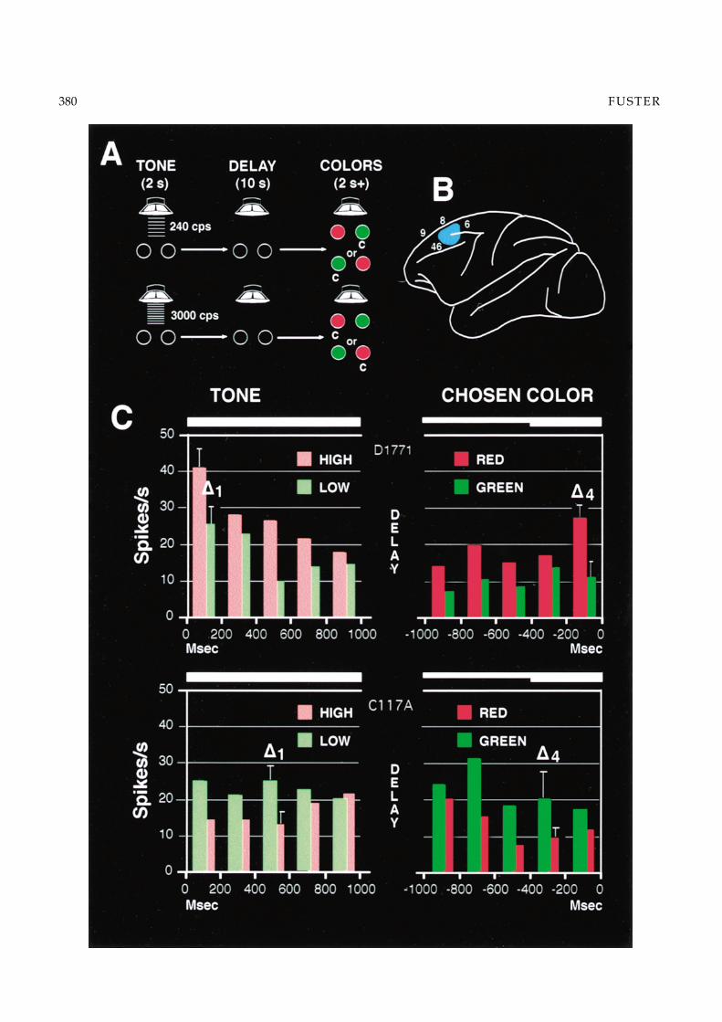
stimuli could be attributed to cognitive association andnot to differences in physical parameters of sensorystimulation. The task (Fig. 5A) consisted of the follow-ing seriatim events: (1) a brief tone of high or low pitch;(2) a delay of 10 sec.; (3) two colors, red and green, pre-sented simultaneously; (4) choice of a color depend-ing on the tone—red for high tone, green for low tone.(Tones and color positions change at random from trialto trial.) In sum, the task was based on the associationof stimuli across time and across modalities.
In the lateral prefrontal cortex of monkeys perform-ing the task (Fig. 5B), a large category of neurons wasfound that, to judge from their firing frequency at thetime of stimulus presentation, discriminated the sen-sory stimuli with different levels of discharge. Someneurons differentiated high tone from low tone, othersred from green, and still others did both. The analysisof firing discharge at the time of the stimuli revealed acorrelation in accord with the task rule: neurons thatpreferred the high tone also preferred the red color,whereas neurons that preferred the low tone also pre-ferred the green color (Fig. 5C). Not only the directionbut also the degree of preference were correlated. Fur-ther, those correlations disappeared or were reversed intrials terminating in error: when the monkey erred, thecells also “erred.’’These results indicate that, during theperformance of a temporal integrative task, neurons inthe prefrontal cortex associate stimuli across time andacross sensory modalities, in accord with the rules of asequential task. A reasonable implication of our resultsis that those neurons are part of networks of long-term executive memory that were formed by the learn-ing of the task, and that those networks are activatedduring the task in order to mediate cross-temporalcontingencies between associated sensory stimuli.
WORKING MEMORY
From the published results in a vast neuropsycholog-ical, physiological, and imaging literature (reviewedin Fuster, 2001), we now know that the mediation ofcross-temporal contingencies, and therefore temporalintegration, rely on two time-bridging functions of thelateral prefrontal cortex: working memory and set.
Working memory (Baddeley, 1986) is the temporaryretention of an item of information—e.g., a sensorycue—for the solution of a problem or for a mental oper-ation. Working memory is memory for the short term,rather than short-term memory. It is attention focusedon an internal representation. Elsewhere (1997a), I haveargued that working memory essentially consists of thetemporary activation of a widely distributed corticalnetwork of long-term memory. My argument is basedon the evidence that, during the short-term retention ofsensory information for a prospective act, neurons inwidespread areas of the cortex exhibit sustained activa-tion. Further, the working memory of a given stimulus
can elicit sustained neuronal activation in several areasof the cortex at the same time. The neuronal correlatesof working memory were first discovered, and havebeen repeatedly confirmed, in the prefrontal cortex ofmonkeys performing delay tasks (Fuster & Alexander,1971; Fuster et al., 1982; Funahashi et al., 1989; Milleret al., 1996).
The sustained activation of prefrontal “memory neu-rons’’ during working memory has the following char-acteristics (Fuster, 1973): (1) it is related in magnitude tothe accuracy of performance of the task; (2) it is depen-dent on the need to perform a prospective motor act;(3) it is not dependent on the expectation of reward; and(4) it can be suppressed or diminished by distraction.In the context of a behavioral task, such as a delay task,the content of working memory is not limited to thespecific sensory parameters of the cue that the animalmust remember for a few seconds to perform the taskwith maximum accuracy. Also ncluded in that contentare other associated features of the cue that are part ofthe long-term executive memory of the task (e.g., po-sition of cue in the apparatus, manipulanda, response,etc.). Consequently, during the memory period (delay),some cells show uniform activation in all trials of thetask, without relation to any particular cue. Others alsoshow sustained activation in all trials, but the magni-tude of that activation differs with the particular cuefor the trial. For example, the three cells in Figure 6(task in Fig. 5B) show sustained delay activation that ishigher in the retention of the low tone than in that ofthe high tone. In addition, the cells show the tone-colorcorrelation described in the previous section. Presum-ably therefore, prefrontal cells of this kind belong toexecutive networks that encode a number of associatedcharacteristics of the cue in the task environment, in-cluding the pitch of the auditory memorandum.
The neural mechanisms of working memory havebeen the subject of many studies. The inactivation, bycooling, of the lateral prefrontal cortex induces a re-versible deficit in the performance of a visual mem-ory task (delayed matching to sample with colors). Atthe same time, it induces a diminution of differencesin the sustained memory activity of cells in the infer-otemporal cortex—visual memory cortex (Fuster et al.,1985). A reasonable interpretation of these findings isthat prefrontal cooling deprives inferotemporal cells ofthe capacity to retain visual stimuli in working mem-ory. This interpretation implies that, in visual work-ing memory, inferotemporal networks are normally un-der a degree of executive control from the prefrontalcortex (Desimone & Duncan, 1995) and are releasedfrom that control by prefrontal cooling. The results arealso compatible with the notion that working memoryis based on the reverberation of activity between theexecutive networks of the prefrontal cortex and the sen-sory networks of posterior cortex. Cooling of either cor-tex would interrupt the reentrant circuits that sustain
380 FUSTER

Frontal lobe and cognitive development 381
Fig. 6. Frequency histograms of three prefrontal cells selective for low tone and green—according to the task rule. Note thesustained, low-tone preferential firing during the working-memory period (delay). (From Fuster et al., 2000, with permission).
that reverberation. Reentrant circuitry is an essentialfeature of some of the most plausible computationalmodels of working memory (Zipser et al., 1993; Compteet al., 2000).
PREPARATORY SET
Whereas working memory is a temporally retrospectivefunction to retain items of recent sensory information,prospective set is a temporally prospective function,also based in the lateral prefrontal cortex, to prepare theorganism for actions contingent on that information.Preparatory set can be appropriately considered the
Fig. 5. A: Diagram of the cross-modal, audio-visual task, as described in the text. B: Lateral view of the monkey’s brain with thearea indicated (blue) where cells were found that discriminated sounds and colors in accord with the rule of the task (numbersrefer to areas in Brodmann’s map). C: Average firing-frequency histograms of two prefrontal cells during the tone and color-choice periods of the task. Both cells are activated in accord with the task rule. The cell on top (D1771) fires preferentially tohigh-pitch tone and red; that on the bottom (C117A) prefers low-pitch tone and green. (From Fuster et al., 2000, with permission).
inclusive component of motor attention (Fuster, 1997b).(The exclusionary component is dealt with below, un-der Inhibitory Control.) This set function of the lateralprefrontal cortex has been substantiated by electro-physiological evidence. Between a sensory cue and amotor response contingent on it, slow potentials canbe recorded from the surface of the frontal lobe in thehuman (Fig. 7) that are related in amplitude to the reac-tion time and the accuracy of the response (Brunia et al.,1985). Two such potentials have been identified, thoughboth seem to be part of a continuum along temporaland frontal-surface gradients. The first is the contingentnegative variation (CNV), also called the “expectancy

382 FUSTER
Fig. 7. Increasing surface potential from the frontal cortex of the human in the interval between a sensory cue (WS) and a motorresponse (RS). The amplitude of the potential is greater when the reaction time (RT) of the subject is fast than when it is slow.(From Brunia et al., 1985, with permission).
wave,’’ which is dependent on the necessity to medi-ate the cross-temporal contingency between cue andresponse. The second is the so-called readiness poten-tial (RP), dependent on the necessity to prepare a mo-tor action. The CNV has a somewhat more anterior,prefrontal, source than the RP, which appears to orig-inate in premotor and motor cortex. Both potentialsincrease in magnitude with time as the response ap-proaches and appear to reflect the increasing activity ofunderlying neurons in preparation for the response.
In the monkey, during the delay period of delayed-matching and delayed-response tasks, the discharge ofsome prefrontal cells increases as the choice or matchingresponse approaches (Niki & Watanabe, 1979; Fusteret al., 1982). In a double-contingency color-matchingtask, the magnitude of that increase (Fig. 8) was foundto depend on the degree of certainty with which theanimal could predict the direction of the prospectivemotor response, to the right or to the left side of a panel(Quintana & Fuster, 1999). These cells appear to rep-resent the neuronal source of the CNV-RP potentials,and thus the neuronal substrate for the preparation ofexecutive action.
The involvement of the lateral prefrontal cortex of themonkey in the preparation for executive action is in alllikelihood related to the role of the cortex of the convex-ity of the frontal lobe of the human in planning. One ofthe most consistent clinical symptoms of patients withlarge injuries of this cortex is the inability to formulate
and to carry out plans of action. The deficit in the abilityto plan for future action seems to reflect, on a broadertemporal scale, the failure of the function of short-termset for action that, as described above, the electrophys-iology of the lateral prefrontal cortex suggests in bothman and monkey.
INHIBITORY CONTROL
The neuropsychology of the frontal lobe in humans andmonkeys points to another temporal integrative func-tion of the frontal lobe: inhibitory control. Lesion exper-iments and clinical evidence (reviewed in Fuster, 1997b)indicate that the neural substrate for this inhibitoryfunction resides mainly in the medial and orbital as-pects of the prefrontal cortex. The apparent physiolog-ical objective of inhibitory influences from orbitome-dial cortex is the suppression of internal and externalinputs that can interfere with whatever structure of be-havior, speech, or cognition is about to be undertakenor currently underway. However, the neurophysiologi-cal mechanisms of prefrontal inhibitory control are stillunknown.
One source of interference with current structuredactions consists of internal biological drives and im-pulses. Patients with orbitomedial prefrontal lesionsexhibit inordinate impulsiveness, irritability, hyper-activity, and poor control of instincts. The disinhib-ited drives and impulses have their origin in thediencephalon and the brain stem. They are normally

Frontal lobe and cognitive development 383
Fig. 8. Average firing of 15 direction-coupled prefrontal cellsduring the period of delay between the sample (S) and thechoice response (R) in a delayed-matching task with colors.The task contains a double contingency: the choice of direc-tional response (right or left) is contingent on the sample colorand, in addition, on a second visual cue at the end of the delay.Some sample colors predict the direction with 100% and oth-ers with 75% probability. Note that cell firing increases grad-ually in anticipation of the response, and that the increase isgreater with 100- than 75-percent predictibility.
under control from the orbitomedial prefrontal cortexvia anatomically identified efferent outputs to thosesubcortical structures, notably the hypothalamus.
Another source of possible interference is a host ofinfluences from sensory systems that are unrelated tocurrent action and can obstruct it or lead it astray. Theseinterfering influences may arrive to the prefrontal cor-tex from sensory areas of posterior cortex; in the courseof goal-directed action, they are probably suppressedby inhibitory feedback from the orbitomedial prefrontalcortex upon those areas. This kind of inhibitory controlfrom the prefrontal cortex is a major component (exclu-sionary component) of sensory attention. In the absenceof it, humans and monkeys with lesions of orbitofrontalcortex exhibit abnormal distractibility in addition to hy-peractivity and hyperreactivity to sensory stimuli. Theexclusionary component of sensory attention is a cog-
nitive function of wide cortical distribution dedicatedto the suppression of sensory distraction. The inhibi-tion of distraction complements the intensive, focusingcomponent of selective sensory attention. Both compo-nents are supported by prefrontal outputs, which exertcontrol over the cognitive functions of other cortical re-gions (Desimone & Duncan, 1995). The attentive controlfrom prefrontal cortex, with its effects of both selectivefocusing and exclusionary inhibition, is essential forthe integrity of any complex structure of goal-directedaction.
A third source of interference is constituted by mo-tor representations of action that are unrelated to, or insome manner incompatible with, actions currently inthe process of temporal structuring. Included amongthem is a large array of motor habits, tendencies andimpulses established in long-term memory and thus inthe cortical and subcortical circuitry of motor systems.The suppression of those untoward influences from themotor sector is the essence of the exclusionary aspectof motor attention.
One of the hallmarks of the psychosocial develop-ment of the child is the progressive establishment ofinhibitory control over internal impulses, over senso-rium, and over motility. As the child grows, the twoprincipal components of attention, inclusive and ex-clusionary, mature gradually. The child becomes morecapable of focusing and concentrating attention on on-going tasks. At the same time, the child becomes lessdistractible, less impulsive, and more capable of self-control. The most striking characteristics of the atten-tion deficit disorders of childhood are the difficulties tofocus and concentrate, the distractibility, the impulsive-ness, and the hyperactivity. All these are manifestationsof the absence of effective inhibitory control. Because ofthe evidence of a critical role of the orbitomedial pre-frontal cortex in this function, it has been reasonablypostulated that the attention deficit disorders of the de-veloping child are attributable to the laggard matura-tion of that portion of the prefrontal cortex (Barkley,1997).
References
BADDELEY, A. (1986) Working Memory. Oxford: ClarendonPress.
BARKLEY, R. A. (1997) Behavioral inhibition, sustained at-tention, and executive functions: Constructing a unifyingtheory of ADHD. Psychological Bulletin 121, 65–94.
BRODMANN, K. (1909) Vergleichende Lokalisationslehre derGrosshirnrinde in ihren Prinzipien dargestellt auf Grund desZellenbaues. Leipzig: Barth.
BRUNIA, C. H. M., HAAGH, S. A. V. M. & SCHEIRS,J. G. M. (1985) Waiting to respond: Electrophysiologicalmeasurements in man during preparation for a volun-tary movement. In Motor Behavior (edited by HEUER,H., KLEINBECK, U. & SCHMIDT, K.-H.) pp. 35–78.New York: Springer.

384 FUSTER
BUTLER, A. B. (1994) The evolution of the dorsal pallium inthe telencephalon of amniotes: Cladistic analysis and anew hypothesis. Brain Research Reviews 19, 66–101.
COMPTE, A., BRUNEL, N., GOLDMAN-RAKIC, P. S. &WANG, X.-J. (2000) Synaptic mechanisms and networkdynamics underlying spatial working memory in a cor-tical network model. Cerebral Cortex 10, 910–923.
DESIMONE, R. & DUNCAN, J. (1995) Neural mechanismsof selective visual attention. Annual Review of Neuroscience18, 193–222.
EDELMAN, G. M. (1987) Neural Darwinism. New York: BasicBooks.
FELLEMAN, D. J. & VAN ESSEN, D. C. (1991) Distributedhierarchical processing in the primate cerebral cortex.Cerebral Cortex 47, 1047–3211.
FINLAY, B. L. & DARLINGTON, R. B. (1995) Linked regu-larities in the development and evolution of mammalianbrains. Science 268, 1578–1584.
FLECHSIG, P. (1901) Developmental (myelogenetic) locali-sation of the cerebral cortex in the human subject. Lancet2, 1027–1029.
FLECHSIG, P. (1920) Anatomie des Menschlichen Gehirnsund Ruckenmarks auf Myelogenetischer Grundlage. Leipzig:Thieme.
FUNAHASHI, S., BRUCE, C. J. & GOLDMAN-RAKIC,P. S. (1989) Mnemonic coding of visual space in the mon-key’s dorsolateral prefrontal cortex. Journal of Neurophys-iology 61, 331–349.
FUSTER, J. M. (1973) Unit activity in prefrontal cortex duringdelayed-response performance: Neuronal correlates oftransient memory. Journal of Neurophysiology 36, 61–78.
FUSTER, J. M. (1997a) Network memory. Trends in Neuro-Sciences 20, 451–459.
FUSTER, J. M. (1997b) The Prefrontal Cortex—AnatomyPhysiology, and Neuropsychology of the Frontal Lobe.Philadelphia: Lippincott-Raven.
FUSTER, J. M. (2001) The prefrontal cortex—An update:Time is of the essence. Neuron 30, 319–333.
FUSTER, J. M. & ALEXANDER, G. E. (1971) Neuron activ-ity related to short-term memory. Science 173, 652–654.
FUSTER, J. M., BAUER, R. H. & JERVEY, J. P. (1982) Cel-lular discharge in the dorsolateral prefrontal cortex ofthe monkey in cognitive tasks. Experimental Neurology 77,679–694.
FUSTER, J. M., BAUER, R. H. & JERVEY, J. P. (1985)Functional interactions between inferotemporal and pre-frontal cortex in a cognitive task. Brain Research 330,299–307.
FUSTER, J. M., BODNER, M. & KROGER, J. K. (2000)Cross-modal and cross-temporal association in neuronsof frontal cortex. Nature 405, 347–351.
GIBSON, K. R. (1991) Myelination and behavioral devel-opment: A comparative perspective on questions ofneoteny, altriciality and intelligence. In Brain Maturationand Cognitive Development (edited by GIBSON, K. R. &PETERSEN, A. C.) pp. 29–63. New York: Aldine deGruyter.
GIEDD, J. N., BLUMENTHAL, J., JEFFRIES, N. O.,CASTELLANOS, F. X., LIU, H., ZIJDENBOS, A.,PAUS, T., EVANS, A. C. & RAPAPORT, J. L. (1999)Brain development during childhood and adolescence: Alongitudinal MRI study. Nature Neuroscience 2, 861–863.
HUTTENLOCHER, P. R. (1979) Synaptic density in humanfrontal cortex—Developmental changes and effects ofaging. Brain Research 163, 195–205.
HUTTENLOCHER, P. R. (1990) Morphometric study of hu-man cerebral cortex development. Neuropsychologia 28,517–527.
JERISON, H. J. (1990) Fossil brains and the evolution ofthe neorcortex. In The Neocortex: Ontogeny and Phylogeny(edited by FINLAY, B. L., INNOCENTI, G. & SCHEICH,H.) pp. 5–19. New York: Plenum Press.
JERNIGAN, T. L. & TALLAL, P. (1990) Late childhoodchanges in brain morphology observable with MRI.Developmental Medicine and Child Neurology 32, 379–385.
JONES, E. G. & POWELL, T. P. S. (1970) An anatomicalstudy of converging sensory pathways within the cere-bral cortex of the monkey. Brain 93, 793–820.
LURIA, A. R. (1966) Higher Cortical Functions in Man.New York: Basic Books.
MILLER, E. K., ERICKSON, C. A. & DESIMONE, R.(1996) Neural mechanisms of visual working memoryin the prefrontal cortex of the macaque. Journal of Neuro-science 16, 5154–5167.
MRZLJAK, L., UYLINGS, H. B. M., VAN EDEN, C. G. &JUDAS, M. (1990) Neuronal development in human pre-frontal cortex in prenatal and postnatal stages. In The Pre-frontal Cortex: Its Structure, Function and Pathology (editedby UYLINGS, H. B. M., VAN EDEN, C. G., DE BRUIN, J. P.C., CORNER, M. A. & FEENSTRA, M. G. P.) pp. 185–222.Amsterdam: Elsevier.
NIKI, H. & WATANABE, M. (1979) Prefrontal and cingu-late unit activity during timing behavior in the monkey.Brain Research 171, 213–224.
NORTHCUTT, G. & KAAS, J. H. (1995) The emergenceand evolution of mammalian neocortex. Trends in Neuro-sciences 18, 373–379.
PANDYA, D. N. & YETERIAN, E. H. (1985) Architectureand connections of cortical association areas. In CerebralCortex, Vol. 4 (edited by PETERS, A. & JONES, E. G.)pp. 3–61. New York: Plenum Press.
PFEFFERBAUM, A., MATHALON, D. H., SULLIVAN,E. V., RAWLES, J. M., ZIPURSKY, R. B. & LIM, K. O.(1994) A quantitative magnetic resonance imaging studyof changes in brain morphology from infancy to lateadulthood. Archives of Neurology 51, 874–887.
PIAGET, J. (1952) The Origins of Intelligence in Children.New York: International Universities Press.
QUINTANA, J. & FUSTER, J. M. (1999) From perception toaction: Temporal integrative functions of prefrontal andparietal neurons. Cerebral Cortex 9, 213–221.
REISS, A. L., ABRAMS, M. T., SINGER, H. S., ROSS, J. L.& DENCKLA, M. B. (1996) Brain development, genderand IQ in children: A volumetric imaging study. Brain119, 1763–1774.
SCHEIBEL, A. B. (1990) Dendritic correlates of higher cog-nitive function. In Neurobiology of Higher Cognitive Func-tion (edited by SCHEIBEL, A. B. & WECHSLER, A.)pp. 239–270. New York: The Guilford Press.
SOWELL, E. R., THOMPSON, P. M., HOLMES, C. J.,JERNIGAN, T. L. & TOGA, A. W. (1999) In vivo ev-idence for post-adolescent brain maturation in frontaland striatal regions. Nature Neuroscience 2, 859–861.

Frontal lobe and cognitive development 385
SOWELL, E. R., THOMPSON, P. M., TESSNER, K. D.& TOGA, A. W. (2001) Mapping continued braingrowth and gray matter density reduction in dorsalfrontal cortex: Inverse relationships during post adoles-cent brain maturation. Journal of Neuroscience 21, 8819–8829.
STEPHAN, H., FRAHM, H. & BARON, G. (1981) New andrevised data on volumes of brain structures in insecti-vores and primates. Folia Primatologia 35, 1–29.
VAN HOESEN, G. W. (1982) The parahippocampal gyrus.Trends in Neurosciences 5, 345–350.
YAKOVLEV, P. I. & LECOURS, A. R. (1967). The myel-ogenetic cycles of regional maturation of the brain. InRegional Development of the Brain in Early Life (edited byMINKOWSKI, A.) pp. 3–70. Oxford: Blackwell.
ZIPSER, D., KEHOE, B., LITTLEWORT, G. & FUSTER,J. M. (1993) A spiking network model of short-term ac-tive memory. Journal of Neuroscience 13, 3406–3420.