from affect to control: functional specialization of the insula in
TRANSCRIPT
From affect to control: Functional specialization of the insula in motivation andregulation
Tor D. Wager
Columbia University
and
Lisa Feldman Barrett
Boston College

Abstract
The insula plays a key role in a wide range of brain processes, from viscerosensation andpain to motivation, emotion, and cognitive control. While human neuroimaging studiesin all these domains report activations in the insula, little systematic attention is paid toanatomical subdivisions that may provide the basis for functional sub-regions. Weconducted a meta-analysis of insular tasks across studies in four domains: emotion, pain,attention switching, and working memory. Using a priori subdivision of the insula basedon anatomical studies, we provide evidence that different sub-regions are preferentiallyactivated in different tasks. We suggest that the ventral anterior insula is most importantfor core affect, a term that describes broadly-tuned motivational states (e.g., excitement)with associated subjective feelings. The dorsal anterior insula, by contrast, may becritical for developing and updating motivational states with specific associated actions(i.e., goals). This region is activated by cognitive control tasks, pain, and some tasks thatelicit affective processing. The posterior insula, including SII and portions of parietaloperculum, is distinctly activated by pain, providing a double dissociation between painand tasks that elicit emotions.

In 1953, scientists found a new way to think about the human mind. Thecomputer metaphor cast the brain as the ultimate machine, the deus ex machina ofconscious will reduced to cogs and wheels cranking away behind the curtain, and a newdiscipline—cognitive science—was born. The computer metaphor continues to bepervasive: information is maintained online in short term memory ‘buffers,’ addressedand tagged, and written to permanent long-term memory storage. Attention works toenhance ‘information processing.’ Reasoning and even some forms of mental imageryproceed through propositional, symbolic representation. In the heyday of the cognitivescience revolution, emotion was regarded as a volume knob, an orphaned sideshow in thegreat circus of the machine.
New developments in recent years are beginning to challenge this view of themind. The cognitive and neural sciences have increasingly merged, and researchersstudying the neural bases of thought and behavior have found a surprising degree ofoverlap between the brain regions engaged in cognitive and affective processes. Theanterior cingulate and insular cortices, among other regions, are engaged reliably inanimal and human studies of both central topics in cognitive science—working memory,long-term memory, control of attention—as well as tasks designed to isolate emotionalprocesses. Understanding behaviors or feelings at a given instant in time is a task thatrequires consideration of the whole organism, and this challenge has forced us to re-examine the ways in which we think about cognitive and emotional processes.
There are two main views on the relationship between cognition and emotion.One view, which has existed in various incarnations since the time of the ancient Greeks,is that cognition and emotion are embodied in two separable, opposing systems. Feelingemotion turns off cognition, and thinking dampens emotional impact (Drevets & Raichle,1998; Mayberg et al., 1999; Metcalfe & Mischel, 1999; Mischel, Shoday, & Peake,1989).
Another view seems initially to stand in opposition to the first: Emotion is criticalto motivating cognition and behavior, and the roots of attentional control lie in affect.According to this view, emotions arise from cognitive appraisals of situations (R. S.Lazarus, 1991; Richard S. Lazarus, 1991b; Scherer, Schorr, Ed, & Johnstone, 2001; C. A.Smith & Ellsworth, 1985; Craig A. Smith & Lazarus, 2001), which particularly involveevaluations of how objects and events affect the self. The basic affective experience thatarises when a self-relevant event occurs has been labeled “core affect” (J. A. Russell &Barrett, 1999). Core affect is the seed of full-blown emotion, and from affectiveresponses arise the physiological and motivated response tendencies that have beenshaped over the course of our evolution to promote adaptive cognitions and behaviors.Thus, in this view, emotion and cognition are not opponents in a zero-sum tug of war.Rather, they are synergistic partners in the game of adaptive self-regulation, each shapingthe direction of the other.
The answer may be that neither of the metaphors of opposition and synergy isadequate. Emotion does not stop cognition; rather, it directs cognition into channels mostappropriate for the situation. In threatening situations, attention is focused on theperceived threat, and extraneous thought stops. In safe situations dominated by positiveaffect, the impulse to explore and build new skills may prime a broad repertoire ofthoughts and behaviors (Fredrickson, 2001). We are at the frontier of exploring the

physical brain systems that give rise to thoughts and feelings, and many of thesequestions may be addressed empirically.
Understanding the roles of certain key brain structures may provide criticalinformation on how affective information shapes attention, and how thoughts direct, or insome cases stem, the flow of affective signals in the brain. Some of the most importantsuch regions are likely to be those that lie at the physical junctions between neocorticaland evolutionarily older subcortical nuclei—the limbic and paralimbic regions, so namedbecause they form a limbus, or border, around the oldest parts of the brain (Maclean,1955, 1958; Papez, 1995). Cortical limbic and paralimbic areas are generally thought toinclude the cingulate cortex, parahippocampal gyrus and entorhinal cortex, orbitofrontalcortex, and the insula.
The insula: A key link between cognition and affect
In this paper, we focus on the insula as a potential nexus for motivated cognitionand emotional behavior. The insula has long been considered part of the emotional andviscerosensory brain (Janig & Habler, 2002; Maclean, 1955), with multiple roles inregulating physiological and psychological homeostasis (Flynn, Benson, & Ardila, 1999).The insula and surrounding operculum contain primary cortical representation of smelland taste (Francis et al., 1999; Rolls, 1996), viscerosensation (Craig, 2002), and painperception (Coghill, Sang, Maisog, & Iadarola, 1999; Davis, Kwan, Crawley, & Mikulis,1998). For this reason, it has been termed "limbic sensory cortex" and associated withthe subjective feeling of emotional states, or the "feeling self" (Craig, 2002, 2003).Recent evidence from neuroimaging studies corroborate this view. The insula iscommonly activated in emotion tasks, predominantly those associated with negative orwithdrawal-related emotions (Phan, Wager, Taylor, & Liberzon, 2002).
Several individual examples illustrate that the insula plays a broad role in thedevelopment of subjective, self-relevant feelings. In a recent study, Sanfey andcolleagues (Sanfey, Rilling, Aronson, Nystrom, & Cohen, 2003) found insular activitywhen participants received an unfair monetary offer from what they believed was anotherhuman—but not when the offer came from a computer. Why does the same loss stingmore if it results from another's intention? It could be because the loss also signals socialrejection, "unfairness," and/or the motivating possibility of reprisal or control of thesituation. Another study found that pain-responsive portions of the anterior insulashowed decreased activity when a placebo—a medication with no real effect, butbelieved by participants to be a potent analgesic—was administered prior to pain (Wager,Rilling et al., 2004). A third study showed common activations in the insula whenfeeling pain and when observing a loved one experience pain (Singer et al., 2004).
However, the story suggested by these studies, that of the insula as a structure forfeeling, is incomplete. It omits a host of studies that suggest alternative roles for theinsula—in particular, a role in cognitive control. If the anterior insula is the seat ofemotional awareness, why is the anterior insula (as we show here) activated in so manycognitive tasks ostensibly devoid of emotion (Wager & Smith, 2003)?
One possibility is that cognitive tasks simply activate a separate portion of theinsula and frontal operculum devoted to attentional control. Without detailed comparisonof results from cognitive and affective tasks, we cannot tell. Another possibility is that

these cognitive tasks, which share as a common feature the requirement for executivecontrol of attention, share common psychological processes with affective tasks. Thesecould include a role for motivated decision making in goal formation, updating of taskrelevance based on affective information, and affective error-detection processes incognitive tasks.
A second unresolved issue is the relationship between pain and affect, and howthat relationship is informed by insular participation in both. Does pain involve affectiverepresentations, as is commonly believed (Melzack & Casey, 1968; Rainville, Duncan,Price, Carrier, & Bushnell, 1997), and do the insular regions involved in "pain affect"form the basis for the "feeling self"? Again, we can ask the same questions aboutwhether pain and emotion activate common parts of the insula, or whether commoninvolvement of the "anterior insula" in pain and emotion is in fact an overgeneralization.
These questions are approached using meta-analysis of human neuroimagingstudies (Fox, Parsons, & Lancaster, 1998) across four domains: emotion, pain, attentionshifting, and working memory. We begin with functional subdivisions suggested bycytoarchitectural and functional studies in animals (Figure 1), and ask whether thesesubregions produce meaningful dissociations between tasks in human neuroimagingstudies.
We then ask whether activation of particular insular subregions, or the pattern ofactivation across subregions, provides useful information about which psychological taskor process elicited the activations. This is a fundamentally different question that istypically investigated in neuroimaging studies to date. Rather than asking, "What brainareas implement attention shifting," we ask, "Is there a pattern of activations thatuniquely identifies attention shifting, and separates it from other processes?"
We present evidence for distinct insular sub-regions, and we use an inductiveapproach to develop hypotheses about the processes they may implement. In this paper,we argue for a distinction between pain and emotional feelings, which activate the dorsaland ventral portions of the anterior insula, respectively. The pattern of activations weobserve also suggests that attentional control tasks activate a specific part (dorsalanterior) of the insula, in common with pain.
We frame our conclusions in terms of hypotheses to be tested. One hypothesis isthat dorsal anterior insula is directly involved in attentional control, and pain activatesthis region because it recruits mechanisms of executive attention (Eccleston & Crombez,1999). Alternatively, we present the hypothesis that executive attention recruits anteriorinsula because this region links general motivational tendencies with specific actionplans—i.e., it is involved in goal formation and re-formation. The ventral anterior insulamay represent motivational states with very general action tendencies (e.g., affiliate,protect), and the dorsal aspect represents motivational states associated with specificaction plans. This distinction is consistent with patterns of results across all the taskdomains we studied.
Methods
Functional subdivisions of insula

Figure 1 shows a digitized parcellation of human insular cortex and the overlyingoperculum into four distinct regions based on the cytoarchitectural and fiber tract tracingstudies of Mesulam and Mufson (Mesulam & Mufson, 1982a, 1982b; Mufson &
Mesulam, 1982). Regions were defined electronically on the single-subject MontrealNeurological Institute (MNI) template (Evans et al., 1992) and masked to include graymatter or voxels within 3 mm of gray matter using custom software in Matlab 6.5(Mathworks, Natick, MA). Each region is discussed in turn below.
Agranular insula / prepyriform cortex (red). The agranular insula— Ag,shown in red, and so named for its indistinct laminar structure and apparent lack ofstellate (granule) cells prominent in cortical input layers—is the part of insula thatsurrounds the prepyriform cortex, the primary site of olfactory input to the cortex. Itspans medial portions of the temporal pole, caudal orbitofrontal gyrus, and ventralanterior insula.
This portion of the insula and adjacent cortex responds to primary odorreinforcers in humans and animals (Critchley & Rolls, 1996; Dade, Zatorre, & Jones-
Figure 1. Anatomical subdivision of insular cortex
ACC
ACCACC
MidC
MidC
MidC
LPFC
TP TP Amy PHPC STG
Figure 1. Anatomical divisions of insular cortex. Anterior ventral agranular (Ag) is shown in red.Dorsal anterior dysgranular and adjacent frontal operculum (Adg) are in yellow. Mid-insula(Mdg) is in blue. SII and adjacent parietal operculum are in green. Arrows show a schematic ofprojection patterns for posterior and anterior regions. ACC, anterior cingulate; MidC, mid-cingulate; LPFC, lateral prefrontal cortex; TP, temporal pole; Amy, amygdala; PHPC,parahippocampal cortex; STG, superior temporal gyrus

Gotman, 2002) and appears to play a role in the representation of drive states (e.g.,hunger, Freeman). It is most closely connected to the medial orbitofrontal cortex, aprimary function of which may be updating the reward value associated with stimuluscues (Baxter, Parker, Lindner, Izquierdo, & Murray, 2000; Rolls, 2004; Wallis, Dias,Robbins, & Roberts, 2001), and also sends and receives projections from the mid-insula,the pericallosal anterior cingulate, and the medial temporal lobes (red arrows in Figure 1).According to Craig (2002), it is via the connection to OFC that the anterior insula has itseffects on the valenced property of core affect.
Evolutionarily, Ag and immediately adjacent portions of temporal pole andorbitofrontal cortex were developed for advanced chemoreception, particularly theassociation of behavioral states (approach/eating and avoidance of noxious environmentsor foods) with particular chemosensory representations. Thus it earned it the title ofprepyriform cortex or primary olfactory cortex, although its selectivity for odors and/ortastes remains under debate. Its evolutionary origins suggest that agranular insula may bepart of a core of a system for evaluating primary reinforcers and determining appropriatemotivational states—that is, core affect. It is expected to be particularly involved wheninternal states (like hunger) drive valuation, or when stimuli in the environment signal adirect link to positive (reward), negative (threat) value, or other motives such as self-protection or affiliation (Fridja). Based on this analysis, we expect this subregion torespond during tasks that induce feeling states in people—that is, tasks that elicitemotions.
Anterior dysgranular insula (Adg, yellow). The superior portion of the anteriorinsula (yellow in Figure 1) is dysgranular, with incomplete laminar structure and acytoarchitectural appearance intermediate between agranular paleocortex and fullydeveloped neocortex (Mesulam & Mufson, 1982a). It blends into the fully-laminatedfrontal operculum. Although it has not been well differentiated from other parts ofanterior insula, this region is commonly activated in tasks that require executive controlof attention, including those that require manipulation of information in working memory(Wager & Smith, 2003), response inhibition (Nee, Jonides, & Wager, 2004), and shiftingattention (Wager, Reading, & Jonides, 2004). However, its role in these tasks has beenunderappreciated, perhaps due to the emphasis in the literature on affective andautonomic facet of anterior insular function (Critchley, Wiens, Rotshtein, Ohman, &Dolan, 2004; Phillips et al., 1997). Its proximity to agranular insula and interpositionbetween this older structure and the lateral prefrontal cortex—thought by many authors tobe the seat of executive control over attention and action (Miller, 2000)—suggest that itmay be important for translating undifferentiated drive states into specific action plans.
Mid-insula (blue). The middle portion of the insula (blue in Figure 1) is alsodysgranular, and it is connected primarily with neighboring areas of insula.
SII and parietal operculum (green). The superior bank of the posterior insulacontains SII, primary sensory cortex for pain and itch (Craig, 2002, 2003; Craig, Chen,Bandy, & Reiman, 2000). Its major bidirectional connections are with parts of theventromedial nucleus of the thalamus, which transmit sensory input from the body(Craig, 2002); multiple regions of the anterior and mid-cingulate (Mesulam & Mufson,1982b; Mufson & Mesulam, 1982), consistent with known cingulate motor regions in themonkey (Picard & Strick, 1996); adjacent S1 and sensorimotor cortex; and parts of theanterior temporal cortex.
Study selection and meta-analysis
Studies for meta-analysis were compiled using Medline and ISI Web of Sciencedatabase searches, and from additional references found in papers, and are drawn fromthe literature on pain, emotion, working memory, and attention shifting. Although we donot claim that the studies here represent an exhaustive list, we have tried to be asinclusive as possible. We analyze activations (not deactivations) by tabulating peakcoordinate locations reported in studies of healthy participants (excluding patientstudies), in keeping with more extensive reports in our previous meta-analyses (Phan etal., 2002; Wager, Phan, Liberzon, & Taylor, 2003; Wager, Reading et al., 2004; Wager &Smith, 2003).
Many studies reported multiple independent comparisons, or contrasts—forexample, an emotion study might compare perception of fear faces vs. a neutral baseline,and perception of angry faces against a separate set of neutral images. In practice, manystudies compared activations in multiple conditions to the same baseline; this strategy,and the fact that the same sample of participants is used across comparisons, preventsthese contrasts from being truly independent, but we treat them here as if they were. Therelevant data used for meta-analysis was whether each independent contrast within eachstudy activated each anatomical region of interest.
Analyses included chi-square analyses, k-nearest neighbor (KNN) classification,and rule-based classification using activation in single areas or pairs of areas to predicttasks. Chi-square analyses test whether contrast counts within a region differ among taskconditions, controlling for the overall number of contrasts studied in each condition. Thisanalysis is similar to those reported in our previous studies, with one improvement: bycounting contrasts, rather than peaks or studies, we can better account for studies withmultiple contrasts while avoiding overweighting of studies that report many peaks. Moremethodological detail can be found in our other papers (Phan et al., 2002; Wager,Reading et al., 2004; Wager & Smith, 2003).
KNN analysis was performed on the presence/absence of regional activations ineach anatomical subregion across different types of tasks. In practice, we formed anindicator matrix, the rows of which were contrasts, and the columns of which coded forboth tasks and regional activations. This matrix is the building block of the multiplecorrespondence analysis framework, which can be used for multivariate analysis ofcategorical data (Bouilland & Loslever, 1998). Task conditions (e.g., approach-relatedemotions and withdrawal-related emotions) were coded in one set of columns, and thepresence of activation in each region was coded in another set of columns. Ones

indicated that the contrast belonged to a task condition or activated a region, and zerosindicated lack of membership or activation.
As task categories in the present analysis were mutually exclusive, the taskindicators were recoded into a class vector that described the task performed in eachcontrast. KNN analysis analyzes the k nearest neighbors (we chose a relatively standardvalue of 3) to each point in the data space, and uses those to form a prediction about thetask type of each point. In our analyses, each contrast was assigned a task classificationbased on the known task classifications of the three contrasts that produced most similaractivation profiles across the eight insular subregions. Errors in classification werecalculated by comparing the known classes with estimated classes (summarized in aconfusion matrix). We chose to derive several summary measures of classificationaccuracy: the hit rate, or correct classification rate for each class; the false alarm rate, orrate at which other task types are classified as a particular task; and a sensitivity measure(A’) based on the combination of hit rate and false alarm rate (Swets, 1988). When onlytwo classes are compared, a single A’ gives the discriminability of the two classes. Formore than two classes, each class has its own A’, reflecting the discriminability of thatclass from the others.
A particular concern with classifiers is that it is relatively easy to construct aclassifier that will perfectly predict classes in a particular dataset, but cannot generalize tonew data because it uses information that is too specific to the particular dataset. A KNNclassifier with k = 1 is an example, as each task will always be given the class neighborof it’s one nearest neighbor – itself. Larger k helps to avoid this problem. An additionalprocedure is cross-validation, which divides the set into a larger training set and a smallertest set. The training set (85% of the data in our analyses) is used to derive classifications,and classification of a smaller test set (15%) is performed using similarities between eachtest contrast and the training data. We divided the dataset into training and testing setsmultiple times (200 in the present analyses) and estimated test classes, hit/false alarmrates and discriminability for each sample, providing a cross-validated estimate of thetrue ability of the KNN algorithm to correctly classify contrasts into task categoriesaccording to their insular activation patterns.
Rule-based classification was performed in a rudimentary fashion in this paper bysimply considering the probability that a study belonged to a particular task category,given activation in each subregion individually and a limited range of task alternatives.This can be written as p(task=T | activity), where p denotes posterior probability giventhe observed data, task is used generally to refer to psychological condition or class, andactivity signifies the presence of an activation peak in a particular region of interest.
In a Bayesian framework, the posterior probability estimate p(task=T | activity) =p(activity | task=T) * p(task=T) / p(activity). The first term, the likelihood of activitygiven task T, is the proportion of contrasts of task type T that activate the brain region.The second term, the prior probability of a task being of type T, could be calculated for aparticular sample based on the frequency of tasks in the database; but we wanted togeneralize to a world in which all tasks could be studied equally frequently, so we assignp(task=T) = 1. p(activity) is the sum of percentages of contrasts activating across tasktypes, and normalizes the posterior probability estimates across tasks to 1. Thisnormalization excludes the possibility that a task belongs to none of the set of possible

tasks in the analysis. Thus, the task with the maximum posterior probability estimate issimply the best choice among relative alternatives.
We report posterior probabilities in the Results as percentage scores that reflectpredictions of task condition given a) observed activity in single regions or combinationsof two regions, and b) the set of alternative tasks included in the analysis. In comparingpain with emotion tasks, for example, observing bilateral SII activity may be associatedwith a 100% posterior probability for pain, but that probability estimate will change ifadditional task types beyond pain and emotion are considered. We leave the fulldevelopment of probability estimates into full classifiers, considering all regions, forfuture work.
Results
Pain and feelings elicited by emotional stimuli activate distinct subregions of insula.
SII activation was strongly associated with painful stimulation (Figure 2), but notwith emotion. Activation in either right or left SII was associated with a 90% posteriorprobability of pain, and bilateral SII activation with 100% probability of pain (no emotionstudies activated bilateral SII). Mid-insula activation in either hemisphere was associatedwith an 80% probability of pain stimulation.
Agranular insula was likewise relatively strongly associated with emotionalprocessing. Left agranular insula was associated with a 77% probability of doing anemotion task, with 60% for the right agranular subregion. The only pain studies toactivate bilateral agranular insula were Bense et al. (Bense, Stephan, Yousry, Brandt, &Dieterich, 2001), the only study to use vestibular pain, and (Becerra, Breiter, Wise,Gonzalez, & Borsook, 2001). Right agranular insula was reported by Brooks et al.(Brooks, Nurmikko, Bimson, Singh, & Roberts, 2002), using thermal pain on the leftarm. There was also a bias toward left agranular insular activation in emotions (Figure3E) that did not hold for frontal opercular and dysgranular region, in contrast withCraig’s (Craig, 2003) view of the right anterior insula as mediating the ‘feeling self.’
Classification results indicate that pain tasks and those that involve emotion arerelatively discriminable based on their patterns of activation. KNN classification resultedin a cross-validated correct classification of 91% for emotion tasks and 52% for paintasks, with a high discriminability (A') of 2.17.
However, to view emotion tasks as ‘of a piece,’ or as a natural kind, is anextremely limited view. Rather, we would like to move towards defining the processeswithin particular emotion tasks that produce activations in the agranular insula. Indeed,additional analyses showed that emotional activations were unevenly distributed acrossemotion induction methods (Figure 3E). Both auditory induction (voices, screams, etc.)and recall-induced emotions produced substantially more frequent activations inagranular insula than did visual inductions.
Comparison of approach and withdrawal related emotions—broadly construed,happiness and anger vs. sadness, fear, and disgust—provided weak support for adorsal/ventral distinction. Although chi-square results for individual areas were notsignificant, approach was associated with agranular activation in each hemisphere,
whereas withdrawal was associated with superior agranular activation in eachhemisphere. Analysis by valence, which included anger as a negative emotion, producedyet less consistent results.
Activation in individual regions was not highly predictive of approach vs.withdrawal state (the highest was right anterior dysgranular insula, associated with a 73%probability of withdrawal), but activation of both left and right anterior dysgranular orboth left and right mid-insula led to 100% withdrawal classification. Thus, no studies ofapproach activated either of these subregions bilaterally. However, the probability that awithdrawal study produces these activations is also low (6%, or three studies (Damasio etal., 2000; Phillips et al., 1997; Simpson et al., 2000)). Damasio et al. studied emotion-induced recall of personal events, and found activations in all portions of the insulaexcept SII. Phillips et al. studied perception of disgust faces during a genderidentification task. Simpson et al. showed this activation in response to aversive picturesduring concurrent number judgments.
Was superior insula activated by the aversive quality of the emotions, or bycognitive demand associated with performing concurrent cognitive tasks? Virtually allstudies activating this region required cognitive demand (chi2 = 5.24, p < .05 in left Adgand 2.04, n.s. in right Adg), with the exception of Canli1998, who showed activation inright Adg. Right Ag showed significantly more frequent activations with no cognitivedemand (chi2 = 6.11, p < .05), indicating that the few studies of visual and auditorypassive perception that activated Ag tended to do so in the right hemisphere. Recall-induced emotions involve cognitive demand de facto, as well as eliciting emotion, butthey did not frequently activate Adg. Thus, cognitive demand seems to be a morepowerful predictor of Adg activation than does the aversive quality of the emotionelicited. Analysis by individual emotion produced no notable distinctions in activation ofinsular subregions.
This pattern suggests that the agranular insula may play a role in the experientialcomponent of emotions—the process of inducing and feeling an emotion, and perhapsexperiencing associated motivational tendencies, rather than simply perceiving emotionalstimuli. An alternative is that subvocal verbalization or language plays a primary role inaurally and recall-induced emotions, given the role of insula in understanding andproducing language (Habib et al., 1995). However, as we discuss below, analysis ofverbalizability of materials in working memory tasks revealed no effects on agranularinsula.
The pattern of results contrasts sharply with that found in the amygdala, which inprevious meta-analyses showed selectivity for visual stimuli, particularly perception offearful faces, suggesting a role for the amygdala in the visual perception of threat(Murphy, Nimmo-Smith, & Lawrence, 2003; Phan et al., 2002). Interestingly, individualstudies have reported deactivations in the amygdala and agranular insula during pain(Derbyshire et al., 1997).

Figure 2. Pain activations in the insula
Figure 2. A-D) Points on the transparent brains reflect pain activation coordinates in insular subregions:green for SII, blue for mid-insula, red for anterior agranular (ventral) insula, and yellow for anteriordysgranular insula (superior). E) Activation counts by independent contrasts for painful stimulation byregion (x-axis) and side of body stimulated. Bar heights reflect the proportion of contrasts within eachbody side that activated each region.
Figure 3. Emotion task-related activations in the insula
A – D) Glass brains showing peak activation coordinates for emotion tasks. E) Contrast counts by type ofmaterial eliciting emotion, as in Figure 2. * indicates significant differences across conditions for a regionat p < .05.
A B C
D E
A B C
D E
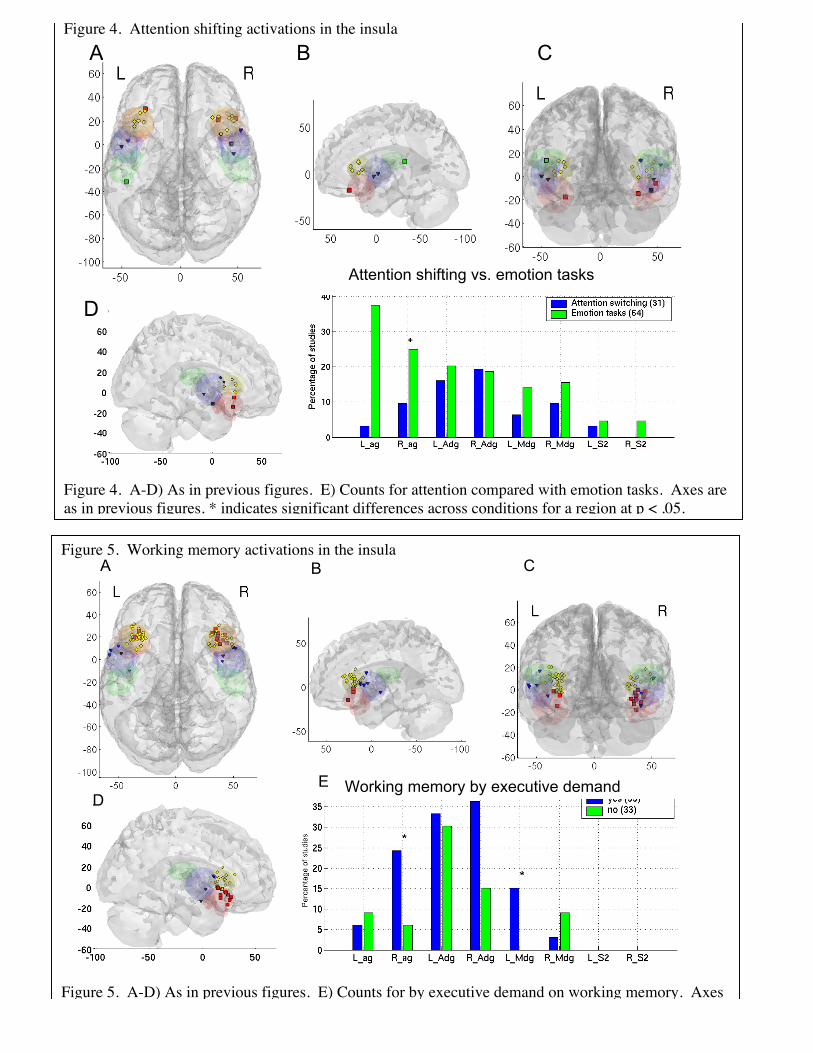
Figure 4. Attention shifting activations in the insula
Figure 4. A-D) As in previous figures. E) Counts for attention compared with emotion tasks. Axes areas in previous figures. * indicates significant differences across conditions for a region at p < .05.
Figure 5. Working memory activations in the insula
Figure 5. A-D) As in previous figures. E) Counts for by executive demand on working memory. Axesare as in previous figures. * indicates significant differences across conditions for a region at p < .05.
A B C
DE Working memory by executive demand
A B C
D
Attention shifting vs. emotion tasks

Shifting attention and executive working memory activate anterior dysgranular insula
Attention shifting results, summarized in Figure 4, show a clear cluster of peaksonly in the left and right superior anterior insula. Most of these peaks lie at the junctionof the dysgranular insula and frontal operculum. Although percentages are not highoverall (25% of switching studies activated this insular subregion), the consistency oftheir location suggests that the result is reliable. Additionally, several studies showingactivation in agranular and mid-insula produced peaks right at the border of the anteriordysgranular region, suggesting that the borders of this area do not quite capture theobserved pattern. Analyses by type of switching (i.e., among locations, tasks, objects,rules, or attributes of objects) yielded no consistent results.
Comparing switching results to results from emotional tasks (Figure 4E), weobserved that there was significantly more agranular insula activity in emotionalcontrasts. Although emotion tasks and shifting tasks activated anterior dysgranular insulaabout equally frequently, KNN classification was able to accurately distinguish switchingfrom emotion tasks, with cross-validated correct classification rates of 60% for switchingand 78% for emotion, and a discriminability A’ of 1.41.
These results demonstrate that executive attention activates a subset of insularregions activated in emotion tasks. One possible conclusion is that elicitations ofemotion involve re-directions of attention, and thus attentional control is a componentprocess engaged when emotions are aroused.
A similar profile of activations was found for executive working memory (Figure5), with one notable exception. Working memory tasks produced consistent activation inright agranular insula, overlapping with emotional task activations. Activation of boththis region and the right anterior dysgranular region were significantly more frequent fortasks involving executive control of working memory, suggesting that right agranularinsula is affected by executive demand. Because the activation profile was otherwisesimilar as that for switching attention, KNN classification did not discriminate wellbetween switching and working memory tasks, with correct classification rates of 23%for switching and 81% for working memory, and a low A’ of –1.41, which suggestsworse-than-chance performance.
Discussion
Our results suggest that the ventral anterior agranular insula is activatedconsistently by neuroimaging studies involved in aurally and recall-generated emotioninduction, particularly in the left hemisphere. Executive manipulation of information inworking memory also engages the right agranular insula, but pain and attention shiftingdo not. We suggest that a key function of the agranular insula may be in representingafferent homeostatic information from the body, for the purposes of subjectiveevaluation. This process is central for generating sets of motivated responses—and, aswe discuss below, is closely tied to the psychological concept of core affect (James A.Russell, 2003). These findings contradict older broad conceptualizations of theemotional brain, which suggest that the right hemisphere is the more emotional, or thatthe left hemisphere is more selective for positive emotions and the right for negative ones

(although this latter may apply to affective styles and specifically to the lateral frontalcortex, as reviewed in (Wager et al., 2003)).
Our findings also suggest that the anterior insula can be subdivided into two parts:the ventral, agranular part discussed above, and a superior dysgranular part that iscontiguous with the frontal operculum (Mesulam & Mufson, 1982a). The localization ofattention switching, working memory, and pain results specifically to the superioranterior insula supports this distinction, as does the mass of activations from emotionalrecall tasks in the ventral anterior insula. If anterior insula represents the ‘feeling self’(Craig, 2002), we may ask which part of the anterior insula is most critical. Our datasuggest the agranular portion is most critical, particularly on the left side—in contrast toCraig’s argument that the interoceptive self is localized to the right anterior insula (Craig,2002). Furthermore, pain produces only superior, agranular activation, suggesting thatthe interoceptive process for pain is different than for emotion; pain affect is not the sameas affect per se.
One possible explanation of the ventral-dorsal distinction is that emotional recalltasks (ventral) elicit a very broad sense of emotionality, with broad motivational/actionrepertoires, whereas pain (dorsal) carries affective signals that motivate very specificescape or avoidance action repertoires. Demand on executive control of attention(dorsal) may also motivate action-specific changes in behavior: When a task is difficult,error signals are generated that the organism is doing the wrong task or doing the righttask in a substandard way. Attention must be reallocated or the strategy changed. Thisis at the heart of “mental effort,” and it is accompanied in cognitive tasks by autonomicreactions. What is “substandard” must be determined by evaluating the anticipatedbenefits and harms of current performance with respect to the self. Thus, the ventralanterior insula is central to broad feelings such as “happy” or “sad” that lead to generalstrategies, and dorsal anterior insula is central to specific affective signals that lead tospecific strategies—“this action is wrong; change it.” Both are essentially processes ofvaluation, requiring assessment of benefit and harm to the self, and they lead to coreaffective/motivational states that proscribe response patterns. We discuss the concepts ofboth valuation and core affect below.
Finally, the parietal operculum (SII) is critical for somatic signals that impacthomeostasis—it is activated relatively uniquely by sensory pain (and related somaticsensations). SII is particularly involved in processing pain, and is perhaps involved moredirectly in representing somatic and visceral autonomic input (Craig, 2002, 2003).However, almost no studies of emotion or attention control activate this subregion. Oneof the oldest theories of emotion is that, to relative degrees, we develop emotion byinterpreting autonomic signals ascending from the body. An extreme form of this theoryis the James-Lange theory—that we “are scared because we run (or sweat, or our heartraces).” If SII represents somatic interoceptive signals, then our results suggest thatsomatic interoception—at least at the relatively early stage of SII processing—is notimportant for emotion. However, it must be noted that interoception itself has producedactivations in various parts of the insula, particularly the anterior portion (Critchley et al.,2004)(Critchley et al., 2004), suggesting the need for more precise localization ofviscerosensory functions across task contexts (Cameron, 2001; Cameron & Minoshima,2002; Critchley, Melmed, Featherstone, Mathias, & Dolan, 2001; Critchley et al., 2004).

The mid-insula was activated by some studies in all domains, but was notactivated by a high percentage of studies in any domain except pain. Our results do notshow any clear, convincing role for the mid-insula that is not more characteristic ofanother subregion, so we restrict the bulk of our interpretations to the more diagnosticsubregions.
In the remainder of the discussion, we elaborate on two key psychologicalconstructs, valuation and core affect, that seem to be related to anterior insula function inparticular.
Valuation. Organisms continually judge situations and objects for their relevanceand value – that is, whether or not their properties signify something important to well-being. A number of studies strongly suggest that such evaluations occur automatically,continuously, and often subconsciously (J. A. Bargh, 1990; J. A. Bargh, Chaiken,Govender, & Pratto, 1992; J. A. Bargh, Chaiken, S., Raymond, P., & Hymes, C., 1996;Chaiken, 1993). Objects and situations rarely have intrinsic value or meaning; rather,value is acquired through cognitive appraisal of their significance and assessment of theirimpact on well-being (Clore, 2000; Richard S. Lazarus, 1991a, 1991b). Thus, valuationis a process that depends on comparison of the external and internal worlds, and valuationis central to what we mean when we say something involves affect. Affective processingincludes those neural processes by which an organism judges, represents, and responds tothe value of objects in the world (Cardinal, 2002).
Core affect. The products of valuation are motivational states—tendencies to actin particular ways, often linked to the accomplishment of goals. Evolution has shapedvaluation processes to produce particular states in particular situations, some highlyautomatic or ‘canalized’, others more flexible. Learning also shapes valuation, linkingparticular situations with particular motivational states. These states we term core affect(James A. Russell, 2003; J. A. Russell & Barrett, 1999).
In our view, the valuation process continuously updates our core affect. In asense, everything that has been said about “emotion” may be true of core affect. Thehardwiring to support it is present at birth (Bridges, 1932; Emde, 1976; Spitz, 1965;Sroufe, 1979). It can be acquired and modified by associative learning (Cardinal, 2002).It can exist and influence behavior without being labeled or interpreted, and can thereforefunction unconsciously, although extreme changes that capture attention or deliberateintrospection may allow core affect to be represented verbally.
Interpreting our meta-analytic findings in the broader context of human andanimal literature, it appears that different subregions of the anterior insula may playdifferent roles in the evaluative/core affective process. One possibility, as we discussedabove, is that Ag is critical for subjective feelings, and Adg is important for signaling aneed for a specific change in strategy.
A second, related possibility arises from theory on emotion that posits general andspecific action tendencies that are core parts of emotion (Frijda, 1988; R. S. Lazarus,1991; Richard S. Lazarus, 1991b). Frijda (Frijda, 1988; R. S. Lazarus, 1991; Richard S.Lazarus, 1991b), for example, argues that emotion is a process of translating meaning(i.e., valuation) to action readiness, which can be as general as the desire to affiliate thatis associated with joy or the desire to engage the world that is associated with excitement,

or as specific as the desire to harm a specific person one is angry at with a specificimplement that is available at hand.
Thus, Ag may represents what might be termed broad, nonspecific actiontendencies associated with general emotions, and Adg may translate affective signals intospecific action plans, given the stimuli and choices available in the current situation.Fredrickson (Fredrickson, 2001) has argued that positive emotions are associated withbroader action tendencies and negative emotions with narrower ones, and our meta-analytic findings in the Ag and Adg are consistent with this pattern. Both are differentstages in the translation of core affect into motivated behavior.
From motivation to attention. The machinery allowing one to pay attention iswidely thought to involve dorsolateral prefrontal and superior parietal cortices (Cutrell &Marrocco, 2002; Sylvester et al., 2003). We do not dispute this view; but what then is therole of the insula, and core affect, in attention?
With regard to the control of attention, stimuli in the environment (e.g., new taskinstructions) or internal states (boredom, hunger) can provide feedback that your currentstate of attention deployment is no longer optimal given your current needs. Changes inhow objects and behaviors are valued can be driven by a number of factors, includingverbal information from others, changes in associated reward, or changes in the internalstate. Whatever the cause, decreases in the value of the current task or state of attentionengender shifts in the motivational state, with consequent shifts in the focus of attention.If valuation of stimuli drives attention, we would expect neurons in dorsal attention-implementing systems to show sensitivity to reward value during attention and workingmemory tasks. As recent evidence demonstrates with increasing certainty, this is the case(Gehring & Willoughby, 2002; Lauwereyns, Watanabe, Coe, & Hikosaka, 2002; Platt &Glimcher, 1999; Shidara & Richmond, 2002).
Future Directions
What we have tried to do in this paper is blend confirmatory and inductiveapproaches to understanding structure-function relationships in the brain. Our analyseswere confirmatory in that we used a pre-defined set of anatomical subregions and asked ifthese boundaries, derived from animal studies, produced meaningful distinctions betweendifferent types of functional neuroimaging activations. However, this analysis does notmean that the subregions we chose are the optimal ones; adaptive parcellation ofanatomical space based on functional activations would be one approach that couldaddress this question, at the cost of sacrificing confirmatory inferential power.
Our analyses were inductive in that we began empirically, by examining thepattern of activations across different tasks, and using the data to form hypotheses abouthow the psychological constructs relate to one another. Thus, rather than beginning withthe assumption that pain invokes emotion, we asked whether brain responses to pain andemotion induction share a common neural substrate within the insula (and concluded thatthey share less than might have been anticipated, single studies notwithstanding (Singeret al., 2004)).
One future direction has just been identified: to derive an anatomical parcellationthat, given knowledge about activation of each parcel, gives the best information aboutthe psychological nature of the task being performed. This will require adaptive analysis

methods targeted at the right level of anatomical detail: too broad, and the map losesspecificity; too narrow, and it is driven by the ideosyncracies of past results and loses theability to generalize to new studies.
A second direction is to apply new methods of classification to produce accuratemappings between brain activity and psychological function. This must be done, at leastusing functional neuroimaging, by generalizing across a mass of mappings frompsychological function to measured brain activity. This essentially Baysian process mustproceed in a meta-analytic framework. We have suggested two types of classifiers here.One is simple rule-based classifiers, of the form [If brain activity in X -> Then TaskA]—for example, if bilateral SII activity is observed, then a pain stimulus is almostcertainly being perceived. The second is pattern-based classifiers such as KNN that usethe pattern of activation and non-activation across all regions. In this study, the firstmethod provided some surprisingly powerful classification rules based on simplepresence of activation in a subregion. However, the pattern-based methods are alsopromising, as they can capture more complex configurations of activation across multipleregions.
More work needs to be done to improve these classifiers and validate them withadditional studies. For instance, on the input side, coordinates could be weighted byreliability or quality measures, as well as by the degree to which they are representativeof activation in a particular subregion. On the algorithmic side, combinations of rule-based and pattern-based classifiers may prove useful.
The critical point is that the pattern of activations across studies has promise forinforming us about the relationships among pain, emotion, perception of emotion, andcognitive control processes—in general terms, about the relationships amongpsychological constructs. In this sense, the usefulness of neuroimaging data can be testedempirically, by the metrics of consistency, reliability, and predictive power.

References
Bargh, J. A. (1990). Auto-motives: Preconscious determinants of social interaction. In E.T. Higgins & R. M. Sorrentino (Eds.), Handbook of motivation and cognition:Foundations of social behavior (Vol. 2, pp. 93-130). New York, NY: GuilfordPress.
Bargh, J. A., Chaiken, S., Govender, R., & Pratto, F. (1992). The generality of theautomatic attitude activation effect. Journal of Personality & SocialPsychological, 62(6), 893-912.
Bargh, J. A., Chaiken, S., Raymond, P., & Hymes, C. (1996). The automatic evaluationeffect: Unconditional automatic attitude activation with a pronunciation task.Journal of Experimental Social Psychology, 32, 104-128.
Baxter, M. G., Parker, A., Lindner, C. C., Izquierdo, A. D., & Murray, E. A. (2000).Control of response selection by reinforcer value requires interaction of amygdalaand orbital prefrontal cortex. J Neurosci, 20(11), 4311-4319.
Becerra, L., Breiter, H. C., Wise, R., Gonzalez, R. G., & Borsook, D. (2001). Rewardcircuitry activation by noxious thermal stimuli. Neuron, 32(5), 927-946.
Bense, S., Stephan, T., Yousry, T. A., Brandt, T., & Dieterich, M. (2001). Multisensorycortical signal increases and decreases during vestibular galvanic stimulation(fMRI). J Neurophysiol, 85(2), 886-899.
Bouilland, S., & Loslever, P. (1998). Multiple correspondence analysis of biomechanicalsignals characterized through fuzzy histograms. J Biomech, 31(7), 663-666.
Bridges, K. M. B. (1932). Emotional development in early infancy. Child Development,3, 324-334.
Brooks, J. C., Nurmikko, T. J., Bimson, W. E., Singh, K. D., & Roberts, N. (2002). fMRIof thermal pain: effects of stimulus laterality and attention. Neuroimage, 15(2),293-301.
Cameron, O. G. (2001). Interoception: the inside story--a model for psychosomaticprocesses. Psychosom Med, 63(5), 697-710.
Cameron, O. G., & Minoshima, S. (2002). Regional brain activation due topharmacologically induced adrenergic interoceptive stimulation in humans.Psychosom Med, 64(6), 851-861.
Cardinal, R. N., Parkinson, J. A., Hall, J., & Everitt, B. J. (2002). Emotion andmotivation: the role of the amygdala, ventral striatum, and prefrontal cortex.Neuroscience and Behavior Reviews, 26(3), 321-352.
Chaiken, S., & Bargh, J. A. (1993). Occurrence versus moderation of the automaticattitude activation effect: Reply to Fazio. Journal of Personality & SocialPsychology, 64(5), 759-765.
Clore, G. L. O., A. (2000). Cognition and emotion: Always, Sometimes, or Never? In R.N. Lane, L. (Ed.), Cognitive neuroscience of emotion (pp. 24-61). New York:Oxford University Press.
Coghill, R. C., Sang, C. N., Maisog, J. M., & Iadarola, M. J. (1999). Pain intensityprocessing within the human brain: a bilateral, distributed mechanism. JNeurophysiol, 82(4), 1934-1943.

Craig, A. D. (2002). How do you feel? Interoception: the sense of the physiologicalcondition of the body. Nat Rev Neurosci, 3(8), 655-666.
Craig, A. D. (2003). Interoception: the sense of the physiological condition of the body.Curr Opin Neurobiol, 13(4), 500-505.
Craig, A. D., Chen, K., Bandy, D., & Reiman, E. M. (2000). Thermosensory activation ofinsular cortex. Nat Neurosci, 3(2), 184-190.
Critchley, H. D., Melmed, R. N., Featherstone, E., Mathias, C. J., & Dolan, R. J. (2001).Brain activity during biofeedback relaxation: a functional neuroimaginginvestigation. Brain, 124(Pt 5), 1003-1012.
Critchley, H. D., & Rolls, E. T. (1996). Olfactory neuronal responses in the primateorbitofrontal cortex: analysis in an olfactory discrimination task. J Neurophysiol,75(4), 1659-1672.
Critchley, H. D., Wiens, S., Rotshtein, P., Ohman, A., & Dolan, R. J. (2004). Neuralsystems supporting interoceptive awareness. Nat Neurosci, 7(2), 189-195.
Crombez, G., Eccleston, C., Baeyens, F., van Houdenhove, B., & van den Broeck, A.(1999). Attention to chronic pain is dependent upon pain-related fear. Journal ofPsychosomatic Research, 47(5), 403-410.
Cutrell, E. B., & Marrocco, R. T. (2002). Electrical microstimulation of primate posteriorparietal cortex initiates orienting and alerting components of covert attention. ExpBrain Res, 144(1), 103-113.
Dade, L. A., Zatorre, R. J., & Jones-Gotman, M. (2002). Olfactory learning: Convergentfindings from lesion and brain imaging studies in humans. Brain, 125, 86-101.
Damasio, A. R., Grabowski, T. J., Bechara, A., Damasio, H., Ponto, L. L., Parvizi, J., etal. (2000). Subcortical and cortical brain activity during the feeling of self-generated emotions. Nature Neuroscience, 3(10), 1049-1056.
Davis, K. D., Kwan, C. L., Crawley, A. P., & Mikulis, D. J. (1998). Functional MRIstudy of thalamic and cortical activations evoked by cutaneous heat, cold, andtactile stimuli. Journal of Neurophysiology, 80(3), 1533-1546.
Derbyshire, S. W., Jones, A. K., Gyulai, F., Clark, S., Townsend, D., & Firestone, L. L.(1997). Pain processing during three levels of noxious stimulation producesdifferential patterns of central activity. Pain, 73(3), 431-445.
Drevets, W. C., & Raichle, M. E. (1998). Reciprocal suppression of regional cerebralblood flow during emotional versus higher cognitive processes: Implications forinteractions between emotion and cognition. Cognition & Emotion Special Issue:Neuropsychological perspectives on affective and anxiety disorders, 12(3), 353-385.
Eccleston, C., & Crombez, G. (1999). Pain demands attention: a cognitive-affectivemodel of the interruptive function of pain. Psychol Bull, 125(3), 356-366.
Emde, R. N., Gaensbauer, T. J., & Harmon, R. J. (1976). Emotional expression ininfancy: A biobehavioral study. Psychological Issues Monograph, 10(1).
Evans, A. C., Marrett, S., Neelin, P., Collins, L., Worsley, K., Dai, W., et al. (1992).Anatomical mapping of functional activation in stereotactic coordinate space.Neuroimage, 1(1), 43-53.
Flynn, F. G., Benson, D. F., & Ardila, A. (1999). Anatomy of the insula - functional andclinical correlates. Aphasiology, 13(1), 55-78.

Fox, P. T., Parsons, L. M., & Lancaster, J. L. (1998). Beyond the single study:function/location metanalysis in cognitive neuroimaging. Curr Opin Neurobiol,8(2), 178-187.
Francis, S., Rolls, E. T., Bowtell, R., McGlone, F., O'Doherty, J., Browning, A., et al.(1999). The representation of pleasant touch in the brain and its relationship withtaste and olfactory areas. Neuroreport, 10(3), 453-459.
Fredrickson, B. L. (2001). The role of positive emotions in positive psychology. Thebroaden-and-build theory of positive emotions. Am Psychol, 56(3), 218-226.
Frijda, N. H. (1988). The laws of emotion. Am Psychol, 43(5), 349-358.Gehring, W. J., & Willoughby, A. R. (2002). The medial frontal cortex and the rapid
processing of monetary gains and losses. Science, 295(5563), 2279-2282.Habib, M., Daquin, G., Milandre, L., Royere, M. L., Rey, M., Lanteri, A., et al. (1995).
Mutism and auditory agnosia due to bilateral insular damage--role of the insula inhuman communication. Neuropsychologia, 33(3), 327-339.
Janig, W., & Habler, H. J. (2002). [Physiology and pathophysiology of visceral pain].Schmerz, 16(6), 429-446.
Keogh, E., Ellery, D., Hunt, C., & Hannent, I. (2001). Selective attentional bias for pain-related stimuli amongst pain fearful individuals. Pain, 91(1-2), 91-100.
Lauwereyns, J., Watanabe, K., Coe, B., & Hikosaka, O. (2002). A neural correlate ofresponse bias in monkey caudate nucleus. Nature, 418(6896), 413-417.
Lazarus, R. S. (1991). Cognition and motivation in emotion. Am Psychol, 46(4), 352-367.Lazarus, R. S. (1991a). Emotion and adaptation. New York, NY: Oxford University
Press.Lazarus, R. S. (1991b). Progress on a cognitive-motivational-relational theory of
emotion. American Psychologist, 46(8), 819-834.Maclean, P. D. (1955). The limbic system ("visceral brain") and emotional behavior.
AMA Arch Neurol Psychiatry, 73(2), 130-134.Maclean, P. D. (1958). Contrasting functions of limbic and neocortical systems of the
brain and their relevance to psychophysiological aspects of medicine. Am J Med,25(4), 611-626.
Mayberg, H. S., Liotti, M., Brannan, S. K., McGinnis, S., Mahurin, R. K., Jerabek, P. A.,et al. (1999). Reciprocal limbic-cortical function and negative mood: convergingPET findings in depression and normal sadness. Am J Psychiatry, 156(5), 675-682.
Melzack, R., & Casey, K. L. (1968). Sensory, motivational, and central controldeterminants of pain. In D. R. Kenshalo (Ed.), The Skin Senses (pp. 423-439).Springfield: C. C. Thomas.
Mesulam, M. M., & Mufson, E. J. (1982a). Insula of the old world monkey. I.Architectonics in the insulo-orbito-temporal component of the paralimbic brain. JComp Neurol, 212(1), 1-22.
Mesulam, M. M., & Mufson, E. J. (1982b). Insula of the old world monkey. III: Efferentcortical output and comments on function. J Comp Neurol, 212(1), 38-52.
Metcalfe, J., & Mischel, W. (1999). A hot/cool-system analysis of delay of gratification:Dynamics of willpower. Psychological Review, 106(1), 3-19.
Miller, E. K. (2000). The prefrontal cortex and cognitive control. Nat Rev Neurosci, 1(1),59-65.
Mischel, W., Shoday, Y., & Peake, P. K. (1989). Delay of gratification in children.Science, 244, 933-938.
Mufson, E. J., & Mesulam, M. M. (1982). Insula of the old world monkey. II: Afferentcortical input and comments on the claustrum. J Comp Neurol, 212(1), 23-37.
Murphy, F. C., Nimmo-Smith, I., & Lawrence, A. D. (2003). Functional neuroanatomy ofemotions: a meta-analysis. Cogn Affect Behav Neurosci, 3(3), 207-233.
Nee, D., Jonides, J., & Wager, T. D. (2004). A meta-analysis of inhibitory tasks inneuroimaging. Manuscript in preparation.Unpublished manuscript.
Papez, J. W. (1995). A proposed mechanism of emotion. 1937. J Neuropsychiatry ClinNeurosci, 7(1), 103-112.
Phan, K. L., Wager, T., Taylor, S. F., & Liberzon, I. (2002). Functional neuroanatomy ofemotion: a meta-analysis of emotion activation studies in PET and fMRI.Neuroimage, 16(2), 331-348.
Phillips, M. L., Young, A. W., Senior, C., Brammer, M., Andrew, C., Calder, A. J., et al.(1997). A specific neural substrate for perceiving facial expressions of disgust.Nature, 389(6650), 495-498.
Picard, N., & Strick, P. L. (1996). Motor areas of the medial wall: a review of theirlocation and functional activation. Cereb Cortex, 6(3), 342-353.
Platt, M. L., & Glimcher, P. W. (1999). Neural correlates of decision variables in parietalcortex. Nature, 400(6741), 233-238.
Rainville, P., Duncan, G. H., Price, D. D., Carrier, B., & Bushnell, M. C. (1997). Painaffect encoded in human anterior cingulate but not somatosensory cortex. Science,277(5328), 968-971.
Rolls, E. T. (1996). The orbitofrontal cortex. Philos Trans R Soc Lond B Biol Sci,351(1346), 1433-1443; discussion 1443-1434.
Rolls, E. T. (2004). The functions of the orbitofrontal cortex. Brain Cogn, 55(1), 11-29.Russell, J. A. (2003). Core affect and the psychological construction of emotion.
Psychological Review, 110(1), 145-172.Russell, J. A., & Barrett, L. F. (1999). Core affect, prototypical emotional episodes, and
other things called emotion: dissecting the elephant. Journal of Personality andSocial Psychological, 76(5), 805-819.
Sanfey, A. G., Rilling, J. K., Aronson, J. A., Nystrom, L. E., & Cohen, J. D. (2003). Theneural basis of economic decision-making in the Ultimatum Game. Science,300(5626), 1755-1758.
Scherer, K. R. E. U. G. D. o. P. G. S., Schorr, A., Ed, & Johnstone, T. (Eds.). (2001).Appraisal processes in emotion: Theory, methods, research. New York, NY:Oxford University Press.
Shidara, M., & Richmond, B. J. (2002). Anterior cingulate: single neuronal signalsrelated to degree of reward expectancy. Science, 296(5573), 1709-1711.
Simpson, J. R., Ongur, D., Akbudak, E., Conturo, T. E., Ollinger, J. M., Snyder, A. Z., etal. (2000). The emotional modulation of cognitive processing: an fMRI study.Journal of Cognitive Neuroscience, 12(Suppl 2), 157-170.
Singer, T., Seymour, B., O'Doherty, J., Kaube, H., Dolan, R. J., & Frith, C. D. (2004).Empathy for pain involves the affective but not sensory components of pain.Science, 303(5661), 1157-1162.

Smith, C. A., & Ellsworth, P. C. (1985). Patterns of cognitive appraisal in emotion.Journal of Personality and Social Psychological, 48, 813-838.
Smith, C. A., & Lazarus, R. S. (2001). Appraisal components, core relational themes, andthe emotions. In W. G. Parrott (Ed.), Emotions in social psychology: Essentialreadings (pp. 94-114). Philadelphia, PA: Psychology Press/Taylor & Francis.
Spitz, R. A. (1965). The first year of life; a psychoanalytic study of normal and deviantdevelopment of object relations. New York: International Universities Press.
Sroufe, L. A. (1979). Socioemotional development. In J. D. Osofsky (Ed.), Handbook ofinfant development (pp. 462-516). New York: Wiley.
Swets, J. A. (1988). Measuring the accuracy of diagnostic systems. Science, 240(4857),1285-1293.
Sylvester, C. Y., Wager, T. D., Lacey, S. C., Hernandez, L., Nichols, T. E., Smith, E. E.,et al. (2003). Switching attention and resolving interference: fMRI measures ofexecutive functions. Neuropsychologia, 41(3), 357-370.
Wager, T. D., Phan, K. L., Liberzon, I., & Taylor, S. F. (2003). Valence, gender, andlateralization of functional brain anatomy in emotion: a meta-analysis of findingsfrom neuroimaging. Neuroimage, 19(3), 513-531.
Wager, T. D., Reading, S., & Jonides, J. (2004). Neuroimaging studies of shiftingattention: a meta-analysis. Neuroimage, 22(4), 1679-1693.
Wager, T. D., Rilling, J. K., Smith, E. E., Sokolik, A., Casey, K. L., Davidson, R. J., et al.(2004). Placebo-induced changes in FMRI in the anticipation and experience ofpain. Science, 303(5661), 1162-1167.
Wager, T. D., & Smith, E. E. (2003). Neuroimaging studies of working memory: a meta-analysis. Cogn Affect Behav Neurosci, 3(4), 255-274.
Wallis, J. D., Dias, R., Robbins, T. W., & Roberts, A. C. (2001). Dissociablecontributions of the orbitofrontal and lateral prefrontal cortex of the marmoset toperformance on a detour reaching task. Eur J Neurosci, 13(9), 1797-1808.