from accumulation to degradation: reprogramming … · senescence process was initial accumulation...
TRANSCRIPT

ORIGINAL RESEARCHpublished: 06 January 2016
doi: 10.3389/fpls.2015.01198
Edited by:Taku Takahashi,
Okayama University, Japan
Reviewed by:Igor Pottosin,
Universidad de Colima, MexicoNikolaos E. Ioannidis,
University of Crete, Greece
*Correspondence:Ewa Sobieszczuk-Nowicka
Specialty section:This article was submitted to
Plant Physiology,a section of the journal
Frontiers in Plant Science
Received: 15 September 2015Accepted: 14 December 2015
Published: 06 January 2016
Citation:Sobieszczuk-Nowicka E, Kubala S,
Zmienko A, Małecka A and Legocka J(2016) From Accumulation
to Degradation: ReprogrammingPolyamine Metabolism Facilitates
Dark-Induced Senescence in BarleyLeaf Cells. Front. Plant Sci. 6:1198.
doi: 10.3389/fpls.2015.01198
From Accumulation to Degradation:Reprogramming PolyamineMetabolism Facilitates Dark-InducedSenescence in Barley Leaf CellsEwa Sobieszczuk-Nowicka1*, Szymon Kubala1, Agnieszka Zmienko2,3, Arleta Małecka4
and Jolanta Legocka1
1 Department of Plant Physiology, Faculty of Biology, Adam Mickiewicz University in Poznan, Poznan, Poland, 2 Laboratory ofMolecular and Systems Biology, Institute of Bioorganic Chemistry – Polish Academy of Sciences, Poznan, Poland, 3 Instituteof Computing Science, Poznan University of Technology, Poznan, Poland, 4 Department of Biochemistry, Faculty of Biology,Adam Mickiewicz University in Poznan, Poznan, Poland
The aim of this study was to analyze whether polyamine (PA) metabolism is involvedin dark-induced Hordeum vulgare L. ‘Nagrad’ leaf senescence. In the cell, the titer ofPAs is relatively constant and is carefully controlled. Senescence-dependent increasesin the titer of the free PAs putrescine, spermidine, and spermine occurred when theprocess was induced, accompanied by the formation of putrescine conjugates. Theaddition of the anti-senescing agent cytokinin, which delays senescence, to dark-incubated leaves slowed the senescence-dependent PA accumulation. A feature of thesenescence process was initial accumulation of PAs at the beginning of the processand their subsequent decrease during the later stages. Indeed, the process wasaccompanied by both enhanced expression of PA biosynthesis and catabolism genesand an increase in the activity of enzymes involved in the two metabolic pathways.To confirm whether the capacity of the plant to control senescence might be linkedto PA, chlorophyll fluorescence parameters, and leaf nitrogen status in senescingbarley leaves were measured after PA catabolism inhibition and exogenously appliedγ-aminobutyric acid (GABA). The results obtained by blocking putrescine oxidationshowed that the senescence process was accelerated. However, when the inhibitor wasapplied together with GABA, senescence continued without disruption. On the otherhand, inhibition of spermidine and spermine oxidation delayed the process. It could beconcluded that in dark-induced leaf senescence, the initial accumulation of PAs leads tofacilitating their catabolism. Putrescine supports senescence through GABA productionand spermidine/spermine supports senescence-dependent degradation processes, isverified by H2O2 generation.
Keywords: barley, leaf, metabolism, polyamines, senescence, transcriptional profiling
Abbreviations: ADC, arginine decarboxylase; AG, N,N-diaminoguanidine; CPA, N-carbamoylputrescine amidohydrolase;DAO, diamine oxidase; DP, diaminopropane; G, guazatine; GABA, γ-aminobutyric acid; GABA-T, γ-aminobutyricaminotransferase; GAD, glutamate decarboxylase; GDH, glutamate dehydrogenase; GOGAT, glutamine oxoglutarateaminotransferase; GS, glutamine synthetase; KIN, kinetin; NR, nitrate reductase; PAO, polyamine oxidase; PAObc, backconversion polyamine oxidase; PAs, polyamines; PCD, programmed cell death; PU, putrescine; P5CS, �1 pyrroline-5-carboxylate synthetase; P5CDH, �1 pyrroline-5-carboxylate dehydrogenase; RuBisCO, ribulose-bis-phosphate carboxylase;SAMDC, S-adenosylmethionine decarboxylase; SD, spermidine; SM, spermine; SM/T, thermospermine; SPMS, sperminesynthase.
Frontiers in Plant Science | www.frontiersin.org 1 January 2016 | Volume 6 | Article 1198

Sobieszczuk-Nowicka et al. Polyamines in Leaf Senescence
INTRODUCTION
In plants and animals, developmental and growth processesrequire selective elimination of either single cells or groups ofcells. This process, termed PCD, is actively controlled by theorganism. In plants, PCDmay involve the elimination of an entireorgan; e.g., a leaf that for many reasons no longer has a useful role.When senescence occurs, it is not a steady state but a gradualevolution of the entire cell, even though it can sometimes bedelayed or reversed, preceding cell death (Cai et al., 2015).
Polyamines are multi-functional polycationic compoundsfound in plants, animals, fungi, and bacteria. PA research inplants has been mainly focused on three aliphatic amines:putrescine, spermidine, and SM/T-SM (Takahashi and Kakehi,2010). In plants, PAs are involved in many physiological anddevelopmental processes. Their roles in growth, metabolism,stress tolerance, and crosstalk with phytohormones or ionchannels and pumps has been recently described in anexcellent book edited by Kusano and Suzuki (2015). Numerousstudies have also linked PAs to the regulation of plant cellsenescence, although the information regarding this aspect is stillfragmentary. PAs have been implicated in the prolonged survivalof excised organs or senescent organs in vivo, such as leaves,flowers, and fruits. However, some contradictory reports discusswhether the PA level increases or decreases during senescence(Legocka and Zajchert, 1999; Lester, 2000; Serafini-Fracassiniet al., 2002, 2010; Seiler and Raul, 2005; Bagni and Tassoni,2006; Kusano et al., 2008; Nambeesan et al., 2010). In one of thefirst reports on the role of PAs in senescing leaves, Cohen et al.(1979) stated that PAs prevented the loss of chlorophyll normallyassociated with the senescence of excised leaves maintainedin the dark. They also suggested that the influence of PAson senescence-related processes might be due to their cationicnature, which enables direct interaction with nucleic acids,phospholipids and many proteins. Mizrahi et al. (1989) reportedthat PAs delayed leaf senescence in oats and petunias, and noticeda strong association of PAs and proteins of high molecularweight, suggesting that this protein-bound PA fraction mightbe involved in the observed changes. PAs were also found toplay an important role in delaying chloroplast degradation in oatleaves exposed to osmotic stress (Besford et al., 1993). Likewise,the addition of SD or SM inhibited protein degradation andchlorophyll loss, and stabilized thylakoid proteins such as D1,D2, cyt f and the large subunit of RuBisCO (Besford et al., 1993;Legocka and Zajchert, 1999; Serafini-Fracassini et al., 2010).
Most studies on the role of PA in plant senescence(Cohen et al., 1979; Mizrahi et al., 1989; Besford et al., 1993;Legocka and Zajchert, 1999; Mattoo et al., 2006; Mattoo andHanda, 2008; Nambeesan et al., 2010; Serafini-Fracassini et al.,2010) investigated the effect of exogenously applied PAs oroverproduced PAs. Another common approach is measuringthe concentration of free PAs in tissue extracts, but thisprovides only a “snapshot” picture of a continuously changingenvironment, as the cellular levels of PAs reflect the balance oftheir synthesis, catabolism, attachment to other molecules andtransport (Kusano and Suzuki, 2015). Very recently, Ioannidiset al. (2014) demonstrated that the mRNA for DAO, PAO
were up-regulated during leaf aging. This finding indicatedthat the internal PA pool undergoes regulation in senescingplant cells. It is not known, however, whether and how PAmetabolism is linked to the sequence of physiological changesthat ultimately lead to cell death. In particular, it is notevident whether PAs act as mediators in this process. Does theselective change in the level of the free, conjugated or boundform of PAs regulate the leaf senescence? What mechanismsactually cause the increase or decrease in PA titer duringsenescence, and which signaling molecules participate in thisresponse? If PAs indeed control the senescence progress, is aparticular PA responsible? Is PA synthesis and/or catabolismaffected and does any senescence-dependent crosstalk betweendifferent branches of PA metabolism occur? With all thesequestions in mind, we decided to investigate whether the abilityof plants to control senescence is related to their capacityto metabolize PAs. Recently, we reported on changes in theactivity of transglutaminase and in the level of PAs bound tothylakoids in an experimental model, in which the senescencein barley leaves was dark-induced (Sobieszczuk-Nowicka et al.,2009, 2015). Here, we extend our studies on the fate of internalPAs in this model by evaluating the levels of PU, SP, SM, andDP in distinct PA fractions (free, conjugated, and apoplasticPAs) and measuring the transcript levels and protein activity ofgenes involved in PA synthesis and catabolism during 10 daysof barley leaf senescence. We also put the observed changesin the broader physiological context by following in parallelthe changes in the accumulation of reactive oxygen species(H2O2), chloroplast decomposition, and effect of hormone(cytokinin) treatment, as well as the expression of genes related tocarbon and nitrogen metabolism and ethylene biosynthesis. Thismultidisciplinary approach contributes to a better understandingof the role of PA in controlled plant cell death. Additionally,the study of the senescence process in the monocotyledonouscrop plant (barley) is an important issue in relation to thecrop yield because leaf senescence, triggered by many typesof environmental stress, is unfavorable for the agricultureindustry.
MATERIALS AND METHODS
Plant Material and TreatmentsBarley (Hordeum vulgare L. ‘Nagrad’) seedlings were grown for7 days on soil under controlled conditions (day/night 16/8 h,23◦C, light intensity 150 μmol m−2 s−1, 60% humidity). Thematerial for the day 0 sample was then collected. Then, primaryleaves were detached and their bases were placed in water(control), 400 μM KIN, 10 μM AG, 50 μM guazatine (G) or10 μM AG and 1 mM GABA. The senescence process wasinitiated by incubation in the dark and samples were collectedafter 3, 7, and 10 days. In detached leaves, PAs are free to effluxin the water or medium, which may potentially decrease theendogenous titer of the PAs. We initially compared the free PAlevels in the detached and intact leaves subjected to darkness-induced senescence. In our model, leaf detachment had littleinfluence on observed PA level.
Frontiers in Plant Science | www.frontiersin.org 2 January 2016 | Volume 6 | Article 1198

Sobieszczuk-Nowicka et al. Polyamines in Leaf Senescence
Isolation and Quantification of PAFractionsTo obtain distinct PA fractions, leaves were first powdered withliquid nitrogen and homogenized with 5% perchloric acid (PCA,100 mg ml−1). The homogenates were centrifuged at 27,000 × gfor 20 min and the supernatant was collected (free PAs fraction).Part of the supernatant was mixed in a 1:1 ratio with 12 M HCland heated at 110◦C for 16 h. The resulting mixture was filteredand dried. Then, the dried material was dissolved in 5% PCA.The hydrolyzed PCA supernatant contained the PAs liberatedfrom the PCA-soluble conjugate fraction. The apoplastic fluidPA fraction was isolated and analyzed as previously described(Yoda et al., 2003, 2006; Moschou et al., 2008). All PA fractionswere then dansylated with dansyl chloride. Dansylated PAs werecollected with toluene and, after toluene evaporation, dissolvedin 800 μl acetonitrile. Quantitative and qualitative analyses ofthe PAs PU, SD, SM, and DP were performed by an HPLCmethod according to Marcé et al. (1995). Detailed informationabout sample application, column, flow rate, gradient elution, andretention times of the different PAs was developed as described bySobieszczuk-Nowicka et al. (2015).
Microarray Data AnalysisThe microarray experiment surveying gene expression in intactsenescing barley leaves after induction by darkness has beendescribed by Zmienko et al. (2015a,b). Gene expression ispresented based on the oligonucleotide probe data as log2-foldchanges relative to day 0. MapMan analysis was performed withthe entire microarray dataset based on the Agilent probe mappingfiles for barley downloaded from the MapMan repository.For genes related to PA metabolism and associated metabolicpathways that were not identified based on MapMan analysis,barley homologs were searched with the BLAST + tool usingamino acid sequences of well-described genes from other plantspecies. Whenever a barley homolog was found, its sequence wasused to search for Agilent oligonucleotide probes present on themicroarray in the sense orientation with the following filters:-evalue 0.0001 -word_size 7 -outfmt 1 -dust no -perc_identity 65.
Enzyme Activity AssaysArginine decarboxylase and SAMDC activities were determinedby radiochemical methods as described by Legocka andZarnowska (2000) and Sobieszczuk-Nowicka et al. (2007),respectively, with minor modifications described below. Plantmaterial was homogenized in 100 mM Tris-HCl (pH 7.6)containing 25 μM pyridoxal phosphate and 50 μM EDTA(200 mg ml−1). Samples were centrifuged at 20,000 × g for30 min at 4◦C. The supernatant was used to assay ADCor SAMDC activity by measuring counts per minute (CPM)of [14CO2] evolution from 1.48 kBq L-[l-14C] arginine (thespecific activity of L-[l-14C] arginine was 11.8 GBq mmol−1)or 3.7 kBq [1-14C]-S-adenosyl-L-[carboxyl-14C]methionine (thespecific activity of S-adenosyl-L-[carboxyl-14C] methionine was2.04 GBq mmol−1) per mg protein. The protein contentof the thylakoid-enriched fraction was determined using thebicinchoninic acid method (Brown et al., 1989).
Diamine oxidase and PAO crude enzyme extracts wereobtained according to the protocol described by Xing et al.(2007) and used to determine DAO and PAO activity.The reaction solutions (3.0 ml) contained 2.5 ml 0.1 Msodium phosphate buffer (pH 6.5), 0.1 ml crude enzymeextracts/apoplastic fluids, 0.1 ml peroxidase (250 U ml−1)and 0.2 ml 4-aminoantipyrine/N,N-dimethylaniline (0.5 mM4-aminoantipyrine and 0.025% N,N-dimethylaniline in 0.1 Mphosphate buffer). The reaction was initiated by the addition of0.1 ml of 20 mM PU or 20 mM SD, respectively. A 0.01 value ofthe changes in absorbance at 555 nm was regarded as one activityunit of the enzyme.
Measurement of Chlorophyll aFluorescence Induction KineticsChl a fluorescence was measured at room temperature with anFSM 1 fluorometer (Hansatech) run by Modfluor software. Thefluorometer was connected to a leaf-clip holder through a fiberoptic cable. Prior to each measurement, the leaves were dark-adapted for 20 min. The procedure of Jackowski et al. (2003) wasfollowed. The minimal fluorescence level (F0) was determinedin dark-adapted leaves using the modulated beam. The maximalfluorescence level (Fm) was determined by a 0.3 s saturating pulse.Then, the leaves were continuously illuminated with a whiteactinic light at an irradiance level equivalent to the one usedfor acclimatization (150 μmol m−2 s−1) until the steady-statevalue of fluorescence (Fs) was reached after 1–2 min. A secondsaturating pulse was then imposed to determine the maximalfluorescence level in the light-adapted state (F’m). The actiniclight was then turned off and the minimal fluorescence level in thelight-adapted state (F’0) was established after a 3 s illumination ofthe leaf with a far-red beam.
The Chl fluorescence kinetic parameters were calculated as:
Maximum quantum yield of PSII in the dark adapted state,Fv/Fm = (Fm−F0)/FmPhotochemical quenching of Chl fluorescence,qP = Fm’−F/Fm’−F0Non-photochemical quenching of Chl fluorescence,NPQ = (Fm−Fm’)/Fm’
Estimation of Plant Nitrogen StatusThe method based on fluorometric measurement of thechlorophyll/flavonoids ratio with the use of a DUALEX 4 FLAV(Force-A, Orsay, France) fluorometer was employed (Cartelatet al., 2005). The amount of Flvwas estimated from the differencein Chl fluorescence induced by UV and red light, since a portionof UV light is absorbed by Flv. The Chl level was measured byleaf red light transmittance, corrected for transmittance causedby other leaf structures.
In vivo Detection of Hydrogen PeroxideSenescing barley leaves were submerged for 12 h in 4 μMdichlorodihydrofluorescein diacetate (DCFH-DA) in 5 mMdimethyl sulfoxide according to Afzal et al. (2003). They werethen washed twice with 50 mM phosphate buffer (pH 7.4) andthe leaves were observed with a confocal microscope (the Zeiss
Frontiers in Plant Science | www.frontiersin.org 3 January 2016 | Volume 6 | Article 1198

Sobieszczuk-Nowicka et al. Polyamines in Leaf Senescence
LSM 510 model, Axioverd 200M, Jena, Germany) with a filter setno. 10, excitation 475 nm, and emission 520 nm or higher.
Spectrophotometric Determination ofHydrogen PeroxideSpectrophotometric determination of hydrogen peroxide (H2O2)based on titanium (Ti4+) was performed according to Becanaet al. (1986). Barley leaves (500 mg) were homogenized in6 ml of 100 mM phosphate buffer, pH 7.8. The homogenate wascentrifuged at 15,000 g for 30min at 4◦C. For spectrophotometricmeasurement, the reaction mixture contained 100 mMphosphate buffer, pH 7.8, plant extract and the titaniumreagent consisting of 0.3 mM 4-(2-pyridylazo)resorcinol and0.3 mM titanium potassium titrate in the ratio 1:1. Absorbancewas measured at λ = 508 nm against a calibration curve preparedfor the content of H2O2 from 0 to 100 nM.
Statistical AnalysisThe differences in the measured parameters were analyzedfor statistical significance using one-way analysis of variance(ANOVA) and the Tukey–Kramer Multiple ComparisonTest. Means were considered as significantly different at ap-value < 0.01. Differential gene expression was evaluated usingBayesian linear modeling (R/Bioconductor, limma package) withBenjamini and Hochberg’s correction of the false discovery rate.The adjusted p-values are indicated as ∗p < 0.05, ∗∗p < 0.005,and ∗∗∗p < 0.0005.
RESULTS
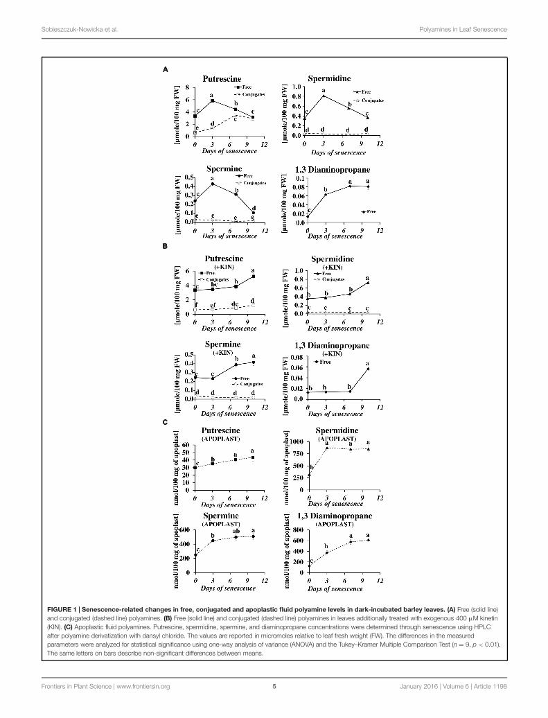
Changes in the Level of Different PAFractions during the Senescence ofDetached Barley LeavesWe have studied the process of senescence of barley leavesusing a dark-induction model that we routinely use in ourresearch (Legocka and Zajchert, 1999; Sobieszczuk-Nowickaet al., 2009, 2015). Dark-induced barley leaf senescence is adynamic process leading through a series of transformationsto the decay of the photosynthetic apparatus, which in turndecreases photosynthetic capacity of the leaf, the disruption of theorganelles and ultimately to cell death. Our recent observationsregarding symptoms of chlorophyll loss in senescing leaves,decomposition of chloroplasts, internucleosomal fragmentationof chromatin, condensation of nuclear DNA and the disruptionof the nucleus indicate that dark-induced leaf senescence engagesPCD mechanisms (Zmienko et al., 2015a). To verify whetherthis process also involves changes in PA titer, we determinedlevels of free and conjugated PAs (PU, SD, SM, and DP) inthe detached leaves throughout the process (Figure 1A). ThePU level increased to a maximum (up to 1.74× of the levelobserved at the onset of senescence induction) at day 3 ofsenescence. At day 7 it slightly decreased and finally returnedto the initial level at day 10. The relative changes in levelof free SD mimicked those of PU, with a maximum increase(2.3×) at day 3, followed by a slight decrease at day 7 and
returning to the initial level at day 10. Likewise, the amountof free SM peaked at day 3 (1.8 times increase) but decreasedfaster and dropped 2.2 times below the initial level at day 10.The amount of free DP showed a stepwise increase throughoutthe entire period of leaf senescence. At day 10 it was 6.2×higher in comparison to day 0. However, the percentage ofDP in the total pool of free PAs was negligible. Accumulationof free PU was accompanied by increased formation of PUconjugates (Figure 1A). Their level increased fivefold betweendays 0 and 10. No other conjugates, either SD or SM,accumulated.
The changes in the levels of free and conjugated PAsduring senescence were also evaluated after the addition ofthe anti-senescing agent KIN. Exogenously added KIN retardsthe senescence process (Sobieszczuk-Nowicka et al., 2009 andreferences therein). Here, KIN slowed the accumulation of allfree PAs in the detached leaves. In those incubated with KIN,high levels of free PU, SD, SM, and DP (comparable to theiramounts at day 3 without KIN addition, see Figure 1A) werereached only at days 7–10 (Figure 1B). The accumulationof PU conjugates was similarly delayed by the addition ofKIN.
In the apoplastic fluids, PA levels also increased during thesenescence process, but the ratios of individual PAs and theiraccumulation profiles were different compared to the free PAfraction (Figure 1C). PU, which was the most abundant of thefree PAs (Figure 1A), was one order of magnitude less abundantthan the other PAs (Figure 1C) in the apoplastic fluids pool. Itdisplayed a slight but continuous increase from 30 nmol/100 mgapoplast at day 0 to 43.5 nmol/100 mg apoplast at day 10. Theamounts of SD and SM increased substantially at day 3 (from324 to 875 nmol/100 mg apoplast for SD and from 250 to450 nmol/100 mg apoplast for SM) and remained approximatelyat that level (SD) or slightly higher (SM) at days 7 and 10.DP levels increased continuously through the entire time ofsenescence, accumulating from 125 to 612 nmol/100mg apoplast,with the most rapid changes observed at days 3 and 7 and a slowerincrease at day 10.
Senescence-Associated Changes in theTranscript Levels and Protein Activity ofGenes Directly Involved in PAMetabolismThe accumulation of various fractions of PAs observed inthe barley leaves undergoing senescence might result fromthe increased synthesis or activation of proteins involved inPA production and also from other mechanisms; e.g., PAtransport. We first investigated whether changes in PA levelswere accompanied by any changes in the expression of genesinvolved in PA metabolism. We utilized previously generatedmicroarray data used to evaluate barley gene expression in thesame experimental model of dark-induced senescence, exceptthat the leaves were not detached (Zmienko et al., 2015a,b).Regarding barley genes involved in PA metabolism, only SPMSand two PAO genes were characterized in detail (Cervelliet al., 2006; Rodriguez-Kessler and Jiménez-Bremont, 2008).
Frontiers in Plant Science | www.frontiersin.org 4 January 2016 | Volume 6 | Article 1198
Sobieszczuk-Nowicka et al. Polyamines in Leaf Senescence
FIGURE 1 | Senescence-related changes in free, conjugated and apoplastic fluid polyamine levels in dark-incubated barley leaves. (A) Free (solid line)and conjugated (dashed line) polyamines. (B) Free (solid line) and conjugated (dashed line) polyamines in leaves additionally treated with exogenous 400 μM kinetin(KIN). (C) Apoplastic fluid polyamines. Putrescine, spermidine, spermine, and diaminopropane concentrations were determined through senescence using HPLCafter polyamine derivatization with dansyl chloride. The values are reported in micromoles relative to leaf fresh weight (FW). The differences in the measuredparameters were analyzed for statistical significance using one-way analysis of variance (ANOVA) and the Tukey–Kramer Multiple Comparison Test (n = 9, p < 0.01).The same letters on bars describe non-significant differences between means.
Frontiers in Plant Science | www.frontiersin.org 5 January 2016 | Volume 6 | Article 1198

Sobieszczuk-Nowicka et al. Polyamines in Leaf Senescence
For this reason, we performed homology-based searches inpublic databases using the sequences of PA metabolism genesfrom other species to identify barley homologs. We thenexamined gene expression during senescence based on theabove microarray datasets (Figure 2). With this approach, up-regulation of four genes was detected. A gene encoding CPAwas induced 1.4-4-fold; a gene encoding DAO was induced 1.6-2.1-fold; and a gene encoding SPMS was induced 3.5-4-fold.The most remarkable changes were detected in the expressionof the SAMDC gene, reaching ninefold induction at day 3and 18.5-17.5-fold induction at later stages of senescence ofleaves. On the other hand, the changes in expression level ofthe gene encoding PAObc were transient and not statisticallysignificant.
Next, we measured the senescence-associated changes in theactivity of proteins engaged in PA metabolism: ADC, SAMDC,DAO, and PAO (Figure 3). A rapid increase in the activity ofboth enzymes involved in PA biosynthesis (SAMDC and ADC)was observed at day 3 (2.4× and 2.6×, respectively), followedby a slight decline at days 7 and 10 but still well-above the
initial level. The activity of enzymes engaged in PA catabolism(DAO and PAO) continuously increased to day 7, when it reachedmaximal values (2× and 5.3×, respectively) and stabilized at thatlevel.
Senescence-Associated Changes inTranscript Levels of Genes Involved inPathways Related to PA MetabolismThe microarray data for dark-induced senescing barley leavesallowed us to also examine the transcriptional profiles ofgenes encoding enzymes from other metabolic pathways thatare strictly related to PA synthesis and turnover (Figure 4).Several genes involved in nitrate metabolism were represented byadequate oligonucleotide probes on the microarray. All showedan increased expression from day 3 throughout the whole periodof leaf senescence (Figure 4A). The expression of the GOGATwas constantly up-regulated, approximately fivefold. The geneencoding GDH was progressively up-regulated, reaching a 4.3-fold increase at day 10. The expression of the gene encoding
FIGURE 2 | The time course of CPA, SAMDC, SPMS, DAO, and PAObc expression in barley leaves undergoing dark-induced senescence. CPA,N-carbamoylputrescine amidohydrolase; SAMDC, SAM decarboxylase; SPMS, spermine synthase; DAO, diamine oxidase; PAObc, back conversion polyamineoxidase. Barley homologs of each gene were found by homology searches in public databases. The gene expression data are from a time-course experimentperformed with an Agilent oligonucleotide microarray (Zmienko et al., 2015a,b). The data are presented as log2-fold changes relative to day 0. Differential geneexpression was evaluated with Bayesian linear modeling (R/Bioconductor, limma package) with Benjamini and Hochberg’s correction of the false discovery rate. Theadjusted p-values are marked with ∗p < 0.05, ∗∗p < 0.005, and ∗∗∗p < 0.0005.
Frontiers in Plant Science | www.frontiersin.org 6 January 2016 | Volume 6 | Article 1198

Sobieszczuk-Nowicka et al. Polyamines in Leaf Senescence
FIGURE 3 | The time course of ADC, SAMDC, DAO, and PAO activitiesin barley leaves undergoing dark-induced senescence. ADC, argininedecarboxylase, SAMDC, S-adenosyl-methionine decarboxylase; DAO,diamine oxidase; PAO, polyamine oxidase. Enzyme activities were determinedas described in Section “Materials and Methods.” The values for ADC andSAMDC activity were reported in CPM relative to protein in mg. DAO and PAOactivity are reported in U relative to time (min) and leaf fresh weight (FW). Thedifferences in the measured parameters were analyzed for statisticalsignificance using one-way ANOVA and the Tukey–Kramer MultipleComparison Test (n = 6, p < 0.01). The same letters on bars describenon-significant differences between means.
GS initially increased 38× and then slightly dropped, but stillremained much higher than at day 0. A similar trend wasobserved for the NR gene, except its induction was much lower(maximum 1.8× at day 3). Two genes involved in ethylenemetabolism and represented on the microarray also were up-regulated:ACC synthase, up to 3.5-fold at day 7, andACC oxidase,up to 8.7-fold at day 10 (Figure 4B). Two genes involved inGABAmetabolism (Figure 4C) and two involved in proline metabolism(Figure 4D) were also investigated. While the gene encodingGAD was down-regulated during the entire period, with thestrongest reduction in expression level at day 10 (3.6-fold), theexpression of the gene encodingGABA-T did not change by morethan 50% during the entire period of leaf senescence (Figure 4C).P5CS was up-regulated at day 3 (1.2-fold) and then was down-regulated with the strongest reduction in expression level atday 7 (0.55-fold), whereas P5CDH was up-regulated during theentire period, with the strongest increase in expression level atday 10 (4.7-fold; Figure 4D).The arginase gene, related to ureametabolism, was progressively up-regulated, with a maximumincrease of 5.8-fold at day 10 (Figure 4E).
Effect of PA Catabolism Inhibition onSenescence-Related Changes in FreePolyamine TiterTranscriptional profiling results revealed that PA catabolismmight be an important element regulating dark-induced leaf
FIGURE 4 | Continued
Frontiers in Plant Science | www.frontiersin.org 7 January 2016 | Volume 6 | Article 1198

Sobieszczuk-Nowicka et al. Polyamines in Leaf Senescence
FIGURE 4 | Continued
The time course of NR, GDH, GS, GOGAT, ACC SYNTHASE, ACC OXIDASE, GABA-T, GAD, P5CS, P5CDH, and ARGINASE expression in barley leavesundergoing dark-induced senescence. (A) genes involved in nitrate metabolism; NR, nitrate reductase; GDH, glutamate dehydrogenase; GS, glutaminesynthetase; GOGAT, glutamine oxoglutarate aminotransferase. (B) genes involved in ethylene synthesis; ACC SYNTHASE; ACC OXIDASE. (C) Genes involved inGABA metabolism; GABA-T, γ-aminobutyrate aminotransferase; GAD, glutamate decarboxylase. (D) Genes involved in proline metabolism; P5CS, �1
pyrroline-5-carboxylate synthetase; P5CDH, �1 pyrroline-5-carboxylate dehydrogenase. (E) Gene involved in urea metabolism; ARGINASE. Barley homologs foreach gene were found by homology searches in public databases. The gene expression data are from the time-course experiment performed with the Agilentoligonucleotide microarray (Zmienko et al., 2015a,b). The data are presented as log2-fold changes relative to day 0. Differential gene expression was evaluated withBayesian linear modeling (R/Bioconductor, limma package) with Benjamini and Hochberg’s correction of the false discovery rate. The adjusted p-values are markedwith ∗p < 0.05, ∗∗p < 0.005, and ∗∗∗p < 0.0005.
senescence, which encouraged us to evaluate the effect ofinhibiting that pathway. The two inhibitors used in the studieswere AG, a competitive inhibitor of soybean DAO (Nikolov et al.,1990; Xing et al., 2007), and guazatine (G), which competitivelyinhibits maize and tobacco PAOs (Cona et al., 2004; Yoda et al.,2006). The efficiency of inhibition of the enzyme activities wasevaluated after treatment with each inhibitor by measuring therespective oxidase activity compared to the control (C – detachedleaves without inhibitor treatment). AG inhibited DAO activityby 69% at day 3 and 58% at days 7–10. G inhibited PAO activityby 90% at day 3 and approximately 75% at days 7–10. We nextlooked at the level of free PAs in inhibitor-treated senescing leaves(Figure 5). The treatment with AG induced a PU level higherthan in the control plants (1.13×, 1.3×, and 1.56× at days 3,7, and 10, respectively), while the accumulation of SD and SMduring senescence was slightly lower, with the differences mostobservable at day 7, when their levels reached 0.7 and 0.62 of
the control amount, respectively. The DP level did not differmuch from the control at any time point (maximum observeddifference was a 1.2× increase at day 10). The inhibition ofPAO activity delayed the accumulation of PU; its level slowlyincreased and at day 10 it reached 5.575 μmol/100 mg FW,comparable with the amount observed in the control at day 3.Treatment with G also drastically lowered DP accumulation. Itslevel was even lower at day 3 than at day 0 and gradually increasedafterward, but even by day 10 it did not reach the level thatwas observed in the control plants at day 3. On the contrary,PAO activity inhibition caused overall elevation of SD and SMlevels compared to the control, but did not alter the accumulationspeed. The SD level in leaves treated with G was 1.5×, 2.06×,and 1.7 higher at days 3, 7, and 10, respectively, than in thecontrol. The SM level in leaves treated with G was 1.2×, 1.6×,and 2.78 higher at days 3, 7, and 10, respectively, than in thecontrol.
FIGURE 5 | Senescence-related changes in free polyamine level in dark-incubated conditions in the presence of PA catabolism inhibitors in barleyleaves. Polyamines (PAs) were estimated in C (solid line)- control leaves and after application of polyamine catabolism inhibitors: +AG (dashed line) – leaves treatedwith 10 μM N,N-diaminoguanidine (AG) – diamine oxidase (DAO) inhibitor; +G (dotted line) leaves treated with 50 μM guazatine (G) – polyamine oxidase (PAO)inhibitor. Putrescine, spermidine, spermine, and diaminopropane concentrations were determined using HPLC after polyamine derivatization with dansyl chloride.The values are reported in micromoles relative to leaf FW. The differences in the measured parameters were analyzed for statistical significance using one-wayANOVA and the Tukey–Kramer Multiple Comparison Test (n = 12, p < 0.01). The same letters on bars describe non-significant differences between means.
Frontiers in Plant Science | www.frontiersin.org 8 January 2016 | Volume 6 | Article 1198

Sobieszczuk-Nowicka et al. Polyamines in Leaf Senescence
Effect of PA Catabolism Inhibition onPhotosynthesis in Senescing BarleyLeavesThe disintegration of chloroplasts and cessation ofphotosynthesis is an important landmark of the leaf senescenceprocess, because the dying process of a plant cell may bereversed as long as the functions of the chloroplasts can be
restored (Van Doorn and Yoshimoto, 2010). We wonderedwhether the capacity of a plant to control senescence couldbe somehow linked to catabolism of PAs. To verify this, wemeasured the photosynthetic quantum conversion parametersand leaf nitrogen (N) status in senescing barley leaves afterthe addition of DAO and PAO inhibitors (Figure 6). In thecontrol leaves, the values of maximum quantum yield of PSII inthe dark-adapted state (Fv/Fm) and photochemical quenching
FIGURE 6 | Senescence-related changes in chlorophyll fluorescence and nitrogen status in dark-incubated conditions in the presence of PAcatabolism inhibitors in barley leaves. The parameters Fv/Fm, maximum quantum yield of PSII in the dark adapted state; qP, photochemical quenching Chlfluorescence; NPQ, non-photochemical quenching of Chl fluorescence; Chl, chlorophyll level; NBI, nitrogen balance index; and Flv, amount of flavonoids weremeasured throughout senescence in C (solid line)- control leaves and after application of polyamine catabolism inhibitors: + AG (dashed line) – leaves treated with10 μM N,N-diaminoguanidine (AG) – diamine oxidase (DAO) inhibitor; + AG/GABA (dashed-dotted line) – leaves treated with 10 μM AG and 1 mM γ-aminobutyricacid (GABA);+ G (dotted line) leaves treated with 50 μM guazatine (G) – polyamine oxidase (PAO) inhibitor. Measurements of Chl fluorescence induction kineticswere taken using a pulse amplitude-modulated PAM fluorimeter. For estimation of plant nitrogen status, a method based on fluorometric measurement ofchlorophyll/flavonoids ratio with a DUALEX 4 FLAV fluorimeter was used. The differences in the measured parameters were analyzed for statistical significance usingone-way ANOVA and the Tukey–Kramer Multiple Comparison Test (n = 25, p < 0.01). The same letters on bars describe non-significant differences between means.
Frontiers in Plant Science | www.frontiersin.org 9 January 2016 | Volume 6 | Article 1198

Sobieszczuk-Nowicka et al. Polyamines in Leaf Senescence
FIGURE 7 | Senescence-related hydrogen peroxide production in dark-incubation in the presence of PAO activity inhibitor in barley leaves. (A) In situdetection of hydrogen peroxide with confocal microscopy and the fluorescent probe 2′ ,7′−dichlorofluorescein. The bar indicates 200 μm. (B) H2O2 level determinedwith a spectrophotometric method based on titanium (Ti4+) estimated throughout senescence in C (solid line)- control leaves and after application of polyamineoxidase (PAO) activity inhibitor, +G (dotted line) leaves treated with 50 μM guazatine (G) – PAO activity inhibitor. The differences in the parameters were analyzed forstatistical significance using one-way ANOVA and the Tukey–Kramer Multiple Comparison Test (n = 9, p < 0.01). The same letters on bars describe non-significantdifferences between means.
of Chl (qP) underwent a relatively small decrease until day 7of senescence and then declined 0.8× and 0.5×, respectively.Accordingly, non-photochemical quenching of Chl fluorescence(NPQ) was at normally low values and did not change muchuntil day 7, after which it rose 2.1× in comparison to day 0. Atthe same time, the Chl and NBI indexes lowered gradually fromday 0 to day 7 and stabilized at values 2.4× and 3.7× lower thanat day 0. The flavonoids ratio (Flv) value raised slowly duringsenescence. At day 10 it reached a level 1.2× higher than at day0. The addition of DAO inhibitor (AG) enhanced the effect ofsenescence on the photosynthetic parameters, slightly loweringthe values of Fv/Fm, Chl, qP and NBI, which generally dropduring the process, and further increasing the values of NPQand Flv, which increase in senescing leaves. In most cases, thiseffect was enhanced at the later stages of senescence (days 7and 10). When AG was applied to leaves together with GABA,the photosynthetic activity parameters and the progress of thechlorophyll degradation process remained at the control level.The effect of applying the PAO inhibitor (G) was opposite to thatof AG, and the senescence-associated changes in photosyntheticefficiency of chloroplasts were minimized. Again, this effect wasmost prominent at the last analyzed stage (day 10).
Effect of PAO Inhibition on HydrogenPeroxide Generation in SenescingLeavesThe physiological role of PAs can also result from theirinvolvement in the plant signaling network. PA oxidation viaoxidases leads to the generation of hydrogen peroxide (H2O2),which is involved in stress responses and the induction of PCD(Moschou et al., 2008). The obvious effect of PAO inhibition ofsenescence-associated changes in cell metabolism encouraged usto analyze the senescence-related changes in H2O2 accumulationand the effect of inhibiting oxidase activity.
In control plants, the DCFH-DA fluorescence intensity,which reflects H2O2 accumulation, increased as leaf senescenceprogressed (Figure 7A). The fluorescence was detected primarilyin the external layers of the leaf tip, in the epidermis and the
midribs. A weak signal was observed in the mesophyll cells, and avery weak signal in the veins and blades of leaves. The same trendwas observed in plants subjected to inhibitor treatment, but theireffect on the overall level of H2O2 production was the opposite.Inhibiting PAO activity lowered the fluorescence signal at days 3to 10. The results of studies using confocal microscopy were alsoconfirmed by the spectrophotometric studies (Figure 7B). Theexposure of the leaf to dark conditions resulted in a continuousincrease in H2O2 content at days 3, 7, and 10 (2.2×, 3.9×, and5.3×, respectively, compared to the control). In leaves incubatedwith a PAO inhibitor, H2O2 content also increased but reachedonly 73% (day 3), 77% (day 7), and 68% (day 10) of the controllevel.
DISCUSSION
Polyamine metabolism is linked to many metabolic pathwaysin the cell by being involved in the formation of signalingmolecules andmetabolites directly related to the cellular responseto environmental changes. The network of these relationshipsis shown in Figure 8, which is useful background to followthe interpretation of the results discussed below. Figure 8 alsosummarizes the time course of expression of genes involved in orrelated to PA metabolism in dark-induced barley leaf senescence.
Our study revealed a senescence-dependent interplay betweenvarious PA forms and individual PAs. At the beginning of thesenescence process, a rapid increase in the level of all free PAswas observed, but at day 7, PU, SD, and SM levels began to dropto or even below the control level, while the DP level continuouslyincreased.
In the cell, the PA titer is carefully controlled (Cogen,1998; Moschou et al., 2012; Moschou and Roubelakis-Angelakis,2013, and references therein). PA biosynthesis, catabolism,conjugation, interconversion, and transport contribute to PAhomeostasis (Angelini et al., 2010; Moschou and Roubelakis-Angelakis, 2013). Thus, the increase in the free PA titer atthe beginning of senescence can constitute a part of a signalthat leads the cell to process induction, or it might maintain
Frontiers in Plant Science | www.frontiersin.org 10 January 2016 | Volume 6 | Article 1198

Sobieszczuk-Nowicka et al. Polyamines in Leaf Senescence
FIGURE 8 | Schematic presentation of possible interaction between polyamine metabolism and other pathways through dark-induced barley leafsenescence. Polyamine biosynthetic routes are indicated with bolded lines. Thin lines mark putrescine-derived alkaloid formation, polyamine conjugation andcatabolic processes. The red-green bars indicate the expression level of genes estimated according to the results of microarray analysis. Numbers in the barsdescribe the senescence days. NR, nitrate reductase; GS, glutamine synthetase; GOGAT, glutamine oxoglutarate aminotransferase; NAGS, N-acetylglutamatesynthase; NAGK, N-acetylglutamate kinase; NAGPR, N-acetylglutamate 5-phosphate reductase; NAOAT, N-acetylornithine transaminase; NAOD, N2-acetylornithinedeacetylase; OCT, ornithine carbamoyltransferase; ASSY, argininosuccinate synthase; ASL, argininosuccinate lyase; ADC, arginine decarboxylase; AIH, agmatineiminohydrolase; CPA, N-carbamoylputrescine amidohydrolase; ODC, ornithine decarboxylase; SAMS, SAM synthetase; SAMDC, SAM decarboxylase; SPDS,spermidine synthase; SPMS, spermine synthase; tSPMS/ACL5, thermospermine synthase/acaulis5; PAO, polyamine oxidase; PAObc, back conversion polyamineoxidase; SSAT, spermidine/spermine N-1 acetyl transferase; DAO, diamine oxidase; GABA-T, γ-aminobutyrate aminotransferase; SSADH, succinic semialdehydedehydrogenase; GAD, glutamate decarboxylase; PHT, putrescine hydroxycinnamoyl transferase; LDC, lysine decarboxylase; NOS, nitric oxide synthase; P5CS, �1
pyrroline-5-carboxylate synthetase; P5CR, �1 pyrroline-5-carboxylate reductase; PDH, proline dehydrogenase; P5CDH, �1 pyrroline-5-carboxylate dehydrogenase;OAT, ornithine-δ-aminotransferase.
its progress. As our study revealed, the PA accumulation uponsenescence could be due to simultaneous up-regulation of aset of genes involved in PA biosynthesis and an increase inenzymatic activity of the proteins they encode. Senescence issensitive to hormonal perturbation, with particular reference tocytokinins (Lim et al., 2007). Cytokinins prevent chloroplastdegradation by retarding both the breakdown of chlorophyll andthe decay of the thylakoid system in senescing leaves. They alsolower the activity of proteolytic enzymes and promote proteinsynthesis in detached barley leaves senescing in the dark (Legockaand Szweykowska, 1981, 1983). The delay of senescence bycytokinins occurs mainly at the transcriptional level, by inhibitingthe expression of senescence-related genes (Lim et al., 2007).
We applied KIN to the model in this study to learn whetherPA accumulation is an integral part of senescence–dependentevents, as well as whether PAs might act as drivers or controllersof the process. We showed that in the presence of KIN, thePA accumulation shifted to later times, confirming that it isa senescence-dependent. It also implied that some additionalsenescence-related signals, blocked by exogenously applied KIN,influence PA synthesis in this process.
When studying dark-induced PA accumulation, it isimportant to note that induced PA accumulation at thebeginning of the process could imply a ROS-scavenging functionof both free and conjugated forms of PAs, as reported byRadyukina et al. (2009) and Legocka et al. (2015). SD and SM,
Frontiers in Plant Science | www.frontiersin.org 11 January 2016 | Volume 6 | Article 1198

Sobieszczuk-Nowicka et al. Polyamines in Leaf Senescence
which have three and four amino groups, are more effective inthe removal of reactive oxygen forms than PU (Kuznetsov andShevyakova, 2007).
We also measured transcript levels and enzymatic activityof the PA catabolic enzymes DAO and PAO, as well as theeffect of their inhibition. Our data indicated that transformationsbetween individual PAs could be an essential element ofdarkness-induced responses and indicate the physiological roleof PA in cell senescence. Inhibiting PAO activity drasticallyslowed down the accumulation of both DP and PU, while thelevels of SD and SM were substantially increased. This is alogical effect, but remarkably also resulted in slowing downthe senescence-associated chlorophyll loss. Application of PAOinhibitors maintained the Chl parameter at a higher level inleaves subjected to senescence compared to those incubatedin the absence of inhibitor, which also resulted in highervalues of the chlorophyll fluorescence parameters such as Fv/Fmand qP. Furthermore, inhibiting PAO activity decreased theamount of H2O2, suggesting that PAO-mediated catabolism ofSD/SM supports senescence-dependent degradation, most likelyby H2O2 synthesis. Plant responses to environmental factorsinvolve the secretion of SD/SM to the apoplast where they arecatabolized, leading to H2O2 production. Depending on theamount of H2O2, the defense response or cell death program isinitiated (Yoda et al., 2003, 2006; Marina et al., 2008; Moschouet al., 2008). In agreement with this model, the high SM/SD poolin the apoplast rapidly increased at day 3 and was maintained ata high level, which corresponded with the gradual accumulationof DP and the increase in H2O2 level.
The initial amount of PU in the apoplastic pool of PAs wasone order of magnitude lower and only slightly increased duringsenescence. However, PU dominated in the free PA fraction,where it initially accumulated to a high level, and then started todrop after day 3. This effect was accompanied by the formationof PU conjugates that accumulated in the senescing leaf to ahigh level, indicating the fate of PU in the senescing cell. Formany years, PA conjugates were considered to be inactive PAforms. However, recent evidence indicates that they are essentialfor certain developmental processes (Bassard et al., 2010; Fuellet al., 2010). Senescence-dependent nitrogen (N) and carbon (C)flow might be then shifted toward PA conjugation, which couldexplain the observed increase in expression of respective proteincoding genes. The hypothesis that PAs are significant players inthe turnover of N molecules in plants has been discussed byMattoo et al. (2010). Furthermore, our results strongly indicatethe involvement of a DAO-mediated PU oxidation process inGABA production. Microarray-based profiling of GAD geneexpression suggested that in senescing leaves, GABA synthesisfrom glutamate is gradually suppressed. PU oxidation mightbecome an alternative source of GABA to the tricarboxylic acidcycle and also for some as yet undefined signaling pathways.The role of GABA as a neurotransmitter is well-establishedin animal cells. In plants, increases in the GABA level havebeen reported in response to different stresses. However, theexact role of GABA under stress conditions still needs tobe defined (Michaeli and Fromm, 2015). Analysis of Chl afluorescence parameters and plant N status revealed that blocking
the PU oxidation pathway accelerated chlorophyll degradation.However, simultaneous addition of exogenous GABA togetherwith a DAO inhibitor was sufficient to prevent accelerateddegradation of photosystem complexes. Together, these resultspoint to a central role of GABA signaling in senescence andindicate that PU may be a key precursor of this transmitter.
CONCLUSION
The results demonstrate that the dark-induced leaf senescenceprocess was supported by the accumulation of SD and SM. Bothwere newly produced and transported into the apoplast, wherethey were readily degraded, yielding hydrogen peroxide andDP, which could regulate or participate in senescence-dependentdegradation processes. However, PU also accumulated in thesenescing leaf. As indicated in the experiment with cytokinin,the PA accumulation was senescence-dependent, although PAanabolism could not promote or abrogate the process started bydark incubation in the absence of additional senescence-relatedsignals. Thus, the accumulation of PU might serve as a precursorof SD and SM but also support the senescence process throughforming PU conjugates, the only PA conjugates forming insenescence, and by supporting via its oxidation GABA synthesis.
The knowledge of the participation of PAs in leaf senescenceis very fragmentary. Our results reported here shed some lighton the problem, in particular on leaf senescence in an importantcrop. These insights allow the development of a frameworkthat would provide more detailed observations into inducedsenescence and its biotechnological applications and wouldstimulate new, important questions about the function of PAs inthe process.
AUTHOR CONTRIBUTIONS
ES-N, JL conceived and designed the experiments. ES-N, SK, AZ,AM performed the experiments. ES-N, SK, AZ, AM analyzedthe data. ES-N, AZ, AM contributed reagents/materials/analysistools. ES-N, AZ wrote the paper. ES-N, SK, AZ, AM, JLparticipated in manuscript finalization and preparation ofFigures.
FUNDING
This work was supported by the National Science Centre researchgrant No N N303 418236 to ES-N and was also partiallyfunded by the Department of Plant Physiology, AdamMickiewiczUniversity in Poznan Statutory Fund Research No S/P-B/010.Publication costs were covered by the KNOW program of thePolish Ministry of Science and Higher Education.
ACKNOWLEDGMENTS
The authors acknowledge the help of the European Centre ofBioinformatics and Genomics, Poznan, Poland, where part of
Frontiers in Plant Science | www.frontiersin.org 12 January 2016 | Volume 6 | Article 1198

Sobieszczuk-Nowicka et al. Polyamines in Leaf Senescence
the experimental work was conducted. SK acknowledges thefinancial support of the National Science Centre for ETIUDAscholarship no. DEC-2013/08/T/NZ9/01019. Apart from our
acknowledgments, we would also like to apologize to thoseauthors whose valuable works could not be cited due to spacelimitation.
REFERENCES
Afzal, M., Matsugo, S., Sasai, M., Xu, B., Aoyama, K., and Takeuchi, T.(2003). Method to overcome photoreaction, a serious drawback to the use ofdichlorofluorescin in evaluation of reactive oxygen species. Biochem. Biophys.Res. Commun. 304, 619–624. doi: 10.1016/S0006-291X(03)00641-7
Angelini, R., Cona, A., Federico, R., Fincato, P., Tavladoraki, P., and Tisi, A. (2010).Plant amine oxidases “on the move”: an update. Plant Physiol. Biochem. 48,560–564. doi: 10.1016/j.plaphy.2010.02.001
Bagni, N., and Tassoni, A. (2006). “The role of polyamines in relation to flowersenescence,” in Floriculture, Ornamental and Plant Biotechnology, ed. J. A.Teixeira da Silva (Isleworth: Global Science Books), 88–95.
Bassard, J.-E., Ullmann, P., Bernier, F., and Werck-Reichhart, D. (2010).Phenolamides: bridging polyamines to the phenolic metabolism.Phytochemistry 71, 1808–1824. doi: 10.1016/j.phytochem.2010.08.003
Becana, M., Aparicio-Tejo, P., Irigoyen, J. J., and Sanchez-Diaz, M. (1986). Someenzymes of hydrogen peroxide metabolism in leaves and root nodules ofMedicago sativa. Plant Physiol. 82, 1169–1171. doi: 10.1104/pp.82.4.1169
Besford, R. D., Richardson, C.M., Campos, J. L., and Tiburcio, A. F. (1993). Effect ofpolyamines on stabilization complexes in thylakoid membranes of osmoticallystressed oat leaves. Planta 189, 201–206. doi: 10.1007/BF00195077
Brown, R., Jarvis, K., and Hyland, K. (1989). Protein measurement usingbicinchoninic acid: elimination of interfering substances. Anal. Biochem. 180,136–139. doi: 10.1016/0003-2697(89)90101-2
Cai, G., Sobieszczuk-Nowicka, E., Aloisi, I., Fattorini, L., Serafini-Fracassini, D.,and Del Duca, S. (2015). Polyamines are common players in different facets ofplant programmed cell death. Amino Acids 47, 27–44. doi: 10.1007/s00726-014-1865-1
Cartelat, A., Cerovic, Z. G., Goulas, Y., Meyer, S., Lelarge, C., Prioul, J.-L., et al.(2005). Optically assessed contents of leaf polyphenolics and chlorophyll asindicators of nitrogen deficiency in wheat (Triticum aestivum L.). Field CropsRes. 91, 35–49. doi: 10.1016/j.fcr.2004.05.002
Cervelli, M., Bianchi, M., Cona, A., Crosatti, C., Stanca, M., Angelini, R., et al.(2006). Barley polyamine oxidase isoforms 1 and 2, a peculiar case of geneduplication. FEBS J. 273, 3990–4002. doi: 10.1111/j.1742-4658.2006.05402.x
Cogen, S. S. (1998). A Guide to Polyamines. New York: Oxford University Press.Cohen, A. S., Popovic, R., and Zalik, S. (1979). Effects of polyamines on chlorophyll
and protein content, photochemical activity, and chloroplast ultrastructureof barley leaf discs during senescence. Plant Physiol. 64, 717–720. doi:10.1104/pp.64.5.717
Cona, A., Manetti, F., Leone, R., Corelli, F., Tavladoraki, P., Polticelli, F., et al.(2004). Molecular basis for the binding of competitive inhibitors of maize PAO.Biochemistry 43, 3426–3435. doi: 10.1021/bi036152z
Fuell, C., Elliott, K. A., Hanfrey, C. C., Franceschetti, M., and Michael, A. J. (2010).Polyamine biosynthetic diversity in plants and algae. Plant Physiol. Biochem. 48,513–520. doi: 10.1016/j.plaphy.2010.02.008
Ioannidis, N. E., Zschiesche, W., Barth, O., Kotakis, C., Navakoudis, E.,Humbeck, K., et al. (2014). The genetic reprogramming of polyaminehomeostasis during the functional assembly, maturation, and senescence-specific decline o f the photosynthetic apparatus in Hordeum vulgare. J. PlantGrow. Reg. 33, 77–90. doi: 10.1007/s00344-013-9387-8
Jackowski, G., Olkiewicz, P., and Zelisko, A. (2003). The acclimative response ofthe main light-harvesting chlorophyll a/b-protein complex of photosystem II(LHCII) to elevated irradiances at the level of trimeric subunits. J. Photoch.Photobiol. B Biol. 70, 163–170. doi: 10.1016/S1011-1344(03)00076-9
Kusano, T., Berberich, T., Tateda, C., and Takahashi, Y. (2008). Polyamines:essential factors for growth and survival. Planta 228, 367–381. doi:10.1007/s00425-008-0772-7
Kusano, T., and Suzuki, H. (2015). Polyamines a Universal Molecular Nexus forGrowth, Survival and Specialized Metabolism. Tokyo: Springer.
Kuznetsov, V. V., and Shevyakova, N. I. (2007). Polyamines and stress tolerance ofplants. Plant Stress 1, 50–71.
Legocka, J., Sobieszczuk-Nowicka, E., Wojtyla, Ł., and Samardakiewicz, S. (2015).Lead-stress induced changes in the content of free, thylakoid- and chromatin-bound polyamines, photosynthetic parameters and ultrastructure in greeningbarley leaves. J. Plant Physiol. 186-187, 15–24. doi: 10.1016/j.jplph.2015.07.010
Legocka, J., and Szweykowska, A. (1981). The role of cytokinins and metabolismof barley leaves. III. The effect on the RNA metabolism in various cellcompartments during senescence. Z. Pflanzenphysiol. 102, 363–374. doi:10.1016/S0044-328X(81)80208-5
Legocka, J., and Szweykowska, A. (1983). The role of cytokinins in the developmentand metabolism of barley leaves. VI. The effect on the protein metabolism invarious cell compartments during leaf senescence. Acta Physiol. Plant. 5, 11–20.
Legocka, J., and Zajchert, I. (1999). Role of spermidine in the stabilization ofthe apoprotein of the light-harvesting chlorophyll a/b-protein complex ofphotosystem II during leaf senescence process.Acta Physiol. Plant. 21, 127–132.doi: 10.1007/s11738-999-0066-0
Legocka, J., and Zarnowska, A. (2000). Role of polyamines in the cytokinin-dependent physiological processes II. Modulation of polyamine levels duringcytokinin-stimulated expansion of cucumber cotyledons. Acta Physiol. Plant.22, 395–401. doi: 10.1007/s11738-000-0079-1
Lester, G. E. (2000). Polyamines and their cellular anti-senescence properties inhoney dew muskmelon fruit. Plant Sci. 160, 105–112. doi: 10.1016/S0168-9452(00)00369-1
Lim, P. O., Kim, H. J., and Nam, H. G. (2007). Leaf senescence. Annu. Rev. PlantBiol. 58, 115–136. doi: 10.1146/annurev.arplant.57.032905.105316
Marcé, M., Brown, D. S., Capell, T., Figueras, X., and Tiburcio, A. F. (1995).Rapid high performance liquid chromatographic method for the quantitation ofpolyamines as their dansyl derivatives: application to plant and animal tissues.J. Chromatog. B Biomed. Appl. 666, 329–335. doi: 10.1016/0378-4347(94)00586-T
Marina, M., Maiale, S. J., Rossi, F. R., Romero, M. F., Rivas, E. I., Gárriz, A., et al.(2008). Apoplastic polyamine oxidation plays different roles in local responsesof tobacco to infection by the necrotrophic fungus Sclerotinia sclerotiorumand the biotrophic bacterium Pseudomonas viridiflava. Plant Physiol. 147,2164–2178. doi: 10.1104/pp.108.122614
Mattoo, A. K., and Handa, A. K. (2008). Higher polyamines restore andenhance metabolic memory in ripening fruit. Plant Sci. 174, 386–393. doi:10.1016/j.plantsci.2008.01.011
Mattoo, A. K., Minocha, S. C., Minocha, R., and Handa, A. K. (2010). Polyaminesand cellular metabolism in plants: transgenic approaches reveal differentresponses to diamine putrescine versus higher polyamines spermidine andspermine. Amino Acids 38, 405–413. doi: 10.1007/s00726-009-0399-4
Mattoo, A. K., Sobolev, A. P., Neelam, A., Goyal, R. K., Handa, A. K., andSegre, A. L. (2006). Nuclear magnetic resonance spectroscopy-based metaboliteprofiling of transgenic tomato fruit engineered to accumulate spermidine andspermine reveals enhanced anabolic and nitrogen-carbon interactions. PlantPhysiol. 142, 1759–1770. doi: 10.1104/pp.106.084400
Michaeli, S., and Fromm, H. (2015). Closing the loop on the GABA shunt in plants:are GABA metabolism and signaling entwined? Front. Plant Sci. 6:419. doi:10.3389/fpls.2015.00419
Mizrahi, Y., Applewhite, P. B., and Galston, A. W. (1989). Polyamine bindingto proteins in oat and petunia protoplasts. Plant Physiol. 91, 738–743. doi:10.1104/pp.91.2.738
Moschou, P. N., Paschalidis, K. A., Delis, I. D., Andriopoulou, A. H., Lagiotis,G. D., Yakoumakis, D. I., et al. (2008). Spermidine exodus and oxidationin the apoplast induced by abiotic stress is responsible for H2O2 signaturesthat direct tolerance responses in tobacco. Plant Cell 20, 1708–1724. doi:10.1105/tpc.108.059733
Moschou, P. N., and Roubelakis-Angelakis, K. A. (2013). Polyamines andprogrammed cell death. J. Exp. Bot. 3, 1061–1066.
Moschou, P. N., Wu, J., Cona, A., Tavladoraki, P., Angelini, R., and Roubelakis-Angelakis, K. A. (2012). The polyamines and their catabolic products are
Frontiers in Plant Science | www.frontiersin.org 13 January 2016 | Volume 6 | Article 1198

Sobieszczuk-Nowicka et al. Polyamines in Leaf Senescence
significant players in the turnover of nitrogenous molecules in plants. J. Exp.Bot. 63, 695–709. doi: 10.1093/jxb/ers202
Nambeesan, S., Datsenka, T., Ferruzzi, M. G., Malladi, A., Mattoo, A. K., andHanda, A. K. (2010). Overexpression of yeast spermidine synthase impactsripening, senescence and decay symptoms in tomato. Plant J. 63, 836–847. doi:10.1111/j.1365-313X.2010.04286.x
Nikolov, I., Pavlov, V., Minkov, I., and Damjanov, D. (1990). Purification and someproperties of an amine oxidase from soybean seedings. Cell Mol. Life Sci. 46,765–767. doi: 10.1007/BF01939962
Radyukina, N. L., Mapelli, S., Ivanov, Y. V., Kartashov, A. V., Brambilla, I., andKuznetsov, V. (2009). Homeostasis of polyamines and antioxidant systems inroots and leaves of Plantago major under salt stress. Russian J. Plant Physiol. 56,323–331. doi: 10.1134/S1021443709030042
Rodriguez-Kessler, M., and Jiménez-Bremont, J. F. (2008). Zmspds2 maizegene: coding a spermine synthase? Plant Signal Behav. 3, 551–553. doi:10.4161/psb.3.8.5697
Seiler, N., and Raul, F. (2005). Polyamines and apoptosis. J. Cell Mol. Med. 9,623–642. doi: 10.1111/j.1582-4934.2005.tb00493.x
Serafini-Fracassini, D., Del Duca, S., Monti, F., Poli, F., Sacchetti, G., Bregoli, A. M.,et al. (2002). Transglutaminase activity during senescence and programmed celldeath in the corolla of tobacco (Nicotiana tabacum) flowers. Cell Death. Diff. 9,309–321. doi: 10.1038/sj.cdd.4400954
Serafini-Fracassini, D., Di Sandro, A., and Del Duca, S. (2010). Sperminedelays leaf senescence in Lactuca sativa and prevents the decay ofchloroplast photosystems. Plant Physiol. Biochem. 48, 602–611. doi:10.1016/j.plaphy.2010.03.005
Sobieszczuk-Nowicka, E., Rorat, T., and Legocka, J. (2007). Polyamine metabolismand S-adenosylmethionine decarboxylase gene expression during thecytokinin-stimulated greening process. Acta Physiol. Plant. 29, 495–502.doi: 10.1007/s11738-007-0099-1
Sobieszczuk-Nowicka, E., Wieczorek, P., and Legocka, J. (2009). Kinetin affects thelevel of chloroplast polyamines and transglutaminase activity during senescenceof barley leaves. Acta Biochim. Pol. 56, 255–259.
Sobieszczuk-Nowicka, E., Zmienko, A., Samelak-Czajka, A., Łuczak, M.,Pietrowska-Borek, M., Iorio, R., et al. (2015). Dark-induced senescence ofbarley leaves involves activation of plastid transglutaminases. Amino Acids 47,825–838. doi: 10.1007/s00726-014-1912-y
Takahashi, T., and Kakehi, J. (2010). Polyamines: ubiquitous polycations withunique roles in growth and stress responses. Ann. Bot. 105, 1–6. doi:10.1093/aob/mcp259
Van Doorn, W., and Yoshimoto, K. (2010). Role of chloroplasts and other plastidsin ageing and death of plants and animals: a tale of Vishnu and Shiva. AgeingRes. Rev. 9, 117–130. doi: 10.1016/j.arr.2009.08.003
Xing, S. G., Jun, Y. B., Hau, Z. W., and Liang, L. Y. (2007). Higher accumulationof γ-aminobutyric acid induced by salt stress through stimulating the activityof diamine oxidases in Glycine max (L.) Merr. roots. Plant Physiol. Biochem. 45,560–566. doi: 10.1016/j.plaphy.2007.05.007
Yoda, H., Hiroi, Y., and Sano, H. (2006). Polyamine oxidase is one of thekey elements for oxidative burst to induce programmed cell death intobacco cultured cells. Plant Physiol. 142, 193–206. doi: 10.1104/pp.106.080515
Yoda, H., Yamaguchi, Y., and Sano, H. (2003). Induction of hypersensitivecell death by hydrogen peroxide produced through polyamine degradationin tobacco plants. Plant Physiol. 132, 1973–1981. doi: 10.1104/pp.103.024737
Zmienko, A., Samelak-Czajka, A., Góralski, M., Sobieszczuk-Nowicka, E.,Kozłowski, P., and Figlerowicz, M. (2015a). Selection of reference genes forqPCR-and ddPCR-based analyses of gene expression in senescing barley leaves.PLoS ONE 10:e0118226. doi: 10.1371/journal.pone.0118226
Zmienko, A., Samelak-Czajka, A., Góralski, M., Sobieszczuk-Nowicka, E., andFiglerowicz, M. (2015b). Time-course transcriptional profiling of senescingbarley leaves. Gen. Data 4, 78–81. doi: 10.1016/j.gdata.2015.03.006
Conflict of Interest Statement: The authors declare that the research wasconducted in the absence of any commercial or financial relationships that couldbe construed as a potential conflict of interest.
Copyright © 2016 Sobieszczuk-Nowicka, Kubala, Zmienko, Małecka and Legocka.This is an open-access article distributed under the terms of the Creative CommonsAttribution License (CC BY). The use, distribution or reproduction in other forumsis permitted, provided the original author(s) or licensor are credited and that theoriginal publication in this journal is cited, in accordance with accepted academicpractice. No use, distribution or reproduction is permitted which does not complywith these terms.
Frontiers in Plant Science | www.frontiersin.org 14 January 2016 | Volume 6 | Article 1198