fracture mandible - add docshare01.docshare.tips to...
TRANSCRIPT

FRACTUREMANDIBLE

Rajesh R Yadav MS (ENT) DORL FCPS
Assistant ProfessorRajawadi Hospital
Formerly RegistrarShri Harilal Bhagwati Hospital
Mumbai, Maharashtra, India
Akancha R Yadav BDS
Dental ConsultantMumbai, Maharashtra, India
Prakash V Dhond MS (ENT) DORL
Honorary ENT ConsultantShri Harilal Bhagwati Hospital
Mumbai, Maharashtra, India
ForewordChris De Souza
FRACTUREMANDIBLE
JAYPEE BROTHERS MEDICAL PUBLISHERS (P) LTDNew Delhi • Panama City • London
®
Website: www.jaypeebrothers.comWebsite: www.jaypeedigital.com
© 2012, Jaypee Brothers Medical Publishers
All rights reserved. No part of this book may be reproduced in any form or by any meanswithout the prior permission of the publisher.
Inquiries for bulk sales may be solicited at: [email protected]
This book has been published in good faith that the contents provided by the authorscontained herein are original, and is intended for educational purposes only. While everyeffort is made to ensure accuracy of information, the publisher and the authors specificallydisclaim any damage, liability, or loss incurred, directly or indirectly, from the use or applica-tion of any of the contents of this work. If not specifically stated, all figures and tables arecourtesy of the authors. Where appropriate, the readers should consult with a specialist orcontact the manufacturer of the drug or device.
Fracture Mandible
First Edition : 2012
ISBN 978-93-5025-801-9
Printed in
Jaypee Brothers Medical Publishers (P) Ltd.
Jaypee Brothers Medical Publishers (P) Ltd4838/24, Ansari Road, DaryaganjNew Delhi 110 002, IndiaPhone: +91-11-43574357Fax: +91-11-43574314Email: [email protected]
J.P. Medical Ltd.83 Victoria Street LondonSW1H 0HW (UK)Phone: +44-2031708910Fax: +02-03-0086180Email: [email protected]
Overseas Offices
Headquarter
Jaypee-Highlights Medical Publishers Inc.City of Knowledge, Bld. 237, ClaytonPanama City, PanamaPhone: +507-317-0496Fax: +507-301-0499Email: [email protected]
®

Dedicated to
Dinesh Yadav
In memory of my brother Dinesh Yadav, who is stillthere with me and in me. His sweet memory alwayskeeps him alive. I miss him in every step of my life.
Rajesh R Yadav

Foreword
I am pleased and honored to write the foreword of this book on the FractureMandible. My initial reaction was one of amazement when I saw how well thebook was written. When I finished reading it, I did feel that it was so wellwritten that it was definitely worth publishing and that all of us should possessa copy of it and learn from it. It is lucid, well organized and extremely wellillustrated. It is also an unusual book dealing with a problem that so far was inthe realm of facial plastic surgery. The book is full of authors’ passion indealing with this problem and this passion is full of enthusiasm and deep insight.I have long felt that otolaryngologists need to expand their expertise and dealwith facial plastic surgery in an in-depth way. As victims of high velocitytrauma find their way to emergency rooms all over globe we will definitely findthat this book become extremely relevant.
I look forward to seeing this book go into several editions and I wish tosee its scope and purpose expand.
I have no doubt that these talented enthusiastic surgeons and authors withtheir passion and vision accomplish all of this.
Chris de Souza MS DORL DNB FACS
Honorary ENT and Skull Base SurgeonTata Memorial Hospital, Mumbai, Maharashtra, IndiaConsultant Otolaryngologist and Head Neck Surgeon
Lilavati Hospital and Holy Family HospitalMumbai, Maharashtra, India

Preface
In the modern era of rapid life, vehicular accidents and violence are acommon occurrence. Fractures of the mandible are gaining attention due tothe upward trend of accidents of two wheelers and other motor vehicles.
Before making an attempt of reducing the fracture, it is of utmostimportance to learn not only the relevant anatomy but also the development,the dentition, the mechanisms of mandibular injuries and the different muscleforces acting on different fragments of mandible.
Although management of mandibular fractures is routinely included inthe realm of plastic and reconstructive surgery or maxillofacial surgery, itmay not be possible to avail of such expertise all at times and in every regionof even a city like Mumbai, let alone managing such cases in more peripheralhospitals. When faced with such situations, we ventured to learn the art of sameand, after managing more than two hundred cases of fracture mandible, wethought of putting our experience on a paper so that others can benefit from ourwork.
We do not claim that this is the best way, but we hope it can be of greathelp to our friends working at different levels especially those with smaller,private setups where, we will be happy to fill in the gaps in the required expertise.
We present here, to you, an overview of different methods of fixation,anesthesia, anatomy and overall treatment. With our own experience, we feltthat even ENT Surgeons can deal with fractures of the mandible confidently.The purpose of this book is to motivate more and more ENT Surgeons to do so.
We have avoided some of the techniques that are not often used now to fixthe mandible (e.g. external fixation techniques, nonrigid fixation techniques,etc.) in order to stay abreast with the current trends in management.
We are grateful to our teachers, paramedical staff and patients who hadshown confidence in us.
We request the readers to point out any shortcomings in our present effortto share our experience as it is a learning process and learning never stops.
Rajesh R YadavAkancha R YadavPrakash V Dhond
Acknowledgments
First and foremost, I would like to thank god for giving me the opportunity andskill to do this work. I am thankful to my parents for always showering theirblessings on us. I am most grateful for the continued motivation and contributionbestowed upon me by my co-editor that includes my mentor Dr Prakash VDhond and Dr Akancha R Yadav. The greater part of my experience comes fromShri Harilal Bhagwati Municipal General Hospital, Mumbai, Maharashtra, India,which for me is more than a temple. Here I had the good fortune of also havingthe expert guidance of Dr Lalit Seth. My sincerest thanks go out to my patientswho have put their faith in my endeavors. I would like to thank the administrators,particularly Dr Mahendra Wadiwala, Dr Dinesh Shetty, Dr Bhatt, Anesthetist,Dr Bhavana Wadiwala and others, who trusted me and allowed me to managesuch cases here. I am thankful to my brother Sunil Yadav who helped me inwriting this book. I am grateful to Dr Ajay Haryani (Plastic Surgeon) fromwhom I learnt the procedure.
I am grateful to Dr Deepak More and Dr Girish Surlikar, my buddies, myfriends, and everything who I trust will be always there for me in need.
Rajesh R Yadav
Contents
1. Dentition.................................................................................. 1
2. Fracture Healing and Biomechanics of Mandible ................ 6
3. Anatomy of Mandible ........................................................... 12
4. Classification of Mandible Fractures .................................. 18
5. History and Clinical Examination........................................ 28
6. Radiology .............................................................................. 38
7. Preliminary Treatment .......................................................... 39
8. General Treatment of Fracture Mandible ............................ 42
9. Anesthesia for Fracture Mandible ....................................... 63
10. Specific Treatment of Fracture Mandible ........................... 74
11. Surgical Approaches ............................................................ 83
12. Fracture of Mandible in Children ....................................... 98
13. Postoperative Care ............................................................. 100
14. Complications ..................................................................... 104
Index ......................................................................................................... 111

Dentition / 1
Dentition1By 5 to 6 months of age the deciduous (temporary) teeth begins to erupt. Thelower central incisors are first teeth to erupt, the child has a total of 20teeth, 10 in upper and 10 in lower dental arch by the age of 20 to 24months. Deciduous teeth consists of incisors, the cuspid teeth, the deciduousmolars. The first permanent molar erupt behind the second deciduous molarby the age of 6 years, at the age of 6 years permanent incisors erupt, atthe age of 9 years the permanent lateral incisors have erupted. At the ageof 10 to 11 years the deciduous molar teeth are replaced by the permanentpremolar teeth. At the age of 12 to 13 years the second permanent molarteeth and permanent canine teeth have erupted. All permanent teeth haveerupted by the age of 14 years. When all the permanent teeth have erupted,the adult has 32 permanent teeth, 8 in each quadrant.
Human primary or deciduous teeth eruption sequenceMaxillaryCentral incisor 7½ monthsLateral incisor 9 monthsCuspid 18 monthsFirst molar 14 monthsSecond molar 24 months
MandibularCentral incisor 6 monthsLateral incisor 7 monthsCuspid 16 monthsFirst molar 12 monthsSecond molar 20 months
Human permanent teeth eruption sequenceMaxillaryCentral incisor 7–8 yearsLateral incisor 8–9 yearsCuspid 11–12 years

2 / Fracture Mandible
First premolar 10–11 yearsSecond premolar 10–12 yearsFirst molar 6–7 yearsSecond molar 12–13 years
MandibularCentral incisor 6–7 yearsLateral incisor 7–9 yearsCuspid 9–0 yearsFirst premolar 10–12 yearsSecond premolar 11–12 yearsFirst molar 6–7 yearsSecond molar 11–13 years
Fig. 1.1: Diagram illustrates various dental and oral terminologies
The Angle’s classification of malocclusion describes the skeletal relationshipbetween the teeth of maxilla and the mandible. The first step in identifyingabnormal occlusal patterns is to count the teeth, identifying those that aremissing and those that are present. Missing teeth in the partially dentulouspatients can produce changes in dental relationships. The relationships betweenthe central incisors of the mandible and the maxilla (the midline

Dentition / 3
relationship to the jaws) and the relationships of the cuspid and the firstmolar teeth on each side serve as a principle guides to the establishmentof proper occlusion. By the study of models, the wear-facet pre-existingocclusion can often easily be recognized. Where the teeth have habitually cometogether are indicated by wear-facets. A patient who had a class III oclussionrelationship (skeletal malocclusion) before injury would be impossible to treatby attempting to force a teeth into a neutral occlusal relationship. A class I(neutral) occlusion is one of which the mesial buccal cusp of the upperfirst molar occludes with the mesial buccal groove of the mandibularfirst molar. The protruding or jetting type of jaw is known as class IIImalocclusion (mesial occlusion), and the retrusive or undeveloped jaw is termedclass II malocclusion (distocclusion). Other abnormalities of occlusalrelationship in the lateral direction, referred to as crossbite. Openbite or absenceof occlusal contact in any area should be noted. This may occur laterally,anteriorly or anterolaterally and may be unilateral or bilateral. In the injuredpatient in whom teeth or segment of bone are missing, it may be difficult todetermine what the normal occlusal relationship should be.
Fig. 1.2: The occlusal relationships between the first molarand cuspid teeth are indicated

4 / Fracture Mandible
Normally patient is helpful in advising the physician about the pre-exisitingocclusal pattern and can comment on whether the teeth are coming togetherproperly. The perception of the patients is one of the most sensitive indicatorsof proper allignment after jaw fracture treatment.
Fig. 1.3: Dental terminology
In head injury patients, when cooperation is not possible study modelsbecome more important. Information may also be obtained from the patientsfamily, from old photographs that demonstrates the dentition or from dentistor orthodontists who may have treated the patient previously or perhaps havetaken radiographs or have models. In older patients wear-facets on the teethgive clues to pre-existing relationships. A patient in neutro-occlusion, forinstance, often shows more wear surfaces on the outer (labial) edges of thelower anterior and on the under (lingual) surfaces of the maxillary anteriorteeth. The wear-facets shows that the teeth previously occluded in a normalrelationship. The patient with a severe retruded jaw usually has no wear-facets on the incisor edges of the lower anterior teeth. The patient who hasa protuding lower jaw may have worn surfaces on the outer anterior edgeof the maxillary teeth. Dental consultation may be helpful when the apparentocclusion does not fit a precise, pre-existing pattern. It is important TORESTORE THE OCCLUSION IN FRACTURE OF THE JAWS TOTHE PREEXISTING DENTAL RELATIONSHIPS. Alternatively (andless desirably) the occlusion should be brought into a range where it caneasily be corrected with orthodontic manipulation. It is necessary that the

Dentition / 5
teeth brought into the best possible occlusal relationship so that adequate chewingsurface and joint function occur after the reduction, fixation, and consolidationof jaw fractures.
SUMMARY
Try to restore occlusion in fracture mandible to pre-existing dentalrelationship.
Three types of occlusion Class I: Normal occlusionClass II: Disto-occlusionClass III: Mesio-occlusion
Try to have knowledge of occlusion of pre-existing dental relationship beforeoperating fracture mandible.
Normal occlusion (pre-existing occlusion) is desired final result of thetreatment of fracture mandible.
6 / Fracture Mandible
2Two types of bone found in the body—cortical and trabecular. Cortical boneis dense and compact. It forms the outer layer of the bone. Trabecular bonemakes up the inner layer of the bone and has a spongy, honeycomb-likestructure. Throughout life, bone is constantly renewed through a two-partprocess called remodeling. This process consists of resorption and formation.During resorption, special cells called osteoclasts break down and removeold bone tissue. During bone formation, new bone tissue is laid down toreplace the old. Several hormones including calcitonin, parathyroid hormone,vitamin D, estrogen (in women), and testosterone (in men), among others,regulate osteoclast and osteoblast function. In the process of fracture healing,several phases of recovery facilitate the proliferation and protection of theareas surrounding fractures and dislocations. The length of the process dependson the extent of the injury.
The process of the entire regeneration of the bone can depend on theangle or dislocation of fracture. While the bone formation usually spans theentire duration of the healing process.
While immobilization and surgery may facilitate healing, a fractureultimately heals through physiological processes. The healing process ismainly determined by the periosteum (the connective tissue membranecovering the bone). The periosteum is one source of precursor cells whichdevelop into chondroblasts and osteoblasts that are essential to the healing ofbone. The bone marrow (when present), endosteum, small blood vessels,and fibroblasts are other sources of precursor cells.
Phases of Fracture Healing
There are three major phases of fracture healing, two of which can be furthersub-divided to make a total of five phases;1. Reactive phase
i. Fracture and inflammatory phaseii. Granulation tissue formation
Fracture Healing andBiomechanics of
Mandible

Fracture Healing and Biomechanics of Mandible / 7
2. Reparative phaseiii. Cartilage callus formationiv. Lamellar bone deposition
3. Remodeling phasev. Remodeling to original bone contour
Reactive
After fracture, the first change seen by light and electron microscopy is thepresence of blood cells within the tissues which are adjacent to the injurysite. Soon after fracture, the blood vessels constrict, stopping any furtherbleeding. Within a few hours after fracture, the extravascular blood cellsform a blood clot, known as a hematoma. All of the cells within the bloodclot degenerate and die. Some of the cells outside of the blood clot, butadjacent to the injury site, also degenerate and die. Within this same area,the fibroblasts survive and replicate. They form a loose aggregate of cells,interspersed with small blood vessels, known as granulation tissue.
Reparative
Days after fracture, the cells of the periosteum replicate and transform. Theperiosteal cells proximal to the fracture gap develop into chondroblasts whichform hyaline cartilage. The periosteal cells distal to the fracture gap developinto osteoblasts which form woven bone. The fibroblasts within the granulationtissue develop into chondroblasts which also form hyaline cartilage. Thesetwo new tissues grow in size until they unite with their counterparts from
Fig. 2.1: Healing of fracture (stage 1)

8 / Fracture Mandible
other parts of the fracture. These processes culminate in a new mass ofheterogenous tissue which is known as the fracture callus. Eventually, thefracture gap is bridged by the hyaline cartilage and woven bone, restoringsome of its original strength.
The next phase is the replacement of the hyaline cartilage and wovenbone with lamellar bone. The replacement process is known as endochondralossification with respect to the hyaline cartilage and bony substitution withrespect to the woven bone. Substitution of the woven bone with lamellarbone precedes the substitution of the hyaline cartilage with lamellar bone.The lamellar bone begins forming soon after the collagen matrix of eithertissue becomes mineralized. At this point, the mineralized matrix is penetratedby channels, each containing a microvessel and numerous osteoblasts. Theosteoblasts form new lamellar bone upon the recently exposed surface of themineralized matrix. This new lamellar bone is in the form of trabecularbone. Eventually, all of the woven bone and cartilage of the original fracturecallus is replaced by trabecular bone, restoring most of the bone’s originalstrength.
Remodeling
The remodeling process substitutes the trabecular bone with compact bone.The trabecular bone is first resorbed by osteoclasts, creating a shallowresorption pit known as a “Howship’s lacuna”. Then osteoblasts depositcompact bone within the resorption pit. Eventually, the fracture callus isremodelled into a new shape which closely duplicates the bone’s original
Fig. 2.2: Healing of fracture (stage 2) Fig. 2.3: Healing of fracture (stage 3)
Fracture Healing and Biomechanics of Mandible / 9
shape and strength. The remodeling phase takes 3 to 5 years depending onfactors such as age or general condition.
Healing of fracture bone is divided into two types:a. Primary bone healing (Direct bone healing)
• Gap healing• Contact healing
b. Secondary bone healing (Indirect bone healing)—when semirigidfixation, nonrigid fixation is done or patient fracture site is not surgicallytreated.
Primary Bone Healing
It occurs when rigidity and anatomic reduction exists. It also takes placein cancellous bone without rigid stabilization if no gross mobility is present.Osteogenic cells and capillaries proliferate in the medullary bone on bothsides of fracture, forming new bone along the fracture site.
Primary bone healing is of two types:
Gap HealingWhen small gaps occur between bone segments, within a few days after fracture,gap healing begins at these points. Blood vessels from periosteum, endosteumor haversian canals invade the gaps, bringing mesenchymal osteoblasticprecursors. Bone is deposited directly on the surfaces of the fractured segmentswithout resorption and without intermediate cartilage formation. Gap < 0.3 mm—lamellar bone forms directly Gap between 0.3 to 1 mm—woven bone forms first followed by lamellar
bone.Formation of lamellar bone occurs over a period of six weeks. Lamellar
bundles are oriented at right angles to the longitudinal axis of remainingbone.
Contact HealingIt occurs through the formation of a bone metabolizing unit (BMU) a boneremodelling unit (BRU) or a bone repair unit (BRU) which are all synonymsfor the newly forming (or regenerating) osteon. Advancing group of osteoclastsfollowed by vessels and cells differentiated into osteoblasts and form newbone.
Osteoclasts begin to cut away cores on either sides of fracture, progressingtowards the fracture side, through necrotizing bone and into opposing bone

10 / Fracture Mandible
ends proceding at a rate of 50 to 80 µm/day. The result in bone provides apathway for vessels in growth and osteoblastic proliferation with formation ofnew bone. Osteon forms at a rate of 1 to 2 µm/day.
Complete reconstruction of cortex takes place within six months. Gaphealing begins almost immediately in areas where a space of up to 1 mm existbetween fracture ends. Gaps are filled by appositional bone formationremodeling then restores the architecture.
In areas of contact healing consolidation is achieved through a haversianremodeling alone. Osteoclasts produce pathways between fracture fragments,which are then bridged by newly formed regenerating osteons.
Factors Affecting Bone Healing
Local Soft tissue trauma Adequate reduction Early fixation Infection Loss of tissue Restoration of function
General Age—younger patients healing is faster Nutrition Medically compromised patient—diabetes melitis, HIV
Biomechanics of Mandible
This biomechanics of the mandible is a complex topic, there are variousforces which are applied on the mandible, e.g. biting force or muscle force. Themasticatory function of mandible is governed by influence of jaw openingmuscle inserted on the lingual aspect of the anterior part and the jaw closingmuscle on the posterior part of the mandible. The anatomical form of mandibularbody and the influence of muscular pull create characteristic stress within thebone.
This forces applied on a mandible causes varying zones of tension andcompression force. Normally, on the superior portion of the mandible, tensionzone is applied and its maximum at the angle of mandible. On the inferiorborder of mandible compression force is applied. A torsional force also existsbetween the canines which increase its strength in midline. Osteosynthesis

Fracture Healing and Biomechanics of Mandible / 11
Fig. 2.4: Biomechanics of mandible
plates are applied in such a way to combat this compression and tension force.Additional osteosynthesis plate is applied at midline to combat the torsionalforce.
SUMMARY
Three phases of bone healing1. Reactive phase
i. Fracture and inflammatory phaseii. Granulation tissue formation
2. Reparative phaseiii. Cartilage Callus formationiv. Lamellar bone deposition
3. Remodeling phasev. Remodeling to original bone contour.
Primary aim of treatment of fracture mandible is to heal fracture mandibleby direct method (primary intension), i.e. gap healing or by contacthealing.
Proper reduction and maintenance of blood supply fasten bone healing. Compression force are at lower border of mandible. Tension force are at upper border of mandible. Torsional force are at between canines and is maximum at midline.
12 / Fracture Mandible
3 Anatomy of Mandible
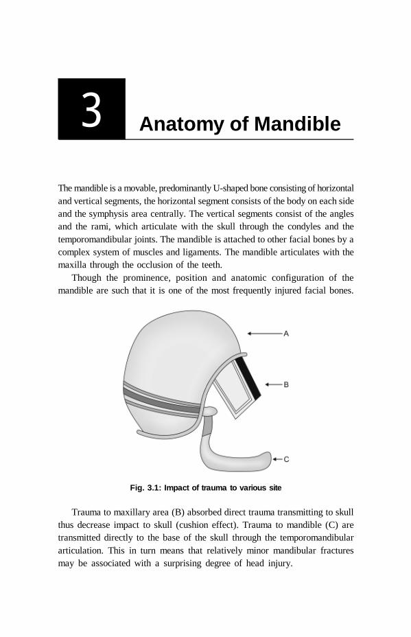
The mandible is a movable, predominantly U-shaped bone consisting of horizontaland vertical segments, the horizontal segment consists of the body on each sideand the symphysis area centrally. The vertical segments consist of the anglesand the rami, which articulate with the skull through the condyles and thetemporomandibular joints. The mandible is attached to other facial bones by acomplex system of muscles and ligaments. The mandible articulates with themaxilla through the occlusion of the teeth.
Though the prominence, position and anatomic configuration of themandible are such that it is one of the most frequently injured facial bones.
Fig. 3.1: Impact of trauma to various site
Trauma to maxillary area (B) absorbed direct trauma transmitting to skullthus decrease impact to skull (cushion effect). Trauma to mandible (C) aretransmitted directly to the base of the skull through the temporomandibulararticulation. This in turn means that relatively minor mandibular fracturesmay be associated with a surprising degree of head injury.
Anatomy of Mandible / 13
The mandible is a strong bone but has several areas of weakness that areprone to fracture. The body of the mandible is composed principally of densecortical bone with a small substantial spongiosa through which blood vessels,lymphatic vessels and nerves pass.
Areas of Weakness
Presence of Teeth
Body of the mandible has two components that is alveolar component whichcarries the teeth and basal bones. The presence of teeth make the bony structureweak, resulting alveolar fracture can occur independent of the basal bone. Teethwhich have long roots or that are embedded in the bone also weaken the structure,external root of canine is the longest amongst all mandibular teeth, presence ofimpacted or unerupted third molar also make the structure weak.
Neck of the Condyle
A thin neck of the mandibular condyle is an area of anatomical weakness andget easily fractured in response to any direct or indirect trauma to the mandible.Therefore, the mandibular condyle acts as a shock absorber in preventing theintracranial injuries.
Symphysis of the MandibleIt is an area of fusion of two halfs of the mandible. The complete bony uniontakes place at the end of the first year of life but this line of fusion remainsrelatively weak point in the structure.
Angle of the MandibleThe trajectories of the mandible change their direction where the body andramus meet. The angle of mandible is anatomically as well as physiologicallyweak structure. It is further weakened by the presence of an impacted tooth.The attachment of the muscles on the mandible anterior to the angle pull itdownwards and backwards whereas the muscles attached posterior to theangle pull upwards and forwards. This is a significant observation in referenceto the displacement of the fractured fragments.
Presence of ForaminaWeaken the structure but this point is contested by many workers as presenceof foramina add to the compactness of the bone. The fracture of the

14 / Fracture Mandible
mandible in the-tooth bearing area normally compound into the oralcavity, and tooth in the line of fracture poses danger of being a source ofinfection.
Mandibular Muscles
The various muscles attached to the mandible can be grouped as:1. Muscles of facial expression2. Muscles of mastication3. Accessory muscles of mastication.
Muscles of Facial Expression
The muscle of facial expression have their origin from the bone and insertioninto the skin. These muscles play no role in the displacement of the fracturedmandible because to displace the bone muscle should have attachment onfixed ends, i.e. a bone only.
Muscles of MasticationMasseter, medial pterygoid, temporalis and external pterygoid are strongmuscles that help in closing and opening movements of the jaw. Thesemuscles play a major role in the fracture displacement especially of the angleand condyle region. These muscles have strong tendonous attachment at thesite of origin and insertion.
The masseter and medial pterygoid muscles that form the sling of themandible displace the ramal fragment upward. They are aided in their action
Fig. 3.2A: Muscle attachment from lateral side

Anatomy of Mandible / 15
Fig. 3.2B: Muscle attachment from medial side
Fig. 3.2C: Muscle attachment (horizontal view)
by temporalis muscle as well. This fragment is usually displaced medially becauseof the larger functional directional pull of the medical pterygoid muscle. Themedial pterygoid is more medially placed in comparison to the masseter musclethat runs a rather vertical course.
The lateral pterygoid muscle that is attached to the neck of the condyleand meniscus runs an anteromedial course up to the pterygoid plate and scaphoidfossa thereby displacing the fractured condyle medially.

16 / Fracture Mandible
Accessory Muscles of Mastication
Muscles like mylohyoid, geniohyoid and digastrics have their origin from thebone as well as insertion into another bone. These are also strong musclesthat pull the body of the mandible downward and medially. Symphysis ispulled downward and backward by suprahyoid group of muscles.Displacement of symphysis is also important because it leads to fall oftongue and respiratory distress on account of the attachment of the tongueto the mandible through genioglossus muscle.
Vascular Supply of Mandible
An effective blood supply is very much important factor in healing of afractured mandible bone. A mandible receives an endosteal supply via theinferior dental artery and vein and these vessels are important in youngpatients. Occasionally, a fracture of the body of the mandible will cause acomplete rupture of the inferior dental artery. Whereas this vessel usuallygoes into spasm with spontaneous arrest of hemorrhage, this is not always thecase and prolific bleeding can occur which is difficult to control. In this rareemergencies, the mandible fracture needs to be reduced immediately bymanipulation and the bone ends held in rough alignment by a wire ligaturearound adjacent teeth. The other and more important blood supply to themandible derives from the periosteum. The periosteal supply becomesincreasingly important with ageing as the inferior dental artery slowlydiminishes in size and eventually disappears. This fact has considerablesignificance for the healing of fractures in the elderly. Open reduction offractures in this age group involves elevation of periosteum from the bonesand further deprivation of blood supply to the fracture side with resultantdelayed or non-union.
Other Important Structure
Nerves
The inferior dental nerve is frequently damaged in fractures of the body andangle of the mandible producing anesthesia or paresthesia within the distributionof the mental nerve on the side of the injury. There are numerous reportedcases where the facial nerve has been damaged by direct trauma over themandibular ramus. Occassionaly, the mandibular division of the facial nerve isdamaged in isolation in association with a fracture of the body or angle.

Anatomy of Mandible / 17
Blood Vessels
Apart from hemorrhage from the inferior dental vessels which has beenmentioned, injury to major blood vessels is unusual in association withmandibular fractures. A large sublingual hematoma may result from rupture ofdorsal lingual veins medial to an angle fracture. The facial vessels are vulnerableto direct trauma where they cross the lower border of the mandible anterior tothe angle.
Temporomandibular Joint
Traumatic arthritis can occur without a fracture of the condyle, from indirecttransmitted violence. A synovial effusion occurs with widing of the jointspace on radiographs. Such a joint is extremely painful and mandibularmovement very restricted. When an intracapsular fracture of the condylarhead occur there may be direct involvement of the temporomandibular jointwith hemorthrosis. If this occurs in a young child it can lead to fibrous or bonyankylosis of the temporomandibular articulation and destruction of the growthpotential of the condyle. Not infrequently a fractured condylar head is drivenbackwards with sufficient force to tear, the adjacent external auditorymeatus and cause bleeding from the external ear. Such bleeding must becarefully distinguished from the middle ear bleeding which signifies a fractureof the base of the skull. Very rarely, the glenoid fossa is fractured as themandibular condyle is driven against this thin part of the temporal bone butusually a fracture of the condylar neck prevents the other more serious injuryoccurring.
SUMMARY
Trivial trauma can cause major injuries so all trauma should be takenseriously.
Area of weakness are:– Presence of third molar (impacted)– Neck of condyle– Symphysis of mandible– Presence of foramina– Angle of mandible.
18 / Fracture Mandible
4 Classification ofMandibular Fractures
Etiology of Fractures
Vehicular accidents and assaults are the primary causes of mandibular facialfractures throughout the world. The other chief causes for these fractures areWork related falls, sporting injuries and industrial trauma. Vehicular accidents Assaults Work related causes Falls Sporting accidents Miscellaneous causes
Thus the causes for maxillofacial fractures can be classified into:a. Intrinsic causesb. Extrinsic causes
Intrinsic Causes (Pathological Fractures)Fractures that occur due to intrinsic weakness of the bone and not due toforce of impact. Pathological fractures occur because of underlying bony orsystemic disease that causes one, many, or all bones of the skeletal systemto be abnormal and thus more susceptible to fracture.
Pathological fractures may occur from any type of trauma. Bending force Torsional force Compressive force or shearing force
Often the only force necessary to cause fracture is the persons weight,especially in the mandible it may be chewing force, thus spontaneous fractureoccurs without overt trauma.
Pathological fracture may occur through any of the following types ofbony pathology. Neoplasia Bony cysts
Classification of Mandibular Fractures / 19
Osteoporotic bone Osteoradionecrosis Caused by secondary nutritional hyperparathyroidism Localized bone infection (osteomylelitis) Osteoporotic bone due to disuse following prolong external fixation or
removal of a rigid internal device.Unfortunately, fracture may occur even as a sequela of improper implant
placement due to the tensile forces acting on the bone during mandibularfunction.
Extrinsic Causes
Direct violence (fracture at the side of impact) Indirect violence (fracture caused due to transmission of impact) Bending forces Torsional forces Compression forces Shearing forces
Factors affecting displacement of the fracture: Muscular pull on the fractured segment Force of the impact Site and direction of the fracture line Muscular tear—damage of muscle attachment might lead to the displacement
of certain fracture (coronoid) Presence of teeth in the posterior segment—presence of posterior teeth
may prevent displacement due to contact with the occlusal surface of themaxillary teeth.
Frequency of the Fracture
In general, incident of fractures of the mandibular body, condyle and angle arerelatively similar, while fractures of the ramus and coronoid process are rare.The literature may suggest that following mean frequency percentages based onlocation.Condyle - 29%Angle - 26%Body - 25%Symphysis - 15%Ramus - 4%Coronoid process - 1%

20 / Fracture Mandible
The mandible is involved in 70% of patients with facial fractures. The numberof mandible fractures per patient ranges from 1.5 to 1.8. Mandible fracturepatterns of a suburban trauma centre found that violent crimes such as assaultand gunshot wounds accounted for a majority of the fractures (50%), whilemotor vehicle accidents were less likely (29%).
The fractures of mandible area are classified based on the followingcriteria:a. Anatomical locationsb. Site of injuryc. Condition of the bone fragments at the fracture sited. According to the direction of the fracture and favourability for treatmente. According to severity of fracturef. Presence or absence of teeth in the jawsg. Clinical and radiological findings
1. Classification based on anatomical location of the fracturesA. Fracture of the symphysisB. Fracture of the canine regionC. Fracture of the body of the mandibleD. Fracture of the angle of the mandibleE. Fracture of the ramusF. Fracture of the condyleG. Fracture of the coronoid processH. Fracture of the dentoalveolar
Fig. 4.1: Various site of fracture

Classification of Mandibular Fractures / 21
2. Classification based on site of injurya. Direct fracture
If the fracture occurs at the site of impact, it is labelled as direct fracture.
Fig. 4.2: Classification according to site of injury
b. Indirect fractureAn indirect fracture is the one that occurs away from the site of injury.A trauma on side of the mandible can cause a direct fracture at the canineregion on the same side and an indirect fracture of the angle of the mandibleor neck of the condyle on contralateral side.
3. Classification based on the condition of the bone fragments at the siteof the fractureThis classification denotes the condition of the bone fragments at the fracturesite and hints at the severity of trauma and damage to the soft tissues.
a. Simple fractureWhen there is break incontinuity of the bone withoutany break in mucosa or skinmembrane thereby the fracturefragments are not exposed tothe external environment sucha fracture is said to be simplefracture. Fig. 4.3: Simple fracture

22 / Fracture Mandible
b. Compound fractureWhen the fractured ends of the boneare associated with the break incontinuity of skin or mucousmembrane thereby communicatingwith the external environmentthrough the wound then it is calledas compound fracture. As a rule,fractures involving the tooth bearingarea are always compound fractures because they communicate with the oralenvironment through gingival sulcus and periodontal ligament.
c. Comminuted fractureWhen the bone is splintered intomore than two fragments, it iscalled as comminuted fracture.These are high impact injuries onaccount of major trauma.
d. Greenstick fractureThe bone in children is soft elasticand there occurs an incompletetype of fractures at times.
These appear as a crack in thebone in which only one cortex ofthe bone is fractured whereas othercortex is bent only as in the caseof a green stick of a tree.
4. Classification according to the direction of fracture line andfavorability for treatmentThis classification is basically restricted to the fractures of the angle of themandible. The line of fracture is considered to determine the type of fixationrequired. A fractured line is considered favorable if the muscular pull resiststhe displacement of the fracture and in case the muscular pull distracts thefractured fragment away from the line of fracture favouring displacement, it is
Fig. 4.6: Greenstick fracture
Fig. 4.4: Compound fracture
Fig. 4.5: Comminuted fracture

Classification of Mandibular Fractures / 23
labelled as unfavorable fracture. Fractures of the angle of the mandible areinfluenced by the pull of the medial pterygoid, masseter and temporalis musclesthat tend to displace the ramus in an upward and medial direction. However, thedisplacement of the fractured fragment is also influenced by the direction ofthe force, magnitude of the impact, site of fracture, presence or absence ofteeth on each side of the fractured line.These fractures can be classified as follows:
a. Horizontally favorable fracturesWhen viewed from the side of the fractureline runs from the lower border of themandible extending upward and backwardto meet the upper border. The upwarddisplacement of the posterior fragment isprevented by the anterior fragment.
b. Horizontally unfavorable fracturesWhen the fracture line runs from the lowerborder of the mandible in an upward andforward direction to meet the alveolar crest,the upward movement of the posteriorfragment is called as horizontallyunfavorable fractures.
c. Vertically favorable fracturesWhen a fracture is viewed from above orocclusal surface, the fracture line that runsfrom buccal plate obliquely backwardstoward the lingual plate, it will resist themedial displacement of the posteriorsegment. Such a fracture is called asvertically favorable fracture.
Fig. 4.7: Horizontally favorablefractures
Fig. 4.8: Horizontallyunfavorable fractures
Fig. 4.9: Vertically favorablefractures

24 / Fracture Mandible
d. Vertically unfavorable fractureWhen a fracture line viewed from above,extends from the buccal cortical platecoming forward to join the lingualcortical plate, it is labelled as verticallyunfavorable fracture because the pos-terior segment can easily get displacedmedially without any hindrance.
5. Classification according topresence or absence of teethTeeth may have important role to play in the management of the fracture sinceocclusion is considered to be a guide in reduction. When a teeth are not present,alternative method of treatment to simple wiring procedures are compelled tobe considered.a. Class I – When teeth are present on both sides of the fracture line.b. Class II – When teeth are present only on one side of the fracture line.c. Class III – When both the fragments on each side of the fracture line are
edentulous.
Fig. 4.10: Vertically unfavorablefractures
Fig. 4.11: Classification according to presence or absence of teeth
6. AO classification of mandibular fracturesThis classification is based on clinical and radiological findings and describesmandibular fractures along with soft tissue involvement. It has five componentsdepending on the types of fractures and other associated findings:
F Number of fracturesL Localization (site)O OcclusionS Soft tissue involvementA Associated fractures
Classification of Mandibular Fractures / 25
These components are described further as under:
Categories of fractures (F)F0 Incomplete fracturesF1 Single fracturesF2 Multiple fracturesF3 Comminuted fracturesF4 Fracture with bone defect
Categories of localization (L)L1 PrecanineL2 CanineL3 PostcanineL4 AngularL5 Supra-angularL6 Processus anticularisL7 Processus muscularisL8 Alveolar process
Categories of occlusion (O)O0 No malocclusionO1 MalocclusionO2 Edentulous mandible
Categories of soft tissue involvement (S)S0 ClosedS1 Open intraorallyS2 Open extraorallyS3 Open intra-extraorallyS4 Soft tissue defect
Categories of associated fractures (A)A0 NoneA2 Fracture and/ or loss of toothA3 Nasal boneA4 ZygomaA5 Le Fort IA6 Le Fort IIA7 Le Fort III
Grades of severity (I-V)Grade I and Grade II: These are closed fractures.
26 / Fracture Mandible
Grade III and Grade IV: These are open fractures
Grade V: This includes open fracture with a bone defect and fractures due togunshot.
Fracture Displacement
The pull of the muscles are described above and the direction of the line of thefracture along with the intensity of the force hitting, the jaw are responsible forthe displacement of the mandibular fragments are described as under.
Fracture Condyle
There is no dislocation of the condyle if only a crack in a neck appears withoutany tear in the capsule of the joint and periosteum of the bone but if there isa fracture causing tear, anterior or medial dislocation of the condyle due to theattachment of lateral pterygoid muscle will take place.
Fracture Angle of the Mandible
As explained earlier the unfavorable lines of the fracture from the treatmentpoint of view in the angle region favor superior and medial displacement of theposterior segment. If the lines are favorable both horizontally and vertically,there are minimal chances of displacement.
Fracture of Body of the Mandible
Both the elevators and depressor muscles play a role in displacement. Themylohyoid muscles displaces the fragments medially and inferiorly. If thefracture line is favorable, no displacement is seen unless there is some extremedegree of violence. In unfavorable line of fracture both in horizontal and verticaldirection, i.e. if the fracture line is running from the lingual to buccal table inan anterior direction and from lower to upper border again in an anteriordirection, the larger fragment of bone bearing the dental arch will be displacedinferiorly and medially.
Fracture in the Canine Region
Bone is weakened in this region due to the long root of the canine hence,fracture is more common in this region. The role of causing displacement isplayed by depressor group of muscles of the jaw. Bilateral fractures cause a lotof displacement depending upon the line of the fracture.

Classification of Mandibular Fractures / 27
If the fracture lines are running towards each other, i.e. converging linesfrom labial to lingual table of the mandible as well as from superior to inferiorborder, no displacement is expected. However, if the lines are unfavorable ie.If the fracture lines are running divergently from labial to lingual table of themandible as well as superior to inferior border, the central fractured fragmentis pulled downward and backward by the mylohyoid, geniohyoid, digastrics andgenioglossus muscles. It is further complicated by the collapse of the fragmenton the lateral side, medially towards each other making a closed reduction verydifficult.
Fracture of Symphysis
A vertical midline fracture normally exhibits no displacement but if the fractureline runs an oblique course, the balance of the muscles is disturbed causingdisplacement of the fragments backwards and downwards.
Fracture of Ramus of the Mandible
The sling of the mandible formed by the masseter and medial pterygoid muscleforms a thick protective case around the ramus and gives it a splinting action.Generally talking, there is no dislocation of the fractured fragments of theramus but in injuries like gunshot wounds, there may be shattering of bone.
Fracture of Coronoid Process
Fractures of the coronoid process of the mandible are not commonly seen. Incases there is a fracture with a tear in the periosteum, the tendonous attachmentof the temporalis muscle will pull the fractured coronoid process upwards.
SUMMARY
Vehicular accidents and assault are main cause of fractures. Condyle and angle are the most common site of the angle Any break in mucosa or a skin with fracture mandible is compound
fracture. Favorable fractures are those fractures in which because of muscle pull
fractured fragments are brought together. Unfavorable fractures are those fractures in which because of muscle pull
fractured fragments are pulled away from each other.
28 / Fracture Mandible
5 History and ClinicalExamination
History
History is very much informative in case of fracture mandible. A detailed history of patient should be taken Any pre-existing disease should be enquired like:
a. Systemic disease like diabetes and hypertensionb. Psyschiatric illnessc. Alcoholic withdrawal symptomd. Epilepsye. Other endocrine, collagen diseasesIn such patient like psyschiatric, alcoholic withdrawn, epilespsy, inter-
maxillary fixation should be avoided. History regarding etiology of fracture should be elicited. In cases of high
velocity (RTA) suspect other fracture also in a body Elicit regarding shape and size of the object causing injury, blow from
a broad, blunt object can cause several fractures while smaller welldefined object may cause single comminuted fractures. Since, impact offorce is concentrated in small area
Try to elicit the direction of impact. Anterior blow on a chin can causeparasymphysis or bilateral condyle fracture.The examination of a patient with the fracture of the mandible takes place
in three stages:1. Instant and rapid assessment2. General clinical examination of the patient3. Local examination of the mandibular fracture
Instant and Rapid Assessment
Vehicular accident or assault patient who has a fracture mandible may sustaininjury on another part of the body which may constitute actual threat to alife. This should be given a first priority than the facial trauma. It is alwaysnecessary to make a rapid assessment and start the resuscitation of patientfirst and then a detailed examination to be done.

History and Clinical Examination / 29
General Examination
Fractures of the mandible are, of course, caused by trauma of varying degreesof severity and is reasonable to consider the possibility that this degree oftrauma may also have caused injury elsewhere in the body. This is especiallytrue if the patient has been involved in a accident such as road trafficaccident or a fall from a considerable height. However, a simple blow on thelower jaw as a result of a fight or during the course of some game may resultin force being transmitted to the cranium which results in serious injury oreven death of the patient.
It is unusual for a patient with a mandibular fracture to be shocked andif this condition is present some more serious injury should be suspected.
Local Examination of the Mandibular Fracture
Preparation for ExaminationBefore any detailed examination of the mandible, the face must be gently cleanedwith swabs to remove blood clot, road dirt, etc. inorder that an accurate evaluationof any soft tissue injury can be made. The mouth similarly should be examinedfor loose or broken teeth or dentures, and any congealed blood removed withswabs held in nontoothed forces. During this gentle cleaning of face, the craniumand cervical spine are carefully inspected and then palpated for signs of injury.Finally, the mandibular fracture is examined in detail.
Extraoral Examination
Inspectiona. Swelling
Many of the physical signs of a fractured bone result from theextravasation of blood from the damaged bone ends. This results invery rapid early swelling from the accumulation of blood within thetissues and later increase in the swelling resulting from increasedcapillary permibiality and oedema. Swelling and ecchymosis indicatethe site of any mandibular fracture.
b. DeformityThere may be obvious deformity in the bony contour of the mandible.
c. Gait of patientIf considerable displacement has occurred the patient is unable toclose the anterior teeth together and the mouth hangs open. A consciouspatient may seek to support the lower jaw with his hand.
30 / Fracture Mandible
PalpationPalpation should begin bilaterally in the condylar region and then continuedownwards and along the lower border of the mandible. If there is moredisplacement it may be possible to palpate deformity or elicty bony crepitus.
Fractures of the body of the mandible are associated with injury to theinferior dental nerve in which case there will be reduced or absent sensation onone or both side of the lower lip.
Intraoral Examination
It is impossible to assist intraoral damage if the parts are obscured byblood.
The buccal and lingual sulci are examined for ecchymosis. Submucosalextravasation of blood is often indicative of underlying fracture, particularlyon the lingual side (Coleman’s sign).
Ecchymosis in the buccal sulcus is not necessarily the result of thefracture as there is considerable soft tissue overlying the bone in this areaand extensive brusing may follow a blow over the lower jaw insufficientto cause a fracture.
However, on the lingual side the mucosa of the floor of the mouthoverlies periosteum of the mandible which, if breached following afracture, will invariably be the cause of any leakage of blood into thelingual submucuosa.
The occlusal plane of the teeth is next examined, or if the patient isedentulous, the alveolar ridge.
It is important to examine all the individual teeth and to note anyluxation or subluxation along with missing crowns, bridges and/or fillings.Individually fractured teeth must be assessed for involvement of the dentineor pulp.
Possible fracture sites are gently tested for mobility by placing a finger andthumb on each side and using pressure to elicit unnatural mobility. If thepatient can cooperate, he is asked to carry out a full range of mandibularmovements and any pain or limitation of movement recorded. Occasionally,this detailed examination fails to confirm.
A mandibular fracture which is thought to be present from the historyand presence of hematoma. In such cases, the flat of both hands shouldbe placed over the two angle of the mandible and gentle pressure exerted.This maneuver will always elicit pain when even a crack fracture ispresent.
History and Clinical Examination / 31
Sign and Symptoms of Mandibular Fractures
Fracture of the mandible can be divided according to their anatomical locationinto eight main types, these are:1. Dentoalveolar2. Condylar3. Coronoid4. Ramus5. Angle6. Body (molar and premolar areas)7. Symphysis and parasymphysis8. Multiple and comminuted fractures.
Dentoalveolar Fractures
Dentoalveolar injuries are defined as those in which avulsion, subluxationor the fracture of the teeth occurs in association with the fractures of thealveolus. They may occur alone or in combination with some other type of mandibular
fractures. Fracture of the crown of individual teeth may occur as a direct result of
trauma or by forcible impaction against the opposing dentition. Meticulous dental examination is essential and any missing fragments of
crown or missing fillings noted. These may be invaded within the softtissues or more rarely swallowed or inhaled.
Fractures of the roots of the teeth may be present which are difficult todiagnose clinically. Exclusively mobile teeth which do not appear to besubluxed are suspect and should be earmarked for later periapicalradiographs.
Individual teeth may be missing and/or recent extraction wound suggest thatthe tooth concerned has been knocked out.
Occasionally, molar and premolar teeth appear superficially normal butclose inspection reveals either a vertical split or a horizontal fracture justbelow the gingival margin resulting from indirect trauma against theopposing dentition or violent impact by a small hard object such asmissile.
Fracture of the alveolus may be present with or without associated injuryto the teeth.

32 / Fracture Mandible
Condylar Fractures
These are the most common overall fractures of the mandible and are oncemost commonly missed on clinical examination. Condylar fracture may beunilateral or bilateral, and they may either involve the joint compartment asintracapsular fractures or the condylar neck when they are regarded asextracapsular. The extarcapsular fracture may exist with or without dislocationof the condylar head, and the upper fragment may either remain angulated onthe lower portion of the ramus or be displaced medially or laterally.
Unilateral Condylar Fractures
There is often swelling over the temporomandibular joint area and theremay be hemorrhage from the ear on that side. Bleeding from the ear resultsfrom laceration of the anterior wall of the external auditory meatus, causedby a violent movement of the condylar head against the skin in this region.
It is important to distinguish bleeding originating in the external auditorycanal from the more serious hemorrhage.
The haematoma surrounding a fractured condyle may track downwardsand backwards below the external auditor canal. This give rise toecchymosis of the skin just below the mastoid process on the same side.This particular physical sign also occur with fractures of the base of theskull when it is known as “ battle’s sign.”
In the recently injured patient there is invariably tenderness over thecondylar area.
When post-traumatic edema is present it is difficult to palpate the condylarhead.
The mandible deviates on opening towards the side of the fracture, and thereis usually painful limitations of protusion and lateral excursion to the oppositeside.
Bilateral Condylar Fractures
As with the unilateral fracture overall mandibular movement is usually morerestricted extraorally then is the case with a unilateral fracture. As with theunilateral fracture derangement of the occlusion will be found only if thecondyle is displaced on one or the other side causing shortening of theramus.
If both fractures have resulted in displacement of the condyles from theglenoid fossa or overriding of the fractured bone ends, an anterior openbiteis found to be present.

History and Clinical Examination / 33
In all cases of bilateral fracture there is a pain and limitation of opening andrestricted protusion and lateral excursions.
Fracture of the Coronoid Process
This is a rare fracture. The fracture can be caused by direct trauma to the ramus but is rarely
in isolation in the circumstances. If the tip of the coronoid process is detached, the fragment is pulled
upward towards the infratemporal fossa by the temporalis muscle. This is difficult fracture to diagnose clinically. There may be tenderness over the anterior part of the ramus and hematoma. Painful limitation of movement, especially protrusion of the mandible may
be found.
Fracture of the Ramus
The fractures of the ramus are not common and there are two main types.
Single Fracture
This is in effect a low condylar fracture with both the coronoid and condylarprocess on the upper fragment.
Comminuted Fracture
Such a fracture always result from direct violence to the side of the face. Swelling and ecchymosis is usually noted both extra and intraorally. There is tenderness over the ramus and movements produced pain over
the same area. Severe trismus is usually present. Inability to close and open mouth.
Fracture of the Angle
There is usually swelling at the angle externally . There may be obvious deformity. Within a mouth a step deformity behind
the last molar tooth may be visible which is more apparent if no teethare present in the molar region.
Anterior open bite in bilateral angle fracture. Ipsilateral open bite in unilateral angle fracture. Retrognathic or flattened appearance of lateral aspect. Inability to close jaw causing premature dental contact.

34 / Fracture Mandible
Fracture of the Body (Molar and Premolar Regions)
The physical signs and symptoms are similar to those of fractures of the angleas far as swelling and bone tenderness are concerned. Inability to open or close mouth. Ecchymosis, swelling, bone tenderness are similar to fracture of angle. Dentate mandible even slight displacement of the fracture causes
derangement of the occlusion. Premature contact occurs on the distal fragment because of displacing
action of the muscles attached to the ramus. When there is a gross displacement, the inferior dental artery may be torn
and this can give rise to severe intraoral hemorrhage. Crepitation on palpation. Flattened appearance of lateral aspect of face.
Fracture of the Symphysis and Parasymphysis
These fractures are commonly associated with fractures of one or bothcondyles.
Ecchymosis on floor of mouth. The thickness of the anterior mandible between the canine regions often
ensures that these fractures are fine cracks which are little displaced andmay be missed if the occlusion is undisturbed locally.
The presence of bone tenderness and a small lingual hematoma may be onlyphysical signs. The fracture line is often oblique which allows overriding ofthe fragments with lingual inversion of the occlusion on each side. As suchfractures are always the result of direct violence there is frequently associatedsoft tissue injury of the chin and lower lip.
Posterior crossbite in symphysis fracture. Posterior open bite or unilateral open bite in parasymphysis fracture. Chances of paresthesia of lower lip as injury of mental nerve after emergence
from foramina. Chances of airway compression in case of bilateral parasymphysis fracture
with loss of tongue and loss of consciousness.
Multiple and Comminuted Fractures
The physical signs of the multiple and comminuted fractures depend on the siteand number of fractures. Multiple and comminuted fractures result from extremedirect violence and are usually associated with more severe soft tissue injury.In general comminuted fractures of the ramus, angle and molar regions are not
History and Clinical Examination / 35
associated with gross displacement of the fragments. However, comminutionof the symphysis allows the lateral segments to collapse and presents a muchmore serious problem of management.
SUMMARY
See the patient as wholea. Resuscitate the patient first.b. Mandible treatment can wait.
Detailed examination can give rough idea of site of fracture. Swelling and ecchymosis suggest underlying fracture mandible. Deviation from normal occlusion, open bite, crossbite suggest fracture
mandible. Deformity, inability to close and open mouth suggestive of fracture mandible. Look for inferior alveolar nerve or mental nerve paresthesia.

36 / Fracture Mandible
6 Radiology
A precise radiological diagnosis is very much important for treatment planof fracture mandible. It depicts:a. A exact relationship of teeth in fracture lineb. A type of fracture, simple or communitedc. Number of fracturesd. Area of fracturese. Degree of displacements.
Radiographs of mandible is divided into:a. Essential view
It is available in all departments of radiology and can be done easily on allpatients.
b. Desirable viewThe equipments for the same view are not available in routine radiologydepartment. The equipments are of specialized nature and cannot be done onseverely injured patients.
Essential Radiographs
a. Left and right oblique lateral view of mandibleThis view are used to demonstrate fracture of mandible ramus, body ofmandible and symphysis region.
b. PosteroanteriorThis view demonstrates fracture of body and angle with the type ofdisplacements. An undisplaced fracture of condyle head is difficult to seein this view as it is obscured by superimposition of mastoid process.
c. Reverse Towne’s projectionThis projection is used to demonstrate fracture of condyle region. As thisavoid superimposition of mastoid bone.
d. Intraoral1. Periapical films are required to demonstrate a relationship of teeth to
the line of fractures and any damage to the teeth itself.

Radiology / 37
2. Occlusal films can help us to evaluate the relationship of tooth root to thefracture.
Desirable Radiographs
Panoramic Films
Panoramic films are useful in defining location and displacement of mandiblefracture. It has a accuracy rate of 92% for diagnosis of fracture. This filmsgive a best single overall view of mandible and are specially valuable fordemonstrating fracture in condyle region. The combination of posterior-anterior view and a pantomogram obviates the need for further radiographs.The sites in which mandible fractures are most commonly under diagnoseon this view are condylar angle and symphysis area especially if there issome blurring by the patients movement or hardware.
Advantages
Simplicity of technique Good details Can visualize mandible and maxilla with root of teeth in one radiograph.
Disadvantages
Impractical for severely traumatic patients Cannot be done in all hospital set ups TMJ area, symphysis, dental and alveolar process region areas of which
fine details cannot be appreciated Difficult to appreciate buccal and lingual bone displacement.
Computed Tomographic Scan
Indications
1. In cases of multiple facial injuries2. Cases of communated fractures3. Cases of missile injuries4. Cases of infected, malunion, nonunion fracture mandible5. In cases of vertical split fractures
The accuracy rate of ct scan is around 92%. This offers a very littleadvantage as a diagnostic tool in lower third of a face and are not justifiedfor isolated mandibular fractures on either clinical or economic ground. Itdemonstrates detail of TM joint injury.

38 / Fracture Mandible
Three-dimensional CT Scan
It can be obtained to compare symmetry and volume of two side of bone offace.
SUMMARY
Posteroanterior view and a pantogram is all what needed in a simplecase of fracture mandible.
Reverse townes view can be used for condyle fractures. CT Scan indicated in complicated comminuted infected fracture mandible
and associated with facial injuries.
Preliminary Treatment / 39
7 Preliminary Treatment
Most of the fractures of the mandible encountered are associated with fracturein other part of body or other injuries in body. It is not common for suchpatients to suffer from shock and evidence of acute circulatory collapse initself is indicative of damage to other important structures. Trauma to themandible does, however, frequently cause concussions from transmitted violenceto the base of the skull.
Airway Maintenance
Relatively minor injuries which cause intraoral bleeding and fracture of teethor dentures can lead to airway obstruction in an unconscious or semi-consciouspatient. The essential first aid required consists of careful examination of themouth and the removal of all fragments of teeth, broken fillings and dentures.If suction is available blood clots and the saliva should be evacuated and thepatient positioned so that further bleeding and secretions can escape from theoral cavity. If the symphysis region is fractured and particularly if it iscomminuted there is some danger of the tongue falling back and obstructingthe airway in a patient who has lost voluntary control of the intrinsicmusculature. Occasionally a suture passed through the dorsum of the tonguemay assist in controlling its position. The most satisfactory posture for anunconscious patient is lying on his side in the position used routinely duringrecovery from a general anesthetic. This position should be opted fortransportation of a patient to an accident unit or another treatment center.
Blood Loss
Serious blood loss is not common in mandibular fractures. Considerable bloodloss can however occur, when there are extensive associated soft tissuelacerations, obvious bleeding points such as the facial vessel should besecured with artery forceps and a temporary dressing applied. Occasionallybrisk and persistent hemorrhage originates from a grossly displaced fracture of

40 / Fracture Mandible
the body of the mandible. This can only be controlled by manual reduction ofthe fracture and temporary partial immobilization by means of a suture or wireligature passed around teeth on each side of the fracture line.
Soft Tissue Lacerations
It is desirable to close a soft-tissue wound within 24 hours of injury, as it isoften possible to gain access and to stabilize bone fragments through overlyingwounds, it is therefore advantageous, where possible, to combine soft tissuerepair with definitive treatment of the fracture. If this is not possible becauseof the patient’s general condition the soft tissue must be dealt with separatelyunder local analgesia as soon as possible after injury. Before closing woundsthey must be cleaned to remove foreign material and so avoid subsequentunsightly tattooing of the scar. Wounds should be gently scrubbed if necessarywith a mild antiseptic cleanser such as 1% cetavlon.
Support of the Bone Fragments
In most of the cases temporary splinting of the fragments is unnecessary andsuch devices as the barrel bandage, webbing head cap with chin support, andElastoplast chin strap are not only superfluous but may in some instances maycause the patient additional discomfort. If this type of first aid is applied it issalutary to observe how often the patient experiences relief when it is removed.Usually if any urgent immobilization of the fragment is required it is best tocarry out a definitive standard fixation technique such as an arch bar and not towaste time with an ineffective temporary fixation.
Pain Control
The majority of the patients with mandibular fractures do not appear to suffermuch a pain, perhaps owing to the frequently associated neuropraxia of theinferior dental nerve. Some mobile fractures of the body of the mandible are,however, extremely uncomfortable and a potent cause of restlessness in acerebrally irritated patient. This situation is one of the rare indications forgiving priority to the immobilization of the mandible in the presence of otherserious injury.
It should be remembered that use of the powerful analgesics such asmorphine is contraindicated as they depress the cough reflex and respiratory

Preliminary Treatment / 41
center and also mask pain which can be diagnostically important (e.g. from aruptured spleen).
Control of Infection
All fractures of body of mandible involving teeth are compound fractures asthey are potential source of infection. Immediately injection augmentin shouldbe given every 12 hourly for first 2 to 3 days. There are also chances ofanaerobic infection. So injection metronidazole or oral metronidazole shouldbe administered.
SUMMARY
Treat the life-threatening condition first. Treatment of fracture mandible can wait.

42 / Fracture Mandible
8 General Treatment ofFracture Mandible
Principal of fracture treatment of the mandible do not differ from fractureelsewhere in the body.
Principles
a. Fracture reduction and fixation to restore anatomical relationships;b. Fracture fixation providing absolute or relative stability as the “personality”
of the fracture, the patient, and the injury requires;c. Preservation of the blood supply to soft tissues and bone by gentle reduction
techniques and careful handling;d. Early and safe mobilization and rehabilitation of the injured part and the
patient.
Reduction
Reduction of fracture means restoration of functional alignment of the bonefragment. In the dentate mandible reduction must be anatomically precisewhen teeth are involved and previously in a good occlusion. Less precisereduction may be accepted if part of the body of mandible is edentulous orthere are no opposing teeth.
The presence of teeth provides an accurate guide in most cases by which thefracture segment can be aligned. The teeth are used to access the reduction,check alignment of the fragment and assist immobilization. However the occlusionis used as a index for accurate reduction it is important to recognise any pre-existing occlusion abnormality like anterior or lateral open bite were facets onindividual teeth can provide valuable clues to previous contact areas. The teethmay on occasion be brought into contact during reduction and yet be occludingincorrectly owing to lingual inclination of fractured segment.
Close reduction can be achieved in a case of mild displaced fracture. Whilewidely displaced, multiple or extensive comminuted fractures will require aopen reduction.

General Treatment of Fracture Mandible / 43
Immobilization
Following accurate reduction of fragment, the fracture side need to beimmobilized to allow the bone healing to occur. The period of theimmobilization depends upon the sites of fractures, the presence of teeth, ageof a patient and absence and presence of a patient.
Period of Mobilization
A simple guide for a period of immobilization for fracture of mandible of atooth bearing area are as follows
Normally a 3 weeks of immobilization is required in a case of youngadult with fracture of angle receiving early treatment in which teeth areremoved from the fracture line.Ifa. Tooth retained in fracture line—add 1 weekb. Fracture at the symphysis—add 1 weekc. Age 40 years and over—add 1 or 2 weekd. Children’s and adolescents—subtract 1 week
Thus if there is a fracture in a symphysis in a 40-year-old patient with toothin a fracture line is retained then a 6 week immobilization is required (basic 3weeks + 1 week for unfavourable site + 1 week for the age + 1 week for theteeth retained in a line of fracture).
This rule is for guidance only. However, the fracture segments need to betested clinically before this immobilization is removed or released.
Fig. 8.1: Type of fixation
44 / Fracture Mandible
Intermaxillary Fixation
Arch Bars
Arch bars are preferred: For temporary fragment stabilization in emergency cases before definitive
treatment As a tension band in combination with rigid internal fixation For long-term fixation in conservative treatment For fixation of avulsed teeth and alveolar crest fractures
General Considerations
There are important points to consider before starting.The occlusion must be checked. In the case of jaw malformations, such
as a deep bite deformity, it may be impossible to use arch bars.One pitfall when using arch bars is the risk of contamination of
bloodborne infection from patients. Passing the wires to secure the arch barcan result in a puncture or tear in the surgeon’s glove and the possibilityof disease transmission to the surgeon.
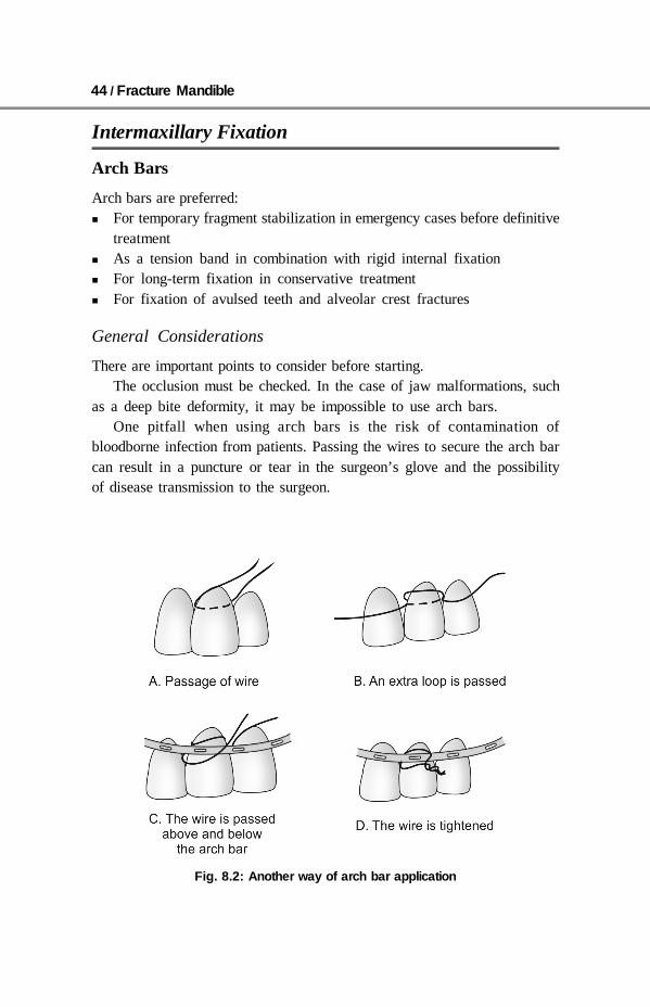
Fig. 8.2: Another way of arch bar application

General Treatment of Fracture Mandible / 45
A
B
C
D
E
Fig. 8.3: Fitting an arch bar. A— Selection of appropriate length and contouring ofErich arch bar. B— Arch bar to be placed on a teeth for a proper measurement.Extra length of the arch bar need to be trimmed and the posterior edge of the barneed to be bend to prevent soft tissue injury. C and D— Wire is passed above andbelow the arch bar and tightened so as not to obstruct the lug. It is important tomake sure that this wires have been tightly applied by checking whether anyvertical movement of arch bar is possible. The wire used are of normally of 26gauge. E— Intermaxillary fixation can be established by either wires or by elastic

46 / Fracture Mandible
Wiring Techniques
Gilmer Method
This is the simplest way to establishintermaxillary fixation by gilmermethod. This technique is simple andeffective but has a disadvantage thatmouth cannot be opened forinspection of the fracture sidewithout removal of wire fixation. Themethod consists of passing wireligatures around neck of availableteeth and twisting them in a clockwisedirection until the wire is tightenedaround its tooth. After adequatenumber of wire has been placed inupper and lower teeth are brought into the occlusion and the wire are twistedone upper to one lower wire. A stainless steel 24 gauge or 26 gauge wire areusually applied.
Eyelet Method
This method of fixation has theadvantage that jaws may be open forinspection by removal of only theintermaxillary ligatures. This methodconsists of twisting a 20 cm lengthof 24 gauge or 26 gauge wire arounda instrument to establish a loop. Bothend of the wire are passed throughthe interproximal space from theouter surface. One end of the wire ispassed around the anterior tooth the other around the posterior tooth. One endof the wire may pass through the loop. The eyelet should project in upper jawabove and in lower jaw below the horizontal twist to prevent ends from impingingon each other. After establishment of sufficient number of eyelets the teeth arebrought into occlusion and ligature are passed in loop fashion between oneupper and one lower eyelet. The interjaw wires are twisted tightly to provideintermaxillary fixation.
Fig. 8.4: Gilmers method of fixation
Fig. 8.5: Eyelet method of fixation

General Treatment of Fracture Mandible / 47
Intermaxillary Fixation Screw Technique
Intermaxillary fixation screws has been introduced as labour saving device.Intermaxillary fixation screws provide a rapid method of immobilization ofteeth in a good dentician in uncomplicated fracture types. The number andposition of this screws to be inserted is based on fracture types, the locationof fracture and the surgeon preference. Screw must be position superior to themaxillary tooth roots and inferior to the mandible tooth root.
Disadvantages are minimal and a focused point of force application tomaintain good intermaxillary fixation. The focus point of force applied mayresult in malocclusion by leaving the posterior dentician in an open bite.
Acrylic Splints
These are useful in maintenance of intermaxillary fixation and in establishingthe continuity of maxillary and mandibular dental arches in particular segmentof missing teeth can be compensated with suitable design splint. These areuseful in maintenance of intermaxillary fixation and in establishing the continuityof maxillary and mandibular dental arches in particular segment of missingteeth can be compensated with suitable design splint. Appliance of this typesare effective but requires detailed dental knowledge and skeletal models ofsplint construction. The splints are fabricated by specially educated physicianwith dental training, dental professional or dental laboratory.
Bonded Modified Orthodontic Brackets
This method is used in patient with minimal displaced fractures patient withgood oral hygiene. This require a help of orthodontic brackets which areapplied on a teeth then applying intermaxillary elastic bends. The orthodonticbrackets are prepared in maxofacial laboratory. This technique requirescomplete elimination of moisture, this is not applicable in cases where thereis intraoral bleeding.
Fig. 8.6: Intermaxillary fixation screw technique

48 / Fracture Mandible
Nonrigid Osteosynthesis
Transosseous wiring Circumferential wiring External pin fixation Bone clamps Trans fixation with kirschner wires (K wires) These fixation been non rigids require intermaxillary fixation.
Semirigid Osteosynthesis
Mandibular plate Dynamic compression plate Lag screw plate
Rigid Osteosynthesis
Reconstruction plate Locking plate Three-dimensional sturd
Miniplates
Mini plates are available in various shapes and lengths but can only be used withnon-locking screws. For mandible 2.5 mm or 2 mm plate are usually used. Theyare most commonly used for fracture mandible.
Dynamic Compression Plates
Screws are need to be inserted bicortically when using plates.
Indications
Simple fractures mandible with excellent bony buttressing, are preffered fordynamic plate compression plate.
Contraindications
Compression plating is contraindicated when there is not good bony buttressingat the fracture site, as is seen in atrophic edentulous mandible fractures,defect fractures, comminuted fractures, and other complex mandibularfractures.

General Treatment of Fracture Mandible / 49
It is also contraindicated in: Simple fractures with an extreme oblique pattern (sagittal fractures) Fracture mandible with no good bony buttress Atropic mandibular fracture Edentulous fracture mandible Defect fractures Communited fractures Other complex fractures
Compression Plating
Principle
As the eccentrically placed compression screws are tightened, the head movesdown the ramp and the bone is compressed together.
Diagrams showing two eccentrically screw inserted but not fully tightenedscrews.
Fig. 8.7: The left screw is fully tightenedand thereby narrowing the gap
Fig. 8.8: Tightening the right screwfinally compresses the fracturetoward each other these compres-sion screws are bicortical in nature

50 / Fracture Mandible
Compression Plating Technique
The compression plate is adapted by slightly over bending and eccentric holeis drilled on either side of the fracture.
Overbending
The plate must be overbend slightlyto close the lingual cortex. Ascompression screws are tightened,the slightly overbent plate closesthe lingual gap. If the plate is notslightly overbent, the buccal cortexwill be well aligned but a gapremains at the lingual cortex
Eccentric Drilling forCompression
There are two drill guides used with compression plating. The yellow drillguide is used for eccentric hole placement. The green drill guide is used forneutral hole placement.
To drill eccentrically, the arrow on the yellow end of the drill guide mustpoint towards the fracture. The number close to the arrow shows the maximalpossible amount of bone movement upon screw insertion.
To place an eccentric hole, the proper gold drill guide must be used and thearrow must point toward the fracture.
Once both the eccentrically placed screws are tightened, the fracture iscompressed. There is no need to compress the fracture additionally so theremaining screws are placed in a neutral position. The appropriate green drillguide is used to place the neutral screws
Screws used for the compression plate are bicortical in nature.
Fig. 8.10: Gap at lingual cortex if platenot overbend
Fig. 8.9: Dynamic compression plate

General Treatment of Fracture Mandible / 51
Option for Off-Angle Drilling
There are times when off-angle drillingis necessary to avoid anatomicstructures such as tooth roots.
Although the hole does not have tobe drilled at 90° to the bone it shouldnot enter the fracture, and it should notbe angled in such a way that it interfereswith the adjacent screws.
Lag Screws and Technique
Principles
This technique, advocated by niederdell-mann et al is not commonly practicebecause it is difficult to excecute andit is easy to achieve in adequate fixation.
Lag Screw Versus Lag Technique
Lag screws and the lag technique compress the fracture fragments together.There are two methods by which to achieve this.
True lag screws (as illustrated here) have threads only on the terminal endof the screw. Therefore, when inserted across a fracture, the threads of the tipof the screw engage the far cortex and the head of the screw engages the nearcortex, causing compression of the fracture fragments upon tightening.
True lag screws are not available. Instead, a lag technique is used. The lagtechnique involves overdrilling the near cortex to the size of the externaldiameter of the screw. When the screw is inserted, it glides through thisoverdrilled hole and the threads only engage the far cortex. As the screw istightened the head of the screw engages the near cortex and the fracture fragmentsare compressed together.
If the near cortex is not overdrilled, the threads of the screw will engageboth near and far cortices preventing compression of the fracture fragments.
Indication
In parasymphysis/symphayis oblique fracture.
Fig. 8.11: Off-Angle drilling
Fig. 8.12: Lag screw principle
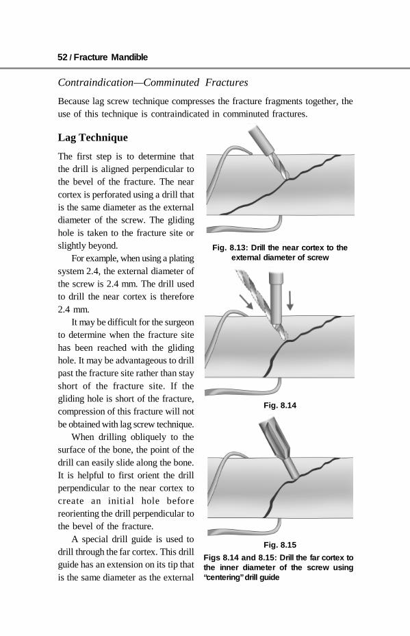
52 / Fracture Mandible
Contraindication—Comminuted Fractures
Because lag screw technique compresses the fracture fragments together, theuse of this technique is contraindicated in comminuted fractures.
Lag Technique
The first step is to determine thatthe drill is aligned perpendicular tothe bevel of the fracture. The nearcortex is perforated using a drill thatis the same diameter as the externaldiameter of the screw. The glidinghole is taken to the fracture site orslightly beyond.
For example, when using a platingsystem 2.4, the external diameter ofthe screw is 2.4 mm. The drill usedto drill the near cortex is therefore2.4 mm.
It may be difficult for the surgeonto determine when the fracture sitehas been reached with the glidinghole. It may be advantageous to drillpast the fracture site rather than stayshort of the fracture site. If thegliding hole is short of the fracture,compression of this fracture will notbe obtained with lag screw technique.
When drilling obliquely to thesurface of the bone, the point of thedrill can easily slide along the bone.It is helpful to first orient the drillperpendicular to the near cortex tocreate an initial hole beforereorienting the drill perpendicular tothe bevel of the fracture.
A special drill guide is used todrill through the far cortex. This drillguide has an extension on its tip thatis the same diameter as the external
Fig. 8.13: Drill the near cortex to theexternal diameter of screw
Fig. 8.14
Figs 8.14 and 8.15: Drill the far cortex tothe inner diameter of the screw using“centering” drill guide
Fig. 8.15

General Treatment of Fracture Mandible / 53
diameter of the screw. The drillguide snugly fits into the holepreviously drilled through the nearcortex.
It is imperative that the fracturefragments be properly reduced priorto drilling through the far cortex..
The drill guide centers the drillthat will be used to drill the farcortex with the hole through the nearcortex. This drill has the diameterthat is similar to the inner diameterof the screw. For instance, when using a 2.4 mm screw, a 1.8 mm drill is usedto drill the far cortex.
When drilling, it is difficult to irrigate the tip of the drill. Therefore, it isimperative that the drill be repeatedly withdrawn so that the irrigant effectivelycools the tip of the drill and washes away bony debris.
Determine the Screw Length
A depth gauge is used to determinethe screw length. It is important toassure that the tip of the screwcompletely engages the far cortex.Because self-tapping screws have apoint on their tips, it is importantthat the tip of the screw completelyexits the far cortex so that the screwthreads engage completely. There-fore, it is always better to select ascrew that is slightly longer than the measurement recorded with the depthgauge.
Screw Insertion
The proper length screw is insertedand tightened. One should observethe near cortex as the screw istightened to assure that cracking orcrazing does not occur from over-tightening.
Fig. 8.16: Special drill guide used to drillfor far cortex
Fig. 8.18: Screw insertion
Fig. 8.17: Determine screw length

54 / Fracture Mandible
Countersink Near Cortex
A countersinking tool is used to create a platform in the near cortex, thesecountersinking should be done by hand instrumentation. Use of powerinstruments can easily penetrate the outer cortex.
The hole created by thecountersinking tool provides aplatform into which the under-surface of the head of the screwwill intimately contact when thescrew is tightened.
Failure to perform propercountersinking causes aneccentric force which can dis-place the fracture fragments upontightening the screws.
The medullary bone offers noresistance to the head of thescrew. Therefore, it is imperativethat countersinking does notremove all of the cortical bonearound the circumference of thehead of the screw. Otherwise, asthe screw is tightened its headwill enter the medullary space and provide no compression of the fracturefragments
Properly applied lag screw resulting in interfragmentary compression.
Locking Plates
Locking plates are available in a variety of plate thicknesses (referred to asprofile). All locking plates can hold either locking head screws or standard(nonlocking) screws.
Locking plates available are: Small profile locking plate Medium profile locking plate Large profile locking plate Extra-large profile locking plate
They are also available in multiple shapes to meet a variety of clinicalapplications.
Fig. 8.19: Improper countersinking
Fig. 8.20: Head entering the medullary space

General Treatment of Fracture Mandible / 55
The threaded head of the 2.0 mm locking head screws is conical. It istherefore possible to insert locking head screws at small angles. A threadeddrill guide is not necessary.
Locking Reconstruction Plates
There is only one thickness of the locking reconstruction plate. However, thereare multiple plate configurations to meet a variety of clinical applications. Thethreaded head of the 2.4 mm locking head screws is cylindrical. Therefore, athreaded drill guide is mandatory to assure the correct perpendicular insertionof 2.4 mm locking head screws. Angulation is not possible.
Reconstruction plates are used for load bearing osteosynthesis of mandibularfractures
Locking Versus Nonlocking Plates—Advantages to aLocking Plate/Screw System
There are several advantages to a locking plate/screw system:
Locking plate and screw systems have advantages over the conventional screwsystems. Conventional plate/screw systems require precise adaptation of theplate to the underlying bone. Without this intimate contact, tightening of thescrews will draw the bone segments toward the plate, resulting in alterations inthe position of the osseous segments and the occlusal relationship. Lockingplate/screw systems offer certain advantages over other plates in this regard.The most significant advantage may be that it becomes unnecessary for theplate to intimately contact the underlying bone in all areas. As the screws aretightened, they “lock” to the plate, thus stabilizing the segments without theneed to compress the bone to the plate. This makes it impossible for the screwinsertion to alter the reduction.
Fig. 8.21: Locking reconstruction plate
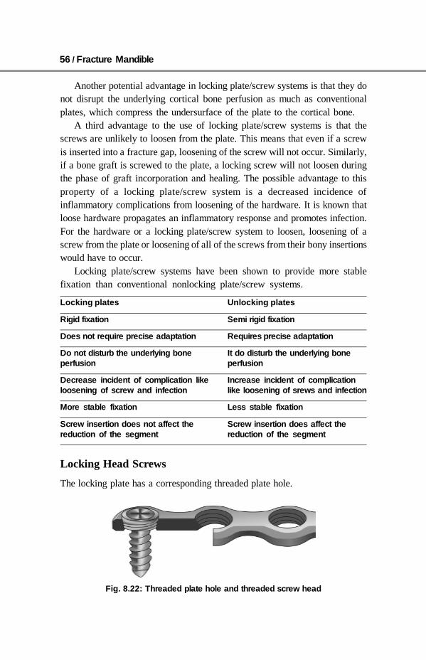
56 / Fracture Mandible
Another potential advantage in locking plate/screw systems is that they donot disrupt the underlying cortical bone perfusion as much as conventionalplates, which compress the undersurface of the plate to the cortical bone.
A third advantage to the use of locking plate/screw systems is that thescrews are unlikely to loosen from the plate. This means that even if a screwis inserted into a fracture gap, loosening of the screw will not occur. Similarly,if a bone graft is screwed to the plate, a locking screw will not loosen duringthe phase of graft incorporation and healing. The possible advantage to thisproperty of a locking plate/screw system is a decreased incidence ofinflammatory complications from loosening of the hardware. It is known thatloose hardware propagates an inflammatory response and promotes infection.For the hardware or a locking plate/screw system to loosen, loosening of ascrew from the plate or loosening of all of the screws from their bony insertionswould have to occur.
Locking plate/screw systems have been shown to provide more stablefixation than conventional nonlocking plate/screw systems.
Locking plates Unlocking plates
Rigid fixation Semi rigid fixation
Does not require precise adaptation Requires precise adaptation
Do not disturb the underlying bone It do disturb the underlying boneperfusion perfusion
Decrease incident of complication like Increase incident of complicationloosening of screw and infection like loosening of srews and infection
More stable fixation Less stable fixation
Screw insertion does not affect the Screw insertion does affect thereduction of the segment reduction of the segment
Locking Head Screws
The locking plate has a corresponding threaded plate hole.
Fig. 8.22: Threaded plate hole and threaded screw head
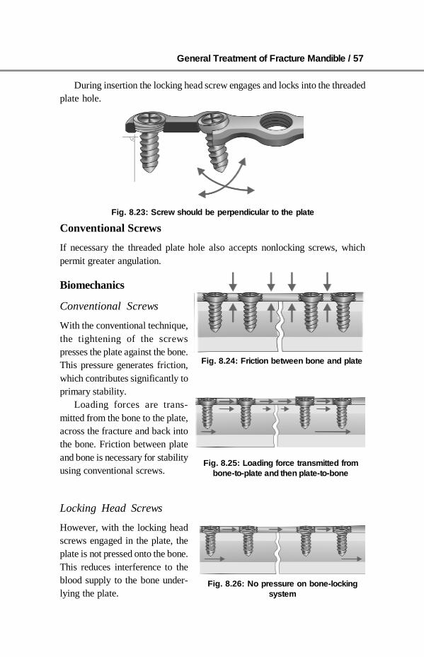
General Treatment of Fracture Mandible / 57
During insertion the locking head screw engages and locks into the threadedplate hole.
Conventional Screws
If necessary the threaded plate hole also accepts nonlocking screws, whichpermit greater angulation.
Biomechanics
Conventional Screws
With the conventional technique,the tightening of the screwspresses the plate against the bone.This pressure generates friction,which contributes significantly toprimary stability.
Loading forces are trans-mitted from the bone to the plate,across the fracture and back intothe bone. Friction between plateand bone is necessary for stabilityusing conventional screws.
Locking Head Screws
However, with the locking headscrews engaged in the plate, theplate is not pressed onto the bone.This reduces interference to theblood supply to the bone under-lying the plate.
Fig. 8.23: Screw should be perpendicular to the plate
Fig. 8.24: Friction between bone and plate
Fig. 8.25: Loading force transmitted frombone-to-plate and then plate-to-bone
Fig. 8.26: No pressure on bone-lockingsystem

58 / Fracture Mandible
Loading forces are transmitted directly from the bone to the screws, thenonto the plate, across the fracture and again through the screws into the bone.Friction between plate and bone is not necessary for stability.
The plate and screws provideadequate rigidity and do not dependon the underlying bone (loadbearing osteosynthesis) when usinga locking reconstruction plate 2.4.
On each side of the fracture, thescrews are locked into the plate aswell as into the bone. The result isa rigid frame construct with high mechanical stability (internal external fixator).
Primary Loss of Reduction
Conventional Plate System
When using conventional platesand screws it is essential to contourthe plate precisely to the bonesurface.
When using conventional plateand screws the plate must beprecisely adapted to the bone,otherwise the tightening of thescrews will lead to a primary lossof reduction in (Fig. 8.28) the plateis not well adapted to the outercortex in lower diagram (Fig. 8.29)shows that when the screws areinserted, the bone will be pulled tothe plate, causing malreduction ofthe fracture.
Locking Plate System
When using a locking plate/screw system, the plate does not have to be preciselyadapted to the bone. When tightening a locking head screw, the screw will notcause a primary loss of reduction as it tightens into the threaded plate hole andwill not draw the bone fragments to the plate.
Fig. 8.27: Loading force transmitting directlyfrom bone-to-screw then to plate and thenscrew-to-bone
Fig. 8.28: Precise adaptation of plate
Fig. 8.29: Imperfect adaptation leads tomalreduction of fracture

General Treatment of Fracture Mandible / 59
Secondary Loss of Reduction
Conventional Plate System
In conventional plate systems, screw loosening may lead to loss of reduction.
Locking Plate System
In a locking system, screw loosening rarely occurs because the screw head islocked to the plate.
Universal Fracture Plate 2.4
They are designed to be used with bicortical screws. Universal fracture platesoffer more biomechanical stability than DCP 2.4 plates
Load-Baring Osteosynthesis(stabilization by splinting)
The plate bears the forces of functionat the fracture site. This is accomp-lished with a locking reconstructionplate. Clinical uses are the managementof atrophic edentulous fractures,comminuted fractures, defect fractures,and other complex mandibularfractures.
Load-Sharing OsteosynthesisFig. 8.30: Load-bearing
osteosynthesis
Fig. 8.31: Lag screw load-sharing osteosynthesis

60 / Fracture Mandible
General Consideration
Stability at the fracture site is created bythe frictional resistance between the boneends and the hardware used for fixation.This requires adequate bony buttressingat the fracture site. Examples of load-sharing osteosynthesis include lag screwfixation technique and compressionplating. Load-sharing osteosynthesiscannot be used with defect fractures orcomminuted fractures, due to the lackof bony buttressing at the fracture site.
Another form of load-sharingosteosynthesis is the miniplate fixationtechnique popularized by Champy. Thisis also known as functionally adequatefixation or semirigid fixation.
Ideal Lines of Osteosynthesis
Champy popularized the treatment ofmandible fractures with miniplate fixationalong the ideal lines of osteosynthesis. This is a form of load-sharingosteosynthesis to be applied in simple fracture patterns having an acceptableamount of bone stock.
Different Levels of Force Distribution
In the load-bearing situation the plate assumes all the forces, in the load-sharing situation there are different levels of force distribution between theplate(s) and the bone.
Load-Bearing
In load-bearing fixation the plate assumes100% of the functional loads.
This is an example of load-bearingosteosynthesis for the treatment of a defectfracture in the angular region. The osteo-synthesis assumes all the masticatory loadswhile the bone graft matures and conso-lidates in a protected environment.
Fig. 8.32: Minihole plate loadsharing osteosynthesis
Fig. 8.33: Champy’s line
Fig. 8.34: Load-bearingosteosynthesis

General Treatment of Fracture Mandible / 61
Intermediate Load-Sharing Situation
Intermediate load-sharing situationwhere the osteosynthesis and the boneshare the functional loads almostequally.
This is an example of load-sharingosteosynthesis for the treatment of asimple angular fracture. The twominiplates share the loads with thebone in an anatomical region wherethe bone stock and force distributionare not ideal.
Ideal Load-Sharing Situation
Ideal load-sharing situation where the bone assumes most of the functionalloads.
In this example, a simple mandibular body fracture was considered suitablefor a single miniplate osteosynthesis in the neutral zone because of the goodbone stock and optimal force distribution.
SUMMARY
Aim of treatment of fracture mandible is to reduce the fracture segmentand fix it so that normal anatomical relationship can be maintained.
Atmost precaution to be taken to preserve the blood supply of the boneand the soft tissue by careful handling and reduction technique.
Attempt should be made for early and safe mobilization and rehabilitationof the patient.
There are many methods of closed reduction but arch bar application ismore commonly used.
Period of immobilization 3–4 weeks for young adult with angle fracture+ 1 week if teeth is in a fracture line+ 1 week for fracture of parasymphysis+ 1 or 2 week for age 40 years and above– 1 week for childrens and adolsent
Dynamic compression plates and lag screws are not commonly used infracture mandible.
Reconstruction plate, miniplate are most commonly used. Rigid osteosynthesis are:
– Reconstruction plate– Locking plates– Three-dimensional sturd.
Fig. 8.35: Intermediate load-sharingosteosynthesis
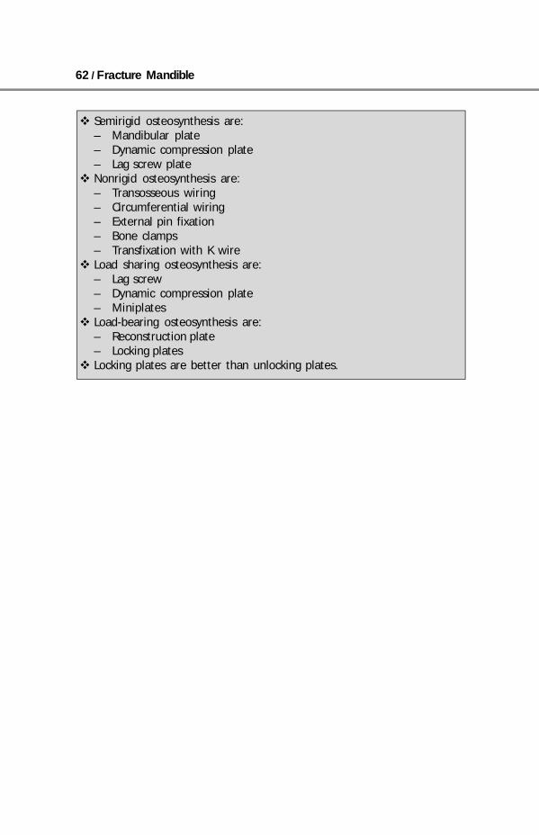
62 / Fracture Mandible
Semirigid osteosynthesis are:– Mandibular plate– Dynamic compression plate– Lag screw plate
Nonrigid osteosynthesis are:– Transosseous wiring– Circumferential wiring– External pin fixation– Bone clamps– Transfixation with K wire
Load sharing osteosynthesis are:– Lag screw– Dynamic compression plate– Miniplates
Load-bearing osteosynthesis are:– Reconstruction plate– Locking plates
Locking plates are better than unlocking plates.

Anesthesia for Fracture Mandible / 63
9 Anesthesia forFracture Mandible
All closed reduction of fracture mandible can be done under local anesthesia.While open reduction is preferably done under general anesthesia. Youngcooperative patient with simple, undisplaced fracture open reduction can betried under local anesthesia. We prefer to apply arch bar under local anesthesiaand in case of open reduction patient is then further given general anesthesia.
Application of arch bar is better done in local anesthesia. As this requirecooperation of the patient in terms of mouth opening and moving the tongue inan appropriate direction, thus this prevents excess retraction and a pressure ona tongue which is required if IMF is done in general anesthesia. Thus this causedecrease edema of tongue and cheek.
General Anesthesia
The anesthesia is same as all routine procedures but few special precautionsneed to be taken.Procedure - Reduction and fixation of fracture mandibleTime - 2 to 3 hours depending upon the severityPostoperative pain - ++Position - Supine with head up tilt with head ring, in case of
extraoral approach a shoulder should be keptBlood loss - VariableIntubation technique - Nasal tube (blind or guided) and IPPV
Fibreoptic intubation may be required.
Preoperative
It is same as all routine anesthesia Careful assessment for associate injury should be done Make a meticulous assessment of airway, there may be several trismus
and soft tissue swelling Explain the patient about postoperative events like mouth closure in a brief Assess nostril patency

64 / Fracture Mandible
Check for evidence of basal skull fracture and CSF leak, which make nasalintubation contraindicated
In a case of severe trismus and where postoperative edema may beanticipated, tracheostomy should be considered
Submantle intubation should be considered in a case of pan facial trauma
Perioperative
Trismus make intubation look potentially difficult preoperatively as themouth opening is markedly limited due to the muscle spasm, hematoma,pain, but this tend to relax following induction
Nose should be packed with 4% lignocaine with adrenaline or otrivinnasal drop should be used
Bilateral mandible fractures can cause increase anterior jaw displacementafter induction but airway maintenance by face mask may not always beeasy due to increase jaw movement, swelling
A rapid sequence induction with suxa-methonium is appropriate A marked swelling may make intubation difficult and awake fibreoptic
intubation may be required Gas induction is often difficult due to pain while applying the face mask Make sure that the patient when comes out from anesthesia should not be
sedated and should be completely awake from the anesthesia If throat pack is placed around the tube should be removed before the
application of wires.
Postoperative
Observe the patient for sometime in a recovery before sending it to the ward Start humidified oxygen Always keep a wire cutter and a suction machine beside the patients bed
(to cut the wires in a case of emergency) Shift the patient with nasopharyngeal airway to the ward.
Local Anesthesia
Two percent lignocaine with adrenaline is used for local anesthesia. In case ofhypertension and other contraindication a plain lignocaine can be used. In caselong duration of anesthesia is required long acting local anesthesia (bupivacaine,ropivacaine, and tetracaine can be used). But 2% lignocaine with adrenalin givesenough duration of anesthesia for arch bar application, so long acting is notusually required.
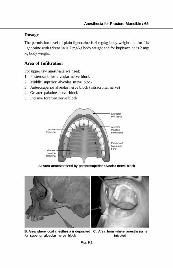
Anesthesia for Fracture Mandible / 65
Dosage
The permission level of plain lignocaine is 4 mg/kg body weight and for 2%lignocaine with adrenalin is 7 mg/kg body weight and for bupivacaine is 2 mg/kg body weight.
Area of Infiltration
For upper jaw anesthesia we need:1. Posterosuperior alveolar nerve block2. Middle superior alveolar nerve block3. Anterosuperior alveolar nerve block (infraorbital nerve)4. Greater palatine nerve block5. Incisive foramen nerve block
A: Area anaesthetized by posterosuperior alveolar nerve block
B: Area where local anesthesia is depositedfor superior alveolar nerve block
C: Area from where anesthesia isinjected
Fig. 9.1

66 / Fracture Mandible
Posterosuperior Alveolar Nerve Block
26 number short needle is used Orient the bevel of the needle towards bone Partially open the patients mouth, pulling the mandible to the side of
injection Retract the patients cheek with your finger Insert the needle into a height of mucobuccal fold over second molar Advance the needle slowly in upward, inward and backward direction Slowly advance to the soft tissue No resistance is filled, if resistance is felt withdraw the needle slightly
and change the direction and re-advance again.
Goal
Goal is to deposit local anesthesia close to the PSA nerve located posteriorsuperior and medial to the maxillary tuberosity.
Middle Superior Alveolar Nerve Block
26 number short needle used Area of insertion, height of muccobuccal fold above the second premolar
buccal fold Aspirate and if no blood then deposit 0.9–1 ml solution.
Anterosuperior Alveolar Nerve Block(Infraorbital Nerve Block)
Nerve anesthetized1. Anterosuperior alveolar2. Middle superior alveolar3. Infraorbital nerve
Technique
Target area—infraorbital foramen (below the infraorbital notch) Landmark—muccobuccal fold, infraorbital notch, infraorbital foramen Area of insertion—height of muccobuccal fold directly over first premolar.
Procedure
Feel for infraorbital notch Move your fingers downward from the notch applying gentle pressure to
the tissue

Anesthesia for Fracture Mandible / 67
Fig. 9.2
A: Area where local anesthesia isdeposited for middle superior alveolar
nerve block
B: Area from where anesthesia isinjected
C: Area anaesthetized by middle superior alveolar nerve block
The bone immediately inferior to the notch is convex, this represents thelower border of orbit and a roof of infraorbital foramen
As your finger continous inferiorly a concavity is felt, this is infraorbitalforamen
Maintain your finger on the foramen or mark the skin at the site Retract the lips, pull the tissue in muccobuccal fold Insert the needle into the height of muccobuccal fold over the first premolar
with the bevel facing bone Reach to your required site

68 / Fracture Mandible
A: Area which is anaesthetized by infraorbital nerve block
B: Area where local anesthesia isinjected for infraorbital block
C: Area where local ansethesia isdeposited
Fig. 9.3
Aspirate and if no blood slowly deposit 1 ml in a target area One can feel that solution is deposited beneath the fingers if needle is
in a right plane.
Greater Palatine Nerve Block
Target area—greater palatine nerve as it passes anteriorly between the softtissue and bone of the heart palate
Landmark—a greater palatine foramen and junction of maxillary alveolarprocess and palatine bone

Anesthesia for Fracture Mandible / 69
B: Area which is blocked by greater palatine block
A: Area where greater palatine foramen is located
Fig. 9.4
Part of insertion—advance the syring from the opposite side of the mouthat the right angle to the target area.
Procedure
Feel for depression of greater palatine foramen with the help of cottonswab and a finger
The foramen is located distal to second molar but it may be either anterioror posterior to its usual position
Inject in the foramen area around few drops.

70 / Fracture Mandible
Lower Jaw Infiltration
1. Inferior alveolar nerve block2. Buccal nerve3. Mental nerve
Inferior Alveolar Nerve Block
Landmark—coronoid notch (greater concavity of anterior border of ramus) Ptrygomandibular raphe Occlusion plane of mandibular posterior teeth
B: Area from which anesthesia is given
A: Area anesthetized by infra-alveolar nerve block
C: Area where infra-alveolar nerve islocated
Fig. 9.5
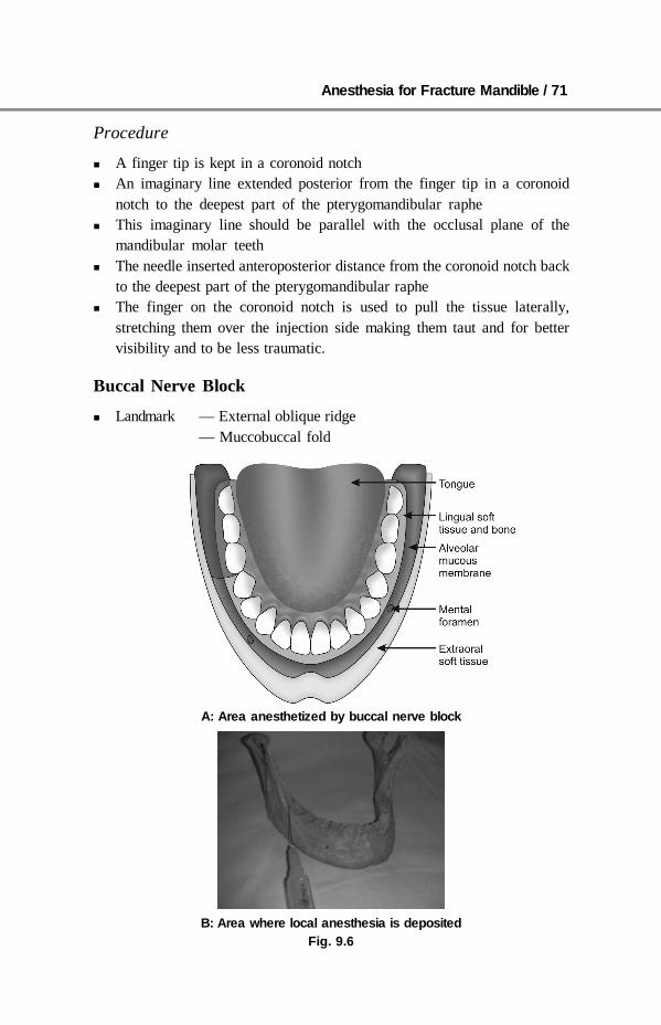
Anesthesia for Fracture Mandible / 71
Procedure
A finger tip is kept in a coronoid notch An imaginary line extended posterior from the finger tip in a coronoid
notch to the deepest part of the pterygomandibular raphe This imaginary line should be parallel with the occlusal plane of the
mandibular molar teeth The needle inserted anteroposterior distance from the coronoid notch back
to the deepest part of the pterygomandibular raphe The finger on the coronoid notch is used to pull the tissue laterally,
stretching them over the injection side making them taut and for bettervisibility and to be less traumatic.
Buccal Nerve Block
Landmark — External oblique ridge— Muccobuccal fold
A: Area anesthetized by buccal nerve block
B: Area where local anesthesia is depositedFig. 9.6

72 / Fracture Mandible
Procedure
A 25 gauge 1 inch long needle is inserted either in retromolar triangle areaor buccalmuccosa just distal to mandibular third molar area
0.5 ml of local anesthesia is injected
Mental Nerve Block
Landmark — Mandibular premolars— Muccobuccal fold
A: Location of mental foramen B: Area in which local anesthesia isinjected for mental nerve block
C: Area anesthetized by mental nerve block
Fig. 9.7
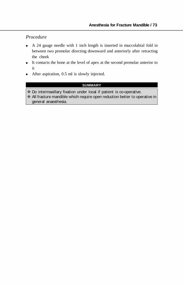
Anesthesia for Fracture Mandible / 73
Procedure
A 24 gauge needle with 1 inch length is inserted in muccolabial fold inbetween two premolar directing downward and anteriorly after retractingthe cheek
It contacts the bone at the level of apex at the second premolar anterior toit
After aspiration, 0.5 ml is slowly injected.
SUMMARY
Do intermaxillary fixation under local if patient is co-operative. All fracture mandible which require open reduction better to operative in
general anaesthesia.
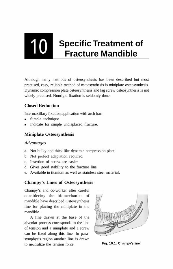
74 / Fracture Mandible
10 Specific Treatment ofFracture Mandible
Although many methods of osteosynthesis has been described but mostpractised, easy, reliable method of osteosynthesis is miniplate osteosynthesis.Dynamic compression plate osteosynthesis and lag screw osteosynthesis is notwidely practised. Nonrigid fixation is seldomly done.
Closed Reduction
Intermaxillary fixation application with arch bar: Simple technique Indicate for simple undisplaced fracture.
Miniplate Osteosynthesis
Advantages
a. Not bulky and thick like dynamic compression plateb. Not perfect adaptation requiredc. Insertion of screw are easierd. Gives good stability to the fracture linee. Available in titanium as well as stainless steel material.
Champy’s Lines of Osteosynthesis
Champy’s and co-worker after carefulconsidering the biomechanics ofmandible have described Osteosynthesisline for placing the miniplate in themandible.
A line drawn at the base of thealveolar process corresponds to the lineof tension and a miniplate and a screwcan be fixed along this line. In para-symphysis region another line is drawnto neutralize the tension force. Fig. 10.1: Champy’s line

Specific Treatment of Fracture Mandible / 75
Fig. 10.2: Minihole plate
Behind the mental foramen only one plate should be applied, immediatelybelow the dental root and above the inferior alveolar nerve.
Between the two canines and in front of mental foramen another platenear the lower border of mandible is applied in addition to the upperplate.
Technique
This miniplates are around 0.9 mm thick and 6 mm wide. The difference betweenholes are standardized. The screws available are from 5 to 15 mm in length. Thediameter of the screws is 2 mm. Screw of 6 or 8 mm in length are usually usedin mandible. The screws are self-tapping.
Fig. 10.3: Screw driver and screw
The plate is bended first so that they can be adapted to bone. 1.5 mm drillbit is used to create a hole almost perpendicular to the plate. Eccentric drilling or repeated insertion of drill produce unfavorable hole
thus diminishing the grip of the screw. During drilling continues liquid cooling is necessary to avoid thermal
necrosis. A giveway indicates penetration of drill in a cortical bone. Then screw is inserted near the fracture side and gradually tightened. This same procedure is carried out for insertion of screw in all the plates. The inferior alveolar nerve region is considered as the neutral zone of
the mandible. Avoid plate and screw fixation in this area that woulddamage the nerve.

76 / Fracture Mandible
Basic treatment of any simple fracture mandible is application of loadsharing osteosynthesis plates (mini plate most commonly used along theChampy’s line).
a. Symphysis fracture and parasymphysis fractureVarious options are:A. Symphysis fractureB. Parsymphysis fractureC. Mini plate + dynamic plate osteosynthesis (load-sharing osteosynthesis)D. Two miniplate osteosynthesis (most preferred and practised load-sharing
osteosynthesis)E. Lag screw osteosynthesis (load-sharing osteosynthesis).
Fig. 10.4
AB
CD
E
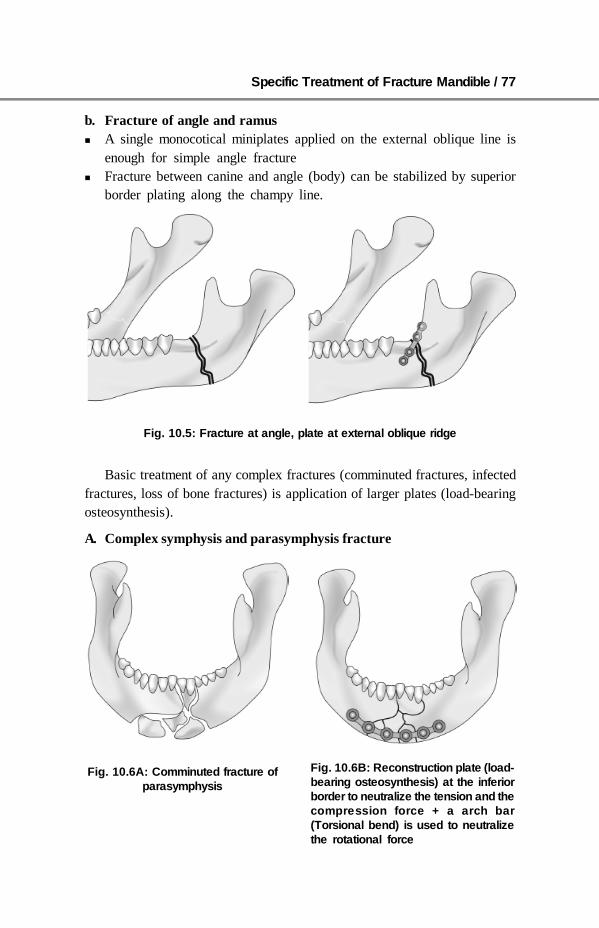
Specific Treatment of Fracture Mandible / 77
b. Fracture of angle and ramus A single monocotical miniplates applied on the external oblique line is
enough for simple angle fracture Fracture between canine and angle (body) can be stabilized by superior
border plating along the champy line.
Basic treatment of any complex fractures (comminuted fractures, infectedfractures, loss of bone fractures) is application of larger plates (load-bearingosteosynthesis).
A. Complex symphysis and parasymphysis fracture
Fig. 10.5: Fracture at angle, plate at external oblique ridge
Fig. 10.6A: Comminuted fracture ofparasymphysis
Fig. 10.6B: Reconstruction plate (load-bearing osteosynthesis) at the inferiorborder to neutralize the tension and thecompression force + a arch bar(Torsional bend) is used to neutralizethe rotational force
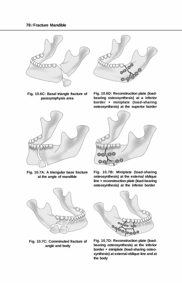
78 / Fracture Mandible
Fig. 10.6C: Basal triangle fracture ofparasymphysis area
Fig. 10.6D: Reconstruction plate (load-bearing osteosynthesis) at a inferiorborder + miniplate (load-sharingosteosynthesis) at the superior border
Fig. 10.7A: A triangular base fractureat the angle of mandible
Fig . 10.7B: Miniplate (load-sharingosteosynthesis) at the external obliqueline + reconstruction plate (load-bearingosteosynthesis) at the inferior border
Fig. 10.7C: Comminuted fracture ofangle and body
Fig. 10.7D: Reconstruction plate (load-bearing osteosynthesis) at the inferiorborder + miniplate (load-sharing osteo-synthesis) at external oblique line and atthe body

Specific Treatment of Fracture Mandible / 79
Mandibular Condyle Fracture
Classification of condyle fracturesa. Fracture of head of condyle—simple or comminuted fractureb. Fracture of neck of condyle—simple or incomplete fracture without any
displacement and complete fracture of neck of condyle with displacementc. Subcondylar fracture—low and highd. Malunited fracture.
The management of mandibular condyle fracture is very much importantespecially in children because this can laed to disturbance of mandibulargrowth pattern or even ankylosis of temporomandibular joint. This is themost overlooked and least diagnosed side of fracture.
Treatment
The aim of treatment of condylar fracture is to reestablish the anatomicalrelationship for providing good function and growth on long term basis.
a. Fracture of head of condyleThe intracapsular fracture of head of a condyle is rare and should be treatedconservatively by intermaxillary fixation for a period of 2 to 4 weeks followedby restoration of function. Severe comminuted fractures of the condyle aretreated by condylectomy especially in unilateral cases. In case of children,costochondral grafts should be given to replace the lost condyle.
Subcondylar fracturesThese are the fractures below the neck of the condyle. These fractures areclassified as: High condylar fracture Low condylar fractures
Fig. 10.8
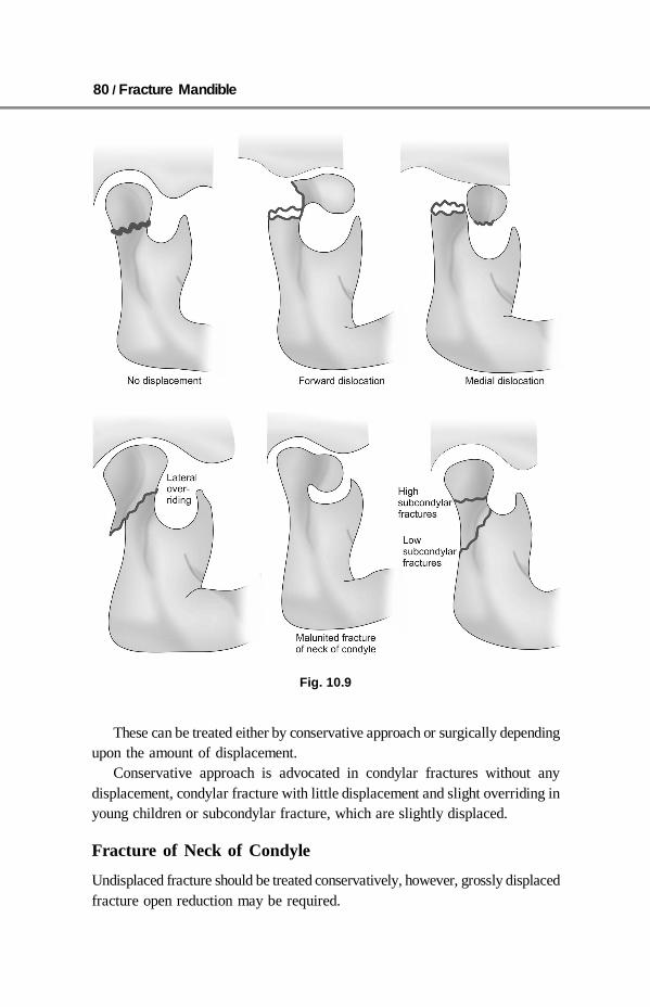
80 / Fracture Mandible
Fig. 10.9
These can be treated either by conservative approach or surgically dependingupon the amount of displacement.
Conservative approach is advocated in condylar fractures without anydisplacement, condylar fracture with little displacement and slight overriding inyoung children or subcondylar fracture, which are slightly displaced.
Fracture of Neck of Condyle
Undisplaced fracture should be treated conservatively, however, grossly displacedfracture open reduction may be required.

Specific Treatment of Fracture Mandible / 81
Open Reduction
In grossly displaced condyle fracture where reduction cannot be achieved byconventional method there is a fear of future dearrangement or ankylosis, anopen reduction is indicated.
The indications are: Cases of condyle fracture with vertical overriding Cases in which normal occlusion by manipulation or traction cannot be
achieved Cases of gross displacement especially bilateral condyle fracture Cases in which condyle fracture interfere with jaw movement Cases in which condyle are partially fused in a wrong position
The condyle is surgically approached by preauricular approach or bysubmandibular, retromandibular approach. The fracture is reduced and isfixed by either by interosseous wiring or by monocotical miniplates.
Fracture of Coronoid Process
This account for 1% of fracture, most of the coronoid fractures do not requireany treatment. Cases in which coronoid hinders with opening of mouth, treatmentis advised. This coronoid fragment is fixed by interosseous wiring or by miniplate. If reduction is not possible and function is impaired then coronoid processis removed.
Mistakes
Common mistakes observed in treatment of rigid fixation: Poor reduction of fractures Interposition of tissue between the fracture line Poor alignment of fractured segment Insufficient screw placement Poor plate bending Poor or lose application of intermaxillary fixation.
Tips
Soft tissue in between the fracture line should be removed as properallignment cannot be attended. Nonhealing can occur if soft tissue is placedbetween fractured fragment.
Use of drill sleeve provide protection to soft tissue. Drill bit used should be 0.5 mm less than screw size. For example, if 2 mm
screw or plate is used 1.5 mm drill bit is used to make hole.

82 / Fracture Mandible
Due to drilling heat is generated, this heat can cause bone necrosis andearly screw loosening so cooling by putting saline over the drill bit.
Avoid damage to mental nerve, tooth root, inferior alveolar canal. Proper bending of a plate should be done. A space of about 5 mm should be kept between two plates.
SUMMARY
In case of simple, undisplaced fracture and no expertise available orcondition of patient does not allow general anesthesia; close reduction offracture mandible most preferably by arch bar method (IMF) is suitable.
In cases of parasymphysis and symphysis fracture in addition to lowerborder miniplate application a superior border miniplate is also appliedto combat torsional force.
A miniplate with two hole on either side of fracture mandible is ideallyfixed.
Champy’s line of osteosynthesis give idea of sight of application of platesin various areas.
In simple undisplaced fracture load sharing plate (mini plate) to be used. In grossly displaced, infected, loss of segment fracture mandible
reconstruction plate should be ideally used. All cases of undisplaced condyle fracture can be treated by closed
reduction. Open reduction in a condyle fracture is indicated in cases where by
manipulation or traction the fracture segment is not reduced or cannot bebrought into normal occlusion.
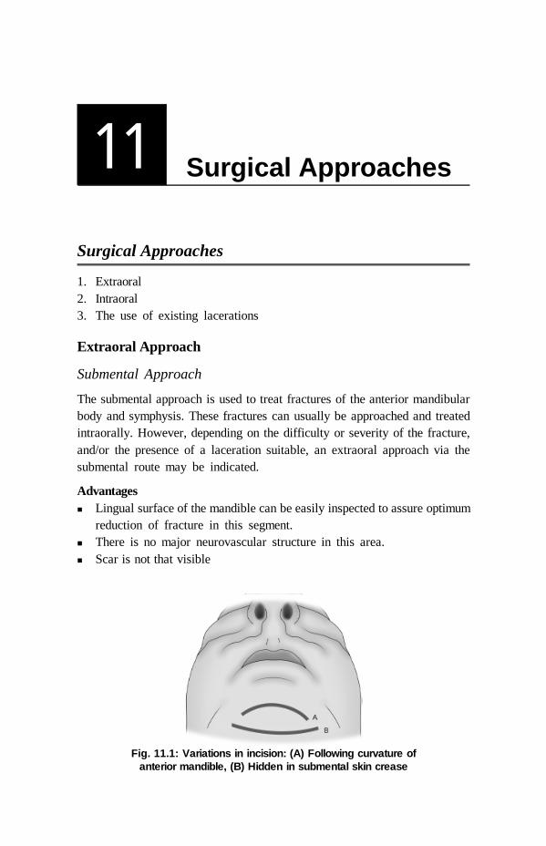
Surgical Approaches / 83
11 Surgical Approaches
Surgical Approaches
1. Extraoral2. Intraoral3. The use of existing lacerations
Extraoral Approach
Submental Approach
The submental approach is used to treat fractures of the anterior mandibularbody and symphysis. These fractures can usually be approached and treatedintraorally. However, depending on the difficulty or severity of the fracture,and/or the presence of a laceration suitable, an extraoral approach via thesubmental route may be indicated.
Advantages Lingual surface of the mandible can be easily inspected to assure optimum
reduction of fracture in this segment. There is no major neurovascular structure in this area. Scar is not that visible
Fig. 11.1: Variations in incision: (A) Following curvature ofanterior mandible, (B) Hidden in submental skin crease
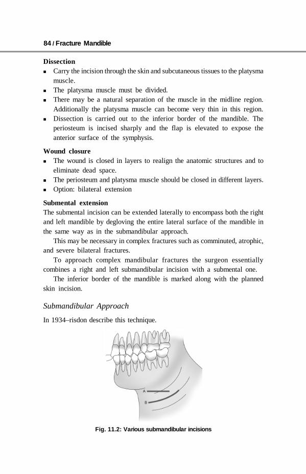
84 / Fracture Mandible
Dissection Carry the incision through the skin and subcutaneous tissues to the platysma
muscle. The platysma muscle must be divided. There may be a natural separation of the muscle in the midline region.
Additionally the platysma muscle can become very thin in this region. Dissection is carried out to the inferior border of the mandible. The
periosteum is incised sharply and the flap is elevated to expose theanterior surface of the symphysis.
Wound closure The wound is closed in layers to realign the anatomic structures and to
eliminate dead space. The periosteum and platysma muscle should be closed in different layers. Option: bilateral extension
Submental extensionThe submental incision can be extended laterally to encompass both the rightand left mandible by degloving the entire lateral surface of the mandible inthe same way as in the submandibular approach.
This may be necessary in complex fractures such as comminuted, atrophic,and severe bilateral fractures.
To approach complex mandibular fractures the surgeon essentiallycombines a right and left submandibular incision with a submental one.
The inferior border of the mandible is marked along with the plannedskin incision.
Submandibular Approach
In 1934–risdon describe this technique.
Fig. 11.2: Various submandibular incisions

Surgical Approaches / 85
PrinciplesThis approach is selected for fractures of the mandibular body and angle regionsunsuitable for intraoral treatment.
This applies to more difficult fracture patterns such as comminuted, atrophic,and defect fractures in order to allow optimal manipulation of the fragments,good control of the lingual cortex and inferior border, and the application ofthe selected hardware.
VariationsThe incision can either be parallel to the inferior border of the mandible atleast2 cm below the angle of the mandible (A) or be placed in an existing skincrease (B) for maximum cosmetic benefit.
If using skin creases for the incision, the orientation of the scalpel blade isparallel to the skin creases.
Subplatysma flap elevated.
Neurovascular structuresThe main neural structure is the marginal mandibular branch of the facial nerve(CN VII). The facial artery and vein are also encountered during this dissection.They are commonly located 1 cm below the angle of the mandible.
This neurovascular structure are identified by Holder Martins method inwhich facial vessels are identified and they are retracted so that they are safein the flap.
The dissection is carried out through the deep cervical fascia.The muscle, periosteum and other soft tissues are retracted superiorly to
expose the body, angle and the ramus of the mandible.Then the fracture line is identified.
Retromandibular Approaches
In 1967, Hinds and Girrotti first describedthis approach.
Skin incisionIncision is made 3 cm above the submandi-bular incision
The incision is carried curving behindthe angle of mandible
Use of normal saline for infilteration isemployed for maintenance of the plane andin a cases where nerve stimulator has to beused.
Fig. 11.3: Retromandibularapproach

86 / Fracture Mandible
Use of 2% lignocaine with adrenalin can be used for vasoconstrictoreffect.
Use of 2% local anesthetic may impair the function of the marginalmandibular nerve and prevent the use of a nerve stimulator during the surgicalprocedure.
Subplatysma flap elevatedSuperior subplatysmal dissection would expose the underlying marginalmandibular branch of the facial nerve (CN VII).
By ligating and dividing the facial artery and vein and then retracting thevessels superiorly, the marginal mandibular branch of the facial nerve remainsincluded in the superior flap and is thus protected.
Divide the pterygomasseteric sling and incise the periosteum at the inferiorborder to expose the fracture site.
This expose the body and angle region.For wound closure, the pterygomasseteric sling is closed.The wound is closed in layers to realign the anatomic structures and
eliminate dead space. The platysma muscle is closed. A drain may be usedif necessary.
PrinciplesThe retromandibular approaches expose the entire ramus from behind theposterior border. They therefore may be useful for procedures involving thearea on or near the condylar process/head, or the ramus itself.
There are two varieties of retromandibular approach used to access theposterior mandible. They differ in the placement of the incision and theanatomic dissection to the mandible.
The transparotid approach has the advantage of close proximity of theskin incision to the area of interest. The retroparotid approach has theadvantage of not dissecting through theparotid gland.
The facelift (rhytidectomy) approach canbe considered as an alternative to retro-mandibular approaches.
The main anatomic structures in thisapproach are the main trunk and branches ofthe facial nerve (CN VII) and the retro-mandibular vein.
Transparotid approach: Skin incisionA vertical incision through skin and sub- Fig. 11.4: Tranparotid approach
skin incision

Surgical Approaches / 87
cutaneous tissue is made, extending from just below the ear lobe towards themandibular angle. It should parallel the posterior border of the mandible.
DissectionThe subcutaneous tissue is undermined, exposing the superficial musculo-aponeurotic system (SMAS).
A vertical incision is made through the SMAS into the parotid gland.
Blunt dissection of the parotid glandBluntly dissect the parotid gland parallel to the direction of the facial nervebranches and towards the posterior border of the mandible. The dissectionshould be anterior to the retromandibular vein.
Branches of the facial nerve may be found during the dissection. A nervestimulator may be helpful to identify them. They should be mobilized andprotected.
Once the posterior border of the mandible has been reached, an incisionis made through the pterygomasseteric sling.
Subperiosteal dissection of the mandibular ramusA periosteal elevator is used to strip the masseter muscle from the ramus.Further dissection superiorly along the posterior border exposes the condylarprocess.
Transparotid approach: Wound closureThe wound is reapproximated in layers for anatomic realignment and avoidanceof dead space. The parotid gland capsule must be closed tightly to preventsalivary fistula. The SMAS is resuspended.
Alternative: Retroparotid approachPrinciplesA frequently used alternative to theretromandibular transparotid approachdescribed above is one in which theparotid gland is lifted rather thandissected through. This requires theincision to be placed more posteriorlywhich means that exposure of themandible is more limited. Rather thanapproaching the mandible from directlyover the ramus, it is approached moreposteriorly. Fig. 11.5: Retroparotid approach

88 / Fracture Mandible
Skin incisionAn oblique incision through skin and subcutaneous tissue is made, extendingfrom the mastoid process to a point just below the angle of the mandible.
DissectionThe subcutaneous tissue is undermined, exposing the superficialmusculoaponeurotic system (SMAS).
An oblique incision is made through the SMAS. The posterior aspect ofthe parotid gland is identified and dissection continues behind the gland.
The gland is lifted off the masseter muscle and retracted anteriorly.Once the posterior border of the mandible has been reached, an incision is
made through the pterygomasseteric sling.
Subperiosteal dissection of the mandibular ramusA periosteal elevator is used to strip the masseter muscle from the ramus.Further dissection superiorly along the posterior border exposes the condylarprocess.
Wound closureThe wound is reapproximated in layers for anatomic realignment andavoidance of dead space.
The SMAS is resuspended.A suction drain may be placed.
Preauricular Approach
PrinciplesThe preauricular approach can be used to access and treat fractures in themandibular condylar head and neck region. Many surgeons perform temporalmandibular joint (TMJ) surgery and routinely use this incision to access thesuperior portion of the mandibular condylar process.
Neurovascular structuresBranches of the facial nerve may be involved in this incision and dissection.The superficial temporal artery and vein are commonly encountered in thissurgical approach. The vessels should be conserved if possible.
Facelift incisionSkin incisionMake the incision in a preauricular skin crease.
DissectionLocating temporalis fascia.

Surgical Approaches / 89
Carry the incision through theskin and subcutaneous tissues to thedepth of the temporalis fascia. Thetemporalis fascia is a glistening whitetissue layer that is best appreciatedin the superior portion of theincision.
The superficial temporal vesselsmay be retracted anteriorly with theskin flap (sectioning some posteriorand superior branches) or left inplace (sectioning frontal branches).
The zygomatic arch can easily bepalpated at this point of the dis-section. The lateral pole of the mandibular condyle can also be palpated. Thiscan be facilitated by having a surgical assistant manipulate the jaw.
Incising temporalis fasciaMake an oblique incision parallel to the frontal branch of the facial nerve,through the superficial layer of the temporalis fascia above the zygomatic arch.
Dissection of the joint capsuleInsert the periosteal elevator beneath the superficial layer of the temporalisfascia and strip the periosteum off the lateral zygomatic arch.
Dissection will be carried inferiorly to expose the capsule of the TMJ.The frontal branch of the facial nerve is protected within the superficial
layer of the deep temporalis fascia.Dissection can be carried inferiorly in a subperiosteal plane to reach the
neck of the mandibularcondyle.A disadvantage of this approach is that the surgeon can reach only a limited
portion of the condylar neck region.
Wound closureIf the TMJ capsule has been incised to access the condylar head it must beclosed as the first step.
The temporalis fascia is closed as the next step.Skin and subcutaneous sutures are placed.A pressure dressing may be placed over this wound according to surgeon’s
preference.
Fig. 11.6: Facelift incision
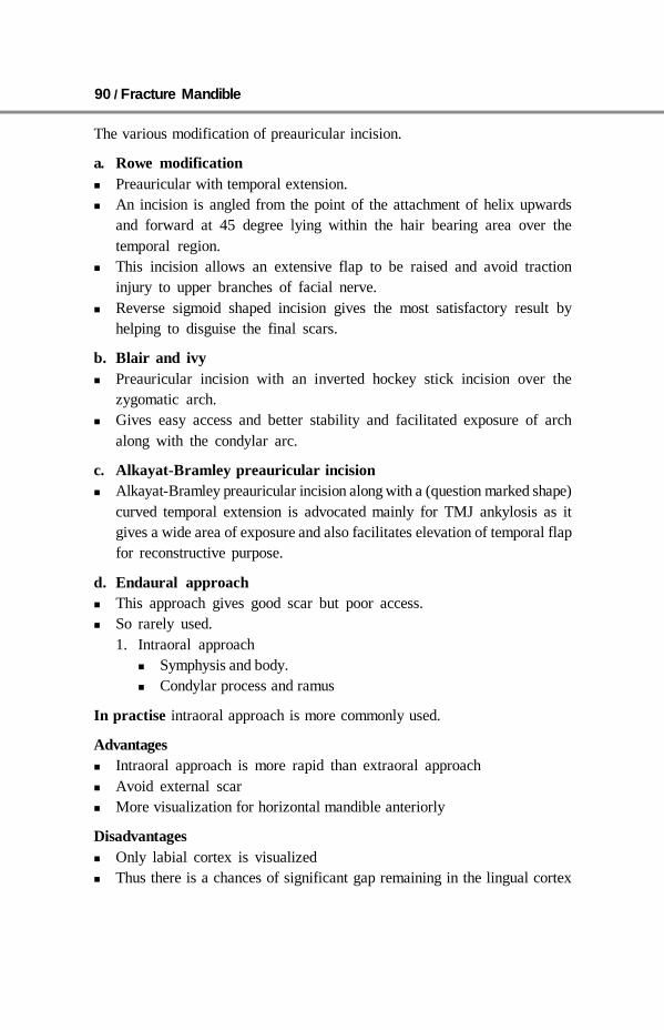
90 / Fracture Mandible
The various modification of preauricular incision.
a. Rowe modification Preauricular with temporal extension. An incision is angled from the point of the attachment of helix upwards
and forward at 45 degree lying within the hair bearing area over thetemporal region.
This incision allows an extensive flap to be raised and avoid tractioninjury to upper branches of facial nerve.
Reverse sigmoid shaped incision gives the most satisfactory result byhelping to disguise the final scars.
b. Blair and ivy Preauricular incision with an inverted hockey stick incision over the
zygomatic arch. Gives easy access and better stability and facilitated exposure of arch
along with the condylar arc.
c. Alkayat-Bramley preauricular incision Alkayat-Bramley preauricular incision along with a (question marked shape)
curved temporal extension is advocated mainly for TMJ ankylosis as itgives a wide area of exposure and also facilitates elevation of temporal flapfor reconstructive purpose.
d. Endaural approach This approach gives good scar but poor access. So rarely used.
1. Intraoral approach Symphysis and body. Condylar process and ramus
In practise intraoral approach is more commonly used.
Advantages Intraoral approach is more rapid than extraoral approach Avoid external scar More visualization for horizontal mandible anteriorly
Disadvantages Only labial cortex is visualized Thus there is a chances of significant gap remaining in the lingual cortex

Surgical Approaches / 91
Intraoral Approach to the Symphysis and Body
Vestibular Incisions
The intraoral approach is the usual accessfor simple fractures of the body,symphysis, and angular regions.
The approach can be extendedposteriorly (dashed line) for better accessto the body, angle and ramus regions.
In complex fractures includingcomminuted, edentulous, and avulsivefractures requiring the placement of load-bearing reconstruction plates, a extraoral approach may provide better access.
Oral contamination is not a contraindication for an intraoral incision.
Mucosal Incision
Unless contraindicated, infiltrate the area with a local anesthetic containinga vasoconstrictor.
Make an incision through the mucosa in the vestibule. Between thecanines the incision is made 10–15 mm away from the attached gingiva ina curvilinear fashion. Posterior to the canine the incision is only 5 mm awayfrom the attached gingiva, staying superior to the mental nerve.
Neurovascular Structures
The mental nerve is a branch of the fifth cranial nerve (trigeminal nerve).This nerve provides sensation to the anterior mandibular vestibule, lip andchin.
When the incision is extended posterior to the canine teeth, the mentalnerve can be damaged. Keep the incision superior to the mental nerve in thebody region.
Particularly in the extended intraoral approach, care must be taken toprotect the mental nerve in the anterior body region.
Surgical Flap Dissection
Dissect a mucosal flap that retracts or is lifted (as shown) to expose thesurface of the mentalis muscle. The branches of the mental nerve are locatedjust underneath the mucosal flap and must be respected.
Fig. 11.7: Intraoral incison forsymphysis and body fracture

92 / Fracture Mandible
Mentalis muscle dissectionThe mentalis muscle is incised near the alveolar bone ridge thus creating astepwise approach which protects the mental nerve. Later, during woundclosure the mentalis muscle should be properly reattached.
Fracture Site Exposure
Elevate a mucoperiosteal flap to expose the fracture.
Extension of ApproachLateral/posterior vestibular incision.
The approach can be extended laterally and posteriorly to provide accessto the body, angle and ramus regions of the mandible.
Right-angled retractors are helpful in this approach.
Wound Closure
After thoroughly irrigating the wound and checking for hemostasis the incisionis closed. Anteriorly, the mentalis muscle is reapproximated to preventdrooping of the chin tissues. The mucosa is closed with interrupted orrunning resorbable sutures.
An elastic pressure dressing on the chin region helps support the softtissues and prevent hematoma formation
Intraoral Approach to the Angle
Principles
Vestibular incisionsThe intraoral approach is used for the majority of simple angle fractures.Depending on whether or not a third molar is to be extracted, there are two
Fig. 11.8: Mucosal incision for angle fracture

Surgical Approaches / 93
surgical approaches. Where there is no third molar present, or where one ispresent but is to be left in place, a purely vestibular incision approximately 5mm away from the attached gingiva is made (A). When an erupted third molaris to be removed, the incision must incorporate the attached gingiva around thebuccal side of the tooth (B, combination of vestibular and envelope incisions).
Oral contamination is not a contraindication for an intraoral incision.Restricted access and contaminationIn complex fractures including comminuted, edentulous, and avulsive
fractures that will require the placement of load-bearing reconstruction plates,a transfacial/extraoral approach can provide better access to treat the injury.
Tips Avoid injury to the sensory buccal nerve Reattach the buccinators muscle to its original position as far as possible
Sensory buccal nerveThe sensory buccal nerve crosses the upper anterior rim of the mandibularascending ramus in the region of the coronoid notch. It is usually below themucosa running above the temporalis muscle fibers. When the posteriorvestibular incision is carried sharply along the bony rim, the buccal nerveis at risk of transsection, followed by numbness in the buccal mucosalregion. Therefore, to protect the nerve, the posterior dissection is to beextended bluntly as soon as the lower coronoid notch is reached.
The photograph shows the sensory buccal nerve.
Buccinator muscleThe lateral mucogingival vestibular incision transsects the lower attachmentof the buccinator muscle. Stripping the mucoperiosteal flap laterally dislocatesthe lower border of the muscle. To reattach the muscle, the sutures forwound closure in the lateral vestibular should not only be superficial. Thesuture should catch all layers (mucosa and muscle) as a safeguard for musclereattachment.
Vestibular incisionUnless contraindicated, infiltrate the area with a local anesthetic containinga vasoconstrictor.
Make an incision through the mucosa in the vestibule approximately5 mm away from the attached gingiva (in the mucogingival junction),extending up the external oblique ridge.
The fracture must be reduced adequately before fixation is applied. Thefixation can be either by transbuccal or right-angled instrumentation.
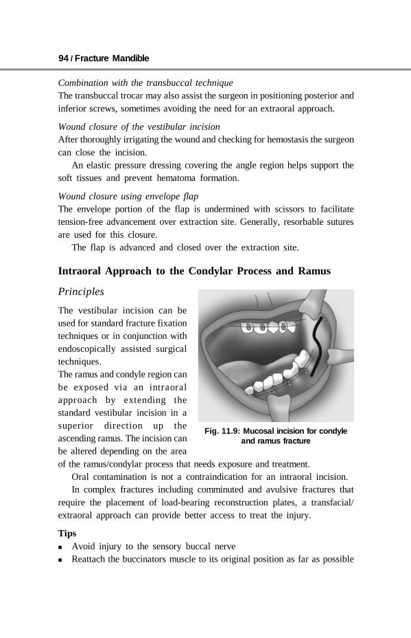
94 / Fracture Mandible
Combination with the transbuccal techniqueThe transbuccal trocar may also assist the surgeon in positioning posterior andinferior screws, sometimes avoiding the need for an extraoral approach.
Wound closure of the vestibular incisionAfter thoroughly irrigating the wound and checking for hemostasis the surgeoncan close the incision.
An elastic pressure dressing covering the angle region helps support thesoft tissues and prevent hematoma formation.
Wound closure using envelope flapThe envelope portion of the flap is undermined with scissors to facilitatetension-free advancement over extraction site. Generally, resorbable suturesare used for this closure.
The flap is advanced and closed over the extraction site.
Intraoral Approach to the Condylar Process and Ramus
Principles
The vestibular incision can beused for standard fracture fixationtechniques or in conjunction withendoscopically assisted surgicaltechniques.The ramus and condyle region canbe exposed via an intraoralapproach by extending thestandard vestibular incision in asuperior direction up theascending ramus. The incision canbe altered depending on the areaof the ramus/condylar process that needs exposure and treatment.
Oral contamination is not a contraindication for an intraoral incision.In complex fractures including comminuted and avulsive fractures that
require the placement of load-bearing reconstruction plates, a transfacial/extraoral approach can provide better access to treat the injury.
Tips Avoid injury to the sensory buccal nerve Reattach the buccinators muscle to its original position as far as possible
Fig. 11.9: Mucosal incision for condyleand ramus fracture

Surgical Approaches / 95
Sensory Buccal Nerve
The sensory buccal nerve crosses the upper anterior rim of the mandibularascending ramus in the region of the coronoid notch. It is usually below themucosa running above the temporalis muscle fibers. When the posteriorvestibular incision is carried sharply along the bony rim, the buccal nerveis at risk of transsection resulting in numbness in the buccal mucosal region.Therefore, to protect the nerve, the posterior incision is to be extendedbluntly as soon as the lower coronoid notch is reached.
Buccinator Muscle
The lateral mucogingival vestibular incision transsects the lower attachmentof the buccinator muscle. Stripping the mucoperiosteal flap laterally dislocatesthe lower border of the muscle. To reattach the muscle, the sutures forwound closure in the lateral vestibular should not only be superficial. Thesuture should catch all layers (mucosa and muscle) as a safeguard for musclereattachment.
Reminder: The buccinator muscle belongs to the mimic muscle system andhas a unique functional structure allowing for a movement comparable to aperistaltic motion. The deep fibers run in parallel bundles from the modiolusto the pterygomandibular raphe at the level of the occlusal plane (intercalarregion) and account for the buccinator mechanism building up a ridge towardsthe occlusal plane. Its detachment can result in an impaired bolus transportout of the buccal space which is troublesome for the patient. The buccinatoris innervated by the motor buccal branch of the facial nerve.
Incision
Unless contraindicated, infiltrate the area with a local anesthetic containinga vasoconstrictor.
Make an incision through the mucosa in the vestibule approximately 5mm away from the attached gingiva (in the mucogingival junction), extendingup the external oblique ridge.
Exposure of Fracture
The lateral surface of the ramus and condylar process is exposed in asubperiosteal plane to visualize the fracture. Right-angled retractors andfiberoptic lighting would facilitate this procedure. The fracture must bereduced adequately before fixation is applied. The fixation can be doneeither by transbuccal or right-angled instrumentation.
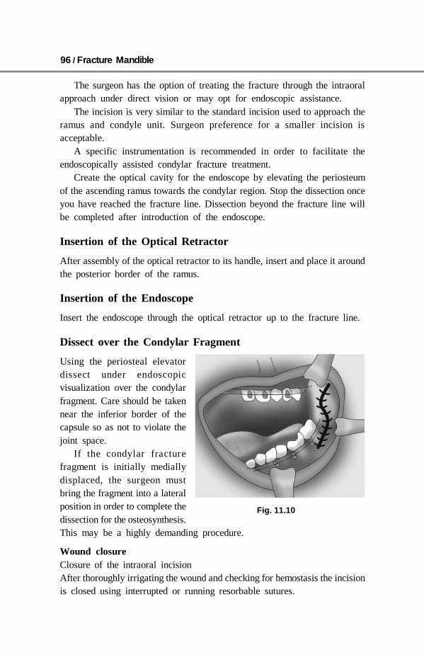
96 / Fracture Mandible
The surgeon has the option of treating the fracture through the intraoralapproach under direct vision or may opt for endoscopic assistance.
The incision is very similar to the standard incision used to approach theramus and condyle unit. Surgeon preference for a smaller incision isacceptable.
A specific instrumentation is recommended in order to facilitate theendoscopically assisted condylar fracture treatment.
Create the optical cavity for the endoscope by elevating the periosteumof the ascending ramus towards the condylar region. Stop the dissection onceyou have reached the fracture line. Dissection beyond the fracture line willbe completed after introduction of the endoscope.
Insertion of the Optical Retractor
After assembly of the optical retractor to its handle, insert and place it aroundthe posterior border of the ramus.
Insertion of the Endoscope
Insert the endoscope through the optical retractor up to the fracture line.
Dissect over the Condylar Fragment
Using the periosteal elevatordissect under endoscopicvisualization over the condylarfragment. Care should be takennear the inferior border of thecapsule so as not to violate thejoint space.
If the condylar fracturefragment is initially mediallydisplaced, the surgeon mustbring the fragment into a lateralposition in order to complete thedissection for the osteosynthesis.This may be a highly demanding procedure.
Wound closureClosure of the intraoral incisionAfter thoroughly irrigating the wound and checking for hemostasis the incisionis closed using interrupted or running resorbable sutures.
Fig. 11.10
Surgical Approaches / 97
Surgical dressingAn elastic pressure dressing covering the ramus/condylar process regionhelps support the soft tissues and prevent hematoma formation.
The Use of Existing Lacerations
Frequently, patients with facial fractures also have lacerations. Very often,these existing soft-tissue injuries can be used to directly access the facialbones for management of the fractures.
The surgeon may elect to extend the laceration to provide adequateaccess to the fractured area, following the relaxed natural skin creases.
Bacterial contamination is not a contraindication for the use of existinglacerations for surgical approach.
Wound closureWound closure for this incision is primary closure of the laceration. Propercleansing, debridement, and hemostasis should be accomplished prior toclosure.
The laceration is closed in layers with resorbable interrupted sutures,realigning the anatomic structures and eliminating dead space: Periosteum Mimic muscles Platysma/SMAS Subcutaneous tissues Damaged facial and trigeminal nerve branches injured Stensen’s duct are
repaired as meticulously as possible. A drain may be used if necessary.
SUMMARY
Vestibular incision is best for simple fracture(symphysis, angle, body)– Preserve mental nerve– Close wound in two layers muscle and mucosa
Extraoral incision for– Complex, difficult, grossly displaced fracture– Preserve the facial nerve– Close the wound in layers– Try to give good scar
Mandibular condyle repair can be done endoscopically.

98 / Fracture Mandible
12 Fracture of Mandible inChildren
Fracture of mandible is not very common in children this is because the bonesare resilient at this age and considerable forces are required to cause a fracture.In children the line of demarcation between medulla and cortex is not welldefined as in adult. There are more chances of green-stick fractures occurringin children. There is greater risk of damage to the developing teeth than thelater years.
The treatment of mandible fracture in children before puberty are ofconservative management. This is because of rapid healing of bones andadaptive potential of bone and its contain dentition.
Some special factors need to be considered during the management offractured mandible in children.
Hindrance in Growth PotentialChildren have tooth germs and unerupted permanent teeths. If these are disturbedthe normal growth of mandible is disturbed. Development of alveolus will beaffeceted in those areas. This damage in a growth potential will be more severein case of infection in a fracture side. The growth in sub condylar regions canbe seriously compromised if there is high condylar fracture leading to restrictionof function as a result of fibrous or bony ankylosis of the temporomandiularjoint.
Fixation in the Deciduous and Mixed Dentition PeriodIn case of severe displaced fracture which requires immobilization of mandible.Some modification of technique are required because of presence ofuninterrupted and partially erupted teeth of permanent dentition and deciduousteeth variable mobility.
Fixation Independent of Teetha. In very young with unerupted or very few deciduous teeth gunning type
splint of lower jaw can be used which can be constructed by trough linedwith black gutta percha and retained by two circumferential wires.

Fracture of Mandible in Children / 99
b. When some occlusions are present with caries and loose deciduous toothin mandible can be suspended on each side with circumferential wires oneach side linked to circumzygomatic wires from above.
c. A simple elasticated bandage chin support can be given in case ofminimally displaced fractures where jaw movements are not that painful.
Fixation Utilizing the TeethPatient with erupted deciduous and permanent teeth: Simple arch bar and eyelet wiring can be done. This arch bar is to be fixed
to the teeth with more thinner, flexible, soft Stainless steel of 0.35 mmdiameter.
Light arch bar of german silver can be used for irregular dentition as theyare more easily adaptable. This should be attached to the tooth by similar 0.35mm diameter wire.
Orthodontic brackets can be bonded directly on a tooth in case of simplefracture.
Unerupted ToothPatient below age of 9–10 years the body of mandible is conjusted withdeveloping teeths so its unsafe to apply transosseous wires or to insert bone painsor plate in them. In cases of gross displacement of fractures the lower border shouldbe wired with caution. The bones pins and plates are contraindicated.
Follow-upThe healing in children is very rapid. Some fractures are stable within a weekand get firmly united in three weeks time. Fracture did not need to be reducedperfectly. Imperfection in reduction can be accepted rather than refracturingthe mandible with possible damage to the developing teeth. In case of abovecircumstances continuing growth and eruption of teeth will compensate for theimperfect alignment of fragment.
A prolonged follow-up is required in order to be sure that there is no long-term effect on both mandibular growth and normal development of permanentdentition. Close cooperation with paedodontist, orthodontist and dentist is ofvital importance.
SUMMARY
Growth of the bones are rapid. Interference with growth potential should be kept in mind. Conservative management to be done for fracture mandible in children. Perfect reduction not required. Prolonged follow-up is required.

100 / Fracture Mandible
13 Postoperative Care
The postoperative care is also very important as the intraoperative care. Withthe advent of this direct osteosynthesis technique postoperative care has simplerand safer. The postoperative care is divided into three phases:1. Immediate postoperative phase—this is the phase when is recovering from
the general anesthesia.2. Intermediate phase—this is a phase before the clinical bony unions has
been established.3. The late postoperative phase—this includes removal of fixation bite-
rehabilitation, physiotherapy and long-term observation of dentition.
Immediate Postoperative Phase
This is a phase in which we are highly dependent on intensive care unitstaff. In case of absence of such facilities an experienced nurse should remainwith the patient till the recovery is complete. An intermaxillary fixation iscarried out instruments like cutters, screw drivers and scissor should be easilyavailable near the bed side so that in a case of emergency this fixation can beremoved immediately. Patient should be return from the theatre withnasopharyngeal airway in position and this should be left in situ until the patientrecovers unconsciousness. In case of patient being unconscious or a patientsedated postoperatively or associated extensive soft tissue injury to theoropharynx. A tongue suture should be taken which should pass transverselyacross the dorsum of the tongue as an additional safeguard.
A suction machine should be kept beside patient to suck any saliva oroozing blood from the mouth.
In case of vomiting with patients consciousness down there may bechances of aspiration. In such cases immediately the intermaxillary fixationshould be removed and accordingly patient need to be intubated.
Postoperative Care / 101
Intermediate Postoperative Phase
General Supervision
Once a patient gains consciousness tongue suture if taken should be removed.Patients occlusion need to be checked as early as possible. Any unacceptablereduction need to be corrected in an early stage. Intermaxillary fixation shouldbe inspected for its loosening of wire or removal of wire. A postoperativeedema should be kept in a mind and should be informed to the patient andthe relatives about it before the operation. Any increase in a swelling withsign of infection require immediate attention.
Prevention of Infection
Cases of fractures of tooth bearing areas injection augmentin + injectionmertrogyl should be given for 5 days. If healing goes well antibiotic can bediscontinued after 5 days. Simple closed fractures of condyle neck do notrequire any prophylactic antibiotic.
Oral Hygiene
This play an important role in the prevention of infection in a fracture line.Hot normal saline mouth washes are given after every meal for consciouspatients in a case of immobilization by any of wiring techniques. Patient isasked to do a toothbrush in a visual manner. The size of the tooth brushshould be of a smaller size. Betadin gargle or 0.2% chlorhexidine gluconatemouth wash significantly reduce the bacterial count and improves a plaquecontrol in patient with intermaxillary fixation.
Patient who does not cooperate, mouth must be cleaned by a nursing staffafter every meal using normal saline solution with the help of hugginson andsyringe. Care must be taken not to direct the stream of fluid down the sideof nay compounded fractures, so introducing infection. Caps splints can becleaned with 1–4% sodium bicarbonate solution. Rubber band if soiled withfood should be changed. The lip should be kept lubricated with petroleumjelly to prevent drying and sticking of the lips. If the lips are excoriated andsore 1% hydrocotisone ointment can be applied.
Feeding
The problem of providing a patient suffering from maxofacial injury withadequate nutrition varies according to whether the patient is conscious andcooperative or is uncooperative.

102 / Fracture Mandible
Conscious Cooperative Patient
Depending upon a size of the gap between the fixation of the patients can havea semi solid or a liquid diet. A diet of 2000–2500 calories is adequate formost patients nutritional requirement. Liquid or a semi solid diet should begiven in consultation with dietitian. Milk and milk products are encouraged forregular daily consumptions. Diet should be supplemented with vitamins ironpreparation and high calorie protein preparation such as complain. Use offlavouring agents should be used and liking of patients should be considered tomaintain the patients interest in a diet. A big diameter straw can be used forsucking liquids.
Unconscious and Uncooperative Patient
Patients fluid and electrolyte should be maintained. A help of a physician,surgeon, and dietitian is required to maintain the nutrition and metabolism ofpatients. Rhyles tube should be inserted to feed the patient and if Rhyles tubefeeding is not adequate parental fluid therapy should be started with consultationof the physician.
Late Postoperative Care
Removal of Fixation
The intermaxillary fixation in terms of wire technique can be removed afterthe period of immobilization of the specific area fracture. Wire ligatures andeyelets should be unwound a few turn to loosen them and the wire cuts insuch a way that there are no residual obstruction in smooth withdrawal ofthe wire. Nevertheless the process is uncomfortable for the patient. Afterremoval, the mouth should be cleaned with antiseptic solutions, antibiotic isgiven for 3–5 days as a prophylaxis to the infection with betadin or 1%chlorhexidine solution for a mouth wash. In a cases of rigid osteosynthesis asit produce stable union not much care is required. Patient should be kept on asoft diet for first two weeks and carefully monitored for any wound infection.There is no need to remove this fixation unless there is an infection or anexposure in the mouth or extrusion from the skin.
Adjustment of Occlusion
Little adjustment of occlusion is required if wiring technique is employed asthe cusps are placed in a correct position under a direct vision at the timeof immobilization. In case of caps splint, however, accurate the splint may be

Postoperative Care / 103
a slight adjustment of occlusion is always required. Slight dearrangement ofocclusion can often be overcome by allowing the patient to masticate normallyas there is sufficient elasticity in recently healed fracture to allow occlusionto correct itself. Patient with fracture of edentulous mandible can seldom weartheir original lower dentures and a new one is required when a fracture ishealing.
Mobilization of Temporomandibular Joint
After removal of wires there may be a slight pain in the temporomandibularjoint in initial period. Patient need to be encouraged for the movement oftemporomandibular joint. Patient is asked to open and close mouth frequentlyto break the muscles spasm initially, after that there should be no difficultyin moving their temporomandibular joint.
SUMMARY
Patients nutrition should be maintained. Oral hygiene should be taken special care. In compound fractures infection should be prevented. Wire cutters, suction machine should be kept at the bed side. A dental reference to be done after removal of wires done for the wiring
technique. Implants need not require to be removed unless and until it is exposed,
infected or extruded.

104 / Fracture Mandible
14 Complications
Serious complication arising as a result of fracture mandible are rare asfractures are treated competently nowadays. Minor complication are commonerthan the major one. The complications are divided into early complicationand late complication.
Early Complication
Hemorrhage
There can be hemorrhage in a soft tissue which may require drainage if itslocalized. Some symphysis and parasymphysis fractures can be accompaniedwith tear in a soft tissue which extends along the floor of mouth as far aspharynx. This tear opens the deep spaces of a neck to blood and saliva thuspermitting infecting to the deep spaces of neck which can track into thethorax. If such tears are present tissue in a floor of mouth should be closedin layer with drain in a dependant portion of the wound with antibioticcoverage.
Carotid Injury
Severe mandibular dislocations may damage the carotid artery, resulting inaneurysm formation or thrombosis with stroke. The condyle is frequentlydriven into the auricular canal, because it is adjacent to it, lacerating thecanal and resulting in bleeding.
Facial Nerve Injury
Contusion and laceration of facial nerve can occur leading to palsy or paralysis.
Infection
By achieving adequate stability of bone fragment in a fracture area reducethe possibility of infection. More chances of infection in cases of periostealstripping which decreases the blood supply.
Complications / 105
Infected fractures will usually demonstrate one or more of the followingsigns/symptoms: Swelling Erythema Trismus Pain Purulent discharge.
Infection occurring in fractures usually results from one or more of thefollowing etiology: Microorganisms Fracture instability Devital tissues (teeth, bone, etc).
The treatment of infected fractures involves: Incision and drainage of abscesses, Irrigations of the wounds as necessary Systemic antibiotics Removal of devital teeth/bone Removal of any loose internal fixation devices Restabilization of fracture Stronger and longer plates need to be applied Function should be permitted after the infection is cleared Bone grafting should be considered in a case of big gap in a fracture.
Avascular Necrosis, Osteitis, and Osteomyelitis
This is more common in a older patient with edentulous mandible, in oldpatient the inferior alveolar artery and vein get smaller in size thus decreaseblood supply to the mandible leading to potential of avascular necrosis. Thiscan be further increased by periosteal stripping.
Treatment
a. Early reduction of fracture with immobilizationb. Drainage of absess if any otherwise osteitis can progress into osteomyelitis
True osteomyelitis in mandible is relatively uncommon. Localized osteitisoccur but this condition rarely progress into true osteomyelitis. The use ofantibiotic, the prompt drainage of area prevent this occurance of osteomyelitis.If osteomyelitis occurs it should be easily demonstrated radiographically asincreased fluffiness and varying opacity of the bone.

106 / Fracture Mandible
Treatment
a. All sequestra and devitalized bone should be removedb. Any internal fixation should be removedc. Higher antibiotic should be startedd. Appropriate drainage if required should be donee. Bone should be stabilized with external fixation
Late Complication
Nonunion
A nonunion occurs when the mandible does not heal in an appropriate timeframe. Healing at the side of mandible fracture is completed within 4–8weeks. Remodelling and bone healing continue histologically for 26 weeks.The result is mobility of the fracture segments present after an adequate healingphase. Patients may also demonstrate malocclusion and infection at the site offracture.
Etiology
Nonunions are usually the result of one or more of the following factors:a. Fracture instability (mobility)b. Infectionc. Inaccurate reductiond. No contact between fragmentse. Decrease blood supply to the bonef. Poor nutritional condition of the patientg. Old edentulous patienth. Lack of water tight intraoral closure, bathes the fracture in bacteria thus
leading to nonunion
Treatment
Treatment will consist of: Identifying the cause Controlling infection Surgical reconstruction: Removing the existing hardware, debridement of
devital bone and/or soft tissues, decortication of bone fragments at thefracture ends, re-establishing occlusion, stabilizing segments using a lockingreconstruction plate 2.4, and autogenous bone graft to this area.
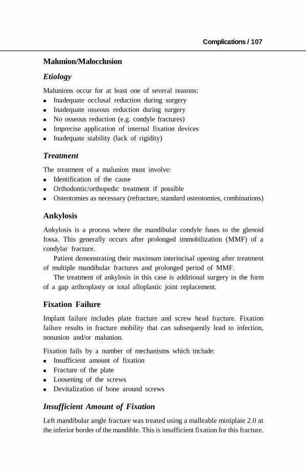
Complications / 107
Malunion/Malocclusion
Etiology
Malunions occur for at least one of several reasons: Inadequate occlusal reduction during surgery Inadequate osseous reduction during surgery No osseous reduction (e.g. condyle fractures) Imprecise application of internal fixation devices Inadequate stability (lack of rigidity)
Treatment
The treatment of a malunion must involve: Identification of the cause Orthodontic/orthopedic treatment if possible Osteotomies as necessary (refracture, standard osteotomies, combinations)
Ankylosis
Ankylosis is a process where the mandibular condyle fuses to the glenoidfossa. This generally occurs after prolonged immobilization (MMF) of acondylar fracture.
Patient demonstrating their maximum interincisal opening after treatmentof multiple mandibular fractures and prolonged period of MMF.
The treatment of ankylosis in this case is additional surgery in the formof a gap arthroplasty or total alloplastic joint replacement.
Fixation Failure
Implant failure includes plate fracture and screw head fracture. Fixationfailure results in fracture mobility that can subsequently lead to infection,nonunion and/or malunion.
Fixation fails by a number of mechanisms which include: Insufficient amount of fixation Fracture of the plate Loosening of the screws Devitalization of bone around screws
Insufficient Amount of Fixation
Left mandibular angle fracture was treated using a malleable miniplate 2.0 atthe inferior border of the mandible. This is insufficient fixation for this fracture.
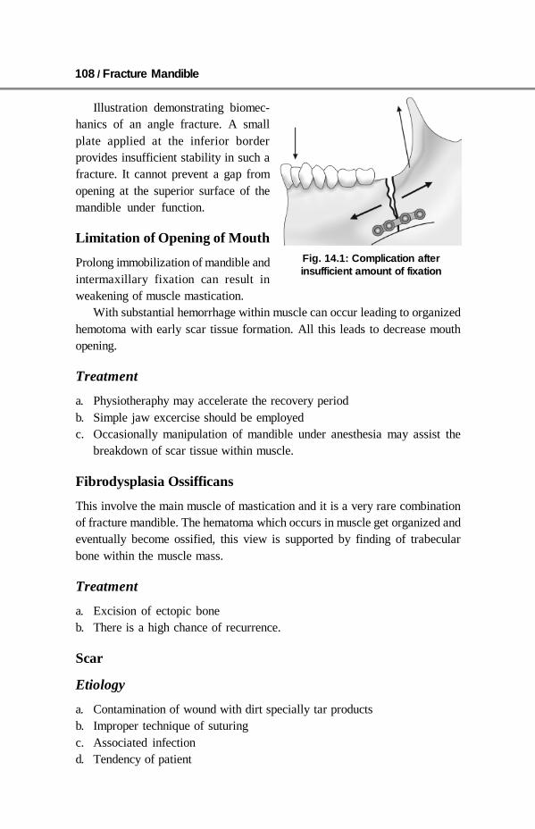
108 / Fracture Mandible
Illustration demonstrating biomec-hanics of an angle fracture. A smallplate applied at the inferior borderprovides insufficient stability in such afracture. It cannot prevent a gap fromopening at the superior surface of themandible under function.
Limitation of Opening of Mouth
Prolong immobilization of mandible andintermaxillary fixation can result inweakening of muscle mastication.
With substantial hemorrhage within muscle can occur leading to organizedhemotoma with early scar tissue formation. All this leads to decrease mouthopening.
Treatment
a. Physiotheraphy may accelerate the recovery periodb. Simple jaw excercise should be employedc. Occasionally manipulation of mandible under anesthesia may assist the
breakdown of scar tissue within muscle.
Fibrodysplasia Ossifficans
This involve the main muscle of mastication and it is a very rare combinationof fracture mandible. The hematoma which occurs in muscle get organized andeventually become ossified, this view is supported by finding of trabecularbone within the muscle mass.
Treatment
a. Excision of ectopic boneb. There is a high chance of recurrence.
Scar
Etiology
a. Contamination of wound with dirt specially tar productsb. Improper technique of suturingc. Associated infectiond. Tendency of patient
Fig. 14.1: Complication afterinsufficient amount of fixation

Complications / 109
Treatment
a. Wait and watch for 1st year as they may soften and fed awayb. Massage of the scarc. Pressure bandaged. Application of lanolinee. Infilteration of injection kenacort and hylinesef. Surgical revision if possible
SUMMARY
Proper reduction, stable and appropriate fixation with prevention of infectioncan prevent most of the complication of fracture mandible.
In cases of edentulous patient, grossly contaminated fractures, poornutritional condition of the patient, complication like malunion, non-union should be kept in mind.
All devitalized structures, infected tissue, loose plates and srews should beremoved and replaced by appropriate ones.
Index / 111
Index
Page numbers followed by f refer to figure
AAccessory muscles of mastication 16Acrylic splints 47Airway maintenance 39Alkayat-Bramley preauricular incision 90Angle of mandible 13Ankylosis 107Anterosuperior alveolar nerve block 66AO classification of mandibular fractures 24Arch bars 44Avascular necrosis 105
BBasal triangle fracture of parasymphysis
area 78fBilateral condylar fractures 32Biomechanics of mandible 10, 11fBlood
loss 39vessels 17
Blunt dissection of parotid gland 87Bonded modified orthodontic brackets 47Buccal nerve block 71Buccinator muscle 93, 95
CCarotid injury 104Categories of
associated fractures 25fractures 25occlusion 25soft tissue involvement 25
Champy’s line 74fof osteosynthesis 74
Closed reduction 74Combination with transbuccal technique 94Comminuted fracture 22, 22f, 33, 52
of angle and body 78fof parasymphysis 77f
Complex symphysis 77Complication after insufficient amount of
fixation 108f
Compound fracture 22, 22fCompression plating 49
technique 50Computed tomographic scan 37Condylar fractures 32Conscious cooperative patient 102Contact healing 9Control of infection 41Conventional
plate system 58, 59screws 57
Countersink near cortex 54
DDental terminology 4fDentoalveolar fractures 31Determine screw length 53, 53fDifferent levels of force distribution 60Dissect over condylar fragment 96Dissection 84, 87, 88
of joint capsule 89Dynamic compression plate 48, 50f
EEccentric drilling for compression 50Eyelet method 46
of fixation 46f
FFace lift incision 88, 89fFacial nerve injury 104Factors affecting bone healing 10Feeding 101Fibrodysplasia ossifficans 108Fixation
failure 107in deciduous and mixed dentition period
98independent of teeth 98utilizing teeth 99
112 / Fracture Mandible
Fracture 18angle of mandible 26condyle 26displacement 26in canine region 26of angle 33
and ramus 77of body 34
of mandible 26of coronoid process 27, 33, 81of head of condyle 79, 80of mandible 20of ramus 33
of mandible 27of symphysis 27
and parasymphysis 34site exposure 92
Frequency of fracture 19
GGap healing 9Gilmer method 46
of fixation 46fGrades of severity 25Greater palatine
block 69nerve block 68
Greenstick fracture 22, 22f
HHead entering medullary space 54fHealing of fracture 7f, 8f
bone 9Hemorrhage 104Hindrance in growth potential 98Horizontally
favorable fractures 23, 23funfavorable fractures 23, 23f
IIdeal
lines of osteosynthesis 60load-sharing situation 61
Incising temporalis fascia 89Inferior alveolar nerve block 70Infra-alveolar nerve block 70Infraorbital
block 68nerve block 66, 68
Insertion ofendoscope 96optical retractor 96
Insufficient amount of fixation 107Intermaxillary fixation 44
screw technique 47, 47fIntermediate load-sharing situation 61Intraoral incison for symphysis and body
fracture 91f
LLag
screw 51principle 51fand technique 51
technique 51, 52Limitation of opening of mouth 108Load-bearing osteosynthesis 59, 61f, 78fLoad-sharing osteosynthesis 59, 78fLocal
anesthesia 64examination of mandibular fracture 29
Location of mental foramen 72Locking
head screws 57plate 54
system 58, 59reconstruction plate 55, 56f
Lower jaw infiltration 70
MMalreduction of fracture 60fMandibular
condyle fracture 79muscles 14
Mental nerve block 72Mentalis muscle dissection 92Middle superior alveolar nerve block 66,
67Minihole plate 75fMiniplate
at external oblique line 78fosteosynthesis 74
Mobilization of temporomandibular joint103
Mucosal incision 91for angle fracture 92ffor condyle and ramus fracture 94f
Multiple and comminuted fractures 34

Index / 113
Muscles offacial expression 14mastication 14
NNeck of condyle 13Nerves 16Neurovascular structures 85, 88, 91Nonrigid osteosynthesis 48
OOff-angle drilling 51fOpen reduction 81Option for off-angle drilling 51Osteitis 105Osteomyelitis 105
PParasymphysis fracture 76, 77Pathological fractures 18Period of mobilization 43Phases of fracture healing 6Posterosuperior alveolar nerve block 65, 66Prevention of infection 101Primary
bone healing 9loss of reduction 58
RRemoval of fixation 102Rigid osteosynthesis 48Rowe modification 90
SScrew
driver and screw 75finsertion 53f, 54
Secondary loss of reduction 59Semirigid osteosynthesis 48Sensory buccal nerve 93, 95Sign and symptoms of mandibular
fractures 31Simple fracture 21, 21fSingle fracture 33Skin incision 85, 88
Soft tissue lacerations 40Subcondylar fractures 79Submental extension 84Subperiosteal dissection of mandibular
ramus 87, 88Support of bone fragments 40Surgical
dressing 97flap dissection 91
Symphysisfracture 76of mandible 13
TTemporomandibular joint 17Threaded plate hole and threaded screw
head 57fThree-dimensional CT scan 38Tranparotid approach skin incision 86fTreatment of infected fractures 105Triangular base fracture at angle of
mandible 78fType of fixation 43f
UUnerupted tooth 99Unilateral condylar fractures 32Universal fracture plate 59Use of existing lacerations 97
VVariations in incision 83fVarious
site of fracture 20fsubmandibular incisions 84f
Vascular supply of mandible 16Vertically
favorable fractures 23, 23funfavorable fracture 24, 24f
Vestibular incision 91-93
WWiring techniques 46Wound closure 84, 88, 89, 92, 96, 97
of vestibular incision 94using envelope flap 94