four-arm single docking full robotic surgery for low ... · four-arm single docking full robotic...
TRANSCRIPT
216
Rev. Col. Bras. Cir. 2014; 41(3): 216-223
RamosRamosRamosRamosRamosFour-arm single docking full robotic surgery for low rectal cancer: technique standardizationThechnical NoteThechnical NoteThechnical NoteThechnical NoteThechnical Note
Four-arm single docking full robotic surgery for low rectal cancer:Four-arm single docking full robotic surgery for low rectal cancer:Four-arm single docking full robotic surgery for low rectal cancer:Four-arm single docking full robotic surgery for low rectal cancer:Four-arm single docking full robotic surgery for low rectal cancer:technique standardizationtechnique standardizationtechnique standardizationtechnique standardizationtechnique standardization
Cirurgia robótica para o tratamento do câncer do reto distal: sistematizaçãoCirurgia robótica para o tratamento do câncer do reto distal: sistematizaçãoCirurgia robótica para o tratamento do câncer do reto distal: sistematizaçãoCirurgia robótica para o tratamento do câncer do reto distal: sistematizaçãoCirurgia robótica para o tratamento do câncer do reto distal: sistematizaçãotécnicatécnicatécnicatécnicatécnica
TCBC JOSÉ REINAN RAMOS1; EDUARDO PARRA-DAVILA2
ABSTRACTABSTRACTABSTRACTABSTRACTABSTRACT
The authors present the four-arm single docking full robotic surgery to treat low rectal cancer. The eight main operative
steps are: 1- patient positioning; 2- trocars set-up and robot docking; 3- sigmoid colon, left colon and splenic flexure
mobilization (lateral-to-medial approach); 4-Inferior mesenteric artery and vein ligation (medial-to-lateral approach); 5-
total mesorectum excision and preservation of hypogastric and pelvic autonomic nerves (sacral dissection, lateral dissection,
pelvic dissection); 6- division of the rectum using an endo roticulator stapler for the laparoscopic performance of a double-
stapled coloanal anastomosis (type I tumor); 7- intersphincteric resection, extraction of the specimen through the anus and
lateral-to-end hand sewn coloanal anastomosis (type II tumor); 8- cylindric abdominoperineal resection, with transabdominal
section of the levator muscles (type IV tumor). The techniques employed were safe and have presented low rates of
complication and no mortality.
Key words:Key words:Key words:Key words:Key words: Rectal neoplasms. Treatment. Surgery. Techniques. Robotics.
1. Robotic Surgery Service, Hospital Samaritano, Rio de Janeiro, RJ, Brazil; 2. Medical Director for Colorectal Surgery, Florida Hospital CelebrationHealth, Orlando, USA.This technical note has a video, available at www.cbc.org.br
INTRODUCTIONINTRODUCTIONINTRODUCTIONINTRODUCTIONINTRODUCTION
The general consensus is still that the majority of adenocarcinomas of the rectum located in its distal
portion (< 5cm from the anal verge) are to be treated byabdominoperineal resection (APR) of the rectum 1.However, with better knowledge of the importance of thecircumferential resection margin, total mesorectal excision(TME) 2, currently joined by the routine use of neoadjuvantchemoradiotherapy (QRT) 3, new operative techniques withsphincter preservation have risen. The individualization ofthe best operation, which is facilitated by examination ofpelvic magnetic resonance imaging (MRI) 4, was advocated5 with the proposition of a classification divided into fourtumor types (supra-anal, juxta-anal, intra-anal and transanal)and, respectively, four types of operations (ultra-low ante-rior resection, partial intersphincteric resection (IR), total IRand APR). Han et al. proposed the extra-elevator or cylindricalcustomization of abdominoperineal resection of the rectum,suggesting that the extent of resection be made inaccordance with the invasion of the elevator muscles ofthe anus 6.
Randomized 7-9 and nonrandomized 10-12 trialsconfirmed the benefits of laparoscopic operation in thetreatment of rectal cancer. Nevertheless, due to the long
learning curve and high conversion rate, the global impactof the use of this method is still small, especially in obeseand male patients. It is estimated that only 10% of casesof colorectal cancer are currently treated by laparoscopy.The use of the robotics platform as a minimally invasiveaccess has gained much interest in the surgical area ofrectal cancer worldwide 13,14. The robotic system enhancesvisualization, exposure and dissection of the noble structuresin a narrow space such as the pelvic cavity.
The purpose of this publication is to present thefull technical aspects of robotic techniques for: the ultra-low anterior resection of the rectum with coloanalanastomosis by double stapling; the intersphinctericresection with manual coloanal anastomosis; the extra-elevator or cylindrical abdominoperineal resection of therectum in the modified lithotomy position.
SURGICAL TECHNIQUESURGICAL TECHNIQUESURGICAL TECHNIQUESURGICAL TECHNIQUESURGICAL TECHNIQUE
1 - Starting position of the patient1 - Starting position of the patient1 - Starting position of the patient1 - Starting position of the patient1 - Starting position of the patientAfter general anesthesia, the patient is placed in
modified lithotomy position (Lloyd-Davis) with arms alongthe trunk, and an oro or nasogastric catheter and a bladdercatheter are inserted. The correct and safe patient position
RamosRamosRamosRamosRamosFour-arm single docking full robotic surgery for low rectal cancer: technique standardization 217
Rev. Col. Bras. Cir. 2014; 41(3): 216-223
is facilitated by the use of Dan Allen leggings and thevacuum plaid (Gel bean bag).
2 - Positioning of the trocars2 - Positioning of the trocars2 - Positioning of the trocars2 - Positioning of the trocars2 - Positioning of the trocarsAfter the umbilical Veress needle puncture and
instillation of CO2, the intra-abdominal pressure is
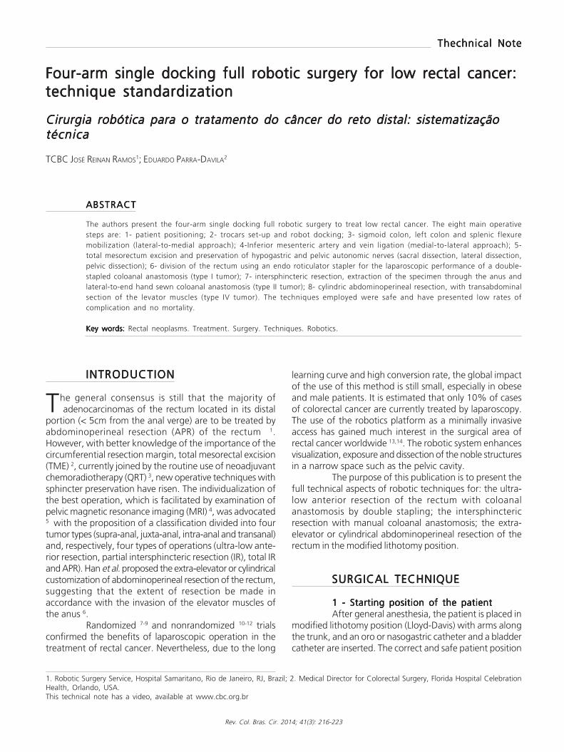
maintained between 10-13mmHg. A short 5-12mm tro-car is inserted on the right flank, where a 30º laparoscopeis used for inspection of the abdominal cavity and insertionof the four other trocars (three 8mm, robotic, permanenttrocars and one 5-12mm long disposable trocar for thecamera) under internal vision. The robotic trocars arepositioned at a distance of 8-10cm from each other in ahalf moon shape (Figure 1). The patient in thenrepositioned to a 20-30° steep Trendelenburg position and10 20º to the right side to properly expose thepromontorium, the bifurcation of the aorta and the inferi-or mesenteric vessels.
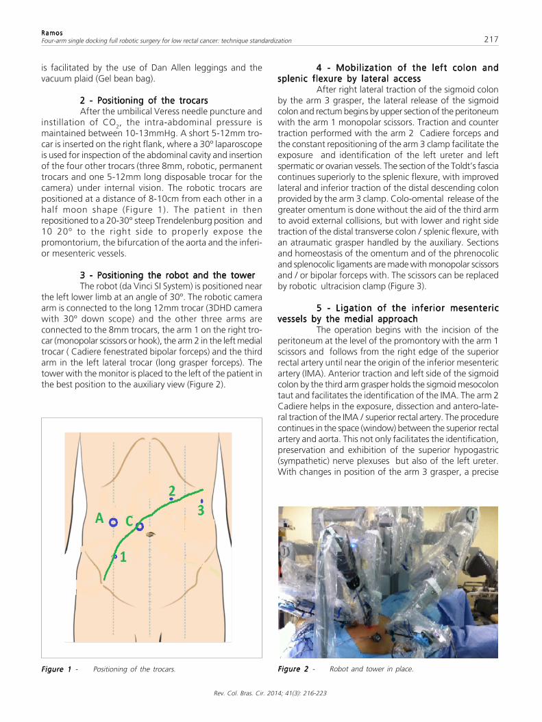
3 - Positioning the robot and the tower3 - Positioning the robot and the tower3 - Positioning the robot and the tower3 - Positioning the robot and the tower3 - Positioning the robot and the towerThe robot (da Vinci SI System) is positioned near
the left lower limb at an angle of 30º. The robotic cameraarm is connected to the long 12mm trocar (3DHD camerawith 30º down scope) and the other three arms areconnected to the 8mm trocars, the arm 1 on the right tro-car (monopolar scissors or hook), the arm 2 in the left medialtrocar ( Cadiere fenestrated bipolar forceps) and the thirdarm in the left lateral trocar (long grasper forceps). Thetower with the monitor is placed to the left of the patient inthe best position to the auxiliary view (Figure 2).
4 - Mobilization of the left colon and4 - Mobilization of the left colon and4 - Mobilization of the left colon and4 - Mobilization of the left colon and4 - Mobilization of the left colon andsplenic flexure by lateral accesssplenic flexure by lateral accesssplenic flexure by lateral accesssplenic flexure by lateral accesssplenic flexure by lateral access
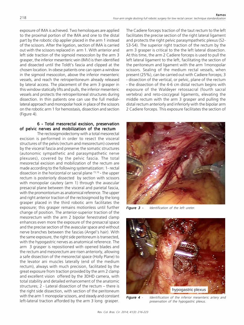
After right lateral traction of the sigmoid colonby the arm 3 grasper, the lateral release of the sigmoidcolon and rectum begins by upper section of the peritoneumwith the arm 1 monopolar scissors. Traction and countertraction performed with the arm 2 Cadiere forceps andthe constant repositioning of the arm 3 clamp facilitate theexposure and identification of the left ureter and leftspermatic or ovarian vessels. The section of the Toldt’s fasciacontinues superiorly to the splenic flexure, with improvedlateral and inferior traction of the distal descending colonprovided by the arm 3 clamp. Colo-omental release of thegreater omentum is done without the aid of the third armto avoid external collisions, but with lower and right sidetraction of the distal transverse colon / splenic flexure, withan atraumatic grasper handled by the auxiliary. Sectionsand homeostasis of the omentum and of the phrenocolicand splenocolic ligaments are made with monopolar scissorsand / or bipolar forceps with. The scissors can be replacedby robotic ultracision clamp (Figure 3).
5 - Ligation of the inferior mesenteric5 - Ligation of the inferior mesenteric5 - Ligation of the inferior mesenteric5 - Ligation of the inferior mesenteric5 - Ligation of the inferior mesentericvessels by the medial approachvessels by the medial approachvessels by the medial approachvessels by the medial approachvessels by the medial approach
The operation begins with the incision of theperitoneum at the level of the promontory with the arm 1scissors and follows from the right edge of the superiorrectal artery until near the origin of the inferior mesentericartery (IMA). Anterior traction and left side of the sigmoidcolon by the third arm grasper holds the sigmoid mesocolontaut and facilitates the identification of the IMA. The arm 2Cadiere helps in the exposure, dissection and antero-late-ral traction of the IMA / superior rectal artery. The procedurecontinues in the space (window) between the superior rectalartery and aorta. This not only facilitates the identification,preservation and exhibition of the superior hypogastric(sympathetic) nerve plexuses but also of the left ureter.With changes in position of the arm 3 grasper, a precise
Figure 1Figure 1Figure 1Figure 1Figure 1 - Positioning of the trocars. Figure 2 Figure 2 Figure 2 Figure 2 Figure 2 - Robot and tower in place.
218
Rev. Col. Bras. Cir. 2014; 41(3): 216-223
RamosRamosRamosRamosRamosFour-arm single docking full robotic surgery for low rectal cancer: technique standardization
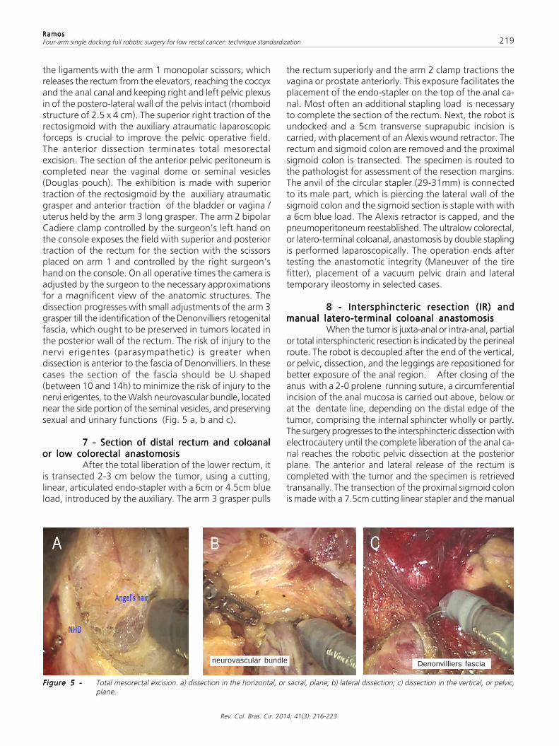
exposure of IMA is achieved. Two hemoloques are appliedto the proximal portion of the IMA and one to the distalpart by the robotic clip applier placed in the arm 1 insteadof the scissors. After the ligation, section of IMA is carriedout with the scissors replaced in arm 1. With anterior andleft side traction of the sigmoid mesocolon by the arm 3grasper, the inferior mesenteric vein (IMV) is then identifiedand dissected until the Toldt’s fascia and clipped at thechosen location. In obese patients one can open a windowin the sigmoid mesocolon, above the inferior mesentericvessels, and reach the retroperitoneum already releasedby lateral access. The placement of the arm 3 grasper inthis window statically lifts and pulls, the inferior mesentericvessels and protects the retroperitoneal structures duringdissection. In thin patients one can use the full medial-lateral approach and monopolar hook in place of the scissorson the robotic arm 1 for hemostasis, dissection and section(Figure 4).
6 - Total mesorectal excision, preservation6 - Total mesorectal excision, preservation6 - Total mesorectal excision, preservation6 - Total mesorectal excision, preservation6 - Total mesorectal excision, preservationof pelvic nerves and mobilization of the rectumof pelvic nerves and mobilization of the rectumof pelvic nerves and mobilization of the rectumof pelvic nerves and mobilization of the rectumof pelvic nerves and mobilization of the rectum
The rectosigmoidectomy with a total mesorectalexcision is performed in order to resect the visceralstructures of the pelvis (rectum and mesorectum) coveredby the visceral fascia and preserve the somatic structures(autonomic sympathetic and parasympathetic nerveplexuses), covered by the pelvic fascia. The totalmesorectal excision and mobilization of the rectum aremade according to the following systematization: 1- rectaldissection in the horizontal or sacral plane 15,16 - the upperrectum is posteriorly dissected by section with scissorswith monopolar cautery (arm 1) through the avascularpresacral plane between the visceral and parietal fascia,with the promontorium as anatomical reference. The upperand right anterior traction of the rectosigmoid by the longgrasper placed in the third robotic arm facilitates theexposure; this grasper remains motionless until furtherchange of position. The anterior-superior traction of themesorectum with the arm 2 bipolar fenestrated clampenhances even more the exposure of the presacral spaceand the precise section of the avascular space and withoutnerve branches between the fascias (Angel’s hair). Withthe same exposure, the right side peritoneum is transected,with the hypogastric nerves as anatomical reference. Thearm 3 grasper is repositioned with opened blades andthe rectum and mesorectum are risen anteriorly, allowinga safe dissection of the mesorectal space (Holly Plane) tothe levator ani muscles laterally (end of the mediumrectum), always with much precision, facilitated by thegreat exposure from traction provided by the arm 2 clampand excellent vision offered by the 3DHD camera, withtotal stability and detailed enhancement of the anatomicstructures; 2 - Lateral dissection of the rectum – there isthe right side dissection, with section of the peritoneumwith the arm 1 monopolar scissors, and steady and constantleft-lateral traction afforded by the arm 3 long grasper.
The Cadiere forceps traction of the taut rectum to the leftfacilitates the precise section of the right lateral ligamentand protects the right pelvic parasympathetic plexus (S2-S3-S4). The superior right traction of the rectum by thearm 3 grasper is critical to the the left lateral dissection.At this time, the arm 2 Cadiere forceps is used to pull theleft lateral ligament to the left, facilitating the section ofthe peritoneum and ligament with the arm 1monopolarscissors. Sealing of the medium rectal vessels, whenpresent (25%), can be carried out with Cadiere forceps; 3- dissection of the vertical, or pelvic, plane of the rectum- the dissection of the 4-6 cm distal rectum begins withexposure of the Waldeyer retossacral (fourth sacralvertebra) and reto-coccygeal ligaments, elevating themiddle rectum with the arm 3 grasper and pulling thedistal rectum anteriorly and inferiorly with the bipolar arm2 Cadiere forceps. This exposure facilitates the section of
Figure 3 -Figure 3 -Figure 3 -Figure 3 -Figure 3 - Identification of the left ureter.
Figure 4 Figure 4 Figure 4 Figure 4 Figure 4 - Identification of the inferior mesenteric artery andpreservation of the hypogastric plexus.
hypogastric plexus
RamosRamosRamosRamosRamosFour-arm single docking full robotic surgery for low rectal cancer: technique standardization 219
Rev. Col. Bras. Cir. 2014; 41(3): 216-223
the ligaments with the arm 1 monopolar scissors, whichreleases the rectum from the elevators, reaching the coccyxand the anal canal and keeping right and left pelvic plexusin of the postero-lateral wall of the pelvis intact (rhomboidstructure of 2.5 x 4 cm). The superior right traction of therectosigmoid with the auxiliary atraumatic laparoscopicforceps is crucial to improve the pelvic operative field.The anterior dissection terminates total mesorectalexcision. The section of the anterior pelvic peritoneum iscompleted near the vaginal dome or seminal vesicles(Douglas pouch). The exhibition is made with superiortraction of the rectosigmoid by the auxiliary atraumaticgrasper and anterior traction of the bladder or vagina /uterus held by the arm 3 long grasper. The arm 2 bipolarCadiere clamp controlled by the surgeon’s left hand onthe console exposes the field with superior and posteriortraction of the rectum for the section with the scissorsplaced on arm 1 and controlled by the right surgeon’shand on the console. On all operative times the camera isadjusted by the surgeon to the necessary approximationsfor a magnificent view of the anatomic structures. Thedissection progresses with small adjustments of the arm 3grasper till the identification of the Denonvilliers retogenitalfascia, which ought to be preserved in tumors located inthe posterior wall of the rectum. The risk of injury to thenervi erigentes (parasympathetic) is greater whendissection is anterior to the fascia of Denonvilliers. In thesecases the section of the fascia should be U shaped(between 10 and 14h) to minimize the risk of injury to thenervi erigentes, to the Walsh neurovascular bundle, locatednear the side portion of the seminal vesicles, and preservingsexual and urinary functions (Fig. 5 a, b and c).
7 - Section of distal rectum and coloanal7 - Section of distal rectum and coloanal7 - Section of distal rectum and coloanal7 - Section of distal rectum and coloanal7 - Section of distal rectum and coloanalor low colorectal anastomosisor low colorectal anastomosisor low colorectal anastomosisor low colorectal anastomosisor low colorectal anastomosis
After the total liberation of the lower rectum, itis transected 2-3 cm below the tumor, using a cutting,linear, articulated endo-stapler with a 6cm or 4.5cm blueload, introduced by the auxiliary. The arm 3 grasper pulls
the rectum superiorly and the arm 2 clamp tractions thevagina or prostate anteriorly. This exposure facilitates theplacement of the endo-stapler on the top of the anal ca-nal. Most often an additional stapling load is necessaryto complete the section of the rectum. Next, the robot isundocked and a 5cm transverse suprapubic incision iscarried, with placement of an Alexis wound retractor. Therectum and sigmoid colon are removed and the proximalsigmoid colon is transected. The specimen is routed tothe pathologist for assessment of the resection margins.The anvil of the circular stapler (29-31mm) is connectedto its male part, which is piercing the lateral wall of thesigmoid colon and the sigmoid section is staple with witha 6cm blue load. The Alexis retractor is capped, and thepneumoperitoneum reestablished. The ultralow colorectal,or latero-terminal coloanal, anastomosis by double staplingis performed laparoscopically. The operation ends aftertesting the anastomotic integrity (Maneuver of the tirefitter), placement of a vacuum pelvic drain and lateraltemporary ileostomy in selected cases.
8 - Intersphincteric resection (IR) and8 - Intersphincteric resection (IR) and8 - Intersphincteric resection (IR) and8 - Intersphincteric resection (IR) and8 - Intersphincteric resection (IR) andmanual latero-terminal coloanal anastomosismanual latero-terminal coloanal anastomosismanual latero-terminal coloanal anastomosismanual latero-terminal coloanal anastomosismanual latero-terminal coloanal anastomosis
When the tumor is juxta-anal or intra-anal, partialor total intersphincteric resection is indicated by the perinealroute. The robot is decoupled after the end of the vertical,or pelvic, dissection, and the leggings are repositioned forbetter exposure of the anal region. After closing of theanus with a 2-0 prolene running suture, a circumferentialincision of the anal mucosa is carried out above, below orat the dentate line, depending on the distal edge of thetumor, comprising the internal sphincter wholly or partly.The surgery progresses to the intersphincteric dissection withelectrocautery until the complete liberation of the anal ca-nal reaches the robotic pelvic dissection at the posteriorplane. The anterior and lateral release of the rectum iscompleted with the tumor and the specimen is retrievedtransanally. The transection of the proximal sigmoid colonis made with a 7.5cm cutting linear stapler and the manual
Figure 5 -Figure 5 -Figure 5 -Figure 5 -Figure 5 - Total mesorectal excision. a) dissection in the horizontal, or sacral, plane; b) lateral dissection; c) dissection in the vertical, or pelvic,plane.
neurovascular bundle Denonvilliers fascia
220
Rev. Col. Bras. Cir. 2014; 41(3): 216-223
RamosRamosRamosRamosRamosFour-arm single docking full robotic surgery for low rectal cancer: technique standardization
coloanal latero-terminal anastomosis is held in a single layerwith 2-0 or 3-0 polyglactin sutures. The operation ends afterpelvic drain placement , the performance of a laterallaparoscopic ileostomy and closure of the two puncturesites.
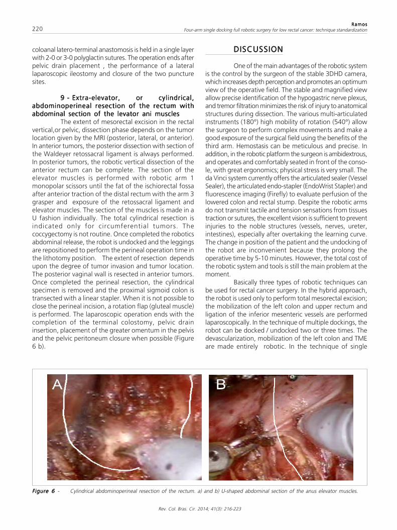
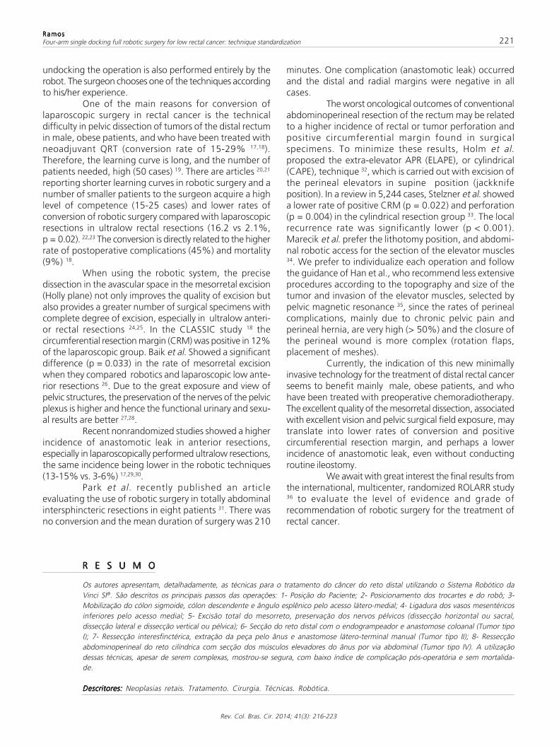
9 - Extra-elevator, or cylindrical,9 - Extra-elevator, or cylindrical,9 - Extra-elevator, or cylindrical,9 - Extra-elevator, or cylindrical,9 - Extra-elevator, or cylindrical,abdominoperineal resection of the rectum withabdominoperineal resection of the rectum withabdominoperineal resection of the rectum withabdominoperineal resection of the rectum withabdominoperineal resection of the rectum withabdominal section of the levator ani musclesabdominal section of the levator ani musclesabdominal section of the levator ani musclesabdominal section of the levator ani musclesabdominal section of the levator ani muscles
The extent of mesorectal excision in the rectalvertical,or pelvic, dissection phase depends on the tumorlocation given by the MRI (posterior, lateral, or anterior).In anterior tumors, the posterior dissection with section ofthe Waldeyer retossacral ligament is always performed.In posterior tumors, the robotic vertical dissection of theanterior rectum can be complete. The section of theelevator muscles is performed with robotic arm 1monopolar scissors until the fat of the ischiorectal fossaafter anterior traction of the distal rectum with the arm 3grasper and exposure of the retossacral ligament andelevator muscles. The section of the muscles is made in aU fashion individually. The total cylindrical resection isindicated only for circumferential tumors. Thecoccygectomy is not routine. Once completed the roboticsabdominal release, the robot is undocked and the leggingsare repositioned to perform the perineal operation time inthe lithotomy position. The extent of resection dependsupon the degree of tumor invasion and tumor location.The posterior vaginal wall is resected in anterior tumors.Once completed the perineal resection, the cylindricalspecimen is removed and the proximal sigmoid colon istransected with a linear stapler. When it is not possible toclose the perineal incision, a rotation flap (gluteal muscle)is performed. The laparoscopic operation ends with thecompletion of the terminal colostomy, pelvic draininsertion, placement of the greater omentum in the pelvisand the pelvic peritoneum closure when possible (Figure6 b).
DISCUSSIONDISCUSSIONDISCUSSIONDISCUSSIONDISCUSSION
One of the main advantages of the robotic systemis the control by the surgeon of the stable 3DHD camera,which increases depth perception and promotes an optimumview of the operative field. The stable and magnified viewallow precise identification of the hypogastric nerve plexus,and tremor filtration minimizes the risk of injury to anatomicalstructures during dissection. The various multi-articulatedinstruments (180°) high mobility of rotation (540°) allowthe surgeon to perform complex movements and make agood exposure of the surgical field using the benefits of thethird arm. Hemostasis can be meticulous and precise. Inaddition, in the robotic platform the surgeon is ambidextrous,and operates and comfortably seated in front of the conso-le, with great ergonomics; physical stress is very small. Theda Vinci system currently offers the articulated sealer (VesselSealer), the articulated endo-stapler (EndoWrist Stapler) andfluorescence imaging (Firefly) to evaluate perfusion of thelowered colon and rectal stump. Despite the robotic armsdo not transmit tactile and tension sensations from tissuestraction or sutures, the excellent vision is sufficient to preventinjuries to the noble structures (vessels, nerves, ureter,intestines), especially after overtaking the learning curve.The change in position of the patient and the undocking ofthe robot are inconvenient because they prolong theoperative time by 5-10 minutes. However, the total cost ofthe robotic system and tools is still the main problem at themoment.
Basically three types of robotic techniques canbe used for rectal cancer surgery. In the hybrid approach,the robot is used only to perform total mesorectal excision;the mobilization of the left colon and upper rectum andligation of the inferior mesenteric vessels are performedlaparoscopically. In the technique of multiple dockings, therobot can be docked / undocked two or three times. Thedevascularization, mobilization of the left colon and TMEare made entirely robotic. In the technique of single
Figure 6 Figure 6 Figure 6 Figure 6 Figure 6 - Cylindrical abdominoperineal resection of the rectum. a) and b) U-shaped abdominal section of the anus elevator muscles.
RamosRamosRamosRamosRamosFour-arm single docking full robotic surgery for low rectal cancer: technique standardization 221
Rev. Col. Bras. Cir. 2014; 41(3): 216-223
undocking the operation is also performed entirely by therobot. The surgeon chooses one of the techniques accordingto his/her experience.
One of the main reasons for conversion oflaparoscopic surgery in rectal cancer is the technicaldifficulty in pelvic dissection of tumors of the distal rectumin male, obese patients, and who have been treated withneoadjuvant QRT (conversion rate of 15-29% 17,18).Therefore, the learning curve is long, and the number ofpatients needed, high (50 cases) 19. There are articles 20,21
reporting shorter learning curves in robotic surgery and anumber of smaller patients to the surgeon acquire a highlevel of competence (15-25 cases) and lower rates ofconversion of robotic surgery compared with laparoscopicresections in ultralow rectal resections (16.2 vs 2.1%,p = 0.02). 22,23 The conversion is directly related to the higherrate of postoperative complications (45%) and mortality(9%) 18.
When using the robotic system, the precisedissection in the avascular space in the mesorretal excision(Holly plane) not only improves the quality of excision butalso provides a greater number of surgical specimens withcomplete degree of excision, especially in ultralow anteri-or rectal resections 24,25. In the CLASSIC study 18 thecircumferential resection margin (CRM) was positive in 12%of the laparoscopic group. Baik et al. Showed a significantdifference (p = 0.033) in the rate of mesorretal excisionwhen they compared robotics and laparoscopic low ante-rior resections 26. Due to the great exposure and view ofpelvic structures, the preservation of the nerves of the pelvicplexus is higher and hence the functional urinary and sexu-al results are better 27,28.
Recent nonrandomized studies showed a higherincidence of anastomotic leak in anterior resections,especially in laparoscopically performed ultralow resections,the same incidence being lower in the robotic techniques(13-15% vs. 3-6%) 17,29,30.
Park et al . recently published an articleevaluating the use of robotic surgery in totally abdominalintersphincteric resections in eight patients 31. There wasno conversion and the mean duration of surgery was 210
minutes. One complication (anastomotic leak) occurredand the distal and radial margins were negative in allcases.
The worst oncological outcomes of conventionalabdominoperineal resection of the rectum may be relatedto a higher incidence of rectal or tumor perforation andpositive circumferential margin found in surgicalspecimens. To minimize these results, Holm et al.proposed the extra-elevator APR (ELAPE), or cylindrical(CAPE), technique 32, which is carried out with excision ofthe perineal elevators in supine position (jackknifeposition). In a review in 5,244 cases, Stelzner et al. showeda lower rate of positive CRM (p = 0.022) and perforation(p = 0.004) in the cylindrical resection group 33. The localrecurrence rate was significantly lower (p < 0.001).Marecik et al. prefer the lithotomy position, and abdomi-nal robotic access for the section of the elevator muscles34. We prefer to individualize each operation and followthe guidance of Han et al., who recommend less extensiveprocedures according to the topography and size of thetumor and invasion of the elevator muscles, selected bypelvic magnetic resonance 35, since the rates of perinealcomplications, mainly due to chronic pelvic pain andperineal hernia, are very high (> 50%) and the closure ofthe perineal wound is more complex (rotation flaps,placement of meshes).
Currently, the indication of this new minimallyinvasive technology for the treatment of distal rectal cancerseems to benefit mainly male, obese patients, and whohave been treated with preoperative chemoradiotherapy.The excellent quality of the mesorretal dissection, associatedwith excellent vision and pelvic surgical field exposure, maytranslate into lower rates of conversion and positivecircumferential resection margin, and perhaps a lowerincidence of anastomotic leak, even without conductingroutine ileostomy.
We await with great interest the final results fromthe international, multicenter, randomized ROLARR study36 to evaluate the level of evidence and grade ofrecommendation of robotic surgery for the treatment ofrectal cancer.
R E S U M OR E S U M OR E S U M OR E S U M OR E S U M O
Os autores apresentam, detalhadamente, as técnicas para o tratamento do câncer do reto distal utilizando o Sistema Robótico daVinci SI®. São descritos os principais passos das operações: 1- Posição do Paciente; 2- Posicionamento dos trocartes e do robô; 3-Mobilização do cólon sigmoide, cólon descendente e ângulo esplênico pelo acesso látero-medial; 4- Ligadura dos vasos mesentéricosinferiores pelo acesso medial; 5- Excisão total do mesorreto, preservação dos nervos pélvicos (dissecção horizontal ou sacral,dissecção lateral e dissecção vertical ou pélvica); 6- Secção do reto distal com o endogrampeador e anastomose coloanal (Tumor tipoI); 7- Ressecção interesfinctérica, extração da peça pelo ânus e anastomose látero-terminal manual (Tumor tipo II); 8- Ressecçãoabdominoperineal do reto cilíndrica com secção dos músculos elevadores do ânus por via abdominal (Tumor tipo IV). A utilizaçãodessas técnicas, apesar de serem complexas, mostrou-se segura, com baixo índice de complicação pós-operatória e sem mortalida-de.
Descritores:Descritores:Descritores:Descritores:Descritores: Neoplasias retais. Tratamento. Cirurgia. Técnicas. Robótica.
222
Rev. Col. Bras. Cir. 2014; 41(3): 216-223
RamosRamosRamosRamosRamosFour-arm single docking full robotic surgery for low rectal cancer: technique standardization
REFERENCESREFERENCESREFERENCESREFERENCESREFERENCES
1. Miles EW. A method of performing abdominoperineal excision forcarcinoma of the rectum and of the terminal portion of the pelviccolumn. Lancet. 1908;2:1812-3.
2. MacFarlane JK, Ryall RD, Heald RJ. Mesorectal excision for rectalcancer. Lancet. 1993;341(8843):457-60.
3. Habr-Gama A, Sabbaga J, Gama-Rodrigues J, São Julião GP,Proscurshim I, Bailão Aguilar P, et al. Watch and wait approachfollowing extended neoadjuvant chemoradiation for distal rectalcancer: are we getting closer to anal cancer management? DisColon Rectum. 2013;56(10):1109-17.
4. Nougaret S, Reinhold C, Mikhael HW, Rouanet P, Bibeau F, BrownG. The use of MR imaging in treatment planning for patients withrectal carcinoma: have you checked the “DISTANCE”? Radiology.2013:268(2):330-44.
5. Rullier E, Denost Q, Vendrely V, Rullier A, Laurent C . Low rectalcancer: classification and standardization of surgery. Dis ColonRectum. 2013;56(5):560-7.
6. Han JG, Wang ZJ, Wei GH, Gao ZG, Yang Y, Yi BQ, et al. Technicalimprovements and results of individual cylindrical abdominoperinealresection for locally advanced low rectal cancer. Zhonghua WaiKe Za Zhi. 2013;51(4):335-8.
7. Jayne DG, Thorpe HC, Copeland J, Quirke P, Brown JM, Guillou PJ.Five-year follow-up of the Medical Research Council CLASICC trialof laparoscopically assisted versus open surgery for colorectalcancer. Br J Surg. 2010;97(11):1638-45.
8. Kang SB, Park JW, Jeong SY, Nam BH, Choi HS, Kim DW, et al.Open versus laparoscopic surgery for mid or low rectal cancerafter neoadjuvant chemoradiotherapy (COREAN trial): short-termoutcomes of an open-label randomised controlled trial. LancetOncol. 2010;11(7):637-45.
9. van der Pas MH, Haglind E. Cuesta MA. Fürst A, Lacy AM, HopWC, et al. Laparoscopic versus open surgery for rectal cancer(COLOR II): short-term outcomes of a randomised, phase 3 trial.Lancet Oncol. 2013;14(3):210-8.
10. Greenblatt DY, Rajamanickam V, Pugely AJ, Heise CP, Foley EF,Kennedy GD. Short-term outcomes after laparoscopic-assistedproctectomy for rectal cancer: results from the ACS NSQIP. AmColl Surg. 2011;212(5):844-54.
11. Arezzo A, Passera R, Scozzari G, Verra M, Morino M. Laparoscopyfor rectal cancer reduces short-term mortality and mormidity:results of a systematic review and meta-analysis. Surg Endosc.2013;27(5):1485-502.
12. Ramos JR, Petrosemolo RH, Valory EA, Polania FC, Peçanha R.Abdominoperineal resection: laparoscopic versus conventional.Laparosc Endosc. 1997;7(2):148-52.
13. Halabi WJ, Kang CY, Jafari MD, Nguyen VQ, Camichael JC, Mills S,et al. Robotic-assisted colorectal surgery in the United States: anationwide analysis of trends and outcomes. World J Surg.2013;37(12):2782-90.
14. Zang Y, Wang F, Zhang P, Sou Y, Qin H, Ma Y. Robotic-assistededversus conventional laparoscopic surgery for colorectal disease,focusing on rectal cancer: a meta-analysis. Ann Surg Oncol.2012;19(12):3727-36.
15. Ramos JR. Ressecção anterior ultrabaixa e interesfinctérica doreto com anastomose coloanal por videolaparoscopia. Rev ColBras Cir. 2009;36(5):459-65.
16. Koh PK, Seow-Choen F, Kwek BH. Total mesorectal excision: theunrecognized pelvic plane. Dis Colon Rectum. 2006;49(2):280-3;discussion 283-4.
17. Rottoli M, Bona S, Rosati R, Elmore U, Bianchi PP, Spinelli A, et al.Laparoscopic rectal resection for cancer: effects of conversion onshort-term outcome and survival. Ann Surg Oncol.2009;16(5):1279-86.
18. Guillou PJ, Quirke P, Thorpe H, Walker J, Jayne DG, Smith AM, etal. Short-term endpoints of conventional versus laparoscopic-assisted surgery in patients with colorectal cancer (MRC CLASICC
trial): multicentre, randomised controlled trial. Lancet.2005;365(9472):1718-26.
19. Kayano H, Okuda J, Tanaka K, Kondo K, Tanigawa N. Evaluationof the learning curve in laparoscopic low anterior resection forrectal cancer. Surg Endosc. 2011;25(9):2972-9.
20. Bokhari MB, Patel CB, Ramos-Valadez DI, Ragupathi M, Haas EM.Learning curve for robotic-assisted laparoscopic colorectal surgery.Surg Endosc. 2011;25(3):855-60.
21. Jiménez-Rodríguez RM, Díaz-Pavón JM, de la Portilla de Juan F,Prendes-Sillero E, Dussort HC, Padillo J. Learning curve for robotic-assisted laparoscopic rectal cancer surgery. Int J Colorectal Dis.2013;28(6):815-21.
22. Baek SJ, Al-Asari S, Jeong DH, Hur H, Min BS, Baik SH, et al.Robotic versus laparoscopic coloanal anastomosis with or withoutintersphincteric resection for rectal cancer. Surg Endosc.2013;27(11):4157-63.
23. Yang Y, Wang F, Zhang P, Shi C, Zou Y, Qin H, et al. Robot-assisted versus conventional laparoscopic surgery for colorectaldisease, focusing on rectal cancer: a meta-analysis. Ann Surg Oncol.2012;19(12):3727-36.
24. Baik SH, Kim NK, Lim DR, Hur H, Min BS, Lee KY. Oncologic outcomesand perioperative clinicopathologic results after robotic-assistedtumor-specific mesorectal excision for rectal cancer. Ann SurgOncol. 2013;28(8):2625-32.
25. Kang J, Yoon KJ, Min BS, Hur H, Baik SH, Kim NK, et al. The impactof robotic surgery for mid and low rectal cancer: a case-matchedanalysis of a 3-arm comparison-open, laparoscopic, and roboticsurgery. Ann Surg. 2013;257(1)95-101.
26. Baik SH, Kwon HY, Kim JS, Hur H, Sohn SK, Cho CH, et al. Roboticversus laparoscopic low anterior resection of rectal cancer: short-term outcome of a prospective comparative study. Ann Surg Oncol.2009;16(6):1480-7.
27. Luca F, Valvo M, Ghezzi TL, Zuccaro M, Cenciarelli S, Trovato C, etal. Impact of robotic surgery on sexual and urinary functions afterfully robotic nerve-sparing total mesorectal excision for rectalcancer. Ann Surg. 2013;257(4):672-8.
28. D’Annibale A, Pernazza G, Monsellato I, Pende V, Lucandri G,Mazzocchi P, et al. Total mesorectal excision: a comparison ofoncological and functional outcomes between robotic andlaparoscopic surgery for rectal cancer. Surg Endosc.2013;27(6):1887-95.
29. Zeng DZ, Shi Y, Lei X, Tang B, Hao YX, Luo HX, et al. Short-termefficacy of da Vinci robotic surgical system on rectal cancer in 101patients. Zhonghua Wei Chang Wai Ke Za Zhi. 2013;16(5):451-4.
30. deSouza AL, Prasad LM, Marecik SJ, Blumetti J, Park JJ, ZimmernA, et al. Total mesorectal excision for rectal cancer: the potentialadvantage of robotic assistance. Dis Colon Rectum.2010;53(12):1611-7.
31. Park SY, Choi GS, Park JS, Kim HJ, Choi WH, Ryuk JP. Robotic-assisted transabdominal intersphincteric resection: a techniqueinvolving a completely abdominal approach and coloanalanastomosis. Surg Laparosc Endosc Percutan Tech. 2013;23(1):e5-10.
32. Stelzner S, Koehler C, Stelzner J, Sims A, Witzigmann H. Extendedabdominoperineal excision versus standard abdominoperinealexcision in rectal cancer—a systematic overview. Int J ColorectalDis. 2011;26(10):1227-40.
33. Holm T, Ljung A, Haggmark T, Jurell G, Lagergren J. Extendedabdominoperineal resection with gluteos maximus flapreconstruction of the pelvic floor for rectal cancer. Br J Surg.2007;94(2):232-8.
34. Marecik SJ, Zawadzki M, Desouza AL, Park JJ, Abcarian H, PrasadLM. Robotic cylindrical abdominoperineal resection withtransabdominal levator transection. Dis Colon Rectum.2011;54(10):1320-5.
35. Han JG, Wang ZJ, Wei GH, Gao ZG, Yang Y, Zhao BC. Randomizedclinical trial of conventional versus cylindrical abdominoperinealresection for locally advanced lower rectal cancer. Am J Surg.2012;204(3):274-82.

RamosRamosRamosRamosRamosFour-arm single docking full robotic surgery for low rectal cancer: technique standardization 223
Rev. Col. Bras. Cir. 2014; 41(3): 216-223
36. Collinson FJ, Jayne DG, Pigazzi A, Tsang C, Barrie JM, Edlin R, et al.An international, multicentre, prospective, randomised, controlled,unblinded, parallel-group trial of robotic-assisted versus standardlaparoscopic surgery for the curative treatment of rectal cancer.Int J Colorectal Dis. 2012;27(2):233-41.
Received on 15/08/2013Accepted for publication 18/10/2013Conflict of interest: none.Source of funding: none.
Address for correspondence:Address for correspondence:Address for correspondence:Address for correspondence:Address for correspondence:José Reinan RamosE-mail: [email protected]