forsus
DESCRIPTION
ForsusTRANSCRIPT

Original Article
Treatment effects of the Forsus Fatigue Resistant Device used with
miniscrew anchorage
Belma I. Aslana; Ebru Kucukkaracaa; Cagri Turkozb; Mufide Dincerc
ABSTRACTObjective: To evaluate the dentofacial effects of the Forsus Fatigue Resistant Device (FRD) usedwith miniscrew anchorage (FRDMS) and compare them with those of conventional FRD and anuntreated Class II control group.Materials and Methods: The sample consisted of 48 Class II subjects. Sixteen patients (13.68 6
1.09 years of age) were treated with FRDMS, whereas 17 subjects (14.64 6 1.56 years of age)were treated with only FRD. Also, a control sample of 15 untreated Class II subjects (14.13 6
1.50 years of age) was constructed. Angular and linear measurements were made on 96 lateralcephalograms. Paired t, one-way analysis of variance, and Tukey tests were used for statisticalanalysis.Results: Class I molar relationship and overjet correction were achieved in an average period of6.5 6 1.97 and 5.5 6 1.80 months in the FRDMS and FRD groups, respectively. No skeletal effectwas determined in both treatment groups. Greater overbite correction was found in the FRD group.Retrusion and extrusion of maxillary incisors, distalization of maxillary molars, and extrusion ofmandibular molars were significant in both treatment groups. Labial tipping of mandibular incisorswas significantly greater in the FRD group than in the FRDMS group.Conclusion: Overjet and molar correction was totally dentoalveolar. Unfavorable labial tipping ofmandibular incisors was effectively minimized with the usage of miniscrews. (Angle Orthod.2014;84:76–87.)
KEY WORDS: Forsus; Mini-screw; Fixed functional appliance; Class II correction
INTRODUCTION
Several removable or fixed functional appliances areused for treatment of Class II division 1 malocclusionswith mandibular deficiency in order to stimulatemandibular growth by forward positioning of the man-dible. Unlike removable functional appliances, fixedfunctional devices have the advantage of not requiringpatient compliance, and they can also be usedconcurrently with brackets.1
Fixed functional devices are categorized as rigid(Herbst, MARA) and semirigid (Jasper Jumper andForsus Fatigue Resistance Device[FRD]) fixed inter-arch appliances.2–4 The Forsus FRD (3M Unitek Corp,Monrovia, Calif) is a semirigid fixed functional appli-ance that was developed to overcome breakageproblems seen with the Jasper Jumper. The FRD isa three-piece, telescoping system incorporating asuperelastic nickel-titanium coil spring that is easy toinstall and thus saves chair time.5 Distal and intrusivemovement of maxillary molars, mesial movement ofmandibular molars, retrusion of maxillary incisors,labial tipping of mandibular incisors, and varyingamounts of skeletal effects have been reported inprevious studies with this appliance.4–10
Data from the literature demonstrates the increasinguse and popularity of miniscrews in orthodonticpractice for mesiodistal and intrusion movements ofteeth.11 However, there are no studies where minis-crews have been used with functional appliances.
Like other fixed functional appliances, an unfavorableeffect of the FRD is flaring of the mandibular anteriors,which limits the skeletal effects of the appliance.4,8,10
a Research Assistant, Gazi University, Faculty of Dentistry,Department of Orthodontics, Ankara, Turkey.
b Assistant Professor, Gazi University, Faculty of Dentistry,Department of Orthodontics, Ankara, Turkey.
c Professor, Gazi University, Faculty of Dentistry, Departmentof Orthodontics, Ankara, Turkey.
Corresponding author: Dr Belma I. Aslan, Research Assistant,Department of Orthodontics, Faculty of Dentistry, University ofGazi, 06510 Emek, Ankara, Turkey(e-mail: [email protected])
Accepted: May 2013. Submitted: March 2013.Published Online: June 17, 2013G 2014 by The EH Angle Education and Research Foundation,Inc.
DOI: 10.2319/032613-240.176Angle Orthodontist, Vol 84, No 1, 2014

Therefore, we hypothesized that during the usage of anFRD, mandibular growth could be stimulated andtipping of mandibular incisors could be avoided byincreasing the anchorage of mandibular dentition withthe use of miniscrews. Consequently, in this study ouraim was to evaluate the response of dentofacialstructures to FRD treatment used with miniscrewanchorage (FRDMS) and compare the results with agroup in which conventional FRD is used and also with amatched untreated Class II control group.
MATERIALS AND METHODS
Study Design
This study was carried out after institutional approvalfor use of humans was obtained from the EthicalCommittee Board of Gazi University, Medical Faculty(B.30.2.GUN.0.20-6138). The sample consisted of 48patients (33 treated, 15 untreated) exhibiting Class IImalocclusion with mandibular retrognathia. Patientselection criteria included the following:
N at least half Class II molar relationship,
N horizontal or normal growth pattern,
N minimum (up to 3 mm) or no crowding,
N no extracted or missing permanent teeth (thirdmolars were excluded),
N active growth period.
Twenty-six subjects had an Angle Class II division 1(13 in the FRDMS group, 13 in the FRD group) andseven a division 2 malocclusion (three in FRDMS, fourin FRD). The patients that were going to be treatedwere randomly divided into two groups. Sixteenpatients (13.68 6 1.09 years of age; five boys, 11girls) were treated by FRDMS to encourage theanchorage of the mandibular dental arch. In thesecond treatment group, 17 patients (14.64 6
1.56 years of age; 10 boys, seven girls) were treatedwith conventional FRD. The treatment was performedduring circumpubertal phases of skeletal developmentas assessed with hand-wrist radiographs. To eliminategrowth and development effects in treatment groups,an untreated control group of 15 subjects (14.13 6
1.50 years of age; seven boys, eight girls) that werecompatible with the patient selection criteria wereobtained from the archives of the OrthodonticsDepartment of Dentistry Faculty of Gazi University.The observation period (5.6 6 2.19 months) of thecontrol group matched the treatment duration oftreatment groups.
Treatment Methods
Fixed Roth appliances with 0.018-inch slots wereattached. In the FRDMS group, mandibular canines
were bonded with 0.018 3 0.018-inch vertical slotbrackets for attachment to miniscrews. In this group,miniscrews (Spider screw 1.5 3 8; Ortho TechnologyInc, Tampa, Fla) were inserted between the mandibularcanine and first premolar root area bilaterally at least1 week before FRD application. An indirect anchoragewas established by using a 0.018 3 0.025-inchstainless steel archwire between the vertical slot ofthe mandibular canine bracket and the miniscrew slot(Figure 1). FRD was inserted after an overjet increasein seven Class II division 2 patients; leveling of maxillaryand mandibular arches in patients with mild crowdingwas achieved. In Class II division 1 cases with well-aligned or slightly crowded arches, the FRD was appliedwithout an initial leveling phase. Stainless steel contin-uous archwires (0.016 3 0.022 inches) were engagedpassively in both arches just before the insertion of theFRD. No lingual crown torque was given to the anteriorpart of the lower archwire. Both arches were cinchedback, and all of the teeth were ‘‘figure 8’’ ligatured tominimize the adverse effects of the FRD and preventslippage. The size of the FRD was selected according tothe manufacturer’s instructions. The maxillary end of
Figure 1. Spider screw inserted between the canine and first
premolar root area.
Figure 2. Forsus FRD used with miniscrews.
EFFECTS OF FORSUS USED WITH MINI-SCREW ANCHORAGE 77
Angle Orthodontist, Vol 84, No 1, 2014

Figure 3. Before and after lateral cephalograms and photos of a patient in the FRDMS group.
Figure 4. Skeletal measurements on lateral cephalograms. VRL indicates vertical reference line, which is perpendicular to the HRL. HRL
indicates horizontal reference line constructed by drawing a line having a 7u difference with the SN plane. 1: SN (mm); 2: SNA (u); 3: A-VRL (mm);
4: FH/NA (u); 5: SN/ANSPNS (u); 6: SNB (u); 7: B-VRL (mm); 8: Pog-VRL (mm); 9: Ar-Pog (mm); 10: FH/NPog (u); 11: ANB (u); 12: N-A-Pog (u);13: N-Me (mm); 14: ANS-Me (mm); 15: S-Go (mm); 16: SN/GoGn (u); 17: Ar-Go-Me (u); 18: ANSPNS/GoMe (u).
78 ASLAN, KUCUKKARACA, TURKOZ, DINCER
Angle Orthodontist, Vol 84, No 1, 2014

the FRD was inserted into the headgear tube of themaxillary molars. The rods of the FRD were placed ontothe mandibular arch wire, distal to the canine brackets(Figure 2). Patients were observed at 4-week intervals,and activation was performed as needed by crimpingstoppers onto the pushrod. The subjects were seatedupright in a dental chair during the assessment ofocclusal change. The FRD was removed when a Class Imolar relationship was achieved, which eventuated in amean time of 6.5 6 1.97 months in the FRDMS groupand 5.5 6 1.80 months in the FRD group (Figure 3).Thereafter, fixed appliances were maintained in order tofinalize the occlusion, and light Class II elastics wereused for retention of treatment results.
Cephalometric Analysis
This investigation was carried out on lateral cepha-lograms obtained just before insertion of FRD (T1) andafter Class I molar relationship was achieved (T2) inboth treatment groups, and the beginning (T1) and end(T2) of the observation period in the control group. Allof the lateral cephalograms were obtained by a singletechnician as the Frankfurt Horizontal plane parallel tothe horizontal plane on the same radiology machine
(Orthophos XG5/Ceph; Sirona Gmbh, Salzburg, Ger-many). Initial and final radiograms of each patient weretraced manually at the same time and superimposedon acetate paper by only one examiner to minimizeany method error. The cephalometric landmarks andskeletal, dental, and soft tissue measurements areillustrated in Figures 4–6. The perpendicular to aconstructed horizontal line, 7u to SN plane, was takenas the reference plane. The reference lines used in thisstudy were also used in previous investigations.10,12
Statistical Method
Data analysis was performed by using the StatisticalPackage for Social Sciences version 22.0 software(SPSS Inc, Chicago, Ill). Shapiro-Wilk test wasperformed to verify normal distribution of the data.Data were expressed as the mean 6 standarddeviation. Whether the differences between pre-andposttreatment measurements (intragroup) were statis-tically significant was evaluated by a Bonferroni-adjusted paired-samples t-test. The differences be-tween groups were compared with a one-way analysisof variance test. If a statistically significant differencewas found, a Tukey multiple-comparison test was used
Figure 5. Dental and soft tissue measurements on lateral cephalograms. 1: SN/OP (u); 2: U1/L1 (u); 3: Overjet (mm); 4: Overbite (mm); 5: Lbsup-
VRL (mm); 6: Lbinf-VRL (mm); 7: Pog’-VRL (mm); 8: Labiomental angle (u).
EFFECTS OF FORSUS USED WITH MINI-SCREW ANCHORAGE 79
Angle Orthodontist, Vol 84, No 1, 2014

to identify which groups were different. To compareunequal values, a chi-square test was used. Signifi-cance level was set at P , .01. When meandifferences were tested according to an F-test, themean statistical power was calculated as 0.84 (PASS.Professional Licance Version 11.07 Copyright 1983-2011 NCSS, LLC, Kayesville, Utah, USA).
Method Error
The same examiner assessed the magnitude ofmethod error by retracing and measuring 20 randomlyselected cephalograms after an interval of 20 days.Intraclass correlation coefficients ranged from 0.99 to1.00 (Table 1).
RESULTS
Appliance breakage was observed in two patients(one with FRDMS, the other with FRD), and mobility ofminiscrews during FRD application was determinedbilaterally in another two patients, in which casesappliances and miniscrews were replaced. Differences
in male-female ratios in the treatment and control groupswere not significant (x2 5 2.530, P 5 .282), which isimportant to avoid the effects of sexual dimorphism oncraniofacial size and changes. Comparison of initialcephalometric values, mean chronological and skeletalages, and mean treatment or observation duration intreatment and control groups are shown in Table 2.
Skeletal, dentoalveolar, and soft tissue cephalomet-ric findings of treatment and control groups wereshown in Tables 3–5, respectively. Comparisonsbetween treatment groups and the control groupduring FRD application (T2–T1) were shown inTables 6 and 7.
Skeletal Changes
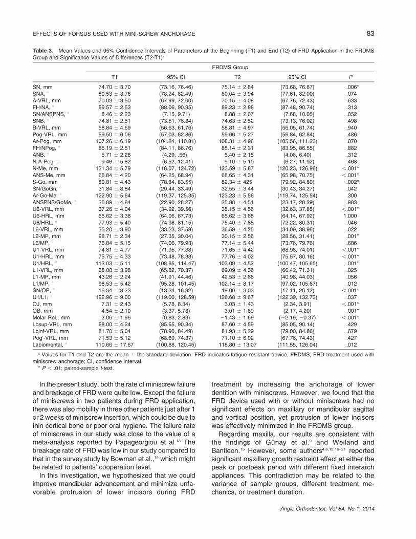
Total anterior facial height (2.25 6 1.94, P , .01),lower anterior facial height (1.81 6 1.54, P , .01), andposterior facial height (1.53 6 1.67, P , .01) increasedin the FRDMS group, whereas only posterior facialheight (1.26 6 1.79, P , .01) demonstrated incrementin the FRD group (Tables 3 and 4).
Figure 6. Horizontal, vertical, and angular measurements of the maxillary and mandibular incisor and molars related to the reference lines. 1: U6-
VRL (mm); 2: U6-HRL (mm); 3: U6/HRL (u); 4: U1-VRL (mm); 5: U1-HRL (mm); 6: U1/HRL (u); 7: L1-VRL (mm); 8: L1-GoMe (mm); 9: L1/GoMe
(u); 10: L6-VRL (mm); 11: L6-GoMe (mm); 12: L6/GoMe (u); 13: Molar relationship (mm).
80 ASLAN, KUCUKKARACA, TURKOZ, DINCER
Angle Orthodontist, Vol 84, No 1, 2014

Dentoalveolar and Soft Tissue Changes
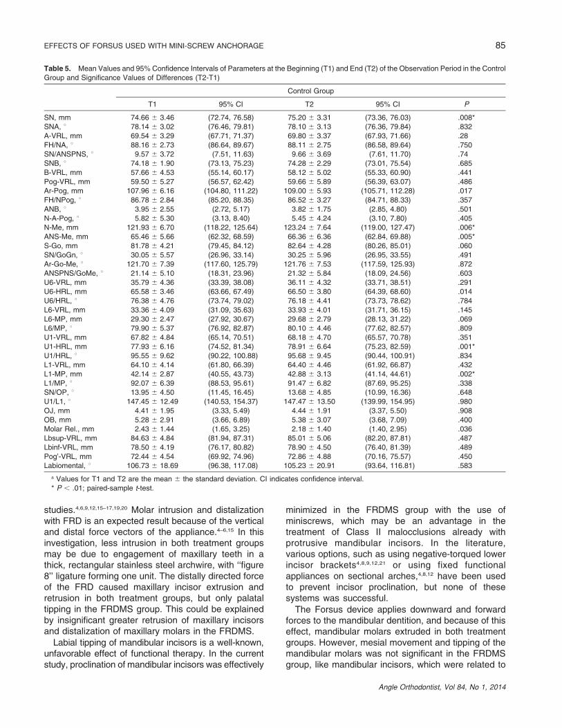
In the control group, the extrusion of maxillary andmandibular incisors was found to be significant(Table 5; P , .01).
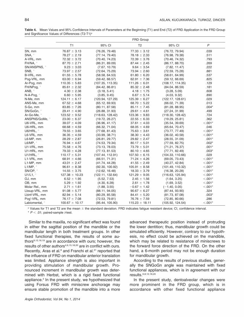
In the posterior segment, maxillary molars moveddistally (2.11 6 1.66, P , .01), and mandibular molarsextruded (1.43 6 1.40, P , .01) in the FRDMS group(Table 3). However, in the FRD group, distalization(1.45 6 0.83, P , .01) and distal tipping (23.92 6
3.34, P , .01) of maxillary molars as well as mesialmovement (1.95 6 1.33, P , .01), extrusion (1.53 6
1.46, P , .01), and mesial tipping (3.24 6 3.19, P ,
.01) of mandibular molars were determined (Table 4).Mesial tipping of mandibular molars in the FRD groupwas greater than that in both the FRDMS and controlgroups (Table 7; P , .01).
In the anterior region, the maxillary incisors retruded(3.16 6 1.90, P , .01), extruded (2.01 6 1.79, P ,
.01), and retroclined in the FRDMS group (8.94 6
5.70, P , .01). However, in the FRD group, maxillary
incisors retruded (1.79 6 1.90, P , .01) and extruded
(0.77 6 0.91, P , .01), and the lower incisors
protruded (2.34 6 1.68, P , .01), intruded (1.45 6
1.27, P , .01), and proclined (9.29 6 3.81, P , .01)
(Tables 3 and 4). Retroclination of maxillary incisors in
the FRDMS group was greater than in the FRD and
control groups (P , .01). Proclination of lower incisors
was greater in the FRD group than in the FRDMS and
control groups (P , .01). Intrusion of lower incisors in
the treatment groups was significant (P , .01) when
compared to the control group (Table 7). The occlusal
plane rotated in a clockwise direction in both the
Table 1. Intraclass Correlation Coefficientsa
ICC P value Difference Mean Difference SD Repeatability
SN, mm 0.99 ,.001 0.08 0.19 0.54
SNA, u 0.99 ,.001 0.03 0.18 0.50
A-VRL, mm 1.00 ,.001 0.05 0.09 0.25
FH/NA, u 0.99 ,.001 20.06 0.20 0.56
SN/ANSPNS, u 0.99 ,.001 0.06 0.28 0.78
SNB, u 1.00 ,.001 0.00 0.00 0.00
B-VRL, mm 1.00 ,.001 0.00 0.00 0.00
Pog-VRL, mm 1.00 ,.001 0.00 0.00 0.00
Ar-Pog, mm 0.99 ,.001 0.02 0.18 0.49
FH/NPog, u 0.99 ,.001 20.04 0.14 0.38
ANB, u 1.00 ,.001 0.01 0.05 0.14
N-A-Pog, u 1.00 ,.001 20.02 0.08 0.21
N-Me, mm 0.99 ,.001 0.18 0.21 0.59
ANS-Me, mm 1.00 ,.001 0.04 0.19 0.52
S-Go, mm 1.00 ,.001 0.07 0.14 0.39
SN/GoGn, u 0.99 ,.001 0.13 0.20 0.56
Ar-Go-Me, u 0.99 ,.001 0.13 0.27 0.75
ANSPNS/GoMe, u 1.00 ,.001 0.04 0.13 0.36
U6-VRL, mm 1.00 ,.001 0.00 0.00 0.00
U6-HRL, mm 1.00 ,.001 0.01 0.05 0.14
U6/HRL, u 1.00 ,.001 0.00 0.00 0.00
L6-VRL, mm 1.00 ,.001 20.01 0.05 0.14
L6-MP, mm 1.00 ,.001 0.00 0.00 0.00
L6/MP, u 1.00 ,.001 0.01 0.09 0.25
U1-VRL, mm 1.00 ,.001 0.02 0.07 0.21
U1-HRL, mm 1.00 ,.001 0.01 0.05 0.14
U1/HRL, u 1.00 ,.001 20.03 0.22 0.61
L1-VRL, mm 1.00 ,.001 0.00 0.00 0.00
L1-MP, mm 1.00 ,.001 20.02 0.14 0.38
L1/MP, u 1.00 ,.001 0.00 0.00 0.00
SN/OP, u 0.99 ,.001 20.03 0.12 0.29
U1/L1, u 1.00 ,.001 0.14 0.23 0.62
OJ, mm 0.99 ,.001 20.05 0.14 0.39
OB (mm) 0.99 ,.001 20.05 0.11 0.30
Molar rel., mm 1.00 ,.001 0.00 0.00 0.00
Lbsup-VRL, mm 1.00 ,.001 0.00 0.00 0.00
Lbinf-VRL, mm 1.00 ,.001 0.00 0.00 0.00
Pog’-VRL, mm 1.00 ,.001 0.01 0.05 0.14
Labiomental, u 1.00 ,.001 20.23 0.59 1.64
a ICC indicates intraclass correlation coefficient; SD, standard deviation.
EFFECTS OF FORSUS USED WITH MINI-SCREW ANCHORAGE 81
Angle Orthodontist, Vol 84, No 1, 2014
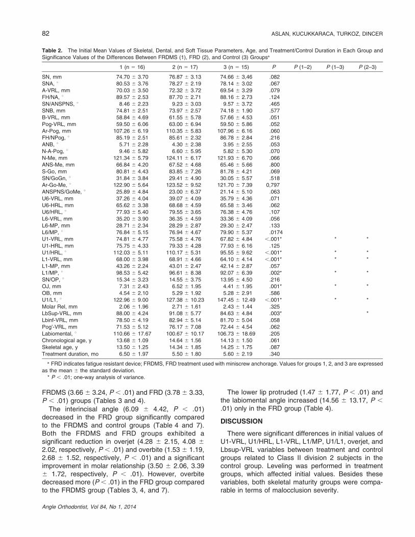
FRDMS (3.66 6 3.24, P , .01) and FRD (3.78 6 3.33,P , .01) groups (Tables 3 and 4).
The interincisal angle (6.09 6 4.42, P , .01)decreased in the FRD group significantly comparedto the FRDMS and control groups (Table 4 and 7).Both the FRDMS and FRD groups exhibited asignificant reduction in overjet (4.28 6 2.15, 4.08 6
2.02, respectively, P , .01) and overbite (1.53 6 1.19,2.68 6 1.52, respectively, P , .01) and a significantimprovement in molar relationship (3.50 6 2.06, 3.396 1.72, respectively, P , .01). However, overbitedecreased more (P , .01) in the FRD group comparedto the FRDMS group (Tables 3, 4, and 7).
The lower lip protruded (1.47 6 1.77, P , .01) andthe labiomental angle increased (14.56 6 13.17, P ,
.01) only in the FRD group (Table 4).
DISCUSSION
There were significant differences in initial values ofU1-VRL, U1/HRL, L1-VRL, L1/MP, U1/L1, overjet, andLbsup-VRL variables between treatment and controlgroups related to Class II division 2 subjects in thecontrol group. Leveling was performed in treatmentgroups, which affected initial values. Besides thesevariables, both skeletal maturity groups were compa-rable in terms of malocclusion severity.
Table 2. The Initial Mean Values of Skeletal, Dental, and Soft Tissue Parameters, Age, and Treatment/Control Duration in Each Group and
Significance Values of the Differences Between FRDMS (1), FRD (2), and Control (3) Groupsa
1 (n 5 16) 2 (n 5 17) 3 (n 5 15) P P (1–2) P (1–3) P (2–3)
SN, mm 74.70 6 3.70 76.87 6 3.13 74.66 6 3,46 .082
SNA, u 80.53 6 3.76 78.27 6 2.19 78.14 6 3.02 .067
A-VRL, mm 70.03 6 3.50 72.32 6 3.72 69.54 6 3.29 .079
FH/NA, u 89.57 6 2.53 87.70 6 2.71 88.16 6 2.73 .124
SN/ANSPNS, u 8.46 6 2.23 9.23 6 3.03 9.57 6 3.72 .465
SNB, mm 74.81 6 2.51 73.97 6 2.57 74.18 6 1.90 .577
B-VRL, mm 58.84 6 4.69 61.55 6 5.78 57.66 6 4.53 .051
Pog-VRL, mm 59.50 6 6.06 63.00 6 6.94 59.50 6 5.86 .052
Ar-Pog, mm 107.26 6 6.19 110.35 6 5.83 107.96 6 6.16 .060
FH/NPog, u 85.19 6 2.51 85.61 6 2.32 86.78 6 2.84 .216
ANB, u 5.71 6 2.28 4.30 6 2.38 3.95 6 2.55 .053
N-A-Pog, u 9.46 6 5.82 6.60 6 5.95 5.82 6 5.30 .070
N-Me, mm 121.34 6 5.79 124.11 6 6.17 121.93 6 6.70 .066
ANS-Me, mm 66.84 6 4.20 67.52 6 4.68 65.46 6 5.66 .800
S-Go, mm 80.81 6 4.43 83.85 6 7.26 81.78 6 4.21 .069
SN/GoGn, u 31.84 6 3.84 29.41 6 4.90 30.05 6 5.57 .518
Ar-Go-Me, u 122.90 6 5.64 123.52 6 9.52 121.70 6 7.39 0,797
ANSPNS/GoMe, u 25.89 6 4.84 23.00 6 6.37 21.14 6 5.10 .063
U6-VRL, mm 37.26 6 4.04 39.07 6 4.09 35.79 6 4.36 .071
U6-HRL, mm 65.62 6 3.38 68.68 6 4.59 65.58 6 3.46 .062
U6/HRL, u 77.93 6 5.40 79.55 6 3.65 76.38 6 4.76 .107
L6-VRL, mm 35.20 6 3.90 36.35 6 4.59 33.36 6 4.09 .056
L6-MP, mm 28.71 6 2.34 28.29 6 2.87 29.30 6 2.47 .133
L6/MP, u 76.84 6 5.15 76.94 6 4.67 79.90 6 5.37 .0174
U1-VRL, mm 74.81 6 4.77 75.58 6 4.76 67.82 6 4.84 ,.001* * *
U1-HRL, mm 75.75 6 4.33 79.33 6 4.28 77.93 6 6.16 .125
U1/HRL, u 112.03 6 5.11 110.17 6 5.31 95.55 6 9.62 ,.001* * *
L1-VRL, mm 68.00 6 3.98 68.91 6 4.66 64.10 6 4.14 ,.001* * *
L1-MP, mm 43.26 6 2.24 43.01 6 2.47 42.14 6 2.87 .057
L1/MP, u 98.53 6 5.42 96.61 6 8.38 92.07 6 6.39 .002* * *
SN/OP, u 15.34 6 3.23 14.55 6 3.75 13.95 6 4.50 .216
OJ, mm 7.31 6 2.43 6.52 6 1.95 4.41 6 1.95 .001* * *
OB, mm 4.54 6 2.10 5.29 6 1.92 5.28 6 2.91 .586
U1/L1, u 122.96 6 9.00 127.38 6 10.23 147.45 6 12.49 ,.001* * *
Molar Rel, mm 2.06 6 1.96 2.71 6 1.61 2.43 6 1.44 .325
LbSup-VRL, mm 88.00 6 4.24 91.08 6 5.77 84.63 6 4.84 .003* *
Lbinf-VRL, mm 78.50 6 4.19 82.94 6 5.14 81.70 6 5.04 .058
Pog’-VRL, mm 71.53 6 5.12 76.17 6 7.08 72.44 6 4.54 .062
Labiomental, u 110.66 6 17.67 100.67 6 10.17 106.73 6 18.69 .205
Chronological age, y 13.68 6 1.09 14.64 6 1.56 14.13 6 1.50 .061
Skeletal age, y 13.50 6 1.25 14.34 6 1.85 14.25 6 1.75 .087
Treatment duration, mo 6.50 6 1.97 5.50 6 1.80 5.60 6 2.19 .340
a FRD indicates fatigue resistant device; FRDMS, FRD treatment used with miniscrew anchorage. Values for groups 1, 2, and 3 are expressed
as the mean 6 the standard deviation.
* P , .01; one-way analysis of variance.
82 ASLAN, KUCUKKARACA, TURKOZ, DINCER
Angle Orthodontist, Vol 84, No 1, 2014
In the present study, both the rate of miniscrew failureand breakage of FRD were quite low. Except the failureof miniscrews in two patients during FRD application,there was also mobility in three other patients just after 1or 2 weeks of miniscrew insertion, which could be due tothin cortical bone or poor oral hygiene. The failure rateof miniscrews in our study was close to the value of ameta-analysis reported by Papageorgiou et al.13 Thebreakage rate of FRD was low in our study compared tothat in the survey study by Bowman et al.,14 which mightbe related to patients’ cooperation level.
In this investigation, we hypothesized that we couldimprove mandibular advancement and minimize unfa-vorable protrusion of lower incisors during FRD
treatment by increasing the anchorage of lowerdentition with miniscrews. However, we found that theFRD device used with or without miniscrews had nosignificant effects on maxillary or mandibular sagittaland vertical position, yet protrusion of lower incisorswas effectively minimized in the FRDMS group.
Regarding maxilla, our results are consistent withthe findings of Gunay et al.9 and Weiland andBantleon.15 However, some authors4,6,12,16–21 reportedsignificant maxillary growth restraint effect at either thepeak or postpeak period with different fixed interarchappliances. This contradiction may be related to thevariance of sample groups, different treatment me-chanics, or treatment duration.
Table 3. Mean Values and 95% Confidence Intervals of Parameters at the Beginning (T1) and End (T2) of FRD Application in the FRDMS
Group and Significance Values of Differences (T2-T1)a
FRDMS Group
T1 95% CI T2 95% CI P
SN, mm 74.70 6 3.70 (73.16, 76.46) 75.14 6 2.84 (73.68, 76.87) .006*
SNA, u 80.53 6 3.76 (78.24, 82.49) 80.04 6 3.94 (77.61, 82.00) .074
A-VRL, mm 70.03 6 3.50 (67.99, 72.00) 70.15 6 4.08 (67.76, 72.43) .633
FH/NA, u 89.57 6 2.53 (88.06, 90.95) 89.23 6 2.88 (87.48, 90.74) .313
SN/ANSPNS, u 8.46 6 2.23 (7.15, 9.71) 8.88 6 2.07 (7.68, 10.05) .052
SNB, u 74.81 6 2.51 (73.51, 76.34) 74.63 6 2.52 (73.13, 76.02) .498
B-VRL, mm 58.84 6 4.69 (56.63, 61.76) 58.81 6 4.97 (56.05, 61.74) .940
Pog-VRL, mm 59.50 6 6.06 (57.03, 62.86) 59.66 6 5.27 (56.84, 62.84) .486
Ar-Pog, mm 107.26 6 6.19 (104.24, 110.81) 108.31 6 4.96 (105.56, 111.23) .070
FH/NPog, u 85.19 6 2.51 (84.11, 86.76) 85.14 6 2.31 (83.95, 86.55) .882
ANB, u 5.71 6 2.28 (4.29, .56) 5.40 6 2.15 (4.06, 6.40) .312
N-A-Pog, u 9.46 6 5.82 (6.52, 12.41) 9.10 6 5.10 (6.27, 11.92) .468
N-Me, mm 121.34 6 5.79 (118.07, 124.72) 123.59 6 5.87 (120.23, 126.96) ,.001*
ANS-Me, mm 66.84 6 4.20 (64.25, 68.94) 68.65 6 4.31 (65.98, 70.75) ,.001*
S-Go, mm 80.81 6 4.43 (78.64, 83.55) 82.34 6 425 (79.92, 84.80) .002*
SN/GoGn, u 31.84 6 3.84 (29.44, 33.49) 32.55 6 3.44 (30.43, 34.27) .042
Ar-Go-Me, u 122.90 6 5.64 (119.37, 125.35) 123.23 6 5.56 (119.74, 125.54) .300
ANSPNS/GoMe, u 25.89 6 4.84 (22.90, 28.27) 25.88 6 4.51 (23.17, 28.29) .983
U6-VRL, mm 37.26 6 4.04 (34.92, 39.56) 35.15 6 4.56 (32.63, 37.85) ,.001*
U6-HRL, mm 65.62 6 3.38 (64.06, 67.73) 65.62 6 3.68 (64.14, 67.92) 1.000
U6/HRL, u 77.93 6 5.40 (74.98, 81.15) 75.40 6 7.85 (72.22, 80.31) .046
L6-VRL, mm 35.20 6 3.90 (33.23, 37.59) 36.59 6 4.25 (34.09, 38.96) .022
L6-MP, mm 28.71 6 2.34 (27.35, 30.04) 30.15 6 2.56 (28.56, 31.41) .001*
L6/MP, u 76.84 6 5.15 (74.06, 79.93) 77.14 6 5.44 (73.76, 79.76) .686
U1-VRL, mm 74.81 6 4.77 (71.95, 77.38) 71.65 6 4.42 (68.98, 74.01) ,.001*
U1-HRL, mm 75.75 6 4.33 (73.48, 78.38) 77.76 6 4.02 (75.57, 80.16) ,.001*
U1/HRL, u 112.03 6 5.11 (108.85, 114.47) 103.09 6 4.52 (100.47, 105.65) .001*
L1-VRL, mm 68.00 6 3.98 (65.82, 70.37) 69.09 6 4.36 (66.42, 71.31) .025
L1-MP, mm 43.26 6 2.24 (41.91, 44.46) 42.53 6 2.66 (40.98, 44.03) .056
L1/MP, u 98.53 6 5.42 (95.28, 101.45) 102.14 6 8.17 (97.02, 105.67) .012
SN/OP, u 15.34 6 3.23 (13.34, 16.92) 19.00 6 3.03 (17.11, 20.12) ,.001*
U1/L1, u 122.96 6 9.00 (119.00, 128.59) 126.68 6 9.67 (122.39, 132.73) .037
OJ, mm 7.31 6 2.43 (5.78, 8.34) 3.03 6 1.43 (2.34, 3.91) ,.001*
OB, mm 4.54 6 2.10 (3.37, 5.78) 3.01 6 1.89 (2.17, 4.20) .001*
Molar Rel., mm 2.06 6 1.96 (0.83, 2.83) 21.43 6 1.69 (22.19, 20.37) ,.001*
Lbsup-VRL, mm 88.00 6 4.24 (85.65, 90.34) 87.60 6 4.59 (85.05, 90.14) .429
Lbinf-VRL, mm 81.70 6 5.04 (78.90, 84.49) 81.93 6 5.29 (79.00, 84.86) .679
Pog’-VRL, mm 71.53 6 5.12 (68.69, 74.37) 71.10 6 6.02 (67.76, 74.43) .427
Labiomental, u 110.66 6 17.67 (100.88, 120.45) 118.80 6 13.07 (111.55, 126.04) .012
a Values for T1 and T2 are the mean 6 the standard deviation. FRD indicates fatigue resistant device; FRDMS, FRD treatment used with
miniscrew anchorage; CI, confidence interval.
* P , .01; paired-sample t-test.
EFFECTS OF FORSUS USED WITH MINI-SCREW ANCHORAGE 83
Angle Orthodontist, Vol 84, No 1, 2014
Similar to the maxilla, no significant effect was foundin either the sagittal position of the mandible or themandibular length in both treatment groups. In otherfixed functional therapies, the results of some au-thors9,12,16,19 are in accordance with ours; however, theresults of other authors4,6,15,18,20 are in conflict with ours.Recently, Aras et al.8 and Franchi et al.21 reported thatthe influence of FRD on mandibular anterior translationwas limited. Appliance strength is also important inproviding stimulation of mandibular growth. Pro-nounced increment in mandibular growth was deter-mined with Herbst, which is a rigid fixed functionalappliance.2 In the present study, we hypothesized thatusing Forsus FRD with miniscrew anchorage mayensure stable promotion of the mandible into a more
advanced therapeutic position instead of protrudingthe lower dentition; thus, mandibular growth could besimulated efficiently. However, contrary to our hypoth-esis, no effect could be achieved on the mandible,which may be related to resistance of miniscrews tothe forward force direction of the FRD. On the otherhand, a 6-month period may not be enough durationfor mandibular growth.
According to the results of previous studies, gener-ally the SNGoGn angle was maintained with fixedfunctional appliances, which is in agreement with ourresults.6,8,12,18,20
In the present study, dentoalveolar changes weremore prominent in the FRD group, which is inaccordance with other fixed functional appliance
Table 4. Mean Values and 95% Confidence Intervals of Parameters at the Beginning (T1) and End (T2) of FRD Application in the FRD Group
and Significance Values of Differences (T2-T1)a
FRD Group
T1 95% CI T2 95% CI P
SN, mm 76.87 6 3.13 (76.26, 79.48) 77.33 6 3.12 (76.72, 79.94) .039
SNA, u 78.27 6 2.19 (77.14, 79.40) 78.18 6 2.33 (76.98, 79.38) .511
A-VRL, mm 72.32 6 3.72 (70.40, 74.23) 72.39 6 3.76 (70.46, 74.32) .793
FH/NA, u 87.70 6 2.71 (86.31, 89.09) 87.44 6 2.45 (86.17, 88.70) .269
SN/ANSPNS, u 9.23 6 3.03 (7.67, 10.79) 9.64 6 3.54 (7.82, 11.47) .222
SNB, u 73.97 6 2.57 (72.64, 75.29) 73.94 6 2.60 (72.59, 75.28) .881
B-VRL, mm 61.55 6 5.78 (58.58, 64.53) 61.80 6 6.20 (58.61, 64.99) .537
Pog-VRL, mm 63.00 6 6.94 (59.42, 66.57) 62.91 6 7.36 (59.12, 66.69) .825
Ar-Pog, mm 110.35 6 5.83 (107.35, 113.35) 111.26 6 6.01 (108.17, 114.35) .129
FH/NPog, u 85.61 6 2.32 (84.42, 86.81) 85.32 6 2.48 (84.04, 86.59) .181
ANB, u 4.30 6 2.38 (3.18, 5.41) 4.18 6 1.75 (3.28, 5.09) .608
N-A-Pog, u 6.60 6 5.95 (3.85, 9.45) 6.67 6 5.14 (4.03, 9.32) .890
N-Me, mm 124.11 6 6.17 (120.94, 127.29) 125.39 6 6.27 (122.17, 128.61) .029
ANS-Me, mm 67.52 6 4.68 (65.12, 69.93) 68.70 6 5.22 (66.02, 71.39) .013
S-Go, mm 83.85 6 7.26 (80.11, 87.58) 85.11 6 7.45 (81.28, 88.95) .004*
SN/GoGn, u 29.41 6 4.90 (26.88, 31.93) 29.61 6 4.61 (27.24, 31.99) .499
Ar-Go-Me, u 123.52 6 9.52 (118.63, 128.42) 123.36 6 9.83 (118.30, 128.42) .724
ANSPNS/GoMe, u 23.00 6 6.37 (19.72, 26.27) 22.55 6 6.33 (19.29, 25.81) .362
U6-VRL, mm 39.07 6 4.09 (36.96, 41.17) 37.61 6 4.03 (35.54, 39.69) ,.001*
U6-HRL, mm 68.68 6 4.59 (66.32, 71.04) 68.05 6 4.69 (65.64, 70.47) .046
U6/HRL, u 79.55 6 3.65 (77.68, 81.43) 75.63 6 3.61 (73.77, 77.49) ,.001*
L6-VRL, mm 36.35 6 4.59 (33.99, 38.71) 38.30 6 4.43 (36.02, 40.58) ,.001*
L6-MP, mm 28.29 6 2.87 (26.81, 29.77) 29.82 6 2.47 (28.55, 31.09) .001*
L6/MP, u 76.94 6 4.67 (74.53, 79.34) 80.17 6 5.01 (77.59, 82.75) .002*
U1-VRL, mm 75.58 6 4.76 (73.13, 78.03) 73.79 6 5.01 (71.21, 76.37) .001*
U1-HRL, mm 79.33 6 4.28 (77.13, 81.53) 80.10 6 4.65 (77.71, 82.49) .003*
U1/HRL, u 110.17 6 5.31 (107.44, 112.91) 107.50 6 6.15 (104.33, 110.66) .029
L1-VRL, mm 68.91 6 4.66 (66.51, 71.31) 71.24 6 4.26 (69.05, 73.43) ,.001*
L1-MP, mm 43.01 6 2.47 (41.74, 44.28) 41.55 6 2.49 (40.27, 42.84) ,.001*
L1/MP, u 96.61 6 8.38 (92.30, 100.92) 105.91 6 8.58 (101.49, 110.32) ,.001*
SN/OP, u 14.55 6 3.75 (12.62, 16.48) 18.33 6 3.79 (16.38, 20.28) ,.001*
U1/L1, u 127.38 6 10.23 (122.11, 132.64) 121.29 6 9.05 (116.63, 125.94) ,.001*
OJ, mm 6.52 6 1.95 (5.52, 7.53) 2.45 6 1.56 (1.64, 3.25) ,.001*
OB, mm 5.29 6 1.92 (4.30, 6.28) 2.61 6 1.78 (1.69, 3.52) ,.001*
Molar Rel., mm 2.71 6 1.61 (1.88, 3.55) 20.67 6 1.42 (21.40, 0.05) ,.001*
Lbsup-VRL, mm 91.08 6 5.77 (88.11, 94.05) 90.67 6 6.27 (87.44, 93.90) .324
Lbinf-VRL, mm 82.94 6 5.14 (80.29, 85.58) 84.41 6 5.20 (81.73, 87.08) .003*
Pog’-VRL, mm 76.17 6 7.08 (72.53, 79.81) 76.76 6 7.59 (72.85, 80.66) .286
Labiomental, u 100.67 6 10.17 (95.44, 105.90) 115.23 6 18.11 (105.92, 124.54) ,.001*
a Values for T1 and T2 are the mean 6 the standard deviation. FRD indicates fatigue resistant device; CI, confidence interval.
* P , .01; paired-sample t-test.
84 ASLAN, KUCUKKARACA, TURKOZ, DINCER
Angle Orthodontist, Vol 84, No 1, 2014
studies.4,6,9,12,15–17,19,20 Molar intrusion and distalizationwith FRD is an expected result because of the verticaland distal force vectors of the appliance.4–6,15 In thisinvestigation, less intrusion in both treatment groupsmay be due to engagement of maxillary teeth in athick, rectangular stainless steel archwire, with ‘‘figure8’’ ligature forming one unit. The distally directed forceof the FRD caused maxillary incisor extrusion andretrusion in both treatment groups, but only palataltipping in the FRDMS group. This could be explainedby insignificant greater retrusion of maxillary incisorsand distalization of maxillary molars in the FRDMS.
Labial tipping of mandibular incisors is a well-known,unfavorable effect of functional therapy. In the currentstudy, proclination of mandibular incisors was effectively
minimized in the FRDMS group with the use ofminiscrews, which may be an advantage in thetreatment of Class II malocclusions already withprotrusive mandibular incisors. In the literature,various options, such as using negative-torqued lowerincisor brackets4,8,9,12,21 or using fixed functionalappliances on sectional arches,4,8,12 have been usedto prevent incisor proclination, but none of thesesystems was successful.
The Forsus device applies downward and forwardforces to the mandibular dentition, and because of thiseffect, mandibular molars extruded in both treatmentgroups. However, mesial movement and tipping of themandibular molars was not significant in the FRDMSgroup, like mandibular incisors, which were related to
Table 5. Mean Values and 95% Confidence Intervals of Parameters at the Beginning (T1) and End (T2) of the Observation Period in the Control
Group and Significance Values of Differences (T2-T1)
Control Group
T1 95% CI T2 95% CI P
SN, mm 74.66 6 3.46 (72.74, 76.58) 75.20 6 3.31 (73.36, 76.03) .008*
SNA, u 78.14 6 3.02 (76.46, 79.81) 78.10 6 3.13 (76.36, 79.84) .832
A-VRL, mm 69.54 6 3.29 (67.71, 71.37) 69.80 6 3.37 (67.93, 71.66) .28
FH/NA, u 88.16 6 2.73 (86.64, 89.67) 88.11 6 2.75 (86.58, 89.64) .750
SN/ANSPNS, u 9.57 6 3.72 (7.51, 11.63) 9.66 6 3.69 (7.61, 11.70) .74
SNB, u 74.18 6 1.90 (73.13, 75.23) 74.28 6 2.29 (73.01, 75.54) .685
B-VRL, mm 57.66 6 4.53 (55.14, 60.17) 58.12 6 5.02 (55.33, 60.90) .441
Pog-VRL, mm 59.50 6 5.27 (56.57, 62.42) 59.66 6 5.89 (56.39, 63.07) .486
Ar-Pog, mm 107.96 6 6.16 (104.80, 111.22) 109.00 6 5.93 (105.71, 112.28) .017
FH/NPog, u 86.78 6 2.84 (85.20, 88.35) 86.52 6 3.27 (84.71, 88.33) .357
ANB, u 3.95 6 2.55 (2.72, 5.17) 3.82 6 1.75 (2.85, 4.80) .501
N-A-Pog, u 5.82 6 5.30 (3.13, 8.40) 5.45 6 4.24 (3.10, 7.80) .405
N-Me, mm 121.93 6 6.70 (118.22, 125.64) 123.24 6 7.64 (119.00, 127.47) .006*
ANS-Me, mm 65.46 6 5.66 (62.32, 68.59) 66.36 6 6.36 (62.84, 69.88) .005*
S-Go, mm 81.78 6 4.21 (79.45, 84.12) 82.64 6 4.28 (80.26, 85.01) .060
SN/GoGn, u 30.05 6 5.57 (26.96, 33.14) 30.25 6 5.96 (26.95, 33.55) .491
Ar-Go-Me, u 121.70 6 7.39 (117.60, 125.79) 121.76 6 7.53 (117.59, 125.93) .872
ANSPNS/GoMe, u 21.14 6 5.10 (18.31, 23.96) 21.32 6 5.84 (18.09, 24.56) .603
U6-VRL, mm 35.79 6 4.36 (33.39, 38.08) 36.11 6 4.32 (33.71, 38.51) .291
U6-HRL, mm 65.58 6 3.46 (63.66, 67.49) 66.50 6 3.80 (64.39, 68.60) .014
U6/HRL, u 76.38 6 4.76 (73.74, 79.02) 76.18 6 4.41 (73.73, 78.62) .784
L6-VRL, mm 33.36 6 4.09 (31.09, 35.63) 33.93 6 4.01 (31.71, 36.15) .145
L6-MP, mm 29.30 6 2.47 (27.92, 30.67) 29.68 6 2.79 (28.13, 31.22) .069
L6/MP, u 79.90 6 5.37 (76.92, 82.87) 80.10 6 4.46 (77.62, 82.57) .809
U1-VRL, mm 67.82 6 4.84 (65.14, 70.51) 68.18 6 4.70 (65.57, 70.78) .351
U1-HRL, mm 77.93 6 6.16 (74.52, 81.34) 78.91 6 6.64 (75.23, 82.59) .001*
U1/HRL, u 95.55 6 9.62 (90.22, 100.88) 95.68 6 9.45 (90.44, 100.91) .834
L1-VRL, mm 64.10 6 4.14 (61.80, 66.39) 64.40 6 4.46 (61.92, 66.87) .432
L1-MP, mm 42.14 6 2.87 (40.55, 43.73) 42.88 6 3.13 (41.14, 44.61) .002*
L1/MP, u 92.07 6 6.39 (88.53, 95.61) 91.47 6 6.82 (87.69, 95.25) .338
SN/OP, u 13.95 6 4.50 (11.45, 16.45) 13.68 6 4.85 (10.99, 16.36) .648
U1/L1, u 147.45 6 12.49 (140.53, 154.37) 147.47 6 13.50 (139.99, 154.95) .980
OJ, mm 4.41 6 1.95 (3.33, 5.49) 4.44 6 1.91 (3.37, 5.50) .908
OB, mm 5.28 6 2.91 (3.66, 6.89) 5.38 6 3.07 (3.68, 7.09) .400
Molar Rel., mm 2.43 6 1.44 (1.65, 3.25) 2.18 6 1.40 (1.40, 2.95) .036
Lbsup-VRL, mm 84.63 6 4.84 (81.94, 87.31) 85.01 6 5.06 (82.20, 87.81) .487
Lbinf-VRL, mm 78.50 6 4.19 (76.17, 80.82) 78.90 6 4.50 (76.40, 81.39) .489
Pog’-VRL, mm 72.44 6 4.54 (69.92, 74.96) 72.86 6 4.88 (70.16, 75.57) .450
Labiomental, u 106.73 6 18.69 (96.38, 117.08) 105.23 6 20.91 (93.64, 116.81) .583
a Values for T1 and T2 are the mean 6 the standard deviation. CI indicates confidence interval.
* P , .01; paired-sample t-test.
EFFECTS OF FORSUS USED WITH MINI-SCREW ANCHORAGE 85
Angle Orthodontist, Vol 84, No 1, 2014
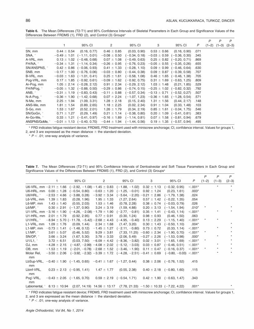
Table 6. The Mean Differences (T2-T1) and 95% Confidence Intervals of Skeletal Parameters in Each Group and Significance Values of the
Differences Between FRDMS (1), FRD (2), and Control (3) Groupsa
1 95% CI 2 95% CI 3 95% CI P
P
(1–2)
P
(1–3)
P
(2–3)
SN, mm 0.44 6 0.54 (0.16, 0.77) 0.46 6 0.85 (0.03, 0.90) 0.53 6 0.66 (0.16, 0.90) .071
SNA, u 20.49 6 1.01 (21.11, 0.01) 20.08 6 0.50 (20.34, 0.18) 20.03 6 0.59 (20.36, 0.30) .345
A-VRL, mm 0.13 6 1.02 (20.48, 0.68) 0.07 6 1.08 (20.49, 0.63) 0.25 6 0.82 (20.20, 0.71) .869
FH/NA, u 20.34 6 1.31 (21.14, 0.34) 20.26 6 0.95 (20.76, 0.23) 20.05 6 0.55 (20.35, 0.26) .655
SN/ANSPNS, u 0.42 6 0.86 (20.06, 0.93) 0.41 6 1.33 (20.28, 1.10) 0.09 6 0.99 (20.46, 0.64) .630
SNB, mm 20.17 6 1.00 (20.76, 0.06) 20.03 6 0.80 (20.44, 0.38) 0.09 6 0.87 (20.39, 0.58) .707
B-VRL, mm 20.03 6 1.63 (21.01, 0.41) 0.25 6 1.61 (20.58, 1.08) 0.46 6 1.65 (20.46, 1.38) .705
Pog-VRL, mm 0.17 6 1.85 (20.92, 0.61) 20.09 6 1.62 (20.92, 0.75) 0.31 6 1.69 (20.63, 1.25) .809
Ar-Pog, mm 1.05 6 2.14 (20.28, 2.12) 0.91 6 2.34 (20.29, 2.12) 1.03 6 1.48 (0.21, 1.85) .529
FH/NPog, u 20.05 6 1.32 (20.88, 0.50) 20.29 6 0.86 (20.74, 0.15) 20.25 6 1.02 (20.82, 0.32) .792
ANB, u 20.31 6 1.19 (20.83, 0.43) 20.11 6 0.88 (20.57, 0.34) 20.13 6 0.71 (20.52, 0.27) .507
N-A-Pog, u 20.36 6 1.90 (21.42, 0.68) 0.07 6 2.24 (21.07, 1.23) 20.36 6 1.65 (21.28, 0.54) .571
N-Me, mm 2.25 6 1.94 (1.09, 3.31) 1.28 6 2.18 (0.15, 2.40) 1.31 6 1.56 (0.44, 2.17) .148
ANS-Me, mm 1.81 6 1.54 (0.89, 2.65) 1.18 6 2.25 (0.02, 2.34) 0.91 6 1.04 (0.33, 1.48) .103
S-Go, mm 1.53 6 1.67 (0.52, 2.01) 1.26 6 1.79 (0.34, 2.19) 0.85 6 1.61 (20.04, 1.75) .546
SN/GoGn, u 0.71 6 1.27 (0.29, 1.49) 0.21 6 1.14 (20.38, 0.80) 0.20 6 1.09 (20.41, 0.81) .383
Ar-Go-Me, u 0.33 6 1.21 (20.41, 0.97) 20.16 6 1.89 (21.14, 0.81) 0.07 6 1.58 (20.81, 0.94) .679
ANSPNS/GoMe, u 20.01 6 1.13 (20.40, 0.70) 20.44 6 1.94 (21.44, 0.56) 0.19 6 1.35 (20.57, 0.94) .495
a FRD indicates fatigue resistant device; FRDMS, FRD treatment used with miniscrew anchorage; CI, confidence interval. Values for groups 1,
2, and 3 are expressed as the mean distance 6 the standard deviation.
* P , .01; one-way analysis of variance.
Table 7. The Mean Differences (T2-T1) and 95% Confidence Intervals of Dentoalveolar and Soft Tissue Parameters in Each Group and
Significance Values of the Differences Between FRDMS (1), FRD (2), and Control (3) Groupsa
1 95% CI 2 95% CI 3 95% CI P
P
(1–2)
P
(1–3)
P
(2–3)
U6-VRL, mm 22.11 6 1.66 (22.92, 21.08) 21.45 6 0.83 (21.88, 21.02) 0.32 6 1.13 (20.32, 0.95) ,.001* * *
U6-HRL, mm 0.00 6 1.28 (20.54, 0.80) 20.63 6 1.20 (21.25, 20.01) 0.92 6 1.24 (0.23, 1.61) .003* *
U6/HRL, u 22.53 6 4.66 (23.88, 0.28) 23.92 6 3.34 (25.64, 22.20) 20.21 6 2.86 (21.79, 1.38) .024
L6-VRL, mm 1.39 6 1.83 (0.28, 1.96) 1.95 6 1.33 (1.27, 2.64) 0.57 6 1.42 (20.22, 1.35) .054
L6-MP, mm 1.43 6 1.40 (0.55, 2.03) 1.53 6 1.46 (0.78, 2.28) 0.38 6 0.74 (20.03, 0.79) .026
L6/MP, u 0.30 6 2.91 (21.37, 0.90) 3.24 6 3.19 (1.59, 4.88) 0.20 6 3.13 (21.54, 1.94) .010* * *
U1-VRL, mm 23.16 6 1.90 (24.26, 22.08) 21.79 6 1.90 (22.77, 20.81) 0.35 6 1.41 (20.43, 1.14) ,.001* * *
U1-HRL, mm 2.01 6 1.79 (0.92, 2.95) 0.77 6 0.91 (0.30, 1.24) 0.98 6 0.93 (0.46, 1.50) .063
U1/HRL, u 28.94 6 5.70 (211.78, 25.42) 22.68 6 4.43 (24.95, 20.40) 0.13 6 2.29 (21.15, 1.40) ,.001* * *
L1-VRL, mm 1.09 6 1.76 (0.09, 1.44) 2.34 6 1.68 (1.47, 3.20) 0.30 6 1.43 (20.50, 1.10) .004* *
L1-MP, mm 20.73 6 1.41 (21.48, 0.12) 21.45 6 1.27 (22.11, 20.80) 0.73 6 0.72 (0.33, 1.14) ,.001* * *
L1/MP, u 3.61 6 5.07 (0.46, 5.52) 9.29 6 3.81 (7.33, 11.25) 20.60 6 2.34 (21.90, 0.70) ,.001* * *
SN/OP, u 3.66 6 3.24 (1.67, 5.30) 3.78 6 3.33 (2.06, 5.49) 20.27 6 2.26 (21.53, 0.98) .000* * *
U1/L1, u 3.72 6 6.51 (0.03, 7.50) 26.09 6 4.42 (28.36, 23.82) 0.02 6 3.01 (21.65, 1.69) ,.001* * *
OJ, mm 24.28 6 2.15 (24.87, 22.99) 24.08 6 2.02 (25.12, 23.03) 0.03 6 0.87 (20.46, 0.51) ,.001* * *
OB, mm 21.53 6 1.19 (22.01, 20.78) 22.68 6 1.52 (23.46, 21.90) 0.11 6 0.47 (20.16, 0.37) ,.001* * * *
Molar Rel,
mm
23.50 6 2.06 (23.92, 22.32) 23.39 6 1.72 (24.28, 22.51) 20.41 6 0.69 (20.80, 20.03) ,.001* * *
LbSup-VRL,
mm
20.40 6 1.90 (21.45, 0.65) 20.41 6 1.67 (21.27, 0.44) 0.38 6 2.06 (20.76, 1.52) .415
Lbinf-VRL,
mm
0.23 6 2.13 (20.95, 1.41) 1.47 6 1.77 (0.55, 2.38) 0.40 6 2.18 (20.80, 1.60) .115
Pog’-VRL,
mm
20.43 6 2.05 (21.65, 0.70) 0.59 6 2.19 (20.54, 1.71) 0.42 6 1.90 (20.63, 1.47) .343
Labiomental, u 8.13 6 10.94 (2.07, 14.19) 14.56 6 13.17 (7.78, 21.33) 21.50 6 10.33 (27.22, 4.22) .001* *
a FRD indicates fatigue resistant device; FRDMS, FRD treatment used with miniscrew anchorage; CI, confidence interval. Values for groups 1,
2, and 3 are expressed as the mean distance 6 the standard deviation.
* P , .01; one-way analysis of variance.
86 ASLAN, KUCUKKARACA, TURKOZ, DINCER
Angle Orthodontist, Vol 84, No 1, 2014
increased anchorage of mandibular dentition withminiscrews.
The decrease in overbite was greater in the FRDgroup than in the FRDMS group due to a combinationof insignificant greater relative intrusion of mandibularincisors and less extrusion of maxillary incisors in theFRD group. In both treatment groups, overjet correc-tion was totally dentoalveolar, mostly by retrusion ofmaxillary incisors in the FRDMS group, whereas lowerincisor protrusion was greater in the FRD group.
Likewise overjet correction, molar correction wascompletely dentoalveolar in both treatments. In theFRDMS group, 3.50-mm molar correction wasachieved by a contribution of 2.11 mm of maxillarymolar distalization and 1.39 mm of mandibular molarmesialization, whereas in the FRD group, 3.39 mm ofmolar correction consisted of 1.45 mm of maxillarymolar distalization and 1.95 mm of mandibular molarmesialization. Consequently, distal movement of max-illary dentition was more apparent in the FRDMSgroup due to increased anchorage of mandibulardentition by miniscrews; the force constructed byFRD was mostly transmitted to the posterior maxillarydentition. In another FRD study, Jones et al.5 reportedthat molar correction was predominately due to mesialmandibular skeletal and dental movements; however,treatment duration was 2.7 years. Karacay et al.4
reported equal amounts of distal maxillary molar andmesial mandibular molar movements with the ForsusNitinol Flat Spring. In the present study, the lower lipprotruded and the labiomental angle increased signif-icantly only in the FRD group due to prominentdentoalveolar changes. These findings are similar tothose of Nalbantgil et al.12
CONCLUSIONS
N Overjet and molar correction was totally dentoalve-olar in both treatment groups.
N Usage of miniscrews during FRD application waseffective in minimizing labial tipping of mandibularincisors when miniscrews stay stable during treatment.
REFERENCES
1. O’Brien K, Wright J, Conboy F, et al. Effectiveness oftreatment for Class II malocclusion with the Herbst or Twin-block appliances: a randomized, controlled trial. Am J OrthodDentofacial Orthop. 2003;124:128–137.
2. Ruf S, Pancherz H. Herbst/multibracket appliance treat-ment of Class II Division 1 malocclusions in early and lateadulthood. A prospective cephalometric study of consecu-tively treated subjects. Eur J Orthod. 2006;28:352–360.
3. Pangrazio-Kulbersh V, Berger JL, Chermak DS, KaczynskiR, Simon ES, Haerian A. Treatment effects of themandibular anterior repositioning appliance on patients
with Class II malocclusion. Am J Orthod Dentofacial Orthop.2003;123:286–295.
4. Karacay S, Akın E, Olmez H, Gurton AU, Sagdıc D. ForsusNitinol Flat Spring and Jasper Jumper corrections of Class IIdivision 1 malocclusions. Angle Orthod. 2006;76:666–672.
5. Jones G, Buschang PH, Kim KB, Oliver DR. Class II non-extraction patients treated with the Forsus fatigue resistantdevice versus intermaxillary elastics. Angle Orthod. 2008;78:332–338.
6. Heinig N, Goz G. Clinical application and effects of theForsus spring. A study of a new Herbst hybrid. J OrofacOrthop. 2001;62:436–450.
7. El-Sheikh MM, Godfrey K, Manosudprasit M, Viwattanatipa N.Force deflection characteristics of the fatigue-resistant devicespring: an in vitro study. World J Orthod. 2007;8:30–36.
8. Aras A, Ada E, Saracoglu H, Gezer NS, Aras I. Comparisonof treatments with the Forsus fatigue resistant device inrelation to skeletal maturity: a cephalometric and magneticresonance imaging study. Am J Orthod Dentofacial Orthop.2011;140:616–625.
9. Gunay EA, Arun T, Nalbantgil D. Evaluation of theimmediate dentofacial changes in late adolescent patientstreated with the ForsusTM FRD. Eur J Dent. 2011;5:423–432.
10. Oztoprak MO, Nalbantgil D, Uyanlar A, Arun T. Acephalometric comparative study of class II correction withSabbagh Universal Spring (SUS2) and Forsus FRD appli-ances. Eur J Dent. 2012;6:302–310.
11. Janssen KI, Raghoebar GM, Vissink A, Sandham A.Skeletal anchorage in orthodontics—a review of varioussystems in animal and human studies. Int J Oral MaxillofacImplants. 2008;23:75–88.
12. Nalbantgil D, Arun T, Sayınsu K, Isık F. Skeletal, dental andsoft tissue changes induced by the Jasper Jumper appliancein late adolescence. Angle Orthod. 2005;75:426–436.
13. Papageorgiou SN, Zogakis IP, Papadopoulos MA. Failurerates and associated risk factors of orthodontic miniscrewimplants: a meta-analysis. Am J Orthod Dentofacial Orthop.2012;142:577–595.
14. Bowman AC, Saltaji H, Flores-Mir C, Preston B, Tabbaa S.Patient experiences with the Forsus Fatigue ResistantDevice. Angle Orthod. 2013;83:437–446.
15. Weiland FJ, Bantleon HP. Treatment of Class II malocclu-sions with the Jasper Jumper appliance—a preliminaryreport. Am J Orthod Dentofacial Orthop. 1995;108:341–350.
16. Cope JB, Buschang PH, Cope DD, Parker J, BlackwoodHO. Quantitative evaluation of craniofacial changes withJasper Jumper therapy. Angle Orthod. 1994;2:113–122.
17. Mills CM, McCulloch KJ. Case report: modified use of theJasper Jumper appliance in a skeletal Class II mixeddentition case requiring palatal expansion. Angle Orthod.1997;4:277–282.
18. Stucki N, Ingervall B. The use of the Jasper Jumper for thecorrection of Class II malocclusion in the young permanentdentition. Eur J Orthod. 1998;20:271–281.
19. Covell DA, Trammell DW, Boero RP, West R. A cephalo-metric study of Class II division 1 malocclusion treated withthe Jasper Jumper appliance. Angle Orthod. 1999;69:311–320.
20. Kucukkeles N, Ilhan I, Orgun A. Treatment efficiency inskeletal Class II patients treated with the Jasper Jumper.Angle Orthod. 2007;77:449–456.
21. Franchi L, Alvetro L, Giuntini V, Masucci C, Defraia E,Baccetti T. Effectiveness of comprehensive fixed appliancetreatment used with the Forsus Fatigue Resistant Device inClass II patients. Angle Orthod. 2011;81:678–683.
EFFECTS OF FORSUS USED WITH MINI-SCREW ANCHORAGE 87
Angle Orthodontist, Vol 84, No 1, 2014

ERRATUM—Aslan BI, Kucukkaraca E, Turkoz C, Dincer M. Treatment effects of the Forsus fatigue resistantdevice used with miniscrew anchorage. Angle Orthod. 2014;84:76–87. Page 76 of the article states that ‘‘The FRDis a three-piece, telescoping system incorporating a superelastic nickel-titanium coil spring…’’ The spring is actuallya stainless steel coil spring. We apologize for the error.
383 Angle Orthodontist, Vol 84, No 2, 2014