formation and characterisation of antimicrobial dextran nanofibers
TRANSCRIPT
____________________ Received July 2010; in final form November 2010.
ROMANIAN J. BIOPHYS., Vol. 20, No. 4, P. 335–346, BUCHAREST, 2010
FORMATION AND CHARACTERISATION OF ANTIMICROBIAL DEXTRAN NANOFIBERS
M.M. SHAWKI *, A.M. HEREBA*, ABEER GHAZAL**
* Bio-Medical Physics Department, Medical Research Institute, Alexandria University, Egypt, e-mail: [email protected]
** Microbiology Department, Medical Research Institute, Alexandria University, Egypt
Abstract. Electrospinning is a fiber spinning technique driven by a high-voltage electrostatic field. Ultra-fine dextran nanofibers (DNF) mats were fabricated by electrospinning, using water as solvent. Antimicrobial DNF were formed by fabricating moxifloxacin antibiotic loaded on the DNF to serve as antimicrobial dextran-moxifloxacin nanofibers (DMNF) mats. Characterization of DNF and DMNF was performed. The moxifloxacin release from DMNF and its antimicrobial effectiveness were studied. The DMNF showed a wide antimicrobial effect against Escherichia coli and Staphylococcus aureus bacteria and that accompanied with sustained moxifloxacin release from the DMNF mat. The potential bio-medical application for the formed nanofibers was discussed.
Key words: electrospinning, dextran, moxifloxacin, nanofibers, antimicrobial.
INTRODUCTION
Present nanofiber technology is one of the most important objects in the recent research topics. An electrospinning technique conveniently allows the preparation of non-woven fibrous materials [12] with interesting characteristics such as fine diameters (ranging from submicron to several nanometers) [1], large surface area per unit mass, high porosity, high gas permeability, and small interfibrous pore size [14].
A typical electrospinning setup usually includes a reservoir of polymer solution with a metallic capillary connected to high voltage and a metallic collector. The hemispherical shape of the droplet at the needle tip is destabilized by the accumulated charges on the surface, and is converted to a Taylor’s cone when high voltage is applied to a polymer solution. At a critical value of the voltage, the electric forces overcome the surface tension on the droplet and a jet of ultra-fine fibers is produced from the tip of the Taylor cone [16].
M.M. Shawki, A.M. Hereba, Abeer Ghazal 2 336
The variables controlling the behavior of the electrified fluid jet during electrospinning can be divided into fluid properties and operating parameters. The relevant fluid properties are viscosity, conductivity, dielectric constant, boiling point, and surface tension. The operating parameters are flow rate, applied electric potential, and the distance between the tip and the collector called air gap [13, 15]. These nanofibers hold great promises as excellent candidates for various biomedical applications, e.g., scaffolds for cell and tissue culture [6, 9], vascular grafts [10], and carriers for drug delivery [5]. For many biomedical applications, the most important characteristics that should be targeted for include biocompatibility and mechanical performance.
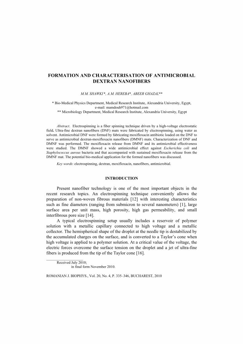
Dextran (Fig. 1a) is a polysaccharide [H (C6H10O5)x OH] with great biocompatibility and biodegradability characters. It is a suitable polymer to be developed as hydrogels, which are becoming increasingly important in biomedical applications, such as carriers for drug delivery [3, 4] and scaffolds for cell and tissue culture [7, 8].
In this study we formed two types of nanofibers using electrospinning technique; dextran nanofibers (DNF) and antimicrobial dextran-moxifloxacin nanofibers (DMNF) mats by uploading moxifloxacin antibiotic on dextran. Moxifloxacin (Fig. 1b) (C21H24FN3O4, bioavailability 86–92%, and protein binding from 30–50%) is a strong antibiotic for a wide range of Gram positive and negative bacteria [17]. We examined the characteristics of these mats, their stability, and the effectiveness of the DMNF mats against two types of bacteria as a function of the moxifloxacin release from the mats.
(a) (b)
Fig. 1. (a) Dextran structure; (b) moxifloxacin structure.
3 Antimicrobial dextran nanofibers
337
MATERIALS AND METHODS
MATERIALS
Dextran T70 for technical and laboratory use only (powder Mw of 70,000 Da) was purchased from Pharmacia fine chemicals AB Uppsala, Sweden. Moxifloxacin antibiotic was purchased as avalox antibiotic tablets; one film-coated tablet contains 400 mg moxifloxacin (Mw of 401.431 g/mol) in the form of 436.8 mg moxifloxacin hydrochloride as the pharmaceutical active ingredient; Bayer healthcare AG, Germany. The nutrient media for bacterial growth, and the reference bacterial strains were formed in the Microbiology Department, Medical Research Institute, Alexandria University, Egypt. All the previous chemicals were purchased as pure chemicals and no further purification was needed before the experiment.
PREPARATION OF THE SOLUTIONS FOR ELECTROSPINNING
The dextran solution was prepared in the optimum conditions described by Watadta R. et al. [18] in which the dextran solution concentration was 1g /mL using deionized water as the solvent for the dextran powder.
The moxifloxacin solution was prepared by dissolving one avalox antibiotic tablet in 100 mL of deionized water, then centrifuged for 15 minutes at 2000 rpm to precipitate the starch coat of the tablet, the supernatant was taken which in turn contained 4 mg moxifloxacin / mL. For the preparation of dextran-moxifloxacin solution; 1 mL of the supernatant was used as the solvent for 1 g dextran powder.
For dextran, dextran-moxifloxacin solutions; each was mixed with 0.02 g magnesium chloride as a catalyst (MgCl2 _ 6H2O; Carlo Erba, Italy), stirred until homogeneous solutions were obtained, mixed with 0.4 mL of 50% w/w glutaraldehyde aqueous solution (Fluka, Switzerland) for cross linking, and further stirred until homogeneous solutions were obtained. The viscosity of the mixture was measured using digital viscometer (Fungilab, Alpha 10064, Barcelona, Spain).
ELECTROSPINNING APPARATUS
The electrospinning unit was constructed locally in the Biophysics Department, Medical Research Institute, Alexandria University. The unit consisted of high voltage power supply (18KV), micro-infusion syringe pump (Shanghai Angel electronic equipment co., Ltd, China), a glass syringe equipped with a stainless-steel blunt-ended needle [outer diameter, 1.0 mm; inner diameter, 0.8 mm], and an aluminum cylinder-type rotated by DC power supply worked as a collector.
M.M. Shawki, A.M. Hereba, Abeer Ghazal 4 338
ELECTROSPINNING PROCESS
Each of the prepared dextran, and dextran-moxifloxacin solutions was contained in a 5 mL glass syringe. A 1 cm long blunt-ended stainless steel gauge-18 needle was used as the nozzle. The feed rate of each solution was adjusted at 0.25 mL/h by the syringe pump. The high-voltage power supply was used to charge the solution by connecting the emitting electrode (positive) to the nozzle and the grounding one to the collector. The distance between the tip of the nozzle and the grounded target was fixed at 15 cm. Generally, the collection time during electrospinning was 10 min. The formed mats were incubated at the oven at 70 °C for 24 h for cross linking process.
SCANNING ELECTRON MICROSCOPE
For the measurements of fiber diameters, fiber layer thickness, and effect of uploaded moxifloxacin on the fibers morphology, the DNF and DMNF mats were cut into pieces, coated with gold using coater (SPI-module TM sputter coater, Japan), then examined using scanning electron microscope (SEM) (Joel, JSM-6360 LA Japan). Fiber diameters were measured directly from SEM images under the same magnification. The average value of the diameters was calculated from 100 measurements.
FTIR SPECTROSCOPY
Infrared spectra of the DNF and DMNF mats were collected on an attenuated total reflectance Fourier transform infrared spectrometry (Alpha-centauri, Shimadzu, Japan, FT-IR-8400S). Around 4–8 mg of each mat was mixed with 200 mg IR-grade potassium bromide (KBr) and ground together, then compressed into tablet by the compressor (Shimadzu-compressor, Japan) and the spectrum was then recorded in the wavelength range of 4500–500 cm–1.
STABILITY OF THE MATS
The degree of stability of the DNF and DMNF mats in terms of weight loss and the degree of swelling in the aqueous solution were examined at pH 7 and an ambient temperature. 10 mg (W1) of dried nanofiber mats were weighed, immersed in a vial containing water for 24 h, then were taken out from the vial and weighed (W2), after that they were dried firstly at an ambient temperature and then at 60 °C over night, and were weighed again (W3). The degrees of stability (weight loss) and swelling percent were expressed by the following equations:
5 Antimicrobial dextran nanofibers
339
Stability (%) = 1 3 1001
W WW−
× (1)
Swelling (%) = 2 3 1003
W WW−
× (2)
DETERMINATION OF MOXIFLOXACIN RELEASE
To determine the release percent of moxifloxacin from DMNF mat; 10 mg of the mat was weighted, immersed in 10 mL pH 7.4 phosphate buffer saline. Buffer samples were taken from the solution at regular time interval (0.5, 1, 1.5, 2, 3 h) and measured spectrophotometrically (using UV/visible spectrophotometer JENWAY, 6305, USA) at 400 nm. The samples were returned to the buffer solution again to avoid any effect on the total concentration. The concentrations were determined from measured standard curve.
EXAMINATION OF THE ANTIMICROBIAL EFFECTS OF THE MATS
There are two methods for determination of the antimicrobial effects of the mats [11].
For the zone of inhibition test, nutrient agar was poured onto disposable sterilized Petri dishes and was allowed to solidify; 100 µl of E. coli bacterial suspension (Gram negative) was streaked over the plate and was spread uniformly. 10 mg pieces of DNF and DMNF were gently placed over the solidified agar gel in different positions of the same Petri dish. The same test was repeated with Staphylococcus aureus (S. aureus) (Gram positive) bacteria. Plates were incubated at 37 °C for 12 h. The bacterial growth was compared with control bacteria (without DNF or DMNF incubation).
For the antibacterial test in liquid medium, sterilized LB medium was measured (5 mL) into sterile tubes. 10 mg of DNF and DMNF were introduced into two different LB solutions, which contained 105 colony forming units (CFU) of E. coli. The mixtures were cultured at 37 °C in a shaking incubator for overnight at 250 rpm. CFU were counted after spotting culture dilutions on solid media, in order to estimate the quantitative antimicrobial effect. The same test was repeated with S. aureus. The CFU were compared with control bacteria (without DNF or DMNF incubation).
STATISTICAL ANALYSIS
The data analysis was performed using the SPSS-10 package (release 3, SPSS Inc., between the means of the different parameters of the samples of the studied mats. A difference was considered significant at probability (p) < 0.05.

M.M. Shawki, A.M. Hereba, Abeer Ghazal 6 340
RESULTS AND DISCUSSION
CHARACTERISATION OF THE PREPARED NANOFIBER MATS
The DNF and DMNF mats were formed then characterized using SEM and FTIR.
Scanning microscope
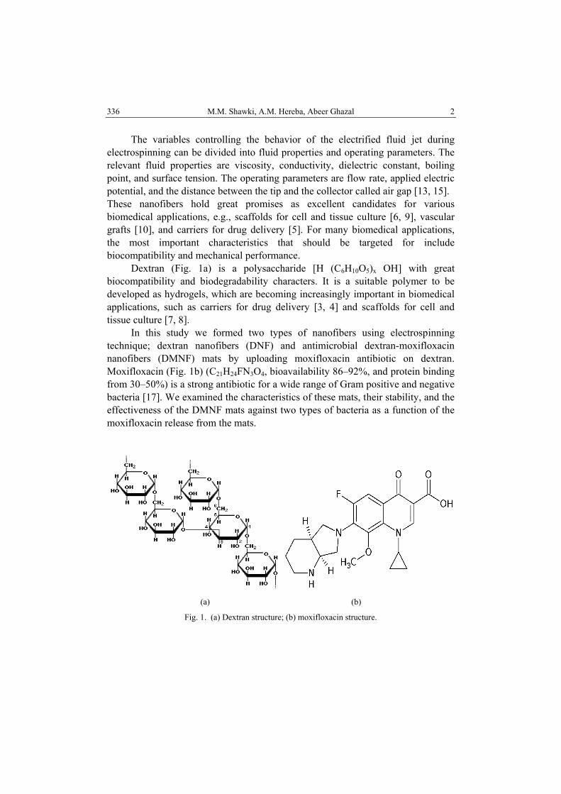
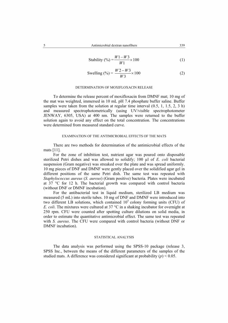
We compared between the mats in three characters; the morphology, fiber diameter, and layer thickness. Figures 2a and 2b show the morphology of DNF and DMNF, respectively, at 1000×. These figures indicate that the DNF mat morphology lacks any uploading material on its surface (Fig. 2a), while figure 2b shows the DMNF mat with many positions with uploaded material (marked with small “x” marks). These data indicate high uploading of moxifloxacin on the DNF.
Figures 2c and 2d show samples of the DNF and DMNF diameters respectively at 30.000×. The average DNF diameter is 113.78 ± 44.24 nm which is significantly less than the diameter of DMNF 271.17 ± 93.81 nm; this change is due to the difference in the solution viscosity from which the fibers prepared, the viscosities of the solutions were 785 and 2000 mPa·s for DNF and DMNF, respectively. Figures 2e and 2f show the layer thickness of DNF and DMNF, respectively, at 140×. The results indicate no significant change in the layer thickness 560.75 ± 33.84 µm, 569.5 ± 21.22 µm for DNF and DMNF respectively.
(a) (c) (e)
7 Antimicrobial dextran nanofibers
341
(b) (d) (f)
Fig. 2. SEM for: (a) DNF mat surface morphology (b) DMNF mat surface morphology (c) DNF dimensions (d) DMNF dimensions (e) DNF mat Layer thickness (f) DMNF mat Layer thickness.
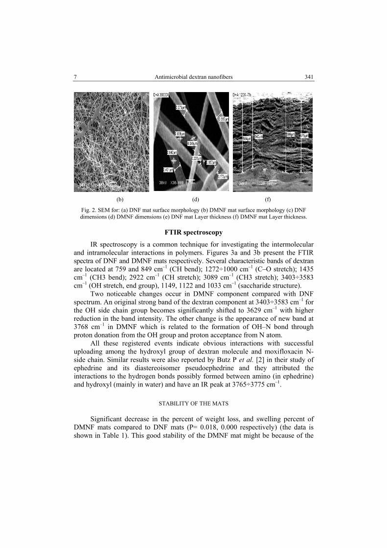
FTIR spectroscopy
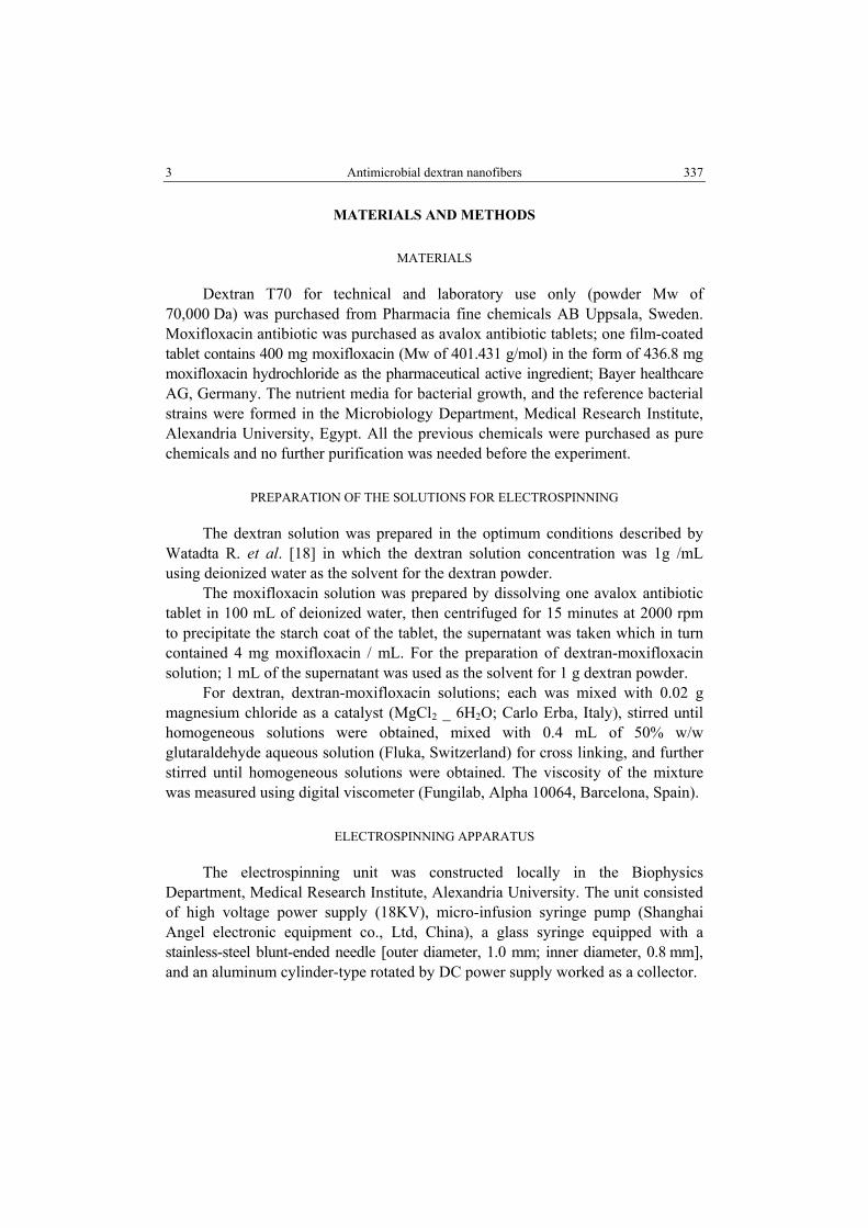
IR spectroscopy is a common technique for investigating the intermolecular and intramolecular interactions in polymers. Figures 3a and 3b present the FTIR spectra of DNF and DMNF mats respectively. Several characteristic bands of dextran are located at 759 and 849 cm–1 (CH bend); 1272÷1000 cm–1 (C–O stretch); 1435 cm–1 (CH3 bend); 2922 cm–1 (CH stretch); 3089 cm–1 (CH3 stretch); 3403÷3583 cm–1 (OH stretch, end group), 1149, 1122 and 1033 cm–1 (saccharide structure).
Two noticeable changes occur in DMNF component compared with DNF spectrum. An original strong band of the dextran component at 3403÷3583 cm–1 for the OH side chain group becomes significantly shifted to 3629 cm–1 with higher reduction in the band intensity. The other change is the appearance of new band at 3768 cm–1 in DMNF which is related to the formation of OH–N bond through proton donation from the OH group and proton acceptance from N atom.
All these registered events indicate obvious interactions with successful uploading among the hydroxyl group of dextran molecule and moxifloxacin N- side chain. Similar results were also reported by Butz P et al. [2] in their study of ephedrine and its diastereoisomer pseudoephedrine and they attributed the interactions to the hydrogen bonds possibly formed between amino (in ephedrine) and hydroxyl (mainly in water) and have an IR peak at 3765÷3775 cm–1.
STABILITY OF THE MATS
Significant decrease in the percent of weight loss, and swelling percent of DMNF mats compared to DNF mats (P= 0.018, 0.000 respectively) (the data is shown in Table 1). This good stability of the DMNF mat might be because of the
M.M. Shawki, A.M. Hereba, Abeer Ghazal 8 342
new formed hydrogen bonds between hydroxyl group of dextran and amine group of moxifloxacin. This fact indicated that the DMNF mat has strength and flexibility in the wet state more than the DNF mat.
(a)
(b)
Fig. 3. FTIR spectra of (a) DNF and (b) DMNF mats.
Table 1
The swelling percent and weight loss percent of DNF and DMNF mats (The weight was taken as an average for five samples)
Mat type Swelling weight (mg)
Drying weight (mg)
Swelling percent Percent of weight loss
DNF mat 119.6 ± 8 7.6 ± 0.4 1307.05 ±16.33 24 ± 3.27 DMNF
mat 53.2 ± 3.5 8.5 ± 0.7 596.67 ± 10.87 15 ± 5.72
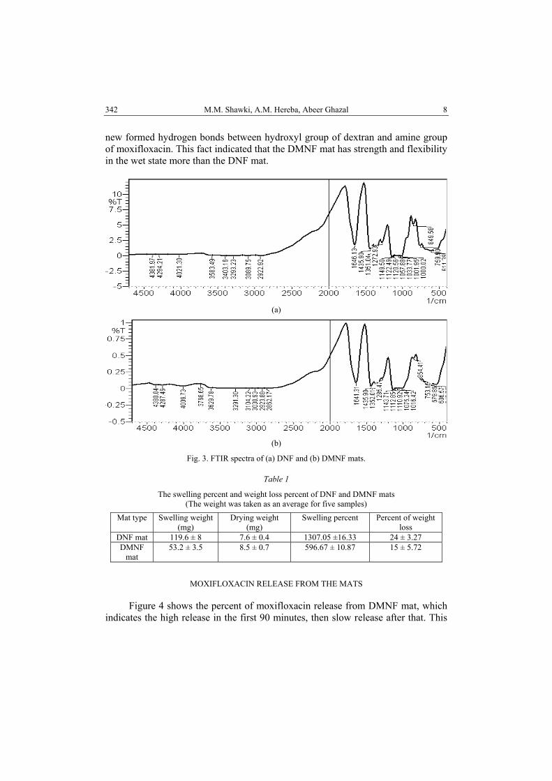
MOXIFLOXACIN RELEASE FROM THE MATS
Figure 4 shows the percent of moxifloxacin release from DMNF mat, which indicates the high release in the first 90 minutes, then slow release after that. This
9 Antimicrobial dextran nanofibers
343
rapid release of the drug is because the attachment of the moxifloxacin with dextran is through hydrogen bonds, which facilitates the drug release in the aqueous solution.
Fig. 4. Percent of moxifloxacin released from the DMNF mat with time.
ANTIMICROBIAL EFFECTS OF DMNF MATS
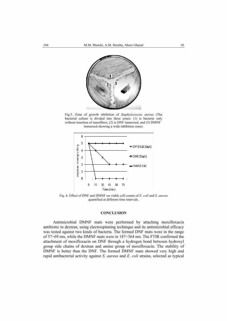
The DMNF mat shows very high antibacterial potency as shown from the wide inhibition zone formed around it indicating the inability of growth of the bacteria at this zone, while visible bacterial growth occurred at DNF mat and at control zone as shown in figure 5 with S. aureus.
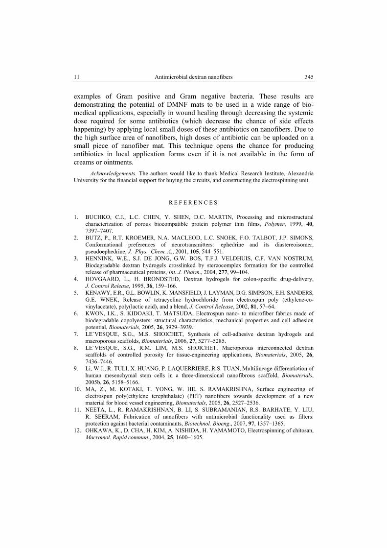
In order to understand the antibacterial efficacy, CFU have been counted for DMNF in comparison with the DNF and control bacterial samples. The counts were used to calculate the surviving number of bacteria. The degree of the antibacterial effect is the ratio of reduction of bacterial colonies [11]. The equation for quantitative antibacterial evaluation is given by:
%R = 100B AB−
× (3)
where R = the reduction rate, A = the number of surviving bacterial colonies with DMNF, and B = the number of surviving bacterial colonies without incubation with DMNF. Zone inhibition test showed that the DMNF are active against the pathogens in a reduction percent of 100% and 99.9% for E. coli and S. aureus, respectively, and the activity remains along six bacterial generations as shown in figure 6, and that because of the fast release of moxifloxacin from DMNF mat with time.
M.M. Shawki, A.M. Hereba, Abeer Ghazal 10 344
Fig.5. Zone of growth inhibition of Staphylococcus aureus (The bacterial culture is divided into three zones: (1) is bacteria only without insertion of nanofibers, (2) is DNF immersed, and (3) DMNF
immersed showing a wide inhibition zone).
Fig. 6. Effect of DNF and DMNF on viable cell counts of E. coli and S. aureus
quantified at different time intervals.
CONCLUSION
Antimicrobial DMNF mats were performed by attaching moxifloxacin antibiotic to dextran, using electrospinning technique and its antimicrobial efficacy was tested against two kinds of bacteria. The formed DNF mats were in the range of 57÷69 nm, while the DMNF mats were in 187÷364 nm. The FTIR confirmed the attachment of moxifloxacin on DNF through a hydrogen bond between hydroxyl group side chains of dextran and amine group of moxifloxacin. The stability of DMNF is better than the DNF. The formed DMNF mats showed very high and rapid antibacterial activity against S. aureus and E. coli strains, selected as typical
11 Antimicrobial dextran nanofibers
345
examples of Gram positive and Gram negative bacteria. These results are demonstrating the potential of DMNF mats to be used in a wide range of bio-medical applications, especially in wound healing through decreasing the systemic dose required for some antibiotics (which decrease the chance of side effects happening) by applying local small doses of these antibiotics on nanofibers. Due to the high surface area of nanofibers, high doses of antibiotic can be uploaded on a small piece of nanofiber mat. This technique opens the chance for producing antibiotics in local application forms even if it is not available in the form of creams or ointments.
Acknowledgements. The authors would like to thank Medical Research Institute, Alexandria University for the financial support for buying the circuits, and constructing the electrospinning unit.
R E F E R E N C E S
1. BUCHKO, C.J., L.C. CHEN, Y. SHEN, D.C. MARTIN, Processing and microstructural characterization of porous biocompatible protein polymer thin films, Polymer, 1999, 40, 7397–7407.
2. BUTZ, P., R.T. KROEMER, N.A. MACLEOD, L.C. SNOEK, F.O. TALBOT, J.P. SIMONS, Conformational preferences of neurotransmitters: ephedrine and its diastereoisomer, pseudoephedrine, J. Phys. Chem. A., 2001, 105, 544–551.
3. HENNINK, W.E., S.J. DE JONG, G.W. BOS, T.F.J. VELDHUIS, C.F. VAN NOSTRUM, Biodegradable dextran hydrogels crosslinked by stereocomplex formation for the controlled release of pharmaceutical proteins, Int. J. Pharm., 2004, 277, 99–104.
4. HOVGAARD, L., H. BRONDSTED, Dextran hydrogels for colon-specific drug-delivery, J. Control Release, 1995, 36, 159–166.
5. KENAWY, E.R., G.L. BOWLIN, K. MANSFIELD, J. LAYMAN, D.G. SIMPSON, E.H. SANDERS, G.E. WNEK, Release of tetracycline hydrochloride from electrospun poly (ethylene-co-vinylacetate), poly(lactic acid), and a blend, J. Control Release, 2002, 81, 57–64.
6. KWON, I.K., S. KIDOAKI, T. MATSUDA, Electrospun nano- to microfiber fabrics made of biodegradable copolyesters: structural characteristics, mechanical properties and cell adhesion potential, Biomaterials, 2005, 26, 3929–3939.
7. LE´VESQUE, S.G., M.S. SHOICHET, Synthesis of cell-adhesive dextran hydrogels and macroporous scaffolds, Biomaterials, 2006, 27, 5277–5285.
8. LE´VESQUE, S.G., R.M. LIM, M.S. SHOICHET, Macroporous interconnected dextran scaffolds of controlled porosity for tissue-engineering applications, Biomaterials, 2005, 26, 7436–7446.
9. Li, W.J., R. TULI, X. HUANG, P. LAQUERRIERE, R.S. TUAN, Multilineage differentiation of human mesenchymal stem cells in a three-dimensional nanofibrous scaffold, Biomaterials, 2005b, 26, 5158–5166.
10. MA, Z., M. KOTAKI, T. YONG, W. HE, S. RAMAKRISHNA, Surface engineering of electrospun poly(ethylene terephthalate) (PET) nanofibers towards development of a new material for blood vessel engineering, Biomaterials, 2005, 26, 2527–2536.
11. NEETA, L., R. RAMAKRISHNAN, B. LI, S. SUBRAMANIAN, R.S. BARHATE, Y. LIU, R. SEERAM, Fabrication of nanofibers with antimicrobial functionality used as filters: protection against bacterial contaminants, Biotechnol. Bioeng., 2007, 97, 1357–1365.
12. OHKAWA, K., D. CHA, H. KIM, A. NISHIDA, H. YAMAMOTO, Electrospinning of chitosan, Macromol. Rapid commun., 2004, 25, 1600–1605.
M.M. Shawki, A.M. Hereba, Abeer Ghazal 12 346
13. RENEKER, D.H., I. CHUN, Nanometre diameter fibers of polymer, produced by electrospinning, Nanotechnology, 1996, 7, 216–223.
14. SAEED, K., S. HAIDER, T.J. OH, S.Y. PARK, Preparation of amidoxime-modified polyacrylonitrile (PAN-oxime) nanofibers and their applications to metal ions adsorption, J. Membr. Sci., 2008, 322, 400–405.
15. SHIN, Y.M., M.M. HOHMAN, M.P. BRENNER, G.C. RUTLEDGE, Experimental characterization of electrospinning: the electrically forced jet and instabilities, Polymer, 2001, 42, 9955–9967.
16. TAYLOR, G.I., Electrically driven jets, Proc. R. Soc. Lond. A., 1969, 313, 453–475. 17. WANG, J.Y., J.T. WANG, T.H. TSAI, C.L. HSU, C.J. YU, P.R. HSUEH, L.N. LEE, P.C. YANG,
Adding moxifloxacin is associated with a shorter time to culture conversion in pulmonary tuberculosis, Int. J. Tuberc. Lung Dis., 2010 , 14, 65–71.
18. WATADTA, R., Y. THAIYING, Y. SAEJENG, Y. JAEJENG, I. JANGCHUD, R. RANGKUPAN, C. MEECHAISUE, P. SUPAPHOL, Electrospun dextran fibrous membranes, Cellulose, 2008, 15, 435–444.