for-127: identifying wood: a primer for everyone, part 5
TRANSCRIPT

E X T E N S I O N
Agriculture and Natural Resources • Family and Consumer Sciences • 4-H Youth Development • Community and Economic Development
COOPERATIVE EXTENSION SERVICEUNIVERSITY OF KENTUCKY COLLEGE OF AGRICULTURE, FOOD AND ENVIRONMENT, LEXINGTON, KY, 40546
P A R T
IDENTIFYING WOOD—A Primer for Everyone 5
The First Separation of Softwood SpeciesTerry Conners, Forestry
FOR-127
Just making the separation between softwoods and hardwoods doesn’t help
much in identifying wood species; that would be sort of like identifying children by their hair color. Let’s look at the next level of wood features that you need to be able to recognize.
Two Categories of Softwoods: Resinous and Non-resinous Species Softwoods have a pretty simple construction, made up as they are of earlywood tracheids, latewood tracheids and rays. All of these cells can be readily seen in this micrograph western redcedar (Figure 5-1).
Figure 5-1. This micrograph of a single growth ring of western redcedar (Thuja occidentalis) shows earlywood tracheids, latewood tracheids, and rays.
Figure 5-2. Where resin exuded from an in-jury on a white pine, whitish rosin remains.
In addition to color, odor and density, softwood samples can have other fea-tures of diagnostic value. The first thing you should determine after deciding that a specimen is a softwood is whether it contains pitch, also known as resin—some species have it, some don’t. Resin is thought to play a role in defending the tree against insect attack and to help stop sap leaks from dehydrating the tree after an injury. Perhaps you’ve noticed pine trees with a crusty, whitish substance on the bark around and below a wound (see Figure 5-2); this substance is rosin, the same material used to condition violin bows for playing. Rosin is what’s left after the volatile solvent in resin (i.e., turpen-tine) evaporates; both the rosin and the turpentine are examples of extractives, because they can be removed from the wood without affecting its structure.

2
Only four native gymnosperm genera in North America normally contain resin, and all of them are in the Pinaceae family (i.e., related to pines).1 The resin-ous genera are:• Pinus (pines)• Picea (spruces)• Pseudotsuga (Douglas-fir2)• Larix (larch, also known as tamarack)
Other gymnosperm genera don’t contain resin, so the presence or absence of resin is useful in species identification. The non-resinous genera include:• Tsuga (hemlocks) • Juniperus, Calocedrus,
Chamaecyparis,3 etc. (various cedar genera)
• Abies (true firs)• Taxodium (bald cypress)• Sequoia (redwood)• And others
Gathering first-hand impressions as to the resinous character of an unknown specimen might seem difficult at first, especially since the wood becomes less tacky as the turpentine evaporates. We can, however, use a hand lens to read-ily determine whether the structures that contain and transport the resin are present or absent. These structures are known as resin canals.
Resin Canals Resin canals are not actually cells; they’re only open spaces that serve as channels for the resin in resinous soft-woods. Resin enters the resin canals from specialized thin-walled, crescent-shaped cells called epithelial cells that surround the resin canal cavity; these cells secrete resin into the canal. Resin canals can be seen on the cross-section fairly easily with a hand lens, where, compared to the surrounding tracheids, they will look
1 Three other genera contain resin canals (Cathaya, Nothotsuga, and Keteleeria), but these are native to China and are not of commercial significance.
2 The name Douglas-fir is hyphenated because it is not a true fir (i.e., not a member of the Abies genus). As you will see, the common names of some other species (including incense-cedar and yellow-poplar) are hyphenated for similar reasons.
3 Chamaecyparis is pronounced Kam-ee-SIP-a-ris.
Figure 5-3. This micrograph of a cross-section of sugar pine (Pinus lambertiana) shows multiple growth rings and numerous large resin canals. Notice how large the resin canals are compared to the tracheids. In this sample, many of the resin canals appear open while others still appear to be full of rosin. Each resin canal is surrounded by epithelial cells, though you can’t make them out at this magnification. A fusiform ray is also present on the right-hand side of the micrograph; the ray looks larger and whiter than the surrounding rays because the section was cut through the middle of the resin canal in the middle of the ray.
like large brownish or whitish holes. Not every resin canal follows the grain; some resin canals extend in the radial direc-tion, where they are embedded within certain kinds of rays called fusiform rays. Softwood rays are pretty small, so you’ll usually need to look at a thin tangential section with a microscope to see these. As it happens, though, you can see both longitudinal resin canals and a fusiform ray on the cross-section of the sugar pine (Pinus lambertiana) micrograph shown in Figure 5-3. You’ll notice that there’s a white line parallel with the rays
fusiform ray
resin canal
resin canal
resin canal
resin canal
about one-quarter of the distance from the right-hand edge of the micrograph. This white line is a fusiform ray that I cut through when I made the section for this micrograph. The line looks broad and white because I happened to cut through the middle of a fusiform ray where the resin canal is; the resin canal the ray is wider than a normal ray at this point, and the resin canal I cut through was filled with rosin. Don’t expect to see resin canals on the cross-section like this all the time—it’s really pretty unusual.

3
Figure 5-4. The vertically oriented brown streaks in this sample of sugar pine (P. lam-bertiana) are longitudinal resin canals and the small brownish specks are the ends of the resin canals in fusiform rays. (Not every resin canal in the photograph is labeled.)
Tip: Non-resinous species may occa-sionally exhibit traumatic resin canals due to an injury (bear claw scratches, for example) or infection. When these are present, they typically line up in tangential groups with several or more resin canals in a row, and normal resin canals will be absent from other growth rings. Traumatic resin canals are an anomaly, but they’re generally fairly easy to recognize. Traumatic resin canals have no diagnostic value.
resin canal
resin canal
resin canal
Figure 5-5. These examples of shortleaf pine (P. echinata) show several growth rings and numerous resin canals. In the low-power micro-graph on the left, the resin canals (some of which are indicated by arrows) resemble small pin-pricks. Notice how plentiful the resin canals are; they’re present in each growth ring, and they’re fairly large and easy to see. The micrograph on the right was taken at a higher magni-fication to show the variation in the ways resin canals can look (open and clogged).
Resin Canals on the Side Grain In some species, particularly the pines, the resin canals are large enough and plentiful enough that they can be readily observed on the side grain, looking like needle-thin brownish streaks aligned with the grain. These streaks are usually short because of the plane at which the saw intersected the resin canals. (The resin canals of spruce, larch and Douglas-fir might be harder to see because they’re smaller and less plentiful in the first place.) The longitudinal resin canals are distinct as dark streaks in Figure 5-4. Fusiform rays also show up in this photo; the resin canals in the fusiform rays show up as very small “fly specks,” as the resin canals in the rays have bled out enough to show against the lighter wood back-ground.
Using Resin Canals to Identify the Genus Because only four genera normally contain resin canals (Pinus, Picea, Pseu-dotsuga, and Larix—the pines, spruces, Douglas-fir, and larches, respectively), the presence or absence of resin canals becomes a feature of considerable diag-nostic value. As a practical matter, if you were to find and identify resin canals in
an unknown sample, the list of possible species is immediately narrowed down to a candidate in one of the above four genera. If resin canals are not located, then you should expect to find the identi-fication among the non-resinous genera. Figure 5-5 shows what the resin canals in another pine, shortleaf pine (P. echinata) look like; in the pines, the resin canals are plentiful, large enough to see easily, and they are present in every growth ring. When the latewood is wide, the resin canals are sometimes easier to notice there because of the color contrast, but as you can see from the photographs below, resin canals are plentiful in the earlywood as well.
resin canal
resin canal
4
Tip: Inexperienced observers often confuse resin canals with vessels (pores) when they’re looking at an unknown wood sample with a hand lens. Don’t decide you’re looking at a piece of hardwood just because you can see holes in the end grain.
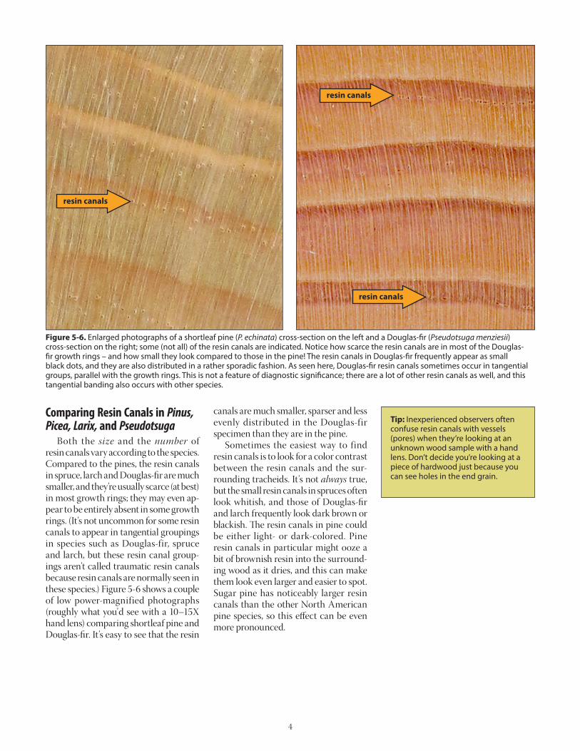
Figure 5-6. Enlarged photographs of a shortleaf pine (P. echinata) cross-section on the left and a Douglas-fir (Pseudotsuga menziesii) cross-section on the right; some (not all) of the resin canals are indicated. Notice how scarce the resin canals are in most of the Douglas-fir growth rings – and how small they look compared to those in the pine! The resin canals in Douglas-fir frequently appear as small black dots, and they are also distributed in a rather sporadic fashion. As seen here, Douglas-fir resin canals sometimes occur in tangential groups, parallel with the growth rings. This is not a feature of diagnostic significance; there are a lot of other resin canals as well, and this tangential banding also occurs with other species.
Comparing Resin Canals in Pinus, Picea, Larix, and Pseudotsuga Both the size and the number of resin canals vary according to the species. Compared to the pines, the resin canals in spruce, larch and Douglas-fir are much smaller, and they’re usually scarce (at best) in most growth rings; they may even ap-pear to be entirely absent in some growth rings. (It’s not uncommon for some resin canals to appear in tangential groupings in species such as Douglas-fir, spruce and larch, but these resin canal group-ings aren’t called traumatic resin canals because resin canals are normally seen in these species.) Figure 5-6 shows a couple of low power-magnified photographs (roughly what you’d see with a 10–15X hand lens) comparing shortleaf pine and Douglas-fir. It’s easy to see that the resin
canals are much smaller, sparser and less evenly distributed in the Douglas-fir specimen than they are in the pine. Sometimes the easiest way to find resin canals is to look for a color contrast between the resin canals and the sur-rounding tracheids. It’s not always true, but the small resin canals in spruces often look whitish, and those of Douglas-fir and larch frequently look dark brown or blackish. The resin canals in pine could be either light- or dark-colored. Pine resin canals in particular might ooze a bit of brownish resin into the surround-ing wood as it dries, and this can make them look even larger and easier to spot. Sugar pine has noticeably larger resin canals than the other North American pine species, so this effect can be even more pronounced.
resin canals
resin canals
resin canals

5
Figure 5-7. Southern pine (P. spp.). The resin canals are much larg-er than the tracheids, and the resin canals are large and plentiful.
Figure 5-8. The resin canals in Douglas-fir (Pseudotsuga menziesii) are much smaller than they are in the pines. For comparison, note that the diameters of the longitudinal tracheids in southern pine (Figure 5-6) are similar in size to those in this figure. Note the differ-ence in the overall diameters of the resin canals between the two species.
Comparing Resinous Species Cross-Sections This is a good point to start comparing the appearance of the various resinous genera. Pines have the largest and most plentiful resin canals, so let’s look at them first and then look at the other resinous genera. (The micrographs in this section are reproduced at the same magnification so you can see the relative size of the resin canals in these species. The magnifica-tion is greater than you would observe with a typical hand lens.) Figure 5-7 is a micrograph of a piece from one of the southern pines (loblolly, shortleaf, longleaf, etc.); some of the resin canals are plugged with resin and some are more open. Notice how large they are compared to the tracheids.
Compare the southern pine mi-crograph to the one of Douglas-fir4 (Pseudotsuga menziesii) (Figure 5-8). Compared to southern pine the resin canals in Douglas-fir are much smaller in diameter, and they are much more sparsely and unevenly distributed (i.e., the number of resin canals in each growth ring typically varies in Douglas-fir, but in the pines just about every growth ring has lots of resin canals). In the following micrograph there’s a single solitary resin canal (it’s smaller than the ones in the southern pine) and a tangen-tial string of even smaller resin canals.
4 These micrographs are displayed at identical magnification, but the sizes of the photos vary according to the growth ring widths of my specimens.
This tangentially oriented grouping isn’t common in Douglas-fir but it does show up occasionally, which is why I've illus-trated it here. As previously mentioned, these resin canals aren’t of any diagnostic value as tangential groupings of them occasionally show up in other species. Neglecting this tangential grouping in Figure 5-8 for the moment, notice how few resin canals are otherwise present in the photograph (just one!). Figure 5-9 is another micrograph, this time illustrating tamarack (Larix laricina), also known as eastern larch, American larch, or hackmatack.5 Once again, the resin canals are smaller than
5 According to Wikipedia, the name hack-matack means “wood used for snow-shoes” in Algonquian!
resin canals
resin canal
resin canals
Where trade names are used, no endorsement is intended, nor criticism implied of similar products not named.
Educational programs of Kentucky Cooperative Extension serve all people regardless of race, color, age, sex, religion, disability, or national origin. Issued in furtherance of Coop-erative Extension work, Acts of May 8 and June 30, 1914, in cooperation with the U.S. Department of Agriculture, Nancy M. Cox, Director of Cooperative Extension Programs, Uni-versity of Kentucky College of Agriculture, Food and Environment, Lexington, and Kentucky State University, Frankfort. Copyright © 2015 for materials developed by University of Kentucky Cooperative Extension. This publication may be reproduced in portions or its entirety for educational or nonprofit purposes only. Permitted users shall give credit to the author(s) and include this copyright notice. Publications are also available on the World Wide Web at www.ca.uky.edu.Issued 12-2015
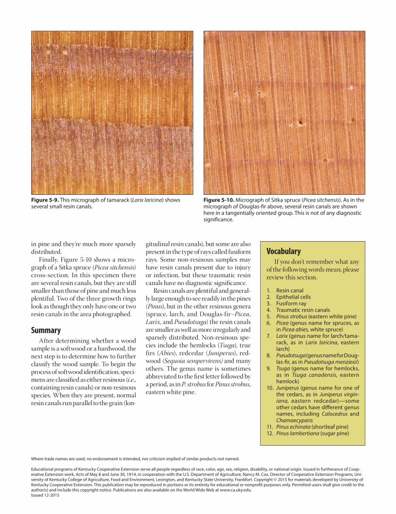
in pine and they’re much more sparsely distributed. Finally, Figure 5-10 shows a micro-graph of a Sitka spruce (Picea sitchensis) cross-section. In this specimen there are several resin canals, but they are still smaller than those of pine and much less plentiful. Two of the three growth rings look as though they only have one or two resin canals in the area photographed.
Summary After determining whether a wood sample is a softwood or a hardwood, the next step is to determine how to further classify the wood sample. To begin the process of softwood identification, speci-mens are classified as either resinous (i.e., containing resin canals) or non-resinous species. When they are present, normal resin canals run parallel to the grain (lon-
Vocabulary If you don’t remember what any of the following words mean, please review this section.
1. Resin canal2. Epithelial cells3. Fusiform ray4. Traumatic resin canals5. Pinus strobus (eastern white pine)6. Picea (genus name for spruces, as
in Picea abies, white spruce)7. Larix (genus name for larch/tama-
rack, as in Larix laricina, eastern larch)
8. Pseudotsuga (genus name for Doug-las-fir, as in Pseudotsuga menziesii)
9. Tsuga (genus name for hemlocks, as in Tsuga canadensis, eastern hemlock)
10. Juniperus (genus name for one of the cedars, as in Juniperus virgin-iana, eastern redcedar)—some other cedars have different genus names, including Calocedrus and Chamaecyparis
11. Pinus echinata (shortleaf pine)12. Pinus lambertiana (sugar pine)
gitudinal resin canals), but some are also present in the type of rays called fusiform rays. Some non-resinous samples may have resin canals present due to injury or infection, but these traumatic resin canals have no diagnostic significance. Resin canals are plentiful and general-ly large enough to see readily in the pines (Pinus), but in the other resinous genera (spruce, larch, and Douglas-fir–Picea, Larix, and Pseudotsuga) the resin canals are smaller as well as more irregularly and sparsely distributed. Non-resinous spe-cies include the hemlocks (Tsuga), true firs (Abies), redcedar (Juniperus), red-wood (Sequoia sempervirens) and many others. The genus name is sometimes abbreviated to the first letter followed by a period, as in P. strobus for Pinus strobus, eastern white pine.
Figure 5-9. This micrograph of tamarack (Larix laricina) shows several small resin canals.
Figure 5-10. Micrograph of Sitka spruce (Picea sitchensis). As in the micrograph of Douglas-fir above, several resin canals are shown here in a tangentially oriented group. This is not of any diagnostic significance.