foot and mouth disease fiebre aftosa. overview organism economic impact epidemiology transmission...
TRANSCRIPT

Foot and Mouth Disease
Fiebre Aftosa

Overview
• Organism• Economic Impact• Epidemiology• Transmission• Clinical Signs• Diagnosis and Treatment• Prevention and Control • Actions to take
Center for Food Security and Public Health, Iowa State University, 2011

THE ORGANISM

The Virus
• Picornaviridae, Aphthovirus– 7 distinct serotypes– Not cross protective
• Cloven-hoofed animals– Two-toed
• Inactivation– pH below 6.5 and above 11
• Survives in milk, milk products, bone marrow, lymph glands
Center for Food Security and Public Health, Iowa State University, 2011

IMPORTANCE

History
• 1929: Last case in U.S.• 1953: Last cases in
Canada and Mexico• 1993: Italy• 1997: Taiwan• United Kingdom
– 1967-68, 1981– 2001, 2007
Center for Food Security and Public Health, Iowa State University, 2011

Economic Impact
• Direct costs– Economic losses
to farmers and producers
– Eradication costs– Millions to
billions of dollars lost
• Indirect costs– Exports shut
down– $14 billion in lost
farm income– $6.6 billion in
livestock exports– Consumer fear
Center for Food Security and Public Health, Iowa State University, 2011
Economically Devastating

EPIDEMIOLOGY

Geographic Distribution
Center for Food Security and Public Health, Iowa State University, 2011

Countries with RoutineFMD Vaccination
Center for Food Security and Public Health, Iowa State University, 2011

Morbidity/ Mortality
• Morbidity 100% in susceptible animal population– U.S., Canada, Mexico, others
• Mortality less than 1%– Higher in young animals and
highly virulent virus strains– Animals generally destroyed to
prevent spread
Center for Food Security and Public Health, Iowa State University, 2011

TRANSMISSION

Animal Transmission
• Respiratory aerosols– Travel long distances– Proper temperature and humidity
• Direct contact – Vesicular fluid– Ingestion of infected animal parts
• Indirect contact via fomites– Boots, hands, clothing
Center for Food Security and Public Health, Iowa State University, 2011
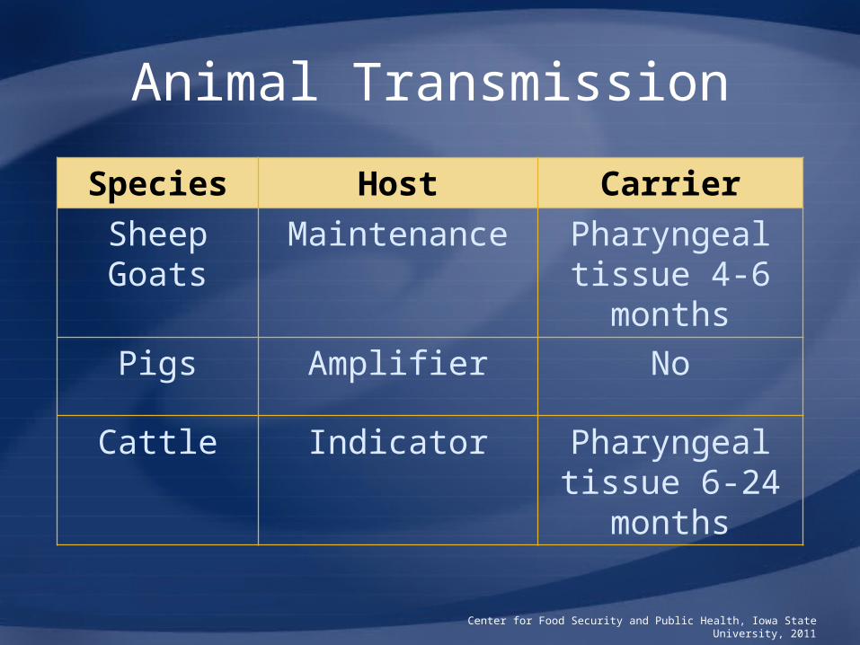
Animal Transmission
Center for Food Security and Public Health, Iowa State University, 2011
Species Host Carrier
Sheep Goats
Maintenance Pharyngeal tissue 4-6 months
Pigs Amplifier No
Cattle Indicator Pharyngeal tissue 6-24
months

Human Transmission
• Clinical disease rare– Infected by direct contact, ingestion of
unprocessed milk/dairy products – Type O, C, rarely A
• Transmit virus to animals– Rarely harbor virus in respiratory
tract for 1-2 days• Low risk of prolonged carriage
– Contaminated boots, clothing, vehicles
Center for Food Security and Public Health, Iowa State University, 2011

DISEASE IN ANIMALS

Clinical Signs
• Incubation period: 2 to 14 days• Fever and vesicles
– Feet, mouth, nares muzzle, teats
– Progress to erosions
• Lameness, reluctance to move, sloughing of hooves
• Abortion• Death in young animals
Center for Food Security and Public Health, Iowa State University, 2011

Clinical Signs: Cattle
• Oral lesions (vesicles)– Tongue, dental pad,
gums, soft palate, nostrils, muzzle
– Excess salivation, drooling, nasal discharge
• Lethargy, loss of body condition
Center for Food Security and Public Health, Iowa State University, 2011
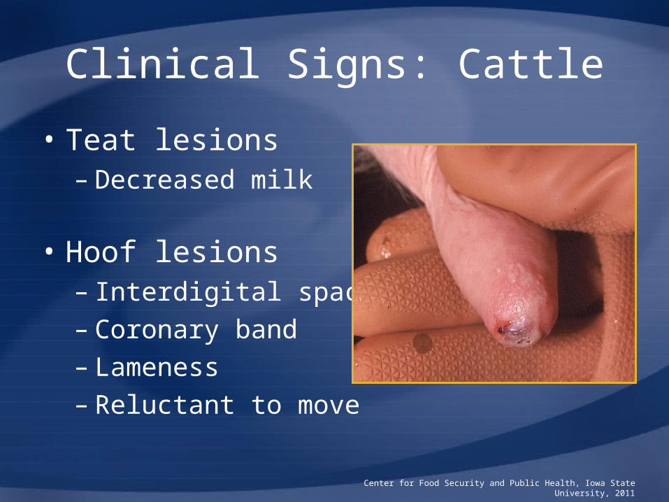
Clinical Signs: Cattle
• Teat lesions– Decreased milk
production
• Hoof lesions– Interdigital space– Coronary band– Lameness– Reluctant to move
Center for Food Security and Public Health, Iowa State University, 2011
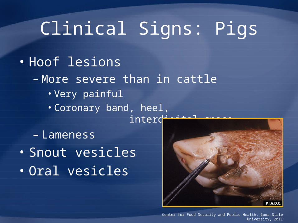
Clinical Signs: Pigs
• Hoof lesions– More severe than in cattle
• Very painful• Coronary band, heel,
interdigital space
– Lameness
• Snout vesicles• Oral vesicles
less common
Center for Food Security and Public Health, Iowa State University, 2011

Clinical Signs: Sheep and Goats
• Mild, if any– Fever– Lameness– Oral lesions
• Makes diagnosisand prevention of spread difficult
Center for Food Security and Public Health, Iowa State University, 2011

Center for Food Security and Public Health, Iowa State University, 2011
Foot & Mouth Disease
Vesicular Stomatitis
Swine Vesicular Disease
Vesicular Exanthema of
Swine
Clinical Signs by Species
All vesicular diseases produce a fever with vesicles that progress to erosions in the mouth, nares, muzzle, teats, and feet
Cattle
Oral & hoof lesions, salivation, drooling, lameness, abortions,
death in young animals, "panters";
Disease Indicators
Vesicles in oral cavity, mammary glands, coronary
bands, interdigital space
Not affected Not affected
Pigs
Severe hoof lesions, hoof sloughing, snout vesicles, less severe
oral lesions: Amplifying Hosts
Same as cattle
Severe signs in animals housed on
concrete; lameness, salivation,
neurological signs, younger more
severe
Deeper lesions with granulation tissue
formation on the feet
Sheep & Goats
Mild signs if any; Maintenance Hosts
Rarely show signs Not affected Not affected
Horses, Donkeys,
Mules Not affected
Most severe with oral and coronary
band vesicles, drooling, rub
mouths on objects, lameness
Not affected Not affected
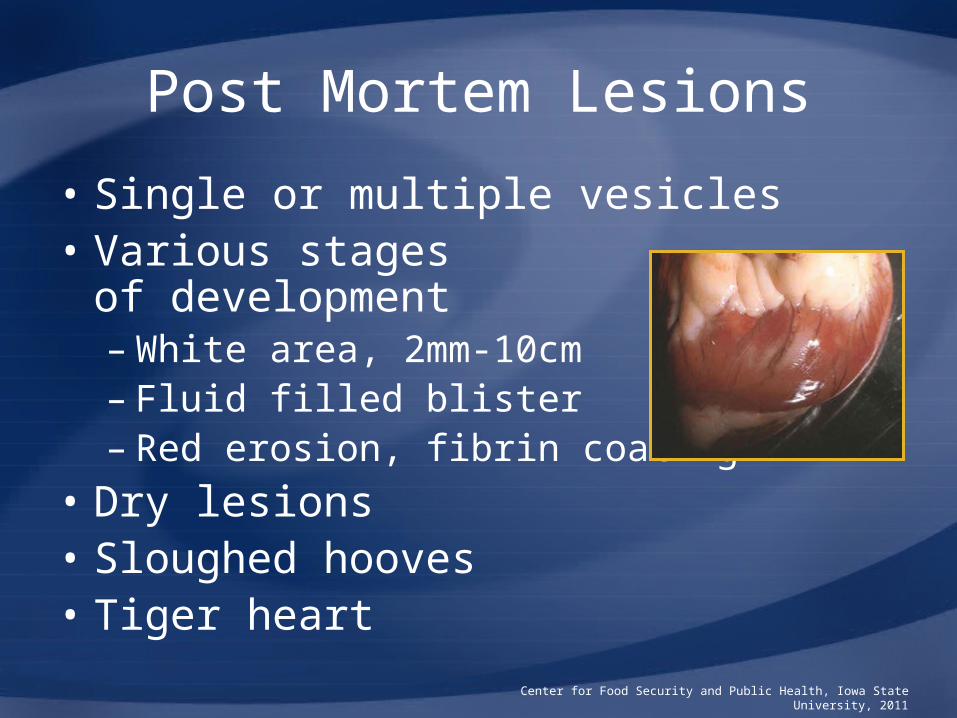
Post Mortem Lesions
• Single or multiple vesicles• Various stages
of development– White area, 2mm-10cm – Fluid filled blister– Red erosion, fibrin coating
• Dry lesions• Sloughed hooves• Tiger heart
Center for Food Security and Public Health, Iowa State University, 2011

Differential Diagnosis
• Swine– Vesicular stomatitis– Swine vesicular disease– Vesicular exanthema
of swine
• Cattle– Rinderpest, IBR, BVD, MCF, Bluetongue
• Sheep– Bluetongue, contagious ecthyma
Center for Food Security and Public Health, Iowa State University, 2011

Sampling
• Before collecting or sending any samples, the proper authorities should be contacted
• Samples should only be sent under secure conditions and to authorized laboratories to prevent the spread of the disease
Center for Food Security and Public Health, Iowa State University, 2011

Clinical Diagnosis
• Vesicular diseases are clinically indistinguishable!
• Suspect animals with salivation or lameness and vesicles
• Tranquilization may be necessary
• Laboratory testing essential
Center for Food Security and Public Health, Iowa State University, 2011
Laboratory Diagnosis
• Initial diagnosis– Virus isolation– Virus identification
• ELISA, RT-PCR, complement fixation
• Serology– ELISA and virus neutralization
• Notify authorities and wait for instructions before collecting samples
Center for Food Security and Public Health, Iowa State University, 2011
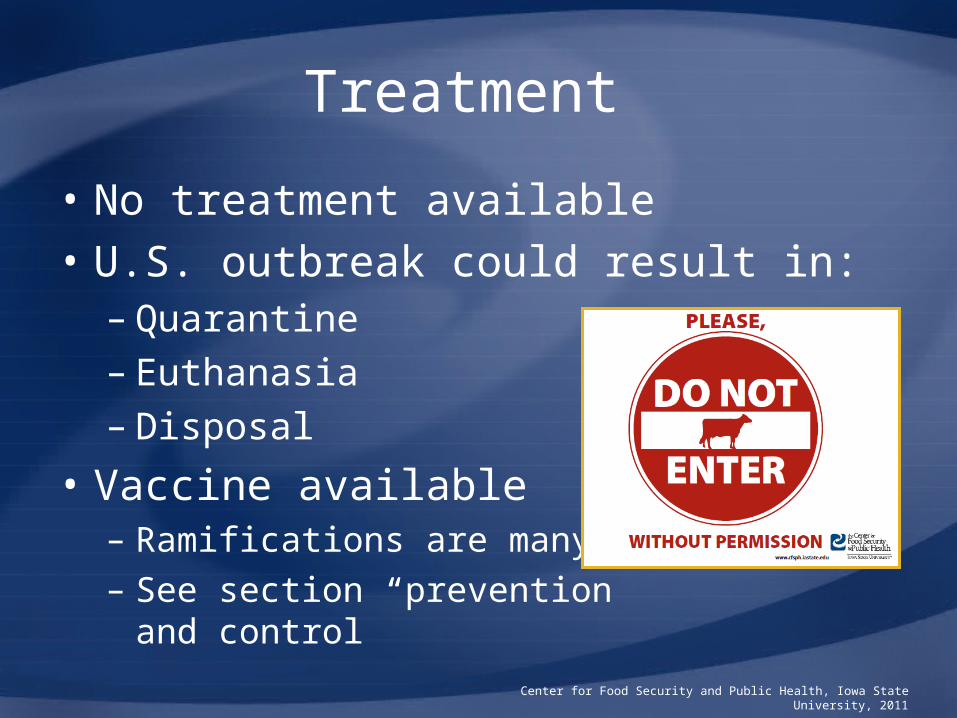
Treatment
• No treatment available• U.S. outbreak could result in:
– Quarantine– Euthanasia– Disposal
• Vaccine available– Ramifications are many– See section “prevention
and control”
Center for Food Security and Public Health, Iowa State University, 2011

DISEASE IN HUMANS
Disease in Humans
• Very low incidence– 40 cases since 1921
• Most reports ended when FMD was eradicated in Europe
– NOT a public health concern• Incubation period: 2 to 6 days• Clinical signs
– Mild headache, malaise, fever– Tingling, burning sensation of fingers,
palms, feet prior to vesicle formation
Center for Food Security and Public Health, Iowa State University, 2011
Clinical Signs: Humans
• Vesicles – Fluid-filled, 2 mm to 2 cm in diameter– Tongue, palate
• Painful• Interfere in eating, drinking, talking
– Vesicles dry up in 2 to 3 days
• Diarrhea• Recover within one week of last
blister appearingCenter for Food Security and Public Health, Iowa State University, 2011

Diagnosis and Treatment
• Clinically FMD in humans resembles:– Coxsackie A group viruses
• Hand, foot, and mouth disease• Herpangina
– Herpes simplex virus– Vesicular stomatitis
• Virus isolation or antibody identification required for diagnosis
• Treatment is supportive careCenter for Food Security and Public Health, Iowa State University, 2011
PREVENTION AND CONTROL

Prevention
• Strict import restrictions– Prohibit live ruminants, swine, and their
products from FMD-affected countries– Heat-treatment of swill (garbage)
fed to pigs• Swine Health
Protection Act
– Travelers, belongings monitored at ports of entry
Center for Food Security and Public Health, Iowa State University, 2011

Prevention
• Suspicious lesions investigated• State planning/training exercises• Federal response plans• Biosecurity protocols for livestock
facilities
Center for Food Security and Public Health, Iowa State University, 2011

Recommended Actions
• Notification of Authorities– Federal Area Veterinarian in Charge
(AVIC) http://www.aphis.usda.gov/animal_health/area_offices/
– State Veterinarians www.usaha.org/stateanimalhealthofficials.aspx
• Quarantine
Center for Food Security and Public Health, Iowa State University, 2011

Recommended Actions
• Confirmatory diagnosis
• Depopulation– Must properly
destroy exposed cadavers, litter, animal products
Center for Food Security and Public Health, Iowa State University, 2011
Center for Food Security and Public Health, Iowa State University, 2011
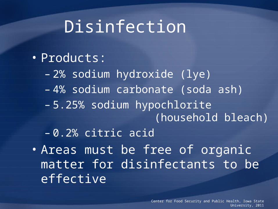
Disinfection
• Products:– 2% sodium hydroxide (lye)– 4% sodium carbonate (soda ash)– 5.25% sodium hypochlorite
(household bleach) – 0.2% citric acid
• Areas must be free of organic matter for disinfectants to be effective

Vaccination
• Killed vaccine, serotype specific• North American Foot-and-Mouth
Vaccine Bank– Plum Island, NY
• Monitor disease outbreaks worldwide• Stock active serotypes and strains• Essential to isolate virus and identify
the serotype to select correct vaccine
Center for Food Security and Public Health, Iowa State University, 2011

Vaccination
• Currently U.S. has no need to vaccinate• But, vaccine may be used in an outbreak• Vaccination issues
– Annual re-vaccination required• Costly, time consuming
– Does not protect against infection, but reduces clinical signs• Spread infection to other animals
– International trade status harmed
Center for Food Security and Public Health, Iowa State University, 2011

Additional Resources
• World Organization for Animal Health (OIE)– www.oie.int
• U.S. Department of Agriculture (USDA)– www.aphis.usda.gov
• Center for Food Security and Public Health– www.cfsph.iastate.edu
• USAHA Foreign Animal Diseases(“The Gray Book”)– http://www.aphis.usda.gov/
emergency_response/downloads/nahems/fad.pdf
Center for Food Security and Public Health, Iowa State University, 2011
Acknowledgments
Development of this presentation was made possible through grants provided to
the Center for Food Security and Public Health at Iowa State University, College of Veterinary Medicine from
the Centers for Disease Control and Prevention, the U.S. Department of Agriculture,
the Iowa Homeland Security and Emergency Management Division, and the
Multi-State Partnership for Security in Agriculture.
Authors: Danelle Bickett-Weddle, DVM, MPH; Co-authors: Anna Rovid Spickler, DVM, PhD; Kristina August, DVMReviewers: James A. Roth, DVM, PhD; Bindy Comito, BA; Heather Sanchez, BS; Glenda Dvorak, DVM, MPH, DACVPM; Kerry Leedom Larson, DVM, MPH, PhD
Center for Food Security and Public Health, Iowa State University, 2011