focus: recombinant antibodies - adipogen.com · 3rd edition functional antibodies focus:...
TRANSCRIPT

www.adipogen.com
3rd Edition
Functional AntibodiesFocus: Recombinant Antibodies
Antibodies are highly specific, naturally evolved molecules that recognize and eliminate pathogenic and disease antigens. The typical antibody consists of two antigen-binding fragments (Fabs), which are linked via a flexible region (the hinge) to a constant Fc region. This structure comprises two pairs of polypeptide chains, each pair containing a heavy and a light chain of different sizes. The Fc portion of the Ig serves to bind various effector molecules of the immune system, as well as molecules that determine the biodistribution of the antibody. Antibodies are produced by: a) Injecting an antigen into mammals (mouse, rat, rabbit, goat, etc). Blood isolated from these animals contains polyclonal antibodies (multiple antibodies that bind to different epitopes of the same antigen), which are purified. b) Hybridoma technology generating monoclonal antibodies (epitope specific). Specific antibody-secreting lymphocytes are isolated from animals and immortalized by fusing them with a cancer cell line. Monoclonal antibodies are routinely used in biochemistry, molecular biology, medical research and as therapeutic agents. Important advances have been made over the past decade to improve the specificity and efficacy of such antibodies by new engineering technologies, including recombinant antibody technology, such as antibody phage display (see page 8 for more information).
Functional Grade Antibodies (FuncAbs™):Antibodies displaying an agonist or antagonist activity (functional grade antibodies (FuncAbs™)) are powerful tools for mimicking or blocking physiological functions in vitro and in vivo. Functional grade antibodies are available free of preservatives and tested for low endotoxin content and may be used for activation, neutralizing or blocking studies, both in vitro or in vivo.
CONTENTS
Blocking/Neutralizing Antibodies 2–5
mAPRIL | m/hAng-2 | hADAM17 | mBAFF |hBTLA | hCD40L | hOX40L | hLAG-3 2–3
mIL-33 | hNetrin-1 |Zika Virus | NS5B (HCV) |mBAFF-R | mNeutrophils 4–5
Inducing/Activating Antibodies 6
mCD40 | mLTbR | hFas | hLewisy/b hapten
Recombinant Monoclonal Antibodies 7–9
Conformation-specific Antibodies 8
Post-translational Modification-specific Antibodies 9
Functional Antibodies from 10–11
NEW mPD-1 Blocking mAb 12
Gold Standards for Inflammasome Research 12
Asc | NLRP3 | cleaved Caspase-1 (p20) Antibodies

2Works in Human Works in Mouse
Ask for BULK Quotes! [email protected]
APPlICAtIoNS: FUNC: Functional Application; ICC: Immunocytochemistry; IHC: Immunohistochemistry; IP: Immunoprecipitation; WB: Western blot FoRMulAtIoN: PF = Preservative free
Blocking/Neutralizing Functional AntibodiesFuncAbs™
anti-Angiopoietin-2 mAb (rec.) (blocking) (Angy-2-1)AG-27B-0016 100 μgAG-27B-0016PF Preservative Free 100 μg | 500 μg | 1 mg
Isotype: Mouse IgG2bλApplication: ELISA, ICC, FUNC (Blocking)
Functional Application Mouse: Inhibits the binding of mouse angiopoietin-2 to mouse Tie-2. ND50* = 50-60ng/ml (for 10ng/ml of mouse angiopoietin-2) Human: Inhibits the binding of human angiopoietin-2 to human Tie-2. ND50* = 8-12ng/ml (for 10ng/ml of human angiopoietin-2)
*ND50 : = 50% neutralizing dose of antibody for a given concentration of ligand.
LIT: Elevated angiopoietin-2 level in patients with continuous-flow left ventricular assist devices leads to altered angiogenesis and is associated with higher nonsurgical bleed-ing: C.E. Tabit, et al.; Circulation 134, 141 (2016)
anti-APRIl (mouse), mAb (rec.) (blocking) (Apry-1-1)
anti-ADAM17 (human), mAb (rec.) (blocking) (D1(A12))
AG-27B-0001 100 μgAG-27B-0001PF Preservative Free 100 μg AG-27B-0001B Biotin 100 μg
Isotype Mouse IgG2bλApplication ELISA, IP, FUNC (Blocking)
Functional Application Inhibits binding of mouse APRIL to mouse BCMA and TACI.
LIT: Production of the plasma-cell survival factor APRIL peaks in myeloid precursor cells from human bone marrow: T. Matthes, et al.; Blood 118, 1838 (2011)
AG-27B-6000PF Preservative Free 100 µg
Isotype Human IgG1Application FUNC (Blocking)
Functional Application Inhibits human ADAM17 activity at 15μg/ml (200nM).
LIT: Cross-domain inhibition of TACE ectodomain: C.J. Tape, et al.; PNAS 108, 5578 (2011) • Targeting ADAM-17 with an inhibitory monoclonal antibody has antitumour effects in triple-negative breast cancer cells: F. Caiazza, et al.; Br. J. Cancer 112, 1895 (2015)
Newly released: anti-APRIl (mouse), mAb (rec.) (blocking) (Apry-1-3) Prod. No. AG-27B-0017
Also available: anti-ADAM17 (human), mAb (rec.) (blocking) (D1(A12)) (Fab) (His) Prod. No. AG-27B-6003PF
Also available: anti-Angiopoietin-2 (human), mAb (rec.) (blocking) (Angy-1-4) Prod. No. AG-27B-0015
UNIQUE
potENt
FIgure: Antagonizing Angiopoietin-2 in vivo with anti-ANG-2, mAb (rec.) (blocking) (Angy-2-1) (AG-27B-0016PF) increases triglyceride levels.
MeThod: After High Fat Diet (HFD) challenges for five weeks in wild-type C57BL/6 mice, control IgG (left panel) or anti-ANG-2 (Clone Angy-2-1) blocking antibody (right panel) (4 µg/g body weight; twice/week) were administrated and afterwards the mice underwent metabolic analyses of the triglycerides levels from both groups.
FIgure: D1(A12) IgG inhibits constitutive shed-ding of TNF-α from IGROV1 (human ovarian cancer cell line) into culture medium. Medium was collected after 48 hours of incubation with or without IgGs at 200nM.

www.adipogen.com
For updated prices and additional information visit www.adipogen.com or contact your local distributor.
3
anti-BAFF (mouse), mAb (blocking) (Sandy-2)
anti-BtlA (human), mAb (blocking) (6F4)
anti-CD40l (human), mAb (rec.) (blocking) (hu5c8)
anti-oX40l (human), mAb (rec.) (blocking) (R4930)
anti-lAG-3 (human), mAb (blocking) (17B4)
anti-VEGF-A (human), mAb (3(6D3))
AG-20B-0063 100 μgAG-20B-0063PF Preservative Free 100 μg
Isotype Mouse IgG1Application IP, FUNC (Blocking)
Functional Application Inhibition of mouse BAFF binding to BAFF-R and TACI (BCMA not tested); blocks BAFF activity in mice.
LIT: Antibodies that block or activate mouse B cell activating factor of the TNF family (BAFF) respectively induce B cell depletion or B cell hyperplasia: C. Kowalczyk-Quintas, et al.; J. Biol. Chem. 291, 19826 (2016)
AG-20B-0049 100 µg
Application ELISA, FACS, FUNC (Blocking)
AG-27B-6002PF Preservative Free 100 µg
Application WB, FACS, FUNC (Blocking)
AG-27B-6001PF Preservative Free 100 µg
Application FACS, FUNC (Blocking)
AG-20B-0012 100 µg AG-20B-0012PF Preservative Free 100 µg
Application FACS, ICC, IHC, IP, WB, FUNC (Blocking)
AG-20T-0105 200 µg
Application ELISA, WB, FUNC (Blocking)
Also available: anti-BAFF (human), mAb (blocking) (4.62) Prod. No. AG-20B-0017
Also available: anti-lAG-3, mAb (blocking) (11E3) Prod. No. AG-20B-0011
UNIQUE
FIgure: anti-BAFF (mouse), mAb (Sandy-2) (preservative free) (Prod. No. AG-20B-0063PF) blocks the action of endogenous BAFF in vivo. MeThod: Wild type C57BL/6 mice were treated at day 0 (single admin-istration) with monoclonal antibody anti-BAFF (mouse), mAb (Sandy-2) (preservative free) (at 2mg/kg). Lymph nodes were prepared at week 2 and analyzed by FACS for the presence of T (CD3) and B (CD19) cells. Untreated BAFF WT and KO mice were analyzed in parallel.
Functional Application Inhibits interaction of BTLA to HVEM or UL144.
Functional ApplicationNeutralizes CD40L function by blocking the interaction between CD40 and CD40L in vitro and in vivo.
Functional ApplicationBinds to the co-stimulatory human OX40L inhibiting its interaction with OX40 in vitro and in vivo.
Functional Application Blocks LAG-3/MHC class II interactions.
Functional Application Inhibits VEGF-A signaling.
LIT: T Cell Intrinsic Heterodimeric Complexes between HVEM and BTLA Determine Receptivity to the Surrounding Microenvironment: T.C. Cheung, et al.; J. Immunol. 183, 7286 (2009)
LIT: Enhancement of T cell activation by immobilized hu5C8 (anti-CD40L) monoclonal antibody: M. Arpinati, et al.; Eur. J. Haematol. 80, 322 (2008)
LIT: OX40L blockade and allergen-induced airway responses in subjects with mild asthma: G.M. Gauvreau, et al.; Clin. Exp. Allergy 44, 29 (2014)
LIT: DLL1-mediated Notch activation regulates endothelial identity in mouse fetal arteries: I. Sörensen, et al.; Blood 113, 5680 (2009)
Ihc GradE
LIT: The negative regulatory function of the lymphocyte-activa-tion gene-3 co-receptor (CD223) on human T cells: L. Macon-Lemaitre and F. Triebel; Immunology 115, 170 (2005)

4APPlICAtIoNS: FUNC: Functional Application; ICC: Immunocytochemistry; IHC: Immunohistochemistry; IP: Immunoprecipitation; WB: Western blot FoRMulAtIoN: PF = Preservative free
Works in Human Works in MouseAsk for BULK Quotes! [email protected]
Blocking/Neutralizing Functional AntibodiesFuncAbs™
anti-Il-33 (mouse), mAb (rec.) (blocking) (Bondy-1-1)AG-27B-0013 100 μgAG-27B-0013PF Preservative Free 100 μg | 500 μg | 1 mg
Isotype Mouse IgG2bApplication ELISA, FUNC (Blocking)
Functional Application Inhibits the binding of mouse IL-33 to ST2/IL-1RAcP.
UNIQUE
FIgure: Binding of IL-33 (mouse) to ST2/IL-1RAcP is inhibited by Bondy-1-1. IL-33 (mouse) was coated on an ELISA plate at 1μg/ml. Bondy-1-1 or an unrelated mAb (recombinant) (Control) were added (starting at 40μg/ml with a twofold serial dilution) together with 100μl of supernatant of cells containing ST2 (human):Fc/IL-1RAcP (human):Fc. After incubation for 1 h at RT, the binding was detected using an anti-Fc human antibody (HRP).
other Blocking Antibodies See www.adipogen.com for More Information !
anti-Zika Virus Envelope Protein (EIII domain), mAb (rec.) (neutralizing) (ZKA64)AG-27B-6004PF Preservative Free 100 µg
Application FUNC (Neutralizing)Functional Application Neutralizes Zika Virus (ZIKV) with an IC50 of ~93ng/ml. Also enhances ZIKV infection in non-permissive K562 cells at a broad range of concentrations (not above 1µg/ml).
LIT: Specificity, cross-reactivity, and function of antibodies elicited by Zika virus infection: K. Stettler, et al.; Science 353, 823 (2016)
NEw
ANtIBoDIES PID APPLICATIONS FUNCTIONAL APPLICATION
anti-Fibronectin (EDA), mAb (blocking) (ISt-9) (PF)
AG-20B-6001PF ELISA, FUNC, ICC, IHC, WB
Inhibits binding of Fibronectin EDA domain to α5-b1, α4-b1 and α9-b1 integrins.
anti-Periostin, mAb (blocking) (oC-20) (PF) AG-20B-6000PF ICC, FUNC Blocks interaction with the integrins αvb3 and αvb5. Inhibits angiogenesis and tumor growth and blocks allergen-induced inflammation in vivo in mice.
anti-NS5B (HCV), mAb (blocking) (5B-12B7) AG-20B-0003 ICC, IP, FUNC Blocks the RNA-dependent RNA polymerase activity in vitro.
anti-tRAIl-R1 (human), mAb (HS101) (PF) AG-20B-0022PF FACS, IP, ICC, FUNC
Inhibition/Neutralizing (blocks TRAIL-R1 mediated killing if applied in solution).
anti-tRAIl-R2 (human), mAb (HS201) (PF) AG-20B-0023PF FACS, IP, ICC, FUNC
Inhibition/Neutralizing (blocks TRAIL-R2 mediated killing if applied in solution).
LIT: Regulation of de novo adipocyte differentiation through crosstalk between adipocytes and pre-adipo-cytes: T.D. Challa, et al.; Diabetes 64, 4075 (2015) • Male-specific IL-33 expression regulates sex-dimorphic EAE susceptibility: A.E. Russi, et al.; PNAS (epub ahead of print) (2018)
AG-27B-0018PF Preservative Free 100 µg | 500 µg
Isotype Human IgG2λApplication ELISA, FUNC (Blocking)
Functional Application Inhibits the activation of human and mouse Netrin-1 to human or mouse receptors DCC or UNC5 (KD antibody-Netrin-1 is 1.5nM).
LIT: Targeting netrin-1/DCC interaction in diffuse large B-cell and mantle cell lymphomas: T. Broutier, et al.; EMBO Mol. Med. 8, 96 (2016)
FIgure: anti-Netrin-1 (human), mAb (rec.) (blocking) (2F5) (preservative free) (Prod. No. AG-27B-0018PF) blocks tumor growth in vivo.Method: Tumor cells (OCI-Ly3) were implanted in SCID mice by subcutaneous injection of 3x10⁶ cells in 100µl of PBS. When tumors reached 150mm3, mice received intraperitoneal injections of blocking anti-Netrin-1 mAb (2F5) at 20mg/kg or an equal volume of the antibody control anti-Netrin-1 (human), mAb (rec.) (H4) (preservative free) (Prod. No. AG-27B-0020PF) every two days. Tumor growth rates from the beginning of treatment are shown.
anti-Netrin-1 (human), mAb (rec.) (blocking) (2F5)
H42F5

www.adipogen.com
For updated prices and additional information visit www.adipogen.com or contact your local distributor.
5
Custom Recombinant Monoclonal Antibodies [RecMAbs™] Antibodies developed from a NON-ANIMAL SOURCE using in vitro antibody phage display technology
FeatUres: • Developed from a human antibody phage display library.• Consists of scFv (single chain fragment variable) composed of VH
(variable domain of the human immunoglobulin heavy chain) and VL (variable domain of the human immunoglobulin light chain) fused to a Fc region.
• Produced in mammalian cells (CHO or HEK 293).• Similar properties compared to monoclonal antibodies developed in
mice / rat (e.g. affinity in the low nanomolar range).
• Standard secondary antibodies can be used.• Ideal for conserved antigens
(which are poorly immunogenic in animals).• Detect conformational epitopes
(e.g. GTP-bound proteins).• Detect protein modifications
(e.g. phosphorylations, ubiquitinations).• Possibility to exchange the Fc region with
Fc from other species. Ask for Custom Production !
anti-BAFF-R (mouse), mAb (9B9)
anti-Neutrophils (mouse), mAb (blocking) (Nimp-R14)
AG-20B-0034 100 μgAG-20B-0034PF Preservative Free 100 μgAG-20B-0034B Biotin 100 μgDifferent labels available.
Isotype Rat IgG2aApplication ELISA, IP, FUNC (Depletion)
AG-20B-0043 100 μgAG-20B-0043PF Preservative Free 500 μg | 2 mg | 10 mgAG-20B-0043B Biotin 100 μgDifferent labels available.
Isotype Rat IgG2aApplication FACS, IHC, ICC, FUNC (Depletion)
Functional Application Depletes B cells in vivo.
LIT: Crucial role for BAFF-BAFF-R signaling in the survival and maintenance of mature B cells: M. Rauch, et al.; PLoS ONE 4, e5456 (2009)
Functional Application Optimal reagent to deplete neutrophils in vivo (250 μg/mouse).
LIT: An immunomodulatory function for neutrophils during the induction of a CD4+ Th2 response in BALB/c mice infected with Leishmania major: F. Tacchini-Cottier, et al.; J. Immunol. 165, 628 (2000)
FIgure: C57BL/6 mice were injected i.v. at day 0 with 0.5mg of 9B9. Abso-lute numbers of splenic T1 and T2/3 immature B cells, B-2 and MZ B cells, CD4 and CD8 T cells in controls (black bars) and 9B9 injected C57BL/6 mice at day 14 after injection (white bars). 5 mice were analyzed for each group.
The best depleting antibody for neutrophils in mice !
FIgure: Mouse neutrophils are depleted in vivo by Nimp-R14. Mice were injected i.p. with 250µg of Nimp-R14 (B) or with Control mAb (A) in BALB/c mouse 6 h prior to Leishmania major infection (3x10⁶ parasites injected in the hind footpad). 3 days later, blood (100µl) was subjected to flow cytometric analysis after staining with APC/CY7-labeled anti-Ly6G antibody (clone 1A8).
potENt
See Page 7 for More Information !

6APPlICAtIoNS: FUNC: Functional Application; ICC: Immunocytochemistry; IHC: Immunohistochemistry; IP: Immunoprecipitation; WB: Western blot FoRMulAtIoN: PF = Preservative free
Works in Human Works in MouseAsk for BULK Quotes! [email protected]
Inducing/Activating Antibodies
anti-CD40 (mouse), mAb (FGK45)
anti-Fas (human), mAb (APo-1-3)
AG-20B-0036 100 μg | 500 μgAG-20B-0036PF Preservative Free 100 μg | 500 μg
Isotype Rat IgG2aApplication FACS, FUNC (Activation)
Functional Application Activates B and NK cells in vitro and in vivo.
LIT: Ovarian insufficiency and early pregnancy loss induced by activation of the innate immune system: A. Erlebacher, et al.; J. Clin. Invest. 114, 39 (2004)
AG-20B-0062PF Preservative Free 50 μg
Isotype Mouse IgG3Application FACS, IP, WB, FUNC (Activation)
Functional Application Induces apoptosis with or without cross-linking (Protein A), depending on cell type.
LIT: Monoclonal antibody-mediated tumor regression by induction of apoptosis: B.C. Trauth, et al.; Science 245, 301 (1989)
anti-ltbR (mouse), mAb (4H8 WH2) AG-20B-0008 100 μg AG-20B-0008PF Preservative Free 100 μg
Isotype Rat IgG2aApplication FACS, FUNC (Activation)
Functional Application for 4H8 WH2: Agonists inducing BAFF, chemokines and integrins in vitro and in vivo.
LIT: LTβR Signaling Induces Cytokine Expression and Up-Regulates Lymphangiogenic Factors in Lymph Node Anlagen. M.F. Vondenhoff, et al.; J. Immunol. 182, 5439 (2009)
FIgure: Systemic immune activation by CD40 ligation. Mice were sacrificed on day 8 after daily treatment on day 4-7 with FGK45 or control. FGK45 treatment, elevated splenocyte numbers in both groups. *P < 0.005. Data represent mean ± SD for three to four mice per group.
FIgure: Induction of growth Inhibition by apoptosis by APO-1-3 or control medium. SKW6.4 cells were pre-incubated with APO-1-3 (100 nglml). ['H)TdR incorporation was measured.
FIgure: Treatment of cultured WT MEFs with agonistic LTbR mAb (4H8 WH2), but not with an isotype matched control mAb, results in the up-regu-lation of IL-7 mRNA expression. MEFs were collected at 6, 24, and 30 h after stimulation with 4H8 WH2. Relative expression levels at t = 0 were set at 1,0. Experiments were performed three times. *, p < 0.05.
Also available: anti-ltbR (mouse), mAb (3C8) Prod. No. AG-20B-0041
T H E staNdard
T H E staNdard
anti-lewisy/b hapten (human), mAb (SC104)AG-20B-6002PF Preservative Free 100 µg
Application ELISA, FACS, ICC, IHC, FUNC (Inducing) Functional Application Directly induces tumor-specific cell death without the need for immune effector cells by induction of caspase-mediated apoptosis. Optimal concentrations used to induce tumor cell death in vitro and in vivo are between 10-30μg/ml.
LIT: Development of second generation monoclonal antibodies recognising Lewisy/b antigen by anti-idiotypic immunisation: L.G. Durrant, et al.; Hybridoma 12, 647 (1993) • A new anticancer glycolipid monoclonal antibody, SC104, which directly induces tumor cell apoptosis: L.G. Durrant, et al.; Cancer Res. 66, 5901 (2006)
NEw
FuncAbs™
www.adipogen.com
For updated prices and additional information visit www.adipogen.com or contact your local distributor.
7
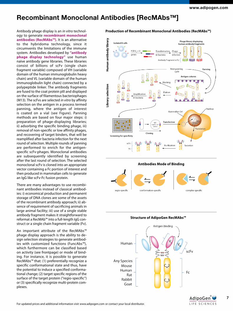
Antibody phage display is an in vitro technol- ogy to generate recombinant monoclonal antibodies (RecMAbs™). It is an alternative to the hybridoma technology, since it circumvents the limitations of the immune system. Antibodies developed by “antibody phage display technology” use human naïve antibody gene libraries. These libraries consist of billions of scFv (single chain fragment variable) composed of VH (variable domain of the human immunoglobulin heavy chain) and VL (variable domain of the human immunoglobulin light chain) connected by a polypeptide linker. The antibody fragments are fused to the coat protein pIII and displayed on the surface of filamentous bacteriophages (M13). The scFvs are selected in vitro by affinity selection on the antigen in a process termed panning, where the antigen of interest is coated on a vial (see Figure). Panning methods are based on four major steps: i) preparation of phage-displaying libraries; ii) adsorbing the specific binding phage, iii) removal of non-specific or low affinity phages, and recovering of target binders, that will be reamplified after bacteria infection for the next round of selection. Multiple rounds of panning are performed to enrich for the antigen-specific scFv-phages. Monoclonal antibodies are subsequently identified by screening after the last round of selection. The selected monoclonal scFv is cloned into an appropriate vector containing a Fc portion of interest and then produced in mammalian cells to generate an IgG like scFv-Fc fusion protein.
There are many advantages to use recombi-nant antibodies instead of classical antibod-ies: i) economical production and permanent storage of DNA clones are some of the assets of the recombinant antibody approach; ii) ab-sence of requirement of sacrificing animals in large animal facility; iii) use of a single stable antibody fragment makes it straightforward to reformat a RecMAb™ into a full-length IgG con-struct or a single chain fragment variable (Fv).
An important attribute of the RecMAbs™ phage display approach is the ability to de-sign selection strategies to generate antibod-ies with customized functions (FuncAbs™), which furthermore can be classified based on activity (see frontpage) or mode of bind-ing. For instance, it is possible to generate RecMAbs™ that: (1) preferentially recognize a specific conformational state and thus, have the potential to induce a specified conforma-tional change; (2) target specific regions of the surface of the target protein (‘‘regio-specific’’) or (3) specifically recognize multi-protein com-plexes.
Recombinant Monoclonal Antibodies [RecMAbs™]
Structure of AdipoGen RecMAbs™
Hum
an a
ntib
ody
libra
ryli
brar
y se
lect
ion
Antibodies Mode of Binding
Production of Recombinant Monoclonal Antibodies (RecMAbs™)

8APPlICAtIoNS: FUNC: Functional Application; ICC: Immunocytochemistry; IHC: Immunohistochemistry; IP: Immunoprecipitation; WB: Western blot FoRMulAtIoN: PF = Preservative free
Works in Human Works in MouseAsk for BULK Quotes! [email protected]
.
RecMAbs™
ANtIBoDIES PID SIZE ISOTYPE APPLICATIONS SPECIES
anti-lRP5/6, mAb (rec.) (Heldy-1-4) AG-27B-0019 100 µg Human IgG2λ FACS Hu, Ms
anti-Giantin, mAb (rec.) (tA10) AG-27B-0003 100 µg Human IgG2λ ICC Hu, Ms
anti-Giantin, mAb (rec.) (tA10) (Atto 488) AG-27B-0003TD 100 µg Human IgG2λ ICC Hu, Ms
anti-Myosin IIA (non-muscle) (heavy chain), mAb (rec.) (SF9)
AG-27B-0010 100 µg Human IgG2λ EM, ELISA, ICC, WB Hu, Ms, Rt, Dr
anti-Myosin IIA (non-muscle) (heavy chain), mAb (rec.) (SF9) (Atto 488)
AG-27B-0010TD 100 µg Human IgG2λ ICC Hu, Ms, Rt, Dr
anti-HMGB1, mAb (rec.) (Giby-1-4) AG-27B-0002 100 µg Human IgG2λ ELISA, WB Hu, Ms, Rt
anti-HMGB1, mAb (rec.) (Giby-1-4) (Biotin) AG-27B-0002B 100 µg Human IgG2λ ELISA, WB Hu, Ms, Rt
anti-Il-1R2 (mouse), mAb (rec.) (Praxy-1-1) AG-27B-0011 100 µg Human IgG2λ ELISA, FACS Ms
anti-Il-33 (mouse), mAb (rec.) (Carly-1-4) AG-27B-0012 100 µg Human IgG2λ ELISA, WB Ms
anti-PEDF (human), mAb (rec.) (Serpy-1-4) AG-27B-0014 100 µg Human IgG2λ ELISA, WB Hu
anti-EGFP, mAb (rec.) (G3) AG-27B-0007 100 µg Human IgG2λ ELISA, ICC, IP N/A
other Recombinant Monoclonal Antibodies
SPECIES: Hu = Human; Ms = Mouse; rt = Rat; Dg = Dog; Dr = Drosophila
Conformation-specific Recombinant Antibodies
anti-Rab1-GtP, mAb (rec.) (RoF7)
anti-Rab6-GtP, mAb (rec.) (AA2)
anti-tubulin-GtP, mAb (rec.) (MB11)
AG-27B-0006 100 μg
Isotype Human IgG2bλApplication ICC, IP Specificity Hu, Ms, Rt, Dg
AG-27B-0004 100 μgAG-27B-0004TD ATTO 488 100 μg
Isotype Human IgG2bλApplication ICC, WBSpecificity Hu, Ms, Dr
AG-27B-0009 100 μg
Isotype Human IgG2bλApplication ICC, WBSpecificity Hu, Ms, Rt, Dr
LIT: Characterization of single chain antibody targets through yeast two hybrid: O. Vielemeyer, et al.; BMC Biotechnol. 10, 59 (2010)
LIT: Recombinant antibodies to the small GTPase Rab6 as conformation sensors: C. Nizak, et al.; Science 300, 984 (2003)
LIT: Detection of GTP-Tubulin Conformation in Vivo Reveals a Role for GTP Remnants in Microtubule Rescues: A. Dimitrov, et al.; Science 322, 1353 (2008)
FIgure: Rab1-GTP is detected by immunocytochemistry using ROF7. Picture courtesy of Dr. Sandrine Moutel & Dr. Franck Perez Lab, Curie Institute, Paris.
FIgure: Rab6-GTP is detected by immunocytochemistry using AA2. Picture courtesy of Dr. Sandrine Moutel & Dr. Franck Perez Lab, Curie Institute, Paris.
FIgure: Tubulin-GTP is detected by immunocytochemistry using MB11. Picture courtesy of Dr. Sandrine Moutel & Dr. Franck Perez Lab, Curie Institute, Paris

www.adipogen.com
For updated prices and additional information visit www.adipogen.com or contact your local distributor.
9
Post-translational modifications (PTMs) are highly dynamic and often reversible processes where protein functional prop-erties are altered by addition of a chemical group or another protein to its amino acid residues. As key cytoskeletal proteins with roles in neuronal development, growth, motility and intra-cellular trafficking, tubulins and microtubules (MTs) are major substrates for PTMs. They include tyrosination/detyrosination, Δ2-tubulin formation, acetylation, phosphorylation, polyami-nation, ubiquitination, polyglutamylation and glycylation (see Figure). Most of these PTMs preferentially take place on tubulin subunits already incorporated into microtubules.
PTMs are involved in fine-tuning of interactions between micro-tubules and different MT-interacting proteins. Most axonal mi-crotubules are detyrosinated and further labeled with acetate and polyglutamate marks. By contrast, the unstable microtu-bules are enriched in carboxy-terminal tyrosination and devoid of glutamate tails. Detyrosination and polyglutamylation of MTs can selectively modulate the affinities and motility of molecular motors. Acetylation seems to control intracellular transport by regulating the traffic of kinesin motors. Microtubules PTMs de-regulation have impact on neuronal development and diseases.
The Tubulin Code: Post-translational Modifications of Tubulins
FIgure: Tubulin PTM Overview. Adapted from C. Janke; J. Cell. Biol. 206, 461 (2014)
Validated Post-translational Modification-specific AntibodiesUNIQUE
ANtIBoDIES PID SIZE ISOTYPE/SOURCE APPLICATION
anti-α-tubulin (acetylated), mAb (tEu318) AG-20B-0068 100 µg Mouse IgG1 ICC, WB
anti-Polyglutamylation Modification, mAb (Gt335) AG-20B-0020 100 µg Mouse IgG1k EM, IHC, ICC, IP, WB
anti-Polyglutamylation Modification, mAb (Gt335) (Biotin) AG-20B-0020B 100 µg Mouse IgG1k ICC, IHC, IP, WB
anti-Polyglutamate chain (polyE), pAb (IN105) AG-25B-0030 50 µg Rabbit ICC, IHC, WB
anti-tubulin (glycylated), pAb (Gly-pep1) AG-25B-0034 100 µg Rabbit ICC, IP, WB
anti-PSD-95 (palmitoylated), mAb (rec.) (PF11) AG-27B-0021 100 µg Human IgG2 ICC, IHC
Recombinant Microtubule-target AntibodiesANtIBoDIES PID SIZE ISOTYPE/SOURCE APPLICATION SPECIES
anti-tubulin-GtP, mAb (rec.) (MB11) AG-27B-0009 100 µg Human IgG2λ ICC Hu, Ms, Rt, Dr
anti-α-tubulin, mAb (rec.) (F2C) AG-27B-0005 100 µg Human IgG2λ ICC, WB Hu, Ms, Bv
anti-α-tubulin, mAb (rec.) (F2C) (Atto 488) AG-27B-0005TD 100 µg Human IgG2λ ICC Hu, Ms, Bv
anti-b-tubulin, mAb (rec.) (S11B) AG-27B-0008 100 µg Rabbit ELISA, ICC, WB Hu, Ms, Rt, Pg, Dr, Mk
RecMAbs™
gly-pep1 (1:5,000) ac-tubulin merge + DAPI
epen
dym
al c
ells
MD
CK
cel
ls
10 µm
10 µm
FIgure: Immunofluorescence staining using anti-Tubulin (glycylated), pAb (Gly-pep1) (Prod. No. AG-25B-0034) of multiciliated ependymal cells and cells with primary cilia. Method: Radial glial cells iso-lated from newborn wildtype mice, and the MDCK cell line were serum starved to induce ciliogenesis. Cells were fixed with a microtubule-stabilizing protocol and stained with anti-Tubulin (glycylated), pAb (Gly-pep1) (1:5,000; red), anti-α-Tubulin (acetylated), mAb (TEU318) (AG-20B-0068) (green) and DAPI (blue). Gly-pep1 staining is observed specifically on the cilia. Picture courtesy of Sudarshan Gadadhar and Carsten Janke, Institut Curie
FIgure: Palmitoylated PSD95 is detected by immunocytochemistry using anti-PSD-95 (palmitoylated), mAb (rec.) (PF11) (Prod. No AG-27B-0021). Method: HeLa cells were cotransfected with DHHC2 (pal-mitoylating enzyme) + PSD95-GFP (A) or DHHC2 (palmitoylating enzyme) alone (B). Cells in "B" were fixed with paraformaldehyde (3%), permeablized in PBS+ BSA 0.2 % + Saponin 0.05 % and incubated with anti-PSD-95 (palmitoylated), mAb (rec.) (PF11) (1µg/ml in PBS-BSA-Saponin). After incubation for 30min at RT and several washes in PBS, cells are treated with a goat anti-human (ATTO488) antibody in PBS-BSA-Saponin for 30min at RT, washed and mounted in Moewiol. Nuclei are stained with DAPI. Merge image is shown at the right. Picture courtesy of Dr. Moutel

10APPlICAtIoNS: FUNC: Functional Application; ICC: Immunocytochemistry; IHC: Immunohistochemistry; IP: Immunoprecipitation; WB: Western blot FoRMulAtIoN: PF = Preservative free
Ask for BULK Quotes! [email protected]
tHe sPeCIalIst For IMMUNologyHIgH QUalIty researCH reageNts
PRoDuCt NAME PID (*) APPLICATIONS FUNCTIONAL APPLICATION
CD4 (human), mAb (QS4120) ANC-147 FUNC, FACS, ELISA Blocks binding of HIV-1 gp120 protein to CD4 and also blocks HLA Class II rosette formation.
CD11a (human), mAb (38) ANC-158 FUNC, FACS, WB Blocks binding of ICAM-1 and ICAM-3 to LFA-1 at 5-10 µg/ml.
CD11b (human), mAb (ICRF44) ANC-159 FUNC, FACS Blocks homotypic neutrophil and monocyte (FMLP induced) aggregation.
CD16 (human), mAb (3G8) ANC-165 FUNC, FACS Blocks binding of complexed IgG to CD16.
CD18 (human), mAb (IB4) ANC-167 FUNC, FACS Blocks binding of ICAM-1 and ICAM-3 to LFA-1.
CD20 (human), mAb (2H7) ANC-169 FUNC, FACS Inhibits B-lymphocyte differentation and induced Ig secretion.
CD21 (human), mAb (Bu33) ANC-170 FUNC, FACS, WB Inhibits binding to CD23.
CD31 (human), mAb (158-2B3) ANC-180 FUNC, FACS Blocks homophilic interaction and heterophilic transendothelial migration.
CD32 (human), mAb (7.3) ANC-181 FUNC, FACS Blocks immune complex binding.
CD40l [CD154] (human), mAb (24-31) ANC-353 FUNC, FACS, ELISA, IHC, WB
Blocks MLR, sgp39 induced human B cell proliferation and T cell dependent B cell differentiation.
CD44 (human), mAb (Bu75) ANC-352 FUNC, FACS, WB Blocks binding of HA to CD44.
CD49d (human), mAb (Bu49) ANC-200 FUNC, FACS Blocks VLA-4 binding to VCAM-1. It can be used to aid in purification of FoxP3+ Treg cells.
Induces IL-8 production by U-937 cells.
CD50 (human), mAb (186-2G9) ANC-201 FUNC, FACS Blocks binding of CD11a (LFA-1) to CD50 (ICAM-3).
CD54 (D1) (human), mAb (15.2) ANC-205 FUNC, FACS, ELISA, WB Inhibits CD54 binding to LFA-1.
CD54 (D2) (human), mAb (8.4A6) ANC-206 FUNC, FACS, ELISA Inhibits CD54 binding to LFA-1.
CD58 (human), mAb (tS2) ANC-210 FUNC, FACS Inhibits HLA-DR mediated T cell cytotoxicity.
CD64 (human), mAb (10.1) ANC-216 FUNC, FACS, WB Blocks binding of FcγRI to immunoglobulin opsonized cells.
CD70 (human), mAb (Bu69) ANC-222 FUNC, FACS, ELISA, ICC, IHC
Inhibits T cell proliferation induced by dendritic cells.
CD62E (human), mAb (HAE-1f) ANC-240 FUNC, FACS Blocks the function of CD62E.
CD62P (human), mAb (G1) ANC-252 FUNC, FACS Blocks the activated endothelium or platelet-neutrophil interaction.
CD62l (human), mAb (lAM 1-116) ANC-261 FUNC, FACS Blocks CD62L function and induces expression of b-1 and b-2 integrins.
CD80 (human), mAb (BB1) ANC-100 FUNC, FACS, ELISA, WB Blocks Th induced B cell Ig synthesis and blocks binding of soluble CD152 Ig fusion protein to CD80.
CD80 (human), mAb (P1.H1.A1.A1) ANC-110 FUNC, FACS, ELISA Blocks binding of soluble CD152 Ig fusion protein to CD80.
(*) The Ancell Product # is build by the prefix (ANC-), main PID (3 digits) and a suffix (3 digits). The last 3 digits define the labels: -020 = Preservatives | -820 = Preservative Free | -030 = Biotin | -040 = FITC | -050 = R-PE | -060 = APC | -520 = F(ab’)2 | -580 = Fab | -070 = PE-Cy7 | -350 = DyLight350 FaB: Fragment Antigen Binding; FaCs: Flow Cytometry; FUNC: Functional Application; ICC: Immunocytochemistry; IHC: Immunohistochemistry; IP: Immunoprecipitation; WB: Western Blot
Blocking/Neutralizing Antibodies [FuncAbs™]
Functional Antibodies

www.adipogen.com
For updated prices and additional information visit www.adipogen.com or contact your local distributor.
11
PRoDuCt NAME PID (*) APPLICATIONS FUNCTIONAL APPLICATION
CD86 (human), mAb (Bu63) ANC-307 FUNC, FACS Blocks MLR and blocks binding of soluble CD152-mouse Ig fusion protein to CD86.
CD94 (human), mAb (HP-3D9) ANC-315 FUNC, FACS Inhibits IL-2 dependent proliferation of NK cells.
CD104 (human), mAb (uMA 9) ANC-325 FUNC, FACS, WB Partially blocks binding to laminin.
CD106 (human), mAb (1.G11B1) ANC-327 FUNC, FACS, ELISA, IHC, WB
Blocks leukocyte adhesion.
CD122 (human), mAb (9A2) ANC-343 FUNC, FACS Inhibits binding of IL-2 to IL-2Rb (CD122).
CD137 (human), mAb (4B4-1) ANC-360 FUNC, FACS, ELISA Blocks binding of CD137-human Ig fusion protein to Raji cells.
CD147 (human), mAb (uM-8D6) ANC-376 FUNC, FACS, IP, WB Inhibits homotypic aggregation, adhesion to matrix proteins and migration through matrigel.
CD152 (human), mAb (ANC152.2/8H5) ANC-359 FUNC, FACS, ELISA Blocks binding of CD152 (CTLA-4)- human Ig fusion protein to its CD80/CD86 receptor.
CD162 (human), mAb (Pl1) ANC-389 FUNC, FACS, WB Blocks binding of CD162 to CD62P.
CD165 (human), mAb (AD2) ANC-392 FUNC, FACS Blocks the function of CD165.
CD166 (human), mAb (3A6) ANC-393 FUNC, FACS Blocks binding of CD6 to CD166.
CD252 (human), mAb (ANC10G1) ANC-400 FUNC, FACS, ELISA Blocks binding of recombinant CD134-mouse Ig fusion protein.
CD257 (human), mAb (ANC2H3) ANC-266 FUNC, ELISA Blocks binding of recombinant human CD257(BAFF) to receptors on Raji cells in flow cytometry.
CD272 (human), mAb (ANC6E9) ANC-272 FUNC, FACS, ELISA Blocks binding of biotinylated CD270(HVEM)-mouse Ig fusion protein to CD272-mouse Ig fusion protein in EIA.
CD278 (human), mAb (ANC6C6) ANC-265 FUNC, FACS, ELISA Blocks binding of recombinant GL50-mouse Ig fusion protein to HPB-MLT cells.
tNF-α (human), mAb (J1D9) ANC-398 FUNC, FACS, WB Neutralizes TNF-α biological activities.
PRoDuCt NAME PID (*) APPLICATIONS FUNCTIONAL APPLICATION
CD3 (human), mAb (uCHt1) ANC-144 FUNC, FACS, WB Activates T cells expressing CD3e.
CD6 (human), mAb (3F7B6) ANC-151 FUNC, FACS, WB Activates T cells.
CD7 (human), mAb (3A1E) ANC-152 FUNC, FACS Induces T cell transmembrane calcium flux.
CD15 (human), mAb (AHN1.1) ANC-164 FUNC, FACS, IHC Activates normal monocytes and inhibits neutrophil chemotaxis.
CD19 (human), mAb (Bu12) ANC-168 FUNC, FACS Induces adhesion of B cells.
CD28 (human), mAb (ANC28.1/5D10) ANC-177 FUNC, FACS, ELISA Stimulates expression of IL-2 from CD28+ cells.
CD40 (human), mAb (BE-1) ANC-189 FUNC, FACS, ELISA, IP Partially activates B cells.
CD40 (human), mAb (EA-5) ANC-300 FUNC, FACS, ELISA Partially activates B cells.
CD43 (human), mAb (DFt1) ANC-192 FUNC, FACS, WB Partially induces apoptosis in hemopoietic progenitor cells and also induces homopoietic aggregation.
CD49d (human), mAb (Bu49) ANC-200 FUNC, FACS Blocks VLA-4 binding to VCAM-1. It can be used to aid in purification of FoxP3+ Treg cells.
Induces IL-8 production by U-937 cells.
CD60 (human), mAb (uM4D4) ANC-212 FUNC, FACS, WB Activates T cells.
CD79b (human), mAb (SN8) ANC-301 FUNC, FACS, WB Induces signal transduction in B cells.
CD105 (human), mAb (SN6) ANC-326 FUNC, FACS, IHC Augments binding of TGF-b1 to CD105 expressing leukemia cells.
IgM (human), mAb (uCHB1) ANC-141 FUNC, FACS, ELISA Delivers a costimulatory signal to B cells in vitro.
(*) The Ancell Product # is build by the prefix (ANC-), main PID (3 digits) and a suffix (3 digits). The last 3 digits define the labels: -020 = Preservatives | -820 = Preservative Free | -030 = Biotin | -040 = FITC | -050 = R-PE | -060 = APC | -520 = F(ab’)2 | -580 = Fab | -070 = PE-Cy7 | -350 = DyLight350 FaB: Fragment Antigen Binding; FaCs: Flow Cytometry; FUNC: Functional Application; ICC: Immunocytochemistry; IHC: Immunohistochemistry; IP: Immunoprecipitation; WB: Western Blot
Blocking/Neutralizing Antibodies [FuncAbs™] continued
Activating/Inducing Antibodies [FuncAbs™]

www.adipogen.com
EuRoPE/RESt oF WoRlDAdipoGen life SciencesTEL +41-61-926-60-40FAX [email protected]
NoRtH & SoutH AMERICAAdipogen Corp.TEL +1-858-457-8383FAX [email protected]
For local distributors please visit our website.
www.adipogen.com
FEB
2018
www.adipogen.com
anti-NlRP3/NAlP3, mAb (Cryo-2)AG-20B-0014 100 µg
Isotype Mouse IgG2bApplication ICC, IHC, IP, WB (1μg/ml) (see online protocol)
FIgure: Mouse NLRP3 is detected in mouse macrophages using the monoclonal antibody to NLRP3 (Cryo-2) (Prod. No. AG-20B-0014).MeThod: Cell extracts from mouse macrophages (BMDMs) WT (+/+) (lane 1), NLRP3 +/- (lane 2) or NLRP3 -/- (lane 3) with or without treatment with LPS (50ng/ml) for 3h, were separated by SDS-PAGE under reducing conditions, transferred to nitrocellulose and incubated with the mAb to NLRP3 (Cryo-2) (1µg/ml). Proteins are visualized by a chemiluminescence detection system.
anti-PD-1 (mouse), mAb (blocking) (1H10)AG-20B-0075 100 μgAG-20B-0075PF Preservative Free 100 μg | 500 μg
Isotype Rat IgG2akApplication FACS, FUNC (Blocking)
Functional ApplicationBlocks PD-1 binding. Induces a rapid activation and proliferation of T cells at concentration of 0.25µg/2x105 cells.
LIT: Antibody-mediated signalling through PD-1 costimulates T cells and enhances CD28- dependent proliferation: M.L. del Rio, et al.; Eur. J. Immunol. 35, 3545 (2005)
NEw
0
5
10
15
20
25
30
Control Ig 1H10 1H10 +anti-CD28
FIgure: PD-1 receptor-induced CD4 T cell activation and proliferation by PD-1 (mouse), mAb (blocking) (1H10) (AG-20B-0075).MeThod: Magnetic bead affinity purified CD4+ T cells from C57BL/6 mice are stimulated in vitro with PD-1 (mouse), mAb (blocking) (1H10), anti-CD28 and rat IgG2a isotype (control Ig) (0.25µg/ 2x10⁵ cells) for 48h. Proliferation is determined by [3H]thymidine incorporation. The presence of anti-CD28 mAb increases 1H10 mAb-mediated proliferation.
Validated Inflammasome Antibodies
anti-Caspase-1 (p20) (mouse), mAb (Casper-1)AG-20B-0042 100 µg AG-20B-0042B Biotin 100 µg
Isotype Mouse IgG1Application WB (1μg/ml) (see online protocol), IHC (PS), IP
FIgure: Mouse caspase-1 (p20) is detected by immunoblotting using anti-Caspase-1 (p20) (mouse), mAb (Casper-1) (Prod. No. AG-20B-0042). MeThod: Caspase-1 was analyzed by Western blot in cell extracts and supernatants of differentiated bone marrow-derived dendritic cells (BMDCs) from wild-type, NLRP3-/- and caspase-1-/- mice activated or not by 5μM nigericin (Prod. No. AG-CN2-0020) for 30 min. Cell extracts and supernatants were separated by SDS-PAGE under reducing conditions, transferred to nitrocellulose and incubated with anti-Caspase-1 (p20) (mouse), mAb (Casper-1) (1μg/ml). Proteins were visualized by a chemiluminescence detection system.
anti-Asc, pAb (Al177)AG-25B-0006 100 µg AG-25B-0006PF Preservative Free 100 μgAG-25B-0006TS ATTO 647N 100 μg
Isotype RabbitApplication ICC, IHC, IP, WB, FUNC (Inhibition)
FIgure: Western blot analysis of human and mouse cell lines using anti-Asc, pAb (AL177) (Prod. No. AG-25B-0006). Total protein extracts from various human (293-T, Jurkat, Raj, Ramos, BJAB, THP-1, U937, K562, Raw, HeLa) and mouse (EL-4, A20) cell lines were run on SDS-PAGE and Pycard detected by anti-Asc, pAb (AL177) at 1:1’000 dilution. Anti-rabbit IgG coupled horse radish peroxidase was used at 1:5’000 dilution for ECL detection.
staNdards
Works in Human Works in Mouse
Gold