focus on the research utility of intravascular ultrasound
TRANSCRIPT

REVIEW Open Access
Focus on the research utility of intravascularultrasound - comparison with other invasivemodalitiesChristos V Bourantas1*, Scot Garg1, Katerina K Naka2, Attila Thury3, Angela Hoye1, Lampros K Michalis2
Abstract
Intravascular ultrasound (IVUS) is an invasive modality which provides cross-sectional images of a coronary artery. Inthese images both the lumen and outer vessel wall can be identified and accurate estimations of their dimensionsand of the plaque burden can be obtained. In addition, further processing of the IVUS backscatter signal helps inthe characterization of the type of the plaque and thus it has been used to study the natural history of theatherosclerotic evolution. On the other hand its indigenous limitations do not allow IVUS to assess accurately stentstruts coverage, existence of thrombus or exact site of plaque rupture and to identify some of the featuresassociated with increased plaque vulnerability. In order this information to be obtained, other modalities such asoptical coherence tomography, angioscopy, near infrared spectroscopy and intravascular magnetic resonanceimaging have either been utilized or are under evaluation. The aim of this review article is to present the currentutilities of IVUS in research and to discuss its advantages and disadvantages over the other imaging techniques.
IntroductionUtilization of conventional coronary angiography hascertain limitations in the prognosis of coronary athero-sclerosis, as the risk of experiencing a coronary eventdoes not only depend upon the severity and extent of alesion, but also on the size, histology and biologicalactivity of the plaque. Some of these limitations can beaddressed by intravascular ultrasound (IVUS) a modalitywhich provides two dimensional (2-D) cross-sectionalarterial images. In these images the lumen, outer vesselwall, plaque and stent can be identified and accuratemeasurements can be obtained. The fact that it providesreliable results in real time and it is widely available hasrendered it a useful tool in clinical practice andresearch. Thus, though it is an expensive and time con-suming procedure and caries a small risk of complica-tions (mainly spasm but also the risk of embolism,thrombus formation and dissection), IVUS is often usedto assess the severity of intermediate lesions, to guidetreatment in high risk patients and complex lesions and
to examine the final outcome after a percutaneous cor-onary intervention (PCI) [1,2].Apart from its clinical applications, IVUS has also
been proven a useful tool in research in the study ofplaque evolution and in the evaluation of new interven-tional or pharmacological treatments. Recent develop-ments in IVUS processing and especially the analysis ofintravascular ultrasound radiofrequency (IVUS-RF)backscatter signal have provided further informationregarding the composition and mechanical properties ofthe plaque and enhanced the role of IVUS in the studyof atherosclerosis [3]. However, IVUS still has indigen-ous limitations such as the noise and the low axial reso-lution, which do not allow detailed visualisation ofcertain lumen and plaque characteristics. For these lim-itations to be addressed, alternative invasive modalitieswith different strengths and weaknesses have beendeveloped such as angioscopy, optical coherence tomo-graphy (OCT), near infrared spectroscopy (NIRS) andintravascular magnetic resonance imaging (IV-MRI).The aim of this review article is to discuss the advan-
tages and disadvantages of IVUS over the other imagingtechniques and highlight its value in research.* Correspondence: [email protected]
1Department of Cardiology, Castle Hill Hospital, Cottingham, East Yorkshire,UKFull list of author information is available at the end of the article
Bourantas et al. Cardiovascular Ultrasound 2011, 9:2http://www.cardiovascularultrasound.com/content/9/1/2
CARDIOVASCULAR ULTRASOUND
© 2011 Bourantas et al; licensee BioMed Central Ltd. This is an Open Access article distributed under the terms of the CreativeCommons Attribution License (http://creativecommons.org/licenses/by/2.0), which permits unrestricted use, distribution, andreproduction in any medium, provided the original work is properly cited.
I) Early application of IVUS imaging in researchThe superiority of IVUS imaging over angiography wereobvious from its initial steps [4,5]. To facilitate its appli-cation in research a number of tools were developedthat provided fast and reliable IVUS processing andquantitative analysis [6-8]. This allowed the use of IVUSin numerous studies which helped us to explain themechanisms of atherosclerotic process and affected theevolution in interventional cardiology. In the early stagesof PCIs IVUS was used to elucidate the mechanisms ofaction of balloon angioplasty (arterial expansion and pla-que fracture) and understand the causes of restenosis(vessel wall negative remodelling and intima prolifera-tion) [9,10]. These data suggested the use of stents as itwas believed that they would reduce the risk of resteno-sis. However, during the initial use of bare metal stents(BMS) high rate of acute and sub-acute stent thrombosisas well as restenosis were noted. IVUS imaging identi-fied as predictors of sub-acute stent thrombosis theincomplete stent strut apposition, the asymmetricalstent expansion and the residual lumen narrowing andalso showed that restenosis mainly occurs within thefirst 6 months post stent implantation [11]. To over-come these problems post stent dilation with larger bal-loons and higher pressures was recommended whileresearch was driven towards the creation of advancedstent platforms and the development of drug elutingstents (DES) [12].
II) Recent applications of IVUS imaging inresearcha) Study of plaque progression - vascular remodellingCoronary angiography is unable to provide detailedinformation regarding plaque burden, as initially athero-sclerosis may not cause luminal narrowing accommodat-ing the evolving plaque in the vessel wall which expandsoutward (positive remodelling). On the other handIVUS permits complete vessel visualisation and accurateassessment of the atherosclerotic burden and thus itappears more sensitive than quantitative coronary angio-graphy (QCA) in detecting the progression of athero-sclerosis [13]. The fact that high plaque burden isrelated to a higher risk of cardiovascular events hasallowed IVUS measurements to be used as surrogateendpoints, instead of clinical endpoints, in trials thatinvestigated the effect of several pharmacological treat-ments on plaque progression [14,15]. In this way IVUSimaging appeared a cost effective technique as it per-mitted studies to be conducted with a smaller numberof patients and completed in shorter time interval.Hence, today it is known that aggressive lipid-lowering
therapy (with high doses of atorvastatin or rosuvastatin)induces plaque regression and that pioglitazone has afavourable effect on coronary atherosclerosis [16-20].
Similarly, the CAMELOT study used IVUS to show thatamlodipine reduces plaque burden while the PERSPEC-TIVE study demonstrated that perindopril does notaffect the progression of the atheroma [21,22]. In addi-tion, serial IVUS examinations were implemented tostudy the effect of new drugs such as the reconstitutedHDL (CSL-111), the dalapladip (a lipoprotein-associatedphospholipase A2 inhibitor) and the pactimibe (a non-selective inhibitor of acyl-coenzyme A:cholesterol acyl-transferase) on plaque development. In these studies itwas found that all the new treatments had a neutraleffect on plaque burden though dalapladip appeared toreduce the lipid core expansion [23-25].The ability of IVUS to display both the plaque and the
whole vessel wall provided us with an insight into themechanisms, and the prognostic value of vascular remo-delling. IVUS has been used to show that negativeremodelling (defined as a shrinkage of the vessel wall atthe lesion site) is more common in elderly patients andstable plaques, whilst on the other hand it appears thatplaques with positive remodelling (defined as vessel wallexpansion at the lesion site) contain more lipid-richcomponents, and are associated with an increased riskfor acute coronary events [26,27].Compared to other invasive imaging techniques IVUS
is superior in quantifying changes in plaque volume andmeasuring vessel wall dimensions and thus is the prefer-able modality for assessing the effect of pharmacologicaltreatment on coronary atherosclerosis. Angioscopy can-not be used to measure the plaque as it provides onlyimaging of the luminal surface and gives no data on thevessel wall. On the other hand, OCT although allowsimaging of the atheroma, and more accurate computa-tion of plaque volume in case of calcium deposits, sinceit lacks the shadowing artefacts, often cannot portray thewhole vessel as it has poor tissue penetration (Figure 1).To address this drawback several new approaches havebeen proposed (e.g. spectral radar OCT, use of a parallelultrasound beam, image processing techniques etc.) how-ever, further development is necessary before these tech-niques can be used in vivo for the evaluation of vascularpathology [28].
b) Characterisation of the type of the plaque - vulnerableplaque detectionIt is well known that IVUS has limited capability in asses-sing plaque composition and detecting the features asso-ciated with plaque’s vulnerability. To address thesedrawbacks analysis of IVUS radiofrequency backscattersignal has been proposed. This signal processing approachwas validated using histopathologic findings as gold stan-dard and it was found that it can identify the type of theplaque with high sensitivity, specificity and predictiveaccuracy [29,30]. This ability, the comprehensive images
Bourantas et al. Cardiovascular Ultrasound 2011, 9:2http://www.cardiovascularultrasound.com/content/9/1/2
Page 2 of 10

and the quantitative measurements that it providesallowed the broad use of IVUS-RF in the study of plaquedevelopment. Several IVUS-RF based studies have showedthat the composition of the atheroma is affected by co-morbidities such as hypertension (increased fibrous pla-ques), diabetes mellitus (larger lipid cores and less fibrousplaques) or the presence of the metabolic syndrome(increased lipid rich plaques); while others demonstratedthat therapy with statins has favourable effects, as it stabi-lizes the plaque by reducing its lipid component [31-33].Kubo et al. used serial IVUS-RF imaging to investigate
the natural evolution of non-obstructive plaques andshowed that in contrast to fibrous and calficied plaqueswhich remain unchanged the intimal thickening andthick cap fibroatheromas may evolve to thin capfibroatheromas at 12 months follow-up [34]. Moreover,the PROSPECT trial used IVUS-RF to study the naturalprogress of atherosclerosis in 700 patients who presentedwith acute coronary syndrome. All patients underwentsuccessful PCI and three vessel imaging with IVUS andIVUS-RF. At 3 years follow-up major adverse cardiacevents occurred in 20.4% of the patients. After examiningthe imaging data at follow-up it was found that presenceof a thin-cap fibroatheroma, plaque burden ≥70% and aminimum luminal area ≤4 mm2 were associated with anincreased risk for cardiovascular events.However, although IVUS-RF appears reliable in char-
acterising the type of the plaque and studying its
evolution, it has limited capability in identifying severalfeatures which according to Naghavi et al. are associatedwith increased vulnerability (Table 1) [35]. Autopsy stu-dies showed that a vulnerable plaque is mainly found insegments with positive remodelling, is infiltrated bymacrophages and consists of a lipid rich core coveredby a thin fibrous cap which is maybe disrupted. The factthat IVUS-RF has a reduced axial resolution (range:100-200 μm) restricts its ability to identify some ofthese features (e.g. plaque disruption, infiltration ofmacrophages) and to measure the thickness of thefibrous cap [36]. To enhance the effectiveness of IVUSin detecting features associated with increased vulner-ability several methodologies have been proposed suchas palpography that measures the local strain of the pla-que and contrast enhanced IVUS which in a singlestudy was implemented to detect the vasa vasorm andincreased neovascularization [37-39]. Though the firstresults appear promising, natural history and multicen-tre randomized interventional trials should be per-formed in order to establish their ability to identify highrisk plaques.OCT has emerged as a promising imaging modality
for evaluating plaque composition and assessing plaque’svulnerability. The high resolution of OCT imagingallows the identification of lipid pools and in contrast toIVUS, detection of the internal and external elasticlamina [40,41]. Another advantage of OCT is the lack ofshadowing artefacts, in case of calcium deposits thatenables visualisation of the adjacent tissues. OCT alsoprovides accurate quantification of fibrous cap thickness,allows reliable evaluation of cap disruption and erosionand is able to clearly visualise the presence and type ofthrombus [36,42]. Finally, it has been speculated thatOCT enables detection of macrophages infiltration,though this has not been evaluated in large scale trials[43]. A limitation of this modality is the restricted axialpenetration that may impede the estimation of lipidpool dimensions, and identification of positive remodel-ling. To overcome this, combination of IVUS-RF andOCT imaging has been suggested [44].Angioscopy, though not useful in characterising pla-
que’s constitution, has been used for the identificationof the high risk plaques as it can reliably demonstratethe presence and the type of thrombus and has similarsensitivity with IVUS in detecting plaque erosion anddisruption [36]. In addition, it has been showed that thecolour of the plaque depends on the thickness of thefibrous cap, with the yellow plaques having the histolo-gical characteristics of a vulnerable plaque (a thinfibrous cap covering a lipid-rich core) and white plaquesbeing stable having thick fibrous caps [45]. However,routine use of angioscopy is severely limited as itrequires full obliteration of the vessel and disruption of
Figure 1 OCT imaging may allow visualisation of the vesselwall behind a calcified plaque (A) but on the other hand oftenfails to fully visualise the arterial wall because of its poorpenetration (B). The limitation of IVUS to identify the type and theextent of the plaque behind the calcium (arrow) (C) has beensuccessfully addressed by IVUS-RF analysis (D).
Bourantas et al. Cardiovascular Ultrasound 2011, 9:2http://www.cardiovascularultrasound.com/content/9/1/2
Page 3 of 10
the coronary circulation and thus it cannot be used tovisualize long coronary segments.
c) Evaluation of drug eluting and bioabsorbable stentsIVUS is a useful tool in evaluating the efficacy of differentinvasive treatments, identifying predictors of in-stentrestenosis and stent thrombosis and understanding theunderlying mechanisms. Its ability to measure neointimalhyperplasia has allowed IVUS to be used in landmarkstudies which examined the effectiveness of DES overBMS [46,47]. It has also been implemented to assess theperformance of new platforms (e.g. MAHOROBA studyshowed failure of the tacrolimus-eluting stent to preventneointimal hyperplasia) and to compare the efficacy oftreatment options for in-stent restenosis [48-50]. More-over, IVUS provided an insight into some of the mechan-isms associated with stent thrombosis in DES era andthus today it is known that stent underexpansion, resi-dual reference segment stenosis, incomplete stent apposi-tion and coronary dissection after DES implantation areassociated with an increased risk for stent thrombosisand future major adverse cardiac events [51-53].IVUS has also been implemented to test the safety pro-
file, vessel wall reactions, and mechanical behavior ofbioabsorbable stents. These stents, been constructed by amaterial which is gradually absorbed, are expected to notimpair the image quality of the non-invasive imaging mod-alities and to have less late thrombosis rates. Several studiesused IVUS to assess luminal dimensions and remodelingafter bioabsorbable stent implantation [54-56]. TheABSORB found favorable results while the PROGRESS-AMS showed a significant late luminal loss after theimplantation of a bioabsorbable magnesium stent fact thatwas attributed to recoil and neointimal hyperplasia [55,56].
In the ABSORB study a multitude of imaging modalities(QCA, IVUS, IVUS-RF, palpography, OCT, and computedtomographic (CT) coronary angiography) were implemen-ted to study the behavior of the bioabsorbable everolimus-eluting stent. In this study IVUS and IVUS-RF appeareduseful in measuring the luminal and plaque dimensionsafter stent implantation and assessing alterations in plaqueconstitution whereas palpography was used to evaluatechanges in the deformability of the vessel wall caused bythe stent absorption [57,58].OCT appears equally useful in assessing the efficacy of
stents. An advantage of OCT is that it can accuratelydetect thrombus and vessel trauma (e.g. depth of dissec-tions caused by balloon inflations and cuts in the athero-sclerotic plaque made by the blades of cutting balloons)during invasive treatments and detect the presence (orabsence) of stent struts coverage [59]. Furthermore, OCTis superior to IVUS in identifying incomplete stent appo-sition and stent malapposition but in contrast to IVUS itcannot assess the effect of vascular remodelling to latestent malapposition (Figure 2) [60,61].Angioscopy has also been utilised, but in less extent,
to assess the efficacy of invasive treatments. Comparingto IVUS, angioscopy allows more reliable evaluation ofvascular trauma after PCIs and can be used to providean insight into stent struts coverage after DES implanta-tion [62]. On the other hand it fails to quantify stentstruts coverage and luminal dimensions and cannotassess stent apposition and vascular remodelling.
d) The role of heamodynamics on the atherosclerotic processOver the last decade IVUS has been combined withangiography for the reconstruction of coronary arteries[63,64]. The obtained models which currently constitute
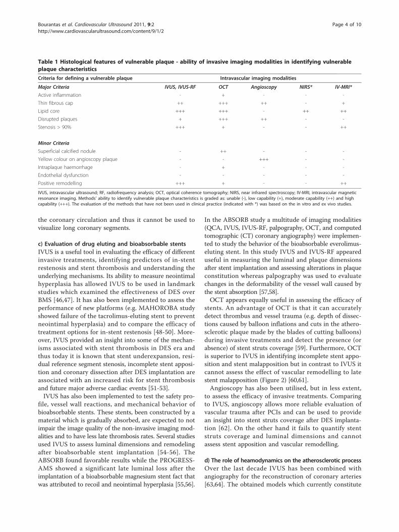
Table 1 Histological features of vulnerable plaque - ability of invasive imaging modalities in identifying vulnerableplaque characteristics
Criteria for defining a vulnerable plaque Intravascular imaging modalities
Major Criteria IVUS, IVUS-RF OCT Angioscopy NIRS* IV-MRI*
Active inflammation - + - - -
Thin fibrous cap ++ +++ ++ - +
Lipid core +++ +++ - ++ ++
Disrupted plaques + +++ ++ - -
Stenosis > 90% +++ + - - ++
Minor Criteria
Superficial calcified nodule - ++ - - -
Yellow colour on angioscopy plaque - - +++ - -
Intraplaque haemorrhage - + - - -
Endothelial dysfunction - - - - -
Positive remodelling +++ + - - ++
IVUS, intravascular ultrasound; RF, radiofrequency analysis; OCT, optical coherence tomography; NIRS, near infrared spectroscopy; IV-MRI, intravascular magneticresonance imaging. Methods’ ability to identify vulnerable plaque characteristics is graded as: unable (-), low capability (+), moderate capability (++) and highcapability (+++). The evaluation of the methods that have not been used in clinical practice (indicated with *) was based on the in vitro and ex vivo studies.
Bourantas et al. Cardiovascular Ultrasound 2011, 9:2http://www.cardiovascularultrasound.com/content/9/1/2
Page 4 of 10

the state of the art in coronary 3-D imaging, providefully and comprehensive arterial representation and havebeen used to investigate the role of local heamody-namics in the atherosclerotic process (Figure 3). Severalinvestigators implemented these models to demonstratethat low and oscillatory shear stresses are associatedwith an increased risk for in-stent restenosis and athero-sclerosis progression in native coronary segments andcoronary bifurcations while recently, it was shown thatlow shear stresses not only act as an atherogenic factorbut also promote the development of vulnerable plaque[65-68]. These studies have provided useful informationand helped us to explain the regional localisation ofatherosclerosis and understand the role of flowdynamics in plaque evolution and destabilisation [69].
e) Future developmentsNIRS relies on the principle that different organic mole-cules absorb and scatter NIRS light to different degrees
Figure 2 IVUS images showing optimal stent expansion (A);neointima formation (B) and late stent malaposition 6 monthsafter a DES implantation (C). OCT imaging allows not onlyassessment of stent expansion but also evaluation of stent strutscoverage (absence of coverage (D) vs. complete coverage (E)) andmeasurement of neointimal hyperplasia (F).
Figure 3 3-D reconstruction of a coronary artery and blood flow simulation. (A) Extraction of the IVUS path from biplane angiography;(B) semi-automated border detection of the IVUS images; (C) placement of the detected borders onto the catheter path and determination oftheir absolute orientation; (D) virtual endoscopy of the reconstructed vessel; (E) blood flow simulation into the final model and determination ofthe endothelial shear stresses.
Bourantas et al. Cardiovascular Ultrasound 2011, 9:2http://www.cardiovascularultrasound.com/content/9/1/2
Page 5 of 10
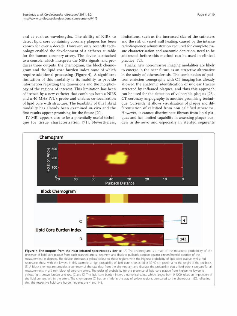
and at various wavelengths. The ability of NIRS todetect lipid core containing coronary plaques has beenknown for over a decade. However, only recently tech-nology enabled the development of a catheter suitablefor the human coronary artery. The device is attachedto a console, which interprets the NIRS signals, and pro-duces three outputs: the chemogram, the block chemo-gram and the lipid core burden index none of whichrequire additional processing (Figure 4). A significantlimitation of this modality is its inability to provideinformation regarding the dimensions and the morphol-ogy of the regions of interest. This limitation has beenaddressed by a new catheter that combines both a NIRSand a 40 MHz IVUS probe and enables co-localizationof lipid core with structure. The feasibility of this hybridmodality has already been examined in-vivo and thefirst results appear promising for the future [70].IV-MRI appears also to be a potentially useful techni-
que for tissue characterisation [71]. Nevertheless,
limitations, such as the increased size of the cathetersand the risk of vessel wall heating, caused by the intenseradiofrequency administration required for complete tis-sue characterisation and anatomic depiction, need to beaddressed before this method can be used in clinicalpractice [72].Finally, new non-invasive imaging modalities are likely
to emerge in the near future as an attractive alternativein the study of atherosclerosis. The combination of posi-tron emission tomography with CT imaging has alreadyallowed the anatomic identification of nuclear tracersattracted by inflamed plaques, and thus this approachcan be used for the detection of vulnerable plaques [73].CT coronary angiography is another promising techni-que. Currently, it allows visualization of plaque and dif-ferentiation of calcified from non calcified atheroma.However, it cannot discriminate fibrous from lipid pla-ques and has limited capability in assessing plaque bur-den in de-novo and especially in stented segments
Figure 4 The outputs from the Near-infrared spectroscopy device. (A) The chemogram is a map of the measured probability of thepresence of lipid core plaque from each scanned arterial segment and displays pullback position against circumferential position of themeasurement in degrees. The device attributes a yellow colour to those regions with the highest probability of lipid core plaque, whilst redrepresents those with the lowest. In this example, a high probability of lipid core is detected at 30-40 cm proximal to the origin of the pullback.(B) A block chemogram provides a summary of the raw data from the chemogram and displays the probability that a lipid core is present for allmeasurements in a 2 mm block of coronary artery. The order of probability for the presence of lipid core plaque from highest to lowest isyellow, light brown, brown, and red. (C and D) The lipid core burden index, a numerical value, which ranges from 0-1000, gives an impression ofthe lipid content within the artery. The chemogram (C) has very little in the way of yellow regions, compared to the chemogram (D), reflectingthis, the respective lipid core burden indexes are 4 and 143.
Bourantas et al. Cardiovascular Ultrasound 2011, 9:2http://www.cardiovascularultrasound.com/content/9/1/2
Page 6 of 10

[74,75]. Unfortunately, MRI imaging has low resolutionwhich limits coronary visualization; hence further devel-opment is required before its implementation in theclinical and research arena.
ConclusionsThrough our review it is apparent that today there is amultitude of imaging modalities able to assess differentmorphological and functional features of the coronaryarteries. Amongst these IVUS constitutes a valuable toolin research as it is the gold standard for studying plaquedevelopment and assessing the effectiveness of pharmaco-logical treatments on plaque’s evolution (Table 2). Thoughit is the most widespread invasive imaging modality it hassignificant intrinsic limitations being the considerablenoise and the low axial resolution which does not allowdetailed assessment of luminal morphology and meticu-lous visualisation of plaque characteristics. Thus, alterna-tive intravascular imaging techniques have beenintroduced which provide additional information regard-ing the composition and vulnerability of the plaque andallow more accurate evaluation of stent deployment andstruts’ coverage. The combination of IVUS with thesemodalities, either as independent or as hybrid imaging,may allow in the future a more detailed insight into themechanisms of plaque progression-regression and providebetter evaluation of the different therapeutic strategies.
Author details1Department of Cardiology, Castle Hill Hospital, Cottingham, East Yorkshire,UK. 2Department of Cardiology, Medical School, University of Ioannina,Ioannina, Greece. 3Department of Cardiology, Albert Szent-Gyorgyi ClinicalCenter, University of Szeged, Szeged, Hungary.
Authors’ contributionsCVB wrote the manuscript while the other authors involved in drafting andrevised the manuscript for important intellectual content. All authors readand approve the final document.
Competing interestsThe authors declare that they have no competing interests.
Received: 27 October 2010 Accepted: 30 January 2011Published: 30 January 2011
References1. Hausmann D, Erbel R, Alibelli-Chemarin MJ, Boksch W, Caracciolo E,
Cohn JM, Culp SC, Daniel WG, De Scheerder I, DiMario C, et al: The safetyof intracoronary ultrasound. A multicenter survey of 2207 examinations.Circulation 1995, 91(3):623-30.
2. Bourantas CV, Naka KK, Garg S, Thackray S, Papadopoulos D, Alamgir FM,Hoye A, Michalis LK: Clinical indications for intravascular ultrasoundimaging. Echocardiography 2010, 27(10):1282-90.
3. Mehta SK, McCrary JR, Frutkin AD, Dolla WJ, Marso SP: Intravascularultrasound radiofrequency analysis of coronary atherosclerosis: anemerging technology for the assessment of vulnerable plaque. Eur HeartJ 2007, 28(11):1283-8.
4. Mallery JA, Tobis JM, Griffith J, Gessert J, McRae M, Moussabeck O,Bessen M, Moriuchi M, Henry WL: Assessment of normal andatherosclerotic arterial wall thickness with an intravascular ultrasoundimaging catheter. Am Heart J 1990, 119(6):1392-1400.3.
5. Nishimura RA, Edwards WD, Warnes CA, Reeder GS, Holmes DR Jr, Tajik AJ,Yock PG: Intravascular ultrasound imaging: in vitro validation andpathologic correlation. J Am Coll Cardiol 1990, 16(1):145-54.
Table 2 Research utility of IVUS imaging. Advantages and disadvantages
IVUS imaging advantages IVUS imaging disadvantages Preferable modality
Assessment of theeffect ofpharmacologicaltreatment
• Able to quantify changes in plaque volume• IVUS-RF allows identification andquantification of changes in plaque’scomposition
• IVUS-RF identifies with moderatesensitivity/specificity a change from lipidto a fibrous plaque
IVUS
Remodellingassessment
• Complete arterial wall visualisation • Unable to identify accurately the outervessel wall border in segments withcalcified plaques.
IVUS
Plaque characterisation • Complete vessel wall visualisation• IVUS-RF allows identification of the type ofthe plaque with good overall sensitivity andspecificity
• IVUS-RF identifies with moderatesensitivity/specificity lipid and fibrousplaques
IVUS-RF and OCT
Detection of vulnerableplaque
• Accurate measurement of luminal dimensions,plaque area and remodelling• IVUS-RF allows identification of the type ofthe plaque with good overall sensitivity andspecificity
• Limited axial resolution - unable tomeasure the fibrous cap• Moderate sensitivity in detectingthrombus and plaque disruption/erosion• Unable to detect macrophages orintraplaque haemorrhage
OCT and IVUS-RF orcombination of different
imaging modalities
Assessment of invasivetreatments
• Reliable assessment of luminal, stent area andintima hyperplasia• Precise evaluation of stent expansion• Reliable evaluation of bioabsorbable stentrecoil
• Limited capability in identifying vesselwall trauma (e.g. erosion, dissection) andthrombus• Incapable of assessing stent strutscoverage
OCT or combination of OCTand IVUS
Role of heamodynamicsin atherosclerosis
• Complete vessel visualisation - plaquecharacterisation• Multitude of automated methodologies thatallows IVUS segmentation and fusion of IVUSand angiography
• Limited capability in detectingvulnerable plaque characteristics
IVUS
IVUS, intravascular ultrasound; RF, radiofrequency analysis; OCT, optical coherence tomography; NIRS, near infrared spectroscopy.
Bourantas et al. Cardiovascular Ultrasound 2011, 9:2http://www.cardiovascularultrasound.com/content/9/1/2
Page 7 of 10

6. Koning G, Dijkstra J, von Birgelen C, Tuinenburg JC, Brunette J, Tardif JC,Oemrawsingh PW, Sieling C, Melsa S, Reiber JH: Advanced contourdetection for three-dimensional intracoronary ultrasound: a validation-invitro and in vivo. Int J Cardiovasc Imaging 2002, 18(4):235-48.
7. Bourantas CV, Plissiti ME, Fotiadis DI, Protopappas VC, Mpozios GV,Katsouras CS, Kourtis IC, Rees MR, Michalis LK: In vivo validation of a novelsemi-automated method for border detection in intravascularultrasound images. Br J Radiol 2005, 78(926):122-9.
8. Bruining N, Verheye S, Knaapen M, Somers P, Roelandt JR, Regar E, Heller I,de Winter S, Ligthart J, Van Langenhove G, de Feijter PJ, Serruys PW,Hamers R: Three-dimensional and quantitative analysis of atheroscleroticplaque composition by automated differential echogenicity. CatheterCardiovasc Interv 2007, 70(7):968-78.
9. Potkin BN, Keren G, Mintz GS, Douek PC, Pichard AD, Satler LF, Kent KM,Leon MB: Arterial responses to balloon coronary angioplasty: anintravascular ultrasound study. J Am Coll Cardiol 1992, 20(4):942-51.
10. Mintz GS, Popma JJ, Pichard AD, Kent KM, Satler LF, Wong C, Hong MK,Kovach JA, Leon MB: Arterial remodeling after coronary angioplasty: aserial intravascular ultrasound study. Circulation 1996, 94(1):35-43.
11. Serruys PW, Luijten HE, Beatt KJ, Geuskens R, de Feyter PJ, van denBrand M, Reiber JH, ten Katen HJ, van Es GA, Hugenholtz PG: Incidence ofrestenosis after successful coronary angioplasty: a time-relatedphenomenon. A quantitative angiographic study in 342 consecutivepatients at 1, 2, 3, and 4 months. Circulation 1988, 77(2):361-71.
12. Stone GW, Hodgson JM, St Goar FG, Frey A, Mudra H, Sheehan H,Linnemeier TJ: Improved procedural results of coronary angioplasty withintravascular ultrasound-guided balloon sizing: the CLOUT Pilot Trial.Clinical Outcomes With Ultrasound Trial (CLOUT) Investigators.Circulation 1997, 95(8):2044-52.
13. Berry C, L’Allier PL, Gregoire J, Lesperance J, Levesque S, Ibrahim R,Tardif JC: Comparison of intravascular ultrasound and quantitativecoronary angiography for the assessment of coronary artery diseaseprogression. Circulation 2007, 115(14):1851-7.
14. Bose D, von Birgelen C, Erbel R: Intravascular ultrasound for theevaluation of therapies targeting coronary atherosclerosis. J Am CollCardiol 2007, 49(9):925-32.
15. Garcia-Garcia HM, Costa MA, Serruys PW: Imaging of coronaryatherosclerosis: intravascular ultrasound. Eur Heart J 2010, 31(20):2456-69.
16. Nissen SE, Nicholls SJ, Sipahi I, Libby P, Raichlen JS, Ballantyne CM,Davignon J, Erbel R, Fruchart JC, Tardif JC, Schoenhagen P, Crowe T, Cain V,Wolski K, Goormastic M, Tuzcu EM: Effect of very high-intensity statintherapy on regression of coronary atherosclerosis: the ASTEROID trial.JAMA 2006, 295(13):1556-65.
17. Nissen SE, Tuzcu EM, Schoenhagen P, Brown BG, Ganz P, Vogel RA,Crowe T, Howard G, Cooper CJ, Brodie B, Grines CL, DeMaria AN, REVERSALInvestigators: Effect of intensive compared with moderate lipid-loweringtherapy on progression of coronary atherosclerosis: a randomizedcontrolled trial. JAMA 2004, 291(9):1071-80.
18. Okazaki S, Yokoyama T, Miyauchi K, Shimada K, Kurata T, Sato H, Daida H:Early statin treatment in patients with acute coronary syndrome:demonstration of the beneficial effect on atherosclerotic lesions byserial volumetric intravascular ultrasound analysis during half a yearafter coronary event: the ESTABLISH Study. Circulation 2004,110(9):1061-8.
19. Gerstein HC, Ratner RE, Cannon CP, Serruys PW, Garcia-Garcia HM, vanEs GA, Kolatkar NS, Kravitz BG, Miller DM, Huang C, Fitzgerald PJ, Nesto RW:Effect of rosiglitazone on progression of coronary atherosclerosis inpatients with type 2 diabetes mellitus and coronary artery disease: theassessment on the prevention of progression by rosiglitazone onatherosclerosis in diabetes patients with cardiovascular history trial.Circulation 2010, 121(10):1176-87.
20. Nissen SE, Nicholls SJ, Wolski K, Nesto R, Kupfer S, Perez A, Jure H, DeLarochelliere R, Staniloae CS, Mavromatis K, Saw J, Hu B, Lincoff AM,Tuzcu EM: Comparison of pioglitazone vs glimepiride on progression ofcoronary atherosclerosis in patients with type 2 diabetes: the PERISCOPErandomized controlled trial. JAMA 2008, 299(13):1561-73.
21. Nissen SE, Tuzcu EM, Libby P, Thompson PD, Ghali M, Garza D, Berman L,Shi H, Buebendorf E, Topol EJ: Effect of antihypertensive agents oncardiovascular events in patients with coronary disease and normalblood pressure: the CAMELOT study: a randomized controlled trial. JAMA2004, 292(18):2217-25.
22. Rodriguez-Granillo GA, Vos J, Bruining N, Garcia-Garcia HM, de Winter S,Ligthart JM, Deckers JW, Bertrand M, Simoons ML, Ferrari R, Fox KM,Remme W, De Feyter PJ: Long-term effect of perindopril on coronaryatherosclerosis progression (from the perindopril’s prospective effect oncoronary atherosclerosis by angiography and intravascular ultrasoundevaluation [PERSPECTIVE] study). Am J Cardiol 2007, 100(2):159-63.
23. Tardif JC, Grégoire J, L’Allier PL, Ibrahim R, Lespérance J, Heinonen TM,Kouz S, Berry C, Basser R, Lavoie MA, Guertin MC, Rodés-Cabau J: Effect ofrHDL on Atherosclerosis-Safety and Efficacy (ERASE) Investigators (2007):Effects of reconstituted high-density lipoprotein infusions on coronaryatherosclerosis: a randomized controlled trial. JAMA 2007,297(15):1675-82.
24. Serruys PW, Garcia-Garcia HM, Buszman P, Erne P, Verheye S,Aschermann M, Duckers H, Bleie O, Dudek D, Botker HE, von Birgelen C,D’Amico D, Hutchinson T, Zambanini A, Mastik F, van Es GA, van derSteen AF, Vince DG, Ganz P, Hamm CW, Wijns W, Zalewski A: Effects of thedirect lipoprotein-associated phospholipase A(2) inhibitor darapladib onhuman coronary atherosclerotic plaque. Circulation 2008, 118(11):1172-82.
25. Nissen SE, Tuzcu EM, Brewer HB, Sipahi I, Nicholls SJ, Ganz P,Schoenhagen P, Waters DD, Pepine CJ, Crowe TD, Davidson MH,Deanfield JE, Wisniewski LM, Hanyok JJ, Kassalow LM: Effect of ACATinhibition on the progression of coronary atherosclerosis. N Engl J Med2006, 354(12):1253-63.
26. Hassani SE, Mintz GS, Fong HS, Kim SW, Xue Z, Pichard AD, Satler LF,Kent KM, Suddath WO, Waksman R, Weissman NJ: Negative remodelingand calcified plaque in octogenarians with acute myocardial infarction:an intravascular ultrasound analysis. J Am Coll Cardiol 2006, 47(12):2413-9.
27. Nakamura M, Nishikawa H, Mukai S, Setsuda M, Nakajima K, Tamada H,Suzuki H, Ohnishi T, Kakuta Y, Nakano T, Yeung AC: Impact of coronaryartery remodeling on clinical presentation of coronary artery disease: anintravascular ultrasound study. J Am Coll Cardiol 2001, 37(1):63-9.
28. Brezinski ME: Optical coherence tomography for identifying unstablecoronary plaque. Int J Cardiol 2006, 107(2):154-65.
29. Kawasaki M, Takatsu H, Noda T, Sano K, Ito Y, Hayakawa K, Tsuchiya K,Arai M, Nishigaki K, Takemura G, Minatoguchi S, Fujiwara T, Fujiwara H: Invivo quantitative tissue characterization of human coronary arterialplaques by use of integrated backscatter intravascular ultrasound andcomparison with angioscopic findings. Circulation 2002, 105(21):2487-92.
30. Hara H, Tsunoda T, Nemoto N, Yokouchi I, Yamamoto M, Ono T, Moroi M,Suzuki M, Sugi K, Nakamura M: Distribution of ultrasonic radiofrequencysignal amplitude detects lipids in atherosclerotic plaque of coronaryarteries: an ex-vivo study. Cardiovasc Ultrasound 2008, 6:18.
31. Sano K, Kawasaki M, Okubo M, Yokoyama H, Ito Y, Murata I, Kawai T,Tsuchiya K, Nishigaki K, Takemura G, Minatoguchi S, Zhou X, Fujita H,Fujiwara H: In vivo quantitative tissue characterization ofangiographically normal coronary lesions and the relation with riskfactors: a study using integrated backscatter intravascular ultrasound.Circ J 2005, 69(5):543-9.
32. Valgimigli M, Rodriguez-Granillo GA, Garcia-Garcia HM, Malagutti P, Regar E,de Jaegere P, de Feyter P, Serruys PW: Distance from the ostium as anindependent determinant of coronary plaque composition in vivo: anintravascular ultrasound study based radiofrequency data analysis inhumans. Eur Heart J 2006, 27(6):655-63.
33. Hong MK, Park DW, Lee CW, Lee SW, Kim YH, Kang DH, Song JK, Kim JJ,Park SW, Park SJ: Effects of statin treatments on coronary plaquesassessed by volumetric virtual histology intravascular ultrasoundanalysis. JACC Cardiovasc Interv 2009, 2(7):679-88.
34. Kubo T, Maehara A, Mintz GS, Doi H, Tsujita K, Choi SY, Katoh O, Nasu K,Koenig A, Pieper M, Rogers JH, Wijns W, Bose D, Margolis MP, Moses JW,Stone GW, Leon MB: The dynamic nature of coronary artery lesionmorphology assessed by serial virtual histology intravascular ultrasoundtissue characterization. J Am Coll Cardiol 2010, 55(15):1590-7.
35. Naghavi M, Libby P, Falk E, Casscells SW, Litovsky S, Rumberger J,Badimon JJ, Stefanadis C, Moreno P, Pasterkamp G, Fayad Z, Stone PH,Waxman S, Raggi P, Madjid M, Zarrabi A, Burke A, Yuan C, Fitzgerald PJ,Siscovick DS, de Korte CL, Aikawa M, Juhani Airaksinen KE, Assmann G,Becker CR, Chesebro JH, Farb A, Galis ZS, Jackson C, Jang IK, Koenig W,Lodder RA, March K, Demirovic J, Navab M, Priori SG, Rekhter MD, Bahr R,Grundy SM, Mehran R, Colombo A, Boerwinkle E, Ballantyne C, Insull W Jr,Schwartz RS, Vogel R, Serruys PW, Hansson GK, Faxon DP, Kaul S, Drexler H,Greenland P, Muller JE, Virmani R, Ridker PM, Zipes DP, Shah PK,
Bourantas et al. Cardiovascular Ultrasound 2011, 9:2http://www.cardiovascularultrasound.com/content/9/1/2
Page 8 of 10

Willerson JT: From vulnerable plaque to vulnerable patient: a call for newdefinitions and risk assessment strategies: Part I. Circulation 2003,108(14):1664-72.
36. Kubo T, Imanishi T, Takarada S, Kuroi A, Ueno S, Yamano T, Tanimoto T,Matsuo Y, Masho T, Kitabata H, Tsuda K, Tomobuchi Y, Akasaka T:Assessment of culprit lesion morphology in acute myocardial infarction:ability of optical coherence tomography compared with intravascularultrasound and coronary angioscopy. J Am Coll Cardiol 2007, 50(10):933-9.
37. Schaar JA, De Korte CL, Mastik F, Strijder C, Pasterkamp G, Boersma E,Serruys PW, Van Der Steen AF: Characterizing vulnerable plaque featureswith intravascular elastography. Circulation 2003, 108(21):2636-41.
38. de Korte CL, Sierevogel MJ, Mastik F, Strijder C, Schaar JA, Velema E,Pasterkamp G, Serruys PW, van der Steen AF: Identification ofatherosclerotic plaque components with intravascular ultrasoundelastography in vivo: a Yucatan pig study. Circulation 2002,105(14):1627-30.
39. Vavuranakis M, Kakadiaris IA, O’Malley SM, Papaioannou TG, Sanidas EA,Naghavi M, Carlier S, Tousoulis D, Stefanadis C: A new method forassessment of plaque vulnerability based on vasa vasorum imaging, byusing contrast-enhanced intravascular ultrasound and differential imageanalysis. Int J Cardiol 2008, 130(1):23-9.
40. Jang IK, Bouma BE, Kang DH, Park SJ, Park SW, Seung KB, Choi KB,Shishkov M, Schlendorf K, Pomerantsev E, Houser SL, Aretz HT, Tearney GJ:Visualization of coronary atherosclerotic plaques in patients usingoptical coherence tomography: comparison with intravascularultrasound. J Am Coll Cardiol 2002, 39(4):604-9.
41. Yabushita H, Bouma BE, Houser SL, Aretz HT, Jang IK, Schlendorf KH,Kauffman CR, Shishkov M, Kang DH, Halpern EF, Tearney GJ:Characterization of human atherosclerosis by optical coherencetomography. Circulation 2002, 106(13):1640-5.
42. Kume T, Akasaka T, Kawamoto T, Ogasawara Y, Watanabe N, Toyota E,Neishi Y, Sukmawan R, Sadahira Y, Yoshida K: Assessment of coronaryarterial thrombus by optical coherence tomography. Am J Cardiol 2006,97(12):1713-7.
43. Tearney GJ, Yabushita H, Houser SL, Aretz HT, Jang IK, Schlendorf KH,Kauffman CR, Shishkov M, Halpern EF, Bouma BE: Quantification ofmacrophage content in atherosclerotic plaques by optical coherencetomography. Circulation 2003, 107(1):113-9.
44. Sawada T, Shite J, Garcia-Garcia HM, Shinke T, Watanabe S, Otake H,Matsumoto D, Tanino Y, Ogasawara D, Kawamori H, Kato H, Miyoshi N,Yokoyama M, Serruys PW, Hirata K: Feasibility of combined use ofintravascular ultrasound radiofrequency data analysis and opticalcoherence tomography for detecting thin-cap fibroatheroma. Eur Heart J2008, 29(9):1136-46.
45. Ueda Y, Ohtani T, Shimizu M, Hirayama A, Kodama K: Assessment ofplaque vulnerability by angioscopic classification of plaque color. AmHeart J 2004, 148(2):333-5.
46. Serruys PW, Degertekin M, Tanabe K, Abizaid A, Sousa JE, Colombo A,Guagliumi G, Wijns W, Lindeboom WK, Ligthart J, de Feyter PJ, Morice MC:Intravascular ultrasound findings in the multicenter, randomized,double-blind RAVEL (RAndomized study with the sirolimus-elutingVElocity balloon-expandable stent in the treatment of patients with denovo native coronary artery Lesions) trial. Circulation 2002,106(7):798-803.
47. Aoki J, Colombo A, Dudek D, Banning AP, Drzewiecki J, Zmudka K,Schiele F, Russell ME, Koglin J, Serruys PW: Peristent remodeling andneointimal suppression 2 years after polymer-based, paclitaxel-elutingstent implantation: insights from serial intravascular ultrasound analysisin the TAXUS II study. Circulation 2005, 12(25):3876-83.
48. Onuma Y, Serruys P, den Heijer P, Joesoef KS, Duckers H, Regar E, Kukreja N,Tanimoto S, Garcia-Garcia HM, van Beusekom H, van der Giessen W,Nishide T: MAHOROBA, first-in-man study: 6-month results of abiodegradable polymer sustained release tacrolimus-eluting stent in denovo coronary stenoses. Eur Heart J 2009, 30(12):1477-85.
49. Alfonso F, Perez-Vizcayno MJ, Hernandez R, Bethencourt A, Marti V, Lopez-Minguez JR, Angel J, Mantilla R, Moris C, Cequier A, Sabate M, Escaned J,Moreno R, Banuelos C, Suarez A, Macaya C: A randomized comparison ofsirolimus-eluting stent with balloon angioplasty in patients with in-stentrestenosis: results of the Restenosis Intrastent: Balloon AngioplastyVersus Elective Sirolimus-Eluting Stenting (RIBS-II) trial. J Am Coll Cardiol2006, 47(11):2152-60.
50. Schiele TM, Konig A, Rieber J, Erhard I, Leibig M, Theisen K, Siebert U,Klauss V: Sirolimus-eluting stent implantation and beta-irradiation for thetreatment of in-stent restenotic lesions: comparison of underlyingmechanisms of acute gain and late loss as assessed by volumetricintravascular ultrasound. Am Heart J 2005, 150(2):351-7.
51. Fujii K, Carlier SG, Mintz GS, Yang YM, Moussa I, Weisz G, Dangas G,Mehran R, Lansky AJ, Kreps EM, Collins M, Stone GW, Moses JW, Leon MB:Stent underexpansion and residual reference segment stenosis arerelated to stent thrombosis after sirolimus-eluting stent implantation: anintravascular ultrasound study. J Am Coll Cardiol 2005, 45(7):995-8.
52. Cook S, Wenaweser P, Togni M, Billinger M, Morger C, Seiler C, Vogel R,Hess O, Meier B, Windecker S: Incomplete stent apposition and very latestent thrombosis after drug-eluting stent implantation. Circulation 2007,115(18):2426-34.
53. Biondi-Zoccai GG, Agostoni P, Sangiorgi GM, Airoldi F, Cosgrave J,Chieffo A, Barbagallo R, Tamburino C, Vittori G, Falchetti E, Margheri M,Briguori C, Remigi E, Iakovou I, Colombo A: Incidence, predictors, andoutcomes of coronary dissections left untreated after drug-eluting stentimplantation. Eur Heart J 2006, 27(5):540-6.
54. Tanimoto S, Bruining N, van Domburg RT, Rotger D, Radeva P, Ligthart JM,Serruys PW: Late stent recoil of the bioabsorbable everolimus-elutingcoronary stent and its relationship with plaque morphology. J Am CollCardiol 2008, 52(20):1616-20.
55. Erbel R, Di Mario C, Bartunek J, Bonnier J, de Bruyne B, Eberli FR, Erne P,Haude M, Heublein B, Horrigan M, Ilsley C, Bose D, Koolen J, Luscher TF,Weissman N, Waksman R: Temporary scaffolding of coronary arteries withbioabsorbable magnesium stents: a prospective, non-randomisedmulticentre trial. Lancet 2007, 369(9576):1869-75.
56. Serruys PW, Ormiston JA, Onuma Y, Regar E, Gonzalo N, Garcia-Garcia HM,Nieman K, Bruining N, Dorange C, Miquel-Hebert K, Veldhof S, Webster M,Thuesen L, Dudek D: A bioabsorbable everolimus-eluting coronary stentsystem (ABSORB): 2-year outcomes and results from multiple imagingmethods. Lancet 2009, 373(9667):897-910.
57. Sarno G, Onuma Y, Garcia HM, Garg S, Regar E, Thuesen L, Dudek D,Veldhof S, Dorange C, Ormiston JA, Serruys PW: IVUS radiofrequencyanalysis in the evaluation of the polymeric struts of the bioabsorbableeverolimus-eluting device during the bioabsorption process. CatheterCardiovasc Interv 2010, 75(6):914-8.
58. Garcia-Garcia HM, Gonzalo N, Pawar R, Kukreja N, Dudek D, Thuesen L,Ormiston JA, Regar E, Serruys PW: Assessment of the absorption processfollowing bioabsorbable everolimus-eluting stent implantation: temporalchanges in strain values and tissue composition using intravascularultrasound radiofrequency data analysis. A substudy of the ABSORBclinical trial. EuroIntervention 2009, 4(4):443-8.
59. Barlis P, Regar E, Serruys PW, Dimopoulos K, van der Giessen WJ, vanGeuns RJ, Ferrante G, Wandel S, Windecker S, van Es GA, Eerdmans P,Juni P, di Mario C: An optical coherence tomography study of abiodegradable vs. durable polymer-coated limus-eluting stent: aLEADERS trial sub-study. Eur Heart J 2010, 31(2):139-42.
60. Matsumoto D, Shite J, Shinke T, Otake H, Tanino Y, Ogasawara D, Sawada T,Paredes OL, Hirata K, Yokoyama M: Neointimal coverage of sirolimus-eluting stents at 6-month follow-up: evaluated by optical coherencetomography. Eur Heart J 2007, 28(8):961-7.
61. Bouma BE, Tearney GJ, Yabushita H, Shishkov M, Kauffman CR, DeJosephGauthier D, MacNeill BD, Houser SL, Aretz HT, Halpern EF, Jang IK:Evaluation of intracoronary stenting by intravascular optical coherencetomography. Heart 2003, 89(3):317-20.
62. Takano M, Ohba T, Inami S, Seimiya K, Sakai S, Mizuno K: Angioscopicdifferences in neointimal coverage and in persistence of thrombusbetween sirolimus-eluting stents and bare metal stents after a 6-monthimplantation. Eur Heart J 2006, 27(18):2189-95.
63. Bourantas CV, Kalatzis FG, Papafaklis MI, Fotiadis DI, Tweddel AC, Kourtis IC,Katsouras CS, Michalis LK: ANGIOCARE: an automated system for fastthree-dimensional coronary reconstruction by integrating angiographicand intracoronary ultrasound data. Catheter Cardiovasc Interv 2008,72(2):166-75.
64. Slager CJ, Wentzel JJ, Schuurbiers JC, Oomen JA, Kloet J, Krams R, vonBirgelen C, van der Giessen WJ, Serruys PW, de Feyter PJ: True 3-dimensional reconstruction of coronary arteries in patients by fusion ofangiography and IVUS (ANGUS) and its quantitative validation.Circulation 2000, 102(5):511-6.
Bourantas et al. Cardiovascular Ultrasound 2011, 9:2http://www.cardiovascularultrasound.com/content/9/1/2
Page 9 of 10

65. Papafaklis MI, Bourantas CV, Theodorakis PE, Katsouras CS, Fotiadis DI,Michalis LK: Association of endothelial shear stress with plaque thicknessin a real three-dimensional left main coronary artery bifurcation model.Int J Cardiol 2007, 115(2):276-8.
66. Stone PH, Coskun AU, Kinlay S, Popma JJ, Sonka M, Wahle A,Yeghiazarians Y, Maynard C, Kuntz RE, Feldman CL: Regions of lowendothelial shear stress are the sites where coronary plaque progressesand vascular remodelling occurs in humans: an in vivo serial study. EurHeart J 2007, 28(6):705-10.
67. Papafaklis MI, Bourantas CV, Theodorakis PE, Katsouras CS, Fotiadis DI,Michalis LK: Relationship of shear stress with in-stent restenosis: baremetal stenting and the effect of brachytherapy. Int J Cardiol 2009,134(1):25-32.
68. Chatzizisis YS, Jonas M, Coskun AU, Beigel R, Stone BV, Maynard C,Gerrity RG, Daley W, Rogers C, Edelman ER, Feldman CL, Stone PH:Prediction of the localization of high-risk coronary atheroscleroticplaques on the basis of low endothelial shear stress: an intravascularultrasound and histopathology natural history study. Circulation 2008,117(8):993-1002.
69. Slager CJ, Wentzel JJ, Gijsen FJ, Thury A, van der Wal AC, Schaar JA,Serruys PW: The role of shear stress in the destabilization of vulnerableplaques and related therapeutic implications. Nat Clin Pract CardiovascMed 2005, 2(9):456-64.
70. Garg S, Serruys PW, van der Ent M, Schultz C, Mastik F, van Soest G, van derSteen AF, Wilder MA, Muller JE, Regar E: First use in patients of acombined near infra-red spectroscopy and intra-vascular ultrasoundcatheter to identify composition and structure of coronary plaque.EuroIntervention 2010, 5(6):755-6.
71. Larose E, Yeghiazarians Y, Libby P, Yucel EK, Aikawa M, Kacher DF, Aikawa E,Kinlay S, Schoen FJ, Selwyn AP, Ganz P: Characterization of humanatherosclerotic plaques by intravascular magnetic resonance imaging.Circulation 2005, 112(15):2324-31.
72. Wilensky RL, Song HK, Ferrari VA: Role of magnetic resonance andintravascular magnetic resonance in the detection of vulnerableplaques. J Am Coll Cardiol 2006, 47(8 Suppl):C48-56.
73. Wykrzykowska J, Lehman S, Williams G, Parker JA, Palmer MR, Varkey S,Kolodny G, Laham R: Imaging of inflamed and vulnerable plaque incoronary arteries with 18F-FDG PET/CT in patients with suppression ofmyocardial uptake using a low-carbohydrate, high-fat preparation. J NuclMed 2009, 50(4):563-8.
74. Springer I, Dewey M: Comparison of multislice computed tomographywith intravascular ultrasound for detection and characterization ofcoronary artery plaques: A systematic review. Eur J Radiol 71 2009,71:275-282.
75. Schepis T, Marwan M, Pflederer T, Seltmann M, Ropers D, Daniel WG,Achenbach S: Quantification of non-calcified coronary atheroscleroticplaques with dual-source computed tomography: comparison withintravascular ultrasound. Heart 2010, 96(8):610-5.
doi:10.1186/1476-7120-9-2Cite this article as: Bourantas et al.: Focus on the research utility ofintravascular ultrasound - comparison with other invasive modalities.Cardiovascular Ultrasound 2011 9:2.
Submit your next manuscript to BioMed Centraland take full advantage of:
• Convenient online submission
• Thorough peer review
• No space constraints or color figure charges
• Immediate publication on acceptance
• Inclusion in PubMed, CAS, Scopus and Google Scholar
• Research which is freely available for redistribution
Submit your manuscript at www.biomedcentral.com/submit
Bourantas et al. Cardiovascular Ultrasound 2011, 9:2http://www.cardiovascularultrasound.com/content/9/1/2
Page 10 of 10