flotillin-1 facilitates toll-like receptor 3 signaling in ... · flotillin-1 facilitates toll-like...
TRANSCRIPT

ORIGINAL CONTRIBUTION
Flotillin-1 facilitates toll-like receptor 3 signaling in humanendothelial cells
Christian Fork • Juliane Hitzel • Benjamin J. Nichols •
Ritva Tikkanen • Ralf P. Brandes
Received: 6 May 2014 / Revised: 22 August 2014 / Accepted: 3 September 2014 / Published online: 10 September 2014
� Springer-Verlag Berlin Heidelberg 2014
Abstract Endothelial cells are important elements in the
vascular response to danger-associated molecules signaling
through toll-like receptors (TLRs). Flotillin-1 and -2 are
markers of membrane rafts but their true endothelial func-
tion is unknown. We hypothesized that flotillins are required
for TLR signaling in human umbilical vein endothelial cells
(HUVECs). Knockdown of flotillin-1 by shRNA decreased
the TLR3-mediated poly-I:C-induced but not the TLR4-
mediated LPS-induced inflammatory activation of HUVEC.
As TLR3 but not TLR4 signals through the endosomal
compartment, flotillin-1 might be involved in the transport
of poly-I:C to its receptor. Consistently, uptake of poly-I:C
was attenuated by flotillin-1 knockdown and probably
involved the scavenger receptor SCARA4 as revealed by
knockdown of this receptor. To determine the underlying
mechanism, SILAC proteomics was performed. Down-
regulation of flotillin-1 led to a reduction of the structural
caveolae proteins caveolin-1, cavin-1 and -2, suggesting a
role of flotillin-1 in caveolae formation. Flotillin-1 and
caveolin-1 colocalized within the cell, and knockdown of
flotillin-1 decreased caveolin-1 expression in an endoplas-
mic reticulum stress-dependent manner. Importantly,
downregulation of caveolin-1 also attenuated TLR3-induced
signaling. To demonstrate the importance of this finding,
cell adhesion was studied. Flotillin-1 shRNA attenuated the
poly-I:C-mediated induction of the adhesion molecules
VCAM-1 and ICAM-1. As a consequence, the poly-I:C-
induced adhesion of peripheral blood mononuclear cells
onto HUVECs was significantly attenuated by flotillin-1
shRNA. Collectively, these data suggest that interaction
between flotillin-1 and caveolin-1 may facilitate the trans-
port of TLR3-ligands to its intracellular receptor and enables
inflammatory TLR3 signaling.
Keywords Endothelium � Lipid rafts � Caveolae � Cavin �Inflammation � Toll-like receptors � Scavenger receptors
Introduction
Endothelial cells are the gate keepers of vascular inflam-
matory signaling. In acute inflammation, the endothelium
attracts and guides leukocytes to the site of affliction.
Chronic endothelial activation promotes atherosclerosis
development and aneurysm formation. Endothelial cells
sense danger-associated molecules (DAMPs) by pattern
recognition receptors, like toll-like receptors (TLR1-9) of
the innate immune system. On the long run, this may
promotes vascular disease as cholesterol and other unspe-
cific pro-atherosclerotic agents are also sensed by TLRs
[31]. In the acute setting of inflammation, however, TLR
Electronic supplementary material The online version of thisarticle (doi:10.1007/s00395-014-0439-4) contains supplementarymaterial, which is available to authorized users.
C. Fork � J. Hitzel � R. P. Brandes (&)
Institute for Cardiovascular Physiology, Medical Faculty,
Goethe-University Frankfurt, Theodor Stern Kai 7,
60690 Frankfurt, Germany
e-mail: [email protected]
C. Fork � R. P. Brandes
German Center for Cardiovascular Research (DZHK),
Partner site RheinMain, Frankfurt, Germany
B. J. Nichols
MRC Laboratory of Molecular Biology, Cambridge CB2 0QH,
UK
R. Tikkanen
Institute of Biochemistry, Medical Faculty, University of
Giessen, Gießen, Germany
123
Basic Res Cardiol (2014) 109:439
DOI 10.1007/s00395-014-0439-4
signaling is needed to facilitate the local endothelial
response to pathogens.
TLR signaling is complex and of the several elements
involved, the MyD88- and TRIF-pathways are of particular
relevance. MyD88 activates nuclear factor (NF)-jB, which
induces inflammatory cytokine expression. The TRIF
pathway stimulates interferon regulatory factor-3 (IRF3),
in addition to NF-jB and induces type I interferons [23].
Importantly, TLR3 does not really activate MyD88 and
therefore only stimulates the TRIF pathway [27].
On the basis of their subcellular localization, TLRs are
divided in two groups. TLR1, 2, 4, 5 and 6 are expressed on
the cell surface where they recognize lipid structures and, in
the case of TLR5, the protein flagellin. TLR3, 7, 8 and 9
reside intracellularly and sense nucleic acids within the en-
dolysosomal compartment [28]. Their intracellular locali-
zation is potentially a consequence of the fact that TLR
binding to nucleic acids requires an acidic environment [20].
Thus, circulating nucleic acids have to be delivered to
intracellular TLRs. The mechanisms underlying this trans-
port are, however, not completely characterized but obvi-
ously have to involve endocytosis of the nucleic acid. At
least two major endocytotic pathways operate in parallel in
endothelial cells: uptake through clathrin-coated pits and
internalization by membrane rafts [10]. Although the con-
cept of membrane rafts is still vigorously debated, there is
consensus that proteins of the flotillin family are enriched in
this fraction and are markers of rafts [32].
Interestingly, despite their ubiquitous expression and
high conservation, the physiological function of flotillins is
far from clear and their role in the endothelium has, to our
knowledge, not yet been determined. The flotillin protein
family consists of flotillin-1 and 2 (also called reggies)
which homo- and hetero-oligomerize [2, 37]. Despite some
other functions [4, 32], flotillins have been linked to
endocytosis and vesicular trafficking but also to signaling
[6, 8, 14, 15, 24, 33, 36, 38]. For example, flotillins mod-
ulate epidermal growth factor receptor auto-phosphoryla-
tion and activation of downstream target molecules [1].
Considering their prominent positioning on lipid rafts,
we here hypothesize that flotillins contribute to vascular
inflammatory signaling in response to the endosomal TLR3
and studied this in human umbilical vein endothelial cells
(HUVECs).
Materials and methods
Materials
Human fibronectin was purchased from BD Biosciences
(Heidelberg, Germany). DAPI, chloroquine, carrageenan,
dextran sulfate, lipopolysaccharide (E. coli), filipin III and
methyl-b-cyclodextrin from Sigma-Aldrich. MG132 was from
Merck, Leupeptin and pepstatin from Applichem. Poly-I:C
high molecular weight, Poly-I:C-rhodamine and lipoteichoic
acid (S. aureus) were from InvivoGen (San Diego, USA).
Phenylmethylsulfonyl fluoride (PMSF) was from Applichem.
Cell culture
Four batches of HUVECs were purchased from Lonza
(Walkersville, MD). Cells were cultured in EBM (Lonza,
Walkersville, MD), supplemented with 8 % FCS, bovine
brain supplement and human recombinant EGF, penicillin
(50 U/ml) and streptomycin (50 lg/ml). Cells of passage
two to four were used throughout the study and all experi-
ments were performed at least in three different batches.
Cells were stimulated in EBM with 1 % FCS, except
thapsigargin treatment it was performed in EBM with 4 %
FCS. Concentration and timing of the experiments are
described in the figure legend. For poly-I:C-rhodamine
uptake, cells were stimulated for 3 h (0.5 lg/ml) washed
with phosphate buffered saline and subsequently images
were acquired by confocal microscopy (LSM 510, Zeiss).
siRNA, shRNA and plasmid transfection
For shRNA treatment, endothelial cells were infected with
lentiviral particles according to Addgene ‘‘plKO.1 Proto-
col’’ (http://www.addgene.org/tools/protocols/plko/). Cells
were selected with puromycin (0.5 lg/ml). The shFlot-1A
target sequence was: 50-CAGAGAAGUCCCAACUAA
UUA-30, for shFlot-1B 50-CUGCUUGGCUUUAGCUUCC
CG-30 and for m-shFlot-1 CAGGATTACTTACACTCGT
TA. Unless otherwise noted, all experiments were per-
formed with shFlot-1A. Target sequence for shFlot-2 was
50-GUGCAGAGAGAUGCUGACAUU-30. Control shRNA
against green fluorescent protein (shGFP) and shScrambled
(shScr) were from Addgene. For siRNA treatment, endo-
thelial cells (80–90 % confluent) were transfected with
GeneTrans II according to the instructions provided by
MoBiTec (Gottingen, Germany). All siRNAs (Stealth
RNAi) were from Invitrogen. siScrambled (siScr) was used
as a negative control. Plasmid overexpression was achieved
with the neon electroporation system (Invitrogen). The
plasmids pFlot-1-GFP-N1 and pFlot-2-GFP-N1 have been
described earlier [30]. Control plasmid mCherry-N1 was
from Clontech. Two siRNAs and shRNAs were used to
exclude that unspecific effects of an individual approach
mediated the effects observed.
Quantitative RT-PCR
Total RNA was extracted with the RNA Mini Kit (Bio&-
Sell). cDNA was prepared with SuperScript III reverse
Page 2 of 13 Basic Res Cardiol (2014) 109:439
123
transcriptase (Invitrogen) and random hexamer primers.
Quantitative real-time PCR was performed with Fast Plus
Eva Green Master Mix and ROX as reference dye (Bio-
tium) in an Mx3005 cycler (Stratagene) with primer
sequences as follows: VCAM-1 forward primer, 50-TGTCATCATCTCTTGTACATGTGG-30, and reverse
primer, 50-GTTTTCTCTTCCTTGAACATCAAG-30; ICA
M-1 forward primer, 50-CAGTGGGCAA-GAACCTTAC
CCTAC-30, and reverse primer, 50-GTTCAGTGCGG
CACGAGAAATTGG-30; IFNß forward primer, 50- AG
TGTCAGAAGCTCCTGTGGC-30 and reverse 50- TGAGG
CAGT ATTCAAGCCTCC-30; CXCL10 forward primer,
50-TGGCATTCAAGGAGTACCTCTCT-30 and reverse 50-CTGATGCAGGTACAGCGTACG-30, SCARA1 forward
primer, 50- CTC CAT TTA CGA AAG TTC GAC TG-30
and reverse 50-ACCAGTACCTTGTCCAAAGTG-30; SC
ARA2 forward primer, 50-AAATCAATGTTCCAAAGCC
CAAG-30 and reverse 50-ACTGCAGCAAGG AGAAG
GAC-30; SCARA3 forward primer, 50-TGGATCCGAAA
GCCCTGAAC-30 and reverse 50-AGAAGGAGCAACTG
CCCATC-30; SCARA4 forward primer, 50-GAGCGTGAA
AATGAA TGGAAGTG-30 and reverse 50-AATGCAG
ATGACAGTACTGTCTC-30; SCARA5 forward primer,
50-TCTTCATCTTAGCAGTGTCCAG-30 and reverse 50-GCAACGAGTCTGACTGGT TC-30; SCARB1 forward
primer, 50-CAGCAGGTCCTTAAGAACGTG-30 and
reverse 50-GTT GTTGTTGAAGGTGATGTTGC-30; SCA
RB3 forward primer, 50-CTATGCTGTATTTGAATC
CGACG-30 and reverse 50-TACACAGGTCTCCCTTCT
TTG-30; SCARF1 forward primer, 50-AGGATGAAGCTG
CAGGTCTG-30 and reverse 50-ACAGGGACCATCCCT
TCTTG-30. Caveolin-1 forward primer-A, 50-ACGTGGT
CAAGATTGACTTTGAAG-30 and reverse-A 50-AGA
TGTGCAGGAAAGAGAGAATG-30. Caveolin-1 forward
primer-B, 50-TAGACTCGGAGG GACATCTC-30 and
reverse-B 50-TCGATCTCCTTGGTGTGCGCGTC-30.Caveolin-2 forward primer, 50-TCGCATCTCAAGCTG
GGCTTCG-30 and reverse 50-TACAAAAGG CATT
AAAATCCAGATG-30. Flotillin-1 forward primer, 50-AAGCTGCCCCAGGTGGCAGAG G-30 and reverse 50-TGTTCTCAAAGGCTTGTGATTCACC-30. Flotillin-2
forward primer, 50-AGGGTGAAAAGGTGAAGCAGGT
CC-30 and reverse 50-TTGGCAGCA ATCTGGGGCAG
G-30. Relative mRNA level of target genes was normalized
to polymerase (RNA)II (DNA-directed) polypeptide A
(POLR2A) with the primer pair 50-GCACCACGTCCAA
TGACAT-30 and 50-GTGCGGCTGCTTCCATAA-30, ana-
lyzed by the DDCT method.
Protein isolation and Western blot analysis
Cells were lysed in buffer containing 10 mmol/l Tris/HCl,
pH 8.0, 150 mmol/l NaCl, 10 mmol/l, 5 mmol/l EDTA,
0.5 % Triton X-100, 60 mmol/l N-octylglucoside, supple-
mented with 40 mg/l PMSF and protease inhibitor mix
(antipain, aprotinin, leupeptin, chymostatin, pepstatin,
trypsin inhibitor; 2 mg/ml each; all from AppliChem).
After determination of protein concentration by Bradford
assay, equal amounts of proteins were boiled in Laemmli
buffer and separated by SDS-PAGE gel. After transfer of
proteins onto nitrocellulose membranes and blocking with
Roti-Block (Carl Roth), the following primary antibodies
were used: anti-flotillin-1, flotillin-2 both from BD Bio-
sciences (610821, 610384) and Santa Cruz (sc-25506, sc-
25507). Caveolin-1 and caveolin-2 from Abnova
(MAB2408) and Cell Signaling (8522). The antibody for
VCAM-1(sc-8304), ICAM-1(sc-8439) and p65(sc-109)
were from Santa Cruz, GAPDH (G8795) from Sigma-
Aldrich. IRF-3 (4302) and pIRF-3 (Ser 385) (4947) were
from Cell Signaling. PERK (Thr 981) (sc-32577) and
eIF2a (Ser52) (sc-101670) from Santa Cruz. Western blot
analysis was performed with an infrared-based laser scan-
ning detection system (Odyssey, Licor). The infrared
fluorescent-dye-conjugated secondary antibodies were
purchased from Licor.
SILAC LTQ-Orbitrap mass spectrometry
and detergent-resistant membrane isolation
Stable isotope labeling of amino acids (SILAC) was per-
formed as described in the manufacturer’s protocol from
Thermo Scientific. The ‘‘SILAC protein quantitation kit—
DMEM:F12’’ was used. Simultaneously to shRNA trans-
duction, knockdown cells were incubated with ‘‘heavy’’13C6 L-lysine ? 13C6
15N4 L-arginin and control shRNA
cells with ‘‘light’’ 12C6 L-lysine ? 12C614N4 L-arginin for
9 days. Cell medium was supplemented with dialyzed
10 % FBS, 0.4 % ECGS/H, 10 ng/ml epidermal growth
factor receptor, 1 lg/ml hydrocortisone and proline
500 lmol/l. Heavy and light labeled cell lysates were
pooled according to their total protein concentration.
Detergent-resistant membranes were isolated as reported
previously by others [17] employing a three-step sucrose
gradient of 40, 30 and 5 %. Detergent-resistant membranes
accumulated according to their density at the interface
between the 30 and 5 % sucrose layer after ultracentrifu-
gation. Samples were digested with trypsin (15 h) followed
by dithiothreitol (DTT) and iodoacetamide treatment and
purified by solid-phase extraction (SPE), subsequently
subjected to Orbitrap XL mass spectrometer (Thermo)
coupled to a nano-HPLC (Agilent). Chromatography was
performed on a C18-reversed phase silica filled column in a
75 lm ID PicoTip emitter (New Objectives) in 60 min
HPLC runs using gradients with 0.1 % formic acid from 5
to 50 % acetonitrile following by 90 % wash and 5 % re-
equilibration steps. Eluted peptides were analyzed by MS/
Basic Res Cardiol (2014) 109:439 Page 3 of 13
123
MS method in positive mode programmed to fragment the
top ten most abundant ions using dynamic exclusion with a
resolution of 30,000 at 400 Th. Single charged precursor
ions were rejected, the fragmentation of peptide ions
occurred in the linear ion trap by CID at 35 % collision
energy. Fragmentation spectra were extracted from RAW
spectra using extract_msn (Thermo) and matched against
the human database (Uniprot) containing 20,366 sequences
with the Mascot server 2.2 search engine. The search
parameters were set as following: 15 ppm deviation on the
precursor and 0.6 Da on fragment masses, fixed carbami-
domethylation of cysteine, variable oxidation of methio-
nine and one missed cleavage. Only peptides with Mascot
scores above the significance threshold of 0.05 were taken
into account.
Fluorescence-activated cell sorting (FACS)
For FACS analysis, cells were detached with Versene and
incubated with TLR3 or TLR4 antibodies (Abcam) for
45 min by 4 �C. After washing, cells were incubated with
the secondary antibody conjugated with Alexa Fluor 488
(45 min, 4 �C). For intracellular staining, cells were per-
meabilized with Cytofix/Cytoperm (BD Biosience) for
20 min by 4 �C, before the primary antibody was added.
Cells were analyzed by the FACS calibur machine (BD
Biosciences, Heidelberg, Germany).
Proximity ligation assay (PLA) and total internal
reflection fluorescence (TIRF) microscopy
PLA was performed with the Duolink kit as described in
the manufacturer’s protocol (Duolink II Fluorescence,
OLink, Uppsala, Sweden). HUVECs were fixed in phos-
phate buffered formaldehyde solution (4 %), permeabilized
with Triton X-100 (0.2 %), blocked with serum albumin
solution (3 %) in phosphate buffered saline and incubated
over night with antibodies against caveolin-1 (Abnova) and
flotillin-1 (Cell Signaling). After washing, samples were
incubated with the respective PLA-probes for 1 h (37 �C),
washed and ligated for 30 min (37 �C). After an additional
washing, amplification with polymerase was allowed for
100 min (37 �C). The basal plasma membrane was shown
by paxillin (BD Bioscience) and the nuclei were stained
using DAPI. Images were acquired by Zeiss TIRF System
LASOS77 in epifluorescence and TIRF mode, which only
illuminates the first 200 nm of the basal compartment of
the cell.
Peripheral blood mononuclear cell (PBMC) adhesion
2 9 104 HUVECS were seeded on l-slides (IBIDI VI 0.4)
and cultured to 95 % confluence, subsequently stimulated
with poly-I:C (10 lg/ml, 5 h) and incubated with PBMC
(provided by DRK-Blutspendedienst, Germany, Hessen)
for 3 min. To distinguish between attached and non-
attached PBMCs, the chamber was subsequently super-
fused with Hank’s buffer containing 0.5 % bovine serum
albumin. Starting with an initial flow rate of 0.35 dyn/cm2,
perfusion was increased to 2, 5, 8 and 15 dyn/cm2 every
30 s. Adherent PBMCs were quantified by microscopy
(AxioObserver, Zeiss).
Statistics
Data are expressed as the mean ± SEM. Statistical anal-
yses were performed by students t test or one-way ANOVA
followed by Bonferronis multiple comparison test for
normally distributed values according to F test. For not-
normally distributed values, Kruskal–Wallis-Test followed
by Dunns correction for the comparison of more than two
groups or Mann–Whitney-Test for two independent groups
was applied. For paired comparisons of two dependent
groups the Mann–Whitney–Wilcoxon-Test was used. Val-
ues of P \ 0.05 were considered statistically significant.
Results
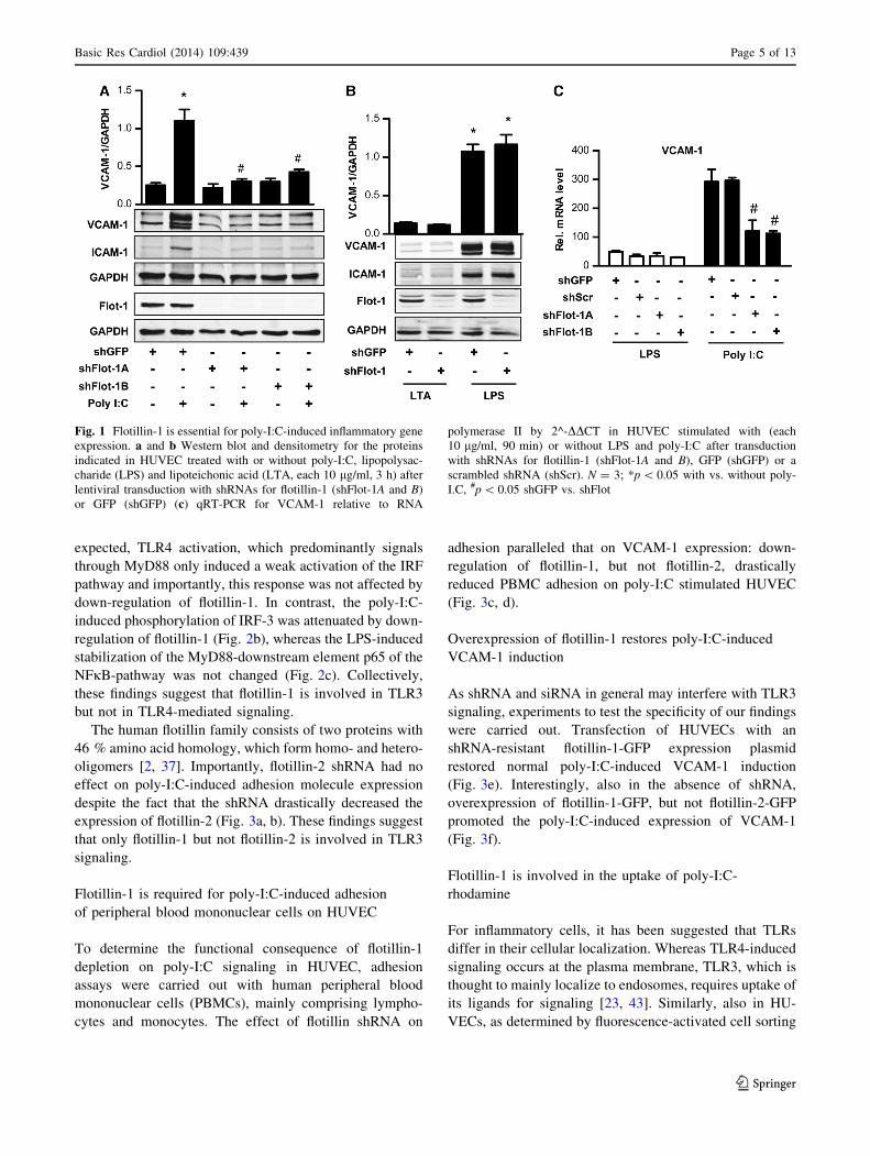
Flotillin-1 is essential for poly-I:C-induced
inflammatory gene expression
The role of flotillin-1 in TLR-induced signaling in human
umbilical vein endothelial cells (HUVEC) was studied by
shRNA and TLR signaling was initiated by selective ago-
nists. Activation of TLR3 by polyriboinosinic polyribocy-
tidylic acid (poly-I:C) and of TLR4 by lipopolysaccharide
(LPS) induced endothelial inflammatory adhesion molecule
expression, whereas the TLR2 ligand lipoteichoic acid had
little effect in endothelial cells (Fig. 1a, b). Knockdown of
flotillin-1 decreased the poly-I:C-induced VCAM-1 and
ICAM-1 expression (Fig. 1a, c), whereas flotillin-1 shRNA
did not affect LPS-induced inflammatory activation
(Fig. 1b, c). Basal level of VCAM-1 and ICAM-1 was not
affected by the control or flotillin-1 shRNA (data not
shown).
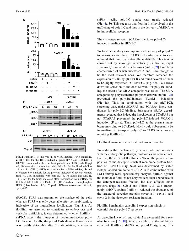
Flotillin-1 is involved in poly-I:C-induced IRF-3
signaling
Endothelial poly-I:C-induced signaling activates the
TRAF-MyD88 pathway as well as the transcription factor
IRF-3, which facilitates type I interferon expression [27].
Down-regulation of flotillin-1 also attenuated the poly-I:C-
induced expression of interferon b (IFNb) as well as of the
interferon related cytokine CXCL10 (Fig. 2a). As
Page 4 of 13 Basic Res Cardiol (2014) 109:439
123
expected, TLR4 activation, which predominantly signals
through MyD88 only induced a weak activation of the IRF
pathway and importantly, this response was not affected by
down-regulation of flotillin-1. In contrast, the poly-I:C-
induced phosphorylation of IRF-3 was attenuated by down-
regulation of flotillin-1 (Fig. 2b), whereas the LPS-induced
stabilization of the MyD88-downstream element p65 of the
NFjB-pathway was not changed (Fig. 2c). Collectively,
these findings suggest that flotillin-1 is involved in TLR3
but not in TLR4-mediated signaling.
The human flotillin family consists of two proteins with
46 % amino acid homology, which form homo- and hetero-
oligomers [2, 37]. Importantly, flotillin-2 shRNA had no
effect on poly-I:C-induced adhesion molecule expression
despite the fact that the shRNA drastically decreased the
expression of flotillin-2 (Fig. 3a, b). These findings suggest
that only flotillin-1 but not flotillin-2 is involved in TLR3
signaling.
Flotillin-1 is required for poly-I:C-induced adhesion
of peripheral blood mononuclear cells on HUVEC
To determine the functional consequence of flotillin-1
depletion on poly-I:C signaling in HUVEC, adhesion
assays were carried out with human peripheral blood
mononuclear cells (PBMCs), mainly comprising lympho-
cytes and monocytes. The effect of flotillin shRNA on
adhesion paralleled that on VCAM-1 expression: down-
regulation of flotillin-1, but not flotillin-2, drastically
reduced PBMC adhesion on poly-I:C stimulated HUVEC
(Fig. 3c, d).
Overexpression of flotillin-1 restores poly-I:C-induced
VCAM-1 induction
As shRNA and siRNA in general may interfere with TLR3
signaling, experiments to test the specificity of our findings
were carried out. Transfection of HUVECs with an
shRNA-resistant flotillin-1-GFP expression plasmid
restored normal poly-I:C-induced VCAM-1 induction
(Fig. 3e). Interestingly, also in the absence of shRNA,
overexpression of flotillin-1-GFP, but not flotillin-2-GFP
promoted the poly-I:C-induced expression of VCAM-1
(Fig. 3f).
Flotillin-1 is involved in the uptake of poly-I:C-
rhodamine
For inflammatory cells, it has been suggested that TLRs
differ in their cellular localization. Whereas TLR4-induced
signaling occurs at the plasma membrane, TLR3, which is
thought to mainly localize to endosomes, requires uptake of
its ligands for signaling [23, 43]. Similarly, also in HU-
VECs, as determined by fluorescence-activated cell sorting
Fig. 1 Flotillin-1 is essential for poly-I:C-induced inflammatory gene
expression. a and b Western blot and densitometry for the proteins
indicated in HUVEC treated with or without poly-I:C, lipopolysac-
charide (LPS) and lipoteichonic acid (LTA, each 10 lg/ml, 3 h) after
lentiviral transduction with shRNAs for flotillin-1 (shFlot-1A and B)
or GFP (shGFP) (c) qRT-PCR for VCAM-1 relative to RNA
polymerase II by 2^-DDCT in HUVEC stimulated with (each
10 lg/ml, 90 min) or without LPS and poly-I:C after transduction
with shRNAs for flotillin-1 (shFlot-1A and B), GFP (shGFP) or a
scrambled shRNA (shScr). N = 3; *p \ 0.05 with vs. without poly-
I.C, #p \ 0.05 shGFP vs. shFlot
Basic Res Cardiol (2014) 109:439 Page 5 of 13
123
(FACS), TLR4 was present on the surface of the cells
whereas TLR3 was only detectable after permeabilization,
indicative of an intracellular localization (Fig. S1). As
flotillins are assumed to contribute to endocytosis and
vesicular trafficking, it was determined whether flotillin-1
shRNA affects the transport of rhodamine-labeled poly-
I:C. In control cells, the poly-I:C-rhodamine fluorescence
was readily detectable after 3 h stimulation, whereas in
shFlot-1 cells, poly-I:C uptake was greatly reduced
(Fig. 4a, b). This suggests that flotillin-1 is involved in the
trafficking of poly-I:C and thus in the delivery of dsRNA to
its intracellular receptors.
The scavenger receptor SCARA4 mediates poly-I:C-
induced signaling in HUVEC
To facilitate endocytosis, uptake and delivery of poly-I:C
to endosomes and thus to TLR3, cell surface receptors are
required that bind the extracellular dsRNA. This task is
carried out by scavenger receptors (SR). So far, eight
structurally unrelated SR subclasses (A-H) [39] have been
characterized of which subclasses A and B are thought to
be the most relevant ones. We therefore screened the
expression of SRs by qRT-PCR and found several of them
to be highly expressed in HUVECs (Fig. 4c). To narrow
down the selection to the ones relevant for poly-I:C bind-
ing, the effect of an SR A antagonist was tested. The SR A
antagonizing polysaccharide polymer dextran sulfate [25]
prevented the poly-I:C-induced VCAM-1 induction
(Fig. 4d). This, in combination with the qRT-PCR
screening data, make SCARA3 and SCARA4 likely can-
didates for poly-I:C binding. Subsequent siRNA experi-
ments revealed that indeed the knockdown of SCARA4 but
not SCARA3 prevented the poly-I:C-induced VCAM-1
induction (Fig. 4e). Thus, poly-I:C at the plasma mem-
brane may bind to SCARA4, which could subsequently be
internalized to transport poly-I:C to TLR3 in a process
requiring flotillin-1.
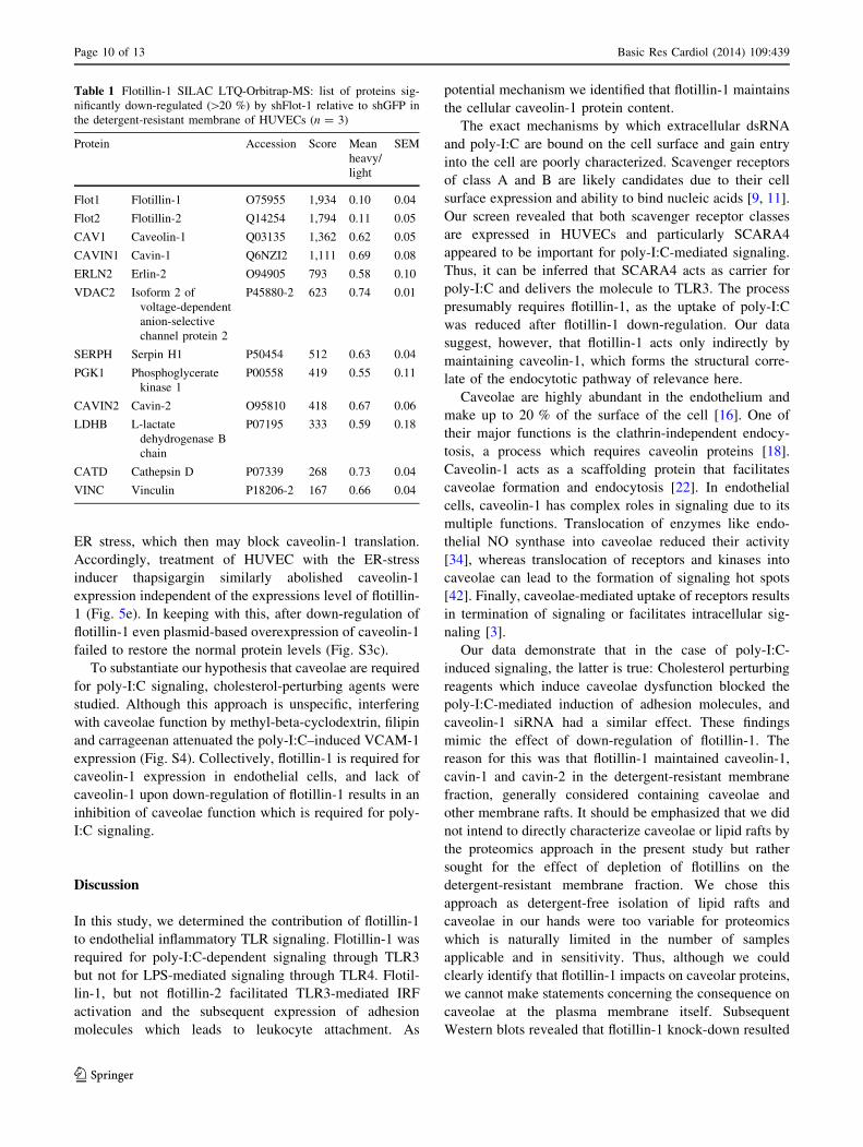
Flotillin-1 maintains structural proteins of caveolae
To address the mechanism by which flotillin-1 interacts
with the endocytotic pathways, proteomics was carried out.
For this, the effect of flotillin shRNA on the protein com-
position of the detergent-resistant membrane protein frac-
tion of HUVECs (Fig. S2a) was determined by stable
isotope labeled amino acids (SILAC) in combination with
ESI-Orbitrap mass spectrometry analysis. shRNA against
the individual flotillins not only reduced their abundance in
the detergent-resistant fraction, but also affected other
proteins (Figs. 5a, S2b–d and Tables 1, S1–S3). Impor-
tantly, shRNA against flotillin-1 reduced the abundance of
the structural caveolae proteins caveolin-1, cavin-1 and
cavin-2 in the detergent-resistant fraction.
Flotillin-1 maintains caveolin-1 expression which is
essential for the poly-I:C response
As caveolin-1, cavin-1 and cavin-2 are essential for cave-
olae function [16, 18], it is plausible that the inhibitory
effect of flotillin-1 shRNA on poly-I:C signaling is a
Fig. 2 Flotillin-1 is involved in poly-I:C-induced IRF-3 signaling.
a qRT-PCR for the IRF-3-inducible genes IFNb and CXCL10 in
HUVEC stimulated with or without poly-I:C and LPS (each 10 lg/
ml, 90 min) after transduction with shRNAs for flotillin-1 (shFlot-
1A and B), GFP (shGFP) or a scrambled shRNA (shScr). b and
c Western blot analysis for the proteins indicated of nuclear extracts
from HUVEC stimulated with poly-I:C (b, 10 lg/ml) and LPS (c,
10 lg/ml) for the times indicated after transduction with shRNA for
flotillin-1 (shFlot-1) or GFP (shGFP). pIRF-3 indicated anti-phospho-
IRF3 (phospho-Ser 385). Topo-1: DNA-topoisomerase. N = 4;
*p \ 0.05
Page 6 of 13 Basic Res Cardiol (2014) 109:439
123

consequence of caveolae dysfunction. Indeed, siRNA
against caveolin-1, similar to flotillin-1 shRNA, attenuated
caveolin-1 expression which was accompanied by
decreased poly-I:C-induced VCAM-1 expression (Fig. 5b).
It appears that flotillin-1 maintains caveolin-1 level, as also
in whole cell lysates, flotillin-1 shRNA but not flotillin-2
depletion decreased caveolin-1 protein levels in HUVECs
and in mouse lung endothelial cells (Figs. 5b, S3a, b).
Also, overexpression of caveolin-1 in shFlot-1 cells failed
to recover caveolin-1 level (Fig. S3c).Caveolin-1 siRNA,
in contrast, had no effect of flotillin-1 expression (Fig. 5b).
Finally, flotillin-1 shRNA had no effect on caveolin-2
protein levels (Fig. S3g) which further supports a selective
role of flotillin-1 for caveolin-1 expression.
Proximity ligation assay and total internal reflection
(TIRF) microscopy of the plasma membrane revealed a
close co-localization of caveolin-1 with flotillin-1 pre-
dominantly within the cell (Fig. 5c), suggesting that
Fig. 3 Poly-I:C-induced adhesion of peripheral blood mononuclear
cells requires flotillin-1. a qRT-PCR and b Western blot with
densitometry for the gene indicated relative to the house-keeping gene
RNA polymerase II or GAPDH in HUVECs stimulated with poly-I:C
(10 lg/ml, a: 90 min, b: 180 min) after transduction with shRNA
against flotillin-1 (shFlot-1), flotillin-2 (shFlot-2) or shGFP N = 4,
p \ 0.05. c and d Representative microscopic image (c) and statistical
analysis (d) of peripheral blood mononuclear cell (PBMCs) adhering
during flow of 0.35 and 15 dyn/cm2 on poly-I:C stimulated (10 lg/
ml, 5 h) HUVECs transduced as indicated. e and f Western blot of the
proteins indicated in HUVECs electroporated with plasmids coding
for mCherry (pmCherry-N1), flotillin-2-GFP (pFlot-2-GFP-N1) and
flotillin-1-GFP (pFlot-1-GFP-N1) stimulated with or without poly-I:C
(10 lg/ml, 180 min) and with (e) or without (f) transduction of shGFP
or shFlot-1. N = 4, *p \ 0.05, ns not significant
Basic Res Cardiol (2014) 109:439 Page 7 of 13
123

flotillin-1 interferes with caveolin-1 stability. However, the
inhibitory effect of flotillin-1 shRNA on caveolin-1 was not
prevented by inhibition of the proteasome or the lysosomal
pathway (Fig. S3d, e). Also, flotillin-1 shRNA had only a
small, yet significant, effect on caveolin-1 mRNA expres-
sion (Fig. S3f) suggesting rather attenuated translation and/
or misfolding caveolin-1 protein. Disturbing of endoplas-
mic reticulum (ER) protein formation and maturation leads
to the ER stress also termed unfolded protein response
(URP), a situation in which ER-localized translation is
attenuated. ER stress activates a signaling pathway which
includes phosphorylation of the type I ER transmembrane
kinase PERK which then directly phosphorylates the
eukaryotic initiation factor 2 eIF2a. Importantly, increased
PERK and eIF2a phosphorylation were present in shFlot-1
cells (Fig. 5d), indicating that loss of flotillin-1 results in
Fig. 4 The scavenger receptor SCARA4 mediates poly-I:C-induced
signaling. a and b Original laser scanning microscopic images (a) and
quantification (b) from HUVEC transduced with shGFP and shFlot-1
after incubation with poly-I:C-rhodamine (red, 0.5 lg/ml, 180 min).
Nuclei are counterstained with DAPI (blue). c q-RT-PCR for the
genes indicated relative to the house-keeping gene RNA polymerase
II in HUVECs. d Representative Western blot and densitometry for
the genes indicated in HUVECs transduced with shGFP or shFlot-1
after pretreatment with and without the class A scavenger receptor
inhibitor dextran sulfate (10 lg/ml, 5 min) and subsequent stimula-
tion with poly-I:C (10 lg/ml, 180 min). N = 4, *p \ 0.05. e Western
blot analysis and densitometry for the proteins indicated and qRT-
PCR for SCARA3 (left bottom) and SCARA4 (right bottom) in
HUVECs treated with scrambled siRNA or two different siRNAs
(-A and -B) against SCARA3 (left, siSCARA3) or SCARA4 (right,
siSCARA4) followed by stimulation with poly-I:C (10 lg/ml, 180).
N = 4, *p \ 0.05
Page 8 of 13 Basic Res Cardiol (2014) 109:439
123

Fig. 5 Flotillin-1 maintains caveolin-1 protein which is required for
the poly-I:C response. a Subset of SILAC-mass spectrometric
analyses on the effect of shFlot-1 on protein abundance in the
detergent-resistant membrane fraction of HUVECs. b Exemplary
Western blot and densitometry for the proteins indicated of HUVECs
transduced with shGFP or shFlot-1 or transfects with scrambled
siRNA (siScr) or caveolin-1 siRNA (siCav-1) with and without
stimulation with poly-I:C (10 lg/ml, 180 min), *p \ 0.05, n = 4.
c Proximity ligation assays for flotillin-1 and caveolin-1 interaction
(red) in HUVECs transduced with shGFP or shFlotillin-1. Down total
internal reflection fluorescence microscopy (TIRF) image, up epi-
fluorescence. Cells were counterstained with paxillin (green) and
DAPI (blue). d and e Exemplary Western blot and densitometry for
the proteins indicated of HUVECs transduced with shGFP or shFlot-1
with and without stimulation of thapsigargin 3.3 lmol/l. N = 4
*p \ 0.05
Basic Res Cardiol (2014) 109:439 Page 9 of 13
123
ER stress, which then may block caveolin-1 translation.
Accordingly, treatment of HUVEC with the ER-stress
inducer thapsigargin similarly abolished caveolin-1
expression independent of the expressions level of flotillin-
1 (Fig. 5e). In keeping with this, after down-regulation of
flotillin-1 even plasmid-based overexpression of caveolin-1
failed to restore the normal protein levels (Fig. S3c).
To substantiate our hypothesis that caveolae are required
for poly-I:C signaling, cholesterol-perturbing agents were
studied. Although this approach is unspecific, interfering
with caveolae function by methyl-beta-cyclodextrin, filipin
and carrageenan attenuated the poly-I:C–induced VCAM-1
expression (Fig. S4). Collectively, flotillin-1 is required for
caveolin-1 expression in endothelial cells, and lack of
caveolin-1 upon down-regulation of flotillin-1 results in an
inhibition of caveolae function which is required for poly-
I:C signaling.
Discussion
In this study, we determined the contribution of flotillin-1
to endothelial inflammatory TLR signaling. Flotillin-1 was
required for poly-I:C-dependent signaling through TLR3
but not for LPS-mediated signaling through TLR4. Flotil-
lin-1, but not flotillin-2 facilitated TLR3-mediated IRF
activation and the subsequent expression of adhesion
molecules which leads to leukocyte attachment. As
potential mechanism we identified that flotillin-1 maintains
the cellular caveolin-1 protein content.
The exact mechanisms by which extracellular dsRNA
and poly-I:C are bound on the cell surface and gain entry
into the cell are poorly characterized. Scavenger receptors
of class A and B are likely candidates due to their cell
surface expression and ability to bind nucleic acids [9, 11].
Our screen revealed that both scavenger receptor classes
are expressed in HUVECs and particularly SCARA4
appeared to be important for poly-I:C-mediated signaling.
Thus, it can be inferred that SCARA4 acts as carrier for
poly-I:C and delivers the molecule to TLR3. The process
presumably requires flotillin-1, as the uptake of poly-I:C
was reduced after flotillin-1 down-regulation. Our data
suggest, however, that flotillin-1 acts only indirectly by
maintaining caveolin-1, which forms the structural corre-
late of the endocytotic pathway of relevance here.
Caveolae are highly abundant in the endothelium and
make up to 20 % of the surface of the cell [16]. One of
their major functions is the clathrin-independent endocy-
tosis, a process which requires caveolin proteins [18].
Caveolin-1 acts as a scaffolding protein that facilitates
caveolae formation and endocytosis [22]. In endothelial
cells, caveolin-1 has complex roles in signaling due to its
multiple functions. Translocation of enzymes like endo-
thelial NO synthase into caveolae reduced their activity
[34], whereas translocation of receptors and kinases into
caveolae can lead to the formation of signaling hot spots
[42]. Finally, caveolae-mediated uptake of receptors results
in termination of signaling or facilitates intracellular sig-
naling [3].
Our data demonstrate that in the case of poly-I:C-
induced signaling, the latter is true: Cholesterol perturbing
reagents which induce caveolae dysfunction blocked the
poly-I:C-mediated induction of adhesion molecules, and
caveolin-1 siRNA had a similar effect. These findings
mimic the effect of down-regulation of flotillin-1. The
reason for this was that flotillin-1 maintained caveolin-1,
cavin-1 and cavin-2 in the detergent-resistant membrane
fraction, generally considered containing caveolae and
other membrane rafts. It should be emphasized that we did
not intend to directly characterize caveolae or lipid rafts by
the proteomics approach in the present study but rather
sought for the effect of depletion of flotillins on the
detergent-resistant membrane fraction. We chose this
approach as detergent-free isolation of lipid rafts and
caveolae in our hands were too variable for proteomics
which is naturally limited in the number of samples
applicable and in sensitivity. Thus, although we could
clearly identify that flotillin-1 impacts on caveolar proteins,
we cannot make statements concerning the consequence on
caveolae at the plasma membrane itself. Subsequent
Western blots revealed that flotillin-1 knock-down resulted
Table 1 Flotillin-1 SILAC LTQ-Orbitrap-MS: list of proteins sig-
nificantly down-regulated ([20 %) by shFlot-1 relative to shGFP in
the detergent-resistant membrane of HUVECs (n = 3)
Protein Accession Score Mean
heavy/
light
SEM
Flot1 Flotillin-1 O75955 1,934 0.10 0.04
Flot2 Flotillin-2 Q14254 1,794 0.11 0.05
CAV1 Caveolin-1 Q03135 1,362 0.62 0.05
CAVIN1 Cavin-1 Q6NZI2 1,111 0.69 0.08
ERLN2 Erlin-2 O94905 793 0.58 0.10
VDAC2 Isoform 2 of
voltage-dependent
anion-selective
channel protein 2
P45880-2 623 0.74 0.01
SERPH Serpin H1 P50454 512 0.63 0.04
PGK1 Phosphoglycerate
kinase 1
P00558 419 0.55 0.11
CAVIN2 Cavin-2 O95810 418 0.67 0.06
LDHB L-lactate
dehydrogenase B
chain
P07195 333 0.59 0.18
CATD Cathepsin D P07339 268 0.73 0.04
VINC Vinculin P18206-2 167 0.66 0.04
Page 10 of 13 Basic Res Cardiol (2014) 109:439
123
in a global depletion of caveolin-1 from the cell, which
may suggest that already at an early stage of membrane
integration of caveolin-1, flotillin-1 might be of relevance.
The combination of proximity ligation assay and TIRF
technique in our study supports this view: although flotil-
lins-1 co-localized with caveolin-1 to some extent, this
effect was largely restricted to intracellular compartments
around the nucleus, but not frequent at the plasma mem-
brane. Our finding of a very restricted intracellular inter-
action of flotillin-1 and caveolin-1 may also help to explain
why by means of membrane isolation or conventional LSM
technique, interactions of the two proteins were not
reported [12, 13]. The fact that the proteins can be co-
immuno-precipitated [41] does not contradict this notion as
it is probably not possible to successfully solubilize
membrane rafts without disrupting antibody-antigen bind-
ing. Thus, in immunoprecipitation studies usually whole
membrane patches are being pulled down.
In the present study, we found no evidence that flotillin-1
prevents the degradation of caveolin-1 as neither lysosomal
nor proteasomal inhibitors restored caveolin-1 level in flo-
tillin-1 knock-down cells. Although this contrasts a previ-
ous observation in an epithelial cell line [40] it points to a
function of flotillins-1 in caveolin-1 synthesis. Depletion of
flotillin-1 in HUVEC induced a small decrease in caveolin-
1 mRNA expression which, however, is not sufficient to
explain the almost complete lack of caveolin-1 protein
under this condition. Therefore, we assumed that the lack of
flotillins-1 prevents de novo synthesis of caveolin-1 at the
level of translation. The homeostasis of the endoplasmic
reticulum (ER) is important for maturation of newly syn-
thesized secretory and transmembrane proteins. Conditions
interfering with the function of ER are called ER stress. ER
stress is induced by accumulation of unfolded proteins
(unfolded protein response, UPR) or by excessive protein
demand caused by viral infection (ER overload response,
EOR) [19, 21]. Activation of ER stress sensors such as
PERK kinase or EIF2a reduces the ER workload and
induces expression of chaperons. Our findings that flotillin-
1 depletion induces ER stress signaling and that caveolin-1
translation itself is sensitive to ER stress explain its reduced
level in shFlot-1 HUVECs.
The question remains how flotillin-1 depletion induces
ER-stress. Flotillin-1 is involved in endosomal sorting,
trafficking from and towards the plasma membrane and the
transport to the ER and Golgi [29, 35]. We speculate that
the lack of flotillin-1 disturbs vesicular trafficking but
further studies will be needed to clarify this aspect.
Flotillin-2 can stabilize flotillin-1 [2, 13, 15] and also in
HUVEC, flotillin-2 shRNA reduced the flotillin-1 protein
expression to some extent. Despite this, loss of flotillin-2
often does not phenocopy the loss of flotillins-1 [7]. For
example, ERK signaling is increased in flotillin-2 knockout
mice whereas depletion of flotillin-1 decreased it [5]. Also
in the present study, depletion of flotillin-2 reduced flotil-
lin-1 expression to some extent but was without effect on
signaling. Potentially, the residual flotillin-1 expression
was sufficient to maintain signaling. Our flotillin-1 over-
expression experiments at least exclude that the anti-
inflammatory effect of flotillin-1 shRNA was mediated by
excess flotillin-2 exhibiting aberrant binding.
It is a limitation of the present study that only cultured
cells were studied but not knockout mice. In fact, such
experiments were carried out by us, but in keeping with the
overall very mild phenotype in the flotillin-1 global knock-
out mice [7, 26], we failed to detect a difference in caveolin-
1 expression between wild type and flotillin-1 global
knockouts (C. Fork. unpublished observations). Thus, it was
important to exclude that the altered responses in the cells
were not a consequence of an unspecific interference of the
shRNAs and siRNAs with TLR signaling. The fundamental
experiments were performed in two different shRNAs with
similar results and overexpression of flotillin-1 restored
normal poly-I:C-induced VCAM-1 expression. To exclude
that the lack of effect in the global knockout mice was a
consequence of a specific function of flotillin-1 in HUVEC,
the key experiments, i.e. the attenuation of poly-I:C induced
VCAM-1 induction by flotillin-1 shRNA, was also carried
out in murine lung endothelial cells, with similar results. We
therefore conclude that so far unidentified compensations
beyond the scope of our present study are operative in global
flotillin-1 knockout mouse.
Conclusion
We have identified that flotillin-1 is an important mediator
of TLR3 signaling in HUVEC. In these cells, acute loss of
flotillin-1 leads to attenuation of caveolar protein abun-
dance. As caveolar proteins turn out to be required for
TLR3 signaling, flotillin-1 might represent a novel target
for anti-inflammatory therapy.
Acknowledgment The authors are grateful for the excellent tech-
nical support by Susanne Schutz, Cindy Hoper, Tanja Luneburg and
Katalin Palfi. This work was funded by the Faculty of Medicine of
Goethe-University, Frankfurt am Main and the DFG excellence
cluster (EX-147) Cardio-Pulmonary System (ECCPS), and the Ger-
man Center for Cardiovascular Research (DZHK).
Conflict of interest On behalf of all authors, the corresponding
author states that there is no conflict of interest.
References
1. Amaddii M, Meister M, Banning A, Tomasovic A, Mooz J,
Rajalingam K, Tikkanen R (2012) Flotillin-1/reggie-2 protein
Basic Res Cardiol (2014) 109:439 Page 11 of 13
123

plays dual role in activation of receptor-tyrosine kinase/mitogen-
activated protein kinase signaling. J Biol Chem 287:7265–7278.
doi:10.1074/jbc.M111.287599
2. Babuke T, Ruonala M, Meister M, Amaddii M, Genzler C, Es-
posito A, Tikkanen R (2009) Hetero-oligomerization of reggie-1/
flotillin-2 and reggie-2/flotillin-1 is required for their endocytosis.
Cell Signal 21:1287–1297. doi:10.1016/j.cellsig.2009.03.012
3. Balogh P, Katz S, Kiss AL (2013) The role of endocytic pathways
in TGF-beta signaling. Pathol Oncol Res 19:141–148. doi:10.
1007/s12253-012-9595-8
4. Banning A, Ockenga W, Finger F, Siebrasse P, Tikkanen R
(2012) Transcriptional regulation of flotillins by the extracellu-
larly regulated kinases and retinoid X receptor complexes. PLoS
One 7:e45514. doi:10.1371/journal.pone.0045514
5. Banning A, Regenbrecht CR, Tikkanen R (2014) Increased
activity of mitogen activated protein kinase pathway in flotillin-2
knockout mouse model. Cell Signal 26:198–207. doi:10.1016/j.
cellsig.2013.11.001
6. Baumann CA, Ribon V, Kanzaki M, Thurmond DC, Mora S,
Shigematsu S, Bickel PE, Pessin JE, Saltiel AR (2000) CAP
defines a second signalling pathway required for insulin-stimu-
lated glucose transport. Nature 407:202–207. doi:10.1038/
35025089
7. Bitsikas V, Riento K, Howe JD, Barry NP, Nichols BJ (2014) The
role of flotillins in regulating abeta production, investigated using
flotillin 1-/-, flotillin 2-/- double knockout mice. PLoS One
9:e85217. doi:10.1371/journal.pone.0085217
8. Carcea I, Ma’ayan A, Mesias R, Sepulveda B, Salton SR, Benson
DL (2010) Flotillin-mediated endocytic events dictate cell type-
specific responses to semaphorin 3A. J Neurosci
30:15317–15329. doi:10.1523/JNEUROSCI.1821-10.2010
9. Dieudonne A, Torres D, Blanchard S, Taront S, Jeannin P, Del-
neste Y, Pichavant M, Trottein F, Gosset P (2012) Scavenger
receptors in human airway epithelial cells: role in response to
double-stranded RNA. PLoS One 7:e41952. doi:10.1371/journal.
pone.0041952
10. El-Sayed A, Harashima H (2013) Endocytosis of gene delivery
vectors: from clathrin-dependent to lipid raft-mediated endocy-
tosis. Mol Ther 21:1118–1130. doi:10.1038/mt.2013.54
11. Ezzat K, Helmfors H, Tudoran O, Juks C, Lindberg S, Padari K,
El-Andaloussi S, Pooga M, Langel U (2012) Scavenger receptor-
mediated uptake of cell-penetrating peptide nanocomplexes with
oligonucleotides. FASEB J 26:1172–1180. doi:10.1096/fj.11-
191536
12. Fernow I, Icking A, Tikkanen R (2007) Reggie-1 and reggie-2
localize in non-caveolar rafts in epithelial cells: cellular locali-
zation is not dependent on the expression of caveolin proteins.
Eur J Cell Biol 86:345–352. doi:10.1016/j.ejcb.2007.03.004
13. Frick M, Bright NA, Riento K, Bray A, Merrified C, Nichols BJ
(2007) Coassembly of flotillins induces formation of membrane
microdomains, membrane curvature, and vesicle budding. Curr
Biol 17:1151–1156. doi:10.1016/j.cub.2007.05.078
14. Ge L, Qi W, Wang LJ, Miao HH, Qu YX, Li BL, Song BL (2011)
Flotillins play an essential role in Niemann-Pick C1-like
1-mediated cholesterol uptake. Proc Natl Acad Sci USA
108:551–556. doi:10.1073/pnas.1014434108
15. Glebov OO, Bright NA, Nichols BJ (2006) Flotillin-1 defines a
clathrin-independent endocytic pathway in mammalian cells. Nat
Cell Biol 8:46–54. doi:10.1038/ncb1342
16. Hansen CG, Shvets E, Howard G, Riento K, Nichols BJ (2013)
Deletion of cavin genes reveals tissue-specific mechanisms for
morphogenesis of endothelial caveolae. Nat Commun 4:1831.
doi:10.1038/ncomms2808
17. Kim KB, Lee JS, Ko YG (2008) The isolation of detergent-
resistant lipid rafts for two-dimensional electrophoresis. Methods
Mol Biol 424:413–422. doi:10.1007/978-1-60327-064-9_32
18. Kirkham M, Nixon SJ, Howes MT, Abi-Rached L, Wakeham DE,
Hanzal-Bayer M, Ferguson C, Hill MM, Fernandez-Rojo M,
Brown DA, Hancock JF, Brodsky FM, Parton RG (2008) Evo-
lutionary analysis and molecular dissection of caveola biogenesis.
J Cell Sci 121:2075–2086. doi:10.1242/jcs.024588
19. Kuang E, Wan Q, Li X, Xu H, Liu Q, Qi Y (2005) ER
Ca2 ? depletion triggers apoptotic signals for endoplasmic
reticulum (ER) overload response induced by overexpressed
reticulon 3 (RTN3/HAP). J Cell Physiol 204:549–559. doi:10.
1002/jcp.20340
20. Lafyatis R, York M, Marshak-Rothstein A (2006) Antimalarial
agents: closing the gate on toll-like receptors? Arthritis Rheum
54:3068–3070. doi:10.1002/art.22157
21. Lawless MW, Greene CM, Mulgrew A, Taggart CC, O’Neill SJ,
McElvaney NG (2004) Activation of endoplasmic reticulum-
specific stress responses associated with the conformational dis-
ease Z alpha 1-antitrypsin deficiency. J Immunol 172:5722–5726
22. Le LS, Kurzchalia TV (2005) Getting rid of caveolins: pheno-
types of caveolin-deficient animals. Biochim Biophys Acta
1746:322–333. doi:10.1016/j.bbamcr.2005.06.001
23. Lee MS, Kim YJ (2007) Signaling pathways downstream of
pattern-recognition receptors and their cross talk. Annu Rev
Biochem 76:447–480. doi:10.1146/annurev.biochem.76.060605.
122847
24. Lee TH, McKleroy W, Khalifeh-Soltani A, Sakuma S, Lazarev S,
Riento K, Nishimura SL, Nichols BJ, Atabai K (2014) Functional
genomic screen identifies novel mediators of collagen uptake.
Mol Biol Cell 25:583–593. doi:10.1091/mbc.E13-07-0382
25. Limmon GV, Arredouani M, McCann KL, Corn Minor RA,
Kobzik L, Imani F (2008) Scavenger receptor class-A is a novel
cell surface receptor for double-stranded RNA. FASEB J
22:159–167. doi:10.1096/fj.07-8348com
26. Ludwig A, Otto GP, Riento K, Hams E, Fallon PG, Nichols BJ
(2010) Flotillin microdomains interact with the cortical cyto-
skeleton to control uropod formation and neutrophil recruitment.
J Cell Biol 191:771–781. doi:10.1083/jcb.201005140
27. Lundberg AM, Drexler SK, Monaco C, Williams LM, Sacre SM,
Feldmann M, Foxwell BM (2007) Key differences in TLR3/poly
I: C signaling and cytokine induction by human primary cells: a
phenomenon absent from murine cell systems. Blood
110:3245–3252. doi:10.1182/blood-2007-02-072934
28. McGettrick AF, O’Neill LA (2010) Localisation and trafficking
of toll-like receptors: an important mode of regulation. Curr Opin
Immunol 22:20–27. doi:10.1016/j.coi.2009.12.002
29. Meister M, Tikkanen R (2014) Endocytic trafficking of mem-
brane-bound cargo: a flotillin point of view. Membranes (Basel)
4:356–371. doi:10.3390/membranes4030356
30. Neumann-Giesen C, Fernow I, Amaddii M, Tikkanen R (2007)
Role of EGF-induced tyrosine phosphorylation of reggie-1/flo-
tillin-2 in cell spreading and signaling to the actin cytoskeleton.
J Cell Sci 120:395–406. doi:10.1242/jcs.03336
31. Ospelt C, Gay S (2010) TLRs and chronic inflammation. Int J
Biochem Cell Biol 42:495–505. doi:10.1016/j.biocel.2009.10.010
32. Otto GP, Nichols BJ (2011) The roles of flotillin microdomains–
endocytosis and beyond. J Cell Sci 124:3933–3940. doi:10.1242/
jcs.092015
33. Pust S, Dyve AB, Torgersen ML, van DB, Sandvig K (2010)
Interplay between toxin transport and flotillin localization. PLoS
One 5:e8844. doi:10.1371/journal.pone.0008844
34. Ramadoss J, Pastore MB, Magness RR (2013) Endothelial ca-
veolar subcellular domain regulation of endothelial nitric oxide
synthase. Clin Exp Pharmacol Physiol 40:753–764. doi:10.1111/
1440-1681.12136
35. Saslowsky DE, Cho JA, Chinnapen H, Massol RH, Chinnapen
DJ, Wagner JS, De Luca HE, Kam W, Paw BH, Lencer WI
(2010) Intoxication of zebrafish and mammalian cells by cholera
Page 12 of 13 Basic Res Cardiol (2014) 109:439
123
toxin depends on the flotillin/reggie proteins but not Derlin-1 or
-2. J Clin Invest 120:4399–4409. doi:10.1172/JCI42958
36. Schneider A, Rajendran L, Honsho M, Gralle M, Donnert G,
Wouters F, Hell SW, Simons M (2008) Flotillin-dependent
clustering of the amyloid precursor protein regulates its endo-
cytosis and amyloidogenic processing in neurons. J Neurosci
28:2874–2882. doi:10.1523/JNEUROSCI.5345-07.2008
37. Solis GP, Hoegg M, Munderloh C, Schrock Y, Malaga-Trillo E,
Rivera-Milla E, Stuermer CA (2007) Reggie/flotillin proteins are
organized into stable tetramers in membrane microdomains.
Biochem J 403:313–322. doi:10.1042/BJ20061686
38. Solis GP, Hulsbusch N, Radon Y, Katanaev VL, Plattner H,
Stuermer CA (2013) Reggies/flotillins interact with Rab11a and
SNX4 at the tubulovesicular recycling compartment and function
in transferrin receptor and E-cadherin trafficking. Mol Biol Cell
24:2689–2702. doi:10.1091/mbc.E12-12-0854
39. Sorensen KK, McCourt P, Berg T, Crossley C, Le CD, Wake K,
Smedsrod B (2012) The scavenger endothelial cell: a new player
in homeostasis and immunity. Am J Physiol Regul Integr Comp
Physiol 303:R1217–R1230. doi:10.1152/ajpregu.00686.2011
40. Vassilieva EV, Ivanov AI, Nusrat A (2009) Flotillin-1 stabilizes
caveolin-1 in intestinal epithelial cells. Biochem Biophys Res
Commun 379:460–465. doi:10.1016/j.bbrc.2008.12.118
41. Volonte D, Galbiati F, Li S, Nishiyama K, Okamoto T, Lisanti
MP (1999) Flotillins/cavatellins are differentially expressed in
cells and tissues and form a hetero-oligomeric complex with
caveolins in vivo. Characterization and epitope-mapping of a
novel flotillin-1 monoclonal antibody probe. J Biol Chem
274:12702–12709
42. Williams JJ, Palmer TM (2014) Cavin-1: caveolae-dependent
signalling and cardiovascular disease. Biochem Soc Trans
42:284–288. doi:10.1042/BST20130270
43. Yamashita M, Chattopadhyay S, Fensterl V, Zhang Y, Sen GC
(2012) A TRIF-independent branch of TLR3 signaling. J Immu-
nol 188:2825–2833. doi:10.4049/jimmunol.1103220
Basic Res Cardiol (2014) 109:439 Page 13 of 13
123