floppy neonate - neonet.ch · the floppy neonate is to distinguish two main groups: ... isolated...
TRANSCRIPT

Floppy neonate
SWISS SOCIETY OF NEONATOLOGY
June 2007

2
© Swiss Society of Neonatology, Thomas M Berger, Webmaster
Rochat M, Boltshauser E, Klein A, Frey B, Bänziger O,
Children‘s Hospital of Zurich, Switzerland

3
INTRODUCTIONThe “floppy neonate“ is a well known and recog-
nized entity for neonatologists. I t is used to de-
scribe a neonate with poor muscle tone. This clinical
diagnosis has numerous etiologies and a specific
diagnosis is not always obvious or easy to establish.
As some etiologies have a very poor prognosis and
the results of the diagnostic tests often take weeks
to be confirmed, the neonatologist must be able to
rely mainly on clinical features to begin counselling
the families at an early stage. This is partly possible,
as Paro-Panjan et al. managed to diagnose 50% of
their cases based on clinical data and the results of
c l in ica l examinat ions a lone (1 ) . Because of the
numerous etiologies, Paro-Panjan et al. (1) and Pra-
sad et al. (2) each recently published an algorithm as
an aid to approaching the floppy neonate.
We would like to present three cases to illustrate the
importance of a diagnostic algorithm to help neo-
natologists arrive at a rapid clinical diagnosis, thus
allowing better management of such infants.

4
CASE REPORT 1 The mother, a 28-year-old, G2/P1 with Stein-Leventhal
syndrome and adipositas per magna, had had a first
pregnancy that ended in a spontaneous abortion in
the 25th week of gestation. The current pregnancy
was complicated by cystitis in the 13th week and
because of pregnancy induced hypertension lung
maturation was induced in the 27th week of gestation.
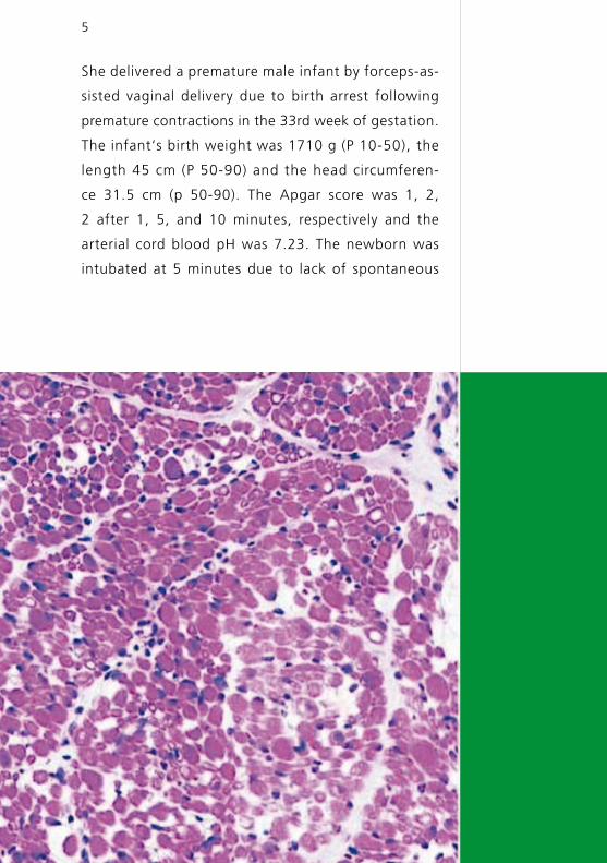
Muscle biopsy showing centrally placed nuclei in the
muscle fibres (arrow).
Fig. 1
5
She delivered a premature male infant by forceps-as-
sisted vaginal delivery due to birth arrest following
premature contractions in the 33rd week of gestation.
The infant‘s birth weight was 1710 g (P 10-50), the
length 45 cm (P 50-90) and the head circumferen-
ce 31.5 cm (p 50-90). The Apgar score was 1, 2,
2 after 1, 5, and 10 minutes, respectively and the
arterial cord blood pH was 7.23. The newborn was
intubated at 5 minutes due to lack of spontaneous
6
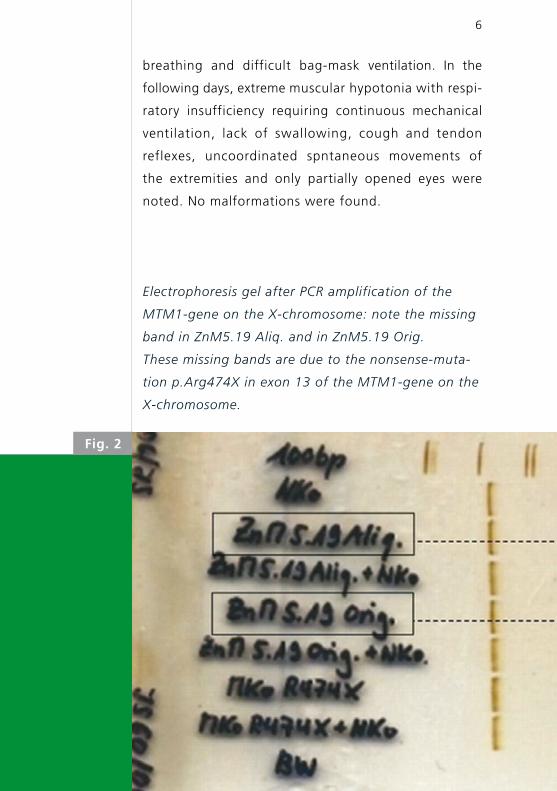
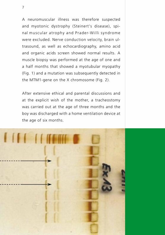
Fig. 2
Electrophoresis gel after PCR amplification of the
MTM1-gene on the X-chromosome: note the missing
band in ZnM5.19 Aliq. and in ZnM5.19 Orig.
These missing bands are due to the nonsense-muta-
tion p.Arg474X in exon 13 of the MTM1-gene on the
X-chromosome.
breathing and difficult bag-mask ventilation. In the
following days, extreme muscular hypotonia with respi-
ratory insufficiency requiring continuous mechanical
ventilation, lack of swallowing, cough and tendon
reflexes, uncoordinated spntaneous movements of
the extremities and only partially opened eyes were
noted. No malformations were found.
7
A neuromuscular illness was therefore suspected
and myotonic dystrophy (Steinert‘s disease), spi-
nal muscu lar at rophy and Prader-W i l l i syndrome
were excluded. Nerve conduction velocity, brain ul-
trasound, as well as echocardiography, amino acid
and organic acids screen showed normal results. A
muscle biopsy was performed at the age of one and
a half months that showed a myotubular myopathy
(Fig. 1) and a mutation was subsequently detected in
the MTM1-gene on the X chromosome (Fig. 2).
After extensive ethical and parental discussions and
at the explicit wish of the mother, a tracheostomy
was carried out at the age of three months and the
boy was discharged with a home ventilation device at
the age of six months.

8
Fig. 3
Spinal muscular atrophy mutation: PCR-RFLP-analysis
of exon 7 on both SMN-genes on the chromosome 5
(upper tracing: normal control, lower tracing: patient 2.
He is now five years old and depends on constant
mechanical ventilation. He has no expressive speech
and severe cognitive impairment, as well as chronic
pneumopathy due to repetitive aspiration episodes.
Because of swallowing difficulties, feeding was initi-
ally administered by a jejunal tube, and subsequent-
ly a fundoplication followed by a gastrostomy was
performed. He has progressive joint contractures,
ptosis, myopia and external ophthalmoplegia. In ad-
dition, he has middle ear deafness, atopic dermatitis
and progressive osteopenia, as well as accelerated
growth and bone age with premature adrenarche. He
has had generalized seizures since the age of two.

9
CASE REPORT 2This boy is the second child of fourth-grade consan-
guineous parents with no family history of genetic
diseases. The 28-year-old mother, a G2/P2 with an
uneventful pregnancy except for slightly decreased
fetal movements, delivered the term infant by ces-
arean section because of transverse presentation at
39 1/7 weeks of gestation. The infant‘s birth weight
was 3980 g (P 75-90), his length 50 cm (P 10-25)
and his head circumference 35 cm (P 50). The Apgar
score was 8, 8, 9, after 1, 5, and 10 minutes, respec-
tively. He required oxygen supplementation using a
face mask for two hours after delivery. Although he
was slightly hypotonic he was discharged home after
a few days in good condition.
His parents consulted the pediatrician at the age of
11 days because of muscular hypotonia. The boy was
then transferred to the intensive care unit because of
generalized muscular hypotonia, decreased spontane-
ous movements, areflexia and irregular spontaneous
10
CASE REPORT 3
breathing efforts. Breastfeeding was not a problem.
Amino acid and organic acid screens, abdomina l
and bra in u l t rasound were norma l . Prader-Willi
syndrome was excluded. A homozygote deletion in
the SMN1-gene on exon 7 of chromosome 5 confir-
med the clinical suspicion of spinal muscular atrophy
(Type 1, Werdnig-Hoffman) (Fig. 3).
Before the diagnosis was confirmed genetically, the
child had been discharged back home because of his
good developmental condition and follow-up is now
being carried out by neurologists in his home country.
A 22-year-old G1/P1 with a pregnancy complicated
by slightly reduced fetal movements and polyhy-
dramnios had a secondary cesarean section at 41 3/7
weeks because of fetal bradycardia for over a period
of 8 minutes. Postnatally, the male infant was weak,
hypotonic, bradycardic with no spontaneous bre-
athing and was therefore intubated. His birth weight
was 3430 g (P 10-50), his length 54 cm (P 90) and his
head circumference 36.5 cm (P 90). No Apgar score
was recorded; the arterial cord blood pH was 7.29.
In the family history (remembered after the delivery)
there had been numerous boys that had died of “we-
akness and no strength to breathe, a few days after
birth” (Fig. 4). A mutation analysis of most women in
the family undertaken 20 years earlier had shown a
mutation in the MTM1-gene on the X-chromosome,
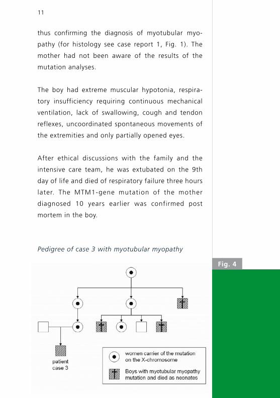
11
thus confirming the diagnosis of myotubular myo-
pathy (for histology see case report 1, Fig. 1). The
mother had not been aware of the results of the
mutation analyses.
The boy had extreme muscular hypotonia, respira-
tory insufficiency requiring continuous mechanical
ventilation, lack of swallowing, cough and tendon
reflexes, uncoordinated spontaneous movements of
the extremities and only partially opened eyes.
After ethical discussions with the family and the
intensive care team, he was extubated on the 9th
day of life and died of respiratory failure three hours
later. The MTM1-gene mutation of the mother
diagnosed 10 years earlier was confirmed post
mortem in the boy.
Pedigree of case 3 with myotubular myopathy
Fig. 4
12
DISCUSSION The floppy infant is a recognised entity characterized
by generalized hypotonia presenting at birth or in
early life (2, 1). Hypotonia presenting in the neonatal
period should always alert the neonatologist because
of the potentially serious underlying condition.
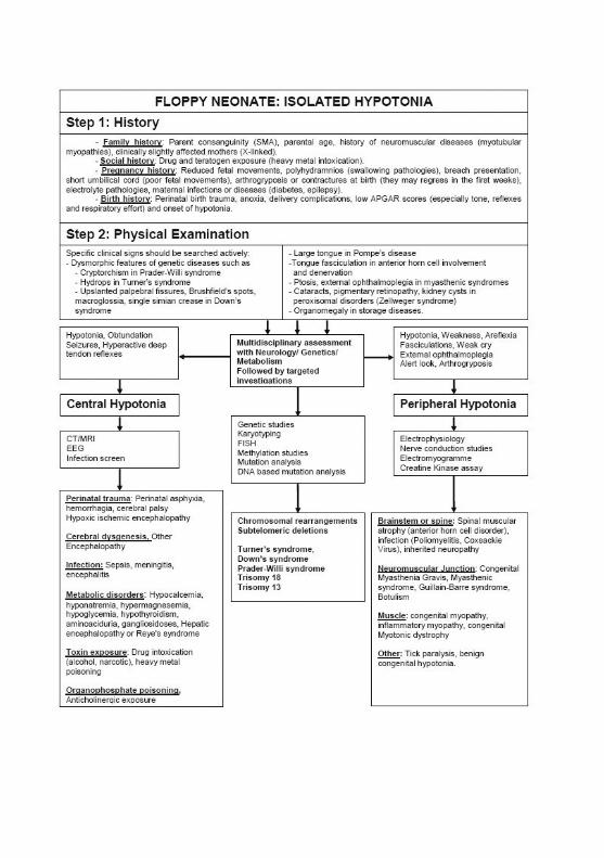
In this report, we discuss a few important points to
be considered when confronted with a floppy neo-
nate and present a diagnostic algorithm in order to
facilitate clinical decision making and to guide the
neonatologist in the subsequent treatment options
(2) (Table, see page 15).
The first clinical step when presented with a floppy
neonate should always be to obtain a detailed history
of the family, social environment, pregnancy, birth
and neonatal period, as thereafter most pathologies
can either be suspected or excluded (2). The second
clinical step is evidently the physical examination
(Table, see page 16). Hypotonia presents in the neo-
nate with full abduction and external rotation of the
legs as well as flaccid extension of the arms. When
traction is delivered to the arms, there is a prominent
head lag. A myopathic facies with paucity of facial
expression as well as a high-arched palate are often
noted in infants with neuromuscular disorders.
Algorithms to help physicians when confronted with
a floppy infant are very useful tools, and several have
been published in the past few years (1-3). A recent
13
publication in the journal of the Swiss Society of
Paediatrics also addressed this interesting topic (4).
An efficient and clinically relevant way to approach
the floppy neonate is to distinguish two main groups:
infants with isolated hypotonia and those with hypo-
tonia coupled with multisystem involvement.
Isolated hypotonia can be subdivided into lesions of
the upper motor neuron, which make up 66% of all
floppy infants; or lesions of the lower motor neuron
which consequently make up 33% (5). Upper motor
neuron lesions present with hypotonia, depressed
levels of consciousness, feeding difficulties, seizures,
apnea, hyperactive deep tendon reflexes, abnormal
body position, abnormal eye movements and abnor-
mal brainstem reflexes. Weakness (except for axial
weakness) is uncommon except in the acute stages.
Lower motor neuron lesions present with profound
weakness as well as hypotonia, paucity of antigravity
movements, hypo/areflexia, weak suck, low-pitched
cry/progressively weaker cry, arthrogryposis, external
ophthalmoplegia, and fasciculations but with an
alert look (6).
If hypotonia presents at birth with respiratory insuf-
ficiency severe enough to require tracheal intubation,
the prognosis is often very poor. Examples include
severe birth trauma, neonatal sepsis, myotubular my-
opathies, and Steinert’s disease.

14
If the onset is in the first days or weeks, with pro-
gressive feeding and breathing difficulties, hypoto-
nia and poor spontaneous movements, the prognosis
is still poor, but the infants can survive for months
or years. Examples include spinal muscular atrophy,
Prader-Willi syndrome, spinal muscular atrophy with
respiratory distress (SMARD), Pompe’s disease, and
congenital myopathy.
A floppy neonate with multisystem involvement
needs a structured, multidisciplinary diagnostic
approach, coordinated by the neonatologist. A rough
distinction should be made between toxic and meta-
bolic lesions. For more details, please see the article
by Prasad et al. (2).
Neonatologists are often confronted with floppy neo-
nates as hypotonia is a relatively common presentation
at birth or in the first few days of life. It is therefore
important to be aware of the different degrees and
presentations of hypotonia and to have knowledge
of diagnostic and prognostic implications. An algo-
rithm is an easy way of categorising symptoms and
helps the clinician to establish a rapid diagnosis and
initiate treatment measures.
As described in the case reports, if the classical symptoms
of peripheral motor neuron lesions are present at birth
(with respiratory insufficiency, lack of swallowing,
cough and tendon reflexes) the prognosis is very
CONCLUSIONS
15
ACKNOW-
LEDGMENTS
poor, independent of the exact diagnosis. Such a
presentation should lead to timely ethical discussi-
ons as well as parental counselling and to the consi-
deration of early extubation.
We wish to thank Professor S. Gallati of the Genetic
Laboratory of the Children’s University Hospital of
Berne for the copy of the electrophoresis gel, Dr. G.
Matyas of the Medical Molecular Genetic and Gene
Diagnostic Department of the Institute of Genetics
at the University of Zurich for the copy of the Spinal
Muscle Atrophy PCR Fragment, as well as Dr. E. He-
wer of the Institute of Neuropathology at the Univer-
sity Hospital of Zurich for the copies of the biopsies.
1. Paro-Panjan D, D. Neubauer D. Congenital hypotonia:
is there an algorithm? J Child Neurol 2004;19:439-442
2. Prasad AN, Prasad C. The floppy infant: contribution of
genetic and metabolic disorders. Brain Dev 2003;25:457-476
3. Johnston HM. The floppy weak infant revisited. Brain Dev
2003;25:155-158
4. Jeannet PY. Der hypotone Säugling. Paediatrica 2006;17:
19-23
5. Richer LP, Shevell MI, Miller SP. Diagnostic profile of
neonatal hypotonia: an 11-year study. Pediatr Neurol
2001;25:32-37
6. Family Practice notebook.com, in http://www.fpnotebook.com
REFERENCES

Swiss Society of Neonatology
www.neonet.ch
CONTACT
SUPPORTED BY
con
cep
t &
des
ign
by
mes
ch.c
h