flexible liposomes for topical applications in cosmetics polyunsaturated fatty acids (vitamin f)...
TRANSCRIPT

Flexible Liposomes for topical Applications in Cosmetics
Dr. Gabriele Blume
Abstract
Liposomes are commonly used in dermal applications as protective systems for active ingredients
and for their moisturising properties. They are spherical vesicles composed of phospholipids with
an aqueous core. Either lipophilic or hydrophilic active ingredients can be incorporated in these
vesicles. But they may also have the property to penetrate into the skin, carrying actives to the
target site, where these molecules will be released. Liposomes acting as such a dermal carrier have
to be small sized, unilamellar and equipped with a flexible membrane.
1. Introduction
Nowadays skin care formulations must meet high standards of efficacy - preferably visible effects –
for the consumers are much more sophisticated than in the past. As a result, consumers expect and
demand real performance from their products. To ensure effectiveness of the cosmetic formulation,
the actives have to be transported to the target site, mostly into the epidermis.
But penetration of substances through the skin is limited by the natural barrier – the stratum
corneum with its “brick and mortar” architecture. Indeed, the interstices in the horny layer, which
seldom exceed more than 20 nm in width, are extremely impermeable even for small molecules.
Water actually trespasses the skin at the rate of 0.4 mg cm-2 h-1.
To overcome the skin barrier, chemical enhancers may be used which typically increase the fluidity
of the lipids in the stratum corneum (sc).1 Despite the fact that Alec Bangham published the first
paper on liposomes in 1963, it was in the early 1980s that Mezei and Gulasekharam reported the
effectiveness of liposomes in topical drug delivery.2,3
2. Liposomes
Liposomes are hollow spheres that are enclosed by one or more bilayer membranes. These bilayer
membranes consist of natural components as for example phospholipids – in particular
phosphatidylcholine (PC), which make these carrier systems biocompatible per se. PC is obtained
either from soy beans or eggs, which differs in its composition of fatty acids. Egg-derived lipids
have a higher content of saturated fatty acids (40% of 16:0 and 18:0) in comparison to soy bean
(80% of 18:1 and 18:2).4

The amphiphilic nature of PC allows them to self-aggregate in an aqueous solution and to form their
spherical structures. Liposomes can be unilamellar or multilamellar as shown in Figure 2 and their
sizes range from 50 nm to several µm depending on their method of production .4
They are capable of delivering either hydrophilic (in the aqueous inner core) or lipophilic
substances (in the lipid bilayer).
Increased rates of skin permeability have been found for various active ingredients, e.g.
progesterone and hydrocortisone, when they were applied topically in liposomal form. Also, less
frequently side effects were observed when liposomal formulations were employed.5,6 In contrast to
these observations, other investigators could not find improved skin penetration of substances when
Figure 1
Figure 2

they were applied in liposomes.7 Also, liposomes themselves did not seem to penetrate through the
sc in these early studies.8
The conflicting results may stem from different lipid compositions of the liposomes employed as in
addition to size. The lipid composition determines the physical characteristics of the liposomes and,
therefore, also the interaction of these carrier systems with the skin.9
In the last few years some papers highlighted important factors that influence the penetration of
active ingredients encapsulated in liposomes. Liquid-state, flexible liposomes showed greater skin
penetration than those in a gel-state, small-sized and unilamellar vesicles seem to result in a higher
degree of skin penetration.10-12 The application form can also influence the penetration kinetics.13
3. Flexible Liposomes
Flexible liposomes are small-sized unilamellar vesicles (80-250 nm) prepared of soy bean
phosphatidylcholine (> 80%) having a high content of linoleic acid. They provide the skin with
essential polyunsaturated fatty acids (vitamin F) which support the formation of ceramide 1 and
with choline which is a part of the natural moisturising factor (NMF). In a clinical study it was
proven that these liposomes have cosmetic properties like wrinkle reduction and an increase in skin
smoothness and furthermore show pharmaceutical effects like decreasing of efflorescence in the
acne treatment.14,15
3.1 Penetration
The demands to bring active ingredients into the deeper skin layer and to get a site-specific
targeting of these molecules with visible cosmetic effects become more and more relevant.
In an ex-vivo study on human skin biopsies, fluorescence markers- liposomally encapsulated or in a
free form - were tested on their ability to penetrate into the skin. The penetration profile was
visualised by confocal laser scanning microscopy (CLSM).
The hydrophilic fluorescent dye carboxyfluorescein (CF) and the lipophilic dye 1,1-diocytadecyl-
3,3,3,3,-tetramethylindocarbo-cyanine perchlorate (DiI) were encapsulated together in liposomes
made of soybean lecithin (PC > 80%) by homogenization and subsequent extrusion through a 0.1
µm microporous filter .
Human abdomen skin was obtained after cosmetic surgery. After removal of subcutaneous fat, the
skin was placed in Franz-type diffusion cells. The liposome preparation and the CF-DiI ethanolic
solution (control) were non-occlusively applied on the epidermal side of the skin. Usually 50 µl of
test preparation were pipetted onto a circular area of 3.5 cm diameter. Three hours after application
the skin surface was rinsed several times with buffer and blotted dry. After this, skin cylinders were

punched, cryo-fixed with CO2 and cut in 10 µm thick pieces. After air drying the specimens were
examined by CLSM (BioRad MRC 1024).
Ethanol itself is a moderate skin penetration enhancer for many substances.16 However, as can be
seen from the Figure 3, an ethanolic solution of the fluorescence dyes is not able to penetrate
noticeably through the stratum corneum during the observed period of time. In contrast, the dyes
encapsulated in the flexible liposomes appear to penetrate through the stratum corneum and enter
the deeper layers of the epidermis. Furthermore the visible fluorescence in the hair follicles
indicates the transportation of molecules through the hair shaft as far as the hair roots (Figure 4).
Figure 3
Figure 4
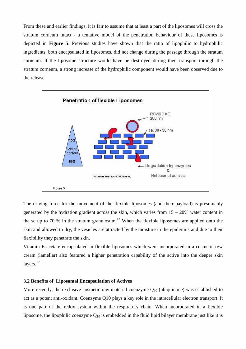
From these and earlier findings, it is fair to assume that at least a part of the liposomes will cross the
stratum corneum intact - a tentative model of the penetration behaviour of these liposomes is
depicted in Figure 5. Previous studies have shown that the ratio of lipophilic to hydrophilic
ingredients, both encapsulated in liposomes, did not change during the passage through the stratum
corneum. If the liposome structure would have be destroyed during their transport through the
stratum corneum, a strong increase of the hydrophilic component would have been observed due to
the release.
Figure 5
The driving force for the movement of the flexible liposomes (and their payload) is presumably
generated by the hydration gradient across the skin, which varies from 15 – 20% water content in
the sc up to 70 % in the stratum granulosum.13 When the flexible liposomes are applied onto the
skin and allowed to dry, the vesicles are attracted by the moisture in the epidermis and due to their
flexibility they penetrate the skin.
Vitamin E acetate encapsulated in flexible liposomes which were incorporated in a cosmetic o/w
cream (lamellar) also featured a higher penetration capability of the active into the deeper skin
layers.17
3.2 Benefits of Liposomal Encapsulation of Actives
More recently, the exclusive cosmetic raw material coenzyme Q10 (ubiquinone) was established to
act as a potent anti-oxidant. Coenzyme Q10 plays a key role in the intracellular electron transport. It
is one part of the redox system within the respiratory chain. When incorporated in a flexible
liposome, the lipophilic coenzyme Q10 is embedded in the fluid lipid bilayer membrane just like it is

realized in natural cell membranes in the skin. This particular natural surrounding of the molecule
provides the full scope of functionality and anti-oxidative property.
A new innovative electron spin resonance (ESR) spectroscopy method was introduced to the
cosmetic market, which enables a benchmarked evaluation of the anti-oxidative properties (AOP) of
formulations ex vivo.18
The penetration and activity of antioxidants can be detected by means of ESR spectroscopy on skin
biopsies. For the measurement the skin biopsy is labeled with test radicals that are allowed to
diffuse into the skin (epidermis) from the dermal side, but not into the stratum corneum. The
cosmetic actives and/or the cosmetic formulations are applied onto the skin`s surface. If the active
passes the stratum corneum and penetrates into the epidermis, it will react with the test radicals and
therefore reduce their number. Accordingly, the ESR signal intensity diminishes and the penetration
and penetration kinetics, respectively, can be followed. A measurement of an untreated skin biopsy
accounts for the intrinsic enzymatic and non-enzymatic radical protection mechanism and has to be
included in the study.
The study reveals the significant and rapid enhancement of the intrinsic protection by liposomal Q10
in contrast to the pure ethanolic Q10 solution as illustrated in Figure 6. The anti-oxidative potential
becomes immediately relevant and 20 minutes after application the enhancement makes already
27%.
Figure 6
Sensitive ingredients (lipophilic vitamins like retinol) can be incorporated in the flexible vesicle
membrane where they are protected from the outside (light, temperature, chemical degradation).
This results in an improvement in the stability of the vitamin over a long time period. 19, 20
Furthermore, the encapsulation of actives into the liposomes may avoid irritation that is caused by
certain actives. Alpha hydroxy acids were introduced into cosmetics for peeling purposes in the
early 1970s, due to the keratolytic and moisturising properties they possess. Generally, the
solutions containing free acids or their salts were deemed intolerable by the test subjects. Of special
note are the formulations containing the liposome-encapsulated AHA salts: These were judged by
the subjects to be pleasant and well tolerated, causing no dermatological changes to the skin.14
3.3 Stability of Flexible Liposomes
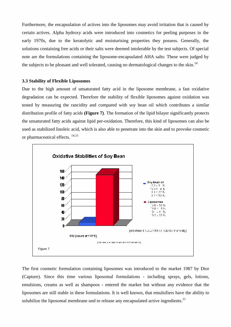
Due to the high amount of unsaturated fatty acid in the liposome membrane, a fast oxidative
degradation can be expected. Therefore the stability of flexible liposomes against oxidation was
tested by measuring the rancidity and compared with soy bean oil which contributes a similar
distribution profile of fatty acids (Figure 7). The formation of the lipid bilayer significantly protects
the unsaturated fatty acids against lipid per-oxidation. Therefore, this kind of liposomes can also be
used as stabilized linoleic acid, which is also able to penetrate into the skin and to provoke cosmetic
or pharmaceutical effects. 14,15
Figure 7
The first cosmetic formulation containing liposomes was introduced to the market 1987 by Dior
(Capture). Since this time various liposomal formulations - including sprays, gels, lotions,
emulsions, creams as well as shampoos - entered the market but without any evidence that the
liposomes are still stable in these formulations. It is well known, that emulsifiers have the ability to
solubilize the liposomal membrane und to release any encapsulated active ingredients.21
Therefore the morphological integrity of the vesicles is not any longer warranted, and the benefits
of the liposomal encapsulation is affected or even nullified.
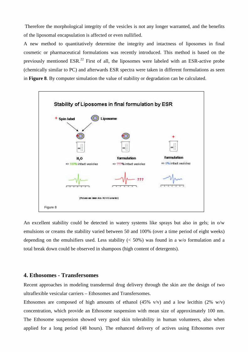
A new method to quantitatively determine the integrity and intactness of liposomes in final
cosmetic or pharmaceutical formulations was recently introduced. This method is based on the
previously mentioned ESR.22 First of all, the liposomes were labeled with an ESR-active probe
(chemically similar to PC) and afterwards ESR spectra were taken in different formulations as seen
in Figure 8. By computer simulation the value of stability or degradation can be calculated.
Figure 8
An excellent stability could be detected in watery systems like sprays but also in gels; in o/w
emulsions or creams the stability varied between 50 and 100% (over a time period of eight weeks)
depending on the emulsifiers used. Less stability (< 50%) was found in a w/o formulation and a
total break down could be observed in shampoos (high content of detergents).
4. Ethosomes - Transfersomes
Recent approaches in modeling transdermal drug delivery through the skin are the design of two
ultraflexible vesicular carriers – Ethosomes and Transfersomes.
Ethosomes are composed of high amounts of ethanol (45% v/v) and a low lecithin (2% w/v)
concentration, which provide an Ethosome suspension with mean size of approximately 100 nm.
The Ethosome suspension showed very good skin tolerability in human volunteers, also when
applied for a long period (48 hours). The enhanced delivery of actives using Ethosomes over
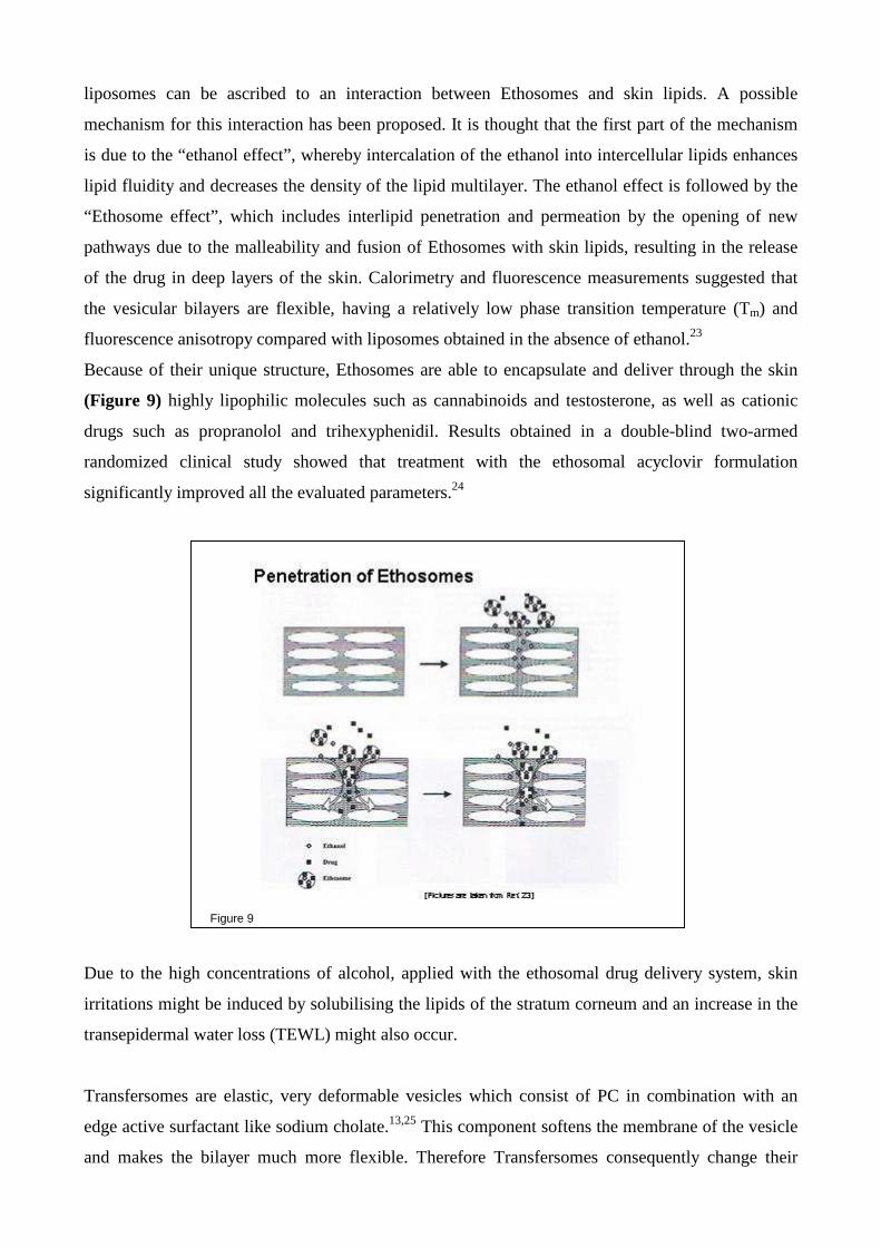
liposomes can be ascribed to an interaction between Ethosomes and skin lipids. A possible
mechanism for this interaction has been proposed. It is thought that the first part of the mechanism
is due to the “ethanol effect”, whereby intercalation of the ethanol into intercellular lipids enhances
lipid fluidity and decreases the density of the lipid multilayer. The ethanol effect is followed by the
“Ethosome effect”, which includes interlipid penetration and permeation by the opening of new
pathways due to the malleability and fusion of Ethosomes with skin lipids, resulting in the release
of the drug in deep layers of the skin. Calorimetry and fluorescence measurements suggested that
the vesicular bilayers are flexible, having a relatively low phase transition temperature (Tm) and
fluorescence anisotropy compared with liposomes obtained in the absence of ethanol.23
Because of their unique structure, Ethosomes are able to encapsulate and deliver through the skin
(Figure 9) highly lipophilic molecules such as cannabinoids and testosterone, as well as cationic
drugs such as propranolol and trihexyphenidil. Results obtained in a double-blind two-armed
randomized clinical study showed that treatment with the ethosomal acyclovir formulation
significantly improved all the evaluated parameters.24
Figure 9
Due to the high concentrations of alcohol, applied with the ethosomal drug delivery system, skin
irritations might be induced by solubilising the lipids of the stratum corneum and an increase in the
transepidermal water loss (TEWL) might also occur.
Transfersomes are elastic, very deformable vesicles which consist of PC in combination with an
edge active surfactant like sodium cholate.13,25 This component softens the membrane of the vesicle
and makes the bilayer much more flexible. Therefore Transfersomes consequently change their
shape easily by adjusting locally to ambient stress. When a suspension of Transfersome vesicles is
applied non-occlusively on the surface of the skin, the water evaporates from the skin surface and
the vesicles start to dry out. Due to the strong hydrophilicity, the vesicles are attracted to the areas
of higher water content in the narrow gaps between adjoining cells in the skin. The phenomenon,
together with the vesicle's extreme ability to deform, enables the Transfersome to temporarily open
the pores through which water normally evaporates between the cells. Such newly activated inter-
cellular passages can accommodate sufficiently deformable vesicles maintaining their integrity but
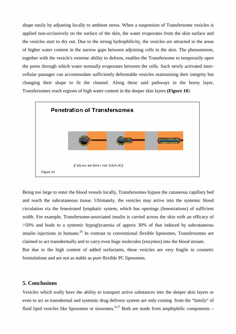
changing their shape to fit the channel. Along these said pathways in the horny layer,
Transfersomes reach regions of high water content in the deeper skin layers (Figure 10).
Figure 10
Being too large to enter the blood vessels locally, Transfersomes bypass the cutaneous capillary bed
and reach the subcutaneous tissue. Ultimately, the vesicles may arrive into the systemic blood
circulation via the fenestrated lymphatic system, which has openings (fenestrations) of sufficient
width. For example, Transfersome-associated insulin is carried across the skin with an efficacy of
>50% and leads to a systemic hypoglycaemia of approx 30% of that induced by subcutaneous
insulin injections in humans.26 In contrast to conventional flexible liposomes, Transfersomes are
claimed to act transdermally and to carry even huge molecules (enzymes) into the blood stream.
But due to the high content of added surfactants, these vesicles are very fragile in cosmetic
formulations and are not as stable as pure flexible PC liposomes.
5. Conclusions
Vesicles which really have the ability to transport active substances into the deeper skin layers or
even to act as transdermal and systemic drug delivery system are only coming from the “family“ of
fluid lipid vesicles like liposomes or niosomes.9,27 Both are made from amphiphilic components –

liposomes from phospholipids and niosomes from non-ionic surfactants. The main assumption is
the size and the fluidity of the membrane at room temperature which ensure the flexible
deformability of the vesicles during their pathway through the stratum corneum. Liposomes prefer
membrane structures that are identical to their own composition; PC is the main component of all
viable cells and save in application. But liposomes hamper in their pH-dependence, their stability is
best between 6.3 and 6.8.28 Repeated applications of niosomes might lead to irritations due to the
accumulation of detergents in the skin. Therefore, flexible liposomes are the only choice to carry
active molecules into the deeper skin layers (biological syringe) by stabilizing the actives and even
acting as pharmaceutical or cosmetic ingredient due to its chemical nature.
References
(1) Hardgraft, J. and Walters, K.A., in: Juninger, H.E. (Ed.), Drug targeting and delivery concepts in dosage form design, Ellis Horwood
Limited., Clichester, UK, 1992, pp 169-177
(2) Bangham, A.D., Physical structure and behaviour of lipids and lipids enzymes, Adv. Lipid Res., 1 (1963) 65-104
(3) Mezei, M. and Gulasekharam, V., Liposomes – selective drug delivery system for the topical route of administration, Life Sci,. 26 (1980)
1473-1477
(4) New, R.R.C. , in New, R.R.C. (Ed.), Liposomes a pratical approach, Oxford University Press, Oxford, UK, 1990, pp 1- 67
(5) Ho, N.F.H., Ganesan, M. G., Weiner, N. D. and Flynn, G. L., Mechanisms of topical delivery of liposomally entrapped drugs, J. Control.
Rel., 2 (1985) 61-65
(6) Lasch, J. and Wohlrab, W., Liposome-bound cortisol: a new approach to cutaneous therapy, Biomed Biochim Acta , 45 (1986) 1295-1299.
(7) Knepp, V.M., Hinz, R., Szoka, F.C. and Guy, R., Controlled drug release from a novel liposomal delivery system. I. Investigation of
transdermal potential, J. Control. Rel., 5 (1988) 211-221.
(8) Knepp, V.M., Szoka, F.C. and Guy, R., Controlled drug release from a novel liposomal delivery system. II. Transdermal delivery
Characteristics, J. Control. Rel., 12 (1990) 25-30.
(9) Choi, M.J. and Maibach, H.J., Liposomes and Niosomes as topical drud delivery systems, Skin Pharmacol. Physiol., 18 (2005) 209-219
(10) .Blume, G., Sacher, M., Teichmüller, D. and Schäfer, U., The role of liposomes and their future perpective, SÖFW-Journal, 129 (2003)
10-14.
(11) Verma, D.D. Verma, S., Blume, G. and Fahr, A., Particle size of liposomes influences dermal delivery of substances into the skin, Int. J.
Pharma., 258 (2003) 141-151
(12) Fresta, M.. and Puglisi, G., Application of liposomes as potential cutaneous drug delivery systems, J. Drug Targeting, 4 (1996) 95-101
(13) Cevc, G. and Blume, G., Lipid vesicles penetrate into intact skin owing transdermal osmotic gradients and hydration force,
Biochim. Biophys. Acta , 1104 (1992) 226-232
(14) Blume, G. and Teichmüller, E., New evidence of the penetration of actives by liposomal carrier system, Cosmetics & Toiletries
Manufacture Worldwide, (1997) 135-139
(15) Ghyczy, M., Nissen, H.P. and Biltz, H., The treatment od Acne vulgaris by phosphatidylcholine from soybeans with a high content of
linoleic acid, J. Appl. Cosmetol., 14 (1996) 37-145
(16) Kitagawa, S., Li, H. and Sato, S., Skin permeation of parabens in excised guinea pig dorsal skin, its modification by penetration enhancers
Chem. Pharm Bull (Tokyo), 45 (1997) 1354-1357
(17) Lampen, P., Pittermann, W., Heise, H.M., Schmitt, M., Jungmann, H. and Kietzmann, M., Penetration studies of vitamin E acteate applied
from cosmetic formulation to the stratum corneum of an in vitro model, J. Cosmet. Sci., 54 (2003) 119-131
(18) Sacher, M., Blume, G., Bakowsky, U. and Jung, K., Antioxidative penetration efficacy of liposomally encapsulated Coenzyme Q10,
SÖFW-Journal, 4 (2006) 48 – 54
(19) Blume, G. and Teichmüller, D., Cyclodextrins, vesicles and particles, SÖFW-Journal, 5 (2004) 14-23
(20) Arsic, I. and Stojanka, V., Protection of encapsulated vitamin A from UV radiation in aqueous dispersions and gel systems, Conference
Proceedings, IFSCC Budapest 1997
(21) Simonnet, JT., Lipid vesicles, Cosmetics & Toiletries Magazine, 109 (1994) 45-52
(22) Jung, K. and Blume, G. (2007) in preparation

(23) Touitou, E., Dayan, N., Bergelson, L., Godin, B. and Eliaz, M., Ethosomes – novel vesicular carriers for enhanced delivery :
characterization and skin permeation properties, J. Control Rel., 65 (2000) 403-418
(24) Godin, B. and Touitou, E., Ethosomes: new prospects in transdermal delivery, Crit. Rev. Ther. Drug. Carrier Syst, 20 (2003) 63-102
(25) Cevc, G., Blume, G., Schätzlein, A., Gebauer, D. and Paul, A., The skin: a pathway for systemic treatment with atches and lipid-based
agent carrier, Advanced Drug Delivery Reviews, 18 (1996) 349-378
(26) Cevc, G., Schätzlein, A. and Blume, G., Transdermal drug carriers: basic properties, optimization and transfer efficacy in the case of
epicutaneously applied peptides, Advanced Drug Delivery Reviews, 36 (1995) 3-16
(27) Honeywell-Nguyen, P.L., Surfactant-based elastic vesicle as skin delivery systems – Drug transport and mechanism of action, Ph.D.
Thesis, University of Leiden, The Netherlands, 2004
(28) Grit, M., Stability of Liposomes – Analytical, chemical and physical aspects, Ph D Thesi,s University of Utrecht, The Netherlands,
1991