fish parasites collected ,at woods hole in 1898.fish parasites collected ,at woods hole in 1898. by...
TRANSCRIPT

FISH PARASITES COLLECTED ,AT WOODS HOLE IN 1898.
By EDWIN LINTON, PH. D.,
Professor of Biology, Washington and Jefferson College.
The following report is divided into two parts.In Part I a list of the hosts which were examined, or from which parasites were
obtained, is given. In each instance brief mention is made of the parasites found, thedates of examination are given, and where the stomach contents were noted ~ recordis entered. In nearly every case in which no note was madeof stomach contents thestomachs were empty.
Adult trematodes and cestodea and a few nematodes have been identified. Manylarval cestodes and most of the nematodes have not yet been ideutified.
The order of arrangement of'hosts is substantially that of Dr. H. M. Smith, "TheFishes fonnd in the Vicinity of Woods Hole" (Bulletin of the United States FishOommission for] 897).
In Part II descriptions are given of new species and of species new to the region.While this report has mainly to do with the entozoa, I have given descriptions of
two ectoparasites: (1) A copepod, found in the cheek of a squeteague (Oynosoionregalis). (2) A tristomum (Epibdella bumpusii sp. nov.), from the skin of a stingray(Dasyatis oentrura). In the description of the latter are incorporated some observations on the process of egg-making as it was seen in this interesting species.
PATHOLOGICAL CONDITIONS.
It was under consideration to arrange in a third part such cases' as might bereferred to as pathological or diseased conditions. This proved undesirable, since itwould have caused needless repetition. For convenience of reference, however, arehere arranged the principal cases where damage, more or less serious, resulted to the~issues of the host from the presence of parasites.
1. Cyst with trematode ova, p. 297, figs. 82-84. 5. Cysts from kidneys of scup, p. 301.2. Immature distoma encysted in the skin of the 6. AcalltltocheiluB nidifex, p. 303, fig, 116.
cunner, p. 296, figs. 76-81. 7. CyjJ1'illodon variegatltB, p. 277. .3. On nho occurrence of cysts in the stomach-wall 8. GaleocerdotigrinuB(not due to entozoa), p. 270,
of the blue-fish, p. 301, fig. 101. fig. 102.4. On cysts in the stomach-wall of the black sea 9. l1forone americana, p. 279.
bass, p. 301, figs. 103,104. 10. CatoBtomuB commersonii, p. 276.
In this connection reference may be made to Tetrarhynohus bioolor, which wasfound burrowing into the stomach coats of the leopard shark (Galeooerdo tigrinus), andto T. elonga,tus, whose extraordinarily long blastocysts appear to be always present inthe liver of the sunfish (Mola mola). Dibothrium plioa,tum appears to produce more 01'
267

268 BULLETIN OF THE UNITED STATES FISH COMMISSION.
less irritation by its attachment to the walls of the. rectum of the. sword-fish (Xiphiusgladius), audEchin01'hynchus proteus, iu almost all caSAS where seen in the squeteague(Oynoscion regalis), the blue-fish tPomatomu« saltatrix), and in former years in thestriped bass (Bocoue lineatus), penetrates. the intestinal wall of its host, causingvarious degeneration alterations in the surrounding tissues.
Summary of "eBultB (for details see Part I):
Host. Parasites.
No. Cestodes. Stomach con-A.cantho· Trema· tents.
Scientific and common names. exam- l'ematodes.lnod. cephala. todes. Encystcd. Free.
1. Mustehls canis, Smooth dog-flab ..•••.i 16 .................... ............. .................... Fcw. Many (2 Crabs, flsh.species).
2. Galeoeerdo tigrlnus, Leopard shark .. 2 Many. ................ ..... _- ...... ................ Numerous Fish, mollusks,etc.
3. Carcharhinus obscnrus, Dusky shark. 4 ................ ............. . .............. . ............. Numerous ~'ish.
Sphyrna zygroua, Hammerhead shark 3 2(5 species).
4. . ............. ............. ............ Few (2spe. Fisb, equid.cles).
5. Carcharfas Iittoralis, Sand shark .•... 14 ... _..... ------ ............. ............... .... 'jici,;:' Numerous Fish.6. Squalus acanthias, Spiny dog-fish ..... 100 1 ............. . ............... 1 Fish.7. RlIja ocellata. Big skate .............. 3 1 .............. ............... . ............. 1 Annelids, s~uill•8: RNa erlnacea, Common skate.. ; •.. _•. 8 5 ........... ................ .. .............. 1 Crabs, anne ids,
Tetronarce occldentalis, Torpedo•••.. 5 3shrimp, etc.
9. ................. .............. ................ 9 Fish.10. Daayatlscentrura, Stingray .......... 7 .................. ............. .............. Few. Many (11 Crustacea.
species).11. M.vllobatls freminviJIel. Sharp-nosed 1 ............... ............ ................ . ............... Numcrous Mollusk. ..
ray. . .3
(3 species).12. Angulll.. chrysypa, Common eel. ..••• 1 ............. .................
Few. I'''''''''''' Fish.la. Olupea harengus, Herrfng , , .......... 1 1 ............. ................. Few............. Shrimp, co I'e-
14. Brevoortia tyrannus, Menhaden .....• 7 3 Few. Man.v(lar.pods.etc.
........... ..............
15. Cyprinodou vartegatua, Short min- I 2 ............... ............. ...............I vre),
now. :::::::::::I~~;~;;~~;.16. Tylosurus murfnus, Gar-fish .......... 3 ............ 1 Fisl,.
17. Sarda sarda, Bonito .................. 37 ............... Numerous Few.................. Fish.18. Seomberomorus regalta, Spanish 1 ............ ............. Numerous, ....~ ............ Fish.
mackerel.19. Xiphias gladlus, Sword-flsh .......... 2 24 ............... ............... .............. 6 Fisll, squid.20. Naucrates ductor, Ptlot-fish .......... 1 . ............ ............ ..............
N;;~cir".;~8. ............
21. Pomatomua saltntrlx, Blue-flab ....... 8 1N~·~I~~~~8·
.............. Fish, squid.22. Palinurlchthys percifonnis, Rudder- 14 ............... 1 ............... Numerous Small crustacea,
fish. (larvee), mollusks, and
2a. Rhombus triacanthus, Butter-flsh.... 9 Numerous Few.squid.
.............. .............. . ..............24. Morone americana, W hite perch ...... 3 ............. ................ Numerous ................ .............. Sl,rimp.25. Uentropristes strfntue, Black sea-baas. 2 Few.. ................ ................. Numerous ................... Fish.26. Stenotomus chrysops, Scup .......... 53 Numerous .............. Few. Few, Many (Iut-. Htdroids. anne-
vas). I da.crustacen,
Cynosclon regalls, Squeteague ....... 47 Man3'· Few. Many. Numeroussquid.
27. . ................ Fish.(Jarvie).
28. Tautogolabrus adsporsus, Cunner.••. 22 ................ .............. Vcryvnn .. Few. . .............. Fish, senweeds,
29. ~heroides maculatus, Puffer .• : ..•..merous.. etc.
3 ................. 1 Numerous 1 ··.......i'~·;:·30. ola mola, gun-flab ................... 1 . ............... ............. Numerous Few.
31. My'oxocepbalus mueus, Sculpin ....... 1\4 species).
1 ............ ................. .............. ..............32. Prionotus carol!nus, Sea robin ........ 9 Few. ............... 3 Few. ............. Fish .33. L0K,bolatllus chamroleonticeps, '.rile· 5 Few. .......... '... 1 ................ 1 Crabs.
sh.34. gr.sanus tau, Toad·flsh· .. : ............ 2 8 .............. •••.•.ir"8W':' .. ·~,ia~y:· ............. Fish.25. erluccins blllnearis, Silver hake.... 0 Il'ew. . ..... ~ .... Numerous )j'lsh .
Pollacbius vlrens, Pollock ............ 1 50 100 Many.(Iarvre),
36. .. ..Fe~:· N;;';;er"o~~'37. Parallchthys dentatus, Summer 24 Few. Fow(2spe. Numerous Squld.flsh.flounder. ,/ ctes). (larvre),
38. Limanda ferrugmca, Sand dab•..... _. 1 .................. ............. . .............. ................. 139. Pseudopleuronectesamerlcanus.wie- 3 ............. ~ . ............ ................ .............. ................
tel' flounder.40. Lophlus plscatorius, Goose-flsh ....... 3 Many. 3 11 Numerous Numerous
(larvre).

FISH PARASI'l'ES COLLECTED AT WOODS HOLE.
List of forms dcscribedin Part II.
269
-Parasite. Host. Plate. Figuro.
----Parasltio copepod .................................... Cynoscion regalis............................... 33 1-5Octo bothrium denticulatum Olsson .................. Pollachius vlrens ............................... 33 6-10Epibdella bumpustl sp. nov .......................... Dasyutla ccntrura .............................. 34 11-15Distomum ocreatum Moliu .......................... {PollaChlus vlrens ....••• ; .................. :., .. } 35 16-24Merlucclus bllinearls ................. ,.........Dlstomum appendiculatum Budolphi (7)............. Paralichthys dentatus .......................... 36 25-26Distomum fmcundum sp. nov ........................ Lopholatilus 6hamroleontlceps.................. { 36 27-35
37 86-37DistommD. vitellosum sp. nov ........................ MerJucclus btltnearls ........................... 37 38--30DistomUIU pudens sp. uov............................ Paralichthya dentatus..••'...................... 37 40-47Distomum vibex sp. uov .............................. Spheroides maculatus ..... -................... 38 48--.'>1Distomum pyrlformelf,' nov ......................... Paliuurlchthys perclformls ..••..•....•...•..••. 38 52-50Distomum areolatum udolphi (1) ................... Moroue amerloana .............................. 39 60-63Distomum dentatum sp. nov ......................... I'aralichthys dentatus.......................... 80 64-67Distomum fragile sp, nov ............................ )!Olamola ...................................... 30 68-70Distomum sp ......................................... Prlonotus oaroJlnus............................. 30 71Distomum sp ......................................... {Steuotomus ohryaops ............................ } 39 72
Purallchthys dentatus.......................... 40 , 73-75Immature dlstoma encysted In skin of cunner........ Tautogolaorus adspersue ....................... 40 76-81Cysts with trematode ova ............................ Morone americana.................. '............ 40 82-84Gaateroatomum ovatum Linton ...................... Lobotos surinamensla ..........................Gasteroatomum arouatum sp. nov .................... Sarda earda..................................... 41 85-00Gasterostornum sp ................................... Tylosurus marlnus ............................. 41 01Oalyptrobothrlum oeeldentale sp, nov ................ Tetrouaroe occidentalls ........................ 41 02-97Plutybothrium sp .................................... tiphrrna ~gronll ......... : ..................... 42 08--99Larval cestode ............... , ................... , ... Sarda sur a ..................................... 42 100Cestode cysts In atomaoh-wall of blue-flsh ........... Pomatomus saltatrlx ........................... 42 , 101Cysts from klduey of sou!: ........................... Stenotomus ohrysope ...........................Cysts In stomach-wall of lack sea-bass ............. Cent.roprlstes strlatus .......................... 42 103-104.Ascaris olavata Rudolph!. ........................... Pollachlus vlrens ............................... 43 105-108.Asoarls habena sp. nov............................... Opsanus tau .................................... 43 109-115Acuuthochellus nidlfex sp. nov ....................... GaJeocerdo tlgrinus ............................ 43' 116-119Iohtheonema saugulueum Uudolpbl (7) .............. ParaJlohthys dentatus .... ' ..................... 43 120-121
References to my former papers have been inserted in Part I in all cases whereforms were identified as belonging to species therein mentioned or.described. As arule, references are made only to record of latest date, and are not repeated underthe same host, but are given under the first date on which the species concerningwhich reference is made was found.
A list of the papers to which references are made is here given for convenience:
Notes on entozoa. of marine fishes of New England. Rept, U. S. 1". C. 1886, pp. 453-511;' pI. I-VI.
Notes on entozoa of murine fishes of New England, Part II. Rept, U. S. F. C. 1887, pp. 719-899; pI.I-XV.
Notes on entozoa of marine fishes of New England, Part III. Rept, U. S. 1". C. 1888, pp. 523-542; pl.LIII-LVIII.
Notes on larval cestode parasites of fishes. Proc, U. S. N. M., vol. XIX (1897), lIP. 787-824; pl. LXI,
LXVIII.
Notes on cestode parasites of fishes. Proc, U. S. N. M., vol, xx (1897), pp.423-456; pI. XXVI-XXXIV.
Notes on trematode parasites of fishes. Proc, U. S. N. M., vol. xx (1897), pp.507-548; pI. XL-LIV.
The authority for the names of fishes used in this report is The Fishes of Northand Middle America, Bulletin U. S. National Museum, No. 47, Jordan & Evermann.

270 llULLE'l'IN OF 'l'HE UNl'l'ED STATES FISH COMMISSION.
PART 1.
1. Mustelus canis, Dog-fish.
(1) July 20; one; small; stomach with fragments of crabs. Oalliobothriwm. verticil/atum (CestodeParasites of Fishes, p.447, pl. XXXIV, figs. 6, 7) and mature proglottides of Bltynchobothriunt bulbifer(Cestode Parasites of Fishes, p.448) in spiral valve,
(2) July 23; one; stomach contents not noted, probably empty. Euormoue numbers of R, bulbifer,yonng and adult together, in spiral valve. No other entozoa noted.
(3) July 25; one; crabs in stomach. Degenerate waxy cysts in stomach-wall. C. verticil/atam,7, spiral valve. R. bulbifer, 23, spiral valve.
(4) July 26; one; stomach contained a partly digested fish, probably a squetcague, which mayhave been taken in the pool, where the dog-fish had been confined for a few days. C. verticil/atum, 2;
,It. bulbifer,12, in spiral valve. •(5) Jnly 29; two; stomach contents not noted. From the spiral valve of one were obtained 19
R. bulbifer and 6 C. verticil/atum; from the other about 50 Rhynchobotlwium tumidnlu1n. There was alsoan unusually large number of small cysts in the stomach-wall. (See Notes on the Larval Cestodesof Fishes, pl. VI, fig, 6.)
(6) July 30; two; stomach contents not noted. In the spiral valve of one there were found 3B. bulbifer and 1 B. tumidulum; from the other, 11 O. vel'ticillatam and 4 It, bulbifer, 'I'he secondspecimen had been in the pool for some time.
(7) Angnst 1; three; stomachs empty. These fish had been in the pool for several days, andhad been dead for some time before they were examined. The alimentary canal showed some signsof decomposition. From the spiral valve of the first were obtained 26 specimens of R. blilbi/m', thescolices still alive and moderately active. From the second 13 specimens of the same species weregot and also 2 of C. verticil/atunt. The latter were in poor condition, the anterior segments havingdisintegrated; the former were in good condition andstiIl active. In the spiral valve of rhe thirdwere 24 R. bulbifer and 10 O.verticil/atum. These parasites were not attached to the mucous membraue,but were lying loose in the contents of the intestine. It would appear that w'itli the beginning ofdecomposition the heads soon detach themselves from the walls of the host.
(8) August 12; one; stomach with crabs. Spiral valve contained 12 specimens of C. vcrticil/atumand 12 of R. tumidulunt.
(9) Augnst 19; one; taken from pool and had been dead for some time. 'I'hree or fonr C. t'CI'tiei/.latum in spiral valve in poor condition. .
(10) August 24; one; the specimen had been kept in confinement for a week or more, and hadbeen dead several hours before it was examined. Nothing in stomach except mucus, and no entozoa inalimentary canal.
(11) August 25; three; same conditions as preceding. A few fragments of B. bulbif'er found inspiral valve, but in poor condition.
It may be concluded from the foregoing examples that entozoa remain living for but a few hoursin the intestinal tract after the death of the host. They quickly become flaccid and soou show theeffect of the digestive fluids, and later of decomposition. Presumably they require the presence ofoxygen in the intestinal blood-vessels, and as soon as this supply is cut off they quickly succumb,When they are placed in normal salt solution while still active they may be kept alive for hours, andby adding a small amount of nutrient material and pepsin will not only live for days but may increasein size.
2. Gal6ocerdo tigrinus, Leopard Shark.
(1) August 11; one; stomach contents were sand, one pod of a string bean, and two tough massesof flesh, mainly coarse fibrous tissue, not identified. The color of these pieces was about that of fresh"sea pork" (Amarrociullt), and the structure something like that of the" foot" of the winkle (SycotypUB).
Mr. Vinal N. Edwards reported to me the contents of the stomach of another specimen taken onAugust 12, but not brought into the laboratory, which consisted of a rather curious collection,

FISH PARASITES CObLECTED A'r WOODS HOLE. 271
namely, one chicken wing with the feathers on it, two slices of beefsteak, 11 few pieces of cucumberrind, two Iurge pieces of "sel1 pork," a piece of rope yarn, partly raveled out, with other debris.Evidently a bucket of waste from the cook's galley of some' passing vessel had been thrown overboard, and the shark had scooped up the wholc mess.
Large numbers of Thysanoccphalmu criBpum (Cestode Parasites of Fishes, p. 448), large and small,with enormous numbers of free proglottides in the spiral valve. The scolices were found attached tothe mucous membrane. The pseudobothria, in such cases, were expanded into a flat fimbriated diskand closely adherent to the mucous membrane. These cestodes were counted and a number of themmeasured. There were 56 with mature proglottides and 238 young. The latter ranged in length from30 to 300 mm. The average of 11 representative forms was 128 mm, Strobiles, which had ripe proglottides, measured 1.25 meters. This represents an actual total length of something like 100 meters;or, allowing for the maturity of tho small specimens, a potential length of 367 meters (approximatelyt mile), without taking into account the free proglottides, of which there were immense numbers.
Acanthocheilus nidife.l! sp, nov. (see Part II, page 303, for description) in crypts in stomach-walfand free in pylorus. .
(2) August 19 j one (2.5 meters in length); stomach contained numerous jaws of squids,someof them of good size; various bones, skull of a fish, numerous car-bones of fish, the operculum of amollusk (Lunatia), seaweed (Fucus), sand and gravel, and a nondescript piece of animal tissue aboutthe size of one's hand, probably the remains of the pectoral fin of a goose-flsh.
Large numbers of Thysanocephaluru cl'ispum, as in every specimen of this shark I have examined,in spiral valve. Also a few small forms not yet identified, heads resembling those of the genus Spongiobothriu.fll. There is, however, a fleshy anterior median eminence on the head. The worms aresmall, and before killing exhibited a tendency to become convoluted.
There were also several free proglottides of an altogether different kind from those of ThyBanocephalU7u, of which, as usual, there were enormous numbers. The eggs of ,/'hysanoccphalurn are fusiformin shape, an unusual form among cestode eggs.
Tctmrhynclms bicolol' (Larval Cestode Parasitea of Fishes, pp.813-815, pl. LXVIII, figs. 1-6), 36specimens, firmly attached to stomach-wall, where they hall formed deep pits, extending into the muscular layers. Head and neck white, back of collar yellowish. These specimens, when removed fromtheir host and placed in sea-water, contract and expand activoly and assume a g-reat variety of shapes.
Two imperfect strobiles withont scolices were found in the stomach. Upon sectioning they werefound to be identical with sections of Thysanoccphalurn and were so identified. I do not know how toaccount for their presence in the stomach.
Acanthocheil1f.s nidi/ex as in shark examined on August 11.Pathological conditions of pylorus of Galeooerdo tigrillus.-The pylorus of each of the specimens of
leopard shark examined was occluded by what appears to be a colloid tumor developed in the submucosa, pI. 42, fig. 102. Although occnrring in different places in the two cases they were of thesame essential structure in each. A brief description of the first is given. The tumor was firstencountered at its anterior end while slitting the pylorus with scissors from the anterior end. Itpresented a smooth globular stopper-like surface, which apparently completely occluded the lumen ofthe pylorus. No passage could be found on passing a probe around the periphery of the tumor. Oncutting into the lumen at the posterior end of the tumor a narrow passage was discovered, which ledback beside the tumor and proved to be continuous with the lumen of the pylorus. 'l'his narrowpassage diverged from the lumen a short distauce in front of the tumor. 'l'IVO raised folds of epithelium, parallel with each other and lying longitudinal to the axis of the pylorus, led into the passage.The anterior end of the tumor lay 24.5 em, back of stomach. It was about 9 em. in length and 2.6 em.in diameter at its anterior end, its posterior end about 9 om. in front of the entrance of the bile duct.'I'heae dimensions include the mucous membrane, which was pushed into the lumen by the developingtumor. The anterior end was the larger, and the diameter grew gradually less to the posterior end,whioh terminated in a blunt point. '1'he paeaage, which remained open, was very narrow, and its epithelium had a different appearance from that of the lumen, both before and behind the tumor.
In the shark examined on August 19 a similar tumor was found about midway of the length ofthe pylorus, also with a narrow passage beside it. The main lumen was 111so interrupted at otherpoints. I find no mention of such structnres in notes made in former years on exumlnatloua of thisshark, and have no recollection of seeing anything like them before.

272 BULLETIN OF THE UNITED STATES FISH COMMISSION.
3. Carcharhinus obscurus, Dusky Shark.
(1) July 18; one; a small skate the only identifiable stomach contents. All the parasites foundin this shark were cestodes, as follows:
Anthobothrium la~iniatum (Cestode Parasites of Fishes, p. 439), numerous, spiral valve.Orygmatobothliurn angustum (Cestode Parasites of Fishes, p. 443), numerous, spiral valve.Phoreiobothrium lasium (Cestode Parasites of Fishes, p. 447), numerous, spiral valve.Tetml'hynchus bisulcatus (Cestode Parasites of Fishes, p. 452), very numerous, pylorus.The pyloric portion of the stomach, which was about 46 em, in length, was crowded throughout
its length with Tetrarhynchus bisulcatlls, of which there were approximately 300 specimens. Theileworms had their heads deeply embedded in the mucous membrane of the pylorus, several of themoften being attached at the same point, the strobiles hanging in a festoon from a common pit in thepylorus wall. The mucous membrane, especially in the vicinity of the pits, was in a highly inflamedcondition. It is quite conceivable that these parasites might occasion the death of their host bygiving rise to such irritation as to occlude the passage by the consequent swelling of the mucousmembrane and underlying tissues.. In several places the strobiles themselves were so numerous as tooffer serious resistance to the passage of food. These specimens were larger than usual, mauy of themwhen straightened, while living, measuring as much as 40 em,
It would appear from a consideration of the occurrence of these parasites iu this case that themost defective part of the alimentary canal of tho shark is not the spiral valve but the slender pylorus.This is borne out also in .the ease of the tiger shark. The three species of eestodes found in the spiralvalve, while occurring in great numbers and attaching themselves to the mucous membrane, aresmall and do not occasion much irritation by their presence. .
(2) July 19; one, stomach contained a' partly digested squeteague, The shark had been confinedin the large pool for a week or more. No parasites in stomach or pylorus. In the spiral valve thefollowing cestodes were found:
Anthobothriull~laeiniatum, few.Discocephalum pileatull~ (Entozoa of Marine Fishes of New England, II, pp. 781-787, Ill. x, figs.
1-7) 12, large and small.Orgymatobothrium angustum, few.The largest specimen of Discocephalun» was over 40 em, in length and 7 rum, in hreadth. The
last segments were almost square and nearly 4 mm, long. 'I'he disk-like head, resembling Ii mushroom anchor, was firmly embedded in the submucous coat in each case, and had to be dissected outbefore it could be removed.
One of the heads was stained in borax carmine and sectioned. Nerve cells were distinguishedin the axis of the head in the basal part of the disk and also in the corrugated portion behind thehead. Fibers from the axis continuous with those in the anterior part of the strobile diverge at thebase of the disk and make up a large part of that organ. These fibers are most abundant and conspicuous in the basal part of the disk, as are also the vessels of the water-vascular system, whichappear, indeed, inthe anterior part of the disk, but are there few.
(3) July 27; one, :roung; remains of young mackerel in stomach. Two species of ccstodes werefound in the spiral valve.
Anthobothriull~laciniatum, 19, both long and short necked varieties.Phol'eiobothrium lasium, 6, largest 32 mm,(4) August 9; one; stomach contained partly digested fish of good size, probably a squetcague.
Unfortunately only the stomach, including the pylorus of this specimen, was examined, the spiralvalve having been taken by another for use as a specimen.
At the lower end of the stomach proper, not yet in the constricted pylorus, were four specimensrepresenting three species, which, in view of the stomach contents, are of special interest.
Echeneibothriull~(') larva, 1, active.Tetrarhynchus bisulcatus, 2, scolices only, active.Nematode, immature, 1, partly digested.The two eestodes are just such as are found in the squeteague, the former in the cystic duct and
intestine; the other (Tetral'hynchus) encysted in the submucosa of the stomach. In the larva therewas a faint indication of two red pigment spots back of the bothria. The nematode appeared to beidentical with immature forms collected from a squeteague on August 5. The condition of these

FISIl PARASITES COLLECTED AT WOODS HOLE.
specimens is intcrcsting when it is remembered that when forms Iike these are taken from n squeteague and placed in ordinary sea water or normal salt solution the nematodes will continue active,often for days, while the ceatodes usually cease activity after less than a day. When the ceatodeswere placed in Lang's acebo-pioro-corrosive fluid bubbles of gas were given oft',indicating the presenceof calcareous bodies.
4. Sphyrna zygcena, HIl1lt1ll6l··hcad Shark.
(1) July 21; one; stomach contained remains of two menhaden. No entozoa in stomach or pylorus.From the spiral valve were obtained two nematodes, three. scolices of Otobothl'ium (Entozoa of MarineFishes, n, pp. 849-853, pl. XIII, figs. 9-15; XIV, figs. 1-4), and five specimens of Phol'ciobothl'iU'Ilt lasiu'Ilt(Cestode Parasites of Fishes, p. 447). The entozoa in this shark were in poor condition, as if partlymacerated.
(2) August 5; one; small; stomach With fragment of partly digested fish. No parasites of anykind found.
(3) Angust 18; one; stomach contained fragments of squids; spiral valve yielded a few specimens of Pltorcioboth1'iu'Ilt lasiu'Ilt. These specimens were exceedingly spiuy, but the spines were easilydetached; bothria had fluted posterior borders, and contracted to about one-half their length whenplaced in picro-sulphuric acid ; length, 12 to 22 mm,
Also from spiral valve one specimen of the genus Platybothl'iu'Ilt (Entozoa of Marine Fishes, pp.820-823, pl. VIII, figs. 8-10; IX, fig. 1). See page 300 for description.
5. Carcharias littoralis, Sand Shark.
(1) July 21; one; stomach empty. Large numbers of the cestode CI'ossobothrium laciniatu'Ilt inspiral valve (Cestode Puraaites of Fishes, pp. 445-446), large and small together; also several of theshort variety noted in former papers, 1. e., forms with mature segments beginning near the head.Whether these are to be looked on as a distinct variety or as individuals in which the proglottisforming energy is nearly spent I am not certain (Entozoa of Marine Fishes of New England, part n,pl. VII, fig. 4, p. 800).
(2) July 23; one; stomach contents not noted, probably empty.Numerous C. laciniatu'Ilt in spiral valve.(3) July 25; one; stomach with partly digested fish, probably flut-flsh.Numerous C.laciniatu'Ilt in spiral valve.Numerous Echinorhyncbi, partly digested, in stomach; one in pylorus, evidently introduced with
the food. Echinorhynohus acus often occurs in great numbers in the flat-flsh (P8cudoplcul'oncctcsamcricanus).
(4) July 27; three; stomachs contained fish (menhaden). The only parasites fonnd were C.lacinlatum, numerous in euoh, In one they were mainly adult, the longest measuring 42 em. In one ofthe others a large number were young. These, contrary to their usual habit,were rather firmly fixedby their sucking-disks to the intestine. One of the shQ.rt variety found in this lot.
(5) July 28; one; stomach with a fish (tautog), Forty-four specimens of a parasitic copepod(Pandaru8) on fins. As usual, large numbers of C. laoiniatum in spiral valve. A large proportion ofthese were young, and there wore no free mature.proglottides, which are always very abundant in lotscontaining mature strobilea. The longest measured about 160 mm. in length..
(6) July 29; one; stomach empty. C. laoiniatum ill considerable numbers in spiral valve.(7) July 30; two; stomachs with partly digested fish. Fewer than ordinary parasites in. spiral
valve. One contained 10 C. laoiniatum from 80 to 110 mm. in length; the other contained the samenumber, all rather small, 5 to 25 mm, in length.
(8) August 1; one; stomach with good-sized squeteugue whioh had been bitten into two pieces.Spiral valve with numerous C. laoiniatulII, young and adult.
(9) Augnst 8; one; stomach empty. C. laoiniatu'Ilt in spiral valve, numerous, young and adult.(10) August 13; one; stomach empty. The shark had been confined in the pool for several days.
C. laciniatum, young and adult, 42 in all, in spiral valve.(11) August 18; one; stomach oontained the olaw of a small crab. C. laoiniatuln,youngand
mature, 87 in all, in spiral valve.Very careful search was made in the spiral valve of a number of the foregoing specimens of sand
sharks for other forms than the over-recurring C. laciniatum, but without success.F, C. B.1899·-18

274 BULLETIN OF THE UNITED STATES ~'ISH COMMISSION.
6. Squalus acanthias, Spiny Doq-fieh,
August 20; viscera. of over 100 examined. These were collected at Rockport, Mass., by Prof.H. V. Neal, of Knox College, Galesburg, Ill. They had been placed in formalin, where thcy badlain about one week before they were bronght to 'Woods Holc. The condition of the material wasfairly good, so that if there had been entozoa in the alimentary canal at the time it was put into theformalin they should have been in good enough state of preservation for ,identification at least. Thetissues of the stomach and spiral valve, the only parts saved, were in fair condition. No evidence ofdecomposition could be detected, and yet, after a careful search, no entozoa were found, except asmall, immature nematode in the stomach of one, and the head and about 3 mm, of the body of acestode, probably Anthobotll1'ium from a spiral valve, with two or three cysts their tissues degenerated,in the stomach wall. Most of the spiral valves had been opened before preserving,
A few fish bones and scales and a small amphipod (GammaruB) were found in the stomach andintestine.
7. Raja ocellata, Big Skate.
(1) August 10; one; stomach empty. This specimen had been put in the pool in April. It hadbeen dead probably a day before it was examined. It was in poor condition, evidently the result ·ofconfiuement. Only mucus found in stomach and intestine. One cyst in stomach wall filled witha cheesy, degenerate tissue. One nematode found in dish during the examination, probably from theintestine; an immature female, 21 nun. iu length, living, though not very active; very transparent;length of msophagus 2 mm.; cuticle thrown into fine transverse wrinkles; posterior end bluntlyrounded with mucronate tip; length of tail 0.11 mm, Under the layer of longitudinal muscles thecells forming the intestinal tract conld be seen. Upon focusing carefully, an open, somewhat reticulated, structure appeared in this cellular layer,
(2) August 11; another specimen taken at Menemsha Bight, Vineyard Sound, hadno parasites.(3) August 16; one; stomach with a large squid (Loligo) and one or two annelids; intestine with
many annelids only partly digested.One entozoau (llhynchobothrium impariBpine) [Cestode Paraeites of Fishes, p. 450] in intestine,
The following measurements of the liviug specimen, in millimeters, are appended; Length 60; lengthof' head and neck about 8, but very variable; average length of last six segments 1.5; length of lastsegment 3; breadth of last segment 1. Bothria on fiat sides of strobile, varying from long ellipticaland parallel to axis of body to cup-Shape with cavities directed forward, then standing at about rightangles to the axis of the body, or even with free borders directed forward in advance of apex of head;free border of bothria emarginate; color of worm yellowish white; first segments begin very closebehind the contractile bulbs, at first broader than long, soon becoming squarish and ultimatelylonger than broad; .reproductive cloaca in a deep lateral notch irregularly alternate and situatedrather nearer the posterior end of the segment.
8. Raja erinacea, Common Skate.
(1) July 20; one; copepods and hermit crab in stomach. One nematode found in stomach.(2) July 21; two; stomachs empty. One nematode in stomach of each. One Echeneib(lthrium
variabile in spiral valve (Cestode Parasites of Fishes, p. 440).(3) July 23; one; stomach empty. Two nematodes in stomach.(4) July 26; one; stomach contained crabs (Panopeus) and annelids (NereiB). No entozoa except
a few cysts, not determinable, in stomach wall.(5) August 12; one; stomach and intestines with partly digested crabs (PanopeuB); female,
with one egg containing an embryo.(6) August 16; two; stomachs with small shrimp (Crangon vulyariB). No entozoa,
9. Tetronarce occidentalis, Torpedo.
(1) July 25; three; stomach and intestine contained nothing but mucus (exceptionally tenaclous and of a brown color), one small fragment of a shell, and a part of a small fish vertebra. Thedigestion of the torpedo appears to be very powerful. The walls of both stomach and intestine areremarkably thick and heavy. The viscera, after removal from the body, were left lying in a pail forabout 2t hours. When they were then examined several holes had been digested through the intes-

FISH PARASITES COLLEc'rED A'r WOODS HOLE. 275
tinal wall. One of the specimens had no entozoa; the other had in the spiral valve 1 large and 6small specimens belonging to Monticelli's genus Calypt"obot1wiulIt, which I refer to a new species,C. occidentali8. See p. 298 for description.
(2) Jnly 26; one; contents of stomach and intestine as in lot (1), viz, brown, viscid mucus. Inthe intestine the only identifiable food substance was the crystalline lens of a fish. Two specimens ofC. occidentali8 in spiral valve. Three cysts in intestinal wall, each containing blastocyst and a larvalRhynchobotMiurll agreeing with form described in Notes on Cestode Paruaites of Fishes, page 800, pl.LXIV, figs. 9-11 (R. impari8pine). The Jiborated larva remains attached to the blastooyst, whiohpoaseases an exhalent pore at the posterior end, and evidently functions as a nntrient vessel for theyoung worm. This torpedo was taken at the same time as those examined on July 25, but had beenkept alive in a tank until the next day. "
(3) August 22; one, large female with one young; the stomach oontained a partly digested flounder(Paralichthy8 dontatl18) about 450cntimetcrs in length. No entozoa except what seemed to be loosesegments, immature, of a small costode in the spiral valve.
10. Dasyatis centrura, Stingray.
(1) July 29; two; stomachs empty. The first specimen yielded the following cestodes:Antlwbotlwiullt pulvinatum, 40 (Cestode Parasites of Fishes, pp. 439-440, pl. XXXIII, fig. 1).Rhinebothri'U111 flexile, 1 (Entozoa of Marine Fishes, II, PP, 768-771, pl. v, figs. 3-5).RhinebotMiulli oanoellaium, 3 (Entozoa of Marine F'ishes, 11,' pp. 771-775, pl. v, figs. 6-8).Anthocephalum gmoile, 10 (Entozoa of Marine Fishes, II, pp. 794-796, pl. VII, figs. 1,2).Phyllobotlwium foliatulIt, 16 (Cestode Parasites of Fishes, p, 443, pl. XXXIII, fig. 6).Paratomia medu8ia, 12 (Cestode Parasites of Fishes, p. 440).
'Rhynohobot1wium hi8pidum, numerous (Ento. Mar. Fishes, II, pp. 833-835, pl. XI, figs. 12-17).Synbothrium filioolle, from cyst, 1 (Larv, Cest. Par. Fishes, pp. 815-820, pl. VIII, figs. 7-12).
A few oysts in spleen and stomach-wall for most part consisting of degenerate tissue. The secondspecimen, a very large one, had been dead some five or six hours before the parasites were removed.They were not in first-class eondition. The following entozoa were obtained:
Rhinobothrium flexile, 1.Spongioboth1'ium val'iabile, 7 (Cestode Parasites of Fishes, p. 442).LecanioephalulIt peltatu111, 9 (Entozoa of Marine Fishes, II, pp. 802-805, pl. IX, figs. 2-4)..Acanthobothl'i1tllt paulum, 30 (Entozoa ~f Marine Fishes II, pp. 816-819, pl. VIII, figs. 1-7).
With exception of the cysts the above-named cestodes were found in the spiral valves of the rays.(2) Angust 1; one; stomach with remains of a crustacean (Calliana88a). The following cestodea
were obtained from the spiral valve: Anthobotlwiullt pulvinatum, 2, and numerous free proglottides;Spongiobothriullt va"iabile, 1; AnthooephalulIt g1'aoHe, 3, longest measuring 46 mm. Phylloboth1'ium foliaium, 9; llhynohobothl'ium hi8pidurn, numerous.
Free proglottides from several of these cestodoa were observed to keep up aotive progressivemovements in sea-water for four hours after they were collected, that is until they were killed. Theresemblance, in such cases, to atrematode is very striking.
{3) August 17; one, small; stomach empty. No parasites, except 11 few cestode oysts in spleenand etomach-wall. Some of these contained blastocysts, but the Iarvie were too young to be identified, probably Rhynohobothrium.
(3) August 18; one. 'I'his ray was placed in the pool and was not killed during my stay atWoods Hole. Six external trematode parasites collected, Epibdella bun~pu8ii sp, nov. See page 286.
(4) Augnst 22; '!lne, small; stomach empty. One Anthobothrium pulvinatltm in spiral valve. Onecyst in spleen from which a blastocyst was obtained, not far enough developed for identification.Other cysts in wall ofstomach and pylorus had degenerated to yellow masses of cheesy consistenoy.
(5) August 25; one; stomach empty. In spiral valve were found: Anthobotlwiunt pulvinatU1n, 2;PhyUobothrium foliatum, 1; Paratwnia meau8iaj llhynohoboth1'ium sp,
11. Myliobatis freminvillei, Bharp-noeed. Ray.
July 27; one; stomaeh contained pieces of fleshy part of some large univalve mollusk, probably8yootYPU8. From tho spiral valve were obtained:
Rhynohobothl'iunt longiool'le (Cestode Parasites of Fishes, p. 441, pl. XXXIII, figs. 2-4) very' numerous,Rhynohobotlwium agile (Oestode Purasrtes of Fishes, p. 451, pl,' XXXIV, figs. 12-15) 30.

276 BULLETIN OF THE UNITED STA'rES FISH COMMISSION.
From the pylorus was obtained a single specimen of TetrarhynchuB robustus (Cestode Parasites ofFishes) p. 452.
One of the larger specimens of R. agile measured 95 mm, in length. It was noticed that thesespecimens contracted. very greatly when placed in the killing fluid (Loug's aceto-picro-mercuriofluid), especially the mature and maturing proglottides, some of the latter contracting to one-fourththeir length.' Specimens were then stretched on the bottom of a glass dish and allowed to lie there ashort time until they were fastened by their own mucilage. They did not then contract when thekilling fluid was placed on them. ' .
lla. Catostomus commersonii, Common Sucker.
August 26, I received a specimen of sucker and a bottle containing a large number of parasiticcopepods, which were sent to me by Dr. H. M. Smith. Along with the specimens was a letter fromJ. W.Titcomb, superintendent of the Fish Commission station at St. Johnsbury, Vt. The fish and parasites had been collected by 1. W. Parks, Montpelier, Vt. Mr. Titcomb wrote:
Through the courtesy of 1. W. Parks, veterinary surgeon at Montpelier, Vt., I have obtained alot of specimens of the parasite which infested the river there this summer and a sucker which hadbeen attacked by tbem. It will be noticed that one of the pectoral fins is quite badly eaten and aspot on the fish below it. These parasites usually attack the pectoral fins first. They are sometimesfound on the eyes of the fish and apparently stand on their heads in working into the fish.
These parasites belong to the genus Argul'uB, probably A. catoBtomi Dana and Herrick, Theabraded place on the side of the fish was examined and the tissues were found to be penetrated by thehyphee of some fungus, presumably a species of Saprolegnia. Since the mouth parts of Argulus arefitted for piercing and sucking, and not for biting, it seems rather hard to account for the frayed andtattered eondition of one of the pectoral fins of this flsh, Because of the presence of the fungus notedabove, I stated in my letter to Dr. Smith relative to this case that these parasites may not have beenwholly to blame for the damage, although the trouble might have been started by them. '
Later I received a letter from Mr. Parks, dated September 20, in which he gives an interestingaccount of his observations on the effect of these parasites on trout and suckers. The followingextracts, give the substance of his observations. After speaking of a fish which had no marks of anykind upon it when he first saw it, which was swimming in Shallow and clear water, he proceeds:
First the fish swam along in the usual manner feeding, but soon became uneasy, this increasinguntil it seemed to become frenzied. This stage does -not last more than 30 minutes, however-andthen it commenced to turn upon its back andbecamc comatose and soon died.' * * To makesure the parasite was the cause of death I obtained trout and suckers from an adjacent stream, andafter placing the sucker in a tank of fresh water I dropped in about fifty of the parasites, which atonce uttacked the fish. While they were upon the sucker I placed three trout in also. In 55 minutesI noticed signs of frenzy and in 75 minutes coma, and in 90 minutes the first trout was dead, and uponexamination I found the left pectoral fin completely stripped, the right eye destroyed, a spot near thetail stripped of the scales the size of a ten-cent piece. I find that suckers can live longer thau trout,also the parasites will go from a sucker to trout.
The Argulidre, according to Claus (Zeitschrift fur Wissenschaft, Zool., xxv, 3, 1875, p. 277), liveon very different sorts offish, and chiefly on the plasma of the blood 'to which.they obtain access bymeans of modified mandibles and maxilhe which are transformed into a piorcing and sucking organ.
1.2. Anguilla chrysypa, Common Eel.
(1) July 25; one; stomach empty.Cestodes: Cysts containing larvte, on mesentery, several, Rhynchobothrium wnparispinc Lt. (Ces
tode Parasites of Fishes, p. 450.)Nematodes; one encapsuled on liver, immature; not yet identified. There was all inflamed patch
on the stomach wall and on the intestine, evidently caused by a wound on the side.(2) August 5; one; partly digested fish in stomach.One hyaline cyst on viscera, containing It Rhynchobothrium larva. When released it remained
attached to the blastocyst.(3) August 29; one; stomach empty.The only entozoon found was a single immature cestode larva of the 'type which I have found
in the alimentary canals of a variety of fish; small, with two red spots on the neck. (Larval CestodeParasites of Fishes, pp. 789-792, pl. LXI, figs. 4-15.) 'I'he stomach and intestine were washed and thecontents looked over very carefully with the above meager result. The specimen had been in anaquarium for a few days.

FISH PARASITES COLLECTED AT WOODS HOLE. 277
13.. CIupea harengus, He1·ring.
September 5; one, young; stomach with enormous numbers of copepods of several species, youngshrimps in large numbers, and numerous crabs in the megalops stage. The fish was taken with a dip'net at the surface where it was feediug. A few small eysts containing blastocysts were found on theviscera. 'I'he blastocyete contained larval Rhynchobothria, the hooks of which agree with thosefigured in my report on larval cestodes (pl. I"XIII, fig. 5). The longer hooks measured about 0.017 mm,One of the cysts, average, measured 2 mm. iu length and 1.4 mm. in the shorter diameter. One encapsuled nematode was found, immature.
14. Brevoortia tyrannus, Me1lhaden.
(1) .July 21; five; stomachs empty.Elongated cysts and biasto cysts on vlscem (Synbothriu.m) (f) (Larval Cestode Parasitee of
Fishes, pp. 815-820, pl. LXVlII, figs. 7-12.) .(2) August 15;' two; stomachs empty save sand and fine material not identifiable with lens.Cestodes: Three elongated cysts on viscera and a considerable number of larval oestodes of
same general type as those found in cystic duct of squeteagne, although the head seemed to beproportionally larger; red pigment back of head observed in some. (Larval Cestode Puraeites ofF'iahes, pp. 789-792, pl. I"XI, figs. 4-15.)
Nematodes: Three small specimens, very slender, and about 8 mm, in length.
15. C.yprinodol1 variegatus, Sliort Minnow.
July 23; two, each with several tumors caused by psoroeperms (MyxoboZus lintoni Gurley).August 23; another specimen, which had been kept for a month in sn vaquarlum, also with
tumors.. On the surface of the tumors a number of small white specks were notloed ; this was afterthe specimen had been Iylng overnight in 2 per cent formalin; these specks were on the surface andlooked like masses of coagulated mucus. When transferred to a slide and examined under considerable magnification they were foundrto be definitely limited clusters of peorosperms. When flattenedunder the cover glass they became elliptical in outline.
Dimensions in millimeters: Length of elliptical mass, 0.25; breadth, 0.2; length of single psorosperm, 0.0141; breadth, 0.010; length of oval bodies, 0.004.
No special search was made for this parasite. Dr. Gorham reported that other specimens similarly affected were seen earlier in the summer. Several specimens were taken during the summerwith these tumors, but no' formal record was kept of them.
16. Tylosurus marinus, Gar-fl8k.
August 27; three, small; stomachs of two empty, other with small fish (silverside).. Larvalcestodea with two red pigment spots in neck in intestine. (Larv. Cest, Parasites of Eishes, pp. 789-792).
Gastel'OstMnnm sp. one, in intestine; see page 298 (fig. 91) for desoripbion. .
17. Sarda sarda, Bonito.
(1) July 20; three; stomachs empty.Tetrarllynclms 'bicoZM' (Larval Cestode Parasites of Fishes, pp. 813-815, pl. LXVIII, figs. 1-6),
from cysts nnder peritoneum.Ga8te1'ostomum arcuatll.m sp, nov. See page 297 for description; vcry numerous in pyloric ereca
and intestine.One small nematode, immature, encapsuled on serous coat of intestine.(2) July 23; one; a small shell in stomach. External copepod parasites in mouth.One larva in blastocyst, enveloped in a delicate cyst; colorless or white with yellow blotches
at the ends. This was fonnd in the muscular tissue near the anus. After removal from the cyst itwas active and crawled with progressive motion on the bottom of a watch glass. It appears to beT. bicolor,
(3) July 28; two; stomachs empty. No parasites found except oopepods, two on one and oneon the other, in mouth.

278 BULLETIN OF THE UNITED STATES FISH COMMISSION.
(4) August 1; one; stomach with nearly digested remains of small fish; no parasites.(5) August 5; eight; stomachs empty except in one case, where nearly digested small fish were
found, also jaws of small squid and small arthropods, apparently copepods and amphipods. Oneslender blastocyst liberated from cyst on pyloric cmca, very active. See page 300 for additionaldetails.
(6) August 8; fourteen; the stomachs of most of them with fragments of nearly digested fish. Afew copepod parasites from the mouth of one, other heads not examined. One cyst from viscera,not determined.
GaBteroBtornum arOltatum, few, from pyloric creca at juncture with intestine. See (1) ante.(7) August 10; seven; August 11, one; stomachs of several contained partly digested small fish.
One larva (Totl'arhynolmB), also a few cysts, not determined, from stomach wall. Two of these hadbecome degenerated. Two elongated cysts on pyloric creca,
(8) August 15; two; stomach contents not noted, probably empty. No parasites found.
J.8. Scomberomorus regalis, Spanieh. Mackerel.
August 16; one; stomach nearly empty, the vertebra of a small fish being all that was distinguished. 'Numerous cysts contaiuing blaatocysts and Iarvre (Synbothrimn) under serous membrane onpyloric cieca and ovaries. (Larval Cestode Parasites of Fishes, p. 815-820, pl. LXVIII, fig. 7-12.)The posterior end of one of the blastocysts was bifurcate. .
J.9. Xiphias gladius, Swol·d-fi,Bh.
July 17; two; stomachs with hake, young cod, and beak of a squid. These fish had a numberof trematode parasites on the gills (TriBtomurn), most of which, however, had been removed before Isaw the fish. The following were obtained by me:
Asoaris inourva, from stomach, 24, large and small together.Rhynohobothrium attenuatum (Larval Cestode Parasites of Fishes, pp. 805-806, pI. LXV, figs. 8-11).
Three found on serous membrane in vicinity of reproductive organs of one of the fish. One at'these larva, while lying in fresh water, extended itself until it was 130 mm, or more in length.
Dibothrium plioaturn (Cestode Parasites of Fishes, pp. 430-431). Two specimens from one hostand one from the other. These specimens were all in the rectum of their several hosts and firmlyattached. In two cases the heads penetrated simply the mucous and submucous coats. The otherspecimen had penetrated the intestinal wall and was surrounded by a globular cyst about 12 mm,in diameter which protruded into the body cavity.
lHstornulll ooccinenm, from gills; 4 specimens. (Trem. Par. Fishes, pp. 509-510, pl. XL, fig. 9.)
20. Naucrates ductor, Pilot-fish.
August 23; ono; stomach empty. No entozoa.
2J.. Pomatomus saltatrix, Blue-fish.
(1) July 20; one; stomach with young herring. Numerous small cestode cysts (TetrarhYllo1Ills)in stomach wall. Elongated cysts (Synbothl'ilt1lt) on mesentery and serous covering of viscera.
(2) July 21; two; stomach of one empty, the other with fragment of squid (Loligo). Numerouscysts in stomach wall (Tetrm'hynohus); several elongated blustoeysts with thin or imperfect cysts onviscera (Synboth1'illm). •
(3) July 23; one; stomach contained a small cunner (TautogolabruB). The usual Inrge numbersof cysts (Tetrarhynolws) in submucosa of stomach. See page 301 for additional'notes.
(4) July 25; one; stomach contained pieces of squid (Loligo). Numerous cestode cysts on visceraand in liver.
(5) July 30; one; stomach empty. Large cyats containing blastocysts, which were active whenliberated, three on mesentery and oue in stomach-wall between mucosa and submucosa. The larvreproved to be examples of the species Rhynchobothriunt epeoioeum, (Larval Cestode Parasites of Fishes,pp. 801-805, pl. LXIV, figs. 13-14; I.XV, figs. 1-7.)
(6) August 8; two; atomachs contained partly digested fish. No entozoa found except a small,immature nematode in the stomach.

FISH PARASITES COLLECTED AT WOODS HOLE. 279
22. Palinurichthys perciformis, Rudder-fish.
(1) August 10; one, small; stomach contents not noted. An enormous number of small distomaon and in the pyloric cwca, Di8/olllltrn-Py,-ijorllllJ. See page 292 for description.
(2) August 19; six; stomach contents not noted. Larval eestodcs in general similar to formsfound in squeteague, flounder, goose-fish, etc., in intestine, but very small. Dimensions of livingspecimens, in millimeters: Length 0.34, breadth 0.17; specimen with head invaginated, length 0.26,breadth 0.14. A few small distoma, D. pyl'ijo1'fltlJ, in intestine. .
Echinorhynchus pri8/is, val'. tenuioorni« (Entozoa of- Marine Fishes, III, pp. 531-532, pls, IV,
figs. 39-41; v, figs. 42-53); from intestine, one.One small immature nematode also found, from 'Intesttne.(3) August 22 j three; stomachs contained small univalve shells (Trittia /rivi/ata), and the slender
crustacean, quite common among hydroids (Cap,-ella georne/rica). Larval cestodes, and numerous smalldistorna, as in lot examined August 10. These entozoa were from the alimentary canal in the vicinityof the pyloric creca.
(4) August 2)5; four; stomachs contained young squid (Loligo pealii) , crustacea. Larval eestodesand small distoma, as in preceding lots, obtained by opening the alimentary canal, and washing contents in a dish of sea water. One of the former appeared to have a more prominent myzorhynchusthan usual.
23. Rhombus triacanthus, Butter-fish.
(1) .July 21; one; stomach contents not noted, probably empty. Numerous immature nematodeson viscera.
(2) July 23; oue; stomach contents not noted.. One small cyst containing blastocyst and larva(Rhynchobothrinrn), and enormous numbers of immature nematodes ou and among the pyloric creoa,The combined bulk of the worms appeared to be almost equal to that of the pyloric osooa,
(3) August 10; three; stomach, contents not noted. Serous coat of pyloric cmca with largenumbers of immature nematodes.
(4) August 22; four; stomach contents not noted. A few small cysts and numerous small,immature nematodes found on pyloric cmca.
24. Morone americana, White Percle,
August 27; three, small j stomach full of shrimps.Distomnnt al'eola/um Rudolphi. See page 293 for description; rather numerous, found in dish in
which viscera had been lying.Numerous pigment patches on viscera generally, especially on liver, but also abundant on
mesentery, stomach, and intestine. A study of the tissue affected with these patches confirmed oertadnconclusions recorded in my Notes on 'I'rematode Parasites of Fishes, page 537.
Large numbers of cysts in various stages of degeneration were found. In most of them ova, whichare without doubt the ova of some dlstcmum, formed the nucleus of the. cyst. These ova measuredabout 0,020 and 0.013 mm, in the two principal diameters. They therefore do not belong to D. al'eola/unt. The principal steps in the degeneration of thc cysts to pigment were represented by, (a) oneor more OVIL with cyst of connective tissue just beginning to form, (b) others with cyst of connectivetissue fully formed, (0) others with cyst and the contained ovum or ova surrounded with a waxysecretion, (d) a waxy mass with no ova visible, also masses of dark-brown, almost black pigment.Sections of the liver were made, but no pathological conditions were noted further than the presenceof pigment patches in the serous coat, some of which contained largo numbers of ova; 6,400 estimatedin one patch through which sections were made, and about half of them mounted serially.
25. Centropristes striatus, Black Sea-bass.
(1) July28j one; stomach empty. 'I'he flsh had been in an aquarium for several weeks. 'I'he onlyparasites found were numerous small cysts containing larval cestodes in the submucosa of the stomach.See page 301 for supplementary note.
(2) August 5; one; stomach with a few smell fish nearly digested. The fish was taken from anaqnarium where it had been kept for several weeks. A few cysts on the mesentery and under theserous coat of the liver. One of the cysts when opened released a blastocyst to which the larval

2,80 BULLETIN 01<' THE UNITED STATES FISH COMMISSION.
Rhynchobothrium remained attached when it was forced out by pressure. A few encapsuled nematodesamong the cysts on the mesentery, the intestines of which wcre somewhat folded or crumpled, whiteby reflected and pale reddish or yellowish brown by tmnemitted Iighb.
26. Stenotomus chrysops, Scup.
(1) July 19; sixteen, about two years old; stomachs empty. Several nematodes and a few cystson serous covering of viscera. Small cestode larvre, similar to those found in squeteague, flounder,etc" in intestine.
(2) June 14; small nematodes and cysts from body cavity, collected by Dr. F. P. Gorham, agreewith lot (1).
(3) July 25; two; stomachs contained annelids and amphipods. Cestode cyst and nematodeson viscera-same as lot (1).
Leech, slender, yellowish·brown, with three longitudinal rows of white blotches, one on each sideand one dorsal, about eighteen in each row; Ruckel'Sbluish-white. Although this leech was found onthe soup, it probably came from one of two flounders which were in the same pail with the soup, Inthe same pail were, in addition to these, an eel, a blue-fish, and two sea-robins.
(4) July 26; one; stomach with yonngsquid. A few nematodes on viscera, same as in lot (1).(5) August 4; one; small globnlar cysts in kidneys, collected by Mr. E. E. Tyzzer. See page 301
for deseription.(6) Auguat, 5; two; stomachs empty, Small immature nematode on mesentery. Dimensions, in
millimeters: Length (alcoholic); 9. Other dimensions from life. The worm was transparent, and thebrownish intestine had an anteriorly projecting divertioulum 0.14 in length; length of resophagua,1.42; head with prominent papilla on ventral lip and two others less distinct; posterior end slenderaouminate; distance from anal aperture to posterior end, 0.14.
(7) August 15; two; stomachs contained hydroids (Penna1·ia). Two small nematodes and onesmall distomum from viscera. The body of the distomum was covered with minute scale-like spines.For further details see page 296 (fig. 72). .
(8) August 22; thirty-one; stomach contents not noted. Careful search was made in the hopeof gettiug more examples of the distomum found in (7). Only a few small, immature nematodes andencysted larval Rhynchobothria found. The latter agree with the form described in my Notes OJ;!
Larval Cestodes of Fishes, pp. 796-797, plate LXIII, figs. 9-13.
27. Cynoscion regalis, Squeteaquc.
(1) July 18; two; stomachs cmpty.Cestodes: Larval Rhynchobothria in cysts on viscera. Larval eestodes iu gall bladder, very
numerous in one, attached in clusters to mucous lining of gall bladder; ill the other few. (Larval Cestode Parasites of Fishes, pp. 789-792, pi. LXI, figs. 4-15.)
Nematodes: Numerous in cysts on viscera. These were small, immature, for the most part of abrown color, especially those recently liberated from cysts.
(2) July 19; five; stomachs contained young herrtng and butter-fish.Cestodes: Numerous cysts containing larval Rhynchobothrla and Tetrarhynchi on serous cov
ering of viscera. The usual larval oestodes in gall bladder and cystic duct, the clusters formingswellings in the cystic ducts of some, which look as if they might occlude the duct in some cases.
Nematodes: Numerous immature nematodes encysted on serous membrane of viscera.Acanthocephala: Echil/.OI·ltynchuB proteus. Two of the fish with several specimens in iutestine.
In each ease the head and globular bulla had penetrated the intestinal wall and were protruding intothe body cavity. (Eutozoa of Marine Fishes, part IH, pp. 537-538, pl. VIII, figs. 85-88.)
(3) July 23; three; stomachs not noted. Cestode cysts. on viscera, especially on mesentery.Large numbers of immature nematodes, free and encapsuled on mesentery.
(4) July 28; three; stomachs with half-digested fish. Numerous cysts (Teiml'ltynchu8) in stomachwall; cystic duets of two with the usual cestode Iarvm,
(5) July 29; eighteen; stomachs with partly digested fish. i The usual entozoa in each, viz:Tetrarhynchus larvro encysted in the stomach wall. Cestode Iarvre in cystic duct, Nematode andcestode cysts in mesentery.
(6) August 5; two; stomachs empty. Cystic ducts with the usual larval costodes, free in thelumen of the duot and in gall bladder, and loosely attached by their heads to the mucous membrane.MaI!8e~ of cestode cysts and encapsuled nematodes on mesentery.

FIRH PARASITES C9LLECTED AT WOODS HOLE. 281
(7) August 15; eight; stomach contents not noted. Cystic ducts with usual larvm, Tetrarhynchuslarvm in stomach walls, not abundant. Numerous small, immature nematodes on mesentery.About 20 specimens of JJ:chinol'hynchu8 proteus in a cluster ill one of the squeteagues, within about25 mm, of the anal end of the rectum. The heads of these worms had penetrated the intestine andthc serous side of the intestine at this place was covered with cysts; some of the latter were openedand revealed waxy concretions similar to those described in a former paper, though iu these casesall were small. (Entozoa of Mariue Fishes, 1886, p. 497, pI. VI, fig. 5, a and b.)
(8) August 16; one; stomach contents not noted, probably empty. Larvre in cystio duct andgall bladder, as usual.
(9) August 25; ten; stomachs with fish and squids. The usual larval cestodes in cystic duct andgall bladder; also in the intestine others similar but smaller, and all with two red blotches in the neck.Larval Tetrarhynchi encysted in stomach wall, small oysts and nematodes on mesentery. One muchelongated blaatocyet on mesentery of one of' the fish. Length of anterior portion in life varying from 7to 14 mm.j length of the posterior slender portion, 75 mm. or more. When placed in the killing fluidthe anterior part, which in life was oblong and transluoent, contracted to a globular shape, 5 mm, inlength, and became tense, opaque, and of a dead white color; the posterior portion, when straightenedin the ldl'ling fluid, measured 90 mm. in length, and was transparent and colorless. 'rho larva, whenIiberated from the anterior portion, was found to have well- developed hooks on the proboscides, andproved to be a scolex of the apecies Tetral'hynchu8 el'inaceU8 Beneden, (Larval Cestode Parasites ofFishes; pp. 811-812, pl. LXVII, figs. 1-8.)
28. Tautogolabrus adeperaus, Cunner.
(1) August 10; six, small, 9 to 10 em, in length; scalns of fish found in stomachs of three. othersempty, one <lyst containing blastocyst and larval Rhynchobothrlum. Tho proboscides were retractedand the specimen was too immature for satisfactory determination. The arrangement of hooks suggested R. bulbifer. (Cestode Parasites of Fishes, p. 448 j Larval Cestode Parasites of Fishes, p, 793.)
(2) August 16; one, a good-sized specimen; in stomach were bits of sea-weed and a tunioate(Cynthia pal·tita). Five or six amber-colored cysts on and in the testes and one of similar nature onliver. These had the general appearance of a ocstode cyst, but contained only waxy, degenerateconnective tissue. 'I'wo of the larger cysts were surrounded with patches of fat cells.
(::3) August 26; ten, small; stomach contents not noted. Several small cysts, containing blastocysts and larvm, on viscera. These appear to be the same asform mentioned in my notes on CestodeParasites of Fishes, page 794, pl. LXIII, fig. 2.
(4) September 5; five; stomach contents 'not noted. No entozoa found except in' one. Skinwith immense numbers of cysts and pigment patches, produciug it blue-black color eifect whioh makesthe infected fish a very conspicuous object, due to immature dlstoma, For further details, see page296 (figs. 76-81).
29. Spheroidos maculatus, Puffer.
(1) June 13 and 14; one on each date; stoma~h contents not noted. Specimens collocted by Dr.F. P. Gorham.
Numerous distoma from intestine and pharynx, large and small of same species. The largestwere from the pharynx, attached to the walls around entrance to the pouch, I refer this distomum'to a new species, D. vibex. See page 291 for deserlption and general account.
One cestode cyst (Tetl'arhynchu8 sp.), a Iernenn, and one specimen of Echinorhynchu8, probablyE. aeus, in bottle with the distoma. Mr. Gorham obtained all of those from the pharynx of the fish.The Echinorhynchus is a femalo; 19n9th, 10 mm, The hoolrs and general proportions, proboscis andbody, agree with E. aCll8. The specimen is much smaller, however, than is usual in that species.Thelemniaei were indistinctly seen.
(2) July 20; one, small, loss than 20 mm, In Icngth. Small distoma, probubly young of D. vibex,in intestine. Collected by Dr. F. P. Gorham.
30. Mo~a mola, Sun-fish.
July ]8; one; alimentary canal filled with digested material of tho consisteuoy of thick soup.Vinal N. Edwards tells me he has usunlly fonnd them" full of jelly-flsh.' Tho fish had been-takenoff No Man's Land by a party from the Marine Biological Laboratory. The external parasites, ofwhich I was told there wcre many, probably Tl'istollmnl l'udoZphianum, had been removed by thecapturing party and were not seen by me.
282 BULLETIN OF THE UNI1'ED STATES FISH COMMISSION.
The following entozoa were found:Dibothrium microcBphalum (Ent. Marine Fishes, II, pp. "736-745, pl. II, figs. 5-18), young and adult
in intestine. The largest specimen measured 50 em, in length and 7 mm. in greatest breadth.Tetrartumonu« elonqatus (Larval Cestode Parasites of Fishes, IJP. 812-813, pI. LXVII, figs. 9-12)
and possibly another species ; enormously long blaatocyste burrowing in the substance of the liver.The enlarged and in some cases globular portion as a rule lay immediately under the serous coat,while the slender, filiform posterior part penetrated the deeper tissue.
Distomum macrocotyle (Trematode Parasites of Fishes, pp. 522-523, pIs. XLV, figs. 1;-11; XLVI,
figs. 1-5), 1 intestine.D. foliatum (Trem. Par. Fishes, pp. 532-534, pis. XLIX, figs. 3-5; L, figs. 1-3; LI, figs. 1-4), 3,
intestines.D. nigroflavlIlIt (Trem. Par. F'ishes, pp. 530-531, pls. XLVIII, flgs. 8-11; XLIX, figs. 1,2), 1, intestine.D. fragile, rather numerous. See page 295 for description.
31. Myoxocephalus eeneus, Sculpin.
July 23; one; nothing identified in stomach. One small nematode in the body cavity.
32. Prionotus carolinus, Gurnard or Sea Robin.
(1) June 5; scolices of Tetrarhynchus bisulcatue found by Dr. F. P. Gorham encysted in stomachand intestinal walls; also the same cestode in muscles, but not encysted there.
(2) July 21; one; stomach empty. One larval Rhynchobothrium and one larval Tetrarhynchusfound in the body cavity.
(3) July 25; two; stomachs cmpty. Nematode, immature, on viscera; no other entozoa found.(4) August 5; three; fish scales in stomach of one, others empty. A few small nematodes
found on mesentery. These were immature, rather thick-walled; inner outline of body wall irregular;posterior tip minutely mucronate; intestine browuish; anterior end truncate.
(5) August 24; two; small; stomachs empty. Three distorna from intestine. Soe pag<. 295 fordescription (fig. 71).
33. Lopholatilus chamreleonticeps, Tile-fish.
September 1; five; stomachs more or less everted and empty; intestines with considerablequantdties of partly digested crabs. The fish were taken in 135 meters (75 fathoms) of water south ofNewport. The viscera of these fish had been put in formalin and were examined by me September 5.The contents of stomachs and intestines were examined with great care for entozoa. There werefound about a half dozen fragments of immature nematodes, evidently taken in with the food; one ofthem was coiled up, as if it hall been encapsuled; one cestode in two pieces, small, could not be identified, but looks like Tronia.
One distomum was found which seems to be new. See page 289, Distomum. fwcundum sp. nov.
34. Opsanus tau, Toad-fish.
September 5; two; fragments of fish in stomach. Nematodes in stomach and intestine of each,Ascaris habena sp. nov. Eight specimens from both. See page 302 for description.
35. Merluccius bilinearis, Hake.
(1) June 4; a vial with specimens collected from a hake by Dr. P. P. Gorham contained partsof pyloric creca and pieces of gills. On the latter were small cysts not identifiable, apparently veryyoung encysted distoma, One small distomum in the vial. A few immature nematodes obtained fromthe pyloric creca. I refer the distomum to D. oCl'catumMolin provisionally. See below.
(2) July 30; one, young; stomach empty. F'ish had died in an aquarium. No parasites found.(3) August 29; one; stomach contained fragments of fish. Larval cestodes in iutestine; numerous
cysts (RlIynchobothl'ium) on mesentery and in walls of stomach; small distoma of two kinds found indish into which contents of intestine had been washed'.
Distomum (Apoblema) oereaium. Molin. See page 298 for further details.Distomum vitellos1l1/t sp, nov. See page 290 for description.

FISH PARASITES COLLEC'l'ED AT WOODS HOLE. 283
36, Pollaohius virens, vottook.
July 14 ; one; collected by H. M. Kelly..dBcal'iB claoata, about 50; stomach. See page 302 for additional notes.DiBtOllWm oereatwm Molin, about 100; stomach. See page 288 f~r additional notes.Octobothrillm dellticlllatu'm Olsson, one; gills. See page 286 for additional notes.
'Rhynchobothrium, encysted; mesentery.
37. Paraliohthys dentatus, Sll,?"mer Flounder.
. (1) July 19; five; stomachs contained only young squid (Loligo pealii). Larval cestodes in cysticduct of one, as in squeteague, also many scattered through the chyle of the intestine. Many cestodes(Tetl'arhynchuB) encysted in walls of stomach and intestine of each. A few nematodes, immature,encapsuled in mesentery of each. .
(2) July 20; one, large; stomach empty; numerous external copepod parasites on skin; one Iemeanparasite affixed to palate; an eneystcd larvre (Tetl'a!'hynchlls) with margins of bothria bristly, insubmucosa at pyloric end of stomach ('1'. mbuBtus). (Cestode Parasites of Fishes, p. 452.) A fewencapsuled nematodes, immature,and an encapsuled Echinorhynchus on mesentery. In the latter thebody was orange-colored, the head and neck translucent, colorless. '
(3) July 22; one; contents of stomach not noted, probably empty; Iarvre (Tetl'arhynehuB) instomach and intestinal wall; and small, immature nematodes in mesentery. See also page 285.
(4) July 23; two; stomach contents not noted, probably empty; one Iernean parasite in mouth;cestode cysts in stomach and intestine, as in foregoing; contents of intestine washed out -andexamined with care; numerous larval cestodes, very small and very active after lying in water foreight hours; same as in foregoing.
(5) July 25; two; stomach contents not noted, probably empty; nematodes on viscera; Tetrarhynehlls Iarvro encysted in stomach wall, rather numerous in vicinity of pylorus.
(6) July 27; one; stomach contents not rioted; large number of larval cestodes from cystic duct;small nematode from viscera.
(7) July 28; one; stomach contained young squid (Loligo); external eopepod parasite on skin ofupper side; cystic duct with large numbers of larval cestodes; rather numerous cysts (Tetl'arhynchuB)in submucous coat of stomach.
(8) July 30; two; stomach contents not noted, probably empty; the usual cvsts in stomachwall; also numerous cysts under serous coat of stomach, As the latter appeared to be new in thishost, the following moasnrcments were taken, in III illi meters: Length of cyst, 1.12; shorter diameter, 0.73; length of blastocyst, 0.81; length of larva, 0.52; length of bothrium, 0.18; breadth. 0.18;length of bulbs, 0.35; length of longest hooks, 0.021 to 0.034; bothria slightly emargtnate. Thehooks are of various shapes and agree with llhynchollOtlwium heterospi1l/!.
(9) Angust 8; one; stomach contents not noted; probably empty. Nematodes and one Eehinorhynell1tB encapsuled in mesentery. The latter had its proboscis partly retracted. When it was placedin the killing fluid the proboscis was gently pulled, when a slender neck made its appearance and thespecimen was identified as a young E. pl·olenB.
(10) August 16; one; stomach contents not noted, probably empty; the usual cysts in stomachwall; numerous small white cysts nuder serous coat of stomach, which nppear to be same as thoserecorded nuder date of July 30 (Rhynehobotll1'iIl1li IwtcroBpine).
(11) August 25; two; stomachs with young soup (StenotomuB cl/1"YBopB) and young squid (Loligopealii). The oommonly occurring cysts were found in the stomuoh wall. The alimentary canals ofthese flounders were washed out and search made for small distoma; only one specimen was found,D. pudenB sp, nov. See under date of September 5 below, also page 290, for description.
(12) Angust 27; on.e; stomach contents not noted, probably empty; parasitic copepods on side;one nematode (Lohtheonema Banr/,ninell'lli) partly embedded on inside of oheek; see page 304 for the(lescription. A few small distoma (DiBtollium dOl/tatmll) were obtained from the intestine; see page294 for description; also two small dlstomn, belonging to the' subgenus Apoblema, which I refer tothe species D. appendie1l1atu1li; see page 289 for description.
(13) September 5; four; stomach contents not noted, probably empty; external oopepod parasites on side; a Ieruenn from mouth of one; two immature encapsuled nematodes and several ~'oungencapsuled Echluorhynclii, orange yellow, from viscera, identified as E. proteus. Numerous distoma(D'lm denBsp, nov.) See under date of August 25 and page 290 for description. The usual cyats werepresent in the stomach walls of these flounders; indeed, they appear to he rarely, if ever, absent.

284 BULLETIN OF THE UNITED STATES FISH COMMISSION.
38. Limanda ferruginea, Sand Dab.
June 29; one specimen of Dibothrium. punctatum (Cestode Parasites of Fishes, pp. 430-431); collected by Mr. S. R. Williams from the inteetine of the flounder on the above date.
39. Pseudopleuronectes americanus, Winter Flounder.
(1) July 25; two, small; stomachs empty; one with six EchinorhynchuB aCUB (Entozoa of MarineFishes, III, pp. 525-528, pls, 1; figs. 1-11; VIII, figs. 89-90) in intestine. These were colorless and yellowish white, with the exception of the burste of the males, which were bright orange.
(2) Specimens of.l£. aCUB from intestine; collected by Mr. S. R. Williams June 11 and July 2.(3) July 25; one; collected by Dr. Ulric Dahlgren; five specimens of E. aCUB from intestine.(4) September 5; one, small; stomach empty; no entozoa found.
40. Lophius piscatorius, Gooee-fish,
(1) August 11; one; stomach empty.. Numerous cestode cysts in the mesentery. One of these was opened and the blastocyst yielded a
specimen of Rhynchobotltrium BpecioBum (Larval Cestode Parasites of Fishes, pp. 801-805, pl. LXIV, figs.13,..14;pl. LXV, figs. 1-7); other species also represented not yet identified. The intestine containedimmense numbers of the larval eestodes, small, and like those observed in this host in previous years,with two red pigment patches in the neck. They possess considerable vitality and were activeafter being in normal salt solution for twenty-four hours. While living, these specimens attachedthemselves firmly to the bottom of the dish with their suckers, the body floating in the water. Evenstrong suction with a pipette often failed to dislodge them at first. (Larval Cestode Parasites ofFishes, pp.789-792, pl. LXI, figs. 4-15.) Several nematodes escapsuled in the mesentery and a considerable number, apparently the same species, free in the intestine. Theile were small and immature.
(2) August 20; one; stomach empty. A number of cestode cysts found in the walls of stomachand intestine, for the most part under the serous coat, but also found involving the deeper layers, someof them even showing more plainly on the inner than on the outer side of the intestinal wall.
Enormous numbers of the small larval form with two red pigment spots in the neck, noted above.No attempt was made to estimate the number. There were certainly many thousands of them withina small area and they occurred for the greater part of the length of the intestine.
Three Acanthocepbali, apparently Eohi1tOl'}tynokuB aOUB (Entozoa of Mar. Fishes, HI, pp.525-528,pl. I, figs. 1-11, pl..VIII, figs. 89-90), 22, 30, and 31 mm, in length, respectively, all females, found inintestine.
(3) May 28. A few nematodes obtained from the liver of a goose-fish by Mr. Lawrence E. Griffenon above date, similar to those mentioned above-in part at least, probably identical with Agamonema oapBulariaDiesing.
In previous years I bave found Ascaris inorescens, Ascaris Bp. (immature), and others probably. belonging to the genus Ascaris, but too young. for satisfactory cletermination.

FISH PARASITES COLLECTED A1' WOODS HOLE.
PAR'r II.
Parasitic Copepod from the Squeteague.
285
[Plate ss, ilgs. 1-5, U. S. N. M. No. 6507.)
I include in this report notice of a copepod parasite-found by Mr. E. E. Tyzzer, July 22, underthe skin on the preopercular bone of a squeteague (Oyno8oion 1·egali8). One specimen was given tome on the date of capture and a sketch wail made of it while it was still alive. There was a mass ofova 'associated with the specimen and a few were attached to the forked tail. Later two other smallerspecimens were given to me, which had been found in the same fish in the same position, but on theopposite side of the head. The larger, when viewed from above, had the following characters:
Head bluntly rounded in front, obscurely cordate behind. A single median, orange-colored pigment spot, suggesting in position the eye of Cyclops, was distinctly seen in the living specimen, butcan not be made out in the alcoholic specimens. One pair of short, obscurely jointed antennre wereseen protruding beyond the anterior border of the head. The body is not clearly articulate, butabout eight constrictions of the body-wall impart an articulate appearance. These constrictionsdivide the body into about eight segments, including the head. There is, then, first the head, whosebreadth equals or even slightly exceeds its length; second. a neck-like segment, narrower than thehead, cylindrical, the diameter about three-fourths the length; following this the third division ofthe body, which is ovoid, enlarged, its diameter more than three times the breadth of the head andIts Ieugthequal to about one-third the entire length of the animal. Behfnd the enlarged segment arefour cylindrical segments diminishing in diameter and slightly also in Iength posteriorly. Thediameter of the first segment behind tho enlarged part is about one-third the diameter of that part; thelast, that is, the eighth segment, is anteriorly cylindrical and posteriorly divides into a forked tail,each fork being equal in length to the combined length of the preceding three segments and standingout at nearly right angles to the axis of the body. ,
From certain faint superficial markings on the dorsum of the enlarged portion there is somereason for believing that it stands for at least three primary divisions of the body. On its anteriorend, also, there is a faint constriction, indicated in the sketch, which, if it were of equal distinctnesswith the other constrictions, would make a short segment, not enumerated in the foregoing. One ofthe smaller specimens when placed in glycerin showed a corresponding constriction in the intestineat this point. The other did.not. Moreover, the intestine in it showed annulatlous anterior to thiswhich did not have any corrcaponding nnnulations in the body-wall.
The color in life was whitish, the intestine dark-brown in its anterior portion. The alcoholicspecimens are white, slightly tinged with yellow. The exterior wall, moreover, is separated a littlefrom the parts beneath, especially behind the enlarged portion, so as to look like a thin transparentcuticle. The opaque iunerpart is studded with sharp-pointed elevations, giving a epinoseappearanceposteriorly (fig. 5). This appearance is presumably due to the shrinking of the inner part away fromthe outer wall. The latter is thin, transparent, and very little erustaceous,
On the under side of the head at its anterior end is a circular aperture within which could bedistinguished a jointed appendage. This appears to be one, the left, of a pair of maxillae. Thereappeared to be three joints to this appendage and what was taken to be the basal joint of its fellow.There was some indication of an additional rudimentary pair of appendages in front of these. Noanal opening could be made out on the large specimen at first, although a longitudinal mark on theventral Bide of last segment, just at the bifurcation, probably represents it. Later it was made out,but was indistinct in the opaque specimen. The two smaller specimens, which were not 'in thst-classcondition when tpey came into my possession, when put in glycerin showed the intestine apparentlyending in an anus which was situated on the ventral side of the last segment just at the bifnrcationand opening posteriorly. .
Dimensions of large specimen in millimeters: Length 13; length of head 0.76, breadth of head0.78; length of second segment I, breadth 0.72; length of third segment 4.5, breadth 2.5; diameter offourth segment 1.5, of seventh 1.3; average length of last five segments 1,2; length of antenna 0.21.

286 BULLE'l'IN OF THE UNITED S'l'ATES FISH COMMISSION.
Octobothrium denticulatum Olsson.
[Plate 33, figs. 6-10, U. S. N. M. No. 6508. Bidrag till Skandinaviens Helminthfauna (1876), page 10, Plate T,figs. 13-17.]
A single specimen collected July 14, by Prof. H. M. Kelly, from the gills of the poltoek (PollachiuevirenB) agrees closely with Olsson's species, whose synopsis I translate:
"Body deprossed, ovate-oblong, tail large, assuming half the length of the animal, canaliculate,each plectanum bearing four pedicels, the pedicels short, cylindrical, their anterior valves extrinsicallydenticulate. Testes in the postero-median part of the body near the tail. Ova with a filament ateach extremity. Length 7 mm., breadth 2 mm;"
The following notes were made on the alcoholio specimen: Bead bluntly triangular; body lanceolate, slightly oonstricted behind the head; bothria a little longer than broad, approximating in lengthto subglobular pharynx. Anterior end for about 0.8 mm. and pedicels white, with tinge of yellow,also white spot in middle and white along mid line near anterior end; remainder of body dark brown.This for the dorsal side; ventral side same, but paler on the brown parts, and the mid line is whitefrom the anterior end to about the level of the second pair of pedicels. Each pedicel appears toexpand into a two-valved disc at the extremity, the valves being supported by a chitinous framework.There is a cluster of denticulate paptlho on the anterior outer fourth of each disc, on what, when itis expanded, is its dorsal surface. Two dark-brown ova lay on the median line about 1 mm. back ofthe pharynx. These were oblong and had a slender filament at each end. The character of thefilaments could not.be made out exactly without mutilating the specimen.
Dimensions of alcoholic specimen in millimeters: Length 8; breadth, anterior 0.39, in front ofperlicels 2, including pedicels 3.5; diameter of single disc 0.65; breadth of one of anterior bothria 0.14,length of same 0.16; breadth of pharynx 0.16, length of same 0.17; length of ovum not includingfilaments 0.19, breadth ofsame 0.07; length of single filament 0.14.
The cirrus, which is armed with a circle of fourteen bifurcate hooks, opens' OIl- the mid-ventralline 0.17 mm. back of th"O pharynx. The length of these hooks is about 0.02 mm, The arrangementof the reproductive organs could not be made out. The vitellarta fill up the greater part of the body,extending from the extreme posterior end, even going a short distance into the bases of the posteriorpedicels, to within less than 1 mm. of the anterior end. The testis and ovary could be seen lying alittle in front of the anterior pedicels, but they were so much hidden by the voluminous vitellaria thattheir outlines could not be made out.
Epibdella bumpusii sp, nov.
[Plate 34, figs. 11-15, U. S. N. M. No. 6509.1
My atteution was first called to this beautiful and interesting form by Dr. .Hormon C. Bumpus.Several specimens were obtained on August 18 from the exterior of the stingray (DaByaUB centru1·a).
Body fiat and leaf-like, smooth, ovate, slightly constricted behind the anterior suckers, bluishwhite and transparent. Anterior suckers crossed by about 22 ribs. Posterior sucker attached bypedicel at posterior margin of body, elliptical, the length slightly exceeding the breadth, armed withfour hooks; the two anterior hooks straightish on the inner and convex on the outer margins, as seenin dorso-ventral view; the two posterior hooks longer, more slender and arcuate, being curvedtoward the lateral margins. Pharynx subglobular. Testes two, about the middle of the body, onopposite sides of the median line, subspherical, Ovary a short distance in front of testes, triangularin outline. Vitelline reservoir immediately in front of ovary and a little toward the left. Reproductive apertures on left side of neck at marginal notch. Cirrus, uterus, and vagina open neartogether, the former being the most anterior and the others following in the order named. Largerpart of the body occupied by the vitelline glands. Ova tetmhedral, with long, slender filament,ejected from uterus as fast as made.
Dimensions of living specimen, in millimeters: Length 12.5, breadth 8.35, breadth of posteriorsucker 4.4, length of anterior sucker 1.25, breadth of same 0,31, breadth of pharynx 0.71. Otherspecimens were somewhat smaller. In a specimen mounted in balsam the length of the ventralsucker is 3.2, the breadth 2.4; the length of the longer hooks is 0.85, of the shorter 0.6. The lengthof the body of this specimen, exclusive of the ventral sucker, is 8, breadth 4.5.
Although somewhat aside from the main purpose of this paper, I append a few observations onthe process of ovulation in this species, first as seen in operation in the living worm, and second asconfirmed by a study of serial sections.

FISH PARASITES COLLECTED A'f WOODS HOLE. 287
1. The prooeee oj egg-making in the living 100rm.-One of the lobes of the yolk reservoir appearsto empty itself suddenly by a short duct into the common duct immediately in front of the ovary.Thence the mass of coarse granular yolk is seen to pass rapidly forward along the duct to the capsulemold, where it is shaped into a tetrahedral form by the muscular walls of the mold. As soon asthe mass of yolk reaches the mold the passage clcaes just behind the mold, where a comparativelysolid base is formed, against which the mass of yolk is hammered into shape by the walls of thomold. At the same time the capsule is built around the mass of yolk. The material of which thecapsule ill formed appears to be secreted by what was interpreted to be the shell gland, which wassituated about midway between the mold and :the ovary.
It was not clearly evident where the slender filament was formed, although I thought I saw itlying in the spiral common duct, between the shell gland and the mold, just before the discharge ofan egg. When the capsule is nearly finished a very small fine granular mass makes its appearance suddeuly in the corumou duct at about the level of the shell gland. 'I'his mass, apparently injeoted intothe common duct from the dorsal side, travels rapidly along the common duet, and as soon as it reachesthe mold the completed egg is ejected forcibly by powerful contractions of the muscular wallsof the mold. The duct through which it passes lies between the cirrus and the seminal receptacle.When an egg is not in transit this uterine duct is difficult to see, the walls being apparently nearlyapproximate.
The rush of yolk from the yolk receptacle to the common duct probably creates sufficient suctionto draw a germ cell from the short communicating duct. Germs were distinctly seen in this duct andthey were also seen to be se't into oscillatory vibration when a mass of yolk was passing, but the yolkmass itself concealed the proximal end of the communicating duct, so that no germ cell was actpallyseen to leave the duct to join the yolk mass, although when the latter reached the egg mold, a germcell could occasionally be seen among the coarse yolk granules. The fine granular mass whioh joinedthe egg just before it was ejected was inferred to come from the seminal duct. This inference is apparently confirmed by structures revealed in serial sections as described below.
Egg-making would proceed actively for some time, 10 minutes or more, then would follow a shortperiod ofrest. Unfortunately the time occupied in making an egg was not noted until the specimenhad been under observation for 2 or 3 hours and had presumably lost much of its vitality. Whennoted the period occupied from the time when a mass of yolk left the reservoir until it was ejeoted asa completed capsule was about 40 seconds.
2. Confirmation oj 80meoj the above-mentionedinjerenoe8.'-Sections, both transverse and horizontal,were made of this interesting worm. The results were highly satisfaotory, but the anatomical detailsare so numerous as to be altogether out of place in this report. I shall mention only certain detailsof structure which explain some of the phenomena of ovulation narrated above.
The duct which leads from the yolk reservoir passes dorsally (fig. 14, yd), hence can not be seenplainly, either in dorsal or ventral view, in the living specimen. The duct from the genu gland alsohas its outlet dorsally, and the two connect in such a manner that when a mass of yolk rushesalong the yolk duot and into the common duet, a suction would be created whioh would tend to draw agerm from the' germ duct. While the germ duct is spacious at its beginning in the germ gland, whichfeature, indeed, could be seen plainly in the living specimen, where numerous ripe germs could be seenoscillating every time a charge of yolk passed toward the shell mold, the duct grows narrowerdistally, and at a short distance from the point ofunion with the yolk duct is but little wider than thediameter of a single germ. Since the amount of yolk which is necessary for It single egg is doubtlessregulated by reflex nervous action, the whole apparatus has become adjusted with wonderful nicety, theseveral parts to each other, so that, when normal conditions prevail, ,just enough suction is createdby the charge of yolk to draw a single waitiug germ cell from the germ duct.
Another fact demonstrated by serial sectiona is that at a point but a short distance from thejuuction of germ duct with yolk duct, the common duct is joined by a small duct which was tracedto the seminal receptacle.. The latter is a thick-walled,' muscular organ, lined with what in thesections look like cilia. It lies to the left of the other reproductive organs and has its external aperture,like them, at a notch on the left side of the head. The seminal duct is very much smaller thau thevas deferens and does not stain so deeply with carmine. The vas deferens in these sections is veryconspicuous and can be traced with ease frOID the testes forward in a somewhat tortuous course tothe seminal vesicle at the base of the cirrus pouch.
This and kindred forms would well repay careful study and are commended to unyoue who Isin search of a thesis for research work.

288 BULLETIN OF THE UNITED S'l'A'l'ES FISH COMMISSION.
Distomum ocreatum Molin.
[Plate 35, figs. 16-24, U. S. N. M. Ne. 6510.]
Two lots of distoma, the first collected July 14 by Prof. H. M. Kelly from intestine of the pollock'(Pollachiu« oirens), the second collected August 29, from the hake (Mel'luceiu8 bililleal'iB), agree closelywith the species obtained from the blue-fish, (Notes on Trematode Parasites of Fishes, Proc, U. S.National Museum, vol. xx, pp. 514-515, pl. LU, fig. 13.)
For purposes of comparison I give the following measurements:
Measurements. INo.1. No.2. Ne.-;l
1------------------1--- --_. ---Length, including appendix...................... 3.66
excluding appeudlx .; 2.88Breadth,anterior 0.40
median 1.05posterior .
Diameter of oral auoker .; .. .. .. .. .. .. 0.30ventral sucker...... .•.. 0.34
Length of pharynx....................... 0.17Breadthofpharynx , 0.14Longer diameter of ovum.. .. .. . .. . .. . 0.025
Lhorter diameter ef ovum 0.013
1. 361. 260.190.370.140.180.200.100.100.026O.OH
1.261.060.190.330.100.150.15
0.026O.~
No.1 is from Merlucciu8, the measurements made 011 a living specimen slightly compressed. Nos.2 and 3 are from Pollachiu8, measuremente made of specimens mouuted in glycerin.
Among the specimens from the hake were two sizes; the measurements given are from one of thelarger specimens. A smaller specimen in life had the following dimensions in millimeters: Length1.63, breadth of anterior sucker 0.20, breadth of posterior sucker 0.18, longer diameter of ovum0.025, shorter diameter 0.013. The appendix was retracted. Another specimen measured 1.92 inlength, the appendiculate portion measuring 0.35 in length. The bodies of the smaller speclmenswere much contracted and the appendages retracted. Sections of the smaller specimens show thatthe seminal vesicle is dorsal to the acetabulum and situated rather more toward the posterior thanthe anterior border. The prostate is large and lies dorsal and antero-dorsal to the acetabulum.
Only one large specimen was found among the preserved specimens. It was sectioned, but sinceit had been 'subjected to pressure during the preliminary examination it was found to be somewhatdistorted. The seminal vesicle is at the anterior edge of the acetabulum and there is a conspicuousprostate behind the pharynx. Sections of' the smaller specimens showed that the excretory vesselsunite in front of the testes, which, as in the specimens from the pollock, are but a short distance backof the acetabulum. The branches of the intestine extend into the appendix. After lying in waterfor some time some of these worms lost the sharply serrate margins, which is a charaot,eristic feature,due to the regular transverse pllcatlous of the cuticle, and in some of the mounted specimens thesetransverse strhe no longer appear.
While examining some of the smaller specimens at the time of collecting, numerous spher-icalmasses were. seen, which at first were taken to be ova. Further oonsiderations proved them. to be conoentric in structure and to lie in the excretory vessels, They were observed in other distoma and appearto be solid excreta. Searoh was made for these spherical bodies in sections, and in the excretoryvessels some were found which appear to be identical, although much smaller.
While these specimens, which I have identificd as D. oereaiumi Molin, agree closely with publisheddescriptions of that species, especially those of Olsson, there is one point which I have not been ableto verify with entire satisfaction. 'The oirrus of D. oereatum. is papillose. The cirrus in the specimens which I have examined appears to be minutely papillose, but none were seen with satisfactorydistinctness. The reproductive aperture is at the under side of, the mouth.
The trilobed character of one of the two vitellaria is clearly shown, and the size of the ova issubstantially the same in all. At the same time sufficient diversity is shown in these several varietiesto make it desirable that those forms belonging to the subgenus .t1poblema, which have equal or nearlyequal suckers, be revised with care.

FISH PARASl'l'ES COLLI!:C'l'ED A'l' WOODS HOLE. 289
Distomum appendiculatum Rudolphi (f).
[Plato 36, figs. 25, 26,U. S. N. M. No. 6511.1
Two small dlstoma, associated with D. delitatllnt, from the flounder (ParaljclttltyB (leniatuB), belongto tho subgenus .t1pob/61na and appear to be near D. appendiculatlwi. Tho specimens; while quitosmall, are adult, ouch contaiuiug numerous ova. Collected August 27.
The following description is based on a mounted specimen : Body cylindrical, crossed by finetransverse strilll about O.C05 millimeter apart. These strim are sharp and clear and make a regularserrate outline at the margins; neckshort, conical, concave ·beneath; mouth subtermlual ; acetabulumat base of neck about twice the diameter of the oral sucker, both suckers nearly globulur; seminalvesicle a short distance back of acetabulum aituated toward dorsal side; between it and tho acetabulum is the large prostate aud cirrus pouch, The external reproductive aperture is on tho mldventralline very close to the mouth; cirrus smooth. Tho teates are two sruall subglobular bodies about 0.17mm, behind the acetabulum, ventrally placed and lying diagonally neal' together on tho medluu line.The vitollaria are two small but wcll-dofined bodies lying ventrally a Hntle toward tho right, theirfront margins about half way botwoen the acetabulum aud the end of the body proper; the right lobeis subglobular, the left somewhat three-lobed. Tho folds of the uterus do not extend into the appendix;ova numerous, lying among the reproductive organs from behind the testes to thc acetabulum; ovaryglobular, lying just in frout of the vitellurln slightly dorsal to lind touching them.
Dimensions of mounted specimen, in millimeters: Length with appendix 1.13, length withoutappendix 0.92, greatest diameter 0.25, diameter of oral sucker 0.065, diameter of acetabulum 0.12,length of pharynx 0.05, diameter of pharynx 0.04, longer diameter of ova 0.027, shorter diameter ofova 0.014. • '
It will be noticed that while the proportions of the suckers are those of D. uppelldiculatulll thecharactcr of the vltellurln shows a ditlsimilarity to that species.
Distomum fcecundum sp. nov.
[Plato 36, IIgs. 27-35; Plato 37, figs. 36,37, U. S. N. M. No. 6512.]
On September 1 the viscera of five tile-fish (Lopho/atilu8 c1lumwleollticep8) taken ill 75 futhomsof water, south of Newport, R. I., were placed in formalin. On September 5 I examined these viscerafor entozoa, finding but few, and only one speciman of Distomum.
'I'he specimen being top thick to permit of a satisfactory examination of the internal structure,it was cut into transverse sections. A study of these yielded such interesting reaults that I feeljuatified in recording the following description: Body unarmed, smooth save for tmusvorso wrinklesprobably due to contraction, thick, blnntly rounded in front, sq uarlsh posteriorly; neck slightlyexcavate beneath; mouth subterminal, circular; acetabulum much larger than oral sucker, sessile,prominent, its aperture a transverse slit; pharynx subglobular; cosophagua very short j brunches ofintestine simple, extending to posterior end; genital aperture in front of acetabulum a little to rightof median line; cirrus and pouch for the greater part dorsal to acetabulum, seminal vesicle dorsal onleft side just in front of ovary, vas deferens accompanied by prostate from seminal vesicle to cirrus'also dorsul ; ovary dorsal back of' posterior third on median line; testes two, transverse, the right alittle in advance of the other, following the ovar~postcriorly,but situated more ventrally than ovary.
In sections proceeding from the head the right testis appears soon after the ovary is first seen,and continues to show in sections after the ovary has disappeared. The shell-gland ill ventral to thoovary. Vitellaria not abundant in this speoimen, which is adult, sttuated along the dorso-luterulregions of the body from the testes to the posterior edge of the acetabulum. The excretory vessel wastraced. from the terminal pore, as a single narrow median canal, to .11 point in front of the ovary,where it divides, the two branches passing one on either side of the acetabulum ventral to thointestine. 'I'he most conspicuous organ in this specimen is the uterus. Its folds fill the body fromthe posterior end to the acetabulum. Both behind and in front of the ovary and testes the uterusoccupies the whole cavity, save the small place occupied by the intestinal branches and excretoryvessels. The ova, of which there are immense numbers, are suiull, oblong-elliptical in outline, withthin shell. The contents of a great many of them were stabled deeply with carmine, Many ofthemwere broken open in she same manner at one end as if a natural line of cleavage existed there, causinga terminal oap-Iike part of the shell to separate. The cells which line the intestinal tract are longand project iuto the lumen with their bluntly rounded and slightly enlarged ends.
Dimensions in millimeters: (1) Specimen entire in oil of cedar. Length 2.75, breadth throughII'. C. B. 1880-10

290 BUJ~LETIN OF THE UNITED STATES FISH COMMISBION.
anterior sucker 1, breadth through acetabulum 1.25, breadth at posterior fourth 1 4, distunce betweensuckers 1.1, thickness behind acetabulum 1.1, thickness at acetabulum 1.5; (2) from sectlous, transversediameter of oral sucker 0.68, vertical diameter of same 0.47, transverse diameter of pharynx 0.3'1,vertical diameter of sume 0.28, length of same (estimated) 0.28, transverse diameter of acetabulum1.03, vertical diameter of same 0.65, greater diameter of ova 0.034 to 0.041, lesser diameter of ova 0.017,transverse diameter of ovary 0.48, vertical diameter of same 0.24, leugth of same (estimated) 0.30.
Some of the details of structure are shown in the sketches, figs. 29-37.
Distomum vitellosum sp, nov.
[Plate 37, figs. 38, 39, U.S.N.M. No. 0513.]
Three small distoma associated with others referred to D. oereatum. Molin, in the hake (J1fcrlucoiI18bilinoal'i8), collected August 29, are here described. 'I'hey were distdnguished from the others at thetime of collecting by their slender conical necks, very prominent acetabulum, relatively large ova,and having the posterior part of the body fllted with subuugular vitelline masses.
The species is probably new. It would seem to be a member of a group of species of whichD. 'umbl'inw Stossich, D. obonatum.Molin, and D. rnQl'myri Stossich are representatives. The characters,80 far as they can be made out from Illy specimens, are: Body smooth, subcyltndrical ; neck short,slender, conical, very contractile in life, in preserved specimens arched above, eoncave and hollowedout beneath; mouth subterminal, aperture teansverse ; pharynx, immediately following oral sucker,elongated; cesophagus not made out, but either none or very short; branches of Intestine simple, notspacious, extending to near the posterior end; acetabulum much larger than oral sucker, prominent,aperture contracts to small, transverse opening with puckered margins, situated about anterior thirdin preserved specimens. Aperture of reproductive organs in front of acetabulum, on left of medianline; testes two, moderately large, median, approximate, and si tuuted near posterior end; ovary illfront of anterior testis and touching it, lying on median liue, but a little toward the right; vitellariaconsisting of numerous rather large subangulur masses, which fill the body behind the testes andextend along the sides as far forward as the acetabulum; ova not numerous and rather large, lyingbetween ovary and ucetabulum.
Dimensions in millimeters: (1) Of a specimen in glrcerin, length 1.42, diameter of 01'111 sucker0.08, diameter of acetabulum 0.25, longer diameter of ova 0.052, shorter diameter of ova 0.031; (2) ofa spccimenJu balsam, length 0.88, diameter of anterior sucker 0.10, diameter of acetabulum 0.17,greatest breadth of body 0.25, length of neck a.22, longer diameter of ova 0.058, shorter diameter ofova 0.034.
Distomum pudens sp, nov.
[l'lato 37, fig&. 40-47, U. S. N. M. No. 6514.]
Certain distoma from the common flounder (Pal'alichtlly8 dontatus) collected September 5 werethought at first to be identical with Dist07lIWIIl sp. from the same host, described on page 296; but whenexamined more closely were found to be different. The alimentary canals of four flounders werewashed out and, after repoated waslJing and decanting, a large number of dlstoma were obtained.These are of various shapes and sizes, but aPllOar to belong to the same species. The largest whenliving measured from 2.7 to 3.7 nun. in length, with maximum breadth. of about 0.8 mm, One of thesmaller specimens measured 1.2 mm, in length and 0.42 mm. in breadth.
The following description is based on preserved material: Body smooth, ovate to linear oblong,somewhat depressed; neck variable, conical, tapering to mouth, or often shortened by inversion ofanterior end; mouth terminal, unarmed; oral sucker nearly circular in outline in a few cases, but inmost considerably broader than long; acetabulum nearly circular in outline, 1. o., when viewed eitherfrom the dorsal or ventral side, and considerably lai'ger than the oral sucker, situated not far fromthe anterior fourth ; pharynx pyriform, with the posterior end the larger, proportions not uniform. Insome eases the length is greater than the breadth, in some it equals the breadth, and in some it is lessthan the breadth; separated from the oral sucker by a distance equal to a little more than its ownlength and from the intestinal rami by a distance less than half its length. These proportions arefor a specimen in which the neck is extended. When the anterior eud is inverted, or even slightlycontracted, the pharynx may follow the oral sucker very closely lind appear to open directly into theintestinal rami. The walls of the intestine are very thin; the intestinal rami are simple and extendto the posterior end of the body. The excretory vessel was seen to be spacious and thin-walled at theposterior end, but was not seen in anterior part of the body. It shonld be noted that the specimenshad lain overnight in water before they were placed in killing and hardening fluid.

FISH PARASI'I'ES COLLECTED AT WOODS HOLE. 291
Testes two, rather large, median, approximate, anterior testis nearly circular in outline whenseen from dorsal or ventral surface, t,he posterior testis a little longer than broad; seminal vesiclelarge, situated toward the right side at base of cirrus pouch, in wliloh it is partly included, behindacetabuluiu, but passing, with cirrus pouch dorsal, to acetabulum to the right, the cirrus, which is aconspicuous organ, opening beside the uterus jnst in front of the acetabulum ; spines were noted insecbions of what in an everted cirrus would be the somewhat bulbous base; ovary globular, muchsmaller than testis, approximate to anterior edge of anterior testis and on the right of tho medianline; the vltellnrla consist of numerous small bodies, which Iie along the lateral margins and at theposterior end; they extend laterally into tho neck as far as pharynx; uterus from genital aperturepasses back ouleft side of acetabulum dorsally to folds of uterus, which lie between the anterior testisand acetabulum and contain rather large, not numerous ova.
A large number of measurements were made of mounted specimens, and considerable variationwas found in the proportions of even such usually constant organs as the suckers and pharynx.Dimensions in millimeters: (1) Of sectioned specimen, length 2.74, greatost breadth 0.57; oral sucker,length 0.14, breadth 0.18; acetabulum, length 0.21, broadthO.21j pharynx, length 0.16, breadth0.13. (2) Of a mounted specimen, length 1.6; oral sucker, length 0.076, breadth 0.114; acetabulum,length 0.155, breadth 0.155; pharynx, longth 0.086, breadth 0.076. Longer diameter of ova in sectionetI specimen 0.055, shorter diameter 0.035; ova in a specimen cleared up in acotic acid measured0.069 in the longer aud 0.035 in the shorter diameter.
'I'heso speciruens agree very closely with D. fasoiatwm. Rndolphi, but differ in the ratio oforal suckerto acetabulum. In D,faBcia/'U1n the ucetabulum is double the diameter of the oral sucker j furthermore,the ussophagus, i , e., that porbion of the alimentary canal. between the pharynx aud the iutcatlnulrami, is represented us longer than tlw pharynx, and the pharynx as following the oral suckerdirectly. .In D. pttllelt8 the diameter of tho aceuabulum, while greater than that of the oral sucker, is nottwice as gl'ellt, and the pharynx is followed by a very short ussophagus, while it is sopurated from theoral sucker by a distuuce about equal to its length, except in oases of' inversion of anterior eud,
Distomum vibex sp.uov,
[l'late 38, figs. 48-61, U. S. N. M. Ne. 6516.1
The following descripnion is based on alcoholic specimens collected by Dr. I!'. P. Gorham, June 14,from the amooth puffer (Spltel'oidc8 maoula/u8), pharynx and intestine: Body unarmed, BUblauccolate,thick, convex above; neck concave beneath; acetabulum much larger than mouth; aperture trnnsverse, in most oaaos retracted, with IJart of the adjacent body wall drawn into its interior; mouthsubterminal, aperture circular jpharynx subglobular, eontlguous to oral sucker; cosopbague short;intestinal rami simple, extending to posterior end of body; excretory vesaols large; testes two,lateral, behind acetabulum and in front of the folds of the uterus; ovary subglobulur, in front oftestes, dorsal; vitellaria lnteral and posterior, extending forward to the acetabulum; genital aperturebehind the pharynx near the median line.
This species resembles lJ.jellisOIBBon, but differs especially in the position of genitul uperture.The specimens vary from 1.25 to ovor 6 mm, in length. Many of the larger ones nre transverselywrinkled. The smaller oues are smooth, and all present a plump appearance. Many of them hadbecome fastened together, probably at the time of immersion in the killing fluid, tho acetabulum ofoue adhering so strongly to another IlS to pull a part of body into a prominent knob.
'I'he following gives dimensions, in millimeters, of a largo and small specimen, alcoholic'
r- Measurements. Large Smallspeclmon. speohnon.
Length ............................................ G.OO 1. 34Diameter at oral sucker ........................... 1.00 0.37Diameter at acotabulum .••••••••••••••••••..•..•.. 1. 80 0.60Greatest breadth .................................. 2.00 0.70Breadthof ornl suokor............................. 0.06 0.33Lellgtl. of oral sucker'.............................. 0.63 0.32Breadth of acetabulum ..,.......................... 0.98 0.00Lengt,h of acetabulum •••••••••••••••••.•••••••.•.•
OM r OWDiatance betwoon suckers ......................... 1.00 0.56Grell!O"t t.hlckneaa of !Jocly........................ 2.00 0.61I,ongor diameter of Ovum.......................... 0.069 ............Shorter diameter of OVUm ......................... 0.029 ............

292 BULLETIN 01" 'fIlE UNITED STATES FISH COMMISSION.
Sections of both the large and. the small specimens were made, and while it does not enter intothe plan of fhls paper to give histological dctaila, the following anatomical details may be hererecorded for purposes of identification: The cnticle is thick, particularly its iuner layer, whichpresents a crenulute outline. Both longitudinal and circular muscles strongly developed, especiallythe former, and in the neck transverse fibers are very abundant. The submuaoulur cell layer is veryconspicuous. Tho pharynx is about half the longth of the oral sucker, and opens into the intestinalrami by a very short cesophugus. The acetabulum is strongly developed, and evidently functions asa powerful suctorlal organ. In all the specimens sectioned it had drawn in a part of the tissues const.ituting tho ventral portion of the base of the neck, while the cavity of the acetabulum containedmaterial which appeared to be pioces of the intestinal mucous membrane of the host. The brunchesof the intestine lie dorso-laterally, and reach to the posterior end of the body. On account of the stateof contraction of tho.jrody, the intolltinal walls nrc much convoluted. The cells liniug the intestlneare large and the ends turned toward the lumen are swollen and stain very slightly with carmine.
'I'he excretory vessels were traced forward to the oral suckor and back to the posterior end, wherethey unite. The vessels are large, their walls thin, granular inner surface staining deeply withcarmine. Near the posterior end the walls become somewhat thickened and appear much folded.The o\'ary is in front of testes, toward the dorsal side and close behind the acetabulum. Some of thesections indicate an obscurely lobed structure. The shell gland lies on the ventral side of the ovaryand immediately behind the acetabulum, The uterus, beginning at the shell gland just behind theacetubulum, fills the posterior part of the adult body with its voluminous folds. It leads forward onthe dorsal side of the acetabulum, and in front of that organ passes ventrally beside the cirrus pouch,the external genital aperture being on the ventral side of the neck, a little to the right of the uiediuuline in one specimen, c. little to the left in another, and, as neal' as could be determined in these highlycontracted specimens, approximately about the anterior third of distunce between the two suckers.The testes are two, laterally placed behind the ovary and ventrally, and near enough so that someof the transverse thin sections of the body passed through both the testes and the ovary.
The semiuul vesicle Iies immediately in front of the acetabulum. It is inclosed in a sphericalmuscular sac, but it and the vas deferens, cirrus, and prostate gland all are inclosed in a speclul sac.This is partly shown in the sketch, fig. 49, p. 81'. No posterior seminal receptacle was made out.
The vitelline glunds are couspicuous voluminous organs lying laterally and posteriorly rathermore ventral than dorsal. They al,pcar to consist of numerous branching glands which extendforward to the posterior edge of the acetabulum. Iu sectione stained lightly with carmine these organsare beautifully differentiated as golden-brown bodies with parts stained red with the carmine. Bothovary and testes stain Iltrongly in carmine.
Distomum pyriforme sp. nov.
[Plato 38, figs. 52-59, U. S. N. M. No. 651O.J
These distoma were found on four occaaions, August 10, 19, 22, 25, In enormous numbers in thepyloric casca of the rudder-fish (l'alinurichthys p~'1'dformi8).
Body very slightly compressed, of various shapes, hut usuully clljptlonl or pyriform in outline,armed with low, flat, rounded, scale-like spines. Neck in some slightly extended; ill others the oralsucker was retractod (fig. 56). Mouth subterminal, orbicular. When the worm ill extended so as togive a favorable view the oral sucker is slightly elongated and separated from the pharynx by n shortcosophugus. The latter, of course, is difficult to make out in contracted specimens. Acetabulum a littlebroader than long, about oqualtng the oral sucker and situated about the middle of the length of thebody. Intestinal branches consplcuous, straight, reaching to the posterior end of the body. 'I'eatostwo, nearly globular, but breadth slightly greater than length in elongated and considerably greaterin contracted specimens, situated well toward the posterior end, close together, one immodtatoly infront of the other. Cirrus pouch elongated, on right side of acetabulum opening in front of thesame; cirrus spinose. Ovary small, round, situated in front of the testes near the seminal vesicle,dorsal, and a little toward the right and close to the acetabulum. Vitellnria voluminous, Iilling thegreater part of the body, especially nt the posterior end and along the Iateral margfns as far forwardas the acetabulum. Uterus evidently short, ova very few and relntively large, lying .between ovaryand acetabulum and equaling in length the dlametcr of that organ. .

FISH PARASITES COLLECTED AT WOODS HOLE. 293
'I'ho following table gives the dtmenaions in millimeters:
0.35 0.20.12 .18.048 .052.ou .052.038 .048.041 .052,0:15 .038.027 .031
No.2. No.3.
~: ~=::=.:.~~" :":,: .. --- -~--""Breudt.h .10Longt,h of oral sucker .00Breadth of oral sucker ,... .00Longth of ltcotnhnlnlll........................ ,055Breadth of ncotnlllllulIL...................... .062Longth of pharynx................ .031Breadth of pharynx ,....... .031
No.1 was a living speclmcu, slightly cornpreased ; Nos. 2 and 3 were mounted in balsam. Aspecimen free in sea water measured 0.36 mm. 'in lcngth contracted and 0.57 mm, when extended.The ova measured 0.055 and 0.031 mm. in the two principal diameters.
The following measurements of living specimens show the various shapes assumed by theseworms:
Longth , _ O.20 0.30 0.33 O.21 0.25 O.34 O.10 0.45Breadth _ 0.20 0.17 0.21 0.11 0.14 0.00 0.10 0.17
Sections were made of some of the pylorie creca nnd revealed numerous distomn embedded in the"contents of the omen (fig. 52). Spiterical bodies with a concentric structure were seen lying in theexoretoey vessel. These masses were not of unlform size; the largest monsured 0.01 mm, in diameter.They appear to be solid excreta. They are much smaller than the ova and moreover are spherical. Intbesc sectious it was seen that tho oral sucker and acetabulum are of substantially the same size. Oneof the larger specimens, which lay in a favorable position, yielded the following measurements (inmillimetcrs) of these parts: Diameter of oral sucker, 0.07; of acetabulum,0.07; diameter of pharynx,0.04; Icngth of body, 0.35; breadth, 0.24.
Alarge portlon of the preserved specimens have the anterior end of body inverted. There-is thusthe greatest variety of outline exhibited by these specimens, long and short oval, sublluenr, elliptical,and pyriform, the latter in some form or other perhaps predominating. The excretory vessel nppearsto be large niHI was seen to expand into a spacious posterior areu in some instances (fig. 55). In thesections the cirrus was seen to be spinous, and the seminal vesicle and prostate were relatively large.The genital aperture is in front of the acetabulum and apparently near 'it. The ova nrc few, nsunllythree 01' four-in one case six were seon-e-but as compared with the size of the worm are very large.
No attempt was made to estimate the numbers of these distoma in a single host. In the Hrstinstance the pyloric oieca were seen to 110 minutely punctured with durk specks. When they wereplaced in a small dish of Ilea water and examined with a hand Ieus, immense numbers of small distomawore seen on the pyloric creca, 'I'he sketch of n part of a section of the pyloric CtOCI~ (fig. 52) givesan imperfect idea of the great numbers of these parasites. When it is remembered that this is whatis shown in a very thin section and that a long series of sections revealed a similar degree of Infectionthroughout the oreea, it may be inferred that the vitality of the host is nffocted serloualy by theirpresence.
Di.stomum areolatum Rndolphi.
[Plato 3D, figs. 00-03, U. S. N. M. No. 0517.]
Some small dlstomn, fouod iu a dish in which vlscera of tho white perch (Mol'one americana) hadbeen lying, are referred, not without some doubt, to this species. The following dcscription is busedon a mounted specimen. Body covered with short, fiat spines, which appear slender on the margins,probably because there seen on edge, 'I'he spines become somewhat scattercd posteriorly, but withcare may be traeed nearly if not quite to posterior end. 'rho body is deprossed, ovate, and broadesttoward posterior end. The autorlor sucker is unarmed, ovate, with circular aperture, subterminaland a little larger than the acebabulum. The latter is sessile, broader than long, und situated aboutthe anterior fourth of the body. Pharynx oblong, shorter than the oral sucker. (Esophagus verysbort, shorter than pharynx. Branches of tho intcstine simple, extending nearly to the posterior end,Excretory vessel spacious, at posterior end of the body. 'I'ostos, two ruther large bodies placed sideby Bide on opposite sides of the modinn Iiuo, with thcir antori or bordors about the middle of the body,The cirrus pouch lies back of tho neotnbul nm and to the right. .

294 BULLE'l'IN OF THE UNITED STATES FISH COMMISSION.
The reproductive aperture is iu front of the acetabulum: The ovary is subglobular and lies onthe left of the median line and is separated from thc acetabulum by the uterus with a few-three orfour-large ova, and the shell gland. The latter lies just back of the acetabulum. On the right sideof the median line and at about the same level as the ovary is the posterior seminal receptacle, Justback of the seminal receptacle and ovary, and lying across the median line, is a lozenge-shaped massof vitelline substance, apparently a yolk reservoir, with ducts Ieadiug to the right and left to thevoluminous vitelline glands. These glands occupy the lateral margins of the body from the posteriorend to the pharynx.
Dimensions in millimeters: (1) In sea water, length variable, but from 0.7 to 0.9; breadth 0.4;oral sucker, length 0.10, breadth, 0.12; acetabulum, length 0.10, breadth 0.0f!; ova, longer diameter0.11, shorter diameter 0.07. (2) Specimen mounted in balsam, length, 1.3; greatest broadth 0.64;diameter of oral sucker 0.17; diumcter of acetnbulum 0.13; length of pharynx 0.28; breadth of same,0.25; distance between suckers (margins) 0.14. In one case whcre the acetabulum was 0.10 long and0.11 broad, all ovum measured 0.117 and 0.076 in the two principal diameters.
Distomum dentatum sp, nov.
[PInto 39, figs. 04-07, U. S. N. M. No. 0518.1
A few small distoma from the flounder (l'a1'aliehthys dentat,lIs), rescmbling in many importantparuiculnrs the species which I have called D. tonne (Proc, U. S. Nat. Mus., vol, xx, p. 535, pl. LIJ, jigs.2-8), are here included. ,
The following description is based mainly on specimens mounted in balsam: Body somewhatdepressed, increasing in bread th toward posterior eud, the proportions varying with different stagesof contraction, but postcrior end usually bluntly rounded, greatest diameter usually at the posteriortcstis; neck short, conical, cyliudrical in front, somewhat depressed at base; neck and body coveredwith short, subtriangular, scale-like spines, which are densely placed anteriorly, but become scattering at posterior fourth and very sparse at posterior end; ventral sucker sessile, larger than oralsucker, nearly circular in outline, with transverse aperture, situated about the anterior third, thoughin some cases where the neck was contracted the suckers were closer together undvthe acetabulumwas then in advance of the anterior third; mouth terminal, surrounded by double circle of atraiglrtishspines, about 24 in each circle, the spines of one circle alternating with those of tlio other; fheoblong pharynx is separated from the oral sucker by It distance approximating its own length, liesclose to the front edge of th~ acetabulum, and opens directly into the intestine.
The branches of the intestine extend to the posterior end of the body. The cirrus pouch, withthe inclosed seminal veslclc, lies behind the' acetabulum and a little to the right. The cirrus passesalong the right dorsal edge of theucetabulum, while the dlstal end of the uterus passes 011 the dorsalleft edge of the same, both coming together at the.reproductdvo aperture in front of the acetabulum,about on the median line. Behind the cirrus pouch and in front of the ovary is the uterus, containinga comparatively small number (40 estimated in one) of ova. The ovary lies a little to the right ofthe medlan line, immediately in front of t~e anterior testis, appearing somewhat triangnlar in outline.The testes are two, large, quadrangular in outline, broader than long, median, approximate, thejunction between thcm not far from posterior third of the body, The vitellnriu are very nl-uudnnt,massed posteriorly, along the lateral margins even into the neck, and around the periphery of thebody over the other organs.
Dimensions, in millimeters:(1) Living specimen: Length 1.14, anterior diameter 0.14, median breadth 0.37, diameter of oral
sucker 0.08, diameter of acetabulum 0.14, longer diameter of ovum 0.07, shorter diameter of ovum 0.03.(2) Specimen mounted in balsam : Length 1.85, anterior diameter 0.17, greatest breadth 0.64,
diameter of oral sucker 0.14, diameter of acetabulum 0.20, length of pharynx 0.14, diameter of pharynx0.10, length of anterior testis 0.21, length of posterior testis 0.28, breadth of each testis 0.31, longerdiameter of ovum 0.06, shorter diameter of ovum 0;03, length of longest oral spines 0.04.
When thcse specimens are compared with D. tenue, besides being- considerably smuller the~' arerelatively broader and much more appressed, The number of oral spines is different, although thisdifference should not be made much of, since observations on a great number of specimens are neededto determine what variations. if any, occur in this respect in tbese species. \

FISH PARASITES OOLLECTED AT WOODS HOLE.
Distomum fragile sp. nov.
295
Distomum sp,
[Plato 30, fig~. 68-70, U. S. N. M. No. 6519.]
Several small dlstoma were found in the intestines of a; aun-flsh (Mola ntOla) 011 July 18. Onaccount of their incousplcuous aize, and beeause of the lnrge amount of other material which waseollected at the same time, these specimens were not given as much attention at the time of collectingns they deserved. Upon '-going over the preserved material I find that it is not in perfect condition,tho delicate necks of the specimens having broken in every case.
The following doscription is based entirely on preserved material: Body unarmed fusiform fromacetabulum back, depressed ; neck elongated, slender, cylindrical, slightly enlarged at mouth. Aootabulum n Ilbtlo larger than mouth, subglobular, at base of neck sessile; mouth terminal or nearly so;pharynx subglobulur, situated a distance equal to twice its length or more behind the posterior edgeof the oral Rucker, followed by a slender msophagus; intestinal crura simple, beginning in the neckabout half way between the pharynx and acetabulum, cxtendiug to near the posterior end of thehody ; testes two, median, approximate, situated near the posterior end of tho body, a little longerthan bronrl ; ovary subtrtnngnlar in outline, lying immediately iu front of the anterior testis and alittle to the right; oirrus and ci rrns pouch immediately in front of the acetabulum and to the left;vitellarlu very abundant, appearing in subaugulnr masses at posterior end sud along dorsal andlaseralroglons of the body to and even in front of the acetabulum; uterine folds between acetabulumand ovary; ova relutively large and in moderate number.
'Dimensions of mounted specimen, in millimeters: Length 1.78, diameter of anterior sucker 0.10,diameter of neck behind mouth 0.07, diametor at acetabulum 0.24, greatest diameter 0.33, distanco ofacetabulum from anterior end 0.71, diameter of aeebubulum 0.14, length of testis 0.17, breadth 0.14,diameter of ovary 0.10, longer diameter of ovum 0.U69, shorter diameter 0.038, length of pharynx 0.06,distnnce between pharynx and anterior Slicker 0.15.
The excretory vessel was not noted unbil sections were reached back of the testes, whero itbecomes a aomcwhnf spaclons vessel. The posterior seminal receptacle is situated immediutely dorsalto the ovary. In the sectioned specimen the testes were seen to occupy the wholo height of thebody cavity. In tho vicinity of the testos the vitellnrta wore seen to lie along the lateral margins, onthe dorsal shlo nearly to the median line, and ou the ventral not quite so fur. Behind the testes theyextend entirely around the cavity in which' :tie the two intestinal crura and the centrally placedexcretory vessel.
[PInto 39, fig. 71, U. S. N. M. No. 6520.j
Brief mention is here made of a distomum, three examples of which were obtained from the searobiu (Pl'iOnotu8 caI'o!inn8) August 24. Two apeciuiens of fish were examined. The alimentary canalwas opened and washed ont in water, with the result given above. My notes, made at the time,characterize these worms as having the head and prominent acetabulum transparent and colorless,the body opaque, white, yellowish belrind the aoetubulum ; neck very short, areuate; body eylindricaland slightl~r irregular.
Dimensions, in millimeters, of a specimen in sea water: Length 1.06, length of oral sucker 0.07,r breadth of Fame 0.11, length of aeetubuluru 0.18, breadth of snme 0.21, diameter of neck ut narrowest
point 0.13, diameter of body 0.26, dorso-ventral diameter of body, including acctabnlum, 0.31, samebehind acotnbulum 0.17, same of neck 0.13, length of neck 0.13. The length of auothor spoclmonwas 1.78. In a mountod specimen the pharynx measurod 0.09 in length and 0.07 in diameter, and theova 0.048 and 0.031 in the two principal diameters.
Following are thospeoifie characters, so far as I have been able to make them ont:Body unarmed, ecaudate, nearly cylindrical; neck short, oyHndrical, varying in position from
arcuate to semieroct; acetabulum pedicellate about twice the diameter of the oral sucker; mouthterminal; msophagus none or very short; branches of the intestine simple, extending nearly to thepostnrior end; testes two, median, juxtaposed, dorsal jovary immediately in front of thll testes,globular, ventral; vi tel lm-ia conspicuous, oxtenrling from the posterior extremity to the acetabulum j
folds of the uterus between the ovary and acetabulum; ova,rl1tller large nud 1I0t very numerous;reproductive aperture Immodintoly in front of the acetabulum.
These specimens possess many charnctcra common to tho forms which I have referred doubt.tullyto D. silll'IJlcxHudolphi. (Trematode Pumaites of Fishes, p. 525).

296 BULLETIN OF THE UNITED STATES FISH COMMISSION.
Distomum Rp.
[Plato 39, fig. 72; Plato 40, figs. 73-75.J-Among tho numerous smnll distoma found during tho summer of 1898, I note briefly a form fonnd
on two occasions, but IlS only a single specimen was obtained in each case formal identification hasnot beou attempted. Both are characterlzod by having tho body armed with minnte, scale-like spines,dense on the neck.. but becoming sparse posteriorly on tho body. Ono WIIS obtained from a soup(Stenotomus cllr!/BopB) August 15, the other from a flounder (ParaliolttltYB del/tatl/B) August 25. Sincethe stomach of the latter contained several small soup, and 1,110 distomum was obtained by washingout the alimentary canal of the flounder, the true host of the worm is quite probably tho soup.
Dlmensions of living specimens, In millimeters:(1) Specimen from scup: Length 0.62, greatest breadth 0.31, diameter of oral sucker 0.09, of
acetabulum 0.09, longer diameter of ova 0.076, shorter diameter of same 0.034.(2) [U.S.N.M., No. 6521.] Specimen from flounder: Length 1, greatest breadth 0.53, diameter of
oral sucker 0.18, of acetabulum 0.18, longer diameters of ova 0.076, shorter diameter of same 0.052.The same specimen mounted in balsam is 1.22 in length and an ovum measured 0.064 and 0.034 iu thetwo principal diameters.
Diaguostic characters, so far as they can be made out from the lattcr specimen, are as follows:Body ovate, depressed, whitish in life, covered with short scalelike spines becoming sparsely scatteredposteriorly; neck short with tendency to he constricted behind oral sucker; mouth subtermlnnl ;ucetabulum equaling or slightly exceeding mouth; pharynx longer than broad; resophagns none;branches of Intestdue, simple, spacious, extcndiug to nenr posterior end ; testes two, median, Lackof middle of body, close together, relatively large, broader than long; genital aperture in front ofacetabalum, a little to tho left, cirrus pouch behind acetabulum; ovary subglobular lying immediatelyin front of nntorlor testis; uterine folds, containing IL fcw (6) relnt.ively large ova, lying between theovary and acetabulum; vltcllaria along lateral margins from the posterior end to acetnbulum.
Immature Distoma encysted in skin of Cunner.
[Plate 40, fig. 76-81, U. S. N. M. No. 6522.}
A cunner (Tautogolabl'ltB ad8per8lt8) was examined. September 5, in which the general surface ofthe body, including' the fins, was covered with minute cysts. Tho appearance of the fish agreed inminutest detail with Rydcr's description of a similar OIHie observed by him (Bulletin U. S. Fish Com.mission for 1884, pages 37-42). Black pigment cells nro very abundant in the vicinity of tho cysts,whore they make black, opaque masses Immediately surrounding the cysts. Pigment is almostcntirelyubsont from the exterior surface of the cyst where the epidermis is tightly stretched. 'I'he cysts themsolves -are nearly transpare}lt. This is true for the larger cysts. The smaller cysts have pigmentcells ovcr their surface, but in no greater abundance than normal. As the,cysts grow, the pigmentcells retreat from the surface and accumulate about the periphery of the cysts as it is seen in opticalsection when a scale with these eysta is put under a cover glass and examined with uid of It microscope.The red pigment of the skin continues to be represented over surface of cyats longer than the black.In all cysts observed pigment cells were absent from surface just above the young worm.
Rydcr thought these cysts were due to the presence of the cercaria of some trematode. He doesnot appear actually to have seen them. Some of the young removed from the cysts proved to beyoung diatoma, thus contirming fhe general conclusion of Ryder.
Sections were made of the fins containing numorous cysts, but without throwing any light onthe probable identity of the adult species represented by these immature forms, The walls of thesecysta, as seen ill soetlou, prove to be rclatively thick. In one which measured 0.32 by 0.25 111m. in thetwo principal diumetors thc wall of the cyst was 0.05 mm, thick.
The following table gives the dimensions, ill millimeters, of living specimens removed from cysts:
r MoasuromOllts. No.I. No.2. N~~ll~
ULC;:~'~I~lU;;j,~~i.~itl; : : : : : : : : : : : : : : : : : : : : : : : : : : : : : : : : : : Z:i~ -~J~ - Z:t+Breadth of anterior Bnckor......................... 0.06 0.05 0.05Longth of pharynx.................... 0.048 0.04 0.041Breridth of pharynx................................ 0.035 0.024 0.021llrClld.th of llcotabululIl ~~~_ .; _ _..... 0.045
Diameter of a single cyst, 0.36, not including Lhe.surrnundlug; pigment.

FISH PARASITES COLLECTED AT WOODS HOLE.
Cysts with Trematode Ova.
297
[Pinto 40, figs. 82-84, U. S. N. M. No. 6523.]
Three specimens of white perch (MOI'OIIe amerioalla), examined on Aug-nat 27, hnd the viscerag'enerall~' covered with pigment patches. A atudy of those not only verified observations of a similarnature pnbllshod by me in vol. xx, Proceodlngs of U. S. National Museum, page 537, but confirmedcer-tain conclusious reached with regard to some waxy masses found in a diseased ovary of this fish.
In the spoelmens ova were found (1) with cyst just beginniug; (2) with thick cyst of connectivetissue; (3) cyst and ovum both surrounded with a waxy secretion, but ovum still plainly visible;(4) 11 waxy mass similar in appearance to (3) but with no ovum visible; (5) masses of very dark-brown,almost black, pigment. The ova were not of uniform size; tIle largest, however, mensured 0.020 mm,and 0.013 mm, in the two principal dinmetera .shown in optical scctiou.
Sections of the liver were made, but no pathological features were noted further than presenceof ova ill pigment patches, of which there were a large number in the serous coat of the liver. 100 ovawere estimated in It single section through one of these pigment pntchos, which would indicatenpproximately 4,000 ova in the pigment patch.
Gasterostomuln ovatum Lt.
(MonoBtom",n orbiC1tlarr. Rudolpht, Linton, Proc, U. S. N. M.,. vel. XX, PI', 541-542, I'J. LIV, figs. 2-5, U. S. N. M. )1"0.4872.1
'I'he specimens from Lobotee sltriltmnensis, referred by me to the genus Monostonnuu, belong to thegenus Gaaterostomum. As they appear to be new, I propose the name GastcrostQmUlIl onatus» for'thespecies, and give the following emended definition:
Body ovate, depressed, flattened ventrally, convex dorsully. Acetabulum subterminal a littlebroader than long. Mouth [fig. 3,p7t" 100. clt.] at about anterior fourth of body. The mouth is easilyovortooked. When a specimen is placed in l1 transparent medium a subglobular pharynx is seen, inappearance like a small ventral sucker. Vitellarlr, arranged in a somewhat semiclrcular band betweenthe mouth and acetabulum along the right side as far as the first testis, and along half the length ofthe left sld« Testes tw.., subglobulur on the right side back of the mouth, and .oue following theother closely. Ovary globular in front of testes nud beside the mouth. Uterus voluminous, crowdedwith small, nearly globular ova, its folds lyilJg along the left side and rnidventrul line from a point alittle in front of the mouth to near the postoi-lcr end, where there is a large roundish mass of ova"which in ventral.view usually obscures the ohlong ovate cirrus pouch. The latter lios near themid-ventral line, its base on a level with the posterior edgo of posterior testis: Tho external genitalaperture is at the posterior end. .
Additional measurements, in millimeters: Length 1.91, greatest breadth (at mouth) 0.92; lengthof acetabulum 0.17, breadth of same 0.2; breadth of mouth 0.07; dlnrneter of oral sucker (pharynx oforiginal descriptdon) 0.14; length of au terior testis 0.25, breadth of same 0.21; length of posteriortestis 0.21, breadth of same 0.25; diameter of ovury 0.17; diameter of ova (average) about 0.017;distance between acetabulum and mouth (centers) 0.56.
Gasterostomum arouatum Rp. nov.
[PInto 41, figs. 8.';.90, U. S. N. M. No. 6524.]
On two occasions small trematodes were found in the bonito (Sm'da sa1'da)-July 20 numerous,Angnst 8 few-in pylorlo emca and intestine. In tho living worm the color of the lateral margins istrunslucont white, anterior yellowish white, posterior yellowish brown where the ova show throughthe body wall; neck very changeable, contracting and extending incessantly. 'Phose prove to belongto the genus Gastel'ostolltum.
The following description is based on preserved specimens: Body slender, cylindrical, taperinggracefully to anterior end, arcuate; posterior end bluntly rounded, covered with minute, low, flutspines, which are dense in front and throughout the greater part of the length of the body ; anteriorsucker terminal with circular aperture; ventral sucker (month) situatclilittl0 in advance of middle,smaller than auterlor sucker, globular; nperture subclrculur ; intestine short, SOOIl oxpnndlng into a

298 BULLETIN 01" THE UNITED STATES FISH COMMISSION.
pouch which has a triangular outline, when seen in liiteral view, immediately in front of and dorsalto the ovary; testes two, su bglobular, the posterior one about mid way between the ventral sucker,the anterior midway between the posterior testis and the ventral sucker. The ovary is slightlysmaller than the anterior testis and lies in front of it and approximate. The cirrus Iies ventrallyat the posterior end. It has very thick walls and extends anteriorly to the posterior testis. Thevitellaria consist of about 32 conspicuous globular, yellowlsh-brown masses, which lie for the mostpart anterior to the ventral sucker. In a specimen which was compressed lightly and viewed fromthe dorsal side these boilies lay in an irregular double lateral line, 16 on each side. About three ofthese lateral masses were posterior to the vontral-sucker, The remainder extended forward to apoint nearly midway between tho anterior and the ventral sucker. The folds of the uterus are veryvoluminous, filling the posterior part of the body and hiding the other organs as far forward as firsttestis, Ova very numerous, small, size somewhat variable, but average about 0.021 mm, and 0.014 mm,for the two principal diameters.
The following measurements, in millimeters, were obtained from a living specimen: Length,1.28; diameter anterior sucker, 0.09; diameter at anterior end, 0.01; median diameter, 0.21; diameterat posterior end, 0.14. In a mounted specimen mcasuring 2.7 mm. in length, the diameter of theanterior sucker was 0.1, the diameter of the ventral sucker was 0.07. In this specimen the ventralsucker was 1.3 mm, from the anterior end, and ~le length of the cirrus wa-s 0.7 mm, A spaeious,thin-walled vessel Iles in the anterior part of the body, terminating blindly a short distance back ofthe anterior sucker, which I take to belong to theexcretory system.
Gasterostomum sp.
[Plate 41, fig. 91, U. S. N. M. No. 6525.]
A single specimen from the gar-fish (Tylo81t1·ItS mal'inus), August 27, is here mentioned.. The bodyis so fnll of ova that detn.ils of the anatomy can not be made ont satisfactorily. The body is ovate,tapering uniformly from about the middle to each extremity.
The following dimensioue are given ill millimeters:(1) In sea water: Length 0.85; diameter, anterior, 0.14; greatest dialJ.leter, near middle, 0,43;
diameter, posterior, 0.17.(2) Specimen mounted in balsam, length, 0.92; greatest diameter, 0.5; length of acetabulum,
side view, 0.25; length of aperture of same, 0.1; diameter of oral, i. e., ventral sucker, 0.076; depthof same, 0.104; longer diameter of ova, 0.017; shorter diameter of ova, 0.01.
The vitellaria, seen from the side, form a cluster of subglobular bodies placed dorsally on a levelwith the space between the acetabulum and mouth. The cirrus and cirrus-pouch are median in position, extending from near the middle of the body to the posterior end. Testes and ovary could notbe seen diatiuctly on account of the voluminous uterus crowded with ova; so far as could he madeont, they appear to lie on tho right side, having about the same position !LS in G. onaium,
Calyptrobothrium occidentale sp. nov,
[Plato 41, figs. 92-97, U. S. N. M, No. 6526.]
One large and six small eestodes from the intestine of the torpedo (Tetranm'cc'occidentali8) July25, and two small specimeus from the same host ou July 26, are here included.
The 'genus Calyptrobotltrinllt was erected by Monticelli (C. 1'iUUii, Nutnralfatn Siciliano, Au. XII,
1893, p. 15, pI. I, figs. 1-4) to accommodate a species found iu Torpedo marmorata,At thc time of collecting I thought that the small specimens on the one hand awl the large spec
imen on the other belonged to distinet species. After a careful comparison, however, I am led tothe belief that they belong to the same species.
Synopsis of species: Head truncate, bothria four, in latcral pairs. Anterior end of bothria withhoraeshoe-shaped sucker, posterior end auriculate ; bothria prominent and retractile, or partly so,in small specimena, nearly sessile in large specimens on aceount of thickening of axial part of head;posterior part of head continuing into a subcylindrical neck, which is about as long as the headproller ill the large specimen, but over three times as long in the small specimens. l~irst segmentsremote from the head very short; strobile linear; posterior sogmonta rectilinear (ripe segments notseen); reproductive cloac.e on lateral margins abon t.middle of'length of segments.

PIsIi PARASITES COLLECTED AT WOODS HOLE. , 299
The following dimensions in millimeters were taken from alcoholic specimen: Diameter of head,lateral 1.95, mnrgtnal, 1. 76; length of bothrlal portion 1.8; distance from anterior end to where neckbegins to diminish .1; thiokneee of neck just back of bothria 1.'1; d}stance to first distinct segments150; breadth of first distinct segments 0.84; 'length of last segments 0.56, breadth 0.9, thlokness 0.37;lougth of head and neck 6. Length of large specimen in life, 250 mm, Small specimens Dot measuredin life. 'I'ho longest preserved small specimens. are 18 mm, in length. A few measurements weremade of the head of one in life, as follows:' Breadth, bothria being extended nearly at right anglesto axis 1.33; length of head proper, about 0.37; distance from anterior end to' base of neck 1.33;diameter of neck jnst behind the bothria 0046, diameter just before itbegius to abruptly diminish0.36; breadth just back of neck 0.24; length of posterior segments 0.32, breadth 0.65. ,
In the small specimens the first indication of segments, which appear as faint transverse annulations, is about 8 mm, back of the head. 'rho last segments are immature. In general proportionsand shape they resemble the segments of the large specimen.
The priucipal difference between the large specimen and the small ones is in the appearance ofthe head rather than in any essentdal dissimilarity of the bothria; In both the bothria are in pairs,aIHI the pairs are on the sides of the head which correspond with tho margins of the body. In thealcoholic specimens the bothria are seen to be arranged in pairs, but fhe am-ieulate parts are directedin opposite directions, SO that the two auriculate portions which are seen on the same side of the headreally ]Ielong to different pairs of bothria (Jig. 93).
In large aml smnll spoclmens alike the anterior part of a bothrium consists of a strong muscularsucker, shaped like a horseshoe, with the break in its border turned toward the posterior tip of thebothrium. 'I'he latter in the small specimens stands out as an auriculate appendage nearly at r'ig lrtangles to the axis of the body, while in the large specimens they are approssed. The neck in eachcase is thiolcer than the anterior llart of the botly,'being, in fact, nearly cylindrical for a short distance back of the head, where it diminishes in nhickness, and, ill the large specimen, also in breadth,rather abruptly. Tllis cylindrical neck in ,the large specimen, proportionally to the head and body,is much larger than in the small specimens. The enlargement appears to affect the nxial part ofhead also, thus filling in the interbothrial spaces and making the bobln'Iu sessile instead of prominent,as in the smaller ones. '
The genus 1'r[onoryuma is suggested by this species, and indeed Monticelli places the genus Call1Ptj'oboilll'imn near thnt genus. The head terminates abruptly without an eminence of any kiud, whichexcludes the genus Monoj·yuma. Again,the muscular auxiliary sucker on the front end of the bothriais of altog...ther different character from the auxiliary noetnbulumof Phyllobotlll'ium.
Sections were made of several of the posterior segments of the large specimen, and, while thesegments are immature, the general arrangement 'of the reproductive organs could be made out. Thocirrus-pouch is pyriform antl Iies near one of the lateral margins, where it opens near middle of thelength of proglottis. Within the bulb lie several convolutions of the vas deferens. The retractedcirrus was minute and not fully developed. A granular appearance on its walls suggested what mightlater develop into spines. Tho globular testieule8 occupy central portion of proglottls, mainly froma little behind the middle to anterior border. The vagina opens in front of the cirrus in a commongenital cloaca. Tho vitelline glands are voluminous and Iio along the lateral margins. TIle ovarywas identified as a smallish, lobulated mass of nuclei lying near the posterior margin of the proglottis,and staining somewhat differently from the vitelline glands. All the organs were for the most partmasses of nuclei, staining deeply in carmine and presenting few differeneos. In tllC' center of thesegments was a mass of nuclei, some of' which appeared to be traveling to tho vitellaria, and othersfOl'ming tho vall deferens and uterus. 'l'helatter, 01' what was so interpreted, appeared as a relativelylarge open space surrounded by a clustering mass of nuclei.
Sections of posterior segments from the small specimens show toatioules already begun and therudiment of a cirrus-pouoh. '
The neck, whcn sectioned, is seen to enlarge from the anterior part of the body by the expansionof the inner parenchyma, which consists of loosely intersccting Ilbers with wide meshes, throughwhich 'the longitudinal vessels pass in strong spirals. 'In the peripheral portions the longitudinalmuscle fibers are very strongly developed. Nuclei are sparse in the central portion of the neck exceptin the vicinity of the spiral longitndinal vessels.
'rho most 0 bvious dltlerenee between thiR species and MonticeIli's species is in the character ofthe neekj in C. j'iUuii tIle neck merges imperceptibly into the body, while in C. oooiclentalo the neck ismuch thicker t.hau the body and narrows rather abruptly It short distance IIRek ot the head.

300 BULLETIN OF THE UNITED STATES FISH COMMISSION.
Platybothrium sp.
[Plate 42, figs. 98;99, U.S.N. M. No. 6527.l
On A.ugust 18, a single specimen of the genus PlatybothriulIt was -obtained from the spiral valveof the hammer-head shark (SphY"na zyurvna). As the genus with the previously-described species(P. cl1"llinlt1l1) rests on a single specimen from tho dusky shark (Carclwrillu8 Oh8CIW1I8), I sha.ll notventure to bestow a specific name on this specimen until more material is available.
The head agrees with P. cerllillum, particularly in the character of the hooks, There are, however, two costrr, on tho posterIor end of each bothrium, a character not clearly made out in P. ccrvinu1/!.The greatest difference is in the size; whereas the length of the specimen npon which the speciesP. ccrvinUlIt was founded was 67 mm., that of the specimen under consideration is only 3.55 mill. Thoneck in this specimen is densely beset with eontcalspiuea, which is not a character of the other. It ispossible that this may be a character pecnliar to young strobiles. The difference in hosts can hardlybe considerod as weighing against probable identity of species, as this specimen was associated withseveral representatives of PhoreiobotlwiulIt la8ium, also first described from the dnsky shark.
Head. as in P. c!J1'vinum, broad, flat, and thin; bothria four, each armed with a pair of twopronged antler-like hooks, connected with each other at the base by a short chitinous bar; bothriatruncate in front, with two short costm behind. Neck spinose, slender, and of nearly uniform size forabout 0.7 mm., then enlarging abruptly, thickened and somewhat fleshy, probably It contraction condition. Segments at first much broader than long, but increasing in length gradually; last segmentlonger than broad, with rounded ends, not mature, but appeared to be loosely attaehorl.
Dimensions of living specimen in millimeters: Length 3.55, length of head 0.31, breadth of11e~rl 0.35, diameter ofneck 0.06, distance to first segment 0.48, length of first segment 0.1, breadth offirst segment 0.28, length of Iast segment 0.5, breadth of last segment 0.33, length of spines on neck0.035, number of segments 6.
The spines are abundant on the neck, becoming sparse on the first segments and occurring onlyscatteringly on the lateral margins of other segments.
Larval Cestode from the Bonito.
[Plate 42, fig.lOO,U. S. N. M. No. 6528.]
Among the few entozoa found in the bonito (SaI'aa 8arda) is a small blastocyst which wasliberated from a eyst on the pyloric croea. 'I'he length of the living specimen was 3 to 6 mm.,depending on the state of contrnctlon. When set free from the cyst it was very active, contractingand expanding and even making some headway in progression in a forward direction. There was asmall aperture ateach end, and along the central region were numerous roundish bodies, There is awell-marked constrlction just back of the head in the alcoholic specimen, 0.13 mm, from the tip,whence it tapers to a blunt point. The mouth communicates with a short canal.
The following dimenslons, in millimeters, are of the specimen mounted in balsam: Length abont4, breadth at anterior constriction 0.31, slightly broader than this a short way back of constrietion,then narrowing to 0.18 at middle,oxpanding again to 0.34 near the posterior end.
Beginning jnst back of the constriction and continuing for about three-fourths of the lengththere' are suspended in the middle of the body an elongated cluster of pyriform structures, eaoh abont0.035 in tho ronger and 0.028 in the shorter diameter. Each is attached by a slender stalk at thesmaller end. I IIave recorded something similar to this in a larval Rhynehobothrium from theintestine of the sand shark (Oarcharios littorali8). [Proceedings of the U. S. National Museum, vol,XIX, p. 7!:!7, pl. LXIII, figs. 14-16.] The walls of the body were very thickly set with nuclei.
The specimen was embedded and cut into longitudinal sections in the attempt to ascertain thenature of these pyriform bodies. Like the parenchyma generally they were scarcely at all stulued bycarmine. By transmitted light they appeared to be of a faint yellowish-brown color. No structurecould be made ont in those central bodies. While many of them are pyriform, this designation doesnot fit all of them. In sections the body wall is seen to be very thin.

PISH PARASITES COLLEC'fED AT WOODS HOLE. 301
On the Occurrence of Cysts in the Stomach Wall of Pomatomus saltatrix.
[Plate 4:;,fig. 101,U.S.N.1t{, No. 6529.J
A piece of the stomach wall, about 8 nim, square, comprising the mucosa and submucosa takenfrom tho stomach of a blue-fish July 23, was stained in borax carmine and sectioned.
One of these sections, measuring 8.mm. in length, had passed through six: distinct cysts, eachcontaining, so far as it was possible to determine, a larval Tetrurhsjnchu«, Some of the cysts contuiuedembryos which were too young for certain tdcutdticutiou. The combined length of these cysts was3.5 mm, The superficial area included in tho secttons or' these cysts represented two-fifths of thoaroa of tho submucosa of the entire section. If this ratio of cysts and submucosa were maintainedthroughout the stomach of the fish it would follow that'something like 12 per cent of the tissue of thestomach consists of foreign tissue if not uctivoly inimical to life at least passively so. The amount ofenergy consumed in building up the protective cysts about these embryos, and of digested and absorbedfood which is diverted to the use of these vagrants, must be considerable. The above is possibly somewhat above the average, although it may be below it, for it is a common thing to tind the submucosa ofthe stomachs of blue-fish, squeteague, flounders, eto., so full of' oyste that the space occupied by the cysts,as seen on superficial view, apIJoars to be quite as much us the space remaining between the cysts.
- The outside wall of each cyst consists of connective tdssue fibers in concentric circles, compactbut merging in places into tho connoctive tissue clements, with numerous nuclei. Within this istho c~'st proper, the outer layer of which stains deeply in carmine and is made up of a few eouoentrlo,plate-Iike, structurolese layers, which are somewhat brittle. Within this is the bluetocyst. The outerlayer of blastocyst and the closely underlying muscular olcmontsetuln moderately, but the inclosedparenchyma, which makes up the interior, stains very little. The embryo, on .tho other hand, stainsquickly and strongly in carmine. The parenchyma in these sections is an opened meshwork of unstainedtissue, with sparse nuclei scattered through it. Near the boundary the nuclei become abundant.
The above-mentioned membrunes were, measured in one cyst with the following result, dimensionsin mtllhnetcrs : Thickness of outer nuoleatod oonnectivo layer 0.02, of inner non-uucleuted layer0.007, of outer layer of bluatocyst 0;007.
Cysts from Kidneys of Soup.
[U. S. N. M. No. 6530.]
Small globular cysts were found in the klduoys of ascup (St6110tOll1118 clWg80p8) August ·1. Theseoysts were about 1.5 nun, in diameter. 'I'wo of them were opened, but nothing could be mudo out of'the contents. 'I'Lere were also smull blotches of black pigment on theaurfue» of the k.iducys,
A fow of these cysts were sectioned, with the following result: The oyets appear to be smalltumors, Lmm, or less in diameter. They are composed entirely of connective tissue and are exceedingly compact. Toward the periphery of the tumor there is a concentric arrangeuient of the fiberswhich is quite dlstineb, portions showing a tendency to separate, or rather to become slightly loosenedfrom the general mass. 'l'hough this concentrio arrangement was traceable from the greaterpart of theperiphery woll toward the center, it was lost near the center, and at one side was iudistdnct. Nucleiwere abuudaut throughout the mnss. Only the tnmors, with what tissues remained adherent to them onremoval from kidneys, were preserved; but the sections disclose an abnormal condition of the adjaconttissues in that thoy are iuflltrated- with blood so as to resemble a blood clot with a few uriniferoustubules penetrating it. In this infiltratod tissue lie also numerous small black pigment' masses.
Such conditions call for further investigation to bring out the actual structure and tho extent towhich the tissues are atrec~ed. No nucleus could be distinguished ill any of the tumors eectionod,
On Cysts in Stomach-wall of the Black Sea-bass (Ccntropristes strfatue);
[Plate 42, Ilgs. 103, 104, U. S. N. M. No. 6531.]
A number of sections were made and mounted serially of a part of the stomach-wall of a blacksea-bass, collected July 28. A study of these sections reveals the fact that some of these o~'sts areformed around blastocysts which contain Iarvm, In a few eases they were developed far enough toshow by the charaoter of tho hooks that they wero near if not identical with forms already describedfrom this host. (Notes on Larval Cestode Parasites of Fishes, pp. 793-794, pl.n, fig. 12.) Others aretoo young to admit of idontification further than that they represent the early stage of some cestodeworm, bnt presumably most if not all of them belong to tho genus BhynohobQthl'£UlIl, and possiblyto a single species.

302 llULLE'l'lN OF '1'HE UNI'l'ED STA'iES FISH COMMISSION.
l<'ig. 103 iM the sketch. of a sectlon through one of these small cysts; the longer diameter of theblastocyst is 0.19 mm., the shorter 0.15. The blastocyst is surrounded by a fine granular coat, 0.007IIUIl. thick, with a few refractile bodies. 'I'his coat in turn is closely iuvested with a thin eovering ofconnective tissue 0.003 mm, thick. Concentric layers of connective t.lssue arranged somewhat looselylie outside of this and are very o.lmudantly supplied with nuclei. The latter layer is about 0.038 mm.thick whcre the layers are most crowded, and 0.055 mm. where more open. Outside the concentricnucleated area the connective tissue is normal, with few nuclei. These cysts lie in the submucosa.
Asoaris olavata Rudolphi.
[Plato 43, figs. 105-108, U. S. N. M. No. 6532.1
About 50 specimens were collected July 14 from tho stomach of a pollock (PQllacMIlB·l'irel1B) byProf. H. M. Kelly. I have obtained thiM species frequennly in former years from tho cod and twicefrom the pollock, although my notes on the species have never been published. Diesing's synopsis ofthe species is:
"Head with two linear posteriorly decurrent alie ; mouth with large rounded lips. Bodyanteriorly very much attenuated, moderately inflected; caudal extremity of the male inflected withmucronate tip; copulatory spines arcuate."
The following characters adapted from von Liustow's description are uddod, being in closeagreement with what i observe in these specimeus: Head and tail ends inflected. Upper lip extended,tho pulp cylindrical, somewhat narrowed in the middle, two roundish projections on the inner side.The anterior border and the base of the npper lip are of equul sise and equal to half of the ~reatest
breadth. The two papflhe are small and situated far toward ,the front, The wsophagus measuresone-fourteenth of the body length. The intestine continues in front, where it springs from theuisopbngue into a ciecum 1.8 mm. in length and lying beside the wsophagus, while the latter likewisecontinuos postertortyIn a crecum which lies beside the i ntesbiue and is of eqnal leugth with the firsteuscuui, but only half as broad. The male is 45 mm. long (see below) and 1 mm, broad, the tailmeasuring 2h of the body length; the tail end iM sharpened to a point, its extreme 'end being besetwith little brilliant elevations; the cirri measure 2.2 IIUII. and arc thus relatively long. Thoroappear to be 27 preanal and 6 postaual papflhc. 'I'ho female has a length of 70 and a breadth of1.3 mm.; the tail is bluntly conical, with somewhat diminished tip; it eqnalslh of the body length.
The foregoing description agrees well with the Indivlduuls under consideration, except that Ifind the length of my specimens falls shorf of the dimensions given by von Linstow. 'I'he largestfemales measured 60 and the largest males 40 mm. in length.
Iu one specimen, a male, examined with some care, the msophagus was about one-tenth of thebody length. 'I'he cephalad prolongation of tho intestine at its juncture with the ussophugus was seendistinctly; the caudad prolongation of the cesophngus was also made out, but less clearly, Thecharacter of the cirri is exactly that given by von Linstow. The upper lip proaents some differencesfrom the above description, the pulp being somewhat clavate in shape and relatively broader neal'the anterior end than Iudicated in von Linstew's figure and" deseription. The lip iM alMo relativelyshorter and broader. The papilho were not studied very closely in fhis lot, but so far as they couldbe determined in a specimen seen in lateral view, they agreed in number and position with publisheddeseriptions of the speciea;
Asoaris habena sp. nov:
[Plato 43, figs. 10U-115,U. S. N. M. No. 6533.]
On September 5, eight nematodes were obtained from stomach and intestines of two specimensof toad-fish (OpBanu8 tau). I have seen this species often in previous years at Woods Hole, in this host.
BOllytapering gracefully from near the posterlor to the anterior end;' jaws' prominent, each withlateral membranes lind two papilhe, rhomboidal but rounded anteriorly, pulp expanding toward tipand becoming broadly club-shaped and two-lobed, each armed with foul' horny teeth, 'fail somewhatvariable in preserved specimens, short conical or even truncate, sometimes mucrouuto at tip, that ofmales shorter than females. Minute lateral wings are present at anterior end, though they were notnoticed until transverse sections were made. The cuticle generally is smooth, but transverse strho,0.01 mill. apart, were noticed near the posterior end in oue case. The posterior end exhibits a strongtendency to curve ventrally in the females as well as in the males. In fact, more success WIIS had instraightening the males than the females in the killing fluid. The greatest diameter, especially in thecase of the females, is near the posterior end.

FISH PAl'A~ITES COLLEC'l'ED AT WOODS HOLE. 303
Iua female measuring 36 mill. in length tho cosopbngua was 6mm. in length.Tho aual paptlhe in a malo woro mude out to have tho following urrungeruouu: There are 28
papilho Oil each side, 24 preanal and 4 postunal. The postanal papilhe are very small. The posterior4 preaual papillm are also very small. These are preceded by 4 of medium size, and these again by16 large, prominent puprllus, While the number appears to be the same on the two sides, those onright side extend a little farther' forward than those on left. The spicules not made out clearly,
Dimensions in millimeters of alcoholic specimen in acetic acid, side view;(1) Of a male; length 29, lougth of head 0.14, diameter of head 0.14, diameter 5 nnu. back of head
0.26, diameter at middle of body length 0.66, diameter 5 mm.. from posterior end 0.77, diameter at analaperture 0.14, length of tail 0.07.
(2) Of a female; length 42, length of head 0.22, diameter of hoad 0.22, diumetor 5 nnn, back ofhead 0..16,diameter at middle of body length 0.95, diameter 5 mui. from tho posterior end 1.5; diameter~t anal aperture 0.33, length of tail, 0.35.
Acallthocheilu6 llidifex sp, nov.
[I'lato 43, figB.1l6-1l9, U. S. N. M. 6534.]
On August 11 and 19, large nematodes, with minute, inconspicuous jaws, were found ill thestomach-wall of the tiger shark (Uuleoeerdo tiyl'in'U8) , which aPllear to belong to au uudeseribedspecies. The body tupers from a short distance iu front of tho middle to the anterior end. while it isof nearly uniform sizo from tho middle to ncar the posterior end, plump and smooth, tho cuticlecrossed by faint transverse strim. Head minute, throe-lobed, Sinco in some cases three srnall Iobescould be aeon distinctly, while in others the throe- lobed character of the mouth is not so plliin]yshown, it would appear that the worm has the power of rctrucnlng these oral lobes. 'I'wo minuteconical papillm, with their points directed forward, could be made out on ono of tho lobes of a smallspecimen. A largo specimen, when viuwed from the ventral Bide in aoetic acid, showed two papillmplainly on each of the laterul lobcs. 'I'he aperture of the mouth is very minute and is turned a littletoward the ventral side. The tail is rather slender couicul, uud about equals in longth the dlurneterof body at unal uperture. Lips of anal aperture in large females rather prominontly rounded.
The unul papflho were not made out with entire satisfaction. A speoimen was prepared ill tIlefollowing' way: After staining, the dorsal portion of the posterior end was cut away and the ventralportiou spread out on a slide, ventral side uppermoet, and mouuted in balsam. In some way the postanal part was lost, so that only the preanal papillm are shown. Those are arranged in a doublo l'OW
on each side of the median line, On the left side the arrangement is regular, and eight puirs, or sixteensingle papillte, were counted. On tho right side, while about the same number of papillus wore counted,they were not arranged so rogularly us 011 the left side. 'l'lJOY were, however, in two rows, as on the Jeftside. The walls of tho intestine nrc much folded, especially toward the llosterior eud. A branch of thointestine extends forward from the base of the cesophagua and Iies bcstdo that organ. Tho thicknessof the cuticle is about 0.05 mill. The ova, with which the uterus of the specimen scct.ionod WIIS
crowded, were 0.04 mm. in diameter, with a trallsparent,non-staining envelope, surrounding a granulnrmass which stains strougly in alum carmine; the diameter of this mass is ubout 0.028 mlll.·
Dimonsions of a large female (alcoholic) in millimeters: Length 125, diameter of head 0.21,greatest diameter of body 2.5, diameter at anus 0.78, longth of tail, 0.78. III another of tho samelongth the diameter in front of the anus was 0.57 and the Iongth of the tail 0.64. In a small speolmcn,also a fomale, 35 lim. in Iength, the length of the ossophugus was 4 nun,
These worms occur in crypts or nests, for the most pltrt in the submucosa of the stomach. Inthe lot colleeted August 11 they were first noticed as hard cyst-Iike places in the stomach wall.Wh4en these were cut into the worms were liberated. There appeared to be a malo and female, atleast It large and a small worm, together Iu most cases. Two of the crypts were Iinod withu continuationof tho stomach epithelium, whieh had ltppltrently closed over the point of entrance from thointerior of the stomach. In one ease the worms lay between the two muscular coats of the stomachwall. One small specimen was free ill the stomach, and oue large one was found along with theviscera, but siuco the head of the shark had been cut oft' before I eXI1I11iIlOd it for entozou, thesenematodes may have been liberated by the docapitation, the plane of which passed through the anteriorend of the stomuch, The specimens were not inolosod in cysts of oouuective tissue.
In the shark examined on August 19 my attention was attracted to these worms by noticing illtho mucous mombraue of the stotllaoh, which had been carefully washed, that thoro wore a few nema- ,

304 BULLE'l'IN OI<' 'l'HE UNITED S'l'A'l'ES FISH COMMISSION.
todes protruding their heads two or three centimeters from the mucous membrane, into which theywould rapidly withdraw when touched. It was tben noticed that they were in the vicinity of swollenmasses, apparently cysts in the stomach wall. An examination of one of these revealed a large nemntode coiled up in this living nest, not encysted, but able to leave the nest whenever occasiondemanded. The mucous membrane was dissected away from one of those worms, showing that it wascoiled up in the submucosa (fig. 116). Aronnd it, for a space some 3 em, square, the tissues were highlyinflamed and filled with extravasated blood. Pus was also observed in at least one of these cavities,occupied by a nematode.
. Although the worms are not completely encysted there is evidently a considerable aeouruulattonof connective tissues in the submucosa ill the vicinity of these nests. Communication seems to bemaintained by the worm between the crypt and the lumen of the stomach.
This habit of making a nest for itself in the stomach wall of its host is certainly an unnsuul one,and for the comfort of a groaning and travailing creation it is to be hoped that there are few parallelcases in nature.
Ichthyonema sanguineum Rudolphi (f).
[Plate 43, flgs. 120-121, U. S. N. M. No. 6535.]
A single example of a blood-red nematode from the inside of the cheek of a flounder (PamliohlhY8denlalu8), where it was partly embedded, appears to be near to or identical with ]ohthyonema 8anguin6U71I. The flesh of the host was mneh inflamed in the vicinity of the worm. The specimen provedto be a female and was crowded with yonug. The latter nro very minute, one end blunt, the otherexceedingly attenuate. I have not examined the young of this genus with great care, although 1 havecollected them at different timcs. In my notes I find that I have been calling the attenuate end theanterior, bnt since this is contrary to authorities on 'this subject I have probably been in error. Mynotes made at the time of collecting would appear to stcto that the progressi vo motion of these wormsis in the direction of the smaller end.
The body of the adnlt is linear and narrows rather abruptly at the anterior end. The headbears foul' broad lobes or flat surfaces, each of which earries two papilhe. The cesophugue, at firstslender, enlarges gradually to a potnt a littlo behind the middle of its length, whence it maintainsnbout the same diameter to its rounded base. The intestine at its beginning i~ but little larger indiameter than the rcsophagus, A slender anterior portion of'the ovary is seen lying beside and acrossthe cesophugus. The uterus is very spacious. The sections of tho anterior end which were madeshow considerable variation in the relanive dimensions of uterus and intestine. In most of the seetion~ the uterus occupies far the greater part of the body cavity, and is filled with the young, ofwhich there is au immense number. Near the postortor end the diameter increases and the posteriorend is bluntly rounded.
The following dimensions, in millimeters, arc of the preserved specimen: Length 30; diameterof head 0.23; length of ussophagus 1.14; diameter of ceaophagus, anterior 0.1, posterior 0;17; greatestdiameter (speclmen somewhat Iluttened) 1; diameter near posterior end 0.85.
EXPLANATION OF PLATES.
a. Acetabulum.c. Cirrus.e. Ova.
~cp. Cirrus pouch.ex. Excretory vessel.g. Genital aperture.
i. Intestine.o, Ovary.
11. Prostate gland.ph. Pharynx.er. Seminalteceptacle.8V. Seminal vesicle.
Tho figures have been reduced about oue-tlfth.
I; Testis.u. Uterus.
I'd. Vas deferens.vg. Vitelline gland.yd. Vitelline duet.
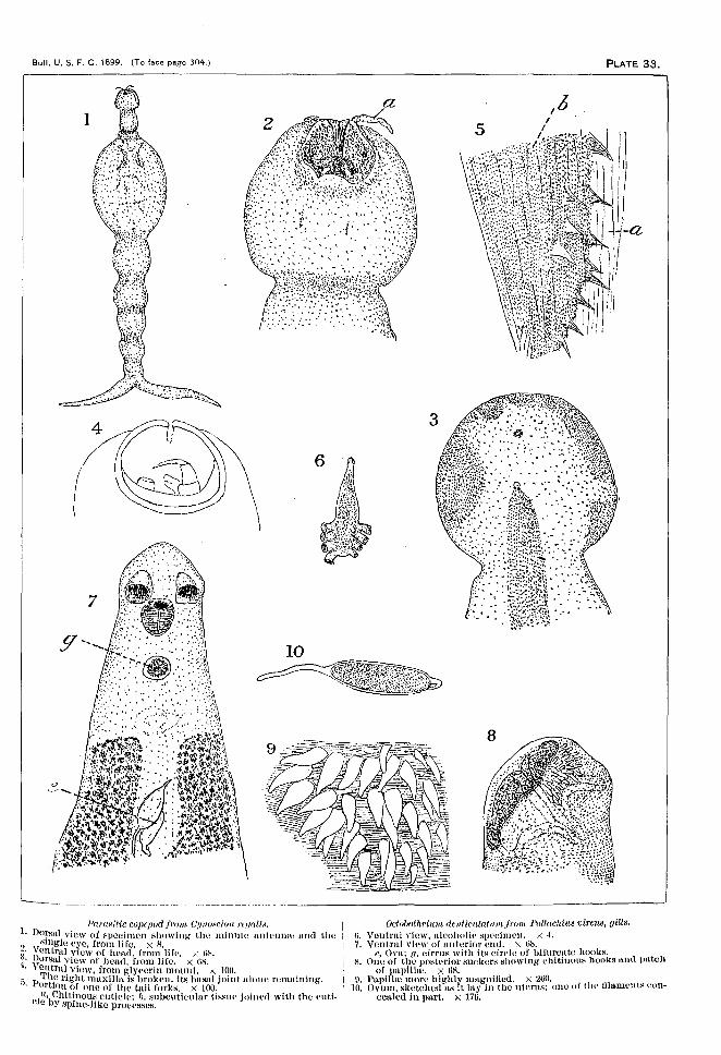
Bull. U. S. F. C. 1899. (To face page 304.) PLATE 33.
10
(
-------l'a'I'llslt-icC()pCp(Jdf/"(}JI/, Cllno~cion 'J'f,f/fllix.
1. Do~sul view of speelmen showing' the nunute nn1mUHU u.nd the2. HlIlgle eye, from life. x 8.3. Ventral view of head, from life. "m'.
I~orStLl view of head, Irom life. X ns.'I. \ ~!ltnll. view, from glyccrin mount. r; 100.~ P Iltl,cfIght mn.xi l ln is broken, its busa) joint u.louu remutntng.'. or IOn of ono of the tnil forks. x 100.
Ia'bChitinonH cuUde; I,. subeutieulur t.issuo jolnerl with the «utie e Y spine-like processes.
Oelo/)othril!1/! denticulanun from. Pollachius vircns. gills.ti. Ventrul view, ulcoholi« specimen. X 4.7. Ventral view of nuturior end. x tl8.
e, Ova: fl, cirrus with its circle of bifurcuto hooks,X. One of the posterior suckers showing ehttinous hooks uud patch
of llllpilhe. X tl8.9. Pllp! he more highly maguitied. x 2t10. .
10. Ovum, sketched us It luy in the uterus; one of t.lronlumcnts concealed in part. X 176.

Bull. U, S, F. C, 1899. (To face page 304,)
12
yr!ldovel
PLATE 34.
13
15
-------------------------------------'
11 Epibdella b/tll/pl/"ii "p. ?/O/'. [ruu: fill' ",,,fer;o/' of Jla"lIaN" ''''''//'III'a,12' Ventral view of spccimeu mounted in bn.lsurn. x 'I.
. Ventrul VIeWof anterior hulf of same showing the Hl'1'HlIgt~mPlltO,f the geuitul orguns, purtlv dillgrnullIllltie.· X 8H.
lei Aperture of cirrus; em, portion of uterus where the eggs nrci~'.' ,<,!ed and the Hhelllllid on; lid, ge"ffi duvt at the point whereu IS J?hH';d by the Hl~or.t Y.o1,k duct (HOC 'lid, 1ig. 14); Uri', germ £1uet
t the point where ]t IS [oinud by the xomlnul duet; XI', seurlnul
lr.ecepttwle ; :tt', opening of I1tC1'11H; '11, opening of vugiun; '1/1', vitel-ino reservoir. .. - .I 1)he line rrom o indi<~llt.CH ll. part or t he ovnrv in whieh the gunu6;(',8 wero free to move nud Were IIIwuys thrown into It Ntlt-te of
:l~lutlOn by the rush of yolk J'I'UlU the reservoir (yl') whtr-hpreeeded the Iormation of eueh egg.
'I'he line 80 touches the somlnnl voseicI« n.t II point where <\regular l?ulsutiou was observed, which still ooutinued iu u spccimen which hud been Iving in seu, water for twelve hours 11lO::;l
of the time nuder n cover glus«. 'l:t A pILiI'of hooks Irom the posterior sucker. x til".14. 'I'runsverse section through the ovurv and Yolk reservoir. X 100.
ed, Gertu duet ut the point where h is j<lined by the short duetIrom the yolk reservoir (lit): lId, germ duet; 0', beginning of the¥ertIl duet in the uvurv in whteh osci llutf ng germs were observedIn the living speelmeri.
1f•. GUrlll cell. x 800.

Bull. U. S. F C. 1899. (To face page 304.) PLATE 35.
19
16
22
})[:'ilf)UlU'Ul ocrcat um [roin /}r;!lw'hilf ... 1'11'('/1,0.:. unt! JJr:rll/('(~i'U,'jlJiUJI('(ll'il'.
In, Anterior end. veutrul view, from life, somewhnt compressed.X GH.
17. PORterior Pll(l of SIl1JW S{H.·('illll'll, veutrul view. from life, some• What COIll}>I'CSSCll. x tiH.
~~: ~ll1lLller spor-tmcn, 1'r010 life, uu teriur view. X 100.Hmull specimeu in glyeeriu, side view.
20. Snlltll specimen. from life. vvntrul view. X lOll.:ll. Portion of excretorv vessel with coneret ionurv masses. from life.
X ·I·J().' ,21. Single concretion. x 7~IO.
2:\. Ova. X 4111,21. Aleoholie ,pI'I'inn'n, ,101',,11 view. x lOO,
I'\O:-4. Ifl to ~3 from Jh'I'Il/('ciu x bilhuaris,No. 24 from Pulhwhiu8 l'jl'l'Ht;.

Bull. U. S. F. C. 1899. (To face page 304.) PLATE 36.
I'
35 2733
c
Distomum appendielllatn", from Parollchtlnt« deniatus.25. Anterior cud, Iateral view. x 300.26. Ovary and vitelline glandsof sumo. X 300.
27 Distomumjeeellndum 8p. ?lm'.jm1/! Loptiouuilu« clumurlconticcp»,28' ~~tcral and ventral views, nlcoholie specimen. x 5.
o ~n.grammatic ventral view, restored from sections. x 2fl.rl)e voluminous uterine foldR huve boon omitted.ex, Left, and ex", right excretory vessel.
29 T a, Aperture of acetabulum.. ran6Verse section through oral Rucker. x 70.
30. Dctnlls of same. X 2nO.31. 'l'runsvcrse section through acet.abulum, about the middle ot lI l <;
latter. The brnttches of the tntcsttne contntn granulur frr ca!mutter.
81. Tmnaverse scctton between ncetnbulum nnrl ovary, x 7b.lm, Lomrltudinul 111118('.]08.
aa. "rulls of excretory vessel near posterior end. x 25u.(Sec fig. :37.)
~-H. 'Ynlls of intestine ut posterior end. x 2:[)O.(Sec Iii:. 37.)
35. Ovu. X 450.

Bull. U.S. F. C. 1899. ITo face page 304.) PLATE 37.
Dbdrnnu1nlmcnnrlwin from. Lnpholnf ilu« clln'1llwfcrmtic(']Js-Continued.31' '1'I. ~t;T1I-l,vc~He section t hrough ()~Tnry and testes. >< 70.:\7 R .:!l.Shell g.1 u n d ; f. right testis: 1'1 heginning' of left tost.i«,
. • L~,tI01!,belrind ovary. X 70.NCP, ]ermiual «x erutorv pore; I', left tc~ti:-:.
WI"ot" t.hn.t the lmdy of th« WOI'IIl WII' f1('x('ll """{I'IlIl\', '"~ :hulb •. it '~crc trHllsvcrHC HcetiollH for the grunter pn.rt. of Oil' length
eClllllC nearly horizontul at the posterior enrl.
Dleunnnn; vlicllonun: NJ), UUll. [rtnn. Mcrlucclu« ldUw:(fl'i.-:.3~. y39. v~~ltrO-]Il~crHl view, from life. X 100.
itrul view of unterlor cud, 11ll1'"11l 1Il01lIlt. X lOU,
Distomum, lHuif'nx X}J. 110/1, ,fI'01n Pm'alit'MlilIS dcutatn«.40. Longf tudinul horizontul section of tL specimen meusurtng t.:!fi
rn rn, in length.41. Longitud innl horizontnl secl lou Ill' H, ~pl'dmm\ monsurhu; 1.1l5
mrn, in length.HI, Anterior sucker rd,l'lldp(l.
.12. Middle purt of lougitud iunl hurizuntul Hl'ptloll of unot hvr ~pt'l·i.mCII. X 70. oe. ()~!'40phHg'11~.
43. Hide view of unturior l'11l1. X 7U.44. Vcntrnl view uuturior suckor retructerl. x 70.45. Ventro-lutcml view of It Hlll'ehncn with eOllit'lll neck. -, 70.4ti. Aeetu.bnlllBl. uterus, and ctrrus, dor~lll.vi4.lW. x 260. .47. Cirrus. elrrus-pouch. prostate nnd HC11111l111 vesetcle, us showu III
It Iougftudinul horizontal Heetioll. X HiO.

Bull. U. S. F. C. 1899, (To face page 304.) PLATE 38.
51
Dlstonuun. 'I'ib(:;~ 8]). nov. fratn. .(,,'plu;roirl,;,'f 'Jlwf'u!afu.'i.48. Ventrul und luternl views or un ulr-oholi« HIll'eillll'll. x-I.49. ~!ediull longitudinul verticul section. x :lot.nO, I runsverse section through ovury uud tl'~h,~:-t. x fl4.51. OVUIll. X 300.
Dlstonucns PlIl'i,[orm,; JoI}J. 'J/fJI'./rrllU l)ali/ll/l'if'!II"!lx }Jf'/'eUol'm;x.
~~. Por~ioll of section of pylork: ('il'('.l, wiC} (l:'t();~l I, >-: 1'0.03, 54. Spccfnreus showing some rl.ifl'l'fellt rorm-. uh-olu rl ic-. su.
f)f). Speeillwn with larg'(' «uudu l ox crctorv \'eHHPI, life. A l711.fiG, Irorsul view. lift.'. x 17li. .
XI', 8emitml rt'('pptllele ('/): ai, !-lplu'r!(,lll hulil'H with ('olll'tmtricstructure.
f)7. Longitud inn l hortzontal -octlou of nutorlor l':~11, showing' invaginuted orul ~1J(·1{(.'1'. '\ :mo.
[lS. Med luu louuitud lun l n'rtit'ltl section. ,,~rlO.flU. Acotubu ltun , cirrus, prostn te, sumlnul vl.....!(·le, uurl ovum. '~4,1O.

Bull. U, S, F. C. 1899. (To face page 304,) PLATE 39.
-t--t
68
62
i~..·.'·'·'.··'
~l u-r~'"
'~h:!"c'-:;-t-'t
67
--------------_.~-_..._..__.- -.-.__.
J);NfmUIII/l. ((/'flulotu 1/1. (t) jl'(JlIt Monnu: WIU'I'icflnll.
ljO. Veil trul view. x 70.til. })OfNHI view of uuteriorpurtiou, lifl'; ~pillt·~ Bot suowu exuept in
front. x lit),I, Opening of I...uun-r's «nun l.
H2. HI,illcH und opening or Luurvr'« cuuu.l, lifo, x: ~·IO.tia. Ventral view, life; spf ne-r not shown. x 100.
IJlfltOJUU,'IU dentut un! .'ifJ. IIIH'./nnn }),u'fll{cltlh!lx drntat u«.64. Vcutrul view, lift': «ketch runde uf'tur the speeitncll hud been
1)'llIg under till' "O\'l'1' glllHH for HOllie time. X 1U0.W'"), ])orloutl view of ornl spiJll':-;. x ,WOo~:7ti .. VClltn,1 vlew of ornl .,-;pilJc,"'i. X ..hJO.u Flirt Hl"lll'-llkl' "piIWHof !Jo,ly. X .f.iO.
lJil4mnfUllfJ'(I!1ilf Nl}, mJi'. [ron: Mo!« uuta.
jiH. Luternl view, al('oho1i(~. x 20.ti~)' Vvnt.m l vtow, 1-IkL'teh of spceuuen in oil of t hyuu-. >.~ 70.70, Ventral view of anterior end. x 100.
}Jhifoilwm oSp.jl'o/n Prlonoiu» curoiinu».
71. Outline...; of two sp ...-enueu-, Hfe. >( ubuut. ;)0.
)Ji.·.:tOIl/.ll1H ~j) . from 8tcnotonw~dll',IIS0j)s.
i'!., ventrul view, life. Npine~ not shown: x tlH.H', Sphurien.l bodies with (~onecntrwsrructurc.

Bull. U. S. F. C. 1899. (To face page 304.)
___- __ 74
I
J
\
75
82 e
DO Ll
IiiH!mnmn "1'. [roin. P«/'([licltt"!1~ <lc1Itatlt~.
73. Vontrnl view. lifo. x 100.i4. Outline of untcrtor end, d()r~'n,l view, life. X 100.if•. Flut H('1l1p-IiI", HpineH Irom b"dy. X 100.
Im:mafu/'(' rfi ..4olllft ('l/('lIloiln{ ill «k!» ttt' n'llfo!lo!aIJI'UN (l(I'~JJ{!I'SUN.
7H. 77. 7H. Outline» of t.hrue ludlvlduu.ls, liI'p. x lOLl. .7g. Anterior end, dorsnl , Jife; sketeh showiug irregularly I'ltp]nO~e
strucurre. x )00.80. C)'stl-! with young distomu., opt.iou l :-:cetion, lifu. X 100.
em, Young distomum: ('11, connecuvc-ussuo cyst; pfj, BlUHH ofpigment accumulated about cysts: l'V.c, pigment cells.
HI. Single cvst, npt.leu l scet ion , life. X 100.
RU('lIs/nl ()I'tI, [run: JJOI'OJl(' inuerican«.X') Ovu nnrl pig-llH.'lIt, from serous..('Ot!!' of li vur. x ann..
('/1, (~lJtltH,(·ti\'t~-tissIW eyHl eOlltnlllillg' 0\,11; r', OVIl \\'11h H('(mmnlution of pig-Jlll'llt.
~3. C\'Ht~ Irmn »un«. ',:~OO.
'('11, ()y~t forlll('d n hout H :·dll~d(' ovum; II', ('y~l, ('olllnillillg- \\'HX~'~l'(·I'('ti(lll.
~H. Hitlg'lL' e\'."t Iroiu :-IlIlH'. '.~ ,HO. ..{'jUVUIU;w, WllXY :-cpretion; I'll, ('ollned1\'l'-tl~SIH\ (.~--"t.

Bull. U. S. F. C. 1899. rTo tace page 304.) PLATE 41.
91.
(im-l!t'l'oxlomnm an'uotltll/. :<)1, HtH'./roU! StU'rla »urd«,Lntr-rnl vtcw of smu ll HJ!(I(·illWH. nhout· 70.I~Hll'rHI "i(n\' of l11r~('f' It4Pl'('!il1PH. 70. '111, Mouth.\ ('utI'HI vip\\" l!ing1'lllllIHH(1.'. '< 'IIi. Moltth.A Ilh1r io r PHd. >< :.WO,II'lHt.. :·wuh'~Hk(· HI.itW)-l from nor-k , >: ·itlttA1"'han !on/iiJtl<linlll "PrJklll ","'lion. ': lMO.
til, MOllth; Il, voutrul ~l11'kl'l' {pharynx;.
flU:-!r't'HRlmtJlflll "'Ii, from 'l'Y{OI{f/i'1{1': muriuus.L!lll!l'nl view. >-: iO. m, Mouth.
('alj!ll!rot,ofhrllf1i1 (}('{'idf'utul,' "}). Hfil'. from Trtranurr« (j(,tidf'utalt':<.H:.1. 1I(IHfl Hutllwnk of l'mnJl sm-r-lmcn, lift', l~"".H:t ll'rollt viow oi' 1'411IH'. nlt'ohotic', :-~ :H.~H. IIPH41 of lul'g't' Sli{'(·iIlWIl. nIt'flliolit,. '-::lLur•. Hing'h' hothriulH (~'l· fig. ~l'.!). lifE.', <: tIS.Htl. Po}o\tt'l'iw' H'~UH'Ht1': Ill' :-:mal1 Spt«·hllt'll. lil\'. ,,6.'-\.~17. 'I't'u H~\'('l'~t' l"t'l'l fon or otlt' 01' t ln- postt~l'iol' I"t.'guwllfs of In rgt' sJll'('i~
nu-u. :-; ,!-;U. •Ill. l~oligitlldinHI v{'~xr'L

Bu ll. U. S. F . C. 1899 . (To face page 304 .) PLATE 42.
1-
Iv if,
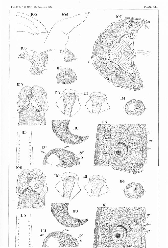
F C 1899 .Bul l. U. S... 304. )(To face page P L AT E 4 3.
115
105
o 17&o
oo
o \oo
115
eooo
oo