first year rotation projects
TRANSCRIPT

Wellcome Trust 4 year Interdisciplinary PhD Programme in Structural, Computational and Chemical Biology
at Birkbeck / UCL / Crick-NIMR
First Year Rotation Projects 2015-2016

1

2
Table of contents
Introductory notes ........................................................................................ 3 Chemical Biology rotation projects .................................................................. 4 Computational Biology rotation projects ........................................................ 15 Structural Biology rotation projects ............................................................... 32

3
Introductory notes In Year 1 of the programme students will choose and undertake one lab rotation project in each of the three core areas of the programme: - Chemical biology - Computational biology - Structural biology During each lab rotation, which will last approximately 12 weeks, you will acquire the research skills and knowledge in which that lab is expert, contribute to journal clubs, decide whether you like the social environment of the lab, and assess whether you would like to study a PhD in that lab during years 2-4. There are timetabled meetings with the Programme Coordinator during which you will discuss the choice of your rotation projects. Rotation start and end dates as well as deadlines for submitting choices are gathered in the table below:
Rotation Start and end dates Deadline for submitting rotation choices
Rotation 1
Monday 12 October 2015 – Friday 8 January 2016
5pm on Thursday 8 October 2015 Your top three preferences for the first rotation should be submitted to Prof Alethea Tabor
Rotation 2
Monday 18 January – Friday 15 April 2016
5pm on Wednesday 6 January 2016 – via Moodle and to Prof Alethea Tabor
Rotation 3
Monday 9 May – Friday 29 July 2016
5pm on Thursday 10 March 2016 – via Moodle and to Prof Alethea Tabor
The projects presented in this booklet are listed by subject area. Please note the following points:
Some projects are linked – which means that they are connected with each other. However ALL these projects may also be taken individually.
Example: Chemical Biology project 15-CH008, linked with Structural Biology project 15-ST029. Students may choose to take these two linked projects (for 2 of the 3 rotations), or just one of them.
Most projects mentioned in the booklet could potentially be extended to PhD
projects.
This booklet is available on the Moodle pages for the WT Joint PhD Programme.
4
Chemical Biology rotation projects Homogeneous antibody-drug conjugates (ADCs) as next generation biologics Dr Vijay Chudasama, Department of Chemistry, UCL
Ref: 15-CH001
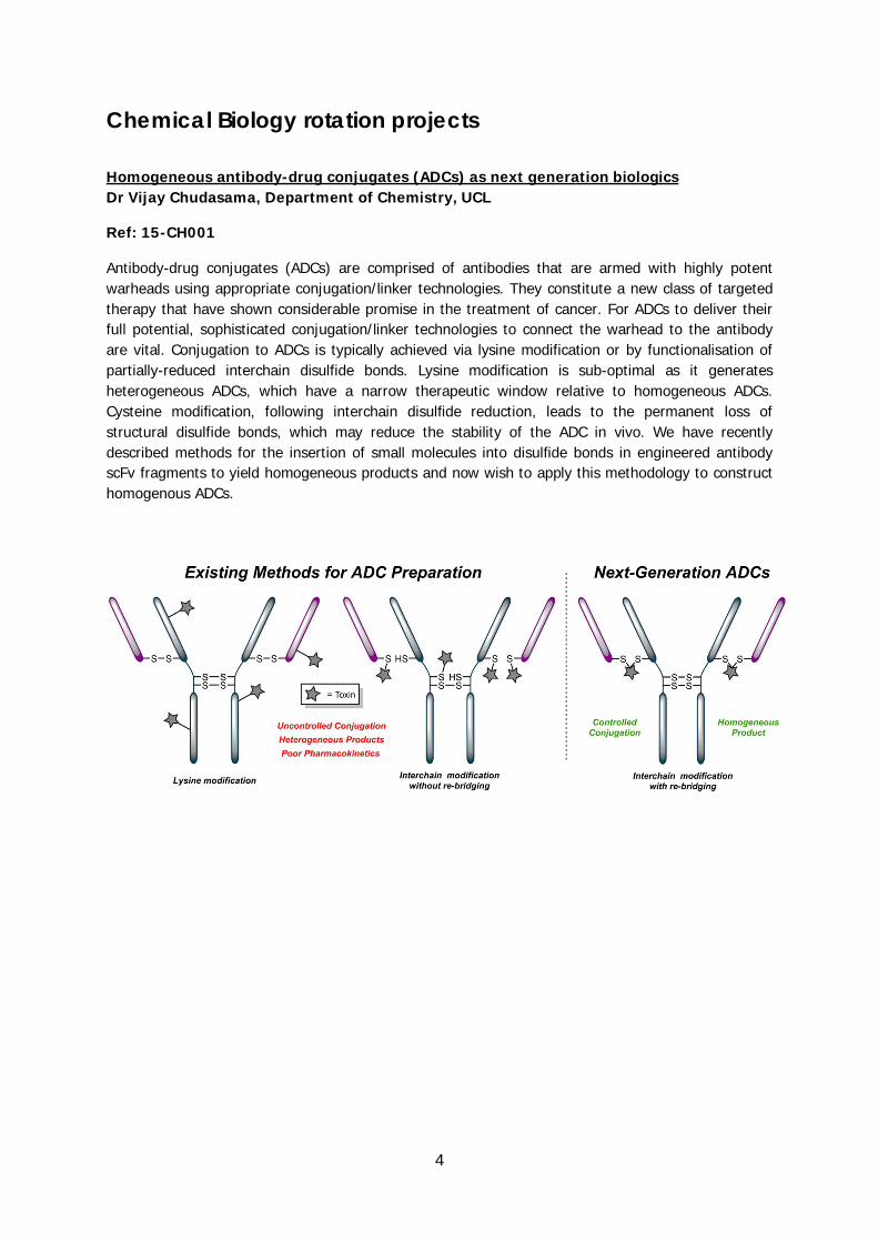
Antibody-drug conjugates (ADCs) are comprised of antibodies that are armed with highly potent warheads using appropriate conjugation/linker technologies. They constitute a new class of targeted therapy that have shown considerable promise in the treatment of cancer. For ADCs to deliver their full potential, sophisticated conjugation/linker technologies to connect the warhead to the antibody are vital. Conjugation to ADCs is typically achieved via lysine modification or by functionalisation of partially-reduced interchain disulfide bonds. Lysine modification is sub-optimal as it generates heterogeneous ADCs, which have a narrow therapeutic window relative to homogeneous ADCs. Cysteine modification, following interchain disulfide reduction, leads to the permanent loss of structural disulfide bonds, which may reduce the stability of the ADC in vivo. We have recently described methods for the insertion of small molecules into disulfide bonds in engineered antibody scFv fragments to yield homogeneous products and now wish to apply this methodology to construct homogenous ADCs.

5
Appraisal of LRG1 antibody internalisation and as an antibody-drug conjugate Dr Vijay Chudasama, Department of Chemistry, UCL Ref: 15-CH002
Aberrant neovascularization contributes to diseases such as cancer, and is the consequence of inappropriate angiogenic signalling. In recent years, it has been shown that in the presence of transforming growth factor-β1 (TGF-β1), leucine-rich alpha-2-glycoprotein 1 (LRG1) is mitogenic to endothelial cells and promotes angiogenesis. LRG1 binds directly to the TGF-β accessory receptor endoglin, which, in the presence of TGF-β1, results in promotion of pro-angiogenic signalling. LRG1 antibody blockade inhibits this switch and attenuates angiogenesis, and thus, could be exploited to inhibit cancer growth. Whilst this is significant, this project, with Professors S. Caddick and S. Moss, is aimed at combining this function with affecting cancer cell deterioration. To do this, the LRG1 antibody will be modified, using state-of-the-art biotechnology, with a suitable fluorophore to appraise internalisation (and thus it’s ability to deliver cargo) into a cancer cell, followed by decoration with a suitable toxic drug to evaluate efficacy in vitro.
6
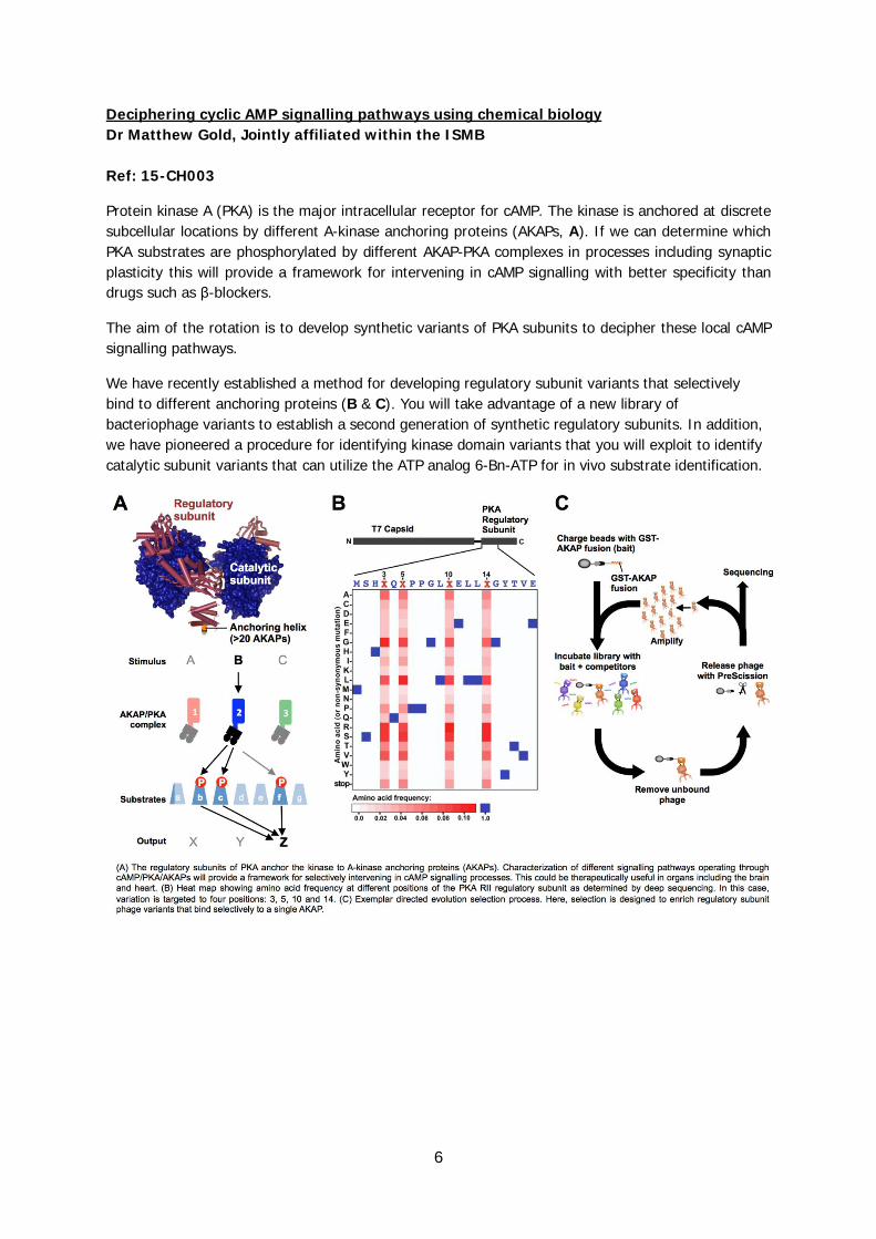
Deciphering cyclic AMP signalling pathways using chemical biology Dr Matthew Gold, Jointly affiliated within the ISMB Ref: 15-CH003
Protein kinase A (PKA) is the major intracellular receptor for cAMP. The kinase is anchored at discrete subcellular locations by different A-kinase anchoring proteins (AKAPs, A). If we can determine which PKA substrates are phosphorylated by different AKAP-PKA complexes in processes including synaptic plasticity this will provide a framework for intervening in cAMP signalling with better specificity than drugs such as β-blockers.
The aim of the rotation is to develop synthetic variants of PKA subunits to decipher these local cAMP signalling pathways.
We have recently established a method for developing regulatory subunit variants that selectively bind to different anchoring proteins (B & C). You will take advantage of a new library of bacteriophage variants to establish a second generation of synthetic regulatory subunits. In addition, we have pioneered a procedure for identifying kinase domain variants that you will exploit to identify catalytic subunit variants that can utilize the ATP analog 6-Bn-ATP for in vivo substrate identification.

7
Synthesis of Novel Isoquinolines using Norcoclaurine Synthase Prof Helen Hailes, Department of Chemistry, UCL Ref: 15-CH004, linked with 15-ST031
Alkaloids are a major class of structurally diverse natural products with a wide range of medical applications. The benzylisoquinoline (S)-norcoclaurine is the first committed intermediate in the biosynthetic pathway to alkaloids including morphine and berberine, and is catalysed by the enzyme norcoclaurine synthase (NCS). The NCS mechanism involves a Pictet-Spengler reaction between the arylethylamine dopamine and aldehyde 4-hydroxyphenylacetaldehyde (HPAA). We have recently over-expressed several NCS enzymes for use in biocatalytic applications and using Coptis japonica NCS2 (CjNCS2) and Thalictrum flavum NCS (TfNCS) established that it exerts excellent tolerance towards variously decorated aryl- and heteroaromatic acetaldehydes and can also accommodate aliphatic aldehydes to give products in high stereoselectivities. Our aim in this rotation project is to use the NCSs with several functionalized aldehydes in cascade reactions, where after the NCS coupling a second (chemical) cyclisation can occur.

8
Enzymatic Synthesis of Cyclic Single-Isomer Chiral Amines Prof Helen Hailes, Department of Chemistry, UCL Prof John Ward, Department of Biochemical Engineering, UCL
Ref: 15-CH005
Single isomer chiral amines are present in many biologically active molecules and routes to these compounds are frequently step-intensive and have poor atom efficiencies. However, transaminases (TAms) are currently attracting significant interest in academia and industry due to their ability to generate chiral amines using sustainable chemistries in a highly stereoselective fashion. Enoyl reductases (EREDs) are interesting enzymes that reduce electron deficient alkenes. Our aim in this rotation project is to use EREDs followed by TAms in sequential, or one-pot reactions for the preparation of cyclic amines with a different groups at the 3-position. We have a number of TAms and EREDs at UCL and these will be used as crude cell lysates with a range of substrates to establish substrate selectivities, and product stereochemistries. Enzyme specific activities will also be determined and a reaction cascade developed using a co-factor recycling system for the ERED step.

9
Genome Mining for Novel Natural Products Dr Philip Lowden, Department of Biological Sciences, Birkbeck Ref: 15-CH006
We have been studying the biosynthesis of the DNA binding anticancer azinomycin natural products, using a combination of organic chemistry and molecular biology. We have sequenced the biosynthetic genes, and identified homologues for many of these in other bacterial genomes. The related pathways identified by bioinformatics share a novel transketolase enzyme predicted to convert glutamic acid semi-aldehyde into novel 7-carbon amino acids.
In this project you will grow these bacteria under a range of different conditions and analyse the cultures using HPLC-MS for production of secondary metabolites predicted from genome sequences. Isotope labelling and selective reactions of the predicted metabolites could be used to try to identify azinomycin-like metabolites. If interesting metabolites are observed, then they will be purified and structurally characterised. RT-PCR will be used to monitor expression of the relevant gene clusters and degenerate PCR may be used to identify similar transketolase genes in other bacterial strains.
Precursor-Directed Biosynthesis of Unnatural Natural Products Dr Philip Lowden, Department of Biological Sciences, Birkbeck Ref: 15-CH007
My group is studying the biosynthetic pathway towards the DNA binding anticancer natural products azinomycins A and B, using organic chemistry and molecular biology techniques. We have recently discovered that the biosynthetic enzymes can accept unnatural substrates, leading to structurally modified metabolites. So far we have only studied commercially available substrates, now we need to synthesise additional substrate analogues, to systematically study the extent of flexibility of the biosynthetic enzymes.
In this project you will synthesise further analogues of the natural naphthoic acid substrate, feed them to the azinomycin producer, and analyse the cultures by HPLC-MS. If novel metabolites are observed, the cultures will be scaled up, new metabolites purified, and characterised by NMR. Analternative goal would be synthesis of isotope-labelled biosynthetic intermediates. The detailed biosynthetic pathway to the azinomycins is still incompletely understood, and these labelled hypothetical intermediates are required to test hypotheses about the pathway.
10
Magnetic and Optical Tweezers Investigation of a Relaxase Dr Justin Molloy, Physical Biochemistry Division, Francis Crick Institute Prof Gabriel Waksman, Research Department of Structural and Molecular Biology, UCL Ref: 15-CH008, linked with 15-ST029
Type IV Secretion Systems (T4SS) are ubiquitous export machineries operating in most bacteria. They are used to transport both proteins and DNA. They play crucial roles in bacterial pathogenesis and also in the spread of antibiotics resistance genes. To transport DNA, these systems must first process the DNA to be transported. They use a protein termed "relaxase" that cleaves the DNA at a particular sequence called "Origin of Transfer or OriT", and reacts covalently with the 5'-end of the cleaved DNA. For this linked rotation, we propose to study a relaxase using single molecule techniques, namely magnetic and optical tweezers. The goal of this rotation is to study the role of DNA supercoiling in relaxase activity. Such a research will provide the first insight as to what are the DNA topological requirements for this important protein.
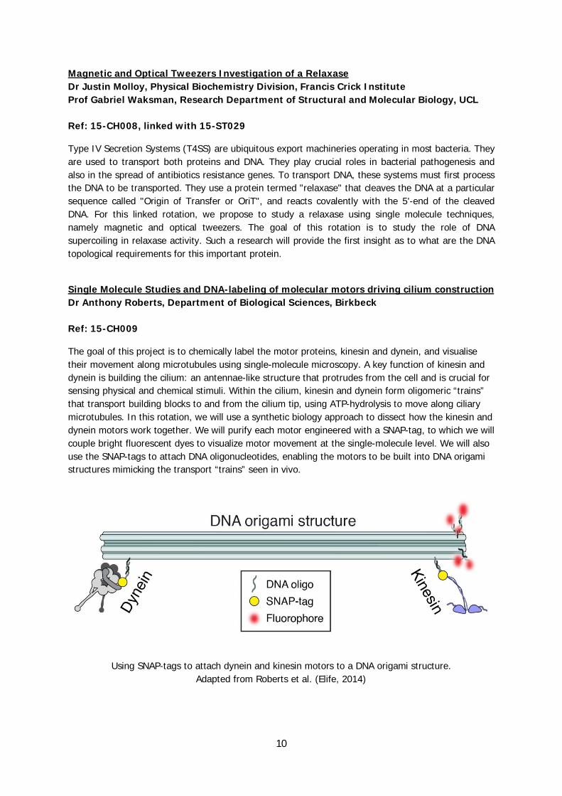
Single Molecule Studies and DNA-labeling of molecular motors driving cilium construction Dr Anthony Roberts, Department of Biological Sciences, Birkbeck Ref: 15-CH009
The goal of this project is to chemically label the motor proteins, kinesin and dynein, and visualise their movement along microtubules using single-molecule microscopy. A key function of kinesin and dynein is building the cilium: an antennae-like structure that protrudes from the cell and is crucial for sensing physical and chemical stimuli. Within the cilium, kinesin and dynein form oligomeric “trains” that transport building blocks to and from the cilium tip, using ATP-hydrolysis to move along ciliary microtubules. In this rotation, we will use a synthetic biology approach to dissect how the kinesin and dynein motors work together. We will purify each motor engineered with a SNAP-tag, to which we will couple bright fluorescent dyes to visualize motor movement at the single-molecule level. We will also use the SNAP-tags to attach DNA oligonucleotides, enabling the motors to be built into DNA origami structures mimicking the transport “trains” seen in vivo.
Using SNAP-tags to attach dynein and kinesin motors to a DNA origami structure.
Adapted from Roberts et al. (Elife, 2014)
11
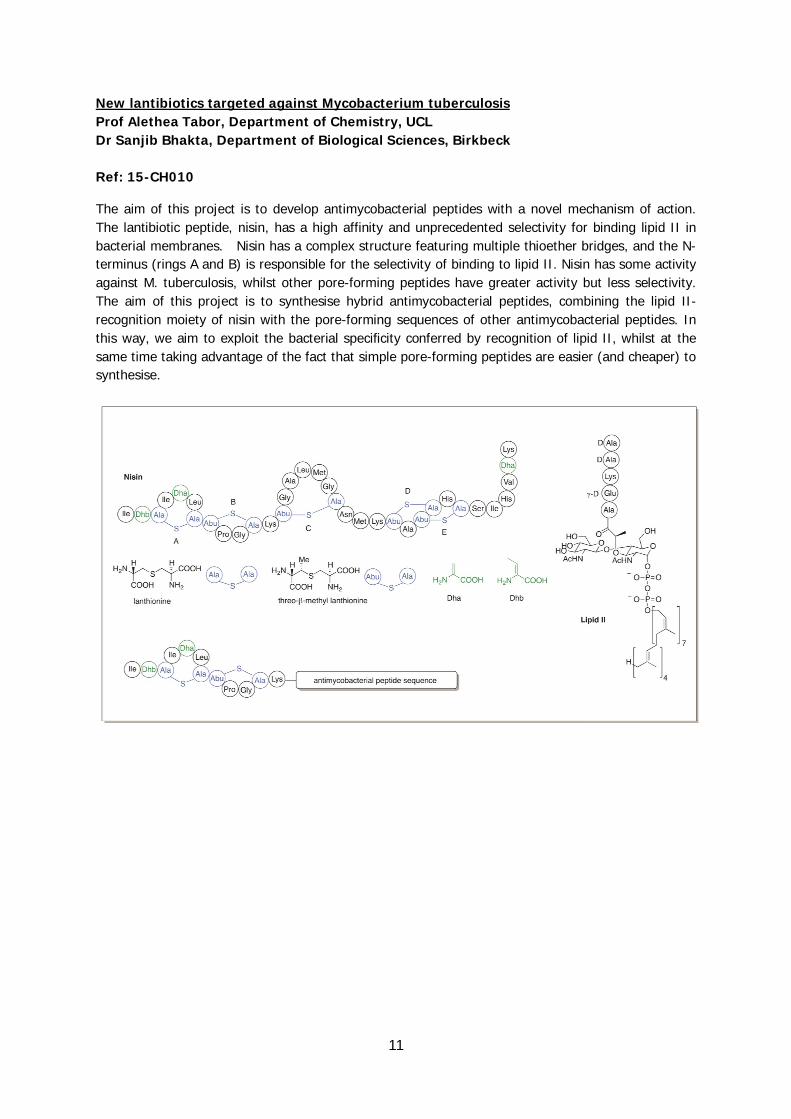
New lantibiotics targeted against Mycobacterium tuberculosis Prof Alethea Tabor, Department of Chemistry, UCL Dr Sanjib Bhakta, Department of Biological Sciences, Birkbeck Ref: 15-CH010
The aim of this project is to develop antimycobacterial peptides with a novel mechanism of action. The lantibiotic peptide, nisin, has a high affinity and unprecedented selectivity for binding lipid II in bacterial membranes. Nisin has a complex structure featuring multiple thioether bridges, and the N-terminus (rings A and B) is responsible for the selectivity of binding to lipid II. Nisin has some activity against M. tuberculosis, whilst other pore-forming peptides have greater activity but less selectivity. The aim of this project is to synthesise hybrid antimycobacterial peptides, combining the lipid II-recognition moiety of nisin with the pore-forming sequences of other antimycobacterial peptides. In this way, we aim to exploit the bacterial specificity conferred by recognition of lipid II, whilst at the same time taking advantage of the fact that simple pore-forming peptides are easier (and cheaper) to synthesise.

12
Cell-permeable analogues of CoA Prof Alethea Tabor, Department of Chemistry, UCL Prof Ivan Gout, Research Department of Structural and Molecular Biology, UCL Ref: 15-CH011
Coenzyme A (CoA) and its derivatives play a critical role in cellular metabolism and the regulation of gene expression. We were the first to report molecular cloning and characterization of mammalian CoA synthase, which mediates the last two stages of CoA biosynthesis. Genetic studies in animal models provided strong evidence that deregulation of CoA biosynthesis/metabolism is implicated with various human pathologies, including diabetes, cancer and neurodegeneration. Since CoA is a negatively charged molecule and does not penetrate cellular membranes, we propose to develop cell permeable analogues of CoA by replacing the diphosphate and phosphate groups by polar, non-charged isosteric replacement groups. We will measure the permeabilisation efficiency of these analogues and their effect on cellular metabolic and signalling processes. A diverse range of techniques will be used on this project, including in silico design and HPLC analysis, mammalian cell culturing and a range of in vitro and cell based assays.

13
Targeting cancer cells with small molecules Dr Salvador Tomas, Department of Biological Sciences, Birkbeck Dr Emmanuel Boucrot, Research Department of Structural and Molecular Biology, UCL Ref: 15-CH012
For small-molecule based drugs, only a fraction of the administered dose will reach the target, leading to lose in efficiency and undesired side effects. Drug carriers that bind specifically to target cells and that specifically deliver the drug to them would eliminate these shortcomings. From our work studying molecular recognition in lipid membranes we believe it is possible to create a drug carrier, in the form of a liposome, that is very selective for a single type of cell, and that this result can be achieved using small, inexpensive molecules, embedded in the liposomal membrane. The aim of this rotation project is synthesize and purify one of these molecules, and to test how efficiently liposomes containing this molecule target cancer cells. The project will involve chemical synthesis methods, biophysical characterization of the liposomes (UV and fluorescence spectroscopy) and fluorescence microscopy.
Investigating the spontaneous formation of biopolymers in the cavity of liposomes. Dr Salvador Tomas, Department of Biological Sciences, Birkbeck Ref: 15-CH013
In living cells reactions take place in membrane bound compartments, often in response to changes in the environment. Learning how the reactions are influenced by this compartmentalization will help us gain an optimal understanding of living organisms at the molecular level and, at the same time, will offer vital clues on the behavior of simple compartmentalized systems, such as prebiotic precursors of cells and cell-inspired artificial systems. The aim of this rotation project is to analyze the ability of liposomal lipid membranes of catalyzing the formation of oligopeptides. Specifically, we will look at the influence of the location of the amino acids (i.e., trapped in the cavity of the liposome or free in solution) in the outcome of the reaction. The project will involve the use of techniques of chemical synthesis and purification, analysis by HPLC and the use of biophysical techniques (Fluorescence and UV spectroscopy).
14
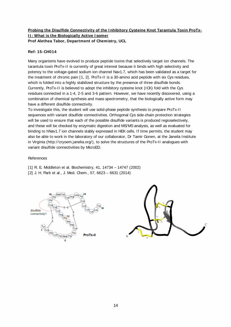
Probing the Disulfide Connectivity of the Inhibitory Cysteine Knot Tarantula Toxin ProTx-II: What is the Biologically Active Isomer Prof Alethea Tabor, Department of Chemistry, UCL Ref: 15-CH014
Many organisms have evolved to produce peptide toxins that selectively target ion channels. The tarantula toxin ProTx-II is currently of great interest because it binds with high selectivity and potency to the voltage-gated sodium ion channel Nav1.7, which has been validated as a target for the treatment of chronic pain [1, 2]. ProTx-II is a 30-amino acid peptide with six Cys residues, which is folded into a highly stabilized structure by the presence of three disulfide bonds. Currently, ProTx-II is believed to adopt the inhibitory cysteine knot (ICK) fold with the Cys residues connected in a 1-4, 2-5 and 3-6 pattern. However, we have recently discovered, using a combination of chemical synthesis and mass spectrometry, that the biologically active form may have a different disulfide connectivity. To investigate this, the student will use solid-phase peptide synthesis to prepare ProTx-II sequences with variant disulfide connectivities. Orthogonal Cys side-chain protection strategies will be used to ensure that each of the possible disulfide variants is produced regioselectively, and these will be checked by enzymatic digestion and MS/MS analysis, as well as evaluated for binding to hNav1.7 ion channels stably expressed in HEK cells. If time permits, the student may also be able to work in the laboratory of our collaborator, Dr Tamir Gonen, at the Janeila Institute in Virginia (http://cryoem.janelia.org/), to solve the structures of the ProTx-II analogues with variant disulfide connectivities by MicroED. References
[1] R. E. Middleton et al. Biochemistry, 41, 14734 – 14747 (2002) [2] J. H. Park et al., J. Med. Chem., 57, 6623 – 6631 (2014)
15
Computational Biology rotation projects Genome-scale detection of functional elements in the fission yeast genome Prof Jürg Bähler, Research Department of Genetics, Evolution and Environment, UCL
Ref: 15-CP001
Our understanding of how genomes regulate cellular function is limited. In particular, thousands of expressed non-coding RNAs are of unknown functions. Fission yeast is a potent model system to investigate this question, because 44% of its genome is non-coding, yet 94% is transcribed. We have mutated large populations of fission yeast cells using transposons. These mutant populations are then grown, so that cells with deleterious mutations decrease as they are out-competed by neutral mutations. Mutations that remain in the population are then systematically sequenced. The student will exploit our transposon mutagenesis and extensive genome/transcriptome data to identify functional regions under different environmental conditions. The transposon-insertion frequencies will be analyzed to locate coding and non-coding genomic regions that lack mutations and are therefore likely functional. This project provides the opportunity to become familiar with cutting-edge approaches for the mining of large-scale sequencing data and the analysis of genome evolution and function.

16
Exploring the (dys)regulation of RAF kinases by allosteric effects. Prof Francesco Luigi Gervasio, Department of Chemistry, UCL
Ref: 15-CP002
The RAF kinases play a fundamental role in the regulation of cellular growth, differentiation and proliferation. The B-RAF isoform is a driver oncogene that is mutated in ~50% of malignant melanomas.1 Thus, there have been significant efforts focussed on developing drugs targeting BRAF. However, most patients on BRAF-specific inhibitors relapse after a short period2. This has led to the realisation that RAF regulation is more complex than we previously thought. In B-Raf dimers, there is a significant allosteric communication, and this has emerged through the discovery of a "paradoxical activation" effect, whereby a drug binding to one monomer trans-activates a partner drug-free monomer.2
In this project, by using enhanced-sampling MD simulations3, we will explore how these interactions dysregulate RAF activity to help the design of second generation inhibitors that overcome resistance.
1. Davies et al. Nature 417, 949 (2002) 2. Poulikakos et al. Nature 464, 427 (2010) 3. Cavalli et al. Accounts Chem Res 48, 277 (2015)
17
In-silico Optimization of Therapeutic Antibodies Prof Francesco Luigi Gervasio, Department of Chemistry, UCL Ref: 15-CP003
The development of monoclonal antibodies (mAbs), identical antibodies harvested from clones of a single parent cell in a reproducible way, has paved the way for several new applications in diagnosis and therapy 1. However, the development of new therapeutic mAbs still presents significant challenges. The standard routes to obtain mAbs are complex and time consuming. A rational model-based approach to their design might greatly speed up the time to clinic and cut the costs.
However, there are still significant hurdles that need to be cleared to fully realize the potential of computer models. The most significant is the prediction of the effect of engineered mutations on mAb structure and dynamics. The aim of this project is to use state-of-the-art enhanced sampling molecular dynamics simulations2 to predict the effects of mutations on both the structure and dynamics of mAb-antigen complexes.
1. Nuñez-Prado et al. Drug Discov Today 1–7 (2015). 2. Sutto & Gervasio PNAS 110, 10616–10621 (2013).
Evolution of cloaking: Phylodynamics of HIV-1 capsid Prof Richard Goldstein, Department of Infection & Immunity, UCL Prof Greg Towers, Department of Infection & Immunity, UCL
Ref: 15-CP004
Why has one zoonotic transmission of HIV-1 infected 60 million people, while the others have infected fewer than 100,000? There is growing support that HIV-1(M) is able to hide from intracellular defensive systems through cloaking and by disabling cellular sensing mechanisms. This raises the possibility that by interfering with the virus’ cloaking mechanism, infected cells can sense, and thus defeat, the virus. This antagonistic relationship between virus and host is a dynamic one, as the rapidly evolving virus competes with the complex host defensive mechanisms that are the results of millions of years of host evolution. This competition results in patterns of sequence change that provide important clues to the way pathogen and host interact at a molecular level. This fusion of molecular evolution and molecular biology provides the opportunity to transform hypotheses generated by evolutionary analyses into experimental tests, and new understanding of how pathogens and hosts interact.
18
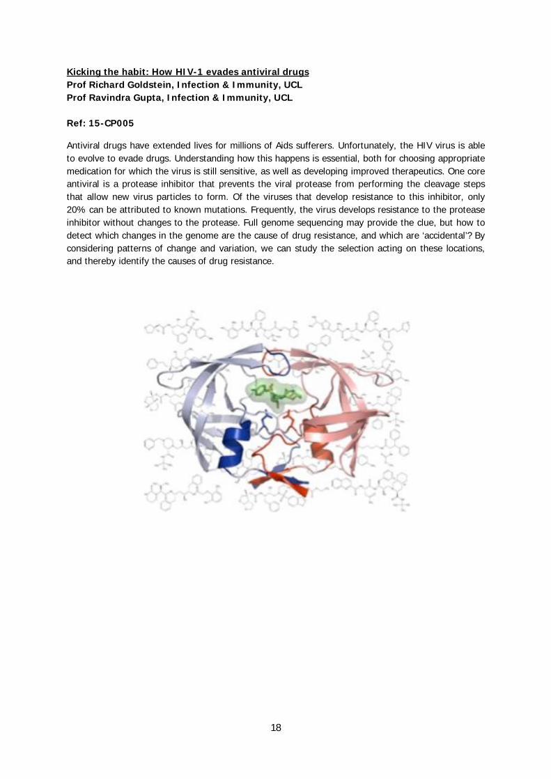
Kicking the habit: How HIV-1 evades antiviral drugs Prof Richard Goldstein, Infection & Immunity, UCL Prof Ravindra Gupta, Infection & Immunity, UCL Ref: 15-CP005
Antiviral drugs have extended lives for millions of Aids sufferers. Unfortunately, the HIV virus is able to evolve to evade drugs. Understanding how this happens is essential, both for choosing appropriate medication for which the virus is still sensitive, as well as developing improved therapeutics. One core antiviral is a protease inhibitor that prevents the viral protease from performing the cleavage steps that allow new virus particles to form. Of the viruses that develop resistance to this inhibitor, only 20% can be attributed to known mutations. Frequently, the virus develops resistance to the protease inhibitor without changes to the protease. Full genome sequencing may provide the clue, but how to detect which changes in the genome are the cause of drug resistance, and which are ‘accidental’? By considering patterns of change and variation, we can study the selection acting on these locations, and thereby identify the causes of drug resistance.
19
VH/VL Packing in antibody modelling Dr Andrew Martin, Research Department of Structural and Molecular Biology, UCL
Ref: 15-CP006
Antibodies offer the ability to bind to an almost infinite range of molecules with high affinity and specificity. Consequently they are now extremely important as drugs with 5 of the top-10 selling drugs in 2014 and approximately 1/3rd of drugs in development being antibodies.
From a structural perspective, antibodies have a flexible, but highly conserved, structure with the variability concentrated in six complementarity determining loops (CDRs) three each from two domains: VH and VL. We have been interested in the structure of antibodies for many years and have analyzed the packing between VH and VL domains as well as creating a predictor of the packing angle. We have also developed methods for modelling antibodies and this project will explore the application of the VH/VL domain packing predictor to improving modelling. The predictor is currently being integrated into our modelling software.
In this project, you will learn best practice in scripting and will develop code to automate modelling of several hundred antibodies of known structure and evaluate the performance.
Moonlighting proteins - what can we learn from antibodies? Dr Andrew Martin, Research Department of Structural and Molecular Biology, UCL Ref: 15-CP007
Recently, the simple view that one gene encodes one protein with one function has been shown to be wrong. As well as alternative splicing, individual proteins may have multiple unrelated functions. Often a protein with a well-established core function, is found to have a secondary function involved in bacterial virulance or in regulation. For example, aconitase (an enzyme of the TCA) loses its iron-sulphur cluster when iron concentrations are low and binds to iron-responsive elements in mRNA that encodes proteins involved in iron uptake. While antibodies are not moonlighting proteins, the ability of antibodies to bind a virtually infinite range of antigens with high affinity and specificity tells us how much change is needed in a protein sequence radically to change binding and thus gives us insight into the evolution of moonlighting functions. This project will compare antibody sequences and the nature of the antigens to which they bind to gain a picture of the degree of change needed.

20
Provar-nD: Towards the development of software for identification of cryptic pockets from protein structural ensembles Dr Irilenia Nobeli, Department of Biological Sciences, Birkbeck Prof Christine Orengo, Research Department of Structural and Molecular Biology, UCL Ref: 15-CP009
Traditional drug design often aims at blocking the primary functional site of a protein, e.g. by competing with the substrate for the active site of an enzyme. However, many drugs are known to act allosterically, and many more probably do so unintentionally, leading to undesired side effects. Finding potential allosteric sites is not trivial, as pockets on the protein surface may only appear transiently, and may not always be visible in X-ray crystallography snapshots. In order to help discover such “cryptic” pockets, we have developed the software Provar, which provides a convenient way to visualize transient pockets using pocket prediction algorithms applied to individual structure snapshots. The focus of this project will be on extending the original Provar code to use homologous proteins from CATH FunFam families, as a way of exploring the potential for conformational flexibility and opening of transient pockets on the surface of a given protein structure.
Predicting the opening of a ligand binding site with Provar. (A) Pocket-lining atoms (red) identified from a PASS analysis of the apo crystal structure of Bcl-2 [PDB:1GJH] capture only part of the binding
site of an acyl-sulfonamide-based ligand, here illustrated by superimposing the ligand from [PDB:2O1Y]. (B) Provar analysis of a tCONCOORD-generated ensemble of apo structures indicates
that pocket-lining residues (red) are found along the full-length of the binding groove in most structures.
Original figure from: Ashford et al. (2012), BMC Bioinformatics, 13:39.
21
The development of a method for studying alternative poly-adenylation using RNA-seq and paired experimental designs Dr Irilenia Nobeli, Department of Biological Sciences, Birkbeck
Ref: 15-CP010
Alternative poly-adenylation, i.e. the selection of different poly-adenylation sites that leads to transcripts with longer or shorter 3’ untranslated regions is a widespread method of gene regulation in eukaryotes. In humans more than 50% of all genes are thought to have alternatively poly-adenylated transcripts. Recently developed methods (DaPars, InPAS) allow the study of alternative poly-adenylation using standard RNA-seq data. However, their models are not suitable for the study of RNA-seq experiments with a paired design (i.e. where each sample from one group or condition is related directly to a sample in another group). This project will attempt to fill this gap by developing code that allows the paired nature of an experiment to be taken into account using packages in the R statistical suite. The code will be applied to existing RNA-seq datasets in public gene expression databases.
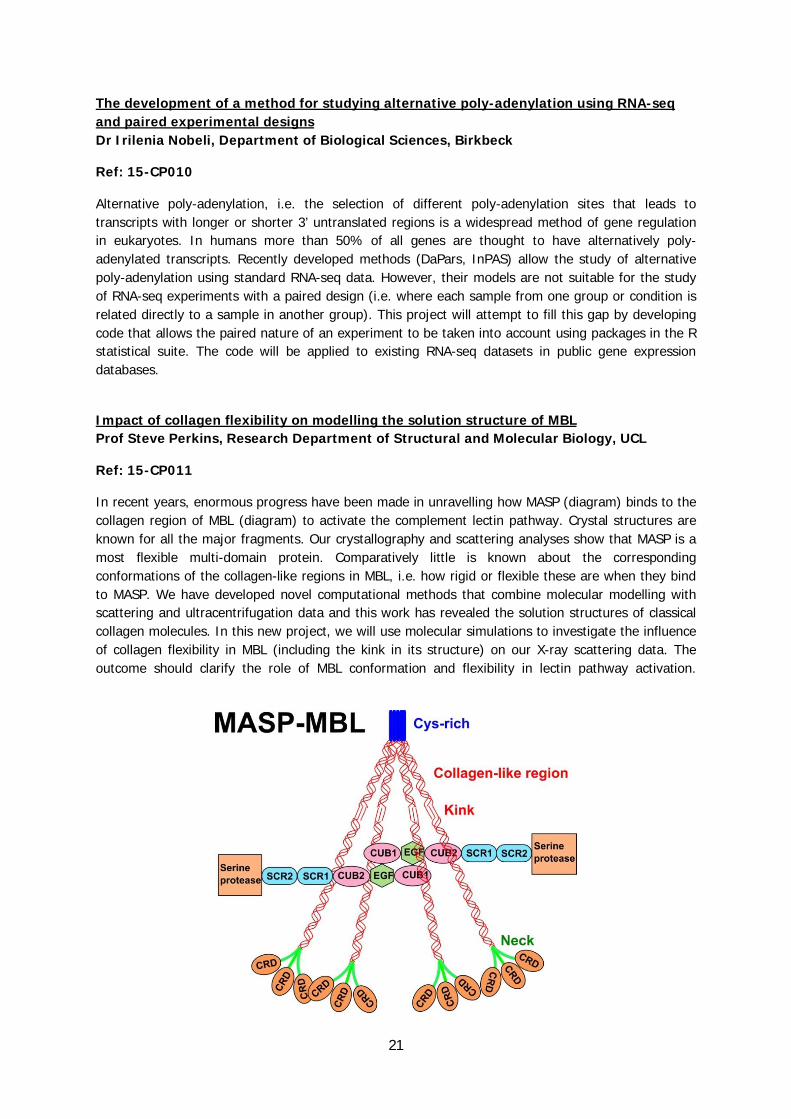
Impact of collagen flexibility on modelling the solution structure of MBL Prof Steve Perkins, Research Department of Structural and Molecular Biology, UCL
Ref: 15-CP011
In recent years, enormous progress have been made in unravelling how MASP (diagram) binds to the collagen region of MBL (diagram) to activate the complement lectin pathway. Crystal structures are known for all the major fragments. Our crystallography and scattering analyses show that MASP is a most flexible multi-domain protein. Comparatively little is known about the corresponding conformations of the collagen-like regions in MBL, i.e. how rigid or flexible these are when they bind to MASP. We have developed novel computational methods that combine molecular modelling with scattering and ultracentrifugation data and this work has revealed the solution structures of classical collagen molecules. In this new project, we will use molecular simulations to investigate the influence of collagen flexibility in MBL (including the kink in its structure) on our X-ray scattering data. The outcome should clarify the role of MBL conformation and flexibility in lectin pathway activation.

22
Electron microscopy and 3D reconstruction of malaria parasite egress from erythrocytes Prof Helen Saibil, Department of Biological Sciences, Birkbeck Dr Michael Blackman, Francis Crick Institute Ref: 15-CP012, linked with 15-ST006
In this rotation, the student will analyse malaria-infected human erythrocytes showing phenotypes deficient in egress, the steps in which mature parasites break through their enclosing vacuole membrane and then the red cell membrane in order to escape and infect fresh erythrocytes. These preparations will have been generated from parasite mutants produced in the Blackman lab (in the linked rotation). At Birkbeck, the cultures will be analysed by electron tomography. This will involve high-pressure freezing, freeze substitution and sectioning. The student will be introduced to the principles and practice of three-dimensional electron microscopy and image reconstruction, using the IMOD software system. Tilt series images will be collected by electron tomography and the resulting reconstructions will be used to examine details of cell and membrane structures in the engineered forms of the parasites at late schizont stages, blocked before or partway through egress.
Sections from tomograms showing malaria parasites (outlined in cyan) inside their vacuole (yellow) inside red blood cells (red). Left, parasites not fully segmented; right, after segmentation, the final
stage before parasite egress. Scale bar, 500 nm.
Identification of regulatory elements involved in arsenite oxidation Dr Joanne Santini, Research Department of Structural and Molecular Biology, UCL
Ref: 15-CP013, linked with 15-ST026
Regulation of arsenite oxidation in the arsenite-oxidising bacterium Rhizoium sp. str. NT-26 is complex and involves a number of both positive and negative regulatory elements. The aim of this project is to identify using a variety of computational tools regulatory elements (e.g. quorum sensing molecules, Sigma factors etc.) that may either positively or negatively regulate expression of the arsenite oxidase (encodes the metabolic enzyme arsenite oxidase) genes.
23
The specificities of cell wall peptidoglycan metabolizing enzymes in M. tuberculosis: insights from a comparative bioinformatics analysis of sequence and structure Dr Adrian Shepherd, Department of Biological Sciences, Birkbeck Ref: 15-CP014
Peptidoglycans, a major component of eubacteria cell walls, consist of long glycan chains cross-linked by short peptides. In the case of Mycobacterium tuberculosis (MTB), where there is an urgent need for novel therapies, peptidoglycan biosynthesis has been identified as a potential therapeutic target (1).
The aims of this project will be to gain insights into the characteristics of MTB peptidoglycan metabolising enzymes through a comparative analysis of sequences and structures from multiple MTB and related strains. It is anticipated that a better understanding of the patterns of conservation and variability within these key enzymes will provide insights into the potential emergence of resistance to drugs that target these enzymes.
The student on this project will gain core bioinformatics skills in the areas of sequence analysis and structure modelling using a range of state-of-the-art tools.
(1) Basavannacharya et al., 2008, Tuberculosis 90.1:16-24.

24
Modelling the interaction of engineered fVIII with the LMAN1-MCFD2 cargo receptor complex Dr Adrian Shepherd, Department of Biological Sciences, Birkbeck Ref: 15-CP015
The activation of coagulation factor VIII (fVIII) involves the removal of a large, disordered B domain that has a vital role in fVIII expression. The full human fVIII glycoprotein is too large to fit inside the gene therapy vector adeno-associated virus (AAV); hence, to facilitate the treatment of haemophilia A using AAV, short linker replacements for the B domain have been evaluated experimentally for their impact on fVIII expression. Interestingly, a subset of these short linkers induce higher levels of expression than the wild-type B domain, although the mechanisms are poorly understood (1).
The aim of this project is to investigate the behavior of fVIII with different short linkers by modelling their interaction with the cargo receptor complex LMAN1-MCFD2. This will involve performing loop optimization with MODELLER in conjunction with existing structural data for LMAN1, MCFD2 and B-domain deleted fVIII.
(1) McIntosh et al., 2013, Blood 121(17):3335-44
Structure of the LMAN1 protein (PDB 1r1z) rendered using PyMOL

25
At the antibody-antigen interface: how accurate are tools that predict the impact of missense mutations on binding strength? Dr Adrian Shepherd, Department of Biological Sciences, Birkbeck Ref: 15-CP016
As a group, we have manually compiled a dataset combining two kinds of data: X-ray structures of antibodies bound to antigens; and matching data (typically from alanine scanning mutagenesis) quantifying the contribution of individual residues to the antibody-antigen interface. Our primary aim has been to gain a better understanding of the properties of epitopes (the footprints of antibodies): do they, for example, commonly have a central "hotspot" of hydrophobic residues?
The aim of this project is complementary, but different. There are several tools, such as BeAtMuSiC (1) and mCSM (2), that can predict the impact of missense mutations on protein-protein interactions given a relevant structure. The aim of this project is to critically evaluate the accuracy of these tools using our dataset.
This project is suitable for students with no background in computational biology.
(1) http://babylone.ulb.ac.be/beatmusic/ (2) http://bleoberis.bioc.cam.ac.uk/mcsm
The role and mechanism of lung surfactant protein B Dr Katherine Thompson, Department of Biological Sciences, Birkbeck Ref: 15-CP017
Lung surfactant protein B, SP-B, is a protein found in lung surfactant present at the air-water interface of the lung. The presence of SP-B at the interface is crucial to prevent alveolar collapse and death from respiratory failure but little is known about the exact role of SP-B and its mode of actions. In this project you will develop a molecular dynamics model of SP-B in a lung surfactant layer with the aim of determining the mechanism by which SP-B interacts with specific lipids and the other lung surfactant proteins, particularly surfactant protein A, SP-A.

26
Molecular Dynamics Calculations on Sodiium Channel/Drug Interactions Prof Bonnie Wallace, Department of Biological Sciences, Birkbeck
Ref: 15-CP020
Voltage-gated sodium channels are responsible for initiation of the action potential in electrically-excitable cells, and mutations in them produce a wide range of channelopathies. This project will utilise molecular dynamics calculations on the interactions of sodium channels with pharmaceutical and potential new drugs, and will be based on our recent crystal structures and electrophysiology studies of this important membrane protein.
References:
Ulmschneider, M.B., Bagnéris, C., McCusker, E.C., DeCaen, P.G., Delling, M., Clapham, D.E., Ulmschneider, J.P. and Wallace, B.A. (2013) Molecular dynamics of ion transport through the open conformation of a bacterial voltage-gated sodium channel. Proc. Nat. Acad. Sci. USA 110, 6364-6369.
Bagnéris, C., DeCaen, P.G., Naylor, C.E., Pryde, D., Nobeli, I., Clapham, D.E., & Wallace, B.A. (2014) The Prokaryotic NavMs Channel as a Structural and Functional Model for Eukaryotic Sodium Channel Antagonism. Proc. Natl. Acad. Sci. USA 111:8428-8433, doi: 10.1073/pnas.1406855111

27
How is conformational change triggered in Hsp90? Dr Mark Williams, Department of Biological Sciences, Birkbeck
Ref: 15-CP021
Heat shock protein 90 is an ATP-dependent molecular chaperone found in all eukaryotes, which has an essential role in assisting specific cell cycle proteins to attain their active conformation. ATP to ADP hydrolysis causes large conformational changes in Hsp90 that are thought to be propagated to the substrate proteins and assist in their activation. However, the structural mechanism through which loss of the phosphate triggers the initial conformational change is not yet understood. Data from NMR show that the apo, ADP and ATP states of the ATPase domain are structurally and dynamically distinct, and give a strong hint as to the main features of the mechanism. Unfortunately, the experimental results are difficult to understand in detail due to the conformational flexibility of the protein. This project will focus on molecular dynamics (MD) simulations of the ATPase domain of Hsp90 and attempt to provide structural insight to help interpret these data.

28
Cryo-EM structure of COPII Dr Giulia Zanetti, Department of Biological Sciences, Birkbeck
Ref: 15-CP022
The function of eukaryotic protein coats directly depends on their assembly dynamics and architecture. The lab focuses on the study of COPII coat structure, in particular to understand cargo specific regulation of protein trafficking.
The aim of this rotation project is to solve the cryo-electron microscopy structure of in vitro assembled COPII. Cryo-tomography data of coated vesicles has been collected on our state-of-the art microscope and direct electron detector. The student will perform 3D-reconstructions, including electron dose and Contrast Transfer Function optimisation. Sub-tomogram averaging methods will be applied and optimised to obtain a structure of the building blocks of the COPII coat, aming to obtain sub-nanometer resolution. X-ray atomic coordinates will be fitted into the COPII density map to obtain a model.
During this project, the student will learn the basic concepts of cryo-EM and image processing, including how to use Matlab and other image processing software.

29
Structural Biology rotation projects Identification of novel riboswitches in Mycobacterium tuberculosis Dr Kristine Arnvig, Research Department of Structural and Molecular Biology, UCL Ref: 15-ST001, linked with 15-CP008
Tuberculosis, caused by Mycobacterium tuberculosis, remains a significant threat to global human health. RNA-based regulation (ribo-regulation) of gene expression, such as small RNAs and riboswitches, has emerged as crucial part of pathogen adaption to the host environment; yet little is known about the role of ribo-regulation of gene expression in M. tuberculosis. The aim of this project is an initial identification of one or more putative riboswitches in M. tuberculosis and the closely related M. bovis BCG, using existing RNAseq data and sequence alignments. Expression of the identified elements will be tested under different infection-related stress conditions using northern blotting and/or quantitative RT-PCR. RNA will be isolated from M. smegmatis or M. bovis BCG. Validation and characterization will be done by 5’ and 3’ RACE and RNA structure probing. Reporter gene assays will be used to further identify conditions where the riboswitch is regulating expression of the associated mRNA.
Characterisation of a conditionally lethal small RNA from Mycobacterium tuberculosis Dr Kristine Arnvig, Research Department of Structural and Molecular Biology, UCL Ref: 15-ST002
Tuberculosis, caused by Mycobacterium tuberculosis, remains a significant threat to global human health. RNA-based regulation (ribo-regulation) of gene expression, such as small RNAs and riboswitches, has emerged as a crucial part of pathogen adaption to the host environment; yet little is known about the role of ribo-regulation of gene expression in M. tuberculosis. We have previously identified a number of small RNAs in M. tuberculosis; one of these is highly conserved amongst mycobacteria, and is lethal when overexpressed from strong promoters in M. tuberculosis, suggesting it may be associated with essential functions in the cell. The project includes an investigation of expression and turnover of this sRNA in Mycobacterium bovis BCG and in Mycobacterium smegmatis, using northern blotting. In addition we will use genetic approaches to identify determinants of the sRNA required for function and lethality.

30
Roles of non-coding RNAs for cellular lifespan in fission yeast Prof Jürg Bähler, Research Department of Genetics, Evolution and Environment, UCL
Ref: 15-ST003
Recent data indicate that transcriptomes contain hundreds of non-coding RNAs (ncRNAs) of largely unknown functions. The student will delete selected ncRNAs in the yeast genome as a basis for functional studies. These deletion mutants will be screened for different phenotypes such as altered chronological lifespan. For further insight into functional relationships between ncRNAs, the student will screen pairwise combinations of gene deletions for epistatic interactions. Integration of such genomic/genetic, computational, and cell biological approaches will thus provide a framework to tease out the roles of ncRNAs during cellular ageing.
The proposed project requires a range of wet-laboratory approaches (yeast cell handling, RNA isolation, sequence library preparation, gene deletion by CRISPR/Cas9 technology, functional profiling and genetic interaction screens) and some computational approaches (handling and mining of RNA-seq data, primer design for gene deletion, and analyses of high-throughput phenotypic screens).
Structural and functional studies of Adamdec1, a unique disintegrin and metalloprotease implicated in chronic bowel disease. Dr Tracey Barrett, Department of Biological Sciences, Birkbeck
Ref: 15-ST004
Lamina propria mononuclear cells form the intestinal lining and are continually exposed to co-evolved/symbiotic bacteria as well as food antigens resulting in homeostasis. A key question, therefore, is how can an active immune response be maintained in such a unique environment that is capable of distinguishing between pathogenic organisms and normal microbiota? Studies performed by our collaborators have identified Adamdec1 (AD1), a unique metalloprotease, as an important factor in this process. To gain insights into the in vitro and in vivo functions of AD1, we have been able to successfully overexpress and purify in multi-milligram quantities a construct encompassing the catalytic domain and other functional motifs. The aim of the rotation project therefore will be the structural and functional characterization of this construct (together with mutants) using X-ray crystallography and biophysical techniques.
31
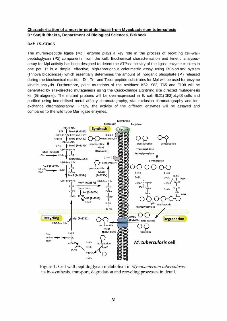
Characterisation of a murein-peptide ligase from Mycobacterium tuberculosis Dr Sanjib Bhakta, Department of Biological Sciences, Birkbeck
Ref: 15-ST005
The murein-peptide ligase (Mpl) enzyme plays a key role in the process of recycling cell-wall-peptidoglycan (PG) components from the cell. Biochemical characterisation and kinetic analyses– assay for Mpl activity has been designed to detect the ATPase activity of the ligase enzyme clusters in one pot. It is a simple, effective, high-throughput colorimetric assay using PiColorLock system (Innova biosciences) which essentially determines the amount of inorganic phosphate (Pi) released during the biochemical reaction. Di-, Tri- and Tetra-peptide substrates for Mpl will be used for enzyme kinetic analysis. Furthermore, point mutations of the residues: K62, S63, T65 and E108 will be generated by site-directed mutagenesis using the Quick-change Lightning site directed mutagenesis kit (Stratagene). The mutant proteins will be over-expressed in E. coli BL21(DE3)pLysS cells and purified using immobilised metal affinity chromatography, size exclusion chromatography and ion-exchange chromatography. Finally, the activity of the different enzymes will be assayed and compared to the wild type Mur ligase enzymes.
32
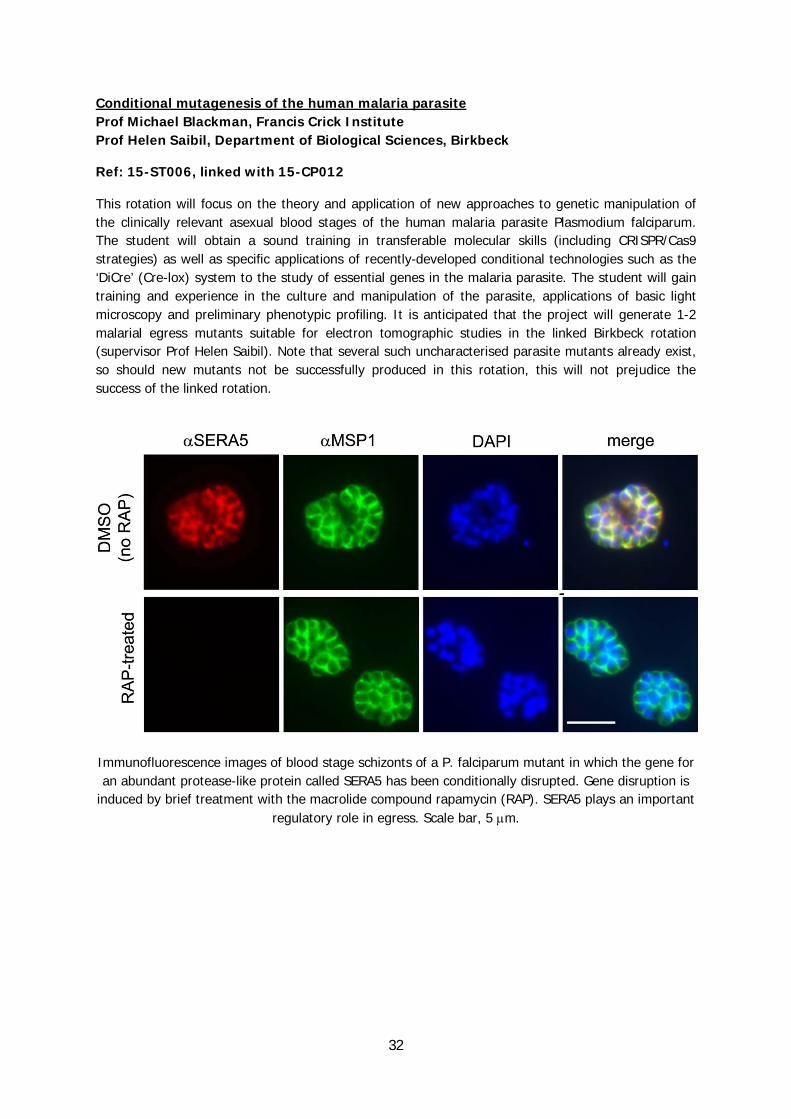
Conditional mutagenesis of the human malaria parasite Prof Michael Blackman, Francis Crick Institute Prof Helen Saibil, Department of Biological Sciences, Birkbeck
Ref: 15-ST006, linked with 15-CP012
This rotation will focus on the theory and application of new approaches to genetic manipulation of the clinically relevant asexual blood stages of the human malaria parasite Plasmodium falciparum. The student will obtain a sound training in transferable molecular skills (including CRISPR/Cas9 strategies) as well as specific applications of recently-developed conditional technologies such as the ‘DiCre’ (Cre-lox) system to the study of essential genes in the malaria parasite. The student will gain training and experience in the culture and manipulation of the parasite, applications of basic light microscopy and preliminary phenotypic profiling. It is anticipated that the project will generate 1-2 malarial egress mutants suitable for electron tomographic studies in the linked Birkbeck rotation (supervisor Prof Helen Saibil). Note that several such uncharacterised parasite mutants already exist, so should new mutants not be successfully produced in this rotation, this will not prejudice the success of the linked rotation.
Immunofluorescence images of blood stage schizonts of a P. falciparum mutant in which the gene for an abundant protease-like protein called SERA5 has been conditionally disrupted. Gene disruption is
induced by brief treatment with the macrolide compound rapamycin (RAP). SERA5 plays an important regulatory role in egress. Scale bar, 5 m.
33
Structural and functional studies of RNA Polymerase II elongation Dr Alan Cheung, Research Department of Structural and Molecular Biology, UCL
Ref: 15-ST007
We investigate the molecular mechanisms of eukaryotic transcription using structural biology, biochemistry and biophysical approaches. We study the transcription of mRNA by RNA Polymerase II (RNAP2), and the alteration of the chromatin environment during gene expression.
RNAP2 activity is governed by many accessory factors that bind RNAP2 and enable its transition through phases of initiation, elongation and termination.
http://tinyurl.com/pol2movie
PubMed ID 22726432
We are interested in the elongation phase and would like to understand how elongation factors stimulate a] the catalytic activity of RNAP2, enabling it to transcribe long distances on the genome without dissociating and b] transcription through nucleosomes, which present a barrier to elongation.
This rotation project will involve classical methods in molecular biology, protein biochemistry and biophysics to analyse the formation of RNAP2 complexes with elongation factors and their transcriptional activity using in vitro assays. This will lead to their structural analysis by crystallography and/or cryoEM.
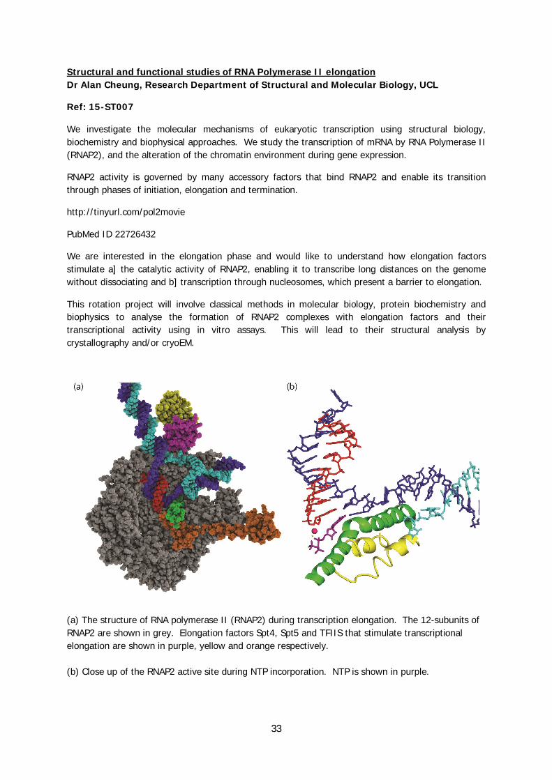
(a) The structure of RNA polymerase II (RNAP2) during transcription elongation. The 12-subunits of RNAP2 are shown in grey. Elongation factors Spt4, Spt5 and TFIIS that stimulate transcriptional elongation are shown in purple, yellow and orange respectively. (b) Close up of the RNAP2 active site during NTP incorporation. NTP is shown in purple.

34
Co-translational folding: using NMR to understand the interactions of a nascent chain with its parent ribosome Prof John Christodoulou, Jointly affiliated within the ISMB
Ref: 15-ST008
Protein synthesis occurs in all kingdoms of life on the ribosome where the nascent chain is synthesized one amino acid at a time and emerges from the exit tunnel(Fig). Using state of the art, high-resolution structural, biophysics and computational methods we provide as complete a description as possible on the folding and misfolding of nascent polypeptides during the translation process(1). Via ribosome nascent chain complexes(Fig) of an immunoglobulin domain and the Parkinson's-disease associated alpha-synuclein we have recently revealed the exterior ribosome surface as having potential chaperone interactions for the emerging chain. Using charge variations in alpha-synuclein nascent chain complexes this will be elucidated during this rotation.
A primary technique is NMR spectroscopy as it is uniquely able to simultaneously provide structure and dynamics of the nascent chain as it is being synthesised.
(1) Protein Folding on the Ribosome Cabrita, L.D.,Dobson, C.M., Christodoulou, J. Current Opin Struct Biol(2010) 20, 33-45.

35
Exploring the chemical and biological space of neuropilin-2/VEGF interaction Prof Snezana Djordjevic, Research Department of Structural and Molecular Biology, UCL
Ref: 15-ST009
Neuropilin-1 (NRP-1) and neuropilin-2 (NRP-2) are transmembrane receptors that are implicated in several human diseases including cancer and age related macular degeneration. NRP-2 is known to be involved in lymphangiogenesis, and a role for NRP-2 has been suggested for several cancers. This project will explore the structural biology of NRP-2 and identify chemical biology tools to study its function.
We will build upon our previous work with NRP-1 and by using similar methodology design a new series of peptides and peptidomimetics that would exhibit selectivity for NRP-2. The design will be informed by X-ray crystallography of NRP-2/ligand-bound complexes and biophysical characterisation of their interaction. The student will carry out crystallisation of NRP2-b1 in complex with the known ligands. The student will also begin work in synthetic chemistry initially undertaking computational analysis of the interactions that would allow moving into the chemical synthesis of a selected series of NRP-2 antagonists.

36
Structural basis of gabapentinoid drug action Dr Matthew Gold, Jointly affiliated within the ISMB
Ref: 15-ST010
Gabapentinoids are blockbuster drugs used to treat conditions including neuropathic pain and epilepsy. They act by binding α2δ voltage-gated Ca2+ channel subunits. The focus of this rotation project is to investigate the structure of α2δ to help understand its role in presynaptic Ca2+ entry and reveal the molecular basis of gabapentinoid binding.
We have developed constructs for α2δ expression in bacteria and eukaryotic cells (A). You will express and purify different regions of α2δ, taking advantage of enzymes at our disposal for modifying glycosylation and pre-protein cleavage (B). Co-crystallisation screens will be set up with natural and synthetic small molecules including pregabalin and gabapentin (C).
The project will be conducted in collaboration with Professor Annette Dolphin, an electrophysiologist who specialises in α2δ. There is scope to expand the project to a full PhD by performing follow-up functional experiments and applying further structural approaches including crosslinking mass spectrometry.

37
Hydrogen-Deuterium Exchange (HDX)-NMR studies of conformational misbehaviour in the serpinopathies Dr Bibek Gooptu, Jointly affiliated within the ISMB Prof John Christodoulou, Jointly affiliated within the ISMB
Ref: 15-ST011
Mutations in proteins of the serpin (serine protease inhibitor) superfamily can cause diseases (serpinopathies) including emphysema, cirrhosis, thrombosis and dementia. Disease-mutations cause serpins to misfold, and populate intermediate states that self-associate, forming pathological polymers (Fig.1A,B). Hydrogen-deuterium exchange (HDX)-NMR spectroscopy can define the abnormal structural dynamics that are central to this process. The Gooptu and Christodoulou groups have pioneered NMR studies of the archetypal serpin α1-antitrypsin. HDX-NMR studies can now report upon its conformational behaviour in solution. We have already optimised experimental conditions and generated preliminary datasets (Fig.1C,D). The student will make and collect HDX-NMR data on isotopically-labelled wild-type α1-antitrypsin to quantify molecular stability residue-by-residue in the physiological state. Analysis of our existing HDX-NMR data for disease-mutant (K154N) α1-antitrypsin will then define disease-relevant conformational behaviour. The project provides excellent training in biochemical methods and leading-edge NMR spectroscopy, and will provide insights into an important range of diseases. Contact: [email protected] and [email protected].

38
The use of nanofiber/microfiber scaffolds for 3D cell culturing and the development of in vitro cancer models Prof Ivan Gout, Research Department of Structural and Molecular Biology, UCL
Ref: 15-ST012
The nanofiber/microfibre scaffolds are state-of-the-art materials with applications in various fields, including space science, solar cells, filtration, biosensors, tissue engineering, drug discovery and cancer treatment. This project stems from an on-going collaborative research program with the University of Cambridge and Xanofi Biotech (USA) and aims at utilising nanofiber/microfiber scaffolds for efficient 3D cell culturing of established cancer cell lines and for developing in vitro 3D tumor models. We will also focus on establishing in vitro cultivation of patient-derived breast cancer and stromal cells, both from primary samples and associated lymph node and distant metastases in optimized nano/microfiber scaffolds in order to evaluate anti-cancer drug sensitivity for personalized anticancer chemotherapy. The applicant will gain the experience in a variety of experimental techniques, including mammalian cell culturing, molecular cloning and generation of recombinant lentiviruses, synthesis and structural analysis of nanofiber/microfiber scaffolds, immunofluorescence, confocal and two-photon microscopy etc.

39
Reconstituting interactions between the Chlamydia virulence effector IPAM, host CEP170 and microtubules Dr Richard Hayward, Jointly affiliated within the ISMB Prof Carolyn Moores, Department of Biological Sciences, Birkbeck
Ref: 15-ST013
Our research seeks to understand interactions between the bacterial pathogen Chlamydia trachomatis and host cells. Chlamydia forges a replicative niche within a modified intracellular compartment termed an 'inclusion' using virulence proteins delivered into the host cell. We have recently identified a bacterial protein (IPAM) sufficient to interfere with host microtubule dynamics (Dumoux et al (2015) J Cell Sci in press) by recruiting and stimulating the host centrosomal protein 170kDa (CEP170). CEP170 is essential for chlamydial control of host microtubules. We now wish to understand the molecular interactions between IPAM, CEP170 and microtubules by reconstituting their activities in vitro using purified proteins.
Confocal micrograph of a cultured cell after infection with Chlamydia trachomatis and triple-stained for tubulin (microtubules, grey), C.trachomatis IPAM (green) and cellular CEP170 (red). Arrow indicates the large chlamydial inclusion, a specialised membrane-bound compartment built by the intracellular bacteria. The endogenous microtubule organising centre (MTOC) containing CEP170 and IPAM at the centrosome is evident beneath the inclusion. Ectopic MTOC are also present at the inclusion periphery. A non-infected cell is present (bottom left) for comparison, which contains a single CEP170-positive MTOC.

40
Studying the transport dynamics of a bacterial secretion machine Dr Stefan Howorka, Department of Chemistry, UCL Prof Gabriel Waksman, Jointly affiliated within the ISMB
Ref: 15-ST014
The type 4 secretion system (T4SS) is a multiprotein molecular machine that transports DNA across the bacterial cell envelope thereby leading to the spread of antibiotic resistance. In this rotation project will exploit the recent structural analysis of one T4SS machine (Low et al 2014 Nature 508, 550) to understand how DNA is shuttled across the membrane. We will reconstitute defined protein complexes into cell-free bilayer systems to help facilitate the simple analysis in the absence of other complicating biological components. The project will involve classical protein biochemistry to express and purify the proteins of one archetypal T4SS, and membrane biophysics covering methods to characterise the bilayer-embedded proteins at the ensemble level (fluorescence microscopy) as well as single-molecule level (single-channel current recordings).
Oncogenic variants of FGFR as clients of Hsp90/Cdc37 system Prof Matilda Katan, Research Department of Structural and Molecular Biology, UCL Ref: 15-ST015
Fibroblast Growth Factor Receptors (FGFRs) are responsible for coordination of numerous developmental and cellular processes such as cellular differentiation and growth. Furthermore, somatic mutations in FGFRs are linked to a range of human cancer types and a number of these mutations have been causatively linked to tumour generation and progression. FGFRs are therefore an important target for anti-cancer therapy. Recent findings that correct folding and stability of FGFRs and, even more so, of several oncogenic variants requires interaction with molecular chaperone Hsp90 and co-chaperone Cdc37, provide another therapeutic opportunity. This rotation project will be part of our ongoing efforts to characterize FGFR/Hsp90/Cdc37 interactions using combination of structural and cellular biology and to assess the effect of Hsp90- and FGFR-inhibitors in cancer cell lines.
41
Lipases and triglyceride synthases of TB Prof Nicholas Keep, Department of Biological Sciences, Birkbeck
Ref: 15-ST016
The aim of this project is to start to determine the structure of a triacylglycerol synthase and related lipase from Mtb. Triglyceride synthases are a family of 15 homologous genes in M.tuberculosis, encoding proteins of around 50kd, for which there is no structural precedent. Tgs1 (Rv3130) has been implicated in a number of studies as being upregulated in dormancy and crucial to survival in the dormant state. In fact deletion of tgs1 prevented entry into dormancy. Nearly all the triglyceride synthases have a related lipase as an adjacent or nearby gene.
We have soluble constructs of both a lipase and triglyceride synthase, which will need scaling up to get quantitites required for crystallography. The project will give a good introduction to the molecular biology and protein expression required for structural biology and protein crystallisation and hopefully X-ray crystallography.
Defining the role of water molecules in the coupling mechanism of cytochrome c oxidase Dr Amandine Marechal, Jointly affiliated within the ISMB
Ref: 15-ST017
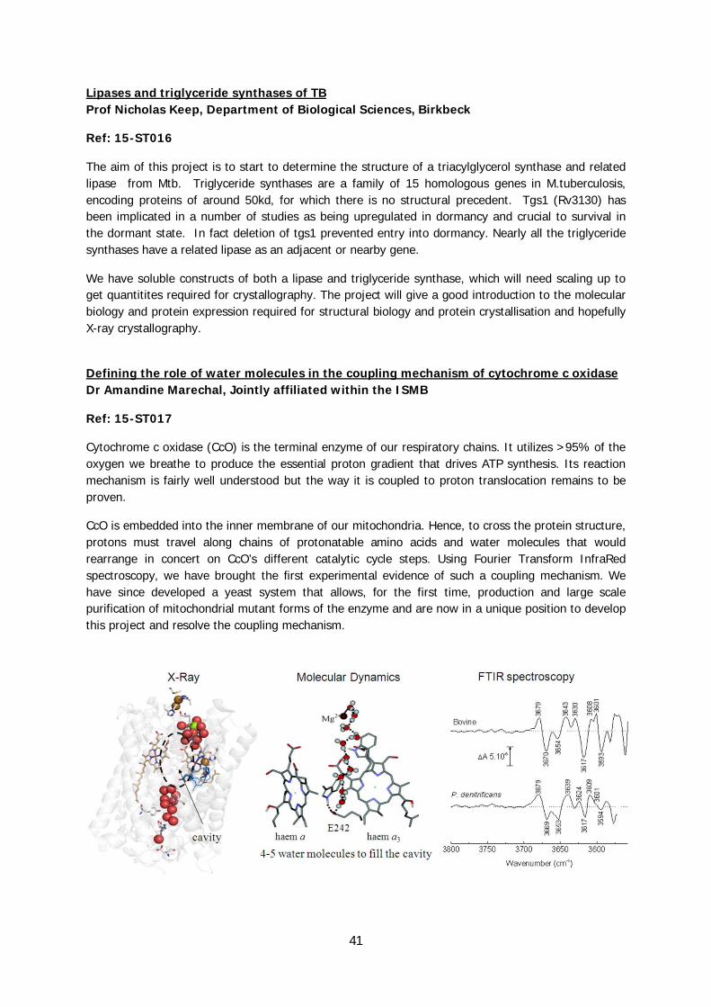
Cytochrome c oxidase (CcO) is the terminal enzyme of our respiratory chains. It utilizes >95% of the oxygen we breathe to produce the essential proton gradient that drives ATP synthesis. Its reaction mechanism is fairly well understood but the way it is coupled to proton translocation remains to be proven.
CcO is embedded into the inner membrane of our mitochondria. Hence, to cross the protein structure, protons must travel along chains of protonatable amino acids and water molecules that would rearrange in concert on CcO’s different catalytic cycle steps. Using Fourier Transform InfraRed spectroscopy, we have brought the first experimental evidence of such a coupling mechanism. We have since developed a yeast system that allows, for the first time, production and large scale purification of mitochondrial mutant forms of the enzyme and are now in a unique position to develop this project and resolve the coupling mechanism.
42
Purification of cytochrome c oxidase from yeast for the purpose of crystallisation Dr Amandine Marechal, Jointly affiliated within the ISMB
Ref: 15-ST018
Cytochrome c oxidase (CcO) is the terminal enzyme of the human respiratory chain. It catalyses the reduction of molecular oxygen to water, conserving the released energy as coupled transmembrane proton transfers.
Several fundamental aspects of CcO’s reaction mechanism and regulation remain unknown. Hypotheses have been formulated based on the available 3D structures of the enzyme (of which only one is of mitochondrial origin, bovine) but no mitochondrial system was available to test them by means of mutagenesis and detailed biophysical characterisation. We have thus developed a yeast system that allows for the first time large scale production and purification of mutant forms of the mitochondrial enzyme.
This project will be focussed on the optimisation of our purification protocol of the yeast CcO for the purpose of crystallisation. Crystallisation trials will be set for the best preparations and any hit will be optimised to the level suitable for X-ray data collection.
43
Regulation of microtubule nucleation by doublecortin and its relatives Prof Carolyn Moores, Department of Biological Sciences, Birkbeck Ref: 15-ST019
Microtubules are essential for the neurogenesis, neuronal migration, and neuronal differentiation that occur during brain development. Microtubule-associated proteins (MAPs) are central regulators of microtubule dynamics, but the molecular mechanisms of many of these proteins remain poorly understood. The focus of this rotation will be the doublecortin (DCX) family of MAPs that regulate microtubules by controlling their nucleation and polymerisation. Mutations in doublecortin (DCX) itself cause severe malformations of the brain, intractable epilepsy, intellectual disability and infant death. Therefore, investigating the mechanisms of microtubule regulation is crucial for understanding neuronal development, and to discover treatments for neurological diseases. DCX-like kinases (DCLK1/2) exhibit functional redundancy with DCX, suggesting a conserved mechanism of microtubule interaction, but this has not been demonstrated. This rotation will involve expression and characterisation of engineered constructs of DCLK1/2 to investigate their microtubule nucleation and stabilisation mechanism, and will provide an introduction to studying microtubules by electron microscopy.
Molecular mechanism of an essential mitotic motor Prof Carolyn Moores, Department of Biological Sciences, Birkbeck Ref: 15-ST020
The microtubule-based spindle orchestrates accurate chromosome segregation during cell division. Kinesins are a superfamily of highly efficient microtubule-based motors that use their ATPase activity to produce force. Kinesin-5 proteins are essential for cell division in most eukaryotes. Drugs that allosterically inhibit kinesin-5 ATPase also block cell division but the structural mechanism of allosteric communication between the ATP-, microtubule- and inhibitor binding sites are not understood. Kinesin-5s have been identified as targets for novel cancer therapies, but elucidation of the kinesin-5 molecular mechanism is essential to the ongoing development of clinically effective anti-mitotic drugs. Cryo-EM and image processing methods are essential tools for determining the structures of the motor-microtubule interaction at subnanometer resolution and provide essential information about motor mechanisms. This rotation will provide an introduction to cryo-electron microscopy and image processing to visualise the conformational changes at the heart of the kinesin-5 motor mechanism.

44
Examination of the SPP1 bacteriophage procapsid by cryo EM and flexible fitting of an atomic model Prof Elena Orlova, Department of Biological Sciences, Birkbeck Dr Maya Topf, Department of Biological Sciences, Birkbeck
Ref: 15-ST021
Bacteriophages represent the most populated biological entity in the Biosphere. Phages as bactericidal agents have been employed for treating bacterial infections both in humans, animals, and food industry. Specificity of phages is that they infect only bacteria and harmless for other organisms. Tailed bacteriophages are large protein-DNA complexes of more than 30 MDa in molecular mass and composed of a protein capsid that shelters a double-stranded DNA genome and a tail structure attached to the capsid. Phages are used as model systems in studies of structure-function relationships of large assemblies that transport macromolecules. To reveal mechanisms of genome packaging and its delivery into bacteria we need to learn a process of phage maturation and analyse structures of phages at sub-nanometer resolution. This will involve analysis of the phage capsids at their initial and final steps of self-assembly to identify conformational changes in the protein structure. The project will therefore incorporate cryo electron microscopy, methods from bioinformatics and structural modelling.
The SPP1 bacteriophage capsid is a self-assembled nanoparticle into which phage DNA is pumped by molecular motor fuelled by ATP. The capsid structures undergo significant expansion during DNA packaging completed by sealing of this nano-container. These nanoparticles are useful for delivery of specific or modified DNA in various medical settings. Our aim is to obtain a structure of the SPP1 bacteriophage procapsid at sub-nanometer resolution using cryo electron microscopy, bioinformatics and structural modelling. The project will involve image processing and methods of flexible fittings of atomic models into EM maps.
Phages undergo extensive conformational changes in their organization to provide rapid and faultless delivery of the phage genome into the host cell. We have obtained the structure of the SPP1 capsid at sub-nanometer resolution of 0.4 nm in its mature state. However the structure of its procapsid, that has a smaller size, remains unknown. To reveal the mechanism of high stability in mature capsids we need:
1. To visualise the SPP1 procapsid at the very initial states of self-assembly 2. To obtain the structure of the SPP1 procapsid with high resolution of at least 0.5nm. 3. To analyse conformational changes in the capsid protein to understand a mechanism of the maturation and to compare it with other phages
During the first year it is recommended that a student will take rotational projects related to training in analysis of atomic structures, predictions and methods of flexible fitting.
45
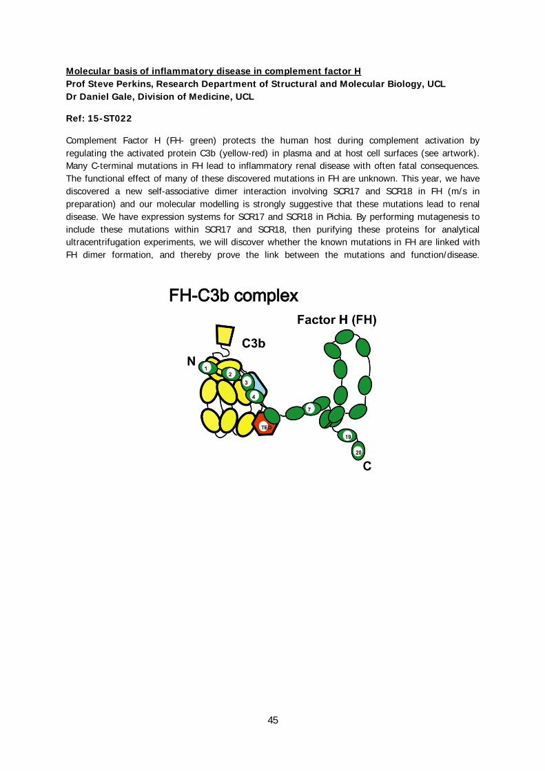
Molecular basis of inflammatory disease in complement factor H Prof Steve Perkins, Research Department of Structural and Molecular Biology, UCL Dr Daniel Gale, Division of Medicine, UCL
Ref: 15-ST022
Complement Factor H (FH- green) protects the human host during complement activation by regulating the activated protein C3b (yellow-red) in plasma and at host cell surfaces (see artwork). Many C-terminal mutations in FH lead to inflammatory renal disease with often fatal consequences. The functional effect of many of these discovered mutations in FH are unknown. This year, we have discovered a new self-associative dimer interaction involving SCR17 and SCR18 in FH (m/s in preparation) and our molecular modelling is strongly suggestive that these mutations lead to renal disease. We have expression systems for SCR17 and SCR18 in Pichia. By performing mutagenesis to include these mutations within SCR17 and SCR18, then purifying these proteins for analytical ultracentrifugation experiments, we will discover whether the known mutations in FH are linked with FH dimer formation, and thereby prove the link between the mutations and function/disease.
46
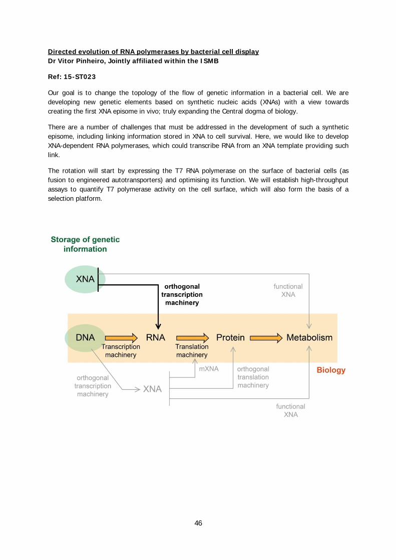
Directed evolution of RNA polymerases by bacterial cell display Dr Vitor Pinheiro, Jointly affiliated within the ISMB
Ref: 15-ST023
Our goal is to change the topology of the flow of genetic information in a bacterial cell. We are developing new genetic elements based on synthetic nucleic acids (XNAs) with a view towards creating the first XNA episome in vivo; truly expanding the Central dogma of biology.
There are a number of challenges that must be addressed in the development of such a synthetic episome, including linking information stored in XNA to cell survival. Here, we would like to develop XNA-dependent RNA polymerases, which could transcribe RNA from an XNA template providing such link.
The rotation will start by expressing the T7 RNA polymerase on the surface of bacterial cells (as fusion to engineered autotransporters) and optimising its function. We will establish high-throughput assays to quantify T7 polymerase activity on the cell surface, which will also form the basis of a selection platform.
47
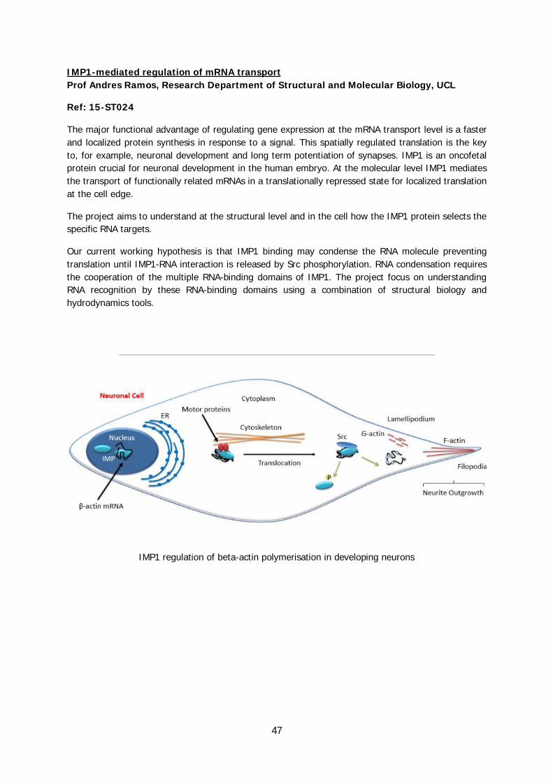
IMP1-mediated regulation of mRNA transport Prof Andres Ramos, Research Department of Structural and Molecular Biology, UCL
Ref: 15-ST024
The major functional advantage of regulating gene expression at the mRNA transport level is a faster and localized protein synthesis in response to a signal. This spatially regulated translation is the key to, for example, neuronal development and long term potentiation of synapses. IMP1 is an oncofetal protein crucial for neuronal development in the human embryo. At the molecular level IMP1 mediates the transport of functionally related mRNAs in a translationally repressed state for localized translation at the cell edge.
The project aims to understand at the structural level and in the cell how the IMP1 protein selects the specific RNA targets.
Our current working hypothesis is that IMP1 binding may condense the RNA molecule preventing translation until IMP1-RNA interaction is released by Src phosphorylation. RNA condensation requires the cooperation of the multiple RNA-binding domains of IMP1. The project focus on understanding RNA recognition by these RNA-binding domains using a combination of structural biology and hydrodynamics tools.
IMP1 regulation of beta-actin polymerisation in developing neurons

48
Building the cilium with ATP-driven molecular motors Dr Anthony Roberts, Department of Biological Sciences, Birkbeck
Ref: 15-ST025
Virtually every cell in the human body builds a cilium: a microtubule-based projection from the plasma membrane. These antennae-like structures are crucial for sensing light, smell, fluid flow, and morphogens. Their dysfunction is associated with obesity, kidney disease, and developmental defects. How the cell builds the cilium – a complex organelle with defined size, architecture, and protein composition – is a major unsolved question. Central to cilium construction are the molecular motors kinesin and dynein. These motors form large oligomeric “trains” that transport building blocks to and from the cilium tip, using ATP hydrolysis to move along microtubules. Determining the mechanism of the motor complexes has been challenging, in part because they are formed by large subunits that fail to fold in bacteria. In this rotation, we will use a newly developed insect cell expression system, single-particle electron microscopy, and single-molecule fluorescence microscopy to investigate how the motors move and are regulated.
Diagram of a cilium highlighting the two classes of molecular motor kinesin and dynein that move
along its length. Adapted from Roberts et al. (Nature Reviews Molecular Cell Biology, 2013) and Vale (Cell, 2003).
49
Role of quorum sensing in arsenic metabolism Dr Joanne Santini, Research Department of Structural and Molecular Biology, UCL Ref: 15-ST026, linked with 15-CP013
Arsenic is a metalloid that in its soluble oxidised forms, arsenite [As(III)] and arsenate [As(V)], is extremely toxic. Rhizobium sp. NT-26 can use As(III) as an electron donor with O2 as terminal electron acceptor and CO2 as sole carbon source. Expression of arsenite oxidase genes (aioB and aioA) is up-regulated by As(III) and involves the transcriptional regulator AioR which is part of a three-component regulatory system. In stationary phase the aioBA and aioXSR genes are further up-regulated by an as yet unknown Quorum sensing mechanism. This study is designed to identify this mechanism and its role in As(III) oxidation.
KSHV LANA recruitment of human Ung2 Dr Renos Savva, Department of Biological Sciences, Birkbeck Dr Tracey Barrett, Department of Biological Sciences, Birkbeck
Ref: 15-ST027
Uracil-DNA glycosylases (UDGs) are highly conserved enzymes which initiate the base excision repair (BER) pathway by excising uracil bases from DNA. Kaposi’s sarcoma-associated herpesvirus (KSHV) has a biphasic replication cycle which includes a lytic replicative phase preceded by a latent phase in which viral gene expression is extremely limited. During latent infection, KSHV recruits a human UDG (UNG2) by virtue of a direct protein-protein interaction with its latency-associated nuclear antigen (LANA) protein in order to maintain viral DNA, a process vital to viral proliferation.
We have recombinantly expressed and purified UNG2 and a relevant domain of LANA in multi-milligram quantities. The project is at the stage of verifying an in vitro interaction with the possible necessity of introducing stabilising mutations to LANA. The aim of the project is to characterise the LANA-UNG2 complex by x-ray crystallography and other relevant structural/biophysical techniques.
Predicting protein structure from sequence via synthetic covariance Dr Renos Savva, Department of Biological Sciences, Birkbeck Prof David Jones, Department of Computer Science, UCL
Ref: 15-ST028
Covariance of residues in homologous protein sequences is correlated with conserved structural features in proteins of known architecture. It is thus possible to use covariance data and secondary structure prediction algorithms to predict three-dimensional protein structures where there are hundreds of homologous database sequences available. It is conjectured that in cases where there is no known structure of a protein, and also very few if any homologues, this approach may yet be utilised via a synthetic recombinant approach: A degenerate lexicon of mutant sequences sharing the feature of conserved predicted secondary structure is synthesised as DNA then interrogated in a laboratory expression system in which a marker permits straightforward selection of stably folded proteins. Selected clones would be DNA sequenced providing a database of pseudo-homologues to be probed for covariance, thus enabling accurate three-dimensional structure predictions to be made. The initial test system is designed and ready to use.

50
Single Molecule Investigation of a Type IV Secretion Substrate Prof Gabriel Waksman, Jointly affiliated within the ISMB Prof Justin Molloy, Division of Physical Biochemistry, Francis Crick Institute Ref: 15-ST029, linked with 15-CH008
Type IV Secretion Systems (T4SS) are ubiquitous export machineries operating in most bacteria. They are used to transport both proteins and DNA. They play crucial roles in bacterial pathogenesis and also in the spread of antibiotics resistance genes. To transport DNA, these systems must first process the DNA to be transported. They use a protein termed "relaxase" that cleaves the DNA at a particular sequence called "Origin of Transfer or OriT", and reacts covalently with the 5'-end of the cleaved DNA. For this linked rotation, we propose to study a relaxase using single molecule techniques, namely magnetic and optical tweezers. The goal of this rotation is to study the role of DNA supercoiling in relaxase activity. Such a research will provide the first insight as to what are the DNA topological requirements for this important protein.
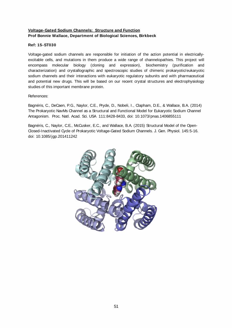
51
Voltage-Gated Sodium Channels: Structure and Function Prof Bonnie Wallace, Department of Biological Sciences, Birkbeck
Ref: 15-ST030
Voltage-gated sodium channels are responsible for initiation of the action potential in electrically-excitable cells, and mutations in them produce a wide range of channelopathies. This project will encompass molecular biology (cloning and expression), biochemistry (purification and characterization) and crystallographic and spectroscopic studies of chimeric prokaryotic/eukaryotic sodium channels and their interactions with eukaryotic regulatory subunits and with pharmaceutical and potential new drugs. This will be based on our recent crystal structures and electrophysiology studies of this important membrane protein.
References:
Bagnéris, C., DeCaen, P.G., Naylor, C.E., Pryde, D., Nobeli, I., Clapham, D.E., & Wallace, B.A. (2014) The Prokaryotic NavMs Channel as a Structural and Functional Model for Eukaryotic Sodium Channel Antagonism. Proc. Natl. Acad. Sci. USA 111:8428-8433, doi: 10.1073/pnas.1406855111
Bagnéris, C., Naylor, C.E., McCusker, E.C., and Wallace, B.A. (2015) Structural Model of the Open-Closed-Inactivated Cycle of Prokaryotic Voltage-Gated Sodium Channels. J. Gen. Physiol. 145:5-16. doi: 10.1085/jgp.201411242

52
Building synthetic pathways for novel alkaloids Prof John Ward, Biochemical Engineering UCL Prof Helen Hailes, Department of Chemistry, UCL
Ref: 15-ST031, linked with 15-CH004
Alkaloids are a diverse family of bioactive compounds synthesised by plants and there are over 14,000 alkaloid structures known. Many alkaloids and their derivatives are used as pharmaceuticals and they represent a huge repository of functional chemical space.
We have been developing synthetic pathways to new alkaloid structures centred around the key coupling enzyme (S)-norcoclaurine synthase (NCS).
NCS uses dopamine and an aldehyde and couples these together to make a new ring. We have used transaminases to generate the active aldehyde in situ in a novel cascade to make the norcoclaurine product. The project will extend our growing synthetic pathways by using aromatic hydroxylases to prepare the dopamine, alcohol dehydrogenases to generate an aldehyde and cytochrome P450s to modify the norcoclaurine rings. These enzymes will be used to build into in vitro and in vivo synthetic cascades.
T. Pesnot, M. Gershater, J. M. Ward, H. C. Hailes, Chem. Commun. 2011, 47, 3242. T. Pesnot, M. C. Gershater, J. M. Ward, H. C. Hailes, Adv. Syn. Cat. 2012, 354, 2997. B. R. Lichman, E. D. Lamming, T. Pesnot, H. C. Hailes, J. Ward, Green Chemistry, 2015, 17, 852. B. R. Lichman, M. C. Gershater, E. D. Lamming, T. Pesnot, A. Sula, N. Keep, H. C. Hailes, J. M. Ward, FEBS J, 2015, 282, 1 H. Hussain and J. Ward. Appl. Env. Microbiol. 2003, 69, 373-382 H. Hussain and J. Ward. Enz Microb Technol, 2003, 32. 790-800
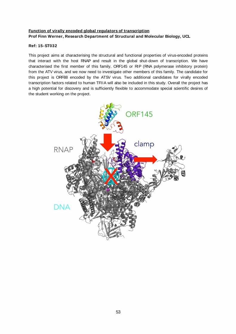
53
Function of virally encoded global regulators of transcription Prof Finn Werner, Research Department of Structural and Molecular Biology, UCL
Ref: 15-ST032
This project aims at characterising the structural and functional properties of virus-encoded proteins that interact with the host RNAP and result in the global shut-down of transcription. We have characterised the first member of this family, ORF145 or RIP (RNA polymerase inhibitory protein) from the ATV virus, and we now need to investigate other members of this family. The candidate for this project is ORF88 encoded by the ATSV virus. Two additional candidates for virally encoded transcription factors related to human TFIIA will also be included in this study. Overall the project has a high potential for discovery and is sufficiently flexible to accommodate special scientific desires of the student working on the project.

54
Cryo-EM structure of COPII Dr Giulia Zanetti, Department of Biological Sciences, Birkbeck
Ref: 15-ST033
The function of eukaryotic protein coats directly depends on their assembly dynamics and architecture. The lab focuses on the study of COPII coat structure, in particular to understand cargo specific regulation of protein trafficking.
The aim of this rotation project is to solve the cryo-electron microscopy structure of in vitro assembled COPII. Cryo-tomography data of coated vesicles has been collected on our state-of-the art microscope and direct electron detector. The student will perform 3D-reconstructions, including electron dose and Contrast Transfer Function optimisation. Sub-tomogram averaging methods will be applied and optimised to obtain a structure of the building blocks of the COPII coat, aming to obtain sub-nanometer resolution. X-ray atomic coordinates will be fitted into the COPII density map to obtain a model.
During this project, the student will learn the basic concepts of cryo-EM and image processing, including how to use Matlab and other image processing software.

55
Single particle electron microscopy of chaperonin complexes with non-native substrate protein Prof Helen Saibil, Department of Biological Sciences, Birkbeck
Ref: 15-ST034
In this rotation the student will prepare complexes of the molecular chaperone GroEL with a non-native substrate protein, and study their structures by single particle electron microscopy. Because the non native protein will be bound in different conformations, it will be necessary to apply classification methods to sort out the different complexes present. Although these methods have been previously applied, there have been substantial advances in electron detectors and single particle software, and we expect that it will now be possible to resolve more details of the folding intermediates bound to the hydrophobic sites on the GroEL chaperonin. It is hoped that this work will eventually lead to a better understanding of how chaperonins assist the folding of a substrate protein. The student will learn how to prepare and biochemically analyse the complexes, and to carry out single particle electron microscopy, classification and 3-dimensional reconstruction.
References
Clare, DK, Bakkes, PJ, van Heerikhuizen, H, van der Vies, SM & Saibil, HR (2009) Chaperonin complex with a newly folded protein encapsulated in the folding chamber. Nature 457, 107-110.
Elad N, Farr GW, Clare DK, Orlova EV, Horwich AL & Saibil HR (2007) Topologies of a substrate protein bound to the chaperonin GroEL. Mol Cell 26, 415-426.
Figure: Models of GroEL with coloured densities showing 3 different conformations of bound, non-native malate dehydrogenase.
