firouzeh dehghan - university of malayastudentsrepo.um.edu.my/4572/1/thesis_2014-_proof_.pdf ·...
TRANSCRIPT
THE EFFECTS OF SEX-STEROIDS AND MENSTRUAL CYCLE/OESTROUS
PHASES ON KNEE LIGAMENT LAXITY IN HUMANS AND RODENTS
FIROUZEH DEHGHAN
THESIS SUBMITTED IN PARTIAL FULFILLMENT OF THE REQUIREMENT FOR
THE DEGREE OF DOCTOR OF PHILOSOPHY
FACULTY OF MEDICINE
UNIVERSITY OF MALAYA
KUALA LUMPUR
2014

ORIGINAL LITERARY WORK DECLARATION
Name of Candidate: FIROUZEH DEHGHAN (I.C/Passport No: P21262064)
Registration/Matric No: MHA100055
Name of Degree: PhD
Title of Project Paper/Research Report/Dissertation/Thesis (“this Work”): THE EFFECTS
OF SEX-STEROIDS AND MENSTRUAL CYCLE/OESTROUS PHASES ON KNEE
LIGAMENT LAXITY IN HUMANS AND RODENTS
Field of Study: EXERCISE PHYSIOLOGY
I do solemnly and sincerely declare that:
(1) I am the sole author/writer of this Work;
(2) This Work is original;
(3) Any use of any work in which copyright exists was done by way of fair dealing and
for permitted purposes and any excerpt or extract from, or reference to or
reproduction of any copyright work has been disclosed expressly and sufficiently
and the title of the Work and its authorship have been acknowledged in this Work;
(4) I do not have any actual knowledge nor do I ought reasonably to know that the
making of this work constitutes an infringement of any copyright work;
(5) I hereby assign all and every rights in the copyright to this Work to the University
of Malaya (“UM”), who henceforth shall be owner of the copyright in this Work
and that any reproduction or use in any form or by any means whatsoever is
prohibited without the written consent of UM having been first had and obtained;
(6) I am fully aware that if in the course of making this Work I have infringed any
copyright whether intentionally or otherwise, I may be subject to legal action or any
other action as may be determined by UM.
Candidate’s Signature Date
Subscribed and solemnly declared before,
Witness’s Signature Date
Name:
Designation:

i
ACKNOWLEDGEMENTS
It is my pleasure to thank the people who have made this thesis possible. First of all,
I would like to express my genuine gratitude to my supervisor, Dr. Naguib Salleh, my
project leader from Department of Physiology, Faculty of Medicine, University of Malaya
for his excellent supervision, advice, and guidance from the beginning stages of this study
as well as providing me with valuable experience throughout the work.
A special thanks to my supervisor, Dr. Ashril Yusof from Department of Exercise
Science, Sport Centre, University of Malaya for the privilege of working under exemplary
guidance and his advice, patience and encouragement throughout this study.
I would like to take this opportunity to appreciate Prof. Dr. Sekaran Muniandy from
Department of Molecular Medicine, Faculty of Medicine, University of Malaya for his
support and providing me such a friendly environment in his lab to work.
I highly appreciated to AP Dr. Abdul Halim Mokhtar from Department of Sport
Medicine, University of Malaya for his kindly support in this project.
My deepest and most heart-felt thanks go to my most beloved family, for their
endless love and support.
To those directly and indirectly involved in this study, your contribution shall not be
forgotten, and please accept my utmost appreciation.

ii
ABSTRAK
Insiden kecederaan lutut "non-contact" dilaporkan lebih tinggi pada wanita
berbanding lelaki. Bagi wanita, insiden ini adalah berkaitan dengan fasa kitaran haid.
Memandangkan ini, kami menyiasat perkara berikut: (i) perubahan dalam kelalaian lutut
dalam model tikus di bawah pengaruh seks-steroids yang berbeza; (ii ) Mekanisme
perubahan dalam kelalaian lutut yang melibatkan ungkapan pembezaan reseptor relaxin dan
( iii ) antara lutut asas kelalaian dan serum tahap hormon seks - steroids dan relaxin di fasa
kitaran haid. Dalam bahagian pertama kajian, tikus ovariectomized WKY telah dibahagikan
kepada kumpulan rawatan yang berbeza (n = 6 bagi setiap kumpulan): dos berbeza 17 β-
oestradiol, progesteron dan testosteron (125 & 250 µg/kg). Hormon telah disuntik
subcutaneously selama 3 hari berturut-turut. Pada masa yang sama, steroid hormon
penghalang reseptor dan penghalang enzim (ICI 182780, PHTPP, MPP, mifepristone,
Flutamide, dan Finasteride) juga telah disuntik dengan rawatan agonist steroid masing-
masing. Pada masa yang sama, tikus-tikus utuh telah dikumpulkan berdasarkan peringkat
kitaran oestrous mereka: proestrus , estrus, metestrous dan diestrus. Dalam ovariectomized
steroid tikus dirawat dan utuh, pelbagai lutut gerakan (ROM) telah diukur dengan
menggunakan goniometer kecil digital. Tikus-tikus telah dikorbankan dan ungkapan
isoforms relaxin reseptor, RXFP1 dan RXFP2 mRNAs dan protein dalam ligamen lutut dan
tendon ditentukan.Sementara itu, pada manusia, perubahan dalam kelalaian lutut di fasa
kitaran haid diperhatikan dalam atlet wanita.Varus lutut dan valgus sudut telah ditentukan
dalam atlet dan bukan atlet fasa kitaran haid mereka dengan menggunakan goniometer
pemerintah ortopedik.Tahap serum oestrogen, progesteron dan relaxin telah ditentukan
dalam subjek manusia di pelbagai fasa kitaran haid mereka.Hasil: data kami menunjukkan
bahawa ROM lutut pada tikus telah meningkat dengan ketara berikutan rawatan oestrogen
iii
dan progesteron bagaimanapun susut berikutan rawatan testosteron.Perubahan ROM lutut
telah dihalang oleh antagonis terhadap reseptor oestrogen, progesteron dan androgen. ROM
lutut adalah tinggi di peringkat proestrus dan diestrus kitaran oestrus. Progesteron dan
oestrogen merangsang manakala testosteron menghalang perubahan RXFP1 dan RXFP2
mRNA dan ungkapan protein yang dihalang oleh penghalang reseptor masing-
masing.Sementara itu, rawatan dengan relaksin meningkatkan ROM lutut oestrogen dan
progesteron dirawat tetapi tidak testosteron dirawat ditunjukkan daripada perubahan
pengawalaturan reseptor relaksin.Pada manusia, perbezaan yang signifikan dalam varus
dan valgus sudut diperhatikan dalam wanita di pelbagai fasa kitaran haid dengan yang
tertinggi pada fasa ovulasi dan haid kitaran.Kajian ini memberikan gambaran tentang kesan
seks steroid pada ROM lutut dan RXFP1 dan ungkapan RXFP2. Dalam tikus, ungkapan
peningkatan RXFP1 dan RXFP2 boleh menjelaskan peningkatan dalam lutut kelalaian
bersama. Pada manusia, meningkat kelalaian di ovulasi dan fasa luteal pertengahan kitaran
tersebut selaras dengan tahap yang tinggi oestrogen dan progesteron. Dalam kedua-dua
tikus utuh dan manusia, korelasi yang kuat antara kelalaian lutut dan tahap serum relaksin
telah diperhatikan. Penemuan kami boleh menjelaskan kelemahan yang mendasari dasar
wanita terhadap kecederaan lutut bukan traumatik di fasa kitaran reproduktif mereka.

iv
ABSTRACT
The incidence of non-contact knee injury was reported higher in female than male.
In female, the occurrence of this injury was related to different phases of the menstrual
cycle. In view of this, we investigated the followings: (i) changes in knee laxity in rodent
model under different sex-steroid influence; (ii) mechanisms underlying changes in knee
laxity, which involves differential expression of relaxin receptors and (iii) correlation
between knee laxity and serum levels of sex-steroids and relaxin at different phases of the
menstrual cycle. In first part of the study, ovariectomized WKY rats were divided into
different treatment groups (n=6 per group): different doses of 17 β-oestradiol, progesterone
and testosterone were administered. The hormones were injected subcutaneously for 3
consecutive days. In parallel, steroid hormone receptor blockers and enzyme inhibitor (ICI
182780, PHTPP, MPP, Mifepristone, Flutamide, and Finasteride) were also injected with
the respective agonist. In parallel, intact rats were grouped based on their oestrous cycle
stages: proestrus, estrus, metestrus and diestrus. In ovariectomized steroid treated and intact
rats, knee range of motion (ROM) was measured by using a digital miniature goniometer.
The rats were sacrificed and expression of relaxin receptor isoforms, RXFP1 and RXFP2
mRNAs and proteins in knee ligaments and tendons were determined. Meanwhile, in
humans, changes in knee laxity were observed at different phases of the menstrual cycle in
the female athletes. The knee varus and valgus angles were determined at different phases
of the menstrual cycle by using an orthopedic goniometer ruler. Blood was withdrawn and
serum levels of oestrogen, progesterone and relaxin were determined in human at different
phases of their menstrual cycle. Our findings showed a significant increase in knee ROM in
rats following oestrogen and progesterone treatment however was decreased following
testosterone treatment. Changes in knee ROM was antagonized by the concomitant

v
administration of oestrogen, progesterone and androgen receptor antagonists. Knee ROM
was high at proestrus and diestrus stages of the oestrous cycle. Progesterone and oestrogen
up-regulated while testosterone down-regulated RXFP1 and RXFP2 mRNA and protein
expressions which were antagonized by the respective steroid hormone antagonist.
Meanwhile, relaxin administration increases knee ROM in oestrogen and progesterone
treated but not testosterone treated groups indicating relaxin receptor up-regulation. In
humans, significant difference in knee varus and valgus angles were observed in female
athletes and non-athletes at different phases of the menstrual cycle which was the highest in
the ovulatory and menstrual phases. Non-athletes have higher medial and lateral knee laxity
as compared to athletes. This study has provided an insight into the mechanism underlying
sex-steroid control of knee ROM via modulating the expression of RXFP1 and RXFP2
receptors. In rodents, increased RXFP1 and RXFP2 expressions could contribute to
increase in knee laxity. In humans, the increase in laxity at ovulatory and mid-luteal phases
of the cycle was consistent with high level of oestrogen and progesterone. In both intact
rats and humans, strong correlations was noted between knee laxity and serum relaxin
levels. Our study has provided the basis underlying female susceptibility towards non-
traumatic knee injury at different phases of their reproductive cycle.

vi
CONTENTS
ACKNOWLEDGEMENTS ...................................................................................... i
ABSTRAK ................................................................................................................. ii
ABSTRACT ............................................................................................................. iv
CONTENTS ............................................................................................................. vi
LIST OF TABLES ................................................................................................... ix
LIST OF FIGURES .................................................................................................. x
CHAPTER 1 INTRODUCTION ............................................................................ 1
1.1 Introductions ..................................................................................................... 2
1.2 Significance of the study ................................................................................... 6
CHAPTER 2 LITRATURE REVIEW .................................................................. 7
2.1 Introductions ..................................................................................................... 8
2.2 Overview of non-traumatic knee injury in female ............................................ 8
2.2.1 Anatomy of the knee joints ................................................................... 10
2.2.2 Physiological control of knee joint movement ..................................... 13
2.2.3 Knee movement and its connective tissue mechanism ......................... 15
2.3 Menstrual cycle ............................................................................................... 16
2.4 Oestrous cycle ................................................................................................. 19
2.5 Overview of Sex-Steroid Biochemistry and Physiology ................................ 20
2.5.1 Sex-steroids and Knee Laxity ............................................................... 23
2.6 Relaxin ............................................................................................................ 23
2.6.1 Relaxin roles in different tissues ........................................................... 25
2.6.2 Relaxin roles in musculoskeletal tissues ............................................... 28
2.7 Summary of literature findings ....................................................................... 39
CHAPTER 3 MATERIALS AND METHODS ................................................... 40
3.1 Materials ......................................................................................................... 42
3.1.1 Animal ................................................................................................... 42
3.1.2 Chemicals and consumable ................................................................... 42
3.1.3 Sterilization ........................................................................................... 44
3.2 Methods ........................................................................................................... 44
3.2.1 Ovariectomy surgical procedures ......................................................... 44
3.2.2 Animal preparation and administration of sex steroid hormone ........... 45
3.2.3 Identification of oestrous cycle stages .................................................. 45
3.2.4 Animal study groups ............................................................................. 46

vii
3.2.5 Investigation of blood serum hormone level ........................................ 47
3.2.6 Knee range of motion (ROM) ............................................................... 48
3.2.7 Real Time PCR (qPCR) ........................................................................ 50
3.2.8 RNA to cDNA converssion .................................................................. 53
3.2.9 Real time PCR running ......................................................................... 54
3.2.10 Data Analysis for Quantification of Gene Expression .......................... 55
3.2.11 Protein expression by Western blotting ................................................ 56
3.2.12 Western blot running ............................................................................. 59
3.2.13 Western blot data Analysis ................................................................... 62
3.3 Human study ................................................................................................... 62
3.3.1 Blood sample collection and serum hormones’ analyses ..................... 63
3.3.2 Basal Body Temperature ....................................................................... 63
3.3.3 Knee rotational movement .................................................................... 64
3.3.4 Data analysis of knee laxity in human .................................................. 66
CHAPTER 4 ............................................................................................................ 67
4.1 Introduction ..................................................................................................... 68
4.2 Results ............................................................................................................. 70
4.2.1 Plasma Sex-Steroid Levels following Subcutaneous Hormone Injection
.............................................................................................................. 70
4.2.2 RXFP1 and RXFP2 Proteins Expression in the Patellar Tendon and
Lateral Collateral Ligament .................................................................. 71
4.2.3 RXFP1and RXFP2 mRNAs Expression in the Patellar Tendon and
Lateral Collateral Ligament .................................................................. 75
4.3 Discussion ....................................................................................................... 79
CHAPTER 5 ............................................................................................................ 86
5.1 Introduction ..................................................................................................... 87
5.2 Results ............................................................................................................. 89
5.2.1 Passive Knee ROM in Testosterone-Treated Ovariectomised Rats ..... 89
5.2.2 RXFP1 and RXFP2 mRNA Expression in Patellar Tendon ................. 91
5.2.3 RXFP1 and RXFP2 Protein Expression in Patellar Tendon ................. 93
5.2.4 RXFP1 and RXFP2 mRNA Expression in Lateral Collateral Ligament
.............................................................................................................. 95
5.2.5 RXFP1 and RXFP2 Protein Expression in Lateral Collateral Ligament
.............................................................................................................. 97
5.3 Discussion ....................................................................................................... 99
CHAPTER 6 .......................................................................................................... 103
6.1 Introduction ................................................................................................... 104
viii
6.2 Results ........................................................................................................... 106
6.2.1 Effect of steroid receptor antagonists on RXFP1 and RXFP2 mRNA
and proteins expression in the patellar tendon ................................... 106
6.3 Effect of steroid receptor antagonists on RXFP1 and RXFP2 mRNA and
proteins expression in the collateral ligaments ............................................. 109
6.4 Effect of steroid receptor antagonists on RXFP1 and RXFP2 mRNA and
proteins expression in the hamstring muscles ............................................... 112
6.5 Knee passive ROM in the presence of steroid receptor antagonist............... 115
6.6 Discussion ..................................................................................................... 116
CHAPTER 7 .......................................................................................................... 120
7.1 Introduction ................................................................................................... 121
7.2 Results ........................................................................................................... 123
7.2.1 Knee Passive ROM at different phases of the oestrus cycle ............... 123
7.2.2 RXFP1 & RXFP2 expressions in hamstring muscle at different phases
of the oestrus cycle ............................................................................. 124
7.2.3 RXFP1 & RXFP2 expression in patellar tendon at different phases of
the oestrus cycle ................................................................................. 126
7.2.4 RXFP1 & RXFP2 expression in collateral ligament at different phases
of the oestrus cycle ............................................................................. 128
7.2.5 Correlation between serum relaxin and progesterone ......................... 131
7.3 Discussion ..................................................................................................... 132
CHAPTER 8 .......................................................................................................... 135
8.1 Introduction ................................................................................................... 136
8.2 Results ........................................................................................................... 138
8.2.1 Body Composition Analyses ............................................................... 138
8.2.2 Changes in Serum Sex-Steroids and Relaxin Levels at Different Phases
of the Menstrual Cycle ....................................................................... 141
8.2.3 Correlations between Sex-Steroids and Relaxin Levels with Knee Joint
Angles ................................................................................................. 142
8.3 Discussion ..................................................................................................... 143
CHAPTER 9: CONCLUSION ............................................................................ 148
REFERENCES ..................................................................................................... 155
Appendices ............................................................................................................ 177
ix
LIST OF TABLES
Table 2-1 Previous reported data of the non-traumatic knee injury and menstrual
cycle phases .................................................................................................................. 9
Table 2-2 Review of previously reported data of relaxin in locomotor system .......... 37
Table 3-1 Chemicals and consumable used in this project ......................................... 43
Table 3-2 Composition of 12 % resolving and 4 % stacking gel ................................ 60
Table 3-3 primary and secondary antibodies that have been used in western blot. .... 62
Table 4-1 Plasma sex steroid level following subcutaneous hormone injection. ....... 71
Table 7-1 Serum hormone level in non ovariectomized rat at different phases of the
oestrus cycle .............................................................................................................. 130
Table 8-1 Characteristics and demographics at different phases of the menstrual
cycle. *p<0.05 as compared to the athletes/non-athletes group ................................ 138
Table 8-2 Knee joint varus/valgus angles on 0 and 30° ............................................ 140
Table 8-3 Sex-steroids and relaxin levels at different phases ................................... 142
Table 8-4 Correlation of sex steroid and knee angles in total polulation ................. 142
Table 8-5 Correlation of sex steroid and Knee angles in athletes ............................. 142
Table 8-6 Correlation of sex steroid and Knee angle in non-athletes ....................... 143
x
LIST OF FIGURES
Figure 2-1 Knee anatomical perspective (www.webmd.com) .................................... 12
Figure 2-2 Motion degrees of human kneehttp://www.jointinjury.com/knee ............ 14
Figure 2-3 Knee rotational movement ........................................................................ 14
Figure 2-4 Knee patellar tendon and collateral ligament ............................................ 16
Figure 2-5 Changing hormone levels, ovarian and uterine events during the menstrual
cycle ............................................................................................................................ 18
Figure 2-6 Human and rat reproductive menstrual cycle ............................................ 20
Figure 2-7 Sex steroid hormone response ................................................................... 22
Figure 2-8 Relaxin structure ....................................................................................... 25
Figure 2-9 Interaction of RLN1, LN2, and RLN3 proteins with their receptors
RXFP1, RXFP2, and RXFP3, respectively, as well as with insulin growth factor
(INSL3) ....................................................................................................................... 27
Figure 2-10 A summary of relaxin role in the locomotor system ............................... 28
Figure 2-11 Mechanism of regeneration in damaged muscle. MSC (muscle satellite
cell), MPC (myogenic progenitor cell) ....................................................................... 33
Figure 2-12 Mechanism of regeneration in damaged muscle ..................................... 35
Figure 3-1 Schematic overview and research methods of the study ........................... 41
Figure 3-2 Identification of the oestrous cycle in rat .................................................. 46
Figure 3-3 Measurement of rat’s ROM by digital miniature goniometer ................... 49
Figure 3-4 Digital Newton meter ................................................................................ 49
Figure 3-5 Real time PCR amplification view ............................................................ 51
Figure 3-6 RNA gel electrophoresis ........................................................................... 53
Figure 3-7 Standard curve for protein determination ................................................. 58

xi
Figure 3-8 Basal body temperatures chart .................................................................. 63
Figure 3-9 Knee rotational movement investigation by Varus-Valgus test at 30° ..... 65
Figure 3-10 Knee rotational movement investigation by Varus-Valgus test at 0° ..... 65
Figure 4-1 The expression of RXFP1 protein in the total homogenate of collateral
ligament ....................................................................................................................... 72
Figure 4-2 The expression of RXFP1 protein in the total homogenate of the patellar
tendon .......................................................................................................................... 73
Figure 4-3 The expression of RXFP2 protein in the total homogenate of collateral
ligament ....................................................................................................................... 75
Figure 4-4 The expression of RXFP1 mRNA in the lateral collateral ligament ......... 76
Figure 4-5 The expression of RXFP1 mRNA in the patellar tendon .......................... 77
Figure 4-6 The expression of RXFP2 mRNA in the lateral collateral ligament ......... 78
Figure 4-7 The expression of RXFP2 mRNA in the patellar tendon from
ovariectomised ............................................................................................................ 79
Figure 5-1 Passive ROM of the rat knee ..................................................................... 91
Figure 5-2 Changes in RXFP1 and RXFP2 mRNA expression in the presence of FLU
and FIN ....................................................................................................................... 92
Figure 5-3 The expression of RXFP1 and RXFP2protein in the patellar tissue
homogenate ................................................................................................................. 94
Figure 5-4 Changes in RXFP1 and RXFP2 mRNA expressionin the lateral collateral
ligament ....................................................................................................................... 96
Figure 5-5 The expression of RXFP1 and RXFP2 protein ......................................... 98
Figure 6-1 RXFP1& RXFP2 mRNA and protein expression ................................... 107
Figure 6-2 RXFP1& RXFP2 mRNA and protein levels in collateral ligament ........ 110
Figure 6-3 RXFP1& RXFP2 mRNA and proteins levels in the hamstringmuscle ... 113

xii
Figure 6-4 Knee passive ROM of ovariectomised rats ............................................. 115
Figure 7-1 Knee passive ROM at different phases of the oestrus cycle ................... 123
Figure 7-2 RXFP1& RXFP2 mRNA and protein expression in the hamstring muscle
................................................................................................................................... 125
Figure 7-3 RXFP1& RXFP2 mRNA and protein expression levels in patellar tendon
................................................................................................................................... 127
Figure 7-4 RXFP1& RXFP2 mRNA and protein expression levels in collateral
ligament ..................................................................................................................... 129
Figure 7-5 Correlation between serum relaxin and progesterone levels. .................. 131

xiii
LIST OF SYMBOLS ANDABBREVIATIONS
AC Adenylate cyclase
ACL Anterior cruciate ligament
APS Ammonium per sulphate
APS Ammonium Persulphate
BBT Basal body temperature
BF Beef Heifer
BSA Bovine Serum Albumin
D Dog
Ds Diestrous
E2 Oestrogen/oestradiol
ELISA Enzyme-linked immunosorbent assay
Es Estrus
F Fish
Fin Finasteride
Flu Flutamide
G Guinea pig
GPCRs G-protein-coupled receptors
H Human
ICI 182/780 7α,17β-[9-[(4,4,5,5,5-Pentafluoropentyl) sulfinyl]nonyl]estra-1,3,5(10)-
triene-3,17-diol
INSL Insulin-like peptide
IP Intraperitoneal
LCL Lateral collateral ligament
M Mouse
MCL Medial collateral ligament
Mife Mifepristone
MMPs Matrix metalloproteinases
MPP 1,3-bis(4-hydroxyphenyl)-4-methyl-5-[4-(2-piperidinylethoxy)phenol]-
1N-pyrozole dihydrochloride
Ms Metestrous
NO Nitric oxide
xiv
OPG Osteoprotegerin
P Progesterone
PBMCs Peripheral blood mononuclear cells
PCR Polymerase Chain Reaction
PHTPP 4-[2-phenyo-5,7-bis(trifluoromrthyl) pyrazolo(1,5-a) pyrimidin-3-yl]
phenol
Ps Proestrous
PT Patellar tendon
PVDF Poly Vinylidene fluoride membrane
R Rat
RA Rheumatoid arthritis
RANKL Receptor activator of nuclear factor κB ligand
Rb Rabbit
RIA Radioimmunoassay
RLN Relaxin
ROM Range of motion
RXFP Relaxin family peptide receptors
SDS-PAGE Sodium dodecyl sulfate polyacrylamide gel
SST Sample separator tube
T Testosterone
TNF Tumour necrosis factor
WB Western blotting
α Alpha
β Beta

1
CHAPTER 1 INTRODUCTION

2
1.1 Introductions
Musculoskeletal injuries are common problem among athletes and are not gender
specific (Arendt et al., 1999), however, female have been reported to have higher risk of
ligament injury as compared to male (Ireland, 2002; Myklebust et al., 1998). Sprains of the
knee joint are common in athletes who participate in direct contact sports such as football
or soccer, who are more likely to injure their collateral and anterior cruciate ligaments. It
has been proposed that the incidence of knee injury in female is related to phases of the
menstrual cycle. The softening of ligaments in the luteal phase of the cycle has long been
attributed to an increase in collagenase enzyme activity (Qin et al., 1997). An increasing
trend of non-contact knee injury has also been reported in the ovulatory phase and this
decrease during the follicular phase however due to the conflicting reports, no conclusion
could be drawn from the previous studies on the effect of menstrual cycle phases on this
injury in female (Wojtys et al., 1998).
The amplitude of joint laxity is related to the joint movement which was limited by
ligaments and tendons. The relationship between different phases of menstrual cycle and
knee joint laxity is still uncertain. Some researchers demonstrated that no association
between knee laxity and phases of the menstrual cycle (Belanger et al., 2004; Karageanes et
al., 2000). Meanwhile, knee laxity has been reported to be high in the ovulatory phase with
a peak in oestrogen concentration (Heitz et al., 1999; Romani et al., 2001). Studies have
revealed that these differences could be related to the differential expression of sex-steroid
receptors in the skeletal muscle (Dragoo et al., 2003; Lemoine et al., 2003) in both humans

3
and animals (Sciore et al., 1998), which could explain differences in the incidence of non-
contact knee injury at different phases of the cycle.
Fluctuation in relaxin receptor expression in the knee was postulated based on the
observed fluctuation in its expression in the uterus, being the lowest during the proliferative
phase and rises sharply at the time of ovulation (Bond et al., 2004). In neonatal porcine
uterus, oestrogen increases RXFP1mRNA expressions, while in cervix, oestrogen increases
and relaxin decreases these receptors expression (Yan et al., 2008). During late pregnancy
in mice, relaxin has no influence on RXFP1 expression (Siebel et al., 2003). Progesterone
has been reported to increase the expression of RXFP1 in the pregnant rat uteri (Vodstrcil
et al., 2010a ) while oestrogen was found to regulate RXFP1 mRNA expression in human
cervix at term (Maseelall et al., 2009). Relaxin receptors expression may vary in the
musculoskeletal tissues and these changes may affect joint laxity. Nonetheless, the effect of
sex steroids on joint laxity in female remains inconclusive.
The laxity of the knee joint could potentially be affected by relaxin. Currently, little
is known with regards to RXFP1 and RXFP2 expressions in the knee. Relaxin levels rise in
the second half of the menstrual cycle in parallel with an increase in progesterone level.
Wang et al. (2009b) reported relaxin receptors were expressed in knee fibrocartilage in
mouse. In humans relaxin receptor expression have been identified in the ACL in male and
female knees (Faryniarz et al., 2006), with higher expression in female (Dragoo et al.,
2003). This gender specific difference suggests the influence of circulating female sex
hormones on the expression of relaxin receptor in the knee. In view of that relaxin has been
implicated in controlling joint laxity and relaxin receptor expression is under sex-steroid
influence, therefore correlation between plasma levels of relaxin and sex-steroids in female

4
need to be established in order to gain insight into the possible association between these
hormones in the pathogenesis underlying non-traumatic knee injury in female.
Knee joint relies on ligaments and surrounding muscles for stability. Sex-steroids
have been reported to influence strength and laxity of knee ligaments and possibly affecting
the muscles. The incidence of knee injury as a result of anterior cruciate ligament (ACL)
tear has been widely reported and this was high during the luteal phase of the menstrual
cycle (Pehrsson et al., 2007). Relaxin receptors have been reported to be expressed in
guinea pig ACL (Dragoo et al, 2003). Concomitant expression of relaxin and oestrogen
receptors have also been reported in the ACL (Fairyniarz et al, 2006). Apart from ACL, the
expression of relaxin receptors in other knee structures including patellar tendon and
collateral ligaments is currently unknown. Together with ACL, collateral ligaments
participate in the control of knee stability during movement. Patellar tendon is an important
part of the extensor mechanism of the lower extremity and is also involved in preventing
excessive knee hyperextension.
We hypothesized that the expression of relaxin receptors, RXFP1 and RXFP2 in the
knee collateral ligaments, patellar tendon and hamstring muscles is under the influence of
sex-steroids (oestrogen, progesterone and testosterone), and changes in these receptors
expression may explain changes in knee laxity under different sex-steroid effect and at
different phases of the menstrual/ oestrous cycle. We further hypothesized that relaxin
levels correlate with the levels of sex-steroids in plasma and this would complement the
influence of sex-steroids on the changes in RXFP1 and RXFP2 expressions. We also
hypothesized that there is a correlation between rotational laxity of the knee and the levels
of circulating sex-steroids and relaxins in females which could explain changes in knee

5
laxity at different phases of the menstrual cycle which were observed in both athletes and
non-athletes females. In this study, ACL was not investigated since the relationship
between sex-steroids and changes in ACL laxities have been well studied previously.
The specific objectives of this study are:
1- to identify the effects of sex steroids (oestrogen, progesterone, and testosterone) on
the expression of RXFP1 and RXFP2 in the patellar tendon, collateral ligament, and
hamstring muscles in rodents.
2- to confirm the effect of individual sex steroids on RXFP1 and RXFP2 expressions
in the patellar tendon, collateral ligaments and hamstring muscles via concomitant
administration of their respective antagonists. Additionally, the effect of sex-steroids
with and without their respective antagonist on knee passive range of motion (ROM)
will also be investigated.
3- to observe the relationship between plasma sex-steroid and relaxin levels with the
expression of relaxin receptor isoforms and changes in knee passive ROM at different
phases of oestrous cycle of rats.
4- to investigate changes in medial and lateral knee laxity during phases of the
menstrual cycle in athletes and non-athletes and to observed the correlation between
serum oestrogen, progesterone, testosterone and relaxin levels with knee laxity at
different phases of menstrual cycle in both groups.

6
1.2 Significance of the study
Many common non-traumatic joint injuries in females especially during sports are
attributed to the fluctuation in the reproductive hormones profiles. Women at a specific
period of their menstrual cycle may be more prone to some form of non-traumatic knee
injury. This study identifies changes in knee laxity and explore mechanisms underlying
these changes which was related to differential expression of relaxin receptor isoforms,
RXFP1 and RXFP2 and the fluctuation of sex-steroids and realxin levels. In view of this,
detailing the mechanism underlying sex steroid influence on knee joint laxity is important
in order to explain the high incidence of knee-related injury of the reproductive age female
especially during sports. This information is valuable to the athletes and trainers in
dedicating a specific time during the menstrual cycle period to performed exercise in order
to reduce the risk of the non-traumatic joint injuries.

7
2 CHAPTER 2 LITRATURE REVIEW
8
2.1 Introductions
This chapter presents information background, which is tailored to the specific
objectives of this study.
2.2 Overview of non-traumatic knee injury in female
The most common injuries related to joints have been reported to involve the ankles
and the knees. Knee injuries which occur during sports is mostly associated to sub-luxation
or dislocation. In view of this, a clear understanding on the injury pattern, the mechanisms
underlying this injury and the risk factors is crucial in exercise physiology and sports
medicine (Junge et al., 2008). Knee injuries undoubtedly affect the athlete performances.
The most common type of injuries are the non-contact, which occurs during activities such
as decelerating, landing, cutting, and pivoting (Traina & Bromberg, 1997). The risk factors
for this injury could be related to the equipments, shoe-surface and internal factors
including the anatomical and hormonal defects (Griffin et al., 2000).
A remarkable number of non-traumatic injuries among women during sports over the
years has led to multiple studies being performed in order to better understand the
underlying mechanism involved. Females are known to be 2 to 9 times more vulnerable
than males towards knee injury (Arendt et al., 1999; Dragoo et al., 2009). In female, the
occurrence was related to different phases of the menstrual cycle (Dragoo et al, 2011a).
Several reports indicated that high incidence of non-contact knee injury happened during
the follicular phase of the cycle, while others reported that the incidence is the highest at
ovulation and in the luteal phase of the cycle (Shultz et al., 2004; Shultz et al., 2005).
These raised the possibility that female sex hormones could be involved in this injury. The
role of sex steroids on knee injury remains poorly understood and represent an area for

9
investigation. Table 2-1 below summarizes the study related to sex-steroid influence on
knee injury in female.
Table 2-1 Previous reported data of the non-traumatic knee injury and menstrual cycle
phases Author Year Target Sampling Model Hormone Conclusion
(Fouladi
et al.)
2012 Knee joint
position
sense (JPS)
menstrual
cycle phases
healthy female
athletes
oestrogen/
progesterone
different levels of knee
JPS across a menstrual
cycle
(Ruedl et
al.)
2011 ACL injury questionnaire skiers female
non-contact ACL
injury
NI ACL injuries in skiers
are intrinsic / extrinsic
risk factors
(Dragoo
et al.)
2011 ACL injury mid-luteal
phase/
questionnaire
female athletes Relaxin/proge
sterone
correlation progesterone
/relaxin with ACL injury
(Montgo
mery &
Shultz)
2010 knee flexors
and
extensors
menstrual
cycle phases
active women oestradiol/
progesterone
/testosterone
no change from time of
menses Isometric knee-
extension /flexion torque
(Cesar et
al.)
2011 knee valgus
angles
menstrual
cycle phases
non-athletic
females
progesterone valgus angles were less
in the luteal phase
compared to both
follicular phases
(Bell et
al.)
2009 hamstring
muscle
properties
change
across
menstrual
cycle
menstrual
cycle phases
Normal women oestrogen hamstring muscle
stiffness not changed in
menstrual cycle/
extensibility increased at
ovulation with oestrogen
increases
(Park et
al.)
2009 knee laxity
and stiffness
menstrual
cycle phases
healthy female Oestradiol/
progesterone
increased knee joint
laxity at ovulation
(Kerksic
k et al.)
2008 muscle
injury, in
eccentric
exercise
NI healthy men and
eight women
Oestradiol/
lactate
dehydrogenas
e
muscle strength changes
were similar among
genders
(Beynno
n et al.)
2006 ACL injury menstrual
cycle phases
Female
recreational
alpine skiers
progesterone preovulatory phase were
more likely to ACL tear
than postovulatory phase
(Agel et
al.)
2006 ACL injury
and ankle
sprains
menstrual
cycle phases
Female basketball
and soccer
players
NI ACL injury /ankle
sprains rate not linked to
hormonal therapy

10
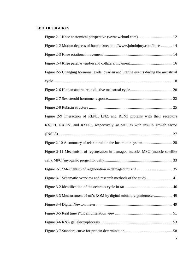
2.2.1 Anatomy of the knee joints
Knee being one of the largest joint in the body is vital for movement. The anatomy
of this joint is reflective of its function in ambulation. The knee joint is a relatively complex
anatomical structure. Knee joint connects thigh bone (femur) to the shin bone (tibia) with
smaller bone that runs alongside the tibia (fibula) and the knee cap (patella) that make the
knee joint. The patella bone is located into the thin anterior wall of the knee joint capsule.
The ligaments, tendons, and capsule are components of joints guard the joint stiffness and
laxity (Schmitz et al., 2013). In addition, knee joint is controlled by a variety of
surrounding connective tissues including ligaments, muscles, tendons, menisci, cartilage,
and bursae to maintain stability.
The ligaments surrounding the knee connect bones and provide stability by limiting
movements and together with menisci and bursae protect the articular capsule. These
ligaments consist of: cruciate ligaments that avoid femur from sliding forward / backward
on the tibia and collateral ligaments that avoid femur from sliding side to side. Collateral
ligaments provide additional stabilization of the knee and direct movement in a correct
direction. They are divided into medial and lateral collateral ligaments that resists knee
rotational movement. The suprapatellar bursae prevents the knee from being pinched during
extension (Gill et al., 2009). Tendons connect the bones to the leg muscles that move the
knee joint. A tendon that extends down from the quadriceps muscle incorporate the patella
bone and attach to the tibia, providing extension at the knee joint. The patellar tendon is
also called patellar ligament because it connects patella to the tibia (Dye et al., 1998).
The elastic cartilage ensures knee movement and protects the bone to slide easily of
the joint surfaces. The menisci protects the ends of bones from rubbing on each other and to
11
effectively deepen the tibial sockets into which the femur attaches. Numerous bursae, or
fluid-filled sacs, help the knee move smoothly (Lewin, 1952). Knee joint consist of
connective tissue with extensive collagen fibres containing cartilage-like cells. Strong
fibres run along the menisci from one attachment to the other, while weaker radial fibres
are interlaced with the former. The joint is bathed in synovial fluid, which is contained
inside the joint capsule. The muscles surrounds the knee consist of the hamstrings,
quadriceps, and calf muscles. These muscles support knee to flex, extend, stabilize, and
work in groups to allow the body to perform important movements such as walking,
running, kicking, and jumping (Amis et al., 2003).
Knee is one of the most distressed joints in the physical activity due to large extreme
of activities and its principal movements (flexion/extension/abduction/adduction). This
joint was mainly designed to support locomotion movement and weight stability of the
body posture. However, it is especially unstable medially and sideways. The knee traumatic
and non-traumatic injuries caused by ligamentous instability can be evaluated to determine
any differences in their stability. Ligamentous instability can be classified as either straight
or rotatory instability (Granchi et al., 2008). Special tests for ligaments assessment exists to
disclose of knee joint function instability. Lachman drawer and pivot-shift test are
performed to elicit cruciate ligaments laxity, while varus/valgus stress test for assessment
of collateral ligaments integrity. These tests are usually performed with the knee in
extension at 90 and 30 degrees (Devan et al., 2004).

12
Figure 2-1 Knee anatomical perspective (www.webmd.com)
There are evidences to suggest that knee laxities are gender specific and the effect
on female knee are greater than male (Hsu et al., 2006; Wojtys et al., 2003).
Additionally, greater knee and ankle laxities have been reported in women as compared
to men (Beynnon et al., 2005). Range of motion (ROM) is defined as a movement
potential of a joint from full flexion to full extension. Many reports have suggested that
hormonal fluctuation could affect knee laxity and subsequently knee range of motion
(ROM). Overuse, age and traumatic injuries can cause structural damage to the knee
that may limit its function. Therefore, a thorough understanding of the anatomy of the
knee is essential to properly diagnose and treat disorders related to knee joint
movements (Herrmann et al., 2013).

13
2.2.2 Physiological control of knee joint movement
The knee joint is an organ, which simultaneously guarantees stability and
movement. The tissues surrounding this joint are highly vascularised and the joint cavity
contains fluid that have important roles in many physiological function and metabolism of
the joint. The fluid or exudates produced by the synovial layer of the tissues distribute
mineral and nutrients to the different parts of the joint. The decrease in synovial blood flow
may cause reduced joint irrigation and may contribute to tissue injury during trauma, where
this may also be related to vasoconstriction (Ar'Rajab et al., 1996).Adequate blood supply
is also important to deliver hormones to their specific receptors in different parts of the
joints which regulates laxity, thus its movement(Junqueira et al., 1986). Knee stability is
also controlled by the nerve innervating the knee joint. Proprioceptors are found in the knee
which sense changes in joint position and the information is relayed via the spinal nerve to
the higher centre in the cerebelum (Marieb, 2009). Input from the higher centre is sent to
the muscles that control knee joint stability which is achieved via varying degree of
contraction of different groups of muscles (Marieb, 2009). Female is more susceptible to
various knee disorders such as arthritis (Kumar et al., 2012). Pathophysiological changes
including degenerative disorders could affect joint metabolism and can cause joint
inflammation. In addition, collagen content of the knee could also be affected therefore
may interfere with knee laxity and joint movements.

14
Figure 2-2 Motion degrees of human
kneehttp://www.jointinjury.com/knee
Figure 2-3 Knee rotational movement
www.faculty.utpa.edu/rafree/res/biomechanics

15
2.2.3 Knee movement and its connective tissue mechanism
Tendons and ligaments are elastic connective tissue surrounding the knee joint that
give support and are vital for knee movements. Knee ligaments connect the thigh to lower
leg bones(Marieb & Hoehn, 2013). Sprains or tears of these ligaments are common in
sports injury. Athletes who participate in direct contact sports such as football or soccer are
more likely to injure their collateral ligaments and anterior cruciate ligament (Dragoo et al.,
2011b). Knee joint relies on these ligaments and the surrounding muscles for stability.
Cruciate ligaments are found inside and collateral ligaments (medial and lateral) are found
on the sides of the knee (Snell, 2011). The cruciate ligaments control back and forth motion
while collateral ligaments control sideway motion of the knee. Tendons are stiff cords of
tissue that connect muscles to bones. The patellar tendon is an important part of the
extensor mechanism of the lower extremity (Snell, 2011). Meanwhile, the role of the
hamstring muscles as a dynamic stabilizer of the knee joint rests in its importance as a joint
compressor and restraining mechanism for anterior motion of the tibia on the femur. These
complexes of connective tissues participate in the control of knee stability during
movement and support it against unusual movement (Marieb, 2009). Cruciate and collateral
ligaments prevent knee anterior-posterior and lateral/medial dislocation respectively. Basic
movement of the knee joint is shown in Figure 2-2 and 2-3 while Figure 2-4 shows the
location of the collateral ligament and patella tendon.

16
Figure 2-4 Knee patellar tendon and collateral ligament
(Dehghan et al., 2013b)
2.3 Menstrual cycle
Menstrual cycle is the changing phase that happens in the uterus and ovary for sexual
reproduction purposes and occurs only in parturient female and other female primates. This
necessary period need to produce oocytes and prepare the uterus for pregnancy (Johnson &
Martin, 2012). The length of the cycle varies greatly among women and fluctuates 25 to 35
days, with 28 days nominated as an average length. Based on the events in the ovary, each
cycle can be divided into three phases; the follicular phase, ovulation, and luteal phase,
which are controlled by normal hypothalamic-pituitary-ovarian axis (Klump et al., 2013).
The menstrual cycle begins from the first day of bleeding and is associated with increasing
amounts of oestrogen in the follicular phase. Approximately mid-cycle, 24–36 hours after
the luteinizing hormone (LH) surges, the dominant follicle releases an egg in an event
called ovulation(Johnson & Martin, 2012). The corpus luteum which is formed post-
ovulation produced large amount of progesterone where under progesterone influence, the
endometrium undergoes changes to the receptive state in preparation for embryo
implantation and the establishment of pregnancy (Marieb, 2009). In an event of no

17
implantation, the corpus luteum degenerates which causes a sharp drop in both oestrogen
and progesterone levels that precede the onset of the next cycle (Sherwood, 2011). Figure
2-5 shows changes in the hormonal profiles (LH, FSH, oestrogen and progesterone), ovary
and endometrium throughout the menstrual cycle.

18
Figure 2-5 Changing hormone levels, ovarian and uterine events during the menstrual cycle
dentistryandmedicine.blogspot.com

19
2.4 Oestrous cycle
Non-primate mammals such as rodents do not display menstrual bleed or menstrual
cycle. In these animals, the reproductive cycle is known as oestrous cycle. In rodent,
oestrous cycle occurs 4 to 5 days, which can be divided into proestrous, oestrus, metestrous
and diestrous (Staley & Scharfman, 2005). Proestrous occurs in the first 12 hours of the
cycle where oestrogen levels peak and was confirmed from the presence of a predominantly
nucleated epithelial cells. Oestrus phase occurs 12 hours following proestrous and is
indicated by the presence of cornified cells in the vaginal smear. Ovulation occurs at the
beginning of oestrus and the end of proestrus phases. Meanwhile, a combined of leucocyte,
cornified and nucleated epithelial cells in the vaginal smear indicate metestrous phase. This
phase occurs for 21-hour period following oestrus. The diestrous phase has the longest
interval time of 57 hours and during this phase, vaginal cells displays predominantly
leucocytes. Corpus luteum activity occur in metestrous and diestrous phases associated with
high progesterone secretion. Due to its short cycle length, rat is a perfect animal model for
investigating changes that occur during the reproductive cycle (Marcondes et al., 2002).

20
Figure 2-6 Human and rat reproductive menstrual cycle
pubs.niaaa.nih.gov
2.5 Overview of Sex-Steroid Biochemistry and Physiology
In females, sex-steroids are produced from the adrenal glands, ovaries and regulate
wide range of physiological functions. The major female sex-steroids are oestrogen and
progesterone, testosterone are also produced in a small amount by the ovary and adrenal
glands. Steroid hormones bind to steroid hormone binding globulin (SHBG) where it is
transported to the target organs through the circulatory system to the specific site of actions.
The binding of sex hormones to SHBG are of different affinity. The free hormones can
leave the circulation and enter into the target cells where they can bind to specific

21
intracellular receptors to initiate the biochemical expression of specific genes (Devlin,
2011).
Free sex hormones, being lipophilic can readily cross the cell membranes and bind to
the intracellular receptors. Steroid receptors are markedly different from gonadotropin
receptors, where the latter are localized in the cell membrane and have several second-
messenger systems as mediators of receptor binding (Marieb, 2009). Following receptor
binding, steroid-receptor complex crosses the nuclear membrane to bind to the nuclear
target which results in the expression of specific genes that can be translated to specific
steroid actions (Devlin, 2011). Steroids regulate largely gene expression at a transcriptional
level. mRNAs are then exported to the cytoplasm, where protein synthesis takes place,
resulting in alterations in cell growth or physiology that are characteristic of the steroid
hormone for that target issue. Figure 2-7 shows the mechanism by which sex-steroid exerts
its intracellular effect.
Upon release into the circulation, the free steroid hormones exert a negative feedback
effect on the pituitary and hypothalamus. At a very high dose however, a positive feedback
effect occur on LH secretion from the pituitary gland, which in turn induces ovulation
(Devlin, 2011). In the second half of the menstrual cycle, oestrogenis also produced by the
corpus luteum (Marieb, 2009). Progesterone which is produced by steroidogenic activity in
the ovary is also a major female sex hormones and is important in the maintenance of
pregnancy (Devlin, 2011). This hormone acts on the reproductive organs, brain, kidney,
lungs and joints especially during pregnancy (Santiago et al., 2001). Elevation of the basal
body temperature following ovulation is due to the thermogenic effect of progesterone in
the hypothalamus. Testosterone is an anabolic hormone which participates in multiple
organs functions in male and is also secreted in a small amount in female mainly by the

22
ovaries and adrenal glands (Cox & John-Alder, 2005). In female, testosterone is involved in
decidualisation while adrenal androgens participate in pubic hair growth and in the post-
menopausal period, is a major precursor for oestrogen (Devlin, 2011).
Figure 2-7 Sex steroid hormone response
(www.thepepproject.net)

23
2.5.1 Sex-steroids and Knee Laxity
Females are exposed to rhythmic rise and fall in the levels of oestrogen and
progesterone throughout the course of the menstrual cycle. These hormones can influence
metabolism of the anterior cruciate ligament (Romani et al., 2003; Yu et al., 1999). Sex
steroids fluctuation during the menstrual cycle has been known to be a risk factor for ACL
injury in females (Slauterbeck et al., 2002). Beside progesterone, oestrogen has also been
reported to have influence on knee laxity. This was based on the findings by Dragoo et al,
(2003) who reported that simultaneous administration of oestrogen and relaxin in guinea
pigs resulted in increased ligament laxity. Meanwhile, in female, testosterone which levels
fluctuates during the menstrual cycle (Dawood & Saxena, 1976) promotes increase muscle
and bone mass and prevent osteoporosis (Cox & John-Alder, 2005; Mooradian et al.,
1987). Testosterone has also been used for postmenopausal hormone therapy in female
(Bolour & Braunstein, 2005).
2.6 Relaxin
Relaxin, a polypeptide hormone which level rises in the second half of the menstrual
cycle increases with a rising level of progesterone. This hormone (with a classical structure
of insulin) interacts with G-protein-coupled receptors (GPCRs) which exist in various
tissues, including the musculoskeletal and non-musculoskeletal systems. Meanwhile, the
presence of relaxin receptor as evidence from relaxin binding assay and immunostaining
has been reported higher in female ACL as compared to male (Faryniaz et al, 2006), which
suggest that female sex-steroids could affect relaxin receptor expression in the ACL.
Relaxin has been widely implicated in the control of knee joint laxity.

24
Relaxin, the mammalian 6-kDa heterodimeric polypeptide hormone, is a member of
the insulin-like superfamily (HISAW, 1926) and consists of seven peptides of high
structural but low sequence similarity. Relaxin is belongs to a family of peptide family
hormones, which is believed to have evolved from insulin early in the evolution of
vertebrates (Bathgate et al., 2006). Relaxin family peptides interact with their receptors,
which exist in various tissues, including musculoskeletal and non-musculoskeletal systems.
The actions of relaxin receptor are mediated by different signalling pathways (Kong
et al., 2010). Relaxin plays an essential role in the biological processes such as metabolism,
growth, pregnancy, and parturition in different species including humans and rodents.
Relaxin circulates in these species during pregnancy emanating from the corpus luteum
(Conrad & Baker, 2013) and placenta (Goh et al., 2013), however temporal pattern of
change and serum concentrations of this hormone are different. In rodents, circulating
relaxin peak concentrations at the end of pregnancy reaches 100 ng/ml, two times greater
than in human (Sherwood OD, 1994). While relaxin plays important role in collagen
catabolism of the pubic symphysis during gestation in lower mammals such as mice and
rats (Samuel et al., 1998), the role of this hormone on pubic symphysis of human is
however unknown (Hashem et al., 2006).

25
Figure 2-8 Relaxin structure
2.6.1 Relaxin roles in different tissues
Several studies have highlighted the therapeutic potential of relaxin for ectopic
pregnancy, male infertility, and heart failure, cardiovascular and musculoskeletal diseases.
Currently, there are seven known relaxin family peptides which are structurally related to
insulin which include relaxin (RLN)1, RLN2, RLN3, insulin-like peptide (INSL)3, INSL4,
INSL5 and INSL6 (Bathgate et al., 2013). RLN1 and RLN2 are strong regulators of
collagen expression and metabolism in fibroblasts, andare differentially expressed in the
corpus luteum, decidua, and endometrium, as well as prostate tissue while RLN3 is specific
to the brain (Sherwood, 2005). RLN1 and RLN2 reconcile the hemodynamic changes
occurring during pregnancy such as cardiac output, renal blood flow, and arterial
compliance (Kirk, 2011) as well as weakening the pelvic ligaments for parturition in
species such as guinea pigs and mice (Sherwood OD, 1993). RLN3 is a highly conserved
neuropeptide in vertebrates, and is involved in a wide range of neuroactivities such as
response to stress and cognition, as well as in neurological disease (Smith et al., 2011).

26
Relaxin binds to relaxin family peptide receptors (RXFP) and exerts its action
through a ligand-receptor system in multiple pathways. The relaxin receptor is involved in
signal transduction between extracellular/intracellular domains. Relaxin1 to 4 hormones are
ligands for the RXFP1, RXFP2, RXFP3, and RXFP4, respectively (Figure 2.8). This family
peptides act on four GPCRs (LGR7, LGR8, GPCR135, and GPCR142) (Kong et al.,
2010).RXFP1 and RXFP2 are composed of large extracellular domains which encompass
of leucine-rich repeats. On the other hand, RXFP3 and RXFP4 proteins are more similar to
small peptide ligands (Summers et al., 2009). Recently, it has been shown that there is a
difference in the ligand binding mode between RXFP1 and RXFP2 (Scott et al., 2012).
RXFP1 and RXFP2 exist in uterus, cervix, vagina, brain, and heart of a number of animal
species. However, production of these proteins differs among tissues of various species.
For example RXFP1 is expressed in rats and mice myometrium (Vodstrcil et al., 2010b),
whereas in human, this receptor is mainly localized to the endometrium (Campitiello et al.,
2011). Moreover, RXFP1 is expressed in the rats and mice heart localized to the atria
where it mediates positive inotropic and chronotropic responses (Piedras-Renteria et al.,
1997), while there is currently no report of this receptor binding or function in the human
heart.

27
Evidence also suggests that the functional domains of RXFP1, the cell type in
which it is expressed, and the ligand used to activate the receptor, all have important roles
in the musculoskeletal system (Figure 2.9). Relaxin alters cartilage and tendon stiffness by
activating collagenase. Relaxin is also involved in bone remodelling process and in healing
of injured ligaments and skeletal muscles (Dragoo et al., 2009; Li et al., 2005). The soft
tissue-healing cascade is composing of three phases: inflammation, regeneration, and
fibrosis. Relaxin is a regulator of both inflammation and fibrosis (Mu et al., 2010). Relaxin
also acts as antifibrotic agent, and favours muscle regeneration and against muscle fibrosis
to promote regrowth of myofibers in skeletal muscle healing.
Figure 2-9 Interaction of RLN1, LN2, and RLN3 proteins
with their receptors RXFP1, RXFP2, and RXFP3,
respectively, as well as with insulin growth factor (INSL3)

28
Figure 2-10 A summary of relaxin role in the locomotor system
(Dehghan et al., 2013b)
2.6.2 Relaxin roles in musculoskeletal tissues
2.6.2.1 Relaxin effects on joints
RXFP1 and RXFP2 receptors orchestrate several activities in joints laxity through
combination with sex-steroids hormones. Relaxin in combination with oestrogens may also
have therapeutic value in the treatment of rheumatoid arthritis (RA) (Ho et al., 2011;
Santora et al., 2005). RA is a chronic and systemic inflammatory disorder that may affect
many tissues and organs, but also causes bone destruction through synovial hypertrophy.
29
However, the incidence and severity of this disease during pregnancy is lower than normal.
During pregnancy, relaxin and oestrogen levels in the serum are elevated leading to
decrease in inflammation in RA patients (D'Elia et al., 2003; Ho et al., 2011). Relaxin
exerts its anti-inflammatory effect through down-regulation of neutrophil function (Bani et
al., 1998) and stimulates leukocyte adhesion and migration in human mononuclear cells
(Figueiredo et al., 2006). A combined treatment using relaxin and oestrogen appears to
reduce circulating tumor necrosis factor (TNF)-α level in rat adjuvant-induced arthritis
model of RA and increased the anti-inflammatory cytokine IL-10 in human
cells.(Figueiredo et al., 2006; Santora et al., 2005). In view of this, relaxin has a potential
beneficial effect in the treatment of synovial diseases.
2.6.2.2 Relaxin effects on bones
Relaxin along with hormones such as oestrogen and growth factors such as TGF-β
help orchestrate the bone remodelling process. These factors regulate a cytokine system
containing three fundamental molecules, the receptor activator of nuclear factor κB ligand
(RANKL), RANK, and osteoprotegerin (OPG). In the RANKL/RANK/OPG system,
RANKL on the preosteoblastic/stromal cells binds to its receptor (RANK) on the
osteoclastic precursor cells and induces expression of a variety of genes to provide the
crucial signal to drive osteoclast recruitment and development (Facciolli et al., 2009). OPG
regulates the system through blocking the effects of RANKL and interfering with RANK
signalling.
Relaxin facilitates differentiation of peripheral blood mononuclear cells (PBMCs)
into mature osteoclasts during osteoclastogenesis by stimulating osteoblastic/stromal cell
production, while oestrogen inhibits this process through increasing OPG production

30
(Facciolli et al., 2009). Therefore, relaxin is one of the osteoclast-activating factors that
increases bone resorption. It is also overexpressed in tumours that promote growth,
differentiation, and invasiveness, which lead to osteolytic metastases (Clezardin & Teti,
2007). Together, these data indicate a possible role of relaxin in osteoclastogenesis
(Facciolli et al., 2009; Ferlin et al., 2010). Relaxin 2 (RLN2) regulates bone metabolism
and proliferation in human osteoblasts. Stimulation of osteoblasts with RLN2 activates
adenylate cyclase and increases cAMP production by G- proteins and thereby increases cell
proliferation (Ferlin et al., 2009). Previous studies have identified an inactivating mutation
in the RXFP2 gene (T222P), which caused idiopathic osteoporosis in young men through
functional osteoblast impairment and reduced bone density (Ferlin et al., 2009). A similar
result was also observed in knockout mouse model (Ferlin et al., 2008). There is also some
evidence to suggest that higher levels of oestrogen and relaxin in pregnant women
correlated with an increased prevalence of congenital dysplasia of the hip (CDH) in
neonates (Saugstad, 1991; Steinetz et al., 2008). In view that relaxin affects both osteoclast
and osteoblast, therefore this hormone is involved in bone remodelling process, and
stimulation of osteoblast by relaxin-2 suggest that this hormone is potentially useful in the
treatment of bone condition such as osteoporosis.
2.6.2.3 Relaxin effects on ligaments
Relaxin hormone alters ligament mechanics due to its collagenolytic effect
mediated by discharge of matrix metalloproteinases (MMPs) (Qin et al., 1997), collagenase
(Wiqvist et al., 1984), and plasminogen activator (Koay et al., 1983). Relaxin treatment in
pregnant cattle increased pelvic width and height (Musah et al., 1986; Perezgrovas &
Anderson, 1982), but not in other joints such as wrist and knee (Marnach et al., 2003;
Weinberg, 1956). Increase in serum relaxin concentration may also correlate with joint
31
laxity (Dragoo et al., 2011b; Lubahn et al., 2006), but this effect during pregnancy is
controversial (Forst et al., 1997). Some studies have reported higher relaxin levels in
pregnant women with pelvic joint instability or hip joint laxity as compared to controls
(Saugstad, 1991; Steinetz et al., 2008), while other studies did not (Ohtera et al., 2002).
Two studies on the relationship between serum relaxin levels and joint laxity reported no
significant association between this hormone level and knee and generalized joint laxity
(Wolf et al., 2013). Studies have also suggested a relationship between higher relaxin and
progesterone serum levels in pregnant females with pelvic girdle pain (PPGP) syndrome
(Kristiansson et al., 1999; MacLennan et al., 1986; Wreje et al., 1995) and pelvic floor
dysfunction (Harvey et al., 2008), whereas other studies have not found such a relationship
(Crelin & Brightman, 1957; Petersen et al., 1994; Vollestad et al., 2012). Study design and
methodological differences may account for some of the conflicting data.
Relaxin appears to play a role in anterior cruciate ligament (ACL) injury (Dragoo et
al., 2009). Oestrogen and relaxin receptors have been found in the human female ACL
(Faryniarz et al., 2006). Studies on the mechanical properties of human ACLs illustrate that
those treated with relaxin have reduced ligament integrity and may be at higher risk of
injury (Dragoo et al., 2011b; Toth & Cordasco, 2001). This finding was also replicated in
an animal model, where rabbits treated with relaxin had significantly weaker ACL’s
compared with controls (Dragoo et al., 2009). Additionally, there was increased anterior
tibia displacement on radiographic assessment, indicating ACL laxity, in animals treated
with relaxin (Dragoo et al., 2009).
There also may be an association between ACL injuries and stages of menstrual
cycle. Occurrence of ACL injuries during the ovulatory phase (midcycle) are more frequent
than the luteal phase (Wojtys et al., 2002). During this period, oestrogen and relaxin levels
32
are high, therefore, activation of the oestrogen and relaxin receptors may be increased (Min
& Sherwood, 1996). Relaxin activates collagenolytic system, which increases collagenase
synthesis and finally degrades the extracellular matrix composition (Garibay et al., 2004;
Guttridge, 2004).
A prospective study of elite female athletes illustrated that players with increased
serum relaxin levels had an increased risk of an ACL tear compared with females with
lower relaxin levels (Dragoo et al., 2011a). Players having a relaxin concentration of
greater than 6.0 pg/ml had more than a 4 times greater risk of ACL injury. Other studies
have collaborated these findings (Beynnon et al., 2006a). Relaxin appears to affect other
ligaments such as volar oblique in perimenopausal women via a receptor-mediated process.
In this ligament relaxin particularly binds and probably reveals in presence of cellular or
extracellular matrix receptors (Lubahn et al., 2006).Taken together, these findings indicate
that while relaxin effects are beneficial to the lower animals especially during pregnancy,
its proposed effect on the peripheral ligament laxity in humans and animals may predispose
the joint to a non-traumatic injury.
2.6.2.4 Relaxin effects on skeletal muscles
Relaxin helps to regulate normal skeletal muscle through two principle-signalling
pathways: adenylate cyclase (AC) and nitric oxide (NO). Relaxin activates the AC
signalling pathway in skeletal muscles through the following signal chain: (Pertseva et al.,
2006; Plesneva et al., 2008; Shpakov et al., 2006; Shpakov et al., 2007; Shpakov et al.,
2004). Relaxin also activates the NO pathway in skeletal muscle via relaxin mediated
activation of receptor tyrosine kinase (Plesneva et al., 2008). NO regulates various
biological processes, and is produced by NO synthase (Stamler & Meissner, 2001). There
33
are data which indicate relaxin stimulates NO synthase signalling in the skeletal muscles of
type 2 diabetic rats, leading to NO dysfunction (Kuznetsova et al., 2010).
Relaxin may be implicated in the skeletal muscle healing process by regulating
inflammation, tissue remodelling and, fibrosis (Formigli et al., 2005; Sherwood, 2005). The
degree of fibrotic response varies with the level of inflammation and injury. Relaxin may
improve spontaneous regeneration of injured skeletal muscle as illustrated in an injured
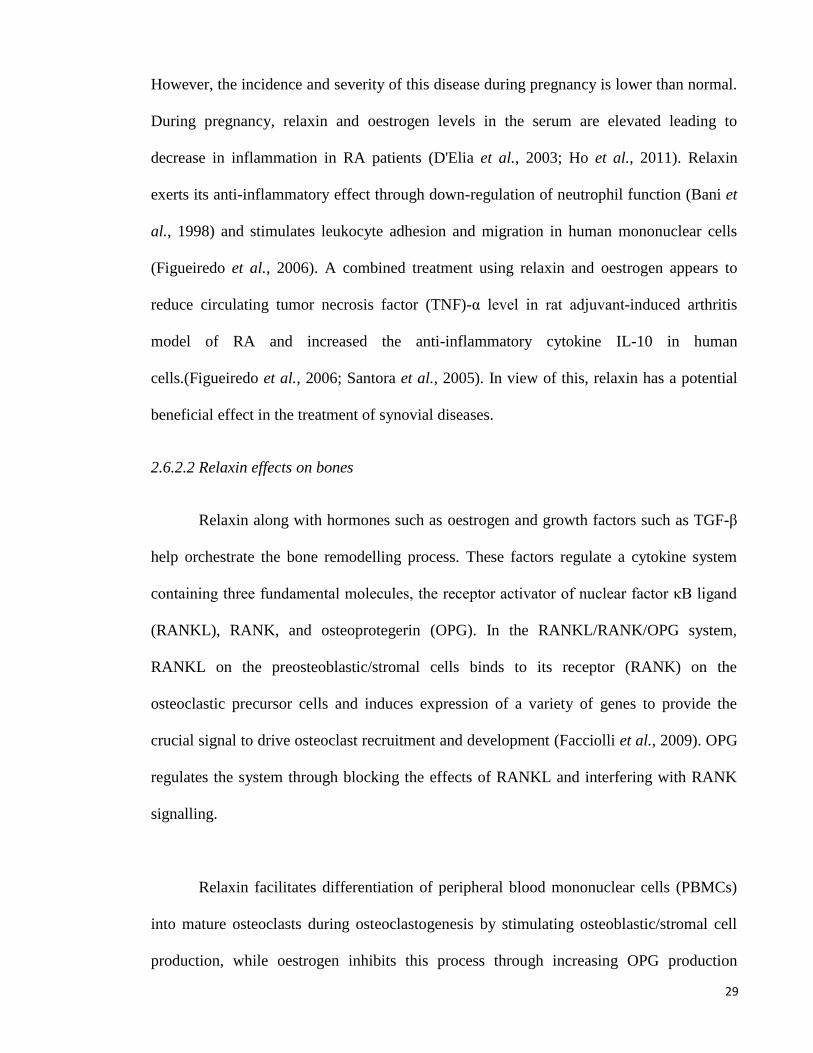
muscle mouse model (Kazumasa et al., 2001; Sato K et al., 2003).During this process,
skeletal muscle cells regenerate and repair to reduce the size of a damaged or necrotic area
and replace it with new living tissue. Degeneration/inflammation is retrogressive changes
in cells and tissues characterized by abnormal structural changes and decreased functions
(Li et al., 2005; Merchav et al., 2005; Mu et al., 2010; Negishi et al., 2005).
Figure 2-11 Mechanism of regeneration in damaged muscle. MSC (muscle
satellite cell), MPC (myogenic progenitor cell)
34
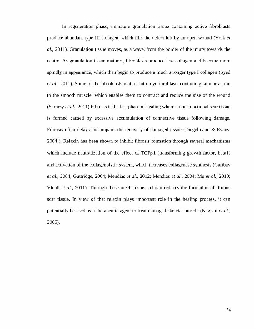
In regeneration phase, immature granulation tissue containing active fibroblasts
produce abundant type III collagen, which fills the defect left by an open wound (Volk et
al., 2011). Granulation tissue moves, as a wave, from the border of the injury towards the
centre. As granulation tissue matures, fibroblasts produce less collagen and become more
spindly in appearance, which then begin to produce a much stronger type I collagen (Syed
et al., 2011). Some of the fibroblasts mature into myofibroblasts containing similar action
to the smooth muscle, which enables them to contract and reduce the size of the wound
(Sarrazy et al., 2011).Fibrosis is the last phase of healing where a non-functional scar tissue
is formed caused by excessive accumulation of connective tissue following damage.
Fibrosis often delays and impairs the recovery of damaged tissue (Diegelmann & Evans,
2004 ). Relaxin has been shown to inhibit fibrosis formation through several mechanisms
which include neutralization of the effect of TGF1 (transforming growth factor, beta1)
and activation of the collagenolytic system, which increases collagenase synthesis (Garibay
et al., 2004; Guttridge, 2004; Mendias et al., 2012; Mendias et al., 2004; Mu et al., 2010;
Vinall et al., 2011). Through these mechanisms, relaxin reduces the formation of fibrous
scar tissue. In view of that relaxin plays important role in the healing process, it can
potentially be used as a therapeutic agent to treat damaged skeletal muscle (Negishi et al.,
2005).

35
2.6.2.5 Relaxin effects on tendons
Relaxin has been reported to effect tendon metabolism by controlling the length of
tendon growth (Wood et al., 2003) and reduce tendon stiffness by increasing tendon laxity
through activation of collagenase. An in-vivo study investigating the growth of rat tails and
human patellar tendons showed that relaxin levels correlate with tendon length (Wood et
al., 2003). Rat tail tendons treated with relaxin exhibited alterations in collagen through
interfering with fibril association and collagen sliding (Wood et al., 2003). Another study
in women with normal menstrual cycle, who did not take any contraception pills,
demonstrated a significant link between serum relaxin levels and patellar tendon stiffness.
Besides, of the reported effects of relaxin on the tendon, potential benefits of relaxin on
tendon repair and remodelling is largely unknown.
2.6.2.6 Relaxin effects on Cartilage
Relaxin appears to decrease knee articular cartilage stiffness (Bonaventure et al.,
1988; Hellio Le Graverand et al., 1998) through induction of collagenase-1, matrix
Figure 2-12 Mechanism of regeneration in damaged muscle

36
metalloproteinase (MMP-1) and MMP-3, which reduces collagen content and expression in
fibrocartilaginous cells. Modulation of MMPs to loss of collagen by hormones may
contribute selectively to degeneration of specific joints fibrocartilaginous explants
facilitated by proteinases (Hashem et al., 2006; Naqvi et al., 2005). The degradation of
extracellular matrix in fibrocartilage is synergized by β-oestradiol. Relaxin exerts its effect
through binding to RXFP1 and RXFP2 receptors. The ratio of RXFP2 in knee meniscus of
pregnant rabbits was shown to be more than RXFP1, which may indicate differential role
of these receptors in the remodelling of fibrocartilage (Wang et al., 2009b). Comparison of
collagen content in articular cartilage of non-pregnant and pregnant rabbits showed that the
total RNA levels and chondrocyte metabolism decreased during pregnancy. Depending on
the level of skeletal maturity, pregnancy can exert both general and specific effects on the
RNA levels in articular cartilage of the rabbit knee. Thus, relaxin may play a role in
women’ susceptibility to musculoskeletal disease. Taken together, these findings suggested
that in female, increased relaxin level may result in undesirable effects on the articular
cartilage.
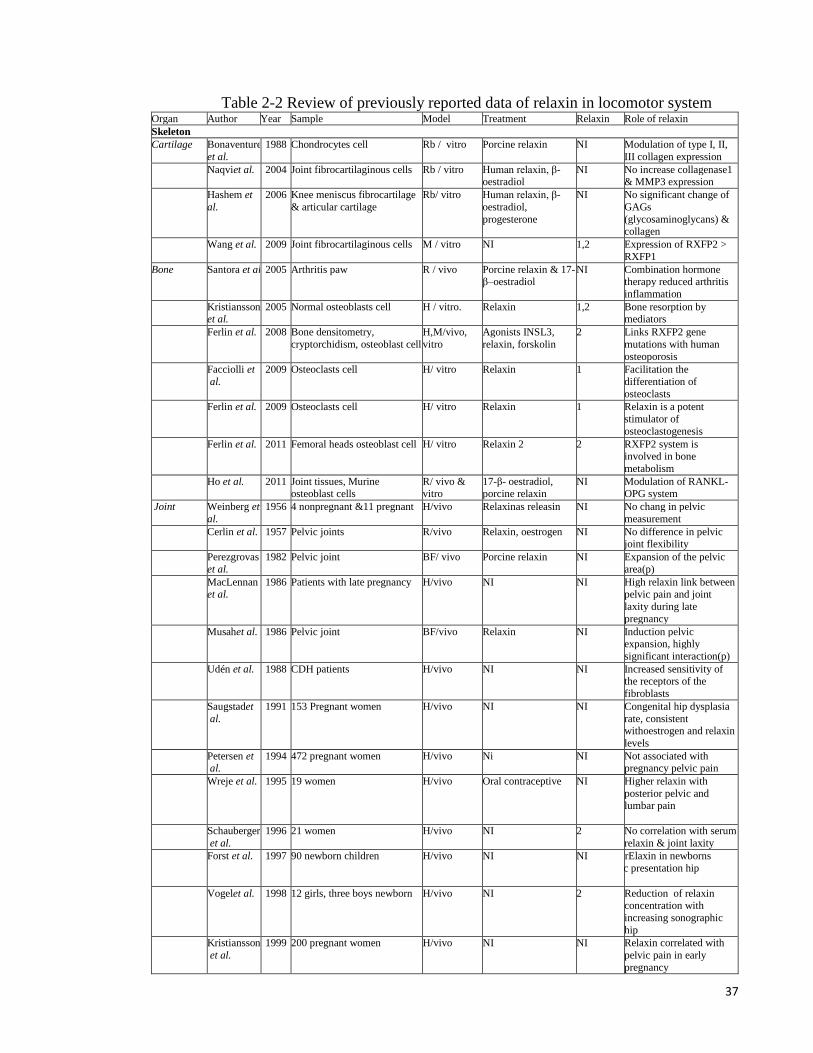
37
Table 2-2 Review of previously reported data of relaxin in locomotor system Organ Author Year Sample Model Treatment Relaxin Role of relaxin
Skeleton
Cartilage Bonaventure
et al.
1988 Chondrocytes cell Rb / vitro Porcine relaxin NI Modulation of type I, II,
III collagen expression
Naqviet al. 2004 Joint fibrocartilaginous cells Rb / vitro Human relaxin, β-oestradiol
NI No increase collagenase1 & MMP3 expression
Hashem et
al.
2006 Knee meniscus fibrocartilage
& articular cartilage
Rb/ vitro Human relaxin, β-
oestradiol,
progesterone
NI No significant change of
GAGs
(glycosaminoglycans) & collagen
Wang et al. 2009 Joint fibrocartilaginous cells M / vitro NI 1,2 Expression of RXFP2 >
RXFP1
Bone Santora et al. 2005 Arthritis paw R / vivo Porcine relaxin & 17-β–oestradiol
NI Combination hormone therapy reduced arthritis
inflammation
Kristiansson et al.
2005 Normal osteoblasts cell H / vitro. Relaxin 1,2 Bone resorption by mediators
Ferlin et al. 2008 Bone densitometry,
cryptorchidism, osteoblast cell
H,M/vivo,
vitro
Agonists INSL3,
relaxin, forskolin
2 Links RXFP2 gene
mutations with human
osteoporosis
Facciolli et
al.
2009 Osteoclasts cell H/ vitro Relaxin 1 Facilitation the
differentiation of
osteoclasts
Ferlin et al. 2009 Osteoclasts cell H/ vitro Relaxin 1 Relaxin is a potent
stimulator of
osteoclastogenesis
Ferlin et al. 2011 Femoral heads osteoblast cell H/ vitro Relaxin 2 2 RXFP2 system is involved in bone
metabolism
Ho et al. 2011 Joint tissues, Murine osteoblast cells
R/ vivo & vitro
17-β- oestradiol, porcine relaxin
NI Modulation of RANKL-OPG system
Joint Weinberg et
al.
1956 4 nonpregnant &11 pregnant H/vivo Relaxinas releasin NI No chang in pelvic
measurement
Cerlin et al. 1957 Pelvic joints R/vivo Relaxin, oestrogen NI No difference in pelvic joint flexibility
Perezgrovas
et al.
1982 Pelvic joint BF/ vivo Porcine relaxin NI Expansion of the pelvic
area(p)
MacLennan et al.
1986 Patients with late pregnancy H/vivo NI NI High relaxin link between pelvic pain and joint
laxity during late
pregnancy
Musahet al. 1986 Pelvic joint BF/vivo Relaxin NI Induction pelvic expansion, highly
significant interaction(p)
Udén et al. 1988 CDH patients H/vivo NI NI Increased sensitivity of the receptors of the
fibroblasts
Saugstadet
al.
1991 153 Pregnant women H/vivo NI NI Congenital hip dysplasia
rate, consistent withoestrogen and relaxin
levels
Petersen et al.
1994 472 pregnant women H/vivo Ni NI Not associated with pregnancy pelvic pain
Wreje et al. 1995 19 women H/vivo Oral contraceptive NI Higher relaxin with
posterior pelvic and lumbar pain
Schauberger
et al.
1996 21 women H/vivo NI 2 No correlation with serum
relaxin & joint laxity
Forst et al. 1997 90 newborn children H/vivo NI NI NI Lower rElaxin in newborns
with pelvic presentation hip
instability
Vogelet al. 1998 12 girls, three boys newborn H/vivo NI 2 Reduction of relaxin concentration with
increasing sonographic
hip
Kristiansson
et al.
1999 200 pregnant women H/vivo NI NI Relaxin correlated with
pelvic pain in early
pregnancy

38
Ohtera et al. 2001 Knee joint of nonpregnant and pregnant
R/vivo NI NI Relaxin preventing the development of joint
contracture
Arnold et al. 2002 Athlete eumenorrheic women and men
H/vivo NI NI No effect on knee laxity
Marnach et
al.
2003 Pregnant women H/vivo NI NI No correlation wrist joint
laxity and relaxin level
Harvey et al. 2008 Pregnant women H/vivo NI NI Higher relaxin and fall significantly faster in
women with PFD
Vøllestad et
al.
2012 212 women pelvic joints H/vivo NI NI Contribution with pelvic
joints laxity but no responses to pain &
disability
Ligament Albert et al. 1997 455 pregnant women H/vivo NI NI No difference in serum relaxin concentration with
pelvic pain
Dragoo et
al.
2003 ACL specimens H/vitro Human recombinant
relaxin
Binding of relaxin female
ACL tissues but not in men ACL
Galeyet al. 2003 ACL specimens H/vitro Human relaxin NI Competence of the ACL
to sequester relaxin
Faryniarzet al.
2006 ACL injuries cells H/vitro NI NI Increased binding of relaxin-labeled in ACL
fibroblasts cellsmore in
female
Lubahn et
al.
2006 Volar oblique
ligament(VOL)
H/vitro NI NI Relaxin binds to (VOL)
perimenopausal women.
Dragoo et al.
2009 ACL specimens G/vivo Porcine relaxin/oestrogen
NI Alternation mechanical properties of the ACL
Dragoo et
al.
2011 Female athletes H/vivo NI 2 Higher relaxin in ACL
tears than without tears
Muscular
Muscle Kuznetsova et al.
1999 Type 1,2diabetes skeletal muscles
R/vitro Insulin/ relaxin/IGF-1
NI No regulation activity of glycogen synthase by
relaxin
Shpakov et al.
2004 Skeletal muscles R/ vitro Inhibitor NI Inhibitor blocked stimulatory effect of
relaxin on adenylate
cyclase
Li et al. 2005 Tibialis anterior muscles M/ vivo Relaxin NI Regeneration and prevention of fibrosis
Merchav et
al.
2005 Internal obturator muscles
withPH
D/vivo NI 1 Pathogenesis role of
relaxin on PH
Negishi et al.
2005 Skeletal muscles, Myoblasts (C2C12 cells)
R/vivo & vitro
Relaxin NI Antifibrosis agent and improve the healing
Shpakov et
al.
2006 Skeletal muscles R/vivo Pig relaxin2 1,2 Relaxin2stimulated the
AC activity
Shpakov et al.
2007 Myoblastic cell line R/ vitro Relaxin 1 Stimulation of adenylyl cyclase activity
Shpakov et
al.
2007 Skeletal muscles R/ vivo Porcine relaxin2 2 Regulation of adenylyl
cyclasesystem under hyperglycemia
Chistyakova
et al.
2009 Skeletal muscles F/ vitro Relaxin 2 Relaxin in increasing G-6-
PDH activity
Kuznetsova et al.
2010 Type 2 diabetes skeletal muscles
R/vitro Streptozotocin NI Stimulation NO synthase in rat skeletal muscles
Mu et al. 2010 C2C12 mouse/ myoblasts
cell , muscle
H,M/vivo,vi
tro
Relaxin 1 Regeneration/increased
angiogenesis/revascularization
Tendon Wood et al. 2003 Tail tendon R/vivo Pentapeptide NKISK,
relaxin
NI Significantly more creep
& changes in tendon
length
Pearsonet
al.
2011 Patellar& medial
gastrocnemius tendon,
H/ vivo NI NI Correlations between
relaxin & patellar tendons
stiffness
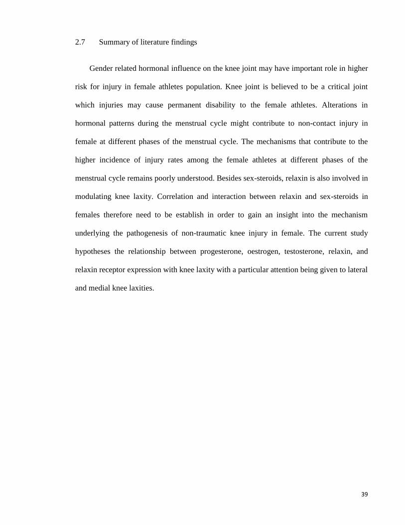
39
2.7 Summary of literature findings
Gender related hormonal influence on the knee joint may have important role in higher
risk for injury in female athletes population. Knee joint is believed to be a critical joint
which injuries may cause permanent disability to the female athletes. Alterations in
hormonal patterns during the menstrual cycle might contribute to non-contact injury in
female at different phases of the menstrual cycle. The mechanisms that contribute to the
higher incidence of injury rates among the female athletes at different phases of the
menstrual cycle remains poorly understood. Besides sex-steroids, relaxin is also involved in
modulating knee laxity. Correlation and interaction between relaxin and sex-steroids in
females therefore need to be establish in order to gain an insight into the mechanism
underlying the pathogenesis of non-traumatic knee injury in female. The current study
hypotheses the relationship between progesterone, oestrogen, testosterone, relaxin, and
relaxin receptor expression with knee laxity with a particular attention being given to lateral
and medial knee laxities.

40
3 CHAPTER 3 MATERIALS AND METHODS

41
Figure 3-1 Schematic overview and research methods of the study

42
3.1 Materials
3.1.1 Animal
All procedures involving experimental animals were approved by the Faculty of
Medicine Animal Care and Use Committee (ACUC), University of Malaya (UM) with
ethics number: FIS/22/11/2011/FD(R). Adult female WKY rats weighing 180-220 grams,
obtained from the Animal House, Faculty of Medicine, UM were caged in a group of six, in
a clean and well ventilated standard environment of 12 hours light: dark cycle. The animals
had free access to soy-free diet (Gold Coin Pellet) and tap water ad libitum.
3.1.2 Chemicals and consumable
The chemicals and consumables used in this project are shown in Table 3.1.

43
Table 3-1 Chemicals and consumable used in this project No Chemicals Company
1 Progesterone (100 mg, purity minimum 99 %) Sigma, St. Louis, MO
2 Testosterone (5 g,purity > 99 %) Sigma, St. Louis, MO
3 17 Beta-oestradiol Sigma, St. Louis, MO
4 Relaxin Sigma, St. Louis, MO
5 Peanut oil (P2144, 1L) Sigma, St. Louis, MO
6 Absolute Ethanol (purity > 99 %) Sigma, St. Louis, MO
7 Pro-Prep protein extraction solution Intron Biotechnology, Korea
8 Micro BCA protein assay Kit Thermo Scientific, Rockford,
USA
9 7α,17β-[9-[(4,4,5,5,5-Pentafluoropentyl)sulfinyl]nonyl]estra-
1,3,5(10)-triene-3,17-diol (ICI 182/780) purity ≥ 98 %
Santa-Cruz Delaware CA, USA
10 4-[2-phenyo-5,7-bis(trifluoromrthyl)pyrazolo(1,5-a)
pyrimidin-3-yl] phenol (PHTPP) purity 99.8 %
Tocris, Bioscience USA
11 1,3-bis(4-hydroxyphenyl)-4-methyl-5-[4-(2-
piperidinylethoxy)phenol]-1N-pyrozole dihydrochloride
(MPP) purity 99.8 %
Tocris, Bioscience USA
12 Mifepristone purity 99.8 % Tocris, Bioscience USA
13 Flutamide purity≥ 98 % Sigma, St. Louis, MO
14 Finasteride purity ≥ 98 % Sigma, St. Louis, MO
15 Beta Actin antibody Abcam, UK
16 RXFP1 antibody Abcam, UK
17 RXFP2 antibody Abcam, UK
18 4CN optic substrate kit Bio Rad, Alfred Nobel Drive
Hercules, CA, USA
19 Poly Vinylidene fluoride membrane (PVDF) Bio Rad, Alfred Nobel Drive
Hercules, CA, USA
20 Bovine Serum Albumin (BSA) Innovative, Peary Court Novi,
Michigan, USA
21 Spectra multicolor broad range Fermentas, USA
22 Tetramethylenediamine (TEMED) Sigma, St. Louis, MO
23 RNA later Ambion,Lincoln Center drive,
USA
24 RNase Free water Ambion,Lincoln Center drive,
USA
25 Micro 96 well plate and cover Applied Biosystems, USA
26 High capacity RNA to cDNA Applied Biosystems, USA
27 Fast advanced Master Mix (Taq man) Applied Biosystems, USA
28 RNA primers assays Applied Biosystems, USA
29 Rat Enzyme-linked immunosorbent assay (ELISA) kit CUSABIO, USA
30 Human Enzyme-linked immunosorbent assay (ELISA) kit CUSABIO, USA

44
3.1.3 Sterilization
All experiments matherials (plastic tips, collection tubs, glass disposables) were
sterilized by autoclaving at 1.05 kg/cm2 (15 psi) on liquid cycle for 20 min. Distilled water
(dH2O) and ultrapure water (Mili-Q or ddH2O) were used for critical parts of the study.
3.2 Methods
3.2.1 Ovariectomy surgical procedures
Ovariectomy is used to omit animal’s internal steroid hormones. To remove
animal’s ovary, rats were anaesthetized with ketamine (80 mg/kg) and xylazine (8 mg/kg).
Ketamine is a hydrochloric salt and used for general anesthesia in combination with a
sedative such as xylazine. Xylazine is a drug that used for relaxation, painkilling, and
general anesthesia in animals. The combination of these two drugs were injected
intraperitoneally (IP). After injection of ketamine and xylazine rat was placed in left lateral
position. Before starting, the rat’s operation area was shaved and swabbed with 70 %
ethanol to remove filth. Small incisions were made in skin and muscle; ovary can be seen
between fats in the peritoneal cavity. The ovaries were removed and remaining part was
returned into the peritoneal cavity. Then incision parts of muscle and skin was sutured.
Ovariectomy operation was completed by dorso-lateral incisions, with same method for
opposite side (Parhizkar et al., 2008). Two weeks after surgery, rats were prepared to
continue into further experiment.

45
3.2.2 Animal preparation and administration of sex steroid hormone
In first part of the study, rats were ovariectomizedtwo weeks prior to steroid
treatments. Based on the treatment, animals were distributed into different groups (n = 6per
group). Hormones were injected subcutaneously at the neck cuff with a 24 hours interval
for 3 days consecutively. Antagonists were also administrated subcutaneously at the neck
cuff, 30 minutes before the hormone injection. For transcriptome and proteomic studies
expression level of relaxin receptor isoforms (RXFP1 and RXFP2) mRNA and protein
were detected in the hamstring muscle, patellar tendon, and collateral ligament tissues.
Control group, which received peanut oil (vehicle), was also treated for three consecutive
days. The drugs were dissolved in peanut oil and were subcutaneously administered.
3.2.3 Identification of oestrous cycle stages
Vaginal secretions were collected by using a plastic pipette filled with 10 μL of
normal saline (NaCl 0.9%). The tip of the pipette was inserted into rat vagina, but not
deeply to avoid cervical stimulation. Unstained material was placed onto a slide and was
observed under a light microscope. The proportion of different cells was used to determine
oestrus cycle phases, in which round and nucleated cells are epithelial cells which define
proestrus; irregular shapes without nuclei are the cornified cells that were observed during
estrus; little round cells are the leukocytes that characterize diestrus. At metestrus, however,
three different kinds of cells could be identified

46
Figure 3-2 Identification of the oestrous cycle in rat
3.2.4 Animal study groups
3.2.4.1 Oestrogen treated group
- Control group
- 4 doses of oestrogen (0.2, 2, 20, 50 g/kg)
- 7α,17 β-[9 - [(4,4,5,5,5-Pentafluoropentyl)sulfinyl]nonyl] estra - 1, 3, 5(10) - triene-
3,17 - diol (ICI 182/780) (25 mg/kg)
- Physiological dosage of oestrogen (0.2 g/kg) + ICI 182/780
- 4 - [2 – phenyo - 5, 7 – bis (trifluoromrthyl) pyrazolo (1, 5 - a) pyrimidin – 3 -
yl]phenol (PHTPP), an ER - blocker (25 mg/kg), as internal control
- Physiological dosage of oestrogen + PHTPP (25 mg/kg)
- 1, 3 – bis (4 - hydroxyphenyl) – 4 – methyl – 5 - [4 - (2 -piperidinylethoxy) phenol]
- 1N - pyrozole dihydrochloride (MPP), an ER - blocker (25 mg/kg) as internal
control
- Physiological dosage of oestrogen + MPP (25 mg/kg)
3.2.4.2 Progesterone treated group
- Control group
- Physiological dosage of progesterone (4 mg/kg)
- Mifepristone (7.5 mg/kg)as internal control
- Progesterone (4 mg/kg) + Mifepristone (7.5 mg/kg)
3.2.4.3 Testosterone treated group
- Control group

47
- 2 doses of testosterone (125 and 250 g/kg)
- 2 doses of testosterone (125 and 250 g/kg)
- 2 doses of testosterone + Flutamide (10mg/kg)
- Flutamide (10 mg/kg) as internal control
- 2 doses of testosterone Finasteride (20 mg/kg)
- Finasteride (20 mg/kg) as internal control
3.2.4.4 Oestrous cycle
- Prostrous
- Estrus
- Metestrous
- Diestrous
3.2.5 Investigation of blood serum hormone level
Blood samples were collected into a separator tube (SST) and allowed to clot for 30
minutes at room temperature. The samples were centrifuged at 3000×g, for 15 minutes.
Serum samples were aliquoted and stored at -20° C. Serum samples were analysed in
duplicate for relaxin by using enzyme-linked immunosorbent assay (ELISA) kit
(CUSABIO - USA). ELISA was performed according to manufacturer's guidelines.
Absorbance for relaxin was determined by using a microplate reader (iMark; Bio - Rad,
Hercules, CA, USA) at a wavelength of 450 nm. A set of standard serial dilutions of known
concentrations were provided by the manufacturer and were used to construct a standard
curve to determine the hormone levels. The overall intra-assay percent coefficient of
variation for rats were 150, 75, 37.5, 18.75, 9.4, 4.7, 2.4, 0 ng/ml.Standard curves were

48
created by serial dilution and hormones concentration in the samples were determined from
the standard curve.
3.2.6 Knee range of motion (ROM)
Rat’s knee ROM was measured using digital miniature goniometer. A day after the
last day of drug treatment, the rats were anesthetized using ketamine and xylazine (80 + 8
mg/kg). The depth of unconsciousness was confirmed from the lack of response to painful
stimuli, which was usually applied at the plantar surface of the foot (Flecknell, 2009). It is
important to maintain the animal in deep anaesthesia in order to prevent active muscle
contraction in response to pain which will increased resistance towards passive traction
(Bazin et al., 2004). The hip and knee joints were fixed and rest on the sensor. Meanwhile,
the lower leg (knee up to the ankle) was tied in-situ to the device arm. Rat knee ROM was
measured by pulling the device arm in a clockwise direction at a minimum constant force
(12 ± 1 N) using a mini digital Newton meter (American Weight, USA; model: AMW – SR
- 1KG). Once the force exceed 13 N, the traction applied was immediately terminated and
the angle obtained was recorded which represents passive extension. Assessment of the
angle was made in different groups of treatment. This device and its function are shown in
Figure 3.3.
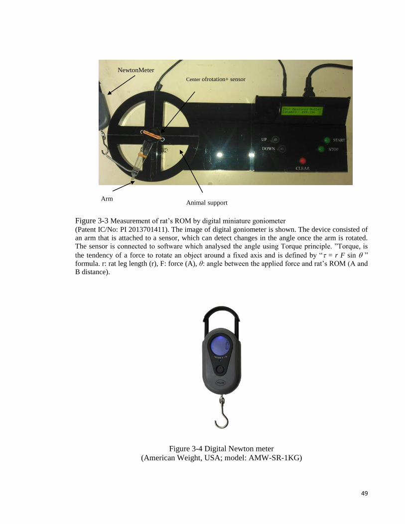
49
Figure 3-3 Measurement of rat’s ROM by digital miniature goniometer
(Patent IC/No: PI 2013701411). The image of digital goniometer is shown. The device consisted of
an arm that is attached to a sensor, which can detect changes in the angle once the arm is rotated.
The sensor is connected to software which analysed the angle using Torque principle. ”Torque, is
the tendency of a force to rotate an object around a fixed axis and is defined by “ = r F sin ”
formula. r: rat leg length (r), F: force (A), θ: angle between the applied force and rat’s ROM (A and
B distance).
Figure 3-4 Digital Newton meter
(American Weight, USA; model: AMW-SR-1KG)
Arm
NewtonMeter
Center ofrotation+ sensor
Animal support

50
3.2.7 Real Time PCR (qPCR)
3.2.7.1 Real Time PCR, principle
The Polymerase Chain Reaction (PCR) is a process for the amplification of specific
fragments of DNA / RNA. Real time PCR is a specialized technique that allows a PCR
reaction to be visualized “in real time” as the reaction progresses and known as a sensitive
technique to calculate gene expression in the data that were collected during the PCR
amplification process. Quantitative PCR (qPCR) is used in variation of molecular biology
studies such as Gene expression analysis: Cancer and Drug research, Disease diagnosis
(Viral quantification), Food testing, animal and plant breeding (Gene copy number), and for
determining of DNA / RNA interfering (Palmer et al., 2003). Although in normal PCR, the
product can only measure at the end of process, in qPCR it can be measured while it occurs.
The development of fluorogenic labelled probes also made it possible to eliminate post-PCR
processing for the analysis of probe degradation. This method able to detect small difference
between samples compare to other methods, however it needs high cost reagents and equipment
(Wong & Medrano, 2005).
Reverse transcription-quantitative of mRNA or Real-time can be achieved as a one-
step or two-step procedure. The most common method for gene expression investigation is
two-step RT-qPCR. With one-step method, the reverse transcription and PCR amplification
steps are performed in a single buffer system while two-step procedure is performed in two
separate reactions that allow storage of cDNA for long time (Wong & Medrano, 2005).
Two-step RT-qPCR was performed in this study. There are two different types of methods
for detection of PCR: TaqMan and SYBR Green. Each method has its limitations and
benefits. TaqMan chemistry (known as fluorogenic 5´ nuclease) enables you to perform

51
multiplex PCR, SYBR Green method (highly specific, double standard DNA binding) has
only Quantification probe. TaqMan assay was used in this study. Real time PCR
amplification is shown in Figure 3.5.
3.2.7.2 Real-time PCR demonstration
3.2.7.3 Sample collection and Tissue processing for RNA isolation
Patellar tendon and collateral ligament tissues were exposed after 3 days treatment
and from the rat knee washed with PBS buffer and soaked in five volume of RNA Later.
RNA later solution was used to stabilize and protect tissues cellular RNA (Ambion, L/N:
1206029). Samples can be preserved in RNA Later solution at – 20 ° C or below for up to
2 years. Total RNA was extracted from 30 mg tissues (wet weight) that were floating into
the RNA later solution using the RNeasy Fibrous tissue Mini kit (QIAGEN, Germany).
Figure 3-5 Real time PCR amplification view

52
3.2.7.4 RNA extraction
RNeasy® Fibrous Tissue kit (QIAGEN, Germany) specific kit for fibre-rich tissues
was used to isolate RNA from hamstring muscle, patellar tendon, and lateral collateral
ligament. The kit contains are: Buffer RLT, Buffer RW1, Buffer RPE, RNase-Free Water,
Proteinase K, RNeasy Mini Spin Columns, Collection Tubes (1.5 ml), RNase-Free DNase I
(lyophilized), Buffer RDD. The tissues were retrieved from RNA later solution with
sterile forceps and drip-dried immediately with an absorbent paper towel, then soak into the
lysis RLT Buffer added with β-mercaptoethanol. A 30 mg (wet tissue) was homogenized
using a rotor–stator TissueRuptor (230V, 50-60 Hz_ QIAGEN, Germany). Homogenate
was incubated at 55°C for 20 minutes with added 590 μl RNase-free water and 10 μl
proteinase K solution to the lysate. The supernatant transferred after Centrifuging (20 –
25°C for 3 minutes) to the new tube and then 0.5 volumes (usually 450 μl) of pure ethanol
added to cleared lysate. It was mixed well by pipetting and Transferred 700 μl of the
sample, to the RNeasy Mini spin column placed in a 2 ml collection tube (2 times), by
discarding the flow through. After washing with RW1 buffer, mixed of 10 μl DNase I stock
solution and 70 μl buffer RDD was added directly to the RNeasy spin column membrane to
remove DNA. DNase I is sensitive to physical denaturation. Mixing should carry out by
gently inverting the tube. This mixture was incubated for 20 minutes at room temperature
(20 – 30°C). The membrane then was washed twice with 500 μl of RPE washing buffer.
RNA was collected by Placing of RNeasy spin column in a new 1.5 ml collection tube and
adding 30 – 50 μl RNase - free water.

53
3.2.7.5 RNA quality control
The pure isolated RNA may be contaminated by DNA, protein or phenol that could
inhibited the RNA downstream solicitation. Therefore, evaluating of RNA integrity and
quality is necessary. The purity and concentration of RNA was evaluated by A260/280,
A260/230 UV absorption ratios (Gene Quant 1300, UK). Agarose gel is the most common
way to evaluate RNA integrity. Each RNA sample was assessed by integrity of Ethidium-
Bromide agarose gel 1 % and evaluated by electrophoresis in TBE buffer. After run the
agarose gel for 45 minutes in 90 V, gel was observed in the specific gel documentation
system (Vilber Lourmat, Fisher Scientific). RNA sharp and clear bands can be seen in 18S
and 28S rRNA. To catch the RNA bands location 1Kb DNA ladder was used.
Figure 3-6 RNA gel electrophoresis
3.2.8 RNA to cDNA converssion
To perform the two-step Real time PCR method, RNA samples need to be converted
to cDNA. Reverse transcription into cDNA was performed by using Two-Step qRT-PCR
kit, High capacity RNA to cDNAby Applied Biosystems_US. This kit contains reagents for
reverse transcription (RT) buffer and enzyme to convert total RNA to single-stranded

54
cDNA. According to the manufacturer’s guidelines total RNA converted to cDNA by
adding of up to 9 μl RNA (1000 ng), 10 μl of RT buffer, and 1 μl of RT enzyme. To
perform reverse transcription of converting, optimized program the thermal cycler
conditions was used. The program was included 37°C for 60 minutes, 95°C for 5 minutes
and hold in 4°C (Kwok & Higuchi, 1989). The total tube of cDNA can be stored in –80 ° C
for long term storage.
3.2.9 Real time PCR running
After converted total RNA to cDNA and their quantification, two-step Real time
PCR was run to evaluate the mRNA gene expression by following the manufacturer
procedures. The amplified region of cDNA was tagged with fluorescence-labeled probe.
The specificity of the primer and the probe ensures that the expression of the target DNA
was specifically evaluated. Real time PCR does not require a time consuming post
amplification gel electrophoresis due to its high sensitivity. TaqMan probe has a sensitivity
of 100 % and a specificity of 96.67 % (Tsai et al., 2012) and is capable of detecting as few
as 50 copies of RNA/ml and as low as 5 - 10 molecules (Hofmann-Lehmann et al., 2002).
All reagents including probes and primers were obtained from Applied Biosystems,
USA. Primers were designed by the same company for specific targets: RXFP1:
Rn01495351; Lot no: 926762, and RXFP2: Rn01412901; Lot no: 651878, amplifies 116 bp
segment of RXFP1 from the whole mRNA length of 2277 bp, and 138 bp segment of
RXFP2 from the whole mRNA length of 2214 bp. Hprt1 and GAPDH were used as
reference genes. Expression levels of Gapdh andHprt1 in each sample were determined for
normalisation as housekeeping genes. The catalogue number for the housekeeping genes
are: GAPDH, Rn99999916_S1, Lot no: 10377343, which amplifies 87 bp segment of

55
GAPDH from the mRNA length of 1307 bp and Hprt1, Rn01527840, Lot No: 1118680,
which amplifies 67 bp, segment from the mRNA length of 1260 bp. The target assay was
validated in-silico using whole rat genome sequences and in - vitro using whole rat cDNA
sequences to ensure target sequences were detected (Applied Biosystems, USA). Nuclease-
free water was used throughout Real-time PCR and qPCR experiments.
To carry out the reaction combination 50 ng of cDNA in 1 µl, 5 µl of master mix
buffer, 1 µl of each target primer, and 3.5 RNase free water was loaded in 96 well plates.
The total volume of PCR mixture was 10 µl in each well. All experiments samples were
loaded in three biological replicates. After prepared the reaction plate, plate was loaded into
the Applied Biosystem Step One Plus thermal cycler. Real time-PCR program was set
according to manufacture protocol included 2 minutes at 50ºC reverse transcriptase, 20
seconds at 95ºC activation of polymerase, denaturation at 95ºC for 1 second and annealing
at 60ºC for 20 seconds. Denaturing and annealing step was performed for 40 cycles.
3.2.10 Data Analysis for Quantification of Gene Expression
Data was analysed according to Comparative fold changes in expression were
method (Wong & Medrano, 2005):
Δ Ct = Ct of target gene – Ct of housekeeping genes average
ΔΔ Ct = ΔCt of treated samples – Δ Ct of non-treated samples
Fold changes = Ct (2-ΔΔCt
)
Amplifications of the target and reference genes were measured in the samples and
reference. Measurements were standardized using the GenEx software. The relative
quantity of target in each sample was determined by comparing normalized target quantity

56
in each sample to normalized target quantity average in the references. Data Assist v3
software from Applied Biosystems was used to calculate RNA folds changes.
3.2.11 Protein expression by Western blotting
3.2.11.1 Western blotting- principle
Western blotting or protein immunoblot is an analytical technique that is commonly
accepted to detect specific proteins in the total extracted protein from tissue or cell lines.
This method separates the protein by running the gel electrophoresis and identifies with
specific antibodies according to their size. The bands blot is on a membrane (nitrocellulose
or PVDF). The first step in western blotting is running the gel electrophoresis to separate
the total protein, then antigen was transferred to the blotting membrane electrophoretically.
In this study PVDF (polyvinylidene difluoride) membrane was used. In the next step,
protein was transferred to PVDF membrane; the remaining protein site was blocked to
reduce nonspecific bands. After blocking, membrane was soaked into the primary and
secondary antibodies to bind and specify the target protein. Finally, the target protein was
visualized with incubation of membrane into the substrate detection reagent (Walker,
1996).
3.2.11.2 Sample collection and Tissue processing for protein extraction
Hamstring muscle, patellar tendon, and collateral ligament tissues were exposed and
immediately frozen in liquid nitrogen (- 196°C) and then stored at – 80°C prior to protein
extraction.

57
3.2.11.3 Protein extraction
Total protein was extracted with using PRO-PREP (Intron, UK) solution kit. This
kit is able to isolate protein from whole cell of tissue. This solution included is of highly
denature ionic detergent such as sodium dodecyl sulphate (SDS), lithium dodecyl sulphate
and sodium deoxycholate that can isolate protein as a monomeric for any protein molecular
weight analysis and western blot. It also contain zwitterioninc detergent CHAPS (3 - [(3 -
Choamidopropyl) dimethylammonio] - 1 - propanesulfonate) which is more effective in
disrupting protein interaction. Total protein was isolated by following the protocol:
1 - 30 mg of hamstring muscle (wet tissue) was separated and soaked into the 400 µl of
extraction solution. Whole part of patellar tendon and collateral ligament were cut and
placed into the 250 µl into the solution. The maximum weight of patellar tendon and
collateral ligament were 15 mg.
2- The tissues were homogenised with using an ultrasonic cell disruptor (Branson) by
putting on ice to disrupt tissues.
3- After 10 minutes homogenising; samples were incubated at – 20oC for 30 minutes to
persuade cell lysis. This step was performed two times to increase protein concentration.
4- The samples were centrifuged 15 minutes, at 4°C, 13000 rpm to separate supernatant.
The supernatant then transferred into the new 1.5 tube and stored in – 20°C.
3.2.11.4 Protein quantification
Protein quantification was performed to determine samples concentration by using
Micro BCA Protein Assay kit (Thermo Scientific™ - US). This kit contains Micro BCA
Reagent A (MA), Micro BCA Reagent B (MB), Micro BCA Reagent C, andBovine Serum
Albumin (BSA) as Standard Ampules at 2.0 mg/mL in a solution of 0.9 % saline and 0.05

58
% sodium azide. The Kit is a detergent-compatible bicinchoninic acid formulation for the
colorimetric detection and quantitation of total protein.A purple-colored reaction product is
formed by the chelation of two molecules of BCA with one cuprous ion (Cu + 1
). This
water-soluble complex exhibits a strong absorbance at 570 nm wavelength that is linear
with increasing protein concentrations. The samples concentration were calculated from a
linear plot of BCA standard concentration (Axis X) of the intensity by changing of colour
in absorbance. This absorbance obtained from the reaction between BSA and BCA
reagents. Standard concentrations of BSA were prepared at 10, 8, 6, 4, 2, 1, 0.5, and 0
µg/ml. The samples then were diluted 1 in 50 with PBS buffer. 50µl of total protein
samples or standard as a serial dilution were added to 50 µl of BCA reagents in a Greiner
UV Transparent 96 Well Plate (Thermo Scientific™ - US) in triplicate. The plate was
incubated for 2 hours at 37 ° C, then the intensity of changed colours were qualified by
using a plate reader at wavelength of 570 nm. Standard curve was generated by serial
dilution and samples protein concentration calculated from the standard curve.
Figure 3-7 Standard curve for protein determination

59
3.2.12 Western blot running
SDS-PAGE running: Electrophoresis gel or SDS-PAGE gel (sodium dodecyl sulfate
polyacrylamide gel electrophoresis) can be one-dimensional or two-dimensional technique
to detect and characterize of specific protein. One-dimensional electrophoresis is used for
most protein and nucleic acid separations. Relocation of protein in electrophoresis is based
on charges, mass, size and strength of electrical field (Bolt & Mahoney, 1997).
The matrix is polyacrylamide gel that is polymer of acrylamide and bis- acrylamide
(Sigma-US), the presence of N, N, Nʹ, Nʹ - tetramethyleethylenediamine (TEMED), and
ammonium per sulphate (APS), catalyses the gel polymerization (Walker, 2007).
According to the protein size, samples separate within the gel and determine by their
molecular weight. High percentage of gel for low molecular weight and low percentage for
high molecular weight is recommended. Ionic detergent SDS is used in polyacrylamide gel,
which denatures the protein and makes them negatively charged. The charge is equally
distributed to protein molecule based on their length and lead to migration according to the
size.
Polyacrylamide gel contain of two different parts (resolving and stacking) with
separate formulation. Resolving part used at the bottom of gel caster with a pH of 8.8 and
stacking part is placed above the resolving with a pH of 6.8. In this study, resolving gel
with 12 % for (RXFP1, RXFP2, and Beta Actin) was used. Stacking gel is used to pack
protein together after loading and mostly prepare in 4 %. The gel was prepared in gel cast
and kept overnight at 4°C. The compositions of each gel are shown in Table 3.2.

60
Table 3-2 Composition of 12 % resolving and 4 % stacking gel
Stock solution Resolving gel 12 % Stacking gel 4 %
Acrylamide 8.3 ml 0.665 ml
4X resolving 4 ml _
4X Stacking _ 1.25 ml
10 % SDS 0.2 ml 50 µl
10 % APS 0.1 ml 25 µl
TEMED 7 µl 5 µl
Double distilled H2O 6.4 ml 3 ml
The following day equal volume of samples (20 µg) were mixed with loading dye,
then boiled for 5 minutes and loaded in the gel. Gel cast then placed inside the
electrophoresis tank and bathed in migration buffer with the recommended time as
instructed by the manufacturer. Pre-stained protein marker was loaded into the one of the
empty wells to assess protein size bands. Rabbit polyclonal Beta Actin (Abcam, UK) was
used as loading control, to check whether all samples have been loaded equally, and
compare the expression of different samples. Electrophoresis was disconnected when the
front dye reached at the bottom of the gel.
Transfer to membrane: The separated samples by electrophoresis, the protein transferred
to PVDF (BIORAD, UK) membrane for immunoblotting. A hydrophobic membrane needs
to prewet into pure methanol (2 minutes) and then into the twobin buffer (5 minutes) which
contain 20 % methanol. The presence of methanol in twobin buffer (transfer buffer) is to
promote dissociation of SDS from the protein and improve adsorption of protein onto the
membrane (Pettegrew et al., 2009). The PVDF membrane was placed next to the gel. The
two were sandwiched between absorbent materials, and clamped between solid supports to

61
maintain tight contact between the gel and membrane without air bubbles. Samples
transformation can perform in wet or semi-dry conditions. In this study, wet condition tank
is used. The gel membrane sandwich was placed in wet transfer tank for electro blotting at
constant voltage of 100 V for 90 minutes. After transferred membrane was exposed and
washed with PBST (phosphate buffer saline-tween 20) for 3 times, 5 minutes.
Blocking step: To block the membrane, two blocking solutions were traditionally used:
non-fat milk or BSA (5 %). Milk is cheaper but is not recommended for studies of
phospho-proteins, it cause high background because the phospho-specific antibody detects
the casein present in the milk. BSA was used to block the membrane 90 minutes at 4°C
under agitation. Washing with PBST 3 times, 5 minutes is necessary on this step before
incubation with primary antibody.
Incubation with antibodies: primary antibody was diluted 1:1000 in PBST buffer and
incubated with membrane 60 minutes at 4°C under agitation. After washing with PBST
buffer, secondary antibody (attached to horseradish peroxidase-HRP) was diluted 1:2000
and incubated for 60 minutes under agitation.
Visualizing:tovisualize the target protein band Opti - 4CN TM
Substrate kit was used. This
kit is colorimetric (HRP) substrate and detected band can be seen by necked eyes. Primary
and secondary antibodies that used in this study are shown in Table 3.3.

62
Table 3-3 primary and secondary antibodies that have been used in western blot.
Target protein Primary antibody Secondary anti body
RXFP - 1/LGR7 Rabbit polyclonal Mouse anti-rabbit HRP
RXFP - 2/ LGR8 Mouse polyclonal Rabbit anti-mouse HRP
Beta Actin Rabbit polyclonal Goat anti-rabbit HRP
3.2.13 Western blot data Analysis
The visualized blots of target protein, membrane were captured by a gel
documentation system (Vilber Lourmat, from Fisher Scientific, USA). The density of each
band was determined using Image J software (National Institutes of Health, USA). The
ratio of each target band over Beta actin was calculated and considered as the expression
level of the target proteins.
3.3 Human study
Fortyhealthy females (20 athletes; 20.3 ± 1.28 years, 163 ± 2.75 cm height and 20
non-athletes; 21.7 ± 2.27 years, 158.5 ± 5.28 cm heights) who were not on hormonal
contraceptives and were confirmed to have regular menstrual cycles after 3 cycles were
monitored for 3 consecutive months volunteered to participate in this study. The inclusion
criteria include no history of knee surgery or injury or history of injury or chronic pain in
both lower extremities for the past 1 year. Subjects were not on specific medications.
Eumenorrhic, Amenorrheic, Polymenorrhoeic subjects were excluded. The participants
were informed the detail of the study from the information sheet, and written informed
consent was provided. This study was approved by the Institutional of Medical Centre
Board, University Malaya with Medical Ethics No: 1010.90.
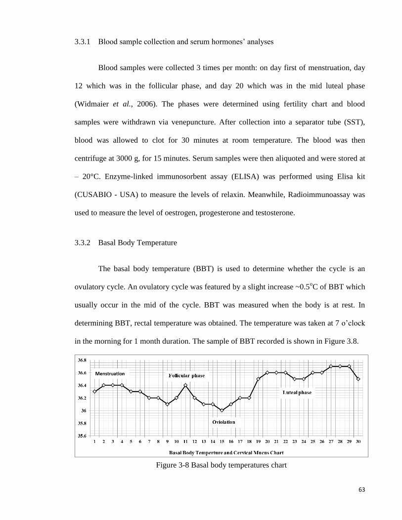
63
3.3.1 Blood sample collection and serum hormones’ analyses
Blood samples were collected 3 times per month: on day first of menstruation, day
12 which was in the follicular phase, and day 20 which was in the mid luteal phase
(Widmaier et al., 2006). The phases were determined using fertility chart and blood
samples were withdrawn via venepuncture. After collection into a separator tube (SST),
blood was allowed to clot for 30 minutes at room temperature. The blood was then
centrifuge at 3000 g, for 15 minutes. Serum samples were then aliquoted and were stored at
– 20°C. Enzyme-linked immunosorbent assay (ELISA) was performed using Elisa kit
(CUSABIO - USA) to measure the levels of relaxin. Meanwhile, Radioimmunoassay was
used to measure the level of oestrogen, progesterone and testosterone.
3.3.2 Basal Body Temperature
The basal body temperature (BBT) is used to determine whether the cycle is an
ovulatory cycle. An ovulatory cycle was featured by a slight increase ~0.5oC of BBT which
usually occur in the mid of the cycle. BBT was measured when the body is at rest. In
determining BBT, rectal temperature was obtained. The temperature was taken at 7 o’clock
in the morning for 1 month duration. The sample of BBT recorded is shown in Figure 3.8.
Figure 3-8 Basal body temperatures chart

64
3.3.3 Knee rotational movement
In this part of study we were examined the knee rotational movement by performing
a varus-valgus examination using a manual orthopaedic goniometer ruler (model:
MR0104PVC material 180°). These tests were used to measure the varus/valgus angles and
the internal/external rotational laxity of the knee joint.Data were collected by measuring
subjects both knees. In order to establish the reliability of the data, varus-valgus test were
accomplished by fixing the knee at 30° and 0°. All procedures were identical to those
described in the clinical examination textbook (Bendjaballah et al., 1997). Varus-Valgus
test were performed by placing unusual stresses forces on the articular surfaces, joint
capsule and ligaments.
Varus test is a stress force applied from the lateral side of the femur by adducting
the ankle. Testing LCL (lateral collateral ligament) is performed by flexing the knee at 30 °
and 0 °, with subjects lying flat. One hand of the examiner was placed over the lateral joint
line while another hand holds the lower leg firmly at the ankle. Meanwhile, valgus force is
a stress force applied on the lateral side by abducting the ankle. Stretching of MCL (medial
collateral ligament) was performed in medial side of the knee by flexing the knee at 30 and
0 °, with patient lying flat with one examiner hand was placed over the medial joint line
and another hand holds the lower leg firmly. In order to ior to check for the relaibility of the
data, these tests were performed by 3 different examiners and the data was gathered prior to
analyses.

65
Figure 3-9 Knee rotational movement investigation by Varus-Valgus test at 30°
Figure 3-10 Knee rotational movement investigation by Varus-Valgus test at 0°

66
3.3.4 Data analysis of knee laxity in human
All the data were presented as mean ± standard deviation, analysed by descriptive
analysis to determine hormone levels and rotational angles of each subject throughout the
menstrual cycle.One-way ANOVA was usedto ascertain the correlation by using SPSS
18.0. p< 0.05 was considered as statistically significant.As for each variables, 3
measurements was used to evaluate the differences between 3 observations, one way
repeated measure Anova was applied and the results revealed that there were no significant
differences among these three observations.Therefore, the mean of these observations was
computed and used for data analysis. Two-way ANOVA was usedto compare athletes and
non-athletes groups. For variables with a normal distribution, Pearson correlation
coefficient and for non-normal distributed variable, Spearman correlation coefficient was
applied.

67
4 CHAPTER 4
Sex-Steroid Regulation of Relaxin Receptor Isoforms
(RXFP1 & RXFP2) Expression in the Patella Tendon
and Lateral Collateral Ligament of Female WKY Rats

68
4.1 Introduction
Relaxin which consist of relaxin-1, -2 and -3 and insulin-like peptides (INSL3, 4, 5
and 6) (Park et al., 2005) is a polypeptide hormone that possesses structural similarity to
insulin and is primarily synthesized by the corpus luteum and placenta (Conrad & Baker,
2013). Relaxin binds to and activates G-protein-coupled receptors (GPCRs):
RXFP1(LGR7) and RXFP2(LGR8) (Scott et al., 2012). Human relaxins H1 and H2
activate both RXFP1 and RXFP2 (Nair, 2012), while rat relaxin1 binds weakly to RXFP2
(Scott et al., 2005). Relaxin H3 binds selectively to RXFP1 as well as RXFP3 (GPCR135)
and RXFP4 (GPCR142) (Rosengren et al., 2006 ). RXFP1 and RXFP2 are the two main
relaxin receptor isoforms (Schwabe & Büllesbach, 2007).
Relaxin activity has been reported in both pregnant and non-pregnant female primates
and non-primate vertebrates (Cernaro et al., 2013; Hayes, 2004). In primates, ovarian
relaxin is not required for successful term pregnancy (Hayes, 2004) however is required to
maintain pregnancy in rats and pigs (Sherwood OD, 1993). During pregnancy in mice,
relaxin is involved in transformation of pubic joint cartilage into a more flexible and elastic
interpubic ligament in order to accommodate the enlarging uterus (Lippert et al., 1987). In
non-pregnant rats, relaxin administration alone or in combination with oestrogen reduces
pubic symphysis collagen content, with progesterone antagonizing this effect (Samuel et
al., 1996). Meanwhile, during pregnancy in rats, relaxin was reported to regulate collagen
catabolism in the pubic symphysis (Samuel et al., 1998). In lower species such as rodents,
relaxin action may be more reflective than in humans where serum relaxin level was found
to have no link to the generalized joint laxity (Wolf et al., 2013).

69
Relaxin affects tissues’ extracellular matrix components including collagen, H2O and
hyaluronan. Relaxin increases cervical hyaluronan content by up-regulating the expression
of hyaluronic acid synthase (Soh, 2012). This effect will result in loosening of tissues’
dense collagen fiber network. Relaxin has been shown to possess anti-fibrotic action by
down-regulating fibroblast activity, increasing collagenases synthesis and inhibit
transforming growth factor (TGF-)-stimulated collagen-l lattice contraction in rat kidney
(Masterson et al., 2004). Relaxin up-regulates matrix metalloproteinases (MMP), a family
of extracellular proteases that interfere with matrix remodeling by their ability to degrade
extracellular matrix components including collagen (Chow et al., 2012). Relaxin has been
reported to stimulate the expression of collagenase-1, MMP-1, MMP-3 (Hashem et al.,
2006), MMP-9, and MMP-13 in fibrocartilaginous cells (Ahmad, 2012) and MMP-2 in
periodontal ligament cells (Ho, 2009; Miao, 2011).
Sex-steroids have been reported to affect relaxin receptor expression. In the human
endometrium, relaxin receptor isoforms expression vary across the menstrual cycle with
low level expressed during the proliferative phase and rises sharply at the time of ovulation
(Bond et al., 2004). In neonatal porcine uterus, oestrogen increases RXFP1mRNA
expression, while in cervix, oestrogen increases and relaxin decreases the expression of this
receptor (Yan et al., 2008).During late pregnancy in mice, relaxin has no influence on
RXFP1 expression (Siebel et al., 2003 ). Progesterone has been reported to increase the
expression of RXFP1 in the pregnant rat uteri (Vodstrcilet al., 2010a ) while oestrogen was
found to regulate RXFP1 mRNA expression in human cervix at term (Maseelall, 2009).

70
Knee is another potential target for relaxin action. Currently, little is known with
regards to the expression of RXFP1 and RXFP2 in the knee. Wang et al. (Wang et al.,
2009a) reported that relaxin receptors were expressed in mouse knee fibrocartilage. In
humans, relaxin receptors have been identified in the anterior cruciate ligament (ACL) in
male and female knee (Faryniarz et al., 2006), with higher expression in female (Dragoo et
al., 2003). This gender specific difference suggests the influence by female sex hormones.
Apart from ACL, the expression of relaxin receptors in other knee structures including
patellar tendon and lateral collateral ligament is currently unknown. Together with ACL,
they participate in the control of knee stability during movement.
We hypothesized that female sex hormones affect RXFP1 and RXFP2 expression in
the patella tendon and lateral collateral ligament, thus affecting knee laxity. This could
explain differences in the incidence of non- contact knee injury between male and female
(Sutton & Bullock, 2013), and in female at different phases of the menstrual cycle (Hansen
et al., 2013). In view of this, we investigated these isoforms expression in the patella
tendon and lateral collateral ligament in rat model in order to provide the mechanism
underlying changes in knee laxity under different sex-steroid influence.
4.2 Results
4.2.1 Plasma Sex-Steroid Levels following Subcutaneous Hormone Injection
In Table 4-1, the average hormones level in the blood in different groups of
treatment is shown. Our findings indicate that serum levels of oestrogen and testosterone
correlate with the doses administered. Subcutaneous injection of 0.2g/ml

71
oestrogenresulted in plasma level of approximately two times higher than the control while
injection of 4 mg progesterone resulted in five times higher level than the control.
Table 4-1 Plasma sex steroid level following subcutaneous hormone injection.
Values were given as mean ± standard error of mean. E – Oestrogen, P – progesterone and
T – testosterone. The level of hormones in the blood correlates with the amount injected via
the subcutaneous route.
4.2.2 RXFP1 and RXFP2 Proteins Expression in the Patellar Tendon and Lateral
Collateral Ligament
In Figure4-1, the expression of RXFP1 protein is the highest in the lateral collateral
ligament of progesterone treated rats with 1.75 fold increase as compared to control.
Treatment with oestrogen at 0.2 µg/ml resulted in a slight but significant increase in the
expression of RXFP1 protein as compared to control (p<0.05). Treatment with higher

72
oestrogen doses (>2µg/ml) resulted in a dose-dependent increase in RXFP1 protein
expression with 43 and 100 percent increase following treatment with 20 and 50µg/ml
oestrogen respectively. Testosterone treatment resulted in a significant decrease in the
expression of RXFP1 (0.54 and 0.45 fold increase following subcutaneous administration
of 125 and 250µg/ml testosterone respectively).
Figure 4-1 The expression of RXFP1 protein in the total homogenate of collateral ligament
from ovariectomised rats treated with different sex-steroids: C- control; 0.2E – 0.2µg/ml
oestrogen; 2E - 2µg/ml oestrogen, 20E - 20µg/ml oestrogen; 50E –50µg/ml oestrogen; P –
4mg progesterone; 125T – 125µg testosterone; 250T – 250µg testosterone, LCL-lateral
collateral ligament. Data were expressed as mean ± SEM, n=6 per treatment group. *
p<0.05 as compared to control, Shapiro-Wilk test, p= 0.319732. A dose-dependent increase
in RXFP1 protein expression was observed following oestrogen treatment. Progesterone
caused the highest increase while testosterone caused a decrease in this isoform expression.
The image was cut in order to arrange the bands in sequence with the label on the x-axis of
the bar chart
In Figure 4-2, the expression of RXFP1 protein in the patellar tendon following
treatment with 4mg progesterone is 2.71 folds higher than the control. Oestrogen treatment
resulted in a dose-dependent increase in this protein expression (1.25 to 2.67 fold increase
following treatment with oestrogen at doses between 0.2 to 50µg/ml). Testosterone

73
treatment resulted in a dose-dependent decrease in RXFP1 protein expression (0.65 and
0.51 fold decrease) following treatment with 125 and 250µg testosterone respectively. The
expression of RXFP1 protein in the patella tendon was higher than the lateral collateral
ligament.
Figure 4-2 The expression of RXFP1 protein in the total homogenate of the patellar tendon
from ovariectomised rats treated with different sex-steroids: C- control; 0.2E – 0.2g/ml
oestrogen; 2E - 2g/ml oestrogen, 20E - 20g/ml oestrogen; 50E –50g/ml oestrogen; P –
4mg progesterone; 125T – 125g testosterone; 250T – 250g testosterone PT- patella
tendon. Data were expressed as mean ± SEM, n=6 per treatment group. * p<0.05 as
compared to control, Shapiro-Wilk test, p= 0.282109. A marked increase in the expression
of RXFP1 protein occurs following treatment with high doses of oestrogen (20 & 50g/ml).
Meanwhile, progesterone treatment caused the highest increase while testosterone caused a
decrease in RXFP1 protein expression in the tendon. The image was cut in order to arrange
the bands in sequence with the label on the x-axis of the bar chart.
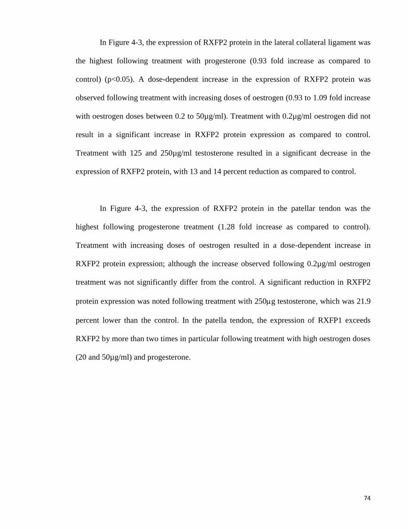
74
In Figure 4-3, the expression of RXFP2 protein in the lateral collateral ligament was
the highest following treatment with progesterone (0.93 fold increase as compared to
control) (p<0.05). A dose-dependent increase in the expression of RXFP2 protein was
observed following treatment with increasing doses of oestrogen (0.93 to 1.09 fold increase
with oestrogen doses between 0.2 to 50µg/ml). Treatment with 0.2µg/ml oestrogen did not
result in a significant increase in RXFP2 protein expression as compared to control.
Treatment with 125 and 250µg/ml testosterone resulted in a significant decrease in the
expression of RXFP2 protein, with 13 and 14 percent reduction as compared to control.
In Figure 4-3, the expression of RXFP2 protein in the patellar tendon was the
highest following progesterone treatment (1.28 fold increase as compared to control).
Treatment with increasing doses of oestrogen resulted in a dose-dependent increase in
RXFP2 protein expression; although the increase observed following 0.2µg/ml oestrogen
treatment was not significantly differ from the control. A significant reduction in RXFP2
protein expression was noted following treatment with 250g testosterone, which was 21.9
percent lower than the control. In the patella tendon, the expression of RXFP1 exceeds
RXFP2 by more than two times in particular following treatment with high oestrogen doses
(20 and 50µg/ml) and progesterone.

75
Figure 4-3 The expression of RXFP2 protein in the total homogenate of collateral ligament
from ovariectomised rats treated with different sex-steroids: C- control; 0.2E – 0.2g/ml
oestrogen; 2E - 2g/ml oestrogen, 20E - 20g/ml oestrogen; 50E –50g/ml oestrogen; P –
4mg progesterone; 125T – 125g testosterone; 250T – 250g testosterone, LCL-lateral
collateral ligament. Data were expressed as mean ± SEM, n=6 per treatment group. *
p<0.05 as compared to control, Shapiro-Wilk test, p= 0.776415. The expression of RXFP2
protein was increased following treatment with oestrogen at doses exceeding 2g/ml.
Meanwhile, progesterone caused the highest increase while testosterone caused a decrease
in RXFP2 protein expression. The image was cut in order to arrange the bands in sequence
with the label on the x-axis of the bar chart.
4.2.3 RXFP1and RXFP2 mRNAs Expression in the Patellar Tendon and Lateral
Collateral Ligament
In Figure 4-4, following progesterone treatment, the expression of RXFP1 mRNA in
the lateral collateral ligament was 2.71 folds higher as compared to control. The lowest
expression was noted following treatment with 250µg testosterone, which was 27 percent
lesser than the control. Treatment with oestrogen resulted in a dose-dependent increase in
RXFP1 mRNA expression (1.21 to 2.13 fold increase following treatment with 0.2 to
50µg/ml oestrogen).
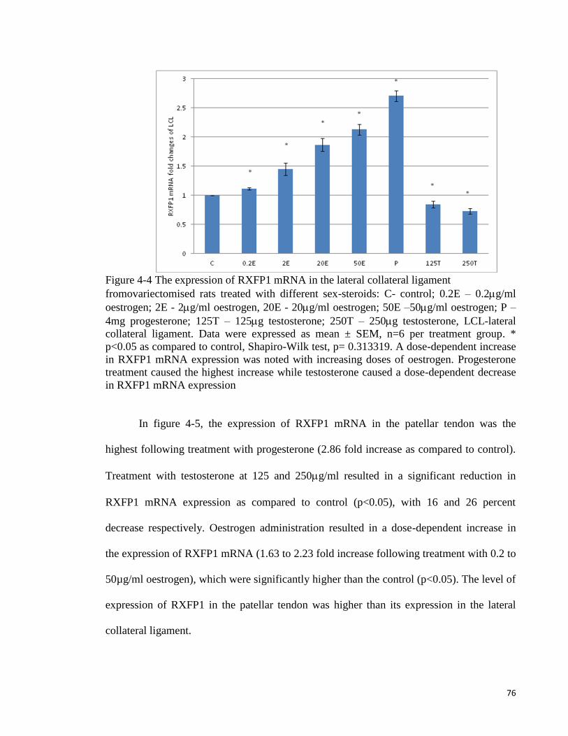
76
Figure 4-4 The expression of RXFP1 mRNA in the lateral collateral ligament
fromovariectomised rats treated with different sex-steroids: C- control; 0.2E – 0.2g/ml
oestrogen; 2E - 2g/ml oestrogen, 20E - 20g/ml oestrogen; 50E –50g/ml oestrogen; P –
4mg progesterone; 125T – 125g testosterone; 250T – 250g testosterone, LCL-lateral
collateral ligament. Data were expressed as mean ± SEM, n=6 per treatment group. *
p<0.05 as compared to control, Shapiro-Wilk test, p= 0.313319. A dose-dependent increase
in RXFP1 mRNA expression was noted with increasing doses of oestrogen. Progesterone
treatment caused the highest increase while testosterone caused a dose-dependent decrease
in RXFP1 mRNA expression
In figure 4-5, the expression of RXFP1 mRNA in the patellar tendon was the
highest following treatment with progesterone (2.86 fold increase as compared to control).
Treatment with testosterone at 125 and 250g/ml resulted in a significant reduction in
RXFP1 mRNA expression as compared to control (p<0.05), with 16 and 26 percent
decrease respectively. Oestrogen administration resulted in a dose-dependent increase in
the expression of RXFP1 mRNA (1.63 to 2.23 fold increase following treatment with 0.2 to
50µg/ml oestrogen), which were significantly higher than the control (p<0.05). The level of
expression of RXFP1 in the patellar tendon was higher than its expression in the lateral
collateral ligament.
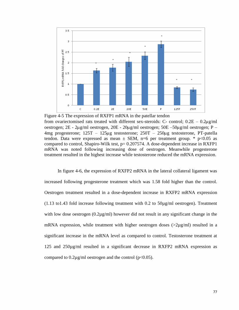
77
Figure 4-5 The expression of RXFP1 mRNA in the patellar tendon
from ovariectomised rats treated with different sex-steroids: C- control; 0.2E – 0.2g/ml
oestrogen; 2E - 2g/ml oestrogen, 20E - 20g/ml oestrogen; 50E –50g/ml oestrogen; P –
4mg progesterone; 125T – 125g testosterone; 250T – 250g testosterone, PT-patella
tendon. Data were expressed as mean ± SEM, n=6 per treatment group. * p<0.05 as
compared to control, Shapiro-Wilk test, p= 0.207574. A dose-dependent increase in RXFP1
mRNA was noted following increasing dose of oestrogen. Meanwhile progesterone
treatment resulted in the highest increase while testosterone reduced the mRNA expression.
In figure 4-6, the expression of RXFP2 mRNA in the lateral collateral ligament was
increased following progesterone treatment which was 1.58 fold higher than the control.
Oestrogen treatment resulted in a dose-dependent increase in RXFP2 mRNA expression
(1.13 to1.43 fold increase following treatment with 0.2 to 50µg/ml oestrogen). Treatment
with low dose oestrogen (0.2µg/ml) however did not result in any significant change in the
mRNA expression, while treatment with higher oestrogen doses (>2µg/ml) resulted in a
significant increase in the mRNA level as compared to control. Testosterone treatment at
125 and 250µg/ml resulted in a significant decrease in RXFP2 mRNA expression as
compared to 0.2µg/ml oestrogen and the control (p<0.05).

78
Figure 4-6 The expression of RXFP2 mRNA in the lateral collateral ligament
from ovariectomised rats treated with different sex-steroids: C- control; 0.2E – 0.2g/ml
oestrogen; 2E - 2g/ml oestrogen, 20E - 20g/ml oestrogen; 50E –50µg/ml oestrogen; P –
4mg progesterone; 125T – 125g testosterone; 250T – 250µg testosterone, LCL-lateral
collateral ligament.Data were expressed as mean ± SEM, n=6per treatment group. * p<0.05
as compared to control, Shapiro-Wilk test, p= 0.668854. Treatment with oestrogen at doses
exceeding 2g/ml resulted in a dose-dependent increase in RXFP2 mRNA expression.
Meanwhile, progesterone caused the highest increase while testosterone treatment resulted
in a decrease in this isoform mRNA expression.
In figure 4-7, the expression of RXFP2 mRNA in the patella tendon was the highest
following treatment with progesterone (2.28 fold increase as compared to control).
Oestrogen treatment resulted in a dose-dependent increase in RXFP2 expression (1.13 to
2.01 fold increase following treatment with 0.2 to 50µg/ml oestrogen). Treatment with 125
and 250µg/ml testosterone resulted in a significant reduction in RXFP2 mRNA expression
(0.79 and 0.5 fold- respectively) as compared to control. RXFP2 mRNA expression in the
patella tendon exceeds its expression in the lateral collateral ligament only following
treatment with 50µg/ml oestrogen and progesterone.

79
Figure 4-7 The expression of RXFP2 mRNA in the patellar tendon from ovariectomised
rats treated with different sex-steroids: C- control; 0.2E – 0.2g/ml oestrogen; 2E - 2g/ml
oestrogen, 20E - 20g/ml oestrogen; 50E –50g/ml oestrogen; P – 4mg progesterone; 125T
– 125g testosterone; 250T – 250g testosterone, PT-patellar tendon. Data were expressed
as mean ± SEM, n=6 per treatment group. * p<0.05 as compared to control, Shapiro-Wilk
test,p= 0.842243. An increase in the RXFP1 mRNA was noted following treatment with
oestrogen at doses exceeding 2g/ml. Progesterone treatment resulted in the highest
increase while testosterone caused a dose-dependent decrease in RXFP2 mRNA
expression.
4.3 Discussion
Our findings have revealed the followings: (i) The expression of RXFP1 protein and
mRNA in knee lateral collateral ligament and patellar tendon were up-regulated by
oestrogen and progesterone, and were down-regulated by testosterone (ii) RXFP2 protein
and mRNA expression were up-regulated by oestrogen at doses exceeding 2µg/ml and
progesterone. Treatment with 0.2µg/ml oestrogen however had no significant effect on the
expression of this receptor isoform, (iii) The expression of RXFP1 protein and mRNA in
the patellar tendon under the influence of oestrogen and progesterone was higher than its
expression in the lateral collateral ligament, (iv) No significant changes in RXFP2

80
expression was noted between patella tendon and lateral collateral ligament except
following treatment with 50µg/ml oestrogen and progesterone, where higher expression
was noted in the former than the latter and (iv) RXFP1 is the main isoform expressed in the
patella tendon and lateral collateral ligament of the rat knee joint.
The effect of relaxin on ligament laxity has long been reported(Becker GJ, 2001).
The first evidence was documented by Hisaw in 1926 where relaxin infusion in mice was
found to cause relaxation of the pelvic ligament (MacLennan, 1981). Research related to
relaxin effect on knee joint laxity has become increasingly important in the recent years due
to the reported increase in the incidence of non-contact knee injury among the female
athletes. Recent evidence suggested that plasma relaxin level correlates with the increase in
incidence of ACL tear in elite female athletes (Dragoo et al., 2011b). In addition to knee,
the laxity of trapezial-metacarpal joint in humans (Wolf et al., 2013) and pubic ligaments in
mice (Lippert et al., 1987)has also been reported to change with changes in plasma relaxin
level.
The influence of female sex-hormones on relaxin action in the knee ligaments has
been proposed. In-vitro binding assay has revealed that relaxin binding to the female ACL
was higher than male ACL. This finding was supported by the increase in the number of
relaxin receptors expressed in the female ACL as compared to male (Dragoo et al., 2003).
Quantitation of relaxin receptors by radioligand binding assay in the human ACL fibroblast
cells has revealed that relaxin binding was present in four out of five female ACL fibroblast
as compared to one out of five male ACL fibroblast (Faryniarz et al., 2006). The direct
influence of oestrogen on ACL laxity has been documented by Dragoo et al (Dragoo et al.,
2009) where three weeks combined infusion of oestrogen and relaxin to the guinea pigs has

81
resulted in increased anterior tibial dislocation, indicating an increase in ACL laxity.
Meanwhile, in rabbit temporomandibular joint and pubic symphysis, concomitant
administration of relaxin with oestrogen was found to cause loss of glycosaminoglycans
and collagen from these tissues, which will lead to an increase in the ligament laxity.
Progesterone administration meanwhile prevented this effect (Hashem et al., 2006).
Although this study has not provided direct evidence of an increase in the expression
of relaxin receptors in the ACL under sex-steroid effect, we have for the first time shown
that oestrogen and progesterone upregulate the expression of relaxin receptor isoforms
(RXFP1 and RXFP2) in the patella tendon and lateral collateral ligament. In other tissues
including uterus (Vodstrcil et al., 2010a) and cervix (Maseelall et al., 2009), oestrogen was
also reported to upregulate the expression of the relaxin receptors. In this study, modulation
of RXFP1 and RXFP2 expression by sex-steroids could provide the basis underlying the
effect of these hormones on knee joint laxity. Recently, relaxin was also found to have
significant effect on the patellar tendon stiffness in healthy, young and normally
menstruating women (Pearson et al., 2011). Although the actual mechanism is unknown,
this effect could possibly be mediated through upregulation of RXFP1 and RXFP2 by sex-
steroids, which levels fluctuate throughout the menstrual cycle. The mechanisms that lead
to the changes in knee ligament laxity following the binding of relaxin to its receptors have
been earlier discussed (introduction section).
Our findings have important implication as it could help to explain differences in the
incidence of non-contact knee injury between male and female. Female was reported to
have two to ten times higher risk of injury than male and is related to phases of the
menstrual cycle (Hansen et al., 2013) which pointed towards the involvement of sex-
82
steroids. The effect of oestrogen on RXFP1 and RXFP2 expression in the patellar tendon
and lateral collateral ligament of rats’ knee confirmed the involvement of this hormone in
modulating knee laxity. At low dose (0.2µg/ml), which correlate with the plasma oestrogen
level of 69.7 ± 0.12pg/ml (table 1), the expression of RXFP1 was higher than the control
although no significant changes in RXFP2 expression were noted. The mean reported
plasma oestrogen level in the early follicular phase of the menstrual cycle was 31 pg/ml
(0.31 X 10⁻⁴
µg/ml) (Fenichel et al., 1989). In rat at proestrous, oestrogen level was found to
be higher at 40.5 pg/ml (0.45 X 10⁻⁴
µg/ml) (Walf et al., 2009 ). In view that 0.2µg/ml
oestrogen administration resulted in the plasma oestrogen level of 69.7 pg/ml, which was
slightly higher than the level observed in humans and rats at the follicular phase and
proestrus stage respectively, there is a possibility that this level could affect knee ligament
laxity via upregulating the expression of relaxin receptor isoforms rendering the tissue to be
more sensitive towards relaxin action.
Meanwhile, administration of 2, 20 and 50 µg/ml oestrogen resulted in the circulating
plasma level of 195.2 ± 0.31, 267.7 ± 0.76 and 329.4 ± 2.33 pg/ml respectively (table 1).
Treatment with oestrogen at doses exceeding 2g/ml resulted in higher expression of
RXFP1 and RXFP2 as compared to the control and 0.2g/ml oestrogen.High oestrogen
level in the blood could be achieved at the time of ovulation, with the reported level of
400pg/ml (0.4 X 10⁻³
µg/ml) (Fenichel et al., 1989). Shultz et al (Shultz et al., 2010)
reported that high incidence of non-contact knee injury occur in the pre-ovulatory phase,
which suggested that increased oestrogen level may have significant impact on the knee
ligament laxity. Several other findings have also reported that increased incidence of non-
contact knee injury during the ovulatory phases of the cycle (Park et al., 2009b; Romani et

83
al., 2003; Wojtys et al., 2002) was related to the effect of high oestrogen doses on knee
ligament laxity. Oestrogen- induced upregulation of relaxin receptors will augment relaxin
effect on the knee.
High plasma oestrogen can be achieved following consumption of oral contraceptive
pills (OCP) that contains 15 to 50 µg oestrogen (Dhont, 2010). In the female athletes, OCP
is consumed for birth control purpose and to prevent menstruation in order to improve their
physical performance (Frankovich & Lebrun, 2000). Few studies have reported that OCP
intake could influence knee laxity; however their findings seem to be conflicting. Hicks-
Little et al, (Hicks-Little et al., 2007) and Cammarata et al (Cammarata & Dhaher, 2008)
reported that female on OCP tend to have higher knee laxity, while others reported the
opposite (Martineau et al., 2004). Meanwhile, few other studies found no relationship
between OCP consumption and knee ligament laxity (Agel et al., 2006; Dhont, 2010).
Differences in sex-steroid content between different OCP formulations could be the reason
for this variability.
While oestrogen has been shown to affect ACL laxity in the animal model (Dragoo et
al., 2009), little is known with regards to the effect of progesterone and testosterone on
knee ligament laxity. Dragoo et al, measured serum relaxin concentration (SRC) in elite
collegiate female athletes and had observed a positive correlation between SRC and serum
progesterone concentration in athletes who are oligomenorrheic, eumenorrheic, not on oral
contraceptive and have ovulatory cycle (Dragoo et al., 2011a). In this study, serum
progesterone level was measured to confirmed luteal phase measurement of relaxin as
relaxin levels are known to be higher in this phase. Other studies involving female athletes
has also indicated that knee laxity was increased during ovulation and in the luteal phase of

84
the menstrual cycle (Hicks-Little et al., 2007). Meanwhile, a positive correlation between
changes in knee laxity and knee joint load has been reported from follicular phase to
ovulation, and from ovulation to the luteal phase (Park et al., 2009b), where the latter phase
is associated with high serum progesterone level. Our findings which revealed an up-
regulation of RXFP1 and RXFP2 isoforms in the patella tendon and lateral collateral
ligament by progesterone further support these observations. The increase in these isoforms
expression in the collateral ligaments predisposes the knee towards non-contact injury
especially during excessive varus or valgus movement.
Progesterone upregulation of RXFP1 and RXFP2 expression in the knee was
supported by findings in other tissues including the pregnant rat uteri, where this hormone
was found to cause an increase in RXFP1 expression in the myometrium (Vodstrcil et al.,
2010a ). In the mammary glands, progesterone was reported to stimulate relaxin receptor
expression which resulted in reduced breast laxity. This effect is due to a decrease in the
collagen content of the mammary gland’s extracellular matrix in the presence of relaxin
(Winn et al., 1994). In this study, the plasma level achieved following subcutaneous
progesterone injection was 23.67 ± 0.13 ng/ml, which was slightly higher than the
maximum plasma level reported during the luteal phase of the cycle in premenopausal
female, at 17.02 ng/ml (Shultz et al., 2006). This indicate that the level of progesterone in
the luteal phase can cause an increase in knee laxity, rendering the knee to be more
susceptible towards a non-contact injury.
Finally, we have also observed that RXFP1 and RXFP2 expression in both patellar
tendon and lateral collateral ligament was down-regulated by testosterone and this effect
was dose-dependent. In the female, the presence of androgen receptor was reported in the

85
ACL (Lovering & Romani, 2005), suggesting that testosterone could influence knee laxity.
Testosterone level was found to fluctuate throughout the menstrual cycle, with the highest
level noted in the luteal phase (Shultz et al., 2004).Down-regulation of RXFP1 and RXFP2
expression by testosterone in the patella tendon and lateral collateral ligament indicated that
knee laxity could be reduced under the influence of this hormone. A recent finding by
O’Leary et al, (O'Leary et al., 2013) indicated that prolonged aerobic exercise in women
with normal menstrual cycle can induce a short-term elevation in plasma testosterone level.
We hypothesized that a continuous short-burst in plasma testosterone may result in down-
regulation of relaxin receptors in these knee structures, thus could help to reduce knee
laxity in exercising women. In addition to this, testosterone has also been reported to
increase the strength of the muscles that control knee joint movement (Bhasin et al., 1996).
Taken together, these findings may explain increased knee joint stiffness in female
following testosterone administration.
In conclusion, our study has provided novel evidences that sex-steroid affect RXFP1
and RXFP2 expression in the patellar tendon and lateral colateral ligament of the rat knee
joint.These findings are important as it may help to provide the basis underlying increased
in incidence of non-contact knee injury in female as compared to male and in female at
different phases of the menstrual cycle. Precaution however should be taken when
projecting these findings to humans as recent evidence suggested that there was apparently
no relationship between serum relaxin and joint laxity (Wolf et al., 2013).

86
5 CHAPTER 5
Testosterone Reduces Knee Passive Range of
Motion and Expression of Relaxin Receptor
Isoforms via 5α-Dihydrotestosterone and
Androgen Receptor Binding

87
5.1 Introduction
Range of motion (ROM), defined as the movement potential of a joint from full flexion
to full extension is controlled by the connective tissue, muscles, tendons, and
ligaments(Schmitz et al., 2013). Knee joints connect femur to tibia and fibula through the
collateral and anterior cruciate ligaments (Marieb & Hoehn, 2013). Medial and lateral
collateral ligaments (MCL and LCL) prevent excessive varus-valgus-forces while anterior
cruciate ligament (ACL) prevents knee hyperextension (Marieb & Hoehn, 2013).The
patellar tendon, which connects patella to tibia tuberosity stabilizes the patella and also
prevents knee hyperextension(Janousek, 1999). Evidence suggests that gender could
influence knee laxity. Females have been reported to have greater knee laxity than males(Hsu
et al., 2006; Wojtys et al., 2003). Additionally, females have also been reported to have
greater ankle laxity (Beynnon et al., 2005). These gender specific differences suggest that
male and female sex hormones participate in controlling the generalized joint laxity. So far,
studies investigating the relationship between sex hormones and joint laxity mainly focused
on the knee and the findings remain inconclusive.
Relaxin, a member of the insulin-like superfamily, has been implicated in the
modulation of joint laxity (Dragoo et al., 2009). In primates, relaxin-1 (Rln1), 2 (Rln2), and
3 (Rln3) are the main isoforms (Chen et al., 2012) while in rats, mice and pigs, Rln1 and
Rln3 are the main subtypes(Sherwood, 2004). Relaxin increases the joint laxity via up
regulating expression and stimulating activity of matrix metalloproteinases (MMPs),
collagenases which stimulates collagen degradation and inhibit collagen synthesis by
fibroblasts (Lee et al., 2012). Relaxin effect is mediated via binding to the relaxin receptor,
which consists of the two main isoforms, RXFP1 and RXFP2 (Scott et al., 2012). Relaxins
88
binding to RXFP1 and RXFP2 are species specific. In humans, Rln1 and Rln2 have similar
binding affinity to RXFP1 and RXFP2, while in rats Rln1 binds weakly to RXFP2 (Scott et
al., 2005). Meanwhile, Rln3 has been reported to bind to RXFP1, RXFP3, and RXFP4
(Bathgate et al., 2005) while insulin-like factor 3 (Insl3), whichis structurally related to
relaxin, selectively binds to RXFP2 (Kumagai et al., 2002). Serum relaxin level fluctuates
throughout the reproductive cycle and is markedly increased during pregnancy (Forst et al.,
1997). Studies have shown a higher plasma relaxin level in pregnant women with pelvic
joint instability and increased hip joint laxity(Saugstad, 1991; Steinetz et al., 2008). While
reports have indicated a positive correlation between the serum relaxin concentration and
joint laxity (Dragoo et al., 2011b), relaxin effect on the joint is more reflective in rodents
than in humans (Sherwood, 2004).
Testosterone, an anabolic steroid produced by the testes, ovaries and adrenal glands has
serum concentration seven to eight times greater in male than in female (He et al., 2007
).Testosterone promotes the development of secondary male sexual characteristics such as
increased in muscle and bone mass and induced male sexual behavior while its active
metabolite, 5α-DHT promotes male pattern hair distribution. Additionally, testosterone also
stimulates erythropoiesis and increase muscle strength (Barrett et al., 2009). Despite being
produced in low amounts, testosterone is essential for several key reproductive processes in
female such as decidualization (Ishikawa et al., 2007). The classical effect of testosterone is
mediated via a genomic pathway involving the androgen receptor (AR)while a non-genomic
pathway mediates its rapid effect(Kouloumenta et al., 2006). An understanding of the
testosterone effect on joint laxity is currently not fully understood. Few studies have
suggested that testosterone could influence knee laxity however, its exact role remain
elusive. Shultz et al.(Shultz et al., 2004) reported that testosterone has a positive rather than

89
a negative relationship with changes in knee laxity in the presence of oestrogen and
progesterone. On the other hand,Rozzi et al.(Rozzi et al., 1999) reported that male athletes
have greater knee joint laxity that female athletes, suggesting a negative influence of
testosterone on laxity.
The notion that testosterone decreases joint laxity is further supported by the reported
increase in the collagen content of the prostate, breast and capsular tissue and an increase in
knee ligament repair strength by testosterone (Lovering & Romani, 2005). In view of the
fact that relaxin increases while testosterone may decrease joint laxity(Shultz et al., 2004),
we hypothesized that testosterone down regulates the expression of the relaxin receptor in
the joint, rendering this tissue to be insensitive towards relaxin action. This study therefore
aimed to investigatethe effect of testosterone on knee joint laxity and changes in relaxin
receptor expression under testosterone influence. Additionally, the involvement of the
androgen receptor and testosterone active metabolite, 5α-DHT, in mediating testosterone
effect was also investigated.
5.2 Results
5.2.1 Passive Knee ROM in Testosterone-Treated Ovariectomised Rats
The degree of knee joint angle (differences between passive flexion and extension)
is shown in Figure 5-1. There was a significant difference in the angle between
testosterone-treated and control groups where testosterone caused a decrease in the passive
ROM. Testosterone effect was antagonized by flutamide (FLU) and finasteride (FIN).
Following treatment with 125 and 250 μg/kg testosterone, the ROM was 6.22° and 6.45°

90
lower respectively than the non-treated group (p <0.05). The degree of knee joint angle in
the groups receiving 125 and 250μg/kg testosterone was greater in the presence of FLU,
which were 13.49° and 15.87°higher respectively (p <0.05) as compared to without FLU
treatment. The angles in the groups receiving 125 and 250μg/kg testosterone were also
noted to be greater in the presence of FIN, which was 12.84° and 14.82°higher respectively
(p <0.05)as compared to without FIN treatment. Knee joint angle was significantly higher
in the presence of relaxinin all groups except in the group receiving both doses of
testosterone where no difference in the angles was noted. The angle was the highest in the
control group, reduced in the presence of testosterone and significantly increased in the
presence of FLU and FIN.

91
Figure 5-1 Passive ROM of the rat knee
Treatment with peanut oil (control), testosterone, testosterone plus FLU or FIN with and
without relaxin. Our findings indicated that treatment with 125 and 250 μg/kg testosterone
significantly reduced the knee ROM as compared to control. In the presence of relaxin,
knee ROM in the control group was significantly higher than in the testosterone-treated
group. Relaxin administration to the group treated with testosterone did not result in a
significant increase in the passive knee ROM as compared to without relaxin. The presence
of FLU and FIN significantly increase the knee ROM which was further increased
following relaxin administration (p <0.05). * p <0.05 as compared to in the absence of
relaxin;†p <0.05 as compared to control;
#p <0.05 as compared to in the absence of FLU or
FIN.C: control, R: relaxin, T125: 125 µg/kg/day testosterone, T250: 250 µ/kg/day
testosteroneFLU: flutamide, FIN: finasteride.
5.2.2 RXFP1 and RXFP2 mRNA Expression in Patellar Tendon
In Figure 5-2, RXFP1 mRNA expression in the patellar tendon following 125 and 250
μg/kg testosterone treatment was lower than the control (p <0.05). Administration of FLU to
125 and 250μg/kg testosterone treated groups resulted in an increase in RXFP1mRNA
expression by approximately 0.69 and 0.86 fold respectively (p <0.05). Finastride
administration to these groups also resulted in 0.20 and 0.22 fold increase in RXFP1
mRNA expression respectively. The expression of RXFP2 mRNA in the patellar tendon in
125 and 250μg/kg testosterone-treated groups following FLU administration was 0.30 and
0.71 fold higher respectively (p <0.05). The expression of RXFP2 mRNA in the group

92
receiving 125 and 250μg/ kg testosterone treatment plus FIN administration was also
increased by 0.38 and 0.39-fold respectively (p <0.05).
Figure 5-2 Changes in RXFP1 and RXFP2 mRNA expression in the presence of FLU and
FIN in the patellar tendon of steroid-replaced ovariectomised rats. Treatment with 125 and
250μg/kg/day testosterone caused a significant decrease in RXFP1 and RXFP2 mRNA
levels as compared to control. Administration of flutamide and finasteride significantly
antagonized the inhibitory effect of testosterone on RXFP1 and RXFP2 expression with
FLU effect relatively higher than FIN for RXFP1, however no significant differences were
noted for RXFP2 mRNA.†p <0.05 as compared to control *p <0.05 as compared to
testosterone only treatment for the respective group. T125: 125 µg/kg/day testosterone,
T250: 250 µg/kg/day testosterone, FLU: flutamide, FIN: finasteride.

93
5.2.3 RXFP1 and RXFP2 Protein Expression in Patellar Tendon
In Figure 5-3, the expression of RXFP1 protein in the patellar tendon was higher in the
group receiving FLU with testosterone (0.79 and 0.83 fold increase respectively as
compared to 125 and 250μg/kg testosterone only treatment (p <0.05). The expression of
RXFP1 protein in the groups receiving FIN with testosterone was also significantly
increased as compared to without FIN(0.59 and 0.52-fold increase respectively as
compared to 125 and 250μg/kg testosterone only treatment)(p <0.05). The expression of
RXFP2 protein in the patellar tendon was increased following treatment with FLU plus the
two doses of testosterone, (0.29 and 0.36-fold increase respectively as compared to 125 and
250μg/kg testosterone treatment alone) (p <0.05). The expression of RXFP2 protein was
significantly increased following treatment with FIN plus testosterone as compared to
testosterone only treatment (0.2 and 0.24-fold increase respectively as compared to 125 and
250μg/kg testosterone) (p <0.05).

94
Figure 5-3 The expression of RXFP1 and RXFP2protein in the patellar tissue homogenate
from ovariectomised rats treated with testosterone with and without FLU and FIN. A
decreasein RXFP1 protein expression was noted following testosterone treatment which
was antagonized by FLU and FIN. The expression of RXFP2 protein was reduced
following testosterone treatment which was also antagonized by FLU and FIN. The
antagonizing effect of FIN indicated that DHT and not testosterone mediates the inhibition
of RXFP1 and RXFP2 protein expression(Data were expressed as mean ± SEM, n = 6 per
treatment group; 125T: 125μg testosterone; 250T: 250μg testosterone; FLU: 10 mg/kg
flutamide; FIN: 20 mg/kg finasteride). †p <0.05 as compared to control *p <0.05 as
compared to testosterone only treatment for the respective group.

95
5.2.4 RXFP1 and RXFP2 mRNA Expression in Lateral Collateral Ligament
In Figure 5-4, the expression of RXFP1 mRNA in the lateral collateral ligament was
reduced following treatment with testosterone at 125 and 250μg/kg/day. Administration of
FLU and FIN resulted in a significant increase in mRNA expression where effect of the
latter suggested DHT-mediated testosterone effect. Meanwhile, decreased RXFP2 mRNA
expression was also noted following testosterone treatment alone (0.31 and 0.33 fold decrease
respectively following exposure to 125 and 250μg/kg testosterone) which was also
antagonized by FLU and FIN (p <0.05).

96
Figure 5-4 Changes in RXFP1 and RXFP2 mRNA expressionin the lateral collateral
ligament of steroid-replaced ovariectomised rats in the presence of FLU and FIN.
Treatment with 125 and 250μg/kg/day testosterone caused a decrease inRXFP1 and RXFP2
mRNA expression as compared to control (testosterone only treatment) (p <0.05).
Administration of flutamide and finasteride caused a significant increase in the expression
of RXFP1 and RXFP2 with FLU inhibition was relatively greater than FIN for RXFP1,
however FIN inhibition was relatively higher than FLU for RXFP2mRNA expression.†p <0.05
as compared to control.*p <0.05 as compared to testosterone only treatment for the
respective group.
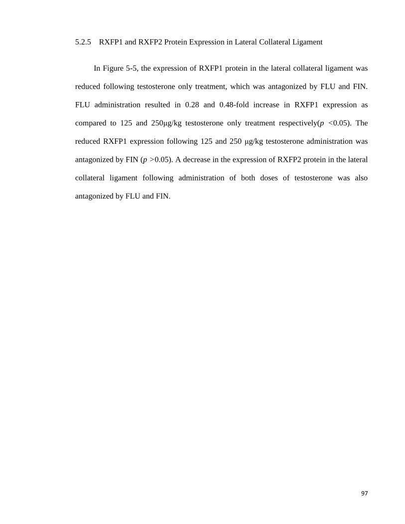
97
5.2.5 RXFP1 and RXFP2 Protein Expression in Lateral Collateral Ligament
In Figure 5-5, the expression of RXFP1 protein in the lateral collateral ligament was
reduced following testosterone only treatment, which was antagonized by FLU and FIN.
FLU administration resulted in 0.28 and 0.48-fold increase in RXFP1 expression as
compared to 125 and 250μg/kg testosterone only treatment respectively(p <0.05). The
reduced RXFP1 expression following 125 and 250 μg/kg testosterone administration was
antagonized by FIN (p >0.05). A decrease in the expression of RXFP2 protein in the lateral
collateral ligament following administration of both doses of testosterone was also
antagonized by FLU and FIN.

98
Figure 5-5 The expression of RXFP1 and RXFP2 protein
RXFP1 and RXFP2 protein from the homogenate of lateral collateral ligaments of
ovariectomised rats treated with 125 and 250 μg/kg testosterone with and without FLU and
FIN administration. A dose-dependent decrease in RXFP1 protein expression was noted
following treatment with both doses of testosterone which was antagonized by FLU and
FIN. Similarly, a decrease in RXFP2 protein expression was observed following treatment
with both doses of testosterone which was antagonized by FLU and FIN. Data were
expressed as mean ± SEM, n = 6 per treatment group; 125T: 125μg testosterone;250T:
250μgtestosterone; FLU: 10 mg/Kg FLU; FIN: 20 mg/kg FIN.†p <0.05 as compared to
control;*p <0.05 as compared to testosterone only treatment for the respective group.

99
5.3 Discussion
To the best of our knowledge, this study is the first to demonstrate the effect of
testosterone on knee laxity in an ovariectomised rat model. Ovariectomy was performed to
investigate sex-steroid effect on the ligament laxity, as removal of endogenous hormone
and replacement with exogenous steroid would ensure that the effect observed was due to
the action of individual steroid rather than the combined effect of multiple steroids
produced by the ovaries. In the female of reproductive age, 25% of the circulating
testosterone originates from the ovaries while the remaining is derived from conversion of
androsteinedione into testosterone in the adrenal gland(Speroff et al., 1989). In humans, the
level of endogenous testosterone has been reported to fluctuate throughout the menstrual
cycle which was the highest at around the time of ovulation (Rao & Kotagi, 1982). A recent
finding by O’Leary et al.(O'Leary et al., 2013) reported that a prolong edaerobic training in
the eumenorrheic women induces a short-term elevation in the plasma testosterone levels,
which appears to be unrelated to the level of oestrogen and the phases of menstrual
cycle.There is currently no report on changes in the testosterone level throughout the
oestrous cycle.
Testosterone has been proposed to influence knee laxity in female. A positive
correlation between plasma testosterone, free androgen index (FAI) and anterior cruciate
ligament stiffness has been reported in young females near ovulation (Lovering & Romani,
2005). The mechanisms underlying testosterone effect on ligament laxity is unknown,
however a limited finding in the prostate gland indicated that testosterone as opposed to
oestrogen affects collagen metabolism (Srinivasan et al., 1986) via down regulating the
expression of oestrogen receptor (ER)-α(Asano et al., 2003). So far, no study has reported

100
testosterone effect on relaxin receptor expression in the joints, although relaxin has been
shown to affect ligament laxity and its receptor was expressed in female knee joint of both
humans(Dragoo et al., 2003) and rodents (Dragoo et al., 2009). Our findings which
revealed down regulation of relaxin receptor expression in the knee ligaments and tendons by
testosterone has provided explanation for the observed decrease in knee passive ROM under
testosterone influence. A decrease in knee passive ROM could be due to decreases in the
relaxin effect on ligament, rendering the tissue to have decreased laxity.
Our study has provided the first direct evidence on testosterone effect on the passive
ROM in rats’ knee. We have shown that testosterone reduces the ROM, which was
inhibited by FLU and FIN. In the group receiving 125 and 250μg/kg/day testosterone
treatment, the presence of relaxin did not significantly increase knee passive ROM. This
finding suggested that knee was not responsive to relaxin in the presence of testosterone
due to relaxin receptor down regulation. The knee ROM was however increased following
FLU or FIN administration, suggesting that inhibition of androgen binding to its receptor
and conversion of testosterone to DHTcaused the increase in knee laxity. FLU inhibition
confirmed the release of DHT-mediated inhibition on relaxin receptor expression through
androgen receptor binding. Relaxin administration resulted in further increase in knee laxity
in the groups receiving both testosterone doses with FLU or FIN, which again suggested the
release of DHT-mediated inhibition on relaxin receptor expression. Meanwhile, increased
knee laxity in the groups receiving testosterone with FLU or FIN without relaxin suggested
that endogenously produced relaxin from sources such as breast(Speroff et al., 1989)might
influence knee laxity.

101
The presence of AR has been reported in human female knee joint (Fytili et al.,
2005; Lovering & Romani, 2005). In this study, inhibition of testosterone-mediated down-
regulation of RXFP1 and RXFP2 protein and mRNA expression in the patellar tendon and
lateral collateral ligament by FLU confirmed AR involvement in mediating testosterone
effect. Our findings have also confirmed DHT involvement in mediating testosterone action
as evidenced from the antagonizing effect of FIN on testosterone-mediated decrease in knee
passive ROM as well as testosterone-mediated down-regulation ofRXFP1 and RXFP2
protein and mRNA expression in these knee structures. FIN is a competitive, selective, and
reversible inhibitor of 5α-reductase, an enzyme that converts testosterone to DHT (Frye et
al., 1998). In females, DHT exerts multiple physiological effects mainly in the uterus and
ovaries (Cárdenas & Pope, 2005). DHT has also been reported to cause an increase in the
twitch and titanic contraction of fast-twitch skeletal muscle in mice (Hamdi & Mutungi,
2010). Although we have shown evidence that DHT is most likely involved in causing a
decrease in knee laxity, further studies are needed to support DHT participation such as
identifying the expression and measuring the activity of 5α- reductase enzyme in the knee.
In our study, a direct effect of DHT on knee laxity and relaxin receptor isoforms expression
was unable to be investigated since this prohibited compound was not commercially
available.
Our findings have important implications in the field of exercise physiology.
Testosterone could affect female knee laxity as its level was reported to be the highest in
the ovulatory phase of the menstrual cycle (Lovering & Romani, 2005). Although we have
found that testosterone decreases knee laxity, the findings by Shultz et al.(Shultz et al.,
2005) who reported a positive correlation between plasma oestrogen and testosterone level
with knee laxity throughout the menstrual cycle suggested dominant effects of oestrogen over

102
testosterone. In female athletes, anabolic steroids are widely used to boost their physical
performances. Despite reported adverse effects, this drug could help to strengthen the knee
via its effect on ligament and tendon laxity, which could improve athletes’ short-term
performance as well as reduce the risk of non-contact knee injury. Additionally, the
strength of the muscles controlling knee joint movement was also increased(Bhasin et al.,
1996). This study has shown for the first time the direct effect of testosterone on knee passive
ROM and RXFP1 and RXFP2 protein and mRNA expression in a rat model, which was
mediated via DHT and involved androgen receptor binding. These therefore could explain
the protective role of testosterone against non-contact knee injury in female. Precaution
however is needed when extrapolating these data to humans since relaxin action on human
joints might not be as pronounced as those observed in rodents (Wolf et al., 2013).

103
6 CHAPTER 6
Oestrogen Receptor (ER)-α, β and Progesterone
Receptor (PR) Mediates Changes in Relaxin
Receptor (RXFP1 and RXFP2) Expression and
Passive Range of Motion of Rats' Knee

104
6.1 Introduction
Sex-steroids have been reported to regulate changes in knee laxity (Dehghan et al.,
2014 ). E2 cause increased (Dragoo et al., 2009) while testosterone cause decreased
(Dehghan et al., 2014) knee laxity in rodents. In humans, knee laxity was reported to
increase at around the time of ovulation, indicating of E2 influence (Zazulak et al., 2006).
Knee laxity was also reported to be the highest in the luteal phase of the menstrual cycle,
suggesting of P4 influence (Heitz et al., 1999). Meanwhile, high E2 level was reported to
cause increased anterior tibial dislocation in rats during pregnancy (Charlton et al., 2001).
The mechanisms underlying E2 and P4 effects on knee laxity are not well understood,
however evidences suggested that these effects could be mediated via changes in collagen
turnover (Song et al., 2014), modulation of enzymes activity involved in collagen
metabolism (Shan et al., 2013) as well as response of tissues towards relaxin via changes in
the amount of receptor expressed (Dehghan et al., 2014 ).
The effect of sex-steroids is preceded by the binding to intracellular receptor,
located either in the cytosol or nuclear compartments (Bayard et al., 1978). Binding of
ligands to this receptor will initiate translocation of ligand-receptor complex to the nucleus
where it acts as a transcription factor which binds to hormone responsive elements (HRE)
on DNA, initiating gene transcription (Wierman, 2007). Two forms of ER have been
identified i.e. ERα and ERβ, which varies in the tissue distribution. Meanwhile, PR also
varies in tissues distribution (Edwards, 2005). ER and PR have been identified in human
anterior cruciate ligament (ACL) (Liu et al., 1996) which could explain changes in knee
laxity under different sex-steroid influence. Sex-steroids modulate joint laxity either
directly or indirectly. E2 was reported to cause a significant loss of glycosaminoglycans

105
(GAGs) and collagen content from the pubic symphysis, temporomandibular joint disc and
articular cartilage of the knee in rabbits (Hashem et al., 2006).E2 also affects the expression
of several isoforms of metalloproteinase (MMPs-1, 3 and 13) as well as tissue inhibitor of
metalloproteinases (TIMP 1) (Lee et al., 2003) which were involved in collagen turnover.
Meanwhile, E2 and P4were also reported to induce protein and mRNA expression of relaxin
receptors (RXFP1 and RXFP2) which result in increase tissue response towards relaxin
(Dehghan et al., 2014 ).
Relaxin, a 6-kDa polypeptide is structurally related to insulin and is produced by the
corpus luteum and placenta . Relaxin exerts various effects on the musculoskeletal system
via binding to several isoforms of relaxin receptor (RXFP1, RXFP2, RXFP3 and RXFP4)
(Dehghan et al., 2013a) and participates in extracellular matrix remodeling (Sherwood,
1994). Relaxin receptor expression has been identified in human female ACL (Dragoo et
al., 2003). Relaxin regulates synthesis and/or degradation of matrix macromolecules via
modulating the activity of several members of MMP enzymes (Naqvi et al., 2005). Serum
relaxin levels have been reported to correlate with the levels of MMP-1 in anterior oblique
ligament of thumb (Wolf et al., 2013) while relaxin effect on MMP-9 was found to be
mediated via binding to RXFP1 (Ahmad et al., 2012).
We hypothesized that E2 and P4 effects on knee laxity and the expression of RXFP1
and RXFP2 receptors were mediated via ER isoforms (ERα and ERβ) and progesterone
receptor (PR) respectively. The aim of this study is therefore to investigate the involvement
of sex-steroid receptors in mediating E2 and P4 effects on knee passive ROM and relaxin
receptor isoforms expression in the patellar tendon, collateral ligaments and hamstring
muscles which are involved in controlling knee joint movement.

106
6.2 Results
6.2.1 Effect of steroid receptor antagonists on RXFP1 and RXFP2 mRNA and proteins
expression in the patellar tendon
C

107
Figure 6-1 RXFP1& RXFP2 mRNA and protein expression
(A) The expression of RXFP1 mRNA in the patellar tendon of ovariectomized rats treated
with E2, P4 and their antagonists. Control; 0.2E – 0.2µg/kg E2; ICI+0.2E- ICI 182/780 (25
mg/kg) + 0.2 µg/kg E2; MPP+0.2E – MPP (25mg/kg) + 0.2µg/kg E2; PHTPP+0.2E –
PHTPP (25 mg/kg) + 0.2 µg/kg E2; P- 4mg/kg P4; Mife+P- 7.5 mg/kg mifepristone +4
mg/kg P4. Data were expressed as mean ± SEM, n = 6 per treatment group and * p < 0.05
as compared to control, #p<0.05 as compared to the respective agonist only treatment. (B)
The expression of RXFP2 mRNA in the patellar tendon of ovariectomized rats treated with
E2, P4 and their antagonists: Control; 0.2E – 0.2 µg/kg E2; ICI+0.2E- ICI 182/780 (25
mg/kg) + 0.2 µg/kg E2; MPP + 0.2E – MPP (25 mg/kg) + 0.2 µg/kg E2; PHTPP+0.2E –
PHTPP (25 mg/kg) + 0.2 µg/kg E2; P- 4 mg/kg P4; Mife + P- 7.5 mg/kg mifepristone +4
mg/kg P4. Data were expressed as mean ± SEM, n = 6 per treatment group and * p<0.05 as
compared to control, #p<0.05 as compared to the respective agonist only treatment. (C)
Ratio ofRXFP1& RXFP2/ β-actin protein expression levels in patellar tendon of
ovariectomized rats treated with E2, P4 and their antagonists. Control; 0.2 E – 0.2 µg/kg
E2; ICI+0.2 E- ICI 182/780 25 mg/kg + 0.2 µg/kg E2; MPP+0.2 E – MPP 25 mg/kg + 0.2
µg/kg E2; PHTPP+0.2 E – PHTPP 25 mg/kg + 0.2 µg/kg E2; P- 4 mg/kg progesterone;
Mife + P- 7.5 mg/kg mifepristone + 4 mg/kg P4. Data were expressed as mean ± SEM,
n=6 per treatment group and * p < 0.05 as compared to control, #p<0.05 as compared to the
respective agonist only treatment.

108
In figure 6-1, the expression level of RXFP1 mRNA in the patellar tendon of
ovariectomized rats received E2 was reduced by approximately 0.81, 1.4 and 1.62 fold in
the presence of ICI 182/780, MPP, and PHTPP respectively as compared to 0.2 µg E2
treatment alone. The expression of RXFP1 mRNA was the highest in the P4 treated group
which was antagonized by approximately 2.31 fold in the presence of PR blocker,
mifepristone.
The expression level of RXFP2 mRNA in patellar tendon following E2 treatment
was significantly reduced by approximately 0.35, 0.97 and 1.12 fold in the presence of ICI
182/780, MPP, and PHTPP respectively as compared to 0.2 µg E2 treatment alone. The
expression of RXFP2 mRNA in the patellar tendon which was the highest following
treatment with P4 was significantly reduced by approximately 1.45 fold following PR
antagonist, mifepristone administration.
Meanwhile, RXFP1 and RXFP2 protein expressions in the patellar tendon were the
highest following P4 treatment. Mifepristone caused a significant decrease in both isoforms
expression. The expressions of RXFP1 and RXFP2 under E2, which were significantly
higher than control, were inhibited by ICI 182780, MPP and PHTPP. PHTPP caused the
highest inhibition on RXFP1 and RXFP2 protein expression.

109
6.3 Effect of steroid receptor antagonists on RXFP1 and RXFP2 mRNA and proteins
expression in the collateral ligaments

110
Figure 6-2 RXFP1& RXFP2 mRNA and protein levels in collateral ligament
(A) The expression of RXFP1 mRNA in collateral ligament of ovariectomized rats treated
with E2, P4 and their respective antagonists. Control; 0.2E – 0.2 µg/kg oestrogen; ICI +
0.2E - ICI 182/780 (25 mg/kg) + 0.2 µg/kg E2; MPP + 0.2 E – MPP (25 mg/kg) + 0.2
µg/kg E2; PHTPP + 0.2 E – PHTPP (25 mg/kg) + 0.2 µg/kg E2; P - 4mg/kg P4; Mife + P -
7.5 mg/kg mifepristone +4 mg/kg P4. Data were expressed as mean ± SEM, n=6 per
treatment group and * p < 0.05 as compared to control, #p<0.05 as compared to the respective
agonist only treatment.(B) The expression of RXFP2 mRNA in collateral ligament of
ovariectomized rats treated with E2, P4 and their antagonists: Control; 0.2 E – 0.2 µg/kg
E2; ICI+0.2 E- ICI 182/780 (25 mg/kg) + 0.2 µg/kg E2; MPP+0.2 E – MPP (25 mg/kg) +
0.2 µg/kg E2; PHTPP+0.2 E –PHTPP (25 mg/kg) + 0.2 µg/kg E2; P-4 mg/kg P4; Mife + P-
7.5 mg/kg mifepristone + 4 mg/kg P4. Data were expressed as mean ± SEM, n=6 per
treatment group and * p < 0.05 as compared to control, #p<0.05 as compared to the respective
agonist only treatment.. (C) Ratio ofRXFP1& RXFP2/ β-actin protein levels in collateral
ligament in presence of oestrogen& progesterone antagonists. Control; 0.2 E – 0.2 µg/kg
E2; ICI + 0.2 E- ICI 182/780 (25 mg/kg) + 0.2 µg/kg E2; MPP+0.2 E – MPP (25 mg/kg) +
0.2 µg/kg E2; PHTPP+0.2 E – PHTPP 25 mg/kg + 0.2 µg/kg E2; P-4 mg/kg P4; Mife + P-
7.5 mg/kg mifepristone + 4 mg/kg P4. Data were expressed as mean ± SEM, n=6 per
treatment group and * p < 0.05 as compared to control, #p<0.05 as compared to the respective
agonist only treatment.

111
In Figure 6-2, the expression levels of RXFP1 mRNA in ovariectomised rats
collateral ligaments of the knee were decreased in the presence of steroid receptor
antagonists. The expression of RXFP1 mRNA was increased following oestrogen treatment
and was reduced in the presence of different antagonists, by approximately 0.96, 0.81 and
1.07 fold decrease following administration of ICI 182/780, MPP and PHTPP respectively.
The expression of RXFP1 mRNA in the collateral ligaments which was the highest
following administration of P4 was reduced in the presence of PR antagonist, mifepristone
by approximately 2.28 fold.
The expression level of RXFP2 mRNA in ovariectomized rats received E2 treatment
was decreased in the presence of ERα and ERβ antagonists. The expression of RXFP2
mRNA in the collateral ligament was approximately 0.93, 0.73 and 1.09 fold lower
following administration of ICI 182/780, MPP and PHTPP respectively as compared to in
the absence of these receptor blockers. The expression of RXFP2 mRNA in collateral
ligament was the highest following P4 treatment which was reduced by approximately 1.09
fold following PR antagonist, mifepristone administration.
Meanwhile, RXFP1 and RXFP2 protein expression in the collateral ligament was
significantly increased under P4 influence and was antagonized by mifepristone. Under E2
influence, these isoforms expression were slight but significantly higher than the control
group. Administration of ICI 182780, MPP and PHTPP resulted in a marked inhibition of
RXFP1 and RXFP2 proteins expression under E2 with PHTPP administration caused the
greatest inhibition.

112
6.4 Effect of steroid receptor antagonists on RXFP1 and RXFP2 mRNA and proteins
expression in the hamstring muscles
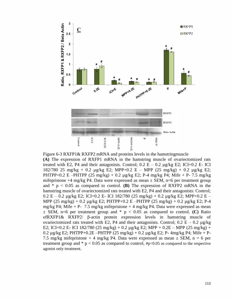
113
Figure 6-3 RXFP1& RXFP2 mRNA and proteins levels in the hamstringmuscle
(A) The expression of RXFP1 mRNA in the hamstring muscle of ovariectomized rats
treated with E2, P4 and their antagonists. Control; 0.2 E – 0.2 µg/kg E2; ICI+0.2 E- ICI
182/780 25 mg/kg + 0.2 µg/kg E2; MPP+0.2 E – MPP (25 mg/kg) + 0.2 µg/kg E2;
PHTPP+0.2 E –PHTPP (25 mg/kg) + 0.2 µg/kg E2; P-4 mg/kg P4; Mife + P- 7.5 mg/kg
mifepristone +4 mg/kg P4. Data were expressed as mean ± SEM, n=6 per treatment group
and * p < 0.05 as compared to control. (B) The expression of RXFP2 mRNA in the
hamstring muscle of ovariectomized rats treated with E2, P4 and their antagonists: Control;
0.2 E – 0.2 µg/kg E2; ICI+0.2 E- ICI 182/780 (25 mg/kg) + 0.2 µg/kg E2; MPP+0.2 E –
MPP (25 mg/kg) + 0.2 µg/kg E2; PHTPP+0.2 E –PHTPP (25 mg/kg) + 0.2 µg/kg E2; P-4
mg/kg P4; Mife + P- 7.5 mg/kg mifepristone + 4 mg/kg P4. Data were expressed as mean
± SEM, n=6 per treatment group and * p < 0.05 as compared to control. (C) Ratio
ofRXFP1& RXFP2/ β-actin protein expression levels in hamstring muscle of
ovariectomized rats treated with E2, P4 and their antagonists. Control; 0.2 E – 0.2 µg/kg
E2; ICI+0.2 E- ICI 182/780 (25 mg/kg) + 0.2 µg/kg E2; MPP + 0.2E – MPP (25 mg/kg) +
0.2 µg/kg E2; PHTPP+0.2E –PHTPP (25 mg/kg) + 0.2 µg/kg E2; P- 4mg/kg P4; Mife + P-
7.5 mg/kg mifepristone + 4 mg/kg P4. Data were expressed as mean ± SEM, n = 6 per
treatment group and * p < 0.05 as compared to control, #p<0.05 as compared to the respective
agonist only treatment.

114
In figure 6-3, the expression level of RXFP1 mRNA in ovariectomized rats'
hamstring muscle under the effect of E2was reduced in the presence of ER antagonists, ICI
182/780, MPP, and PHTPP by approximately 0.92, 0.71 and 1.07 fold respectively as
compared to 0.2 µg E2 treatment alone. Expression of RXFP1 mRNA in the hamstring
muscle was the highest following treatment with P4 and was markedly reduced in the
presence of PR antagonists, mifepristone by approximately 2.31 fold as compared to P4
treatment alone.
The expression level of RXFP2 mRNA in ovariectomized rats hamstring muscle
was reduced by approximately 0.91, 0.83 and 1.09 fold following treatment with ICI
182/780, MPP, and PHTPP respectively as compared to treatment with 0.2 µg E2 alone.
The expression of RXFP2 mRNA was the highest following P4 treatment which was
reduced by approximately 1.09 fold in the presence of PR antagonist, mifepristone.
Meanwhile, in the hamstring muscle, RXFP1and RXFP2 proteins were expressed the
highest in P4 treated group which were antagonized by mifepristone. E2 treatment resulted
in slightly higher RXFP1 and RXFP2 protein expression which were markedly inhibited
following administration of PHTPP, MPP and ICI 182 780.

115
6.5 Knee passive ROM in the presence of steroid receptor antagonist
Figure 6-4 Knee passive ROM of ovariectomised rats
Rats received different doses of E2, P4 and the receptor antagonists with and without
relaxin. Control; E – 0.2 g/kg E2; E+RLN- E2 plus Relaxin, E+ICI- E2 plus 25 mg/kg ICI
182/780; E+ICI+RLN- E2 plus 25 mg/kg ICI 182/780 plus Relaxin, E+M- E2 plus 25
mg/kg MPP, E+M+RLN- E2 plus 25 mg/kg MPP+Relaxin, E+PH- E2 plus 25 mg/kg
PHTPP, E+PH+RLN- E2 plus 25 mg/kg PHTPP plus Relaxin, P-4 mg/kg P4; P+RLN-
ICI-P4 plus Relaxin, P+MF- P4 plus Mifepristone (7.5 mg/kg) and P+MF+RLN- P4 plus
Mifepristone (7.5 mg/kg) plus Relaxin. Data were expressed as mean ± SD, n=6 per
treatment group and * p < 0.05 as compared to control, #p<0.05 as compared to the without
receptor blocker, †p<0.05 as compared to withour relaxin. Knee passive ROM was
following E2 and P4 treatments.
In Figure 6-4, the increase in knee passive ROM in E2 treated group (p<0.05 as
compared to control) was antagonized by MPP and PHTPP however not by ICI 182780.
Knee passive ROM was the highest following P4 treatment (mean = 92.4 o
±4.13) which
was antagonized by mifepristone (p<0.01). Relaxin administration caused an even higher
increase in knee passive ROM in the control, E2 and P4-treated groups, however no
significant changes in knee ROM were observed in the groups received E2 plus MPP, E2
116
plus PHTPP treatments as compared to similar groups without relaxin. Meanwhile, relaxin
administration in the group received P4plus mifepristone treatment also did not cause any
significant changes in knee passive ROM.
6.6 Discussion
This study reveals the presence of ERα, ERβ and PR in patellar tendon, collateral
ligaments and hamstring muscle of rats. In this study, the administration of PHTPP, an ERβ
antagonist markedly reduce the expression of RXFP1 and RXFP2 mRNA in the patellar
tendon, collateral ligaments and hamstring muscles. PHTPP is a specific ERβ antagonist
with a 36-fold selectivity over ERα, therefore its inhibition on oestrogen effect would
confirm the involvement of ERβ subtypes in mediating the biological effects of oestrogen
(Compton et al., 2004). Previous study has also reported the presence of ER (general) in the
synoviocytes, fibroblasts and ligament's blood vessel of the knee joint (Liu et al., 1996)
which could support our functional observations.
Meanwhile, the effect of ICI 182780 (fulvestrant), a potent anti-oestrogen which
was known to disrupt receptor translocation into the nucleus as well as increase receptor
degradation (Osborne et al., 2004; Wakeling, 2000) on E2-mediated expression of relaxin
receptors was also documented in this study. ICI 182 780 binds to ER with affinity
approximately 100 times greater than tamoxifen, a partial anti-oestrogen however with no
agonist activity on oestrogen sensitive tissue such as uterus (Robertson, 2001). Our findings
which indicate that ICI 182780 administration did not result in a significant decrease in
RXFP1 and RXFP2 mRNA expression in the patellar tendon of rats received E2 treatment,
suggested that ligand-receptor complex translocation into the nucleus may not be required
117
for mediating E2 effect in this tissue, most probably due to the presence of abundance
nuclear ER. In mouse, ICI 182780 has been reported to disrupt nuclear-cytoplasmic
shuttling where the receptor could be retained in the cytoplasm, therefore blocking E2 effect
(Dauvois et al., 1993). We speculated that in the tendon, cytoplasmic shuttling may not
occur, therefore E2 effects could be minimally affected by ICI 182780. There is also a
possibility ICI 182780 may not reached the target site in the tendon due its poor
vascularization as compared to synovium (Rempel & Abrahamsson, 2001). In contrast, ICI
182780 exerts a strong inhibition on RXFP1 and RXFP2 mRNA expressions in the
collateral ligaments and hamstring muscles indicating that these tissues were responsive
towards ICI 182780 inhibition. Administration of a specific ERα antagonist, MPP also
resulted in a significant inhibition of RXFP1 and RXFP2 mRNA in the patellar tendon,
collateral ligament and hamstring muscles, however the degree of inhibition were
significantly lesser than following PHTPP treatment. This finding indicates that despite of
its presence, ERα is not the main isoform expressed in these tissues.
Our findings further revealed that RXFP1 and RXFP2 protein expressions follow an
almost identical pattern to that of mRNA. Administration of ICI 182780, MPP and PHTPP
significantly inhibit these proteins expression in the muscle, tendon and ligaments
surrounding the knee joint. PHTPP exerts the strongest inhibition therefore confirmed that
ERβ is the main ER isoform expressed. Milder inhibition by MPP suggested that ERα was
also involved in mediating RXFP1 and RXFP2 expression while variable degree of
inhibition by ICI 182780 indicates that E2 in general is required for the expression of
relaxin receptor isoforms proteins.

118
Meanwhile, the involvement of PR in mediating the up-regulation of RXFP1 and
RXFP2 mRNA in the patellar tendon, collateral ligaments and hamstring muscles was
confirmed from the inhibition by mifepristone. In addition to these knee-related structures,
PR was also reported to be expressed in the synovium and ACL of human knees (Liu et al.,
1996), which could explain P4 effect on knee laxity.
Our functional study revealed the lack of relaxin effect in rats received ERα, ERβ
and PR antagonists which indicate that these receptors were required to mediate relaxin
effects on the knee. Relaxin administration caused increased in knee passive ROM in the
groups treated with E2 and P4 however not in the control group, suggesting that these
hormones were required to mediate relaxin effects in causing increase in knee passive
ROM. Relaxin could act directly via inducing the activity of MMP, an enzyme responsible
for collagen breakdown (Naqvi et al., 2005). E2 and P4have been shown to up-regulate the
expression of relaxin receptors in ovariectomised rats' knee (Dehghan et al., 2014 ),
therefore relaxin effects were augmented in the presence of these hormones. Our findings
confirmed that both E2 and P4 up-regulate RXFP1 and RXFP2 in the patellar tendons,
hamstring muscles and collateral ligaments via ERβ>>ERα and PR which cause the
increase in knee laxity under these hormones influences.
In conclusion, this study has provided evidences to support the involvement of ERα,
ERβ and PR in mediating E2 and P4-induced increase in knee passive ROM and RXFP1 and
RXFP2 expression in the structures controlling knee joint movements. We have shown that
E2 effects were mediated mainly via binding to ERβ. Modulation of relaxin receptor
expression by selective antagonists to ERβ and PR could be useful to prevent the increase

119
in knee laxity under different sex-steroid influence thus could help to lower the incidence of
non-contact knee injury which is related to different phases of the reproductive cycle.

120
7 CHAPTER 7
Changes in Laxity and Expression of Relaxin
Receptor Isoforms (RXFP1 and RXFP2) of Rats'
Knee at Different Phases of Oestrus Cycle

121
7.1 Introduction
Joint laxity has been reported to be modulated by hormones. In lower species such
as rodents, relaxin, a polypeptide produced by the corpus luteum in non-pregnant females
(Dehghan et al., 2014)was found to reduce laxity of the pelvic joint ligament during
pregnancy in species such as guinea pigs, mice and bats (Sherwood et al., 1992). In rats,
relaxin administration has been reported to reduce the organization and mechanical strength
of periodontal ligaments (Madan et al., 2007). In human females, serum relaxin levels was
found to positively correlate with the incidence of anterior cruciate ligament tear,
suggesting of relaxin influence on knee laxity (Dragoo et al., 2011a). Additionally, relaxin
receptor has also been reported to be expressed in the anterior cruciate ligament (Dragoo et
al., 2003). Meanwhile, in rats, both relaxin receptor isoforms, RXFP1 and RXFP2 were
found to be expressed in knee collateral ligaments and patellar tendon (Dehghan et al.,
2014 ).
There were evidences, which suggest the involvement of female hormones in
modulating joint laxity. Following pregnancy in rats, knee joint contracture which was
created by prolonged immobilization was reduced as compared to the non-pregnant rats,
suggesting the influence of progesterone in the decrease in joint laxity (Ohtera et al., 2002).
A pregnancy-associated increase in laxity of medial collateral ligament was also observed
in rabbits (Hart et al., 2000). Increased in the incidence of back pain in humans during
pregnancy is also related to increase in pelvic ligament laxity, influence by progesterone
with little influence by relaxin (Aldabe et al., 2012). In ovariectomised sheep,
administration of physiologic dose of oestrogen and oestrogen receptor antagonist was
found to have no significant effect on anterior cruciate and medial colateral ligaments of the
knee (Strickland et al., 2003). Several studies in humans have indicated the influence of

122
female sex hormones i.e. oestrogen and progesterone on knee laxity. Zazulak et al,
(Zazulak et al., 2006) and Park et al, (Park et al., 2009a) reported that laxity was the highest
at ovulation indicating of oestrogen influence. Knee laxity was also high in the luteal phase
of the cycle, indicating of progesterone influence (Shultz et al., 2005).
Despite of its use as experimental model, changes in knee laxity in rodent
throughout phases of the oestrus cycle is widely unknown. Additionally, mechanisms
underlying these changes have not been fully elucidated. Based on the reported modulation
of relaxin receptor expression by sex-steroids (Dehghan et al., 2014 ), we hypothesized that
expression of relaxin receptor isoforms in the knee and muscles controlling knee joint
movement changes at different phases of the oestrus cycle. The aims of this study were
therefore to investigate changes in knee passive range of motion (ROM) and expression of
relaxin receptor isoforms, RXFP1 and RXFP2 in the patellar tendon, collateral ligaments
and hamstring muscles which could contribute to the observed changes in knee laxity at
different phases of the cycle.

123
7.2 Results
7.2.1 Knee Passive ROM at different phases of the oestrus cycle
Figure 7-1 Knee passive ROM at different phases of the oestrus cycle
Ps-proestrus; Es-estrus; Ms; metestrus; Ds; diestrus. Data were expressed as mean ± SEM
and n = 6 per treatment group. Knee ROM was the highest at proestrus and diestrus phases
and the lowest at estrus and metestrus phases.
Figure 7-1 shows knee passive ROM in intact female rats at different phases of the
oestrus cycle. The ROM was the highest at proestrus and diestrus. Meanwhile, knee ROM
was the lowest at estrus and metestus which were significantly lower than proestrus and
diestrus.

124
7.2.2 RXFP1 & RXFP2 expressions in hamstring muscle at different phases of the oestrus
cycle

125
Figure 7-2 RXFP1& RXFP2 mRNA and protein expression in the hamstring muscle
at different phases of the oestrus cycle. The expression of (A) RXFP1 mRNA (B) RXFP2
mRNA and (C) RXFP1& RXFP2 proteins in the hamstring muscle. The highest mRNA
and protein expression levels were noted at proestrus phase. Ps-proestrus; Es-estrus; Ms;
metestrus; Ds; diestrus. Data were expressed as mean ± SEM and n = 6 per treatment
group.
Figure 7- 2 shows (A) RXFP1 mRNA, (B) RXFP2 mRNA and (C) RXFP1 and
RXFP2 over β-actin protein expressions in the hamstring muscles of intact rats at different
phases of the oestrus cycle. Our findings indicate that RXFP1 and RXFP2 mRNA levels
were the highest at proestrus and diestrus (proestrus >> diestrus) followed by metestrus and
estrus. The expression was the lowest at oestrus. In the hamstring muscle, RXFP1 mRNA
level was higher than RXFP2. Meanwhile, RXFP1 and RXFP2 proteins were expressed the
highest during proestrus followed by diestrus. The expression was the lowest at metestrus.
The levels of RXFP1 protein expressed in the muscles were higher than RXFP2.

126
7.2.3 RXFP1 & RXFP2 expression in patellar tendon at different phases of the oestrus
cycle

127
Figure 7-3 RXFP1& RXFP2 mRNA and protein expression levels in patellar tendon
at different phases of the oestrus cycle. The expression of (A) RXFP1 mRNA (B) RXFP2
mRNA and (C) RXFP1& RXFP2 protein in patellar tendon. mRNA and protein expression
levels were highest at proestrus and diestrus phases. Ps-prosterous; Es-estrus; Ms;
metestrus; Ds; diestrus. Data were expressed as mean ± SEM and n =6 per treatment group.
Figure 7-3 shows (A) RXFP1 mRNA, (B) RXFP2 mRNA and (C) RXFP1 and
RXFP2 over β-actin protein expressions in the patellar tendon of intact rats at different
phases of the oestrus cycle. Our findings indicate that expression of RXFP1 mRNA was the
highest during proestrus followed by diestrus phases of the cycle. The expression was the
lowest at estrus phase. RXFP2 mRNA expression was also the highest at proestrus
followed by diestrus and the lowest at estrus. Meanwhile, RXFP1 and RXFP2 proteins
were expressed the highest at proestrus followed by diestrus and the lowest at estrus and
metestrus phases. No differences in expression levels of RXFP1 were noted between
proestrus and diestrus phases of the cycle.

128
7.2.4 RXFP1 & RXFP2 expression in collateral ligament at different phases of the oestrus
cycle

129
Figure 7-4 RXFP1& RXFP2 mRNA and protein expression levels in collateral ligament
at different phases of the oestrus cycle. The expression of (A) RXFP1 mRNA (B) RXFP2
mRNA and (C) RXFP1& RXFP2 protein expression in collateral ligament. mRNA and
protein expressions were the highest at proestrus phase followed by diestrus phase. Ps-
proestrus; Es- estrus; Ms; metestrus; Ds; diestrus. Data were expressed as mean ± SEM and
n = 6 per treatment group.
Figure 7-4 shows (A) RXFP1 mRNA, (B) RXFP2 mRNA and (C) RXFP1 and
RXFP2 over β-actin protein expressions in the collateral ligaments of intact rats at different
phases of the oestrus cycle. Our findings indicate that RXFP1 mRNA expression was the
highest at proestrus and slightly lower at diestrus. However, no significant different were
noted in RXFP1 expression between these cycle phases. RXFP1 mRNA expression was the
lowest at estrus and metestrus. Meanwhile, RXFP2 expression was the highest at proestrus

130
which was approximately 1.5 fold higher than diestrus. The expression was the lowest at
estrus. RXFP1 and RXFP2 proteins expressions were also the highest at proestrus followed
by diestrus and the lowest at estrus and metestrus. It was noted that the levels of expression
of RXFP2 mRNA and protein in the ligament exceed RXFP1 which was in contrast to the
patellar tendon and hamstring muscles where RXFP1 isoform was the most dominant.
Table 7-1 Serum hormone level in non ovariectomized rat at different phases of the oestrus
cycle
Oestrus cycle Hormone level
(Mean ± SD)
Oestrogen:
- Proestrus
- Estrus
- Metestrus
- Diestrus
39.376± 4.56 pg/ml
12.64± 2.98 pg/ml
8.035± 1.09 pg/ml
16.487± 2.32 pg/ml
Progesterone
- Proestrus
- Estrus
- Metestrus
- Diestrus
22.87± 2.65 ng/ml
14.92± 2.054 ng/ml
16.96± 2.73 ng/ml
24.03± 3.085 ng/ml
Relaxin
- Proestrus
- Estrus
- Metestrus
- Diestrus
12.34 ± 3.96 pg/ml
8.04 ± 1.17 pg/ml
9.48 ± 1.75 pg/ml
18.03 ± 2.85 pg/ml
Table 7 shows the levels of oestrogen, progesterone and relaxin at different phases
of the cycle in intact rats. The level of oestrogen was the highest at proestrus which was 2.3
fold higher than at diestrus. The level at estrus was lower than diestrus. Oestrogen level was
the lowest at metestrus. Progesterone level was the highest at diestrus which was 1.7 times
higher than at estrus. The level at proestrus was slightly lower than at diestrus. Progesterone
level was the lowest at estrus. Meanwhile relaxin level was the highest at diestrus followed
by proestrus. Relaxin level was the lowest at estrus which was nearly 2.5 times lower than
at diestrus.

131
7.2.5 Correlation between serum relaxin and progesterone
Figure 7-5 Correlation between serum relaxin and progesterone levels.
A strong positive correlation (r = 0. 901) was observed between serum progesterone
and relatin levels throughout the oestrus cycle.

132
7.3 Discussion
This study reported for the first time changes in knee passive ROM in rats at
different phases of the oestrus cycle. Our findings indicate that the ROM was the highest at
proestrus which correlates with the highest serum oestrogen and progesterone levels and at
diestrus which correlate with the highest serum progesterone levels. The serum hormone
levels observed in this study were consistent with the reports in rats where progesterone and
oestrogen levels peak at proestrus with another progesterone peak was observed at diestrus
(Staley & Scharfman, 2005). The influence of female sex-hormone on knee laxity has been
demonstrated by Woodhouse et al, (Woodhouse et al., 2007) where intact female rats
received daily injection of synthetic oestrogen (ethinylestradiol) and progesterone
(levonogestrel) have a significantly reduced ACL stiffness as compared to the non-treated
rats during femur-ACL-tibia loaded-to-failure test. Dragoo et al, (Dragoo et al., 2009)
demonstrated that combined administration of relaxin and oestrogen to guinea-pigs resulted
in increased anterior tibial translation and a significantly weaker ACL on load-to-failure
testing. Oestrogen at increasing doses were also reported to cause decreased human and
rabbit ACLsfibroblast proliferation and collagen synthesis in culture (Thornton et al.,
2007). In humans, reports have indicated that high oestrogen levels in the ovulatory phases
of the menstrual cycle were associated with high incidence of ACL injury (Beynnon et al.,
2006a) most probably due to decreased ACL laxity under the influence of this hormone
(Shultz et al., 2005).
Progesterone has also been reported to affect ligament laxity. In humans, correlation
between serum progesterone levels and ACL stiffness has been suggested from the
observed decreased in ACL laxity in the post-ovulatory (luteal) phase of the menstrual
cycle (Park et al., 2009b; Romani et al., 2003). Human female athletes on oral

133
contraceptive pills containing high doses oestrogen and progesterone have greater ACL
laxity than athletes not on OCPs (Lee et al., 2014). The mechanisms underlying oestrogen
and progesterone effect on knee laxity is not fully understood, however there were
evidences which indicate that oestrogen stimulates collagenase, an enzyme responsible for
collagen breakdown (Rajabi, 1991). Oestrogen and progesterone have recently been shown
to up-regulate the expression of the receptors for relaxin, RXFP1 and RXFP2 in knee
structures of rats (Dehghan et al., 2014 ). Oestrogen and progesterone have also been
shown to inhibit collagen synthesis in the tendon and skeletal muscles (Hansen et al.,
2009), which could contribute to the decreased in knee laxity.
Our findings also indicate that serum relaxin level was the highest at diestrus phase
of the cycle. This was consistent with the reported increase in relaxin synthesis by the
corpus luteum which was formed following ovulation and was maintained throughout
diestrus phase of the cycle (Fields & Fields, 1985). In lower species such as rodents, relaxin
affects laxity of the joints (Sherwood et al., 1992) although no correlation between
peripheral joint laxity and serum relaxin levels were noted in human females during
pregnancy (Schauberger et al., 1996). Despite of the lack of correlation, relaxin receptor
expression has been identified in humans' ACL ligaments (Dragoo et al., 2003)and
ligament fibroblast (Faryniarz et al., 2006) apart from the reported expression in the rats'
patellar tendon and collateral ligaments (Dehghan et al., 2014). Relaxin has been reported
to induce activity of matrix metalloproteinases (MMPs) which are involved in the collagen
breakdown (Hashem et al., 2006; Naqvi et al., 2005). The high levels of relaxin at diestrus
and proestrus as observed in this study could contribute to the increase in knee passive
ROM. A strong correlation between serum relaxin and progesterone levels suggested that

134
both hormones might be involved in the increase in knee laxity especially during diestrus
phases of the cycle.
Our findings further indicated the changes in expression of two main receptors for
relaxin, RXFP1 and RXFP2 in the hamstring muscles, patellar tendon and collateral
ligaments, which participate in maintaining knee stability. We have shown that RXFP1 and
RXFP2 expressions were the highest during proestrus and diestrus phases of the cycle, with
the expression levels during the former were slightly greater than the latter. In muscles and
tendon, RXFP1 was the main isoform while in the ligament, RXFP2 levels was the highest.
Increased expression of these receptor isoforms could explain the augmented response of
the hamstring muscle, patellar tendon and collateral ligament to relaxin in causing a
reduction of knee laxity as reflected from the increase in passive ROM.
In conclusion, our study has shown that knee passive ROM in rats changes at
different phases of the oestrus cycle, which most likely correlates with the changing levels
of sex-steroid hormones. Additionally, fluctuation in relaxin level might also contribute to
these observed effects. Differential regulation of RXFP1 and RXFP2 in the knee and its
related structures by the changing levels of sex-steroids could complement changes in the
relaxin levels to produce maximum effect on knee laxity.

135
8 CHAPTER 8
Changes in Serum Sex-Steroid and Relaxin
Levels throughout the Menstrual Cycle and its
Relationship with Lateral and Medial Knee
Laxities in Athlete and Non-Athlete Females

136
8.1 Introduction
Knee joint, being the largest in the body, has its stability maintained by the shape of
condyles and menisci and the surrounding structures. The four knee ligaments i.e. anterior
cruciate ligament (ACL), posterior cruciate ligament (PCL), medial collateral ligament
(MCL) and lateral collateral ligament (LCL), capsules (posteromedial and posterolateral)
and iliotibial tract significantly contribute to maintaining knee stability (Fuss, 1989). In full
extension, posterior oblique ligament and posteromedial capsule resist valgus stress while
at 30 degrees flexion, MCL is the primary restraint (Messner & Gao, 1998). MCL together
with posterior oblique ligament also resists abnormal internal tibial rotation (Kakarlapudi &
Bickerstaff, 2000). Lateral collateral together with popliteofibular and cruciate ligaments
act as a primary stabilizer to the lateral opening of the joint (Ullrich et al., 2002). The laxity
and integrity of MCL and LCL could be assessed by varus and vulgus stress tests (Romero
et al., 2002).
Menstrual cycle is associated with fluctuations in the levels of sex-steroids i.e.
oestrogen and progesterone (Bäckström et al., 1982). In addition, testosterone (Liening et
al., 2010) and relaxin (Johnson et al., 1993) levels were also reported to fluctuate. For a 28
days menstrual cycle, days 1–9 marked the follicular phase with oestrogen predominates,
days 10–14 marked the ovulatory phase, where oestrogen continues to prevail and reaches
its peak while days 15–28 is the luteal phase where progesterone levels surpass that of
oestrogen (Heitz et al., 1999). Relaxin, which is secreted by the ovaries and reaches its
peak during the luteal phase, exerts multiple systemic effects especially during pregnancy
(Parry & Vodstrcil, 2007). Meanwhile, a very low level of testosterone fluctuates
throughout the cycle and is involved in oestrogen formation and preparation of the
endometrium for decidualization (Belanger et al., 2004).
137
Female has two to ten times greater risk of non-traumatic knee injury than male
(Hansen et al., 2013). The reason for this is unclear, however evidences suggested that
female sex-hormones could influence knee laxity (Park et al., 2009a). There were
conflicting reports with regards to the effect of sex-hormones on knee laxity. While some
reported that female anterior knee laxity was the highest in the follicular (pre-ovulatory)
phase (Beynnon et al., 2006b; Hewett et al., 2007), others reported that laxity was the
greatest in the ovulatory (Adachi et al., 2008; Park et al., 2009a; Wojtys et al., 1998) and
luteal phases (Dragoo et al., 2011a; Hicks-Little CA, 2007 ) of the cycle. There were also
reports which indicate that no differences in laxity was observed across phases of the
menstrual cycle (Karageanes et al., 2000 ). In addition to sex-steroids, relaxin was also
reported to influence knee ligament laxity where high relaxin levels correlate with the
increase in laxity (Dragoo et al., 2011a).
While most studies reported the influence of sex-steroids on ACL laxity, changes in
LCL and MCL laxities remains largely unknown. Additionally, relationship between sex-
steroids and relaxin with medial and lateral knee laxities have never been reported. We
hypothesized that medial and lateral knee laxities changes at different phases of the
menstrual cycle, therefore contributes towards difference in the incidence of non-contact
knee injury with cycle phases. This study therefore aimed to investigate changes in medial
and lateral knee laxities in female athletes and non-athletes at different menstrual cycle
phases and their relationship with the levels of sex-steroids and relaxin in the circulation.

138
8.2 Results
8.2.1 Body Composition Analyses
Table 8-1 Characteristics and demographics at different phases of the menstrual cycle.
*p<0.05 as compared to the athletes/non-athletes group Variables Athletes
Mean ± SD
Non-Athletes
Mean ± SD
Age (years) 20.3 ± 1.28 21.7 ± 2.27
Height (cm) 163 ± 2.75 158.5 ± 5.28
Weight (kg)
- Menstruation
- Follicular phase
- Luteal phase
58 ± 7.93
57.5 ± 7.75
58 ± 7.93
56.5 ± 9.96
55.7 ± 9.99
56.1 ± 10
Total Body Water (kg)
- Menstruation
- Follicular phase
- Luteal phase
30.8 ± 2.42*
30.4 ± 2.33*
30.6 ± 2.45*
27 ± 3.19
27 ± 3.1
27.1 ± 3.2
Total Protein (kg)
- Menstruation
- Follicular phase
- Luteal phase
7.18 ± 0.75
7.21 ± 0.81
7.21 ± 0.85
8.24 ± 0.66*
8.24 ± 0.66*
8.22 ± 0.65*
Total Mineral (kg)
- Menstruation
- Follicular phase
- Luteal phase
3.08 ± 0.25*
3.03 ± 0.26*
3.05 ± 0.27*
2.73 ± 0.32
2.69 ± 0.3
2.71 ± 0.33
Skeletal Muscle Mass (kg)
- Menstruation
- Follicular phase
- Luteal phase
23 ± 2.05*
22.7 ± 2.02*
22.8 ± 1.91*
19.7 ± 2.55
19.7 ± 2.47
19.8 ± 2.58
Body Fat Mass (kg)
- Menstruation
- Follicular phase
- Luteal phase
15.7 ± 5.34 (26.7%)
15.9 ± 5.44 (26.7%)
16.1 ± 5.5 (27.3%)
19.1 ± 7.02(33.4%)*
19 ± 7.34 (33 %)*
19 ± 7.06 (33 %)*
Body Mass Index (kg/m2)
- Menstruation
- Follicular phase
- Luteal phase
21.9 ± 2.55
21.8 ± 2.59
22 ± 2.67
22.2 ± 3.37
22.2 ± 3.51
22.2 ± 3.39
Waist Hip Ratio 0.82 ± 0.04 0.84 ± 0.06*
Fitness Score (points) 75.1 ± 2.93* 69.3 ± 5.51
Basal Metabolic Rate (kcal)
- Menstruation
- Follicular phase
- Luteal phase
1284 ± 75.8*
1272 ± 75*
1275 ± 77.2*
1160 ± 96.6
1167 ± 90.9
1170 ± 94.7

139
Differences between body compartment in athletes and non-athletes at different
phases of the menstrual cycle are shown in Table 3. Body composition measured by
bioelectrical impedance analysis (BIA) method. The average total body water, total protein,
total mineral, skeletal muscle mass, body fat mass, body mass index, waist hip ratio, fitness
score and basal metabolic rate in athletes and non-athletes population were presented. Data
showed the mean and standard deviation for the impedance measurements. Measurement
was performed twice and the average values entered was the final measurement. The
Levene’s equality of variable assumption stated that there were no significant difference
between the variables (Cohen, 1988).
Our findings indicate that athletes mean weight were generally higher than non-
athletes although the differences were not statistically significant. The weight was higher
in the luteal phase and menstruation as compared to the follicular phase. Athletes were in
general have higher total body water than non-athletes. There were no significant difference
in total body water between phases of the menstrual cycle. Meanwhile, total protein content
was significantly higher in non-athletes as compared to athletes (p<0.05) and no significant
difference was noted between phases of the menstrual cycle. Total mineral content in
athletes was significantly higher than non-athletes and in both groups, total mineral content
was the highest in the menstruation phase of the cycle.
Skeletal muscle mass was found to be higher in athletes as compared to non-athletes
(p<0.05) and was not significantly differ between phases of the menstrual cycle. Body fat
mass was higher in non-athletes than athletes (p<0.05) which also not significantly differ at
different cycle phases. Body mass index was slightly lower in athletes than non-athletes
although no significant difference was noted between the two groups. Waist hip ratio was

140
significantly lower in athletes than non-athletes, while fitness score index was much higher
in athletes than non-athletes (p<0.05). Finally, athletes have higher basal metabolic rate
(BMR) than non-athletes with no significant difference was noted between phases of the
menstrual cycle.
Table 8-2 Knee joint varus/valgus angles on 0 and 30° Athletes
Mean(SD)
Non-Athletes
Mean (SD)
T value P value Effect size
Varus 0°
- Menstruation
- Follicular phase
- Luteal phase
3.75 (1.13)
1.80 (0.43)
5.45 (1.01)
5.33(1.02)
3.44(0.95)
7.19(1.01)
4.63
7.00
5.46
<0.05
<0.05
<0.05
1.47
2.38
1.72
Valgus 0°
- Menstruation
- Follicular phase
- Luteal phase
3.09 (1.21)
1.39 (0.51)
4.76 (1.32)
4.76(.89)
3.44(.56)
5.56(.72)
4.99
12.09
2.37
<0.05
<0.05
<0.05
1.60
3.83
0.78
Varus 30°
- Menstruation
- Follicular phase
- Luteal phase
11.48(1.21)
9.35(0.75)
14.25(1.18)
12.80(1.03)
10.95(0.72)
15.11(1.05)
3.74
6.89
2.44
<0.05
<0.05
<0.05
1.18
2.18
0.77
Valgus 30°
- Menstruation
- Follicular phase
- Luteal phase
9.58(1.56)
7.63(1.00)
11.64(1.29)
11.71(1.51)
10.11(0.88)
12.98(1.79)
4.40
8.36
2.73
<0.05
<0.05
<0.05
1.39
2.64
0.87
Table 8-2 shows the degree of knee angles at 0o and 30
oflexion in varus and valgus stress
tests in athletes and non-athletes. In varus 0o test, non-athletes have higher knee angle as
compared to athletes at all phases of the cycle. In both groups, knee angle was the highest
in the luteal phase, followed by menstruation phase and the lowest in the follicular phase.
In varus 30o test, non-athletes have higher knee angle than athletes at all phases of the cycle
(p<0.05). Similarly, in both groups, the highest knee angle was noted in the luteal phase,
followed by menstruation and the lowest in the follicular phase.
In valgus 0o test, non-athletes appear to have greater knee angle in all phases of the
cycle as compared to athletes (p<0.05). The highest angle was noted in the luteal phase,
followed by menstruation and follicular phases. In valgus 30o test, non-athletes was still

141
found to have greater knee angle as compared to athletes at all phases of the cycle with the
highest noted in the luteal phase.
8.2.2 Changes in Serum Sex-Steroids and Relaxin Levels at Different Phases of the
Menstrual Cycle
Table 10 shows the values of serum sex-steroids and relaxin levels in athletes and
non-athletes females at different phases of the menstrual cycle. Our findings indicate that
the highest oestrogen level was observed in the follicular phase in both groups. There were
no significant differences in oestrogen levels between athletes and non-athletes.
Meanwhile, progesterone levels were the highest in luteal phase with athletes have a
significantly lower progesterone level than non-athletes. Similarly, progesterone level was
also higher in non-athletes as compared to athletes at menstruation, although this level was
approximately 12 times lower than in the luteal phase of the cycle. No significant
difference in the level was observed in the follicular phase between the two groups.
Meanwhile, testosterone level, which was far lower than oestrogen and
progesterone, was found to be higher in non-athletes than athletes. In the non-athletes,
testosterone level was the highest in the follicular phase followed by luteal phase. The
lowest level was noted in athletes at menstruation. Serum level of relaxin was the highest in
athletes as compared to the non-athletes, particularly during the follicular and luteal phases
of the cycle. No significant difference in relaxin level was noted between the two groups
during the menstruation phase.

142
Table 8-3 Sex-steroids and relaxin levels at different phases Athletes
Mean(SD)
Non-Athletes
Mean(SD)
T value P value Effect size
Oestrogen (pg/ml)
- Menstruation
- Follicular phase
- Luteal phase
170.20(39.86)
507.35(161.52)
439.50(150.54)
197.65(71.97)
492.30(276.96)
409.45(163.89)
1.49
0.21
0.60
>0.05
>0.05
>0.05
0.50
0.07
0.19
Progesterone (ng/ml)
- Menstruation
- Follicular phase
- Luteal phase
2.28(1.00)
1.80(1.24)
24.74(11.34)
4.19(2.53)
1.74(0.60)
31.33(8.09)
3.14
0.20
2.12
<0.05**
>0.05
<0.05**
1.08
0.07
0.68
Testosterone (ng/ml)
- Menstruation
- Follicular phase
- Luteal phase
0.82(0.38)
1.21(0.54)
1.22(0.61)
1.24(0.47)
1.39(0.48)
1.37(0.63)
3.08
1.12
0.76
<0.05**
>0.05
>0.05
0.99
0.36
0.24
Relaxin (pg/ml)
- Menstruation
- Follicular phase
- Luteal phase
2.10(0.56)
1.38(0.87)
15.58(5.36)
1.69(1.27)
0.34(0.45)
10.35(2.96)
1.33
4.73
3.82
>0.05
<0.05**
<0.05**
0.45
1.58
1.26
8.2.3 Correlations between Sex-Steroids and Relaxin Levels with Knee Joint Angles
Table 8-4 Correlation of sex steroid and knee angles in total polulation 0
o 30
o
varus valgus varus valgus
Oestrogen
- Pearson
-0.15
-0.17
-0.09
-0.14
Progesterone
- Spearman
0.70**
0.62**
0.76**
0.62**
Testosterone
- Pearson
0.07
0.12
0.12
0.07
Relaxin
- Spearman
0.58**
0.50**
0.65**
0.47**
Table 8-5 Correlation of sex steroid and Knee angles in athletes 0
o 30
o
varus valgus varus valgus
Oestrogen
- Pearson
-0.13
-0.15
-0.04
-0.14
Progesterone
- Spearman
0.66**
0.58**
0.70**
0.59**
Testosterone
- Pearson
0.05
0.03
0.06
-0.06
Relaxin
- Spearman
0.69**
0.65**
0.74**
0.64**

143
Table 8-6 Correlation of sex steroid and Knee angle in non-athletes 0
o 30
o
varus valgus varus valgus
Oestrogen
- Pearson
-0.17
-0.26*
-0.13
-0.17
Progesterone
Spearman
0.72**
0.65**
0.80**
0.60**
Testosterone
- Pearson
-0.10
0.01
0.01
0.00
Relaxin
- Spearman
0.81**
0.73**
0.80**
0.62**
* Correlation is significant at the 0.05 level
** Correlation is significant at the 0.01 level
Table 11, 12 and 13 show correlation between sex-steroid and knee joint angle in all
subjects, in athletes and non-athletes respectively. In general, there were strong correlations
between serum progesterone and relaxin levels with knee joint angles as determined by
varus and valgus tests (both at 0o and 30
o). In athletes, a significant correlation between
progesterone and relaxin levels with knee joint angles in both tests were also observed.
Similar observation was noted in non-athlete subjects. The highest correlation was
observedbetween serum progesterone and relaxin levels with knee angle at 300 in varus test
in athletes and non-athletes.
8.3 Discussion
Our findings revealed the followings: (i) athletes have higher total body water, total
mineral content, skeletal muscle mass, fitness score and BMR however lower total protein
content, body fat mass and waist to hip ratio as compared to non-athletes, (ii) athletes have
lower medial and lateral knee angles than non-athletes, (iii) both athletes and non-athletes
have the highest medial and lateral knee angles in the luteal phase of the cycle, (iv) in both
athletes and non-athletes, the levels of progesterone and relaxin were the highest in the
luteal phase while oestrogen was the highest in the follicular phase, (v) progesterone levels

144
in athletes was lower than non-athletes in the luteal and menstruation phases of the cycle
and (vi) there was a strong correlation between serum progesterone and relaxin levels and
knee joint angles (both in varus and valgus tests) in athletes and non-athletes.
Our findings which indicate that medial and lateral knee laxities were the highest in
the luteal phase of the cycle correlate with higher serum progesterone and relaxin levels and
were supported by several previous observations. Higher incidence of anterior cruciate
ligament tear and non-contact knee injury in the post-ovulatory (Zazulak et al., 2006) and
luteal (Hicks-Little CA, 2007 ; Park et al., 2009a) phases have been reported. This increase
could be attributed to higher plasma progesterone and relaxin levels in the luteal phase as
both hormones have been implicated in the increase in knee laxity. Recent evidence
suggested that anterior laxity and valgus movement of the knee correlate with blood
progesterone level (Bell DR, 2014 ). Progesterone, which levels is high during pregnancy
has been implicated in the increase in medial collateral ligamentlaxity in rabbits (Hart et al.,
2000). Meanwhile, serum biomarkers of collagen degradation (ICTP) was reported to be
high in early luteal phase in eumenorrhoeic women which correlates with increased anterior
knee laxity (Shultz et al., 2012), pointing towards progesterone influence. Heitz et al (Heitz
et al., 1999)reported that a significant increase in ACL laxitywas observed with peak levels
of progesterone in physically active females.
Relaxin levels also correlated with the increase in knee laxity. Our findings indicate
that the level of relaxin was the highest in the luteal phase and correlate with the increase in
serum progesterone level. This finding is consistent with the report that serum relaxin levels
positively correlates with progesterone (Dragoo et al., 2011b). Futhermore, Dragoo et al,
(Dragoo et al., 2011a) reported a strong correlation between serum relaxin levels and
145
incidence of ACL tear in elite collegiate female athletes .Relaxin administration to guinea
pigs also resulted in weaker ACL laxity (Dragoo et al., 2009). It has been proposed that
relaxin acts via several mechanisms to cause increase in the laxity of the ligaments. Relaxin
has been reported to up-regulate matrix metalloproteases (MMPs) (Naqvi et al., 2005)
which cause increase collagen breakdown, therefore increase ligament laxity (Pearson et
al., 2011). Progesterone may enhance relaxin action which could result in a further decrease
in the laxity of the ligaments. Progesterone has been reported to up-regulates the expression
of relaxin receptor isoforms RXFP1 and RXFP2 in rat's knee joint (Dehghan et al., 2014 ).
Besides the rats' knee, relaxin receptors have also been found in human's knee (Dragoo et
al., 2003)and carpometacarpal (Wolf et al., 2013) joints.
We have shown that medial and lateral knee laxities in non-athletes as measured by
varus and valgus tests at 0o and 30
o were higher than athlete females with the highest
laxities in the luteal phase of the cycle. Progesterone levels were higher in non-athletes than
athletes, which could be one of the reason for the increase in laxity as previously discussed.
There were no significant differences in oestrogen and testosterone levels between athletes
and non-athletes in the luteal and follicular phases of the cycle although testosterone level
is slightly lower in athlete during menstruation phase. Although oestrogen level was high in
the follicular phase, laxity appears to be the lowest. Our finding is in contrast with several
studies which reported increased incidence of non-contact knee injury during ovulatory
phases of the cycle (Park et al., 2009b; Romani et al., 2003; Wojtys et al., 2002), correlates
with high levels of oestrogen. We concluded that oestrogen could confer protection against
non-contact knee injury in female. In addition, our findings also revealed that serum
progesterone and relaxin levels were the lowest in the follicular phase, which suggest that
these hormones may have little influence on knee laxity during this phase. Our finding was

146
supported by the observation by Wojtys et al, (Wojtys et al., 1998) who reported
significantly fewer injuries in the follicular phase as compared to post-ovulatory phase.
Meanwhile, in this study, the influence of testosterone on medial and lateral knee laxities
were rather vague. No significant difference in testosterone levels were noted between
athletes and non-athletes females during the follicular and luteal phases of the cycle. A
recent finding by O’Leary et al, (O'Leary et al., 2013) indicated that prolonged aerobic
exercise in women with normal menstrual cycle can induce a short-term elevation in
plasma testosterone level, however this effect was not seen in our study. Shultz et al,
(Shultz et al., 2004; Shultz et al., 2005) reported that testosterone effect on knee laxity
could be masked by the effect of other predominant hormones namely oestrogen and
progesterone.
Our finding revealed that serum relaxin level in athletes is approximately 4 and 1.5
times higher during the follicular phase and luteal phase respectively than non-athletes. In
athletes, relaxin level is the highest in the luteal phase (15.58±5.36pg/ml) which suggest
that relaxin may not have a role in determining knee laxity in humans. The increase in
medial and lateral knee laxity in the luteal phase could be due to higher serum
progesterone. The lower knee laxity in athletes could also be contributed by a greater
muscular control in view that athletes have greater skeletal muscle mass and greater fitness
ratio which could help to exert greater tone that resist an increase in knee joint movement.
In this study, varus and valgus tests were performed by three (3) experience examiners and
were found to have high intra-rater reliability and adequate inter-rater reliability. Wiertsema
et al, (Wiertsema et al., 2008) reported that clinical examination such as theLachman test is
a reliable measure to determine anterior–posterior laxity of the ACL while arthrometer was
used mainly to determine anterior tibiofemoral movement with limited application in
determining medial and lateral laxities of the knee.

147
In conclusion, our study has demonstrated for the first time changes in medial and
lateral knee laxities in athletes and non-athletes females at different phases of the menstrual
cycle. We have shown that serum sex-hormones in particular progesterone has a strong
association with the increase in medial and lateral laxities of the knee. Although relaxin
levels is increased in parallel to the increase in progesterone level, this hormone however
may have little influence on laxity. In contrast to the popular belief, oestrogen may not have
role in the increase in laxity particularly in the follicular phase of the cycle.

148
9 CHAPTER 9: CONCLUSION
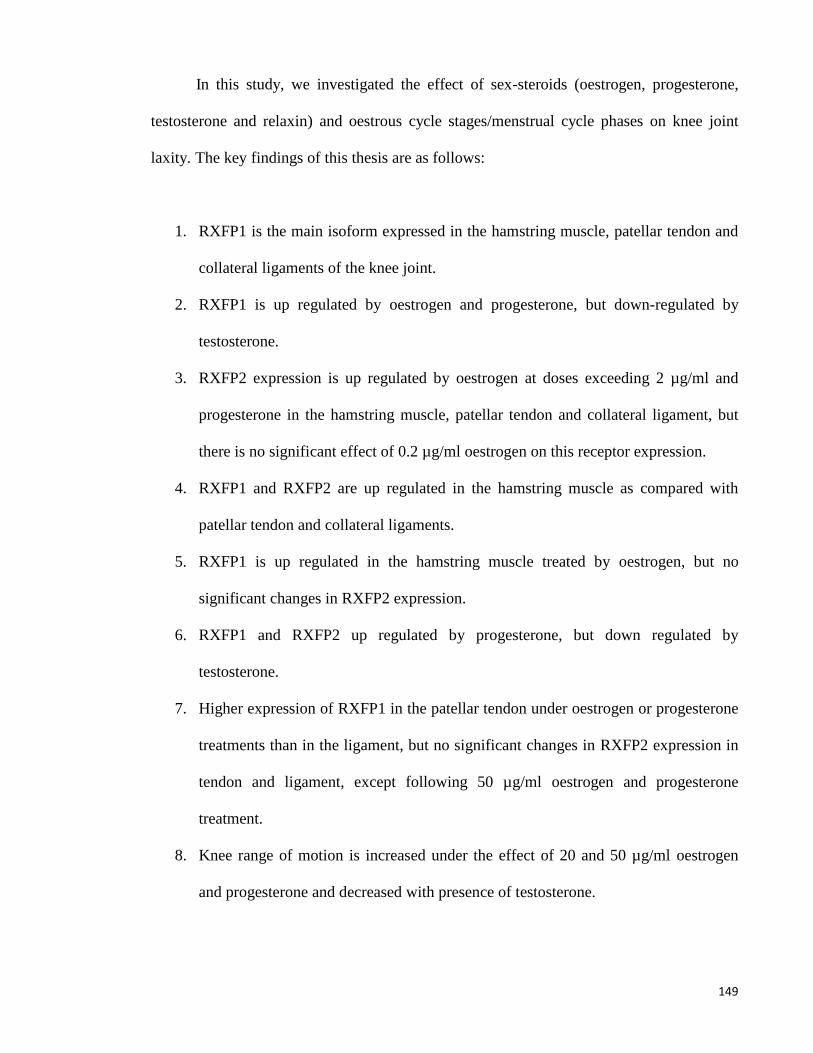
149
In this study, we investigated the effect of sex-steroids (oestrogen, progesterone,
testosterone and relaxin) and oestrous cycle stages/menstrual cycle phases on knee joint
laxity. The key findings of this thesis are as follows:
1. RXFP1 is the main isoform expressed in the hamstring muscle, patellar tendon and
collateral ligaments of the knee joint.
2. RXFP1 is up regulated by oestrogen and progesterone, but down-regulated by
testosterone.
3. RXFP2 expression is up regulated by oestrogen at doses exceeding 2 µg/ml and
progesterone in the hamstring muscle, patellar tendon and collateral ligament, but
there is no significant effect of 0.2 µg/ml oestrogen on this receptor expression.
4. RXFP1 and RXFP2 are up regulated in the hamstring muscle as compared with
patellar tendon and collateral ligaments.
5. RXFP1 is up regulated in the hamstring muscle treated by oestrogen, but no
significant changes in RXFP2 expression.
6. RXFP1 and RXFP2 up regulated by progesterone, but down regulated by
testosterone.
7. Higher expression of RXFP1 in the patellar tendon under oestrogen or progesterone
treatments than in the ligament, but no significant changes in RXFP2 expression in
tendon and ligament, except following 50 µg/ml oestrogen and progesterone
treatment.
8. Knee range of motion is increased under the effect of 20 and 50 µg/ml oestrogen
and progesterone and decreased with presence of testosterone.

150
9. Oestrogen antagonists (ICI 182/780, MPP, PHTPP) administration blocked
oestrogen influence on RXFP1 and RXFP2 expressions in hamstring muscle,
patellar tendon, and collateral ligament.
10. Knee range of motion is reduced following treatment with 0.2 µg/ml dose
oestrogen + ICI 182/780/ MPP/ PHTPP
11. Decreased knee range of motion and RXFP1 and RXFP2 expressions in presence
of mifepristone + progesterone in all hamstring muscle, patellar tendon and
collateral ligaments tissues.
12. RXFP1andRXFP2expressionsare increased in presence of flutamide and finastride
in hamstring muscle, patellar tendon and collateral ligament.
13. Knee range of motion in increased in the presence of flutamide and finastride.
14. The correlations of RXFP1andRXFP2 expressions with knee range of motion are
observed at proestrus and diestrus stages of the oestrous cycle.
15. The mean of varus-valgus angle is higher in the non-athletes group compared to
athletes group throughout the menstrual cycle phases.
16. Greater varus-valgus angle is observed in luteal phase than follicular phase in
athletes and non-athletes population
17. There is a significant correlation between progesterone/relaxin and knee laxity
among athletes and non-athletes groups in different phases of the menstrual cycle
18. There is no significant correlation between oestrogen/testosterone between both
athletes and non-athletes groups in different phases of the menstrual cycle.

151
In conclusion, investigation of complex relationship between several hormones and
expression of relaxin receptor isoforms on rat knee tissues indicated that RXFP1 is the
main isoform in these tissues and expressed higher in the hamstring muscle more than
patellar tendon and collateral ligaments. Progesterone and oestrogen up regulated and
testosterone down regulated the RXFP1 and RXFP2 expressions in the rat knee tissues.This
effect could be due to a decrease of collagen content in these tissues and extracellular
matrix in the presence of relaxin hormone. Hence, the effects of reproductive hormones on
knee surrounding tissues suggested that these hormones influence knee laxity. We
postulated that increase in plasma testosterone may result in down-regulation of relaxin
receptors in these knee structures, thus could help to reduce knee laxity in exercising
women. The highest level of progesterone in the luteal phase can cause an increase in knee
laxity, rendering the knee to be more susceptible towards a non-contact injury. In addition
to this, testosterone has also been reported to increase the strength of the muscles that
control knee joint movement.
Moreover, rat knee range of motion increased under treatment of oestrogen and
progesterone and decreased under testosterone. Progesterone treatment resulted in the
highest increase in the knee range of motion correlated to both relaxin receptor isoforms’
expressions in patellar tendon and lateral collateral ligaments. Increased expression of
RXFP1, as compared with RXFP2, mediates increased knee joint laxity in response to high
doses of oestrogen and physiological dose of progesterone. Our findings thus could explain
higher vulnerability of female than male towards non-traumatic knee with oestrogen and
progesterone fluctuation at various phases of reproductive cycle.

152
Our results also illustrated that testosterone by interceding relaxin receptors decreased
knee laxity and thereby range of motion. However, flutamide and finasteride antagonists
showed suppressor effect on testosterone and consequently altered RXFP1 and RXFP2
expressions in hamstring muscle, patellar tendon, and lateral collateral ligaments,
inequitably.Taken together, these findings may explain increased knee joint stiffness in
female following testosterone administration. These findings provide better understanding
of the underlying mechanism of sex-steroid modulation of relaxin receptors expression and
its influence on the knee laxity. Further studies should investigate the direct and indirect
mechanisms underlying these associations.
In this study, we also examined the expression of relaxin receptor during oestrous cycle.
Higher expressions of RXFP1and RXFP2 observed in proestrous and diestrous stages
correlated to higher knee range of motion. Progesterone and relaxin levels were at their
highest in the same stage as greater knee range of motion was observed. Relaxin hormone
can stimulates degradation of collagen content and prevents fibrogenesis (Samuel, 2005).
Ligamentous and tendonous laxity can partially be influenced by relaxin hormone during
proestrous and diestrous stages.
We evaluated serum oestrogen, progesterone, testosterone, relaxin hormones, and
varus-valgus assessments in female athletes and non-athletes. In line with our previous
findings, we found there was positive correlation between progesterone/relaxin and knee
rotational movement during different phases of a menstrual cycle. The knee rotational
movement for athletes and non-athletes did not show any relationship with
oestrogen/testosterone. The highest level of progesterone/relaxin and the greatest angle of
knee were observed in luteal phase in both groups and lowest level observed in follicular

153
phase. Since progesterone and relaxin are involved in collagen metabolism, these hormones
may influence knee joint laxity via this mechanism.
This study provided insight into the effect of sex-steroids on RXFP1 and RXFP2
expressions on the hamstring muscle, patellar tendon, and collateral ligament, and
relationship between oestrogen, progesterone, testosterone, and relaxin with knee rotational
movement during reproductive cycles. Increased expression of RXFP1, as compared with
RXFP2, mediates increased knee joint laxity in response to high doses oestrogen and
progesterone and at proestrous and diestrous stages of the oestrous cycle. Our findings thus
could explain higher vulnerability of female than male towards non-traumatic knee with
oestrogen and progesterone fluctuation at various phases of menstrual cycle. Finally, these
findings provide better understanding of the underlying mechanism of sex steroid
modulation and relaxin hormone interaction and its influence on the knee laxity.Future
studies on fluctuation of sex steroid hormones interaction in both gender are warranted to
elucidate the physiological cause of this selectivity in the impact of relaxin and endocrine
system on the phenotype of various tissue.
The human body joints’ appears to react to menstruation hormones levels (oestrogen,
progesterone, testosterone, and relaxin) by causing a change at the cellular level of the knee
tissue thus resulting in changing of knee laxity. Endogenous and exogenous female sex
steroids have effect on many metabolic parameters but these agents do not restrict the
female athletes to compete in sports, recreationally or professionally. It is important to
recognize all components of the knee injury mechanism in order to address the potential for
prevention and screening programs that considers for risk factors and multifaceted interplay
of these factors. More studies is needed to investigate the influence of hormones on joint

154
laxity as well as the influence of other factors that are associated with the female menstrual
cycle, such as weight, age, altered reaction times, the circulation of hormones, shoe-surface,
athletes level, fatigue, environmental conditions, water retention, to find an explanation for
the disproportional distribution of knee injuries.

155
REFERENCES
Adachi, N., Nawata, K., Maeta, M., & Kurozawa, Y. (2008). Relationship of the menstrual
cycle phase to anterior cruciate ligament injuries in teenaged female athletes. Arch
Orthop Trauma Surg, 128(5), 473-478.
Agel, J., Bershadsky, B., & Arendt, E. A. (2006). Hormonal therapy: ACL and ankle injury.
Med Sci Sport Exer, 38(1), 7-12.
Ahmad, N., Wang, W., Nair, R., & Kapila, S. (2012). Relaxin induces matrix-
metalloproteinases-9 and-13 via RXFP1: Induction of MMP-9 involves the PI3K,
ERK, Akt and PKC-ζ pathways. Molecular and Cellular Endocrinology, 363(1),
46-61.
Aldabe, D., Ribeiro, D. C., Milosavljevic, S., & Bussey, M. D. (2012). Pregnancy-related
pelvic girdle pain and its relationship with relaxin levels during pregnancy: a
systematic review. European Spine Journal, 21(9), 1769-1776.
Amis, A. A., Bull, A. J., Gupte, C. M., Hijazi, I., Race, A., & Robinson, J. R. (2003).
Biomechanics of the PCL and related structures: posterolateral, posteromedial and
meniscofemoral ligaments. Knee Surgery, Sports Traumatology, Arthroscopy,
11(5), 271-281.
Ar'Rajab, A., Dawidson, I., & Fabia, R. (1996). Reperfusion injury. New horizons
(Baltimore, Md.), 4(2), 224.
Arendt, E. A., Agel, J., & Dick, R. (1999). Anterior cruciate ligament injury patterns among
collegiate men and women. J Athl Train, 34(2), 86-92.
Asano, K., Maruyama, S., Usui, T., & Fujimoto, N. (2003). Regulation of estrogen receptor
alpha and alpha expression by testosterone in the rat prostate gland. Endocr J, 50(3),
281-287.
Bäckström, C., McNeilly, A., Leask, R., & Baird, D. (1982). Pulsatile secretion of LH,
FSH, prolactin, oestradiol and progesterone during the human menstrual cycle.
Clinical endocrinology, 17(1), 29-42.
Bani, D., Masini, E., Bello, M. G., Bigazzi, M., & Sacchi, T. B. (1998). Relaxin protects
against myocardial injury caused by ischemia and reperfusion in rat heart. The
American journal of pathology, 152(5), 1367-1376.
Barrett, K. E., Barman, S. M., Boitano, S., & Brooks, H. L. (2009). Ganong Review of
Medical Physiology. England: McGraw-Hill Medical
Bathgate, R. A., Ivell, R., Sanborn, B. M., Sherwood, O. D., & Summers, R. J. (2005).
Receptors for relaxin family peptides. [Research Support, Non-U.S. Gov'tReview].
Annals of the New York Academy of Sciences, 1041, 61-76.
156
Bathgate, R. A., Ivell, R., Sanborn, B. M., Sherwood, O. D., & Summers, R. J. (2006).
recommendations for the nomenclature of receptors for relaxin family peptides.
Pharmacol. Rev, 58(1), 7-31.
Bathgate, R. A. D., Halls, M. L., van der Westhuizen, E. T., Callander, G. E., Kocan, M., &
Summers, R. J. (2013). Relaxin Family Peptides and Their Receptors. Physiol Rev,
93(1), 405-480.
Bayard, F., Damilano, S., Robel, P., & Baulieu, E.-E. (1978). Cytoplasmic and Nuclear
Estradiol and Progesterone Receptors in Human Endometrium*. The Journal of
Clinical Endocrinology & Metabolism, 46(4), 635-648.
Bazin, J. E., Constantin, J. M., & Gindre, G. (2004). Laboratory animal anaesthesia:
influence of anaesthetic protocols on experimental models. Ann Fr Anesth, 23(8),
811-818.
Becker GJ, H. T. (2001). Relaxin and renal fibrosis. Kidney Int., 59(3), 1184-1185.
Belanger, M. J., Moore, D. C., Crisco, J. J., Fadale, P. D., Hulstyn, M. J., & Ehrlich, M. G.
(2004). Knee laxity does not vary with the menstrual cycle, before or after exercise.
The American journal of sports medicine, 32(5), 1150-1157.
Bell, D.R, B. J., Hackney AC, Marshall SW, Beutler AI, Padua DA. (2014 ). Jump-Landing
Biomechanics and Knee-Laxity Change Across the Menstrual Cycle in Women
With Anterior Cruciate Ligament Reconstruction. J Athl Train.
Bell, D. R., Myrick, M. P., Blackburn, J. T., Shultz, S. J., Guskiewicz, K. M., & Padua, D.
A. (2009). The effect of menstrual-cycle phase on hamstring extensibility and
muscle stiffness. J Sport Rehabil, 18(4), 553-563.
Bendjaballah, M. Z., Shirazi-Adl, A., & Zukor, D. (1997). Finite element analysis of human
knee joint in varus-valgus. Clinical Biomechanics, 12(3), 139-148.
Beynnon, B. D., Bernstein, I. M., Belisle, A., Brattbakk, B., Devanny, P., Risinger, R., &
Durant, D. (2005). The effect of estradiol and progesterone on knee and ankle joint
laxity. The American journal of sports medicine, 33(9), 1298-1304.
Beynnon, B. D., Johnson, R. J., Braun, S., Sargent, M., Bernstein, I. M., Skelly, J. M., &
Vacek, P. M. (2006b). The Relationship Between Menstrual Cycle Phase and
Anterior Cruciate Ligament Injury: A Case-Control Study of Recreational Alpine
Skiers. The American journal of sports medicine, 34(5), 757-764.
Bhasin, S., Storer, T. W., Berman, N., Callegari, C., Clevenger, B., Phillips, J., Casaburi, R.
(1996). The effects of supraphysiologic doses of testosterone on muscle size and
strength in normal men. New England Journal of Medicine, 335(1), 1-7.
Bolour, S., & Braunstein, G. (2005). Testosterone therapy in women: a review. Int J Impot
Res, 17(5), 399-408.

157
Bolt, M. W., & Mahoney, P. A. (1997). High-efficiency blotting of proteins of diverse sizes
following sodium dodecyl sulfate-polyacrylamide gel electrophoresis. [Research
Support, U.S. Gov't, P.H.S.]. Anal Biochem, 247(2), 185-192.
Bonaventure, J., Delatour, B., Tsagris, L., Eddie, L. W., Tregear, G., & Corvol, M. T.
(1988). Effect of Relaxin on the Phenotype of Collagens Synthesized by Cultured
Rabbit Chondrocytes. Biochimica et Biophysica Acta, 972(2), 209-220.
Bond, C. P., Parry, L. J., Samuel, C. S., Gehring, H. M., Lederman, F. L., Rogers, P. A. W.,
& Summers, R. J. (2004). Increased Expression of the Relaxin Receptor (LGR7) in
Human Endometrium during the Secretory Phase of the Menstrual Cycle. Journal of
Clinical Endocrinology & Metabolism, 89(7), 3477-3485.
Cammarata, M. L., & Dhaher, Y. Y. (2008). The differential effects of gender,
anthropometry, and prior hormonal state on frontal plane knee joint stiffness.
Clinical biomechanics (Bristol, Avon), 23(7), 937-945.
Campitiello, M. R., De Franciscis, P., Mele, D., Izzo, G., Sinisi, A., Delrio, G., &
Colacurci, N. (2011). Endometrial LGR7 expression during menstrual cycle.
Fertility and sterility, 95(8), 2511-2514.
Cárdenas, H., & Pope, W. F. (2005). Estrogen receptors in the uterus and ovarian follicles
of gilts treated with dihydrotestosterone. Domestic animal endocrinology, 29(3),
523-533.
Cernaro, V., Lacquaniti, A., Lupica, R., Buemi, A., Trimboli, D., Giorgianni, G., Buemi,
M. (2013). Relaxin: New Pathophysiological Aspects and Pharmacological
Perspectives for an Old Protein. Med Res Rev, n/a-n/a.
Cesar, G. M., Pereira, V. S., Santiago, P. R., Benze, B. G., da Costa, P. H., Amorim, C. F.,
& Serrao, F. V. (2011). Variations in dynamic knee valgus and gluteus medius onset
timing in non-athletic females related to hormonal changes during the menstrual
cycle. Knee, 18(4), 224-230.
Charlton, W. P., Coslett-Charlton, L. M., & Ciccotti, M. G. (2001). Correlation of estradiol
in pregnancy and anterior cruciate ligament laxity. Clin Orthop Relat Res, 387, 165-
170.
Chen, C. Z., Southall, N., Xiao, J., Marugan, J. J., Ferrer, M., Hu, X., Agoulnik, A. I.
(2012). Identification of Small-Molecule Agonists of Human Relaxin Family
Receptor 1 (RXFP1) by Using a Homogenous Cell-Based cAMP Assay. Journal of
biomolecular screening.
Chow, B.S. Zhao C, Bathgate RA, Hewitson TD, Samuel CS. (2012). Relaxin signals
through a RXFP1-pERK-nNOS-NO-cGMP-dependent pathway to up-regulate
matrix metalloproteinases: the additional involvement of iNOS. PLoS One., 7(8),
e42714.

158
Clezardin, P., & Teti, A. (2007). Bone metastasis: pathogenesis and therapeutic
implications. [Research Support, Non-U.S. Gov't Review]. Clin Exp Metastasis,
24(8), 599-608.
Cohen, J. (1988). Statistical power analysis for the social sciences.
Compton, D. R., Sheng, S., Carlson, K. E., Rebacz, N. A., Lee, I. Y., Katzenellenbogen, B.
S., & Katzenellenbogen, J. A. (2004). Pyrazolo [1, 5-a] pyrimidines: estrogen
receptor ligands possessing estrogen receptor β antagonist activity. Journal of
medicinal chemistry, 47(24), 5872-5893.
Conrad, K. P., & Baker, V. L. (2013). Corpus luteal contribution to maternal pregnancy
physiology and outcomes in assisted reproductive technologies. American Journal
of Physiology - Regulatory, Integrative and Comparative Physiology, 304(2), R69-
R72.
Cox, R. M., & John-Alder, H. B. (2005). Testosterone has opposite effects on male growth
in lizards (Sceloporus spp.) with opposite patterns of sexual size dimorphism. J Exp
Biol, 208(24), 4679-4687.
Crelin, E. S., & Brightman, M. W. (1957). The pelvis of the rat: its response to estrogen
and relaxin. The Anatomical record, 128(3), 467-483.
D'Elia, H. F., L.A., M., C., O., E., N., & H., C. (2003). Hormone replacement therapy in
rheumatoid arthritis is associated with lower serum levels of soluble IL-6 receptor
and higher insulin-like growth factor 1. Arthritis research & therapy, 5(4), R202-
209.
Dauvois, S., White, R., & Parker, M. G. (1993). The antiestrogen ICI 182780 disrupts
estrogen receptor nucleocytoplasmic shuttling. J Cell Sci, 106(4), 1377-1388.
Dawood, M. Y., & Saxena, B. B. (1976). Plasma testosterone and dihydrotestosterone in
ovulatory and anovulatory cycles. [Research Support, U.S. Gov't, P.H.S.]. American
journal of obstetrics and gynecology, 126(4), 430-435.
Dehghan, F., Haerian, B., Muniandy, S., Yusof, A., Dragoo, J., & Salleh, N. (2013). The
effect of relaxin on the musculoskeletal system. Scand J Med Sci Sports.
Dehghan, F., Muniandy, S., Yusof, A., & Salleh, N. (2014b). Testosterone Reduces Knee
Passive Range of Motion and Expression of Relaxin Receptor Isoforms via 5α-
Dihydrotestosterone and Androgen Receptor Binding. International journal of
molecular sciences, 15(3), 4619-4634.
Dehghan, F., Muniandy, S., Yusof, A., & Salleh, N. (2014a). Sex-steroid regulation of
relaxin receptor isoforms (RXFP1 & RXFP2) expression in the patellar tendon and
lateral collateral ligament of female WKY rats. Int J Med Sci, 11(2), 180-191.

159
Devan, M., R., Pescatello, L. S., Faghri, P., & Anderson, J. E. (2004). A prospective study
of overuse knee injuries among female athletes with muscle imbalances and
structural abnormalities. J Athl Train, 39(3), 263.
Devlin, T. M. (2011). Textbook of biochemistry: with clinical correlations: John Wiley &
Sons Hoboken, NJ.
Dhont, M. (2010). History of oral contraception. The European Journal of Contraception
and Reproductive Health Care, 15(S2), S12-S18.
Diegelmann, R. F., & Evans, M. C. (2004 ). Wound healing: an overview of acute, fibrotic
and delayed healing. Front Biosci, 1(9), 283-289.
Dragoo, J. L., Castillo, T. N., Braun, H. J., Ridley, B. A., Kennedy, A. C., & Golish, S. R.
(2011a). Prospective Correlation Between Serum Relaxin Concentration and
Anterior Cruciate Ligament Tears Among Elite Collegiate Female Athletes.
American Journal of Sports Medicine, 39(10), 2175-2180.
Dragoo, J. L., Castillo, T. N., Korotkova, T. A., Kennedy, A. C., Kim, H. J., & Stewart, D.
R. (2011b). Trends in serum relaxin concentration among elite collegiate female
athletes. International journal of women's health, 3, 19-24.
Dragoo, J. L., Lee, R. S., Benhaim, P., Finerman, G. A., & Hame, S. L. (2003). Relaxin
receptors in the human female anterior cruciate ligament. The American journal of
sports medicine, 31(4), 577-584.
Dragoo, J. L., Padrez, K., Workman, R., & Lindsey, D. P. (2009). The effect of relaxin on
the female anterior cruciate ligament: Analysis of mechanical properties in an
animal model. The Knee, 16(1), 69-72.
Dye, S. F., Vaupel, G. L., & Dye, C. C. (1998). Conscious neurosensory mapping of the
internal structures of the human knee without intraarticular anesthesia. The
American journal of sports medicine, 26(6), 773-777.
Edwards, D. P. (2005). Regulation of signal transduction pathways by estrogen and
progesterone. Annu. Rev. Physiol., 67, 335-376.
Facciolli, A., Ferlin, A., Gianesello, L., Pepe, A., & Foresta, C. (2009). Role of relaxin in
human osteoclastogenesis. Annals of the New York Academy of Sciences, 1160, 221-
225.
Faryniarz, D. A., Bhargava, M., Lajam, C., Attia, E. T., & Hannafin, J. A. (2006).
Quantitation of estrogen receptors and relaxin binding in human anterior cruciate
ligament fibroblasts. In Vitro Cell Dev Biol Anim, 42(7), 176-181.
Fenichel, P., Grimaldi, M., Olivero, J. F., Donzeau, M., Gillet, J. Y., & Harter, M. (1989).
Predictive value of hormonal profiles before stimulation for in vitro fertilization.
Fertility and sterility, 51(5), 845.
160
Ferlin, A., Pepe, A., Facciolli, A., Gianesello, L., & Foresta, C. (2010). Relaxin stimulates
osteoclast differentiation and activation. Bone, 46(2), 504-513.
Ferlin, A., Pepe, A., Gianesello, L., Garolla, A., Feng, S., Facciolli, A., . . . Foresta, C.
(2009). New roles for INSL3 in adults. [In Vitro]. Annals of the New York Academy
of Sciences, 1160, 215-218.
Ferlin, A., Pepe, A., Gianesello, L., Garolla, A., Feng, S., Giannini, S., . . . Foresta, C.
(2008). Mutations in the insulin-like factor 3 receptor are associated with
osteoporosis. Journal of bone and mineral research, 23(5), 683-693.
Fields, P. A., & Fields, M. J. (1985). Ultrastructural localization of relaxin in the corpus
luteum of the nonpregnant, pseudopregnant, and pregnant pig. Biology of
reproduction, 32(5), 1169-1179.
Figueiredo, K. A., Mui, A. L., Nelson, C. C., & Cox, M. E. (2006). Relaxin stimulates
leukocyte adhesion and migration through a relaxin receptor LGR7-dependent
mechanism. Journal of Biological Chemistry, 281(6), 3030-3039.
Flecknell, P. A. (2009). Laboratory Animal Anaesthesia. Academic Press.
Formigli, L., Francini, F., Chiappini, L., Zecchi-Orlandini, S., & Bani, D. (2005). Relaxin
favors the morphofunctional integration between skeletal myoblasts and adult
cardiomyocytes in coculture. Annals of the New York Academy of Sciences, 444-
445.
Forst, J., Forst, C., Forst, R., & Heller, K. D. (1997). Pathogenetic relevance of the
pregnancy hormone relaxin to inborn hip instability. [Clinical Trial]. Arch Orthop
Trauma Surg, 116(4), 209-212.
Fouladi, R., Rajabi, R., Naseri, N., Pourkazemi, F., & Geranmayeh, M. (2012). Menstrual
cycle and knee joint position sense in healthy female athletes. [Research Support,
Non-U.S. Gov't]. Knee surgery, sports traumatology, arthroscopy : official journal
of the ESSKA, 20(8), 1647-1652.
Frankovich, R. J., & Lebrun, C. M. (2000). Menstrual cycle, contraception, and
performance. Clin Sports Med., 19(2), 251-271.
Frye, S. V., Bramson, H. N., Hermann, D. J., Lee, F. W., Sinhababu, A. K., & Tian, G.
(1998). Discovery and development of GG745, a potent inhibitor of both isozymes
of 5 alpha-reductase. Pharm Biotechnol, 11, 393-422.
Fuss, F. K. (1989). Anatomy of the cruciate ligaments and their function in extension and
flexion of the human knee joint. American Journal of Anatomy, 184(2), 165-176.
Fytili, P., Giannatou, E., Papanikolaou, V., Stripeli, F., Karachalios, T., Malizos, K., &
Tsezou, A. (2005). Association of repeat polymorphisms in the estrogen receptors α,
β, and androgen receptor genes with knee osteoarthritis. Clin Genet, 68(3), 268-277.

161
Garibay, T., Jermelina, L., Okazaki, K. J., Tashima, L. S., Yamamoto, S., Bryant-G., &
Gillian, D. (2004). Regulation of the human relaxin genes H1 and H2 by steroid
hormones. Molecular and Cellular Endocrinology, 219(1–2), 115-125.
Gill, T. J., Van de Velde, S. K., Wing, D. W., Oh, L. S., Hosseini, A., & Li, G. (2009).
Tibiofemoral and patellofemoral kinematics after reconstruction of an isolated
posterior cruciate ligament injury: in vivo analysis during lunge. The American
journal of sports medicine, 37(12), 2377-2385.
Goh, W., Yamamoto, S. Y., Thompson, K. S., & Bryant-Greenwood, G. D. (2013).
Relaxin, Its Receptor (RXFP1), and Insulin-Like Peptide 4 Expression Through
Gestation and in Placenta Accreta. Reproductive Sciences, 20(8), 968-980.
Granchi, D., Cenni, E., Tigani, D., Trisolino, G., Baldini, N., & Giunti, A. (2008).
Sensitivity to implant materials in patients with total knee arthroplasties.
Biomaterials, 29(10), 1494-1500.
Griffin, L. Y., Agel, J., Albohm, M. J., Arendt, E. A., Dick, R. W., Garrett, W. E., . . .
Wojtys, E. M. (2000). Noncontact anterior cruciate ligament injuries: risk factors
and prevention strategies. J Am Acad Orthop Surg, 8(3), 141-150.
Guttridge, D. C. (2004). Signaling pathways weigh in on decisions to make or break
skeletal muscle. [Review]. Curr Opin Clin Nutr Metab Care, 7(4), 443-450.
Hamdi, M. M., & Mutungi, G. (2010). Dihydrotestosterone activates the MAPK pathway
and modulates maximum isometric force through the EGF receptor in isolated intact
mouse skeletal muscle fibres. The Journal of physiology, 588(3), 511-525.
Hansen, M., Couppe, C., Hansen, C. S. E., Skovgaard, D., Kovanen, V., Larsen, J. O., . . .
Kjaer, M. (2013). Impact of oral contraceptive use and menstrual phases on patellar
tendon morphology, biochemical composition and biomechanical properties in
female athletes. Journal of Applied Physiology.
Hansen, M., Miller, B. F., Holm, L., Doessing, S., Petersen, S. G., Skovgaard, D., . . .
Pingel, J. (2009). Effect of administration of oral contraceptives in vivo on collagen
synthesis in tendon and muscle connective tissue in young women. Journal of
Applied Physiology, 106(4), 1435-1443.
Hart, D. A., Reno, C., Frank, C. B., & Shrive, N. G. (2000). Pregnancy affects cellular
activity, but not tissue mechanical properties, in the healing rabbit medial collateral
ligament. Journal of Orthopaedic Research, 18(3), 462-471.
Harvey, M. A., Johnston, S. L., & Davies, G. A. (2008). Mid-trimester serum relaxin
concentrations and post-partum pelvic floor dysfunction. Acta Obstet Gynecol
Scand, 87(12), 1315-1321.

162
Hashem, G., Zhang, Q., Hayami, T., Chen, J., Wang, W., & Kapila, S. (2006). Relaxin and
beta-estradiol modulate targeted matrix degradation in specific synovial joint
fibrocartilages: progesterone prevents matrix loss. Arthritis research & therapy,
8(4), R98.
Hayes, E. (2004). Biology of primate relaxin: A paracrine signal in early pregnancy?
Reprod Biol Endocrin, 2(1), 36.
He, H., Yang, F., Liu, X., Zeng, X., Hu, Q., Zhu, Q., & Tu, B. (2007 ). Sex hormone ratio
changes in men and postmenopausal women with coronary artery disease.
Menopause, 14(3 ), 385-390.
Heitz, N. A., Eisenman, P. A., Beck, C. L., & Walker, J. A. (1999). Hormonal changes
throughout the menstrual cycle and increased anterior cruciate ligament laxity in
females. J Athl Train, 34(2), 144-149.
Hellio Le Graverand, M. P., Reno, C., & Hart, D. A. (1998). Influence of pregnancy on
gene expression in rabbit articular cartilage. [Research Support, Non-U.S. Gov't].
Osteoarthritis and cartilage / OARS, Osteoarthritis Research Society, 6(5), 341-
350.
Herrmann, P., Thoele, P., & Heppert, V. (2013). Infected knee prostheses : Part 1: Early
infection or acute hematogenous infection. Oper Orthop Traumatol, 25(3), 236-241.
Hewett, T. E., Zazulak, B. T., & Myer, G. D. (2007). Effects of the Menstrual Cycle on
Anterior Cruciate Ligament Injury Risk: A Systematic Review. The American
journal of sports medicine, 35(4), 659-668.
Hicks-Little, C. A., Thatcher, J. R., Hauth, J. M., Goldfuss, A. J., & Cordova, M. L. (2007).
Menstrual cycle stage and oral contraceptive effects on anterior tibial displacement
in collegiate female athletes. Journal of sports medicine and physical fitness, 47(2),
255-260.
HISAW., F. L. (1926). Experimental relaxation of the pubic ligament of guinea pig. Proc.
Soc. Exp. Biol. Med., 23, 6614563.
Ho, T. Y., Santora, K., Chen, J. C., Frankshun, A. L., & Bagnell, C. A. (2011). Effects of
relaxin and estrogens on bone remodeling markers, receptor activator of NF-kB
ligand (RANKL) and osteoprotegerin (OPG), in rat adjuvant-induced arthritis.
Bone, 48(6), 1346-1353.
Ho, T. Y. D., M. A.Bartol, F. F.Bagnell, C. A. (2009). Relaxin promotes matrix
metalloproteinase-2 and decreases Wnt/beta-catenin expression in the neonatal
porcine heart. [In VitroResearch Support, U.S. Gov't, Non-P.H.S.]. Annals of the
New York Academy of Sciences, 1160, 287-288.

163
Hofmann-Lehmann, R., Williams, A. L., Swenerton, R. K., Li, P. L., Rasmussen, R. A.,
Chenine, A. L., . . . Ruprecht, R. M. (2002). Quantitation of simian cytokine and
beta-chemokine mRNAs, using real-time reverse transcriptase-polymerase chain
reaction: variations in expression during chronic primate lentivirus infection. AIDS
Res Hum Retroviruses, 18(9), 627-639.
Hsu, W. H., Fisk, J. A., Yamamoto, Y., Debski, R. E., & Woo, S. L. (2006). Differences in
torsional joint stiffness of the knee between genders: a human cadaveric study. The
American journal of sports medicine, 34(5), 765-570.
Ireland, M. L. (2002). The female ACL: why is it more prone to injury? Orthop Clin North
Am, 33(4), 637-651.
Ishikawa, T., Harada, T., Kubota, T., & Aso, T. (2007). Testosterone inhibits matrix
metalloproteinase-1 production in human endometrial stromal cells in vitro.
Reproduction, 133(6), 1233-1239.
Janousek, A. T. J., D. G.Clatworthy, M.Higgins, L. D.Fu, F. H. (1999). Posterior cruciate
ligament injuries of the knee joint. [Review]. Sports Medicine, 28(6), 429-441.
Johnson, I. J., & Martin, H. (2012). Essential reproduction: John Wiley & Sons.
Johnson, M., Carter, G., Grint, C., & Lightman, S. (1993). Relationship between ovarian
steroids, gonadotrophins and relaxin during the menstrual cycle. Acta
endocrinologica, 129(2), 121-125.
Junge, A., Engebretsen, L., Alonso, J. M., Renstrom, P., Mountjoy, M., Aubry, M., &
Dvorak, J. (2008). Injury surveillance in multi-sport events: the International
Olympic Committee approach. Br J Sports Med, 42(6), 413-421.
Junqueira, L., Carneiro, J., & Long, J. (1986). Cartilage. Basic Histology: Los Altos: Lange
Medical Publications.
Kakarlapudi, T., & Bickerstaff, D. (2000). Knee instability: isolated and complex. Br J
Sports Med, 34(5), 395-400.
Karageanes, S. J., Blackburn, K., & Vangelos, Z. A. (2000). The association of the
menstrual cycle with the laxity of the anterior cruciate ligament in adolescent
female athletes. [Comparative Study]. Clin J Sport Med, 10(3), 162-168.
Kazumasa, F., Neil, B., Arvydas, U., Felix, R., Freddie, H. F., & Johnny, H. (2001). The
Use of an Antifibrosis Agent to Improve Muscle Recovery after Laceration. The
American journal of sports medicine, 29(4), 394-402.
Kerksick, C., Taylor, L. t., Harvey, A., & Willoughby, D. (2008). Gender-related
differences in muscle injury, oxidative stress, and apoptosis. [Comparative Study].
Med Sci Sport Exer, 40(10), 1772-1780.

164
Kirk, P. C. (2011). Maternal vasodilation in pregnancy: the emerging role of relaxin.
American Journal of Physiology - Regulatory, Integrative and Comparative
Physiology, 301(2), R267-R275.
Klump, K. L., Keel, P. K., Racine, S. E., Burt, S. A., Neale, M., Sisk, C. L., . . . Hu, J. Y.
(2013). The interactive effects of estrogen and progesterone on changes in
emotional eating across the menstrual cycle. J Abnorm Psychol, 122(1), 131-137.
Koay, E. S., Too, C. K., Greenwood, F. C., & Bryant-Greenwood, G. D. (1983). Relaxin
stimulates collagenase and plasminogen activator secretion by dispersed human
amnion and chorion cells in vitro. [Comparative Study]. J Clin Endocr Metab,
56(6), 1332-1334.
Kong, R. C., Shilling, P. J., Lobb, D. K., Gooley, P. R., & Bathgate, R. A. (2010).
Membrane receptors: structure and function of the relaxin family peptide receptors.
Molecular and Cellular Endocrinology, 320(1-2), 1-15.
Kouloumenta, V., Hatziefthimiou, A., Paraskeva, E., Gourgoulianis, K., & Molyvdas, P. A.
(2006). Non-genomic effect of testosterone on airway smooth muscle. British
journal of pharmacology, 149(8), 1083-1091.
Kristiansson, P., Svardsudd, K., & Von Schoultz, B. (1999). Reproductive hormones and
aminoterminal propeptide of type III procollagen in serum as early markers of
pelvic pain during late pregnancy. American journal of obstetrics and gynecology,
180(1 Pt 1), 128-134.
Kumagai, J., Hsu, S. Y., Matsumi, H., Roh, J. S., Fu, P., Wade, J. D., Hsueh, A. J. (2002).
INSL3/Leydig insulin-like peptide activates the LGR8 receptor important in testis
descent. [Research Support, U.S. Gov't, P.H.S.]. The Journal of biological
chemistry, 277(35), 31283-31286.
Kumar, V., Abbas, A. K., Fausto, N., & Mitchell, R. (2012). Robbins basic pathology:
Elsevier Health Sciences.
Kuznetsova, L. A., Chistyakova, O. V., Bondareva, V. M., Sharova, T. S., & Pertseva, M.
N. (2010). Disturbance of regulation of NO synthase activity by peptides of insulin
family in rat skeletal muscles in streptozotocin model of neonatal type 2 diabetes
mellitus. Doklady Biochemistry and Biophysics, 432(1), 123-125.
Kwok, S., & Higuchi, R. (1989). Avoiding false positives with PCR. Nature, 339(6221),
237-238.
Lee, H., Petrofsky, J. S., Daher, N., Berk, L., & Laymon, M. (2014). Differences in anterior
cruciate ligament elasticity and force for knee flexion in women: oral contraceptive
users versus non-oral contraceptive users. Eur J Appl Physiol, 114(2), 285-294.
165
Lee, W. J., Choi, I. K., Lee, J. H., Lee, J. S., Kim, Y. O., Rah, D. K., & Yun, C. O. (2012).
Relaxin-expressing adenovirus decreases collagen synthesis and up-regulates matrix
metalloproteinase expression in keloid fibroblasts: in vitro experiments. Plast
Reconstr Surg, 130(3), 407e-417e.
Lee, Y. J., Lee, E. B., Kwon, Y. E., Lee, J. J., Cho, W. S., Kim, H. A., & Song, Y. W.
(2003). Effect of estrogen on the expression of matrix metalloproteinase (MMP)-1,
MMP-3, and MMP-13 and tissue inhibitor of metalloproternase-1 in osteoarthritis
chondrocytes. Rheumatology international, 23(6), 282-288.
Lemoine, S., Granier, P., Tiffoche, C., Rannou-Bekono, F., Thieulant, M. L., &
Delamarche, P. (2003). Estrogen receptor alpha mRNA in human skeletal muscles.
Med Sci Sport Exer, 35(3), 439-443.
Lewin, P. (1952). The knee and related structures. Kimpton, London.
Li, Y., Negishi, S., Sakamoto, M., Usas, A., & Huard, J. (2005). The use of relaxin
improves healing in injured muscle. Annals of the New York Academy of Sciences,
1041, 395-397.
Liening, S. H., Stanton, S. J., Saini, E. K., & Schultheiss, O. C. (2010). Salivary
testosterone, cortisol, and progesterone: two-week stability, interhormone
correlations, and effects of time of day, menstrual cycle, and oral contraceptive use
on steroid hormone levels. Physiology & behavior, 99(1), 8-16.
Lippert, T. H., Steinberg, A., Seeger, H., Voelter, W., & Ruoff, H. J. (1987). Dose response
relationship of relaxin on the pubic ligament of the mouse. Clin Exp Obstet
Gynecol., 14(2), 80-83.
Liu, S. H., Al‐Shaikh, R., Panossian, V., Yang, R. S., Nelson, S. D., Soleiman, N., . . .
Lane, J. M. (1996). Primary immunolocalization of estrogen and progesterone target
cells in the human anterior cruciate ligament. J Orthopaed Res, 14(4), 526-533.
Lovering, R. M., & Romani, W. A. (2005). Effect of testosterone on the female anterior
cruciate ligament. American Journal of Physiology - Regulatory, Integrative and
Comparative Physiology, 289(1), R15-R22.
Lubahn, J., Ivance, D., Konieczko, E., & Cooney, T. (2006). Immunohistochemical
detection of relaxin binding to the volar oblique ligament. [Research Support, Non-
U.S. Gov't]. J Hand Surg Am, 31(1), 80-84.
MacLennan, A. (1981). Relaxin--a review. Aust N Z J Obstet Gynaecol., 21(4), 195-202.
MacLennan, A. H., Nicolson, R., Green, R. C., & Bath, M. (1986). Serum relaxin and
pelvic pain of pregnancy. [Research Support, Non-U.S. Gov't]. Lancet, 2(8501),
243-245.

166
Madan, M. S., Liu, Z. J., Gu, G. M., & King, G. J. (2007). Effects of human relaxin on
orthodontic tooth movement and periodontal ligaments in rats. American Journal of
Orthodontics and Dentofacial Orthopedics, 131(1), 8. e1-8. e10.
Marcondes, F. K., Bianchi, F. J., & Tanno, A. P. (2002). Determination of the estrous cycle
phases of rats: some helpful considerations. [Research Support, Non-U.S. Gov't].
Braz J Biol, 62(4A), 609-614.
Marieb, E. M., & Hoehn, K. N. (2013). Anatomy & Physiology (5 ed.): Benjamin
Cummings.
Marieb, E. N. (2009). Essentials of Human Anatomy and Physiology, 9e.
Marnach, M. L., Ramin, K. D., Ramsey, P. S., Song, S. W., Stensland, J. J., & An, K. N.
(2003). Characterization of the relationship between joint laxity and maternal
hormones in pregnancy. Obstetricians and Gynecologists, 101(2), 331-335.
Martineau, P. A., Al-Jassir, F., Lenczner, E., & Burman, M. L. (2004). Effect of the oral
contraceptive pill on ligamentous laxity. Clin J Sport Med, 14(5), 281-286.
Maseelall, P., Gardner, J., Wojtczuk, A., Weiss, G., & Goldsmith, L. T. (2009). Relaxin
receptor LGR7 (RXFP1) is regulated by estrogen. Annals of the New York Academy
of Sciences, 1160(1), 91-92.
Masterson, R., Hewitson, T. D., Kelynack, K., Martic, M., Parry, L., Bathgate, R., Becker,
G. (2004). Relaxin down-regulates renal fibroblast function and promotes matrix
remodelling in vitro. Nephrology Dialysis Transplantation, 19(3), 544-552.
Mendias, C. L., Gumucio, J. P., Davis, M. E., Bromley, C. W., Davis, C. S., & Brooks, S.
V. (2012). Transforming growth factor-beta induces skeletal muscle atrophy and
fibrosis through the induction of atrogin-1 and scleraxis. Muscle a d Nerve, 45(1),
55-59.
Mendias, C. L., Tatsumi, R., & Allen, R. E. (2004). Role of cyclooxygenase-1 and -2 in
satellite cell proliferation, differentiation, and fusion. Muscle a d Nerve 30(4), 497-
500.
Merchav, R., Feuermann, Y., Shamay, A., Ranen, E., Stein, U., Johnston, D. E., & Shahar,
R. (2005). Expression of relaxin receptor LRG7, canine relaxin, and relaxin-like
factor in the pelvic diaphragm musculature of dogs with and without perineal
hernia. Veterinary surgery : VS, 34(5), 476-481.
Messner, K., & Gao, J. (1998). The menisci of the knee joint. Anatomical and functional
characteristics, and a rationale for clinical treatment. J Anat, 193(2), 161-178.
Miao, D. F., J. C.Timm, J. C.Joo, N. E.Kapila, Y. L. (2011). The Treponema denticola
chymotrypsin-like protease dentilisin induces matrix metalloproteinase-2-dependent
fibronectin fragmentation in periodontal ligament cells. [Research Support, N.I.H.,
ExtramuralResearch Support, Non-U.S. Gov't]. Infect Immun, 79(2), 806-811.

167
Min, G., & Sherwood, O. D. (1996). Identification of specific relaxin-binding cells in the
cervix, mammary glands, nipples, small intestine, and skin of pregnant pigs.
[Research Support, Non-U.S. Gov't]. Biology of reproduction, 55(6), 1243-1252.
Montgomery, M. M., & Shultz, S. J. (2010). Isometric knee-extension and knee-flexion
torque production during early follicular and postovulatory phases in recreationally
active women. [Research Support, N.I.H., Extramural]. J Athl Train, 45(6), 586-
593.
Mooradian, A. D., Morley, J. E., & Korenman, S. G. (1987). Biological Actions of
Androgens. Endocrine reviews, 8(1), 1-28.
Mu, X., Urso, M. L., Murray, K., Fu, F., & Li, Y. (2010). Relaxin regulates MMP
expression and promotes satellite cell mobilization during muscle healing in both
young and aged mice. American Journal of Pathology, 177(5), 2399-2410.
Musah, A. I., Schwabe, C., Willham, R. L., & Anderson, L. L. (1986). Pelvic development
as affected by relaxin in three genetically selected frame sizes of beef heifers.
Biology of reproduction, 34, 363-369.
Myklebust, G., Maehlum, S., Holm, I., & Bahr, R. (1998). A prospective cohort study of
anterior cruciate ligament injuries in elite Norwegian team handball. Scand J Med
Sci Sports, 8(3), 149-153.
Nair, V. B. S., C. S.Separovic, F.Hossain, M. A.Wade, J. D. (2012). Human relaxin-2:
historical perspectives and role in cancer biology. [Research Support, Non-U.S.
Gov'tReview]. Amino Acids, 43(3), 1131-1140.
Naqvi, T., Duong, T. T., Hashem, G., Shiga, M., Zhang, Q., & Kapila, S. (2005). Relaxin's
induction of metalloproteinases is associated with the loss of collagen and
glycosaminoglycans in synovial joint fibrocartilaginous explants. Arthritis research
& therapy, 7(1), 1-11.
Negishi, S., Li, Y., Usas, A., Fu, F. H., & Huard, J. (2005). The effect of relaxin treatment
on skeletal muscle injuries. American Journal of Sports Medicine, 33(12), 1816-
1824.
O'Leary, C. B., Lehman, C., Koltun, K., Smith-Ryan, A., & Hackney, A. C. (2013).
Response of testosterone to prolonged aerobic exercise during different phases of
the menstrual cycle. Eur J Appl Physiol, 113(9), 2419-2424.
Ohtera, K., Zobitz, M. E., Luo, Z. P., Morrey, B. F., O'Driscoll, S. W., Ramin, K. D., & An,
K. N. (2002). Effect of pregnancy on joint contracture in the rat knee. [Research
Support, Non-U.S. Gov't]. Journal of Applied Physiology, 92(4), 1494-1498.
Osborne, C. K., Wakeling, A., & Nicholson, R. I. (2004). Fulvestrant: an oestrogen
receptor antagonist with a novel mechanism of action. British journal of cancer, 90,
S2-S6.

168
Palmer, S., Wiegand, A. P., Maldarelli, F., Bazmi, H., Mican, J. M., Polis, M., Coffin, J. M.
(2003). New real-time reverse transcriptase-initiated PCR assay with single-copy
sensitivity for human immunodeficiency virus type 1 RNA in plasma. J Clin
Microbiol, 41(10), 4531-4536.
Parhizkar, S., Latiff, R. I., & Abdul, L. (2008). Incision choice in laparatomy: a comparison
of two incision techniques in ovariectomy of rats. World Applied Sciences Journal,
4(4), 537-540.
Park, J.-I., Chang, C., & Hsu, S. (2005). New Insights into Biological Roles of Relaxin and
Relaxin-related Peptides. Reviews in Endocrine and Metabolic Disorders, 6(4),
291-296.
Park, S. K., Stefanyshyn, D. J., Loitz-Ramage, B., Hart, D. A., & Ronsky, J. L. (2009a).
Changing hormone levels during the menstrual cycle affect knee laxity and stiffness
in healthy female subjects. The American journal of sports medicine, 37(3), 588-
598.
Park, S. K., Stefanyshyn, D. J., Ramage, B., Hart, D. A., & Ronsky, J. L. (2009b).
Relationship between knee joint laxity and knee joint mechanics during the
menstrual cycle. Br J Sports Med, 43(3), 174-179.
Parry, L. J., & Vodstrcil, L. A. (2007). Relaxin physiology in the female reproductive tract
during pregnancy Relaxin and Related Peptides (pp. 34-48): Springer.
Pearson, S. J., Burgess, K. E., & Onambélé, G. L. (2011). Serum relaxin levels affect the in
vivo properties of some but not all tendons in normally menstruating young
women. Exp Physiol, 96(7), 681-688.
Pehrsson, M., Westberg, L., Landen, M., & Ekman, A. (2007). Stable serum levels of
relaxin throughout the menstrual cycle: a preliminary comparison of women with
premenstrual dysphoria and controls. Arch Womens Ment Health, 10(4), 147-153.
Perezgrovas, R., & Anderson, L. L. (1982). Effect of porcine relaxin on cervical dilatation,
pelvic area and parturition in beef heifers. Biology of reproduction, 26(4), 765-776.
Pertseva, M., Shpakov, A., Kuznetsova, L., Plesneva, S., & Omeljaniuk, E. (2006).
Adenylyl cyclase signaling mechanisms of relaxin and insulin action: Similarities
and differences. Cell biology international, 30(6), 533-540.
Petersen, L. K., Hvidman, L., & Uldbjerg, N. (1994). Normal serum relaxin in women with
disabling pelvic pain during pregnancy. Gynecologic and obstetric investigation,
38(1), 21-23.
Pettegrew, C. J., Jayini, R., & Islam, M. R. (2009). Transfer buffer containing methanol can
be reused multiple times in protein electrotransfer. J Biomol Tech, 20(2), 93-95.
169
Piedras-Renteria, E. S., Sherwood, O. D., & Best, P. M. (1997). Effects of relaxin on rat
atrial myocytes. I. Inhibition of I(to) via PKA-dependent phosphorylation.
American Journal of Physiology - Heart and Circulatory Physiology, 272(4),
H1791-H1797.
Plesneva, S. A., Kuznetsova, L. A., Shpakov, A. O., Sharova, T. S., & Pertseva, M. N.
(2008). Structural and functional organization of the adenylyl cyclase signaling
mechanism of insulin-like growth factor 1 revealed in the muscle tissue in
vertabrates and invertebrates. Zhurnal evoliutsionnoi biokhimii i fiziologii, 44(5),
459-466.
Qin, X., Garibay-Tupas, J., Chua, P. K., Cachola, L., & Bryant-Greenwood, G. D. (1997).
An autocrine/paracrine role of human decidual relaxin. I. Interstitial collagenase
(matrix metalloproteinase-1) and tissue plasminogen activator. Biology of
reproduction, 56(4), 800-811.
Rajabi, M., Solomon, Samuel, Poole, Robin. (1991). Hormonal Regulation of Interstitial
Collagenase in the Uterine Cervix of the Pregnant Guinea Pig*. Endocrinology,
128(2), 863-871.
Rao, A. J., & Kotagi, S. G. (1982). Serum Testosterone Levels during the Menstrual Cycle
and Early Pregnancy in the Bonnet Monkey (<I>Macaca Radiata</I>).
Endocrinologia Japonica, 29(3), 271-275.
Rempel, D., & Abrahamsson, S. O. (2001). The effects of reduced oxygen tension on cell
proliferation and matrix synthesis in synovium and tendon explants from the rabbit
carpal tunnel: an experimental study in vitro. J Orthopaed Res, 19(1), 143-148.
Robertson, J. F. R. (2001). Faslodex (ICI 182, 780), a novel estrogen receptor
downregulator—future possibilities in breast cancer. J Steroid Biochem Mol Biol,
79(1), 209-212.
Romani, W., Curl, L. A., Lovering, R. M., & McLaughlin, K. (2001). The effect of
endogenous estradiol levels at three phases of the menstrual cycle on anterior
cruciate ligament stiffness in active females. J Athl Train, 2, S62.
Romani, W., Patrie, J., Curl, L. A., & Flaws, J. A. (2003). The correlations between
estradiol, estrone, estriol, progesterone, and sex hormone-binding globulin and
anterior cruciate ligament stiffness in healthy, active females. J Womens Health,
12(3), 287-298.
Romero, J., Duronio, J. F., Sohrabi, A., Alexander, N., MacWilliams, B. A., Jones, L. C., &
Hungerford, D. S. (2002). Varus and valgus flexion laxity of total knee alignment
methods in loaded cadaveric knees. Clin Orthop Relat Res, 394, 243-253.
Rosengren, K. J., Lin, F., Bathgate, R. A., Tregear, G. W., Daly, N. L., Wade, J. D., &
Craik, D. J. (2006 ). Solution structure and novel insights into the determinants of
the receptor specificity of human relaxin-3. The Journal of biological chemistry,
281(9), 5845-5851.
170
Rozzi, S. L., Lephart, S. M., Gear, W. S., & Fu, F. H. (1999). Knee Joint Laxity and
Neuromuscular Characteristics of Male and Female Soccer and Basketball Players.
The American journal of sports medicine, 27(3), 312-319.
Ruedl, G., Ploner, P., Linortner, I., Schranz, A., Fink, C., Patterson, C., . . . Burtscher, M.
(2011). Interaction of potential intrinsic and extrinsic risk factors in ACL injured
recreational female skiers. [Research Support, Non-U.S. Gov't]. Int J Sports Med,
32(8), 618-622.
Samuel, C., Coghlan, J., & Bateman, J. (1998). Effects of relaxin, pregnancy and
parturition on collagen metabolism in the rat pubic symphysis. Journal of
Endocrinology, 159(1), 117-125.
Samuel, C. S. (2005). Relaxin: antifibrotic properties and effects in models of disease. Clin
Med Res, 3(4), 241-249.
Samuel, C. S., Butkus, A., Coghlan, J. P., & Bateman, J. F. (1996). The effect of relaxin on
collagen metabolism in the nonpregnant rat pubic symphysis: the influence of
estrogen and progesterone in regulating relaxin activity. Endocrinology, 137(9),
3884-3890.
Santiago, J. R., Nolledo, M. S., Kinzler, W., & Santiago, T. V. (2001). Sleep and sleep
disorders in pregnancy. Ann Intern Med, 134(5), 396-408.
Santora, K., Rasa, C., Visco, D., Steinetz, B., & Bagnell, C. (2005). Effects of relaxin in a
model of rat adjuvant-induced arthritis. [Research Support, Non-U.S. Gov't]. Annals
of the New York Academy of Sciences, 1041, 481-485.
Sarrazy, V., Billet, F., Micallef, L., Coulomb, B., & Desmoulière, A. (2011). Mechanisms
of pathological scarring: Role of myofibroblasts and current developments. Wound
Repair Regen, 19, s10-s15.
Sato K, Li Y, Foster W, Fukushima K, Badlani N, Adachi N, . . . Huard J. (2003).
Improvement of muscle healing through enhancement of muscle regeneration and
prevention of fibrosis. Muscle Nerve. Sep, 28(3), 365-372.
Saugstad, L. F. (1991). Persistent pelvic pain and pelvic joint instability. European journal
of obstetrics, gynecology, and reproductive biology, 41(3), 197-201.
Schauberger, C. W., Rooney, B. L., Goldsmith, L., Shenton, D., Silva, P. D., & Schaper, A.
(1996). Peripheral joint laxity increases in pregnancy but does not correlate with
serum relaxin levels. American journal of obstetrics and gynecology, 174(2), 667-
671.
Schmitz, R. J., Sauret, J. J., & Shultz, S. J. (2013). Anterior tibiofemoral intersegmental
forces during landing are predicted by passive restraint measures in women. Knee,
20(6), 493-499.

171
Schwabe, C., & Büllesbach, E. (2007). Relaxin, the Relaxin-Like Factor and Their
Receptors. In A. Agoulnik (Ed.), Relaxin and Related Peptides (Vol. 612, pp. 14-
25): Springer New York.
Sciore, P., Frank, C. B., & Hart, D. A. (1998). Identification of sex hormone receptors in
human and rabbit ligaments of the knee by reverse transcription-polymerase chain
reaction: evidence that receptors are present in tissue from both male and female
subjects. J Orthop Res, 16(5), 604-610.
Scott, D. J., Layfield, S., Riesewijk, A., Morita, H., Tregear, G. W., & Bathgate, R. A.
(2005). Characterization of the mouse and rat relaxin receptors. [Research Support,
Non-U.S. Gov't]. Annals of the New York Academy of Sciences, 1041, 8-12.
Scott, D. J., Rosengren, K. J., & Bathgate, R. A. D. (2012). The Different Ligand-Binding
Modes of Relaxin Family Peptide Receptors RXFP1 and RXFP2. Molecular
Endocrinology, 26(11), 1896-1906.
Shan, B., Li, W., Yang, S.-Y., & Li, Z.-R. (2013). Estrogen up-regulates MMP2/9
expression in endometrial epithelial cell via VEGF-ERK1/2 pathway. Asian Pacific
journal of tropical medicine, 6(10), 826-830.
Sherwood, L. (2011). Human physiology: from cells to systems: Cengage Learning.
Sherwood, O. (1994). Relaxin. [Neill J. New York: Raven Press]. In The Physiology of
Reproduction(Edited by Knobil E).
Sherwood, O., Downing, S., Guico-Lamm, M., Hwang, J., O'Day-Bowman, M., & Fields,
P. (1992). The physiological effects of relaxin during pregnancy: studies in rats and
pigs. Oxf Rev Reprod Biol, 15, 143-189.
Sherwood, O. D. (2004). Relaxin’s Physiological Roles and Other Diverse Actions.
Endocrine reviews, 25(2), 205-234.
Sherwood OD, D. S., Guico-Lamm ML, Hwang JJ, O'Day-Bowman MB, Fields PA.
(1993). The physiological effects of relaxin during pregnancy: studies in rats and
pigs. Oxf Rev Reprod Biol., 15, 143-189.
Sherwood OD, e. b. K. E., Neill JD, Greenwald GS, Markert CL, Pfaff DW, editors.
(1994). Relaxin In: The Physiology of Reproduction. New York: Raven,, 861–1008
Sherwood, O. D. F., Phillip A. & Steinetz, Bernard G (Editors). . (2005). Relaxin and
related peptides. Annals of the New York Academy of Science, 1041
Shpakov, A. O., Gur'yanov, I. A., Kuznetsova, L. A., Plesneva, S. A., Shpakova, E. A.,
Vlasov, G. P., & Pertseva, M. N. (2006). Peptide derivatives of the LGR7 relaxin
receptor control the functional activity of relaxin-sensitive adenylate cyclase.
[Research Support, Non-U.S. Gov't]. Doklady. Biochemistry and biophysics, 407,
109-112.

172
Shpakov, A. O., Kuznetsova, L. A., Plesneva, S. A., Bondareva, V. M., & Pertseva, M. N.
(2007). Functional coupling of hormone receptors with G proteins in the adenylate
cyclase system of the rat muscle tissues and brain under conditions of short-term
hyperglycemia. Bulletin of experimental biology and medicine, 144(5), 684-688.
Shpakov, A. O., Kuznetsova, L. A., Plesneva, S. A., & Pertseva, M. N. (2004). Role of
phosphatidylinositol-3-kinase and protein kinase Cζ in the adenylate cyclase signal
mechanism of action of relaxin in muscle tissues of rats and mollusks. Bulletin of
experimental biology and medicine, 138(4), 372-375.
Shultz, S. J., Gansneder, B. M., Sander, T. C., Kirk, S. E., & Perrin, D. H. (2006). Absolute
serum hormone levels predict the magnitude of change in anterior knee laxity across
the menstrual cycle. J Orthopaed Res, 24(2), 124-131.
Shultz, S. J., Kirk, S. E., Johnson, M. L., Sander, T. C., Perrin, D. H., & SANDRA, J.
(2004). Relationship between Sex Hormones and Anterior Knee Laxity across the
Menstrual Cycle. Med Sci Sports Exerc, 36, 1165–1174.
Shultz, S. J., Levine, B. J., Nguyen, A. D., Kim, H., Montgomery, M. M., & Perrin, D. H.
(2010). A comparison of cyclic variations in anterior knee laxity, genu recurvatum,
and general joint laxity across the menstrual cycle. . J Orthopaed Res, 28, 1411-
1417.
Shultz, S. J., Sander, T. C., Kirk, S. E., & Perrin, D. H. (2005). Sex differences in knee
joint laxity change across the female menstrual cycle. J Sports Med Phys Fitness,
45(4), 594-603.
Shultz, S. J., Wideman, L., Montgomery, M. M., Beasley, K. N., & Nindl, B. C. (2012).
Changes in serum collagen markers, IGF-I, and Knee joint laxity across the
menstrual cycle. J Orthopaed Res, 30(9), 1405-1412.
Siebel, A. L., Gehring, H. M., Reytomas, I. G., & Parry, L. J. (2003 ). Inhibition of
oxytocin receptor and estrogen receptor-alpha expression, but not relaxin receptors
(LGR7), in the myometrium of late pregnant relaxin gene knockout mice.
Endocrinology, 144(10), 4272-4275.
Siebel AL, G. H., Reytomas IG, Parry LJ. (2003 ). Inhibition of oxytocin receptor and
estrogen receptor-alpha expression, but not relaxin receptors (LGR7), in the
myometrium of late pregnant relaxin gene knockout mice. Endocrinology, 144(10),
4272-4275.
Slauterbeck, J. R., Fuzie, S. F., Smith, M. P., Clark, R. J., Xu, K., Starch, D. W., & Hardy,
D. M. (2002). The Menstrual Cycle, Sex Hormones, and Anterior Cruciate
Ligament Injury. J Athl Train, 37(3), 275-278.
Smith, C. M., Ryan, P. J., Hosken, I. T., Ma, S., & Gundlach, A. L. (2011). Relaxin-3
systems in the brain--the first 10 years. [Research Support, Non-U.S. Gov't
Review]. J Chem Neuroanat, 42(4), 262-275.

173
Snell, R. S. (2011). Clinical anatomy by regions: Lippincott Williams & Wilkins.
Soh, Y. M. T., A.Mahendroo, M.Conrad, K. P.Parry, L. J. (2012). Relaxin regulates
hyaluronan synthesis and aquaporins in the cervix of late pregnant mice. [Research
Support, N.I.H., Extramural Research Support, Non-U.S. Gov't]. Endocrinology,
153(12), 6054-6064.
Song, X., Zhao, P., Wang, G., & Zhao, X. (2014). The Effects of Estrogen and Androgen
on Tear Secretion and Matrix Metalloproteinase-2 Expression in Lacrimal Glands of
Ovariectomized Rats. Invest Ophth Vis Sci, 55(2), 745-751.
Speroff, L., Glass, R. H., & Kase, N. G. (1989). Hormone Biosynthesis, Metabolism and
Mechanism of Action. Baltimore, MD, USA: Williams & Wilkins.
Srinivasan, N., Aruldhas, M., & Govindarajulu, P. (1986). Sex steroid induced changes in
collagen of the prostate and seminal vesicle of rats. J Androl, 55-58.
Staley, K., & Scharfman, H. (2005). A woman's prerogative. Nature neuroscience, 8(6),
697-699.
Stamler, J. S., & Meissner, G. (2001). Physiology of nitric oxide in skeletal muscle.
Physiol. Rev, 81(1), 210-231.
Steinetz, B. G., Williams, A. J., Lust, G., Schwabe, C., Bullesbach, E. E., & Goldsmith, L.
T. (2008). Transmission of relaxin and estrogens to suckling canine pups via milk
and possible association with hip joint laxity. Am J Vet Res, 69(1), 59-67.
Strickland, S. M., Belknap, T. W., Turner, S. A., Wright, T. M., & Hannafin, J. A. (2003).
Lack of hormonal influences on mechanical properties of sheep knee ligaments. The
American journal of sports medicine, 31(2), 210-215.
Summers, R. J., Bathgate, R. A., Wade, J. D., van der Westhuizen, E. T., & Halls, M. L.
(2009). Roles of the receptor, the ligand, and the cell in the signal transduction
pathways utilized by the relaxin family peptide receptors 1-3. [Research Support,
Non-U.S. Gov't]. Annals of the New York Academy of Sciences, 1160, 99-104.
Sutton, K. M., & Bullock, J. M. (2013). Anterior Cruciate Ligament Rupture: Differences
Between Males and Females. J Am Acad Orthop Sur, 21(1), 41-50.
Syed, F., Ahmadi, E., Iqbal, S. A., Singh, S., McGrouther, D. A., & Bayat, A. (2011).
Fibroblasts from the growing margin of keloid scars produce higher levels of
collagen I and III compared with intralesional and extralesional sites: clinical
implications for lesional site-directed therapy. British Journal of Dermatology,
164(1), 83-96.
Thornton, G. M., Bailey, S. J., Shao, X., Morck, D., Hart, D. A., & Achari, Y. (2007).
Influence of Early Ovariohysterectomy on the Mechanical Properties of Rabbit
Medial Collateral Ligament. Paper presented at the ASME 2007 Summer
Bioengineering Conference.

174
Toth, A. P., & Cordasco, F. A. (2001). Anterior cruciate ligament injuries in the female
athlete. [Review]. J Gend Specif Med, 4(4), 25-34.
Traina, S. M., & Bromberg, D. F. (1997). ACL injury patterns in women. Orthopedics,
20(6), 545-549; quiz 550-541.
Tsai, Y.-L., Wang, H.-T. T., Chang, H.-F. G., Tsai, C.-F., Lin, C.-K., Teng, P.-H., . . . Lee,
P.-Y. (2012). Development of TaqMan Probe-Based Insulated Isothermal PCR
(iiPCR) for Sensitive and Specific On-Site Pathogen Detection. Plos One, 7(9),
e45278.
Ullrich, K., Krudwig, W. K., & Witzel, U. (2002). Posterolateral aspect and stability of the
knee joint. I. Anatomy and function of the popliteus muscle-tendon unit: an
anatomical and biomechanical study. Knee Surgery, Sports Traumatology,
Arthroscopy, 10(2), 86-90.
Vinall, R., Mahaffey, C., Davis, R., Luo, Z., Gandour-Edwards, R., Ghosh, P., de Vere
White, R. (2011). Dual Blockade of PKA and NF–κB Inhibits H2 Relaxin-Mediated
Castrate-Resistant Growth of Prostate Cancer Sublines and Induces Apoptosis.
Hormones and Cancer, 2(4), 224-238.
Vodstrcil, L. A., Shynlova, O., Verlander, J. W., Wlodek, M. E., & Parry, L. J. (2010a).
Decreased expression of the rat myometrial relaxin receptor (RXFP1) in late
pregnancy is partially mediated by the presence of the conceptus. Biology of
reproduction, 83(5), 818-824.
Vodstrcil, L. A., Shynlova, O., Westcott, K., Laker, R., Simpson, E., Wlodek, M. E., &
Parry, L. J. (2010b ). Progesterone withdrawal, and not increased circulating
relaxin, mediates the decrease in myometrial relaxin receptor (RXFP1) expression
in late gestation in rats. Biology of reproduction, 83(5), 825-832.
Volk, S. W., Wang, Y., Mauldin, E. A., Liechty, K. W., & Adams, S. L. (2011).
Diminished Type III Collagen Promotes Myofibroblast Differentiation and
Increases Scar Deposition in Cutaneous Wound Healing. Cells Tissues Organs,
194(1), 25-37.
Vollestad, N. K., Torjesen, P. A., & Robinson, H. S. (2012). Association between the
serum levels of relaxin and responses to the active straight leg raise test in
pregnancy. Man Ther, 17(3), 225-230.
Wakeling, A. E. (2000). Similarities and distinctions in the mode of action of different
classes of antioestrogens. Endocrine-Related Cancer, 7(1), 17-28.
Walf, A. A., Koonce, C., Manley, K., & Frye, C. A. (2009 ). Proestrous compared to
diestrous wildtype, but not estrogen receptor beta knockout, mice have better
performance in the spontaneous alternation and object recognition tasks and reduced
anxiety-like behavior in the elevated plus and mirror maze. Behav Brain Res,
196(2), 254-260.

175
Walker, J. M. (1996). The protein protocols handbook (Vol. 1996): Springer.
Walker, J. M. (2007). The Protein Protocols Hand book. Human press
Wang, W., Hayami, T., & Kapila, S. (2009a). Female hormone receptors are differentially
expressed in mouse fibrocartilages. Osteoarthritis and cartilage / OARS,
Osteoarthritis Research Society, 17(5), 646-654.
Wang, W., Hayami, T., & Kapila, S. (2009b). Female hormone receptors are differentially
expressed in mouse fibrocartilages. Osteoarthritis and cartilage / OARS,
Osteoarthritis Research Society, 17(5), 646-654.
Weinberg, A. (1956). An x-ray pelvimetric study of relaxin extract in pelvic expansion.
Surg Gynecol Obstet, 103(3), 303-306.
Widmaier, E. P., Raff, H., & Strang, K. T. (2006). Vander's human physiology.
Wierman, M. E. (2007). Sex steroid effects at target tissues: mechanisms of action.
Advances in physiology education, 31(1), 26-33.
Wiertsema, S., Van Hooff, H., Migchelsen, L., & Steultjens, M. (2008). Reliability of the
KT1000 arthrometer and the Lachman test in patients with an ACL rupture. Knee,
15(2), 107-110.
Winn, R. J., Baker, M. D., Merle, C. A., & Sherwood, O. D. (1994). Individual and
combined effects of relaxin, estrogen, and progesterone in ovariectomized gilts. II.
Effects on mammary development. Endocrinology, 135(3), 1250-1255.
Wiqvist, I., Norstrom, A., O’Byrne, E., & Wiqvist, N. (1984). Regulatory influenceof
relaxin on human cervical and uterine connective tissue. ActaEndocrinol (Copenh),
106(1), 127-132.
Wojtys, E. M., Huston, L. J., Boynton, M. D., Spindler, K. P., & Lindenfeld, T. N. (2002).
The Effect of the Menstrual Cycle on Anterior Cruciate Ligament Injuries in
Women as Determined by Hormone Levels. The American journal of sports
medicine, 30(2), 182-188.
Wojtys, E. M., Huston, L. J., Lindenfeld, T. N., Hewett, T. E., & Greenfield, M. H. (1998).
Association Between the Menstrual Cycle and Anterior Cruciate Ligament Injuries
in Female Athletes. The American journal of sports medicine, 26(5), 614-619.
Wojtys, E. M., Huston, L. J., Schock, H. J., Boylan, J. P., & Ashton-Miller, J. A. (2003).
Gender differences in muscular protection of the knee in torsion in size-matched
athletes. The Journal of bone and joint surgery, 85-A(5), 782-789.
Wolf, J. M., Williams, A. E., Delaronde, S., Leger, R., Clifton, K. B., & King, K. B.
(2013). Relationship of serum relaxin to generalized and trapezial-metacarpal joint
laxity. J Hand Surg Am, 38(4), 721-728.

176
Wong, M. L., & Medrano, J. F. (2005). Real-time PCR for mRNA quantitation.
Biotechniques, 39(1), 75.
Wood, M. L., Luthin, W. N., Lester, G. E., & Dahners, L. E. (2003). Tendon creep is
potentiated by NKISK and relaxin which produce collagen fiber sliding. Iowa
Orthop J, 23, 75-79.
Woodhouse, E., Schmale, G. A., Simonian, P., Tencer, A., Huber, P., & Seidel, K. (2007).
Reproductive hormone effects on strength of the rat anterior cruciate ligament. Knee
Surgery, Sports Traumatology, Arthroscopy, 15(4), 453-460.
Wreje, U., Kristiansson, P., Aberg, H., Bystrom, B., & Von Schoultz, B. (1995). Serum
levels of relaxin during the menstrual cycle and oral contraceptive use. Gynecol
Obstet Invest, 39(3), 197-200.
Yan, W., Chen, J., Wiley, A. A., Crean-Harris, B. D., Bartol, F. F., & Bagnell, C. A.
(2008). Relaxin (RLX) and estrogen affect estrogen receptor alpha, vascular
endothelial growth factor, and RLX receptor expression in the neonatal porcine
uterus and cervix. Reproduction, 135(5), 705-712.
Yu, W. D., Liu, S. H., Hatch, J. D., Panossian, V., & Finerman, G. A. (1999). Effect of
estrogen on cellular metabolism of the human anterior cruciate ligament. Clin
Orthop Relat Res(366), 229-238.
Zazulak, B. T., Paterno, M., Myer, G. D., Romani, W. A., & Hewett, T. E. (2006). The
effects of the menstrual cycle on anterior knee laxity. Sports Medicine, 36(10), 847-
862.

177
AppendicesAppendix A: reagent preparation
SDS-page
1. Acrylamide (Acrylamide, MW: 71.08 kd and bisacrylamide, MW 154.17 kda)
Stock concentration of acrylamide: 30%
Stock concentration of bisacrylamide: 0.8%
Acrylamide (30g) and bisacrylamide (0.8g) was dissolved in double distilled water.
The solutions were topped up to 100ml with double distilled water and stored in dark
at 4 ºC.
2. 4 x resolving buffer (ph 8.8) (Tris, MW: 121.1 kda)
Stock solution (1.5M): 18.17g of tris was dissolved in double distilled water and
volume topped up to 100ml. The solution was stored at 4ºC.
3. 4 x stacking buffer (ph 6.8) (Tris, MW: 121.1 kdh)
Stock solution (0.5M): 3.03g of tris was dissolved in double distilled water and volume
topped up to 50ml. The solution was stored at 4ºC.
4. Sodium dodoceyl sulphate (SDS) (MW: 288.38 kdh)
Stock solution (10%): 5g of sodium dodoceyl sulphate was dissolved in double
distilled water and volume topped up to 50ml. The solution was stored at 4 ºC.
5. Ammonium persulphate (APS) (MW : 228.2 kda)
Stock solution (10%): 0.1g of ammonium persulphate was dissolved in double
distilled water and volume topped up to 1ml. The solution was stored at 4 ºC.
6. 2 x treatment buffer
Stock concentration of tris: 0.125M (ph 6.8)
Stock concentration of SDS: 4%
Stock concentration of glycerol: 20%
Stock concentration of dithiothreitol: 0.2M
Stock concentration of bromophenol blue: 0.02%
Glycerol (2ml), dithiothreitol (0.31g), bromophenol blue (0.002g), SDS (4ml of 10%
SDS) and tris (2.5ml of 0.5M tris) was dissolved in double distilled water. The solution
were topped up to 10ml with double distilled water and stored at 20ºC.

178
7. Tank buffer (Tris,MW: 121.1 kd and glycine, MW 75.07 kda)
Stock concentration of tris: 0.025M (ph 8.3)
Stock concentration of glycine: 0.192M
Stock concentration of SDS: 0.1%
Tris (3.03g), glycine (14.41 g) and SDS (1g) was dissolved in double distilled water.
The solution were topped up to 1000L with double distilled water and stored at RTºC.
8. Recipe of 2 mini resolving SDS-page gel (gel percentage: 12%)
Composition volume
Acrylamide 8.3ml
4 x resolving buffer 4.0ml
Sodium dodoceyl sulphate 0.2ml
Ammonium persulphate 100µl
Double distilled water 6.4ml
TEMED 6.75µl
9. Recipe of 2 mini stacking SDS-page gel
Composition volume
Acrylamide 0.665ml
4 x stacking buffer 1.25ml
Sodium dodoceyl sulphate 50µl
Ammonium persulphate 25µl
Double distilled water 3ml
TEMED 2.5µ
Reagent used for Coomassie blue staining
1. Coomassie blue solution
Stock concentration of coomassie: 0.025%
Stock concentration of methanol: 40%
Stock concentration of acetic acid: 7%
Coomassie (0.05g), methanol (80ml) and acetic acid (14ml) was topped up to 200ml
with double distilled water and stored at RTºC.
179
2. Destain solution ǀ
Stock concentration of methanol: 40%
Stock concentration of acetic acid: 7%
Methanol (80ml) and acetic acid (14ml) was topped up to 200ml with double
distilled water and stored at RTºC.
3. Destain solution ǁ
Stock concentration of methanol: 40%
Stock concentration of acetic acid: 7%
Methanol (10ml) and acetic acid (14ml) was topped up to 200ml with double
distilled water and stored at RTºC.
Western-blot
1. Towbin buffer
Stock concentration of tris: 25mM
Stock concentration of glycine: 192mM
Stock concentration of glycine: 192mM
Tris (3.03g) and glycine (14.4g) was dissolved in double distilled water. The
solution were topped up to 1L with double distilled water and stored at RT.
2. Phosphate buffer saline-tween (TBST)
Stock concentration of bovine serum albumin (BSA): 1%
Stock concentration of tween: 0.1%
Bovine serum albumin (2.5ml) and tween (250µl) was dissolved in double distilled
water. The solution were topped up to 250ml with double distilled water and stored
at 4ºC.
3. Phosphate buffer saline
Stock concentration of potassium phosphate (Kh2PO4):
Stock concentration of sodium phosphate (NA2HPO4):
Stock concentration of sodium chloride (NACl2):
Stock concentration of potassium chloride (KCl):
Potassium phosphate (0.12g), sodium phosphate (0.72g), sodium chloride (4.0g)
and potassium chloride (0.1g) was dissolved in double distilled water. The solution
were topped up to 500ml with double distilled water and stored at 4ºC.
180
Appendix B: research outcomes
Publications:
1. Firouzeh Dehghan, Sekaran Muniandy, Ashril Yusof and Naguib
Salleh*.2013. Sex-Steroid Regulation of Relaxin Receptor Isoforms (RXFP1 &
RXFP2) Expression in the Patellar Tendon and Lateral Collateral Ligament of
Female WKY Rats. International J Medical Sciences- Accepted 26 Nov 13
2. Firouzeh Dehghan, Batoul Sadat Haerian,Sekaran Muniandy,Ashril Yusof,
Jason L Dragoo,Naguib Salleh* 2013. The effect of Relaxin on the
Musculoskeletal System.Scandinavian Journal of Medicine and Science in
Sports.Accepted 2 Oct 2013
3. Firouzeh Dehghan, Sekaran Muniandy, Ashril Yusof & Naguib Salleh* 2014.
Testosterone Decreases Knee Passive Range of Motion and Down-Regulates
Relaxin Receptor Isoforms Expression in the Patellar Tendon and Lateral
Collateral Ligament of Female Rats via 5 - Dihydrotestosterone and involves
Androgen Receptor binding. Int Journal Molecular Sciences.Accepted 14 Jan
2014
4. Firouzeh Dehghan, Sekaran Muniandy, Ashril Yusof & Naguib
Salleh.Differential Effects of Oestrogen and Progesterone on Knee Range of
Motion and Relaxin Receptor Isoforms (RXFP1 & RXFP2) Expression in the
presence of Sex-Steroid Antagonists.Under review
5. Firouzeh Dehghan, Sekaran Muniandy, Ashril Yusof & Naguib Salleh. Relaxin
Receptor Isoforms (RXFP1 & RXFP2) Expression and Knee Range of Motion
throughout the rat oestrous cycle stages. Under review
6. Firouzeh Dehghan, Sekaran Muniandy, Ashril Yusof & Naguib
Salleh.Investigating the effect of menstrual cycle phases on knee joint laxity in
the female athletes and non-athletes. In preparation for submission
Presentation:
1. Firouzeh Dehghan, Sekaran Muniandy, Ashril Yusof & Naguib Salleh. 27th
Annual Scientific Meeting in Kuantan, Pahang, Malaysia, 6-8 Sept 2013 (Oral
presentation)
2. Firouzeh Dehghan, Sekaran Muniandy, Ashril Yusof & Naguib
Salleh.International Conference on Biological and Chemical Sciences, Dubai,
UAE, April, 8-9, 2014 (Oral presentation)
3. Firouzeh Dehghan, Sekaran Muniandy, Ashril Yusof & Naguib Salleh.
Experimental Biology 2014, April 26-30, San Diego, California (poster
presentation)