finger dislocations and ligament injuries: finger...
TRANSCRIPT

Finger Dislocations and Ligament Injuries:
Finger MCP and PIP
David G Dennison, MD
Mayo Clinic
AAHS 2015 Review Course
Disclosure – Research grant money received from DePuy
No material within this presentation is related to any industry support.
1. MCP Joint Anatomy
Condyloid joint – Multi-planar motion
MC head – cam shaped; base of P1 shallow concave
MCP immobilization should be in flexion to prevent loss of flexion from
tight collateral ligaments
Volar plate
Weaker dorsal capsule
Ligaments
Proper collateral (PCL)
Accessory Collateral (ACL)
A1 pulley – directly volar to VP
Sagittal bands – attach to VP

2. MCP Dislocations, Fracture Dislocations and Ligament Injuries
Goals
Identify injury immediately
Don’t make a reducible MCP joint irreducible by incorrect reduction
Minimize or avoid complications of injury and surgery
Background
Fractures may be present in 50% of dislocations
Complications - Stiffness, AVN, arthritis, pain
Definitions : Simple versus Complex Dislocations
Simple – reducible with closed reduction
Complex – requires operative reduction, usually due to trapped VP
Dorsal Dislocation (more common)
Mechanism – Hyperextension of MCP joint
J Am Acad Orthop Surg 2009;17:318-324
Simple MCP Dislocation
Volar plate torn but P1 remains in contact with MC head
No interposed tissue, hyperextended joint
Reduction (with anesthesia – local, sedation)
No traction – this may trap the volar plate
and may convert a simple dislocation to
a complex dislocation
Wrist and PIP flexed – relax flexor tendons
Slide P1 (dorsal to volar ) and flex MCP
Post- reduction
Radiographs to confirm concentric reduction
Dorsal blocking splint is usually sufficient
Assess lateral stability in flexion
Rarely need to repair collateral ligament
(May occur with rotational injuries)

Complex MCP Dislocation
Volar plate now interposed within MCP joint
P1 dorsal to MC, volar plate now dorsal to MC head
Parallel alignment of MC-P, volar skin dimpling
Sesamoid seen within the joint is pathognomonic
J Am Acad Orthop Surg 2009;17:318-324
MC head may also be trapped in volar palm structures:
FDP
Lumbrical
Natatory and superficial transverse MC ligament
This also brings the NV bundle closer to the skin
Biomechanical cadaver study indicates it is usually the volar
plate that traps the joint and not the palmar “noose” described
by Kaplan. (Afifi et al, JHS 2009 34A:1506-1511)
Open Reduction – Dorsal, Volar or combined
Dorsal Approach (e.g. Farabeuf)
Dorsal midline incision
Longitudinally divide the volar plate
Reduce the MCP joint
Inspect the articular surfaces
ORIF/Debride osteochondral fragments
Assess stability
Safe – away from displaced volar digital nerve
Cannot reach other volar structures
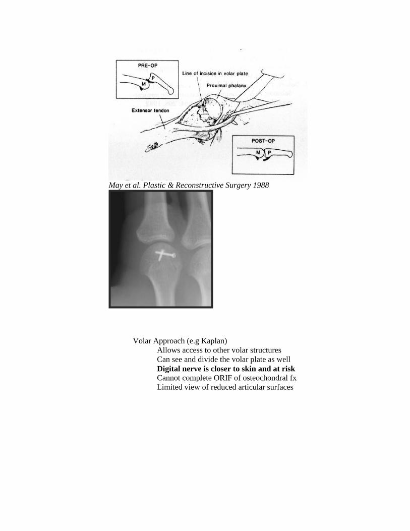
May et al. Plastic & Reconstructive Surgery 1988
Volar Approach (e.g Kaplan)
Allows access to other volar structures
Can see and divide the volar plate as well
Digital nerve is closer to skin and at risk
Cannot complete ORIF of osteochondral fx
Limited view of reduced articular surfaces

Volar MCP Dislocation (less common)
Mechanism - Hyperextension and hyperflexion described
J Am Acad Orthop Surg 2009;17:318-324
Simple Dislocation
Dorsal capsule torn and may be interposed in joint
Complex Dislocation
Other structures may interfere:
Extensor tendons
Distal volar plate/collateral ligaments
FDI
Closed reduction
Volar to Dorsal translation of P1 on the MC head
Begin with Flexed MCP and then Extend MCP
Open Reduction
Fewer reports of which direction is best for approach
Dorsal allows inspection of the joint, repair of capsule and
extraction of extensor tendon or ligament
Volar allows access to volar plate
3. Finger MCP Collateral Ligament Injuries
Avulsions – Origin or insertion
Avulsion fractures – Usually one volar “corner” of base of proximal phalanx
Displaced fx (intra-articular) may develop pseudo-arthrosis or nonunion
Border digits common
Index and small finger
Physical Exam
Test in flexion of MCP for stability in radioulnar deviation
May find rotatory instability or subtle malrotation of fingers
Radiographs
3 views and Brewerton view may identify fractures
May see volar subluxation of P1
Treatment
Conservative
Partial tears or suitable endpoint to deviation stress
More likely to try conservative for LF and RF;
buddy taping is possible to adjacent finger
Operative
Large /displaced avulsion fractures
Open repair – ORIF
Percutaneous fixation
Arthroscopically assisted repair
Volar approach (Brunner over A1)
Also good for access for reduction/fixation
Unstable Ligament rupture/avulsion
Dorsal approach
Suture repair
Suture anchors work well
Many reports of good-excellent outcome with repair
4. Locked MCP joints
MCP joints can be locked in flexion, usually Index and Long
Often presents as “trigger finger” referral after stuck for some time.
Except – PIP has full motion
Two patient types:
1. Osteoarthritis (usually “older”)
Ligament caught over osteophyte; often Long finger
Osteophyte often visible by Brewerton view
Treatment - often open release
Dorsal Approach
Good visualization
May reduce ligament around osteophyte or débride
osteophyte etc.
Volar Approach
Access to volar plate and volar ligament insertion
May be difficult to see dorsally due to flexed joint
2. Younger patients
Considered to be a soft tissue redundancy that traps
(usually) the radial collateral ligament of the index finger
Closed reduction advocated (with and without anesthesia)
Begin with hyperflexion, radial deviation, supination and
then extension

5. PIP Joint Anatomy
Bicondylar joint; 110 degrees of rotation 8-11 degrees of pro-supination
Significant stability from articular surfaces with flexion/grip/grasp
Ligaments (similar to MCP)
Proper Collateral
Accessory Collateral
Volar plate – strong insertion at lateral edges of P2
Transverse Retinacular ligament
Central slip of extensor mechanism
6. PIP Dislocations - Dorsal, Volar and Lateral
Dorsal (P2 dorsal to P1)
Most common
Mechanism – usually Hyperextension, may have axial load too
Eaton described 3 types:
I Volar plate avulsion fracture off base of P1
Tx- buddy taping and early motion
II Dorsal dislocation Tx – Digital block, traction and reduction
Check stability
Radiographs to confirm concentric reduction
Extension block splinting and edema control
Initiate flexion as soon as possible (pain/swelling)
Progress towards full extension weekly
Make progress towards full extension to avoid the
common complication of PIP flexion contracture
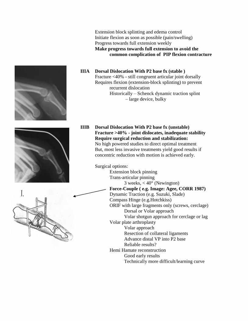
IIIA Dorsal Dislocation With P2 base fx (stable ) Fracture <40% - still congruent articular joint dorsally
Requires flexion (extension-block splinting) to prevent
recurrent dislocation
Historically – Schenck dynamic traction splint
– large device, bulky
IIIB Dorsal Dislocation With P2 base fx (unstable)
Fracture >40% - joint dislocates, inadequate stability
Require surgical reduction and stabilization:
No high powered studies to direct optimal treatment
But, most less invasive treatments yield good results if
concentric reduction with motion is achieved early.
Surgical options:
Extension block pinning
Trans-articular pinning
3 weeks, < 40° (Newington)
Force-Couple ( e.g. Image: Agee, CORR 1987) Dynamic Traction (e.g. Suzuki, Slade)
Compass Hinge (e.g.Hotchkiss)
ORIF with large fragments only (screws, cerclage)
Dorsal or Volar approach
Volar shotgun approach for cerclage or lag
Volar plate arthroplasty
Volar approach
Resection of collateral ligaments
Advance distal VP into P2 base
Reliable results?
Hemi Hamate reconstruction
Good early results
Technically more difficult/learning curve

P2 base Pilon Fractures – result from more axial load
Difficult injuries –
Complications include arthritis and stiffness
Soft tissue stiffness - major complication of open reduction
Need to maintain congruent surfaces and initiate motion
Results are equal or better for Dynamic traction
compared to percutaneous fixation or ORIF
(Ruland et al. J Hand Surg 2008;33A:19–25.
Force-Couple is not applicable for pilon fractures as it
does not distract the joint .
Volar Dislocations – Less common
Pure volar dislocations Central slip injured with volar dislocation
Treatment : closed reduction
If stable PIP extension splint 6 wks w/ active DIP motion
If unstable or with dorsal P2 fracture – may require repair
Dorsal P2 fx – ORIF, Perc or Extension block pinning
Volar rotatory dislocation
Mechanism - P2 spins on remaining intact ligament
Condyle ruptures between central slip and lateral band
Closed Reduction - requires rotation as well to reduce
MCP and PIP flexion assist with reduction of
condyle from soft tissue and then joint reduction

Open reduction may be required through a dorsal or
midaxial incision
Central slip/extensor mechanism inspected/repaired
Post reduction splinting – PIP extension with DIP motion
Lateral Dislocations
Mechanism – Deviation of joint with rupture of one collateral
ligament and volar plate
Treatment – Reduction, most by closed maneuver
Radiographs of joint to confirm concentric reduction
Look for joint incongruity = trapped soft tissue/bone
Rarely requires open reduction for trapped ligament
Midaxial or dorsal incision
Stiffness is much more common than instability
Ligament repair is rarely required
Perhaps with more chronic instability
Chronic or subacute PIP dislocations
May attempt open reduction if no arthritis
May consider:
Volar Plate Arthroplasty
Hemi-hamate reconstruction
PIP fusion
PIP Arthroplasty?

References:
1. Afendras G; Abramo A; Mrkonijic A; Geijer M; Kopylov P; and Tagil M:
Hemi-hamate osteochondral transplantation in the proximal interphalangeal dorsal
fracture dislocations: A minimum 4 year follow-in in eight patients. J Hand Surg
[Er], 35: 261-341, 2010.
2. Afifi MA; Medoro A; Salas, C.; Taha M; and Cheema, T.: A cadaver model
that investigates irreducible metacarpophalangeal joint dislocation. . J Hand Surg
[AM], 34: 1506-1511, 2009.
3. Agee JM: Unstable fracture dislocations of the proximal interphalangeal joint.
Treatment with the force couple splint. . Clinical Orthopaedics and Related
Research, (214): 101-12, 1987.
4. Ashmed D IV; Rothkopf DM; Walton RL; and JB, J.: Treatment of hand
injuries by external fixation. . Journal of Hand Surgery, 17(5): 956-964, 1992.
5. Bain GI; Mehta JA; Heptinstall RJ; and Bria, M.: Dynamic external fixation
for injuries of the proximal interphalangeal joint. . J Bone Joint Surg Am, 80(6):
1014-9, 1998.
6. Baldwin LW; Miller DL; Lockart LD; and EB, E.: Metacarpophalangeal -joint
dislocations of the fingers. . J Bone Joint Surg Am, 49: 1587-1590, 1967.
7. Barry K; McGee H; and Curtin J: Complex dislocation of the metacarpo-
phalangeal joint of the index finger: A comparison of the surgical approaches. . J
Hand Surg [Br], 13: 466-468, 1988.
8. Betz RR; Browne EZ; Perry GB; and EJ, R.: The complex volar
metacarpophalangeal-joint dislocation: A case report and review of the literature.
J Bone Joint Surg Am, 64: 1374-1375, 1982.
9. Bischoff R; Buechler U; De Roche R; and Jupiter J: Clinical results of tension
band fixation of avulsion fractures of the hand. Journal of Hand Surgery 19(6),
1994.
10. Boden RA; Cavale N; and Fleming AN: Kaplan's puckering sign in complex
dorsal dislocation of the metacarpophalangeal joint of the index finger. J Hand
Surg [Br], 31: 576-577, 2006.
11. Chadha M, and Dhal A: Vulnerability of the radial digital neurovascular bundle
of the index finger while using Kaplan's volar approach for irreducible dislocation
of the second metacarpophalangeal joint. . Injury, 35: 1182-1184, 2004.
12. Deitch MA; Kiefhaber TR; BR, C.; and Stern PJ: Dorsal fracture dislocations
of the proximal interphalangeal joint: Surgical complications and long-term
results. . Journal of Hand Surgery [Am], 24(5): 914-23, 1999.
13. Delaere OP; suttor PM; Degolla R; Leach R; and Pieret PJ: Early surgical
treatment for collateral ligament rupture of metacarpophalangeal joints of the
fingers. . J Hand Surg [AM], 28(2): 209-15, 2003.
14. Dionysian E, and Eaton RG: The long-term outcome of volar plate arthroplasty
of the proximal interphalangeal joint. Journal of Hand Surgery, 25(3): 429-37,
2000.
15. Durham-Smith G, and McCarten GM: Volar plate arthroplasty for closed
proximal interphalangeal joint injuries. Journal of Hand Surgery [Br], 17(4): 422-
8, 1992.
16. Eaton RG, and JW, L.: Joint injuries and their sequelae. Clin of Plastic
Surgery, 3(1): 85-98, 1976.
17. Eaton RG, and Dray GJ: Dislocations and ligament injuries in the digits, in
Green DP (ed),Edited, 647-668, New York, NY, Churchill Livingstone, 1982.
18. Eaton RG, and Malerich MM: Volar plate arthroplasty of the proximal
interphalangeal joint. A review of ten year's experience. Journal of Hand Surgery
[Am], 5(2): 260-8, 1980.
19. Ellis SJ; Cheng R; Prokopis, P.; Chetboun A; Wolfe, S.; Athanasian EA; and
Weiland AJ: Treatment of proximal interphalangeal dorsal fracture-dislocation
injuries with dynamic external fixation: a pins and rubber band system. J Hand
Surg [AM], 32(8), 2007.
20. Flatt, A.: Fracture-dislocation of an index metacarpophalangeal joint and an ulnar
deviating force in the flexor tendons. J Bone Joint Surg Am, 48: 100-104, 1966.
21. Gellman H; Massanelli GL; and WC, G.: The effect of MCP position on strain
of the collateral ligaments of the MCP joints in the hand during immobilization. .
Edited by meeting, A. S. f. S. o. t. H. t. a., Seattle, 2000.
22. Green DB, and Terry GC: Complex dislocation of the metacarpophalangeal
joint: Correlative pathological anatomy. J Bone Joint Surg Am, 55: 1480-1486,
1973.
23. Hamilton SC; Stern, P.; Fassler PR; and Kiefhaber, T.: Mini-screw fixation
for the treatment of proximal interphalangeal joint dorsal fracture-dislocations. . J
Hand Surg [AM], 31: 1349-54, 2006.
24. Hastings H II, and Ernst JM: Dynamic external fixation for fractures of the
proximal interphalangeal joint. . Hand Clinic, 9(4): 659-74, 1993.
25. Hubbard LF: Metacarpophalangeal dislocations. Hand Clin, 4: 39-44, 1988.
26. Hunt JC; Watts HB; and Glasgow JD: Dorsal dislocation of the
metacarpophalangeal joint of the index finger with particular reference to open
dislocation. J Bone Joint Surg Am, 49(8): 1572-1578, 1967.
27. Ikeda M; Ishii T; Kobayaski Y; Mochida J; Saito I; and Oka Y: Percutaneous
pinning of the displaced volar plate avulsion fracture of the PIP joint. Hand Surg,
14: 113-9, 2009.
28. Kang, L.; Rosen A; Potter HG; and Weiland, A.: Rupture of the radial
collateral ligament of the index metacarpophalangeal: diagnosis and surgical
treatment. J Hand Surg [AM], 32: 789-94, 2007.
29. Klein DM, and Belsole RJ: Percutaneous treatment of carpal, metacarpal, and
phalangeal injuries. Clinical Orthopaedics and Related Research, 375: 116-25,
2000.
30. Lee JY, and Teoh LC: Dorsal Fracture dislocations of the proximal
interphalangeal joint treated by open reduction and interfragmentary screw
fixation: indications, approaches, and results. J Hand Surg [Br], 31: 138-46,
2006.
31. Lee, L.; Lee HM; Hou YT; Hung ST; Chen JK; and Shih JT: Surgical
outcome of the volar plate arthroplasty of the proximal interphalangeal joint using
the mitek micro GII suture anchor. . J Trauma, 65: 116-22, 2008.
32. Marck KW: A tricky trigger finger: a patient iwht intermittent intrinsic locking
of the metacarpophalangeal joint. . Netherlands Journal of Surgury, 39(5): 151-2,
1987.
33. McElfresh EC, and Dobyns, J.: Intra-articular metacarpal head fractures. .
Journal of Hand Surgery, 8(4): 383-93, 1983.
34. McElfresh EC; Dobyns, J.; and ET, O. B.: Management of fracture-dislocation
of the proximal interphalangeal joints by extension-block splinting. . J Bone Joint
Surg Am, 54(8): 1705-11, 1972.
35. McLaughlin HL: Complex "locked" dislocation of the metacarpophalangeal
joints. J Trauma, 5: 683-688, 1965.
36. Minami A; An KN; 3rd, C. W.; Linscheid RL; and Chao EY: Ligamentous
structures of the metacarpophalangeal joint: a quantitative anatomic study. .
Journal of Orthpaedic Surgery and Research 1: 361-8, 1984.
37. Murphy AF, and Stark HH: Closed dislocation of the metacarpophalangeal
joint of the index finger. . J Bone Joint Surg Am, 49: 1579-1586, 1967.
38. Newington DP; Davis TR; and Barton NJ: The treatment of dorsal fracture-
dislocation of the proximal interphalangeal joint by closed reduction and
Kirschner wire fixation: A 16-year follow up. Journal of Hand Surgery [Br],
26(6): 537-40, 2001.
39. Patel MR, and Bassini L: Irreducible palmar metacarpophalangeal joint
dislocation due to junctura tendinum interposition: A case report and review of
the literature. . J Hand Surg [AM], 25: 166-172, 2000.
40. Qvick L, and Wilhelm K: Ligamental injuries of the MCP joints. Z Plast Chir. ,
4(4): 260-5, 1984.
41. Ruland RT; Hogan CJ; Cannon DL; and Slade JF: Use of dynamic distraction
external fixation for unstable fracture-dislocations of the proximal interphalangeal
joint. J Hand Surg [AM], 33(1): 19-25, 2008.
42. Schenck RR: The dynamic traction splint-fabrication and use. . Edited, American
Society for Surgery of the Hand - Video Library, 1992.
43. Schneck RR: Dynamic traction and early passive movment for fractures of the
proximal interphalangeal joint. . Journal of Hand Surgery, 11(6): 850-8, 1986.
44. Slade JF; Baxamusa TH; and SW, W.: External fixation of proximal
interphalangeal joint fracture dislocations. . Atlas of the Hand Clinics 5(1): 1-29,
2000.
45. Slade JF; Wolfe SW; and Gutow AP: The treatment of unstable proximal
interphalangeal joint fractures: How to apply and use a dynamic mini-external
fixator. Edited, American Society for Surgery of the Hand - Video Library, 1996.
46. Stern PJ, and Roman RJ: Pilon fractures of the proximal interphalangeal joint.
Journal of Hand Surgery, 16(5): 844-50, 1991.
47. Tavin E, and Wray RC Jr: Complex dislocation of the index
metacarpophalangeal joint with entrapment of a sesamoid. . Ann Plastic Surg, 40:
59-61, 1998.

48. Theumann NH et al.: MR imaging of the metacarpophalangeal joints of the
fingers: evaluation of 38 patients with chronic joint disability. . Skeletal
Radiology, 34(4): 210-6, 2005.
49. Vahey JW, and Wegner DA: Effect of proximal interphalangeal fracture
deformity on extensor tendon function. . Journal of Hand Surgery, 23(4): 673-81,
1998.
50. Weiss AP, and Hastings H II: Distal unicondylar fractures of the proximal
phalanx. Journal of Hand Surgery [Am], 18(4): 594-9, 1993.
51. Wolfe SW, and LD, K.: Intra-articular impaction fractures of the phalanges. .
Journal of Hand Surgery, 20(2): 327-33, 1995.
52. Yagi M; Yamanaka K; K, Y.; Sato N; and Inoue, A.: Successful manual
reduction of locked metacarpophalangeal joints in fingers. . Journal of Bone and
Joint Surgery, 82: 366-371, 2000.
53. Zemel NP: Metacarpophalangeal joint injuries in fingers. Hand Clin, 8: 745-754,
1992.
Metacarpal and Phalangeal Fractures
David G Dennison, MD
Mayo Clinic
AAHS 2015 Review Course
Introduction
Metacarpal Fractures
30-40% of hand fractures, most common 5th MC neck (young male)
Phalangeal Fractures
Feehan and Sheps:
o 50% of hand fractures were phalangeal fractures
o Male 2:1, especially in patients 15-40 y/o
o Women are more likely to have hand fractures when >65 y/o
Treatment Principles and Background
Fracture pattern and soft tissue injury varies with mechanism
“Injury” films - assess the instability of the fracture
Displaced fractures can be unstable even when the reduction looks good….
o Initial deformity – best measure of instability.
Outcome - proportional to the volume or severity of the injury
Proximal phalanx, comminuted fractures and fractures with soft tissue injuries
have worse outcomes
Goals of operative reduction and fixation are to:
o Provide sufficient stabilization
o Minimize pain
o Allow early motion
o Minimize stiffness.
o Limit dissection and soft tissue injury, keep hardware countersunk and
not too close to the joint to minimize stiffness
Facilitate motion by 4 weeks
Postoperative evaluation, monitoring and therapy are critical in most cases
Fracture types in general
Transverse, oblique, spiral, comminuted
o Open, closed, contaminated, soft tissue injury, bone loss
o Pediatric fractures
Thicker periosteum – more soft tissue stability

Metacarpal and Phalangeal Fractures :Functional Anatomy, Mechanics and
Deformity (Excluding the thumb)
Functional Anatomy
o Radial side of the hand is rigid through the CMC
o Ulnar side has more motion through the CMC joints
20-30 degrees
o Transverse arch to hand and carpus
Mechanics and Deformity –Intrinsic, flexor and extensor muscle forces
produce angular deformities
Metacarpal Shaft
o Intrinsic muscles produce an apex dorsal angulation; intermetacarpal
ligament and interosseous muscles may limit shortening and
malrotation
Rotation of 5 degrees = 1.5 cm of potential overlap
Proximal Phalanx (P1)
o Typically transverse fractures result in APEX VOLAR angulation as a
result of the flexion force of the intrinsic muscle insertion on the base
of the proximal phalanx and the extension force of the central slip
across the PIP joint and distal portion of the proximal phalanx.
Extension lag - as much as 12 degrees for every 1 mm of
shortening in the proximal phalanx
o Oblique or spiral fractures may penetrate through soft tissue limiting
closed reduction
Middle Phalanx (P2)
o Midshaft P2 fractures - variable presentation
o Distal 1/3 fractures - apex volar as the FDS flexes the shaft of P2 and
the extensor tendon extends the distal P2.
o Proximal P2 fractures - result in apex dorsal deformity if proximal to
the FDS insertion due to the extension from the central slip and flexion
of the FDS
Distal Phalanx (P3)
o Few forces cross the tuft
o Growth plate or base of P3 injuries may be unstable
some result in flexion (apex dorsal) deformity
e.g. Seymour fracture

Extra-articular Metacarpal and Phalangeal Fractures (excluding the thumb):
Metacarpal Shaft
o Acceptable alignment:
No clinical malrotation
Index and long – less than 10 degrees
Ring and small – less than 20 degrees
Shortening of 2mm = 7 degrees of extension lag,
may tolerate 3-4mm of shortening
o Closed treatment –
Cast immobilization, MP flexed, IP free; duration 3-4 weeks
Frequent checks with X-rays
o Operative treatment
Percutaneous Kwires (transverse, longitudinal, oblique) or
transverse Kwire into adjacent intact MC shaft works well
IM nail –works well but less anatomic restoration of alignment
ORIF
Oblique or spiral fractures - lag screws or compress
fracture and place a position screw
o Plate with lag screw has increased rigidity
Transverse or comm. – plate dorsal or lateral
Tension band or intraosseous wiring also useful
Ex Fix – certain role with severe soft tissue injury, infection or
loss of bone
Kwires with small systems (ie Hoffman phalangeal Ex
fix)
Syringe with PMMA works well too
Metacarpal Neck
o Acceptable Alignment
No clinical malrotation
Angulation (not to exceed):
Index 10-15 degrees
Long 10-15 degrees
Ring 30-40 degrees
Small 40-50 (60+) degrees
o Closed treatment
Reduction and cast application with a good three point mold
Historically by Jahss maneuver MP and PIP flexed..
The maneuver is good; the position for any period of
time is bad!
I use a Beckenbaugh cast which is applied while the
finger is in a finger trap with longitudinal traction and a
cast with 3 pt mold is applied with the MP extended
and the PIP free for about 3-4 weeks)
Close F/U
o Operative Treatment
Any wound that may communicate with the joint should be
explored, débrided and irrigated; “fight bites” should not be
missed.
Closed reduction and K wire fixation is usually acceptable
Open treatment for unstable, irreducible fractures
K wires, condylar blade plates, screws
Metacarpal Base
o Usually involves the 5th CMC
o Look for injury at adjacent CMC
30 degree Pronated AP and Lateral and/or CT
o Closed treatment – nondisplaced : short arm cast
o Displaced articular fractures or unstable CMC joint – deserve
reduction and fixation, especially the 5th
Phalangeal Shaft
o General operative indications:
Displaced fractures with malrotation, angulation or excessive
shortening
Guide: any malrotation, angulation > 10 degrees, shortening >
2 mm, apposition <50%
Distal phalanx with unstable fracture and nail matrix injury
Multiple fractures in the same hand, finger, extremity, or with
polytrauma
Open fractures or replantation
o Closed treatment includes casting/splinting with MP flexed and IP
joints extended. Initiate motion as swelling subsides and callus and
stability are present.
Frequent f/u
If you use less than a cast - you need a cooperative patient….
o Operative Treatment
Closed reduction, perc pinning is mainstay, K wire reduction
forcep is a valuable tool
CR/IF an option as well as ORIF
Plates : P1 – lateral better than dorsal regarding finger stiffness
P2 rarely plate..tendon scarring

Intra-articular Metacarpal and Phalangeal Fractures (excluding the thumb):
Metacarpal Head
o Good imaging is a necessity to make a good plan
Brewerton views
CT scan
Plan approach accordingly
o Nondisplaced
Cast with MP flexed, close f/u
o Displaced intra-articular fractures
ORIF (large fragments)
Arthroscopic assisted reduction and percutaneous fixation
Keep in mind - distal fractures have higher rate of
osteonecrosis
P1 Base
o Unicondylar or avulsion fractures
Nondisplaced – cast with/ without buddy tape
Displaced fractures may lead to nonunion or pseudoarthrosis
and instability
Closed versus open reduction
K wire fixation is satisfactory, screws and cannulated
screws have been used with success
Unicondylar
Midaxial approach, dorsal for direct visualization of
joint surface
Avulsion fracture (typically volar fragment):
Arthroscopic reduction and fixation reported
Volar approach through or around A1 pulley
o Bicondylar fractures
Similar indications
Dorsal or lateral approach to MP joint
Blade plate (lateral) may be helpful
P1 Head
o Unicondylar or bicondylar
o Nondisplaced – cast/splint intrinsic plus – WATCH CLOSELY
o Displaced fractures
Attempt closed reduction with K wires and compression clamp,
threaded Kwires can help hold the reduction.
Articular reduction may require open reduction and screw,
Kwire, threaded Kwire or tension band fixation. Bicondylar
may benefit from blade plate.
Better joint motion when the hardware is kept away from the
joint and periarticular tissues

P2 Base
o Volar plate avulsion fractures
Follows hyperextension or dorsal dislocation:
Goal - Maintain congruent reduction
Extension block splint with ACTIVE flexion
Good for smaller fragments with congruent joint
Force couple (eg: Agee)
Useful for reduction of joint subluxation
ORIF
Rarely– volar approach and ORIF with large fragment
and coronal fracture plane
Volar plate arthroplasty
May help manage comminuted volar fragments and
later reconstruction
Hamate osteochondral autograft
Reconstruct the volar lip
o Central Slip Avulsion Fractures
Nondisplaced fractures – splint PIP in extension, mobilize DIP
Displaced fractures
Closed reduction, percutaneous fixation
Extension block pinning versus direct fixation of
fragments
ORIF for large fragments
o Pilon type
Difficult problem regardless of the method used to treat
Min/Nondisplaced
Early ROM, close F/U
May choose to unload the joint as well (see below)
Displaced
Dynamic Ex Fix with distraction close f/u and PIP
ROM
Closed reduction and perc pins
Compass hinge
Schenk splint
Early ROM
Rare….ORIF
Mallet Fracture
o Closed treatment with DIP full time extension
For smaller fragments with a congruent joint
Check extension splint films for joint and fracture alignment
6 weeks with ACTIVE PIP motion, then 6 weeks PM splint
o Closed reduction, extension block pinning –
For large displaced fragments (40-60%) of joint
Joint subluxation
o Rarely ORIF, higher complication rate

The Thumb: Metacarpal and Phalangeal Fractures
Thumb
o P1 and P2 fracture indications and treatment are similar to fingers
o Metacarpal extra-articular shaft fractures
Tolerate 20-30 degrees of angulation
Slight malrotation may be acceptable due to motion at Thumb
CMC joint
CR/PF when practical
ORIF for comminuted, displaced, unstable fractures
EPL/EPB interval for dorsal approach to Th MC
Plates, screws, Kwires, condylar blade plates and 2 mm
locked plates are available for fixation
o Intra-articular base of thumb metacarpal fractures
Need good imaging
Robert’s view: (AP) of Thumb MC-Trapezial joint
Bennett Fracture – most common
APL pulls thumb MC proximal, volar oblique ligament
intact and attaches to the remaining small volar bone
fragment
Reduction requires traction, depression of the base of
the metacarpal (extension) and pronation
K wires fixation may be from Th MC to Tzm and/or
Index MC – does not have to pass through the small
volar fragment as long as the fracture is reduced and
stabilized
May be assisted by thumb CMC arthroscopy or open
reduction
Rolando Fracture
T, Y or 3 part intra-articular fracture
CT for good visualization
Displaced fractures (>2mm?)
o Open reduction/internal fixation
o Dorsal approach
o Wagner approach
o K wire, screws, Ex Fix, Locked plate and
condylar plate fixation
o Bone graft

o Thumb MCP Ulnar collateral ligament injuries
Ulnar collateral ligament (most common)
o Rupture
1. Partial or complete
Physical exam
o Test at 0 and 30 deg of
MP flexion
o Palpate for Stener lesion
o Compare to opposite side
for radial and ulnar
deviation and endpoint
> 40 degrees
suspicious for
complete tear
Stress Xrays???
MRI
o Evaluate for Stener lesion
or complete tear and
associated injuries
(capsule, EPB, RCL)
o Avulsion fracture
1. Size
2. Displacement, articular incongruity
3. Nondisplaced – cast
Some controversy…verus
4. Closed reduction and pinning with cast
5. Open reduction, internal or perc fixation
o Fractures and avulsions can occur together!
Techniques - Closed Reduction of Shaft Fractures:
Relax the intrinsics with flexion of the MCP
In general - Traction, accentuate the deformity and then reverse the deformity.
o Irreducible fractures are usually due to interposed soft tissue
Check clinical alignment and rotation.
o Flexion and extension
o Axial plane of the fingernails when the finger cannot be flexed.
Buddy tape with the MCP flexed and IPs extended
o Obtain films to evaluate reduction, splints may then be applied
Close f/u (weekly) to evaluate for maintenance of the reduced position and to
begin motion by 3-4 weeks.

Open Reduction: Approaches to the Metacarpals and Phalanges
Metacarpal
Dorsal
o Incision is standard although exposes tendons to scarring
Lateral
o Incision may be used for index and small finger – keeps implants and
scar away from extensor tendons
Phalangeal
Dorsal
o Extensor tendon splitting
o Advantage - good visualization
o Disadvantage – direct scarring to the extensor tendon
o PIP exposure for articular (especially unicondylar) fractures is through
a central slip-lateral band splitting approach
Lateral
o Midaxial or midlateral incisions
o Approach through, around, or with excision of part of the lateral band
o Advantage – may minimize scar, allows access for condylar or fixed
angle plate
o Disadvantage – may be more difficult to visualize fracture and work
through this window
Volar
o Brunner
o Center at PIP flexion crease for access to PIP with retraction of flexor
tendons
ORIF coronal volar lip fractures
Volar plate arthroplasty etc.
Implants or Fixation for Metacarpal and Phalangeal Fractures:
Kirschner wires (K wires) – simple, available, cheap, versatile
o Internal splint, minimal or no compression
o Two crossed wires should not intersect at the fracture
o Two wires required for rotational control
o Stability is related to:
number of K wires
arrangement of wires relative to the fracture,
diameter of the K wires
o Fracture must be reduced (and compressed) prior to pin fixation
o K Wire perpendicular to the fracture plane best resists bending, torque
and distraction with oblique/spiral fracture
o K wire perpendicular to the long axis of the bone best resists longitudinal
shortening in oblique/spiral fractures.
o K wire reduction forcep is a valuable tool

Stainless wire (26-28 Gauge)
o 90:90 intraosseous wiring
o tension band with K-wires
o cerclage wire
Screws
o Provide compression
o Modular/ headless/ cannulated
o Strength proportional to minor diameter
o lag screw – most compression perpendicular to the fracture plane
resists longitudinal force best when perpendicular to the long axis
of the bone
Plates
o Bending strength proportional the cube of its thickness and inversely to
the cube of the length.
o Provide stability and allow early motion
o Four cortices (2 bicortical screws) on each side of the fracture for
phalangeal fractures is advisable
o Allows compression with eccentric screw placement
o Stainless or Titanium
Titanium may stress shield less
o Condylar plates, T-plates, compression plates, H plates
Conventional screws
o Small locked plates – fixed angle implant
Preserve periosteum, stable fixation especially with small or
osteoporotic bone
o Plates have been associated with complications in up to 36% of fractures
Stiffness, nonunion, plate prominence, infection and tendon
rupture
However, those fractures with more stiffness were usually
open phalangeal fractures; plate or injury?.
Intramedullary (IM) nails, or rods, or K wires
o Most well suited for transverse or short oblique fractures
Bioabsorbable implants
o Evolving role, no routine implants at this time
o Plates, pins, screws
o Similar dissection for application
External fixation
o Applied in midaxial or dorsal midline plane to minimize tendon scarring
dorsal midline may be best secondary location
o Phalangeal fixators systems
Multiple K-wires with PMMA filled syringe

Putting it all together, some examples -
Distal Phalanx
Tuft: Most common, few deforming forces
o Subungual hematoma:
>50% suggests nailbed laceration
Controversy – some literature shows no significant improvement
with nail removal and repair (Roser 1999).
o Displaced nail plates should be removed and the nailbed repaired with
absorbable suture
o Requires splint at DIP and MOTION at PIP
Sensory problems may persist; desensitization may help.
Fibrous union is common and usually asymptomatic
Shaft: Transverse, Longitudinal, Comminuted – higher energy injuries : crush or
saw
o Open injuries or those with nail plate avulsion require irrigation and
debridement, stabilization (K wire or nail plate) and nailbed repair with
attention to the germinal and sterile matrices
o Seymour fracture or displaced fractures with nail avulsion or displacement
may require reduction and K wire fixation if the reduced nail plate does
not provide enough stability.
Middle Phalanx Fractures
Shaft
o Transverse
IM or crossed K wires
Try to spare DIP and PIP joints and peri-articular soft tissue
Trans-articular for small distal middle phalanx neck fractures
o Oblique/Spiral
Percutaneous reduction/compression with K wire reduction forcep
Multiple transverse K wires (perpendicular to fx and/or shaft)
May use threaded K wire to minimize gapping
Open or percutaneous lag screws
o Comminuted/Segmental
K wires
Ex Fix
Multiple K wires
Transverse or oblique “fragment to fragment”
Proximal Phalanx Fractures
Shaft
o Transverse
IM or crossed K wires
Antegrade, retrograde and transarticular
MP should be flexed if wire goes across the joint
o Oblique/Spiral
Percutaneous reduction and compression with K wire reduction
forcep
Multiple transverse/oblique K wires
May use threaded K wire to minimize gapping
Open or Percutaneous reduction and fixation with lag screws
Lag screws alone are sufficient once the fracture plane is
longer than twice the diameter
o Short oblique/spiral – may require additional
support: K wires or plate
o Comminuted/Segmental
K wires
External fixator
Condylar blade plate - lateral approach
Dorsal plate – dorsal tendon splitting approach
Rehabilitation and Postoperative care
Elevation and compression to minimize edema
Mobilize other fingers and joints
Splint to protect the finger or hand
Cannot wait for radiographic obliteration of fracture planes for phalanx fractures,
by 4 weeks active motion should begin in most cases
Pearls:
Always examine the other hand before you start
Mark the midaxial line prior to percutaneous fixation (wires placed above this are
in a safe zone)
K-wire reduction forcep is a great tool, needles can also help to direct wires and
protect soft tissue.
Oscillation feature on the K-wire driver can help minimize soft tissue injury.
Minimize trauma to the tendons and soft tissue
Make sure you move them…by 4 weeks
Be satisfied with your reduction and fixation before you leave the OR
Fracture pattern, soft tissue injury, and surgeon preference or experience must
guide the surgical approach and choice of implant.
References
1. Agee J: Treatment principles for proximal and middle phalangeal fractures.
Orthop Clin N Am 23:35, 1992
2. Al Qattan MM, Hashem F, Helmi A: Irreducible tuft fractures of the distal
phalanx. J Hand Surg 28B:18, 2003
3. Al-Qattan MM: Juxta-epiphyseal fractures of the base of the proximal phalanx of
the fingers in children and adolescents. J Hand Surg 27B:24, 2002
4. Belsky MR, Eaton RG, Lane LB: Closed reduction and internal fixation of
proximal phalangeal fractures. J Hand Surg 9A:725, 1984
5. Bosscha K, Snellen JP: Internal fixation of metacarpal and phalangeal fractures
with AO minifragment screws and plates: A prospective study. Injury 24:166,
1993
6. Botte MJ, Davis JL, Rose BA, et al: Complications of smooth pin fixation of
fractures and dislocations in the hand and wrist. Clin Orthop 276:194, 1992
7. Crawford G: Screw fixation for certain fractures of the phalanges and
metacarpals. J Bone Joint Surg 58A:487, 1976
8. Crofoot CD, Saing M, Raphael J: Intrafocal pinning for juxtaarticular phalanx
fractures. Tech Hand Upper Extrem Surg 9:164, 2005
9. Dabezies EJ, Schutte JP: Fixation of metacarpal and phalangeal fractures with
miniature plates and screws. J Hand Surg 11:283, 1986
10. Diwaker HN, Stothard J: The role of internal fixation in closed fractures of the
proximal phalanges and metacarpals in adults. J Hand Surg 11B:103, 1986
11. Drenth DJ, Klasen HJ: External fixation for phalangeal and metacarpal fractures.
J Bone Joint Surg 80B:227, 1998
12. Duncan RW, Freeland AE, Jabaley ME, Meydrech EF: Open hand fractures: an
analysis of the recovery of active motion and of complications. J Hand Surg
18A:387, 1993
13. Feehan L, Sheps S: Incidence and Demographics of Hand Fractures in British
Columbia, Canada: A Population-Based Study. J Hand Surg 31(7):1068, 2006
14. Fitoussi F, Lu W, Ip W, Chow S: Biomechanical properties of absorbable
implants in finger fractures. J Hand Surg 23B:79, 1998
15. Flatt AE: Closed and open fractures of the hand: Fundamentals of management.
Postgrad Med 39:17, 1996
16. Freeland AE, Jabaley ME, Burkhalter WE, Chaves AM: Delayed primary bone
grafting in the hand and wrist after traumatic bone loss. J Hand Surg 9:22, 1984
17. Freeland AE: Hand Fractures: repair, reconstruction, and rehabilitation. 1st ed.
Churchill Livingstone, Philadelphia, 2000.
18. Gonzalez MH, Hall Jr RF: Intramedullary fixation of metacarpal and proximal
phalangeal fractures of the hand. Clin Orthop 327:47, 1996

19. Green DP, Anderson JR: Closed reduction and percutaneous pin fixation of
fractured phalanges. J Bone Joint Surg 55A:1651, 1973
20. Gutow A, Slade J, Mahoney J: Hand Fractures and Joint Injuries. p. 3. In Trumble
TE (ed.) Hand Surgery Update 3: Hand Elbow and Shoulder. Amer Soc Surgery
of the Hand, Rosemont, IL, 2003
21. Halliwell PJ: The use of external fixators for finger injuries: pin placement and
tethering of the extensor hood. J Bone Joint Surg 80B:1020, 1998
22. Hastings II H: Unstable metacarpal and phalangeal fracture treatment with screws
and plates. Clin Orthop 214:37, 1987
23. Hornbach EE, Cohen MS: Closed reduction and percutaneous pinning of fractures
of the proximal phalanx. J Hand Surg 26B:45, 2001
24. Horton TC, Hatton M, Davis TR: A prospective randomized controlled study of
fixation of long oblique and spiral shaft fractures of the proximal phalanx: closed
reduction and percutaneous Kirschner wiring versus open reduction and lag screw
fixation. J Hand Surg 28B:5, 2003
25. Kawamura K, Chung KC: Fixation choices for closed simple unstable oblique
phalangeal and metacarpal fractures. Hand Clin 22:287, 2006
26. Kozin S, Thoder J, Lieberman G: Operative treatment of metacarpal and
phalangeal shaft fractures. J Am Acad Orthop Surg 8:111, 2000
27. Kurzen P, Fusetti C, Bonaccio M, Nagy L: Complications after plate fixation of
phalangeal fractures. J Trauma 60:841, 2006
28. Lins RE, Myers BS, Spinner RJ, Levin LS: A comparative mechanical analysis of
plate fixation in a proximal phalangeal fracture model. J Hand Surg 21A:1059,
1996
29. Lu W, Furumachi K, WY I, Chow S: Fixation for comminuted phalangeal
fractures. A biomechanical study of five methods. J Hand Surg 21B:765, 1996
30. Margic K: External fixation of closed metacarpal and phalangeal fractures of
digits. A prospective study of one hundred consecutive patients. J Hand Surg
31B:30, 2006
31. Matloub HS, Jensen PL, Sanger JR, Grunert BK, Yousif NJ: Spiral fracture
fixation techniques. A biomechanical study. J Hand Surg 18B:515, 1993
32. Nunley JA, Kloen P: Biomechanical and functional testing of plate fixation
devices for proximal phalangeal fractures. J Hand Surg 16A:991, 1991
33. Orbay JL, Touhami A: The treatment of unstable metacarpal and phalangeal shaft
fractures with flexible nonlocking and locking intramedullary nails. Hand Clin
22:279, 2006
34. Ouellette EA, Dennis JJ, Latta LL, Milne EL, Makowski AL: The role of soft
tissues in plate fixation of proximal phalanx fractures. Clin Orthop 418:213, 2004
35. Page S, Stern P: Complications and range of motion following plate fixation of
metacarpal and phalangeal fractures. J Hand Surg 23:827, 1998

36. Papadonikolakis A, Li Z, Smith BP, Koman LA: Fractures of the phalanges and
interphalangeal joints in children. Hand Clin 22:11, 2006
37. Roser S, Gellman H: Comparison of nailbed repair versus nail trephination for
subungual hematomas in children. J Hand Surg 24(6):1166, 1999
38. Schatzker J: Screws and plates and their application. p. 179. In Muller ME,
Allgower M, Schneider R, Willenegger H (eds): Manual of Internal Fixation:
Techniques Recommended by the AO-ASIF Group. 3rd ed. Springer-Verlag, New
York, 1991
39. Seymour N: Juxta-epiphysial fracture of the terminal phalanx of the finger. J
Bone Joint Surg 48B:347, 1966
40. Simon RR, Wolgin M: Subungual hematoma: association with occult laceration
requiring repair. Am J Emerg Med 5:302, 1987
41. Smith RJ: Balance and kinetics of the fingers under normal and pathological
conditions. Clin Orthop 104:92, 1974
42. Swanson T, Szabo R, Anderson D: Open hand fractures: prognosis and
classification. J Hand Surg 16A:101, 1991
43. Tencer AF, Johnson KD, Kyle RF, Fu FH: Biomechanics of fractures and fracture
fixation. Instr Course Lect 42:19, 1993
44. Vahey J, Wegener D, Hastings H: Effect of proximal phalangeal fracture
deformity on extensor tendon dysfunction. J Hand Surg 23:673, 1998
45. Viegas SF, Ferren EL, Self J, Tencer AF: Comparative mechanical properties of
various Kirschner wire configurations in transverse and oblique phalangeal
fractures. J Hand Surg 13:246, 1988
46. Trigg, S. Fractures of the Metacarpals and Phalanges. AAHS Review 2005