financial disclosures fetal imaging - university of ... · financial disclosures ... microsoft...
TRANSCRIPT

In-utero imaging, Decision making and surgical planning
Fetal Imaging
Mariana L. Meyers, MDDirector of Fetal Imaging CHCO‐CFCC
Pediatric Radiology ‐ CHCO
Financial Disclosures
• No relevant financial relationships with any commercial interests.
Mariana Meyers
Goals and Objectives
• Goal:
- Understand the utility of fetal MRI
• Objectives:
- Identify the main imaging features of fetal neck and chest pathologies affecting the airway
- Demonstrate how fetal MRI aids in the diagnosis of different neck pathologies
Do we really need fetal MRI?
• US is still the first screening/diagnostic tool. MRI does not replace it
• Helps predict outcome, mortality and morbidity in fetuses with multiple pathologies, such as cervical teratoma or CDH
• Helps plan surgery and delivery: neck masses, shunt placements, EXIT procedures
• Helps identify additional abnormalities not seen on US
Fetal airway compromise
Lesions causing airway obstruction
1. Facial lesions
2. Neck lesions
3. Thoracic lesions
Facial lesions
• Oral lesions
• Cleft lip/palate
• Micrognathia
Meyers, Mariana, MD In-Utero Imaging, Decision Making, and Surgical Fetal Imaging

Epulis
http://fn.bmj.com/content/early/2011/03/22/adc.2011.214411.full
• Benign. Arise from gingival mucosa of alveolar ridge
• Congenital gingival granular cell tumors (adults)
• Pedunculated/ well defined
• Protrudes through the mouth
• Vascular pedicle
• Usually doesn’t affect swallowing/ airway
• Hypoechoic in US
• Hypointense T2 MRI- slightly hyper T1
• Ddx: epignathus, hemangioma, dermoid, teratoma
Epulis
Meyers, Mariana, MD In-Utero Imaging, Decision Making, and Surgical Fetal Imaging

1/14/1312/27/12
Giant epignathus
Meyers, Mariana, MD In-Utero Imaging, Decision Making, and Surgical Fetal Imaging
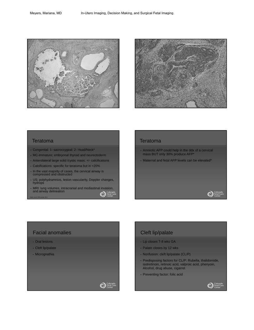
Teratoma
• Congenital: 1- sacroccygeal; 2- Head/Neck*
• MC-immature; embryonal thyroid and neurectoderm
• Anterolateral large solid /cystic mass; +/- calcifications
• Calcifications: specific for teratoma but in <20%
• In the vast majority of cases, the cervical airway is compressed and obstructed
• US: polyhydramnios, lesion vascularity, Doppler changes, hydrops
• MRI: lung volumes, intracranial and mediastinal invasion, and airway delineation
Rubio, et al, SPR poster 2011
Teratoma
• Amniotic AFP could help in the ddx of a cervical mass BUT only 30% produce AFP*
• Maternal and fetal AFP levels can be elevated*
Facial anomalies
• Oral lesions
• Cleft lip/palate
• Micrognathia
Cleft lip/palate
• Lip closes 7-8 wks GA
• Palate closes by 12 wks
• Nonfusion: cleft lip/palate (CL/P)
• Predisposing factors for CL/P: Rubella, thalidomide, isotretinoin, retinoic acid, valproic acid, phenyoin, Alcohol, drug abuse, cigarret
• Preventing factor: folic acid
Meyers, Mariana, MD In-Utero Imaging, Decision Making, and Surgical Fetal Imaging
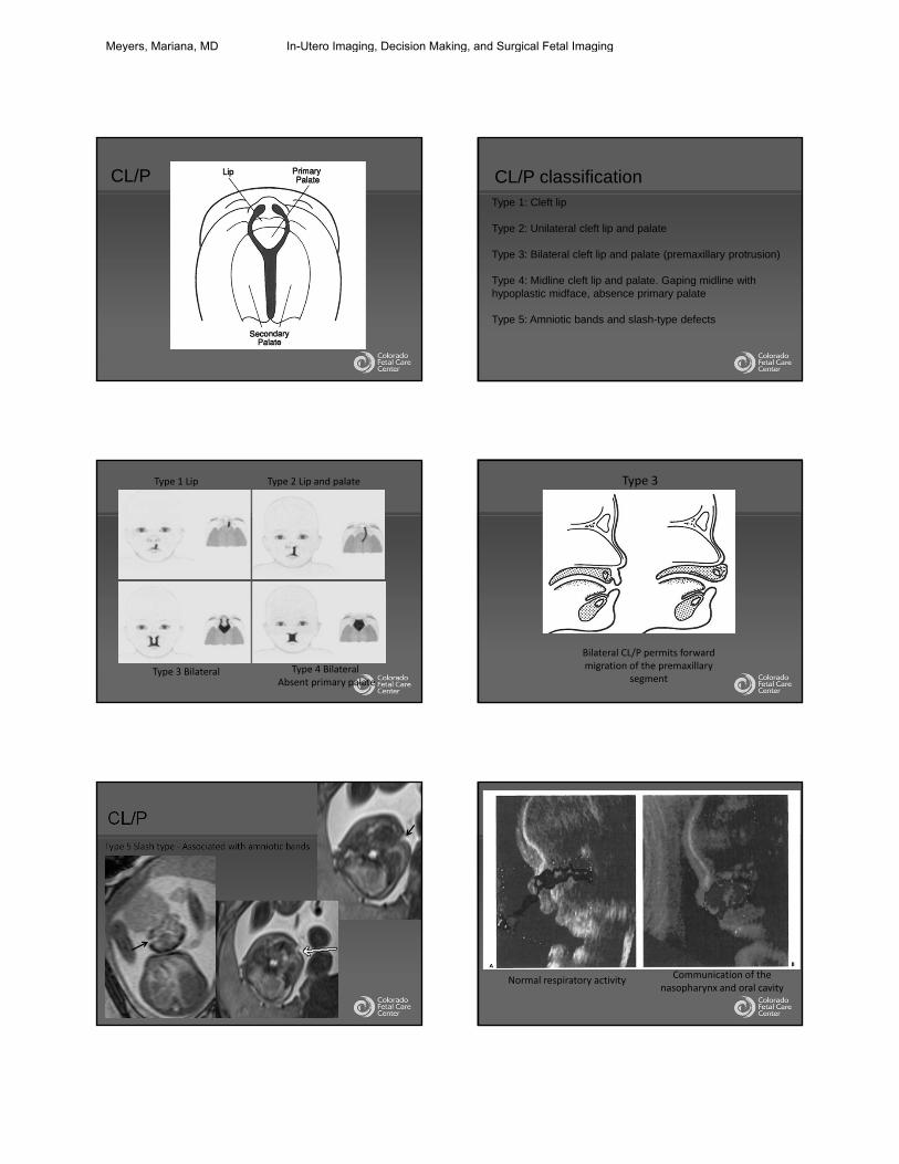
CL/P CL/P classificationType 1: Cleft lip
Type 2: Unilateral cleft lip and palate
Type 3: Bilateral cleft lip and palate (premaxillary protrusion)
Type 4: Midline cleft lip and palate. Gaping midline with hypoplastic midface, absence primary palate
Type 5: Amniotic bands and slash-type defects
Type 1 Lip Type 2 Lip and palate
Type 3 Bilateral Type 4 Bilateral Absent primary palate
Bilateral CL/P permits forward migration of the premaxillary
segment
Type 3
CL/PType 5 Slash type ‐ Associated with amniotic bands
Normal respiratory activityCommunication of the
nasopharynx and oral cavity
Meyers, Mariana, MD In-Utero Imaging, Decision Making, and Surgical Fetal Imaging

CL/PAssociated with multiple syndromes
T
T
Bilateral
Meyers, Mariana, MD In-Utero Imaging, Decision Making, and Surgical Fetal Imaging

Facial anomalies
• Oral lesions
• Cleft lip/palate
• Micrognathia
No nasopharynx/oropharynx
Narrow maxilla
Piriform sinuses
Oropharynx level
Broad nose
Left ear
Right ear
Meyers, Mariana, MD In-Utero Imaging, Decision Making, and Surgical Fetal Imaging

Small stomach
Cleft lip?
• Treacher- Collins Syndrome
• Goldenhar syndrome
• Roberts syndrome
• Pierre Robin syndrome
• Nager syndrome
• Chromosomal abnormalities: 13/18
• Crouzon’s syndrome
• Cornelia de Lange
• Smith Lemli-Optiz syndrome
Differential diagnosis
• Linear relationship between mandibular growth and gestational age or BPD
• Cutoff level of less than 23, the jaw index had a 100% sensitivity and 98.1% specificity in diagnosing micrognathia
Micrognathia index by US
Objective Diagnosis of Micrognathia in the Fetus: The Jaw Index. Obstetrics & Gynecology . Issue: Volume 93(3), March 1999, p 382–386
Meyers, Mariana, MD In-Utero Imaging, Decision Making, and Surgical Fetal Imaging

Mandibular index= AP mandibular diameter/ BPD x 100
ProfileMandibular index 28.3%
Figure 4 . Boxplot of the jaw index in normal fetuses and fetuses with micrognathia. Boxes indicate quartiles (25th and 75th percentiles), and vertical bars indicate ranges.
Obstet Gynecol 1999; 93:382-6. Objective diagnosis of micrognathia in the fetus: the jaw index.
Fetal airway compromise
Lesions causing airway obstruction
1. Facial lesions
2. Neck lesions
3. Thoracic lesions
1. Congenital lesions of the neck: BCC, CTC, ranula, TDC, Dermoid/ epidermoid,
2. Inflammatory lesions: goiter
3. Vascular malformations:
– Low flow: LM/ mixed lesions
– High flow: AVF, AVM
4. Tumors: hemangioma, teratoma, myofibromatosis, neuroblastoma, rhabdosarcoma, etc.
Neck pathology
1. Congenital lesions of the neck: BCC, CTC, ranula, TDC, Dermoid/ epidermoid,
2. Inflammatory lesions: goiter
3. Vascular malformations:
– Low flow: LM/ mixed lesions
– High flow: AVF, AVM
4. Tumors: hemangioma, teratoma,myofibromatosis, neuroblastoma, rhabdosarcoma, etc.
Neck pathology
Meyers, Mariana, MD In-Utero Imaging, Decision Making, and Surgical Fetal Imaging

Cervical thymic cyst
• Persistence of the thymopharyngeal tract
• Lateral superior neck. Midline inferior
• Left + common
• Unilocular vs multilocular
• Close to the carotid space: splays CA and JV
• Anechoic/ debris. Hyperintense T2
• Ddx: LM, TDC, BCC
• Good prognosis
Nguyen Q, et al. Cervical thymic cyst: case reports and review of the literature. Laryngoscope. 1996; 106 (3, pt1: 247-252)
Branchial cleft cyst• Defect in embryogenesis of the branchial apparatus
• Several types: Type II most common*
• Lateral neck although can extend to midline
• Anechoic/ hypoechoic. Debris. Thin walled
• Hypo T1- Hyper T2
• Isolated or associated to syndrome: brachio-oto-renal
syndrome
• Ddx: LM, TDC
• Can cause airway compression. EXIT
Bilobed enlargementMay have cystsRare compression of the airway/esophagusLook for polyhydramnios
Hyperthyroid goiter:- Tachycardia- Advanced bone age- Hydrops- IUGR- Hepatosplenomegaly
• Causes: Mom with thyroid problems (Hashimoto’s, Graves, iodine
ingestion, antithyroid medications)
• Best views-coronal; sagittal
• US-homogeneous, bilobed, echogenic, hypervascular; possibly poly,
high output cardiac failure
• MRI-hypointense on T2 (nl-iso). Bright T1
• Complications: hyperextension of head-airway/ obstruction
• Check fetus thyroid status by cord blood (0.5-1.4 % risk fetal loss)
• TTx: correct Mom’s issues; Intra-amniotic/muscular T4*
• Good prognosis
Thyroid Goiter
www.fetus.net / Bianchi DW. Fetology: Diagnosis and Management of the fetal patient. 2nd Edition. McGraw-Hill Medical 2010
Meyers, Mariana, MD In-Utero Imaging, Decision Making, and Surgical Fetal Imaging

Cervical teratoma
Teratoma
• Congenital: 1- sacroccygeal; 2- Head/Neck*
• MC-immature; embryonal thyroid and neurectoderm
• Anterolateral large solid /cystic mass; +/- calcifications
• Calcifications: specific for teratoma but in <20%
• In the vast majority of cases, the cervical airway is
compressed and obstructed
Rubio, et al, SPR poster 2011
Teratoma
• US: polyhydramnios, lesion vascularity, Doppler
changes, hydrops
• MRI: lung volumes, intracranial and mediastinal
invasion, and airway delineation
• Amniotic AFP could help in the ddx of a cervical
mass BUT only 30% produce AFP*
• Maternal and fetal AFP levels can be elevated*
Teratoma
Sometimes the mass is resected while on utero‐placental bypass
Meyers, Mariana, MD In-Utero Imaging, Decision Making, and Surgical Fetal Imaging

Vascular malformations/tumors
• Vascular malformations- Low flow: Lymphatic/ Venolymphatic
- High flow: AVM-AVF
• Vascular tumors:- Hemangioma
Mulliken JB, Glowacki J. Plast Reconstr Surg 1982;69:412–422Donnelly LF et al. AJR Am J Roentgenol. 2000;174 (3):597-608
Vascular malformations/tumors
• Vascular malformations- Low flow: Lymphatic/ Venolymphatic
- High flow: AVM-AVF
• Vascular tumors:- Hemangioma
Mulliken JB, Glowacki J. Plast Reconstr Surg 1982;69:412–422Donnelly LF et al. AJR Am J Roentgenol. 2000;174 (3):597-608
Lymphatic malformations
• Nuchal: macrocystic lesions (cystic hygroma) 1st trimester
• Non-nuchal: microcystic lesions (lymphangioma)
• Failure in communication of the jugular lymphatic sac and jugular veins at 40 days GA
• Nuchal LM: 51 % associated with chromosomal anomalies (T 13-18-21, Turner’s), cardiac and skeletal malformations
• Non-nuchal LM: isolated or Gorham- Stout, Klippel-Trenaunay
Kathary et. Al. Pediatr Radiol. 2001; 31 (10): 727-731Fundamental and advanced fetal imaging. Wolters Kluwer. Kline-Fath, Bulas. The neck chapter. 491-507. 2015
Nose
Chin
Meyers, Mariana, MD In-Utero Imaging, Decision Making, and Surgical Fetal Imaging

Fetal airway compromise
Lesions causing airway obstruction
1. Facial lesions
2. Neck lesions
3. Thoracic lesions
Congenital High Airway Obstruction Syndrome (CHAOS)
• Absence of all or part of trachea
- Trachea atretic, stenotic or thick web
- Aberrant pulmonary budding off the foregut
- Most have connection with esophagus
- Associated anomalies and syndromes, Fraser or DiGeorge
• Prognosis poor
• Rare
CHAOS
• Prenatal
- Intrauterine demise
- Hydrops
• Postnatal
- EXIT to ECMO
- Tracheal intubation
CHAOS
• US- Symmetric enlarged echogenic lungs
- Heart compressed and diaphragm flattened
- Dilated bronchi
- Hydrops
- Decreased or increased amniotic fluid
• MRI- Lungs hyperintense and large with inverted diaphragms
- Heart compressed
- Dilated high signal fluid filled trachea and bronchi
25 weeks
Meyers, Mariana, MD In-Utero Imaging, Decision Making, and Surgical Fetal Imaging

Differential: Bilateral CCAM
CDH
23 weeks
34 weeks
CDH
Don’t Forget to Look At The…..
Mediastinum
25 WEEKS
Meyers, Mariana, MD In-Utero Imaging, Decision Making, and Surgical Fetal Imaging

Esophageal atresia
Thank you!
Meyers, Mariana, MD In-Utero Imaging, Decision Making, and Surgical Fetal Imaging