final program & abstracts - biliary atresia · past meetings and local organizing chairs 1968...
TRANSCRIPT

June
29
– Ju
ly 3
, 200
8Ja
ckso
n La
ke L
odge
, Wyo
min
g, U
SA
ww
w.p
aps2
008.
com
41
FINAL PROGRAM & ABSTRACTS

Section I PAPS Members Handbook
PAPS Board of Directors / Delegates .................................................................................................I-01
Past Officers................................................................................................................................................I-02
Honorary Members .................................................................................................................................I-03
Coe Medal Recipients .............................................................................................................................I-03
New Members ...........................................................................................................................................I-03
Future Meetings........................................................................................................................................I-03
Past Meetings & Local Organizing Chairs ........................................................................................I-04
M. James Warden Guest Assistant Program Participants...........................................................I-05
PAPS 2008 Organizing Committee ....................................................................................................I-05
PAPS 2008 Program Committee .........................................................................................................I-06
Publications Committee ........................................................................................................................I-06
PAPS Artifacts ............................................................................................................................................I-07
Section II Program at a Glance
Program at a Glance.............................................................................................................................. II-01
Section III Scientific Program
Educational Objectives ....................................................................................................................... III-01
Accreditation Statement .................................................................................................................... III-01
Gans Memorial Lecture....................................................................................................................... III-02
GAP Fellowship Presentation............................................................................................................ III-02
Scientific Overview............................................................................................................................... III-03
Section IV PAPS 2008
General Information............................................................................................................................. IV-01
Speakers Information .......................................................................................................................... IV-02
Commercial Exhibit .............................................................................................................................. IV-03
Social Program ....................................................................................................................................... IV-05
Optional Social Activities.................................................................................................................... IV-07
Section V Abstracts
Oral Presentations..................................................................................................................................V-01
Poster Presentations .............................................................................................................................V-57
Case Presentations...............................................................................................................................V-105
Venue
Jackson Lake Lodge
Co
nfe
ren
ce A
rea
Ta
ble
of C
on
ten
ts
Blue Heron Lounge
Sunset Terrace
Trappers RoomWomen
Men
Explorers Room
Wrangler Room
Balcony
Apparel Shop
Main Lounge
PAPS
Registration
Desk
Wyoming Room (PAPS Hospitality Suite)located one level up.
Breakfast
PAPS Sessions &
Welcome Reception
Posters & Case Reports
Exhibits
PAPS Cyber Café
view from the Main Lounge

Map of Area
Jackson Lake Lodge
Grand Teton Wyoming, USA
Section I
PAPS Members Handbook
Ma
p o
f A
rea

Pacific Association of Pediatric Surgeons Board of DirectorsThe members of the Board of Directors are Present Officers, Delegates and the Immediate Past President.
President Richard E. Black
President-Elect Paul K. H. Tam
Immediate Past President Kevin C. Pringle
Secretary Harry Applebaum
Treasurer Kevin P. Lally
Archivist Alan Woodward
GAP Committee Chairperson Cynthia Reyes
Delegates
Australia Ralph C. Cohen
John M. Hutson
John Pitkin
Canada Robin Eccles
China Paul K.H. Tam
Japan Toshihiro Muraji
Naomi Iwai
Hiroaki Kitagawa (Secretary, PAPS Japan)
Tatsuo Kuroda
Korea Seong-Cheol Lee
Mexico Jaime Olvera-Duran
Taiwan Soul-Chin Chen
U.S.A. Rebecka L. Meyers
Cynthia Reyes
Walter Chwals
I PA
PS
ME
MB
ER
S H
AN
DB
OO
K
I-01

Past Officers
Presidents
Stephen L Gans 1969-70 John R Campbell 1988-89John K Stevenson 1970-71 R. Stuart Ferguson 1989-90Alexander H Bill, Jr 1971-72 Dale G Johnson 1990-91Keijiro Suruga 1972-73 Morton M Woolley 1991-92Nate A Myers 1973-74 Martin J Glasson 1992-93Jens G Rosenkrantz 1974-75 Hiroshi Akiyama 1993-94Murray R Kliman 1975-76 Giovanni Porras-Ramirez 1994-95Daniel M Hays 1976-77 Takahiro Ito 1995-96Takashi Ueda 1977-78 Marshall Z Schwartz 1996-97Joaquin C Azpiroz 1978-79 Htut Saing 1997-98Douglas Cohen 1979-80 Philip A King 1998-99Walton KT Shim 1980-81 Marvin W Harrison 1999-2000Graham C Fraser 1981-82 Takeshi Miyano 2000-01Keiichi Ikeda 1982-83 James B Atkinson 2001-02Eric W Fonkalsrud 1983-84 Rosslyn Walker 2002-03E Durham Smith 1984-85 Eui Ho Hwang 2003-04Rodolfo Franco-Vasquez 1985-86 Stephen G. Jolley 2004-05Chadwick F Baxter 1986-87 Jer-Nan Lin 2005-06Wen-Tsung Hung 1987-88 Kevin C. Pringle 2006-07
Richard E. Black 2007-08Secretaries
Jens G Rosenkrantz 1969-71 Dale G Johnson 1985-88John R Campbell 1971-73 John C German 1988-91Edward A Free 1973-75 Marshall Z Schwartz 1991-95George A Hyde 1975-77 Marvin W Harrison 1995-99Walton KT Shim 1977-79 Stephen G Jolley 1999-01Pieter A De Vries 1979-82 Robert S Sawin 2001-04William C Bailey 1982-85 Harry Applebaum 2004-
Treasurers
Eric W Fonkalsrud 1969-70 Ann M Kosloske 1985-90Alfred A de Lorimer 1970-72 Marvin W Harrison 1990-95Daniel M Hays 1972-74 Dale G Hall 1995-99David L Collins 1974-77 James B Atkinson 1999-01Ernest B Haws 1977-84 Richard E Black 2001-07David Hodge 1984-85 Kevin P. Lally 2007-
Archivists
Nate A Myers 1987-92 Alan Woodward 1992-
M James Warden Guest Assistance Program Chairs
M James Warden 1987-99 Cynthia Reyes 2004-Philip A King 1999-2004
Honorary Members
Herbert E. Coe
Sir Kenneth Fraser
T Y Nelson
Tamotsu Fukuda
Joseph Steigrad
Jesus Lozoya-Solis
Osamu Wakabayashi
Ovar Swenson
William P Longmire, Jr
Jin-zhe Zhang
F Douglas Stephens
New Members
Hong Kong
Kenneth Kak Yuen Wong
Japan
Masayuki Obatake
Tatsuro Tajiri
Yasuharu Ohno
Takashi Shimotake
Keiichi Uchida
Eiichi Deguchi
USA
Sunghoon Kim
Philip Kent Frykman
Garret Seth Zallen
Fombe Ndiforchu
Cathy Shin
Future Meetings
May 10-14, 2009 Hong Kong, China Paul K.H. Tam
May 23-27, 2010 Kobe, Japan Naomi Iwai
April 17-21, 2011 Cancun, Mexico Jaime Olvera-Duran
I PA
PS
ME
MB
ER
S H
AN
DB
OO
K
I-03
I P
AP
S M
EM
BE
RS
HA
ND
BO
OK
I-02

Past Meetings and Local Organizing Chairs
1968 Founders organizing meeting, Seattle, WA, Orcas Island, USA Alexander H Bill
1969 Ojai, CA, USA Stephen L Gans Eric W Fonkalsrud Dan Hayes1970 Melbourne, VIC, Australia Nate Myers1971 Harrison Hot Springs, BC, Canada Phillip G Ashmore1972 Tokyo, Japan Keijiro Suruga1973 San Diego, CA, USA David L. Collins1974 Salishan, OR, USA John R Campbell 1975 Honolulu, HI, USA Walton KT Shim 1976 San Francisco, CA, USA Alfred A de Lorimer1977 Sydney, NSW, Australia Douglas Cohen 1978 Osaka, Japan Takashi Ueda1979 Mazatlan, Mexico Rodolfo Franco Vazquez1980 Colorado Springs, CO, USA William C Bailey 1981 Maui, HI, USA Walton KT Shim1982 Vancouver, BC, Canada Graham C Fraser1983 Fukuoka, Japan Keiichi Ikeda1984 San Diego, CA, USA Timothy G Canty 1985 Rotorua, New Zealand R Stuart Ferguson1986 Puerto Vallarta, Mexico Joaquin C Aspiroz1987 Rosario-Orcas, WA, USA John L Cahill 1988 Taipei, Taiwan Wen-Tsung Hung1989 Portland, Oregon Marvin W Harrison 1990 Kona, HI, USA Walton KT Shim1991 Hong Kong, China Htut Saing1992 Abuquerque, NM, USA Patrick F Jewell 1993 Cairns, QLD, Australia Mervyn M Lander1994 Kagoshima, Japan Horoshi Akiyama1995 Hualtuco, Mexico Giovanni Porras-Ramirez1996 Singapore VT Joseph 1997 Phoenix, AZ, USA Joseph T Zerella1998 Maui, HI, USA Walton KT Shim1999 Beijing, China Jin-Zhe Zhang2000 Las Vegas, NV, USA Stephen G Jolley 2001 Kyoto, Japan Takeshi Miyano2002 La Jolla, CA, USA Harry Applebaum2003 Sydney, NSW, Australia Ralph Cohen 2004 Seoul, Korea Eui Ho Hwang2005 Vancouver, BC, Canada Eric M Webber2006 Taipei, Taiwan Jer-Nan Lin2007 Queenstown, New Zealand Kevin C Pringle2008 Jackson Hole, WY, USA Rebecka L Meyers
M James Warden Guest Assistance Program Participants
1989 Mario Vanela Chile
Luis Pedroza Mexico
1990 Luis Canchez Peru
1991 Nguyen Xuan Thu Vietnam
1992 Leopoldo Torres Mexico
1994 Xisheng Zhang China
Amaung Maung Myanmar
1996 Zhou Yuan China
1997 Ricardo Peniche Mexico
1998 Chi Mean Hea Cambodia
1999 Alexsey Podkamenev Russia
2000 Luis Mondragon Mexico
2001 Zeng Shan China
2002 Sandra Montedonico-Rimassa Chile
Anna Shapinka Russia
2003 Sar Vuthy Cambodia
2004 Mclee Aite Mathew Papua New Guinea
2005 Alejandro Ayon Nicaragua
2006 Surachai Saranrittichei Thailand
2007 Safwat Andrawes Kenya
2008 Daniel Acosta Farina Ecuador
PAPS 2008 Local Organizing Committee
Rebecka Meyers (Chair) & Michael Howard
Richard & Kathy Black
Eric Scaife & Caroline Milne
Michael & Rosemarie Matlak
Michael Rollins
Gail Beauregard
Jamee Carpenter
Krissie Norton
Lisa WIlkes
Symposium Coordinators
“Pediatric Surgery in Developing Countries”
Steven Bickler
Georges Azzie
I PA
PS
ME
MB
ER
S H
AN
DB
OO
K
I-05
I P
AP
S M
EM
BE
RS
HA
ND
BO
OK
I-04

PAPS 2008 Program Committee
Chair Rebecka L. Meyers
Members
Australia/New Zealand Andrew Barker
Spencer Beasley
Canada Robin Eccles
China Shan Zheng
Hong Kong Paul K. H. Tam
Japan Naomi Iwai
Hiroyuki Kobayashi
Toshihiro Muraji
Korea Seong-Cheol Lee
Mexico Jamie Olvera-Duran
USA Sherif Emil
Diana Farmer
Eric Scaife
Don Shaul
Publications Committee
Atsuyuki Yamataka (chair)
Walter Chwals (vice-chair)
Don Shaul
Kevin Lally
James Dunn
Eric R. Scaife
Yasuo Ito
Hideo Takamatsu
Masahiro Fukuzawa
Tomoaki Taguchi
Kenneth Wong
Anette Jacobsen
Ralph Cohen
Eric Webber
Andrew Holland
PAPS Artifacts
Artifact – a simple object produced by human workmanship
The Presidential Badge-
The Past President’s Badge-
The Flag-
The Coe Medal-
The Gavel-
The Baxter-Myers Tennis Trophy-
The Kimura Golf Trophy-
The Archives Cabinet-
Presentation to British Association of Paediatric Surgeons-
The Presidential Badge
This badge was presented by the British Association of Paediatric Surgeons to their
colleagues in the Pacific in 1972. It is handed over to the incoming President each year at
the Annual Meeting, usually in a presentation at the Annual Banquet.
The Past President’s Badge
Douglas Cohen suggested to the Board of Directors that it would be appropriate for Past
Presidents to wear a badge identifying them at Annual Scientific Meetings and included
the concept of a brooch for wives of Past Presidents.
Having approval of the Board, he selected a design for the badges, copied from the PAPS
flag, which had been designed by Peter Jones. Amor Metal Makers in Sydney produced
the badges. Douglas Cohen then presented the first badges in Mexico in 1979 when he
assumed the role of President. An additional supply of badges were obtained for the
Secretary in 1984 when Durham Smith was President.
The Flag
Foundation Member Peter Jones designed the PAPS flag in collaboration with Miss
Vivienne James, Medical Artist at Royal Children’s Hospital in Melbourne Australia. It was
made by Evan Evans Flags of 680 Elizabeth Street, Melbourne, and flew for the first time
at the 3rd Annual Meeting of PAPS in Melbourne in 1970.
Each year, the flag adorns the meeting site and moves round the Pacific Ocean with
successive Meeting Organizing Committees.
I PA
PS
ME
MB
ER
S H
AN
DB
OO
K
I-07
I P
AP
S M
EM
BE
RS
HA
ND
BO
OK
I-06

The Coe Medal
The Coe medal was initially conceived to honor the memory of Herbert E. Coe, MD. Based
in Seattle, he was a founding father of pediatric surgery on the Pacific shore of the United
States. It is the highest honor presented by PAPS, and is awarded to someone who has
practiced on the Pacific Rim and who has made outstanding contributions to Pediatric
Surgery. Keiichi Ikeda, Professor Emeritus at Kyushi University and a former President of
PAPS 1982-83, will receive this award on Wednesday morning, July 2, 2008.
In 1984, John Stevenson was placed in charge of plans to develop a Medal of Honor
bearing the likeness of Herbert Coe, with $1,800 being allocated for the first fifty medals.
Dr. Stevenson also convened a committee of Alexander Bill, Douglas Cohen, Morio Kasai
and Murray Kliman to establish criteria for the awarding of the medal. It was decided in
1985 that the first medal, cast in pewter with antique gold finish, would be presented to
Mrs. Coe. In 1986 the Board of Directors approved the following guidelines for selection
of its future recipients:
The recipients would be recognized as having made outstanding contributions to 1.
pediatric surgery.
Contributions should be considered in any related field of pediatric surgery, any 2.
of the pediatric surgical specialties, pediatric surgical research, or anything that is
considered to have raised the status of pediatric surgery. Service to PAPS per se,
however meritorious, should not be considered an appropriate contribution unless
the nominee was considered to have contributed in some additional appropriate
way.
Except in most special circumstances, the medal would be awarded to those 3.
individuals who are working or have worked in the area covered by PAPS.
In order to enhance the value of the award, not more than one medal should be 4.
given in any one year. It should also not be necessary to make the award every
year. A candidate for the award could be nominated by any PAPS member in good
standing.
The nomination should be forwarded to the secretary and should include enough 5.
information for members of the Board to formally review and, if appropriate, second
the nomination. The final selection of the recipient for the Coe Medal will be made
by vote of the Board of Directors.
The selection should be made 4 months in advance of the annual meeting of the 6.
Association to allow the recipient, if possible, to plan to attend that meeting to
receive the medal.
The addition of two more guidelines followed:
Although no limitation is placed on the nomination of any candidate, special 6.
consideration would be given to nominees who are or have been working in the
Pacific Basin or whose work is seen as having particular relevance for pediatric
surgeons working in the area.
A list of previous recipients will be sent out each time the selection committee 7.
guidelines are promulgated to avoid the problem of possibly recommending
somebody who is already a recipient.
In 1987, the Board of Directors voted to make an exception to the rule of awarding a
single madal in one year and award medals to both Alexander Bill and Morio Kasai to
mark the 20th Anniversary of PAPS in 1998 in Seattle, the home of Dr. Coe.
List of Recipients
1986 Mrs. Coe
1987 Alexander Bill and Morio Kasai
1988 Keijiro Suruga
1989 Nate Myers
1990 Stephen Gans
1992 Morton Woolley
1993 Durham Smith
1994 Takashi Ueda
1995 Daniel Hays
1998 Eric Fonkalsrud
2001 Justin Kelly
2002 Alberto Pena
2003 Ken Kimura
2007 John Hutson
2008 Keiichi Ikeda
I PA
PS
ME
MB
ER
S H
AN
DB
OO
K
I-09
I P
AP
S M
EM
BE
RS
HA
ND
BO
OK
I-08

The Gavel
In May 1971, John Stevenson presented a gavel to PAPS. The head of the gavel was
fashioned from hawthorn wood, which flowers in May in the northern hemisphere. May
1967 was the birthday of our Association. The handle was made from holly, a holy tree
used on special occasions in ancient times to represent goodness and purity.
It is significant that the wood was obtained from trees felled by Herbert Coe in the year
before his death and stored in his basement for future woodworking. The trees had
originally been brought by Dr. Coe’s parent from England and planted when they settled
in Seattle in 1888. The timber was later obtained from his widow. It is fitting that the
Association has a gavel used at Annual Meetings made from wood belonging to one of
our esteemed honorary members who was instrumental in beginning the specialty of
Pediatric Surgery.
The Baxter-Myers Tennis Trophy
This unique folding tennis racquet was presented to President Nate Myers in 1974 by
Chad Baxter. Nate had frequently blamed borrowed tennis racquets for his failure to win
points in PAPS Tennis Tournaments. Chad sawed one of his wife Jean’s racquets in half
and hinged it to facilitate transport across the Pacific. It was decided by the Executive
Committee in 1975 that this would become the permanent trophy for the tournament
each year. One member was so impressed with the idea of a folding racquet that he
enquired “Where can I buy one?”
There were difficulties in ensuring the trophy was available each year. Each winner
needed to attend the meeting the following year to hand on the trophy. Concerns arose
about it being lost, so a small model was commissioned to be presented each year and
the original stayed in the archives cabinet in Melbourne.
1974 S. Gans, K. Suruga
1975 D. Vitale, K. Harikoshi
1977 Ueda
1978 Nishinomiya, Ikoma
1979 Y. Sanada
1983 N. Myers, K. Suruga
1985 R. Fowler, I. Kern
1986 N. McMullin
1987 E. Durham Smith
1988 H.K. Goon
1989 W.K.T. Shim
1990 F. Ikoma
1991 G. Mya
1992 D. Vitale
This is not the first unique tennis racquet associated with Pediatric Surgery, as Sir Denis
Browne invented his own personal racquet, and played at Wimbledon.
Due to lack of participants, the tennis tournament will not be taking place this year.
The Kimura Golf Trophy
Because of the great success, novelty and convenience of the folding tennis racquet, Ken
Kimura cut and hinged a golf club in 2001 for a similar purpose and to date he seems to
have won it more than most other members!
The Archives Cabinet
In 1989, Nate Myers was appointed as Archivist and soon after, he informed Dr. John
German, Secretary of PAPS, that suitable accommodation for the Assocation’s archival
material could be made available in the Archives Room at Royal Children’s Hospital
in Melbourne. Subsequently, he wrote to Anne Kosloske, Treasurer, suggesting that a
cabinet be purchased for $500 to house the material in the Department of Surgery at
RCH Melbourne.
The Gift from PAPS to BAPS
In London in July 1973, a silver candelabra was presented by the Pacific Association of
Pediatric Surgeons to the British Association of Paediatric Surgeons in recognition of
their leadership. This was presented to Mr. H.H. Nixon by Mr. N.A. Myers, Presidents of the
Associations at the time. It was subsequently stolen, and replaced afterwards.
I PA
PS
ME
MB
ER
S H
AN
DB
OO
K
I-11
I P
AP
S M
EM
BE
RS
HA
ND
BO
OK
I-10

Section II
Program at a Glance

II PR
OG
RA
M A
T A
GL
AN
CE
II
PR
OG
RA
M A
T A
GL
AN
CE
II-02II-01
Sunday, June 29 Monday, June 30 Tuesday, July 1 Wednesday, July 2 Thursday, July 3
06:30
Regi
stra
tion
(06:
30 -
13:0
0) M
ain
Loun
ge
Breakfast (06:30 - 08:30)
Regi
stra
tion
(06:
30 -
09:0
0) M
ain
Loun
ge Breakfast (06:30 - 08:30)
Regi
stra
tion
(06:
30 -
13:0
0) M
ain
Loun
ge
Breakfast (06:30 - 08:30)
Regi
stra
tion
(06:
30 -
12:0
0) M
ain
Loun
ge
Breakfast (06:30 - 08:30) 06:30
07:00 Poster Session 1 - Oral & Display (07:00 - 08:00)
Wrangler Room
Pediatric Surgery in Developing Countries
(07:00 - 09:00)Explorers Room
Poster Session 2 - Oral & Display (07:00 - 08:00)
Wrangler Room
Poster Session 3 - Oral & Display (07:00 - 08:00)
Wrangler Room
07:00
07:30 07:30
08:00
Publications Committee
Meeting (08:00 - 12:00)Wyoming Room
Session 1
Neonatal and Fetal (08:00 - 10:00)Explorers Room
Session 3Hepatobiliary and Nutrition
(08:00 - 09:55)Presentation of the Coe Medal(09:55 - 10:10) Explorers Rm.
Session 5
Trauma, Policy & Misc. (08:00 - 09:40) Explorers Rm.
08:00
08:30 08:30
09:00 09:00
09:30
Official Tour
Yellowstone National Park(09:30 - 19:00)
GAP Lecture (09:40 - 10:00) 09:30
10:00Coffee Break/Case
Reports (10:00 - 10:30)Coffee Break/Case Reports
(10:00 - 10:30)Coffee Break/Case Reports
(10:00 - 10:30)10:00
10:30 Session 2
Thoracic, Spleen & Oncology
(10:30 - 12:30) Explorers Room
Session 4
Gastrointestinal(10:30-12:30)
Explorers Room
Session 6
Anorectal, Hernia, Urology & Closing
(10:30 - 12:30) Explorers Room
10:30
11:00 11:00
11:30 11:30
12:00
Regi
stra
tion
(12:
00 -
18:0
0) M
ain
Loun
ge
12:00
12:30
Board of Directors Meeting
(12:15 - 16:15)Wyoming Room
Buy-in Activities
Golf (12:45 - 20:00)Whiteriver Rafting
(13:30-19:30)
GANS Lecture (12:30-13:15) 12:30
13:00 Buy-in Bike Tour (13:30 - 16:30) Optional Activities booked through the Jackson Lake
Lodge: Float Trips, Horseback Riding, Fishing, Boat Rentals
and Lake Cruises
13:00
13:30 Annual Business Meeting (13:30 - 14:30) Explorers Rm.
13:30
14:00 14:00
14:30 Optional Activities booked through the Jackson Lake Lodge:
Float Trips, Horseback Riding, Fishing, Boat
Rentals and Lake Cruises
14:30
15:00 Exit Board of Directors Meeting (15:00 - 16:00)
Explorers Rm.
15:00
15:30 15:30
16:00 Optional Activities booked through the Jackson Lake
Lodge: Float Trips, Horseback Riding, Fishing, Boat Rentals
and Lake Cruises
16:00
16:30Optional Activities
booked through the Jackson Lake Lodge:
Float Trips, Horseback Riding,
Fishing, Boat Rentals and Lake
Cruises
16:30
17:00 President’s Dinner (by invitation) (17:00 - 21:30)
Annual Banquet
Reception & DInner
Diamond Cross Ranch(17:30 - 23:00)
17:00
17:30 17:30
18:00
Buy-in Event
Bar J Chuck Wagon Supper & Western
ShowTransportation Provided
(16:30 - 23:00)
18:00
18:30
Welcome
Reception
(18:00-21:00)Explorers Room
18:30
19:00 19:00
19:30 19:30
20:00 20:00
20:30 20:30
21:00 21:00
22:00 22:00
23:00 23:00
Exhibit Hours (06:30-13:00) Exhibit Closed Exhibit Hours (06:30-13:00) Exhibit Hours (06:30-10:30)
Cyber Café (07:00-18:00) Cyber Café (07:00-10:00) Cyber Café (07:00-18:00) Cyber Café (07:00-12:00)
Hospitality Suite (11:00-22:00) Hospitality Suite (11:00-22:00) Hospitality Suite (11:00-22:00) Hospitality Suite (11:00-15:00)

Section III
Scientific Program

III SC
IEN
TIF
IC P
RO
GR
AM
Educational Objectives The PAPS Annual Meeting is designed to provide comprehensive continuing education in the field of pediatrics surgery. It is PAPS’ intent to bring together the world’s leading authorities to present and discuss the most recent clinical and research efforts.
Our organization is focused on clinical pediatric surgery and the international, cross cultural sharing of clinically innovative surgical techniques. Surgeons at our meeting concentrate on learning the newest surgical techniques which may have initially been developed and popularized in one country and can now be applied on an international scale.
Accreditation Statement This activity has been planned and implemented in accordance with the Essential Areas and Policies of the Accreditation Council for Continuing Medical Education through the joint sponsorship of the American College of Surgeons and the Pacific Association of Pediat-ric Surgeons. The American College of Surgeons is accredited by the ACCME to provide continuing medical education for physicians.
AMA PRA Category 1 Credits™ The American College of Surgeons designates this educational activity for a maximum of 13.5 AMA PRA Category 1 Credits™. Physicians should only claim credit commensurate with the extent of their participation in the activity.
Disclosure InformationIn order to comply with the ACCME’s Updated Standards for Commercial Support, The American College of Surgeons, as the accredited provider of this activity, has implemented a disclosure process to ensure that anyone in a position to control the content of the educational activity has disclosed all relevant financial relationships with any commercial interest. Per these standards, it is mandatory that both the program committee and speakers complete disclosures. Members of the program committee were required to disclose all financial relationships and speakers were required to disclose any financial relationship as it pertains
to the content of the presentations. ACS defines a “commercial interest” as any proprietary entity producing health care goods or services consumed by, or used on patients. The ACCME does not consider providers of clinical service directly to patients to be commercial interests. The ACS considers “relevant” financial relationships as financial transactions (in any amount) occurring within the past 12 months that may create a conflict of interest.
The updated standards also require that ACS, through our joint sponsorship partners, manage any reported conflict and eliminate the potential for bias during the session. The program committee members and speakers were contacted and there were no disclosures of potential conflicts of interest. However, if you perceive a bias during a session, please report the circumstances on the session evaluation form.
Please note we have advised the speakers that it is their responsibility to disclose at the start of their presentation if they will be describing the use of a device, product, or drug that is not FDA approved or the off-label use of an approved device, product, or drug or unapproved usage.
The requirement for disclosure is not intended to imply any impropriety of such relationships, but simply to identify such relationships through full disclosure, and to allow the audience to form its own judgments regarding the presentation.
III-01
American College of SurgeonsDivision of Education

III SC
IEN
TIF
IC P
RO
GR
AM
Gans Memorial Lecture
Wednesday, July 2 12:30 – 13:15Explorers Room
This lecture is given in memory of Stephen L. Gans, MD, the founder and first President of the Pacific Association of Pediatric Surgeons (PAPS). Under the terms of the bequest that funds this lecture, the lecture should be a topic that does not relate to Pediatric Surgery and the Lecturer should be an authority on the lecture material and reside in the same locale as the Annual Meeting location.
Michael Dunn is a writer, director, photographer, and producer for Dunn Communications Inc., a Salt Lake City advertising agency and film production company. Among his peer distinctions over the years are a gold and silver medal from the New York Film Festival, several ADDY’s from the American Advertising Federation, and four CLIO’s – an award considered the “Oscar” of the advertising industry. He also won an Emmy Award for public service in 1992 from the National Academy of Television Arts and Sciences. In the spring of 2000, he was honored by the Utah Advertising Federation as the inaugural recipient of the Young Professional of the Year Award.
In the late summer of 1994, Michael was attacked and severely malled by a grizzly bear while running in Grand Teton National Park not 10 miles from the Jackson Lake Lodge. Miraculously, he survived the attack and will share his remarkable story with us.
M. James Warden Guest Assistance Program Fellowship
Thursday, July 3 09:40 – 10:00Explorers Room
The GAP Fellowship Lecture will be presented by Dr. Daniel Acosta Farina.
Dr. Daniel Acosta Farina is a pediatric surgeon from Manta, Ecuador. Dr. Farina graduated from medical school in Guayaquil, Ecuador in 1991. He did his surgery residency in Manga, Ecuador, and his pediatric surgery training in Barcelona, Spain, with Dr. Boix-Ochoa. Dr. Cynthia Reyes will introduce Dr. Farina prior to the GAP lecture on Thursday, July 3. We are pleased to welcome Dr. Farina and his wife to PAPS.
III-02

III SC
IEN
TIF
IC P
RO
GR
AM
III
SC
IEN
TIF
IC P
RO
GR
AM
MONDAY, JUNE 30 CONTINUED...SUNDAY, JUNE 29
08:00 - 12:00
Publications Committee Meeting Wyoming Room
12:00 - 18:00
Registration Open Main Lounge
12:15 - 16:15
Board of Directors Meeting Wyoming Room
18:00 - 21:00
Welcome Reception Explorers Room
MONDAY, JUNE 30
06:30 - 13:00
Registration Open Main Lounge
06:30 - 08:30
Breakfast Sunset Terrace / Blue Heron Lounge
06:30 - 13:00
Exhibits Open Trappers Room
07:00 -
08:00
Oral Poster Session 1 Wrangler Room
Moderators:
Harry Applebaum and Ralph Cohen
TIME TITLE SESSION # NAME
07:00 - 7:05
Vitamin A Deficiency in Pregnant Rats Affects Renal Development and Tumor Formation in Filial Rats
P01 Kai Li
07:05 - 07:10
Alterations in sub-cellular localization of the tran-scriptional co-activator CITED1 in development and embryonal tumors
P02 Harold Lov-vorn, III
07:10 - 07:15
The implications of surgical intervention in the treatment for neuroblastoma
P03 Tatsuro Tajiri
07:15 - 07:20
Growth-promoting effect of bisphenol A on neu-roblastoma in vitro and in vivo
P04 XianminXiao
07:20 - 07:25
Clinical features and outcomes of malignant liver tumor in children
P05 Shigeru Ono
07:25 - 07:30
Diagnosis And Management of Neonatal Hepatic Hemangioma
P06 Kui-Ran Dong
07:30 - 07:35
Acute abdomen in the neonate can result from appendicitis
P07 DickensSaint-Vil
07:35 - 07:40
The impact of strict infection control on survival rate of prenatally diagnosed isolated congenital diaphragmatic hernia
P08 Nobuyuki Morikawa
07:40 - 07:45
Computed tomography evaluation of congenital esophageal atresia with fistula: A 10-year reprisal
P09 KennethWong
07:45 - 07:50
Jejunal Free Flap Salvage for Failed Esophageal Replacement
P10 Jessica Ray-hanabad
07:50 - 07:55
Magnetic Alteration of Pectus Excavatum Deformi-ties: Development of Patient-Friendly, Practical Orthotic Braces
P11 Patrick Cur-ran
07:00 -
08:00
Display Poster Session 1 Wrangler Room
TIME TITLE SESSION # NAME
The Impact of Iatrogenic Gastroschisis on Pul-monary Maturation in a Fetal Rabbit Model of a Diaphragmatic Hernia
P12 Gong Chen
Minimally Invasive Technique in Treatment of Complex, Subcutaneous Abscesses
P13 Alan Ladd
Thoracoscopic bullectomy for spontaneous pneu-mothorax in paediatric patients
P14 Paul Tam
Hepatoblastoma: an institution’s experience P15 Dae-Yeon Kim
Abdominal closure of gastroschisis by coverage of the defect with the preserved umbilical cord
P16 Takashi Watanabe
Thoracoscopic Chondrotomy Alleviates Postopera-tive Pain after Nuss Procedure
P17 John Lee
Transumbilical approach for neonatal surgical diseases - Wound less operation
P18 Tatsuro Tajiri
Continuous veno-venous hemodialysis filtration (CVVHDF) for systemic inflammatory response syndrome (SIRS) in a piglet enterotomic peritonitis model
P19 Kui-Ran Dong
Complications of Intestinal Stomas in Children P20 Bha-nuprakash Mariyappa Rathnamma
Use of a Thoracostomy tube to Guide a Sternal bar across the Chest during a Nuss procedure
P21 Aaron Strumwasser
Magnetic Alteration of Asymmetric Pectus Excava-tum Deformities: A Case Study
P22 Patrick Cur-ran
III-04III-03

III SC
IEN
TIF
IC P
RO
GR
AM
III
SC
IEN
TIF
IC P
RO
GR
AM
MONDAY, JUNE 30 CONTINUED...MONDAY, JUNE 30 CONTINUED...
07:00 -
08:00
Case Reports - Displayed on rotation
throughout the day
Wrangler Room
TIME TITLE SESSION # NAME
Jejunal ectopic pancreas causing intestinal ob-struction in a neonate
C01 Diana Lon-doño
Malignant Change from Infantile Fibromatosis to Fibrosarcoma after Regression of Tumor in Lower Leg
C02 Masako Kubo
A Three-stage Reconstruction of the Trachea and the Esophagus in Tracheal Agenesis.
C03 Noriaki Usui
Thymopharyngeal duct cyst: an unusual cause of respiratory compromise
C04 Masao Yasu-fuku
Neonatal transthoracic needle puncture of Large Congenital Cystic Adenomatoid Malformations (CCAMs) of the Lung with Respiratory Distress – A Useful Temporizing Measure in the Acute Manage-ment
C05 Aneetha Pasupati
Telangiectatic focal nodular hyperplasia of the liver: Spontaneously regressive tumor-like lesion in infancy
C06 ShigeruUeno
Cardiac dysfunction after the surgery for pheo-chromocytoma in children-Report of three cases-
C07 Shin Shin-yama
Abdominal Inflammatory myofibroblastic tumor in child
C08 Hyun-Young Kim
Simultaneous Modified Ravitch Procedure and Latissimus Dorsi Transfer for Chest Wall Deformity Repair in Poland’s Syndrome
C09 Michael Dingeldein
08:00 -
10:00
Scientific Session 1 - Neonatal and Fetal Explorers Room
Moderators:
Diana Farmer and Paul Tam
TIME TITLE SESSION # NAME
08:00 - 08:10
A Unique Surgical Approach: 9 Years Experience of Patent Ductus Arteriosus Ligation in Premature Infants at Children’s Hospital Oakland
O01 Rita Kwan
08:10 - 08:20
Vacuum Assisted Closure For Complicated Neona-tal Abdominal Wounds
O02 Gregory Lopez
08:20 - 08:30
The Diminishing Role of Contrast Enema in Simple Meconium Ileus
O03 DanielCopeland
08:30 - 08:40
Dramatic improvement of the survival in antena-tally diagnosed congenital diaphragmatic hernia -gentle ventilation and circulatory stabilization
O04 Kouji Masu-moto
08:40 - 08:50
Thoracoscopic Repair of Congenital Diaphragmat-ic Hernia (CDH) with Patch In Neonates: Prelimi-nary Experience
O05 Yigit Guner
08:50 - 08:55
Permacol: A Potential Biologic Patch Repair for CDH
O06* Ian Mitchell
08:55 - 09:05
Growth of Diaphragm after Repair of High-risk Congenital Diaphragmatic Hernia
O07* Shinkichi Kamata
09:05 - 09:15
An Intriguing Surge of Hypertrophic Pyloric Stenosis
O08 JamesGreen, Jr.
09:15 - 09:25
Gastroschisis, Atresia, Dysmotility (GAD): Experi-ence with a Distinct Clinical Entity
O09 J. Phillips
09:25 - 09:35
Preservation of Extra-corporeal Tissue in Closing Gastroschisis Augments Intestinal Length
O10 JoaquinEstrada
09:35 - 09:45
Correlation of Omphalocele Size With Incidence of Associated Anomalies
O11 Hari Kumar
09:45 - 09:55
Image guided fetal surgery for complicated mono-chorionic diamniotic pregnancies
O12 Shinjiro Hirose
09:55 - 10:00
Tribute to Chadwick F. Baxter Richard Black
10:00 - 10:30
Coffee Break Wrangler Room & Trappers Room
10:00 - 10:30
Poster Display Session 1 (see above for details) Wrangler Room
10:00 - 10:30
Case Reports (see above for details) - Displayed on rotation throughout the day
Wrangler Room
10:30 -
12:30
Scientific Session 2 - Thoracic, Spleen,
Oncology & Miscellaneous
Explorers Room
Moderators:
Sherif Emil and Shan Zheng
TIME TITLE SESSION # NAME
10:30 - 10:40
Repair of Long Gap Esophageal Atresia: Gastric Conduits May Improve Outcome.
O13 Catherine Hunter
10:40 - 10:50
An animal model study for tissue-engineered trachea fabricated from a biodegradable scaffold using chon-drocytes to augment repair of tracheal stenosis
O14 Makoto Komura
III-06III-05
* 006 & 007 will have a combined discussion.

III SC
IEN
TIF
IC P
RO
GR
AM
III
SC
IEN
TIF
IC P
RO
GR
AM
MONDAY, JUNE 30 CONTINUED...
10:50 - 11:00
H-Type Tracheoesophageal fistula:The Melbourne Experience From 1977-2007.
O15 JapinderKhosa
11:00 - 11:10
Video Assisted Thoracic Surgery (VATS) for Sponta-neous Pneumothorax (SP) in Children: Is There an Optimal Technique?
O16 Ryan Bialas
11:10 - 11:20
Costochondral Changes in Pectus Chest Wall after Nuss Procedure-Sonographic Findings
O17 Pei-Yeh Chang
11:20 - 11:30
Total thyroidectomy in the pediatric patient – comparing benign and malignant disease
O18 Mehul Raval
11:30 - 11:40
Surgical Treatment for Epidermoid Cysts of the Spleen in Children
O19 Melissa Hayward
11:40 - 11:50
Prognostic significance of circulating tumor cells and bone marrow micrometastasis in advanced neuroblastoma
O20 Tatsuo Kuroda
11:50 - 12:00
Long-term outcome and toxicity in children treated with intraoperative radiotherapy for neuroblastoma
O21 Tomoro Hishiki
12:00 - 12:10
Implication of Prokineticin signaling in neuroblas-toma cancer stem cells and tumor progression
O22 Paul Tam
12:10 - 12:20
The Efficacy of PET CT Scan in the Evaluation of Pediatric Abdominal Neoplasms
O23 Mansour Tawfeeq
12:20 - 12:30
Sentinel Lymph Node Biopsy in the Pediatric Population
O24 KennethGow
12:45 - 20:00
Golf
(meet in Main Lobby at 12:45 pm for bus)Jackson Hole Golf & Tennis Club
13:30 - 19:30
Whitewater River Rafting
(meet in Main Lobby at 13:30 for bus)Snake River
16:30 - 23:00
Bar J Chuck Wagon Supper & Western Show
(meet in Main Lobby at 16:30 pm for bus)Bar J
17:00 - 21:30
President’s Dinner
(by invitation)
TUESDAY, JULY 1
06:30 - 09:00
Registration Open Main Lounge
06:30 - 08:30
Breakfast Sunset Terrace / Blue Heron Lounge
07:00 - 09:00
Pediatric Surgery in Developing Countries Explorers Room
Moderators:
Georges Azzie & Steven Bickler
09:30 - 19:00
Official Tour: Yellowstone National Park
(meet in Main Lobby at 9:15 for bus)
WEDNESDAY, JULY 2
06:30 - 13:00
Registration Open Main Lounge
06:30 - 08:30
Breakfast Sunset Terrace / Blue Heron Lounge
06:30 - 13:00
Exhibits Open Trappers Room
07:00 -
08:00
Oral Poster Session 2 Wrangler Room
Moderators:
Seong-Cheol Lee and Tatsuo Kuroda
TIME TITLE SESSION # NAME
07:00 - 7:05
Transumbilical laparoscopic-assisted appendec-tomy as a first choice for acute appendicitis in children
P23 Yasuharu Ohno
07:10 - 07:15
Improvement of Lithium Button Battery Alleviate Gastrointestinal Wall Injury
P24 ShinsukeOhashi
07:15 - 07:20
Does irrigation during laparoscopic appendec-tomy favour abcess formation ?
P25 Catherine Paris
07:20 - 07:25
High Dose Intravenous Methylprednisolone Resolves Esophageal Stricture Resistant to Balloon Dilatation with Intralesional Injection of Dexam-ethasone
P26 Nobuyuki Morikawa
07:25 - 07:30
Management of Pediatric Intussusception in General Hospitals: Diagnosis, Treatment, and Dif-ferences Based on Age
P27 Shant Shek-herdemian
07:30 - 07:35
Effect of fat supplementation for maintenance of gut integrity in elemental diet-fed rats
P28 Shinya Kawano
07:35 - 07:40
Subcuteneous Fixation of Laparoscopic Gastros-tomy Tube (GT) is Superior to Temporary Fixation
P29 Mikael Pet-rosyan
07:40 - 07:45
Hepatic Fibrosis Scan with Liver Stiffness Score; the Useful Pre-endoscopic Screening Test for the Detection of an Esophageal Varix in Postoperative Biliary Atresia Patients
P30 Seok Joo Han
07:45 - 07:50
Biliary ductal and vascular anomalies around the hilum in congenital bile duct cysts
P31 Richa Lal
TUESDAY, JULY 1 CONTINUED...
III-08III-07
III SC
IEN
TIF
IC P
RO
GR
AM
III
SC
IEN
TIF
IC P
RO
GR
AM
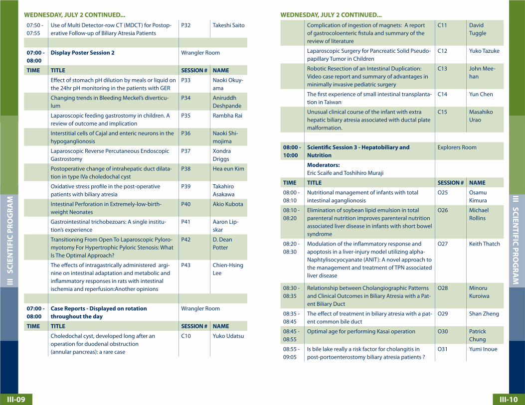
WEDNESDAY, JULY 2 CONTINUED...WEDNESDAY, JULY 2 CONTINUED...
07:50 - 07:55
Use of Multi Detector-row CT (MDCT) for Postop-erative Follow-up of Biliary Atresia Patients
P32 Takeshi Saito
07:00 -
08:00
Display Poster Session 2 Wrangler Room
TIME TITLE SESSION # NAME
Effect of stomach pH dilution by meals or liquid on the 24hr pH monitoring in the patients with GER
P33 Naoki Okuy-ama
Changing trends in Bleeding Meckel’s diverticu-lum
P34 Aniruddh Deshpande
Laparoscopic feeding gastrostomy in children. A review of outcome and implication
P35 Rambha Rai
Interstitial cells of Cajal and enteric neurons in the hypoganglionosis
P36 Naoki Shi-mojima
Laparoscopic Reverse Percutaneous Endoscopic Gastrostomy
P37 Xondra Driggs
Postoperative change of intrahepatic duct dilata-tion in type IVa choledochal cyst
P38 Hea eun Kim
Oxidative stress profile in the post-operative patients with biliary atresia
P39 Takahiro Asakawa
Intestinal Perforation in Extremely-low-birth-weight Neonates
P40 Akio Kubota
Gastrointestinal trichobezoars: A single institu-tion’s experience
P41 Aaron Lip-skar
Transitioning From Open To Laparoscopic Pyloro-myotomy For Hypertrophic Pyloric Stenosis: What Is The Optimal Approach?
P42 D. Dean Potter
The effects of intragastrically administered argi-nine on intestinal adaptation and metabolic and inflammatory responses in rats with intestinal ischemia and reperfusion:Another opinions
P43 Chien-HsingLee
07:00 -
08:00
Case Reports - Displayed on rotation
throughout the day
Wrangler Room
TIME TITLE SESSION # NAME
Choledochal cyst, developed long after an operation for duodenal obstruction (annular pancreas): a rare case
C10 Yuko Udatsu
Complication of ingestion of magnets: A report of gastrocoloenteric fistula and summary of the review of literature
C11 David Tuggle
Laparoscopic Surgery for Pancreatic Solid Pseudo-papillary Tumor in Children
C12 Yuko Tazuke
Robotic Resection of an Intestinal Duplication: Video case report and summary of advantages in minimally invasive pediatric surgery
C13 John Mee-han
The first experience of small intestinal transplanta-tion in Taiwan
C14 Yun Chen
Unusual clinical course of the infant with extra hepatic biliary atresia associated with ductal plate malformation.
C15 Masahiko Urao
08:00 -
10:00
Scientific Session 3 - Hepatobiliary and
Nutrition
Explorers Room
Moderators:
Eric Scaife and Toshihiro Muraji
TIME TITLE SESSION # NAME
08:00 - 08:10
Nutritional management of infants with total intestinal aganglionosis
O25 OsamuKimura
08:10 - 08:20
Elimination of soybean lipid emulsion in total parenteral nutrition improves parenteral nutrition associated liver disease in infants with short bowel syndrome
O26 Michael Rollins
08:20 - 08:30
Modulation of the inflammatory response and apoptosis in a liver-injury model utilizing alpha-Naphtylisocyocyanate (ANIT): A novel approach to the management and treatment of TPN associated liver disease
O27 Keith Thatch
08:30 - 08:35
Relationship between Cholangiographic Patterns and Clinical Outcomes in Biliary Atresia with a Pat-ent Biliary Duct
O28 Minoru Kuroiwa
08:35 - 08:45
The effect of treatment in biliary atresia with a pat-ent common bile duct
O29 Shan Zheng
08:45 - 08:55
Optimal age for performing Kasai operation O30 Patrick Chung
08:55 - 09:05
Is bile lake really a risk factor for cholangitis in post-portoenterostomy biliary atresia patients ?
O31 Yumi Inoue
III-10III-09

III SC
IEN
TIF
IC P
RO
GR
AM
III
SC
IEN
TIF
IC P
RO
GR
AM
WEDNESDAY, JULY 2 CONTINUED...WEDNESDAY, JULY 2 CONTINUED...
09:05 - 09:15
Living donor liver transplantation for biliary atre-sia: single-center experience of 92 cases
O32 KoichiMizuta
09:15 - 09:25
Serum hyaluronic acid as a marker of prognosis for adult patients with biliary atresia
O33 Masato Shinkai
09:25 - 09:35
Is it a type I-fusiform or a type IV-A congenital bile duct cyst?
O34 Richa Lal
09:35 - 09:45
Totally laparoscopic correction of choledochal cyst using an intracorporeal Roux-en-Y jejunojejunos-tomy
O35 Soo Min Ahn
09:45 - 09:55
Contemporary Outcomes of Pediatric Laparoscop-ic Cholecystectomy
O36 Christine Whyte
09:55 - 10:10
Presentation of the Coe Medal Richard Black
10:00 - 10:30
Coffee Break Wrangler Room &Trappers Room
10:00 - 10:30
Exhibits Trappers Room
10:00 - 10:30
Poster Session 2 (see above for details) Wrangler Room
10:00 - 10:30
Case Reports (see above for details) - Displayed on rotation throughout the day
Wrangler Room
10:30 -
12:30
Session 4 - Gastrointestinal (Small Intestine,
Appendicitis, GERD, GT)
Explorers Room
Moderators:
Spencer Beasley and Naomi Iwai
TIME TITLE SESSION # NAME
10:30 - 10:40
The Role of CT Scan and Ultrasound in Omental Infarction and Epiploic Appendagitis in Children
O37 Sanjay Krish-naswami
10:40 - 10:50
Diagnosing acute appendicitis: are we overusing radiological investigations?
O38 KennethWong
10:50 - 11:00
Video-Assisted Transumbilical Appendectomy: A New Technique Providing Cosmetic and Ecological Benefits
O39 KensukeOhashi
11:00 - 11:10
An Evidence Based Definition For Perforated Ap-pendicitis Derived From a Prospective, Random-ized Trial
O40 Daniel Ostlie
11:10 - 11:20
Effect 0f Long-Chain Triglyceride vs Medium-Chain Triglyceride on Mucosal Adaptation of Remained Intestine After Massive Resection
O41 Hong-ShieeLai
11:20 - 11:30
Involvement of bone marrow-derived cells in villous regeneration in a novel mouse intestinal villi-injury model
O42 Takayuki Masuko
11:30 - 11:40
Mechanism of Rho-kinase mediated Ca2+
independent contraction in aganglionic smooth muscle of Hirschsprung’s disease model rat
O43 Junko Aki-yoshi
11:40 - 11:50
Stapled Intestinal Anastomoses in Infants O44 LindseyWrighton
11:50 - 12:00
Anterior Fundoplication at the Time of Congenital Diaphragmatic Hernia Repair
O45 Yigit Guner
12:00 - 12:10
National Frequency of Antireflux Procedures and Gastrostomy : Analysis of Hospital Type, Patient Age and Presence of Neurologic Impairment
O46 David Par-trick
12:10 - 12:20
Experience With a Hybryd, Minimally Invasive Gas-trostomy For Children With Abnormal Epigastric Anatomy
O47 Michael Gauderer
12:20 - 12:30
Modified Approach to Laparoscopic Gastrostomy Tube Placement Minimizes Complications
O48 Mara An-tonoff
12:30 - 13:15
GANS Lecture Michael Dunn
13:30 - 14:30
Annual Business Meeting Explorers Room
13:30 - 16:30
Guided Bike Tour
(meet in Main Lobby at 13:30 pm for bus)
15:00 - 16:00
Exit Board of Directors Meeting Explorers Room
17:30 - 22:00
Annual Banquet - Reception & Dinner
(shuttle service begins at 17:00 pm)Diamond Cross Ranch
THURSDAY, JULY 3
06:30 - 12:00
Registration Open Main Lounge
06:30 - 08:30
Breakfast Sunset Terrace / Blue Heron Lounge
06:30 - 10:30
Exhibits Open Trappers Room
07:00 -
08:00
Oral Poster Session 3 Wrangler Room
Moderators:
Cynthia Reyes and Soul-Chin Chen
III-12III-11

III SC
IEN
TIF
IC P
RO
GR
AM
III
SC
IEN
TIF
IC P
RO
GR
AM
THURSDAY, JULY 3 CONTINUED...
TIME TITLE SESSION # NAME
07:00 - 7:05
Preserved urethral plate urethroplasty for repeat hypospadias repair: report of 249 cases
P44 Weiping Zhang
07:05 - 07:10
Ureteral Dilation in Ureteropelvic Junction Ob-struction Patients
P45 Pei-Yeh Chang
07:10 - 07:15
Induction of Wnt5a-expressing mesenchymal cells adjacent to the cloacal plate is essential for its proximodistal elongation and subsequent anorec-tal development
P46 Mitsuyuki Nakata
07:15 - 07:20
Two Institutions results using total urogenital mobilization for urogenital sinus.
P47 Arturo Aranda
07:20 - 07:25
A Prospective Review of Prognostic Indicators and Complications Rates in Hypospadias Surgery
P48 N Fraser
07:25 - 07:30
Long-term follow-up of orchidopexy: What do patients know about their need for testicular self-examination and their risk of testicular tumours?
P49 Kevin Prin-gle
07:30 - 07:35
Circumcision of the outer preputial layer for true phimosis in children
P50 Yoshiko Watanabe
07:35 - 07:40
Ontogeny of imunohistochemical staining of nor-mal embryonic ampullary activity in fetal lambs
P51 Hiroaki Kitagawa
07:40 - 07:45
Is preoperative transanal catheter useful to avoid enterocolitis and colostomy in patients with Hirschsprung’s disease?
P52 ShigeruTakamizawa
07:45 - 07:50
Reproducibility of nuclear transit studies to assess colonic tranit time in children with slow transit constipation
P53 John Hutson
07:50 - 07:55
Gender Disparity in the Development of Surgical Site Infections (SSI) After Intestinal Stoma Closure in Children: a Single Institution Experience
P54 Nikunj Chokshi
07:55 - 08:00
Building a robotics program P55 John Mee-han
07:00 -
08:00
Display Poster Session 3 Wrangler Room
TIME TITLE SESSION # NAME
Dysplastic Kidneys in Chidren - Do They Grow? P56 Nia Fraser
Accidents happen: Children’s perception of trauma P57 ClaudiaMueller
Surgical Therapies for Intractable Constipation in Children
P58 Akio Kubota
Round skin incisional plasty of a giant umbilical hernia
P59 Masayuki Kubota
Impact of ultrasound-guided subclavian veni-puncture for central venous cannulation in infants and children
P60 Masaaki Kuda
Surgical Management of CAPD Catheter-related Complications in Children
P61 Tetsuya Ishimaru
Laparoscopic approach to incarcerated/sliding inguinal hernia in children in comparison with open approach
P62 Masao Endo
Leaching di(2-ethylhexyl) phthalate out of gas-trostomy connecting tubes made from polyvinyl chloride.
P63 Sae Tanaka
Surgical Therapies for Intractable Constipation in Children
P64 Akio Kubota
The use of penrose ring drains in soft tissue abcesses decreases pain and improves cosmetic results in children
P65 Michael Dingeldein
07:00 -
08:00
Case Reports - Displayed on rotation
throughout the day
Wrangler Room
TIME TITLE SESSION # NAME
Management of a severe laryngopharyngeal vascular malformation with airway obstruction: case report
C16 Toshihiro Muraji
A successful non-operative management with endoscopic retrograde biliary drainage (ERBD) for posttraumatic intrapancreatic biliary stenosis in a child: A case report
C17 ShigeruTakamizawa
Surgical Correction of Congenitally Kinked Bilat-eral Carotid Arteries
C18 Aimee Levy
Anal Canal Duplication: case review and summary of the world’s literature
C19 Heather Carpentier
Identification of a HOXD13 mutation in a VACTERL patient: implication for the Sonic hedgehog pathway
C20 Maria Gar-cia-Barcelo
Laparoscopic diagnosis of ruptured intraperito-neal hydrocele that mimics appendicitis
C21 ClaudiaMueller
A case of zonal hypoganglionosis in Hirschsprung’s disease
C22 Yuko Bitoh
THURSDAY, JULY 3 CONTINUED...
III-14III-13

III SC
IEN
TIF
IC P
RO
GR
AM
III
SC
IEN
TIF
IC P
RO
GR
AM
THURSDAY, JULY 3 CONTINUED...
A Case of Neonatal Testicular Torsion Treated on the Birth Day
C23 Tomio Ogawa
Bilateral Congenital Lumbar Hernias Associated with Bilateral Cryptorchidism
C24 Ali Zhum-khawala
08:00 -
09:40
Session 5 - Trauma, Policy & Miscellaneous Explorers Room
Moderators:
Robin Eccles and Jaime Olvera-Dunn
TIME TITLE SESSION # NAME
08:00 - 08:10
Abdominal Involvement in Pediatric Heart and Lung Transplant Recipients with Post-Transplant Lymphoproliferative Disease Increases the Risk of Mortality
O49 Cindy Tai
08:10 - 08:20
Role and effectiveness of angioembolization in the management of pediatric patients with blunt hepatic or splenic injury
O50 Atsushi Takahashi
08:20 - 08:30
Non-Surgical Treatment of Splenic Trauma in the Absence of CT: 15 Years Experience in Russia
O51 Anna Shap-kina
08:30 - 08:40
Natural History of Non-Operative Management For Grade 4 and 5 Liver and Spleen Injury in Children
O52 JeannieYang
08:40 - 08:50
Laparoscopic Tenckhoff Catheter Placement in Children Using a Securing Suture in the Pelvis: Comparison to the Open Approach
O53 DanielCopeland
08:50 - 09:00
Computed tomography before transfer to a level I pediatric trauma center risks substantially increased radiation exposure
O54 Walter Chw-als
09:00 - 09:10
Recombinant Activated Factor VII Reduces Trans-fusion Requirements in Critically Ill Infants with Active Hemorrhage
O55 Howard Jen
09:10 - 09:20
Pediatric Trauma Resuscitation- Shooting for Par O56 Eric Scaife
09:20 - 09:30
Indication for Pediatric Muscle Biopsy Determines Usefulness
O57 Ramin Jamshidi
09:35 - 09:40
Tribute to Anne Marie King Rossalyn Walker
09:40 - 10:00
GAP Fellowship Lecture
Pediatric Surgery in EcuadorDaniel Acosta Farina
10:00 - 10:30
Coffee Break Wrangler Room & Trappers Room
10:00 - 10:30
Exhibits Trappers Room
10:00 - 10:30
Display Poster Session 2 (see above for details) Wrangler Room
10:00 - 10:30
Case Reports (see above for details) Wrangler Room
10:30 -
12:30
Session 6 - Anorectal, Hernia, Urology
Moderators:
Don Shaul and Hiroaki Kitagawa
TIME TITLE SESSION # NAME
10:30 - 10:40
Improvements in Constipation and Fecal Control following Resection of Megarectum in Anorectal Malformation Patients
O58 Nikunj Chokshi
10:40 - 10:45
Immunologic characterization of enterocolitis as-sociated with Hirschsprung’s disease: Deficiency of mucosal immunity in aganglionic intestine
O59* Tetsuya Mitsunaga
10:45 - 10:50
Comparing Cost and Safety of Primary or Staged Repair of Neonatal Hirschsprung’s Disease
O60* Ricky Shinall
10:50 - 11:00
Total colonic aganglionosis with or without small bowel involvement: a changing profile for 30 years nationwide survey in Japan
O61* Satoshi Ieiri
11:00 - 11:10
Advances in the Management of Pilonidal Disease in Pediatric Surgery: Experience With the the Use of Rhomboid Excision and Limberg Flap in 16 Adolescents
O62 Sani Yamout
11:10 - 11:20
Imperforate Anus with Congenital Absent Vagina: Surgical Treatment and Clinical Characteristics
O63 Hiroyuki Koga
11:20 - 11:25
Scrotal fixation in the management of low unde-scended testes
O64** Owen Greene
11:25 - 11:35
Testicular position based prevalence of a patent processus vaginalis in laparoscopic assisted trans-scrotal orchidopexy
O65** Toshihiko Watanabe
11:35 - 11:45
Repairing hypospadias with severe chordee by preserving the entire urethral plate using a wide U-shaped incision very distal to the meatus
O66 Atsuyuki Yamataka
THURSDAY, JULY 3 CONTINUED...
III-16III-15
* 059, 060 & 061 will have a combined discussion.** 064 & 065 will have a combined discussion.

III
SC
IEN
TIF
IC P
RO
GR
AM
11:45 - 11:55
Can a pressure-limited vesico-amniotic shunt tube preserve normal bladder function?
O67 Takeshi Aoba
11:55 - 12:05
Renal Preservation in Neurogenic Bladder-Where are we Heading?
O68 Bhanuprakash Mariyappa Rathnamma
12:05 - 12:15
Laparoscopic Extra-Vesical Ureteral Reimplanta-tion (LEVUR): The success of a simplified technique
O69 Arturo Aranda
12:15 - 12:25
Retroperitoneal laparoscopic dismembered pyelo-plasty: 4 years expreince
O70 Yunli Bi
12:25 - 12:35
Comparing open and pneumovesical approach for ureteric reimplantation in pediatric patients
O71 Patrick Chung
THURSDAY, JULY 3 CONTINUED...
III-17
Section IV
PAPS 2008

General Information
PAPS 2008 Conference Secretariat
Advance Group
Suite 101 – 1444 Alberni Street Vancouver, BC V6G 2Z4 Canada Phone: +1.604.688.9655 ext. 2 Fax: +1. 604.685.3521Email: [email protected]
Registration Desk
The Registration Desk is located in the Main Lounge of the Jackson Lake Lodge and is open at the following times:
Sunday, June 29 12:00 to18:00
Monday, June 30 06:30 to13:00
Tuesday, July 1 06:30 to 09:00
Wednesday, July 2 06:30 to13:00
Thursday, July 3 06:30 to12:30
Name Badges
Name badges will be provided to all delegates and participants when you check-in at the PAPS 2008 Registration Desk. Please wear your name badge at all times. It is your admission pass to breakfasts, meeting sessions and all social program events.
Name badges are color coded as follows:
PAPS Members: BlueNon-Members: Red Residents / Trainees: PurpleSponsors / Exhibitors: Black Spouse / Companion: Yellow
Jackson Lake Lodge
Customary tipping guidelines are $3.00 per person, per day for housekeeping and $6.00 per person, roundtrip for luggage handling. Customary gratuity rates are 15 - 20%.
Transportation
There will be a complimentary shuttle bus running between the Jackson Lake Lodge, the Flagg Ranch Resort, and the Hatchet Resort on the dates of June 29 through July 3. A schedule will be available at the PAPS Registration Desk.
IV P
AP
S 2
00
8
IV-02
IV
PA
PS
20
08
Smoking
All lodge guest rooms and public areas are non-smoking. ***When outdoors, ensure
that cigarettes are properly extinguished and disposed of in properly marked
containers. Improperly disposed cigarettes are a major cause of forest fires.***
Garbage
The Lodge is located within a natural setting. All garbage must be properly placed within the marked receptacles, and food products must not be left out.
Wildlife
Please respect the wildlife. Do not feed, and use caution and common sense if you should spot wildlife. If you have any questions, do not hesitate to ask the staff at the Jackson Lake Lodge.
Bears
Please be aware that at least one hiking trail has been closed due to an increase in the number of bears in the area. While there is no reason to be concerned, the hotel is asking that people are aware of the situation and ask that people do not hike alone and stay on the marked trails.
Communication
The hotel is equipped with wireless internet throughout. Verizon has the strongest signal for cell phone service. There are no televisions in the guest rooms.
Weather
The average high for late-June in Jackson Hole is 75° F / 25° C and it cools down to 50° F / 10° C in the evenings.
Sales Tax
Most purchases are subject to the Wyoming State sales tax of 6%.
Speaker Center
There is no Speaker Center at this conference. If you are a speaker, please arrive at least 20 minutes before the start of your session. Please submit your Presentation to the Registration Desk at least one day prior to your session. If you have a PowerPoint presentation that needs to be re-formatted or checked, please ask at the Registration Desk. We will have additional computers available for you to work on your presentation.
Scientific Session Information
Presenters will have 5 minutes for presentation and 5 minutes discussion. Time extensions will not be permitted. The CD or memory stick containing your presentation must be submitted to the Registration Desk one day before your presentation. For those presenting on Wednesday July 2, please note that the Registration Desk will be closed for most of the day on Tuesday July 1, so please ensure that your presentation is handed in on Monday June 30. No dual slide projection presentations are permitted.
IV-01

IV P
AP
S 2
00
8IV
P
AP
S 2
00
8
Poster Session Information
Poster Sessions are scheduled on Monday June 30, Wednesday July 2, and Thursday July 3 during the breakfast period and coffee breaks. Display Only Posters and Oral Presentation Posters will be in the Wrangler Room. Setup and teardown times are as follows:
Poster Session 1
Setup:Monday, June 30 06:00 – 06:45
Teardown:Monday, June 30 12:30 – 13:30
Poster Session 2
Setup:Wednesday, July 2 06:00 – 06:45
Teardown:Wednesday, July 2 13:30 – 14:30
Poster Session 3
Setup:Thursday, July 3 06:00 – 06:45
Teardown:Thursday, July 3 12:30 – 13:30
Case Reports Information
Case Reports are scheduled on Monday June 30, Wednesday July 2, and Thursday July 3 during the breakfast period and coffee breaks. Case Reports will be presented in the Wrangler Room.
Commercial Exhibit
This year will include a three-day commercial exhibition in the Trappers Room. The Commercial Exhibit is open to conference delegates during the following times:
Monday, June 30 06:30 – 13:00
Tuesday, July 1 Closed
Wednesday, July 2 06:30 – 13:00
Thursday, July 3 06:30 – 10:30
IV-04IV-03
Conference Sponsors
Gold
Bronze
Supporters

IV P
AP
S 2
00
8IV
P
AP
S 2
00
8
Social Program
Opening Ceremony & Welcome Reception
Date & Time: Sunday, June 29 18:00 – 21:00Location: Explorers Room (Jackson Lake Lodge)
All participants and registered accompanying persons are cordially invited to the Welcome Reception. Enjoy local entertainment as you reconnect with old friends and mingle with new colleagues. Hors d’oeuvres and refreshments will be served.
Additional tickets will be sold at the Registration Desk for $50Dress: Smart casual (no ties)
Hors d’oeuvres and refreshments will be served through the support of an educational grant from Karl Storz Endoscopy-America, Inc.
Group Tour
Yellowstone National Park
Date & Time: Tuesday, July 1 09:30 – 19:00Location: Jackson Lake Lodge, Main Entrance
This full day tour leaves from the main entrance of the Jackson Lake Lodge at 9:30 AM
sharp. Additional buses will leave from the Flagg Ranch and Hatchet Resort at 9:00
AM sharp. This guided bus tour visits all the major highlights of this first National Park including Old Faithful, the historic lake area, the Grand Canyon of the Yellowstone, Yellowstone Falls, hot pots, geysers, and geothermal natural wonders. Boxed Lunch will be at the historic Old Faithful Inn and will be provided to all tour participants.
Additional tickets will be sold at the Registration Desk for $70 eachDress: Comfortable. Don’t forget to bring sunscreen, and a warm jacket in case it gets cooler outside (later in the evening).
Tour and lunch will be supported by an educational grant from Megadyne Medical Products.
Annual Banquet, Reception, Dinner and Song Fest
Date & Time: Wednesday, July 2 17:30 – 22:00Location: Diamond Cross Ranch
Shuttle buses will begin departing from the main entrance of the Jackson Lake Lodge at 17:00 until 18:30. One bus will depart the Flagg Resort at 17:00, and one bus will depart the Hatchet Resort at 17:30. The Gala Final Banquet will take place at the picturesque Diamond Cross Ranch. This event will be one to be remembered, and will feature a horse-whisperer demonstration, award presentations, passing of the gavel to the new President (Paul Tam from Hong Kong), and our legendary international Song Fest. Prepare your singing voice and bring your appetite! The Annual Banquet is an evening not to be missed.
Additional tickets will be sold at the Registration Desk for $150 each ($100 for children).Dress: Smart casual (no ties).
The Annual Banquet is sponsored by educational grants from Ethicon Endo-Surgery Inc. and Banner Health.
Breakfasts
Date & Time: Monday – Thursday, June 30 – July 3 06:30 – 08:30Location: Sunset Terrace (with overflow in the Blue Heron Lounge)Breakfasts are served buffet-style and are open to all delegates and accompanying persons.
Hospitality Suite
Date & Time: Monday, June 30 11:00 – 22:00 Tuesday, July 1 11:00 – 22:00 Wednesday, July 2 11:00 – 22:00 Thursday, July 3 11:00 – 15:00Location: Wyoming RoomStop in, relax and watch a movie in the Hospitality Suite. Located on the second level, all delegates and Accompanying Persons are welcome to stop in and enjoy the Suite.
Cyber Café
Date & Time: Monday, June 30 07:00 – 18:00 Tuesday, July 1 07:00 – 10:00 Wednesday, July 2 07:00 – 18:00 Thursday, July 3 07:00 – 12:00Location: Wrangler RoomLocated on the second level, all delegates and Accompanying Persons are welcome to access the Internet and use the computers at the Cyber Café. Please be courteous to your fellow delegates, and limit your use to 20 minutes when others are waiting.
Optional / Buy-in Social Activities
Tennis (Baxter-Myers Trophy)
Cancelled due to lack of registrants.
Golf
Date & Time: Monday, June 30 12:45 to 20:00Location: Jackson Hole Golf & Tennis ClubCost: $240.00Those participating in the golf outing are asked to assemble at the main entrance of the hotel at 12:45 PM for your transfer to the club. The shuttle departs at 12:50 PM
sharp. The golf outing includes 18 holes of golf, round-trip transportation, lunch and tournament services. The hire of clubs is an additional cost of $50.00.
IV-06IV-05

IV
PA
PS
20
08
Whitewater River Rafting
Date & Time: Monday, June 30 13:30 to 19:30Location: Snake RiverCost: $85.00Those participating in the whitewater river rafting outing are asked to assemble at the main entrance of the hotel at 13:20 PM for your transfer to the river. Your shuttle departs at 13:30 PM sharp. Wear casual clothing you don’t mind getting wet. Shorts and soft soled shoes are ideal. Synthetic blends are recommended over cotton fabrics. Bring sweatshirts and towels for after the trip. Cameras are NOT recommended. Don’t forget sunscreen, sunglasses and hats! Rain jackets and pants are provided when necessary. Life jackets are US Coast Guard approved and must be worn at all times. Optional neoprene wetsuits, booties and gloves are available for rental.
Bar J Chuck Wagon Supper & Western Show
Date & Time: Monday, June 30 16:30 to 22:30Location: Bar JCost: $20.00 for transportation + $20 - $30 for dinner & ticketCome and experience the spirit of the old west and a bit of real western hospitality on a working cattle ranch, with a rib-stickin’ dinner and an evening of family entertainment you’ll never forget. The bus will depart the Jackson Lake Lodge at 16:30 PM, returning at 22:30 PM. Sign up will be at the Conference Registration desk before 10:30 AM on
Monday, June 30, and is limited to 40 people.
Guided Bike Tour
Date & Time: Wednesday, July 2 13:30 to 16:30Location: Grand Teton National ParkCost: $100.00
The Bike tour meets in front of the Jackson Lake Lodge on Wednesday, July 2 at 13:20 PM, for a departure at 13:30 PM sharp. This guided tour is the exclusive Pacific Creek Tour in Grand Teton National Park and it travels along the Pacific Creek. An in and out and back route, the riding time is approx 1.5 hours, and is about 6 miles (10 km) in length – more experienced riders will have the option of travelling with a guide for an additional 4 miles (6km). Each tour includes mountain bike, helmet, FREE water bottle, transportation plus a local mountain bike guide.
Optional Tours
Those wishing to participate in activities are asked to visit the Activities Desk located in the main lobby of the Jackson Lake Lodge, for more detailed information on your chosen tour(s). Tours include: scenic float trips, horseback rides, lake cruises and fishing.Tours are also available at the Flagg Ranch Resort.
IV-7
Section V
Abstracts

Oral Presentations
Neonatal and Fetal
O01
A Unique Surgical Approach: 9 Years Experience of Patent Ductus
Arteriosus Ligation in Premature Infants at Children’s Hospital Oakland
Kwan, Rita O.1 Betts, James2 Idowu, Olajire2 Kim, Sunghoon2
1. University of California, San Francisco-East Bay Department of Surgery, Oakland, CA, USA; 2. Children’s Hospital & Research Center Oakland Department of Pediatric Surgery, Oakland, CA, USA
Introduction: Various surgical techniques have been described for the treatment of pat-ent ductus arteriosus (PDA) in premature infants. We report our experience of PDA suture ligation in premature infants performed by Pediatric Surgeons using a unique surgical technique that has been in use at the Children’s Hospital Oakland (CHO).
Methods: Between 1996 and 2005, 242 consecutive surgical PDA ligations performed at CHO were retrospectively reviewed. Of 242 patients, 79 were excluded from the study because PDA was associated with a cardiac lesion, hence corrected by our Cardiac Sur-geons. All remaining patients had PDA ligation using our indirect dissection technique: a suture is circumferentially placed around the PDA by performing lateral dissection of the supra- and infra-aorta at the junction of PDA. The undersurface of the PDA is not dis-sected.
Results: A total of 163 patients underwent indirect dissection and ligation of the PDA. The median birth weight was 817grams. Two patients had PDA recurrence requiring re-ligation. There were two intra-operative bleeding that contributed to two mortalities; however, these patients had disseminated intravascular coagulation at the time of the operation. There was no incidence of vocal cord paralysis.
Conclusion: Indirect PDA dissection and ligation is a safe surgical technique with low complication rate. In contrast to published series of vocal cord paralysis secondary to recurrent laryngeal nerve injury ranging from 3-8.8%, no incidence of recurrent laryngeal nerve injury was found. The recurrence rate was 1.2%, comparable to published results ranging from 1.2% to 2.1%. The mortality rate was 1.2%.
V A
BS
TR
AC
TS
V-1

V
AB
ST
RA
CT
S
V-2
V A
BS
TR
AC
TS
V-3
results, our clinical impression suggests decreased effectiveness of the contrast enema resulting in more surgical interventions in contemporary practice.
Methods: A retrospective, multi-institutional review over a 15 year period was conduct-ed for neonates diagnosed with meconium ileus by contrast enema. Complicated meco-nium ileus was excluded (n=10). Groups compared were historical group (HG = before 2002) relative to contemporary group (CG = after 2002). Enemas were performed using 17% Cysto-Conray exclusively. T test was used for comparison of continuous variables.
Results: Thirty-five patients were identified (20 females and 15 males). Obstruction was relieved in 7 neonates (20% overall rate). No difference was identified in gestational age, gender, birth weight, or age at diagnosis between groups. Average enema attempt per patient was decreased in the CG group compared to HG (1.3 vs. 1.9). The success rate in the CG group was 6% (1 of 17) compared to 33% (6 of 18) in HG. Overall one year and long term survival rates were 100% and 94%, respectively.
Conclusions: In this review, success of contrast enema for relief of meconium ileus has significantly decreased over time. These findings may be due to reluctance to repeat enemas. As a result, higher rates of operative intervention are now observed. In stable patients, surgeons should require repeat enemas prior to exploration.
O04
Dramatic Improvement of the Survival in Antenatally Diagnosed
Congenital Diaphragmatic Hernia - Gentle Ventilation and Circulatory
Stabilization
Masumoto, Kouji; Esumi, Genshiro; Teshiba, Risa; Nagata, Kouji; Taguchi, TomoakiDepartment of Pediatric Surgery, Reproductive and Developmental Medicine, Graduate School of Medical Sciences, Kyushu University, Fukuoka, Japan
Background: In patients with antenatal diagnosed congenital diaphragmatic hernia (AD-CDH), no definitive treatment strategy has yet been established. In our depart-ment, from 1997 to 2003, we carried out fetal stabilization (FS) using both morphine and diazepam via the placenta just before performing a cesarean section. In contrast, from 2004 to the present, we selected a combination of gentle ventilation (GV) and a delayed operation, which was performed at time when the patient’s circulatory stabilization (CS), was achieved.
Patients and Methods: This study included 22 patients in the FS group and 16 patients in the GV+CS group, respectively. We compared the outcomes in both groups and further investigated the outcome in AD-CDH patients with either a patch repaired operation, liver-up, or lower lung-to-thorax transverse area ratio (L/T, <0.10) in both groups.
Results: The overall survival rate (SR) was 59.1% (13/22) in the FS group and 93.8% (15/16) in the GV+CS group, respectively. Regarding the patients with the lower L/T, the SR was 53.8% (7/13) in the FS group and 85.7% (6/7) in the GV+CS group. In the patients using a patch, the SR in the GV+CS group (87.5%, 7/8) was better than that in the FS group (44.4%, 4/9). Regarding liver-up, the SR in the GV+CS group (87.5%, 7/8) was also better than that in the FS group (57.8%, 11/19).
O02
Vacuum Assisted Closure for Complicated Neonatal Abdominal Wounds
Koeppel, Robin; Lopez, Gregory; Emil, SherifUniversity of California Irvine, Orange, CA, USA
Purpose: Vacuum assisted closure (VAC) has been employed for difficult wounds in adults and children. However, neonatal experience remains scant. We reviewed our expe-rience with VAC for complex abdominal wounds in the neonatal intensive care unit.
Methods: A neonatal VAC protocol was instituted in 2004. The medical records of patients treated with this protocol over the ensuing three years were retrospectively reviewed. Data retrieval included clinical characteristics, surgical interventions, wound details, details of VAC use, wound outcomes, and patient outcomes. Continuous data are reported as mean ± SD (range).
Results: Ten VAC applications occurred in 8 neonates over the study period. Gestational age and age at VAC application were 30 + 6.9 (24-40) weeks, and 84.5 + 51 (21-165) days, respectively. Birth weight and weight at VAC application were 1495 + 1118 (615-3415) grams, and 3515 + 2118 (989-7965) grams, respectively. Five neonates were of extremely low birth weight (< 1000 g). Diagnoses included necrotizing enterocolitis (3), spontane-ous intestinal perforation (2), gastroschisis (2), and congenital diaphragmatic hernia (1). All wound complications occurred after laparotomies (7 elective, 3 emergent), and included fascial dehiscence (8), and skin necrosis (2). Three wounds included intestinal stomas, and three included enterocutaneous fistulas. Average wound area at VAC initia-tion was 13.6 + 6.0 (8.5-25) cm2. Duration of VAC use was 19.1 + 15.3 (7-60) days. VAC resulted in complete wound closure in all cases. Three ventral hernias occurred after VAC. Two were repaired during subsequent laparotomies, and one was repaired as an isolated procedure. VAC did not result in any local or systemic complications. Five patients (63%) survived to discharge.
Conclusions: Vacuum assisted closure for complicated abdominal wounds is safe and successful in neonates of any gestational age and birth weight. It provides effective wound management, even in the presence of stomas or enterocutaneous fistulas.
O03
The Diminishing Role of Contrast Enema in Simple Meconium Ileus
Copeland, Daniel R.1 St Peter, Shawn D.2 Sharp, Susan W.2 Tolleson, Joshua S.3 Jackson, Richard J.4 Kokoska, Evan R.4 Little, Danny C.4 Smith, Samuel D.4
1. Arkansas Children’s Hospital, University of Arkansas for Medical Sciences, Little Rock, AR, USA; 2. Children’s Mercy Hospitals and Clinics, Kansas City, MO, USA; 3. University of Arkansas for Medical Sciences, Little Rock, Arkansa, USA; 4. Arkansas Children’s Hospital, Little Rock, AR, USA
Purpose: Contrast enema is the initial study of choice for simple meconium ileus (MI) to confirm diagnosis and to relieve obstruction. Despite favorable historically published

V
AB
ST
RA
CT
S
V-4
V A
BS
TR
AC
TS
V-5
ingrowth, and thus lower recurrence rates. The purpose of this study is to compare the outcomes of CDH repair with the synthetic Gore-Tex to Permacol®, a cross-linked porcine dermal collagen patch.
Methods: We performed a retrospective review of 100 consecutive patients with CDH and survival >30 days at Children’s Medical Center of Dallas from 1999-2007. The inci-dence and timing of recurrence, as well as comorbidities were assessed.
Results: Primary repair was performed in 63 patients and patch repair in 37, divided between Gore-Tex (29) and Permacol (8). Overall recurrences were: 1 (2%), 8 (28%), and 0 in the Primary, Gore-Tex, and Permacol groups, respectively. Median follow up was 57 months for Gore-Tex and 20 months for Permacol. The median time to recurrence in the Gore-Tex group was 12 months. Both the Gore-Tex and Permacol groups had similar comorbidities, including a high incidence of congenital heart disease (76% and 75% respectively) and ECMO support (38% and 25 %).
Conclusion: Prior studies have shown that the incidence of reherniation using Surgisis (non cross-linked porcine submucosal collagen) did not differ from those of Gore-Tex. Our results suggest that Permacol has a lower incidence of recurrence compared to Gore-Tex and is a promising alternative biologic graft for CDH repair.
O07
Growth of Diaphragm after Repair of High-risk Congenital Diaphragmatic
Hernia
Kamata, Shinkichi; Usui, Noriaki; Sawai, Toshio; Nose, Keisuke; Fukuzawa, MasahiroDept. of Pediatric Surgery, Osaka University Graduate School of Medicine, Osaka, Japan
Background/Purpose: The growth and function of the repaired diaphragm have not been well elucidated, which may contribute to pulmonary function and chest-wall de-formity. We measured the lower lung diameter (LLD), diaphragmatic diameter (DD) and height (DH) on the postero-anterior plain chest radiograph using PACS.
Methods: Thirty-six CDH children aged 10.4 ± 4.8 years who had presented with respira-tory distress within 6 hours of life underwent clinical evaluation, including a lung V/Q scan. As a control, chest radiographs of 89 children aged 9.0 ± 5.5 with minor surgery were analyzed. LLD, DD and DH were defined as the horizontal length of each lung at the top of the diaphragm, the horizontal length of the each diaphragm at the bottom of the diaphragm and the vertical length between the top and the bottom of the diaphragm, respectively.
Results: LLD, DD and DH in controls were significantly correlated with age, then each value was expressed as % of age-based estimated values. Ipsilateral (treated side) LLD and DD were significantly decreased. The perfusion of the ipsilateral lung was best corre-lated with ipsilateral DD. Five patients had chest wall deformity, and 7 had scoliosis (Cobb angle > 10 degrees). Patients with scoliosis had decreased ipsilateral LLD, DD and DH. The Cobb angle was correlated with LLD and DD.
Conclusion: Our strategy using both GV and CS is therefore considered to be more effec-tive than that using FS in the treatment of AD-CDH patients.
O05
Thoracoscopic Repair of Congenital Diaphragmatic Hernia (CDH) with
Patch in Neonates: Preliminary Experience
Guner, Yigit S.1,2 Chokshi, Nikunj K.1 Nguyen, Nam X.1 Stein, James E.1 Shin, Cathy E.1
1. Childrens Hospital Los Angeles, Los Angeles, CA, USA; 2. University of California Davis, Los Angeles, CA, USA
Introduction: Although thoracoscopic primary CDH repair is gaining acceptance, thoracoscopic patch repair is not as commonly performed. Here, we describe our initial experience with neonatal thoracoscopic patch repair of left sided CDH.
Methods: We reviewed the charts of all neonates who underwent thoracoscopic repair of CDH between 11/2004-12/2007. Neonates that underwent thoracoscopic repair were physiologically stable with resolved pulmonary hypertension and minimal to moderate ventilatory support.
Results: During the study period there were 55 left sided CDH repairs. Thoracoscopic repair was performed in 33% (n=18) of the neonates. Five neonates underwent thoraco-scopic closure with patch. Mean birth weight was 3.1g (2.9-4.5), gestational age was 37.4 weeks (33-39), and mean Apgar (1&5) score was 7.3 (5.5-8.5). All 5 patients were weaned off from inhaled nitric oxide prior to repair and the maximal resolution of pulmonary hypertension coincided with day of life 6.8 (4-11). The mean pre-operative peak inspira-tory pressure (PIP) was 23cm H2O and the average ΔPIP (post-operative - pre-operative) was 2cm H2O. Average operating room time was 149 minutes. There were no conversions to open repair. There were no instances of intra-operative respiratory or cardiac insta-bility. One patient had a recurrence 4 months after initial repair. There were no deaths. Post-operatively, mean time to feedings was 8.8 days (4-15), and none required high frequency ventilation or ECMO. Average time to discharge was post-operative day 20.6. Follow-up was 2-30 months.
Conclusion: Thoracoscopic patch repair of neonatal CDH is technically feasible, and can be safely performed in patients with minimal ventilatory requirements.
O06
Permacol: A Potential Biologic Patch Repair for CDH?
Mitchell, Ian C.1 Garcia, Nilda M.1 Barber, Robert2 Fischer, Anne C.1
1. University of Texas Southwestern Medical Center at Dallas, Dallas, TX, USA; 2. Children’s Medical Center of Dallas, Dallas, TX, USA
Purpose: The most commonly used patch repair for large Congenital Diaphragmatic Hernia (CDH) defects is Gore-Tex®. However, up to 40% of patients with Gore-Tex repairs have recurrent herniation. A “bioprosthetic” material is thought to yield greater fibroblast

V
AB
ST
RA
CT
S
V-6
V A
BS
TR
AC
TS
V-7
O09
Gastroschisis, Atresia, Dysmotility (GAD): Experience with a Distinct Clinical
Entity
Phillips, J. D.1 Redden, Courtney2 Raval, Mehul V.1 Weiner, Timothy1
1. University of North Carolina at Chapel Hill, Chapel Hill, NC, USA; 2. University of North Carolina School of Medicine, Chapel Hill, NC, USA
Purpose: Intestinal atresia (IA) occurs in 10-20% of infants with gastroschisis (GS). Their prolonged need for total parenteral nutrition (TPN) and high mortality has usually been attributed to short bowel syndrome (SBS). We describe a distinct subset of GS/IA patients with severe dysmotility, and propose possible treatment strategies.
Methods:Retrospective review of 177 GS patients managed between 1993 and 2007.
Results: 21 patients(12%) had IA. 6(29%) did well, with gradual progression to full diet;1 died at birth of midgut infarction;1 died of NEC;4 with SBS (small bowel=30-41 cm) died of liver failure due to TPN (3) or sepsis (1). 9(43%) were felt to have GAD, defined as: adequate small bowel length at laparotomy (mean 146 cm;range 66-233 cm), massive intestinal dilatation, and stasis. Intestine, including anastomoses, was widely patent by radiographic studies and repeat laparotomy. 5 of 9 GAD patients (56%) survived. Each had surgery to “rescue” their dysfunctional intestine, at mean age 140 days (range 52-271 days): 4 had dilated bowel resected, with tapering enteroplasties of mean 37 cm (range 5-115 cm)--all 4 survived;3 had diverting stomas created (at mean age 90 dys) to allow partial feeds/intestinal decompression, followed by stoma closure--1 of 3 survived;1 non-survivor had a Kimura “patch” and 1 had a re-do anastomosis. Survivors weaned off TPN, at mean age 349 days (range 42-814 days).
Conclusions: Over 1/3 of GS/IA patients appear to have significant intestinal dysmotility without true SBS nor obstruction. Successful treatment may be achieved with the use of tapering enteroplasty and/or temporary diverting stomas.
O10
Preservation of Extra-corporeal Tissue in Closing Gastroschisis Augments
Intestinal Length
Estrada, Joaquin J.1 Lugo, Brian1 Petrosyan, Mikael1 Lee, Steven2 Anselmo, Dean M.1 Ford, Henri R.1 Grikscheit, Tracy1 Stein, James E.1 Wang, Kasper S.1 Shaul, Donald B.1
1. Childrens Hospital Los Angeles, Los Angeles, CA, USA; 2. Kaiser Permanente- Kaiser Sunset-Los Angeles, Los Angeles, CA, USA
Purpose: Prenatal closure of the umbilical ring in gastroschisis often results in amor-phous, non-viable appearing extracorporeal bowel that is resected during the repair. However, it is unclear whether such remnant intestine is truly non-viable. We examined the outcomes of patients when this tissue is preserved.
Methods: An IRB approved, retrospective review of all patients with closing gastroschisis from 1996-2006 was performed.
Conclusion: The growth of the repaired diaphragm was impaired, which may contribute to decreased perfusion of the ipsilateral lung and scoliosis. LLD and DD are simple but useful parameters in the follow-up of patients with CDH.
O08
An Intriguing Surge of Hypertrophic Pyloric Stenosis
Green, Jr., James F.1 Gauderer, Michael W.2 Cass, Anna L.3 Blackhurst, Dawn W.3 Chandler, John C.2 Abrams, Randel S.2
1. Children’s Hospital, Greenville Hospital System, University Medical Center, Greenville, SC, USA; 2. Children’s Hospital, Greenville Hospital System, University Medical Group, Greenville, SC, USA; 3. Greenville Hospital System, University Medical Group, Greenville, SC, USA
Purpose: Despite its clinical importance, many aspects of hypertrophic pyloric stenosis (HPS), including its etiology and variations in incidence, remain enigmatic. While the oc-currence of HPS in our region has previously paralleled reported rates of 2-5 per 1000 live births, a dramatic and sustained increase in the rate of HPS at our institution prompted this study.
Method: Data on infants with HPS between 1998 and 2007 at our regional children’s hospital were analyzed. All cases were confirmed by ultrasound and palpation. Other infant surgical pathologies were analyzed for comparison. Statewide data for HPS were also analyzed.
Results: HPS had remained steady at an average of 34 infants per year from 1998 to 2004 (5.7/1000 live births); however, the number nearly doubled to 59 in 2005 (9.7/1000), tripled to 94 in 2006 (15.5/1000), and reached 79 in 2007. Conversely, rates of gastroschi-sis, Hirschsprung’s, and inguinal hernia remained relatively steady. Gender, age and insur-ance distribution of HPS patients remained stable (78% male, 42 days old, 54% Medicaid).
A statewide increase of HPS from 2003 to 2006 was demonstrated (2.27/1000 to 4.10/1000, p<.001). Within the state, the HPS incidence in regions with regional referral centers increased (3.23/1000 to 6.04/1000, p<.001), while the incidence in other regions did not (0.67/1000 to 0.86/1000, p=.475). A coincidental time correlation was observed between the increase of HPS and a change in the state’s WIC contract brand formula.
Conclusion: In the absence of corresponding population or birth rate changes as a pos-sible explanation, the increase might be attributed to environmental causes or significant referral pattern changes (i.e. identifiable etiologic factors or fewer general surgeons operating for HPS). Both possibilities have clinical, economic, and other implications for pediatric surgeons. The observation of this “surge” in hypertrophic pyloric stenosis calls on other centers to analyze their HPS population.

V
AB
ST
RA
CT
S
V-8
V A
BS
TR
AC
TS
V-9
Conclusion: Classic anomalies associated with omphalocele are present with near equal incidence between patients with small or large omphaloceles. Cardiac anomalies have a lower incidence among patients with smaller omphaloceles. Concurrent intestinal anomalies appear to have a unique association with smaller omphalocele anomalies.
Type of Anomaly Omphalocele Omphalocele p-value
≤ 4 cm (%) > 4 cm (%)
Intestinal 23.1 zero 0.012 vessel umbilical cord 7.7 3.8 0.61Beckwidth Wiedemann Synd 11.5 3.8 0.35Pentalogy of Cantrell zero 7.4 0.49Cardiac 7.7 33.3 0.04Neural tube 3.8 3.7 1.0Craniofacial 19.2 3.7 0.1Renal 15.4 18.5 1.0Limb 7.7 7.4 1.0
O12
Image Guided Fetal Surgery for Complicated Monochorionic Diamniotic
Pregnancies
Hirose, Shinjiro1 Curran, Patrick1 Feldstein, Vickie A.2 Farmer, Diana L.1 Farrell, Jody3 Lee, Hanmin1
1. UCSF Pediatric Surgery, San Francisco, CA, USA; 2. UCSF Radiology, San Francisco, CA, USA; 3. UCSF Fetal Treatment Center, San Francisco, CA, USA
Background/Purpose: Monochorionic diamniotic twin pregnancies with one anoma-lous or severely damaged twin pose difficult clinical management. The demise of one twin results in a high mortality rate for the normal twin. Alternatively, survival of an anomalous twin may result in a severely handicapped child. In order to maximize survival of the normal twin, we have adopted a strategy of selective reduction using minimally invasive, image guided, radio-frequency surgery.
Methods: We performed a retrospective analysis of all patients who underwent im-age guided radio frequency ablation at UCSF from 1998 to 2008 (n = 18). We examined survival rate, gestational age at presentation, gestational age at delivery, birth weight, and infant and maternal complications. All mothers and fetuses underwent surgery with gestational ages varying between seventeen and twenty five weeks with a mean of twenty weeks gestation.
Results: Eighteen mothers have undergone RFA ablation for an anomalous twin. Thirteen of seventeen infants were born alive. One infant is still in utero. Mean gestational age at presentation was nineteen weeks. Mean gestational age at birth was thirty two weeks. Mean birth weight was 2448 grams. There were no significant maternal complications. All infants to date are neurologically intact.
Results: 91 children were treated for gastroschisis during the study period. Eight (9%) patients had closing gastroschisis with a mass of tissue connected by a vascular pedicle. The male: female ratio was 1:3. Four patients underwent abdominal exploration with resection of the mass and gastroschisis closure. Histologic review in 3 of these 4 cases revealed normal intestinal wall architecture. All patients in this group developed short bowel syndrome, requiring long-term parenteral nutrition (mean 257 days). One patient died awaiting transplant, 1 is currently listed, and 2 are now tolerating enteral feeds. Con-versely, 4 patients underwent abdominal exploration with internalization of the remnant tissue, a maneuver referred to as “parking”, along with either silo, or primary closure of the gastroschisis. At subsequent re-exploration, 3 out of 4 patients were found to have vi-able intestine and bowel continuity was re-established. The mean parenteral nutrition re-quirement for this group was 149 days. The mass atrophied in 1 patient and was resected. That patient developed short bowel syndrome and is currently listed for transplant.
Conclusion: The extracorporeal masses associated with closing gastroschisis are often regarded as atretic and useless and are resected. In this series, we show that this tissue, when preserved, may exhibit normal intestinal architecture and absorptive function. Therefore, such remnant tissue should be routinely preserved as it may significantly increase bowel length and minimize parenteral nutrition requirement.
O11
Correlation of Omphalocele Size With Incidence of Associated Anomalies
Kumar, Hari R.1 Jester, Andrea L.1 Ladd, Alan P.2
1. Indiana University School of Medicine, Indianapolis, IN, USA; 2. Riley Hospital for Chil-dren, Indianapolis, IN, USA
Omphalocele is classically associated with congenital anomalies such as VACTERL as-sociation, pentalogy of Cantrell, and Beckwidth-Wiedemann syndrome. Recent reports have demonstrated non-classical associations, often occurring in smaller omphaloceles. We review our experience in treating patients with congenital omphalocele to determine whether size correlates with the type or incidence of associated anomalies.
Methods: A retrospective review was performed of patients with omphalocele treated at a tertiary-care, pediatric hospital over a seven-year period, under IRB approval. Patients were separated into two groups: small (4 cm or less) or large (greater than 4 cm) ompha-loceles. Size of the abdominal wall opening was confirmed by reported physical exam or with operative coding. A Fisher’s exact test was used for comparison of non-parametric data between the groups, with a p-value less than 0.05 being significant.
Results: Fifty-three cases of omphalocele were observed during the seven-year period. Twenty-six cases were classified as small omphaloceles and 27 cases classified as large. The incidence of various anomalies within the groups is listed in the Table. Intestinal anomalies, including Meckel’s diverticulum and intestinal atresia, were only seen in patients with small omphaloceles. The presence of cardiac anomalies had a greater asso-ciation with large omphaloceles. Incidence of chromosomal anomalies was nearly equal in each group.

V
AB
ST
RA
CT
S
V-10
V A
BS
TR
AC
TS
V-11
Table:
Conduit Stricture Ischemia Leak Wound
Infection
Pneu-
motho-
rax
Pneu-
monia
GERD
(severe)
Other
Native Esopha-gus
50%(5/10)
20%(2/10)
20%(2/10)
10%(1/10)
10%(1/10)
None None 10% (1/10) tracheal injury
Colon 11%(1/9)
22% (2/9) 22%(2/9)
22% (2/9) None 11%(1/9)
22%(2/9)
11% (1/9) vocal cord paralysis. 11% (1/9) late bowel obstruc-tion
Gastric Tube
33%(1/3)
None None None 33% (1/3) None None 33% (1/3) GI bleed at staple line
Gastric Interposi-tion
50%(1/2)
None None None None None None None
O14
An Animal Model Study for Tissue-Engineered Trachea Fabricated From a
Biodegradable Scaffold Using Chondrocytes to Augment Repair of Tracheal
Stenosis
Komura, Makoto1 Komura, Hiroko1 Kanamori, Yutaka1 Tanaka, Yuujirou1 Suzuki, Kan1 Sugi-yama, Masahiko1 Nakahara, Saori1 Kawashima, Hiroshi1 Iwanaka, Tadashi1 Ikada, Yosihito2
Tabata, Yasuhiko1
1. Department of Pediatric Surgery Graduate School of Medicine, University of Tokyo, Bunkyou-ku, Japan; 2. Graduate School of Medicine, Nara Medical University, Nara, Japan
Introduction: Construction of engineered respiratory tract without using tissue-engineered cartilage has been reported. Such engineered tracts can maintain luminal patency due to improved rigidity, as they are derived from a substitute engineered tissue or an artificial non-absorbable rigid material. We have designed an engineered graft fabricated from a biodegradable scaffold using chondrocytes. This study investigated the feasibility of a tissue-engineered airway fabricated from a biodegradable scaffold using autologous chondrocytes in a rabbit model.
Material and Methods: Chondrocytes were isolated from the auricular cartilage of the New Zealand white rabbit. They were then seeded onto composite scaffolds consisting of a collagen sheet on the inner side, a polyglycolic acid non-woven mesh in the middle, and a copolymer (L-lactide/epsilon-caprolactone) coarse mesh on the outside. After overnight incubation, the engineered graft was implanted into a 0.5 „e 0.5 cm defect cre-ated in the mid-ventral portion of the cervical trachea. Then, gelatin sponges that slowly released the basic fibroblast growth factor (b-FGF) were placed onto the tissue-engi-neered trachea cartilage. These constructs were retrieved after 3 months and examined histologically.
Conclusion: An anomalous twin poses a distinct hazard to the normal twin. Our novel image guided technique appears to be safe with regards to the mother. Outcomes at birth have been good with 76% survival. Minimally invasive surgery is particularly relevant for fetal surgery cases to minimize maternal complications as well as rates of preterm labor.
Thoracic, Spleen, Oncology, and Miscellaneous
O13
Repair of Long Gap Esophageal Atresia: Gastric Conduits May Improve
Outcome.
Hunter, Catherine J.; Petrosyan, Mikael; Ford, Henri R.; Nguyen, NamChildrens Hospital Los Angeles, Los Angeles, CA, USA
Introduction: Treatment of long gap esophageal atresia (LEA) is a major challenge in pediatric surgery. Options for reconstruction include native esophagus, or replacement with stomach, colon, or small intestine. However, the superior conduit among these op-tions remains undefined.
Aim: The aim of this study was to assess the utility of specific conduits in LEA repair, and to define the complications inherent to each procedure. Based on these findings we sought to define an optimal approach to the surgical management of this anomaly.
Methods: The medical records of all patients admitted to Childrens Hospital Los Angeles with a diagnosis of esophageal atresia during a 20-year period (June of 1987 to May of 2007) were reviewed. Demographics, choice of conduit, postoperative outcomes and complications were recorded.
Results: Twenty-eight cases of long gap atresia were identified. Ten patients underwent primary anastomosis either after serial pouch dilations (9/10) and/or after a lengthen-ing procedure (2/10). There were nine colonic interpositions, and the remainder was reconstructed with gastric tube construction (n = 3), or gastric interposition (n = 2). One patient died prior to repair, and two are currently awaiting treatment. The complications identified for each type of reconstruction are noted in Table 1. A second esophageal reconstruction was required in 4 patients because of initial conduit ischemia. Two isch-emic events occurred in the primary colonic interposition group, and two in the native esophageal repairs. All patients, except for one who was transferred to an outside institu-tion, received long-term follow up (mean 4.2 years: range 0.5-11.5 years).
Conclusions: Our data suggest that the morbidity of esophageal repair is high regardless of the conduit selected. Surgeon expertise and the patient’s anatomy should be con-sidered when selecting an appropriate option. The use of native esophagus is generally preferred; however it is associated with a high rate of stricture. Notably, outcomes with gastric interposition and gastric tube formation are promising. Since no conduit ischemia was noted in patients with gastric conduits, and a lower complication rate is observed, we suggest that gastric conduits should be favored as an initial reconstructive option.

V
AB
ST
RA
CT
S
V-12
V A
BS
TR
AC
TS
V-13
O16
Video Assisted Thoracic Surgery (VATS) for Spontaneous Pneumothorax
(SP) in Children: Is There an Optimal Technique?
Bialas, Ryan C.; Weiner, Timothy M.; Phillips, J. D.University of North Carolina at Chapel Hill, Chapel Hill, NC, USA
Purpose: Thoracoscopy, or video-assisted thoracic surgery (VATS), for treatment of spon-taneous pneumothorax (SP), was first reported in children by Rodgers (1986). Small series have shown success with apical blebectomy, mechanical or chemical (talc) pleurodesis, or combination techniques. We report the largest pediatric series of VATS for SP to date, to assess outcomes and compare techniques.
Methods: Retrospective review of all children undergoing VATS for SP between 1999 and 2007. Mann-Whitney U tests and Chi-square used (p<.05=significant).
Results: 32 patients underwent 41 VATS procedures (32 initial-30 unilateral, 2 bilateral; 9 subsequent-7 contralateral, 2 ipsilateral recurrences). 3 trocars used; blebectomies with endoscopic staplers; all had chest tubes placed. Mean follow-up=46 months.
Technique
Operative
time
(mins)
Postop
airleak
(dys)
Prolonged
postop air-
leak (>7dys)
Postop IV
pain meds
(dys)
Postop
length of
stay (dys)
Postop
chest
tube
(dys)
Recurrences
Blebectomy plus mechanical pleurodesis (n=31)
63.5 (42-98) 2.0 (0-19) 3/31 (10%) 1.3 (0-7) 4.3* (2-11) 4.1* (2-19)2/31*(6%)
major, 5/31* (16%) minor
Blebectomy plus chemical pleurodesis (n=6)
70.6 (46-90) 3.2 (0-11) 1/6 (8.3%) 1.0 (0-5) 7.0* (3-14) 6.5* (3-14) 0/6*
Blebectomy alone (n=1)
55 25 1/1 (100%) 2 10 25 0/1
Chemical pleurodesis (n=2)
61 (60-62) 1.5 (0-3) 0/2 2.5 (0-5) 5.5 (5-6) 5 (5-5) 0/1
Mechanical pleurodesis (n=1)
60 0/1 7 7 6 0/1
(Means, plus ranges. *=p<.05)
Recurrences: Major=required repeat ipsilateral VATS, had new lower lobe blebs not seen at initial VATS. Minor=small, within 1-2 months, treated nonoperatively.
Conclusions: 1)Children with SP undergoing VATS have a high risk of requiring contralat-eral VATS (28%);2)Blebs are almost always identified at the time of VATS (95%), but 12.5% are on lower lobes;3)Each technique, if it includes pleurodesis, is associated with accept-able outcomes;4) Blebectomy plus chemical (talc) pleurodesis appears to have less risk of ipsilateral recurrence but longer postop stay and need for chest tube;5)Rarely, ipsilateral recurrences require repeat VATS (2/39, or 5%).
Results: The biodegradable scaffold using seeded chondrocytes could maintain the airway structure before the formation of the extracellular matrix. Tracheal epithelial regeneration occurred in the internal lumen of this composite scaffold. Three months after implantation, staining of the sections with toluidine blue and safranine O showed cartilage accumulation in the engineered trachea wall in three rabbit.
Conclusion: This composite biodegradable scaffold may be useful for developing tissue-engineered trachea. Moreover, a gelatin sponge slowly releasing b-FGF might enhance the growth of the tissue-engineered trachea cartilage.
O15
H-Type Tracheoesophageal fistula:The Melbourne Experience From
1977-2007.
Khosa, Japinder; Auldist, A.; Taylor, R.Royal Childrens Hospital, Melbourne, VIC, Australia
Objective: Review our experience in H-TOF in terms of diagnostic workup, associated disorders, surgical technique, short and long term outcomes.
Methods: We retrospectively reviewed charts of patients diagnosed and treated for congenital H-TOF from 1977-2007.
Results: 29 patients were diagnosed to have H-TOF. 25/ 29 patients had the anomaly diagnosed in the first 2 weeks of life. Their symptoms included coughing with feeds, episodic cyanosis and aspiration pneumonia. Diagnosis was made by oesophagogram, 5 needed a second oesophagogram to show the fistula. All were repaired via right sided cervical approach. 16/ 29 patients had tissue interposed between oesophagaeal and tracheal anastomosis and none had a recurrence. Of the 13 with no interposed tissue, 3 had recurrence. Short term complications included right recurrent laryngeal nerve palsy (6) of which only 1 persisted and 2 oesophageal strictures. Long term problems included oesophageal dysmotility (3), gastroesophageal reflux (10), asthma (2) and tracheomalacia (2). There were 2 deaths in the series, 1 preoperative from associated anomalies and 1 three months postoperative from SIDS.
Conclusions: A high index of suspicion for an H-TOF should be maintained in the pres-ence of neonatal respiratory symptoms, as the condition can be associated with a delay in diagnosis. Oesophagogram is the diagnostic mainstay and may have to be repeated. We advocate interposing muscle or adjacent tissue if possible between the oesphagus and tracheal anastomosis to prevent recurrence.

V
AB
ST
RA
CT
S
V-14
V A
BS
TR
AC
TS
V-15
O18
Total Thyroidectomy in the Pediatric Patient - Comparing Benign and
Malignant Disease
Raval, Mehul V.1 Browne, Marybeth2 Chin, Anthony2 Anjelos, Peter3 Zimmerman, Donald4
Reynolds, Marleta2
1. Northwestern University Department of Surgery, Chicago, IL, USA; 2. Children’s Memo-rial Hospital, Chicago, IL, USA; 3. University of Chicago, Chicago, IL, USA; 4. Children’s Memorial Hospital - Division of Endocrinology, Chicago, IL, USA
Purpose: Total thyroidectomy (TT) is a safe and efficacious treatment for malignant thy-roid disease in children. The role of TT in benign thyroid disease is less well defined. The goal of this study was to compare the safety of TT performed for benign and malignant disease.
Methods: The medical records of 31 patients undergoing TT from January 1997 to June 2007 at a single center were reviewed. The benign cohort totaled 15 patients and the malignant cohort totaled 16 patients consisting of 9 with a preoperative diagnosis of malignant disease, 4 with a nodule and history of cancer or radiation exposure, and 3 with ret proto-oncogene mutation.
Results: The most common complication observed was transient hypocalcemia noted in 10 of 16 patients (62%) with malignancy and 6 of 15 patients (40%) with benign disease. Permanent hypocalcemia, defined as need for calcium supplemental 6 months post-procedure, was observed in one patient with malignancy and one patient with benign disease (6.2% versus 6.6%). A single parathyroid gland was reimplanted in 2 patients with malignancy and 2 patients with benign disease, with only one of these patients in each cohort having permanent hypocalcemia. No cases of recurrent laryngeal nerve palsy, nerve paralysis, tracheal injury, tacheostomy, or wound infection were encountered in either cohort. One case of significant keloid scar was noted in a patient with malignancy. There were no cases of relapse hyperthyroidism in the benign cohort.
Conclusions: Similar rates of postoperative complications can be expected with TT for benign thyroid disease as compared to TT for malignant disease. TT is a safe treatment option for benign thyroid disease in children.
O19
Surgical Treatment for Epidermoid Cysts of the Spleen in Children
Hayward, Melissa J.1 Houseknecht, Eileen2 Stafford, Perry1
1. Dept. of Pediatric General and Thoracic Surgery, Bristol-Myers Squibb Children’s Hos-pital, University of Medicine and Dentistry of New Jersey-Robert Wood Johnson Medical School, New Brunswick, NJ, USA; 2. Children’s Hospital of Philadelphia, Dept. of Periopera-tive Nursing, Philadelphia, PA, USA
Epidermoid cysts of the spleen are relatively unusual in children. Surgical treatment historically required a total splenectomy, but recent reports have emphasized the impor-tance of splenic preservation using partial splenectomy (cystectomy or cyst marsupiliza-
O17
Costochondral Changes in Pectus Chest Wall after Nuss Procedure-
Sonographic Findings
Zeng, Qi1 Lai, Jin-yao2 Chang, Pei-yeh2 Wang, Xiao-man3 Lee, Jhon-liau4 Chia, Shu-ti5
Wang, Chao-jan6 Chen, Jeng-chang2
1. Department of Thoracic Surgery, Beijing Children Hospital, Beijing, China; 2. Depart-ment of Pediatric Surgery, Chang Gung Memorial Hospital, Taoyuan, Taiwan; 3. Depart-ment of Radiology, Beijing Children Hospital, Beijing, China; 4. Division of Pediatric Or-thopedics, Chang Gung Memorial Hospital, Taoyuan, Taiwan; 5. Department of Pediatric Surgery, Chia Yi Christian Hospital, Chia Yi, Taiwan; 6. Department of Radiology, Chang Gung Memorial Hospital, Taoyuan, Taiwan
Introduction: The pre-bent bar might generates stress and strain over the chest wall after Nuss procedure. We investigated the possible chest wall damages, which might be overlooked on chest X-rays, with sonography after surgery.
Methodology: This is a collaborate study of two institutes. The new and long-term follow-up patients (4-19 years old), during July- December 2007, were enrolled. Sonog-raphy was performed at one day before and 1 week after the procedure. The follow-up patients were examined at least 3 months after the procedure.
Results: All of the patients had significant deformation of the bilateral 2nd to 6th carti-lages after the procedure. However, the cartilages showed no fracture. Twenty four out of 78 patients had acute angulation of costochondral junction(CCJ) or rib fracture at either above or below the bar insertion or exit site. The two patients with two bars had right 5th and 6th rib fracture. The cases with rib fracture (or angulation of CCJ) were significantly older (P<0.01) and had significantly more elevation of the sternum (P<0.01) than those without fracture. It made no difference in pectus index and gender between patients with and without rib frature. Sonography after 3 months of operation showed evidence of healing of all fractures.
Conclusions: The Nuss procedure created significant deformations of the cartilages, without chondral fracture. About 30%of patients had evidence of rib fracture or acute angulation of CCJ, which was related to the age at operation or the height of elevation of the sternum.
V
AB
ST
RA
CT
S
V-16
V A
BS
TR
AC
TS
V-17
tion, including 2 survivors operated after stem cell transplantation. CTC was detected in 55.6% in the stage 4 patients examined, but never detected in the stage 3 patients, and was little associated with MYCN amplification. These patients with CTC showed the 5-year survival rate of 42.0% with all deaths related to systemic metastasis, whereas the CTC negative patients died of local relapse or chemotherapy-associated complications. Kaplan-Meier survival rate was significantly lower in the patients with CTC or persistent bone marrow micrometastasis compared to those without detectable micrometastasis (33.8% vs. 87.5%, P<0.05).
Conclusions: CTC and persistent micrometastasis may indicate significantly high risk regardless of MYCN amplification. Intensive and uninterrupted chemotherapy should be essential for the patients with systemic micrometastasis, while highly intensive surgery with reduced chemotherapy seemed applicable for those with amplified MYCN and no micrometastasis.
O21
Long-term Outcome and Toxicity in Children Rreated with Intraoperative
Radiotherapy for Neuroblastoma
Hishiki, TomoroChiba University Graduate School of Medicine, Chiba, Japan
Objective: To review the long-term outcome and toxicity of intraoperative radiotherapy (IORT) for neuroblastoma.
Materials and Methods: Within the past three decades, 31 neuroblastoma cases (INSS Stage2B: 1Astage 4: 30) were treated with IORT as a part of multimodal therapy. Age of patients at diagnosis ranged from 9 to 128 months old (median 40.5 months). Electron beam (6MeV) was used at doses ranging from 10Gy to 15Gy.
Results: 7 out of 31 patients survived disease free for over five years. Of the 14 cases treated after 1990 who received HDC and TBI followed by autologous hemopoietic stem cell rescue, 11 experienced recurrence, 6 of which were from metastatic sites. There was only 1 recurrence from the irradiated field, while 4 recurrences occurred locally adjacent to the IORT field. A total of 7 patients were alive disease-free and assessable for long term toxicity that IORT may be responsible for. Vascular complications were observed in 5 of these patients. These included renal hypertension due to renal artery stenosis in 4 patients, lower extremity disability in one patient due to aortic stenosis, and 1 severe ischemic colitis due to obstruction of superior mesenteric artery.
Conclusions: IORT gives excellent local control to the irradiated field but physical restric-tions may cause insufficiency in covering the target volume. Close and continuous obser-vation is required in patients treated with IORT, since late sequelae such as major vascular stenosis might sometimes become life threatening.
tion). We have examined the surgical technique and outcome of partial splenectomy for epidermoid splenic cysts at a children’s hospital during the laparoscopic era.
Twelve children (8F;4M) were admitted at a single institution from 1992 to 1999 with epidermoid splenic cysts. Their average age was 13.3 years (range 8-16). Cyst diameter averaged 7 cm (range 2.5-18), and ten children were symptomatic (abdominal pain, pleu-risy). Diagnostic imaging employed ultrasound alone in two and both ultrasound and computerized tomography in ten children. Multiple cysts were seen in three children; the cyst location was variable.
All children underwent partial splenectomy using an open (7) or laparoscopic (5) ap-proach; one child was converted from laparoscopic to open due to cyst location (pos-teriolateral). Three cysts were resected primarily (open=2, lap=1) and nine were widely marsupilized (open=5, lap=4). Average operative blood loss (58ml), length of surgery (108min), time to diet (15hrs) and to discharge (3 days) were not significantly different between groups. There were minimal complications (open: 1 wound infection) and no deaths. Seven of the patients were contacted for follow up from three to seven years after surgery. Five children remained asymptomatic, and three had recurrent symptoms. Imaging studies obtained demonstrated no recurrence of cysts.
Laparoscopic partial splenectomy for epidermoid splenic cysts is safe, effective, and equally efficacious as an open procedure.
O20
Prognostic Significance of Circulating Tumor Cells and Bone Marrow
Micrometastasis in Advanced Neuroblastoma
Kuroda, Tatsuo1 Matsuoka, Kentaro2 Morikawa, Nobuyuki3 Fujino, Akihiro3 Honna, Toshiro3
Kumagai, Masaaki4 Masaki, Hidekazu5 Saeki, Morihiro3
1. Dept.of Surgery, National Center for Child Health and Development, Tokyo, Japan; 2. Dept. of Pathology, National Center for Child Health and Development, Tokyo, Japan; 3. Dept. of Surgery, National Center for Child Health and Development, Tokyo, Japan; 4. Dept. of Pediatric Hematology and Oncology, National Center for Child Health and Development, Tokyo, Japan; 5. Dept. of Radiology, National Center for Child Health and Development, Tokyo, Japan
Purpose: To assess the clinical outcomes and implications of surgery in advanced neuro-blastoma with systemic micrometastases.
Materials and Methods: Medical records were retrospectively reviewed in 49 patients with advanced neuroblastoma (INSS stage 4 n=35, stage 3 n=14) treated in our depart-ment according to the united chemotherapeutic regimen since 1991. Circulating tumor cells (CTC) in the peripheral blood and bone marrow micrometastasis were explored sequentially during the treatment using our RT-PCR methods in 27. The clinical outcomes were analyzed statistically in relation to micrometastasis, MYCN amplification, and other clinical features.
Results: Overall survival rate was 67.3% (33/49). Disease free survival was obtained in 56.3% (9/16) in the patients showing amplified MYCN after intensive surgery with radia-

V
AB
ST
RA
CT
S
V-18
V A
BS
TR
AC
TS
V-19
Methods: Children who underwent PET CT in the work-up for abdominal neoplasms between July 2004 and Jan 2008 were identified. Retrospective review of all radiologic studies, operative notes, and pathology reports was undertaken.
Results: A total of thirty patients were collected. These included Burkitt’s lymphoma (8), neuroblastoma (6) , rhabdomyosarcoma (4), sarcoma (2), ovarian tumor (2),Wilms’ tumor (2) , paraganglioma (1), hepatocellular carcinoma (1),germ cell tumor (1) ,gastrointestinal stromal tumor (1), adrenocortical carcinoma(1), and adrenal adenoma (1). A total of 48 PET CT scans (19 pre-operatively and 28 post-operatively) were performed. In compari-son to standard imaging techniques, pre-operative PET CT scans were more accurate in assessing presence of metastatic disease as well as response to adjuvant therapy. Post-operative studies were valuable in evaluating residual disease at the primary site as well as metastatic disease (especially nodal metatases). PET CT was extremely useful when standard imaging studies were equivocal, sparing several patients a repeat laparotomy. Interestingly, avidity on PET CT correlated with degree of differentiation on pathologic specimens in cases where pathology was available.
Conclusions: Preliminary data indicate that PET CT is a promising tool in the evaluation of pediatric abdominal malignancies.
O24
Sentinel Lymph Node Biopsy in the Pediatric Population
Gow, Kenneth W.1 Rapkin, Louis B.2 Olson, Thomas2 Wyly, Brad3 Durham, Megan4 Shehata, Bahig M.5
1. Children’s Hospital & Regional Medical Center, Seattle, WA, USA; 2. Aflac Center for Can-cer and Blood Disorders, Atlanta, GA, USA; 3. Department of Radiology, Children’s Health-care of Atlanta, Atlanta, GA, USA; 4. Department of Surgery, Emory University, Atlanta, GA, USA; 5. Department of Pathology, Children’s Healthcare of Atlanta, Atlanta, GA, USA
Background: Sentinel lymph node biopsy (SLNB) is beginning to be used more com-monly for staging pediatric solid tumors. There are few series reported to date.
Methods: We reviewed our database of procedures performed between September 2002 and 2007. We included all patients from birth to 18 years of age who underwent SLNB.
Results: Twenty patients were identified (11 male, 9 female) with a median age of 10.9 years. SLNB was performed for 10 sarcomas (5 synovial, 3 alveolar rhabdomyosarcoma, 1 epitheliod, 1 sarcoma NOS), 9 skin neoplasms (4 melanomas, 3 Spitz nevi, 2 melano-cytomas), and 1 acinic cell carcinoma. All patients underwent pre-operative Technetium 99m sulfur microcolloid injection for lymphoscintigraphy and intraoperative four quad-rants subdermal injection with Lymphazurin ®1%. Six patients required either sedation or general anesthesia for lymphoscintigraphy. Intraoperative gamma probe was used identifying the radioisotope. The primary lesions were lower extremity (n=8), upper extremity (n=5), trunk (n=4), and head and neck (n=3). The regions of SLNB were groin (n=9), axilla (n=8), and neck (n=3). At least one lymph node was identified and biopsied in each procedure. 5/20 patients (25%) had metastatic disease detected in the sentinel lymph node removed. Of these five, four had a skin neoplasm and one had a sarcoma. All
O22
Implication of Prokineticin signaling in neuroblastoma cancer stem cells
and tumor progression
Ngan, Elly S.; Tam, Paul K.The University of Hong Kong, Hong Kong, China
Background: Neuroblastoma (NB), derived from improperly differentiated neural crest cells (NCCs), possesses a unique propensity to exhibit either a spontaneously regres-sion or an unrestrained growth. Cancer stem cells (CSC) are the critical determinants for these enigmatic clinical outcomes. Prokineticin signaling is implicated in the growth and neuronal differentiation of NCCs and the NB progression. In this project, we further delineated its role(s) in NB CSCs.
Methods: Expression profiles of Prokineticin receptors in different NB subclones were analyzed using real-time PCR. Implication of Prokineticin in the NB CSCs was directly demonstrated using flow cytometric analysis. Clonegenicity, migration capability and proliferation of the Prokineticin responsive subpopulations were also examined in this study.
Results: Prokineticin receptors are differentially expressed in NB cell lines. Their expres-sions were higher in a neuronal subclone (SH-SY-5Y) than its parental clone (SK-N-SH), suggesting the implication of Prokineticins in the growth of the malignant neuroblastic subpopulation. Subsequent flow cytometric analysis showed that Prokineticin increases the CSC subpopulation. Isolated CSCs consistently expressed the Prokineticin recep-tors and were highly responsive to Prok-1 treatment. In addition, Prok-1 also increased the p75NTR+ subpopulation in two other NB stem cell lines, implying that Prok-1 also promotes CSC progression through the malignant neuroblastic lineage. These p75NTR+ cells were highly proliferative and migratory, may contribute to the malignant phenotype of the NB.
Conclusions: Prokineticin promotes the growth of CSCs and differentiation to highly proliferative neuroblast. Understanding the novel roles of Prokineticin signaling will have profound implications for the diagnosis and therapeutic interventions for NB.
O23
The Efficacy of PET CT Scan in the Evaluation of Pediatric Abdominal
Neoplasms
Murphy, James J.; Tawfeeq, Mansour; Chang, Brent; Jamieson, DouglasBritish Columbia Children’s Hospital, Vancouver, BC, Canada
Background: PET CT provides both functional and anatomic information in a single diagnostic test. It has the potential to be a valuable tool in the evaluation of pediatric abdominal tumors. The goal of this study is to report our early experience with this technology.

V
AB
ST
RA
CT
S
V-20
V A
BS
TR
AC
TS
V-21
O26
Elimination of Soybean Lipid Emulsion in Total Parenteral Nutrition
Improves Parenteral Nutrition Associated Liver Disease in Infants with
Short Bowel Syndrome
Rollins, Michael D.; Book, Linda S.; Meyers, Rebecka L.Primary Children’s Medical Center, University of Utah, Salt Lake City, UT, USA
Background: Parenteral nutrition (PN) associated liver disease is a potentially fatal complication for children with intestinal failure from short bowel syndrome. Decreasing soybean lipid emulsion (SLE) in one study and substitution of fish oil based emulsion in another resulted in improvement in cholestasis in PN dependent children. We present an additional 5 cases in which hyperbilirubinemia resolved after removal of SLE.
Methods: From a retrospective chart review we identified five infants with intestinal failure whose cholestasis resolved after elimination or reduction of SLE.
Results: Short bowel syndrome in these patients resulted from gastrochisis with atresia (n=2), volvulus (n=1), necrotizing enterocolitis (n=1), and intestinal atresia (n=1). Viable small bowel ranged from 30 to 60 cm. Standard PN for 4/5 patients included 3 g/kg soy-bean lipid emulsion with total calories ranging from 60-100 cal/kg. Total bilirubin peak (8.1-13.8 mg/dl) occurred just prior to elimination or reduction of SLE to <1g/kg in the PN formula at 3 to 11 months of age. One patient receiving only 1g/kg SLE, had a bilirubin peak of 4.7g/dl at 5 months, when SLE was eliminated. In all patients hyperbilirubinemia resolved after elimination (n=3) or reduction (n=2) of SLE within 1.5 to 3 months. Total PN calories required to maintain growth generally did not change during this period.
Conclusion: Decreasing or eliminating SLE may improve hepatic function in PN depen-dent infants. A prospective randomized controlled trial to determine the role of soybean lipid emulsion in PN associated liver disease is needed.
O27
Modulation of the Inflammatory Response and Apoptosis in a Liver-injury
Model Utilizing alpha-Naphtylisocyocyanate (ANIT): A novel Approach to
the Management and Treatment of TPN Associated Liver Disease
Thatch, Keith A.1 Yoo, Edward Y.2 Mendelson, Kim G.2 Duke, Duane S.3 Schwartz, Marshall Z.2
1. St. Christopher’s Hospital for Children/St. Luke’s-Roosevelt Hospital Center, Philadel-phia, PA, U.S.A.; 2. St. Christopher’s Hospital for Children/Drexel University College of Medicine, Philadelphia, PA, USA; 3. Walter Reed Army Medical Center, Washington, D.C., USA
Purpose: The major side-effect of total parenteral nutrition(TPN) is liver injury leading to liver failure requiring transplantation. This study was designed to assess specific growth factors in modulating the hepatic response in an ANIT-induced liver injury model.
patients with skin neoplasms were offered lymphadenectomy. There were no complica-tions in our series and all patients are alive with no recurrence of disease in the nodal basin at an average follow-up of 2.2 years.
Conclusions: Sentinel lymph node biopsy is a feasible in children. As in adults, it allows better identification of nodal drainage of malignancy to achieve an accurate biopsy. However, in children, many will require sedation, and in some smaller patients, care must be undertaken to collimate the probe as the site of injection may be near the nodal drainage site.
Hepatobiliary and Nutrition
O25
Nutritional management of infants with total intestinal aganglionosis
Kimura, Osamu; Iwai, Naomi; Deguchi, Eiichi; Ono, Shigeru; Fumino, Shigehisa; Sasaki, Yasunari; Shimadera, ShinichiDepartment of Pediatric Surgery, Graduate School of Medical Science, Kyoto Prefectural University of Medicine, Kyoto, Japan
Purpose: Nutritional management of infants with total intestinal aganglionosis(TIA) is difficult because of parenteral nutrition(PN)-induced liver dysfunction or sepsis from severe enteritis. The aim of this report is to evaluate the outcomes of infants with TIA in our department, focusing on their nutritional management.
Methods: Six infants with TIA from 1980 to 2007 were reviewed retrospectively. Simple jejunostomy was performed 60 to 70 cm below the ligament of Treitz in the initial 2 and 30 cm below the ligament of Treitz in 1(group 1). Jejunostomy with extended myectomy-myotomy(EMM) was performed 30 cm below the ligament of Treitz in the remaining 3 in-fants (group 2). DNA analysis concerning germline mutations of the RET proto-oncogene was performed in 4 infants.
Results: All of the infants in the group 1 could not tolerate enteral feeding(EN) because of the non-functioning jejunostomy and died due to sepsis or liver failure. Two of the 3 infants in the group 2, aged 4 and 7 years old respectively, are doing well on home PN with EN. Another 2-year-old girl could not gain enough body weight because of a large amount of ostomy output. Growth hormone therapy(GHT) was started and now her body weight is increasing with normal liver function. DNA analysis of all 4 patients revealed mutations of RET proto-oncogene.
Conclusion: EMM was effective to make the stoma functioning and to decrease the incidence of severe enteritis. In addition, strict nutritional managements should be at-tempted including EN combined with PN and GHT for infants with TIA.

V
AB
ST
RA
CT
S
V-22
V A
BS
TR
AC
TS
V-23
requirement for long-term follow-up. BA with a patent biliary duct may be at a risk for occurrence of IHBC with cholangitis after surgery.
O29
The Effect of Treatment in Biliary Atresia with a Patent Common Bile Duct
Zhao, Rui; Zheng, Shan; Wang, Wei; Chen, Lian; Xiao, XianminChildren’s Hospital of Fudan University, Shanghai, China
Objective: To evaluate the effect of hepatic portocholecytostomy (HPC) in the treatment of biliary atresia with a patent common bile duct.
Methods: The medical records of 24 patients with biliary atresia and a patent common bile duct were reviewed between 1995 and 2006.The patients were separated into two groups. Group A was treated with the Kasai procedure, while group B was treated with hepatic portocholecytostomy. We compared the rates of jaundice clearance, survival rates of the native liver, the incidence of cholangitis, and the clinical outcomes between the two groups. Normal gallbladders were collected to compare with the pathologic specimens. Van-Gieson stains were used to detect the severity of fibrosis and immuno-histochemical methods were used to investigate the expression of CD68.
Results: Six months after surgery, the rate of jaundice clearance was 86.67% in group A and 33.3% in group B (P<0.05). The incidence of cholangitis in group A was 60.0% and 44.4% in group B (P>0.05). The 1-year survival rates of the 2 groups were 78.57 and 66.7%, respectively (P>0.05), while the 3-year survival rates of the 2 groups were 78.57 and 33.3%, respectively (P<0.05). According to the criteria for a cure state, there was an obvious difference between the two groups (P<0.05). The fibrosis and CD68+ cell infiltra-tion were more severe in the gallbladders of patients with biliary atresia than in healthy controls (P<0.05).
Conclusion: The effect of HPC was inferior to that of the Kasai procedure in patients with biliary atresia and a patent common bile duct.
O30
Optimal Age for Performing Kasai Operation
Chung, Patrick H.; Wong, Kenneth K.; Saing, Htut; Tam, Paul K.The University of Hong Kong, Hong Kong, China
Introduction: Although the result of Kasai operation remains variable, it has been proposed that early operation, preferably within 60 days of life, leads to higher success rate. However, this belief is being challenged in recent studies which show no difference between early or late operation up to a limit. Yet, the optimal age is not determined. The aim of this study is to determine the optimal time for Kasai operation.
Methods: A retrospective study was carried out for all patients who received Kasai op-eration between 1980 and 2006. The demographic data of the patients were noted and they were divided into four groups according to their age at operation (group A: on or
Methods: Female Sprague-Dawley rats were divided into four groups: control(N=5); liver injury control(ANIT, 100mg/kg, N=8); ANIT+epidermal growth factor(EGF, 150μg/kg/day, N=10); and ANIT+hepatocyte growth factor(HGF, 250μg/kg/day, N=9). Rats were given intraperitoneal injections of saline(control) or ANIT, and implantation of an osmotic mini-pump for 7 days of continuous intravenous saline(liver-injury control), EGF or HGF. Liver biopsies were obtained 7 and 14 days after initiation of treatment and evaluated for IL-6 and TNF-α expression by immunofluorescent(IF) staining, and assessment of apoptosis by TUNEL technique. All animals were euthanized at 14 days. Student’s t test(p-value<0.05) was used to determine statistical significance.
Results: Compared to controls, ANIT treatment increased apoptosis and IL-6 and TNF-α staining at day 7 and 14(all p<0.01). EGF(p<0.025) and HGF(p<0.001) groups induced less IL-6 expression at day 14 in comparison to liver-injury controls. In addition, the interval decrease in IL-6 expression between day 7 and 14 was greater in EGF(p<0.001) and HGF(p<0.001) groups compared to liver-injury controls. At day 14, HGF demonstrated decreased TNF--α expression compared to the liver-injury controls(p<0.005). Apoptotic activity was also significantly less for the EGF(p<0.011) and HGF(p<0.0012) groups.
Conclusion: EGF and HGF modulated the hepatic inflammatory response and apoptotic index in this established liver-injury model and may diminish or prevent liver damage in patients with TPN-induced liver injury.
O28
Relationship Between Cholangiographic Patterns and Clinical Outcomes in
Biliary Atresia with a Patent Biliary Duct
Kuroiwa, Minoru; Nishi, Akira; Toki, Fumiaki; Yamamoto, Hideki; Suzuki, NorioGunma Children’s Medical Center, Shibukawa, Japan
Aim: Intraoperative cholangiography (IOC) rarely exhibits a proximal or distal bile duct in biliary atresia (BA). However, less information is available regarding the sequelae and outcomes of BA with a patent biliary duct. This study was undertaken to evaluate the relationship between the cholangiographic patterns and clinical outcomes in BA.
Methods: The subjects were 53 patients including 19 with a patent biliary duct (type I, subtype “a”, and type III with a cyst (type III-cyst)). The patient’s demographics and clinical outcomes were retrospectively reviewed.
Results: IOC revealed 6 patients with type I, 11 with subtype”a”, 2 with type III-cyst, and 34 with type III. Five of the patients with type I had a cystic lesion (type I-cyst). Hepatic portojejunostomy was carried out in all patients except in 3 patients. All the patients with type I and subtype “a” became jaundice-free after surgery; however, half of those with type I developed intrahepatic biliary cyst (IHBC) with cholangitis. IHBC was also observed in 9% of subtype “a”, 32% in type III, and 100% in type III-cyst. The survival rate of patients with native liver is 100% in type I and subtype “a”, 68% in type III, and 50% in type III-cyst.
Conclusion: A favorable outcome could be expected for BA with type I and subtype”a”; however, half of the type I BA patients developed IHBC with cholangitis, suggesting the

V
AB
ST
RA
CT
S
V-24
V A
BS
TR
AC
TS
V-25
from 0.7 to 2.9 years (mean: 1.7 years) post-PE to be detected. In the short- or long-term, requirement of LT or mortality was not related to BL formation.
Conclusion: Our results suggest that in the short-term, BL is not a risk factor for cholangi-tis, however, in the long-term, it is a strong risk factor.
O32
Living Donor Liver Transplantation for Biliary Atresia: Single-Center
Experience of 92 Cases
Mizuta, Koichi1 Kawano, Youichi1 Egami, Satoshi1 Shimizu, Atsushi1 Sanada, Yukihiro1
Hishikawa, Shuji2 Hyodo, Masanobu3 Sakuma, Yasunaru3 Fujiwara, Takehito3 Kobayashi, Eiji2 Yasuda, Yoshikazu; Kawarasaki, Hideo; 1. Department of Transplant Surgery, Jichi Medical University, Tochigi, Japan; 2. Division of Organ Replacement Research, Jichi Medical University, Tochigi, Japan; 3. Department of Gastrointestinal Surgery, Jichi Medical University, Tochigi, Japan
Purpose: The aim of this study is to present experience of LDLT for biliary atresia (BA) in our institution.
Patients and Methods: Ninety-two BA patients (28 males and 64 females) underwent LDLT between May 2001 and January 2008. Their clinical records were retrospectively analyzed. The mean age was 3.0 years and 31 patients (34%) were below 1 year of age at LDLT. The mean body weight was 13.1 kg and 50 patients (54%) were below 10 kg at LDLT. Eighty-nine patients (97%) had performed previous Kasai operation prior to LDLT. The donors were fathers in 45, mothers in 45, grandfather in 1 and grandmother in 1. The left lateral segment was the most common type of graft used (n=74; 80%).
Results: Postoperative surgical complications were hepatic arterial complications in 8, hepatic vein stenosis in 7, portal vein stenosis in 13, biliary stenosis in 16, intestinal perforation in 4 and intraabdominal bleeding in 1. The overall rejection rate was 43%. CMV infection was observed in 26% and EBV disease was occurred in 8%. Two patients received retransplantation including one patient who received a third graft from living donor. Two patients were died of gastrointestinal perforation and one patient died due to intracranial bleeding. The overall mortality rate was 3%. The mean follow-up period was 3.2 years. Both the 1-year and 5-year recipient survival rates were 97%.
Conclusions: LDLT is an effective treatment for BA with a good quality of life also on long-term follow-up.
before 60 days, group B: between 61 and 80 days, group C: between 81 and 100 days and group D: on or after 101 days). Successful outcome was defined as clearance of jaundice (less than 20umol/L) without liver transplantation within the first year after operation. Correlation between age of operation and outcome was made.
Results: Kasai operation was performed in 98 patients (43 boys and 55 girls) in the study period. Twelve patients were excluded due to the loss for follow up after operation. Of the remaining 86 patients, 39 (45.3%) belonged to group A, 24 (27.9%) belonged to group B, 18 (20.9%) belonged to group C and 5 (5.8%) belonged to group D. The mean pre-operative bilirubin levels were highest in group D (p=0.032). While success-ful outcome was seen in 22 patients (56.4%) in group A, 19 patients (79.1%) in group B, 10 patients (55.6%) in group C, and none of the patients in group D achieved successful outcome (p=0.039).
Conclusion: We conclude that the optimal age for Kasai operation leading to successful early outcome is between 61 to 80 days. We postulate the result maybe attributable to a better portal anatomy with formation of a fibrous cord for dissection in this age group. On the other hand, we do not recommend operation beyond 100 days based on our findings in this study. However, early referral is still encouraged for better preparation of patients for the operation.
O31
Is Bile Lake Really a Risk Factor for Cholangitis in Post-Portoenterostomy
Biliary Atresia Patients ?
Tamura, Tsuyoshi; Kato, Yoshifumi; Kobayashi, Hiroyuki; Okazaki, Tadaharu; Ichikawa, Satoko; Takahashi, Toshiaki; Inoue, Yumi; Kusafuka, Junichi; Lane, Geoffrey J.; Yamataka, AtsuyukiDept. of Pediatric General and Urogenital Surgery, Juntendo University School of Medi-cine, Tokyo, Japan
Aim: To examine if bile lake (BL) formation in post-portoenterostomy (PE) biliary atresia (BA) patients is a risk factor for cholangitis and subsequent liver transplantation (LT).
Methods: Introduction of routine ultrasonography (US) for assessing BL in 1989 was used to divide 121 post-PE patients into pre- (n=56) and post- (n=65) US groups. BL in relation to cholangitis, requirement for LT and outcome were examined in 2 groups.
Results: Overall incidence of BL in pre-US was 7/56 (13%) and 10/65 (15%) in post-US. In pre-US, 28 survived (native liver in 10, transplanted liver in 18). Multiple BL (MBL) were present in 5/28 (18%) survivors and 2/28 (7%) non-survivors. Mean total number of cho-langitis (MTNC) for 23 non-BL survivors (6.7; mean follow-up: 23.0 years) was significantly less than for the 5 MBL survivors (31.6; mean follow-up: 26.0 years) (p < .01). In post-US, 58 survived (native liver in 35; transplanted liver in 23). BL were present in 9/58 (16%) sur-vivors (single in 5, MBL in 4) and 1/7 (14%) non-survivors had MBL. MTNC for the 4 MBL survivors (2.3; mean follow-up: 6.3 years) and the 49 non-BL survivors (1.8; mean follow-up: 5.0 years) were not significantly different. All MBL developed from single BL and took

V
AB
ST
RA
CT
S
V-26
V A
BS
TR
AC
TS
V-27
Results: All patients have been clinically asymptomatic (follow up duration: 6 months-7 years). The changes in IHD diameter on postoperative MRCP were as follows: (1) cylindri-cal -cylindrical: 12/15 cases had significant (>50%) reduction; (2) cystic-cylindrical: all had significant reduction; (3) cystic-cystic: all had persistent significant ectasia.
Conclusions:
1. In the management of CBDC with IHD dilatation, it is more important to identify sites of ductal stenosis around the hilum and extend the B-E A proximal to these sites of stenosis.
2. Contiguous cylindrical IHD dilatation frequently resolves following wide hilar B-E A. 3. However, cystic IHD ectasia persists in group 3 despite being clinically asymptomatic
following a wide hilar B-E A. Long term follow-up is mandatory.4. The understanding of this concept is more important as compared to classifying the
anatomy according to the Todani’s classification.
O35
Totally Laparoscopic Correction of Choledochal Cyst Using an
Intracorporeal Roux-en-Y Jejunojejunostomy
Ahn, Soo Min1 Han, Seok Ju2 Oh, Jung-tak3 Choi, Seung Hoon3 Hwang, Eui Ho3
1. Department of Pediatric Surgery, Hallym University, Sacred Heart Hospital, Hallym University College of Medicine, Anyang, Korea; 2. Department of Pediatric Surgery,Yonsei University College of Medicine, Seoul, Korea; 3. Department of Surgery, Yonsei University College of Medicine, Seoul, Korea
Background and Purpose: Laparoscopic-assisted excision of choledochal cyst and Roux-en-Y hepaticoenterostomy is an attractive treatment option. However, even the skilled surgeons did not prefer the intracorporeal Roux-en-Y jejunojejunal anastomosis due to the technical difficulties. The authors present the feasibility of laparoscopic total intracor-poreal correction of choledochal cyst including the Roux-en-Y jejunojejunostomy.
Cases: There were four girls, of ages ranging from 3 months to 4 years. All had type I choledochal cysts. One was asymptomatic, having been noted on prenatal ultrasonog-raphy. Five ports were utilized: one 11-mm telescope port at the umbilicus, one 5-mm left subcostal port for liver retraction, two 3-mm operating ports on both sides of the umbilicus, and one left lower quadrant 5-mm assistant port. Cholangiograms were not performed. Under the laparoscopic guidance, the gall bladder and the dilated bile duct were excised completely. The ligament of Treitz was identified and the Roux limb was developed and brought retrocolic to the porta hepatis. The jejunojejunostomy was made intracorporeally using an EndoGIA through the umbilical port with hand-sewn reinforce-ment. The hepaticojejunostomy was created using interrupted 4-0 absorbable suture. The median operating time was 275 min (range 210-360 min). The intracorporeally fash-ioned jejunojejunostomy took 40±10 min. At a mean follow-up of 3.5 months, all patients are anicteric and symptom-free.
O33
Serum Hyaluronic Acid as a Marker of Prognosis for Adult Patients with
Biliary Atresia
Shinkai, Masato; Ohhama, Youkatsu; Take, Hiroshi; Kitagawa, Norihiko; Kudoh, Hironori; Mochizuki, Kyoko; Hatata, TomokoKanagawa Children’s Medical Center, Yokohama, Japan
Background and Objectives: Recently, a growing number of the patients with bililary atresia (BA) have survived to adulthood with native livers after portoenterostomy. How-ever, most of the adult survivors have already developed liver cirrhosis and will require liver transplantation (LT). For successful LT it is necessary to identify the patients who require LT before deterioration of the liver functions.
Methods: We have measured serum concentrations of hyaluronic acid (HA) and propep-tide of type III procollagen (PIIIP) at least once in a year in 26 adult survivors with native livers. Twenty-one patients continued to survive with native liver for the follow-up period ranging from 1.1 to 11.9 year (Group A). The remaining 5 patients required LT within a year (Group B). No patients died with native livers. Data were expressed as median and range, and statistical comparison was performed by Mann-Whitney U test.
Results: Serum HA levels were significantly elevated in Group B (179, 138~214 ng/ml) compared with Group A (14, 0~326 ng/ml) (p<0.05). Five of the six patients with serum HA levels greater than 100 ng/ml required LT within a year, and all patients with HA levels less than 100 ng/ml continued to survive with native livers more than a year. Serum PIIIP levels did not show statistical difference between the two groups.
Conclusion: Serum HA levels may be a complementary marker for adult survivors with BA who should be considered for LT.
O34
Is it a Type I-fusiform or a Type IV-A Congenital Bile Duct Cyst?
Lal, Richa; Krishna, R P.; Verma, Ashish; Gupta, R K.Sanjay Gandhi Post Graduate Institute of Medical Sciences, Lucknow, India
Introduction: The distinction between type I-fusiform and type IV-A congenital bile duct cyst (CBDC) is unclear in the Todani’s classification and often creates confusion. This prospective study addresses this debate.
Material and Method: Twenty eight patients (age<18 years) with CBDC and intrahepatic ductal (IHD) dilatation were prospectively studied by pre and post operative cholangio-grams. The preoperative configuration of extrahepatic and IHD dilatation was classified into 3 types:(1) cylindrical-cylindrical (n=15), (2) cystic-cylindrical (n=5) and (3) cystic-cystic (n=8). All group 3 patients had ductal stenosis around the hilum with upstream cystic ectasia. A wide hilar biliary-enteric anastomosis (B-EA) was performed in all.The anastomosis was extended proximal to the confluence and site of stenosis in group 3.

V
AB
ST
RA
CT
S
V-28
V A
BS
TR
AC
TS
V-29
Gastrointestinal (Small Intestine, Appendicitis, GERD, GT)
O37
The Role of CT Scan and Ultrasound in Omental Infarction and Epiploic
Appendagitis in Children
Krishnaswami, Sanjay1 Frost, Megan1 Bates, Gregory2 Caniano, Donna A.3 Nwomeh, Bene-dict C.3
1. Department of Surgery, Oregon Health Sciences University, Portland, OR, USA; 2. Na-tionwide Children’s Hospital, Columbus, OH, USA; 3. Division of Pediatric Surgery, Dept. of Surgery, Ohio State University College of Medicine, Columbus, OH, USA
Aim: Primary omental infarction (POI) and epiploic appendagitis (EA) are rare causes of abdominal pain in children, often leading to operation due to diagnostic uncertainty. We assess the role ofcomputer tomography (CT) and ultrasound (US) in the diagnosis and potential non-operative management of these disorders.
Methods: We performed a 10-year retrospective review of all children with image-diagnosed POI/EA at two tertiary pediatric centers. Clinical, radiographic, operative and pathologic data were examined. The primary outcome measured was accuracy of imag-ing diagnosis, demonstrated by either successful non-operative management or direct operative confirmation.
Results: In 42 children (M=25, F=17: age 3-17 years), imaging indicated either POI, EA or could not distinguish between these. The mean duration of symptoms at presenta-tion was 3.6 days. Frequent clinical findings were pain (100%), peritoneal signs (41%), anorexia (40%), and nausea (31%). Leukocytosis was rare. All patients were initially man-aged non-operatively. Surgical intervention was undertaken if progression of symptoms caused concern for alternate pathology. Thirty-seven of 42 patients (88%)were either suc-cessfully managed non-operatively (n=25) or had confirmation of diagnosis on explora-tion (n=12). Operation revealed no pathology in 2 patients and discordant pathology in 3 patients (appendicitis, inguinal hernia and pinworms).
CT or US Diagnosis N Non-Operative Operative Diagnosis Other
Total Confirmed Diagnosis
POI 30 16 14 12 2EA 9 7 2 2Indistiguishable (POI or EA) 3 2 1 1Total 42 25 (60%) 17 (40%) 12 5
Conclusion: This study, one of the largest reported series of POI and EA, demonstrates that CT and US are accurate modalities in the diagnosis of these disorders. While progres-sion of symptoms should always prompt suspicion for alternate diagnoses which require surgical intervention, patients with initial diagnosis of POI/EA by CT scan or ultrasound can confidently undergo non-operative management.
Conclusion: Total intracorporeal retrocolic Roux-en-Y jejunojejunostomy is feasible dur-ing the laparoscopic correction of choledochal cyst. This procedure is no longer exceed-ingly challenging and time-consuming.
O36
Contemporary Outcomes of Pediatric Laparoscopic Cholecystectomy
Whyte, Christine1 Gao, Wendy2 Groh, Katherine2 Lee, Betsy2 Weinberg, Gerard1
1. Division of Pediatric Surgery, Children’s Hospital at Montefiore, Albert Einstein College of Medicine, Bronx, NY, USA; 2. Albert Einstein College of Medicine, Bronx, NY, USA
Background: We report our recent experience of laparoscopic cholecystectomy (LCC) for gallstone disease at a children’s hospital.
Materials and Methods: Records for a consecutive series of LCC from January 1996 to Dec 2006 were reviewed (IRB # 06-01-353E). The study included all patients who had LCC.
Results: One hundred eighty four patients had LCC with 5 conversions. The mean age was 16 years (range 5-20). Risk factors included obesity (22%), sickle cell disease (20%), hereditary spherocytosis (1%), teenage pregnancy (9%), parenteral nutrition or chemo-therapy (2%), weight loss (1%), none (45%). There were 85 (46%) acute and 99 (54%) elective cases. 33 (18%) presented with biliary pancreatitis or signs of choledocholithia-sis. They had intraoperative cholangiography plus endoscopic retrograde cholangiopan-creatography (ERCP) if required. One had exploration of the common bile duct (CBD). Six complications occurred in 5 patients (bile leak in 2; intra-abdominal abscess in 1; retained stone in 1; post-ERCP pancreatitis in 2). There were no injuries to the bile ducts. Patients with sickle cell disease had longer anesthesia times than the rest (168±68 vs.137±40min, P<0.05), had a similar incidence of choledocholithiasis (25% vs 12%), required more ERCP (22% vs. 8%, P=0.008) and had longer post-surgical hospitalization (4 ±3.9 vs.1.7 ±2 days P<0.0001)
Conclusions: LCC is safe and efficacious for pediatric and adolescent gallbladder disease. The incidence of major complication is low. ERCP retrieval of common bile duct stones is satisfactory and few patients require complex surgical procedures for choledocholithia-sis. Sickle cell patients are the most challenging group of children who need LCC.
V
AB
ST
RA
CT
S
V-30
V A
BS
TR
AC
TS
V-31
Methods: 17 pediatric patients (Male=10, Female=7, Age=11±2.9) underwent VATA between October 2005 and January 2008. 16 patients are operated for interval appen-dectomy, 1 patient is operated immediately after onset.
Our technique is listed below.
1. The umbilical skin is incised by scissors, but not scalpel2. Abdominal wall is lifted up by using Octopus® retractor combined with 3 different
length arms3. LapProtector Mini-Mini® (Hakko, Japan) is inserted into the navel wound to protect
and expand the wound. 4. The appendix is mobilized by Harmonic Scalpel®5. The appendix is delivered through the umbilicus. 6. A standard extracorporeal appendectomy is performed. 7. The umbilical ring is closed and the wound irrigated.Result: All 17 cases successfully underwent VATA without any surgical complications. Operation time is 63±16 minutes. Length of hospital stay is 2.8±0.8 days. Appendicolith was found in 5 cases.
Conclusion: Single port-VATA has advantages in cosmetic appearance, and our tech-nique additionally provides both ecological and economical benefits brought about by avoiding carbon-dioxide emissions and expenditure for disposable stuffs. Compared to standard 3-port laparoscopic appendectomy, our procedure may be a bit difficult but is less invasive and safe enough especially for the cases of interval appendectomy.
O40
An Evidence Based Definition For Perforated Appendicitis Derived from a
Prospective, Randomized Trial
St. Peter, Shawn D.; Sharp, Susan W.; Holcomb III, George W.; Ostlie, Daniel J.Children’s Mercy Hospital, Kansas City, MO, USA
Background: Appendicitis is the most common emergent condition in general surgery yet there is no evidence based definition for perforation. Therefore, all retrospective data published on perforated appendicitis are unreliable due to an ill defined denominator. Over 2 years beginning April 2005 we performed a prospective randomized trial investi-gating two different antibiotic regimens. During this study, we strictly defined perfora-tion as a hole in the appendix or a fecalith in the abdomen. Prior to the prospective study, perforation was staff surgeon opinion. We investigated the abscess rates in perforated and non-perforated populations prior to and during the study to see if the definition was safe without increasing risk of abscess in patients treated as non-perforated.
Methods: Records of all patients undergoing laparoscopic appendectomy for appendi-citis during immediate 2-years prior to employing the definition were compared to those treated in the 2 years after the definition was implemented. Interval and incidental ap-pendectomies were excluded. The postoperative abscess rates when perforation was not
O38
Diagnosing Acute Appendicitis: Are we Overusing Radiological
Investigations?
Wong, Kenneth; Cheung, Tammy; Lan, Lawrence; Tam, PaulDepartment of Surgery, University of Hong Kong, Hong Kong, China
Introduction: Acute appendicitis is the most common emergency presenting to pedi-atric surgeons. With proper history and thorough physical examination, the diagnosis of the condition clinically should approach 90%. With the increasing ease of performing radiological investigations due to technological advances, more ultrasound and com-putered tomography (CT) are utilized to help diagnosing appendicitis. The aim of this study is to review the trend of diagnosing appendicitis in a single center and discuss the implications.
Methods: A retrospective analysis was carried out for all patients who were admitted with acute appendicitis between 1997 and 2007. The methods of diagnosis were divided into 3 groups: clinical; ultrasound; CT. The demographics and operative findings were noted. Statistical analysis was done using Fisher’s exact test and paired t-test when ap-propriate. A value of p<0.05 was considered to be statistically significant.
Results: During this period, a total of 254 patients (167 boys and 87 girls) were admitted with appendicitis. The average age at presentation was 12 years and the mean dura-tion of symptoms before presentation was 2 days. Over the 11 years, there was an initial rise of the use of ultrasound (10% in 1997 to a peak of 60% in 2005). This percentage decreased with a corresponding rise of the use of CT scan (0% in 1997 to 35% in 2007). There was no correlation found between the use of adjunct investigations and the sever-ity of appendicitis found at operation, suggesting an over-reliance of CT.
Conclusion: It appears that there is an increasing trend in employing radiological inves-tigations for the diagnosis of appendicitis over the past 11 years. With the association of cancer in later life and early radiation exposure well documented, it would be advisable to avoid the use of CT if possible.
O39
Video-Assisted Transumbilical Appendectomy: A New Technique Providing
Cosmetic and Ecological Benefits
Ohashi, Kensuke1 Sugamoto, Yuji2 Iino, Masatoshi2 Kusafuka, Takeshi3
1. Nihon University School of Medicine, Mishima, Japan; 2. Numazu City Hospital, Nu-mazu, Japan; 3. Nihon University School of Medicine, Tokyo, Japan
Background: We established a new approach of Video-Assisted Transumbilical Appen-dectomy (VATA) performed without using disposable laparoscopic ports and subsequent pneumoperitoneum. The procedure is cosmetically, ecologically and economically distin-guished. We present our technique and result of 17 cases.

V
AB
ST
RA
CT
S
V-32
V A
BS
TR
AC
TS
V-33
Conclusion: LCT has better stimulative effects on both mucosal regeneration and ab-sorbability of the remained intestine after massive resection. However, LCT induced more severe fatty changes in liver.
O42
Involvement of Bone Marrow-Derived Cells In Villous Regeneration in a
Novel Mouse Intestinal Villi-Injury Model
Masuko, Takayuki1 Matsumoto, Taro2 Kusafuka, Takeshi1 Koshinaga, Tsugumichi1
1. Division of Pediatric Surgery, Department of Surgery, Nihon University School of Medicine, Tokyo, Japan; 2. Division of Cell Regeneration and Transplantation, Advanced Medical Research Center, Nihon University School of Medicine, Tokyo, Japan
Purpose: Mucosal disorder of intestine, intestinal inflammatory bowel disease (IBD) is in-tractable disease. The incidence of pediatric IBD is increasing. Intestinal stem cells located at the bottom of the crypt play an important role in villous regeneration. Recent studies provided that bone marrow-derived cells (BMCs) helped part of the repair process in injured villi. We evaluated contributions of BMCs to the regeneration of injured villi using a novel mouse intestinal villi-injury model.
Method: All animal procedure carried out under Nihon University School of Medicine procedural and ethical guidelines. Bone marrow cells isolated from GFP transgenic mice were transplanted into forty C57BL/6 mice. After the transplantation, intestinal injury was induced by an intravenous injection of 2.1 mg/kg of snake venom. Intestinal tissues were removed, fixed in paraformaldehyde, embedded in paraffin and sectioned at 3, 7 and 14 days after the injury. Hematoxylin-Eosin (H-E) staining and immunohistochemistry for GFP were performed.
Result: H-E staining showed that snake venom administration made reversible villous shed extensively within 24 hours, whereas proliferative epithelial cells in the crypts were preserved. Three days after administration proliferated epithelial cells resealed regen-erating lamina propria and those lamina propria were elongated into intestinal lumen. Immunohistochemistry showed the number of GFP-positive cells increased significantly from 3 to 14 days after the injury in the lamina propria. Approximately 10% of these GFP-positive cells were also positive for alpha-smooth muscle actin.
Conclusion: These results suggest BMCs mainly recruit into the lamina propria during the villous repair process and transdifferentiate into intestinal myofibroblasts (IMFs).
defined were compared to the abscess rates of those for whom perforation was strictly defined.
Results: There were 292 patients treated as acute appendicitis in the 2 years prior to the definition and 388 patients after the definition. There were 131 patients treated as perforated prior to the definition and 161 after the definition. The abscess rates before and after the definition was applied are shown in Table 1. There was a rise in abscess rate in those with perforated appendicitis applying the strict criteria for perforation, however, the abscess rate in all those treated as non-perforated did not increase.
Conclusions: Defining perforation as a hole in the appendix or a fecalith in the abdo-men is effective in identifying the patients at risk for abscess. Application of these criteria allow substantial reduction in therapy for patients with supperative or gangrenous ap-pendicitis who do not posses the same abscess risk. These data outline the first evidence based definition of perforation.
Before Definition After Definition
Abscess Rate (%) Abscess Rate (%)
Acute appendicitis 1.7% 0.8%Perforated appendicitis 14.4% 18.0%
O41
Effect of Long-Chain Triglyceride Vs Medium-Chain Triglyceride on Mucosal
Adaptation of Remained Intestine After Massive Resection
Lai, Hong-shiee; Chen, Wei-jaoDepartment of Surgery, National Taiwan University Hospital, Taipei, Taiwan
Aim: Regenerative adaptation of remained intestine occurs after massive resection. The content of diet should affect the adaptation. We evaluate the effect of long-chain triglyc-eride (LCT) vs medium-chain triglyceride (MCT) on the regenerative adaptation.
Methods: Male Wistar rats weighted 200g were used. A 75% massive resection of small intestine was performed, followed with gastrostomy tube feeding by LCT vs MCT compo-nent diets. Laparotomy with gastrostomy was control group. The rats were sacrificed two or five weeks after operation. Body weight, gut weight, mucosal weight, and villi height of remained jejunum and ileum were chosen as indicators for comparison. DNA synthetic activity, DNA content, and absorbability of everted ileal sac were also compared.
Results: All indicators were significantly increased after massive resection when com-pared with control group. The LCT fed rats demonstrated a greater changes than the MCT fed rats in DNA synthetic rate (230±29 vs 181±21 DPM/μg DNA, p<0.01), DNA content (342±44 vs 268±29 μg/cm, p<0.01), mucosal weight (62.5±9.7 vs 28.7±5.2 mg/cm, P<0.01), and villi height (696±60 vs 594±57 μm, P<0.05) of remained ileum five weeks af-ter massive resection. The absorbability of everted ileal sac for C14-glucose (3.01±0.42 vs 1.88±0.25 10000DPM, P<0.01), C14-leucine (1.45±0.19 vs 0.98±0.12 10000DPM, P<0.01), C14-linoleic acid (4.11±0.62 vs 2.46±0.29 10000DPM, P<0.01) also demonstrated a signifi-cantly greater absorbability in LCT than MCT fed rats.

V
AB
ST
RA
CT
S
V-34
V A
BS
TR
AC
TS
V-35
Methods: Infants under one year of age who underwent an intestinal anastomosis be-tween 1999 and 2007 at a large children’s hospital were identified. Stapled anastomoses were constructed in a side-to-side fashion using standard or endoscopic linear cutters (Ethicon Endosurgery) with 2.5 mm staples. Demographic data, operative indications and the number and technique of intestinal anastomoses was assessed. Outcome variables including operative time, anastomotic failure and death were recorded.
Results: 295 subjects were identified. Hand-sewn anastomoses were performed in 189 cases and stapled anastomoses in 106. Patients who had a stapled anastomosis were older (105 v 44 days) and larger (5.2 v 3.1 kg) although 25 stapled anastmoses were performed in infants between 600 and 1000 gm. When a stapled anastomosis was used operative time was significantly reduced overall (102 v 128 min) and for individual proce-dures including intestinal resection for NEC (85 v 132 min) and colostomy closure (104 v 141 min). There was no difference between hand-sewn and stapled anastomoses in the incidence of adhesive obstruction, anastomotic stricture or anastomotic leak.
Conclusions: When permitted by intestinal size in infants under one year of age, stapled anastomoses were safe and effective and significantly reduced operative time.
O45
Anterior Fundoplication at the Time of Congenital Diaphragmatic Hernia
Repair
Guner, Yigit S.1,2 Marr, Clifford2 Greenholz, Stephen K.2
1. Childrens Hospital Los Angeles, Los Angeles, CA, USA; 2. University of California Davis, Sacramento, CA, USA
Purpose: The loss of normal anatomic barriers in neonates with congenital diaphrag-matic hernia (CDH), can predispose children to gastroeophageal reflux (GER). Subsequent symptoms can be recalcitrant to medical management. In an attempt to improve post-operative feeding we have added a modified anterior fundoplication to restore natural gastric and esophageal positioning.
Methods: Between 1997 and 2006, 9 newborns with high-risk anatomy underwent repair of CDH combined with anterior fundoplication (Boix-Ochoa). The anatomic indica-tions for concomitant fundoplication were absence of an intra-abdominal esophagus, an obtuse angle of His, and a small, vertically oriented stomach. A delayed surgical approach was utilized, awaiting maximal resolution of pulmonary hypertension.
Results: For these 9 infants, the average age at repair was 8 days. Five had primary muscle closure, 4 required extensive synthetic patches. Eight patients survived to dis-charge and 7 were on full oral feedings. One required partial gastrostomy feedings for an improving oral aversion. Two with continued symptoms of GER and failure to thrive required conversion to a 360 degree wrap after 18 months of medical management. This was performed in conjunction with a planned, staged muscle flap reconstruction in one patient. There were no complications related to the fundoplication.
O43
Mechanism of Rho-kinase Mediated Ca2+ Independent Contraction in
Aganglionic Smooth Muscle of Hirschsprung’s Disease Model Rat
Akiyoshi, Junko; Ieiri, Satoshi; Nakatsuji, Takanori; Taguchi, TomoakiDepartment of Pediatric Surgery, Reproductive and Developmental Medicine, Graduate School of Medical Sciences, Kyushu University, Fukuoka, Japan
Purpose: The lack of ganglion cells is the main cause of bowel movement disorder in Hirschsprung’s disease. However, research based on the properties of intestinal smooth muscle is rare and not focused, as yet. We previously reported on the contractile features of smooth muscle of Hirschsprung’s disease rat model and clarified the down regula-tion of the Ca2+ channel expression. The downstream signal transduction from the Ca2+
channel leading to the contraction, including the Ca2+ independent pathway, were not elucidated. In this study, Rho-kinase, which plays an important part of the phasic smooth muscle contraction, was inspected in the Hirschsprung’s disease rat model.
Methods: The endothelin B receptor gene-deficient (EDNRB(-/-)) rat has an aganglionic segment of colon. The colonic smooth muscle strips were loaded with a Ca2+ indicator dye, fura-PE3/AM. The changes in the fluorescence intensity of the Ca2+-fura-PE3 complex were monitored using a front-surface fluorometer.
Results: The peak levels of the [Ca2+]i and the force induced by Substance P (SP) were 54.2% and 658.1% in the aganglionic segment, 64.8% and 402.6% in the ganglionic segment of the EDNRB(-/-), and 78.1% and 624.0% in the EDNRB(+/+). By using Y-27632, a specific Rho-kinase inhibitor, the [Ca2+]i and the force induced by SP were decreased to 69.5% and 102.6% in the aganglionic segment, 43.9% and 185.0% in the ganglionic segment of the EDNRB(-/-), 45.0% and 233.3% in the EDNRB(+/+), respectively. In the aganglionic segment, the SP induced contraction was the same level of the EDNRB(+/+). However the proportion of Y-27632 inhibition in the aganglionic segment of the ED-NRB(-/-) was larger than those of the EDNRB(+/+).
Conclusions: In the aganglionic segment of EDNRB(-/-), the Ca2+ independent pathway especially engaged Rho-kinase was hyper-activated. This phenomenon is assumed to represent a type of compensation for the down regulation of the Ca2+ channel and the Ca2+ dependent contraction.
O44
Stapled Intestinal Anastomoses in Infants
Wrighton, Lindsey; Curtis, Jennifer; Gollin, GeraldLoma Linda University School of Medicine and Children’s Hospital, Loma Linda, CA, USA
Background: Mechanical staplers are commonly used for intestinal anastomoses in chil-dren and adults but their safety and efficacy in infants has not been well documented. We reviewed our experience with stapled intestinal anastomoses in infants under one year of age and compared operative data and outcome to that of infants who underwent hand-sewn anastomoses.

V
AB
ST
RA
CT
S
V-36
V A
BS
TR
AC
TS
V-37
O47
Experience with a Hybryd, Minimally Invasive Gastrostomy For Children
With Abnormal Epigastric Anatomy
Gauderer, Michael W.Children`s Hospital, Greenville Hospital System University Medical Center, Greenville, SC, USA
Background: Traditional “open” (Stamm), percutaneous (PEG), or laparoscopically assist-ed gastrostomies meet most direct gastric access needs. However, pronounced epigastric anatomical abnormalities, extensive adhesions post peritonitis or previous interventions, can render these approaches difficult or impossible. A procedure was developed that combines the safety of direct gastric visualization with the simplicity of the percutaneous gastrostomy.
Method: A very small epigastric incision is made, adhesions lysed, intestinal loops and/or liver retracted. A large (>24Fr.) rubber catheter is inserted in the child`s mouth and advanced into the stomach. With the help of the catheter, a portion of the anterior gas-tric wall is identified and the stoma site chosen. A double-armed monofilament suture (e.g.3.0) is selected. One of the curved needles is passed through the gastric wall and through the catheter and then cut. The other needle is passed though the abdominal wall at the most suitable stoma site. When the catheter (with the embedded suture) is withdrawn through the mouth, a tract is established. The suture is replaced by a guide wire which allows a PEG type catheter to be placed by the “pull” PEG technique.
Results: This approach was employed in 12 children over a 10 year span (ages 1 month to 5 years): s/p NEC (3); complex gastroschisis, short gut (3); CP, VP shunt, previous opera-tions/infections (2); complex omphalocele (1); dwarfism (1); morphological abnormalities (1); repaired prune belly (1). There were no complications. A similar approach was used in several other children in whom the laparotomy incision was remote from the gastros-tomy site.
Conclusion: Using a very small incision, this technique permits safe and precise gastric and abdominal wall catheter site selection and placement. Gastrotomy as well as purse string and fixation sutures are not needed, and the dangers of accidental catheter dis-lodgement are minimized.
O48
Modified Approach to Laparoscopic Gastrostomy Tube Placement
Minimizes Complications
Antonoff, Mara B.1 Acton, Robert D.2 Hess, Donovan J.2 Saltzman, Daniel A.2
1. Department of Surgery, University of Minnesota, Minneapolis, MN, USA; 2. Division of Pediatric Surgery, Department of Surgery, University of Minnesota, Minneapolis, MN, USA
Purpose: Complications from previously published techniques for laparoscopic gastros-tomy tube placement include skin pressure necrosis and extraluminal migration. We de-veloped a modified technique utilizing subcutaneous stay-sutures in order to minimize
Conclusion: Anatomic predictors of severe GER can be efficiently countered at the time of CDH repair. A modified fundoplication should be considered in the operative manage-ment of high-risk infants.
O46
National Frequency of Antireflux Procedures and Gastrostomy: Analysis of
Hospital Type, Patient Age and Presence of Neurologic Impairment
Partrick, David A.; Morrato, Elaine; Kempe, Allison; Beaty, Brenda; Roach, Jonathan P.; Ziegler, Moritz M.The Children’s Hospital, University of Colorado Health Sciences Center, Denver, CO, USA
Purpose: The introduction of laparoscopic techniques has increased the number of anti-reflux procedures performed each year. The purpose of this study was to define the cur-rent frequency of antireflux surgery (ARS) as well as gastrostomy placement and further characterize this large pediatric population.
Methods: We conducted a retrospective cohort study of children undergoing ARS in the U.S. using the 2003 Kids Inpatient Database. ICD-9-CM surgery and diagnosis codes were used to identify and define the population.
Results: 7,999 antireflux procedures and 13,240 gastrostomy procedures were per-formed in children in 2003. 54% of children undergoing ARS had a gastrostomy placed concurrently. 35% of the antireflux procedures were performed at a designated children’s hospital, 35% at a children’s unit within a general hospital, and 21% at a general hospital. The frequency was relatively evenly split between <1 year (34%), 1-4 years (32%), and > 5 years (34%). 41% of children undergoing ARS had neurologic impairment (NI), and the frequency of gastrostomy was more common in these patients (66% in NI vs. 46% in non-NI children, p<0.0001). Children with NI undergoing antireflux surgery and concur-rent gastrostomy were younger compared to those without NI undergoing ARS without gastrostomy (p < 0.05).
Conclusions: Antireflux surgery has become one of the more common procedures performed by pediatric surgeons. These procedures are being performed at all types of hospitals and in all ages of children. The high risk, younger population with neurologic impairment would benefit from the multidisciplinary approach available at specialized children’s hospitals.

V
AB
ST
RA
CT
S
V-38
V A
BS
TR
AC
TS
V-39
Results: PTLD was diagnosed in 14 patients (Table). The average age at time of trans-plant was 4.8 years; the average time to development of PTLD was 2.8 years. Most were EBV naïve at time of transplant, but all seroconverted when diagnosed with PTLD. 8 had extensive abdominal involvement. 4 required surgical interventions: 1 for intussuscep-tion, 3 for bowel perforation, and 1 for tumor debulking. All had life-long follow up, with an average follow-up of 3 years. 10 out of 14 patients with PTLD expired; mortality was related to PTLD in 5 out of 10. 4 out of 5 who died of PTLD had abdominal involvement.
Conclusions: Our data show that EBV infection after pediatric heart/lung transplantation is a major risk factor for developing PTLD and is associated with a high risk of mortal-ity. Patients with PTLD who present with abdominal involvement are more likely to die from PTLD than those without abdominal disease. Delay in diagnosis may contribute to the high mortality. Therefore, prompt evaluation for possible abdominal involvement of PTLD may decrease mortality associated with this devastating problem.
Abdominal PTLD
(n=8)
Non-abdominal
PTLD (n=6)
All patients with
PTLD (n=14)
Type of transplant 6 heart, 1 lung, 1 heart/lung
2 heart, 3 lung, 1 heart/lung
8 heart, 4 lung, 2 heart/lung
Age at time of transplant (years) 3.7 ± 4.8 (2 mos. to 12 yrs)
6.5 ± 4.5 (9 mos. to 11 yrs)
4.8 ± 4.7 (2 mos. to 12 yrs)
Diagnosis of PTLD after transplant (years) 4.0 ± 4.2 (5 mos. to 12 yrs)
1.0 ± 1.3 (2 mos. to 3 yrs)
2.8 ± 3.7 (2 mos. to 12 yrs)
Mortality rate 6/8 4/6 10/14
Mortality due to PTLD 4/6 1/4 5/10
O50
Role and Effectiveness of Angioembolization in the Management of
Pediatric Patients with Blunt Hepatic or Splenic Injury
Takahashi, AtsushiDepartment of General Surgical Science, Gunma University Graduate School of Medicine, Maebashi, Japan
Background: The role and effectiveness of angioembolization (AE) in the management of pediatric patients with blunt hepatic or splenic injury remain unclear.
Materials and Methods: We reviewed our experiences regarding AE for pediatric pa-tients with blunt hepatic (2 cases) or splenic (3 cases) injury. According to the American Association for the Surgery of Trauma organ injury grading system, the patients had grade III or IV injuries as determined by CT, and they exhibited contrast blush sign. The indication for AE were hemodynamic instability and/or signs of continuous hemorrhage.
Results: AE was performed at 3-24 hours after the occurrence of injury. In all patients, the points of hemorrhage were detected by angiography. The points of hemorrhage in the 2 patients with hepatic injury were the anterior or posterior branch of the right hepatic ar-tery, and selective AE was performed for these branches. In 1 patient with splenic injury, the point of hemorrhage was a site proximal to the splenic artery; AE was performed. The
such complications. This study aimed to identify, quanitify, and characterize major and minor complications of the modified procedure.
Methods: Charts were reviewed of all pediatric patients undergoing laparoscopic gas-trostomy tube placement over a 79-month period by a practice covering two institutions. Complications requiring reoperation, readmission, or outpatient treatment were identi-fied and classified as major (intraperitoneal placement, extraluminal migration, gastric volvulus, death) or minor (cellulitis, stitch abscess).
Results: Laparoscopic gastrostomy tubes were placed via modified procedure in 82 pa-tients. One patient (1.22%) developed gastric volvulus 14 months after tube placement requiring reoperation; this was attributed to gastrostomy tract elongation, unrelated to the specific technique modification. Two (2.44%) high-risk patients with significant comorbidities were readmitted for wound infections, two (2.44%) received outpatient antibiotics for cellulitis, and three (3.66%) developed stitch abscesses which resolved with local care. No patients were identified with initial intraperitoneal placement, intrap-eritoneal location upon tube replacement, extraluminal migration, tube-related pressure necrosis, or procedure-related death.
Conclusion: Subcutaneous placement of absorbable stay-sutures for laparoscopic gastrostomy tubes offers significant benefits. We eliminated complications associated with presence of external sutures, as well as those associated with early removal of such sutures. This modified technique avoids additional visits for suture removal, avoids pres-sure necrosis from external stay-sutures, and provides improved adherence of stomach to abdominal wall, thereby preventing extraluminal migration and intraperitoneal tube replacement.
Trauma, CC, Policy, Miscellaneous
O49
Abdominal Involvement in Pediatric Heart and Lung Transplant Recipients
with Post-Transplant Lymphoproliferative Disease Increases the Risk of
Mortality
Tai, Cindy C.; Curtis, Jennifer L.; Lee, Bonny; Szmuszkovicz, Jackie R.; Horn, Monica V.; Ford, Henri R.; Woo, Marlyn S.; Wang, Kasper S.Childrens Hospital Los Angeles, Los Angeles, CA, USA
Introduction: Post-transplant lymphoproliferative disease (PTLD) is a serious complica-tion in transplant recipients. Abdominal involvement of PTLD has been reported, but the prognosis remains undefined. The purpose of this study was to identify the incidence, predisposing factors, and outcome of abdominal involvement of PTLD in pediatric car-diothoracic transplant patients.
Methods: We performed a retrospective chart review of 134 patients (50 heart, 77 lung, 7 heart/lung) who underwent transplantation at our institution (1995-2005).

V
AB
ST
RA
CT
S
V-40
V A
BS
TR
AC
TS
V-41
O52
Natural History of Non-Operative Management For Grade 4 and 5 Liver and
Spleen Injury in Children
Yang, Jeannie C.; Sharp, Susan W.; Holcomb III, George W.; Ostlie, Daniel J.; St. Peter, Shawn D.Children’s Mercy Hospital, Kansas City, MO, USA
Background: Non-operative management is standard treatment for blunt liver or spleen injuries. However, there are few reports outlining the natural history outcomes of severe blunt hepatic and splenic trauma. Therefore, we reviewed our experience with non-oper-ative management of grade 4 or 5 liver and spleen injuries.
Methods: A retrospective review was conducted on patients with Grade 4 or 5 blunt liver and/or spleen injury over the past 10 years. Demographics, hospital course data, and outpatient data were analyzed. Continuous variables are expressed as mean ± standard deviation and were compared using a 2-tailed t test. Discrete variables were compared using Fisher’s exact. Significance is defined as P < 0.05.
Results: There were 74 high grade injuries in 72 patients, comprised of 44 spleen injuries and 30 liver injuries (2 with both). Mean age was 8.2 ± 4.9 for liver injuries and 11.3 ±4.0 for spleen injuries. There were 27 grade 4 and 3 grade 5 liver injuries compared to 43 grade 4 and 1 grade 5 spleen injuries. Percentage of patients with associated renal, orthopedic or neurosurgical injuries did significantly differ between groups. Outcomes are listed in Table 1 (2 combined injuries are excluded). The only mortality from solid organ injury was a grade 4 liver injury with portal vein disruption, and 3 others died from head injuries. There was 1 complication from spleen injury: a pleural effusion treated with tap. There were 5 patients with complications from liver injury requiring 18 therapeutic procedures. Three patients (10%) with liver injury required readmission: one 5 times, one 3 times, and another once.
Conclusion: Patients with high grade liver injuries have a longer recovery course, more complications and greater utilization of resources than patients with similar injuries of the spleen. Liver (N=28) Spleen (N=42) P-Value
Intensive Care Stay (Days) 3.2 ± 3.6 1.3 ± 1.6 0.004
Length of Hospitalization (Days) 10.2 ± 10.9 5.5 ± 3.0 0.009
Packed Red Blood Cells Used (Units) 1.2 ± 1.7 0.27 ± 0.73 0.003
Patients requiring transfusion (Number) 12 (42.9%) 6 (14.3%) 0.01
Number of radiographs 9.2 ± 8.9 3.4 ± 4.5 < 0.001
Total Charges $105K ± $114K $24 ± $14K 0.001
collateral arteries were preserved. The splenic blood flow was confirmed by CT at 1 week after AE. In the other 2 patients with splenic injury, selective AE was performed for the arteries supplying the ruptured site. In all patients, the hemorrhage was stopped by the AE. No complications related to the injury or AE occurred.
Conclusions: To avoid the problems associated with an emergent laparotomy and to preserve organ function, AE should be introduced for patients who exhibit hemodynam-ic instability or the signs of continuous hemorrhage.
O51
Non-Surgical Treatment of Splenic Trauma in the Absence of CT: 15 Years
Experience in Russia
Shapkina, Anna N.; Shapkin, Vladimir V.Department of Pediatric Surgery, Vladivostok State Medical University, Vladivostok, Russia
Background: Pediatric surgical guidelines emphasize conservative treatment of splenic injury in children. The management of splenic trauma has changed in Russia in recent years, although CT is still unavailable in the most of Russian hospitals.
Purpose: Our purpose was to analyze the treatment strategy in children with blunt splenic trauma in Vladivostok University Children’s Hospital during a 15-years time pe-riod: 1992-2006.
Methods: We report a retrospective analysis of treatment of 104 children under 15 years of age. No CT was available at our hospital; we have on call ultrasonography (since 1992) and laparoscopy (since 1997).
Results: There were 104 children (76 boys and 28 girls) with median age 9.86 years. The main cause of trauma were falls. In the period 1992-1996 (36 cases) we started to use US; the frequency of conservative treatment was 15 (41.7%) and 21 children (58.3%) under-went splenectomy. In 1997-2001 (33 cases) we started laparoscopy. 19 children (57.6%) were treated conservatively, 7 (21.2%) laparoscopically and 7 (21.2%) underwent open surgery and splenectomy. In the last five years period (2002-2006) we treated 35 children and used well-developed US/ laparoscopy-based protocol. So, 24 children (68.6%) had no surgery, 9 (25.7%) only laparoscopy and 2 children (5.7%) open surgery and partial splenic resection, no splenectomy was done.
Conclusions: Conservative treatment is possible even in the absence of CT. Constant monitoring, team work and US allow to avoid surgical treatment in most cases.

V
AB
ST
RA
CT
S
V-42
V A
BS
TR
AC
TS
V-43
increasing radiation exposure if the scans must be repeated due to failure to properly perform the imaging or problems related to image availability.
Methods: We reviewed our Level I Pediatric Trauma Registry data from January 1, 2004 to December 31, 2006. The incidence of CT studies initially obtained at referring commu-nity hospitals which required a repeat scan of the same anatomic field within four hours following transfer to our trauma center was compared with the incidence of repeat scans within four hours if the initial CT evalution was obtained at our institution following transfer (Fischer’s exact test).
Results: Transfer patients were grouped based on head (Group A) or abdominal CT evaluation (Group B). In Group A, repeat head CT scans were required within four hours following transfer in 29/31 children with an initial community hospital head CT vs. 0/50 in children without an initial community hospital head scan (p<0.0001). In Group B, repeat abdominal CT scans were required in 3/8 children with an initial community hospital abdominal CT scan vs 0/20 if the initial scan was obtained at our institution (p<0.01).
Conclusion: This study demonstrates that a significant number of pediatric trauma pa-tients first evaluated at a community hospital and then transferred to our Level I Pediatric Trauma Center for definitive care are subjected to increased radiation exposure if the transferring facility evaluation includes CT imaging.
O55
Recombinant Activated Factor VII Reduces Transfusion Requirements in
Critically Ill Infants with Active Hemorrhage
Jen, Howard C.; Shew, Stephen B.Division of Pediatric Surgery, David Geffen School of Medicine at UCLA, Los Angeles, CA, USA
Introduction: Recombinant activated factor VII (rFVIIa) is infrequently used off-label in infants despite a paucity of data in this population. We report a retrospective review of rFVIIa use in infants focusing on safety and efficacy.
Method: Between 2002 and 2007, 32 critically ill, non-hemophiliac infants < 1 y/o received rFVIIa at our institution. Indications of rFVIIa and development of post-rFVIIa vascular thrombosis were extracted from their medical records. Transfusion requirements were calculated 8-hours before and after rFVIIa administration.
Results: Infants received on average two doses of rFVIIa at a median dosage of 90μg/kg. Active hemorrhage was the indication for rFVIIa in 24 infants, which consisted of post-operative bleeding in 16 and non-surgical bleeding in 8. The remaining 8 infants had preoperative coagulopathy. Vascular thrombosis was noted in 4 infants (13%) and was not related to the number of doses or dosage of rFVIIa, transfusion requirements and indwelling catheters. For infants who had active hemorrhage, rFVIIa was able to significantly reduce the requirements of 1) pRBCs by 36.17 ml/kg (p<0.005); 2) platelets by 10.31 ml/kg (p<0.01); and 3) cryoprecipitates by 2.19 ml/kg (p<0.05). This reduction equates to a decreased charge of $1640.
O53
Laparoscopic Tenckhoff Catheter Placement in Children Using a Securing
Suture in the Pelvis: Comparison to the Open Approach
Copeland, Daniel R.1 Blaszak, Richard T.2 Tolleson, Josh S.3 Jackson, Richard J.2 Smith, Samuel D.2 Kokoska, Evan R.2
1. Arkansas Children’s Hospital, University of Arkansas for Medical Sciences, Little Rock, AR, USA; 2. Arkansas Children’s Hospital, Little Rock, AR, USA; 3. University of Arkansas for Medical Sciences, Little Rock, AR, USA
Purpose: Secure placement of peritoneal dialysis catheters in the pelvis using laparos-copy has been described by various techniques. We describe placement utilizing an Endo Loop device, securing the catheter in the pelvis, and compare this method with standard open technique in children.
Methods: A retrospective institutional review was conducted for children requiring peri-toneal access from 2001 to present. 50 cases were grouped into laparoscopic (LG) and open (OG) groups. The two groups were case-matched based on age. The primary out-come identified was number of catheter migrations. Peritonitis was defined by positive culture result or abdominal pain associated with peritoneal WBC>100. T-test was used for continuous data analysis and Fisher’s exact for categorical data.
Results: Twenty-seven patients underwent thirty-six procedures in LG, whereas twenty-three patients had thirty-two catheter related open procedures. Ages were similar com-paring LG and OG (10.9 vs. 9.9 years). There was no difference in operative times, catheter life, or episodes of peritonitis. Exit site infections were decreased in the LP group (0.6 vs. 1.6 episodes per patient year). There was no difference in the number of catheter migra-tions (3 vs. 5), however, time to migration was statistically significant (9 vs. 2.4 months, p<0.05).
Conclusions: Laparoscopic placement of peritoneal dialysis catheters using a securing suture in the pelvis is a more durable technique when compared to open placement. Extending the time to catheter dysfunction secondary to migration remains critical in children when peritoneal dialysis is used for acute renal failure or as a bridge to renal transplantation.
O54
Computed Tomography Before Transfer to a Level I Pediatric Trauma Center
Risks Substantially Increased Radiation Exposure
Alaedeen, Diya I.1 Robinson, Ann V.2 Fitzenrider, Ellen1 Cizmar, Laura1 Chwals, Walter J.1,3
1. Rainbow Babies & Children’s Hospital, Cleveland, OH, USA; 2. University Hospitals/Case Medical Center, Cleveland, OH, USA; 3. Case Western Reserve University, Cleveland, OH, USA
Introduction: Community hospitals commonly obtain computed tomographic (CT) imaging of pediatric trauma patients before triaging to a level I trauma center, potentially

V
AB
ST
RA
CT
S
V-44
V A
BS
TR
AC
TS
V-45
Methods: Retrospective review of medical records for all children undergoing muscle biopsy at an academic tertiary care center between 1/1/1996 and 8/1/2006.
Results: 142 muscle biopsies were performed on 127 children. Mean age at biopsy was 5.3 years (median 3.3, range 8 days - 21 years) with 48% female. Follow-up was maintained for an average 41 months (median 26, range 1 - 125). Specific pathological diagnoses were obtained from 33/141 (23%). Of all children biopsied, changes in medical therapy resulted from 11/141 (8%) of the procedures. 6 of the 11 (54%) patients whose treatment changed had pre-biopsy suspicion of inflammatory or neoplastic processes. Multiple biopsies were performed in 13 patients with one (8%) of these patients’ treat-ment adjusted as a result (indication for repeat biopsy was suspicion of neoplasia). Fifteen neonates younger than 100 days underwent a total of 17 biopsies; none changed medical management.
Conclusions: Diagnostic muscle biopsy for evaluation of neuromuscular disorders is of variable utility. Specific diagnoses were revealed in 23% of patients, and treatment adjusted for 8% of all patients. Greatest treatment impact resulted from biopsies for neoplastic or inflammatory disease. Biopsies either for mitochondrial myopathy or of neonates rarely changed management. Further study is required to evaluate the poten-tial benefit for genetic counseling of families when considering mitochondrial disorders.
Anorectal, Hernia, Urology
O58
Improvements in Constipation and Fecal Control following Resection of
Megarectum in Anorectal Malformation Patients
Chokshi, Nikunj K.; Guner, Yigit S.; Stein, James E.; Shaul, Donald B.Childrens Hospital Los Angeles, Los Angeles, CA, USA
Purpose: A subset of children with anorectal malformations develops localized dilation of the rectosigmoid (megarectum). Megarectum causes severe constipation and over-flow pseudoincontinence. We hypothesize that resection of megarectum with anastomo-sis of colon to the rectal stump is beneficial.
Methods: Following IRB approval, we examined bowel management variables includ-ing: laxative use, hospitalizations for fecal impaction, operative disimpactions, and fecal control. Study period was 1997-2007.
Results: Eleven patients had megarectum, documented by contrast enema, from >200 evaluated in our bowel management program. All eleven underwent resection. All were medically managed with a bowel regimen prior to resection (Table). One patient had a Malone antegrade continence enema (MACE) procedure prior to resection. Operative resection of megarectum resulted in a decrease in laxative use, decrease in hospitaliza-tions for severe constipation, and an increase in fecal control (Table). There were no peri-operative complications. Three children had concurrent antegrade continence enema procedures (2 MACE, one cecostomy tube). One child had megarectum recurrence, treated by MACE procedure 3 years postoperatively.
Conclusion: This is the first large case series demonstrating the efficacy of rFVIIa in critically ill infants with active hemorrhage by significantly reducing their transfusion requirements. In addition, vascular thrombosis was not associated with increase in either the number of doses or dosage of rFVIIa.
O56
Pediatric Trauma Resuscitation- Shooting for Par
Scaife, Eric R.1 Hansen, Kris W.2 Metzger, Ryan R.1
1. University of Utah, Primary Children’s Medical Center, Salt Lake City, UT, USA; 2. Primary Children’s Medical Center, Salt Lake City, UT, USA
Background: The American College of Surgeons requires that pediatric trauma centers provide surgical leadership and a meaningful presence in trauma care. At our institution, the pediatric emergency room fellows manage trauma resuscitations under the direc-tion of the trauma surgeon. In order to provide educational guidance and uniformity, we developed a guideline termed the “Par Trauma” protocol, then assessed the impact of this protocol.
Methods: A retrospective review of trauma service activations was conducted for two time periods: T1 (2/1/06-7/31/06; prior to institution of Par Trauma) and T2 (2/1/07-7/31/07; after Par Trauma institution).
Results: There were 287 activations during T1, and 291 activations during T2. Time to chest X-ray was significantly reduced in T2 (p<0.0001). Lateral C-spine and AP pelvic X-rays were performed in 61% and 22% of patients in T1, respectively. During T2, these rates decreased to 13% and 3%, respectively. A survey of trauma team personnel indi-cated that 67% of respondents felt that a team leader was more routinely identified after implementing the protocol. 93% of respondents felt that quality of care had improved, and 100% of the emergency room fellows felt the protocol had improved their under-standing of the process.
Conclusions: A Par Trauma protocol is a tool that can be utilized for organization of pediatric trauma resuscitation. It improves the clarity of roles during resuscitation and eliminates unnecessary procedures from the initial resuscitation.
O57
Indication for Pediatric Muscle Biopsy Determines Usefulness
Jamshidi, Ramin; Harrison, Michael R.; Lee, Hanmin; Nobuhara, Kerilyn K.; Miniati, Doug-las; Farmer, Diana L.University of California, San Francisco, CA, USA
Purpose: Children are often referred for diagnostic skeletal muscle biopsy during evalu-ation of neuromuscular disorders. Little data is published on the yield of this procedure, thus we aim to quantify the utility of muscle biopsy.

V
AB
ST
RA
CT
S
V-46
V A
BS
TR
AC
TS
V-47
O60
Comparing Cost and Safety of Primary or Staged Repair of Neonatal
Hirschsprung’s Disease
Shinall, Ricky1 Koehler, Elizabeth2 Shyr, Yu2 Lovvorn, III, Harold N.3
1. Vanderbilt University School of Medicine, Nashville, TN, USA; 2. Vanderbilt University School of Medicine, Department of Biostatistics, Nashville, TN, USA; 3. Vanderbilt Univer-sity Children’s Hospital, Nashville, TN, USA
Purpose: Since primary pull-through is gradually replacing the traditional staged ap-proach in the treatment of neonatal Hirschsprung’s Disease (HD), we questioned whether primary neonatal correction of HD incurs higher costs or increased incidence of adverse events (AE) compared with staged care.
Methods: After IRB approval, we reviewed retrospectively the medical records of children diagnosed with HD at our institution between 1997 and 2007. Inclusion criteria were HD diagnosed and managed operatively by age 30 days; exclusion criteria were total colonic aganglionosis or severe preoperative Hirschsprung’s-associated enterocolitis (HAEC). 60 subjects fulfilled criteria. 27 underwent primary correction; 33 had staged repair. We measured inflation-adjusted total costs, direct costs, and total charges. AE measured were anastomotic stricture, wound infection, line infection, postoperative HAEC, need for colostomy after definitive correction, and death. A generalized linear model was used to examine differences between groups.
Results: We found no statistically significant difference in costs or AE between primary and staged repair. Inflation-adjusted median financial data for primary or staged repair were, respectively: Total costs $36,283 vs. $40,178 (p=0.678); Direct costs $18,379 vs. $23,937 (p=0.093); Total Charges $110,496 vs. $102,492 (p=0.601). AE occurred in 48% of primary repair subjects and 36% of staged repair subjects (p=0.434); no single AE differed significantly between the two groups. Babies having primary repair averaged 26.07 total hospital days compared with 28.91 days for staged repair (p=0.680).
Conclusions: We found no statistical evidence that primary neonatal correction of HD adds cost or risk of AE compared with a staged approach.
O61
Total Colonic Aganglionosis with or without Small Bowel Involvement: A
Changing Profile for 30 Years Nationwide Survey in Japan
Ieiri, Satoshi1 Suita, Sachiyo2 Nakatsuji, Takanori1 Akiyoshi, Junko1 Taguchi, Tomoaki1
1. Department of Pediatric Surgery, Reproductive and Developmental Medicine, Gradu-ate School of Medical Sciences, Kyushu University, Fukuoka, Japan; 2. Director of Kyushu University Hospital, Fukuoka, Japan
Background and Aim: To identify recent trends in diagnosis and treatment for total co-lonic aganglionosis with or without small bowel involvement (TCSA), the authors carried out the nationwide survey and analyzed the patients with TCSA in Japan.
Operative Laxative Daily Hospitalizations Operative Fecal
Status Use Enemas Disimpaction Control
Pre 8/11 7/11 7/11 7/11 3/11
Post 4/11 5/11 (Antegrade) 1/11 0/11 10/11
Conclusion: Resection of megarectum is beneficial; careful selection of patients is key in successfully managing these children. In a subset of patients a concomitant MACE proce-dure can be an integral option for fecal control following megarectum resection.
O59
Immunologic Characterization of Enterocolitis Associated with
Hirschsprung’s Disease: Deficiency of Mucosal Immunity in Aganglionic
Intestine
Mitsunaga, Tetsuya; Hishiki, Tomoro; Saito, Takeshi; Terui, Keita; Nakata, Mitsuyuki; Mat-suura, Gen; Yoshida, HideoDepartment of Pediatric Surgery, Graduate School of Medicine, Chiba University, Chiba, Japan
Purpose: Because the risk of enterocolitis in Hirschsprung’s disease is high, prevention and treatment of enterocolitis is important in disease management. Although this type of enterocolitis is thought to be caused by bacterial translocation with intestinal stasis, some reports suggest immunological abnormality in aganglionic intestine. We focused on the mucosal immunity in the aganglionic intestine and analyzed the expression of Toll-like receptors (TLRs) related to innate immunity and the expression pattern of cy-tokine which acts as a bridge to acquired immunity.
Materials and Methods: Micro specimens obtained by rectal or stoma site biopsies from infant patients were used with parental consent. The details of the specimens are as follows: normal, three; ganglionic segment in Hirschsprung’s disease, six; aganglionic segment of Hirschsprung’s disease, eight; and hypoganglionosis, four. Total RNA was extracted and quantitative analyses of TLRs and cytokine (IFN-γ, IL-4) mRNA were done using real-time PCR.
Results: In the aganglionic segment of Hirschsprung’s disease, expression of TLR2 and TLR4 was decreased compared with the normal mucosa. There were no differences between the ganglionic segment of Hirschsprung’s disease and normal mucosa. IFN-γ expression was low in the aganglionic segment and high in the ganglionic segment of Hirschsprung’s disease compared with the normal mucosa. There was no clear tendency of mRNA expression in the hypoganglionosis.
Conclusion: In the aganglionic segment of Hirschsprung’s disease, a few TLRs regulating innate immunity were expressed and the level of IFN-γ acting as Th1-type immunological adjuvants was low. This low activity of mucosal immunity suggests characteristic immu-nodeficiency in aganglionic intestine.

V
AB
ST
RA
CT
S
V-48
V A
BS
TR
AC
TS
V-49
RELF is an effective method for the management of PD in adolescents. The 6% recurrence rate compares favorably to other procedures with recurrence rates in the 40% range. Despite the limitations of this study, the low morbidity, hospital stay and recurrence rate noted with our initial experience are very encouraging.
O63
Imperforate Anus with Congenital Absent Vagina: Surgical Treatment and
Clinical Characteristics
Koga, Hiroyuki1 Yamataka, Atsuyuki2 Okawada, Manabu2 Coran, Arnold G.1 Hirschl, Ronald B.1 Geiger, James D.1 Tobayama, Shigeru3 Teitelbaum, Daniel H.1
1. Section of Pediatric Surgery, University of Michigan, Ann Arbor, MI, USA; 2. Dept of Pediatric General and Urogenital Surgery, Juntendo University School of Medicine, Tokyo, Japan; 3. Seirei Hamamatsu General Hospital, Hamamatsu, Japan
Introduction: The association of vaginal atresia (or Mayer-Rokitansky Syndrome) with imperforate anus is quite rare, and can present with great difficulties in both diagnosis and treatment. The aim of this study is to describe the clinical characteristics, surgical treatment and outcomes in this group of children.
Method: The records of 15 patients with this association were identified and records retrospectively reviewed.
Results: Mean follow up was 10 years (range 1 to 31.3 years). One died from post-operative sepsis. The diagnosis of the associated uterovaginal atresia was made before surgery in 12, during surgery in 2, in 1 mis-diagnosed at another hospital where only an anoplasty was done. Major associated anomalies were found in 10 patients (Table); 9 of which had major urologic disorders. 10 patients underwent a single stage total repair, and 5 a staged repair (Table). The neovagina was created with an isolated segment of sigmoid colon or cecum in 7, and used the distal rectal fistula in 4; one had a pullthrough of a small remnant vagina; and 3 are awaiting this surgery. Overall bowel and urinary function varied considerably depending on the level of imperforate anus and associated urinary or spinal anomalies (Table).
Conclusion: This series is the largest report to date on this combination of imperforate anus and vaginal atresia, and highlights the large number of complicating factors and anomalies in these children. It emphasizes the need for meticulous inspection of the perineum in females with imperforate anus. A high index of suspicion is necessary to establish the diagnosis and avoid an inadequate treatment.
Methods: Patients data were collected in 3 phases: I, 135 patients between 1978 and 1982; II, 107 patients between 1988 and 1992; and III, 101 patients between 1998 and 2002, respectively. In total 343 cases were analyzed.
Results: The incidence was 1:59059, 1:58084 and 1:58375 and the male/female ratio were 1.5:1, 1.5:1, and 2.2:1 in each phase, respectively. The patients with associated anomalies increased 22.2% in III, in comparison with 15.0% in II and 15.2% in I. The patients with family history were 12.4%, 7.5%, and 11.9% for I, II, and III, respectively. The incidence of preoperative enterocolitis decreased over time. Regarding the defini-tive operative methods, Simple Duhamel procedures and right colon patch methods increased over time, while Martin’s procedure (left colon patch) decreased. The mortality was 40.0%, 21.5%, and 15.8% for I, II, and III, respectively. However, high mortality rates were continued in those cases with small bowel involvement; 53.6%, 33.3%, and 35.5% for I, II, and III, respectively.
Conclusions: Male patients and associated anomalies were increased in the last 10 years. Simple Duhamel procedures and right colon patch methods increased, but Martin’s procedure decreased. A marked decrease in the overall mortality rate was observed. However, further efforts are still required to decrease the mortality, especially in cases of total colonic aganglionosis with small bowel involvement.
O62
Advances in the Management of Pilonidal Disease in Pediatric Surgery:
Experience With the the Use of Rhomboid Excision and Limberg Flap in 16
Adolescents
Yamout, Sani Z.; Caty, Michael G.; Lee, Yi-horng; Glick, Philip L.Women and Children’s Hospital of Buffalo, Buffalo, NY, USA
Rhomboid excision with Limberg flap (RELF) repair for the management of pilonidal dis-ease (PD) in adults has been shown to be highly effective. Wide excision allows complete removal of involved tissue, and the rotational flap results in immediate tensionless cover-age of the defect, which improves pain and morbidity. The rotational flap also flattens the deep natal crease which is believed to be partly responsible for producing an environ-ment conducive to the recurrence of PD.
This is a retrospective review of 16 adolescents, ages 13 to 19 (mean, 16 years), who underwent excision of PD using a RELF over a period of two years. Mean follow up was 11 months. Procedures were performed once all evidence of infection had resolved and were completed with no intra operative complications. Patients were allowed to return to unrestricted activity after one month. Mean operative time and hospital stay were 92 minutes and 1.8 days, respectively. One patient had recurrence of his disease (6%) and one needed prolonged wound care after a wound infection (6%). Six others had minor complications. Four patients (25%) had minimal wound breakdown that resolved promptly with dressing change. One patient (6%) had a superficial wound infection that was treated with oral antibiotics. One patient (6%) had residual pain several months postoperatively.

V
AB
ST
RA
CT
S
V-50
V A
BS
TR
AC
TS
V-51
Conclusion: In our study 52 of 55 (94.5%) low UDT lacked a hernial sac and were success-fully fixed by SF. We believe SF is a viable, simple and safe alternative to inguinal orchi-dopexy in the management of low UDT in the majority of patients.
Sponsor: Cynthia Reyes Email: [email protected]
O65
Testicular Position Based Prevalence of a Patent Processus Vaginalis in
Laparoscopic Assisted Trans-Scrotal Orchidopexy
Watanabe, Toshihiko; Nakano, Miwako; Endo, MasaoSaitama City Hospital, Saitama, Japan
Background/Purpose: Maldescent testicles are often accompanied patency of proces-sus vaginalis(PPV) communicating hydroceles and indirect inguinal hernias. The preva-lence of PPV depend on the testicular position is still not clearly defined.
Methods: A total of 183 laparoscopic assisted trans-scrotal orchidopexy were performed in 152 patients with maldescended testes. Patient age ranged from 4 months to 13 years(median 2 years). Testicular position was divided into 5 groups as follows: intra-abdominal(IA), high canalicular(HC), low canalicular(LC), high scrotal(HS) and retractile testicle(RET). Charts were retrospectively reviewed to assess testicular position as well as the likelihood of a PPV observed on laparoscopic view.
Results: One hundred and twenty patients had unilateral maldescended testis on the right side in 61(40%) and on the left side in 59(38%) and 32(21%) children had bilateral lesion. A total of 183 testes were successfully placed within the scrotum using our proce-dure. Among these, 16(8%) were accompanied by inguinal hernia and 34(18%) went with hydrocele at physical examination. Five patients had history of incarcerated hernia. Out of 183 testes, 4 testes (2%) were classified into IA, 52 (28%) into HC, 55 (30%) into LC, 31 (16%) into HS and 41 (22%) into RET. The prevalence of a PPV depend on testicular posi-tion was 100%, 82%, 75%, 94% and 27% in IA, HC, LC, HS and RET, respectively.
Conclusion: Laparoscopy combined trans-scrotal orchidopexy has advantage in the accurate diagnosis in PPV and cosmetic results. The prevalence of a PPV in undescended testicle except RET is 82%.
O66
Repairing Hypospadias with Severe Chordee by Preserving the Entire
Urethral Plate Using a Wide U-Shaped Incision Very Distal to the Meatus
Yamataka, Atsuyuki; Shimotakahara, Akihiro; Doi, Takashi; Lane, Geoffrey J.Dept. of Pediatric General and Urogenital Surgery, Juntendo University School of Medi-cine, Tokyo, Japan
Purpose: To report a new technique for repairing hypospadias with severe chordee.
Methods: 11 (8 midshaft; 3 proximal) hypospadias patients with severe chordee were treated using a long, wide U-shaped flap created by incising the penis ventrally very
Type of imperfo-
rate anus (N)
Major associated
anomaliesType of repair Type of repair
Outcome (14 survi-
vors)
anorectal agen-esis-1
urological-9, tethered cord-2
Primary Anova-gionplasty (10)
Staged Procedure (5)
Urinary: Good conti-nence- 6/8. Remainder are not evaluated: 1- Lost to f/u. 3- w/ Ure-terostomy. 2- <3 y.o.
rectovesical-1 esophagus atresia with tracheal esophagus fis-tula-1
posterior sagittal anorectovagi-noplasty with laparotomy-5
1st surgery: anoplasty, 2nd surgery: colo-vagionplasty-2
Stooling: Good continence-5. Fecal soiling-3*. Lost to f/u-1. Permanent Ostomy-1. Currently w/ Ostomy-1. <3 y.o.-2
rectovestibular-8 duodenal atresia-2 laparoscopically assisted anorec-tovaginoplasty-4
Perineal anoplasty vaginoplasty, pending -3
anterior perinial fistula-2
cardiac anomaly-6 abdominalperineal anorec-tovaginoplasty-1
anovestibular-3 congenital dia-phragm hernia-1
*1 with tethered cord.
O64
Scrotal Fixation in the Management of Low Undescended Testes
Singh, Shailinder J.; Greene, Owen J.; Sutton, Paul A.University Hospital, QMC, Nottingham, UK
Aim Of The Study: Scrotal Fixation (SF) is a known technique for low undesceded testis (UDT). SF assumes that low UDT have no PPV and can be managed via scrotal mobilisa-tion alone. We report our results of SF for low UDT.
Methods: All procedures for palpable UDT, performed by the senior author during two periods (1995-1998 & 2001-2007) were identified retrospectively. Children diagnosed with palpable UDT were examined under General Anaesthesia (GA); if the testis could be manipulated into the scrotum, it assumed low UDT and SF were performed. This involves scrotal incision extending to the tunica vaginalis. The cord is mobilised from below and the testis fixed in a sub-dartos pouch. The author always looks for PPV during SF, a PPV leads to an inguinal approach.
Results: This retrospective review involved 113 children and 134 UDT. SF was performed in 55 testes; inguinal orchidopexy in 75 and 4 testes were excised. The median age at SF was 5.5 years (IQR 3- 9.3). Three SF were converted to an inguinal orchidopexy when a PPV was discovered. SF produced two complications (3.6%); one scrotal haematoma and one wound infection. No postoperative hernias or atrophied testis were seen and none required a redo operation.

V
AB
ST
RA
CT
S
V-52
V A
BS
TR
AC
TS
V-53
Result: The pressure-volume curve pattern showed that Group B bladders had very poor compliance. Bladder volume and bladder-wall thickness is shown below.
Group A (n=6) GroupB (n=5) GroupC (n=10)
Bladder volume (ml) 53±14 5±2.4 57.3±12
Thickness (μm) 338±94.2 741±128 374±120
The mean bladder volume (P<0.01) in Group B was significantly smaller and bladder-wall thickness (P<0.05) than Group A and C. Histologically, the bladder in Group B showed prominent submucosal fibrotic change, but Group A bladders were almost the same as controls.
Conclusion: This study supports the concept that a pressure-limited shunt tube for V-A shunting can preserving the normal fetal bladder filling and emptying cycle.
O68
Renal Preservation in Neurogenic Bladder-Where are we Heading?
Mariyappa Rathnamma, Bhanuprakash; Brown, S.; Kukkady, A.; Samarakkody, U.Waikato Hospital, Hamilton, New Zealand
Aim: To review the rationale and methods of managing neurogenic bladder aiming at preserving renal function and achieving social continence.
Methods: 47 neurogenic bladder patients were identified from 1982 to 2007. Parameters assessed include urodynamic abnormalities, imaging, bladder dysfunction management, vesicoureteric reflux[VUR], renal growth and function. Type of neurogenic bladder was determined by clinical grounds, imaging and urodynamic studies.
Results: 37 patients[78.72%] had high pressure low volume bladder, 7 [14.89%] had low pressure high volume bladder and 3[6.38%] had sphincter incompetence. Clean intermittent catheterisation[CIC], oxybutynin and antibiotic prophylaxis were the main stay of treatment. 21 patients [44.68%] had VUR [8 unilateral and 13 bilateral] which resolved spontaneously in 12 [57.14%], 5 [23.80%] had ureteric reimplantation and 4[19.04%] had subureteric macroplastique injection. 5 patients [10.63%] had Vesicos-tomy, 9 patients[19.14%] had bladder augmentation using ileum or dilated ureter. 7 patients[14.89%] had Mitrofanoff’s procedure, using Appendix in 4 [57.14%] and bladder tube in 3[42.85%].
Mitrofanoff`s conduit was revised in 2, using Monti Yang ileal tube in one and bladder tube in another. 3 patients[6.38%] underwent bladder neck procedures like collagen injection, sling procedure and artificial sphincter insertion. Botox injection was used recently in 4 patients, who had poor response to oxybutynin. Renal scarring was noted in 7 patients[14.89%] with one developing hypertension. Renal growth was within normal limits in 43patients[14.89%]. 3 patients died unrelated to neurogenic bladder.
Conclusions: Timely intervention results in excellent renal preservation. Early treatment with CIC and Oxybutynin decreases the need for bladder augmentation. Mitrofanoff`s
distal to the meatus and dissecting to preserve the entire urethral plate and blood supply as far as the coronal sulcus (U-flap). The urethra was divided just proximal to the meatus during dissection. Buck’s fascia was incised semi-circumferentially on the ventral side of the penis to release the chordee resulting in a diamond-shaped defect that was covered with a pedicled tunica vaginalis flap. The U-flap was returned to the ventral penile shaft and sutured in place. A button hole was made distal to the U-flap and was anastomosed to the cut end of the urethra to create a neo-meatus. Snodgrass urethroplasty was per-formed 6-18 months later in all 11 patients.
Results: Mean age at U-flap surgery was 22.3 months. Although there was temporary duskiness in 2 patients with proximal hypospadias, all U-flaps were eventually viable. Postoperatively, the neo-meatus appeared to be more proximal because the penis was straighter. Artificial erection before urethroplasty confirmed successful chordee release in all patients. At urethroplasty, the urethral plate was found to be intact without scarring, providing compliant tissue for creating a neo-urethra. After a mean follow-up of 15.7 months, all patients have satisfactory penises without urethral stenosis or diverticulum, although 1 had subcoronal fistula.
Conclusions: Because our technique preserves the entire urethral plate intact for sec-ondary urethroplasty, it would appear to be well suited for treating hypospadias with severe chordee.
O67
Can a Pressure-Limited Vesico-Amniotic Shunt Tube Preserve Normal
Bladder Function?
Aoba, Takeshi1 Kitagawa, Hiroaki2 Pringle, Kevin C.3 Koike, Junki4 Nagae, Hideki2 Zuccollo, Jane3 Shimada, Jin2 Seki, Yasuji2
1. Division of Pediatric Surgery, St. Marianna University School of Medicine, Kanagawa, Japan; 2. Division of Pediatric Surgery, St. Marianna University School of Medicine, Kawa-saki, Japan; 3. Department of Obstetrics and Gynecology, Wellington School of Medicine, Wellington, New Zealand; 4. Department of Pathology, St. Marianna University School of Medicine, Kawasaki, Japan
Introduction: We have previously shown that a vesico-amniotic shunt (V-A shunt) produces fibrotic bladders with poor compliance even in the normal fetal lamb. We hy-pothesized that using a ventriculo-peritoneal shunt (V-P shunt) as a V-A shunt in normal bladders may preserve the filling/emptying cycle and thus normal bladder development.
Materials and Methods: V-A shunting in normal fetal lambs was performed at 74 days’ gestation using a V-P shunt to maintain the continuous bladder pressure (Group A) and the usual free-draining shunt tube used clinically (Group B). A purse string suture was placed in the bladder wall, and the peritoneal shunt tube was inserted into the bladder. Sham operated lambs were used as controls (Group C). They were all delivered at term (145 days), and the pressure volume curve, bladder volume and the histology of the blad-der wall, focusing on the bladder-wall thickness and structure were compared among these groups.

V
AB
ST
RA
CT
S
V-54
V A
BS
TR
AC
TS
V-55
O70
Retroperitoneal Laparoscopic Dismembered Pyeloplasty: 4 Years Expreince
Bi, Yunli; Ruan, ShuangsuiChildren’s Hospital of Fudan University, Shanghai, China
Objective: Retroperitoneal laparoscopic dismembered pyeloplasty for correction of ureteropelvic junction obstruction (UPJO) in the pediatric population is comparable to open dismembered pyeloplasty in success rates. Experience with this procedure however is extremely limited in infants. We review our experience with this technique in both children and infants.
Material and Methods: The hospital records of the selected patients undergoing retro-peritoneal laparoscopic dismembered pyeloplasty for UPJO from November 2003 to No-vember 2007 were reviewed. They were divided into two group. Those who were young than 2 years were included into the infants’ group (group 1), and those older than 2 years were in children’s group (group 2). All cases were investigated for PUJO by ultrasound, DTPA renography, and IVP. The age, operative time, days for hospital stay, and success rate were reviewed separately in two group.
Results: Forty nine patients were identified, all of whom were treated by retroperito-neal laparoscopic dismembered pyeloplasty. A pigtail was used for ureteral stent and removed 2 month postoperative by vesicoscopy.
Nineteen cases were in group 1, and thirty in group 2. There were 2 cases of conver-sion in group 1, and 2 cases in group 2. The operative time was 2.5h to 4.0h for group 1(mean 3.5h) and 2.5h to 4.5 h(mean 3.1h) for group 2 respectively. And hospital stay were 6.7days in group 1 and 5.4 days in group 2, which was largely decided by perirenal drainage volume.
Ultrasound was carried out 3 months and DPTA 6 months after the operation. Forty one cases were followed. There were 2 cases of anastomosis stenosis, all in group 2, which required pelvic stoma and open reoperation at 6 months postoperatively. No major com-plications occurred in other cases.
Conclusion: Our series of patients undergoing laparoscopic pyeloplasty had good results in both children and infants. We consider this technique suitable for correction of UPJO in pediatric patients.
O71
Comparing Open and Pneumovesical Approach for Ureteric
Reimplantation in Pediatric Patients
Chung, Patrick H.; Tang, Dorathy Y.; Wong, Kenneth K.; Yip, Peter K.; Tam, Paul K.The University of Hong Kong, Hong Kong, China
Introduction: The surgical approach of ureteric reimplantation has expanded from tra-ditional open approach to laparoscopic pneumovesical approach in recent years. Since 2005, our centre has started to perform the latter approach in pediatric patients requir-
procedure using appendix has fewer complications. Early results of Botox injection is encouraging.
O69
Laparoscopic Extra-Vesical Ureteral Reimplantation (LEVUR): The Success
of a Simplified Technique
Aranda, Arturo1 Riquelme, Mario2
1. Christus Muguerza Hospital, Garza Garcia, Mexico; 2. Christus Muguerza Hotpital, Monterrey, Mexico
Background: Initially, LEVUR was described by several authors as an effective modality, but with a high level of difficulty. This extremely tough technique caused the approach not to popularize. The usual technique involved retracting the uterus and fallopian tube anteriorly and then opening the broad ligament creating a very small working area. We describe a simplification in the technique that greatly reduces the difficulty, making it a fast, safe, and effective surgical procedure with better overall results than open surgery and other minimally invasive techniques.
Materials and Methods: With IRB approval the surgeries were electively scheduled. Patient is placed in a supine position with a 5mm trocar as the umbilical port. Then, two working ports are placed in the flanks. Dissection of the ureter is carried proximally to distal. Once near the bladder, we cut the round ligament in order to expand the surgical area and facilitate the continuation of the dissection. On the first cases of these series we used to place a retractor stitch to hold the uterus and fallopian tubes out of the way of the dissection, after opening the broad ligament. This proved to be a very laborious dissection. Then, the creation of the detrussor tunnel and suturing became also onerous due to the intromission of the retracted uterus and fallopian tubes.
The modification of our technique is now to cut the round ligament and the easily retract the fallopian tubes posteriorly, without opening the broad ligament. By doing this, there is a great increase in our working area making the operation easier, having a more gentle and wide range of movement of our dissection.
Also, bleeding was decreased during the detrussor tunnel creation using the harmonic scalpel.
Results: By including the opening of the round ligament as a step of the operation, and using the harmonic scalpel for the detrussor tunnel creation, surgical times and the tech-nical difficulty were greatly decreased.
Conclusion: LEVUR is a fast and effective surgical technique with great advantages over open surgery and other MIS procedures for Vesicoureteral reflux.

V
AB
ST
RA
CT
S
V-56
V A
BS
TR
AC
TS
V-57
Monday Poster Presentations
P01
Vitamin A Deficiency in Pregnant Rats Affects Renal Development and
Tumor Formation in Filial Rats
Li, Kai; Zeng, Shu Q.; Gao, Jie C.; Chen, Lian; Wang, YingChildren’s Hospital of Fudan University, Shanghai, China
Aim: We established the vitamin A deficient model of pregnant rats in order to evaluate the effect of vitamin A deficiency on renal embryonic development and tumor formation in filial rats.
Material and Methods: Nine pregnant Wistar rats were divided into 2 groups (1) VAD Group: 6 rats were given non-vitamin-A diet from 2 weeks before mating till delivery. (2) ND Group: 3 rats were given normal diet. 15 random neonate rats from each group were sacrificed on the next day of delivery. The rest neonates were given normal diet until sacrificed during one year. Serum levels of vitamin A, morphology of kidney, incidence of tumor formation and RXRα mRNA expression in renal tissue were assessed for the filial rats.
Results: 51 and 44 neonate rats were born for VAD Group and ND Group respectively. The detection rate of nephrogenic rests (NRs) from neonates in VAD Group (60%) was significantly higher than that in ND Group (26.7%, P<0.05). The incidence of nephro-blastoma was 13.9% in filial rats of VAD Group and 0% for ND Group. The detection rate of NRs for filial rats of VAD Group (30.6%) was significantly higher than that of ND Group (6.9%, P<0.01). The expression of RXRα mRNA in tumor tissue of the filial rats of VAD Group (3.17±0.15) was significantly lower than that in kidney tissue of ND Group (3.58±0.20, P<0.01).
Conclusions: Deficiency in vitamin A for pregnant rats resulted in renal dysplasia, in-creased NRs and higher incidence of nephroblastoma.
P02
Alterations in Sub-Cellular Localization of the Transcriptional Co-Activator
CITED1 in Development and Embryonal Tumors
Lovvorn, III, Harold N.1 Westrup, Jenifer2 Boyle, Scott3 Correa, Hernan4 de Caestecker, Mark5
1. Vanderbilt University Children’s Hospital, Department of Pediatric Surgery, Nashville, TN, USA; 2. Vanderbilt University Children’s Hospital, Nashville, TN, USA; 3. Washington University School of Medicine, St. Louis, MO, USA; 4. Vanderbilt University Children’s Hospital, Department of Pathology, Nashville, TN, USA; 5. Vanderbilt University School of Medicine, Division of Nephrology, Nashville, TN, USA
Background: CITED1, a CBP/p300-interacting transcriptional co-activator, is expressed in the condensing metanephric mesenchyme (MM) of embryonic kidneys and hepatic
ing ureteric reimplantation. We report our early experience to compare the results with traditional open approach.
Materials and methods: The medical records of all pediatric patients who underwent ureteric reimplantation from 2000 to 2007 were reviewed. They were divided into open and pneumovesical group according to the surgical approach. The demographic data and pre-operative diagnoses were noted. Comparison in terms of operative duration, hospital stay and complication rate was made.
Results: During the study period, a total of 22 ureteric reimplantation procedures were carried out. Thirteen and 9 patients were operated by open and pneumovescial approaches respectively. The mean age for operation in the two groups were similar (p=0.049). While the average operation time is 38.0 minutes longer in the pneumovesical group (p=0.063), patients from this group had a hospital stay 2.3 days shorter than the open group (p=0.042). Regarding curative success rate, both groups show similar result and were able to achieve more than 95% success rate. No major complications were found in the two groups. Cost effective analysis favors pneumovesical approach in this study.
Conclusion: In summary, penumovesical approach is safe and effective for ureteric reim-plantation in children. It offers less post-operative morbidities and shorter hospital stay when compared to traditional open method.

V
AB
ST
RA
CT
S
V-58
V A
BS
TR
AC
TS
V-59
cases with stage 4 over 1 year old who underwent complete resection of primary tumor and local irradiation, however, 4 of 6 cases died of metastatic recurrence.
Conclusions: In neuroblastomas under 12 months, the main treatment is the initial tumor extirpation. In advanced neuroblastomas over 1 year, local recurrence can be avoided by complete resection of primary tumor and local irradiation, however, the main treatment for matastasis is the systemic chemotherapy, and the surgeon should avoid the injury of main organ at the radical operation of primary tumor, not in order to delay the administration of intensive chemotherapy after operation.
P04
Growth-Promoting Effect of Bisphenol A an Neuroblastoma in Vitro and in
Vivo
Zhu, Haitao1 Xiao, Xianmin1,2 Zheng, Jicui1 Zheng, Shan1
1. Department of Surgery, Children’s Hospital, Fudan University, Shanghai, China; 2. Shanghai Medical College, Shanghai, China
Purpose: The purpose of this study was to investigate whether bisphenol A (BPA), a com-mon environmental estrogen-like contaminant, promotes the growth of neuroblastoma.
Methods: In vitro, SK-N-SH human neuroblastoma cells were divided into 3 groups: no treatment (controls), treated with 17beta-estradiol (1 ng/ml, E2 group), and BPA ( 2 ≈Ìg/ml, BPA group). The number of viable cells was measured as absorbance value, and DNA proliferation index (PI) was determined at day 5. Additive treatment of the estrogen receptor antagonist ICI 182,780 was also observed. Furthermore, SK-N-SH cells were injected subcutaneously into ovariectomized nude mice. The mice were gavaged with vehicle (controls, n=9), E2 (500 ≈Ìg /kg/d, E2 group, n=11), or BPA (200 mg/kg/d, BPA group, n=10) for 18 days. The tumors were measured for volume and weight. PI, micro-vascular density, and VEGF expression in tumor tissue were determined by flow cytom-etry, immunocytochemistry, and Western blot, respectively.
Results: In BPA group in vitro, there were 20% increase in absorbance value (p < .01) and 70% increase in PI (p < .01) in comparison with controls. ICI 182,780 could completely inhibit this promoting effect. In BPA group in vivo, there were 59% increase in tumor volume (p < .05), 82% increase in tumor weight (p < .05), 19% increase in PI (p < .05), 83% increase in microvascular density (p < .01), and 54% increase in VEGF expression (p < .05) in comparison with controls. The results of E2 groups both in vitro and in vivo were similar to those in BPA groups.
Conclusions: These findings indicate that BPA can promote the growth of neuroblasto-ma to a level similar to that of E2. Estrogen receptor-dependent pathway and angiogen-esis may contribute to the underlying mechanisms.
primordium. While normally down regulated as these progenitor cells differentiate, CITED1 expression persists in nephrogenic rests, undifferentiated blastema, and primitive epithelia of Wilms’ tumors (WT). Unlike the MM in which CITED1 is localized predomi-nantly to the cytoplasm, it is expressed in both nuclear and cytoplasmic compartments of undifferentiated WT blastema. As hepatoblastomas are also derived from (hepatic) embryonic progenitor cells, we questioned whether CITED1 expression is similarly dys-regulated in hepatoblastomas.
Methods: Using immunohistochemical techniques, we characterized CITED1 expres-sion in murine fetal livers (e12.5 days) and in five hepatoblastomas (IRB approved). Tumors were selected that contained both embryonal and fetal elements. Histology was reviewed for CITED1 cellular distribution and sub-cellular localization.
Results: Fetal hepatocytes show ubiquitous CITED1 expression that localizes predomi-nantly to the cytoplasm. Hepatoblastomas show consistent CITED1 expression within embryonal and fetal, but not stromal, elements. In contrast to fetal hepatocytes, CITED1 expression is enriched in the nucleus of embryonal tumor elements yet predominates in the cytoplasm of differentiated fetal histology tumor cells.
Conclusions: Consistent with expression patterns in the developing kidney and Wilms’ tumor, CITED1 is expressed intensely in the poorly differentiated embryonal regions of hepatoblastomas and is enriched in the nuclear compartment of embryonal, but is cyto-solic in fetal, tumor cells. We propose that alterations in CITED1 nuclear trafficking may signal a persistent undifferentiated state in Wilms’ tumors and hepatoblastomas, and may be a target for differentiation strategies.
P03
The Implications of Surgical Intervention in the Treatment for
Neuroblastoma
Tajiri, Tatsuro; Kinoshita, Yoshiaki; Souzaki, Ryota; Tanaka, Sakura; Taguchi, TomoakiPediatric Surgery, Kyushu University, Fukuoka, Japan
Purpose: The role of surgical resection in the treatment for neuroblastomas is still con-troversial. In the present study, the implications of surgical intervention in neuroblastoms were assessed from cases in one institution.
Methods: In 123 neuroblastoma patients treated in our institution from 1985 to 2004, clinical characteristics, biology of tumor and extension of resection were analyzed.
Results: Of 82 neuroblastomas under 12 months, cases with stage 1,2,4S were 70 (85%), and cases with MYCN amplification were 2 (2%), cases with complete resection of primary tumor were 59 (72%), and case with dead of disease was only one (1%). Of 41 neuroblastomas over 1 year old, cases with stage 3,4 were 32 (78%), and cases with MYCN amplification were 15 (37%), cases with complete resection of primary tumor were 19 (46%), and cases with dead of disease was 26 (63%). Of 41 neuroblastomas over 1 year old, there were no significant difference of survival rate between 19 cases with complete resection and 22 cases with incomplete resection. No local recurrence was found in six

V
AB
ST
RA
CT
S
V-60
V A
BS
TR
AC
TS
V-61
8 cases were single focus, 4 cases were multiple focus and 2 have diffuse changes at the liver. The diameter of single focus of this group is around 53~91mm. Three cases of single focus received surgical resection and 2 got biopsy. Other 10 cases only received steroid treatment. Those who has cardiac insufficiency and pneumonia received digitalis, diuresis and antibiotic treatment. One neonate who has cardiac insufficiency and pneu-monia had MODS after operation, dead at last. One has multi- focus in the liver gave up the treatment after biopsy. Others got 5~17 months follow up. Two cases who got total tumor resection have no recurrence. In those who received conservative therapy, all the hemangiomas disappear by oneself within one year.
Conclusion: The diagnosis of hepatic hemangioma can be made from symptom, ultra-sound and CT scan. The pathologic sample is not necessary. Steroid therapy is the most effective. Therapy postively for congestive heart failure is helpful for cure of the liver hemangioma. Invasive therapy such as surgical operation etc can increase the risk of complications, so it is not fit for the neonatal hepatic hemangioma treatment.
P07
Acute Abdomen in the Neonate Can Result from Appendicitis
Bouchard, Sarah; Beaunoyer, Mona; Saint-Vil, DickensHôpital Sainte-Justine, Montréal, QC, Canada
Introduction: Neonatal appendicitis is difficult to diagnose. The reported mortality is close to 30%. We reviewed its clinical presentation in order to expedite the diagnosis and treatment of infants with this condition.
Methods: Retrospective review of infants with appendicitis under 1 month of age from 1975 to 2008. Data collected included age, duration of symptoms, investigation, pre/post-operative diagnosis, pathology, complications and hospital stay.
Results: There were 8 cases with a mean age of 14.6 days (1-28 days). The presenting symptoms included: irritability (5/8), vomiting (4/8), abdominal distention (4/8), tender-ness (3/8), fever (3/8), right lower quadrant (RLQ) mass (2/8) and lethargy (1/8). The mean duration of symptoms was 37 hours (range:12-72). Appendicitis was suspected pre-oper-atively in one infant. The 2 infants with RLQ mass were thought to have infected dupli-cations pre-operatively. Radiologic investigations were non-contributory. Of the seven infants who underwent appendectomy, 6 (86%) had perforated appendicitis. Two babies operated on for giant omphalocele and malrotation were found to have perforated ap-pendicitis at the time of laparotomy. The only death occurred in a baby with trisomy 13 with sepsis who was found to have peritonitis secondary to perforated appendicitis at autopsy. One infant required re-operation for bowel obstruction 3 months post-appen-dectomy. The mean hospital stay was 19 days.
Conclusion: Although rare, appendicitis should be included in the differential diagnosis of an irritable infant with vomiting, abdominal distension and/or RLQ mass. The perfora-tion rate was high, but mortality was low. Appropriate antibiotics should be initiated as soon as this diagnosis is suspected.
P05
Clinical Features and Outcomes of Malignant Liver Tumor in Children
Ono, Shigeru; Fumino, Shigehisa; Kimura, Osamu; Deguchi, Eiichi; Iwai, NaomiDepartment of Pediatric Surgery, Graduate School of Medical Science, Kyoto Prefectural University of Medicine, Kyoto, Japan
Background/Purpose: Hepatoblastoma(HB) is generally considered curable by com-plete surgical resection only. Pre-operative chemotherapy, based on CDDP and THR-ADR, has improved the rate of resectability for HB in Japan. Since 1988 we routinely performed preoperative chemotherapy after surgical biopsy and second look surgery under the pro-tocol. The aim of the current study is to assess the surgical contribution with preoperative chemotherapy in patients with HB.
Patients/Methods: Between 1988 and 2007, 15 patients (7 males and 8 females) with HB were treated in our institute. We reviewed clinical data of HB retrospectively.
Results: The average age at diagnosis was 4 years (range; 3 months to 13 years). The first symptoms included abdominal mass in 8, febrile episode in 2, hepatomegaly in 2, and abdominal pain, vomiting, and incidental detection in one each. Pretreatment alpha-fetoprotein(AFP) ranged from 4,516 to 920,000 (averaged 373,706 ng/ml). According to the PRETEXT grouping system, these patients included PRETEXT group I in one patient, group II in 5, group III in 8, and group IV in 1. All patients showed a decrease in serum AFP and complete resectability of tumor after chemotherapy except one patient with PRE-TEXT IV tumor. Postoperative complications included a biliary fistula that needed surgical intervention in one patient. Overall survival rate was 66.7%(10/15).
Conclusion: Preoperative chemotherapy facilitated easier and safer complete resection of HB. However, the overall survival rate of HB remains unsatisfactory. Therefore, further therapeutic strategy for PRETEXT IV tumor or unresectable tumor, including a new che-motherapy regimen or liver transplantation, should be discussed.
P06
Diagnosis and Management of Neonatal Hepatic Hemangioma
Dong, Kui-ran; Zheng, Shan; Xiao, XianmingChildren’s Hospital of Fudan University, Shanghai, China
Objective: To summarize the experience for the diagnosis and treatment of neonatal hepatic hemangioma in our hospital.
Methods: fifteen cases of neonatal hepatic hemangioma were managed in our hospital in the past 5 years. The initial symptom, combination symptom, diagnosis and treatment were analysised.
Results: The initial symptom of neonatal hepatic hemangioma were abdominal mass, hepatomegaly, jaundice and pneumonia. The combination symptoms were multiple skin hemangioma and cardiac insufficiency. Ultrasound and CT scan showed the typical characteristics of the liver hemangioma. There were three typs of hepatice hemangioma,

V
AB
ST
RA
CT
S
V-62
V A
BS
TR
AC
TS
V-63
(CT) for the measurement of gap length. Here, we report our cumulative experience in using this technique.
Methods: A retrospective study was carried out between 1998 and 2007 on all patients with a diagnosis of esophageal atresia. CT with reconstruction was performed for all of them. The gap length was measured by the pediatric radiologists and also subsequently intra-operatively.
Results: A total of 29 patients were retrieved. 2 patients had pure atresia with no fistula and 1 had H-type fistula and were excluded. Of the remaining 26 patients, only 10 had complete sets of data describing both the radiological and clinical gap length and were included. The preoperative information was found to correlate accurately with opera-tive findings (difference range from 0 to 12mm). Right sided aortic arch were seen in 2 patients in CT. This helped the planning of the operative approach.
Conclusions: Our experience has shown that CT scan has proved to be an important tool for pre-operative investigation for neonates with EA. We highly recommend its use.
P10
Jejunal Free Flap Salvage for Failed Esophageal Replacement
Sydorak, Roman M.; Rayhanabad, Jessica; Lee, Steven L.; Applebaum, HarryKaiser Permanente, Los Angeles Medical Center, Los Angeles, CA, USA
Purpose: Management of a failed esophageal replacement can be difficult. A free jejunal interposition graft, often used in reconstruction for hypopharyngeal carcinoma, is one alternative to salvage such a situation. This graft is an isolated segment of intestine trans-posed with microvascular surgical anastomosis.
Methods: Between 1992 and 2007, two cases of failed esophageal interposition were managed by jejunal interposition graft. The first case involved a 3 year old boy who de-veloped a severe caustic stricture following lye ingestion. He was treated by esophagec-tomy followed by reversed gastric tube interposition. Several attempted revisions to cor-rect a proximal stricture were unsuccessful. Following resection of the proximal stricture, a jejunal free flap, based on the thyrocervical artery and vein, was used to bridge the area. The second case involved a 21 year old female with long gap esophageal atresia. Following failure of a primary repair at age 2 years, she was managed with a reversed gastric tube at 7 years of age. However, upon stopping her antireflux medications at age 18, she developed a stricture that was not amenable to serial dilations. Surgical treat-ment with jejunal free flap esophagoplasty using the transverse cervical artery and the external jugular vein was done.
Results: Flap monitoring consisted of exteriorization of a small jejunal segment which was ligated at the bedside on postoperative day three. Complications included a short segment stricture responsive to dilations in the first case and an esophagocutaneous fistula managed by outpatient fistula closure utilizing a sternocleidomastoid muscle flap in the second. Both patients were able to swallow and achieve normal caloric intake without relying on tube feedings.
P08
The Impact of Strict Infection Control on Survival Rate of Prenatally
Diagnosed Isolated Congenital Diaphragmatic Hernia
Morikawa, Nobuyuki1 Kuroda, Tatsuo1 Honna, Toshiro1 Kitano, Yoshihiro2 Ito, Yushi3 Naka-mura, Tomoo3 Nakagawa, Satoshi4 Shimizu, Naoki4 Hayashi, Satoshi5 Sago, Haruhiko5
1. Division of General Surgery, National Center for Child Health and Development, Tokyo, Japan; 2. Division of General Surgery, Saitama Children’s Medical Center, Saitama, Japan; 3. Division of Neonatology, National Center for Child Health and Development, Tokyo, Japan; 4. Division of Anesthesia and Intensive Care, National Center for Child Health and Development, Tokyo, Japan; 5. Division of Fetal Medicine, National Center for Child Health and Development, Tokyo, Japan
Background/Purpose: Although the prognosis of congenital diaphragmatic hernia (CDH) is determined by the degree of pulmonary hypoplasia, there may be an occult con-tribution of infection to outcomes. The purpose of this study is to evaluate the effects of our new supportive therapy to prevent infectious complications on survival rate of CDH.
Methods: Among 57 cases with CDH treated between 2002 and 2007, 44 prenatally diagnosed isolated cases were enrolled in this study. All patients were managed by a lung-protective strategy and delayed surgery. Since January 2006, we have optimized our perioperative care to reduce infectious complications by using peripherally inserted central catheters, early establishment of enteral nutrition, and restriction of invasive pro-cedures including extracorporeal membranous oxygenation (ECMO). The survival rate, intubation period, and maximum serum C-reactive protein (CRP) level were compared before and after the introduction of refined supportive therapy.
Results: There were 25 cases (12 liver-up, 13 liver-down) treated before 2006 and 19 cas-es (8 liver-up, 11 liver-down) managed after 2006. ECMO was required for stabilization in five cases before 2006. The survival rates of total, liver-up, and liver-down cases improved from 60%, 42%, 77%, to 84%, 63%, 100% after 2006, respectively. The intubation period was shortened from 47 to 22 days, and the maximum serum CRP level declined from 12.8±2.3 to 2.2±0.4 mg/dl after 2006. Five cases developed sepsis before 2006 whereas no patients suffered from sepsis or pneumonia after 2006.
Conclusion: The new supportive therapy with strict infection control improved survival rate of CDH without using ECMO.
P09
Computed Tomography Evaluation of Congenital Esophageal Atresia with
Fistula: A 10-Year Reprisal
Wong, Kenneth; Chan, Ivy H.; Tam, Paul K.Department of Surgery, University of Hong Kong, Hong Kong, China
Background: The management of neonates with esophageal atresia (EA) is influenced by the gap length between the proximal and distal pouches, and also by any associated congenital anomalies. We previously described the use of sagittal computed tomography

V
AB
ST
RA
CT
S
V-64
V A
BS
TR
AC
TS
V-65
P12
The Impact of Iatrogenic Gastroschisis on Pulmonary Maturation in a Fetal
Rabbit Model of a Diaphragmatic Hernia
Chen, Gong; Zheng, ShanChildren’s Hospital of Fudan University, Shanghai, China
Purpose: To investigate the impact of iatrogenic gastroschisis on pulmonary maturation in CDH.
Materials and Methods: 21 pregnant rabbits underwent fetal surgery on day 23 of gestation with left diaphragmatic hernia was made (DH group) and a thoracotomy was made for control (CR group). Another 11 pregnant rabbits underwent an iatrogenic gastroschisis procedure 3 days after the DH was made. The left diaphragmatic hernia procedures were performed on 2 fetuses. Three days later, an iatrogenic gastroschisis was created in one end-fetus (GS group) and the other fetus remained as a control (CGS group). The weight of the liver and lung, the radial alveolar count (RAC), the mean terminal bronchiole density (MTBD), the alveolar area/total area (S%), and the medial wall thickness of the pulmonary arterioles (MT%) were measured.
Results: In the CR group, the wet lung weight/body weight was 0.0262±0.005, the RAC was 3.17±1.00, the MTBD was 3.42±1.20, and the S% was 58.12±11.80%. The CGS and DH groups had a lower Lw/Bw, RAC, S%, and a higher MTBD and MT%. The differences in Lw/Bw, RAC, MTBD, and S% were not significant between the GS, CGS, and DH groups. The Hw/Bw in the GS, CGS, and DH groups was significantly higher than that in the CR group (P<0.05).Moreover, the Hw/Bw versus the Lw/Bw showed a tendency toward a negative correlation.
Conclusion: Iatrogenic gastroschisis did not improve pulmonary maturation.
P13
Minimally Invasive Technique in Treatment of Complex, Subcutaneous
Abscesses
Ladd, Alan P.1 Quilty, Jennifer2 Levy, Marc S.2
1. Riley Hospital for Children, Indianapolis, IN, USA; 2. Arnold Palmer Hospital for Children, Orlando, FL, USA
The treatment of complex, subcutaneous abscesses often results in large incisions with difficult wound care. The rising prevalence of Methicillin Resistant Staphylococcus Aureus (MRSA) has correlated with an escalating number of pediatric patients with subcutane-ous abscesses. We present a novel technique for the minimally invasive drainage of complex subcutaneous abscesses.
Methods: A retrospective review of patients from two, tertiary-care, pediatric hospitals was performed under IRB approval. Data on patients’ demographics, cultured organism, length of hospital stay and outcomes were collected.
Conclusion: A jejunal free flap is a useful tool for salvage of a failed esophageal replace-ment, especially a recalcitrant proximal stricture.
P11
Magnetic Alteration of Pectus Excavatum Deformities: Development of
Patient-Friendly, Practical Orthotic Braces
Curran, Patrick F.; Jamshidi, Ramin; Fechter, Richard; Christensen, Darrell; Moran, Arthur; Bratton, Barbara; Harrison, MichaelUniversity of California, San Francisco, CA, USA
Purpose: A magnetic implant coupled with an external magnet can generate sustained force sufficient to remodel skeletal deformities. For correction of pectus excavatum, we position an external magnet in a custom molded orthotic brace to apply a static sternal force. Since patient wear compliance is directly related to ease of use, comfort, and aesthetics we developed an external brace that was user-friendly while improving brace functionality.
Methods: Orthotic braces were custom fit to each patient and molded to the desired shape of the chest wall at the end of therapy. A lightweight, low-profile brace was developed and modified to each patient’s specifications. Orthotic braces evolved to incorporate three novel functions: adjustability, force measurement, and data storage. A screw displacement mechanism was incorporated to allow patients to substantially adjust magnetic force by minimal increments in external magnet position (F α d3). A force transducer was added to the brace to continuously sample pulling force of the external magnet. A miniature data logger was incorporated to log force data at variable intervals. These data were also used by investigators to monitor brace wear compliance. In addi-tion, a HIPAA compliant online interactive forum for users and investigators was utilized to allow remote evaluation.
Results: Modifications to the orthotic system were largely dictated by patient and family preferences, and by ongoing collaboration among clinicians, clinical engineers, orthotists and surgical research fellows. These increased patient compliance and improved brace functionality. The online forum allowed remote monitoring of patient related brace is-sues and assessment of brace compliance.
Conclusions: Collaborative development of orthotic devices ensures compliance while preserving the role of the device. Remote evaluation is possible.
V
AB
ST
RA
CT
S
V-66
V A
BS
TR
AC
TS
V-67
Conclusion: This study confirms that the presence of bulla is commonly seen in patients with spontaneous pneumothorax who fail initial management. Thoracoscopic bullec-tomy, a safe and effective operation, should be offered to this group of patients.
P15
Hepatoblastoma: an Institution’s Experience
Kim, Dae-yeon; Nam, So-hyun; Kim, Seong-chul; Kim, In-kooAsan Medical Center, Seoul, Korea
Survival of patients with hepatoblastoma has been improved as recent progress of treat-ment strategy. We reviewed thirty-four patients pathologically proven and treated at our institute over 13 years. Fifteen girls and nineteen boys treated between 1994 and 2006 were retrospectively reviewed. Their median age at diagnosis was 16.5 month. There were four low-birth-weight infants, two of whom were very-low-birth-weight infants. The pretreatment extent of disease (PRETEXT) staging system was used to record the extent of the primary liver tumor by ultrasound or computed tomography. Complete hepatic resections including living related liver transplantation were possible in 28 patients of 34 patients (82.4%). There was no intraoperative mortality. Initial primary resection was done in seven patients, while twenty-one patients received delayed operation following neoadjuvant chemotherapy. All patients with unresected hepatoblastomas died regard-less of chemotherapy. The 5-year overall survival 61.7%. Pulmonary metastasis rate at di-agnosis was 41.2% (14/34), did not influence on survival (p>0.05). The 5-year OS rates for PRETEXT I was 100%, 84.2% for PRETEXT II, 40.0% for PRETEXT III, and 44.4% for PRETEXT IV, respectively. There was a significant relationship between PRETEXT at diagnosis and survival (p<0.05). We suggest that well-designed surgical management including metas-tasectomy based on accurate evaluation about the resectability can improve survival of patients with hepatoblastoma.
P16
Abdominal Closure of Gastroschisis by Coverage of the Defect with the
Preserved Umbilical Cord
Hasegawa, Toshimichi1 Watanabe, Takashi2 Kubota, Akio3 Okuyama, Hiroomi3 Kawahara, Hisayoshi3 Uehara, Syuuichirou3 Ishikawa, Nobuki3 Mitani, Yasuyuki3
1. Osaka Medical Center and Research Institute for Maternal and Child Health, Mino, Japan; 2. Hannan City Hospital, Hannan, Japan; 3. Osaka Medicsl Center and Research Instirute for Maternal and Child Health, Izumi, Japan
Backgraound/Purpose: Cosmetic appearance of the umbilicus following abdominal closure is not always satisfactory in gastroschisis. Based on the hypothesis that spontane-ous contratcion of the umbilicus may lead to the satisfactory umbilical plasty, we have covered the abdominal defect with the preserved umbilicus to close the abdominal wall in gastroschisis.
The operative technique employs drainage of the abscess through peripheral stab inci-sions. Cavity debridement and irrigation is followed by placement of a vessel-loop drain through the drainage incisions and securing the ends together. Wound cleansing and dry gauze dressings are performed twice a day. The drains are removed following resolution of cellulitis and drainage.
Results: One hundred twenty-eight patients were treated over a 14 month period. The ratio of females to males was 1.25:1. Average patient age was 51.5 months (median 21 months) and ranged from 5 weeks to 18 years. The average length of stay was 1.5 days, though 30 patients were treated on an outpatient basis. MRSA was identified in 76% of the cultured specimens. Average length of drainage was 9 days (range 3-29 days). There were no recurrences. There was no morbidity related to the drainage procedures.
Conclusions: We present a novel technique for the drainage and treatment of complex abscesses in children with no morbidity from the controlled wound, no recurrence, and limited wound care.
P14
Thoracoscopic Bullectomy for Spontaneous Pneumothorax in Paediatric
Patients
Chung, Patrick H.; Wong, Kenneth K.; Lan, Lawrence C.; Tam, Paul K.The University of Hong Kong, Hong Kong, China
Introduction: Primary spontaneous pneumothorax is a condition that carries significant morbidities and mortalities if not managed properly. Thoracotomy with bullectomy has been the treatment of choice for persistent air leak or recurrence after initial chest drain insertion. With the advancement in minimal invasive surgery, the thoracoscopic ap-proach can dramatically reduce the complications of open thoracotomy. We review our experience in managing spontaneous pneumothorax in children using thoracoscopy.
Materials and Methods: The medical records of all patients who were discharged with the diagnosis of spontaneous pneumothorax from 1997 to 2007 were reviewed. The demographic data and management were noted. For those patients who underwent thoracoscopic surgery, the intra-operative findings, post-operative outcomes and com-plications were compared.
Results: During the study period, a total of 17 patients with spontaneous pneumothorax were identified. The mean age is 15.1 years with male predominance. They all received chest drain insertion as the primary treatment modality. Nine patients failed the initial management and subsequently received thoracoscopic surgery with a mean interval of 10.0 day after initial chest drain insertion. Among these patients, bullae were found in 7 patients (2 patients had more than one bullae). The bullae were excised with mean operative time being 68.8 minutes. No conversion to thoracotomy was reported. All patients received paracetamol as pain control and there was no associated complication. No recurrence was found at 1-year follow up.
V
AB
ST
RA
CT
S
V-68
V A
BS
TR
AC
TS
V-69
had significant decrease in subjective pain on the side of greater deformity or on the sides the chondrotomies were performed. Bilateral chondrotomies did not appear to significantly improve subjective pain control or lessen the number of hospital days com-pared unilateral chondrotomies. No complications such as bleeding or pneumothorax occurred due to the chondrotomies
Conclusions: Thoracoscopic chondrotomy appears to reduce Nuss procedure postopera-tive pain which resulted in our experience a quicker transition to oral pain medications and discharge to home
P18
Transumbilical Approach for Neonatal Surgical Diseases: Wound Less
Operation
Tajiri, Tatsuro; Ieiri, Satoshi; Kinoshita, Yoshiaki; Masumoto, Kouji; Nishimoto, Yuko; Tagu-chi, TomoakiPediatric Surgery, Kyushu University, Fukuoka, Japan
Background/Purpose: Transumbilical approach by a circumumbilical incision mainly has been used for pyloromyotomy. This study aims to assess clinical significance of the transumbilical approach for neonates with a variety of surgical intraabdominal diseases from the interest of minimally invasive and cosmetic surgery.
Methods: In the 13 neonates with surgical disease ( 3 hypertrophic pyloric stenosis, 3 ileal atresia, 2 jejunal atresia, 1 duodenal stenosis, 1 duodenal atresia, 1 malrotation, 1 segmental dilatation of ileum, 1 ovarian cyst ), the transumbilical approachs by a half circumumbilical incision were performed in our institution. The clinical features of 13 cases were evaluated.
Results: Eight cases except for the patients with 3 hypertrophic pyloric stenosis, a malrotation and a ovarian cyst underwent the operation within 4 days of birth. In 10 of 13cases, the umbilicus was incised on its upper half circumference, while the umbilicus of 3 cases was incised on its under half circumference. In one ileal atresia with remarkable oral intestinal dilatation, a slight additional transverse incision was added. In 3 cases ( one case of ileal atresia, a case of ovarian cyst, and a case of segmental dilatation of ileum), the laparoscopy-assisted transumbilical surgery was done. In all cases, no operative complications were found. Postoperatively, there was no wound in appearance and the umbilicus was normal.
Conclusion: In neonates with a wide variety of surgical intraabdominal disease, the tran-sumbilical approach with or without laparoscopic assist is feasible, safe, and cosmetically excellent.
Materials and Methods: We have applied this method in 8 cases (5 female, 3 male) of gastroschisis without herniation of the liver or other major anomaly. Seven patients were diagnosed before birth. Gestational age was 35 -38 weeks and birth weight 1824 -3150g. The patient was laid in right-sided position and decompression of the stomach and intestine was made. After reduction of the herniated viscera the abdominal defect was covered with the preserved 2 -5 cm of umbilicus. These were covered by the visiderm.
Results: The abdominal defect ranged from 1.5 - 3 cm in diameter. The herniated viscera were less than 5 organs of the stomach, small and large intestine, urinary bladder and ovary. Preoperative waiting time was 4 - 25 hours. Two patients were operated on without anesthesia and 6 under endotracheal intubation. The operating time was 15 - 40 minutes. One patient with severely edematous intestine suffered from intestinal perfora-tion postoperatively and underwent closure of the abdominal wall by transection of the whole umbilicus. The other 6 patients receiving general anesthesia were extubated in 1 - 3 days. The 7 patients without intestinal perforation started oral feeding in 3 - 10 days and discharged from hospital in 15 - 53days. These 7 patients had umbilical hernia shortly after operation, however, their abdominal defects were gradually reducing in size and 2 patients achieved complete closure at 6 and 8 months.
Conclusion: We have attempted abdominal closure with coverage of the umbilicus. Although indication in case with severely edematous intestine should be made care-fully, satisfactory umbilical plasty may be achieved by spontaneous contraction of the umbilicus.
P17
Thoracoscopic Chondrotomy Alleviates Postoperative Pain after Nuss
Procedure
Lee, John H.1 Dua, Monica1 Idowu, Olajire2 Kim, Sunghoon2
1. Department of Surgery, University of California San Francisco - East Bay, Oakland, CA, USA; 2. Department of Pediatric Surgery, Children’s Hospital & Research Center Oakland, Oakland, CA, USA
Introduction: Postoperative discomfort following the repair of pectus excavatum has been a longstanding contributor to patient dissatisfaction in the healing period. Although the Nuss procedure is minimally invasive, postoperative pain is severe. We describe a simple intraoperative modification that improves postoperative pain without compromising cosmesis or function.
Methods: Six patients received either bilateral, left-sided or right-sided chondrotomies. Under thoracoscopic view, an electrocautery was used to score and cut cartilages near the costochondral junction along the posterior aspects of the ribs both superior and inferior to the expected site of the Nuss bar. The chondrotomies were performed on the side of the chest demonstrating greater asymmetric deformity in four patients. In two patients, both sides were cut irrespective of the asymmetry of the deformity.
Results: Patients were transitioned to oral medications for pain control within 24-36 hours from an epidural block and were discharged by postoperative day 3 to 4. Patients
V
AB
ST
RA
CT
S
V-70
V A
BS
TR
AC
TS
V-71
Results: 41 patients underwent 42 procedures. Mean age at creation of stoma was 1.4 years + 3.6 years with a range between 1 day and 13.9 years. 21 patients underwent colostomies and 19 ileostomies. 16 complications were recorded in 15 patients for stoma formation including prolapse (6 patients), stomal bleeding (2), non functioning stoma (2), wound breakdown (2) , obstruction (1), wound infection (1), parastomal hernia (1) and ischaemic stoma (1). 14% needed surgical revision. 27 patients (64%) underwent surgical closure with 1 (2%) reporting spontaneous healing. Average length for stoma in situ was 42.1 weeks
Conclusions: Findings correlate with current studies, with complication rates falling within the range of current data. More complications are seen with stoma formation than post closure Ileostomies have higher incidence of post formation complications
P21
Use of a Thoracostomy Tube to Guide a Sternal Bar Across the Chest During
a Nuss Procedure
Strumwasser, Aaron M.1 Kim, Sunghoon2
1. Children’s Hospital of Oakland, Oakland, CA, USA; 2. Children’s Hospital and Research Center, Oakland, CA, USA
Introduction: Numerous technical modifications to the Nuss procedure have been performed since its inception in 1997. Passing the sternal bar underneath the sternum and out of pleural cavity is sometimes very difficult due to the angulations of ribs and the bar. We describe a modification of passing the sternal bar using a thoracostomy tube to decrease the friction of passing the bar across the chest.
Methods: Once umbilical tapes have been passed across the chest, a thoracostomy tube (size 36 Fr) is attached to the sternal bar using sutures. The thoracostomy tube then acts as a dilator/guider as the bar is passed under the sternum and out of the chest cavity.
Result: Six patients underwent a Nuss procedure using the thoracostomy modification. None sustained major blunt or penetrating injury to mediastinal structures or the ribs during passage of the sternal bar attached to the thoracostomy tube. Thoracostomy tube made the passage of sternal bar less traumatic and easier in all cases.
Conclusion: The pliability of the chest tube allows for the ability to smoothly dissect through the thorax, resulting in decreased shear forces. This modification allows easier passage of the sternal bar across the chest during a Nuss procedure.
P19
Continuous Veno-Venous Hemodialysis Filtration (CVVHDF) for Systemic
Inflammatory Response Syndrome (SIRS) in a Piglet Enterotomic Peritonitis
Model
Dong, Kui-ran; Gong, Jinyu; Lu, Guoping; Xiao, Xianming; Zheng, ShanChildren’s Hospital of Fudan University, Shanghai, China
Objective: The treatment on neonatal intestinal perforation peritonitis is still unsatisfied, the effects of continuous veno-venous hemodialysis filtration (CVVHDF) on a SIRS piglet model coursed by enterotomic peritonitis were observed.
Methods: Twelve shanghai white piglet, weight 7~9Kg, who received cecumotomy to course peritonitis, were divided into control(n=6)and CVVHDF(n=6) groups randomizly. When the standard of SIRS were reached,the CVVHDF group were received continuous veno-venous hemodialysis filtration for 8 hours. The body temperature, heart rate, blood pressure, central venous pressure, blood gas analysis, CBC, serum TNF-α,IL-6,IL-1β of both groups were measured at 0,2,4,8 and 16h after CVVHDF initation.
Results: All the animals reach the SIRS standard in 4~6h after operation. After 8 hourse of CVVHDF, HR, R, BP, and serum TNF-α were significantly lower(143.4±20.7/min, 58.2±7.3/min, 81.6±11mmhg, 196.9±27.9pg/ml) than those in the control group(240.1±34.1, 88.4±14.4/min, 104±7.1, 343.7±234.6pg/ml)(p<0.05). But the Plasma level of IL-6 (60.4±18.5pg/ml)of CVVHDF group is highter than control( 32.7±17.4pg/ml)(p=0.041). There are no difference of SaO2, CBC and serum IL-β between two groups.
Conclusion: Useing CVVHDF in the early stage treatment of enterotomic peritonitis can decreases part of inflammatory reaction, and is helpful for hematodynamic of SIRS. But part of SIRS index remain unchanged.
P20
Complications of Intestinal Stomas in Children
Kukkady, Askar1 Sznerch, Nathan2 Samarakkody, Udaya1 Brown, Stuart1 Mariyappa Rath-namma, Bhanuprakash1
1. Waikato Hospital, Hamilton, New Zealand; 2. University of Cardiff, Cardiff, UK
Background: Colostomy or Ileostomy formation in children is carried out for a variety of reasons, including Anorectal malformations, Hirschsprung’s disease & Bowel perforations. The rate of reported complications associated with stomas remains high, ranging from 28 - 80.5%. These include skin excoriation, prolapse, stomal obstruction, bleeding, surgical revision and also death. The aim of this study was to analyse the complications associ-ated with the creation and subsequent closure of intestinal stomas in children.
Methods: A retrospective study of patients (neonates, infants and children < 15 years) over a 5 year period was carried out. 41 patients who underwent ileostomy or colostomy could be studied in detail. Analysed parameters include primary diagnosis, procedure, location of stoma, age at formation/closure and complications.

V
AB
ST
RA
CT
S
V-72
V A
BS
TR
AC
TS
V-73
Wednesday Poster Presentations
P23
Transumbilical Laparoscopic-Assisted Appendectomy as a First Choice for
Acute Appendicitis In Children
Ohno, Yasuharu; Muraoka, Izumi; Morimura, Toshiya; Takahashi, Hiroshi; Yonekawa, Hironobu; Satomi, AkiraDepartment of Pediatric Surgery, Saitama Medical School Hospital, Iruma, Japan
Background: Our group has relied on the transumbilical laparoscopic-assisted appen-dectomy (one-trocar LAA) as a first choice for acute appendicitis in children since 1998. In this report we discuss a treatment strategy for acute appendicitis, based on our experi-ence.
Method: The subjects were 395 patients who had undergone one-trocar LAA. The pa-tients were divided into three groups according to the grade of appendicitis. The groups were compared examining the medical charts and operation records.
Results: [Group I: catarrhal, 55 patients] preoperative CRP 2.7±3.9 mg/dl, operation time 42±23 min, conversion to open surgery (OS) 5 (9.1%), wound infection 3 (5.5%), compli-cations 6 (10.9%). [Group II: phlegmonous 182] CRP 3.0±5.5 mg/dl, operation time 41±26 min, conversion to OS 10 (5.5%), wound infection 19 (10.4%), complications 25 (13.7%). [Group III: gangrenous 158] CRP 7.4±7.8 mg/dl, operation time 64±39 min, conversion to OS 39 (24.7%), wound infection 23 (12.6%), complications 39 (24.7%). The main reasons for the conversion to OS were adhesive or mass-forming appendicitis, difficult hemosta-sis, or unidentifiable. The complications included ileus, remnant abscess, enterocolitis, cecal injury, and appendiceal rupture. CRP, operation time, conversion to OS, and compli-cations were all significantly different in Group III.
Conclusions: This report showed that one-trocar LAA was effective with uneventful postoperative course in 85% of patients with phlegmonous appendicitis and 60% with gangrenous. We consider one-trocar LAA to serve its purpose well, with good postopera-tive quality of life. If laparoscopic observation reveals potential difficulty in performing the one-trocar LAA, a prompt conversion may be a practical selection.
P22
Magnetic Alteration of Asymmetric Pectus Excavatum Deformities: A Case
Study
Curran, Patrick; Jamshidi, Ramin; Fechter, Richard; Christensen, Darrell; Bratton, Barbara; Moran, Arthur; Harrison, MichaelUniversity of California, San Francisco, CA, USA
Purpose: Sustained magnetic force can gradually remodel chest wall deformities, but asymmetric deformities present unique challenges. In one of our first patients, single anterior-posterior orientation of the external magnet worsened the asymmetry of the sternal deformity. We hypothesized that selective focusing of the magnetic field may generate sufficient torque to correct the asymmetry. We report the development of this technique in one patient.
Methods: We implanted a titanium encapsulated disc magnet on the anterior surface of a sternum in a 14 year-old male with a moderately-severe asymmetric pectus excavatum deformity (Haller index = 3.71). Initial brace design oriented the external magnet parallel to the desired end position of the corrected sternum. A modified system shielded the ex-ternal magnet’s field to increase applied force at the deepest point of the defect. Patient brace wear compliance and force applied were monitored in monthly examinations.
Results: Initial brace design generated greatest force at the most extruded side of the implant, worsening asymmetry of the defect. Focusing the magnetic field with mu metal to exert greater force at the most depressed portion of the defect generates a torque on the sternum while correcting the deformity. Patient compliance was greater than 80%.
Conclusions: A focused magnetic field can generate torque on magnetic implants in skeletal structures. This technique must be utilized when correcting asymmetric pectus deformities with this procedure.

V
AB
ST
RA
CT
S
V-74
V A
BS
TR
AC
TS
V-75
Methods: A retrospective chart review of patients operated on for suspected appendici-tis was conducted from September 2004 to 2006. Types of appendicitis and surgery, use of irrigation and abcess formation were analysed.
Results: Out of 470 patients, 27 developed a post-operative abcess (5,7%). The incidence of abcess formation following laparoscopic and open appendectomy was 6,1% vs 3,4%, p=0,345. More abcesses occured following perforated than simple appendicitis regard-less of the use of irrigation: 20% vs 1,5% with irrigation (p=0,001) and 30% vs 2,7% with-out irrigation (p=0,005). In the laparoscopic group there was a tendency toward higher rate of abcess formation when irrigation was used, 9,7% vs 4,3%, p=0,07.
Conclusion: The rate of post-operative abcess was not higher in the laparoscopic group. This retrospective study showed a trend towards higher abcess rate with the use of irriga-tion during laparoscopic appendectomy
P26
High Dose Intravenous Methylprednisolone Resolves Esophageal
Stricture Resistant to Balloon Dilatation with Intralesional Injection of
Dexamethasone
Morikawa, Nobuyuki; Honna, Toshiro; Kuroda, Tatsuo; Tanaka, Hideaki; Takayasu, Hajime; Fujino, Akihiro; Tanemura, Hiroko; Matsukubo, MakotoDivision of General Surgery, National Center for Child Health and Development, Tokyo, Japan
Background/Purpose: One of the most serious problems in patients with long-gap esophageal atresia or corrosive esophagitis is esophageal stricture, which may require esophageal resection and replacement. We describe two cases with persistent esopha-geal stricture successfully managed by high dose intravenous methylprednisolone fol-lowing balloon dilatation.
Methods: High dose methylprednisolone with gradual tapering (daily 25, 15, 10, 5, 2 mg/kg for 4 days each) plus cimetidine and ampicillin for one week was intravenously administrated immediately after balloon dilatation of the esophageal stenosis. This was followed by oral prednisolone (daily 2, 1 0.5 mg/kg for one week each) for persistent esophageal stricture.
Results: High dose intravenous methylprednisolone therapy was given to two patients. One patient was a 5-year-old boy with long-gap esophageal atresia who had undergone repair of the esophagus resulting in severe anastomotic stenosis of 3 cm in length. The other case was a 10-year-old boy with corrosive stenosis caused by alkali ingestion. Both patients had been requiring balloon dilatation of the esophagus with intralesional injection of dexamethasone every three weeks for more than one year to tolerate oral feeding. After the high dose methylprednisolone protocol was initiated, the symptoms of dysphagia or choking dramatically improved in both patients, and they remained symptom-free for four and three months. There were complications of moon faces that resolved concomitantly with the withdrawal of oral prednisolone in both cases.
P24
Improvement of Lithium Button Battery Alleviate Gastrointestinal Wall
Injury
Ohashi, Shinsuke1 Ohashi, Eisuke2 Ashizuka, Shuichi1 Kuwashima, Naruo1 Kurobe, Masashi1 Yoshizawa, Joji1 Suzuki, Masafumi3 Ohki, Takao1
1. Department of Surgery, Jikei University School of Medicine, Tokyo, Japan; 2. Ohashi Pediatric Surgery and Pediatrics Clinic, Mishima, Japan; 3. Department of Pathology, Jikei University School of Medicine, Tokyo, Japan
Aim: Ingestion of lithium button battery causes serious damage to the gastrointestinal tract. In some reported cases, the ingestion had led to tracheo-esophageal fistula. Our goal in this study is to alleviate the damage of gastrointestinal wall by modification of lithium Button Battery.
Methods: Lithium Button Battery (CR2025) and Modified Lithium Button Battery (CR2025), in which the edge was covered with heat shrink tube, were place on the mu-cosal surface of harvested pig esophagus, stomach and small intestine. These specimens were fixed with formalin for a duration of 20 and 40 minutes for esophagus, 5, 10, 20 and 30 minutes for stomach, and 5, 10, 20 and 30 minutes for small intestine. The depth of wall injury was measured microscopically following Hematoxilin-Eosin staining.
Results: All specimens showed coagulation necrosis.
The depth of wall injury by non-modified battery vs modified battery measured in μm after a period of duration in minutes were:
Esophagus: 20 min: 50 / -, 40 min: 353 / 252.
Stomach: 5 min: 439 / 177, 10 min: 732 / 297, 30 min: 1600 (Widely beyond mucosal layer) / 1400 (mostly within mucosal layer).
Small intestine: 5 min: 390 / 135, 10 min: 468 / 387, 20 min: 900 / 664, 30 min: 739 / 651.
All specimens in modified battery showed less injury compared with non-modified bat-tery.
Conclusion: Our result shows that modifications of Lithium Button Battery can alleviate the depth of injury to the gastrointestinal wall.
P25
Does Irrigation During Laparoscopic Appendectomy Favour Abcess
Formation
Bouchard, Sarah; Paris, CatherineHôpital Sainte-Justine, Montréal, QC, Canada
Background: Previous studies have suggested an increased rate of intraabdominal abcess formation following laparoscopic appendectomy. The aim of this study was to evaluate the hypothesis that intraperitoneal irrigation during laparoscopy increases abcess formation by producing a greater peritoneal contamination.
V
AB
ST
RA
CT
S
V-76
V A
BS
TR
AC
TS
V-77
P28
Effect of Fat Supplementation for Maintenance of Gut Integrity in
Elemental Diet-Fed Rats
Kawano, Shinya1 Sanada, Yutaka1 Chiba, Masahiro1 Nakagami, Tomokazu1 Gomi, Akiara2
Toki, Akira3 Hirai, Yoshinori1
1. The Pediatric Surgical Services, Showa University Fujigaoka Hospital, Yokohama, Japan; 2. The Pediatric Surgical Services, Showa University Northern Yokohama Hospital, Yokohama, Japan; 3. The Pediatric Surgical Services, Showa University Hospital, Yokohama, Japan
Aim: The purpose of this study is to investigate the mechanism of atrophic change in ileal mucosa supplied with an elemental diet (ED).
Material nnd Methods: Twenty-five Wistar male rats with body weight of 160-180 g were divided into three groups. The first group was fed regular rat chow (control, n=5).The second group was given ED containing 0.6% long-chain triglycerides (ED group, n=10). The third group was force-fed fat-enriched ED (FED) containing 3.5% long-chain triglycerides (FED group, n=10). Each group received an isocaloric diet (300 kcal/kg/day), either orally and/or through a nasogastric tube for 2 weeks.
After euthanizing, ileal samples were taken for microbiological and light and electron mi-croscopic examinations. The morphological changes of the intestinal mucosa, including the crypt cell proliferation rate (CCPR), the height of the villi, and the depth of the crypts were determined.
Results: There were no statistical differences in the body weight, the height of the villi, the depth of the crypts, and the number of epithelial cells per villous. CCPR was signifi-cantly diminished in the ED group compared with the control group. On the other hand, there was no significant difference in CCPR between the FED and control groups.
Conclusion: It was proven that the introduction of ED will not soon bring about an atrophic change of the ileum morphologically. In the long term, the decreased CCPR due to ED could lead to ileal atrophy. An additive fat intake appears to be a great help in maintaining gut mucosa integrity when enteral nutrition is supplied by ED.
P29
Subcuteneous Fixation of Laparoscopic Gastrostomy Tube (GT) is Superior
to Temporary Fixation
Petrosyan, Mikael1,2 Qureshi, Faisal G.1 Estrada, Joaquin J.1 Hunter, Catherine1 Wang, Kasper1 Stein, James1 Upperman, Jeffrey S.1 Ford, Henri R.1 Nguyen, Nam X.1
1. Childrens Hospital Los Angeles, Los Angeles, CA, USA; 2. Keck School of Medicine, Los Angeles, CA, USA
Background: Laparoscopic GT placement is frequently performed in the pediatric popu-lation. Various methods of fixation have been advocated. These include: 1) permanent fixation by suturing the stomach to the anterior abdominal wall with subcutaneously
Conclusion: When pursuing non-operative management of persistent esophageal stric-tures with balloon dilatation, high dose intravenous methylprednisolone is an effective alternative to intralesional injection of dexamethasone.
P27
Management of Pediatric Intussusception in General Hospitals: Diagnosis,
Treatment, and Differences Based on Age
Shekherdemian, Shant; Lee, Steven L.; Sydorak, Roman M.; Applebaum, HarryKaiser Permanente, Los Angeles Medical Center, Los Angeles, CA, USA
Purpose: The most common age of presentation for ileocolic intussusception in children ranges from 6 months to 4 years. The purpose of this study was to compare the differ-ences in diagnosis and treatment of pediatric intussusception based upon age in general hospitals.
Methods: An IRB approved, retrospective review of pediatric patients treated for intus-susception at 10 medical centers between 1996-2006 was performed. Patients were divided into groups based upon age: Group A consisted of patients < 6 months, Group B consisted of patients 6 months to 4 years, and Group C patients were > 4 years. Diagnos-tic modality, operative reports, and hospital records were reviewed for each group.
Results: 167 patients were treated for intussusception during the study period. Median age was 9.9 months (59 days - 16.7 years) and male:female = 2.5:1. The table summarizes outcomes by patient age. Group A, Group B Group C, Total
<6 mo 6 mo to 4 yrs >4 yrs (n=167)
(n=31) (n=113) (n=23)
Contrast enema 30 (97%) 101 (89%) 5 (22%) 136 (81%)
Successful reduction by contrast enema 7 (23%) 50 (50%) 3 (60%) 60 (44%)
Other diagnostic modality 1 (3%) 12 (11%) 18 (78%) 31 (19%)
Operation 23 (74%) 67 (59%) 21 (91%) 111 (30%)
Bowel resection 12 (52%) 47 (70%) 9 (43%) 68 (61%)
Hospital stay (days) 4.4 ± 2.3 3.7 ± 2.2 5.4 ± 1.9 4.1 ± 2.2
Successful reduction by contrast enema was least likely in Group A (p<0.05). The diagno-sis of intussusception was least likely to be made by contrast enema in Group C (p<0.05). Group C was also the most likely to require operative intervention (p<0.05). The number of patients requiring bowel resection and the length of hospital stay were similar in all 3 groups.
Conclusion: Contrast enema reduction of intussusception is moderately successful in general hospitals. The lowest reduction rate in children occurred in patients less than 6 months and the diagnosis of intussusception in older children was rarely made by con-trast enema. Overall, there was a high operative rate, particularly at the extremes of ages.

V
AB
ST
RA
CT
S
V-78
V A
BS
TR
AC
TS
V-79
ent LS scores cut-off values were analyzed with receiver operating characteristic (ROC) curves to determine the optimal cut-off value of LS score for distinguishing of esopha-geal varix.
Results: The result of fiberoptic endoscopic examinations divided the patient into two groups, the esophageal varix (EVx) group in 11 patients and non-esophageal varix (nEVx) group in 13 patients. LS score in EVx group was significantly higher than in nEVx group. Measurement of LS score is significantly helpful to predict the esophageal varix in chil-dren with biliary atresia (the area under the ROC curve: 0.86). The optimal cut-off value of LS score for the prediction of esophageal varix was 14.55 Kpa with sensitivity of 0.82 and specificity of 0.92.
Conclusions: HF scan with cut-off value of LS score at 14.55 Kpa is a useful and non-invasive pre-endoscopic screening study to detect an esophageal varix in children with biliary atresia after Kasai portoenterostomy.
P31
Biliary Ductal and Vascular Anomalies Around the Hilum in Congenital Bile
Duct Cysts
Lal, Richa; Hari, Ranjit; Behari, AnuSanjay Gandhi Post Graduate Institute of Medical Sciences, Lucknow, India
Introduction: Biliary ductal and vascular anomalies at the hilum present a technical chal-lenge in the surgery of congenital bile duct cysts (CBDC).
Materials and Methods: Seventeen of 60 (28%) consecutive patients (age<12 years) of CBDC were identified to have an arterial (n= 10) or a biliary ductal anomaly (n=10); 3 cases had both. In the pre-MRCP period (before January 2005), no ductal anomaly could be detected on preoperative ultrasonography or intraoperative cholangiogram. Each was detected at surgery for the first time. Post January 2005, ductal anomalies were accu-rately delineated on preoperative MRCP in 4/5 cases.
The biliary anomalies were: right aberrant duct joining the common hepatic duct (n=9) or cystic duct (n=1). More specific anatomy on preoperative MRCP was: aberrant right an-terior sectoral (n=1) and posterior sectoral (n=3) duct. Based on the size and its distance from the confluence, the aberrant duct was incorporated in the biliary-enteric anastomo-sis (B-EA) in all patients as follows: i) double ostia B-E A (n=1) ii) ductoplasty with single ostium B-E A (n=9) .
The right hepatic arterial anomalies (n=10) were: (i) replaced (n=3) (ii) accessory (n=2) ( iii) crossing anterior to the cyst (n=5). No aberrant artery was ligated or injured.
Results: Except one postoperative minor bile leak, all patients have well functioning B-E A at follow up.
Conclusion: It is important to anticipate, identify and appropriately manage biliary ductal and vascular anomalies. Preoperative MRCP facilitated accurate delineation of the aberrant ductal system.The aberrant ducts should be incorporated into the B-E A and not ligated.
tunneled permanent suture or 2) temporary fixation by suturing the stomach to the ab-dominal wall with sutures that are removed in 3-5 days. We sought to determine whether permanent fixation results in lower incidence of complications.
Methods: A retrospective review of all patients undergoing laparoscopic GT insertion from February 2004 to 2007 was performed Demographics, surgical technique, post operative course and complications were analyzed.
Results: There were 109 laparoscopic G-tubes placed; 46 were permanently fixed while 63 were temporarily fixed. No significant differences between the groups with respect to age, weight, indication for GT, or operative time was noted. Overall complication rate was 9%. The complication rate was significantly higher in the temporarily fixed group. The most common complication was separation of the stomach from the anterior abdominal wall (n=6). There was no significant difference in complication rates according to patient age, weight, or antibiotic administration. All patients with stomach separation required reoperation.
Permanent Temporary p
n= 46 n=63
Complications 1 (2%) 9 (15%) < 0.001
Surgical Site Infection 1 3 NS
Stomach Separation none 6 < 0.001
Re-operation none 6 < 0.001
Conclusions: Permanent fixation of the stomach to the anterior abdominal is associ-ated with a lower complication rate, than temporary fixation without Permanent fixation should be the preferred approach when performing laparoscopic GT insertion.
P30
Hepatic Fibrosis Scan with Liver Stiffness Score; the Useful Pre-endoscopic
Screening Test for the Detection of an Esophageal Varix in Postoperative
Biliary Atresia Patients
Han, Seok Joo; Park, Youn Joon; Oh, Jung-takDivision of Pediatric Surgery, Severance Children’s Hospital, Department of Surgery, Yon-sei University College of Medicine, Seoul, Korea
Background/Purpose: The endoscopic examination for the detection of esophageal varix is important study during the follow-up of children with biliary atresia after Kasai portoenterostomy. However, routine pediatric endoscopic examination in this young age includes hazard of deep sedation and air way obstruction. In this study, we examine whether the hepatic fibrosis scan is useful pre-endoscopic screening test for the detec-tion of esophageal varix in children with biliary atresia.
Methods: Twenty-four patients of biliary atresia after Kasai portoenterostomy were in-cluded in this study. Liver stiffness (LS) score was measured on the right lobe of the liver through the intercostal spaces using the hepatic fibrosis (HF) scan. Fiberoptic endoscopic examination was also performed in all patients for evaluation of esophageal varix. Differ-

V
AB
ST
RA
CT
S
V-80
V A
BS
TR
AC
TS
V-81
P33
Effect of Stomach pH Dilution by Meals or Liquid on the 24hr pH
Monitoring in the Patients with GER
Okuyama, Naoki; Kubota, Masayuki; Hirayama, Yutaka; Kobayashi, Kumiko; Watanabe, Mami; Satoh, KanakoDepartment of Pediatric Surgery, Niigata University Graduate School of Medical and Dental Sciences, Niigata, Japan
Purpose: For the children with GER, 24hr pH monitoring has become a standard evalu-ation technique. Even though stomach pH is significantly affected by meal or liquid, a factor of pH dilution in the stomach by meal has not been evaluated before.
Patients and Methods: The 24hr pH monitoring was performed for 30 children (26 boys and 4 girls), using 4 channel sensors. Two portion in stomach, upper and lower esopha-gus were checked. Patients were divided into 3 groups by the kind of meal, Group 1; milk, Group 2; regular foods for children, Group 3; tube feeding. A mean period of pH>4.0 in stomach after meal and total periods of pH>4.0 in stomach in a day were calculated for each patients. And the mean period of pH>4.0 in the stomach was checked before and after Nissen’s fundoplication in 6 patients.
Results: A mean period pH>4.0 in stomach after meal were 66.1±35.0 min in Group 1, 54.9±44.5 min in Group 2, 80.9±47.3 min in Group 3, respectively. Total sum of period pH>4.0 in stomach in a day were 523.0±192.5 min in Group 1, 164.6±133.5 min in Group 2, 351.4±208.9 min in Group 3. A mean period of pH>4.0 in stomach were 95.4±55.6 min before surgery, and decreased to 35.6±14.2 min after surgery.
Conclusions: A mean period of pH>4.0 in stomach increase over 1 hr after each meal, and prolonged for 2 to 6 hrs in a day. They were decreased after surgery. Those results suggests that GER prolonged the period pH>4.0 in the stomach after meal.
P34
Changing Trends in Bleeding Meckel’s Diverticulum
Kumar, Rajendra; Deshpande, Aniruddh; Beck, Andrew; Lewis, Guy; Chen, StanJohn Hunter Children’s Hospital, Newcastle, NSW, Australia
Aim: Bleeding MD is currently considered as a disease of children less than 4 years of age (mean - 2.8 years). The recommended management of bleeding MD includes - Tc 99m Meckel’s scan (MS) (sensitivity: 60 to 80%) explorative laparotomy or laparoscopy. This trend seems to be changing in recent period, with the advent of laparoscopy. This study aims to analyze these changing trends
Methods: The clinical data of seven children (median age of 14.5 years - range, 8-17 years) who presented with acute bleed over a four year period were reviewed. All pre-sented with moderate to massive bleeding and two required multiple transfusions. MS was performed in all and laparoscopy in all except one.
P32
Use of Multi Detector-row CT (MDCT) for Postoperative Follow-up of Biliary
Atresia Patients
Saito, Takeshi; Hishiki, Tomoro; Terui, Keita; Mitsunaga, Tetsuya; Nakata, Mitsuyuki; Mat-suura, Gen; Komatsu, Shugo; Yahata, Eriko; Yoshida, HideoDept. of Pediatric Surgery, Graduate School of Medicine, Chiba University, Chiba-shi, Japan
Purpose: To evaluate the usefulness of high resolution MDCT in the postoperative fol-low-up of intrahepatic bile duct (IHBD) dilatation and the latent development of systemic collateral vessels in biliary atresia (BA) patients.
Materials and Methods: 28 BA patients after primary corrective surgery (age range, 6 months to 27 years, median age, 14 years) underwent 16-slice MDCT scanning with contrast injection. Using multiplanar reconstructed and three-dimensional images, the extent and localization of IHBD dilatation resulting in postoperative cholangitis, and systemic collateral vessels resulting from portal hypertension were evaluated. The MDCT findings were compared to those of ultrasonography (US) or gastrofiberscopy (GFS).
Results: MDCT detected dilated IHBDs in 16 BA cases (57%), while only 6 cases were found on US. Fourteen of 16 cases with dilated IHBDs had a past history of cholangitis. Though 11 cases with prominent esophageal and gastric varices were found on MDCT, 8 cases with esophageal varices and only 3 cases with gastric varices were identified on GFS. Three cases diagnosed as having mild esophageal varices on GFS were found to have severe varices deep in the esophageal wall on the MDCT-reconstructed images. As well, latent collateral vessels were found on MDCT in 4 cases with paraumbilical-inner thoracic (PU-IT) shunts, 9 cases with spleno-renal (S-R) shunts, 1 case with a gastro-renal shunt, and 3 cases with small intestinal varices. On US, only 2 PU-IT shunt cases and 1 S-R shunt case were identified.
Conclusions: Operator-dependent US can fail to detect IHBD dilatations and the devel-opment of abnormal collateral veins in BA patients. GFS is very effective in diagnosing the presence of esophageal varices but not of gastric varices. High resolution MDCT can demonstrate the precise location of IHBD dilatation that could possibly lead to cholan-gitis, as well as completely visualize the abnormal shunts that develop due to portal hypertension.

V
AB
ST
RA
CT
S
V-82
V A
BS
TR
AC
TS
V-83
P36
Interstitial Cells of Cajal and Enteric Neurons in the Hypoganglionosis
Shimojima, Naoki1 Mukai, Makio2 Yamada, Yohei1 Ohno, Michinobu1 Fuchimoto, Yasushi1
Hoshino, Ken1 Morikawa, Yasuhide1
1. Keio Univeristy School of Medicine Department of Surgery Division of Pediatric Surgery, Tokyo, Japan; 2. Keio Univeristy School of Medicine Department of Pathology, Tokyo, Japan
Background/Purposes: Interstitial cells of Cajal (ICCs) are pacemaker cells which have a key role in regulation of autonomic mechanical activities and coordinated gastrointesti-nal peristalsis in gastrointestinal tract. A loss of ICCs could result in gastrointestinal motor dysfunction. Studies of ICCs in hypoganglionosis are very few and previous reports showed a loss or deficiency of ICCs in hypoganglionosis. The aim of this study was to examine the distribution of ICCs and their relationship to the enteric nerves in hypogan-glionosis.
Methods: Full-thickness bowel specimens were obtained from 4 patients with hypogan-glionosis at diagnostic biopsy in the neonatal period. In addition, specimens were taken at extensive resection or small bowel transplantation at 11 months, 3 years, 11 years, and 14 years of age, respectively. Paraffin sections underwent an immunohistochemical stain-ing for c-Kit, neurofilament, neuron-specific enolase (NSE), and S-100 protein.
Results: At diagnostic biopsy, specimens from small bowel and colon showed decreased number and immaturity of ganglion cells in myenteric plexus in all 4 patients. In contrast, 2 patients revealed many c-Kit positive ICCs in small bowel. Other 2 patients showed decreased number of c-Kit positive ICCs in the small bowel. Specimens from colon in 2 patients revealed sparse c-Kit positive ICCs.
Conclusion: C-Kit positive ICCs were clearly demonstrated in some patients and distribu-tion pattern did not correlate with that of ganglion cells. Unlike enteric neurons, ICCs are not derived from the neural crest and their origin is same as that of smooth muscles. Taken together, ICCs is not supposed to be an essential cause of motility dysfunction in hypoganglionosis.
P37
Laparoscopic Reverse Percutaneous Endoscopic Gastrostomy
Driggs, Xondra A.1 Kim, Sunghoon2 Idowu, Olajire2
1. University of California East Bay Surgical Residency Program, Castro Valley, CA, USA; 2. Childrens Hospital and Research Center Oakland, Oakland, CA, USA
Introduction: Gastrostomy is a common procedure for long term feeding access. Several approaches are currently used including percutaneous endoscopy gastrostomy (PEG), laparoscopic, and open techniques. Although the rate of complications associated with PEG placement is low, they can be life threatening. We describe a new technique combin-ing PEG technique with laparoscopic guidance.
Results: MS was 100% positive. Laparoscopic excision of MD was performed in all but one. Complete recovery was achieved in all with no further bleeding at two year follow-up. Histopathological examination showed the presence of ectopic gastric mucosa close to its apex in all specimens but ulceration was seen only in four.
Conclusions:
1. Bleeding MD, contrary to the literature, seems to be a disease of peripubertal children and laparoscopy seems to be safe and effective modality in the diagnosis and treat-ment.
2. The ability of MS to diagnose bleeding MD depends on the amount of ectopic gastric mucosa in the MD, which we postulate to increase in size during pubertal growth spurt. This would support high incidence of bleeding MD and high sensitivity of MS seen in the peripubertal group in this study.
P35
Laparoscopic Feeding Gastrostomy in Children. A Review of Outcome and
Implication
Rai, Rambha; Ong, Caroline; Chan Hon, Chui; Yee, Low; TL, Yap; Jacobsen, AnetteK K Women’s and Children’s Hospital, Singapore, Singapore
Background: Gastrostomy feeding is indicated as long term nutritional support for chil-dren with neurological impairment and dysphagia. We report our experience with feed-ing gastrostomy in terms of indications, demographics, weight gain and complications.
Methods: Records of patients, who underwent laparoscopic feeding gastrostomy over past five and half years, were reviewed retrospectively.
Results: From Jan 2001 to Jun 2006, there were 60 cases (mean age 2.5 yrs { 1 mths to 14 yrs}). 45 patients(75%) were neurologically impaired and 15 (25%) had other systemic anomalies with swallowing dysfunction. Concurrent fundoplication was also performed in 58 cases associated with gastroesophageal reflux. There were no intraoperative complications. The average follow up was 2.7 yrs( 0.5- 7 yrs). Compara-tive weight gain was noticed in 38 patients( 64%). Minor complications( leak,wound infection,granulation,blockage and dislodgement of tube) was seen in 26 patient ( 43%)). 4 patient (6%) developed major complication (gastric outlet obstruction-2, wound ab-scess-1, intraperitoneal leak-1), which was managed accordingly. 3 cases(5% ) developed persisitent gastrocutaneous fistula after removal of gastrostomy button, which needed surgical closure. 8 patients died due to unrelated reasons.
Conclusions: Laparoscopic feeding gastrostomy is beneficial and safe for children with swallowing dysfunction especially with neurological impairment. It has resulted in im-provement of nutritional and psychosocial status of child.

V
AB
ST
RA
CT
S
V-84
V A
BS
TR
AC
TS
V-85
P39
Oxidative Stress Profile in the Post-Operative Patients with Biliary Atresia
Asakawa, Takahiro1 Yagi, Minoru1 Tanaka, Yoshiaki1 Kobayashi, Hidefumi1 Asagiri, Kimio1
Tanikawa, Ken2
1. Department of Pediatric Surgery, Kurume University School of Medicine, Kurume, Ja-pan; 2. Department of Pathology, Kurume University School of Medicine, Kurume, Japan
Background/Purpose: There are not a few post-operative patients with biliary atresia (BA) who suffer from liver dysfunction including chronic inflammation even without jaundice after Kasai’s hepatic portoenterostomy. This implies that the background involves continuous inflammation remaining in the liver, and the excessive production of active oxygen or radical is considered to be the deteriorating factor. So we evaluated the presence and degree of oxidative stress in the post-operative patients with BA.
Patients and Methods: Twelve outpatients who were carried out Kasai’s hepatic por-toenterostomy were evaluated. We measured activation of serum superoxide dismutase, that is, the active oxugen products, density of antioxygen bioenzyme, such as serum Manganese-superoxide dismutase and Copper / Zinc-superoxide dismutase, urine 8-iso-prostaglandinF2α
Result: All of the oxidative stress markers in the post-operative patients with BA showed higher than those in the controls. Moreover, 8-OHdG immunohistochemical staining were positive, 84±4.8% in hepatic cells of the portal area in the post-operative patients with BA.
Conclusion: The post-operative patients with BA were under highly oxidative stress, even if their liver dysfunction were mild without jaundice. Antioxidant therapy might be necessary for decrease of oxidative stress in the post-operative patients with BA.
P40
Intestinal Perforation in Extremely-low-birth-weight Neonates
Kubota, Akio; Kawahara, Hisayoshi; Hasegawa, Toshimichi; Okuyama, Hiroomi; Uehara, ShuichiroOsaka Medical Center for Maternal and Child Health, Izumi, Japan
Background: The nation-wide survey conducted by the Japanese Society of Pediatric Surgeons demonstrates that the mortality of intestinal perforation (IP) has risen up over the past 15 years, which is currently higher than other neonatal surgical conditions. IP has rapidly been increased as more extremely-low-birth-weight neonates (ELBWs) have been rescued recently. IP occurring in ELBWs can be categorized into necrotizing en-terocolitis (NEC), focal intestinal perforation (FIP) and meconium-related ileus (MRI). MRI, characterized by delayed meconium excretion and microcolon, has not been described in terms its clinical profiles and pathogenesis.
Purpose: To elucidate the pathogenesis of IP in ELBWs.
Materials: The subjects consisted of 35 ELBWs with IP due to NEC (10), FIP (18) and MRI (7).
Methods: An endoscope is passed from the mouth into the stomach. Under direct lap-aroscopic visualization through an umbilical port, the endoscope is positioned against the stomach wall without inflating the stomach. A sclerotherapy needle is then passed through the endoscopic biopsy port then through the stomach wall. A Maryland grasper is passed through the skin site and used to pull the sclerotherapy needle through the abdominal wall. The needle is then removed and the remainder of the procedure follows the standard PEG placement with the exception that the snugness of the PEG tube is verified by laparoscopic visualization.
Results: This procedure has been successful employed in 10 patients without morbidity or mortality.
Conclusions: This new technique combines the advantages of laparoscopy, including precision of tube placement and avoidance of gastrocolic fistulas, with those of PEG placement including low rates of gastric leak, skin erosion, and minimal post operative ileus. It also avoids laparotomy and the need for the stomach to be sutured to the ab-dominal wall. The technique is simple and easy to employ on all patient age groups with minimal patient risk.
P38
Postoperative Change of Intrahepatic Duct Dilatation in Type Iva
Choledochal Cyst
Kim, Hea Eun1 Lee, Suk-koo1 Seo, Jeong-meen1 Jung, Su Min2
1. Devision of Pediatric Surgery, Sungkyunkwan University School of Medicine, Sam-sung Medical Center, Seoul, Korea; 2. Department of Surgery, Sungkyunkwan University School of Medicine, Samsung Medical Center, Seoul, Korea
By Todani classification of choledochal cyst, type IVa is multiple cysts of extra- and intra-hepatic ducts. Type IVa shows ductal stricture, especially around the hepatic hilum in imaging study. We were concerned about the postoperative change of intrahepatic duct dilatation in type IVa. We reviewed 76 choledochal cysts from 1995 to 2005 and followed up the intrahepatic duct dilation in choledochal cyst after excision of extrahepatic cyst with hepaticojejunostomy. On the basis of preoperative imaging including magnetic resonance cholangiopancreatography, computed tomography, abdominal ultrasonogra-phy, 76 choledochal cysts were classified as type I (n=52, 68.4%), II (n=1, 1.3%), IVa (n=23, 30.3%) by Todani classification. Follow-up of 49 patients accompanying intrahepatic duct dilatation was done with abdominal ultrasonography. Mean duration of follow-up was 37 months (median, 30 months; range 3 months to 104 months). 27 patients were type I, 22 patients were type IVa. The complete regression of dilated intrahepatic duct occurred in 24 (88.9%) patients with type I, 15 (68.2%) patients with type IVa. The incomplete regression of dilated intrahepatic duct occurred in 3 (11.1%) patients with type I and 6 (27.3%) patients with type IVa. Only one patient (4.5%) in type IVa showed no change in ductal dilatation during follow-up period for 48 months. We conclude that most of type IVa choledochal cyst in initial diagnosis were proved type I with secondary intrahepatic dilatation, because the intrahepatic dilatation tends gradually regress in size after the excision of an extrahepatic cyst with a wide hepaticojejunostomy.

V
AB
ST
RA
CT
S
V-86
V A
BS
TR
AC
TS
V-87
P42
Transitioning From Open to Laparoscopic Pyloromyotomy for Hypertrophic
Pyloric Stenosis: What is the Optimal Approach?
Potter, D. Dean; Partrick, David A.; Bruny, Jennifer L.; Bealer, John F.; Moulton, Steven L.; Karrer, Frederick M.; Ziegler, Moritz M.The Children’s Hospital, University of Colorado Health Sciences Center, Denver, CO, USA
Background: Pyloromyotomy for hypertrophic pyloric stenosis (HPS) is a common proce-dure in infants. At our institution, laparoscopic pyloromyotomy (LAP) has been gradually introduced, eventually gaining wide acceptance. The purpose of this study is to compare the outcomes for open and LAP techniques.
Methods: Using OR case logs, we identified all pyloromyotomies performed at our institution from January 2003 to October 2007. Clinical data and operative reports were reviewed. Open pyloromyotomy was performed via a right upper quadrant incision. Laparoscopic pyloromyotomy was performed via 3 abdominal port sites.
Results: We identified 177 infants who underwent laparoscopic pyloromyotomy and 71 open. Nearly all pyloromyotomies performed since 2006 have been LAP. Mean age was similar (LAP: 39+19 days and open: 38+16 days). Mean surgical times for LAP vs. open pyloromyotomies were also similar (27+10 minutes vs. 28+12 minutes, respectively). The postoperative stay was statistically less in the laparoscopic group (1.5+1.2 days) com-pared to 1.8+1.8 days for the open group (p=0.02). The major complication rate was 2.8% in both groups (Laparoscopic-- 1 gastric perforation, 2 umbilical fascial dehiscences, 1 readmission for pylorospasm and 1 incomplete myotomy: Open-- 1 incisional hernia and 1 gastrointestinal bleed). Minor complications occurred in 5% of the laparoscopic group and 7% in the open group.
Conclusions: Laparoscopic pyloromyotomy for HPS results in quick operative times and shorter hospital stay. Otherwise, the LAP and open groups were similar in regard to out-comes. All surgeons at our institution now utilize a laparoscopic approach recognizing the safety and ease of the technique.
P43
The Effects of Intragastrically Administered Arginine on Intestinal
Adaptation and Metabolic and Inflammatory Responses in Rats with
Intestinal Ischemia and Reperfusion: Another Opinions
Lee, Chien-hsingChanghua Christian Hospital, Changhua, Taiwan
Purpose: Enteral supplementation of arginine in intestinal ischemia-reperfusion injury has been widely studied but the effect of arginine still remains controversial.
The aim of this proposed study is to investigate the mechanism and effects of intra-gas-trical supplemented arginine on intestinal adaptation and metabolic and inflammatory responses in rats with intestinal ischemia and reperfusion.
Results: NEC patients showed systemic deterioration including infectious signs be-fore perforation, unlike FIP and MRI. The incidence of NEC decreased significantly after prophylactic routine administration of probiotics in the NICU, although those of FIP and MRI were not changed. Histological study revealed abrupt interruption of muscle layer near the perforation in FIP patients, suggesting congenital muscle defect involved in the pathogenesis. Contrast enema in MRI demonstrated meconium impaction and signifi-cant caliber change in the distal ileum, suggesting prenatal bowel dysmotility. Histologi-cal abnormalities in myenteric plexus were not observed in FIP and MRI.
Conclusion: The most contributing factors for IP is intraluminal bacterial infection in NEC, congenital muscle defects with dyscordinated bowel movement in FIP, and func-tional bowel obstruction starting before birth in MRI. The underlying mechanisms of those conditions required further studies.
P41
Gastrointestinal Trichobezoars: A Single Institution’s Experience
Lipskar, Aaron M.; Soffer, Samuel Z.; Rosen, Nelson G.; Hong, Andrew R.; Dolgin, Stephen E.; Glick, Richard D.Schneider Children’s Hospital, North Shore-Long Island Jewish Health System, New Hyde Park, NY, USA
Purpose: Gastrointestinal trichobezoars remain the most common type of bezoar encountered in pediatric patients. We present a large single institutional experience and characterize patient demographics, characteristics, and surgical management.
Methods: A retrospective chart review of all patients cared for by the division of pediat-ric surgery was performed from July 2002 to July 2007. Eight patients with trichobezoars were identified.
Results: All 8 patients were female, ranging in age from 3 to 13 years. One had a significant psychiatric history. Two patients were asymptomatic; in the other children, symptoms included abdominal pain, anorexia, and vomiting. Radiographic evaluation included plain films (n=7), contrast studies (n=1), CAT scans (n=5), ultrasounds (n=3), and MRI (n=1). Three patients underwent upper endoscopy but the bezoar could not be retrieved. All patients underwent exploratory laparotomy, two after diagnostic laparos-copy. All of the bezoars were removed via a single enterotomy or gastrotomy without resection. A wound protector was used in 5 cases. There was one wound infection in a patient in whom a wound protector was not utilized. There have been no long-term complications or recurrences (follow-up 5-36 months).
Conclusions: Gastrointestinal trichobezoars occur mostly in young girls. Patients can be asymptomatic, and may not have a known psychiatric history. Due to the size, nature, and consistency of these lesions, endoscopy and laparoscopy are unlikely to be effective. Additionally, laparoscopy may risk severe peritoneal contamination. Laparotomy with a wound protector can be used safely and effectively to treat this disease and minimize infectious complications.

V
AB
ST
RA
CT
S
V-88
V A
BS
TR
AC
TS
V-89
Thursday Poster Presentations
P44
Preserved Urethral Plate Urethroplasty for Repeat Hypospadias Repair:
Report of 249 Cases
Zhang, Weiping; Sun, Ning; Huang, ChengruBeijing Children’s Hospital Affiliated to Capital Medical University, Beijing, China
Purpose: To evaluate the application of preserved urethral plate urethroplasty for repeat hypospadias repair.
Methods: 112 failed hypospadias cases were repaired by tubularized incised plate urethroplasty (TIP) from 2000 to 2006 . The age is from 2-16 years old and mean age is 3.7 years. 113 failed hypospadias cases were repaired by Duplay urethroplasty. The age is from 2-14 years old and mean age is 3.5 years. 24 failed hypospadias cases were repaired by onlay island flap urethroplasty. Most cases of these three groups have big urethral fistula. We chose Duplay urethroplasty to repair cases with enough penile skin, and used TIP for scarce skin of penis, and used onlay urethroplasty to repair cases with no scar dor-sal prepuce. De-epithelialized local skin flap is mainly used to protect neourethra for TIP.
Results: 95 (84.8%)cases were successful out of 112 TIP procedure. There were 13 ure-thral fistula and 4 urethral stricture. 92 (81.4%) cases were successful out of 113 Duplay procedure. There were 18 urethral fistula and 3 urethral stricture. There is no significant difference between two groups of results (P=0.495). 23 (95.8%)cases were successful out of 24 onlay procedure, there was only one urethral fistula. The result of operation is related to the blood supply of urethral plate.
Conclusions: Surgical procedure for repeat hypospadias repair should be chosen accord-ing to the condition of penis and the experiences of surgeon. TIP can get same satisfac-tory result comparing with Duplay method for failed hypospadias repair.
P45
Ureteral Dilation in Ureteropelvic Junction Obstruction Patients
Chang, Pei-yeh1 Lai, Jin-yao1 Wang, Chao-jan2 Chen, Jeng-chang1
1. Dept. of Pediatric Surgery, Chang Gung Memorial Hospital, Taoyuan, Taiwan; 2. Dept. of Radiology, Chang Gung Memorial Hospital, Taoyuan, Taiwan
Background: Ureteral dilation in ureteropelvic junction obstruction (UPJO) patients is seldom reported in the literature, perhaps because the diagnosis of ureteral dilation can be difficult in the pre- and peri-operative period. We reviewed our UPJO patients’ mag-netic resonant urography (MRU) to define the incidence of UPJO associated with ureteral dilation.
Methodology: Records of 62 consecutive severe hydronephrosis patients who visited our institution, from July 2005 to March 2007, were retrospectively reviewed. Thirty seven patients accepted our suggestion to take MRU studies, including 3D T2-weighted series
Method: Male Wistar rats (n=36) will be performed with intra-gastrical cannulation for infusion of 2% arginine-(ARG group and AIR group) or protifar-(PTF group and PIR group) supplemented enteral nutrition with or without a 3-hr vascular occlusion in 15cm of ileum. After feeding for seven days, rats will be sacrificed. The small intestine will be col-lected for the observation of morphological changes and for the measurements of DNA, protein. The nutritional status, such as, body and organ weights, carcass composition, and serum concentrations of albumin, triglyceride, NO, cytokines, and plasma amino acid will be also determined.
Result: Our result showed that enteral supplementation of arginine in intestinal isch-emia-reperfusion injury did not increase nutritional status and change the morphology of intestine. But the production of nitric oxide had significantly changed by interaction of arginine and ischemia-reperfusion injury. Furthermore ,the citrulline and ornithine did not increase significantly after arginine supplementation.
Conclusion: These results suggest that long term intragastrically administered with arginine may not have beneficial effects on nutritional status, inflammatory response, and intestinal morphology in rats with ischemia and reperfusion injury. The arguments regarding the route, timing, duration, and dose of L-arginine administration for patient with intestinal ischemia and reperfusion injury remain existed and need further investi-gation.
V
AB
ST
RA
CT
S
V-90
V A
BS
TR
AC
TS
V-91
Conclusion: The Sd mutation affects the Shh signaling cascade, down-regulating the expression of Wnt5a. In turn, this reduces the expression of Axin2, leading to disrupting development of the proximal part pf the cloacal plate. Similarly, development of the proximal part of the cloacal plate is disrupted in ATRA-treated mice due to down-regula-tion of Wnt5a expression.
P47
Two Institutions Results Using Total Urogenital Mobilization for Urogenital
Sinus
Sepulveda, Saul1 Riquelme, Mario2 Aranda, Arturo3
1. Hospital General de Durango, SSA, Durango, Mexico; 2. Christus Muguerza Hospital, Monterrey, Mexico; 3. Christus Muguerza Hospital, Garza Garcia, Mexico
Background: Total urogenital mobilization has been popularized to treat urogenital sinus malformations and some cloacal malformations. Case reports have been published from around the world sharing their experience with this technique. We present our case series with detail.
Materials and Methods: After IRB approval, a retrospective study was performed, reviewing the charts of 5 patients operated from 2001 to 2007. Age range was from 6 months to 11 years (mean 3.2). We performed a colostomy only on the first case. Two cases had associated congenital adrenal hyperplasia. One had other associated malfor-mations: neurogenic bladder, PDA, hallus varus, scoliosis and cerebral palsy. The common canal length were: 1, 1.5, 3, 3.5 and 6.5cm. Bowel preparation was performed on all the patients but the one with a colostomy.
Results: Surgical times range were 90-180 minutes (mean 144 minutes). The mean initia-tion of oral intake was 57 hours (range 24 to 96 hours). No major surgical complications were noted during the operation and the recovery was uneventful. Discharge was on the 5th day and follow-up has been from 8 months to 4 years (median 10 months). The patient with other associated malformations is incontinent. The other 4 have good void-ing control.
Conclusion: By using the similar technique of total urogenital mobilization for the com-plex malformation of urogenital sinus has proven a feasable and reproducible operation with good overall clinical results.
P48
A Prospective Review of Prognostic Indicators and Complications Rates in
Hypospadias Surgery
More, Bharat B.; Patil, Kalpana; Mushtaq, Imran; Murphy, Feilim; Fraser, NEvelina Children’s Hospital, London, UK
Introduction: Due to the variations in results in the hypospadias literature predicting the outcome of surgery is extremely challenging for the paediatric urologist. In order to iden-
and dynamic contrast-enhanced sequences. The maximal ureteral diameter on MRU was measured. Ureteral size over 7mm was defined as dilation.
Results: Twelve out of 24 UPJO patients who were operated between 1998 and 2005 had dilated ureter in MRU. Three out of 13 severe hydronephrosis patients (who had no previ-ous operation) whose sonogram showed pelvic anterior-posterior diameter over 2.5cm had ureteral dilation on MRU examination. The diameter in these 16 dilated ureters was 9.48±2.43mm. The ureteral size of contralateral normal kidney side was 3.65±1.25mm. While the ureters on the UPJO or severe hydronephrosis side, which were not dilated, had 4.13±1.46mm in diameter. All dilated ureter was patent in dynamic contrast-enhanced MR images. The renal function of the DTPA scan, age at operation and age at MRU exami-nation were not correlated with the presence of ureteral dilation. All of these patients have no reflux on VCUG studies.
Conclusions: Nonobstructive nonrefluxing dilated ureter are commonly associated with UPJO patients. Further studies are warranted to elucidate the pathophysiology.
P46
Induction of Wnt5a-expressing Mesenchymal Cells Adjacent to the
Cloacal Plate is Essential for its Proximodistal Elongation and Subsequent
Anorectal Development
Nakata, Mitsuyuki; Hishiki, Tomorou; Saitou, Takeshi; Terui, Keita; Mitsunaga, Tetsuya; Matsuura, Gen; Yoshida, HideoDepartment of Pediatric Surgery, Chiba University Graduate School of Medicine, Chiba, Japan
Purpose: The causes of anorectal malformations remain poorly understood. We explored patterns and genes responsible for anorectal malformations.
Method: We analyzed two model mice: Danforth’s short tail (Sd) and all-trans retinoic acid (ATRA) -treated mice with histology, cell proliferation, apoptosis and gene expression
Results: In the wild-type, from 10.5 dpc, extensive outgrowth of the genital tubercle was apparent and, accordingly, the cloacal plate extended proximodistally. At 12.5 dpc, the distal part of the cloacal plate was juxtaposed to the distal tip of the genital tubercle, whereas the proximal part was juxtaposed to the caudal end of the hindgut. And the cloacal cavity was separated into the urogenital sinus and the hindgut by the outgrowth of urorectal septum. The tip of the urorectal septum contacted the proximal part of the cloacal plate. This attachment site of the cloacal plate was formed an anus at 13.5 dpc. In two model mice at 12.5 dpc, the cloacal plate failed to extend proximodistally and, con-sequently, the proximal part of the cloacal plates was not formed. At 10.5 dpc in ATRA-treated mice, we observed a significant increase in the number of TUNEL-positive cells in the posterior half of the cloacal plate, but not in Sd mice. And in Sd mice the expression of Shh was reduced in the cloacal plate but not in ATRA-treated mice. Furthermore, the expression of Wnt5a in the mesoderm around the cloacal plate and Axin2 in the cloacal plate were reduced in both mice.
V
AB
ST
RA
CT
S
V-92
V A
BS
TR
AC
TS
V-93
Results: 105 patients were identified. Only 56 patients were able to be contacted, with 55 successful questionnaires administered (response rate of 52.4%). 27.3% had some idea of their increased risk of cancer and/or infertility. 41.8% recalled being told of the need for self-examination. Of those who had an outpatient visit with the surgeon at ≥11 years of age, 85.7% knew of the need for self-examination, compared to only 26.8% of those who had their last visit at age ≤10.
Conclusion: Cryptorchid boys have limited knowledge of their testicular cancer risk and the importance of self examination. Specialised follow-up through to puberty may be the most effective method of delivering that information to patients.
The follow-up of patients can be very challenging, mainly due to the mobility of the population. This, and limited health resources, make following a young child through to puberty difficult and costly.
P50
Circumcision of the Outer Preputial Layer for True Phimosis in Children
Watanabe, Yoshiko; Ito, Yasuo; Nirasawa, Yuji; Ukiyama, Etsuji; Yoshida, FumikoDepartment of Pediatric Surgery, Kyorin University, Tokyo, Japan
Purpose: To preserve the natural appearance of prepuce after surgery for phimosis, we developed a new operation to strip the outer preputial layer of the stenotic ring.
Methods: From 2002, we have applied our procedure in 9 patients (mean age 5.8 year-old with a range of 2-11). Types of phimosis, volume of bleeding, operation time, and complications were reviewed. Operations were carried out under general anaesthesia. The prepuce is gently retracted until the stenotic ring is exposed. Three longtudinal skin incision are made across the stenotic ring down to the inner preputial layer, one dorsally and two laterally as in Welsh procedure. The slits of the prepuce were extended as far as the glans can be exposed easily. Then skin bridges of the outer preputial layer between three slits were excised avoiding the dorsal vessels underneath. As the result, the outer preputial layer of stenotic ring are all removed circumferentially. The wounds are sutured with fine absorbable stiches circumferentially.
Results: The types of phimosis were bottleneck in 6 cases, pinhole in 2 cases, and scar-ring in one. Operation time was 25 minutes in an average. Blood loss was from minimum to 8 ml. Bipolar coagulation was used in 7 cases. There was no complication after surgery. Patients and families are satisfied with the natural appearance.
Conclusions: This procedure is simple and safe with good cosmetic results .
tify prognostic factors and to delineate an accurate complication rate we prospectively collected data on all the 242 children who had undergone hypospadias surgery over a six year period in a single Paediatric Urology centre from 2001 to 2006.
Methods: All patients were prospectively examined by a paediatric urologist. Data collected consisted of the position of the meatus, the depth of the glandular groove, presence of chordee, operation type, age of surgery, dressing usage, catheter placement, post operative appearance and the fistula rate.
Results: Single stage Snodgrass repair was carried in 196 and 46 patients had two stage Braca repair. The meatal positions were glanular in 21, distal (coronal and subcoronal ) in 121, midpenile in 43, proximal penile in 24 and penoscrotal in 33 patients. Glanular groove was good in 150, moderate in 14 and poor in 78 children. Chordee was mild in 34, moderate in 20 and severe in 49 patients. 46 had two stage procedures (glanular (n=2), distal (n=2), midpenile (n=6), proximal penile (n=8) and penoscrotal in (n=28) patients. The overall fistula rate was 12% with 9% rate for single stage and 24% for two stage repairs ( p = 0.01 by Fisher’s exact test ). The fistula rate was 10.6% in good urethral groove group, 14.2% in moderate and 14.1% in poor urethral groove groups ( p = 0.73 by χ. Analysing the patients with proximal and penoscrotal hypospadias, fistula rate was 33.3% for one stage procedures and 11.6% for two stage procedures (p = 0.1 by Fisher’s exact test). Fistula rate was unaffected by the type of dressing used. Other problems encountered were meatal stenosis in 6, skin breakdown in 4, graft contracture in 4 and poor cosmesis in 4.
Conclusions: The overall fistula rate was 12% with 9% rate for single stage and 24% for two stage repairs and was not affected by the type of urethral groove, severity of chordee or type of dressing used.
P49
Long-Term Follow-Up of Orchidopexy: What Do Patients Know About Their
Need for Testicular Self-Examination and Their Risk of Testicular Tumours?
Hermiz, Samer; Pringle, Kevin C.Department of Obstetrics & Gynaecology, School of Medicine & Health Sciences, Univer-sity of Otago, Wellington, New Zealand
Background: Men with a history of cryptorchidism have an increased risk of testicular cancer estimated to be 5-10 times normal.
Aims: To discover what post-pubertal patients who have undergone orchidopexy know about cryptorchidism and its implications.
Methods: After approval by the Central Regional Ethics Committee, children who under-went orchidopexy for undescended testis between 1988 and 1995 and were ≥16 years, were identified and their contact details obtained. Phone questionnaires were admin-istered, with the questions designed to assess the patients’ knowledge of their cryp-torchidism and orchidopexy, understanding of their increased risk for testicular tumours, and need for regular testicular self-examination.

V
AB
ST
RA
CT
S
V-94
V A
BS
TR
AC
TS
V-95
with long-segment (LS) of HD. An indwelling transanal catheter (ITAC) was evaluated for its efficacy to prevent enterocolitis and avoid colostomy.
Methods: Eighteen patients (2000-2007) with pathologically proven HD who preop-eratively treated by ITAC were retrospectively reviewed. The age of ITAC placement and operation, duration of catheter placement, operative method, and number of episode of enterocolitis were evaluated.
Results: Subjects were comprised of 18 patients with HD (SS; n=14, LS; n=4). ITAC place-ment was continued until definitive operation in 14 patients with SS or LS, while ITAC was removed in 4 patients with SS because these patients were thought to be cared by intermittent catheter insertion. None of 18 patients required colostomy before definitive operation (Laparoscopic Swenson pull-through; n=14, Open Swenson pull-through; n=3, Laparoscopic Soave pull-through; n=1). The median age of ITAC placement, operation, and median duration of ITAC placement were 20 days (range, 3-243 days), 56 days (range, 26-256 days) and 32 days (range, 6-89 days), respectively. Enterocolitis was encountered in two patients (SS; n=1, LS; n=1) whose tube were considered small in size for colonic irrigation.
Conclusion: ITAC with intermittent colonic irrigation is useful for preoperative decom-pression and avoid enterocolitis and colostomy until definitive operation for HD.
P53
Reproducibility of Nuclear Transit Studies to Assess Colonic Tranit Time in
Children with Slow Transit Constipation
Clarke, Melanie C.1 Chase, Janet W.2 Gibb, Susie1 Catto-Smith, Anthony G.1 Hutson, John M.1 Southwell, Bridget R.2
1. Royal Children’s Hospital, Melbourne, Melbourne, VIC, Australia; 2. Murdoch Children’s Research Institute, Melbourne, VIC, Australia
Background: Slow transit constipation (STC) is a form of chronic constipation character-ised by prolonged passage of faecal matter through the colon. It is diagnosed by dem-onstrating delayed colonic transit (CT) on gastrointestinal transit studies. Traditionally, radio-opaque marker studies are performed, however their reliability and reproducibility is questionable. Recently, radioisotope nuclear transit studies (NTS) have been used but their reliability as never been formally assessed. This study assessed the reproducibility of NTS in evaluating CT in children with STC.
Methods: Children with chronic constipation for >2 years who had undergone 2 sepa-rate NTS to assess their CT (where the first study had identified slow CT) were identified. Ethical approval was granted by the local Health and Research Ethics Committee (HREC).
Results: 7 children (4 male) with 2 NTS were identified. Mean age at first study was 7.0 yrs (5.4-10.8 yrs), mean age at second study was 11.4 yrs (9.7-14.2 yrs) and mean time between studies was 4.4 yrs (1-8.5 yrs). Statistical analysis (paired t-test)comparing the geometric centres of radioisotopic activity at 6, 24, 30 and 48hrs for the 2 studies did not demonstrate any significant difference in CT time.
P51
Ontogeny of Imunohistochemical Staining of Normal Embryonic Ampullary
Activity in Fetal Lambs
Kitagawa, Hiroaki1 Pringle, Kevin C.2 Aoba, Takeshi1 Koike, Junki3 Zuccollo, Jane2 Nagae, Hideki1 Tsuchiya, Kyoko1 Seki, Yasuji1
1. Division of Pediatric Surgery, St. Marianna University School of Medicine, Kawasaki, Japan; 2. Department of Obstetrics and Gynecology, Wellington School of Medicine, Wellington, New Zealand; 3. Department of Pathology, St.Marianna University School of Medicine, Kawasaki, Japan
Purpose: Recently, we demonstrated that CD10 was detected on developing visceral and parietal glomerular epithelial cells and proximal tubular epithelial cells, but not on the other epithelial cells, including the distal tubules and collecting ducts, in the normal kidney. We present the changes of ampullary activity in fetal lambs using CD-10 and PAS staining to see the development of normal fetal kidneys.
Method: We delivered normal fetal lambs at 50, 54, 60, 70, 74, 80 and 145 days (term) gestation by caesarian section. The kidneys were removed and processed for histological examination. H&E, PAS and CD-10 were used to track the normal ampullary develop-ment.
Results: Nephrons are induced by dichotomous branching of the ampullae of the col-lecting ducts (PAS +) found in 5-0day lambs. CD10 was negative in the nephrogenic zone. In 60-day lambs, the ampullae advance toward the capsule, inducing nephrons, which are linked by a common duct to a collecting duct. CD10 was detected on developing visceral and parietal glomerular epithelial cells and proximal tubular epithelial cells. In 70-80-day lambs PAS and CD10 were both negative in the distal tubules and Henle’s loop (both identifiable at this stage). The nephrogenic zone is still prent at 80 days but disap-peared by 145 days.
Conclusion: Renal development in our 50-day lambs is similar to that found at 15 weeks human fetuses. Nephron generation increases rapidly at 60 days if the nephrogenic zones are intact. Distal tubules and collecting ducts rapidly develop around 70 days to create the renal medulla. The nephrogenic zone disappeared by term.
P52
Is Preoperative Transanal Catheter Useful to Avoid Enterocolitis and
Colostomy in Patients With Hirschsprung’s Disease?
Takamizawa, Shigeru1 Nishijima, Eiji1 Yokoi, Akiko1 Satoh, Shiiki1 Bitoh, Yuko1 Nakao, Ma-koto1 Arai, Hiroshi1 Okamoto, Tatsuya1 Takano, Yoichi1 Muraji, Toshihiro2
1. Kobe Children’s Hospital, Kobe, Japan; 2. Ibaraki Children’s Hospital, Mito, Japan
Background/Purpose: Preoperative enterocolitis is one of the most serious complica-tions in patients with Hirschsprung’s disease (HD). Although colonic decompression with/without irrigation through intermittently introduced catheter to prevent enterocoli-tis is effective in patients with short-segment (SS) of HD, colostomy is required in patients

V
AB
ST
RA
CT
S
V-96
V A
BS
TR
AC
TS
V-97
Conclusion: Female gender is associated with a significantly greater risk of SSI. Subcuta-neous drains do not reduce the incidence of SSI.
P55
Building a Robotics Program
Meehan, John J.Seattle Children’s Hospital & Regional Medical Center, Mercer Island, WA, USA
Many children’s hospitals struggle initailly developing their robotic programs as no guidelines or recommendations exist in the literature. From the lessons learned in the lead author’s experience, the basic framework for establishing, developing, and maintain-ing a progressive and successful pediatric robotics program are presented.
From October 2002, until September 2007, we performed robotic procedures in 180 patients on the pediatric surgery service. Initially intended for the adult urology service, our service was able to negotiate dedicated robot availability one day per week. Patient age ranged from 1 day to 31 years with 173 patients (96.1%) 18 years or younger. After the first year, a robotics coordinator - an experienced scrub nurse - was hired and acted as a liaison between the surgeons, nurses, and robot manufacturer. Case volume and OR efficiency improved throughout the time period. In the last 2 years, three cases were performed in one day on 9 occasions. Overall conversion rate was 12.5%. The conversion rate was the highest the first 2 years at 28%, and then settled out to 7-9% over each of the last 3 years. No conversions occurred due to robotic instrument failures or injuries. A total of 40 different procedures were performed robotically, with 83% never attempted by the surgeon minimally invasively before.
Robotic surgery is safe, effective, and enabling for the treatment of an enormous variety of minimally invasive procedures in children. Positive team attitude and commitment are important driving components. Inconsistent use leads to inefficiency. A dedicated robot-ics coordinator helps streamline efficiency.
P56
Dysplastic Kidneys in Chidren - Do They Grow?
Fraser, Nia; Paul, Anu; Broderick, Nigel; Williams, Alun; Shenoy, ManojNottingham University Hospitals NHS Trust, Nottingham, UK
Background: Dysplastic kidneys (DK) are a major cause of childhood chronic renal failure (CRF). They may be primary, or secondary to urinary tract obstruction or VUR. Recognised sonographic features of DK include increased cortical echogenicity, reduced corticomedullary differentiation and cysts. Little is known about growth of these kidneys. We hypothesized that DK do not grow as measured by serial sonography and aimed to establish whether increasing ultrasound (US) abnormality correlated with development of CRF.
Geometric Centres (GC)
Time post ingestion 6hrs 24hrs 30hrs 48hrs
Mean SD Mean SD Mean SD Mean SD
Study 1st Study 1.78 0.26 2.30 0.41 2.88 0.94 3.20 0.98
2nd Study 1.54 0.43 2.23 0.36 2.85 0.56 3.39 0.57
Conclusions: NTS appear to be an effective and reproducible means of assessing CT in children with STC and can be reliably used as a means of assessing response to treat-ment.
P54
Gender Disparity in the Development of Surgical Site Infections (SSI) After
Intestinal Stoma Closure in Children: a Single Institution Experience
Chokshi, Nikunj K.; Estrada, Joaquin; Austin, Mary; Khemani, Roby; Anselmo, Dean; Ford, Henri R.Childrens Hospital Los Angeles, Los Angeles, CA, USA
Purpose: Intestinal stoma closure (ISC) is associated with an increased SSI incidence. Controversy persists regarding optimal approaches to reduce SSI associated with ISC. We sought to determine the incidence and risk factors for SSI in pediatric patients undergo-ing ISC.
Methods: Following IRB approval, we reviewed all ISC (N=104) performed at our institu-tion from 2003-2007. Variables examined included: gender; stoma indication; stoma type; stoma duration; age at closure; mechanical bowel preparation; peri-operative antibiotics; and skin closure ± subcutaneous drain, or ± reapproximation of subcutaneous tissue. To determine SSI predictors, we performed Fisher’s exact test and logistic regression analyses.
Results: We performed 85 colostomy closures, and 19 ileostomy or jejunosomy closures. Nine patients developed SSI: 6 superficial; 1 deep incisional; and 2 organ/space infec-tions. Six required drainage. Although there was no difference in gender in patients undergoing ISC, 8/9 patients with SSI were female (Table). Univariate analysis showed increased SSI incidence in patients who were: >2 yrs old; female; or who did not have reapproximation of subcutaneous tissue. Logistic regression analysis revealed that only female gender was significant (OR=11.7; CI 1.19-115.1). Total SSI Univariate Logistic
(N=104) (N=9) Analysis Regression
Gender (#Male: #Female) 54:50 1:8 P = 0.012 OR 11.7 CI 1.19-115.1
Age >2 years 22/104 (21%) 6/9 (67%) P = 0.004 OR 1.19 CI 1.07-1.33
Closure Subcutaneous tissue 99/104 (95%) 7/9 (78%) P = 0.058 OR 0.114 CI 0.02-0.80
Subcutaneous drain placement 80/104 (77%) 7/9 (78%) P = NS OR 1.05 CI 0.20-5.45

V
AB
ST
RA
CT
S
V-98
V A
BS
TR
AC
TS
V-99
childhood trauma. Further investigation into how children’s own beliefs might be utilized to create effective messages that promote child safety is warranted.
P58
Surgical Therapies for Intractable Constipation in Children
Kubota, Akio; Kawahara, Hisayoshi; Hasegawa, Toshimichi; Okuyama, Hiroomi; Uehara, Shuichiro; Ishikawa, Nobuki; Mitani, YasuyukiOsaka Medical Center for Maternal and Child Health, Izumi, Japan
Purpose: To evaluate the effectiveness of surgical intervention for medically intractable pediatric constipation.
Materials: The subjects consisted of 29 patients (age: 2 ~ 22 years ) with medically intrac-table constipation: elongated sigmoid colon (ESC) (9), spina bifida (SB) (6), anorectal mal-formation (ARM) (12), chronic idiopathic intestinal pseudoobstruction syndrome (CIIPS) (1), Hirschsprung’s disease (HD) with mental retardation (1). Surgical procedures included sigmoid- or left hemi-colectomy in 6, colectomy combined with antegrade continence enema procedures (ACE) in 11, and isolated ACE in 12. ACE was conducted with appendi-costomy (18), cecostomy (3) and colostomy (2).
Results: Defecation was obtained more than once every 3 days after colectomy without glycerin enema in all ESC. ACE achieved complete stool excretion within an hour and the remaining one-day clean-time in all SB. ACE enabled 11 of 12 ARM patients to discon-tinue GE and made them free from overflow incontinence. ACE was ineffective for fecal staining in HD. In an ARM patient with marked colic dilatation and a patient with CIIPS, an injection of Daikenchu-to, herbal medicine, through appendicostomy significantly increased the frequency of defecation and attenuated abdominal distention.
Conclusion: Surgical intervention, colectomy alone or combined with ACE or isolated ACE, is worthwhile of being considered as options for the treatment of medically intrac-table constipation, which possibly improve the quality of life in the pediatric patients.
P59
Round Skin Incisional Plasty of a Giant Umbilical Hernia
Kubota, Masayuki; Okuyama, Naoki; Hirayama, Yutaka; Kobayashi, Kumiko; Watanabe, Mami; Satoh, KanakoNiigata University Graduate School of Medical and Dental Sciences, Niigata, Japan
Purpose: The purpose of this paper is to introduce our technique of umbilical plasty us-ing a round skin incision.
Methods: A round skin incision measuring 5 - 7 mm in diameter was created at the top of the umbilicus. Next, the hernia sac attached to the center skin was exposed downward to the base of the hernia. After removal of the hernia sac attached to the center skin, the closure of the hernia ring was thus performed. Thereafter, the skin that has been pulled out was trimmed off to produce a suitable skin tube usually measuring about 10mm in
Methods: Children with DK born 1980 - 2005 were drawn from our nephrourology data-base. Details retrieved included fetomaternal scans, presentation mode, serial creatinine and GFR. A single pediatric radiologist reviewed all the US. Features noted included renal length (plotted on standard growth curves, renal length v. height) and dysplastic appear-ance. Mean follow up was 7.5 years.
Results: 83 renal units were analyzed in 53 children (41 boys). 9 children had primary dysplasia. 33 were secondary: VUR (19), PUV (8), PUJO (3), other (3). 11 had unknown aetiology.
Fetomaternal scans were abnormal in 32. The remainder presented with UTI, high creati-nine, screening for other anomalies, and short stature.
44 DK (53%) showed statistically significant decreased growth velocity (95% CI 42 -64%).
32 children developed CRF. These had poorly growing bilateral DK, solitary DK, or scarred DK. In children with CRF, worsening dysplasia was seen in 53% (95% CI 35 -71%).
Conclusion: More than half DK showed poor growth velocity. Together with sonographic deterioration, this carries high predictive value for development of CRF. We recommend serial sonography to follow renal growth and dysplastic appearance in children with DK.
P57
Accidents happen: Children’s Perception of Trauma
Mueller, Claudia M.1 Chagnon, Francois2 St-Vil, Dickens3
1. Ste-Justine Hospital, University of Montreal, Montreal, QC, Canada; 2. University of Sherbrooke, Sherbrooke, QC, Canada; 3. University of Montreal, Montreal, QC, Canada
Purpose: Children, particularly during adolescence, are among the most frequent vic-tims of traumatic injury in North America. However, few researchers have examined what children believe to cause the trauma they experience. We propose that an understanding of children’s causal attributions for traumatic events will contribute to the promotion of effective child safety education.
Methods: 31 children between the ages of ten and sixteen years who were hospitalized at an urban Level I pediatric trauma center were asked to describe the cause of their injuries. In addition, they were asked whether or not they believed themselves capable of preventing a traumatic incident in the future, and if so, by what means they would do so.
Results: Of the 31 children studied, 24 viewed themselves as simply “unlucky,” while only seven considered their trauma to be caused by their own actions. Further, most (18 of 31) did not believe themselves to be capable of escaping future injury. Those children who viewed themselves as able to avoid harm focused on concrete behavior changes, e.g., drive slowly, look around, drink less, wear a helmet.
Conclusions: No matter what risky behaviors might underlie their trauma, most children attributed these occurrences to bad luck and viewed themselves as unable to prevent harm. We propose that children’s belief in the power of chance should be addressed di-rectly in public service announcements. In addition, the specific suggestions they made for behavior change can be incorporated in the development of prevention strategies for

V
AB
ST
RA
CT
S
V-100
V A
BS
TR
AC
TS
V-101
P61
Surgical Management of CAPD Catheter-related Complications in Children
Ishimaru, Tetsuya1 Uchida, Hiroo1 Yotsumoto, Katsumi1 Gotoh, Chikashi1 Yoshida, Mariko1
Kitano, Yoshihiro1 Iwanaka, Tadashi2
1. Saitama Children’s Medical Center, Iwatsuki, Japan; 2. University of Tokyo, Bunkyo-ku, Japan
Background and Purpose: Continuous ambulatory peritoneal dialysis (CAPD) is an es-tablished treatment for children with end-stage renal diseases. However, the incidence of complications related to CAPD catheter is reported to be high in adults. We herein report our experience on CAPD catheter-related complications in pediatric population.
Methods: Medical records of patients who underwent CAPD catheter insertion during the period between January 2001 and October 2007 were retrospectively reviewed.
Results: Sixty-two CAPD catheter insertions were performed in 53 patients and 21 surgi-cal complications were encountered in 16 patients. The median age of the patients was 75.5 months (range: 0-220 months). The complications included mechanical obstruction (n=10), exit-site infection (n=6), peritonitis (n=3), pleural effusion (n=1), and dialysate leak (n=1). Mechanical obstruction occurred early in the postoperative period with a me-dian interval of 8 days (range: 0-22 days) and was managed by omentectomy (n=5 mostly laparoscopic), catheter replacement (n=3) and readjustment of catheter tip position (n=2). Exit-site infection occurred later with a median interval of 359 days (range: 126-701 days). This complication was frequently seen in younger patients with the median age of 5 months (range: 2-200) and was managed by reinsertion (n=2), removal (n=2), or cuff-shaving procedure (n=2).
Conclusions: The incidence of CAPD catheter-related complications was as high as 30.2%. Exit-site infection was a late complication of younger children. Mechanical ob-struction occurred early in the postoperative period and was sometimes associated with omental wrapping. Prophylactic omentectomy with or without catheter fixation may be necessary at the initial operation to prevent this complication.
P62
Laparoscopic Approach to Incarcerated/Sliding Inguinal Hernia in Children
In Comparison with Open Approach
Endo, Masao; Watanabe, Toshihiko; Nakano, MiwakoSaitama City Hospital, Saitama, Japan
Purpose: Contrary to usual herniorrhaphy for pediatric indirect inguinal hernia, it becomes much difficult and troublesome when the hernia contains incarcerated and/or sliding viscera. Laparoscopic approach is considered theoretically superior in respect of identifying the correct relation of the anatomic structures under direct vision. The purpose of this paper is to validate the efficacy of laparoscopic approach in comparison with open approach.
height. The skin opening was approximated with three 5-0 sutures to shape a round skin defect to a longitudinally approximated wound and each suture bit the upper, middle and lower part of the closed hernia ring. As a result, a deep bilaterally symmetrical umbi-licus could thus be created.
Results: In the past 6 months, this technique was performed in 10 consecutive patients with a mean age of 3 years, ranging from 7 to 76 months. The identification and removal of the hernia sac was easier than in the usual operation using the half-circumferential skin incision. Up to now, no operative complications such as skin necrosis, infection or edema have been experienced. The parents of our patients expressed a high degree of satisfaction with the results in the short-term follow-up of 6 months.
Conclusions: The present technique is easily applicable with good cosmetic results, especially for the treatment of giant umbilical hernias
P60
Impact of Ultrasound-Guided Subclavian Venipuncture for Central Venous
Cannulation in Infants and Children
Kuda, Masaaki; Maeda, Kosaku; Tanabe, Yoshihide; Yanagisawa, Satohiko; Baba, KatsuhisaDepartment of Pediatric Surgery, Jichi Medical University, Shimotsuke City, Japan
Percutaneous central venous cannulation in infants and children remains challenging procedure. The use of real-time ultrasound (US)-guidance made central venous access easier and safer in adults. The subclavian vein (SCV) is often the preferred site for long-term central venous catheterization in infants and children. We developed ultrasound-guided SCV puncture technique for central venous access in infants and children.
Methods: Between 2006 & 2007, 36 patients(6m.o.-12y.o.) were registered for central venous catheter placement using by SCV approach. All procedure was performed under general anesthesia. Venipancture method, guided by US, was based on a short axis im-age of the vein (short axis approach) using by newly designed small ultrasound probe & needle guide. The principle of this technique is to keep the same direction of the punc-ture needle and the vein. The catheter was advanced into the vein, using of the guide wire, and its position was confirmed by fluoroscopy.
Results: SVC venipuncture was successfully completed in thirty-three patients (92%) and there was no complications following this procedure. The cause of the failure was multiple puncture, which was insufficient grasp of the SCV. Shortly after repeat practice of the procedure, puncture became more accurate and quicker.
Conclusion: The use of US-guidance makes subclavian venous access easy, quick and safe in infants and children.

V
AB
ST
RA
CT
S
V-102
V A
BS
TR
AC
TS
V-103
day, 3-12mo, the European Chemicals Bureau, 2004). This study indicates it’s very impor-tant for children to choose enteral feeding sets without DEHP.
P64
Surgical Therapies for Intractable Constipation in Children
Kubota, Akio; Kawahara, Hisayoshi; Hasegawa, Toshimichi; Okuyama, Hiroomi; Uehara, Shuichiro; Ishikawa, Nobuki; Mitani, YasuyukiOsaka Medical Center for Maternal and Child Health, Izumi, Japan
Purpose: To evaluate the effectiveness of surgical intervention for medically intractable pediatric constipation
Materials: The subjects consisted of 29 patients (age: 2 ~ 22 years ) with medically intrac-table constipation: elongated sigmoid colon (ESC) (9), spina bifida (SB) (6), anorectal mal-formation (ARM) (12), chronic idiopathic intestinal pseudoobstruction syndrome (CIIPS) (1), Hirschsprung’s disease (HD) with mental retardation (1). Surgical procedures included sigmoid- or left hemi-colectomy in 6, colectomy combined with antegrade continence enema procedures (ACE) in 11, and isolated ACE in 12. ACE was conducted with appendi-costomy (18), cecostomy (3) and colostomy (2).
Results: Defecation was obtained more than once every 3 days after colectomy without glycerin enema in all ESC. ACE achieved complete stool excretion within an hour and the remaining one-day clean-time in all SB. ACE enabled 11 of 12 ARM patients to discon-tinue GE and made them free from overflow incontinence. ACE was ineffective for fecal staining in HD. In an ARM patient with marked colic dilatation and a patient with CIIPS, an injection of Daikenchu-to, herbal medicine, through appendicostomy significantly increased the frequency of defecation and attenuated abdominal distention.
Conclusion: Surgical intervention, colectomy alone or combined with ACE or isolated ACE, is worthwhile of being considered as options for the treatment of medically intrac-table constipation, which possibly improve the quality of life in the pediatric patients.
P65
The Use of Penrose Ring Drains in Soft Tissue Abcesses Decreases Pain and
Improves Cosmetic Results in Children
Dingeldein, Michael W.; Hederman, Erin E.; Parikh, Trupti R.; Holterman, Mark J.Rush University, Chicago, IL, USA
This abstract describes a simple alternative to the traditional incision and drainage with packing method of managing soft tissue abscesses.
The practice of making a linear, cruciate, or elliptical incisions over the center of fluctu-ance is well known and provides effective treatment but can lead to unsightly scars. These wounds also require frequent packing changes, which can be extremely painful and time-consuming especially in young children. At our institution, we first make a small incision to drain the abscess and a second counter-incision to thread a fl inch Pen-
Methods: A consecutive series of 87 children with incarcerated/sliding hernia (I/S) among 1768 children with indirect inguinal hernia/hydrocele (1996~2007) were analyzed in terms of distribution of sex, age, viscera contained in the sac, operation time and com-plication between open herniorrhaphy (OH) and laparoscopic herniorrhaphy (LH) groups. Data from the whole cohort (WC) were served as control. The cohort consisted of 1043 boys and 725 girls, 83 with bilateral hernia, 1023 with right and 662 with left. Either OH or LH was selected according to parental preference under informed consent.
Results: Ages in I/S group averaged 1y11m in contrast to 3y10m in WC. Ratio of girls and left-sided hernia increased in I/S (31 boys and 56 girls; 41 right and 41 left). Herniation of cecum, appendix, omentum and small intestine occurred mainly in boys accounting 83.8%. Mean operation time in OH was 53.7 minutes which exceeded the averaged time of bilateral OH in WC (47.4 minutes), while in LPC group it was 38.0 minutes correspond-ing to bilateral LH in WC (37.6 minutes). A girl in OH group had an injury to ovarian duct, which was anastomosed under surgical microscope.
Conclusion: Laparoscopic approach to incarcerated/sliding inguinal hernia was much effective, easy, time saving and safe.
P63
Leaching di(2-ethylhexyl) Phthalate out of Gastrostomy Connecting Tubes
Made from Polyvinyl Chloride
Tanaka, Sae1 Toki, Akira1 Yatsuzuka, Masashi1 Sanada, Yutaka1 Gomi, Akira1 Suzuki, Jun-ichi1 Suganuma, Rie1 Naito, Michiko1 Nakayama, Noriyoshi1 Kurata, Naomi2
1. Pediatric Surgery Services, Showa University School of Medicine, Tokyo, Japan; 2. Pro-motion Center of Pharmaceutical Education, Showa University School of Pharmaceutical Science, Tokyo, Japan
Background/Aim: Polyvinyl chloride (PVC) is widely used as material for medical devices. Di(2-ethylhexyl) phthalate(DEHP) has been the main plasticizer to impart flexibility to PVC products. However, DEHP leaches from PVC products into fluid because DEHP is lipophilic and not chemically bound to PVC. DEHP is a reproductive and developmental toxicant in male rodents. Therefore, administration sets, enteral feeding tubings and ir-rigation sets have altered to ones without DEHP so far. But many PVC tubings of gastros-tomy sets still contain DEHP. Exposure to DEHP should be prevented for under develop-ing youngsters since they are sensitive to it. The purpose of this study is to determine the amount of DEHP release from gastrostomy connecting tubes in practice.
Materials/Methods: Standard formula Racol® (EN Otsuka pharmaceutical Co., Ltd.) was passed through gastrostomy connecting tubing contained DEHP in low (8ml/h, 12hours) and high speed (100ml/h, 2hours). We determine the concentration of DEHP in each sample using HPLC.
Results: DEHP concentration in Racol® increased gradually. The mean cumulative amount of DEHP in speedy administration for 2 hours was 61.5μg.
Conclusions: In the case of administrating 200ml Racol® to infant 5kg in weight, the sum of intake DEHP becomes 5 times higher than the tolerable daily intake for age (25μg/kg/

V
AB
ST
RA
CT
S
V-104
V A
BS
TR
AC
TS
V-105
Case Study Presentations
C01
Jejunal Ectopic Pancreas Causing Intestinal Obstruction in a Neonate
Londoño, Diana C.; Applebaum, HarryKaiser Permanente Los Angeles, Los Angeles, CA, USA
Ectopic pancreas may be found on the stomach, duodenum and rarely on the jejunum. When seen on the stomach, it has been associated with gastric outlet obstruction. How-ever, ectopic pancreas found on the jejunum causing a proximal small bowel obstruction has not been previously noted.
A full-term male infant began having bilious vomiting on the first day of life. An ab-dominal x-ray demonstrated several distended loops of small bowel with distal air. An upper gastrointestinal series performed showed an abnormal anatomic position of the Ligament of Treitz, and a partial malrotation was diagnosed. At laparotomy for a planned Ladd’s Procedure, a complete malrotation without volvulus was noted, along with a proximal jejunal obstruction. However, the actual cause of the obstruction was not Ladd’s bands as expected, but a 1 x 2 cm nodule on the submucosal layer of the antimesenteric border of the bowel. This mass externally compressed the bowel lumen, causing a tight stenosis. The main pancreas was identified in its normal position. A short jejunal segment containing the mass was resected, with primary reanastamosis. Pathology demonstrated ectopic pancreatic tissue.
The associated ectopic pancreas and malrotation represent anomalous development and positioning of the embryonic foregut. When operating for correction of malrotation, it cannot be assumed that Ladd’s bands are the cause of proximal intestinal obstruction, and the bowel itself must be carefully inspected. When ectopic tissue causes an obstruc-tion, removal of a short segment of bowel either by wedge or segmental resection will provide a cure.
C02
Malignant Change from Infantile Fibromatosis to Fibrosarcoma after
Regression of Tumor in Lower Leg
Kubo, Masako; Oyachi, Noboru; Obana, KazukoYamanashi Prefectural Central Hospital, Kofu, Japan
Purpose:To review the diagnosis and management of infantile fibromatosis and to de-fine the factor of malignant change.
Methods: We describe a case of fibrosarcoma in a lower leg which derived from benign infantile fibromatosis after pertial regression.
Results:A mature female baby was found to have a huge mass on her right lower leg at birth. The diagnosis of biopsy specimen was infantile fibromatosis.The tumor has
rose drain through the two sites. To prevent the drain from falling out, we suture together the two free ends of the Penrose to form a ring. Post operative care with a “ring” drain involves daily soaking in the bathtub and no painful packing is necessary. Ring drains can be placed as an outpatient procedure or in the clinic setting under local anesthesia for a variety of infections ranging from hidradenitis suppurativa to abdominal wall, gluteal, and perianal/perirectal abcesses. The drain is left in place for 7-14 days. Removal is simple and virtually pain free. The incisions quickly heal once the drain is removed.
In conclusion, the ring drain is associated with minimal maintenance, reduced pain from frequent skin and dressing changes, and an enhanced cosmetic result because of the small wound sizes.

V
AB
ST
RA
CT
S
V-106
V A
BS
TR
AC
TS
V-107
C04
Thymopharyngeal duct cyst: an unusual cause of respiratory compromise
Yasufuku, Masao1 Maeda, Kosaku2 Takano, Yoichi3 Hatakeyama, Tadashi3
1. Division of Pediatric Surgery, Departmet of Surgery, Kobe University Graduate School of Medicine, Kobe, Japan; 2. Department of Pediatric Surgery, Jichi Medical University School of Medicine, Simono, Japan; 3. Department of Pediatric Surgery, Himeji Red Cross Hospital, Himeji, Japan
Background: The thymus develops from the third pharyngeal pouch and descends from the neck into the anterior mediastinum. The thymic remnant is often presented as a cervical mass during childhood. The thymopharyngeal duct cyst is one type of cystic thymic remnant and its location ranges from the lateral neck to the upper mediastinum. We present two unique cases of neck mass composed of cystic thymopharyngeal duct associated with respiratory embarrassment.
Case One: A 4-week-old girl showed a neck mass extending into the mediastinum ac-companied by wheezing. The mass developed rapidly, became infected with abscess formation and associated with respiratory distress. She was initially treated with percuta-neus drainage of the abscess. Case Two: A 6-month-old boy developed severe respira-tory problems requiring preoperative tracheal intubation. Three-dimensional computed tomography and bronchoscopy revealed tracheal compression of the neck mass extend-ing into the anterior mediastinum.
Both these masses were completely excised through a cervical incision without any post-operative respiratory problems. Histological examination proved the presence of mature thymic elements within the cyst wall of both cases.
Conclusion: Thymopharyngeal duct cysts must be taken into consideration in differen-tial diagnosis of pediatric neck mass. Complete surgical excision is the most appropriate therapy.
C05
Neonatal Transthoracic needle Puncture of Large Congenital Cystic
Adenomatoid Malformations (CCAMs) of the Lung with Respiratory
Distress - A Useful Temporizing Measure in the Acute Management
Pasupati, Aneetha; Kumar, Rajendra; Whitehead, Bruce; James, AlJohn Hunter Children’s Hospital, Newcastle, NSW, Australia
Neonates with large CCAMs can develop severe respiratory distress caused by hyperin-flation of the cystic component and compression of the contralateral lung. Respiratory management is difficult as preoperative ventilation can cause further expansion of the cysts requiring urgent surgery.
We describe two neonates who had severe respiratory distress with large CCAMS and were managed initially by transthoracic puncture of the cyst as a temporizing measure avoiding artificial ventilation. The first child developed respiratory distress on day five,
regressed in size spontaneously until she was one year- and nine month-old when it has become larger.
Then resection of the tumor was done successfully without functional disturbance of the leg. However, three months later,the metastasis was found in lymhpnods around the right knee joint and inguinal lesion, and the left lung, which were treated by chemother-apy (VAC). Metastatic lesions exised afer ten courses of chemotherapy contained no vi-able tumor cells. She ahs been well and been able to walk for 5 years after chemotherapy.
Conclusions: Malignant change from infantile fibromatosis to fibrosarcoma is very rare. Some of fibromatosis will disappear spontaneously. This malignant change might be due to activation of aggressive ability of multipotential fibroblast. Careful follow-up is manda-tory for timely intervention.
C03
A Three-stage Reconstruction of the Trachea and the Esophagus in Tracheal
Agenesis.
Usui, Noriaki; Tani, Gakuto; Takama, Yuichi; Soh, Hideki; Uehara, Shuichiro; Shimizu, Yoshi-yuki; Nose, Keisuke; Sawai, Toshio; Yoneda, Akihiro; Fukuzawa, MasahiroDepartment of Pediatric Surgery, Osaka University Graduate School of Medicine, Suita, Japan
In a few surviving cases of tracheal agenesis, the esophagus has been utilized as a substi-tute for the trachea and oral feeding has not been possible, except for one case in which the esophagus was reconstructed by colonic interposition. We performed a phased reconstruction of the trachea and esophagus in a case of tracheal agenesis.
Case: Respiratory distress developed immediately after birth in a female infant with a birth weight of 2240g which improved after esophageal intubation. The diagnosis of Floyd’s type II tracheal agenesis was made by flexible fiberscopic examinations. A gas-trostomy, abdominal esophageal banding, a cervical upper esophagostomy and a lower esophagostomy (pseudotracheostomy) were performed within 15 hours after birth. The stenotic tracheoesophageal fistula was dilated with a balloon dilator more than 10 times by 6 months of age. Through a right thoracotomy, the tracheoesophageal fistula was resected and the anastomosis of middle esophagus and carinal trachea was performed by a cardiopulmonary bypass at 8 months of age. An external stent with a ringed EPTFE graft was placed around the esophagus to prevent esophageal collapse. The upper pouch of the lower esophagus was left in the right thorax. After tracheal reconstruc-tion, the abdominal esophageal band was released and the cervical upper esophagus was elongated two times. Finally, the cervical upper esophagus and the lower thoracic esophagus were anastomosed in the right chest for a reconstruction of the esophagus at 15 months of age. The patient now has a neurologically and physiologically normal development for her age.
Conclusion: We developed a novel procedure for the three-stage reconstruction of the trachea and the esophagus, which may be generally applicable for the treatment of new-born infants in critical condition suffering from tracheal agenesis.

V
AB
ST
RA
CT
S
V-108
V A
BS
TR
AC
TS
V-109
Case 1: An 8-year-old boy visited a local physician complaining of sweatness, palpi-tation and dyspnea on exertion lasting for a year. Evaluation revealed hypertension (140/118 mmHg), cardiomegaly, low cardiac ejection fraction ((EF) 13%) and left adrenal tumor. The high value of plasma norepinephrine 60900pg/ml (normal values: 100~450) suggested pheochromocytoma. The tumor was resected. The plasma catecholamines values were reduced to normal range on the 20th post-op day. However, cardiac EF had remained being less than 50% for 5 months.
Case 2: A 7-year-old boy had been suffering from general fatigue and sweating for five months. Tachycardia, low cardiac EF (34%), right adrenal tumor and high plasma norepi-nephrine value (6102pg/ml) were pointed out. Operation was performed. The plasma catecholamines level reduced to normal range in 20 days. Low cardiac EF had continued for two months after the surgery.
Case 3: An 11-year-old girl complained of headache and sweating noticed two months before. Further evaluation reviled hypertension (190/138mmHg), high level of plasma catecholamine concentration (16680pg/ml) and right adrenal tumor. Cardiac EF was 58%. After medical control of hypertension, the tumor was resected. Hypertesion subsided and plasma catecholamine concentration reduced by the several days after the surgery.
Conclusion: Phaeochromocytomas are rare endocrine tumors that secrete excessive amounts of catecholamines which can lead hypertension and cardiomyopathy. Surgical resection of tumor cured their symptoms, hypertension. However, cardiac dysfunction may continue for several months after the surgery.
C08
Abdominal Inflammatory Myofibroblastic Tumor in Child
Kim, Hyun-young1 Park, Kwi-won2 Jjung, Sung-eun2 Lee, Seong-cheol2 Kim, Woo-ki2
1. Gacheon University Gil Medical Center, Department of Surgery, Incheon, Korea; 2. Seoul National University College of Medicine, Department of Surgery, Seoul, Korea
Background: Inflammatory myofibroblastic tumor (IMT) is a rare reactive lesions char-acterized by the feature of myofibroblasts and a mixed inflammatory infiltrate that rarely undergoes malignant transformation.
Extrapulmonary IMTs in children have been described involving the mesentery, omen-tum, retroperitoneum, abdominal soft tissues, liver, bladder, mediastinum, head and neck, extremity, appendix, and kidney.
Method: Medical records of children treated with abdominal IMT between 1985 and 2005 were reviewed retrospectively.
Result: Seven children were treated for IMT with the mean age of 3.16y(range 1.08y to 14y) Tumors were located in T-colon mesentery(n=2), omentum(n=1), porta hepatis(n=2), complex site(antrum, duodenum, common bile duct)(n=2) with the symptom as abdomi-nal mass, fever, jaundice, abdominal pain.
The tumor mass was excised totally in T-colon mesentery IMT, omentum IMT and there is no evidence of recurrence (follow periods: 3y8m, 5y9m, 1y 10m).
during air transfer and puncture of CCAM was performed in remote location before retrieval. The second child had three ante natal puncture of the cysts as well as post natal punctures. Surgery was performed within 24 hours after stabilization in both neonates. Both patients were sufficiently well for surgery to be performed with one patient going to make an excellent recovery.
C06
Telangiectatic Focal Nodular Hyperplasia of the Liver: Spontaneously
Regressive Tumor-like Lesion in Infancy
Ueno, Shigeru; Hirakawa, Hitoshi; Hinoki, Tomoya; Kato, YukoTokai University School of Medicine, Isehara, Japan
Tumor-like lesion of the liver in an infant can warrant assuming it as hepatoblastoma but without rare exception. This paper reports a two-month-old girl with a distinctive mass in the liver which had been diagnosed as telangiectatic focal nodular hyperplasia (FNH) after biopsy but disappeared within two years. The nature of this novel entity and its management are discussed based on literature review.
The patient with poor weight gain and frequent vomiting was referred after she had been found to have a tumor-like lesion in her right hepatic lobe by ultrasonography. The lesion was 5x3 cm in size and was distinctively demonstrated as a mass by MRI. Her serum alpha-fetoprotein (AFP) level was above normal range (27,210 ng/ml). Open biopsy was done with intent to start chemotherapy with preoperative diagnosis to be hepatoblastoma. However, the tumor was not confirmed to be malignant pathologically and was diagnosed as telangiectatic FNH by the JPLT central reviewers. Careful follow-up imaging studies as well as serial AFP measurements revealed constant tumor shrinkage and AFP decline and finally no longer detectable by any imaging study with normal AFP.
Telangiectatic FNH of the liver is a new entity which accounts for about 15% cases of FNH but has been described only in 3 infants in the literature. Two were found in the resected specimens from infants whose preoperative diagnosis included hepatoblastoma and the other in a stillborn baby. A recent genomic study of the disease revealed that it is closer to hepatocellular adenoma rather than to FNH and it might have malignant potential but our experience would indicate this entity to be more benign nature.
C07
Cardiac Dysfunction After the Surgery For Pheochromocytoma in Children:
Report of Three Cases
Shinyama, Shin1 Matsufuji, Hiroshi1 Kaji, Tatsuru1 Shimono, Ryuuichi1 Murakami, Kenichi1
Nakame, Kazuhiko1 Kawano, Takahumi1 Suzuhigashi, Masaya1 Takamatsu, Hideo2
1. Department of Pediatric Surgery, Kagoshima University Hospital, Kagoshima, Japan; 2. Kagoshima University Hospital, Kagoshima, Japan
Introduction: Pheochromocytomas are uncommon tumors. We present three children with pheochromocytoma.

V
AB
ST
RA
CT
S
V-110
V A
BS
TR
AC
TS
V-111
bile ducts. Magnetic resonance cholangiopancreatography did not show an apparent anomalous pancreatobiliary ductal junction (APBDJ). Upper gastrointestinal contrast study findings showed a dilated duodenum and good passage through anastomosis. Upper gastrointestinal endoscopy disclosed the intact papilla of Vater. Endoscopic retro-grade cholangiopancreatography(ERCP) was not performed.
Operation was performed. The bile in the cyst contained high levels of amylase (14330IU/l), suggesting the presence of an APBDJ. Intraoperative cholangiography revealed an APBDJ. A diffusely dilatated extrahepatic bile duct was resected, and a hepa-ticoduodenostomy was performed after cholecystectomy. The patient was discharged without complications.
Some cases of APBDJ associated with duodenal obstruction (annular pancreas) are reported in literature. We need to see that a few cases with annular pancreas develop choledochal cyst and need reoperation long after the first operation (duodeno-duode-nostomy).
C11
Complication of Ingestion of Magnets: A Report of Gastrocoloenteric
Fistula and Summary of the Review of Literature
Savage, Adam M.1 Ramji, Faridali G.2 Tuggle, David W.3
1. Oklahoma University College Of Medicine - MSIV, Oklahoma City, OK, USA; 2. Okla-homa University College Of Medicine, Dept Of Pediatric Radiology, Oklahoma City, OK, USA; 3. Oklahoma University College Of Medicine, Dept Of Pediatric Surgery, Oklahoma City, OK, USA
Ingestion of foreign bodies in children is a common occurrence. The majority of these do not result in morbidity. However intervention is often required when coins or batteries are ingested. One such object that requires special attention is toy magnets. When a single magnet is swallowed, it usually passes through the intestinal tract. However when a series of more than 2 magnets are swallowed, the tendency of these magnets to attract each other across intestinal wall has resulted into multitude of complications. These include gastrointestinal necrosis, perforation, fistula, and volvulus and death has also been reported.
Case Report: We report our summary of review of literature as well as report recent case of a 10-year-old boy with perforation of the stomach, transverse colon, and small intestine following the ingestion separately of 4 individual magnets, removed from toy darts. The initial radiographs and CT revealed a long metallic foreign body to be in the stomach. As the patients symptoms were out of proportion to the relatively benign radiographic appearance, an ultrasound, using a high resolution 12 MHz transducer, revealed possible penetration of the stomach. Subsequently this was supported when 3D reconstruction of the CT data was performed. These together convinced the pediatric surgeon for an early intervention.
Discussion: Clinicians must be aware of the risks of mere observation and counseled on the importance of timely surgical action when multiple magnets are ingested. Childcare
In porta hepatis IMT, liver transplantations were performed and there is no evidence of recurrence (follow period: 3y8m, 5y7m).
In case of complex site IMT, the surgical procedure was only mass partial excision and during follow up period(1y2m, 9m), these patients survived without the growing residual tumors.
Conclusion: The optical treatment in the Abdominal Inflammatory IMT in children is mass complete excision including liver transplantation.
C09
Simultaneous Modified Ravitch Procedure and Latissimus Dorsi Transfer
for Chest Wall Deformity Repair in Poland’s Syndrome
Lu, Chia-yang J.; Dingeldein, Michael W.; Kim, Anthony W.; Ostric, Srdjan; Liptay, Michael J.; Holterman, Mark J.Rush University, Chicago, IL, USA
Poland’s Syndrome is a constellation of rare congenital anomalies that include hypopla-sia of breast and underlying subcutaneous tissue, absence of the costosternal portion of the pectoralis major muscle, deformity or absence of ribs, absence of axillary hair, and syndactyly. Various surgical techniques have been described to repair such chest wall defects. We report a case of simultaneous modified Ravitch Procedure and latissimus dorsi transfer in a 15 year old boy with Poland’s Syndrome. While the modified Ravitch procedure has been employed in the repair of pectus excavatum and the latissimus dorsi muscle transfer has been employed in the repair of Poland’s syndrome, we describe its combined use in an adolescent with a severe pectus excavatum associated with Poland’s syndrome. Using this combination of established operations, we provided a successful outcome. At three month follow up, the patient reported excellent symptomatic relief and cosmetic satisfaction.
C10
Choledochal Cyst, Developed Long After an Operation for Duodenal
Obstruction (Annular Pancreas): A Rare Case
Udatsu, Yuko1 Hasegawa, Toshimichi2
1. Rinku General Medical Center, Izumisano, Japan; 2. Osaka University, Suita, Japan
A rare case of choledochal cyst associated with an annular pancreas is reported.
A girl with Down syndrome had duodenal obstruction due to annular pancreas and malrotation. At the day of the birth, a side-to-side duodeno-duodenostomy and a Ladd’s procedure were performed. Afterward she was healthy, but twelve years after the initial operation she developed upper abdominal pain and fever. Abdominal ultrasonography demonstrated stones in a dilated common bile duct and dilated intrahepatic bile ducts. Total bilirubin was 4.1mg/dl. Amylase was 1394IU/l. Computed tomography showed annular pamcreas and a dilatated common bile duct with stones and dilated intrahepatic

V
AB
ST
RA
CT
S
V-112
V A
BS
TR
AC
TS
V-113
surgery to perform a resection of a large ileal intestinal duplication followed by intracor-poreal primary ileal-ileal anastamosis.
Materials and Methods: An 8 year old girl with long standing history chronic abdominal pain was noted to have a large ileal duplication by CT. Resection was planned with the 3 arm Da Vinci surgical robot (Intuitive Surgical, Sunnyvale, CA) using 3 ports - one 12 mm 3-D camera port, and two 8 mm instrument ports. One additional non-robotic accessory port was used by the bedside assistant. The robotic Gyrus PK (Gyrus ACMI, Minneapolis, MN) was used to thermally seal the mesentery. Absorbable suture was used to construct the anastamosis in a running locked fashion for the posterior portion and interrupted for the anterior portion.
Results: The operation was completed in 2 hours. No intraoperative or post-operative complications occurred. She was started on clear liguids the night of surgery and was sent home on post-operative day one.
Conclusions: Robotic surgery is safe for performing resections and anastmosis for intes-tinal anomalies in children. This technology can enable surgeons to perform minimally invasive procedures that may seem to complex for standard non-articulating laparo-scopic instruments.
C14
The First Experience of Small Intestinal Transplantation in Taiwan
Chen, Yun1 Wu, Jiann-ming1 Lin, Tzu-yu2 Chiu, Kuan-ming3 Chu, Shu-hsun3
1. Department of Surgery, Far Eastern Memorial Hospital, Panciao, Taiwan; 2. Depart-ment of Anethesiology, Far Eastern Memorial Hospital, Panciao, Taiwan; 3. Cardiovascular Center, Far Eastern Memorial Hospital, Panciao, Taiwan
Outcomes of intestinal transplantation have continued to improve in recent years. Intestinal transplantation has recently become an accepted therapy for selected patients with short-bowel syndrome or other forms of irreversible intestinal failure. We reported in a 9-year-old girl who received isolated small intestinal transplantation. She is a case of megacystic microcolon intestinal hypoperistalsis syndrome (MMIHS) underwent total pa-rental nutrition since 2 years old. Repeated episodes of catheter related sepsis and adhe-sion ileus were noted. She received cadaveric isolated small intestinal transplantation on Oct 27, 2007. Post-operation course was smooth. The bowel functioned well and she was completely off TPN at 30 days after operation and discharged 32 days after transplanta-tion. The postoperative immunosuppression agent was tacrolimus without steroid. She ate well with satisfactory weight gain. Our case report suggest that isolated small bowel transplantation could be a treatment of choice for patient with MMIHS. The detailed operation and perioperative treatment will be discussed. This was the first experience of small bowel transplantation in Taiwan.
providers should be educated concerning the potential risks of small magnets used in toys.
C12
Laparoscopic Surgery for Pancreatic Solid Pseudopapillary Tumor in
Children
Tazuke, Yuko; Iiboshi, Yasuhiko; Okada, Toshihiro; Seki, Yasuji; Fujimoto, JiroFirst Dept.of Surgery, Hyogo College of Medicine, Nishinomiya, Japan
Background: Recently, laparoscopic surgeries were gradually applied for the pancreatic solitary tumors in adults. But there were a few cases reported of laparoscopic pancre-atectomy in children. We experienced 2 cases with solid pseudopapirally tumor (SPT), which was single mass in pancreatic tail. We report the experiences of SPTs treated with a laparoscopic, spleen-preserving, tumor resection/distal pancreatectomy.
Case1: 15years, female. When she had abdominal pain at 12-years old, a cystic small mass was noticed in hilum of spleen, retrospectively. At 15 years-old, the mass was diagnosed to be SPT in pancreatic tail, because the radiological findings showed a round mass (3x4cm) with solid and cystic components.
Case 2: 15years, female. When she had abdominal pain and a slight fever, a pancreatic single round mass, SPT (3x3cm) located around hilum of spleen, was detected by com-puted tomography. There was no bleeding and calcification inside of tumor.
Operations: Patient position was right semi-lateral position and supine position by rotating the table. 3-ports were inserted (12mm,12mm,5mm). After the mobilization of pancreatic tail and the freeing the tail from splenic vessels, SPT was resected carefully us-ing surgucal devices (Harmonic ®/ Ligasure ® /Electrotome). Operative time of both cases was about 120minutes, and minimal blood losses occurred. There were no complications (pancreatic leakage, pancreatic infarction, fluid collections). The length of postoperative hospital stays were 7days. Aesthetic results were satisfy for girls.
Conclusions: Laparoscopic spleen-preserving, tumor resection/distal pancreatectomy for SPTs were performed for children. This procedure was able to complete safety within a reasonable operative time, with minimum complications and aesthetic good result.
C13
Robotic Resection of an Intestinal Duplication: Video Case Report and
Summary of Advantages in Minimally Invasive Pediatric Surgery
Meehan, John J.Seattle Children’s Hospital & Regional Medical Center, Mercer Island, WA, USA
Background: Robotic surgery can enable surgeons to perform minimally invasive procedures they may have otherwise nver attempted with standard non-wristed rigid laparoscopic instruments and 2-D vision. We present a video presentation using robotic

V
AB
ST
RA
CT
S
V-114
V A
BS
TR
AC
TS
V-115
C16
Management of a Severe Laryngopharyngeal Vascular Malformation with
Airway Obstruction: Case Report
Muraji, Toshihiro1 Sakamoto, Koichi2 Anagawa, Fumi3 Shapiro, Nina4
1. Ibaraki Children’s Hospital, Mito, Japan; 2. Kobe Children’s Hospital, Kobe, Japan; 3. Nara Medical University Hospital, Kashiwara, Japan; 4. UCLA Medical Center, Los Angeles, CA, USA
This 9 year-old boy was first seen at age 45 days with respiratory distress. A laryngobron-choscopy revealed a right-sided large supraglottic vascular malformation distorting the epiglottis and obstructing the airway. Systemic steroid therapy was attempted under tracheostomy but was unsuccessful. Over the subsequent 2 years, serial MR imaging and laryngobrochoscopy showed no spontaneous regression. Angiography performed for possible embolization failed to show feeding arteries. At 3 years of age, two sessions of percutaneous transcervical intralesional sclerotherapy was performed, using 90% Etha-nol twice in two-week intervals. An additional Nd-YAG laser and the final intralesional sclerothreapy performed via laryngoscopy allowed tracheostomy decannulated at age 5 years. However, he had a recurrence after nearly one-year symptom-free period. The vascular malformation became almost the same size, compromising the airway similarly. This vascular malformation was totally excised at 5 years of age. With a right superior laryngeal nerve injury, he has a mildly raspy voice without tracheostomy for 5 years at this time of follow-up.
Retrospectively, excision at the time of minimal size following percutaneous sclerotherapy was the best possible combination of treatment for such a large vascular malformation.
C17
A Successful Non-Operative Management with Endoscopic Retrograde
Biliary Drainage (Erbd) for Posttraumatic Intrapancreatic Biliary Stenosis in
a Child: A Case Report
Takamizawa, Shigeru1 Nozaki, Nahoko2 Aoyama, Nobuo3 Nishijima, Eiji1 Satoh, Shiiki1
Yokoi, Akiko1 Muraji, Toshihiro4
1. Kobe Children’s Hospital, Kobe, Japan; 2. Chibune General Hospital, Osaka, Japan; 3. Aoyama Clinic, GI Endoscopy & IBD Center, Kobe, Japan; 4. Ibaraki Children’s Hospital, Mito, Japan
Background: An isolated extrahepatic bile-duct stenosis in children is a rare compli-cation of blunt abdominal trauma. Although surgical intervention or percutaneous transhepatic cholangiodrainage is usually considered, less invasive treatment has been also reported. We present our experience of non-operative management with insertion of an endoscopic retrograde biliary drainage (ERBD) catheter in a case of posttraumatic common bile duct (CBD) stenosis.
Case: A 15-year-old boy presented with jaundice 45 days after blunt trauma of the upper abdomen with steel cable while running in the tennis court. Although hyperbili-
C15
Unusual Clinical Course of the Infant with Extra Hepatic Biliary Atresia
Associated with Ductal Plate Malformation
Urao, Masahiko1 Miyano, Takeshi2 Kojima, Kuniaki3 Niijima, Shinichi4 Ohtomo, Yoshiyuki4
Ogura, Kanako5
1. Juntendo University Nerima Hospital, Section of Pediatric Surgery, Nerima, Japan; 2. Juntendo University Nerima Hospital, Nerima, Japan; 3. Juntendo University Nerima Hospital, Dep. of General Surgery, Nerima, Japan; 4. Juntendo University Nerima Hospital, Dep. of Pediatrics, Nerima, Japan; 5. Juntendo University Nerima Hospital, Dep of Pathol-ogy, Nerima, Japan
Recently, the correlation between embryological malformation of bile duct (ductal plate malformation; DPM)) and extra hepatic biliary atresia(EHBA) has been reported from several institutes. Most of the reports stated that association of DPM has little difference on clinical course, (longer duration of jaundice reduction), from EHBA without DPM. We will present the unusual clinical course of EHBA associated with DPM who had treated continuous daily antibiotics injection therapy.
Patient was recognized the jaundice and hepatomegary at three months of age. Then she was referred to the section of pediatric surgery, Juntendo University Nerima Hospital. Patient was diagnosed as EHBA, and was undergone port-enterostomy. Patient’s serum bilirubin had decreased less than 2.0mg/dl in one month after surgery favorably. Forty five days after surgery, however, patient had high grade postprandial fever without increasing serum bilirubin. While increasing the incidence of postprandial fever, serum Alkaline phosphatase(ALP) and white blood cell count elevated remarkably, but not the bilirubin. Frequent fever and fasting induced remarkable growth retardation required total parenteral nutrition. Antibiotics were effective to reduce the high fever temporally, although inflammation recurs after a while. We performed open liver biopsy in order to detect the cause of fever. In pathological study, peripheral inflammation with fibrosis and precirrhotic change were seen in the liver. Cytokeratine 7 immuno-histochemical staining showed that increased number of bile ducts locating circular around each portal tracts. We suspected this case has an association of EHBA and congenital hepatic fibrosis which is included in DPM. We then gave this patient the continuous dairy injection of Sulbactum. This therapy was effective to suppress the inflammation and increasing her body weight. Serums ALP, WBC level reduced to normal range with improvement of her condition. In this case, it is possible to think that her symptoms caused from DPM, but not from EHBA.
Therefore, we have to suspect the association of DPM with EHBA, when EHBA shows unusual course of inflammation with remarkable elevation of ALP without elevation of bilirubin. Liver biopsy is effective to diagnose DPM.

V
AB
ST
RA
CT
S
V-116
V A
BS
TR
AC
TS
V-117
C19
Anal Canal Duplication: Case Review and Summary of The World’s
Literature
Carpentier, Heather; Maizlin, Ilan; Bliss, DavidOregon Health and Science University, Portland, OR, USA
Background: Anal canal duplication (ACD) is a rare entity. The recent presentation of a 3 month-old female (JW) with ACD prompted a review of her case and the world literature.
Materials and Methods: With institutional review board approval, all relevant chart data regarding JW was obtained. Medline searches were performed using key words, “anal duplication,” “duplication of the anus,” and “anal canal duplication.” Original publications referenced in subsequent articles were obtained to gather the most accurate informa-tion.
Results: Demographic characteristics, symptoms, and surgical treatment of JW mirrored the world experience. Of the 42 prior cases, 18 were from Japan or Korea while the others were from Western Europe. The female to male ratio was 13:1. The mean age of presen-tation was 24 months (range 0-144 months) and nearly half were found incidentally (n=20). Others presented with infectious complications ranging from perianal drainage to epidural abscess. Thirty-six patients had an external opening in the midline posterior to the normal anus; only one of these had an internal opening to the native anus. There were 22 associated anomalies, including spinal, urogenital, cardiac, and palatal defects. Thirty of the patients underwent successful resection from a perineal approach with rare complications. Surgical information was unavailable for 10 cases.
Conclusions: ACD’s characteristically present as an extra-perineal orifice in females. They are frequently associated with additional congenital anomalies especially of the midline. Generally, ACD’s are resectable via a perineal approach with excellent outcomes.
C20
Identification of a HOXD13 Mutation in a VACTERL Patient: Implication for
the Sonic Hedgehog Pathway
Garcia-Barcelo, Maria M.1 Wong, Kenneth K.1 Lui, Vincent C.1 Yuan, Zheng-wei2 So, Man-ting1 Ngan, Elly S.1 Miao, Xiao-ping1 Khong, Pek-lan1 Tam, Paul K.1
1. The University of Hong Kong, Hong Kong, China; 2. China Medical University, She-nyang, China
VACTERL acronym is assigned to a non-random association of malformations in humans with poorly known etiology. It is comprised of vertebral defects (V), anal atresia (A), car-diac anomaly (C), tracheoesophageal fistula with esophageal atresia (TE), renal dysplasia (R) and limb lesions (L). Here, we report, for the first time, a female patient with VACTERL association with a 21 base-pair deletion in the exon 1 triplet repeats of HOXD13, a sonic hedgehog (SHH) downstream target. Thus far, no mutations have ever been found in VACTERL patients, and our data provides the first piece of evidence of the implication of the SHH pathway in VACTERL. Moreover, our data suggest that the human HOXD13 is not
rubinemia (total/direct bilirubin level were 15.9/10.9 mg/dl) was revealed, transaminase and serum amylase remained normal. Magnetic resonance cholangiopancreatography revealed CBD stenosis in the pancreas and dilated CBD and intrahepatic bile ducts. An ERBD catheter (10 Fr x 7 cm) was inserted after endoscopic sphincterotomy. Jaundice was gradually improved and the patient was discharged from the hospital two weeks after catheter insertion. Although ERBD catheter dropped off into the duodenum two months after insertion, dilated bile duct gradually reduced the size and the patient has been free from jaundice and hospitalization for 14 months.
Conclusion: ERBD catheter can be an alternative treatment for isolated extrahepatic bile-duct injury in children.
C18
Surgical Correction of Congenitally Kinked Bilateral Carotid Arteries
Levy, Aimee D.1 Puapong, Devin P.2 Garza, Jennifer J.2 Tuggle, David W.2
1. University of Oklahoma Department of General Surgery, Oklahoma City, OK, USA; 2. University of Oklahoma Department of Pediatric Surgery, Oklahoma City, OK, USA
Introduction: During embryologic development, the internal carotid artery(ICA) is formed from the third aortic arch and dorsal aorta and thus is initially kinked. Straight-ening of the ICA occurs when the heart and great vessels descend into the thoracic cavity. An anomaly in this process may result in redundancy or kinking of the ICA. In the pediatric population, surgical management of congenital carotid abnormalities is rarely reported.
Methods: This is a single case report of a pediatric patient with congenitally kinked bilat-eral carotid arteries who underwent surgical repair.
Case Report: A 6-year-old male was found to have a pulsatile, right neck mass. A duplex scan and CT angiography revealed bilateral kinking of the internal carotid arteries at their takeoff with hemodynamic changes. Due to concern for cerebrovascular compromise that could potentially lead to devastating neurological deficits, the decision was made to partially resect and primarily reconstruct both internal carotid arteries in a two-staged operation. The first operation was carried out on the more severely kinked right ICA. Six months later, the second operation was performed on the left ICA. Postoperative duplex evaluation revealed bilateral straightened internal carotid arteries with normal flow pat-terns. The patient is doing well without complications noted at eight months follow-up for the right ICA and two months follow-up for the left ICA. Pathology revealed intimal thickening and luminal narrowing.
Conclusions: Although congentially kinked carotid arteries are rare, the potential for neurologic impairment exists. We propose surgical correction of pediatric congenital carotid artery kinking when there is concern for cerebrovascular compromise.

V
AB
ST
RA
CT
S
V-118
V A
BS
TR
AC
TS
V-119
in the dilated zone (391/281, 279/400). Our pathological finding was classified into the zonal aganglionosis category.
Conclusion: Although the disease is considered to be due to abnormal cranio-caudal migration of ganglion cells, there is a case with atypical presence of ganglion cells in the distal narrow segment, in which a simple rectal biopsy may not be able to provide a definitive diagnosis.
C23
A Case of Neonatal Testicular Torsion Treated on the Birth Day
Ogawa, Tomio; Hoshi, Jun; Niimi, Ryo; Uchida, HideoTeikyo University, Itabashi-Ku, Japan
We experienced a case of testicular torsion diagnosed at birth and the testis was saved operatively on the day of birth.
A boy was delivered by normal delivery at a gestational age of 40 weeks. His birth weight was 3712g. At birth, left testis was noticed abnormally swelling and transferred to our institute 5 hours after birth. Echo examination suggested no blood flow to the testis. Torsion of left testis was suspected. Emergency operation was performed 11 hours after birth and torsion of the testis was certified. After bringing back the torsion the testis showed dark colored swelling but seemingly patent blood flow, so we kept the testis and fixed it in the scrotum. We performed another operation to fix the right testis after 12 days to avoid another side torsion. After three months of age, both testis showed normal size in place and echo examination showed patent blood flow. This is a rare case of neo-natal testicular torsion diagnosed at birth and saved by the operation on the birth day.
only implicated in limb malformations but also in the development of gut and genitouri-nary structures, as predicted from the mouse models.
C21
Laparoscopic Diagnosis of Ruptured Intraperitoneal Hydrocele That Mimics
Appendicitis
Mueller, Claudia M.1,2 St-Vil, Dickens2 Youssef, Sami2
1. Ste-Justine Hospital, Montreal, QC, Canada; 2. University of Montreal, Montreal, QC, Canada
We describe the case of an eight-year-old boy who presented to the emergency room with severe right lower quadrant pain. The pain had a sudden onset approximately three hours before his arrival at the hospital. An initial diagnosis of appendicitis was consid-ered. However, the patient denied episodes of vomiting, diarrhea or any other gastro-intestinal symptoms. Furthermore, he was afebrile and had a normal white blood cell count. His abdominal exam was significant for lower abdominal tenderness. An abdomi-nal ultrasound was performed which showed a right-sided cystic structure surrounded by free fluid in the pelvis. A diagnostic laparoscopy revealed an intraperitoneal hydrocele with partial rupture. An open repair of the hydrocele and an associated inguinal hernia was performed. We discuss this unusual case of a ruptured intraperitoneal hydrocele that mimicked appendicitis in a young boy.
C22
A Case of Zonal Hypoganglionosis in Hirschsprung’s Disease
Bitoh, Yuko; Satoh, Shiiki; Nishijima, EijiKobe Children’s Hospital, Kobe, Japan
Introduction: We report a rare case of zonal aganglionosis in the narrow segment of the rectum in Hirschsprung’s disease.
Case: A full-term male newborn weighing 2626g was referred to our hospital on day 12 of the life because of progressive abdominal distention. No delayed passage of meco-nium was observed. His father had Hirschsprung’s disease. Contrast enema showed caliber change in the sigmoid colon. Rectal biopsy on day 14 taken 2 cm above the mucocutaneous junction of posterior rectal wall showed no ganglion cells. The newborn was diagnosed as Hirschsprung’s disease and underwent laparoscopy-assisted Swenson pull-through on day 34. The postoperative course was uneventful.
Pathological findings: The excised specimen was a 9 cm long, including 4.5 cm of the narrow segment and 4.5 cm of the transitional zone and dilated segment. In the most distal rectum, 1cm above dentate line, ganglion cells were found (the number per sec-tion around the whole circumference: Meissner/Auerbach = 0/24, 1/2, 9/13, 0/5). No ganglion cells were observed in any other part of the narrow segment. Ganglion cells appeared gradually in the transitional zone (86/58, 227/163), and a number were present

V
AB
ST
RA
CT
S
V-120
V A
BS
TR
AC
TS
V-121
Abrams, Randel S. . . . . . . . . . . . . . . . . . . . . . . . . . . . . . . . . . .6Acton, Robert D. . . . . . . . . . . . . . . . . . . . . . . . . . . . . . . . . . 37Ahn, Soo Min . . . . . . . . . . . . . . . . . . . . . . . . . . . . . . . . . . . . 27Akiyoshi, Junko . . . . . . . . . . . . . . . . . . . . . . . . . . . . . . . 34, 47Alaedeen, Diya I. . . . . . . . . . . . . . . . . . . . . . . . . . . . . . . . . . 42Anagawa, Fumi . . . . . . . . . . . . . . . . . . . . . . . . . . . . . . . . . 115Anjelos, Peter . . . . . . . . . . . . . . . . . . . . . . . . . . . . . . . . . . . . 15Anselmo, Dean M. . . . . . . . . . . . . . . . . . . . . . . . . . . . . . . 7, 96Antonoff, Mara B. . . . . . . . . . . . . . . . . . . . . . . . . . . . . . . . . 37Aoba, Takeshi . . . . . . . . . . . . . . . . . . . . . . . . . . . . . . . . . 52, 94Aoyama, Nobuo . . . . . . . . . . . . . . . . . . . . . . . . . . . . . . . . . 115Applebaum, Harry . . . . . . . . . . . . . . . . . . 63, 76, 105, 120Arai, Hiroshi . . . . . . . . . . . . . . . . . . . . . . . . . . . . . . . . . . . . . . 94Aranda, Arturo . . . . . . . . . . . . . . . . . . . . . . . . . . . . . . . . 54, 91Asagiri, Kimio . . . . . . . . . . . . . . . . . . . . . . . . . . . . . . . . . . . . 85Asakawa, Takahiro . . . . . . . . . . . . . . . . . . . . . . . . . . . . . . . 85Ashizuka, Shuichi . . . . . . . . . . . . . . . . . . . . . . . . . . . . . . . . 74Auldist, A. . . . . . . . . . . . . . . . . . . . . . . . . . . . . . . . . . . . . . . . . 12Austin, Mary . . . . . . . . . . . . . . . . . . . . . . . . . . . . . . . . . . . . . 96Baba, Katsuhisa . . . . . . . . . . . . . . . . . . . . . . . . . . . . . . . . . 100Barber, Robert . . . . . . . . . . . . . . . . . . . . . . . . . . . . . . . . . . . . .4Bates, Gregory . . . . . . . . . . . . . . . . . . . . . . . . . . . . . . . . . . . 29Bealer, John F. . . . . . . . . . . . . . . . . . . . . . . . . . . . . . . . . . . . . 87Beaty, Brenda . . . . . . . . . . . . . . . . . . . . . . . . . . . . . . . . . . . . 36Beaunoyer, Mona . . . . . . . . . . . . . . . . . . . . . . . . . . . . . . . . 61Beck, Andrew . . . . . . . . . . . . . . . . . . . . . . . . . . . . . . . . . . . . 81Behari, Anu . . . . . . . . . . . . . . . . . . . . . . . . . . . . . . . . . . . . . . 79Betts, James . . . . . . . . . . . . . . . . . . . . . . . . . . . . . . . . . . . . . . .1Bi, Yunli . . . . . . . . . . . . . . . . . . . . . . . . . . . . . . . . . . . . . . . . . . 55Bialas, Ryan C. . . . . . . . . . . . . . . . . . . . . . . . . . . . . . . . . . . . . 13Bitoh, Yuko . . . . . . . . . . . . . . . . . . . . . . . . . . . . . . . . . .94, 118Blackhurst, Dawn W. . . . . . . . . . . . . . . . . . . . . . . . . . . . . . . . .6Blaszak, Richard T. . . . . . . . . . . . . . . . . . . . . . . . . . . . . . . . . 42Bliss, David . . . . . . . . . . . . . . . . . . . . . . . . . . . . . . . . . . . . . 117Book, Linda S. . . . . . . . . . . . . . . . . . . . . . . . . . . . . . . . . . . . . 21Bouchard, Sarah . . . . . . . . . . . . . . . . . . . . . . . . . . . . . . 61, 74Boyle, Scott . . . . . . . . . . . . . . . . . . . . . . . . . . . . . . . . . . . . . . 57Bratton, Barbara . . . . . . . . . . . . . . . . . . . . . . . . . . . . . . 64, 72Broderick, Nigel . . . . . . . . . . . . . . . . . . . . . . . . . . . . . . . . . . 97Brown, Stuart . . . . . . . . . . . . . . . . . . . . . . . . . . . . . . . . . 53, 70Browne, Marybeth . . . . . . . . . . . . . . . . . . . . . . . . . . . . . . . 15Bruny, Jennifer L. . . . . . . . . . . . . . . . . . . . . . . . . . . . . . . . . . 87Caniano, Donna A. . . . . . . . . . . . . . . . . . . . . . . . . . . . . . . . 29Carpentier, Heather . . . . . . . . . . . . . . . . . . . . . . . . . . . . . 117Cass, Anna L. . . . . . . . . . . . . . . . . . . . . . . . . . . . . . . . . . . . . . . .6Catto-Smith, Anthony G. . . . . . . . . . . . . . . . . . . . . . . . . . 95
Caty, Michael G. . . . . . . . . . . . . . . . . . . . . . . . . . . . . . . . . . . 48Chagnon, Francois . . . . . . . . . . . . . . . . . . . . . . . . . . . . . . . 98Chan, Ivy HY. . . . . . . . . . . . . . . . . . . . . . . . . . . . . . . . . . . . . . 62Chan Hon, Chui . . . . . . . . . . . . . . . . . . . . . . . . . . . . . . . . . . 82Chandler, John C. . . . . . . . . . . . . . . . . . . . . . . . . . . . . . . . . . .6Chang, Brent . . . . . . . . . . . . . . . . . . . . . . . . . . . . . . . . . . . . . 18Chang, Pei-Yeh . . . . . . . . . . . . . . . . . . . . . . . . . . . . . . . . 14, 89Chase, Janet W. . . . . . . . . . . . . . . . . . . . . . . . . . . . . . . . . . . 95Chen, Gong . . . . . . . . . . . . . . . . . . . . . . . . . . . . . . . . . . . . . . 65Chen, Jeng-Chang . . . . . . . . . . . . . . . . . . . . . . . . . . . . 14, 89Chen, Lian . . . . . . . . . . . . . . . . . . . . . . . . . . . . . . . . . . . . 23, 57Chen, Stan . . . . . . . . . . . . . . . . . . . . . . . . . . . . . . . . . . . . . . . 81Chen, Wei-Jao . . . . . . . . . . . . . . . . . . . . . . . . . . . . . . . . . . . . 32Chen, Yun . . . . . . . . . . . . . . . . . . . . . . . . . . . . . . . . . . . . . . . 113Cheung, Tammy . . . . . . . . . . . . . . . . . . . . . . . . . . . . . . . . . . 30Chia, Shu-Ti . . . . . . . . . . . . . . . . . . . . . . . . . . . . . . . . . . . . . 14Chiba, Masahiro . . . . . . . . . . . . . . . . . . . . . . . . . . . . . . . . . . 77Chin, Anthony . . . . . . . . . . . . . . . . . . . . . . . . . . . . . . . . . . . 15Chiu, Kuan-Ming . . . . . . . . . . . . . . . . . . . . . . . . . . . . . . . . 113Choi, Seung Hoon . . . . . . . . . . . . . . . . . . . . . . . . . . . . . . . . 27Chokshi, Nikunj K. . . . . . . . . . . . . . . . . . . . . . . . . . . .4, 45, 96Christensen, Darrell . . . . . . . . . . . . . . . . . . . . . . . . . . . 64, 72Chu, Shu-Hsun . . . . . . . . . . . . . . . . . . . . . . . . . . . . . . . . . . 113Chung, Patrick H.Y. . . . . . . . . . . . . . . . . . . . . . . . . 23, 55, 66Chwals, Walter J. . . . . . . . . . . . . . . . . . . . . . . . . . . . . . . . . . 42Cizmar, Laura . . . . . . . . . . . . . . . . . . . . . . . . . . . . . . . . . . . . 42Clarke, Melanie C. . . . . . . . . . . . . . . . . . . . . . . . . . . . . . . . . 95Copeland, Daniel R. . . . . . . . . . . . . . . . . . . . . . . . . . . . . 2, 42Coran, Arnold G. . . . . . . . . . . . . . . . . . . . . . . . . . . . . . . . . . 49Correa, Hernan . . . . . . . . . . . . . . . . . . . . . . . . . . . . . . . . . . . 57Curran, Patrick . . . . . . . . . . . . . . . . . . . . . . . . . . . . . .9, 64, 72Curtis, Jennifer L. . . . . . . . . . . . . . . . . . . . . . . . . . . . . . . 34, 38de Caestecker, Mark . . . . . . . . . . . . . . . . . . . . . . . . . . . . . . 57Deguchi, Eiichi . . . . . . . . . . . . . . . . . . . . . . . . . . . . . . . . 20, 60Deshpande, Aniruddh . . . . . . . . . . . . . . . . . . . . . . . . . . . . 81Dingeldein, Michael W. . . . . . . . . . . . . . . . . . . . . . 103, 110Doi, Takashi . . . . . . . . . . . . . . . . . . . . . . . . . . . . . . . . . . . . . . 51Dolgin, Stephen E. . . . . . . . . . . . . . . . . . . . . . . . . . . . . . . . 86Dong, Kui-Ran . . . . . . . . . . . . . . . . . . . . . . . . . . . . . . . . 60, 70Driggs, Xondra Alexis. . . . . . . . . . . . . . . . . . . . . . . . . . . . . 83Dua, Monica . . . . . . . . . . . . . . . . . . . . . . . . . . . . . . . . . . . . . 68Duke, Duane S. . . . . . . . . . . . . . . . . . . . . . . . . . . . . . . . . . . . 21Durham, Megan . . . . . . . . . . . . . . . . . . . . . . . . . . . . . . . . . 19Egami, Satoshi . . . . . . . . . . . . . . . . . . . . . . . . . . . . . . . . . . . 25Emil, Sherif . . . . . . . . . . . . . . . . . . . . . . . . . . . . . . . . . . . . . . . . .2Endo, Masao . . . . . . . . . . . . . . . . . . . . . . . . . . . . . . . . .51, 101
Author Index
Note: Page numbers in Author Index refer to Section IV, Abstracts
C24
Bilateral Congenital Lumbar Hernias Associated with Bilateral
Cryptorchidism
Zhumkhawala, Ali1,2 Sydorak, Roman1 Lee, Steven1 Applebaum, Harry1 Hurwitz, Richard2
1. Kaiser Permanente Los Angeles Medical Center, Department of Surgery, Los Angeles, CA, USA; 2. Kaiser Permanente Los Angeles Medical Center, Department of Urology, Los Angeles, CA, USA
There is a paucity of data regarding congenital lumbar hernias. One third are associ-ated with lumbocostovertebral syndrome, which consists of hemivertebrae, absent ribs, myelomeningocele, and abdominal wall musculature hypoplasia with rare association of genitourinary anomalies.
A full term male infant was noted to have a large right lumbar hernia through the tri-angle of Petit, bilateral cryptorchidism, sacral and L5 hemivertebrae, multiple absent and bifid ribs, tethered cord, dysmorphic facies, left pelvic kidney, chordee with mild glanular hypospadia, and enlarged bladder. At early repair of the right lumbar hernia, a normal-appearing right abdominal testicle was found. At age 2 months, he underwent elective repair of a similar contralateral hernia and left single-stage orchidopexy.
The embryologic pathogenesis of lumbocostovertebral syndrome is attributed to a single somatic defect occurring between the 3rd and 5th week of gestation, affecting the development of somites and their differentiation into myotomes and sclerotomes. The spinal cord defects are due to a neurulation error. Other studies have shown that testicular descent also begins in the first 8 weeks of fetal development. It is postulated that muscle development in the gubernaculum is dependent upon neural innervation and androgen stimulation. In our patient, the innervation dependent abdominal wall and gubernacular muscle growth and contraction are possibly retarded as a result of neurulation errors. This infant was shown to have a low testosterone and this may have also contributed to abnormal testicular descent.
In lumbocostovertebral syndrome, a coordinated approach to correction of somatic and genitourinary abnormalities is advisable.

V
AB
ST
RA
CT
S
V-122
V A
BS
TR
AC
TS
V-123
Kato, Yoshifumi . . . . . . . . . . . . . . . . . . . . . . . . . . . . . . . . . . 24Kato, Yuko . . . . . . . . . . . . . . . . . . . . . . . . . . . . . . . . . . . . . . 108Kawahara, Hisayoshi . . . . . . . . . . . . . . . . . .67, 85, 99, 103Kawano, Shinya . . . . . . . . . . . . . . . . . . . . . . . . . . . . . . . . . . 77Kawano, Takahumi . . . . . . . . . . . . . . . . . . . . . . . . . . . . . . 108Kawano, Youichi . . . . . . . . . . . . . . . . . . . . . . . . . . . . . . . . . 25Kawarasaki, Hideo . . . . . . . . . . . . . . . . . . . . . . . . . . . . . . . 25Kawashima, Hiroshi . . . . . . . . . . . . . . . . . . . . . . . . . . . . . . 11Kempe , Allison . . . . . . . . . . . . . . . . . . . . . . . . . . . . . . . . . . 36Khemani, Roby . . . . . . . . . . . . . . . . . . . . . . . . . . . . . . . . . . . 96Khong, Pek-lan . . . . . . . . . . . . . . . . . . . . . . . . . . . . . . . . . . 117Khosa, Japinder . . . . . . . . . . . . . . . . . . . . . . . . . . . . . . . . . . 12Kim, Anthony W. . . . . . . . . . . . . . . . . . . . . . . . . . . . . . . . . 110Kim, Dae-Yeon . . . . . . . . . . . . . . . . . . . . . . . . . . . . . . . . . . . 67Kim, Hea eun . . . . . . . . . . . . . . . . . . . . . . . . . . . . . . . . . . . . 84Kim, Hyun-Young . . . . . . . . . . . . . . . . . . . . . . . . . . . . . . . 109Kim, In-Koo . . . . . . . . . . . . . . . . . . . . . . . . . . . . . . . . . . . . . . 67Kim, Seong-Chul . . . . . . . . . . . . . . . . . . . . . . . . . . . . . . . . . 67Kim, Sunghoon . . . . . . . . . . . . . . . . . . . . . . . . . 1, 68, 71, 83Kim, Woo-Ki . . . . . . . . . . . . . . . . . . . . . . . . . . . . . . . . . . . . . 109Kimura, Osamu . . . . . . . . . . . . . . . . . . . . . . . . . . . . . . . 20, 60Kinoshita, Yoshiaki . . . . . . . . . . . . . . . . . . . . . . . . . . . . 58, 69Kitagawa, Hiroaki . . . . . . . . . . . . . . . . . . . . . . . . . . . . . 52, 94Kitagawa, Norihiko . . . . . . . . . . . . . . . . . . . . . . . . . . . . . . . 26Kitano, Yoshihiro . . . . . . . . . . . . . . . . . . . . . . . . . . . . .62, 101Kobayashi, Eiji . . . . . . . . . . . . . . . . . . . . . . . . . . . . . . . . . . . 25Kobayashi, Hidefumi . . . . . . . . . . . . . . . . . . . . . . . . . . . . . 85Kobayashi, Hiroyuki . . . . . . . . . . . . . . . . . . . . . . . . . . . . . . 24Kobayashi, Kumiko . . . . . . . . . . . . . . . . . . . . . . . . . . . . 81, 99Koehler, Elizabeth . . . . . . . . . . . . . . . . . . . . . . . . . . . . . . . . 47Koeppel, Robin . . . . . . . . . . . . . . . . . . . . . . . . . . . . . . . . . . . .2Koga, Hiroyuki . . . . . . . . . . . . . . . . . . . . . . . . . . . . . . . . . . . 49Koike, Junki . . . . . . . . . . . . . . . . . . . . . . . . . . . . . . . . . . 52, 94Kojima, kuniaki . . . . . . . . . . . . . . . . . . . . . . . . . . . . . . . . . 114Kokoska, Evan R. . . . . . . . . . . . . . . . . . . . . . . . . . . . . . . . 2, 42Komatsu, Shugo . . . . . . . . . . . . . . . . . . . . . . . . . . . . . . . . . 80Komura, Hiroko . . . . . . . . . . . . . . . . . . . . . . . . . . . . . . . . . . 11Komura, Makoto . . . . . . . . . . . . . . . . . . . . . . . . . . . . . . . . . 11Koshinaga, Tsugumichi . . . . . . . . . . . . . . . . . . . . . . . . . . . 33Krishna, R Phani. . . . . . . . . . . . . . . . . . . . . . . . . . . . . . . . . . 26Krishnaswami, Sanjay . . . . . . . . . . . . . . . . . . . . . . . . . . . . 29Kubo, Masako . . . . . . . . . . . . . . . . . . . . . . . . . . . . . . . . . . . 105Kubota, Akio . . . . . . . . . . . . . . . . . . . . . . . . . .67, 85, 99, 103Kubota, Masayuki . . . . . . . . . . . . . . . . . . . . . . . . . . . . . 81, 99Kuda, Masaaki . . . . . . . . . . . . . . . . . . . . . . . . . . . . . . . . . . 100Kudoh, Hironori . . . . . . . . . . . . . . . . . . . . . . . . . . . . . . . . . . 26Kukkady, Askar . . . . . . . . . . . . . . . . . . . . . . . . . . . . . . . . 53, 70Kumagai, Masaaki . . . . . . . . . . . . . . . . . . . . . . . . . . . . . . . . 16Kumar, Hari R. . . . . . . . . . . . . . . . . . . . . . . . . . . . . . . . . . . . . . .8Kumar, Rajendra . . . . . . . . . . . . . . . . . . . . . . . . . . . . .81, 107Kurata, Naomi . . . . . . . . . . . . . . . . . . . . . . . . . . . . . . . . . . . 102
Kurobe, Masashi . . . . . . . . . . . . . . . . . . . . . . . . . . . . . . . . . 74Kuroda, Tatsuo . . . . . . . . . . . . . . . . . . . . . . . . . . . . 16, 62, 75Kuroiwa, Minoru . . . . . . . . . . . . . . . . . . . . . . . . . . . . . . . . . 22Kusafuka, Junichi . . . . . . . . . . . . . . . . . . . . . . . . . . . . . . . . 24Kusafuka, Takeshi . . . . . . . . . . . . . . . . . . . . . . . . . . . . . 30, 33Kuwashima, Naruo . . . . . . . . . . . . . . . . . . . . . . . . . . . . . . . 74Kwan, Rita Ophelia. . . . . . . . . . . . . . . . . . . . . . . . . . . . . . . . .1Ladd, Alan P. . . . . . . . . . . . . . . . . . . . . . . . . . . . . . . . . . . . 8, 65Lai, Hong-Shiee . . . . . . . . . . . . . . . . . . . . . . . . . . . . . . . . . . 32Lai, Jin-Yao . . . . . . . . . . . . . . . . . . . . . . . . . . . . . . . . . . . 14, 89Lal, Richa . . . . . . . . . . . . . . . . . . . . . . . . . . . . . . . . . . . . . 26, 79Lan, Lawrence C.L. . . . . . . . . . . . . . . . . . . . . . . . . . . . . 30, 66Lane, Geoffrey J. . . . . . . . . . . . . . . . . . . . . . . . . . . . . . . 24, 51Lee, Betsy . . . . . . . . . . . . . . . . . . . . . . . . . . . . . . . . . . . . . . . . 28Lee, Bonny . . . . . . . . . . . . . . . . . . . . . . . . . . . . . . . . . . . . . . . 38Lee, Chien-Hsing . . . . . . . . . . . . . . . . . . . . . . . . . . . . . . . . . 87Lee, Hanmin . . . . . . . . . . . . . . . . . . . . . . . . . . . . . . . . . . . 9, 44Lee, Jhon-Liau . . . . . . . . . . . . . . . . . . . . . . . . . . . . . . . . . . . 14Lee, John H. . . . . . . . . . . . . . . . . . . . . . . . . . . . . . . . . . . . . . . 68Lee, Seong-Cheol . . . . . . . . . . . . . . . . . . . . . . . . . . . . . . . 109Lee, Steven L. . . . . . . . . . . . . . . . . . . . . . . . . . . 7, 63, 76, 120Lee, Suk-Koo . . . . . . . . . . . . . . . . . . . . . . . . . . . . . . . . . . . . . 84Lee, Yi-Horng . . . . . . . . . . . . . . . . . . . . . . . . . . . . . . . . . . . . 48Levy, Aimee D. . . . . . . . . . . . . . . . . . . . . . . . . . . . . . . . . . . 116Levy, Marc S. . . . . . . . . . . . . . . . . . . . . . . . . . . . . . . . . . . . . . 65Lewis, Guy . . . . . . . . . . . . . . . . . . . . . . . . . . . . . . . . . . . . . . . 81Li, Kai . . . . . . . . . . . . . . . . . . . . . . . . . . . . . . . . . . . . . . . . . . . . 57Lin, Tzu-Yu . . . . . . . . . . . . . . . . . . . . . . . . . . . . . . . . . . . . . . 113Lipskar, Aaron M. . . . . . . . . . . . . . . . . . . . . . . . . . . . . . . . . . 86Liptay, Michael J. . . . . . . . . . . . . . . . . . . . . . . . . . . . . . . . . 110Little, Danny C. . . . . . . . . . . . . . . . . . . . . . . . . . . . . . . . . . . . . .2Londoño, Diana Cristina. . . . . . . . . . . . . . . . . . . . . . . . . 105Lopez, Gregory . . . . . . . . . . . . . . . . . . . . . . . . . . . . . . . . . . . .2Lovvorn, III, Harold N. . . . . . . . . . . . . . . . . . . . . . . . . . 47, 57Lu, Chia-yang Jeff. . . . . . . . . . . . . . . . . . . . . . . . . . . . . . . . 110Lu, Guoping . . . . . . . . . . . . . . . . . . . . . . . . . . . . . . . . . . . . . 70Lugo, Brian . . . . . . . . . . . . . . . . . . . . . . . . . . . . . . . . . . . . . . . .7Lui, Vincent C.H. . . . . . . . . . . . . . . . . . . . . . . . . . . . . . . . . . 117Maeda, Kosaku . . . . . . . . . . . . . . . . . . . . . . . . . . . . . 100, 107Maizlin, Ilan . . . . . . . . . . . . . . . . . . . . . . . . . . . . . . . . . . . . . 117Mariyappa Rathnamma, Bhanuprakash . . . . . . . . 53, 70Marr, Clifford . . . . . . . . . . . . . . . . . . . . . . . . . . . . . . . . . . . . . 35Masaki, Hidekazu . . . . . . . . . . . . . . . . . . . . . . . . . . . . . . . . 16Masuko, Takayuki . . . . . . . . . . . . . . . . . . . . . . . . . . . . . . . . 33Masumoto, Kouji . . . . . . . . . . . . . . . . . . . . . . . . . . . . . . . 3, 69Matsufuji, Hiroshi . . . . . . . . . . . . . . . . . . . . . . . . . . . . . . . 108Matsukubo, Makoto . . . . . . . . . . . . . . . . . . . . . . . . . . . . . . 75Matsumoto, Taro . . . . . . . . . . . . . . . . . . . . . . . . . . . . . . . . . 33Matsuoka, Kentaro . . . . . . . . . . . . . . . . . . . . . . . . . . . . . . . 16Matsuura, Gen . . . . . . . . . . . . . . . . . . . . . . . . . . . . 46, 80, 90Meehan, John J. . . . . . . . . . . . . . . . . . . . . . . . . . . . . . .97, 112
Estrada, Joaquin J. . . . . . . . . . . . . . . . . . . . . . . . . . .7, 77, 96Esumi, Genshiro . . . . . . . . . . . . . . . . . . . . . . . . . . . . . . . . . . . .3Farmer, Diana L. . . . . . . . . . . . . . . . . . . . . . . . . . . . . . . . . 9, 44Farrell, Jody . . . . . . . . . . . . . . . . . . . . . . . . . . . . . . . . . . . . . . . .9Fechter, Richard . . . . . . . . . . . . . . . . . . . . . . . . . . . . . . . 64, 72Feldstein, Vickie A. . . . . . . . . . . . . . . . . . . . . . . . . . . . . . . . . .9Fischer, Anne C. . . . . . . . . . . . . . . . . . . . . . . . . . . . . . . . . . . . .4Fitzenrider, Ellen . . . . . . . . . . . . . . . . . . . . . . . . . . . . . . . . . 42Ford, Henri R. . . . . . . . . . . . . . . . . . . . . . . . .7, 10, 38, 77, 96Fraser, N . . . . . . . . . . . . . . . . . . . . . . . . . . . . . . . . . . . . . . . . . 91Fraser, Nia . . . . . . . . . . . . . . . . . . . . . . . . . . . . . . . . . . . . . . . 97Frost, Megan . . . . . . . . . . . . . . . . . . . . . . . . . . . . . . . . . . . . . 29Fuchimoto, Yasushi . . . . . . . . . . . . . . . . . . . . . . . . . . . . . . . 83Fujimoto, Jiro . . . . . . . . . . . . . . . . . . . . . . . . . . . . . . . . . . . 112Fujino, Akihiro . . . . . . . . . . . . . . . . . . . . . . . . . . . . . . . . 16, 75Fujiwara, Takehito . . . . . . . . . . . . . . . . . . . . . . . . . . . . . . . . 25Fukuzawa, Masahiro . . . . . . . . . . . . . . . . . . . . . . . . . . . 5, 106Fumino, Shigehisa . . . . . . . . . . . . . . . . . . . . . . . . . . . . 20, 60Gao, Jie Chun. . . . . . . . . . . . . . . . . . . . . . . . . . . . . . . . . . . . . 57Gao, Wendy . . . . . . . . . . . . . . . . . . . . . . . . . . . . . . . . . . . . . . 28Garcia, Nilda M. . . . . . . . . . . . . . . . . . . . . . . . . . . . . . . . . . . . .4Garcia-Barcelo, Maria M. . . . . . . . . . . . . . . . . . . . . . . . . . 117Garza, Jennifer J. . . . . . . . . . . . . . . . . . . . . . . . . . . . . . . . . 116Gauderer, Michael W.L. . . . . . . . . . . . . . . . . . . . . . . . . . 6, 37Geiger, James D. . . . . . . . . . . . . . . . . . . . . . . . . . . . . . . . . . 49Gibb, Susie . . . . . . . . . . . . . . . . . . . . . . . . . . . . . . . . . . . . . . . 95Glick, Philip L. . . . . . . . . . . . . . . . . . . . . . . . . . . . . . . . . . . . . 48Glick, Richard D. . . . . . . . . . . . . . . . . . . . . . . . . . . . . . . . . . . 86Gollin, Gerald . . . . . . . . . . . . . . . . . . . . . . . . . . . . . . . . . . . . 34Gomi, Akiara . . . . . . . . . . . . . . . . . . . . . . . . . . . . . . . . . . . . . 77Gomi, Akira . . . . . . . . . . . . . . . . . . . . . . . . . . . . . . . . . . . . . 102Gong, Jinyu . . . . . . . . . . . . . . . . . . . . . . . . . . . . . . . . . . . . . . 70Gotoh, Chikashi . . . . . . . . . . . . . . . . . . . . . . . . . . . . . . . . . 101Gow, Kenneth William. . . . . . . . . . . . . . . . . . . . . . . . . . . . 19Green, Jr., James F. . . . . . . . . . . . . . . . . . . . . . . . . . . . . . . . . .6Greene, Owen JC. . . . . . . . . . . . . . . . . . . . . . . . . . . . . . . . . 50Greenholz, Stephen K. . . . . . . . . . . . . . . . . . . . . . . . . . . . . 35Grikscheit, Tracy . . . . . . . . . . . . . . . . . . . . . . . . . . . . . . . . . . . .7Groh, Katherine . . . . . . . . . . . . . . . . . . . . . . . . . . . . . . . . . . 28Guner, Yigit S. . . . . . . . . . . . . . . . . . . . . . . . . . . . . . . .4, 35, 45Gupta, R K. . . . . . . . . . . . . . . . . . . . . . . . . . . . . . . . . . . . . . . . 26Han, Seok Joo . . . . . . . . . . . . . . . . . . . . . . . . . . . . . . . . . . . . 78Han, Seok Ju . . . . . . . . . . . . . . . . . . . . . . . . . . . . . . . . . . . . . 27Hansen, Kris W. . . . . . . . . . . . . . . . . . . . . . . . . . . . . . . . . . . . 44Hari, Ranjit . . . . . . . . . . . . . . . . . . . . . . . . . . . . . . . . . . . . . . . 79Harrison, Michael R. . . . . . . . . . . . . . . . . . . . . . . . 44, 64, 72Hasegawa, Toshimichi . . . . . . . . . . . . 67, 85, 99, 103, 110Hatakeyama, Tadashi . . . . . . . . . . . . . . . . . . . . . . . . . . . . 107Hatata, Tomoko . . . . . . . . . . . . . . . . . . . . . . . . . . . . . . . . . . 26Hayashi, Satoshi . . . . . . . . . . . . . . . . . . . . . . . . . . . . . . . . . . 62Hayward, Melissa J. . . . . . . . . . . . . . . . . . . . . . . . . . . . . . . . 15
Hederman, Erin E. . . . . . . . . . . . . . . . . . . . . . . . . . . . . . . . 103Hermiz, Samer . . . . . . . . . . . . . . . . . . . . . . . . . . . . . . . . . . . 92Hess, Donovan J. . . . . . . . . . . . . . . . . . . . . . . . . . . . . . . . . . 37Hinoki, Tomoya . . . . . . . . . . . . . . . . . . . . . . . . . . . . . . . . . 108Hirai, Yoshinori . . . . . . . . . . . . . . . . . . . . . . . . . . . . . . . . . . . 77Hirakawa, Hitoshi . . . . . . . . . . . . . . . . . . . . . . . . . . . . . . . 108Hirayama, Yutaka . . . . . . . . . . . . . . . . . . . . . . . . . . . . . 81, 99Hirose, Shinjiro . . . . . . . . . . . . . . . . . . . . . . . . . . . . . . . . . . . . .9Hirschl, Ronald B. . . . . . . . . . . . . . . . . . . . . . . . . . . . . . . . . 49Hishikawa, Shuji . . . . . . . . . . . . . . . . . . . . . . . . . . . . . . . . . 25Hishiki, Tomoro . . . . . . . . . . . . . . . . . . . . . . . . 17, 46, 80, 90Holcomb III, George W. . . . . . . . . . . . . . . . . . . . . . . . . 31, 41Holterman, Mark J. . . . . . . . . . . . . . . . . . . . . . . . . . 103, 110Hong, Andrew R. . . . . . . . . . . . . . . . . . . . . . . . . . . . . . . . . . 86Honna, Toshiro . . . . . . . . . . . . . . . . . . . . . . . . . . . . 16, 62, 75Horn, Monica V. . . . . . . . . . . . . . . . . . . . . . . . . . . . . . . . . . . 38Hoshi, Jun . . . . . . . . . . . . . . . . . . . . . . . . . . . . . . . . . . . . . . 119Hoshino, Ken . . . . . . . . . . . . . . . . . . . . . . . . . . . . . . . . . . . . 83Houseknecht, Eileen . . . . . . . . . . . . . . . . . . . . . . . . . . . . . 15Huang, Chengru . . . . . . . . . . . . . . . . . . . . . . . . . . . . . . . . . 89Hunter, Catherine J. . . . . . . . . . . . . . . . . . . . . . . . . . . . 10, 77Hurwitz, Richard . . . . . . . . . . . . . . . . . . . . . . . . . . . . . . . . 120Hutson, John M. . . . . . . . . . . . . . . . . . . . . . . . . . . . . . . . . . . 95Hwang, Eui Ho . . . . . . . . . . . . . . . . . . . . . . . . . . . . . . . . . . . 27Hyodo, Masanobu . . . . . . . . . . . . . . . . . . . . . . . . . . . . . . . 25Ichikawa, Satoko . . . . . . . . . . . . . . . . . . . . . . . . . . . . . . . . . 24Idowu, Olajire . . . . . . . . . . . . . . . . . . . . . . . . . . . . . . .1, 68, 83Ieiri, Satoshi . . . . . . . . . . . . . . . . . . . . . . . . . . . . . . . 34, 47, 69Iiboshi, Yasuhiko . . . . . . . . . . . . . . . . . . . . . . . . . . . . . . . . 112Iino, Masatoshi . . . . . . . . . . . . . . . . . . . . . . . . . . . . . . . . . . . 30Ikada, Yosihito . . . . . . . . . . . . . . . . . . . . . . . . . . . . . . . . . . . 11Inoue, Yumi . . . . . . . . . . . . . . . . . . . . . . . . . . . . . . . . . . . . . . 24Ishikawa, Nobuki . . . . . . . . . . . . . . . . . . . . . . . . . 67, 99, 103Ishimaru, Tetsuya . . . . . . . . . . . . . . . . . . . . . . . . . . . . . . . 101Ito, Yasuo . . . . . . . . . . . . . . . . . . . . . . . . . . . . . . . . . . . . . . . . 93Ito, Yushi . . . . . . . . . . . . . . . . . . . . . . . . . . . . . . . . . . . . . . . . . 62Iwai, Naomi . . . . . . . . . . . . . . . . . . . . . . . . . . . . . . . . . . . 20, 60Iwanaka, Tadashi . . . . . . . . . . . . . . . . . . . . . . . . . . . . .11, 101Jackson, Richard J. . . . . . . . . . . . . . . . . . . . . . . . . . . . . . 2, 42Jacobsen, Anette . . . . . . . . . . . . . . . . . . . . . . . . . . . . . . . . . 82James, Al . . . . . . . . . . . . . . . . . . . . . . . . . . . . . . . . . . . . . . . 107Jamieson, Douglas . . . . . . . . . . . . . . . . . . . . . . . . . . . . . . . 18Jamshidi, Ramin . . . . . . . . . . . . . . . . . . . . . . . . . . 44, 64, 72Jen, Howard C. . . . . . . . . . . . . . . . . . . . . . . . . . . . . . . . . . . . 43Jester, Andrea L. . . . . . . . . . . . . . . . . . . . . . . . . . . . . . . . . . . . .8Jjung, Sung-Eun . . . . . . . . . . . . . . . . . . . . . . . . . . . . . . . . 109Jung, Su Min . . . . . . . . . . . . . . . . . . . . . . . . . . . . . . . . . . . . . 84Kaji, Tatsuru . . . . . . . . . . . . . . . . . . . . . . . . . . . . . . . . . . . . . 108Kamata, Shinkichi . . . . . . . . . . . . . . . . . . . . . . . . . . . . . . . . . .5Kanamori, Yutaka . . . . . . . . . . . . . . . . . . . . . . . . . . . . . . . . 11Karrer, Frederick M. . . . . . . . . . . . . . . . . . . . . . . . . . . . . . . . 87

V
AB
ST
RA
CT
S
V-124
V A
BS
TR
AC
TS
V-125
Rosen, Nelson G. . . . . . . . . . . . . . . . . . . . . . . . . . . . . . . . . . 86Ruan, Shuangsui . . . . . . . . . . . . . . . . . . . . . . . . . . . . . . . . . 55Saeki, Morihiro . . . . . . . . . . . . . . . . . . . . . . . . . . . . . . . . . . . 16Sago, Haruhiko . . . . . . . . . . . . . . . . . . . . . . . . . . . . . . . . . . 62Saing, Htut . . . . . . . . . . . . . . . . . . . . . . . . . . . . . . . . . . . . . . . 23Saint-Vil, Dickens . . . . . . . . . . . . . . . . . . . . . . . . . . . . . . . . . 61Saito, Takeshi . . . . . . . . . . . . . . . . . . . . . . . . . . . . . 46, 80, 90Sakamoto, Koichi . . . . . . . . . . . . . . . . . . . . . . . . . . . . . . . 115Sakuma, Yasunaru . . . . . . . . . . . . . . . . . . . . . . . . . . . . . . . . 25Saltzman, Daniel A. . . . . . . . . . . . . . . . . . . . . . . . . . . . . . . . 37Samarakkody, U. . . . . . . . . . . . . . . . . . . . . . . . . . . . . . . . . . 53Samarakkody, Udaya . . . . . . . . . . . . . . . . . . . . . . . . . . . . . 70Sanada, Yukihiro . . . . . . . . . . . . . . . . . . . . . . . . . . . . . . . . . 25Sanada, Yutaka . . . . . . . . . . . . . . . . . . . . . . . . . . . . . . .77, 102Sasaki, Yasunari . . . . . . . . . . . . . . . . . . . . . . . . . . . . . . . . . . 20Satoh, Kanako . . . . . . . . . . . . . . . . . . . . . . . . . . . . . . . . 81, 99Satoh, Shiiki . . . . . . . . . . . . . . . . . . . . . . . . . . . . 94, 115, 118Satomi, Akira . . . . . . . . . . . . . . . . . . . . . . . . . . . . . . . . . . . . 73Savage, Adam M. . . . . . . . . . . . . . . . . . . . . . . . . . . . . . . . . 111Sawai, Toshio . . . . . . . . . . . . . . . . . . . . . . . . . . . . . . . . . 5, 106Scaife, Eric R. . . . . . . . . . . . . . . . . . . . . . . . . . . . . . . . . . . . . . 44Schwartz, Marshall Z. . . . . . . . . . . . . . . . . . . . . . . . . . . . . . 21Seki, Yasuji . . . . . . . . . . . . . . . . . . . . . . . . . . . . . . 52, 94, 112Seo, Jeong-Meen . . . . . . . . . . . . . . . . . . . . . . . . . . . . . . . . 84Sepulveda, Saul . . . . . . . . . . . . . . . . . . . . . . . . . . . . . . . . . . 91Shapiro, Nina . . . . . . . . . . . . . . . . . . . . . . . . . . . . . . . . . . . 115Shapkin, Vladimir V. . . . . . . . . . . . . . . . . . . . . . . . . . . . . . . 40Shapkina, Anna N. . . . . . . . . . . . . . . . . . . . . . . . . . . . . . . . . 40Sharp, Susan W. . . . . . . . . . . . . . . . . . . . . . . . . . . . . .2, 31, 41Shaul, Donald B. . . . . . . . . . . . . . . . . . . . . . . . . . . . . . . . 7, 45Shehata, Bahig M. . . . . . . . . . . . . . . . . . . . . . . . . . . . . . . . . 19Shekherdemian, Shant . . . . . . . . . . . . . . . . . . . . . . . . . . . 76Shenoy, Manoj . . . . . . . . . . . . . . . . . . . . . . . . . . . . . . . . . . . 97Shew, Stephen B. . . . . . . . . . . . . . . . . . . . . . . . . . . . . . . . . . 43Shimada , Jin . . . . . . . . . . . . . . . . . . . . . . . . . . . . . . . . . . . . 52Shimadera, Shinichi . . . . . . . . . . . . . . . . . . . . . . . . . . . . . . 20Shimizu, Atsushi . . . . . . . . . . . . . . . . . . . . . . . . . . . . . . . . . 25Shimizu, Naoki . . . . . . . . . . . . . . . . . . . . . . . . . . . . . . . . . . . 62Shimizu, Yoshiyuki . . . . . . . . . . . . . . . . . . . . . . . . . . . . . . 106Shimojima, Naoki . . . . . . . . . . . . . . . . . . . . . . . . . . . . . . . . 83Shimono, Ryuuichi . . . . . . . . . . . . . . . . . . . . . . . . . . . . . . 108Shimotakahara, Akihiro . . . . . . . . . . . . . . . . . . . . . . . . . . 51Shin, Cathy E. . . . . . . . . . . . . . . . . . . . . . . . . . . . . . . . . . . . . . .4Shinall, Ricky . . . . . . . . . . . . . . . . . . . . . . . . . . . . . . . . . . . . . 47Shinkai, Masato . . . . . . . . . . . . . . . . . . . . . . . . . . . . . . . . . . 26Shinyama, Shin . . . . . . . . . . . . . . . . . . . . . . . . . . . . . . . . . 108Shyr, Yu . . . . . . . . . . . . . . . . . . . . . . . . . . . . . . . . . . . . . . . . . . 47Singh, Shailinder Jit. . . . . . . . . . . . . . . . . . . . . . . . . . . . . . . 50Smith, Samuel D. . . . . . . . . . . . . . . . . . . . . . . . . . . . . . . . 2, 42So, Man-ting . . . . . . . . . . . . . . . . . . . . . . . . . . . . . . . . . . . . 117Soffer, Samuel Z. . . . . . . . . . . . . . . . . . . . . . . . . . . . . . . . . . 86
Soh, Hideki . . . . . . . . . . . . . . . . . . . . . . . . . . . . . . . . . . . . . 106Southwell, Bridget R. . . . . . . . . . . . . . . . . . . . . . . . . . . . . . 95Souzaki, Ryota . . . . . . . . . . . . . . . . . . . . . . . . . . . . . . . . . . . 58St. Peter, Shawn D. . . . . . . . . . . . . . . . . . . . . . . . . . .2, 31, 41St-Vil, Dickens . . . . . . . . . . . . . . . . . . . . . . . . . . . . . . . .98, 118Stafford, Perry . . . . . . . . . . . . . . . . . . . . . . . . . . . . . . . . . . . . 15Stein, James E. . . . . . . . . . . . . . . . . . . . . . . . . . . . 4, 7, 45, 77Strumwasser, Aaron M. . . . . . . . . . . . . . . . . . . . . . . . . . . . 71Sugamoto, Yuji . . . . . . . . . . . . . . . . . . . . . . . . . . . . . . . . . . . 30Suganuma, Rie . . . . . . . . . . . . . . . . . . . . . . . . . . . . . . . . . . 102Sugiyama, Masahiko . . . . . . . . . . . . . . . . . . . . . . . . . . . . . 11Suita, Sachiyo . . . . . . . . . . . . . . . . . . . . . . . . . . . . . . . . . . . . 47Sun, Ning . . . . . . . . . . . . . . . . . . . . . . . . . . . . . . . . . . . . . . . . 89Sutton, Paul A. . . . . . . . . . . . . . . . . . . . . . . . . . . . . . . . . . . . 50Suzuhigashi, Masaya . . . . . . . . . . . . . . . . . . . . . . . . . . . . 108Suzuki, Jun-ichi . . . . . . . . . . . . . . . . . . . . . . . . . . . . . . . . . 102Suzuki, Kan . . . . . . . . . . . . . . . . . . . . . . . . . . . . . . . . . . . . . . 11Suzuki, Masafumi . . . . . . . . . . . . . . . . . . . . . . . . . . . . . . . . 74Suzuki, Norio . . . . . . . . . . . . . . . . . . . . . . . . . . . . . . . . . . . . 22Sydorak, Roman M. . . . . . . . . . . . . . . . . . . . . . . . 63, 76, 120Szmuszkovicz, Jackie R. . . . . . . . . . . . . . . . . . . . . . . . . . . . 38Sznerch, Nathan . . . . . . . . . . . . . . . . . . . . . . . . . . . . . . . . . 70Tabata, Yasuhiko . . . . . . . . . . . . . . . . . . . . . . . . . . . . . . . . . 11Taguchi, Tomoaki . . . . . . . . . . . . . . . . . . . .3, 34, 47, 58, 69Tai , Cindy C. . . . . . . . . . . . . . . . . . . . . . . . . . . . . . . . . . . . . . 38Tajiri, Tatsuro . . . . . . . . . . . . . . . . . . . . . . . . . . . . . . . . . . 58, 69Takahashi, Atsushi . . . . . . . . . . . . . . . . . . . . . . . . . . . . . . . 39Takahashi, Hiroshi . . . . . . . . . . . . . . . . . . . . . . . . . . . . . . . . 73Takahashi, Toshiaki . . . . . . . . . . . . . . . . . . . . . . . . . . . . . . . 24Takama, Yuichi . . . . . . . . . . . . . . . . . . . . . . . . . . . . . . . . . . 106Takamatsu, Hideo . . . . . . . . . . . . . . . . . . . . . . . . . . . . . . . 108Takamizawa, Shigeru . . . . . . . . . . . . . . . . . . . . . . . . .94, 115Takano, Yoichi . . . . . . . . . . . . . . . . . . . . . . . . . . . . . . . .94, 107Takayasu, Hajime . . . . . . . . . . . . . . . . . . . . . . . . . . . . . . . . . 75Take, Hiroshi . . . . . . . . . . . . . . . . . . . . . . . . . . . . . . . . . . . . . 26Tam, Paul . . . . . . . . . . . . . . . . . . 18, 23, 30, 55, 62, 66, 117Tamura, Tsuyoshi . . . . . . . . . . . . . . . . . . . . . . . . . . . . . . . . . 24Tanabe, Yoshihide . . . . . . . . . . . . . . . . . . . . . . . . . . . . . . . 100Tanaka, Hideaki . . . . . . . . . . . . . . . . . . . . . . . . . . . . . . . . . . 75Tanaka, Sae . . . . . . . . . . . . . . . . . . . . . . . . . . . . . . . . . . . . . 102Tanaka, Sakura . . . . . . . . . . . . . . . . . . . . . . . . . . . . . . . . . . . 58Tanaka, Yoshiaki . . . . . . . . . . . . . . . . . . . . . . . . . . . . . . . . . . 85Tanaka, Yuujirou . . . . . . . . . . . . . . . . . . . . . . . . . . . . . . . . . 11Tanemura, Hiroko . . . . . . . . . . . . . . . . . . . . . . . . . . . . . . . . 75Tang, Dorathy Y.Y. . . . . . . . . . . . . . . . . . . . . . . . . . . . . . . . . 55Tani, Gakuto . . . . . . . . . . . . . . . . . . . . . . . . . . . . . . . . . . . . 106Tanikawa, Ken . . . . . . . . . . . . . . . . . . . . . . . . . . . . . . . . . . . 85Tawfeeq, Mansour . . . . . . . . . . . . . . . . . . . . . . . . . . . . . . . 18Taylor, R. . . . . . . . . . . . . . . . . . . . . . . . . . . . . . . . . . . . . . . . . . 12Tazuke, Yuko . . . . . . . . . . . . . . . . . . . . . . . . . . . . . . . . . . . . 112Teitelbaum, Daniel H. . . . . . . . . . . . . . . . . . . . . . . . . . . . . 49
Mendelson, Kim G. . . . . . . . . . . . . . . . . . . . . . . . . . . . . . . . 21Metzger, Ryan R. . . . . . . . . . . . . . . . . . . . . . . . . . . . . . . . . . 44Meyers, Rebecka L. . . . . . . . . . . . . . . . . . . . . . . . . . . . . . . . 21Miao, Xiao-ping . . . . . . . . . . . . . . . . . . . . . . . . . . . . . . . . . 117Miniati, Douglas . . . . . . . . . . . . . . . . . . . . . . . . . . . . . . . . . 44Mitani, Yasuyuki . . . . . . . . . . . . . . . . . . . . . . . . . . 67, 99, 103Mitchell, Ian C. . . . . . . . . . . . . . . . . . . . . . . . . . . . . . . . . . . . . .4Mitsunaga, Tetsuya . . . . . . . . . . . . . . . . . . . . . . . . 46, 80, 90Miyano, Takeshi . . . . . . . . . . . . . . . . . . . . . . . . . . . . . . . . . 114Mizuta, Koichi . . . . . . . . . . . . . . . . . . . . . . . . . . . . . . . . . . . . 25Mochizuki, Kyoko . . . . . . . . . . . . . . . . . . . . . . . . . . . . . . . . 26Moran, Arthur . . . . . . . . . . . . . . . . . . . . . . . . . . . . . . . . 64, 72More, Bharat B. . . . . . . . . . . . . . . . . . . . . . . . . . . . . . . . . . . . 91Morikawa, Nobuyuki . . . . . . . . . . . . . . . . . . . . . . 16, 62, 75Morikawa, Yasuhide . . . . . . . . . . . . . . . . . . . . . . . . . . . . . . 83Morimura, Toshiya . . . . . . . . . . . . . . . . . . . . . . . . . . . . . . . 73Morrato, Elaine . . . . . . . . . . . . . . . . . . . . . . . . . . . . . . . . . . . 36Moulton, Steven L. . . . . . . . . . . . . . . . . . . . . . . . . . . . . . . . 87Mueller, Claudia Meta. . . . . . . . . . . . . . . . . . . . . . . . .98, 118Mukai, Makio . . . . . . . . . . . . . . . . . . . . . . . . . . . . . . . . . . . . 83Muraji, Toshihiro . . . . . . . . . . . . . . . . . . . . . . . . . . . . .94, 115Murakami, Kenichi . . . . . . . . . . . . . . . . . . . . . . . . . . . . . . 108Muraoka, Izumi . . . . . . . . . . . . . . . . . . . . . . . . . . . . . . . . . . 73Murphy, Feilim . . . . . . . . . . . . . . . . . . . . . . . . . . . . . . . . . . . 91Murphy, James J. . . . . . . . . . . . . . . . . . . . . . . . . . . . . . . . . . 18Mushtaq, Imran . . . . . . . . . . . . . . . . . . . . . . . . . . . . . . . . . . 91Nagae, Hideki . . . . . . . . . . . . . . . . . . . . . . . . . . . . . . . . 52, 94Nagata, Kouji . . . . . . . . . . . . . . . . . . . . . . . . . . . . . . . . . . . . . .3Naito, Michiko . . . . . . . . . . . . . . . . . . . . . . . . . . . . . . . . . . 102Nakagami, Tomokazu . . . . . . . . . . . . . . . . . . . . . . . . . . . . 77Nakagawa, Satoshi . . . . . . . . . . . . . . . . . . . . . . . . . . . . . . . 62Nakahara, Saori . . . . . . . . . . . . . . . . . . . . . . . . . . . . . . . . . . 11Nakame, Kazuhiko . . . . . . . . . . . . . . . . . . . . . . . . . . . . . . 108Nakamura, Tomoo . . . . . . . . . . . . . . . . . . . . . . . . . . . . . . . 62Nakano, Miwako . . . . . . . . . . . . . . . . . . . . . . . . . . . . .51, 101Nakao, Makoto . . . . . . . . . . . . . . . . . . . . . . . . . . . . . . . . . . . 94Nakata, Mitsuyuki . . . . . . . . . . . . . . . . . . . . . . . . . 46, 80, 90Nakatsuji, Takanori . . . . . . . . . . . . . . . . . . . . . . . . . . . . 34, 47Nakayama, Noriyoshi . . . . . . . . . . . . . . . . . . . . . . . . . . . . 102Nam, So-Hyun . . . . . . . . . . . . . . . . . . . . . . . . . . . . . . . . . . . 67Ngan, Elly S.W. . . . . . . . . . . . . . . . . . . . . . . . . . . . . . . .18, 117Nguyen, Nam X. . . . . . . . . . . . . . . . . . . . . . . . . . . . . .4, 10, 77Niijima, Shinichi . . . . . . . . . . . . . . . . . . . . . . . . . . . . . . . . . 114Niimi, Ryo . . . . . . . . . . . . . . . . . . . . . . . . . . . . . . . . . . . . . . . 119Nirasawa, Yuji . . . . . . . . . . . . . . . . . . . . . . . . . . . . . . . . . . . . 93Nishi, Akira . . . . . . . . . . . . . . . . . . . . . . . . . . . . . . . . . . . . . . 22Nishijima, Eiji . . . . . . . . . . . . . . . . . . . . . . . . . . . 94, 115, 118Nishimoto, Yuko . . . . . . . . . . . . . . . . . . . . . . . . . . . . . . . . . 69Nobuhara, Kerilyn K. . . . . . . . . . . . . . . . . . . . . . . . . . . . . . 44Nose, Keisuke . . . . . . . . . . . . . . . . . . . . . . . . . . . . . . . . . 5, 106Nozaki, Nahoko . . . . . . . . . . . . . . . . . . . . . . . . . . . . . . . . . 115
Nwomeh, Benedict C. . . . . . . . . . . . . . . . . . . . . . . . . . . . . 29Obana, Kazuko . . . . . . . . . . . . . . . . . . . . . . . . . . . . . . . . . . 105Ogawa, Tomio . . . . . . . . . . . . . . . . . . . . . . . . . . . . . . . . . . 119Ogura, Kanako . . . . . . . . . . . . . . . . . . . . . . . . . . . . . . . . . . 114Oh, Jung-Tak . . . . . . . . . . . . . . . . . . . . . . . . . . . . . . . . . 27, 78Ohashi, Eisuke . . . . . . . . . . . . . . . . . . . . . . . . . . . . . . . . . . . 74Ohashi, Kensuke . . . . . . . . . . . . . . . . . . . . . . . . . . . . . . . . . 30Ohashi, Shinsuke . . . . . . . . . . . . . . . . . . . . . . . . . . . . . . . . . 74Ohhama, Youkatsu . . . . . . . . . . . . . . . . . . . . . . . . . . . . . . . 26Ohki, Takao . . . . . . . . . . . . . . . . . . . . . . . . . . . . . . . . . . . . . . 74Ohno, Michinobu . . . . . . . . . . . . . . . . . . . . . . . . . . . . . . . . 83Ohno, Yasuharu . . . . . . . . . . . . . . . . . . . . . . . . . . . . . . . . . . 73Ohtomo, Yoshiyuki . . . . . . . . . . . . . . . . . . . . . . . . . . . . . . 114Okada, Toshihiro . . . . . . . . . . . . . . . . . . . . . . . . . . . . . . . . 112Okamoto, Tatsuya . . . . . . . . . . . . . . . . . . . . . . . . . . . . . . . . 94Okawada, Manabu . . . . . . . . . . . . . . . . . . . . . . . . . . . . . . . 49Okazaki, Tadaharu . . . . . . . . . . . . . . . . . . . . . . . . . . . . . . . . 24Okuyama, Hiroomi . . . . . . . . . . . . . . . . . . . .67, 85, 99, 103Okuyama, Naoki . . . . . . . . . . . . . . . . . . . . . . . . . . . . . . 81, 99Olson, Thomas . . . . . . . . . . . . . . . . . . . . . . . . . . . . . . . . . . . 19Ong, Caroline . . . . . . . . . . . . . . . . . . . . . . . . . . . . . . . . . . . . 82Ono, Shigeru . . . . . . . . . . . . . . . . . . . . . . . . . . . . . . . . . . 20, 60Ostlie, Daniel J. . . . . . . . . . . . . . . . . . . . . . . . . . . . . . . . . 31, 41Ostric, Srdjan . . . . . . . . . . . . . . . . . . . . . . . . . . . . . . . . . . . 110Oyachi, Noboru . . . . . . . . . . . . . . . . . . . . . . . . . . . . . . . . . 105Parikh, Trupti R. . . . . . . . . . . . . . . . . . . . . . . . . . . . . . . . . . 103Paris, Catherine . . . . . . . . . . . . . . . . . . . . . . . . . . . . . . . . . . 74Park, Kwi-Won . . . . . . . . . . . . . . . . . . . . . . . . . . . . . . . . . . 109Park, Youn Joon . . . . . . . . . . . . . . . . . . . . . . . . . . . . . . . . . . 78Partrick, David A. . . . . . . . . . . . . . . . . . . . . . . . . . . . . . . 36, 87Pasupati, Aneetha . . . . . . . . . . . . . . . . . . . . . . . . . . . . . . . 107Patil, Kalpana . . . . . . . . . . . . . . . . . . . . . . . . . . . . . . . . . . . . 91Paul, Anu . . . . . . . . . . . . . . . . . . . . . . . . . . . . . . . . . . . . . . . . 97Petrosyan, Mikael . . . . . . . . . . . . . . . . . . . . . . . . . . .7, 10, 77Phillips, J. Duncan. . . . . . . . . . . . . . . . . . . . . . . . . . . . . . 7, 13Potter, D. Dean . . . . . . . . . . . . . . . . . . . . . . . . . . . . . . . . . . . 87Pringle, Kevin C. . . . . . . . . . . . . . . . . . . . . . . . . . . . 52, 92, 94Puapong, Devin P. . . . . . . . . . . . . . . . . . . . . . . . . . . . . . . . 116Quilty, Jennifer . . . . . . . . . . . . . . . . . . . . . . . . . . . . . . . . . . . 65Qureshi, Faisal G. . . . . . . . . . . . . . . . . . . . . . . . . . . . . . . . . 77Rai, Rambha . . . . . . . . . . . . . . . . . . . . . . . . . . . . . . . . . . . . . 82Ramji, Faridali G. . . . . . . . . . . . . . . . . . . . . . . . . . . . . . . . . 111Rapkin, Louis B. . . . . . . . . . . . . . . . . . . . . . . . . . . . . . . . . . . 19Raval, Mehul V. . . . . . . . . . . . . . . . . . . . . . . . . . . . . . . . . . 7, 15Rayhanabad, Jessica . . . . . . . . . . . . . . . . . . . . . . . . . . . . . 63Redden, Courtney . . . . . . . . . . . . . . . . . . . . . . . . . . . . . . . . . .7Reynolds, Marleta . . . . . . . . . . . . . . . . . . . . . . . . . . . . . . . . 15Riquelme, Mario . . . . . . . . . . . . . . . . . . . . . . . . . . . . . . 54, 91Roach, Jonathan P. . . . . . . . . . . . . . . . . . . . . . . . . . . . . . . . 36Robinson, Ann V. . . . . . . . . . . . . . . . . . . . . . . . . . . . . . . . . . 42Rollins, Michael D. . . . . . . . . . . . . . . . . . . . . . . . . . . . . . . . . 21

V
AB
ST
RA
CT
S
V-126
V A
BS
TR
AC
TS
V-127
Terui, Keita . . . . . . . . . . . . . . . . . . . . . . . . . . . . . . . . 46, 80, 90Teshiba, Risa . . . . . . . . . . . . . . . . . . . . . . . . . . . . . . . . . . . . . . .3Thatch, Keith A. . . . . . . . . . . . . . . . . . . . . . . . . . . . . . . . . . . 21TL, Yap . . . . . . . . . . . . . . . . . . . . . . . . . . . . . . . . . . . . . . . . . . . 82Tobayama, Shigeru . . . . . . . . . . . . . . . . . . . . . . . . . . . . . . . 49Toki, Akira . . . . . . . . . . . . . . . . . . . . . . . . . . . . . . . . . . .77, 102Toki, Fumiaki . . . . . . . . . . . . . . . . . . . . . . . . . . . . . . . . . . . . . 22Tolleson, Joshua S. . . . . . . . . . . . . . . . . . . . . . . . . . . . . . 2, 42Tsuchiya, Kyoko . . . . . . . . . . . . . . . . . . . . . . . . . . . . . . . . . . 94Tuggle, David W. . . . . . . . . . . . . . . . . . . . . . . . . . . . 111, 116Uchida, Hideo . . . . . . . . . . . . . . . . . . . . . . . . . . . . . . . . . . . 119Uchida, Hiroo . . . . . . . . . . . . . . . . . . . . . . . . . . . . . . . . . . . 101Udatsu, Yuko . . . . . . . . . . . . . . . . . . . . . . . . . . . . . . . . . . . . 110Uehara, Shuichiro . . . . . . . . . . . . . . . . 67, 85, 99, 103, 106Ueno, Shigeru . . . . . . . . . . . . . . . . . . . . . . . . . . . . . . . . . . . 108Ukiyama, Etsuji . . . . . . . . . . . . . . . . . . . . . . . . . . . . . . . . . . . 93Upperman, Jeffrey S. . . . . . . . . . . . . . . . . . . . . . . . . . . . . . 77Urao, Masahiko . . . . . . . . . . . . . . . . . . . . . . . . . . . . . . . . . 114Usui, Noriaki . . . . . . . . . . . . . . . . . . . . . . . . . . . . . . . . . . 5, 106Verma, Ashish . . . . . . . . . . . . . . . . . . . . . . . . . . . . . . . . . . . . 26Wang, Chao-Jan . . . . . . . . . . . . . . . . . . . . . . . . . . . . . . 14, 89Wang, Kasper S. . . . . . . . . . . . . . . . . . . . . . . . . . . . . .7, 38, 77Wang, Wei . . . . . . . . . . . . . . . . . . . . . . . . . . . . . . . . . . . . . . . 23Wang, Xiao-Man . . . . . . . . . . . . . . . . . . . . . . . . . . . . . . . . . 14Wang, Ying . . . . . . . . . . . . . . . . . . . . . . . . . . . . . . . . . . . . . . 57Watanabe, Mami . . . . . . . . . . . . . . . . . . . . . . . . . . . . . . 81, 99Watanabe, Takashi . . . . . . . . . . . . . . . . . . . . . . . . . . . . . . . 67Watanabe, Toshihiko . . . . . . . . . . . . . . . . . . . . . . . . .51, 101Watanabe, Yoshiko . . . . . . . . . . . . . . . . . . . . . . . . . . . . . . . 93Weinberg, Gerard . . . . . . . . . . . . . . . . . . . . . . . . . . . . . . . . 28Weiner, Timothy M. . . . . . . . . . . . . . . . . . . . . . . . . . . . . . 7, 13Westrup, Jenifer . . . . . . . . . . . . . . . . . . . . . . . . . . . . . . . . . . 57Whitehead, Bruce . . . . . . . . . . . . . . . . . . . . . . . . . . . . . . . 107Whyte, Christine . . . . . . . . . . . . . . . . . . . . . . . . . . . . . . . . . 28Williams, Alun . . . . . . . . . . . . . . . . . . . . . . . . . . . . . . . . . . . . 97Wong, Kenneth K.Y. . . . . . . . . . . . . 23, 30, 55, 62, 66, 117Woo, Marlyn S. . . . . . . . . . . . . . . . . . . . . . . . . . . . . . . . . . . . 38Wrighton, Lindsey . . . . . . . . . . . . . . . . . . . . . . . . . . . . . . . . 34Wu, Jiann-Ming . . . . . . . . . . . . . . . . . . . . . . . . . . . . . . . . . 113
Wyly, Brad . . . . . . . . . . . . . . . . . . . . . . . . . . . . . . . . . . . . . . . 19Xiao, Xianmin . . . . . . . . . . . . . . . . . . . . . . . . . . . . . . . . . 23, 59Xiao, Xianming . . . . . . . . . . . . . . . . . . . . . . . . . . . . . . . . 60, 70Yagi, Minoru . . . . . . . . . . . . . . . . . . . . . . . . . . . . . . . . . . . . . 85Yahata, Eriko . . . . . . . . . . . . . . . . . . . . . . . . . . . . . . . . . . . . . 80Yamada, Yohei . . . . . . . . . . . . . . . . . . . . . . . . . . . . . . . . . . . 83Yamamoto, Hideki . . . . . . . . . . . . . . . . . . . . . . . . . . . . . . . 22Yamataka, Atsuyuki . . . . . . . . . . . . . . . . . . . . . . . 24, 49, 51Yamout, Sani Z. . . . . . . . . . . . . . . . . . . . . . . . . . . . . . . . . . . 48Yanagisawa, Satohiko . . . . . . . . . . . . . . . . . . . . . . . . . . . 100Yang, Jeannie C. . . . . . . . . . . . . . . . . . . . . . . . . . . . . . . . . . . 41Yasuda, Yoshikazu . . . . . . . . . . . . . . . . . . . . . . . . . . . . . . . . 25Yasufuku, Masao . . . . . . . . . . . . . . . . . . . . . . . . . . . . . . . . 107Yatsuzuka, Masashi . . . . . . . . . . . . . . . . . . . . . . . . . . . . . . 102Yee, Low . . . . . . . . . . . . . . . . . . . . . . . . . . . . . . . . . . . . . . . . . 82Yip, Peter K.F. . . . . . . . . . . . . . . . . . . . . . . . . . . . . . . . . . . . . 55Yokoi, Akiko . . . . . . . . . . . . . . . . . . . . . . . . . . . . . . . . . .94, 115Yoneda, Akihiro . . . . . . . . . . . . . . . . . . . . . . . . . . . . . . . . . 106Yonekawa, Hironobu . . . . . . . . . . . . . . . . . . . . . . . . . . . . . 73Yoo, Edward Y. . . . . . . . . . . . . . . . . . . . . . . . . . . . . . . . . . . . 21Yoshida, Fumiko . . . . . . . . . . . . . . . . . . . . . . . . . . . . . . . . . 93Yoshida, Hideo . . . . . . . . . . . . . . . . . . . . . . . . . . . . 46, 80, 90Yoshida, Mariko . . . . . . . . . . . . . . . . . . . . . . . . . . . . . . . . . 101Yoshizawa, Joji . . . . . . . . . . . . . . . . . . . . . . . . . . . . . . . . . . . 74Yotsumoto, Katsumi . . . . . . . . . . . . . . . . . . . . . . . . . . . . . 101Youssef, Sami . . . . . . . . . . . . . . . . . . . . . . . . . . . . . . . . . . . 118Yuan, Zheng-wei . . . . . . . . . . . . . . . . . . . . . . . . . . . . . . . . 117Zeng, Qi . . . . . . . . . . . . . . . . . . . . . . . . . . . . . . . . . . . . . . . . . 14Zeng, Shu Qi. . . . . . . . . . . . . . . . . . . . . . . . . . . . . . . . . . . . . 57Zhang, Weiping . . . . . . . . . . . . . . . . . . . . . . . . . . . . . . . . . . 89Zhao, Rui . . . . . . . . . . . . . . . . . . . . . . . . . . . . . . . . . . . . . . . . 23Zheng, Jicui . . . . . . . . . . . . . . . . . . . . . . . . . . . . . . . . . . . . . . 59Zheng, Shan . . . . . . . . . . . . . . . . . . . . . . . 23, 59, 60, 65, 70Zhu, Haitao . . . . . . . . . . . . . . . . . . . . . . . . . . . . . . . . . . . . . . 59Zhumkhawala, Ali . . . . . . . . . . . . . . . . . . . . . . . . . . . . . . . 120Ziegler, Moritz M. . . . . . . . . . . . . . . . . . . . . . . . . . . . . . 36, 87Zimmerman, Donald . . . . . . . . . . . . . . . . . . . . . . . . . . . . . 15Zuccollo, Jane . . . . . . . . . . . . . . . . . . . . . . . . . . . . . . . . 52, 94

Prin
ted
in C
anad
a