figure 5.1 fibers of extracellular matrix (ecm) enzymatic activity phospholipid cholesterol...
TRANSCRIPT
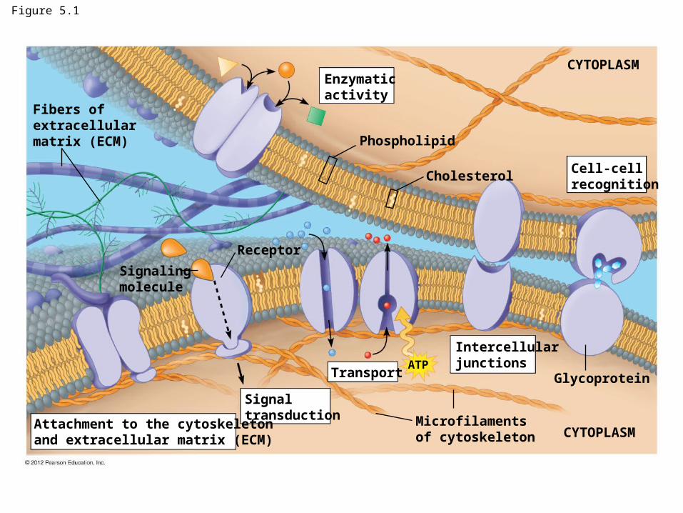
Figure 5.1
Fibers ofextracellularmatrix (ECM)
Enzymatic activity
Phospholipid
Cholesterol
CYTOPLASM
CYTOPLASM
Cell-cellrecognition
Glycoprotein
Intercellularjunctions
Microfilamentsof cytoskeleton
ATPTransport
Signaltransduction
Receptor
Signalingmolecule
Attachment to the cytoskeletonand extracellular matrix (ECM)
Passive transport
Passive transport = diffusion across cell membrane
– No energy required!!
Moves with concentration gradient
Examples:
– Urea, CO2, O2, Water, small hydrophobic
© 2012 Pearson Education, Inc.
Animation: Membrane Selectivity
Animation: Diffusion
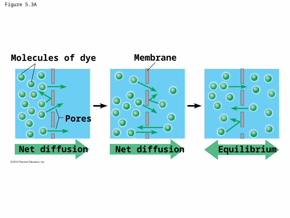
Figure 5.3A
Molecules of dye Membrane
Pores
Net diffusion Net diffusion Equilibrium
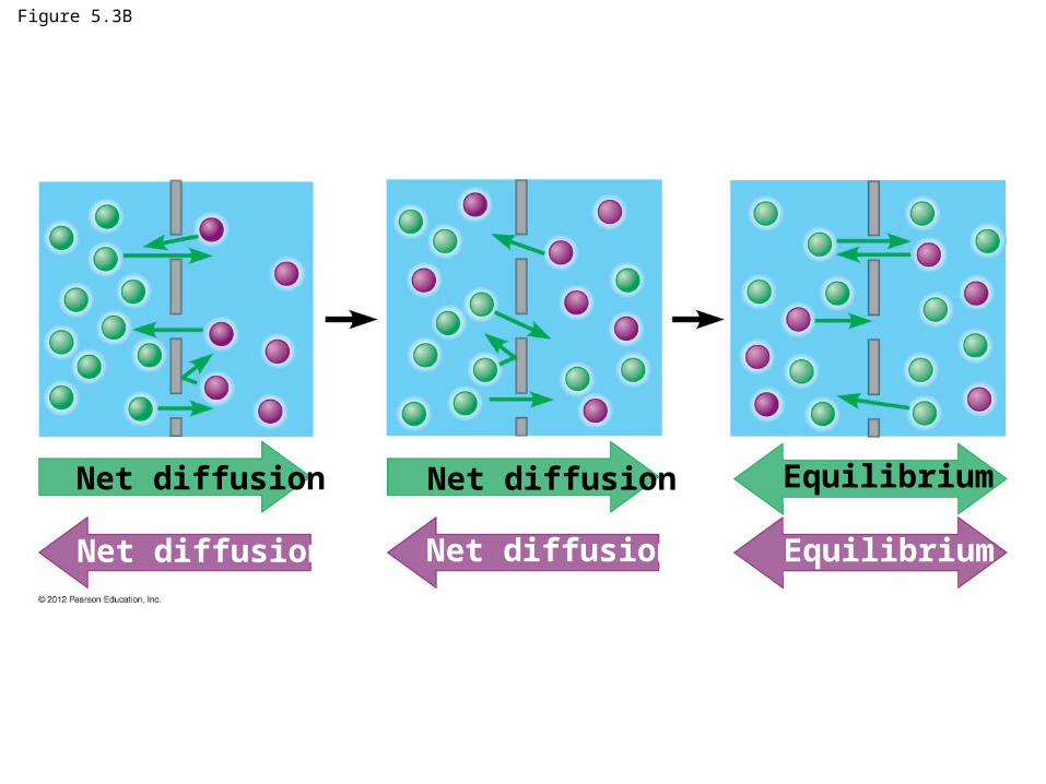
Figure 5.3B
Net diffusion Net diffusion
Net diffusion Net diffusion
Equilibrium
Equilibrium
© 2012 Pearson Education, Inc.
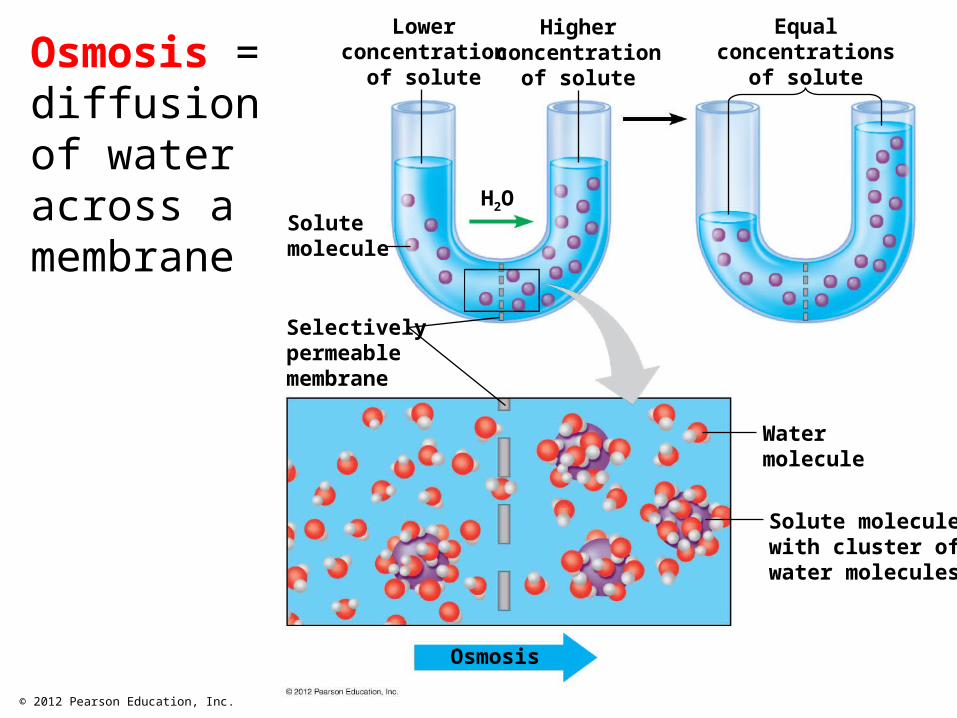
Osmosis
Solute moleculewith cluster ofwater molecules
Watermolecule
Selectivelypermeablemembrane
Solutemolecule
H2O
Lowerconcentration
of solute
Higherconcentration
of solute
Equalconcentrations
of soluteOsmosis = diffusion of water across a membrane
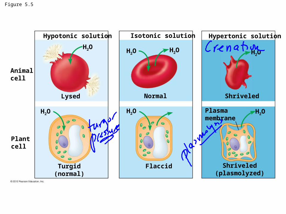
Figure 5.5
Animalcell
Plantcell
Turgid(normal)
Flaccid Shriveled(plasmolyzed)
Plasmamembrane
Lysed Normal Shriveled
Hypotonic solution Isotonic solution Hypertonic solution
H2O H2O H2O H2O
H2O H2O H2O

Osmoregulation = Water Balance
Osmoreguatation = all organisms must regulate internal water concentrations to remove excess water or prevent water loss
– Remove excess water:
– Contractile vacuoles - protists
– Freshwater organisms – kidneys, gills
– Prevent water loss:
– Guard cells in plants
© 2012 Pearson Education, Inc.
Video: Paramecium Vacuole
Video: Chlamydomonas
Video: Turgid Elodea
Video: Plasmolysis
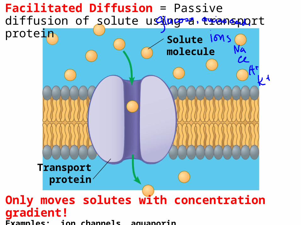
Facilitated Diffusion = Passive diffusion of solute using a transport protein
Solutemolecule
Transportprotein
Only moves solutes with concentration gradient!Examples: ion channels, aquaporin
5.7 SCIENTIFIC DISCOVERY: Research on another membrane protein led to the discovery of aquaporins
Dr. Peter Agre received the 2003 Nobel Prize in chemistry for his discovery of aquaporins.
His research on the Rh protein used in blood typing led to this discovery.
© 2012 Pearson Education, Inc.
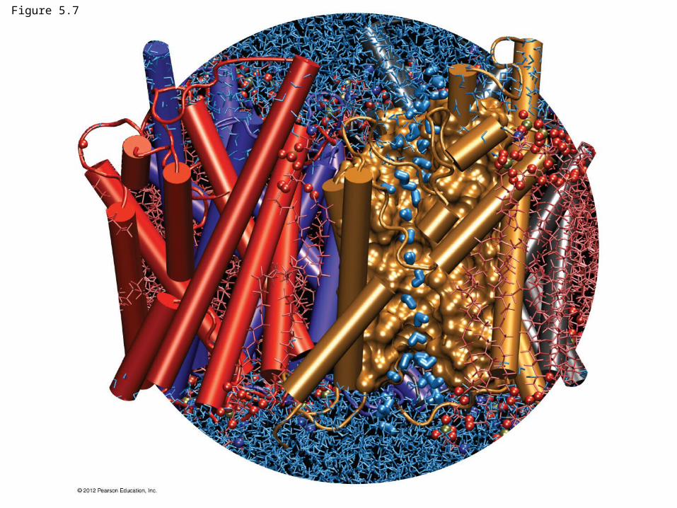
Figure 5.7
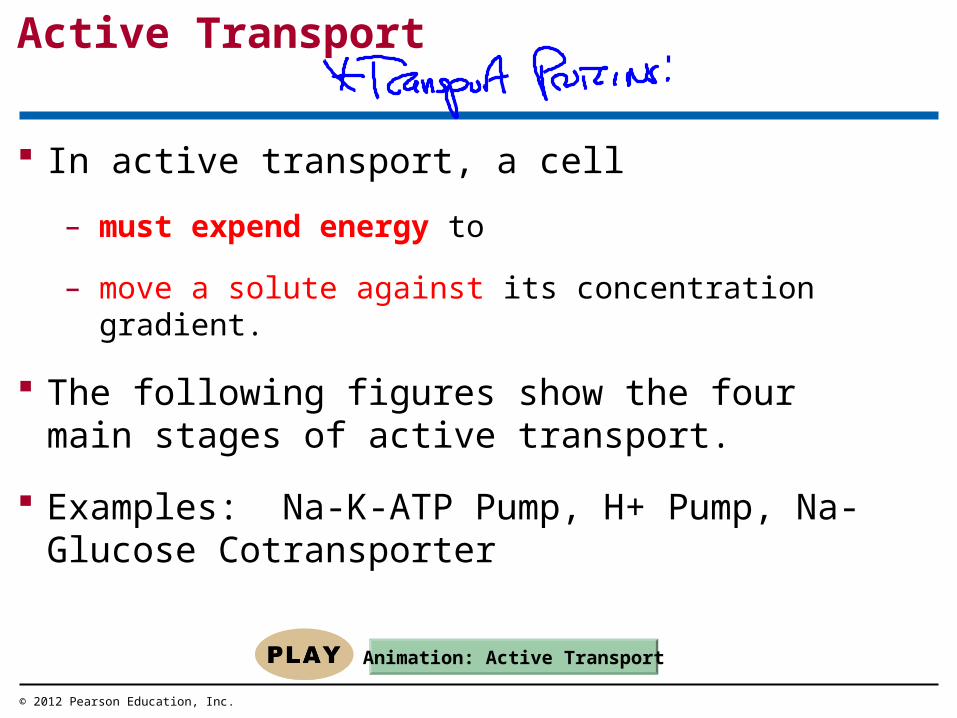
Active Transport
In active transport, a cell
– must expend energy to
– move a solute against its concentration gradient.
The following figures show the four main stages of active transport.
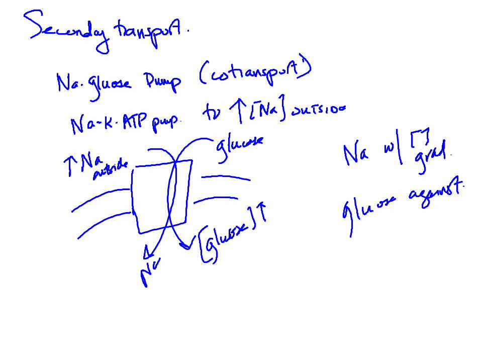
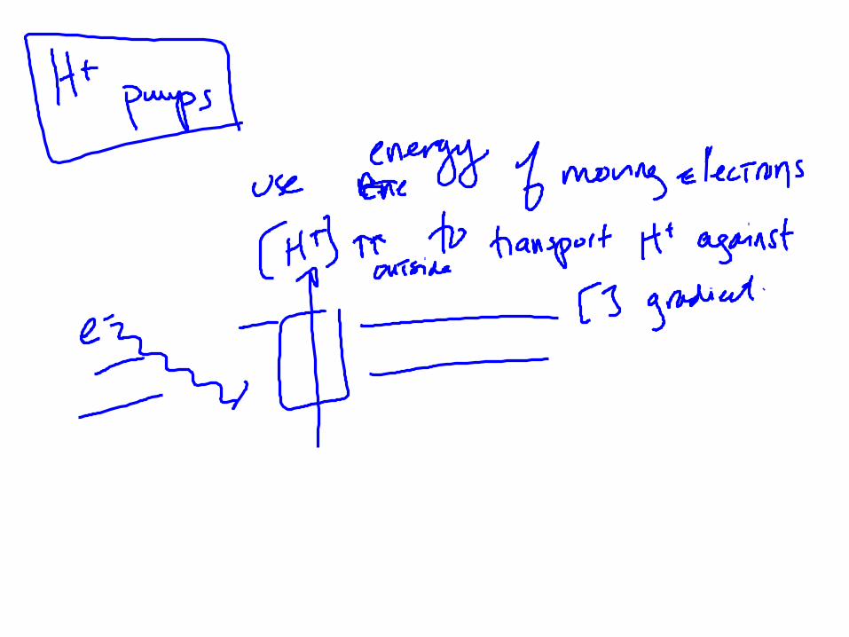
Examples: Na-K-ATP Pump, H+ Pump, Na-Glucose Cotransporter
© 2012 Pearson Education, Inc.
Animation: Active Transport

Figure 5.8_s4
Transportprotein
Solute ADPATP P P PProtein
changes shape.Phosphatedetaches.
Solute binding Phosphateattaching
Transport Proteinreversion
4321

5.9 Exocytosis and endocytosis transport large molecules across membranes
There are three kinds of endocytosis.
1. Phagocytosis is the engulfment of a particle by wrapping cell membrane around it, forming a vacuole.
2. Pinocytosis is the same thing except that fluids are taken into small vesicles.
3. Receptor-mediated endocytosis uses receptors in a receptor-coated pit to interact with a specific protein, initiating the formation of a vesicle.
© 2012 Pearson Education, Inc.
Animation: Phagocytosis
Animation: Exocytosis
Animation: Receptor-Mediated Endocytosis
Animation: Pinocytosis
Animation: Exocytosis and Endocytosis Introduction
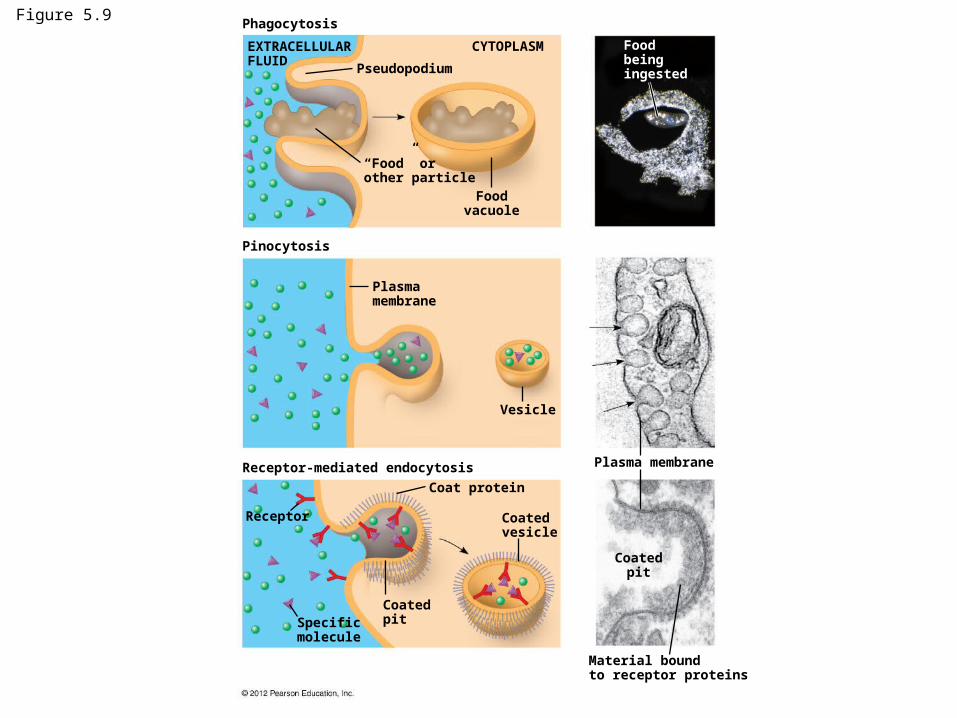
Figure 5.9Phagocytosis
Pinocytosis
Receptor-mediated endocytosis
EXTRACELLULARFLUID
CYTOPLASM
Pseudopodium
“Food” orother particle
Foodvacuole
Foodbeingingested
Plasma membrane
Plasma membrane
Vesicle
Receptor
Specificmolecule
Coatedpit
Coatedvesicle
Coat protein
Coatedpit
Material boundto receptor proteins

ENERGY AND THE CELL
© 2012 Pearson Education, Inc.
5.10 Cells transform energy as they perform work
Energy = capacity to cause change or to perform work.
Two kinds of energy:
1. Kinetic energy is the energy of motion.
2. Potential energy is energy that matter possesses as a result of its location or structure.
Heat = thermal energy
Chemical energy = potential energy available in bonds within molecules and released in a chemical reaction.
– Most relevant energy to living organisms
© 2012 Pearson Education, Inc.
Animation: Energy Concepts
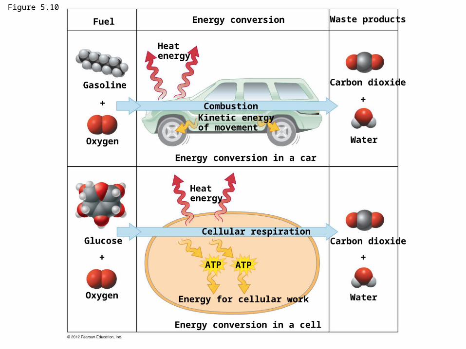
Figure 5.10
Fuel Energy conversion Waste products
Gasoline
Oxygen
Oxygen
Glucose
Heatenergy
CombustionKinetic energyof movement
Energy conversion in a car
Energy conversion in a cell
Energy for cellular work
Cellular respiration
ATP ATP
Heatenergy
Carbon dioxide
Carbon dioxide
Water
Water

Thermodynamics = study of energy transformations
First law of thermodynamics = energy in the universe is constant
– Biological organisms cannot produce energy - only convert forms of energy
Second law of thermodynamics = energy conversions increase the disorder (entropy) of the universe.
– No energy transformations are 100 % efficient
– Usuable energy lost as heat
– Energy transformations are one-way street
– Biological organisms require constant supply of energy to maintain order!!
© 2012 Pearson Education, Inc.

Metabolism = total of an organism’s chemical reactions
Chemical reactions are either
– Exergonic reactions release energy.
– These reactions release the energy in covalent bonds of the reactants.
– Cellular respiration
An endergonic reaction
– requires an input of energy; products contain more chemical/potential energy
– Photosynthesis
Energy coupling = energy released from exergonic reactions drive endergonic reactions!!
© 2012 Pearson Education, Inc.
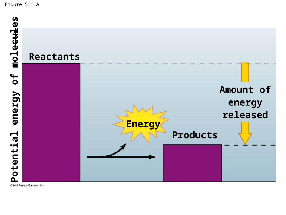
Figure 5.11A
Reactants
EnergyProducts
Amount ofenergy
released
Po
ten
tial
en
erg
y o
f m
ole
cule
s

Figure 5.11B
Reactants
Energy
Products
Amount ofenergy
required
Po
ten
tial
en
erg
y o
f m
ole
cule
s

Cells need energy to perform work!!
There are three main types of cellular work:
1. chemical
2. mechanical
3. transport
ATP drives all three of these types of work.
© 2012 Pearson Education, Inc.
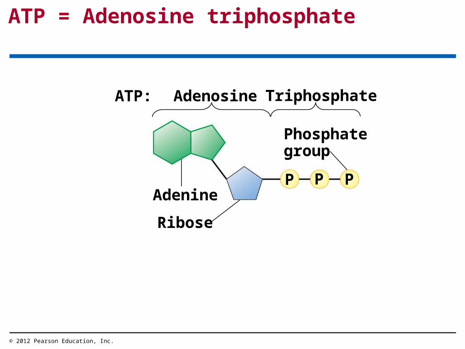
ATP = Adenosine triphosphate
© 2012 Pearson Education, Inc.
AdenineP P P
Phosphategroup
ATP: Adenosine Triphosphate
Ribose
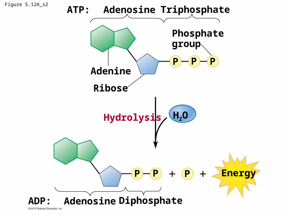
Figure 5.12A_s2
ADP: Adenosine Diphosphate
P P P Energy
H2OHydrolysis
Ribose
AdenineP P P
Phosphategroup
ATP: Adenosine Triphosphate

ATP drives cellular work
Hydrolysis of ATP releases energy by transferring phosphate from ATP to some other molecule
– phosphorylation.
© 2012 Pearson Education, Inc.

Figure 5.12B
ATP ATP ATP
ADP ADP ADPP P P
P
P
P
PP
P
Chemical work Mechanical work Transport work
Reactants
Motorprotein
Solute
Membrane protein
Product
Molecule formed Protein filament moved Solute transported

How Does Cell Regenerate ATP?
© 2012 Pearson Education, Inc.
Energy fromexergonicreactions
Energy forendergonicreactions
ATP
ADP P
ATP = renewable source of energy for the cell.
ATP cycle = energy released in an exergonic reaction is used in an endergonic reaction to generate ATP.
HOW ENZYMES FUNCTION
© 2012 Pearson Education, Inc.
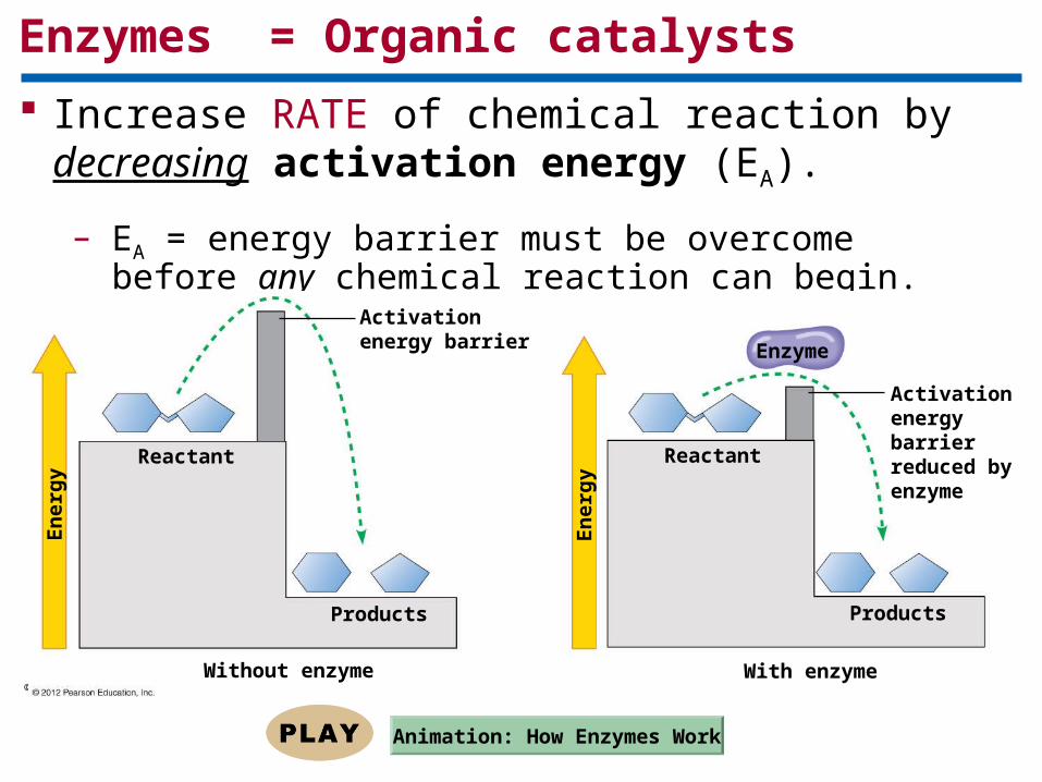
Enzymes = Organic catalysts
Increase RATE of chemical reaction by decreasing activation energy (EA).
– EA = energy barrier must be overcome before any chemical reaction can begin.
© 2012 Pearson Education, Inc.
Activationenergy barrier
Reactant
Products
Without enzyme With enzyme
Reactant
Products
Enzyme
Activationenergy barrierreduced byenzyme
En
erg
y
En
erg
y
Animation: How Enzymes Work
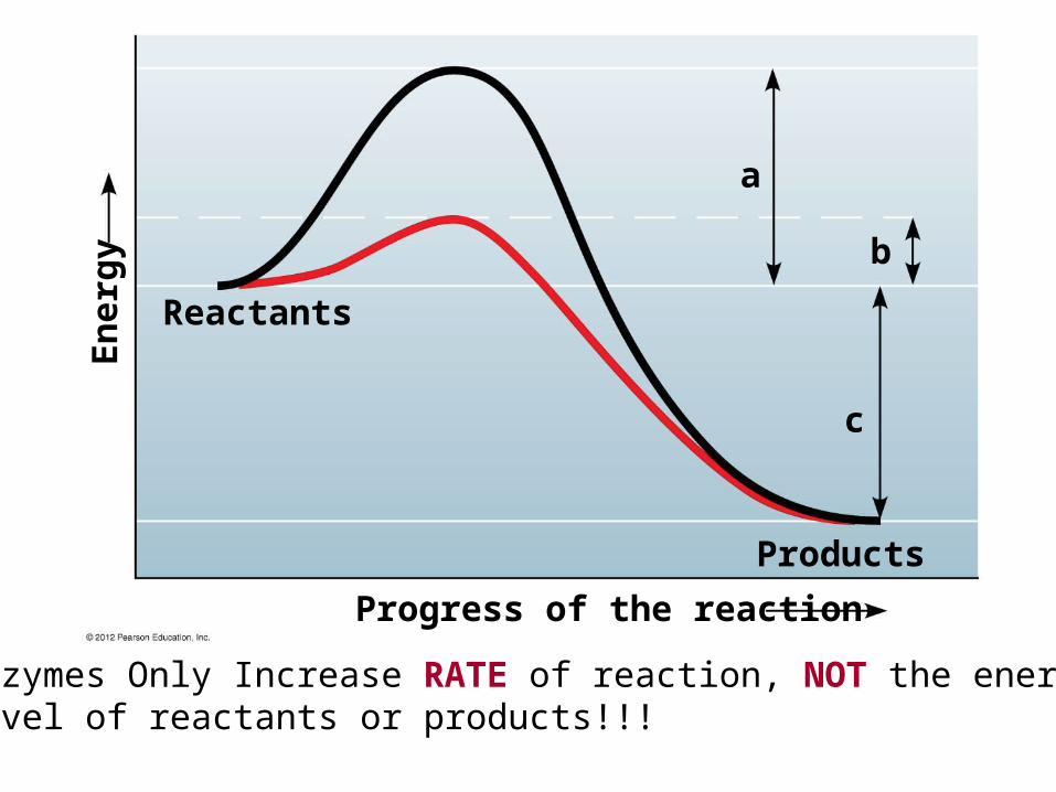
Reactants
Products
En
erg
y
Progress of the reaction
a
b
c
Enzymes Only Increase RATE of reaction, NOT the energyLevel of reactants or products!!!
A specific enzyme catalyzes each cellular reaction
An enzyme
– Is specific in substrate(s) it binds
– And reaction it catalyzes
Substrate = reactant
A substrate binds at enzyme active site.
Enzymes are specific because their active site fits only specific substrate molecules
– Active site is result of 3D folding of protein
© 2012 Pearson Education, Inc.
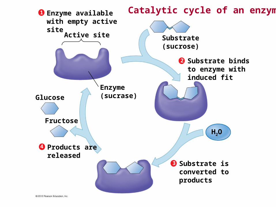
4
3
2
1
Products arereleased
Fructose
Glucose
Enzyme(sucrase)
Active site
Enzyme availablewith empty activesite
Substrate(sucrose)
Substrate bindsto enzyme withinduced fit
Substrate isconverted toproducts
H2O
Catalytic cycle of an enzyme
Factors that Effect Enzyme-Catalyzed Reactions
For every enzyme, there are optimal conditions under which it is most effective.
– Temperature
– pH
– Substrate Concentration
– Enzyme Concentration
– Cofactors/coenzymes
– Inhibitors
© 2012 Pearson Education, Inc.
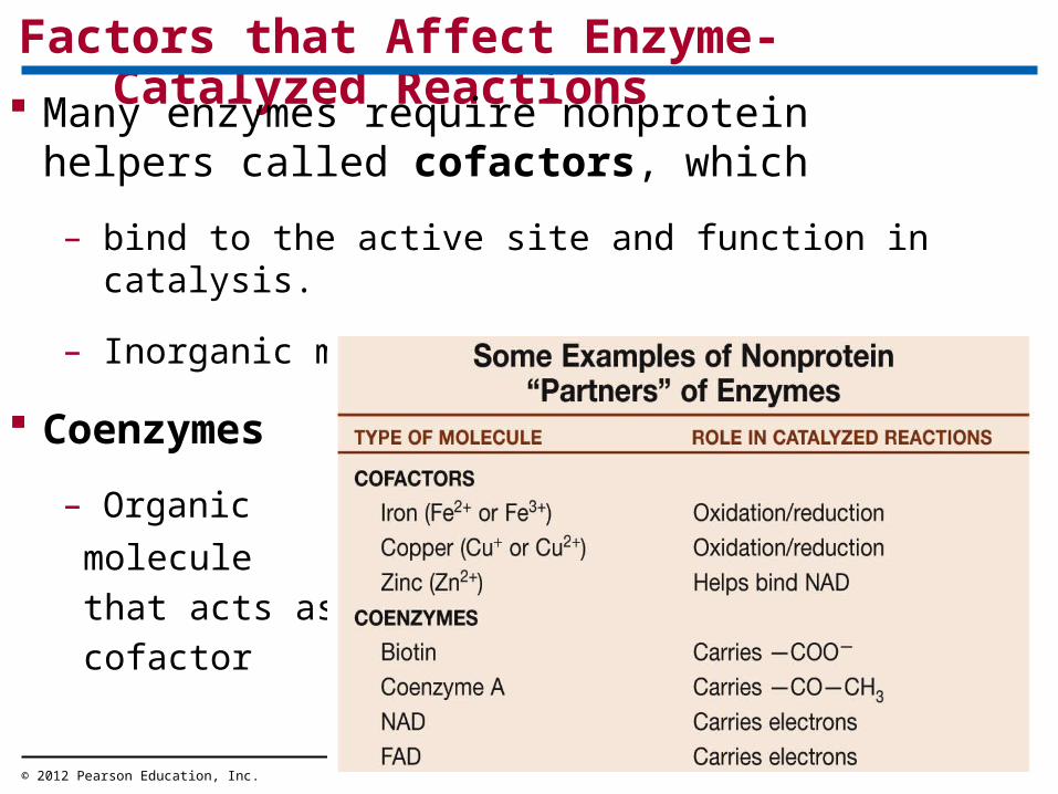
Factors that Affect Enzyme-Catalyzed Reactions
Many enzymes require nonprotein helpers called cofactors, which
– bind to the active site and function in catalysis.
– Inorganic molecules
Coenzymes
– Organic
molecule
that acts as
cofactor
© 2012 Pearson Education, Inc.

QuickTime™ and aTIFF (Uncompressed) decompressor
are needed to see this picture.
Enzyme Concentration

Substrate Concentration
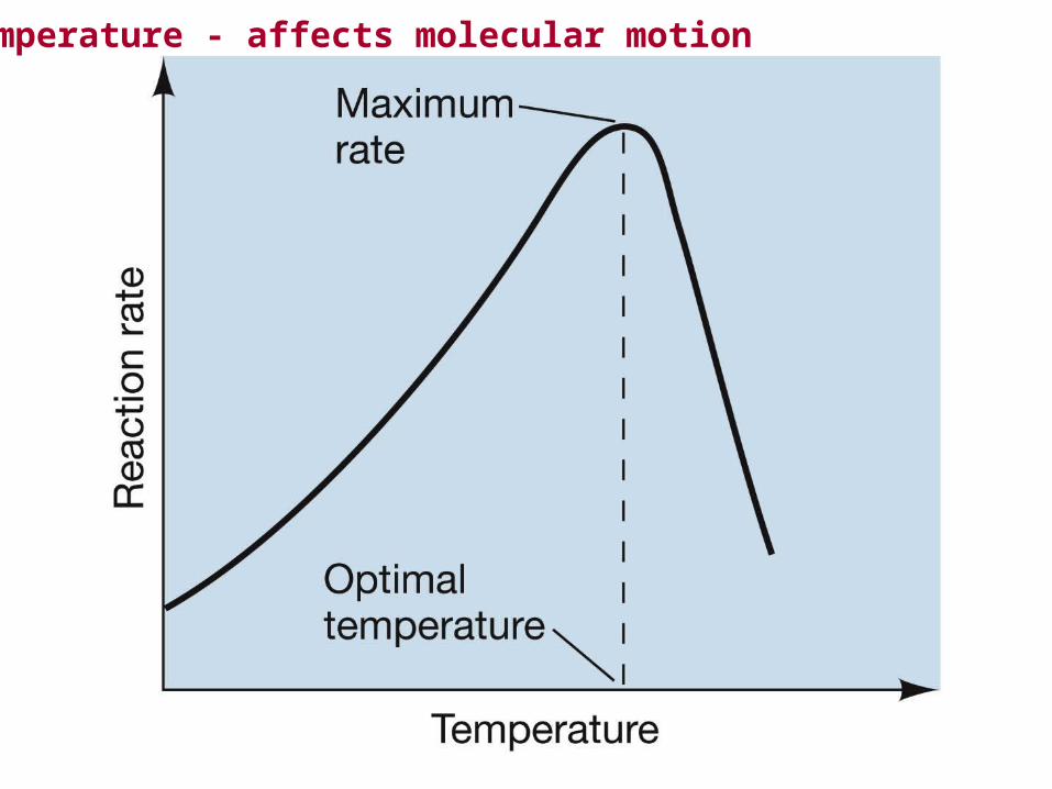
Temperature - affects molecular motion
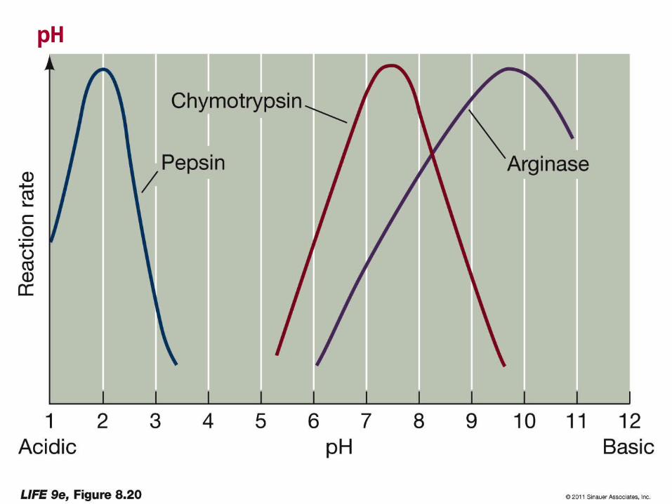
pH

Enzyme inhibitors can regulate enzyme activity Inhibitor = chemical that interferes with an enzyme’s activity.
© 2012 Pearson Education, Inc.
Substrate
Enzyme
Allosteric site
Active site
Normal binding of substrate
Competitiveinhibitor
Noncompetitiveinhibitor
Enzyme inhibition
Competitive inhibitors
– block substrates from entering the active site and
– reduce an enzyme’s productivity.
Noncompetitive inhibitors
– bind to the enzyme somewhere other than the active site,
– change the shape of the active site, and
– prevent the substrate from binding.
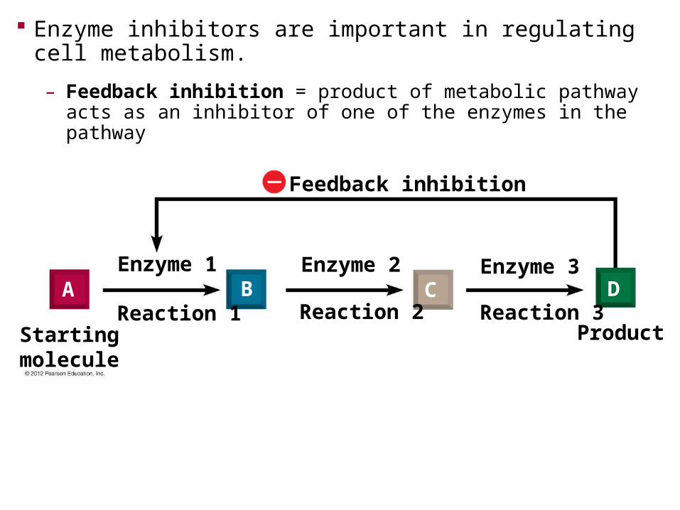
Feedback inhibition
Startingmolecule
Product
Enzyme 1 Enzyme 2 Enzyme 3
Reaction 1 Reaction 2 Reaction 3A B C D
Enzyme inhibitors are important in regulating cell metabolism.
– Feedback inhibition = product of metabolic pathway acts as an inhibitor of one of the enzymes in the pathway

1. Describe the fluid mosaic structure of cell membranes.
2. Describe the diverse functions of membrane proteins.
3. Relate the structure of phospholipid molecules to the structure and properties of cell membranes.
4. Define diffusion and describe the process of passive transport.
You should now be able to
© 2012 Pearson Education, Inc.
5. Explain how osmosis can be defined as the diffusion of water across a membrane.
6. Distinguish between hypertonic, hypotonic, and isotonic solutions.
7. Explain how transport proteins facilitate diffusion.
8. Distinguish between exocytosis, endocytosis, phagocytosis, pinocytosis, and receptor-mediated endocytosis.
You should now be able to
© 2012 Pearson Education, Inc.
9. Define and compare kinetic energy, potential energy, chemical energy, and heat.
10. Define the two laws of thermodynamics and explain how they relate to biological systems.
11. Define and compare endergonic and exergonic reactions.
12. Explain how cells use cellular respiration and energy coupling to survive.
You should now be able to
© 2012 Pearson Education, Inc.

You should now be able to
13. Explain how ATP functions as an energy shuttle.
14. Explain how enzymes speed up chemical reactions.
15. Explain how competitive and noncompetitive inhibitors alter an enzyme’s activity.
16. Explain how certain drugs, pesticides, and poisons can affect enzymes.
© 2012 Pearson Education, Inc.
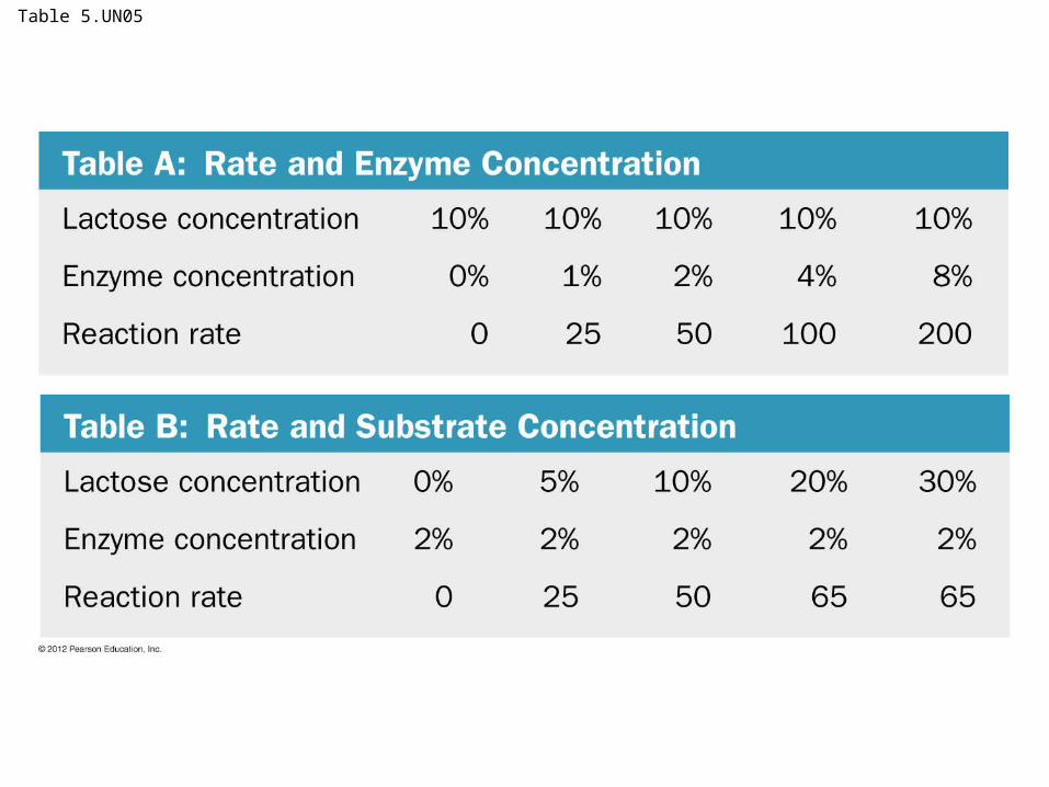
Table 5.UN05
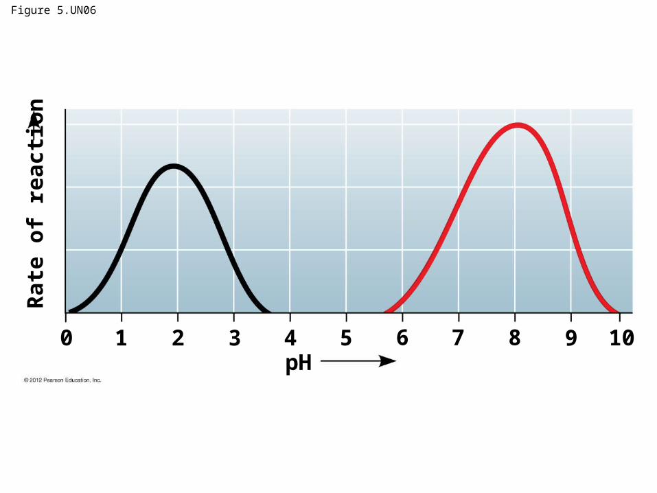
Figure 5.UN06
pH
Rat
e o
f re
acti
on
109876543210