fig. 24-1, p. 864 ch. 24 – the digestive system the overall idea is to obtain nutrients from the...
TRANSCRIPT
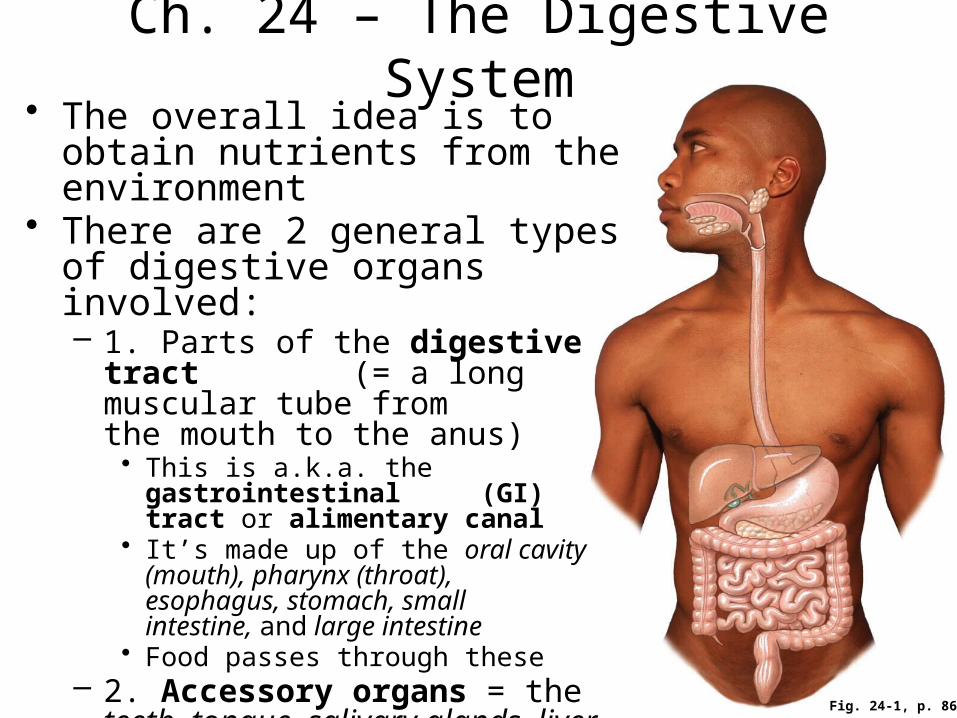
Fig. 24-1, p. 864
Ch. 24 – The Digestive System• The overall idea is to obtain
nutrients from the environment • There are 2 general types of
digestive organs involved:– 1. Parts of the digestive tract
(= a long muscular tube from the mouth to the anus)• This is a.k.a. the gastrointestinal
(GI) tract or alimentary canal• It’s made up of the oral cavity
(mouth), pharynx (throat), esophagus, stomach, small intestine, and large intestine
• Food passes through these– 2. Accessory organs = the teeth,
tongue, salivary glands, liver, gallbladder, and pancreas• Food does not pass through these
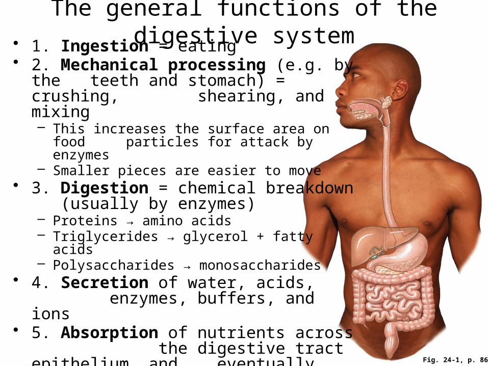
The general functions of the digestive system• 1. Ingestion = eating• 2. Mechanical processing (e.g. by the
teeth and stomach) = crushing, shearing, and mixing– This increases the surface area on food
particles for attack by enzymes– Smaller pieces are easier to move
• 3. Digestion = chemical breakdown (usually by enzymes)– Proteins → amino acids– Triglycerides → glycerol + fatty acids– Polysaccharides → monosaccharides
• 4. Secretion of water, acids, enzymes, buffers, and ions
• 5. Absorption of nutrients across the digestive tract epithelium, and eventually into the blood
• 6. Excretion = the removal of waste (via defecation)
Fig. 24-1, p. 864
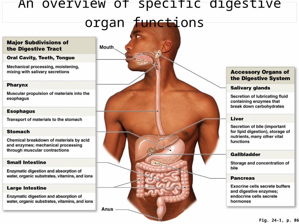
Fig. 24-1, p. 864
An overview of specific digestive organ functions
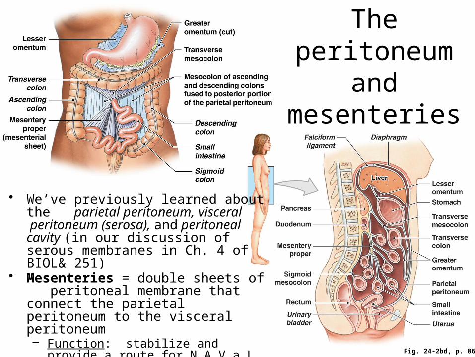
The peritoneum and
mesenteries
• We’ve previously learned about the parietal peritoneum, visceral peritoneum (serosa), and peritoneal cavity (in our discussion of serous membranes in Ch. 4 of BIOL& 251)
• Mesenteries = double sheets of peritoneal membrane that connect the parietal peritoneum to the visceral peritoneum– Function: stabilize and provide a route
for N.A.V.a.L to the abdominopelvic organs
Fig. 24-2bd, p. 866

Fig. 24-3, p. 867
The general histology of the GI tract• Most parts of the tube have most (if not all) of these features• Additional details are found in the textbook and on p. 16 of
the Lab Manual Lumen (interior)
Exterior
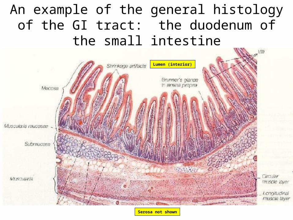
An example of the general histology of the GI tract: the duodenum of the small intestine
Lumen (interior)
Serosa not shown

Fig. 24-4, p. 868
The movement of digestive materials
• The muscular layers of the GI tract have gap junctions between cells and pacesetter cells that spontaneously depolarize– They can also be stimulated
autonomically by the CNS and ENS (see the next slide)
• Peristalsis = waves of rhythmic contractions that propel the “food mass” (= bolus) forward through the tract
• Segmentation (not shown here) = irregular contractions in the intestines that mix/churn/fragment the bolus

Fig. 24-5, p. 869
The regulation
of digestive activities
E.g. gastrin, secretin, etc. We’ll discuss some of the main ones later in the chapter.
These short reflexes are part of the enteric nervous system (ENS), the lesser known third division of the ANS
E.g. prostaglandins and histamine
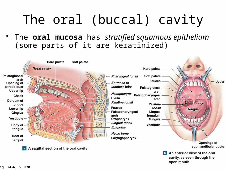
Fig. 24-6, p. 870
The oral (buccal) cavity• The oral mucosa has stratified squamous epithelium (some
parts of it are keratinized)
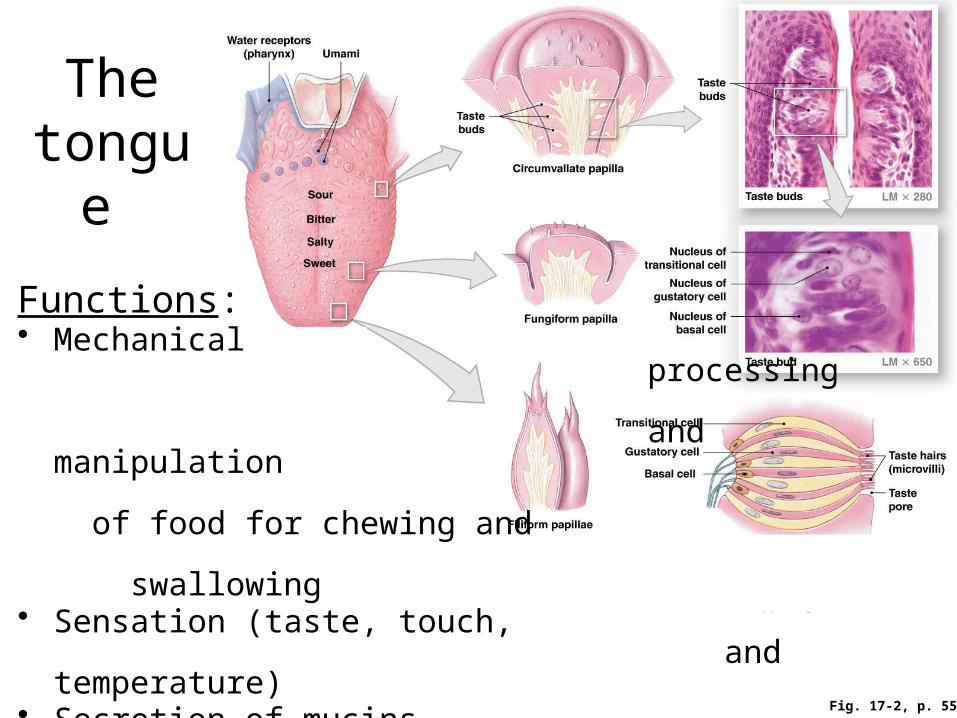
Fig. 17-2, p. 554
The tongue
Functions:• Mechanical
processing and manipulation of food for chewing and swallowing
• Sensation (taste, touch, and temperature)
• Secretion of mucins (= mucus glycoproteins)
• Secretion of lingual lipase, which initiates the digestion of fats
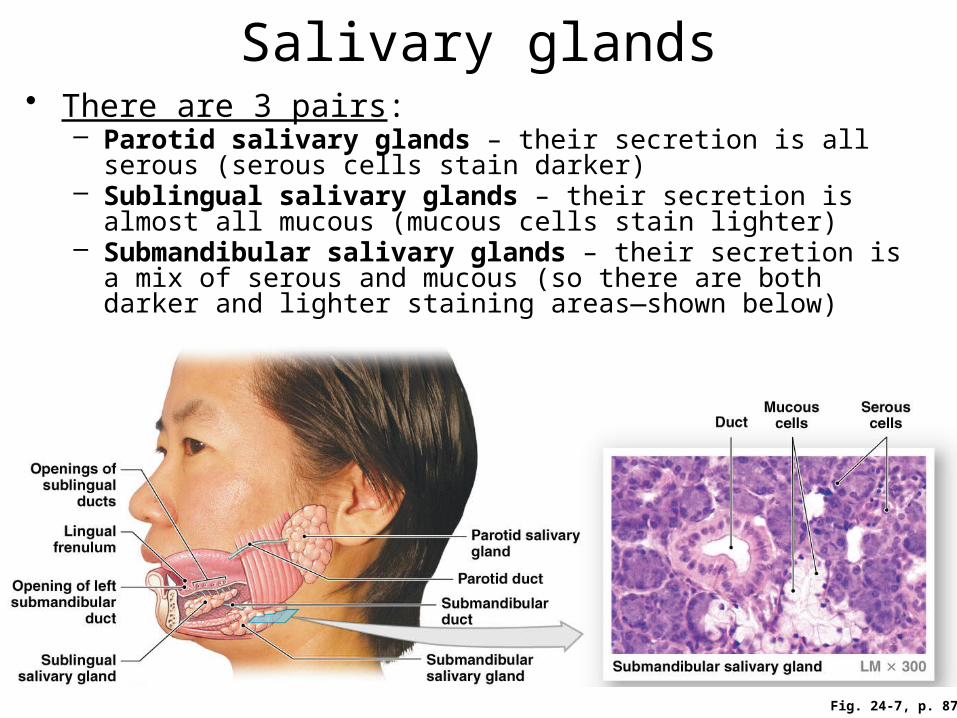
Fig. 24-7, p. 872
Salivary glands• There are 3 pairs:
– Parotid salivary glands – their secretion is all serous (serous cells stain darker)
– Sublingual salivary glands – their secretion is almost all mucous (mucous cells stain lighter)
– Submandibular salivary glands – their secretion is a mix of serous and mucous (so there are both darker and lighter staining areas—shown below)

Saliva• Functions: moisten and lubricate food, rinse/flush the mouth,
dissolve chemicals for taste bud stimulation, initiate the chemical digestion of complex carbos (by salivary amylase)
• Composition:– 99.4% water; the rest is solutes, which include:
• Ions, salivary amylase, buffers (so pH ~ 7.0), waste products, IgA antibodies, lysozyme, mucus (which helps with the lubrication of food)
• Secretion:– 1-1.5 liters per day!– Parasympathetic stimulation → salivation– Sympathetic stimulation → dry mouth– Secretion is increased by:
• 1. Food in the mouth, the taste of food, and chewing (even without food in the mouth)
• 2. The smell, sight, or sound of food• 3. Stomach or small intestine irritation (in order to dilute, rinse, or buffer
the unpleasant stimulus)
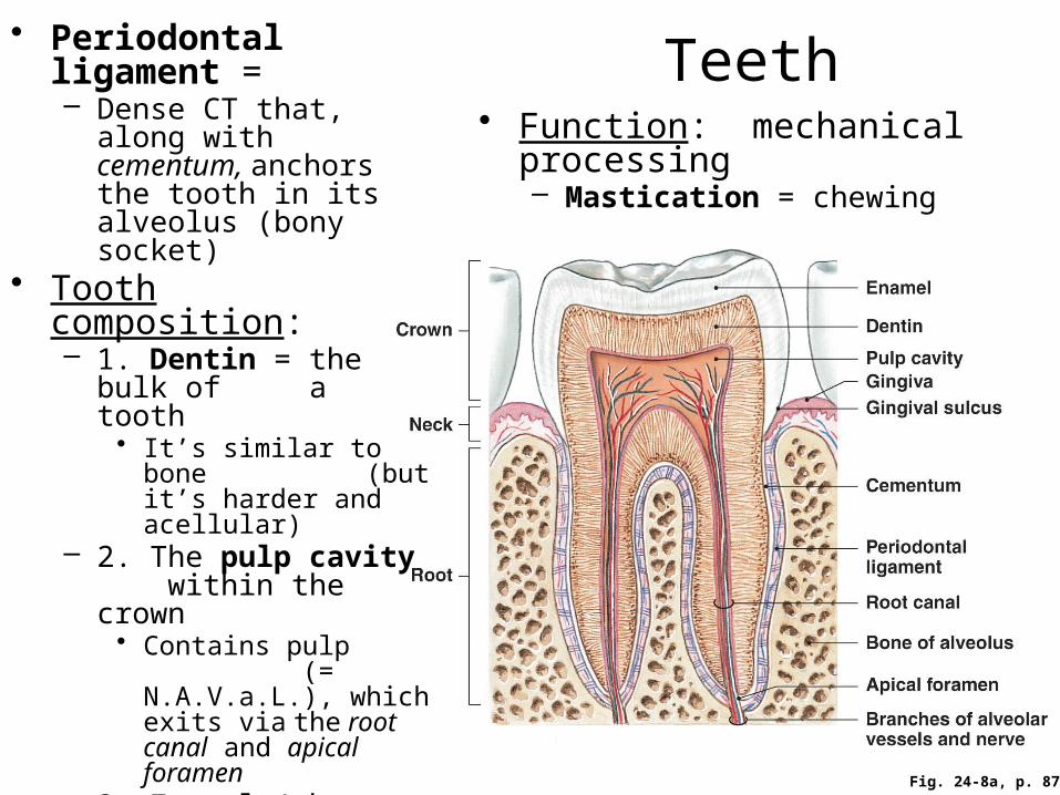
Teeth• Periodontal ligament =
– Dense CT that, along with cementum, anchors the tooth in its alveolus (bony socket)
• Tooth composition:– 1. Dentin = the bulk of
a tooth• It’s similar to bone
(but it’s harder and acellular)
– 2. The pulp cavity within the crown
• Contains pulp (= N.A.V.a.L.), which exits via the root canal and apical foramen
– 3. Enamel (the hardest biologically- made substance)
• = crystalline calcium phosphate
• Covers and protects the dentin of the crown
Fig. 24-8a, p. 873
• Function: mechanical processing– Mastication = chewing

Fig. 24-8b, p. 873
Types of teeth
• Deciduous (1°) teeth = 20; typically appear between 6-24 months of age– Per quadrant: 2 incisors, 1 cuspid
(canine), and 2 molars• Permanent (2°) teeth = 32; most
appear between 6-12 years of age– Per quadrant: 2 incisors, 1 cuspid
(canine), 2 bicuspids (premolars), and 3 molars
Fig. 24-9, p. 875

Fig. 24-6a, p. 870
The pharynx (throat)
• = a common passageway for solid food, liquids, and air
• Has underlying skeletal muscle for swallowing
• Has 3 regions:– 1. Nasopharynx –
is lined with PCCE; contains the pharyngeal tonsil (adenoid)
– 2. Oropharynx – is lined with stratified squamous epithelium (nonkeratinized); contains the palatine and lingual tonsils
– 3. Laryngopharynx – has the same epithelium as the oropharynx
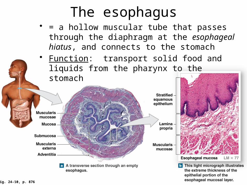
Fig. 24-10, p. 876
The esophagus• = a hollow muscular tube that passes through the
diaphragm at the esophageal hiatus, and connects to the stomach
• Function: transport solid food and liquids from the pharynx to the stomach

Fig. 24-10, p. 876
• 1. The mucosa includes a stratified squamous epithelium (nonkeratinized)
• 2. The submucosa contains esophageal (mucous) glands for lubrication of the bolus– The mucosa and submucosa are
folded, allowing for expansion during swallowing
• 3. The muscularis externa gradually transitions (moving from superior to inferior) from skeletal muscle to smooth muscle– Muscle tone keeps the upper and
lower ends closed (except when a bolus passes through), acting as “sphincters”
• These are not true anatomical sphincters (= circular muscular valves) like the pyloric sphincter and ileocecal valve
• 4. Adventitia (not serosa) = fibrous CT for attachment
Histology of the esophagus
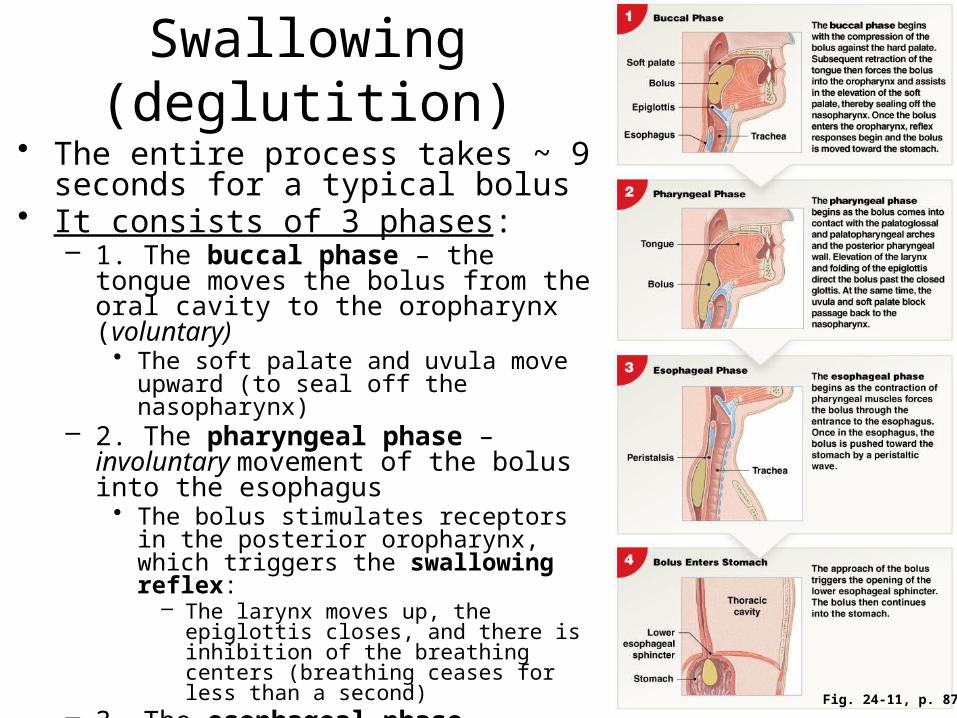
Fig. 24-11, p. 878
Swallowing (deglutition)• The entire process takes ~ 9 seconds
for a typical bolus• It consists of 3 phases:
– 1. The buccal phase – the tongue moves the bolus from the oral cavity to the oropharynx (voluntary)
• The soft palate and uvula move upward (to seal off the nasopharynx)
– 2. The pharyngeal phase – involuntary movement of the bolus into the esophagus
• The bolus stimulates receptors in the posterior oropharynx, which triggers the swallowing reflex:
– The larynx moves up, the epiglottis closes, and there is inhibition of the breathing centers (breathing ceases for less than a second)
– 3. The esophageal phase – involuntary peristalsis pushes the bolus toward the stomach

Stomach terminology• Fundus = the dome-shaped region superior to the esophageal
opening• Body = the main central region• Pylorus = the region that
connects with the duodenum via the pyloric sphincter
• Rugae = folds of mucosa that allow the stomach to stretch
• Chyme = a viscous, acidic, soupy mix of partially digested food
Fig. 24-12b, p. 879
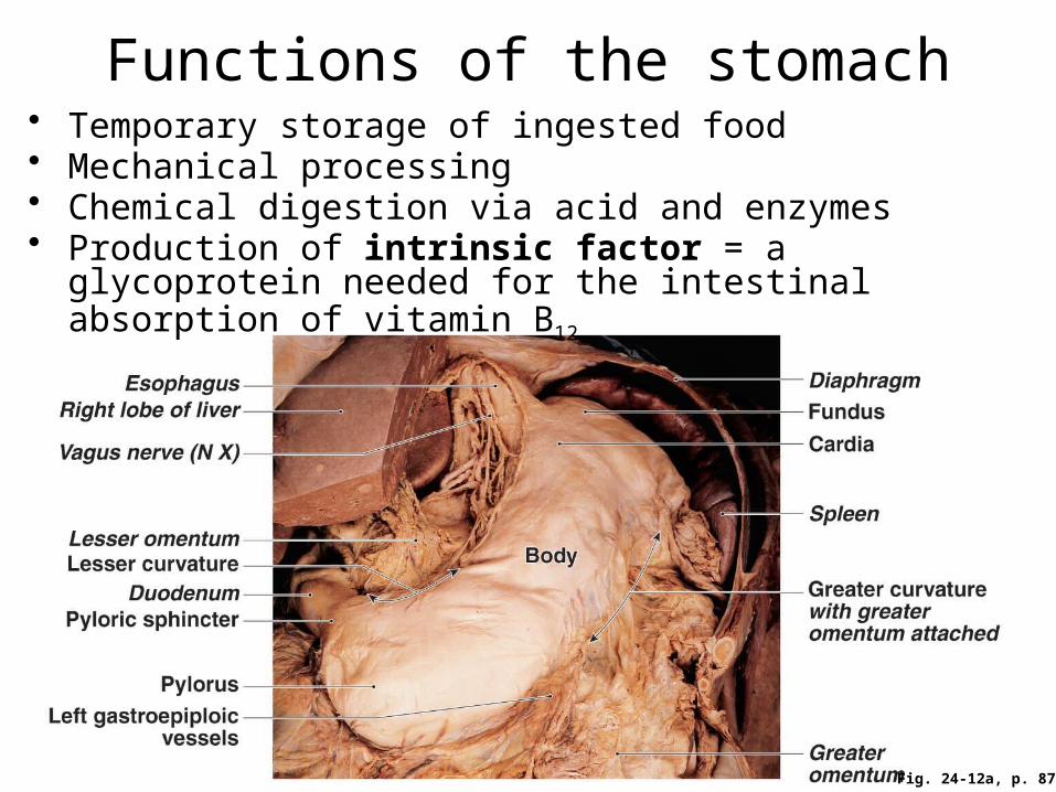
Functions of the stomach• Temporary storage of ingested food• Mechanical processing• Chemical digestion via acid and enzymes • Production of intrinsic factor = a glycoprotein needed for the
intestinal absorption of vitamin B12
Fig. 24-12a, p. 879

Fig. 24-13a, p. 880
Histology of the stomach
lining
• 1. Mucosa – is folded into gastric pits that open into deeper gastric glands
• 2. Submucosa• 3. Muscularis
externa = 3 layers of smooth muscle– An inner
oblique layer is present in addition to the circular and longitudinal layers
• 4. Serosa
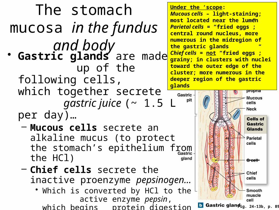
Fig. 24-13b, p. 893
The stomach mucosa in the fundus and body• Gastric glands are made
up of the following cells, which together secrete gastric juice (~ 1.5 L per day)…– Mucous cells secrete an alkaline
mucus (to protect the stomach’s epithelium from the HCl)
– Chief cells secrete the inactive proenzyme pepsinogen…• Which is converted by HCl to the
active enzyme pepsin, which begins protein digestion
– Parietal cells secrete HCl and intrinsic factor
Under the ‘scope:Mucous cells – light-staining; most located near the lumenParietal cells = “fried eggs”; central round nucleus, more numerous in the midregion of the gastric glandsChief cells = not “fried eggs”; grainy; in clusters with nuclei toward the outer edge of the cluster; more numerous in the deeper region of the gastric glands

Fig. 24-14, p. 881
HCl secretion by parietal cells• Keeps the stomach contents at a pH of about 1.5-2.0• Functions:
– Kill most microbes– Denature the proteins (including enzymes) in food– Help break down plant materials and the CT in meat– Help convert inactive pepsinogen to active pepsin

Fig. 24-13b, p. 880
The stomach mucosa in the pylorus• Pyloric glands
– Consist mostly of mucous cells that secrete an alkaline mucus, which helps neutralize the HCl before chyme enters the duodenum
– G cells – secrete gastrin (a hormone), which:• ↑ Stomach
motility• ↑ The secretion
of gastric juice
Mucous
Mucous
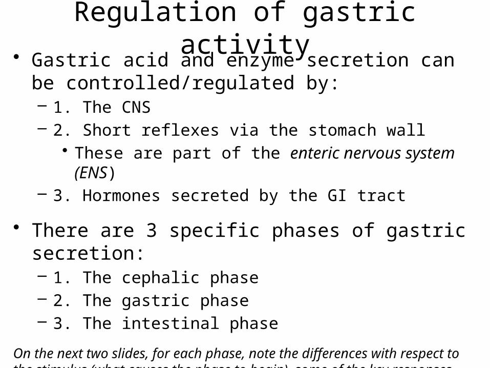
Regulation of gastric activity• Gastric acid and enzyme secretion can be
controlled/regulated by:– 1. The CNS– 2. Short reflexes via the stomach wall
• These are part of the enteric nervous system (ENS)– 3. Hormones secreted by the GI tract
• There are 3 specific phases of gastric secretion:– 1. The cephalic phase– 2. The gastric phase– 3. The intestinal phase
On the next two slides, for each phase, note the differences with respect to the stimulus (what causes the phase to begin), some of the key responses, and the overall function
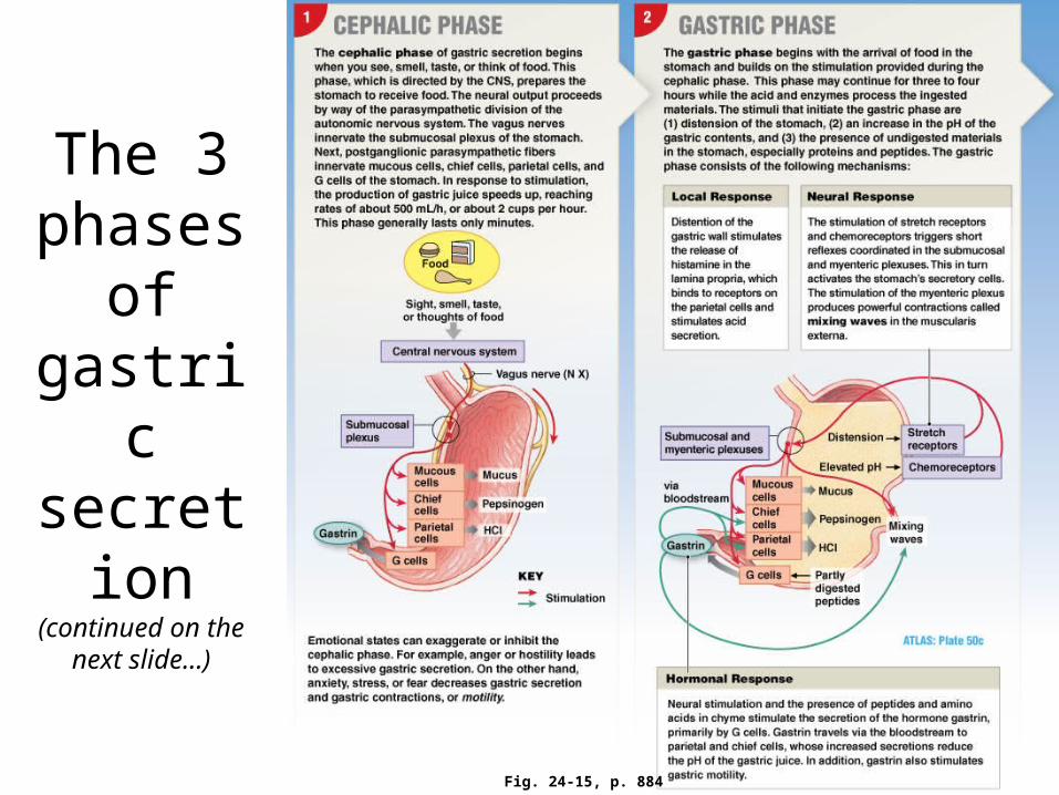
The 3 phases
of gastric secretion
(continued on the next slide…)
Fig. 24-15, p. 884
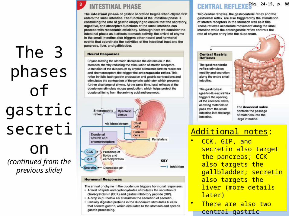
The 3 phases
of gastric secretion(continued from the
previous slide)
Additional notes:• CCK, GIP, and secretin also
target the pancreas; CCK also targets the gallbladder; secretin also targets the liver (more details later)
• There are also two central gastric reflexes (described above) that stimulate activity of the small intestine
Fig. 24-15, p. 885
Digestion and absorption in the stomachDigestion• A. Limited digestion of proteins to peptides and small
polypeptides by pepsin begins in the stomach; why is it limited?– Pepsin works best when the pH of all of the stomach contents reaches
~ 2, which takes a while– Chyme spends a relatively short duration in the stomach– Pepsin only attacks certain peptide bonds (not all of them)
• B. The digestion of carbohydrates and lipids continues…– By salivary amylase and lingual lipase, which continue to work for
about 1-2 hours after eating (until the pH of the stomach contents < 4.5)
Absorption• No nutrient absorption occurs in the stomach because:
– Mucus lines the stomach lumen and blocks the epithelium– No membrane transport proteins for nutrients are present– The epithelium is not very permeable to H2O– Digestion has not yet been completed when chyme leaves the stomach
• There is some absorption of alcohol, aspirin, and other lipid-soluble drugs
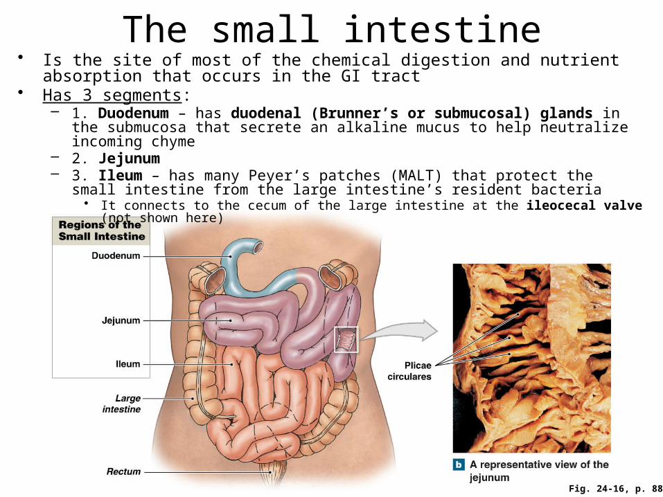
Fig. 24-16, p. 886
The small intestine• Is the site of most of the chemical digestion and nutrient absorption that occurs in
the GI tract• Has 3 segments:
– 1. Duodenum – has duodenal (Brunner’s or submucosal) glands in the submucosa that secrete an alkaline mucus to help neutralize incoming chyme
– 2. Jejunum – 3. Ileum – has many Peyer’s patches (MALT) that protect the small intestine from the
large intestine’s resident bacteria• It connects to the cecum of the large intestine at the ileocecal valve (not shown here)
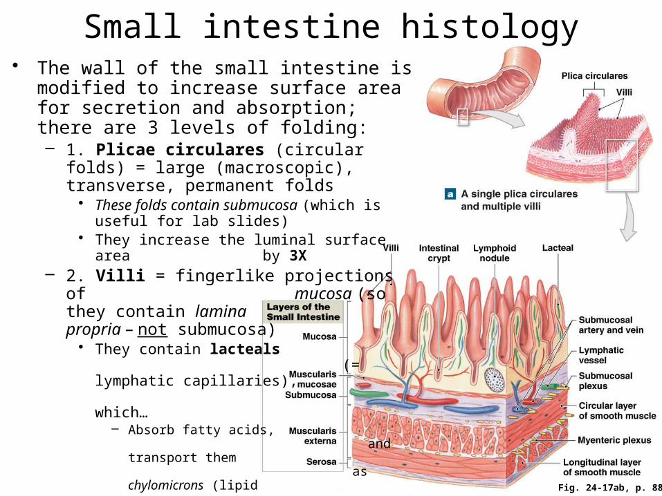
Small intestine histology• The wall of the small intestine is modified to
increase surface area for secretion and absorption; there are 3 levels of folding:– 1. Plicae circulares (circular folds) = large
(macroscopic), transverse, permanent folds• These folds contain submucosa (which is useful
for lab slides)• They increase the luminal surface area
by 3X– 2. Villi = fingerlike projections of
mucosa (so they contain lamina propria – not submucosa)
• They contain lacteals (= lymphatic capillaries), which…
– Absorb fatty acids, and transport them as chylomicrons (lipid + protein) to the blood via the lymphatic system
• They increase the luminal surface area by 10X
Fig. 24-17ab, p. 887

Small intestine histology
Fig. 24-17cd, p. 887
• 3. Microvilli = microscopic projections of the epithelial cell membrane that contain cytoplasm (not lamina propria or submucosa)– They form the brush border and
contain brush border (digestive) enzymes
– They increase the surface area by 20X• So the total increase in surface area (for
digestion and absorption) compared to the wall of a simple, unfolded tube is:
3 x 10 x 20 = 600X! (~ 2200 ft2 total)
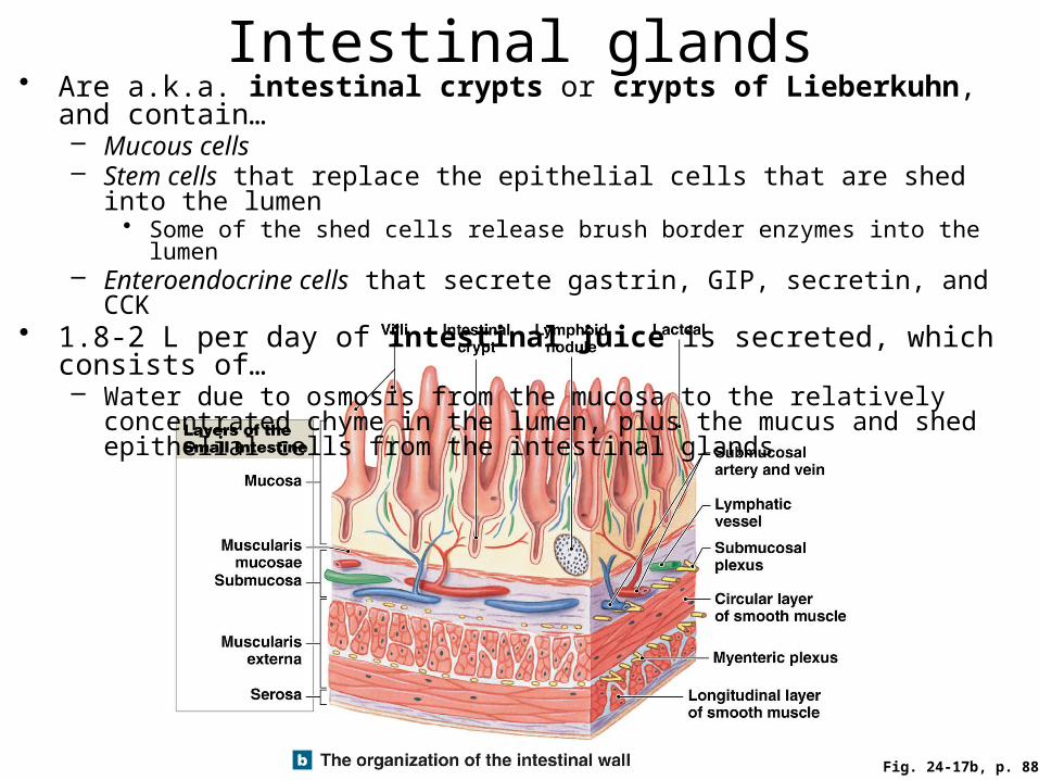
Fig. 24-17b, p. 887
Intestinal glands• Are a.k.a. intestinal crypts or crypts of Lieberkuhn, and contain…
– Mucous cells– Stem cells that replace the epithelial cells that are shed into the lumen
• Some of the shed cells release brush border enzymes into the lumen– Enteroendocrine cells that secrete gastrin, GIP, secretin, and CCK
• 1.8-2 L per day of intestinal juice is secreted, which consists of…– Water due to osmosis from the mucosa to the relatively concentrated chyme in
the lumen, plus the mucus and shed epithelial cells from the intestinal glands
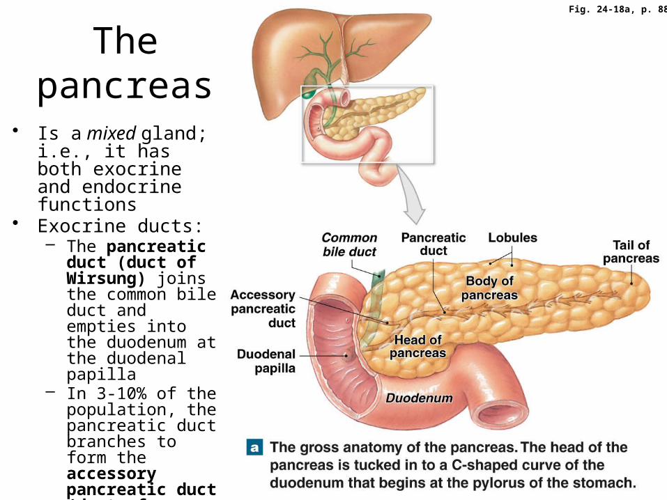
Fig. 24-18a, p. 889
The pancreas
• Is a mixed gland; i.e., it has both exocrine and endocrine functions
• Exocrine ducts:– The pancreatic
duct (duct of Wirsung) joins the common bile duct and empties into the duodenum at the duodenal papilla
– In 3-10% of the population, the pancreatic duct branches to form the accessory pancreatic duct (duct of Santorini), which also opens into the duodenum

Pancreatic histology
Fig. 24-18b, p. 889
• 1. The exocrine portion = acini and ducts (= ~ 99% of the pancreas)– The acini secrete digestive
enzymes– Duct cells secrete water and
buffers (which help neutralize chyme)
• Enzymes + H20 + buffers = pancreatic juice
– ~ 1 L per day is secreted• 2. The endocrine portion =
pancreatic islets (islets of Langerhans)– These secrete (mostly) the
hormones glucagon and insulin

More on pancreatic juice• Its pH = 7.5-8.8 • Its secretion is controlled by the vagus nerve and duodenal
hormones, especially…– Secretin, which is released when chyme enters the duodenum
• It targets pancreatic duct cells to secrete a watery buffer solution – CCK, which also is released when chyme (especially chyme that
contains lipids and proteins) enters the duodenum• It targets pancreatic acini to produce and secrete pancreatic enzymes
• Its enzymes include (see Table 24-3 for much more detail)…– Pancreatic alpha-amylase– Pancreatic lipase– Nucleases– Proteolytic enzymes (= proteases and peptidases)
• These are secreted as inactive proenzymes (to protect the cells of the pancreas itself)
– E.g. trypsinogen, chymotrypsinogen, procarboxypeptidase, and proelastase are converted into active trypsin, chymotrypsin, carboxypeptidase, and elastase, respectively
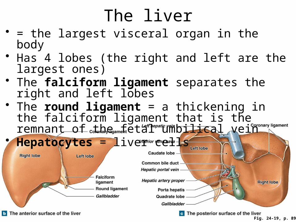
The liver• = the largest visceral organ in the body• Has 4 lobes (the right and left are the largest ones)• The falciform ligament separates the right and left
lobes• The round ligament = a thickening in the falciform
ligament that is the remnant of the fetal umbilical vein• Hepatocytes = liver cells
Fig. 24-19, p. 891
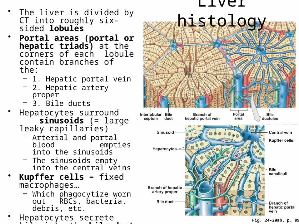
Liver histology
Fig. 24-20ab, p. 892
• The liver is divided by CT into roughly six-sided lobules
• Portal areas (portal or hepatic triads) at the corners of each lobule contain branches of the:– 1. Hepatic portal vein– 2. Hepatic artery proper– 3. Bile ducts
• Hepatocytes surround sinusoids (= large leaky capillaries)– Arterial and portal blood
empties into the sinusoids– The sinusoids empty into the
central veins• Kupffer cells = fixed
macrophages…– Which phagocytize worn out
RBCs, bacteria, debris, etc.• Hepatocytes secrete bile into
the bile duct system (part of which is shown here in green), which carries bile out of and away from the liver

More liver histology
Under the ‘scope!
Fig. 24-20c, p. 892
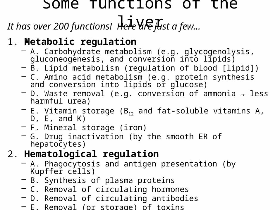
Some functions of the liverIt has over 200 functions! Here are just a few…
1. Metabolic regulation– A. Carbohydrate metabolism (e.g. glycogenolysis, gluconeogenesis,
and conversion into lipids)– B. Lipid metabolism (regulation of blood [lipid])– C. Amino acid metabolism (e.g. protein synthesis and conversion into
lipids or glucose)– D. Waste removal (e.g. conversion of ammonia → less harmful urea)– E. Vitamin storage (B12 and fat-soluble vitamins A, D, E, and K)– F. Mineral storage (iron)– G. Drug inactivation (by the smooth ER of hepatocytes)
2. Hematological regulation– A. Phagocytosis and antigen presentation (by Kupffer cells)– B. Synthesis of plasma proteins– C. Removal of circulating hormones– D. Removal of circulating antibodies– E. Removal (or storage) of toxins
3. The synthesis and secretion of bile (see the next slide for the functions of bile)

Bile
• About 1 liter is produced per day by hepatocytes
• Contains: – Mostly water, minor amounts of bile salts (derived from cholesterol),
cholesterol, pigments (bilirubin), and ionic buffers• Functions:
– A. The emulsification of fats • Bile salts have hydrophilic and -phobic ends, so they surround large fat
globules and break them into small lipid droplets in a watery environment– This increases the surface area for digestion by lipases, and aids absorption
– B. The excretion of bilirubin
• Remember, bilirubin is a pigment derived from the heme of hemoglobin• Bilirubin (liver) → (small intestine to large intestine) → urobilinogens and
stercobilinogens → urobilins and stercobilins (excreted in feces)
• Note: > 90% of the bile salts in bile are reabsorbed by the ileum and cecum for recycling back to the liver
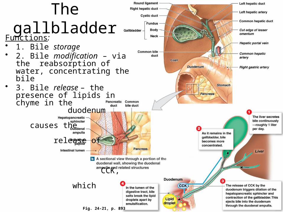
The gallbladder
Fig. 24-21, p. 893
Functions:• 1. Bile storage• 2. Bile modification – via the
reabsorption of water, concentrating the bile
• 3. Bile release – the presence of lipids in chyme in the duodenum causes the release of CCK, which causes the hepato- pancreatic sphincter (sphincter of Oddi) to relax and the gallbladder to contract
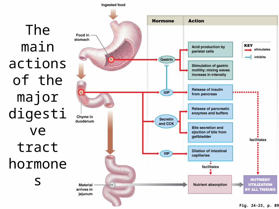
Fig. 24-23, p. 897
The main actions of the major digestive
tract hormones
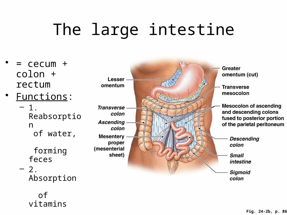
The large intestine
• = cecum + colon + rectum
• Functions:– 1. Reabsorption
of water, forming feces
– 2. Absorption of vitamins produced by bacteria (e.g. vitamin K)
– 3. Storage of feces prior to defecation
Fig. 24-2b, p. 866

Fig. 24-24a, p. 899
The large intestine
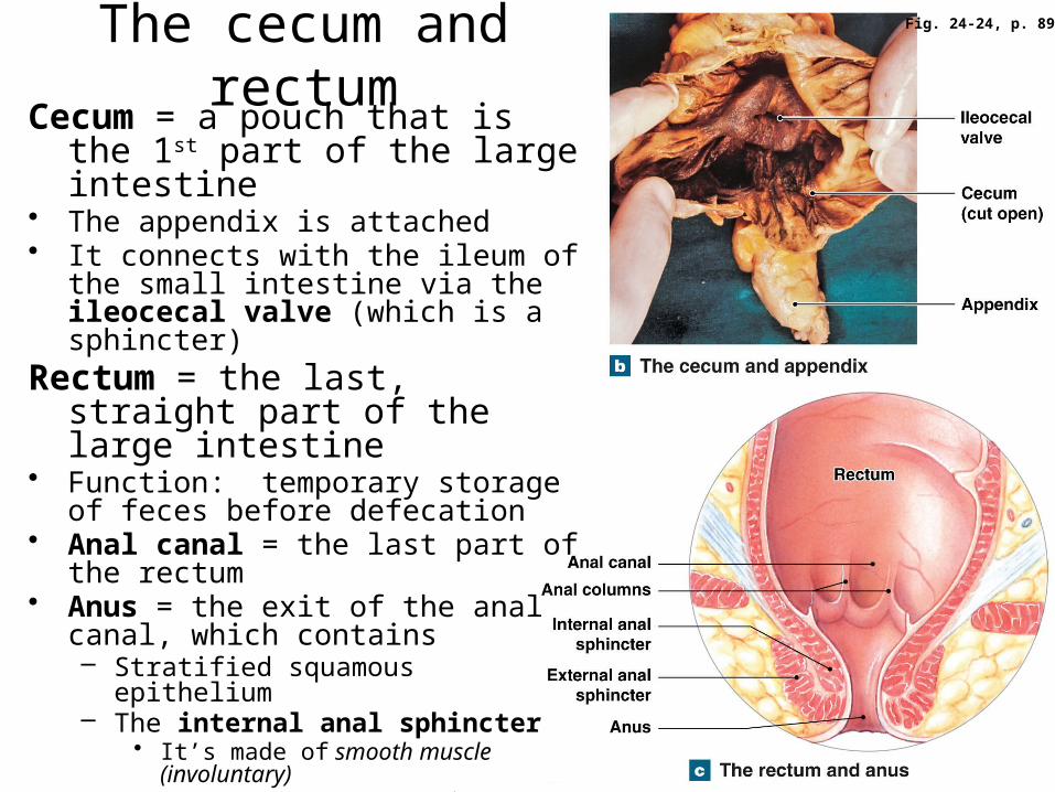
Fig. 24-24, p. 899
The cecum and rectumCecum = a pouch that is the 1st
part of the large intestine• The appendix is attached• It connects with the ileum of the small
intestine via the ileocecal valve (which is a sphincter)
Rectum = the last, straight part of the large intestine
• Function: temporary storage of feces before defecation
• Anal canal = the last part of the rectum
• Anus = the exit of the anal canal, which contains – Stratified squamous epithelium– The internal anal sphincter
• It’s made of smooth muscle (involuntary)
– The external anal sphincter• It’s made of skeletal muscle
(voluntary)

Fig. 24-25, p. 901
Histology of the large intestine• No villi are present, and no digestive enzymes are produced• The mucous (goblet) cells of the intestinal glands (crypts) produce mucus for
lubrication• Lymphoid nodules are present in the mucosa and submucosa• The outer, longitudinal layer of the muscularis externa has been reduced to 3 distinct
bands called taeniae coli– Their muscle tone produces haustra (= pouches)
Physiology of the large intestine• 1. The large intestine is the site of absorption of:
– Water (you lose only ~ 2% of the total water in the GI tract to the feces!)
– Ions (electrolytes)– Vitamins produced by normal resident bacteria (e.g. K,
biotin, and B5)– Bile salts (by the cecum)– Some urobilinogens (to be excreted in the urine)
• 2. Movements of the large intestine include:– A. Peristalsis (which is typically slow)– B. Segmentation movements (= haustral churning)– C. Mass movements = powerful peristalsis a few times a
day• The stimulus = the stretch of the stomach or duodenum• These move feces from the transverse colon onward into the
rectum– D. Defecation (see the next slide)

Fig. 24-26 p. 902
The defecation
reflex
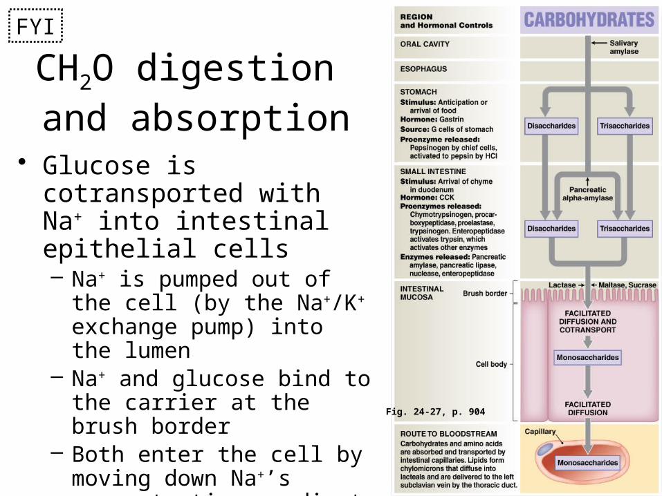
CH2O digestion and absorption
• Glucose is cotransported with Na+ into intestinal epithelial cells– Na+ is pumped out of the cell
(by the Na+/K+ exchange pump) into the lumen
– Na+ and glucose bind to the carrier at the brush border
– Both enter the cell by moving down Na+’s concentration gradient
– The Na+ is pumped back out
Fig. 24-27, p. 904
FYI
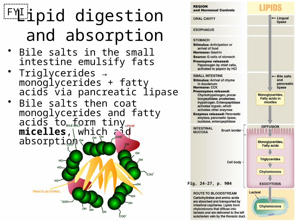
Fig. 24-27, p. 904
Lipid digestion and absorption
• Bile salts in the small intestine emulsify fats
• Triglycerides → monoglycerides + fatty acids via pancreatic lipase
• Bile salts then coat monoglycerides and fatty acids to form tiny micelles, which aid absorption
FYI
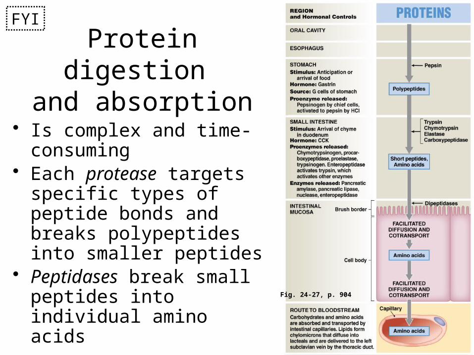
Fig. 24-27, p. 904
Protein digestion and absorption
• Is complex and time-consuming
• Each protease targets specific types of peptide bonds and breaks polypeptides into smaller peptides
• Peptidases break small peptides into individual amino acids
FYI

Fig. 24-28, p. 907
The secretion and absorption of water• Total water ingested +
secretions = 9200 mL• Water lost in feces = 150
mL• Most absorption of water
occurs in the small intestine• Water is absorbed
osmotically following the absorption of solutes (nutrients and ions)– See Table 24-2 for more FYI
info on the absorption of vitamins and ions
FYI