fig. 1a fig. 1b - guerbet · fig. 1a fig. 1b fig. 1c fig. 1d ... the tumor. one of the most...
TRANSCRIPT
2010-6
Fig. 1bFig. 1a
Fig. 1dFig. 1c
94326_RD Codali_2010-2_06.indd 294326_RD Codali_2010-2_06.indd 2 17/02/11 15:0517/02/11 15:05

2010-6
Clinical history
A 47-year-old male consulted the emergency department in our hospital with a history of short standing but aggravating peri-umbilical pain. In the past week he had no stools, a loss of appetite combined with episodes of nausea and vomitus. Clinical investigation showed diffuse abdominal tenderness and a distended abdomen. A contrast-enhanced CT scan of the abdomen was performed (Fig. 1). During the further clinical work-up an intra-abdominal E. coli sepsis was diagnosed.
Imaging fi ndings
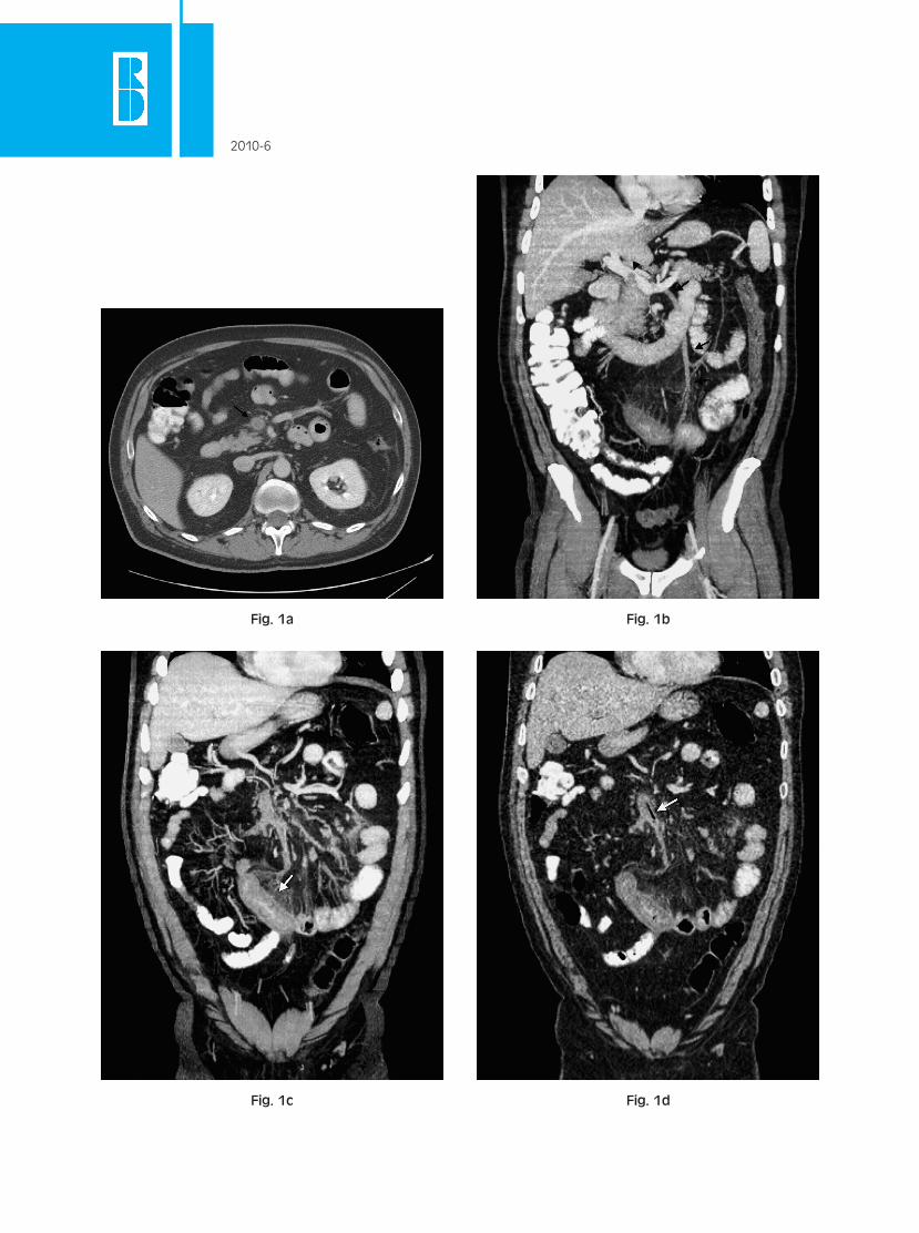
Figure 1: Contrast-enhanced CT scan of the abdomen.Fig. 1a: Axial image of the abdomen at the level of the superior mesenteric vein. Engorgement of the mesenteric vein with a central endoluminal fi lling defect (black arrow). Mesenteric edema. Rim enhancing of the wall of the vein. Fig. 1b: Coronal Maximum Intensity Projection image (MIP). Filling defect in de portal vein (black arrowhead). No contrast fi lling of the superior mesenteric vein (black arrows). Mesenteric edema.Fig. 1c: Coronal MIP image. Bowel wall thickening of more than 3mm (white arrow), poor enhancement of the bowel wall. Fig. 1d: Coronal MIP image. Gas in the mesenteric vein (white arrow).
Blom R.M., Bracke P., Brusselaers H., Degryse H.Department of Medical Imaging, AZ KLINA, Augustijnslei 100, 2930 Brasschaat, [email protected]
94326_RD Codali_2010-2_06.indd 394326_RD Codali_2010-2_06.indd 3 17/02/11 15:0517/02/11 15:05

2010-6
Based on the imaging fi ndings the diagnosis of thrombosis of the superior mesenteric vein and partial thrombosis of the portal vein, with subsequent venous infarction of the adjacent small bowel was made.
Comment
Mesenteric venous thrombosis is an uncommon but potentially lethal cause of bowel ischemia. In com-parison with arterial occlusive disease, which is much more frequent, venous occlusive disease makes up a much smaller percentage (10-15%) of mesenteric ischemia. Venous ischemia is more frequent in younger patients, whereas arterial ischemia is more frequent in the elderly.The non-specifi c clinical signs and symptoms of mesenteric vascular disease delay the diagnosis and contribute to the high mortality and morbidity rates. Because of these high rates and the high sensitivity of contrast-enhanced CT investigations, there is an important role for the radiologist in the diagnosis of this entity.The risk of acute mesenteric venous thrombosis increases in patients with hypercoagulable states. Other pre-existing conditions or risk factors include; visceral infection, portal hypertension, perforated viscus, blunt abdominal trauma, previous abdominal surgery, pancreatitis, smoking and/or use oral contracep-tives. Malignancy may cause thrombosis because of a hypercoagulable state or by direct extension of the tumor. One of the most frequent causes, well illustrated in our case, is intra-abdominal sepsis.No underlying cause is found in 25-50% of patients diagnosed with mesenteric venous thrombosis. Contrast-enhanced CT scan is the preferred examination technique in case of suspected mesenteric thrombosis, because it permits the combined evaluation of the vascular structures, the bowel wall as well as the adjacent mesentery. Sensitivity rates for contrast-enhanced CT reach at least 90%. CT fi ndings of mesenteric venous thrombosis include, well-defi ned endoluminal fi lling defects of low attenuation in contrast with well-defi ned, rim enhancing venous walls. Collateral circulation, engorgement of the mesenteric veins and mesenteric edema may be present. Associated symptoms of bowel ischemia may be present and include a bowel wall thickening (> 3mm) as a result of submucosal edema. The thickened wall may appear hyperattenuating due to the venous engorgement. In an advanced stadium of bowel ischemia intestinal pneumatosis may be present. Less commonly in the advanced stadia mesenterial or portal gas can be seen. Management starts with the treatment of the underlying cause and systemic administration of throm-bolytic drugs. Acute venous thrombosis has a mortality rate of 30% and a recurrence risk of 25% without a proper anticoagulant therapy.
Key words
Mesenteric venous thrombosis – venous ischemia
References
1. Rhee RY, Gloviczki P, Mendonca CT, et al. Mesenteric venous thrombosis: still a lethal disease in the 1990s. J Vasc Surg 1994; 20: 688-697.
2. Abdu RA, Zakhour BJ, Dallis DJ. Mesenteric venous thrombosis 1911 to 1984. Surgery 1987; 101: 383-388.
3. Bradbury MS, Kavanagh PV, Bechtold RE, et al. Mesenteric venous thrombosis: diagnosis and nonin-vasive imaging. Radiographics 2002; 22: 527-541.
4. Alvi AR, Khan S, Niazi SK, et al. Acute mesenteric venous thrombosis: improved outcome with early diagnosis and prompt anticoagulation therapy. Int J Surg 2009; 7: 210-213.
Blom R.M., Bracke P., Brusselaers H., Degryse H.Department of Medical Imaging, AZ KLINA, Augustijnslei 100, 2930 Brasschaat, [email protected]
94326_RD Codali_2010-2_06.indd 494326_RD Codali_2010-2_06.indd 4 17/02/11 15:0517/02/11 15:05